EP2618753B1 - Fusionskäfig mit einem kombinierten biologischen abgabesystem - Google Patents
Fusionskäfig mit einem kombinierten biologischen abgabesystem Download PDFInfo
- Publication number
- EP2618753B1 EP2618753B1 EP11827323.4A EP11827323A EP2618753B1 EP 2618753 B1 EP2618753 B1 EP 2618753B1 EP 11827323 A EP11827323 A EP 11827323A EP 2618753 B1 EP2618753 B1 EP 2618753B1
- Authority
- EP
- European Patent Office
- Prior art keywords
- hollow tube
- bone graft
- plunger
- distal end
- fusion cage
- Prior art date
- Legal status (The legal status is an assumption and is not a legal conclusion. Google has not performed a legal analysis and makes no representation as to the accuracy of the status listed.)
- Active
Links
- 230000004927 fusion Effects 0.000 title claims description 334
- 210000000988 bone and bone Anatomy 0.000 claims description 378
- 239000000463 material Substances 0.000 claims description 220
- 239000007943 implant Substances 0.000 claims description 66
- 238000003780 insertion Methods 0.000 claims description 46
- 230000037431 insertion Effects 0.000 claims description 46
- 208000014674 injury Diseases 0.000 claims description 15
- 230000006378 damage Effects 0.000 claims description 14
- 208000027418 Wounds and injury Diseases 0.000 claims description 10
- 238000004891 communication Methods 0.000 claims description 8
- 230000033001 locomotion Effects 0.000 claims description 8
- 230000000921 morphogenic effect Effects 0.000 claims description 3
- 108090000623 proteins and genes Proteins 0.000 claims description 3
- 102000004169 proteins and genes Human genes 0.000 claims description 3
- 230000004323 axial length Effects 0.000 claims 1
- 238000000034 method Methods 0.000 description 81
- 238000001356 surgical procedure Methods 0.000 description 30
- 238000013461 design Methods 0.000 description 20
- 238000002513 implantation Methods 0.000 description 16
- 239000012620 biological material Substances 0.000 description 11
- 210000004027 cell Anatomy 0.000 description 9
- 210000001519 tissue Anatomy 0.000 description 9
- 230000008901 benefit Effects 0.000 description 8
- 239000007788 liquid Substances 0.000 description 8
- 229910052751 metal Inorganic materials 0.000 description 8
- 239000002184 metal Substances 0.000 description 8
- 239000000560 biocompatible material Substances 0.000 description 7
- 230000008468 bone growth Effects 0.000 description 7
- 230000007246 mechanism Effects 0.000 description 7
- 210000003484 anatomy Anatomy 0.000 description 6
- 238000013459 approach Methods 0.000 description 6
- 238000000151 deposition Methods 0.000 description 6
- 230000000694 effects Effects 0.000 description 6
- 238000002347 injection Methods 0.000 description 6
- 239000007924 injection Substances 0.000 description 6
- 230000006870 function Effects 0.000 description 5
- 230000008569 process Effects 0.000 description 5
- 230000008733 trauma Effects 0.000 description 5
- 208000002193 Pain Diseases 0.000 description 4
- 229910001069 Ti alloy Inorganic materials 0.000 description 4
- 230000001154 acute effect Effects 0.000 description 4
- 210000000577 adipose tissue Anatomy 0.000 description 4
- 230000000903 blocking effect Effects 0.000 description 4
- 230000001054 cortical effect Effects 0.000 description 4
- 230000012010 growth Effects 0.000 description 4
- 238000003306 harvesting Methods 0.000 description 4
- 230000003116 impacting effect Effects 0.000 description 4
- 208000015181 infectious disease Diseases 0.000 description 4
- 238000012986 modification Methods 0.000 description 4
- 230000004048 modification Effects 0.000 description 4
- 210000005036 nerve Anatomy 0.000 description 4
- 230000011164 ossification Effects 0.000 description 4
- 238000012856 packing Methods 0.000 description 4
- 230000000149 penetrating effect Effects 0.000 description 4
- 230000035515 penetration Effects 0.000 description 4
- 238000009832 plasma treatment Methods 0.000 description 4
- 230000001737 promoting effect Effects 0.000 description 4
- 239000002002 slurry Substances 0.000 description 4
- 239000007787 solid Substances 0.000 description 4
- 229910001220 stainless steel Inorganic materials 0.000 description 4
- 239000010935 stainless steel Substances 0.000 description 4
- 229910000838 Al alloy Inorganic materials 0.000 description 3
- 241001455445 Alfaro Species 0.000 description 3
- 229910000599 Cr alloy Inorganic materials 0.000 description 3
- 208000003618 Intervertebral Disc Displacement Diseases 0.000 description 3
- 206010050296 Intervertebral disc protrusion Diseases 0.000 description 3
- 239000004696 Poly ether ether ketone Substances 0.000 description 3
- RTAQQCXQSZGOHL-UHFFFAOYSA-N Titanium Chemical compound [Ti] RTAQQCXQSZGOHL-UHFFFAOYSA-N 0.000 description 3
- 239000000788 chromium alloy Substances 0.000 description 3
- 239000002131 composite material Substances 0.000 description 3
- 230000009977 dual effect Effects 0.000 description 3
- 239000008187 granular material Substances 0.000 description 3
- 238000007443 liposuction Methods 0.000 description 3
- 229910001092 metal group alloy Inorganic materials 0.000 description 3
- 150000002739 metals Chemical class 0.000 description 3
- 230000000399 orthopedic effect Effects 0.000 description 3
- 239000004033 plastic Substances 0.000 description 3
- 229920003023 plastic Polymers 0.000 description 3
- 229920002530 polyetherether ketone Polymers 0.000 description 3
- 229920000642 polymer Polymers 0.000 description 3
- 230000001172 regenerating effect Effects 0.000 description 3
- 230000008439 repair process Effects 0.000 description 3
- 125000006850 spacer group Chemical group 0.000 description 3
- 210000000130 stem cell Anatomy 0.000 description 3
- 238000003860 storage Methods 0.000 description 3
- 239000000758 substrate Substances 0.000 description 3
- 239000010936 titanium Substances 0.000 description 3
- 229910052719 titanium Inorganic materials 0.000 description 3
- 239000011345 viscous material Substances 0.000 description 3
- 238000012800 visualization Methods 0.000 description 3
- 239000011800 void material Substances 0.000 description 3
- 102000007350 Bone Morphogenetic Proteins Human genes 0.000 description 2
- 108010007726 Bone Morphogenetic Proteins Proteins 0.000 description 2
- 102100020760 Ferritin heavy chain Human genes 0.000 description 2
- 101001002987 Homo sapiens Ferritin heavy chain Proteins 0.000 description 2
- 206010061246 Intervertebral disc degeneration Diseases 0.000 description 2
- 241001465754 Metazoa Species 0.000 description 2
- 208000020307 Spinal disease Diseases 0.000 description 2
- 210000000683 abdominal cavity Anatomy 0.000 description 2
- 210000004504 adult stem cell Anatomy 0.000 description 2
- 230000004075 alteration Effects 0.000 description 2
- 229940112869 bone morphogenetic protein Drugs 0.000 description 2
- 238000012512 characterization method Methods 0.000 description 2
- 238000006073 displacement reaction Methods 0.000 description 2
- 239000007789 gas Substances 0.000 description 2
- 239000003102 growth factor Substances 0.000 description 2
- 230000035876 healing Effects 0.000 description 2
- 230000001788 irregular Effects 0.000 description 2
- 210000004705 lumbosacral region Anatomy 0.000 description 2
- 238000013508 migration Methods 0.000 description 2
- 210000003205 muscle Anatomy 0.000 description 2
- 210000001640 nerve ending Anatomy 0.000 description 2
- 239000011148 porous material Substances 0.000 description 2
- 238000003825 pressing Methods 0.000 description 2
- 239000000047 product Substances 0.000 description 2
- 230000002035 prolonged effect Effects 0.000 description 2
- 230000004044 response Effects 0.000 description 2
- 210000004872 soft tissue Anatomy 0.000 description 2
- 230000000007 visual effect Effects 0.000 description 2
- 208000008035 Back Pain Diseases 0.000 description 1
- XDTMQSROBMDMFD-UHFFFAOYSA-N C1CCCCC1 Chemical compound C1CCCCC1 XDTMQSROBMDMFD-UHFFFAOYSA-N 0.000 description 1
- 241001631457 Cannula Species 0.000 description 1
- 229920000049 Carbon (fiber) Polymers 0.000 description 1
- 102000008186 Collagen Human genes 0.000 description 1
- 108010035532 Collagen Proteins 0.000 description 1
- 206010010214 Compression fracture Diseases 0.000 description 1
- 241000282412 Homo Species 0.000 description 1
- 206010023509 Kyphosis Diseases 0.000 description 1
- 229910001182 Mo alloy Inorganic materials 0.000 description 1
- 206010028980 Neoplasm Diseases 0.000 description 1
- 208000020339 Spinal injury Diseases 0.000 description 1
- 208000007103 Spondylolisthesis Diseases 0.000 description 1
- 241000251539 Vertebrata <Metazoa> Species 0.000 description 1
- MTHLBYMFGWSRME-UHFFFAOYSA-N [Cr].[Co].[Mo] Chemical compound [Cr].[Co].[Mo] MTHLBYMFGWSRME-UHFFFAOYSA-N 0.000 description 1
- 210000001015 abdomen Anatomy 0.000 description 1
- 230000003187 abdominal effect Effects 0.000 description 1
- 230000002159 abnormal effect Effects 0.000 description 1
- 229920000122 acrylonitrile butadiene styrene Polymers 0.000 description 1
- 230000003213 activating effect Effects 0.000 description 1
- 230000004913 activation Effects 0.000 description 1
- 238000004458 analytical method Methods 0.000 description 1
- 238000010420 art technique Methods 0.000 description 1
- 230000002146 bilateral effect Effects 0.000 description 1
- 230000000975 bioactive effect Effects 0.000 description 1
- 230000005540 biological transmission Effects 0.000 description 1
- 238000001574 biopsy Methods 0.000 description 1
- 230000015572 biosynthetic process Effects 0.000 description 1
- 230000000740 bleeding effect Effects 0.000 description 1
- 239000002639 bone cement Substances 0.000 description 1
- 239000000316 bone substitute Substances 0.000 description 1
- 210000004556 brain Anatomy 0.000 description 1
- 239000004917 carbon fiber Substances 0.000 description 1
- 239000000919 ceramic Substances 0.000 description 1
- 239000003795 chemical substances by application Substances 0.000 description 1
- 239000011248 coating agent Substances 0.000 description 1
- 238000000576 coating method Methods 0.000 description 1
- 229920001436 collagen Polymers 0.000 description 1
- 230000007850 degeneration Effects 0.000 description 1
- 208000018180 degenerative disc disease Diseases 0.000 description 1
- 238000002716 delivery method Methods 0.000 description 1
- 230000001419 dependent effect Effects 0.000 description 1
- 230000008021 deposition Effects 0.000 description 1
- 238000007599 discharging Methods 0.000 description 1
- 208000037265 diseases, disorders, signs and symptoms Diseases 0.000 description 1
- 208000035475 disorder Diseases 0.000 description 1
- 239000006185 dispersion Substances 0.000 description 1
- 239000003814 drug Substances 0.000 description 1
- 229920001971 elastomer Polymers 0.000 description 1
- 210000001671 embryonic stem cell Anatomy 0.000 description 1
- RTZKZFJDLAIYFH-UHFFFAOYSA-N ether Substances CCOCC RTZKZFJDLAIYFH-UHFFFAOYSA-N 0.000 description 1
- -1 ether ketone ketone Chemical class 0.000 description 1
- 239000002360 explosive Substances 0.000 description 1
- 239000004744 fabric Substances 0.000 description 1
- 239000000835 fiber Substances 0.000 description 1
- 210000002082 fibula Anatomy 0.000 description 1
- 239000000945 filler Substances 0.000 description 1
- 239000010408 film Substances 0.000 description 1
- 230000009969 flowable effect Effects 0.000 description 1
- 239000012530 fluid Substances 0.000 description 1
- 210000001624 hip Anatomy 0.000 description 1
- 230000001976 improved effect Effects 0.000 description 1
- 230000001939 inductive effect Effects 0.000 description 1
- 230000010354 integration Effects 0.000 description 1
- 208000021600 intervertebral disc degenerative disease Diseases 0.000 description 1
- 150000002500 ions Chemical class 0.000 description 1
- 230000002262 irrigation Effects 0.000 description 1
- 238000003973 irrigation Methods 0.000 description 1
- 210000005067 joint tissue Anatomy 0.000 description 1
- 210000003127 knee Anatomy 0.000 description 1
- 239000004816 latex Substances 0.000 description 1
- 229920000126 latex Polymers 0.000 description 1
- 210000004185 liver Anatomy 0.000 description 1
- 230000005389 magnetism Effects 0.000 description 1
- 238000004519 manufacturing process Methods 0.000 description 1
- 239000003550 marker Substances 0.000 description 1
- 230000009347 mechanical transmission Effects 0.000 description 1
- 239000002923 metal particle Substances 0.000 description 1
- VNWKTOKETHGBQD-UHFFFAOYSA-N methane Chemical compound C VNWKTOKETHGBQD-UHFFFAOYSA-N 0.000 description 1
- 230000005012 migration Effects 0.000 description 1
- 238000012978 minimally invasive surgical procedure Methods 0.000 description 1
- 230000000116 mitigating effect Effects 0.000 description 1
- 210000002894 multi-fate stem cell Anatomy 0.000 description 1
- 210000000944 nerve tissue Anatomy 0.000 description 1
- 229920000620 organic polymer Polymers 0.000 description 1
- 239000008188 pellet Substances 0.000 description 1
- 210000004197 pelvis Anatomy 0.000 description 1
- 229920001643 poly(ether ketone) Polymers 0.000 description 1
- 229920001660 poly(etherketone-etherketoneketone) Polymers 0.000 description 1
- 239000004814 polyurethane Substances 0.000 description 1
- 229920002635 polyurethane Polymers 0.000 description 1
- 238000002360 preparation method Methods 0.000 description 1
- 238000002601 radiography Methods 0.000 description 1
- 239000012260 resinous material Substances 0.000 description 1
- 238000012552 review Methods 0.000 description 1
- 239000005060 rubber Substances 0.000 description 1
- 239000004576 sand Substances 0.000 description 1
- 206010039722 scoliosis Diseases 0.000 description 1
- 239000000565 sealant Substances 0.000 description 1
- 210000003491 skin Anatomy 0.000 description 1
- 241000894007 species Species 0.000 description 1
- 239000000126 substance Substances 0.000 description 1
- 239000013589 supplement Substances 0.000 description 1
- 229920003051 synthetic elastomer Polymers 0.000 description 1
- 229920002994 synthetic fiber Polymers 0.000 description 1
- 229920001059 synthetic polymer Polymers 0.000 description 1
- 239000005061 synthetic rubber Substances 0.000 description 1
- 238000002560 therapeutic procedure Methods 0.000 description 1
- 239000010409 thin film Substances 0.000 description 1
- 230000005945 translocation Effects 0.000 description 1
- 239000012780 transparent material Substances 0.000 description 1
- 230000000472 traumatic effect Effects 0.000 description 1
- 238000011179 visual inspection Methods 0.000 description 1
Images


Classifications
-
- A—HUMAN NECESSITIES
- A61—MEDICAL OR VETERINARY SCIENCE; HYGIENE
- A61F—FILTERS IMPLANTABLE INTO BLOOD VESSELS; PROSTHESES; DEVICES PROVIDING PATENCY TO, OR PREVENTING COLLAPSING OF, TUBULAR STRUCTURES OF THE BODY, e.g. STENTS; ORTHOPAEDIC, NURSING OR CONTRACEPTIVE DEVICES; FOMENTATION; TREATMENT OR PROTECTION OF EYES OR EARS; BANDAGES, DRESSINGS OR ABSORBENT PADS; FIRST-AID KITS
- A61F2/00—Filters implantable into blood vessels; Prostheses, i.e. artificial substitutes or replacements for parts of the body; Appliances for connecting them with the body; Devices providing patency to, or preventing collapsing of, tubular structures of the body, e.g. stents
- A61F2/02—Prostheses implantable into the body
- A61F2/30—Joints
- A61F2/44—Joints for the spine, e.g. vertebrae, spinal discs
- A61F2/4455—Joints for the spine, e.g. vertebrae, spinal discs for the fusion of spinal bodies, e.g. intervertebral fusion of adjacent spinal bodies, e.g. fusion cages
- A61F2/447—Joints for the spine, e.g. vertebrae, spinal discs for the fusion of spinal bodies, e.g. intervertebral fusion of adjacent spinal bodies, e.g. fusion cages substantially parallelepipedal, e.g. having a rectangular or trapezoidal cross-section
-
- A—HUMAN NECESSITIES
- A61—MEDICAL OR VETERINARY SCIENCE; HYGIENE
- A61F—FILTERS IMPLANTABLE INTO BLOOD VESSELS; PROSTHESES; DEVICES PROVIDING PATENCY TO, OR PREVENTING COLLAPSING OF, TUBULAR STRUCTURES OF THE BODY, e.g. STENTS; ORTHOPAEDIC, NURSING OR CONTRACEPTIVE DEVICES; FOMENTATION; TREATMENT OR PROTECTION OF EYES OR EARS; BANDAGES, DRESSINGS OR ABSORBENT PADS; FIRST-AID KITS
- A61F2/00—Filters implantable into blood vessels; Prostheses, i.e. artificial substitutes or replacements for parts of the body; Appliances for connecting them with the body; Devices providing patency to, or preventing collapsing of, tubular structures of the body, e.g. stents
- A61F2/02—Prostheses implantable into the body
- A61F2/30—Joints
- A61F2/46—Special tools or methods for implanting or extracting artificial joints, accessories, bone grafts or substitutes, or particular adaptations therefor
- A61F2/4601—Special tools or methods for implanting or extracting artificial joints, accessories, bone grafts or substitutes, or particular adaptations therefor for introducing bone substitute, for implanting bone graft implants or for compacting them in the bone cavity
-
- A—HUMAN NECESSITIES
- A61—MEDICAL OR VETERINARY SCIENCE; HYGIENE
- A61F—FILTERS IMPLANTABLE INTO BLOOD VESSELS; PROSTHESES; DEVICES PROVIDING PATENCY TO, OR PREVENTING COLLAPSING OF, TUBULAR STRUCTURES OF THE BODY, e.g. STENTS; ORTHOPAEDIC, NURSING OR CONTRACEPTIVE DEVICES; FOMENTATION; TREATMENT OR PROTECTION OF EYES OR EARS; BANDAGES, DRESSINGS OR ABSORBENT PADS; FIRST-AID KITS
- A61F2/00—Filters implantable into blood vessels; Prostheses, i.e. artificial substitutes or replacements for parts of the body; Appliances for connecting them with the body; Devices providing patency to, or preventing collapsing of, tubular structures of the body, e.g. stents
- A61F2/02—Prostheses implantable into the body
- A61F2/30—Joints
- A61F2/46—Special tools or methods for implanting or extracting artificial joints, accessories, bone grafts or substitutes, or particular adaptations therefor
- A61F2/4603—Special tools or methods for implanting or extracting artificial joints, accessories, bone grafts or substitutes, or particular adaptations therefor for insertion or extraction of endoprosthetic joints or of accessories thereof
- A61F2/4611—Special tools or methods for implanting or extracting artificial joints, accessories, bone grafts or substitutes, or particular adaptations therefor for insertion or extraction of endoprosthetic joints or of accessories thereof of spinal prostheses
-
- A—HUMAN NECESSITIES
- A61—MEDICAL OR VETERINARY SCIENCE; HYGIENE
- A61F—FILTERS IMPLANTABLE INTO BLOOD VESSELS; PROSTHESES; DEVICES PROVIDING PATENCY TO, OR PREVENTING COLLAPSING OF, TUBULAR STRUCTURES OF THE BODY, e.g. STENTS; ORTHOPAEDIC, NURSING OR CONTRACEPTIVE DEVICES; FOMENTATION; TREATMENT OR PROTECTION OF EYES OR EARS; BANDAGES, DRESSINGS OR ABSORBENT PADS; FIRST-AID KITS
- A61F2/00—Filters implantable into blood vessels; Prostheses, i.e. artificial substitutes or replacements for parts of the body; Appliances for connecting them with the body; Devices providing patency to, or preventing collapsing of, tubular structures of the body, e.g. stents
- A61F2/02—Prostheses implantable into the body
- A61F2/30—Joints
- A61F2002/30001—Additional features of subject-matter classified in A61F2/28, A61F2/30 and subgroups thereof
- A61F2002/30108—Shapes
- A61F2002/3011—Cross-sections or two-dimensional shapes
- A61F2002/30138—Convex polygonal shapes
- A61F2002/30153—Convex polygonal shapes rectangular
-
- A—HUMAN NECESSITIES
- A61—MEDICAL OR VETERINARY SCIENCE; HYGIENE
- A61F—FILTERS IMPLANTABLE INTO BLOOD VESSELS; PROSTHESES; DEVICES PROVIDING PATENCY TO, OR PREVENTING COLLAPSING OF, TUBULAR STRUCTURES OF THE BODY, e.g. STENTS; ORTHOPAEDIC, NURSING OR CONTRACEPTIVE DEVICES; FOMENTATION; TREATMENT OR PROTECTION OF EYES OR EARS; BANDAGES, DRESSINGS OR ABSORBENT PADS; FIRST-AID KITS
- A61F2/00—Filters implantable into blood vessels; Prostheses, i.e. artificial substitutes or replacements for parts of the body; Appliances for connecting them with the body; Devices providing patency to, or preventing collapsing of, tubular structures of the body, e.g. stents
- A61F2/02—Prostheses implantable into the body
- A61F2/30—Joints
- A61F2002/30001—Additional features of subject-matter classified in A61F2/28, A61F2/30 and subgroups thereof
- A61F2002/30316—The prosthesis having different structural features at different locations within the same prosthesis; Connections between prosthetic parts; Special structural features of bone or joint prostheses not otherwise provided for
- A61F2002/30535—Special structural features of bone or joint prostheses not otherwise provided for
- A61F2002/30593—Special structural features of bone or joint prostheses not otherwise provided for hollow
-
- A—HUMAN NECESSITIES
- A61—MEDICAL OR VETERINARY SCIENCE; HYGIENE
- A61F—FILTERS IMPLANTABLE INTO BLOOD VESSELS; PROSTHESES; DEVICES PROVIDING PATENCY TO, OR PREVENTING COLLAPSING OF, TUBULAR STRUCTURES OF THE BODY, e.g. STENTS; ORTHOPAEDIC, NURSING OR CONTRACEPTIVE DEVICES; FOMENTATION; TREATMENT OR PROTECTION OF EYES OR EARS; BANDAGES, DRESSINGS OR ABSORBENT PADS; FIRST-AID KITS
- A61F2/00—Filters implantable into blood vessels; Prostheses, i.e. artificial substitutes or replacements for parts of the body; Appliances for connecting them with the body; Devices providing patency to, or preventing collapsing of, tubular structures of the body, e.g. stents
- A61F2/02—Prostheses implantable into the body
- A61F2/30—Joints
- A61F2002/30001—Additional features of subject-matter classified in A61F2/28, A61F2/30 and subgroups thereof
- A61F2002/30316—The prosthesis having different structural features at different locations within the same prosthesis; Connections between prosthetic parts; Special structural features of bone or joint prostheses not otherwise provided for
- A61F2002/30535—Special structural features of bone or joint prostheses not otherwise provided for
- A61F2002/30601—Special structural features of bone or joint prostheses not otherwise provided for telescopic
-
- A—HUMAN NECESSITIES
- A61—MEDICAL OR VETERINARY SCIENCE; HYGIENE
- A61F—FILTERS IMPLANTABLE INTO BLOOD VESSELS; PROSTHESES; DEVICES PROVIDING PATENCY TO, OR PREVENTING COLLAPSING OF, TUBULAR STRUCTURES OF THE BODY, e.g. STENTS; ORTHOPAEDIC, NURSING OR CONTRACEPTIVE DEVICES; FOMENTATION; TREATMENT OR PROTECTION OF EYES OR EARS; BANDAGES, DRESSINGS OR ABSORBENT PADS; FIRST-AID KITS
- A61F2/00—Filters implantable into blood vessels; Prostheses, i.e. artificial substitutes or replacements for parts of the body; Appliances for connecting them with the body; Devices providing patency to, or preventing collapsing of, tubular structures of the body, e.g. stents
- A61F2/02—Prostheses implantable into the body
- A61F2/30—Joints
- A61F2/30767—Special external or bone-contacting surface, e.g. coating for improving bone ingrowth
- A61F2/30771—Special external or bone-contacting surface, e.g. coating for improving bone ingrowth applied in original prostheses, e.g. holes or grooves
- A61F2002/3082—Grooves
- A61F2002/30827—Plurality of grooves
- A61F2002/30828—Plurality of grooves parallel
-
- A—HUMAN NECESSITIES
- A61—MEDICAL OR VETERINARY SCIENCE; HYGIENE
- A61F—FILTERS IMPLANTABLE INTO BLOOD VESSELS; PROSTHESES; DEVICES PROVIDING PATENCY TO, OR PREVENTING COLLAPSING OF, TUBULAR STRUCTURES OF THE BODY, e.g. STENTS; ORTHOPAEDIC, NURSING OR CONTRACEPTIVE DEVICES; FOMENTATION; TREATMENT OR PROTECTION OF EYES OR EARS; BANDAGES, DRESSINGS OR ABSORBENT PADS; FIRST-AID KITS
- A61F2/00—Filters implantable into blood vessels; Prostheses, i.e. artificial substitutes or replacements for parts of the body; Appliances for connecting them with the body; Devices providing patency to, or preventing collapsing of, tubular structures of the body, e.g. stents
- A61F2/02—Prostheses implantable into the body
- A61F2/30—Joints
- A61F2/46—Special tools or methods for implanting or extracting artificial joints, accessories, bone grafts or substitutes, or particular adaptations therefor
- A61F2/4603—Special tools or methods for implanting or extracting artificial joints, accessories, bone grafts or substitutes, or particular adaptations therefor for insertion or extraction of endoprosthetic joints or of accessories thereof
- A61F2002/4625—Special tools or methods for implanting or extracting artificial joints, accessories, bone grafts or substitutes, or particular adaptations therefor for insertion or extraction of endoprosthetic joints or of accessories thereof with relative movement between parts of the instrument during use
- A61F2002/4627—Special tools or methods for implanting or extracting artificial joints, accessories, bone grafts or substitutes, or particular adaptations therefor for insertion or extraction of endoprosthetic joints or of accessories thereof with relative movement between parts of the instrument during use with linear motion along or rotating motion about the instrument axis or the implantation direction, e.g. telescopic, along a guiding rod, screwing inside the instrument
Definitions
- This disclosure relates to orthopedic surgery, and more specifically to an apparatus and method for near-simultaneous and integrated delivery of bone graft material during the placement of surgical cages or other medical implants in a patient's spine.
- Spinal fusion is a process by which two or more of the vertebrae that make up the spinal column are fused together with bone grafts and internal devices (such as rods) that heal into a single solid bone. Spinal fusion can eliminate unnatural motion between the vertebrae and, in turn, reduce pressure on nerve endings.
- spinal fusion can be used to treat, for example, injuries to spinal vertebrae caused by trauma; protrusion and degeneration of the cushioning disc between vertebrae (sometimes called slipped disc or herniated disc); abnormal curvatures (such as scoliosis or kyphosis); and weak or unstable spine caused by infections or tumors.
- lumbar fusion there are five main types of lumbar fusion, including: posterior lumbar fusion ("PLF”), posterior lumbar interbody fusion (“PLIF”), anterior lumbar interbody fusion (“ALIF”), circumferential 360 fusion, and transforaminal lumbar interbody fusion (“TLIF”).
- PLF posterior lumbar fusion
- PLIF posterior lumbar interbody fusion
- ALIF anterior lumbar interbody fusion
- TLIF transforaminal lumbar interbody fusion
- Some disadvantages of traditional methods of spinal surgery include, for example, the pain associated with the procedure, the length of the procedure, the complexity of implements used to carry out the procedure, the prolonged hospitalization required to manage pain, the risk of infection due to the invasive nature of the procedure, and the possible requirement of a second procedure to harvest autograft bone from the iliac crest or other suitable site on the patient for generating the required quantity of cancellous and/or cortical bone.
- Bone graft typically includes crushed bone (cancellous and cortical), or a combination of these (and/or other natural materials), and may further comprise synthetic biocompatible materials. Bone graft of this type is intended to stimulate growth of healthy bone.
- bone graft shall mean materials made up entirely of natural materials, entirely of synthetic biocompatible materials, or any combination of these materials. Bone graft often is provided by the supplier in a gel or slurry form, as opposed to a dry or granule form. Many companies provide various forms of bone graft in varying degrees of liquidity and viscosity, which may cause problems in certain prior art delivery devices in both prepackaged or packaged by the surgeon embodiments. In addition, the method of delivery of bone graft to a particular location varies depending on the form of the bone graft utilized.
- Autogenous bone bone from the patient
- allograft bone bone from another individual
- small pieces of bone are placed into the space between the vertebrae to be fused.
- larger solid pieces of bone are used to provide immediate structural support.
- Autogenous bone is generally considered superior at promoting fusion.
- this procedure requires extra surgery to remove bone from another area of the patient's body such as the pelvis or fibula.
- allograft bone and other bone graft substitutes although eliminating the need for a second surgery, have drawbacks in that they have yet to be proven as cost effective and efficacious substitutes for autogenous bone fusion.
- BMPs bone morphogenic proteins
- Another alternative is the use of a genetically engineered version of a naturally occurring bone growth factor.
- This approach also has limitations. Specifically, surgeons have expressed concerns that genetically engineered BMPs can dramatically speed the growth of cancerous cells or cause non-cancerous cells to become more sinister. Another concern is unwanted bone creation. There is a chance that bone generated by genetically engineered BMPs could form over the delicate nerve endings in the spine or, worse, somewhere else in the body.
- Regenerative medicine which harnesses the ability of regenerative cells, e.g., stem cells (i.e., the unspecialized master cells of the body) to renew themselves indefinitely and develop into mature specialized cells, may be a means of circumventing the limitations of the prior-art techniques.
- stem cells i.e., both embryonic and adult stem cells
- adipose tissue has been shown to be a source of adult stem cells (See e.g. Zuk, Patricia Z.
- Adipose tissue (unlike marrow, skin, muscle, liver and brain) is comparably easy to harvest in relatively large amounts with low morbidity (See e.g. Commons, G.W., Halperin, B., and Chang, C.C. (2001) "Large-volume liposuction: a review of 631 consecutive cases over 12 years” Plast.
- the devices for inserting bone graft material are applied within a cannula inserted or placed in the surgical area, further limiting the size and/or profile of the bone graft insertion device.
- some traditional devices such as the large barrel/plunger designs and/or the chalking gun designs simply cannot be used as they cannot be inserted within the cannula.
- Traditional devices for inserting bone graft deliver the bone graft material at the bottom of the delivery device along the device's longitudinal axis. Such a delivery method causes the bone grafting material to become impacted at the bottom of the delivery device, and promotes risk of rupture of the surgical area by penetrating the annulus and entering the abdominal cavity. Further, traditional devices that deliver bone graft material along their longitudinal axis may cause rupture of the surgical area or harm to the patient because of the ensuing pressure imparted by the ejected bone graft material from the longitudinal axis of the device.
- the method of delivery of bone graft to a particular location varies depending on the form of the bone graft utilized.
- various dispensing devices have been developed having applicators designed to accommodate this type of bone graft.
- One such device is disclosed by U.S. Pat. No. 5,925,051 issued to Mikhail on Jul. 20, 1999 (“Mikhail”).
- Mikhail provides a caulking gun type dispenser for introducing bone graft in an enlarged bone (e.g. femoral) cavity.
- the device preferably includes a barrel pre-loaded with bone graft and a cannulated ejector positioned over a multi-section guide wire.
- Mikhail is designed solely for use with slurry-type bone graft, and does not accommodate bone graft in granule form, which often varies in size among granules and does not have the same "flow" or viscosity characteristics as slurry-type bone graft. Thus, the applicator of Mikhail is insufficient for introducing most bone graft to a surgical site in a patient.
- U.S. Pat. No. 6,019,765 issued to Thornhill et al. on Feb. 1, 2000 also teaches a bone graft delivery device.
- the bone graft device applicator of Thornhill is used to apply bone graft to an artificial joint without having to remove a previously implanted prosthesis component.
- the applicator device includes a hollow tube with an actuation mechanism for discharging the bone graft from the device via a nozzle coupled to a distal end of the tube.
- the bone graft delivery device of Thornhill may include various components for loading the device with the bone graft, and may further include a plurality of nozzles each having a geometry suited for a particular application.
- the Thornhill delivery device is designed for use with bone slurry, and requires much custom instrumentation and different sized parts to achieve success in many bone graft delivery applications, which in turn increases the time to assemble and use the delivery device and may create further problems during the surgical operation.
- U.S. Pat. No. 5,697,932 issued to Smith et al. on Dec. 16, 1997 discloses yet another bone graft delivery system and method.
- Smith a hollow tube of pre-loaded bone graft and a plunger are used to facilitate delivery of the bone graft to a bone graft receiving area.
- a positioning structure is provided on the plunger to maintain the plunger in a desirable position with respect to the hollow tube.
- Adjunct positioning means may also be provided to ensure that the plunger remains in the desirable position during the packing of bone graft into the bone graft receiving area.
- the device disclosed by Smith is clearly designed solely for slurry type bone graft, and does not provide an effective opening for receiving the desired amount of bone graft.
- the hollow tube shown by Smith is narrow and does not have a footing or other apparatus associated with the delivery device for preventing the device from penetrating, for example, the abdominal region of a patient, which may occur during tamping or packing of the bone graft. This in turn may cause serious injury to a patient if not controlled, and for these reasons the device of Smith is also insufficient for delivery of bone graft to a surgical site.
- the barrel includes an exit port through which bone graft material placed into the barrel exits the barrel.
- a plunger member is disposed coaxially within the barrel member for pushing the bone graft material toward the exit port in the barrel.
- a mechanism is provided for advancing the plunger member within the barrel member.
- the plunger member can be rotatable and include external threads that are complimentary to and engaged with the internal threads in the barrel member to assist in moving the bone graft material toward the exit port in the barrel.
- US 20060264964 A1 discloses a graft syringe assembly for delivering bone graft material is disclosed.
- the graft syringe assembly comprises a syringe subassembly including a syringe barrel having an inner chamber adapted for receiving bone graft material, a plunger adapted for expelling bone graft material from the inner chamber, the plunger slidably received within the inner chamber, and a syringe adapter coupled to the syringe barrel.
- the graft syringe assembly further comprises a connection subassembly coupled to the syringe adapter, and a delivery tube subassembly coupled to the connection subassembly, wherein the connection subassembly is configured to allow the delivery tube subassembly to rotate relative to the syringe subassembly.
- US 20100087828 A1 discloses a curable material delivery cannula device and method are disclosed.
- the device includes a cannula and a hub.
- the cannula includes an open proximal end, a deflectable segment forming a pre-set curve, a lumen, and side orifice(s) adjacent, and proximally spaced from, the distal end and fluidly connected to the lumen.
- the deflectable segment straightens.
- the deflectable segment reverts to the curved shape, which may be used to create a void in the bone for receiving curable material.
- the distal end has a blunt tip for non-traumatic interface with bodily material.
- curable material such as bone cement, is delivered from the side orifice(s) in a radial direction relative to the lumen.
- US 20090264892 A1 discloses a a method of treating a vertebra.
- the method comprises the steps of accessing an interior of a vertebra and introducing a sufficient amount of artificial biocompatible material which does not set to a hardened condition in storage, into said bone, with sufficient force to move apart fractured portions of said bone.
- US 20070185496 A1 discloses an injection system for injection of a relatively high viscous material into hard tissue surrounded by soft tissue in an animal body.
- the injection system comprising an elongated tubular member having a proximal section defining a syringe chamber and a distal section defining a cannula.
- the cannula having an inside diameter and an outside diameter and a length sufficient for insertion into the hard tissue.
- the proximal section defining the syringe chamber having an inside diameter greater than the inside diameter of the cannula, the proximal section adapted to be inserted in the soft tissue.
- the cannula having a bore communicating with the syringe chamber.
- the injection system comprising a syringe fitted for insertion within the syringe chamber of the proximal section.
- the syringe having an open end for communicating with the bore of the cannula.
- the syringe adapted for containing the high viscous material to be injected through the open end and the bore into the hard tissue respectively whereby the injection of the high viscous material is facilitated.
- US 20090275995 A1 discloses medical devices for treating osteoplasty procedures such as vertebral compression fractures. More particularly, embodiments of this relate to instruments and methods for controllably restoring vertebral body height by controlling the geometry of fill material introduced into cancellous bone.
- a method of treating bone includes injecting a volume of fill material into a bone and selectively modifying a viscosity of the bone filler to control the direction of flow of the fill material within the bone.
- a system for treating bone using this method includes an introducer for delivering fill material into the bone and an energy source selectively coupleable to the fill material to alter the viscosity of the fill material via an energy emitter in the introducer.
- US 20020049448 A1 discloses an assembly for injecting flowable material into bone, comprising a tubular body including an interior bore to carry a material flow, the tubular body having a longitudinal axis and including a dispensing end, an opening in the dispensing end communicating with the bore to dispense the material flow, a plunger located at least partially within the tubular body, the plunger adapted to be displaced along the longitudinal axis of the tubular body, an advancement mechanism attached to the plunger, whereby the advancement mechanism displaces the plunger a first longitudinal displacement in response to a first delivery impulse, and a second longitudinal displacement in response to a second delivery impulse.
- Traditional devices for inserting a fusion cage or other medical implants into a patient's spine or other surgical area are distinct and separate from traditional devices that deliver bone graft material to the surgical site. For example, once an implant has been positioned, then bone growth material is packed into the internal cavity of the fusion cage. Also, sometimes the process is reversed, i.e., the bone growth is inserted first, and then the implant. These bone growth inducing substances come into immediate contact with the bone from the vertebral bone structures which project into the internal cavity through the apertures.
- Two devices are thus traditionally used to insert bone graft material into a patient's spine and to position and insert a fusion cage. These devices thus necessitate a disc space preparation followed by introduction of the biologic materials necessary to induce fusion and, in a separate step, application of a structural interbody fusion cage.
- the problems associated with separate administration of the biologic material bone graft material and the insertion of a fusion cage include applying the graft material in the path of the cage, restricting and limiting the biologic material dispersed within the disk space, and requiring that the fusion cage be pushed back into the same place that the fusion material delivery device was, which can lead to additional trauma to the delicate nerve structures.
- Fusion cages provide a space for inserting a bone graft between adjacent portions of bone.
- Such cages are often made of titanium and are hollow, threaded, and porous in order to allow a bone graft contained within the interior of the cage of grow through the cage into adjacent vertebral bodies.
- Such cages are used to treat a variety of spinal disorders, including degenerative disc diseases such as Grade I or II spondylolistheses of the lumbar spine.
- Surgically implantable intervertebral fusion cages are well known in the art and have been actively used to perform spinal fusion procedures for many years. Their use became popularized during the mid 1990's with the introduction of the BAK Device from the Zimmer Inc., a specific intervertebral fusion cage that has been implanted worldwide more than any other intervertebral fusion cage system.
- the BAK system is a fenestrated, threaded, cylindrical, titanium alloy device that is capable of being implanted into a patient as described above through an anterior or posterior approach, and is indicated for cervical and lumbar spinal surgery.
- the BAK system typifies a spinal fusion cage in that it is a highly fenestrated, hollow structure that will fit between two vertebrae at the location of the intervertebral disc.
- Spinal fusion cages may be placed in front of the spine, a procedure known as anterior lumbar interbody fusion, or ALIF, or placed in back of the spine.
- the cages are generally inserted through a traditional open operation, though laparoscopic or percutaneous insertion techniques may also be used.
- Cages may also be placed through a posterior lumbar interbody fusion, or PLIF, technique, involving placement of the cage through a midline incision in the back.
- a typical procedure for inserting a common threaded and impacted fusion cage is as follows. First, the disc space between two vertebrae of the lumbar spine is opened using a wedge or other device on a first side of the vertebrae. The disk space is then prepared to receive a fusion cage. Conventionally, a threaded cage is inserted into the bore and the wedge is removed. A disk space at the first side of the vertebrae is then prepared, and a second threaded fusion cage inserted into the bore. Alternatively, the disk space between adjacent vertebrae may simply be cleared and a cage inserted therein. Often, only one cage is inserted obliquely into the disk space. Use of a threaded cage may be foregone in favor of a rectangular or pellet-shaped cage that is simply inserted into the disk space. Lastly, bone graft material may be inserted into the surgical area using separate tools and devices.
- Brantigan discloses a traditional spinal back surgical method involving the implantation of a spinal fusion cage.
- the cage surfaces are shaped to fit within prepared endplates of the vertebrae to integrate the implant with the vertebrae and to provide a permanent load-bearing strut for maintaining the disc space.
- Brantigan teaches that these cages typically consist of a homogeneous nonresorbable material such as carbon-reinforced polymers such as poly ether ether ketone (PEEK) or polyether ketone ether ketone ketone (“PEKEKK”).
- PEEK poly ether ether ketone
- PEKEKK polyether ketone ether ketone ketone
- U.S. Pat. Appl. 20070043442 of Abernathie et al. discloses another traditional spinal back surgical method involving the implantation of a spinal fusion cage.
- Abernathie relates generally to an implantable device for promoting the fusion of adjacent bony structures, and a method of using the same. More specifically, Abernathie relates to an expandable fusion cage that may be inserted into an intervertebral space, and a method of using the same.
- Abernathie includes an aperture in the fusion cage to allow bone growth therethrough, as a separate procedure to the insertion of the fusion cage.
- the cages or plugs are commonly made of an inert metal substrate such as stainless steel, cobalt-chromium-molybdenum alloys, titanium or the like having a porous coating of metal particles of similar substrate metal, preferably titanium or the like as disclosed, for example, in the Robert M. Pilliar U.S. Pat. Nos. 3,855,683 issued Dec. 24, 1974 and 4,206,516 issued June 10, 1980 .
- These plugs may take the form of flat sided cubical or rectangular slabs, cylindrical rods, cruciform blocks, and the like.
- U.S. Pat. No. 5,906,616 issued to Pavlov et al. (“Pavlov”) discloses a fusion cage of various cylindrical and conical shapes and a method of insertion. Like Brantigan, Pavlov teaches the separate process and procedure for the insertion of a fusion cage and the insertion of bone graft.
- U.S. Pat. No. 5,702,449 (“McKay ”) discloses a spinal implant comprising a cage made of a porous biocompatible material reinforced by an outer sleeve made of a second material which is relatively stronger under the compressive load of the spine than the biocompatible material.
- Perez-Cruet discloses an interbody device assembly consisting of a fusion device and an insertion device.
- the insertion device positions the fusion device between two vertebrae, provides bone graft material, and then detaches from the fusion device, leaving the fusion device in place to restore disc space height.
- the Perez-Cruet device is designed to receive bone graft material from its insertion device and distribute the material away from the fusion device.
- a center plate is positioned immediately downstream of the received bone graft material and directs the bone graft to opposing sides of the fusion device. (See, for example, Fig.
- the Perez-Cruet fusion device is unlikely to completely fill the areas near of its fusion cage and deliver bone graft material to the surrounding bone graft site.
- none of the Perez-Cruet fusion device embodiments feature a defined interior space or a cage-style design. Indeed, the Perez-Cruet fusion device explicitly teaches away from a contained-interior, fusion-cage-style device, asserting that its fusion device fills all of the disc space as opposed to a cage design, which contains the bone material.
- the Perez-Cruet does not feature a distal tip that functions to precisely position the fusion device and stabilize the device during delivery of bone graft material.
- U.S. Pat. No. 7,985,256 issued to Grotz et al. discloses an expandable spinal implant for insertion between opposed vertebral end plates.
- the implant is a cylinder block of slave cylinders; a central cavity between the cylinders receives bone graft material and pistons positioned within the cylinders provide a corrective bone engaging surface for expanding against a first vertebral end plate.
- the insertion tool used to place the spinal implant includes a handle and hollow interior for housing hydraulic control lines and a bone graft supply line.
- the Grotz system does not allow precise positioning or delivery of bone graft material without an implant and requires a complex and bulky insertion tool.
- U.S. Pat. Appl. 2010/0198140 to Lawson discloses a tool comprising a cannula with an open slot at the distal end and a closed tip. Lawson's tool employs tamps to push bone aside and open up a void for filling; solid bone pellets are then rammed down the hollow interior of the cannula by a tamper and delivered to the surgical site. Lawson does not allow precise positioning or delivery of viscous bone graft material and has no capability to interconnect or integrate with an implant such as a bone graft fusion cage.
- Alfaro discloses a delivery system for an intervertebral spacer and a bone grafting material comprising a spacer disengagingly attached to a hollow handle.
- the handle comprises a chamber and bone grafting material-advancing means for introducing bone grafting material from the chamber into the spacer and the intervertebral spaces.
- the Alfaro system does not allow precise positioning or delivery of bone graft material through a distal tip that precisely positions the fusion device and stabilizes the device during delivery of bone graft material, and does not allow primarily lateral injection of bone graft fusion material.
- the prior art bone graft delivery devices listed above typically must come pre- loaded with bone graft, or alternatively require constant loading (where permissible) in order to constantly have the desired supply of bone graft available. Moreover, these bone graft delivery devices generally cannot handle particulate bone graft of varying or irregular particulate size. Furthermore, the prior art devices for inserting a fusion cage or other medical implant into a patient's spine or other surgical area are commonly distinct and separate from traditional devices that deliver bone graft material to the surgical site. As such, two devices are traditionally used to insert bone graft material into a patient's spine and to position and insert a fusion cage.
- the problems associated with separate administration of the biologic material bone graft material and the insertion of a fusion cage include applying the graft material in the path of the cage, restricting and limiting the biologic material dispersed within the disk space, and requiring that the fusion cage be pushed back into the same place that the fusion material delivery device was, which can lead to additional trauma to the delicate nerve structures. These problems can be a great inconvenience, cause avoidable trauma to a patient and make these prior art devices unsuitable in many procedures.
- the present invention solves these needs.
- the present invention allows biologic material to flow directly to the fusion cage and be dispersed within the disc space in a single step, and can precisely and simply deliver particulate bone graft of varying or irregular particulate size.
- the present invention allows application of bone graft material through a detachable fusion cage, eliminates otherwise restriction of the volume of biologic material that may be dispersed within the disk space, and eliminates the requirement that the fusion cage be pushed back into the same place that the fusion material delivery device was, which can lead to additional trauma to the delicate nerve structures.
- the present invention relates to bone graft insertion apparatus for near-simultaneous and integrated delivery of bone material during the placement of surgical cages or other medical implants in a patient's spine as claimed hereafter.
- Preferred embodiments are set forth in the dependent claims.
- inventions of the present disclosure relate to an apparatus and method for near-simultaneous and integrated delivery of bone graft material during the placement of surgical cages or other medical implants in a patient's spine.
- the integrated fusion cage and delivery device (the “device") is comprised generally of a tubular member and a plunger for expelling bone graft from the tubular member, through a surgical fusion cage, and into a bone graft receiving area, then disengaging the fusion cage at the surgical site in a human patient.
- the apparatus and method allows the biologic material to flow directly into and through the fusion cage and dispersed within the disc space in a single step, and leave the detachable fusion cage in the surgical area.
- Other embodiments and alternatives to this device are described in greater detail below.
- the present invention is directed to near-simultaneous and integrated delivery of bone graft material during the placement of surgical cages or other medical implants into a patient's spine.
- the delivery of the bone graft material may be to any area of the body, and in particular to the intervetebral joints of the spine, for achieving bone graft fusion.
- the device may be used without the optional near-simultaneous and integrated placement of surgical cages with the delivery of bone graft material.
- the invention may be used in the repair of a bone joint or in connection with the implantation of prosthetic devices in the body, including, by way of example but not limitation, the hip, knee and a variety of spinal joints.
- the present invention may be used in primary surgery, in which a bone graft is being supplied to promote new bone growth or to reconstruct a joint for the first time, as well as in revision surgery, in which a follow-up procedure is being performed in an area that has previously been subject to one or more surgeries.
- the invention may be used in any application where material is to be delivered with precision to a confined area where access is restricted, to include surgical procedures, repair of installed or uninstalled mechanical or electrical devices, and arming or disarming of explosive devices.
- the present invention has applications in the general field of skeletal repair and treatment, with particular application to the treatment of spinal injuries and diseases.
- the principles of the present invention can also find application in other areas, specifically where there is a desire to constrain added fluid material to particular regions.
- the present invention finds application in methods where the objective is to confine added material to predetermined areas of interest and to prohibit the undesired translocation of such material until an operation is complete and/or until a predetermined later time.
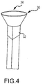
- one aspect of the invention is to provide an integrated fusion cage and graft delivery device that comprises a tubular member, which is substantially hollow or contains at least one inner lumen and that has a generally rectangular cross-sectional shape.
- This generally rectangular cross- sectional shape offers a larger amount of surface area through which bone graft material may be inserted and ejected from the hollow tubular member.
- this generally rectangular shape is more congruent with the size or shape of the annulotomy of most disc spaces, which frequently are accessed by a bone graft delivery device for delivery of bone graft.
- the tool cross-section need not be limited to a generally rectangular shape.
- cross-sections of an oval shape or those with at least one defined angle to include obtruse, acute, and right angles can provide a shape in some situations that is more congruent with the size or shape of the annulotomy of a particular disc space.
- a substantially round shape may also be employed that provides the surgeon with an indication of directional orientation.
- integrated fusion cage spinal fusion implant
- biological implant biological implant
- fusion cage a biological implant
- the hollow tubular member further comprise at least one opening on a lateral face or surface of the hollow tubular member, at one distal end, for ejecting bone graft material into a bone graft receiving area, such as a disc space, such that the bone graft material is ejected from the hollow tubular member through an additional implant, such as a structural cage implant.
- the graft material is dispersed into the area of the debrided disc space surrounding and within the cage.
- the structural cage implant is removably attached to the hollow tubular member so as to be deposited into the surgical area.
- the device may be used in an integrated and near-simultaneous method for depositing bone graft material into a debrided disc space through a structural cage implant and leaving the structural implant.
- one aspect of the invention is to provide an integrated fusion cage detachable component of the integrated fusion cage and graft delivery device that comprises a biological implant that fits over the distal end of the substantial hollow tube, and which has a shape that is substantially congruent with the distal end of the hollow tube.
- the shape and configuration of the integrated fusion cage need not be limited to a generally rectangular shape.
- cross-sections of an oval shape or those with at least one defined angle to include obtruse, acute, and right angles can provide a shape in some situations that is more congruent with the size or shape of the annulotomy of a particular disc space.
- a substantially round shape may also be employed that provides the surgeon with an indication of directional orientation.
- the fusion cage has a tapered tip, and several open channels along the medial surfaces.
- the fusion cage and/or the bone graft delivery portion of the integrated fusion cage and graft delivery device is of oblong or rectangular, or square shape.
- the integrated fusion cage and graft delivery device is designed to avoid blocking or impacting bone graft material into a surgical disc space, thereby limiting the bone graft material that may be delivered, and not allowing available fusion space to be fully exploited for fusion.
- the fusion cage has a keel-shaped tip to separate disk and prevent annular penetration. Also, the fusion cage has dual portals for bone graft discharge, with the medial openings larger than the lateral openings. Further, the fusion cage may be designed in variable heights and lengths so that it fits snugly into the prepared disk space.
- a preferred method of using the integrated fusion cage and graft delivery device comprises precisely inserting the integrated fusion cage and graft delivery device, in one or more of the embodiments contained herein, into the surgical area.
- the integrated fusion cage and graft delivery device is then filled with bone graft material in its one or more substantially hollow tubes, the one or more plungers are inserted into the one or more hollow tubes, and the one or more plunger are pushed into the one or more hollow tubes, guided precisely as enabled by the minimal profile of the device, therein controllably depositing the bone graft material into the surgical area through and into the surgical implant cage.
- the surgical implant device may then be selectably detached from the integrated fusion cage and graft delivery device so as to remain at the surgical site.
- Another method of using the integrated fusion cage and graft delivery device comprises inserting the integrated fusion cage and graft delivery device into a prepared disk space, such that the fusion cage portion fits snugly into the prepared disk space (the fusion cage is designed in variable heights and lengths so as to fit snugly into the prepared disk space), pushing the plunger through the hollow shaft so as to push biological fusion material (e.g. bone graft) through the hollow shaft to flow the biological material through the fusion cage's open lateral and/or medial portals in communication with the hollow tube and plunger, thereby delivering biological material into the prepared disk space, after which the fusion cage is detached from the hollow tube and left in the disk space.
- the fusion cage is left in the disk space with a maximum and/or optimal amount of biological material near-simultaneously delivered within the fusion cage and/or surrounding the fusion cage in the disk space.
- Using the integrated fusion cage and graft delivery device as described overcomes a problem associated with the traditional separate application of bone graft material and insertion of a fusion cage.
- the volume of disk space which does not contain bone graft material is limited, which, for example, limits the effectiveness of the surgical procedure.
- bone graft may be inserted into, for example, a cylindrically-shaped area of radius r to a height h of 8mm, and then a cylindrically-shaped fusion cage inserted of height h of 14mm.
- the surgical area is left without a complete volume of bone graft material, i.e.
- the bone graft area is left without 7ir (14mm - 8mm), or 67ir of bone graft material. (Note that this represents a 75% increase in bone graft material delivered to the surgical site for these example dimensions). This effectively dilutes the bone graft material and reduces its effectiveness.
- the present invention can substantially or completely fill the available disk space with bone graft material, because distraction of the disk space is performed substantially simultaneously with application of the fusion cage. Because more biological material is delivered to the prepared disk space, the fusion rate should increase. Also, by directly implanting fusion material, e.g. bone graft material, though a fusion cage positioned for detachment (and then detached) as a single step, time is saved and there is less manipulation of the sensitive nerve tissue at the fusion site (which increases safety).
- the integrated fusion cage and graft delivery device may be used without the surgical implant delivery device portion such that the method comprises precisely inserting the integrated fusion cage and graft delivery device, in one or more of the embodiments contained herein, into the surgical area that may already contain one or more additional implants, such as a structural cage implant.
- the integrated fusion cage and graft delivery device is then filled with bone graft material in its one or more substantially hollow tubes, the one or more plungers are inserted into the one or more hollow tubes, and the one or more plunger are pushed into the one or more hollow tubes, guided precisely as enabled by the minimal profile of the device, therein controllably depositing the bone graft material into the surgical area without depositing bone graft material into the path of any structural cage implant or other implant that may already be present.
- the integrated fusion cage may be introduced into a spinal target site without the use of the graft delivery device that is through use of any of a variety of suitable surgical instruments having the capability to engage the implant.
- the integrated fusion cage is capable of being used in minimally invasive surgical procedures, needing only a relatively small operative corridor for insertion.
- the integrated fusion cage of the present invention may be used in a variety of configurations in a fusion procedure, including but not limited to (and by way of example only) unilateral, paired unilateral and bilateral.
- the integrated fusion cage and graft delivery device and method of use is applicable to position and deliver fusion cages from the side, directly anterior or in the anterior fusion cages of the cervical spine.
- the integrated fusion cage and graft delivery device comprises a hollow tube or contains at least one inner lumen constructed to receive bone graft, and a plunger adapted for insertion at least partially within the hollow tube and preferably through the full extent of the hollow tube.
- the plunger of some embodiments is generally of the same geometric configuration as the hollow interior potion of the hollow tube so that the plunger, once fully inserted in to the hollow tube, is substantially congruent with the hollow interior portion of the hollow tube, e.g. both the plunger and the hollow tube are substantially the same shape and/or class.
- the plunger preferably extends about the same length as the hollow tube, and further comprises an end portion, e.g. at least one knob or handle for grasping and manipulation by a user, or in robotic or automated or semi-automated control or surgeries, by a machine.
- the hollow interior portion of the hollow tube further comprises a sloped or curved surface at a distal end (e.g. positioned near a place for deposit of bone graft material) adjacent and opposite a lateral window or opening in a lateral face of the hollow tube.
- the plunger also comprises a sloped or curved surface at a distal end of the plunger. The plunger terminates opposite the curved surface at its distal end with a laterally faced surface, which corresponds to the lateral window or opening at the distal end of the hollow tube.
- the distal end of the hollow tube is fitted with a substantially conformal fusion cage that covers the exterior surface of the hollow tube, fitted with one or more openings that align with one or more openings of the hollow tube.
- the plunger may be inserted into the opening of the hollow tube, and extended the entire length of the hollow tube, at least to a point where the laterally faced surface of plunger is in communication with the lateral window or opening at the distal end of the hollow tube.
- This configuration permits a user to eject substantially all of the bone graft material that is placed into the hollow tube in a lateral direction at the bone graft receiving area, through the substantially conformal and detachable fusion cage that covers the exterior surface of the hollow tube, optionally detach the detachable fusion cage, during a surgical procedure.
- the integrated fusion cage and graft delivery device comprises an integrated fusion cage that comprises a first proximal end and a second distal end, wherein the first proximal end contains an opening adapted to allow fitting and/or engagement to the distal end of the hollow tube.
- This fitting and/or engagement may be over the external surface of the hollow tube or inside the interior of the hollow tube.
- the integrated fusion cage may comprise one or more medial openings and one or more lateral openings that align with one or more openings at the distal end of the hollow tube.
- the integrated fusion cage may contain surfaces, such as belts or striations, along one or more medial surfaces of the integrated fusion cage.
- the integrated fusion cage is configured such that when a plunger, once fully inserted in to the hollow tube, is substantially congruent with the hollow interior portion of the hollow tube, e.g. both the plunger and the hollow tube are substantially the same shape and/or class and bone graft material is delivered through the integrated fusion cage into the surgical area.
- the spinal fusion implant of the present invention may be used to provide temporary or permanent fixation along an orthopedic target site.
- the spinal fusion implant of the present invention may be provided with any number of additional features for promoting fusion, such as one or more apertures extending between the top and bottom surfaces which allow a boney bridge to form through the spinal fusion implant.
- the spinal fusion implant may also be provided with any number of suitable anti- migration features to prevent the implant from migrating or moving from the disc space after implantation.
- suitable anti-migration features may include, but are not necessarily limited to, angled teeth or ridges formed along the top and bottom surfaces of the implant and/or rod elements disposed within the distal and/or proximal ends.
- the spinal fusion implant may be provided with one or more radiographic markers at the proximal and/or distal ends. These markers allow for a more detailed visualization of the implant after insertion (through radiography) and allow for a more accurate and effective placement of the implant.
- the distal end of the spinal fusion implant may have a conical (bullet-shaped) shape including a pair of first tapered (angled) surfaces and a pair of second tapered (angled) surfaces.
- the first tapered surfaces extend between the lateral surfaces and the distal end of the implant, and function to distract the vertebrae adjacent to the target intervertebral space during insertion of the spinal fusion implant.
- the second tapered surfaces extend between the top and bottom surfaces and the distal end of the spinal fusion implant, and function to maximize contact with the anterior portion of the cortical ring of each adjacent vertebral body.
- the second tapered surfaces provide for a better fit with the contour of the vertebral body endplates, allowing for a more anterior positioning of the spinal fusion implant and thus advantageous utilization of the cortical rings of the vertebral bodies.
- the integrated fusion cage and graft delivery device comprises a detachable fusion cage that is detachable, or removably attached, by any of several means.
- the fusion cage is substantially conformal with the distal end of the hollow tube in that it covers the exterior surface of the hollow tube, wherein the fusion cage is configured with one or more openings that align with one or more openings of the hollow tube.
- the fusion cage of the integrated fusion cage and graft delivery device forms an interference fit with the fusion cage, such that when the integrated fusion cage and graft delivery device is inserted into the surgical area, the integrated fusion cage and graft delivery device presses against bone and/or vertebrates such that when an axial force is applied to the integrated fusion cage and graft delivery device in a rearward direction (toward the proximal end of the integrated fusion cage and graft delivery device), the fusion cage detaches from the integrated fusion cage and graft delivery device and thereby remains in the surgical area.
- the fusion cage is substantially filled with bone graft material after the fusion cage is implanted. In another embodiment for the integrated fusion cage and graft delivery device and its method of use, the fusion cage is substantially filled with bone graft material simultaneously with the implantation of the fusion cage.
- the fusion cage and/or the bone graft material associated with the fusion cage may be accessed during subsequent surgical operations.
- the fusion cage is a separate device, for example a pre-packaged implant device, which may be installed independently from the integrated fusion cage and graft delivery device or installed in coordination with the integrated fusion cage and graft delivery device. In either situation, the device may be used to provide bone graft material in and/or around the pre-packaged implant.
- some or all of the bone graft material is provided as a component of a per-packaged implant.
- the detachable fusion cage is detachable by way of a indent-tab that penetrates the interior of the hollow tube, such, when the plunger is substantially inserted into the hollow tube, the indent-tab is pushed out from the interior of the hollow tube so as to no longer be attached to the integrated fusion cage and graft delivery device, thereby remaining in the surgical area.
- the hollow tube is of cylindrical shape and includes one or more locking tabs or indent tabs configured to engage one or more locking slots of the fusion cage.
- the locking tabs may permanently or not permanently engage the locking slots, and may be of a shape to include rectangular, circular and oblong.
- the locking tabs and locking slots engage one another by rotating the hollow tube clockwise and are released by counterclockwise rotation.
- the configuration of the locking tabs and locking slots the locking tabs and locking slots engage one another by rotating the hollow tube counterclockwise and are released by clockwise rotation.
- the fusion cage has internal ramps which assist in directing the bone graft material to one or more openings in the fusion cage.
- the detachable fusion cage is detachable by way of receipt of an electrical, mechanical, pneumatic, hydraulic or other communication imparted by the user upon the plunger and/or hollow tube so as to detach the fusion cage and thereby deposit the fusion cage into the surgical area.
- the detachable fusion cage is detachable by way of a Luer taper or Luer fitting connection, such as in a Luer-Lok® or Luer-Slip® configuration or any other Luer taper or Luer fitting connection configuration.
- a Luer taper or Luer fitting connection such as in a Luer-Lok® or Luer-Slip® configuration or any other Luer taper or Luer fitting connection configuration.
- the detachable fusion cage is detachable by way of a pedicle dart by threadable rotation to achieve attachment, detachment, and axial movement.
- Other ways include a quick key insertion, an external snap detent, or magnetic attraction or any other structure.
- U.S. Patent Application is incorporated herein by reference in order to provide an illustrative and enabling disclosure and general description of means to selectably detach the fusion cage of the integrated fusion cage and graft delivery device: U.S. Patent Appl. No. 2009/0187194 to Hamada .
- the detachable fusion cage is detachable by use of magnetism. More specifically, the detachable fusion cage can be made to feature a magnetic field pattern and a resulting force R that are adjustable and may be of different character than the rest of the integrated fusion cage and graft delivery device. With permanent magnets, such adjustments can be made mechanically by orienting various permanent magnet polar geometries and corresponding shapes relative to one another.
- U.S. Pat. No. 5,595,563 to Moisdon describes further background regarding such adjustment techniques, which is hereby incorporated by reference in its entirety.
- electromagnets could be used in combination with permanent magnets to provide adjustability.
- the magnets and corresponding fields and the resultant magnetic field pattern can include both attraction forces from placement of opposite pole types in proximity to one another and repulsion forces from placement of like pole types in proximity to one another.
- “repulsive magnetic force” or “repulsive force” refers to a force resulting from the placement of like magnetic poles in proximity to one another either with or without attractive forces also being present due to opposite magnetic poles being placed in proximity to one another, and further refers to any one of such forces when multiple instances are present.
- U.S. Pat. No. 6,387,096 is cited as a source of additional information concerning repulsive forces that are provided together with attractive magnetic forces, which is hereby incorporated by reference.
- one or more of surfaces of the fusion cage are roughened or otherwise include bone-engaging structures to secure purchase with vertebral surfaces.
- the selectable detachable feature between the detachable fusion cage and the integrated fusion cage and graft delivery device can include one or more tethers, cables, braids, wires, cords, bands, filaments, fibers, and/or sheets; a nonfabric tube comprised of an organic polymer, metal, and/or composite; an accordion or bellows tube type that may or may not include a fabric, filamentous, fibrous, and/or woven structure; a combination of these, or such different arrangement as would occur to one skilled in the art.
- the selectable detachable feature between the detachable fusion cage and the integrated fusion cage and graft delivery device can be arranged to present one or more openings between members or portions, where such openings extend between end portions of the fusion cage.
- U.S. Patent Application is incorporated herein by reference in order to provide an illustrative and enabling disclosure and general description of means to selectably detach the fusion cage of the integrated fusion cage and graft delivery device: U.S. Patent Appl. No. 2011/0015748 to Molz et al.
- the detachable fusion cage is detachable by use of plasma treatment.
- plasma treatment in this context is an ionized gas containing excited species such as ions, radicals, electrons and photons. ( Lunk and Schmid, Contrib. Plasma Phys., 28: 275 (1998 )).
- plasma treatment refers to a protocol in which a surface is modified using a plasma generated from process gases including, but not limited to, O 2 , He, N 2 , Ar and N 2 O. To excite the plasma, energy is applied to the system through electrodes. This power may be alternating current (AC), direct current (DC), radiofrequency (RF), or microwave frequency (MW).
- AC alternating current
- DC direct current
- RF radiofrequency
- MW microwave frequency
- the plasma may be generated in a vacuum or at atmospheric pressure.
- the plasma can also be used to deposit polymeric, ceramic or metallic thin films onto surfaces ( Ratner, Ultrathin Films (by Plasma deposition), 11 Polymeric Materials Encyclopedia 8444-8451, (1996 )).
- Plasma treatment is an effective method to uniformly alter the surface properties of substrates having different or unique size, shape and geometry including but not limited to bone and bone composite materials.
- Plasma Treatment may be employed to effect magnetic properties on elements of the integrated fusion cage and graft delivery device, or to provide selectable detachment of the fusion cage.
- Patent Application is incorporated herein by reference in order to provide an illustrative and enabling disclosure and general description of means to selectably detach the fusion cage of the integrated fusion cage and graft delivery device: U.S. Patent No. 7749555 to Zanella et al.
- the fusion cage may be selectably detachable to the integrated fusion cage and graft delivery device, for example, by means that mechanically grasp the head, means that attach by vacuum, and means that attach by friction, or other means known to those of skill in the art for attaching the head of an apparatus to the shaft of an apparatus.
- the distal end of the integrated fusion cage and graft delivery device be equipped with various other tools to aid in the procedure.
- tools may include, for example, devices used to assess the condition of the implantation site and surrounding tissue. This may include, for example, a device that transmits or provides an image or signal which carries an image for visual inspection and photography.
- an image capture device may include, for example, a device to illuminate the implant site coupled with an image capture and/or transmission device.
- Another tool may also include, for example, a device that aids in irrigation or drainage of the surgical site, a tool used to sample or biopsy tissue.
- Another embodiment for the integrated fusion cage and graft delivery device comprises a hollow tube constructed to receive bone graft, where the hollow tube has a proximal and distal end, a plunger adapted for insertion at least partially within the hollow tube at the proximal end of the hollow tube, whereby the plunger is constructed and arranged with respect to the hollow tube so as to prevent rotation of the plunger during insertion into said hollow tube, whereby the plunger has a distal end that is contoured to an interior surface of the distal end of the hollow tube such that the contoured distal end of the plunger is nearly congruent with the interior surface of the distal end of the hollow tube for removing substantially all of the bone graft received by the hollow tube and whereby the bone graft is delivered to the graft receiving area.
- Still another embodiment provides a rifling structure in the hollow tube interior that facilitates rotational movement of the plunder along a lengthwise axis of the hollow tube, therein delivering a substantially steady pressure and/or rate of delivery of the bone graft material as the plunger descends the hollow tube when the plunger is forced through the hollow tube.
- the rifling or screwlike movement may also translate to a predetermined delivery of material per full rotation, e.g. each 360 degree rotation of the plunger equates to 5 cc of bone graft material delivered to the bone graft site.
- the tube may also be at least portions of biocompatible material which can stay in the canal without impairing the final implantation.
- it may thus be a material that is resorbable, such as a resorbable polymer, in the canal after implantation, so as not to interfere with the growth of the bone or stability of the implant.
- a further embodiment of the invention provides pre-packaged inserts for loading into the hollow tube element, or if there are a plurality of hollow tube elements, into one or more of the hollow tube elements.
- the pre-packaged inserts may be of varying lengths, and/or layered of differing materials or components, to include the patient's own bone graft matter.
- Another embodiment of the present invention provides an integrated fusion cage and graft delivery system, by which a hollow tube and/or a hollow tube/plunger assembly can be prepared prior to opening a patient, thus minimizing the overall impact of the grafting aspect of a surgical implantation or other procedure.
- the hollow tube may be made to be stored with bone graft in it for a period of time, whether the tube is made of plastic, metal or any other material.
- a further embodiment of the present invention provides a bone graft insertion apparatus comprising a hollow tube constructed to receive bone graft, the hollow tube having a proximal and distal end whereby the hollow tube contains least one opening on a surface of the distal end of the hollow tube.
- the at least one opening is preferably positioned other than completely along the axial or longitudinal axis of the device.
- the number and size and shape of such openings can vary but are preferably adapted to deliver bone graft material in a direction substantially transverse to the axial extent of the substantially hollow tube.
- two or more apertures are provided.
- apertures are on the same side of the hollow tube, where in others, apertures are on different sides (e.g. opposing sides) of a hollow tube.
- a plunger adapted for insertion at least partially within the hollow tube, is constructed and arranged with respect to the hollow tube so as to present at least one substantially flat contour, whereby the plunger has a distal end that is contoured to an interior surface of the distal end of the hollow tube such that the contoured distal end of the plunger is nearly congruent with the interior surface of the distal end of the hollow tube.
- This facilitates removing substantially all of the bone graft received by the hollow tube whereby the bone graft is delivered to the graft receiving area. It is important to remove substantially all of the bone graft material as it is expensive and/or difficult to obtain.
- the distal end of the plunger is flexible to allow, for example, the user to maneuver the distal end and thereby any bone graft material in the hollow tube to the implantation site.
- the flexible aspect of certain embodiments can be both passive and active in nature.
- Active flexibility and manipulation in the distal end of the plunger may incorporate, for example, the manipulative capabilities of an endoscope, including components for manipulation such as guidewires along the longitudinal axis of the shaft of the plunger.
- the distal end and the proximal end of the hollow tube are in communication via a conduit to enable electrical, hydraulic, pneumatic, or mechanical transmission, the later such as a wire.
- a conduit to enable electrical, hydraulic, pneumatic, or mechanical transmission, the later such as a wire.
- Such hydraulic communication allows, for example, a medical or other liquid to be delivered or extracted from the surgical area.
- Such mechanical communication allows, for example, the distal end to be maneuvered precisely.
- the hollow tube and/or plunger may be curved and/or may have an angular aspect, in addition to the sloped or curved surface at a distal end of the hollow tube/plunger.
- This shape may, for example, aid the surgeon in more comfortably introducing the delivery device to the implant site, be shaped to better accommodate implantation sites on right or left sides of the body, or be shaped to better accommodate right or left-handed surgeons.
- the delivery device may have multiple angles and curved aspects which enable aspects of embodiments of the present disclosure or aid in ergonomics.
- the device further comprises a footing or shelf at the distal end of the tubular device that is nearest the operating site for preventing or mitigating risk of injury to the patient during surgery.
- the footing may be flexible, semi-flexible, semi-rigid or rigid. The footing serves to protect the anatomy of the patient from being penetrated by the hollow tube of the integrated fusion cage and graft delivery device when the plunger is being inserted or during tamping of the hollow tube or the plunger by the surgeon, which may occur during the surgical procedure for a variety of reasons.
- the distal tip region of the hollow tube comprises a softer, mailable and/or less rigid material than the remainder of the hollow tube.
- the distal tip could be made of a bioactive collagen.
- these detachable devices may include a detachable funnel for gathering and inserting bone graft material at a graspable end of the integrated fusion cage and graft delivery device.
- the present disclosure may also comprise a plunger that has a detachable handle, which may be selectively removed to avoid blocking the surgeon's view of the operating site.
- the integrated fusion cage and graft delivery device may further comprise a detachable footing or shelf at one distal end of the hollow tubular member.
- this footing or shelf is selectively positionable about various points along the hollow tube.
- a distal portion of the hollow tube has a rotatable portion that can be positioned to deliver bone graft material to areas of a disc space in a manner such that a surgeon has angular directional control as to where bone graft material is directed.
- Other detachable elements are also contemplated with the present invention, such as a funnel at the proximal end of the hollow tube, or exterior or interior guide wires attached to the hollow tube, or a camera which is positioned near the delivery site of the bone graft material.
- a method of introducing bone graft material to a desired operating site is provided by use of a hollow tubular member comprising a generally rectangular cross- sectional area, whereby the desired operating area is capable of receiving at least one plunger.
- the one or more plunger having at least one distal end which is designed to accommodate ejecting bone graft or other material to be inserted into the joint space or between intervertebral members in a generally lateral direction, as opposed to a generally longitudinal direction (in relation to the direction of the primary axis of the device).
- distal end of the tubular device need not be limited to those specific embodiments described above.
- Other forms, shapes or designs that enable the foregoing aspects of the present invention are hereby incorporated into this disclosure.
- Forms, shapes and designs that relate to the provision of an end of a tubular device to perform lateral introduction of bone or bone substitute to an operating site are considered to be within the scope of the present disclosure.
- One aspect of the present invention provides an integrated fusion cage and graft delivery device system for delivering bone graft, in a partially formed, fully formed or unformed condition to a bone graft receiving area in a body.
- the hollow tube of the integrated fusion cage and graft delivery device may further comprise a funnel on the graspable end of the hollow tube, which may be selectively positioned about the graspable end of the hollow tube, for facilitating insertion of new or additional bone graft into the hollow tube.
- the funnel may take on a variety of shapes and sizes, depending on the nature of the bone graft material being inserted in the hollow tube.
- the substantially hollow tube provides that the hollow tube is telescoping, thereby allowing its length to be adapted to the particular desires of the surgeon and/or the surgical area.
- the plunger may also be telescoping to substantially conform to the configuration and/or size and/or shape of the substantially hollow tube.
- the size and/or shape of the one or more hollow tubes of the device are sized to fit, and/or not substantially obscure access to the aperture of, the cannula or cannulas that the device is fitted through for delivery of bone graft material to the operating site.
- the device's one or more pair of hollow tubes and plungers do not substantially impair access to the operating site nor the surgeon's view of the operating site.
- a bone graft insertion apparatus comprises: a hollow tube constructed to receive bone graft, said hollow tube having an extended axis and a proximal end and a distal end, said distal end having an interior surface; a plunger adapted for inserting into said proximal end of said hollow tube, said plunger having a distal end being contoured to said interior surface of said distal end of said hollow tube such that bone graft material within said hollow tube is delivered to a graft receiving area through at least one opening near the distal end of said hollow tube; wherein said graft receiving area is configured to accommodate at least one substantially hollow implant.
- a bone graft insertion apparatus comprises: a hollow tube constructed to receive bone graft, said hollow tube having a proximal and distal end; whereby said hollow tube contains least one opening on a surface of said distal end of said hollow tube; whereby said opening on a surface of said distal end of said hollow tube is positioned other than completely along the axial or longitudinal axis of the device; a plunger adapted for insertion at least partially within said hollow tube at proximal end of said hollow tube; whereby said plunger is constructed and arranged with respect to said hollow tube so as to present at least one substantially flat contour; whereby said plunger has a distal end that is contoured to an interior surface of said distal end of said hollow tube such that said contoured distal end of said plunger is nearly congruent with said interior surface of said distal end of said hollow tube for removing substantially all of said bone graft received by said hollow tube; whereby said bone graft is delivered to a graft receiving area.
- a method of inserting bone graft comprises: preparing a surgical area to receive bone graft; inserting a tool into said surgical area, said tool consisting essentially of a hollow tube adapted to receive bone graft, a plunger adapted for insertion into said hollow tube, said plunger constructed to prevent rotation during insertion into said hollow tube, said plunger having a distal end contoured to the interior surface of the distal end of said hollow tube; providing bone graft material into the said hollow tube of said tool; inserting said plunger into the proximal end of said hollow tube; inserting said distal end of said hollow tube of said tool into surgical area; applying force to said plunger thereby advancing said plunger through said hollow tube wherein said bone graft is inserted into said surgical area.
- an integrated fusion cage and graft delivery device apparatus comprises: a hollow tube constructed to receive bone graft, the hollow tube having an extended axis and a proximal end and a distal end, the distal end having an interior surface; a plunger adapted for inserting into the proximal end of the hollow tube, the plunger having a distal end being contoured to the interior surface of the distal end of the hollow tube; a selectably detachable fusion cage, the selectably detachable fusion cage having at least one opening that substantially aligns with at least one opening near the distal end of the hollow tube, such that bone graft material within the hollow tube is delivered to a graft receiving area through at least one opening of the selectably detachable fusion cage; and the selectably detachable fusion cage having a means for detachment whereby the fusion cage is delivered to the graft receiving area.
- embodiments of the present disclosure may have various sizes.
- the sizes of the various elements of embodiments of the present disclosure may be sized based on various factors including, for example, the anatomy of the implant patient, the person or other device operating the apparatus, the implant location, physical features of the implant including, for example, with, length and thickness, and the size of operating site or the size of the surgical tools being used with the device.
- embodiments of the present disclosure may be constructed of materials known to provide, or predictably manufactured to provide the various aspects of the present disclosure. These materials may include, for example, stainless steel, titanium alloy, aluminum alloy, chromium alloy, and other metals or metal alloys. These materials may also include, for example, PEEK, carbon fiber, ABS plastic, polyurethane, rubber, latex, synthetic rubber, and other fiber-encased resinous materials, synthetic materials, polymers, and natural materials.
- the plunger element could be flexible, semi-rigid, or rigid and made of materials such as stainless steel, titanium alloy, aluminum alloy, chromium alloy, and other metals or metal alloys.
- the tubular element could be flexible, semi-rigid, or rigid and made of materials such as stainless steel, titanium alloy, aluminum alloy, chromium alloy, and other metals or metal alloys.
- the plunger and hollow tube are composed of plastic and are intended for one use only and then discarded.
- some or all elements of the device, or portions of some or all of the elements are luminescent.
- some or all elements of the device, or portions of some or all of the elements include lighting elements.
- the hollow tube and/or plunger are made of a substantially transparent material and/or are rigidly opaque.
- embodiments of the present disclosure may be controlled by means other than manual manipulation.
- Embodiments of the present disclosure may be designed and shaped such that the apparatus may be controlled, for example, remotely by an operator, remotely by an operator through a computer controller, by an operator using proportioning devices, programmatically by a computer controller, by servo-controlled mechanisms, by hydraulically-driven mechanisms, by pneumatically-driven mechanisms or by piezoelectric actuators.
- Embodiments of the present disclosure present several advantages over the prior art including, for example, the speed of the procedure, the minimally invasive aspect of the procedure, the ability to introduce the implant material to the implant site with minimal risk and damage to the surrounding tissue, the lower risk of infection, more optimally placed implant material, a more stable delivery device which is designed to reduce the likelihood of the implant material becoming dislodged prior to fixation, and fewer tools in a surgical site due to the integration of several components required to provide bone graft to a bone graft receiving area. Further, the lower profile of the device allows improved viewing of the area intended for receipt of bone graft material, and use of a reduced set and size of elements therein provided a less expensive device.
- the device disclosed provides that substantially all of the bone graft material may be ejected from the device and delivered to the surgical site, rather than wasted as unretrievable matter remaining inside the device.
- the ability to remove substantially all of the bone graft material is of significant benefit because the bone graft material is expensive and/or hard to obtain.
- the present invention relates to a device and method for integrated and near- simultaneous delivery of bone graft material and a fusion cage to any portion of a patient which requires bone graft material and/or a fusion cage.
- the foregoing description of the various embodiments contemplates delivery to, for example, a window cut in a bone, where access to such window for bone grafting is difficult to obtain because of orientation of such window, presence of muscle tissue, risk of injury or infection, etc.
- the integrated fusion cage and graft delivery device is formed such that the one or more hollow tubes and/or plungers may be helpful in selectively and controllably placing bone graft material and a fusion cage in or adjacent to such window.
- the integrated fusion cage and graft delivery device is formed to allow delivery of bone graft material and/or a fusion cage in a direction other than solely along the longitudinal axis of the device, and in some embodiments transverse to the primary axis used by the surgeon or operator of the device when inserting the device into a cannula or other conduit to access the surgical site.
- This same concept applies to other areas of a patient, whether or not a window has been cut in a bone, for example in a vertebral disc space, and may be used whether this is a first surgery to the area or a follow-up surgery.
- the present invention also contemplates the delivery of bone graft material and/or a fusion cage with or without the use of a plunger, and with or without the use of various other tools described in greater detail herein.
- an integrated fusion cage and graft delivery device portion is shown, which is comprised of a hollow tubular member or hollow tube or contains at least one inner lumen 2, which has a first proximate end 6 (which is referred to elsewhere in this specification as the "graspable end” of hollow tube 2), and a second distal end 8, with a general hollow structure therebetween.
- the hollow tube 2 allows bone graft material to be inserted into the opening 4 at the graspable end 6 of the hollow tube 2, and ultimately exited from the hollow tube 2 through the distal end 8.
- the hollow tube 2 also comprises at least one sloped or curved surface 10 at or near the distal end 8 of the hollow tube 2.
- cross-section need not be limited to a generally rectangular shape.
- cross-sections of an oval shape or those with at least one defined angle to include obtruse, acute, and right angles can provide a shape in some situations that is more congruent with the size or shape of the annulotomy of a particular disc space.
- a plunger 12 is shown for use with the hollow tube 2 of Figure 1A .
- the plunger 12 is generally of the same geometry as the hollow potion of the hollow tube 2, extending at least the same length of hollow tube 2.
- the plunger 12 may includes, as depicted in Figure IB at least one knob or handle 16 for grasping by a user of the plunger 12.
- the plunger 12 also comprises at least one sloped or curved surface 20 at or adjacent to a distal end 18 of the plunger 12.
- the plunger 12 terminates in a generally flat, horizontal surface 19, which corresponds to the opening at the distal end 8 of the hollow tube 2 shown in Figure 1A .
- the plunger 12 may be inserted into the opening 4 of the hollow tube 2 shown in Figure 1A , and extended the entire length of the hollow tube 2, at least to a point where the horizontal surface 19 of plunger 12 is in communication with the distal end 8 of the hollow tube 2.
- This configuration permits a user to eject substantially all of the bone graft material that is placed into the hollow tube 2 during a surgical procedure.
- the plunger need not terminate in a generally flat, horizontal surface to effect the substantial removal of all of the bone graft material placed into the hollow tube; more specifically, any shape that allows conformance between the internal contour of the distal end of the hollow tube and the distal end of the plunger will effect the substantial removal of the bone graft material. Further details about the relationship are described below in regard to Figure 2 .
- a contoured leading edge is provided on the plunger to correspond with the internal contour of distal end of the hollow tube of the delivery device.
- This contoured plunger serves several purposes: First, it maintains the plunger in a desirable rotational position with respect to the hollow tube (i.e., prevent the plunger from inadvertently or intentionally being manipulated to rotate about the longitudinal axis of the hollow tube); Second, it ensure that when the plunger is fully inserted, the plunger removes substantially all of the bone graft material from the hollow tube.
- the contoured plunger corresponding to the contoured tubular member, allows immediate identification of the orientation of the device, and more specifically the direction of eject of the bone graft material into the surgical area.
- Alternative positioning means may also be provided to ensure that the plunger remains in the desirable position during delivery of bone graft into the hollow tube, for example by a machined bevel or edge on the outer surface of the plunger, and a corresponding groove in the interior surface of the hollow tube, which must be aligned when inserting the plunger in the hollow tube.
- the distal end 8 of the hollow tube 2 has an opening with a height A and width B according to the needs of the surgeon, the location of the bone graft receiving area, the nature of the surgical operation to be performed, and the quantity and type of bone graft that is being inserted in (and ultimately ejected from) this integrated fusion cage and graft delivery device.
- the height A of the opening at the distal end 8 of the hollow tube 2 is in the range of 4mm to 9mm, and in a most preferred embodiment is about 7mm.
- the width B of the opening at the distal end 8 of the hollow tube 2 is in the range of 7mm to 14mm, and in a most preferred embodiment is about 10mm.
- the distal end 8 of the hollow tube 2 and the distal end 18 of the plunger 12 comprise a curved or sloped surface which extends at least a certain distance laterally away from the generally longitudinal axis of the hollow tube 2/plunger 12, that in other embodiments, the distal end 8 of the hollow tube 2 (and thereby, the distal end 18 of the plunger 12) do not extend a lateral distance away, but rather terminate along the longitudinal wall of the hollow tube 2.
- the hollow tube 2 may have a distal end 8 which has an opening that is carved out of the side of the wall of the hollow tube 2, such that it appears as a window in the tubular body of hollow tube 2.
- the horizontal face 19 of the plunger 12 would also be a face on the outer surface of plunger 12, without extending any lateral distance away from the body of plunger 12. According to this embodiment, the plunger 12 would still retain the curved or sloped surface at the opposite end of the horizontal face 19 (referred to in Figure IB as 20) and similarly the hollow tube 2 would still comprise a sloped or curved surface 10 opposite the opening at distal end 8.
- the plunger 12 shown in Figure IB may further comprise a secondary handle (not shown in Figure IB), which includes an opening about at least one end of secondary handle such that it is permitted to couple with handle 16 of plunger 12.
- the secondary handle may be larger, contain one or more rings or apertures for placing a users hand and/or fingers, or may simply be of a more ergonomic design, for accommodating use of the plunger 12 during a surgical operation.
- the secondary handle is selectively removable, which permits a surgeon to use the secondary handle for inserting the plunger 12, and then at a later point remove the secondary handle, for instance, to improve visibility through the incision or through the hollow tube 2, and/or to determine whether substantially all of the bone graft material has been ejected from the hollow tube 2.
- the plunger 12 is shown inserted into the hollow tube 2, such that the horizontal face 19 is substantially planar with the opening at the distal end 8 of the hollow tube 2.
- the geometry of plunger 12 is such that it fits snuggly or tightly in the interior of the hollow tube 2.
- This configuration is such that the sloped or curved surface 10 of the hollow tube 2 is substantially congruent to the sloped or curved surface 20 (not shown in Figure 2 ), thereby allowing the plunger to be inserted into the hollow tube 2 and allowing substantially all of bone graft material which is placed into the hollow tube 2 to be ejected by the user.
- the hollow tube 2 comprises a footing 11 at the distal end 8 of the hollow tube 2.
- This footing 11 extends in a lateral direction, opposite the direction of the opening at the distal end 8 of the hollow tube 2.
- the purpose of this footing 11 is to prevent injury to the annulus of a patient, or other sensitive anatomy adjacent the bone graft receiving area.
- This footing 11 is helpful when a surgeon or other user of the integrated fusion cage and graft delivery device is using the plunger 12 to drive bone graft through the hollow tube 2, or using another tool, such as a tamp, mallet, or other driving or impacting device to strike the plunger 12 and/or hollow tube 2 during the surgical procedure.
- the hollow tube 2 would have a generally angular distal end 8, which may cause damage to the patient during these types of procedures.
- the footing 11 prevents the distal end 8 of the hollow tube 2 from penetrating the annulus or other sensitive anatomy of the patient.
- the footing 11 may also operate to ensure a fixed position of the distal end 8 of the hollow tube 2 in the surgical site. This in turn allows a user to ensure that bone graft ejecting the distal end 8 of the hollow tube 2 is being ejected laterally, and in the desired direction. This may be important, for example, when the integrated fusion cage and graft delivery device is placed within a disc space, and bone graft is being ejected laterally from the distal end 8 of the hollow tube 2 in a specific direction.
- the footing 11 may also serve as a visual marker for the surgeon, as it extends away from the horizontal wall of the hollow tube 2, and is therefore visible at the distal end 8 of the hollow tube 2. As shown in Figure 3 , the presence of the footing 11 does not affect the interior slope or curved surface 10A of the hollow tube 2, so that the plunger 12 of the design shown in Figure IB may still be used with the hollow tube 2 of this alternate embodiment.
- a removable funnel 30 which comprises an opening 34 which is generally larger in diameter or dimension when compared to the opening 4 of the hollow tube 2.
- This removable funnel 30 further comprises a sleeve 32, the sleeve 32 having an internal cross-section which is substantially congruent with the external cross-section of the proximal end 6 of the hollow tube 2.
- the funnel 30 is selectively removable from the proximal end 6 of the hollow tube 2, and may allow a surgeon to more easily place new or additional bone graft into the hollow tube 2 by way of the opening 34 of the funnel 30.
- This funnel 30 may be used in connection with a hollow tube 2 that has been pre-filled with bone graft, or a hollow tube which is not pre-filled with bone graft.
- the funnel may be selectively positioned on the proximal end 6 of the hollow tube 2 at any point during the surgical operation when the surgeon desires new or additional bone graft be placed in the hollow tube 2 of the integrated fusion cage and graft delivery device.
- the integrated fusion cage and graft delivery device is shown with the embodiment of the hollow tube 2 further comprising a footing 11, and a distal end opening for ejecting bone graft in a generally lateral direction, here in the interior of a disc space 40.
- the disc is shown with an opening on one end for inserting the distal end 8 of the hollow tube 2 of the integrated fusion cage and graft delivery device.
- the present invention comprises a lateral opening, which as shown in Figure 5 allows a surgeon to eject bone mill into the lateral direction and thereby into the opened areas of the disc space 40.
- a surgeon has the option to rotate the direction of the opening in the distal end 8 of the hollow tube 2 for ejecting additional bone graft to other open areas in the disc space 40.
- the surgeon may remove the hollow tube 2 without disturbing the disc or anatomy of the patient.
- the surgeon may also accomplish the delivery of bone graft without displacing any cage or other structural implantable device which may be present in or adjacent the disc space.
- the hollow tube 2 further comprising a footing 11, and a distal end opening for ejecting bone graft in a generally lateral direction, may effect the delivery of bone graft in a lateral direction simultaneous with delivery in a longitudinal direction.
- a integrated fusion cage and graft delivery device portion is shown, comprised of a hollow tubular member 2, which has a first proximate end 6 and a second distal end 8, with a general hollow structure therebetween.
- the generally hollow tube 2 is shown with one of two lateral openings at the distal end 8 of the tubular member 2 viewable (the other is viewable in Figure 7 ).
- the plunger member 12 is shown. The manner of insertion of plunger member 12 into tubular member 2 is also provided.
- the hollow tube 2 allows bone graft material to be inserted into the opening 4 at the proximal end 6 of the hollow tube 2, and ultimately exited from the hollow tube 2 through the second distal end 8 from the lateral openings at the distal end 8 of the hollow tubular member 2.
- a preferred embodiment of the distal end 10 of the tubular member 2 and the distal end 20 of the plunger member 12 is provided.
- the wedge-shaped feature 50 of the distal end 10 of the tubular member 2 and the wedge-shaped end 52 of the distal end 20 of the plunger member 12 is discussed in additional detail with respect to Figures 8 and 9 below.
- the ability to remove substantially all of the bone graft material is an important feature of the invention because bone graft material is traditionally expensive and may require surgery to obtain.
- FIG. 7 a perspective view of a preferred embodiment of the hollow tubular member 2 is provided. Consistent with Figure 6 , the generally hollow tube 2 is shown with one of two lateral openings at the distal end 8 of the tubular member 2 viewable (the other is viewable in Figure 6 ).
- the hollow tube 2 allows bone graft material to be inserted into the opening 4 at the proximal end 6 of the hollow tube 2, and ultimately exited from the hollow tube 2 through the second distal end 8 from the lateral openings at the distal end 8 of the hollow tubular member 2.
- bone graft material is ejected into the surgical area in two lateral directions.
- the openings at the distal end 8 of the hollow tubular member 2 need not be positioned exclusively on one or more lateral sides of the distal end 8 of the tubular member to allow bone graft material to be provided to the surgical site in other than a purely axial or longitudinal direction. Further, one skilled in the art will appreciate that the specific absolute and relative geometries and numbers of lateral openings may vary, for example the distal end 8 of the tubular member 2 may have more than two openings that are of different shape (e.g. oval, rectangular).
- FIG. 8 an elevation view of the wedge-shaped distal end 50 of the tubular member 2 is provided.
- the proximal end of the plunger would conform to the same shape, to allow close fitting of the plunger and the hollow tubular member.
- This contoured plunger serves several purposes: First, it maintains the plunger in a desirable rotational position with respect to the hollow tube (i.e., prevent the plunger from inadvertently or intentionally being manipulated to rotate about the longitudinal axis of the hollow tube); Second, it ensure that when the plunger is fully inserted, the plunger removes substantially all of the bone graft material from the hollow tube.
- the contoured plunger corresponding to the contoured tubular member, allows immediate identification of the orientation of the device, and more specifically the direction of eject of the bone graft material into the surgical area.
- the plunger need not terminate in a wedge-shape surface to effect the substantial removal of all of the bone graft material placed into the hollow tube; more specifically, any shape that allows conformance between the internal contour of the distal end of the hollow tube and the distal end of the plunger will effect the substantial removal of the bone graft material.
- FIG. 9 an elevation view of the opening 4 of the distal end 6 of the hollow tubular member 2 is provided.
- the opening 4 at the proximal end 6 of the hollow tube 2 allows deposit of bone graft material.
- the cross-section of the opening 4 at the proximal end 6 of the hollow tube 2 is generally square.
- a generally square cross-section is depicted, the cross- section need not be limited to a generally square shape.
- cross-sections of an oval shape or those with at least one defined angle to include obtruse, acute, and right angles can provide a shape in some situations that is more congruent with the size or shape of the annulotomy of a particular disc space.
- FIGS 10A-D sequential elevation views of the square-shaped embodiment of the integrated fusion cage and graft delivery device 1 are provided, depicting the complete insertion of the plunger 12 into the hollow tubular member 2.
- the wedge-shaped distal end 50 of the tubular member 2 is depicted.
- each of Figures 10A-D depict the additional length of the plunger element 12 when inserted into the tubular member 2.
- Figure 10A shows one of two lateral openings at the distal end 8 of the hollow tubular member 2.
- Figure IOC shows another of the two lateral openings at the distal end 8 of the hollow tubular member 2.
- the openings at the distal end 8 of the hollow tubular member 2 need not be positioned exclusively on one or more lateral sides of the distal end 8 of the tubular member to allow bone graft material to be provided to the surgical site in other than a purely axial or longitudinal direction. Further, one skilled in the art will appreciate that the specific absolute and relative geometries and numbers of lateral openings may vary, for example the distal end 8 of the tubular member 2 may have more than two openings that are of different shape (e.g. oval, rectangular).
- a fusion cage 60 of an integrated fusion cage and graft delivery device 1 portion is shown, which is comprised of a integrated fusion cage 60 that comprises a first proximal end 62 and a second distal end 64 wherein the first proximal end contains an opening 66 adapted to allow fitting and/or engagement to the distal end 8 of the hollow tube 2.
- This fitting and/or engagement may be over the external surface of the hollow tube 2 or inside the interior of the hollow tube 2.
- the integrated fusion cage 60 may comprise one or more medial openings 68 that align with one or more openings at the distal end 8 of the hollow tube 2.
- the integrated fusion cage 60 may contain non-smooth surfaces, such as belts or striations, along one or more medial surfaces 70 of the integrated fusion cage 60.
- the integrated fusion cage 60 is configured such that when a plunger 12, once fully inserted in to the hollow tube 2, is substantially congruent with the hollow interior portion of the hollow tube 2, e.g. both the plunger 12 and the hollow tube 2 are substantially the same shape and/or class and bone graft material is delivered through the integrated fusion cage 60 into the surgical area.
- the fusion cage 60 has a tapered tip, and several open channels along the medial and lateral surfaces.
- the fusion cage 60 and/or the bone graft delivery portion of the integrated fusion cage and graft delivery device is of oblong or rectangular or square shape.
- the integrated fusion cage and graft delivery device 1 is designed to avoid blocking or impacting bone graft material into a surgical disc space, thereby limiting the bone graft material that may be delivered, and not allowing available fusion space to be fully exploited for fusion.
- the fusion cage 60 has a keel-shaped tip to separate disk and prevent annular penetration.
- the fusion cage 60 may have dual portals for bone graft discharge, with the medial openings 68 larger than the lateral openings 69.
- the fusion cage may be designed in variable heights and lengths so that it fits snugly into the prepared disk space.
- Figure 12A shows an embodiment of the integrated fusion cage 60 with a second distal end 64 tapered to a flat rectangular shape end.
- Figure 12B shows an embodiment of the integrated fusion cage 60 with a second distal end 64 tapered to a wedged-shaped end. Such a configuration would be, for example, would be conformal with the wedge-shaped distal end 50 of the hollow tube 2, as shown in Figures 6-8 .
- Figure 12C shows an embodiment of the integrated fusion cage 60 with belts of striations imparted to the upper medial surface 70 of the fusion cage 60.
- an integrated fusion cage and graft delivery device 1 is shown, comprised of a hollow tubular member 2, which has a first proximate end 6 and a second distal end 8, with a general hollow structure therebetween.
- the generally hollow tube 2 is shown with one of two lateral openings at the distal end 8 of the tubular member 2 viewable.
- the plunger member 12 is shown and the fusion cage 60.
- the manner of insertion of plunger member 12 into tubular member 2 is also provided, as is the manner of insertion of fusion cage 60 over tubular member 2 and into the fusion cage proximal end opening 66.
- the hollow tube 2 allows bone graft material to be inserted into the opening 4 at the proximal end 6 of the hollow tube 2, and ultimately exited from the hollow tube 2 through the second distal end 8 from the lateral openings at the distal end 8 of the hollow tubular member 2 and through the medial openings 68 and/or the lateral openings 69 of the fusion cage 60.
- FIGS 14A-D sequential elevation views of the square-shaped embodiment of the integrated fusion cage and graft delivery device 1 are provided, depicting sequential elevation views of the integrated fusion cage and graft delivery device 1 with the plunger portion 12 fully inserted into the tubular portion 2 and the fusion cage 60 fully inserted over the tubular portion 2.
- the openings at the distal end 8 of the hollow tubular member 2 need not be positioned exclusively on one or more lateral sides of the distal end 8 of the tubular member to allow bone graft material to be provided to the surgical site in other than a purely axial or longitudinal direction.
- lateral and medial openings may vary, for example the distal end 8 of the tubular member 2 may have more than two openings that are of different shape (e.g. oval, rectangular).
- a fusion cage 60 of an integrated fusion cage and graft delivery device portion is shown, which is comprised of a integrated fusion cage 60 that comprises a first proximal end 62 and a second distal end 64 wherein the first proximal end contains an opening 66 adapted to allow fitting and/or engagement to the distal end 8 of the hollow tube 2.
- This fitting and/or engagement may be over the external surface of the hollow tube 2 or inside the interior of the hollow tube 2.
- the integrated fusion cage 60 may contain non-smooth surfaces, such as belts or striations, along one or more medial surfaces 70 of the integrated fusion cage 60.
- the integrated fusion cage 60 is configured such that when a plunger 12, once fully inserted in to the hollow tube 2, is substantially congruent with the hollow interior portion of the hollow tube 2, e.g. both the plunger 12 and the hollow tube 2 are substantially the same shape and/or class and bone graft material is delivered through the integrated fusion cage 60 into the surgical area.
- the fusion cage 60 has a tapered tip, and several open channels along the medial and lateral surfaces.
- the fusion cage 60 is of a square shape and the bone graft delivery portion of the integrated fusion cage and graft delivery device is of a cylindrical shape.
- the integrated fusion cage and graft delivery device 1 is designed to avoid blocking or impacting bone graft material into a surgical disc space, thereby limiting the bone graft material that may be delivered, and not allowing available fusion space to be fully exploited for fusion.
- the fusion cage 60 has a keel-shaped tip to separate disk and prevent annular penetration and has internal ramps 72 which assist in directing the bone graft material to one or more lateral openings 69.
- the plunger end 18 may be configured to be conformal with the internal ramps 72 of the fusion cage 60, as depicted in Fig. 15B .
- the fusion cage 60 may have dual portals for bone graft discharge, with the medial openings 68 larger than the lateral openings 69. Further, the fusion cage may be designed in variable heights and lengths so that it fits snugly into the prepared disk space.
- the hollow tube 2 is of cylindrical shape and includes one or more locking tabs 80 configured to engage one or more locking slots 82 of the fusion cage 60.
- the locking tabs 80 may permanently or not permanently engage the locking slots 82, and may be of shape to include rectangular, circular and oblong.
- the instruments used with the integrated fusion cage and graft delivery device described above in its varying embodiments may include one or more tamps, preferably having a configuration which at least in part corresponds in shape and contour of the hollow tube portion of the delivery device.
- the one or more tamps may be adapted to correspond further to the shape and contour of the graspable end of the plunger, for use in driving the plunger through the hollow tube portion of the delivery device to ensure any remaining bone graft located in the hollow tube is delivered to the graft receiving area.
- a bone graft tamping device may also be provided, which is adapted to be telescopically inserted into the hollow tube after the plunger is removed from the hollow tube.
- the bone graft tamping device may include one or more longitudinal channels along the outer circumference of the bone graft packer for permitting any trapped air to flow from the bone graft receiving area to the graspable end of the hollow tube during packing of bone graft.
- the bone graft packer may further include a handle at one end designed ergonomically for improving ease of use. The bone graft packer in this embodiment thereby facilitates packing of bone graft within the hollow tube.
- the hollow tube may also be fitted with a passageway wherein a surgical tube or other device may be inserted, such as to deliver a liquid to the surgical area or to extract liquid from the surgical area.
- a surgical tube or other device such as to deliver a liquid to the surgical area or to extract liquid from the surgical area.
- the plunger is adapted in cross- section to conform to the hollow tube's cross-section.
- a kit of surgical instruments comprises a plurality of differently sized and/or shaped hollow tubes and a plurality of differently sized and/or shaped plungers.
- Each of the plungers correspond to at least one of the hollow tubes, whereby a surgeon may select a hollow tube and a plunger which correspond with one another depending upon the size and shape of the graft receiving area and the amount or type of bone graft to be implanted at such area.
- the corresponding hollow tubes and plungers are constructed and arranged such that bone graft can be placed within the hollow tubes with the plungers, and inserted nearly completely into the hollow tubes for removing substantially all of the bone graft material from the hollow tubes, such as in the preferred embodiments for the plunger described above.
- more than one hollow tube / plunger combination permits at least two different columns of material to be selectably delivered to the targeted site, e.g. one of bone graft material from the patient and another of Bone Morphogenetic Protein (BMP), or e.g. two different types of bone graft material or one delivering sealant or liquid.
- BMP Bone Morphogenetic Protein
- one or both hollow tubes could be preloaded with bone graft material.
- the kit of surgical instruments may comprise a plurality of differently sized and/or shaped graft retaining structures, each corresponding to at least one hollow tube and at least one plunger.
- the bone graft receiving area can be any area of a patient that requires delivery of bone graft.
- the bone graft is delivered in a partially formed manner, and in accordance with another aspect of the present invention, requires further formation after initial delivery of the bone graft.
- Another embodiment of the present invention provides a method by which a hollow tube and a plunger associated with the hollow tube are provided to facilitate delivery of the bone graft to a bone graft receiving area.
- the present invention provides a bone graft delivery system, by which a hollow tube and/or plunger assembly may be prepared prior to opening a patient, thus minimizing the overall impact of the grafting aspect of a surgical implantation or other procedure.
- the hollow tube may be made to be stored with bone graft in it for a period of time, whether the tube is made of plastic, metal or any other material.
- the integrated fusion cage and graft delivery device may either come with a pre-filled hollow tube, or a non- filled hollow tube, in which the surgeon will insert bone graft received from the patient (autograft), or from another source (allograft). In either case, the surgeon may first remove any wrapping or seals about the hollow tube, and/or the pre-filled bone graft, and insert the hollow tube into the patient such that the distal end of the hollow tube is adjacent the bone graft receiving area.
- the surgeon may then insert the distal end of the plunger into the opening at the proximal end of the hollow tube, and begin pressing the distal end of the plunger against the bone graft material in the hollow tube.
- the plunger and hollow tube cooperate similar to that of a syringe, allowing the surgeon to steadily and controllably release or eject bone graft from the distal end of the hollow tube as the plunger is placed farther and farther into the opening in the hollow tube.
- the surgeon may remove the plunger from the hollow tube, and complete the surgery.
- the surgeon may elect to place additional bone graft into the hollow tube, and repeat the steps described above.
- the pre-filled bone graft elements may be color-coded to readily identify the type of bone graft material contained therein.
- the present invention may be carried out by a method in which access is provided to a graft receiving area in a body, bone graft is placed into a hollow tube having a proximal end and a distal end, the hollow tube, together with the bone graft, is arranged so that the proximal end of the hollow tube is at least adjacent to the graft receiving area and permits lateral or nearly lateral (n relation to the longitudinal axis of the hollow tube and plunger assembly) introduction of bone graft to the graft receiving area.
- This method prevents loss of bone graft due to improper or limited orientation of the integrated fusion cage and graft delivery device, and further allows a user to achieve insertion of a desired quantity of bone graft by way of the contoured plunger and hollow tube configuration described according to preferred embodiments herein.
- the method of the present invention may also be carried out by providing a hollow tube having a proximal end and a distal end, constructed so that it may receive a measurable quantity of bone graft, and so that the proximal end may be arranged at least adjacent to a bone graft receiving area, and so that bone graft can be delivered from the proximal end of the hollow tube through the distal end of the hollow tube and eventually to the bone graft receiving area upon movement of the plunger in a generally downward direction through the hollow tube (i.e., in a direction from the proximal end to the distal end).
- a graft retaining structure may also be provided for use in connection with the contoured edge of the plunger, such that the graft retaining structure is positioned between the contoured edge of the plunger and the bone graft, but which is adhered to the bone graft and remains at the graft receiving area following removal from the hollow tube.
- this graft retaining structure may also be employed with another tool, such as a graft packer, which is employed either before or after the hollow tube is removed from the graft receiving area.
- the one or more plungers corresponding to the one or more hollow tubes are positioned with distal ends near the proximate end of the horizontal tube before use, said plungers having a detent to retain plunger in ready position without undesired movement before surgeon chooses which one or more plungers to extend through hollow horizontal tube and deliver bone graft material and/or desired material to the surgical area.
- a hollow tube and plunger assembly in which the hollow tube and/or the plunger assembly is disposable.
- the tube may be made of a biocompatible material which remains at least partially in the patient without impairing the final implantation.
- the hollow tube may be formed from a material that is resorbable, such as a resorbable polymer, and remain in the patient after implantation, so as not to interfere with the growth of the bone or stability of any bone graft or implant.
- the current design preferably comprises a hollow tubular member comprising a rounded edge rectangular shaft, which may be filled or is pre-filled with grafting material.
- the loading is carried out by the plunger.
- the rectangular design is preferable as it allows the largest surface area device to be placed into the annulotomy site of a disk, but in other embodiments may be formed similar to conventional round shafts.
- the other preferred feature includes a laterally-mounted exit site for the graft material. The combination of this design feature allows direction-oriented dispersion of the graft material. This allows ejection of the graft material into an empty disk space as opposed to below the hollow tube, which would tend to impact the material and not allow its spread through a disk space.
- a rectangular design allows the user to readily determine the orientation of the device and thereby the direction of entry of the bone graft material into the surgical area.
- a feature may be obtained alternatively through exterior markings or grooves on the exterior on the hollow tube. Such exterior grooves or markings would allow use of a range of cross-sections for the device, to include a square, circle, or oval while allowing the user to readily determine the orientation of the device relative to the direction of entry of the bone graft material into the surgical area.
- a further feature of this design is that an anti-perforation footing or shelf is paced on the bottom of the hollow tube to prevent annular penetration and/or injury to the patient's abdomen or other anatomy adjacent the bone graft receiving area.
- Another alterative embodiment to the design described herein includes a removable funnel attachment. This allows easy loading of the cannula with the funnel and with its removal easy visualization of the operating site without visual blockage through the microscope.
- all or some of the elements of the device or sections of all or some of the device may be disposable.
- Disposable medical devices are advantageous as they typically have reduced recurring and initial costs of manufacture.
- the distal tip or end of the plunger device is composed of a different material to the rest of the plunger, so as the material at the distal end of the plunger is sponge-like or softer-than or more malleable than the rest of the plunger so as upon engagement with the interior distal end of the hollow tube, the distal end of the plunger substantially conforms to the interior configuration of the hollow tube.
- the plunger distal end may be made of a material that is adaptable to substantially conform to the interior shape of the distal end of the hollow tube. Such configurations enable substantially all of the material contained within the plunger to be delivered to the targeted site.
- FIG. 1 Another alterative embodiment to the design described herein includes a navigation aid on one or more surfaces of the tubular body to permit surgeon to know how far the device has been inserted or to ensure proper alignment relative to a transverse bone graft delivery site (i.e. disc space). Such capability is particularly important when the patient or surgical area is not positioned immediately below the surgeon, or multiple procedures are being performed.
- a navigation aid allows more immediate and reliable locating of the surgical area for receiving of bone graft material.
- the hollow tube is scored or marked or provides some affirmative indication, actively or passively, to the surgeon to indicate degree of delivery of the material, e.g. bone graft material, to the delivery site, and/or position of the plunger element.
- the exterior of the hollow tube could be color-coded and/or provided with bars.
- a computer and/or electro-mechanical sensor or device is used to provide feedback to the surgeon to indicate degree of delivery of the material, e.g. amount of cc's of bone graft material, to the delivery site, and/or position of the plunger element.
- the plunger could include an activation device, which is often in a liquid or semi-liquid state, and that may be injected once the semi-solid portion of the morphogenic protein has been displaced by the movement of the plunger through the tubular body. That is, the plunger pushes the dry material, and once completed has a bulb or other device on the usable end to insert the liquid portion of the activating agent through the inner lumen within the plunger to evacuate the liquid from the plunger and out an opening at the non-usable end of the plunger so as to contact the dry material already inserted into the disc space)
- an activation device which is often in a liquid or semi-liquid state, and that may be injected once the semi-solid portion of the morphogenic protein has been displaced by the movement of the plunger through the tubular body. That is, the plunger pushes the dry material, and once completed has a bulb or other device on the usable end to insert the liquid portion of the activating agent through the inner lumen within the plunger to evacuate the liquid from the plunger and out
Claims (15)
- Knochentransplantateinbringgerät zur fast gleichzeitigen und integrierten Abgabe von Knochenmaterial während der Platzierung von chirurgischen Käfigen oder deren medizinischen Implantaten in eine Wirbelsäule eines Patienten, umfassend:ein hohles Rohr (2), welches ausgebildet ist, Knochentransplantat aufzunehmen, wobei das hohle Rohr (2) eine verlängerte Achse und ein proximales Ende (6) und ein distales Ende (8) aufweist, wobei das hohle Rohr (2) einen rechteckigen oder quadratischen inneren Querschnitt von dem proximalen Ende (6) zu dem distalen Ende (8) aufweist, wobei das distale Ende (8) eine innere Oberfläche aufweist;einen Stößel (12), welcher vorgesehen ist zur Einbringung in das proximale Ende (6) des hohlen Rohrs (2), wobei der Stößel (12) eine äußere Oberfläche eines distalen Endes (20) mit rechteckigem oder quadratischem Querschnitt aufweist, welche konturiert ist zu dem rechteckigen oder quadratischen inneren Querschnitt des hohlen Rohrs (2), dadurch gekennzeichnet, dass das hohle Rohr an seinem distalen Ende eine oder mehrere laterale Öffnungen aufweist und wobei der Stößel (12) ausgebildet und vorgesehen ist, um in Bezug auf das hohle Rohr (2) beweglich zu sein, wobei bei Bewegung des Stößels (12) in einer Richtung von dem proximalen Ende zu dem distalen Ende des hohlen Rohrs (2) Knochentransplantatmaterial an einen Transplantataufnahmebereich abgebbar ist durch eine oder mehrere laterale Öffnungen nahe dem distalen Ende (8) des hohlen Rohrs (2), wobei die eine oder mehreren lateralen Öffnungen im Wesentlichen die Abgabe von Knochentransplantatmaterial unmittelbar entlang der Achse des hohlen Rohrs (2) ausschließt.
- Gerät nach Anspruch 1, wobei ein abnehmbarer Fusionskäfig mit einer oder mehreren Öffnungen ausgestaltet ist, welche mit einer oder mehreren Öffnungen des hohlen Rohrs (2) fluchten.
- Gerät nach Anspruch 2, wobei der abnehmbare Fusionskäfig ein ausdehnbarer abnehmbarer Fusionskäfig ist.
- Gerät nach Anspruch 2, wobei der abnehmbare Fusionskäfig eine oder mehrere nicht-glatte Oberflächen entlang einer oder mehrerer Oberflächen aufweist.
- Gerät nach Anspruch 1, weiter umfassend:
einen abnehmbaren Trichter, welcher ausgebildet ist, sich mit dem proximalen Ende (6) des hohlen Rohrs (2) zu verbinden, wobei der abnehmbare Trichter das Einbringen von Knochentransplantaten in das hohle Rohr (2) erleichtert. - Gerät nach Anspruch 1, wobei die Abgabe von Knochentransplantatmaterial an eine Eingriffsstelle in einer Wirbelsäule eines Patienten erfolgt.
- Gerät nach Anspruch 1, wobei das distale Ende von mindestens einem von dem Stößel (12) und dem hohlen Rohr (2) aus einem Material ausgebildet ist, welches flexibler ist als das Material umfassend das proximale Ende (6) des hohlen Rohrs (2), wodurch ein Verletzungsrisiko eines Patienten reduziert wird.
- Gerät nach Anspruch 1, wobei das hohle Rohr (2) vorgeladen ist mit einem von Knochentransplantat und knochenmorphogenetischem Protein.
- Gerät nach Anspruch 1, wobei der Stößel (12) eine axiale Länge aufweist, welche mindestens ausreicht, wobei mindestens ein Anteil des distalen Endes (20) des Stößels benachbart zu der einen oder mehreren Öffnungen angeordnet ist, wenn vollständig in das hohle Rohr (2) eingebracht.
- Gerät nach Anspruch 1, wobei das distale Ende (8) des hohlen Rohrs (2) ausgebildet ist, um mit einen abnehmbaren Fusionskäfig ineinander zu greifen.
- Gerät nach Anspruch 1, wobei das hohle Rohr (2) mindestens einen ineinanderschiebbaren Anteil aufweist, um Veränderung in der Länge des hohlen Rohrs (2) aufzunehmen.
- Gerät nach Anspruch 1,
wodurch die Öffnung auf einer Oberfläche des distalen Endes (8) des hohlen Rohrs (2) außer vollständig entlang der axialen oder longitudinalen Achse des Geräts angeordnet ist;
wobei der Stößel (12) geeignet ist zum mindestens teilweisen Einbringen in das hohle Rohr (2) an dem proximalen Ende (6) des hohlen Rohrs (2);
wobei der Stößel ausgebildet und angeordnet ist in Bezug auf das hohle Rohr (2), um mindestens eine im Wesentlichen flache Konturierung aufzuweisen;
wobei der Stößel (12) eine äußere Oberfläche eines distalen Endes (20) mit rechteckigem oder quadratischem Querschnitt, welcher konturiert zu der inneren Oberfläche des distalen Endes (8) des hohlen Rohrs (2) ist, so dass das konturierte distale Ende (20) des Stößels (12) nahezu deckungsgleich zu der inneren Oberfläche des distalen Endes (8) des hohlen Rohrs (2) ist, um im Wesentlichen alle durch das hohle Rohr (2) empfangenen Knochentransplantaten zu beseitigen. - Gerät nach Anspruch 12, weiter umfassend:
ein Mittel zum Bereitstellen von Kommunikation zwischen dem proximalen Ende (6) des hohlen Rohrs (2) und dem Knochentransplantatbereich. - Gerät nach Anspruch 13, weiter umfassend:
einen abnehmbaren Trichter, welcher an dem proximalen Ende (6) des hohlen Rohrs (2) angeordnet ist, wobei der abnehmbare Trichter (30) das Einbringen des Knochentransplantates in das hohle Rohr (2) erleichtert. - Gerät nach Anspruch 12, wobei die Abgabe von Knochentransplantatmaterial zu einer Eingriffsstelle in einer Wirbelsäule eines Patienten ist.
Applications Claiming Priority (3)
| Application Number | Priority Date | Filing Date | Title |
|---|---|---|---|
| US12/886,452 US8906028B2 (en) | 2009-09-18 | 2010-09-20 | Bone graft delivery device and method of using the same |
| US201161439712P | 2011-02-04 | 2011-02-04 | |
| PCT/US2011/052278 WO2012040152A1 (en) | 2010-09-20 | 2011-09-20 | Fusion cage with combined biological delivery system |
Publications (3)
| Publication Number | Publication Date |
|---|---|
| EP2618753A1 EP2618753A1 (de) | 2013-07-31 |
| EP2618753A4 EP2618753A4 (de) | 2014-11-19 |
| EP2618753B1 true EP2618753B1 (de) | 2018-05-30 |
Family
ID=45874118
Family Applications (1)
| Application Number | Title | Priority Date | Filing Date |
|---|---|---|---|
| EP11827323.4A Active EP2618753B1 (de) | 2010-09-20 | 2011-09-20 | Fusionskäfig mit einem kombinierten biologischen abgabesystem |
Country Status (7)
| Country | Link |
|---|---|
| EP (1) | EP2618753B1 (de) |
| JP (1) | JP6062857B2 (de) |
| AU (1) | AU2011305680B2 (de) |
| CA (1) | CA2811018C (de) |
| ES (1) | ES2684118T3 (de) |
| IL (1) | IL224873A (de) |
| WO (1) | WO2012040152A1 (de) |
Families Citing this family (3)
| Publication number | Priority date | Publication date | Assignee | Title |
|---|---|---|---|---|
| CN110267625B (zh) * | 2017-02-06 | 2022-09-16 | 德普伊新特斯产品公司 | 能够弯曲的移植物容纳保持架 |
| CN110573091A (zh) * | 2017-03-15 | 2019-12-13 | 史密夫和内修有限公司 | 移植物制备和递送器械及方法 |
| CN108577955B (zh) * | 2018-04-02 | 2024-03-29 | 陈小敏 | 一种腰椎侧前方入路自锁式椎间融合固定装置 |
Citations (1)
| Publication number | Priority date | Publication date | Assignee | Title |
|---|---|---|---|---|
| US20080086142A1 (en) * | 2006-10-06 | 2008-04-10 | Kohm Andrew C | Products and Methods for Delivery of Material to Bone and Other Internal Body Parts |
Family Cites Families (63)
| Publication number | Priority date | Publication date | Assignee | Title |
|---|---|---|---|---|
| DE2224027A1 (de) | 1972-05-17 | 1973-11-22 | Ford Werke Ag | Verfahren zur rund- und planlaufkorrektur von stahlblechscheibenraedern, insbesondere fuer kraftfahrzeuge |
| GB1550010A (en) | 1976-12-15 | 1979-08-08 | Ontario Research Foundation | Surgical prosthetic device or implant having pure metal porous coating |
| US4277184A (en) * | 1979-08-14 | 1981-07-07 | Alan Solomon | Disposable orthopedic implement and method |
| US4743256A (en) | 1985-10-04 | 1988-05-10 | Brantigan John W | Surgical prosthetic implant facilitating vertebral interbody fusion and method |
| US7452359B1 (en) | 1988-06-13 | 2008-11-18 | Warsaw Orthopedic, Inc. | Apparatus for inserting spinal implants |
| CA2521196C (en) | 1993-06-10 | 2007-04-17 | Karlin Technology, Inc. | Bone removal device for use in performing spinal surgery |
| FR2706309B1 (fr) * | 1993-06-17 | 1995-10-06 | Sofamor | Instrument de traitement chirurgical d'un disque intervertébral par voie antérieure. |
| JP2605313Y2 (ja) | 1993-12-28 | 2000-07-10 | 旭光学工業株式会社 | 脊椎後方矯正部材の固定具 |
| GB9403362D0 (en) * | 1994-02-22 | 1994-04-13 | Summit Medical Ltd | Bone cement mixing apparatus |
| CA2551185C (en) | 1994-03-28 | 2007-10-30 | Sdgi Holdings, Inc. | Apparatus and method for anterior spinal stabilization |
| US5527312A (en) | 1994-08-19 | 1996-06-18 | Salut, Ltd. | Facet screw anchor |
| JPH10508766A (ja) | 1994-09-15 | 1998-09-02 | サージカル ダイナミックス インク | 円錐状腹側融合ケージとその植え込み方法 |
| US5697932A (en) | 1994-11-09 | 1997-12-16 | Osteonics Corp. | Bone graft delivery system and method |
| US5674296A (en) | 1994-11-14 | 1997-10-07 | Spinal Dynamics Corporation | Human spinal disc prosthesis |
| US5702449A (en) | 1995-06-07 | 1997-12-30 | Danek Medical, Inc. | Reinforced porous spinal implants |
| US5595563A (en) | 1995-09-05 | 1997-01-21 | Moisdon; Roger G. F. | Method and apparatus for maintaining the position of body parts |
| JP3371688B2 (ja) * | 1996-06-11 | 2003-01-27 | 三菱マテリアル株式会社 | 生体用セメント注入器 |
| US5718707A (en) | 1997-01-22 | 1998-02-17 | Mikhail; W. E. Michael | Method and apparatus for positioning and compacting bone graft |
| US6241771B1 (en) | 1997-08-13 | 2001-06-05 | Cambridge Scientific, Inc. | Resorbable interbody spinal fusion devices |
| US6019765A (en) | 1998-05-06 | 2000-02-01 | Johnson & Johnson Professional, Inc. | Morsellized bone allograft applicator device |
| WO1999060957A1 (en) | 1998-05-27 | 1999-12-02 | Nuvasive, Inc. | Methods and apparatus for separating and stabilizing adjacent vertebrae |
| US6030401A (en) | 1998-10-07 | 2000-02-29 | Nuvasive, Inc. | Vertebral enplate decorticator and osteophyte resector |
| FR2790946B1 (fr) * | 1999-03-19 | 2002-05-03 | Achkar Ibrahim El | Injecteur de greffons osseux |
| US6113602A (en) | 1999-03-26 | 2000-09-05 | Sulzer Spine-Tech Inc. | Posterior spinal instrument guide and method |
| US6328738B1 (en) | 1999-11-24 | 2001-12-11 | Loubert Suddaby | Anterior cervical fusion compression plate and screw guide |
| US7462195B1 (en) | 2000-04-19 | 2008-12-09 | Warsaw Orthopedic, Inc. | Artificial lumbar interbody spinal implant having an asymmetrical leading end |
| US6387096B1 (en) | 2000-06-13 | 2002-05-14 | Edward R. Hyde, Jr. | Magnetic array implant and method of treating adjacent bone portions |
| WO2002000143A1 (en) | 2000-06-27 | 2002-01-03 | Kyphon Inc. | Systems and methods for injecting flowable materials into bones |
| US7025771B2 (en) * | 2000-06-30 | 2006-04-11 | Spineology, Inc. | Tool to direct bone replacement material |
| EP1303236B1 (de) * | 2000-07-14 | 2008-12-03 | Kyphon SARL | Vorrichtungen zur behandlung von wirbelkoerpern |
| US6929647B2 (en) * | 2001-02-21 | 2005-08-16 | Howmedica Osteonics Corp. | Instrumentation and method for implant insertion |
| AU2002336069A1 (en) | 2001-03-27 | 2002-10-08 | Bret A. Ferree | Anatomic posterior lumbar plate |
| WO2002081007A2 (en) | 2001-04-09 | 2002-10-17 | Medtronic, Inc. | Methods of isolating blood components using a centrifuge and uses thereof |
| US6719795B1 (en) | 2001-04-25 | 2004-04-13 | Macropore Biosurgery, Inc. | Resorbable posterior spinal fusion system |
| US6569201B2 (en) | 2001-09-28 | 2003-05-27 | Depuy Acromed, Inc. | Hybrid composite interbody fusion device |
| JP4249021B2 (ja) | 2001-09-28 | 2009-04-02 | リットランド、ステファン | ねじまたはフック多軸システム用コネクティング・ロッド、および使用方法 |
| US7595043B2 (en) | 2001-12-07 | 2009-09-29 | Cytori Therapeutics, Inc. | Method for processing and using adipose-derived stem cells |
| US7077864B2 (en) | 2002-02-12 | 2006-07-18 | Cross Medical Products, Inc. | Vertebral interbody cage with translatable locking screw |
| US7025769B1 (en) | 2002-06-04 | 2006-04-11 | Nuvasive, Inc. | Surgical fixation system and related methods |
| US7014640B2 (en) | 2003-03-28 | 2006-03-21 | Depuy Products, Inc. | Bone graft delivery device and method of use |
| US7316689B2 (en) * | 2003-04-23 | 2008-01-08 | The Cleveland Clinic Foundation | Apparatus for depositing bone grafting material |
| WO2006011152A2 (en) | 2004-06-17 | 2006-02-02 | Disc-O-Tech Medical Technologies, Ltd. | Methods for treating bone and other tissue |
| US7655010B2 (en) * | 2003-09-30 | 2010-02-02 | Depuy Spine, Inc. | Vertebral fusion device and method for using same |
| CA2552806C (en) * | 2004-01-09 | 2013-04-02 | Kent D. Yundt | Method, system and apparatus for interbody fusion |
| CA2556718A1 (en) | 2004-02-18 | 2005-08-25 | Mcgill University | A device for injecting a viscous material into a hard tissue |
| US7406775B2 (en) | 2004-04-22 | 2008-08-05 | Archus Orthopedics, Inc. | Implantable orthopedic device component selection instrument and methods |
| US7559932B2 (en) | 2004-12-06 | 2009-07-14 | Dfine, Inc. | Bone treatment systems and methods |
| US8057548B2 (en) | 2005-03-16 | 2011-11-15 | Dennis Lee Abernathie | Spinal fusion cage, method of design, and method of use |
| US7364657B2 (en) | 2005-03-31 | 2008-04-29 | Abbott Cardiovascular Systems Inc. | Integrated system for on-site cell acquisition, processing, and delivery |
| US7811328B2 (en) | 2005-04-29 | 2010-10-12 | Warsaw Orthopedic, Inc. | System, device and methods for replacing the intervertebral disc with a magnetic or electromagnetic prosthesis |
| US7513901B2 (en) * | 2005-05-19 | 2009-04-07 | Warsaw Orthopedic, Inc. | Graft syringe assembly |
| US7867277B1 (en) | 2005-07-15 | 2011-01-11 | Nuvasive Inc. | Spinal fusion implant and related methods |
| US7985256B2 (en) | 2005-09-26 | 2011-07-26 | Coalign Innovations, Inc. | Selectively expanding spine cage, hydraulically controllable in three dimensions for enhanced spinal fusion |
| US7799035B2 (en) | 2005-11-18 | 2010-09-21 | Carefusion 2200, Inc. | Device, system and method for delivering a curable material into bone |
| US7749555B2 (en) | 2006-01-25 | 2010-07-06 | Medtronic, Inc | Modification of chemical forces of bone constructs |
| GB2437783B (en) * | 2006-04-26 | 2011-11-02 | Summit Medical Ltd | A medical treatment material delivery apparatus |
| US20070255231A1 (en) * | 2006-04-27 | 2007-11-01 | Sdgi Holdings, Inc. | Vented directional delivery cannula with openings of different shape for use with flowable materials and method for use thereof |
| US9039768B2 (en) * | 2006-12-22 | 2015-05-26 | Medos International Sarl | Composite vertebral spacers and instrument |
| US7824427B2 (en) | 2007-01-16 | 2010-11-02 | Perez-Cruet Miquelangelo J | Minimally invasive interbody device |
| US20090124980A1 (en) | 2007-07-10 | 2009-05-14 | Shandong Weigao Group Medical Polymer Co., Ltd. | Syringe |
| US8236006B2 (en) | 2008-01-17 | 2012-08-07 | Life Spine, Inc. | One step entry pedicular preparation device and disc access system |
| US20100198140A1 (en) | 2009-02-05 | 2010-08-05 | Kevin Jon Lawson | Percutaneous tools and bone pellets for vertebral body reconstruction |
| US20100262245A1 (en) | 2009-02-18 | 2010-10-14 | Alfaro Arthur A | Intervertebral spacer |
-
2011
- 2011-09-20 AU AU2011305680A patent/AU2011305680B2/en not_active Ceased
- 2011-09-20 JP JP2013529407A patent/JP6062857B2/ja not_active Expired - Fee Related
- 2011-09-20 ES ES11827323.4T patent/ES2684118T3/es active Active
- 2011-09-20 EP EP11827323.4A patent/EP2618753B1/de active Active
- 2011-09-20 WO PCT/US2011/052278 patent/WO2012040152A1/en active Application Filing
- 2011-09-20 CA CA2811018A patent/CA2811018C/en not_active Expired - Fee Related
-
2013
- 2013-02-21 IL IL224873A patent/IL224873A/en active IP Right Grant
Patent Citations (1)
| Publication number | Priority date | Publication date | Assignee | Title |
|---|---|---|---|---|
| US20080086142A1 (en) * | 2006-10-06 | 2008-04-10 | Kohm Andrew C | Products and Methods for Delivery of Material to Bone and Other Internal Body Parts |
Also Published As
| Publication number | Publication date |
|---|---|
| JP6062857B2 (ja) | 2017-01-18 |
| WO2012040152A1 (en) | 2012-03-29 |
| EP2618753A4 (de) | 2014-11-19 |
| EP2618753A1 (de) | 2013-07-31 |
| CA2811018C (en) | 2016-05-17 |
| CA2811018A1 (en) | 2012-03-29 |
| AU2011305680B2 (en) | 2014-09-11 |
| ES2684118T3 (es) | 2018-10-01 |
| IL224873A (en) | 2017-08-31 |
| JP2013540491A (ja) | 2013-11-07 |
| AU2011305680A1 (en) | 2013-03-21 |
Similar Documents
| Publication | Publication Date | Title |
|---|---|---|
| US9060877B2 (en) | Fusion cage with combined biological delivery system | |
| US9173694B2 (en) | Fusion cage with combined biological delivery system | |
| US10195053B2 (en) | Bone graft delivery system and method for using same | |
| US11666455B2 (en) | Bone graft delivery devices, systems and kits | |
| US8685031B2 (en) | Bone graft delivery system | |
| US9186193B2 (en) | Fusion cage with combined biological delivery system | |
| US9629729B2 (en) | Biological delivery system with adaptable fusion cage interface | |
| US10973656B2 (en) | Bone graft delivery system and method for using same | |
| US11744717B2 (en) | Bone graft delivery system and method for using same | |
| US20220304824A1 (en) | Bone graft delivery system and method for using same | |
| US20230059830A1 (en) | Bone graft delivery systems and methods for using same | |
| EP2618753B1 (de) | Fusionskäfig mit einem kombinierten biologischen abgabesystem | |
| US10245159B1 (en) | Bone graft delivery system and method for using same | |
| WO2013119445A1 (en) | Fusion cage with combined biological delivery system | |
| EP3357459A1 (de) | Knochenimplantateinführungsvorrichtung mit positionierungsgriff |
Legal Events
| Date | Code | Title | Description |
|---|---|---|---|
| PUAI | Public reference made under article 153(3) epc to a published international application that has entered the european phase |
Free format text: ORIGINAL CODE: 0009012 |
|
| 17P | Request for examination filed |
Effective date: 20130408 |
|
| AK | Designated contracting states |
Kind code of ref document: A1 Designated state(s): AL AT BE BG CH CY CZ DE DK EE ES FI FR GB GR HR HU IE IS IT LI LT LU LV MC MK MT NL NO PL PT RO RS SE SI SK SM TR |
|
| DAX | Request for extension of the european patent (deleted) | ||
| REG | Reference to a national code |
Ref country code: HK Ref legal event code: DE Ref document number: 1188101 Country of ref document: HK |
|
| A4 | Supplementary search report drawn up and despatched |
Effective date: 20141017 |
|
| RIC1 | Information provided on ipc code assigned before grant |
Ipc: A61B 17/58 20060101AFI20141013BHEP |
|
| 17Q | First examination report despatched |
Effective date: 20160616 |
|
| GRAP | Despatch of communication of intention to grant a patent |
Free format text: ORIGINAL CODE: EPIDOSNIGR1 |
|
| INTG | Intention to grant announced |
Effective date: 20180105 |
|
| GRAS | Grant fee paid |
Free format text: ORIGINAL CODE: EPIDOSNIGR3 |
|
| GRAA | (expected) grant |
Free format text: ORIGINAL CODE: 0009210 |
|
| AK | Designated contracting states |
Kind code of ref document: B1 Designated state(s): AL AT BE BG CH CY CZ DE DK EE ES FI FR GB GR HR HU IE IS IT LI LT LU LV MC MK MT NL NO PL PT RO RS SE SI SK SM TR |
|
| REG | Reference to a national code |
Ref country code: GB Ref legal event code: FG4D |
|
| REG | Reference to a national code |
Ref country code: CH Ref legal event code: EP |
|
| REG | Reference to a national code |
Ref country code: AT Ref legal event code: REF Ref document number: 1002753 Country of ref document: AT Kind code of ref document: T Effective date: 20180615 |
|
| REG | Reference to a national code |
Ref country code: DE Ref legal event code: R096 Ref document number: 602011048911 Country of ref document: DE |
|
| REG | Reference to a national code |
Ref country code: IE Ref legal event code: FG4D |
|
| REG | Reference to a national code |
Ref country code: FR Ref legal event code: PLFP Year of fee payment: 8 |
|
| REG | Reference to a national code |
Ref country code: ES Ref legal event code: FG2A Ref document number: 2684118 Country of ref document: ES Kind code of ref document: T3 Effective date: 20181001 |
|
| REG | Reference to a national code |
Ref country code: NL Ref legal event code: MP Effective date: 20180530 |
|
| REG | Reference to a national code |
Ref country code: LT Ref legal event code: MG4D |
|
| REG | Reference to a national code |
Ref country code: HK Ref legal event code: GR Ref document number: 1188101 Country of ref document: HK |
|
| PG25 | Lapsed in a contracting state [announced via postgrant information from national office to epo] |
Ref country code: FI Free format text: LAPSE BECAUSE OF FAILURE TO SUBMIT A TRANSLATION OF THE DESCRIPTION OR TO PAY THE FEE WITHIN THE PRESCRIBED TIME-LIMIT Effective date: 20180530 Ref country code: NO Free format text: LAPSE BECAUSE OF FAILURE TO SUBMIT A TRANSLATION OF THE DESCRIPTION OR TO PAY THE FEE WITHIN THE PRESCRIBED TIME-LIMIT Effective date: 20180830 Ref country code: BG Free format text: LAPSE BECAUSE OF FAILURE TO SUBMIT A TRANSLATION OF THE DESCRIPTION OR TO PAY THE FEE WITHIN THE PRESCRIBED TIME-LIMIT Effective date: 20180830 Ref country code: CY Free format text: LAPSE BECAUSE OF FAILURE TO SUBMIT A TRANSLATION OF THE DESCRIPTION OR TO PAY THE FEE WITHIN THE PRESCRIBED TIME-LIMIT Effective date: 20180530 Ref country code: SE Free format text: LAPSE BECAUSE OF FAILURE TO SUBMIT A TRANSLATION OF THE DESCRIPTION OR TO PAY THE FEE WITHIN THE PRESCRIBED TIME-LIMIT Effective date: 20180530 Ref country code: LT Free format text: LAPSE BECAUSE OF FAILURE TO SUBMIT A TRANSLATION OF THE DESCRIPTION OR TO PAY THE FEE WITHIN THE PRESCRIBED TIME-LIMIT Effective date: 20180530 |
|
| PG25 | Lapsed in a contracting state [announced via postgrant information from national office to epo] |
Ref country code: LV Free format text: LAPSE BECAUSE OF FAILURE TO SUBMIT A TRANSLATION OF THE DESCRIPTION OR TO PAY THE FEE WITHIN THE PRESCRIBED TIME-LIMIT Effective date: 20180530 Ref country code: HR Free format text: LAPSE BECAUSE OF FAILURE TO SUBMIT A TRANSLATION OF THE DESCRIPTION OR TO PAY THE FEE WITHIN THE PRESCRIBED TIME-LIMIT Effective date: 20180530 Ref country code: GR Free format text: LAPSE BECAUSE OF FAILURE TO SUBMIT A TRANSLATION OF THE DESCRIPTION OR TO PAY THE FEE WITHIN THE PRESCRIBED TIME-LIMIT Effective date: 20180831 Ref country code: RS Free format text: LAPSE BECAUSE OF FAILURE TO SUBMIT A TRANSLATION OF THE DESCRIPTION OR TO PAY THE FEE WITHIN THE PRESCRIBED TIME-LIMIT Effective date: 20180530 |
|
| REG | Reference to a national code |
Ref country code: AT Ref legal event code: MK05 Ref document number: 1002753 Country of ref document: AT Kind code of ref document: T Effective date: 20180530 |
|
| PG25 | Lapsed in a contracting state [announced via postgrant information from national office to epo] |
Ref country code: NL Free format text: LAPSE BECAUSE OF FAILURE TO SUBMIT A TRANSLATION OF THE DESCRIPTION OR TO PAY THE FEE WITHIN THE PRESCRIBED TIME-LIMIT Effective date: 20180530 |
|
| PG25 | Lapsed in a contracting state [announced via postgrant information from national office to epo] |
Ref country code: PL Free format text: LAPSE BECAUSE OF FAILURE TO SUBMIT A TRANSLATION OF THE DESCRIPTION OR TO PAY THE FEE WITHIN THE PRESCRIBED TIME-LIMIT Effective date: 20180530 Ref country code: EE Free format text: LAPSE BECAUSE OF FAILURE TO SUBMIT A TRANSLATION OF THE DESCRIPTION OR TO PAY THE FEE WITHIN THE PRESCRIBED TIME-LIMIT Effective date: 20180530 Ref country code: AT Free format text: LAPSE BECAUSE OF FAILURE TO SUBMIT A TRANSLATION OF THE DESCRIPTION OR TO PAY THE FEE WITHIN THE PRESCRIBED TIME-LIMIT Effective date: 20180530 Ref country code: DK Free format text: LAPSE BECAUSE OF FAILURE TO SUBMIT A TRANSLATION OF THE DESCRIPTION OR TO PAY THE FEE WITHIN THE PRESCRIBED TIME-LIMIT Effective date: 20180530 Ref country code: RO Free format text: LAPSE BECAUSE OF FAILURE TO SUBMIT A TRANSLATION OF THE DESCRIPTION OR TO PAY THE FEE WITHIN THE PRESCRIBED TIME-LIMIT Effective date: 20180530 Ref country code: CZ Free format text: LAPSE BECAUSE OF FAILURE TO SUBMIT A TRANSLATION OF THE DESCRIPTION OR TO PAY THE FEE WITHIN THE PRESCRIBED TIME-LIMIT Effective date: 20180530 Ref country code: SK Free format text: LAPSE BECAUSE OF FAILURE TO SUBMIT A TRANSLATION OF THE DESCRIPTION OR TO PAY THE FEE WITHIN THE PRESCRIBED TIME-LIMIT Effective date: 20180530 |
|
| PG25 | Lapsed in a contracting state [announced via postgrant information from national office to epo] |
Ref country code: SM Free format text: LAPSE BECAUSE OF FAILURE TO SUBMIT A TRANSLATION OF THE DESCRIPTION OR TO PAY THE FEE WITHIN THE PRESCRIBED TIME-LIMIT Effective date: 20180530 Ref country code: IT Free format text: LAPSE BECAUSE OF FAILURE TO SUBMIT A TRANSLATION OF THE DESCRIPTION OR TO PAY THE FEE WITHIN THE PRESCRIBED TIME-LIMIT Effective date: 20180530 |
|
| REG | Reference to a national code |
Ref country code: DE Ref legal event code: R097 Ref document number: 602011048911 Country of ref document: DE |
|
| PLBE | No opposition filed within time limit |
Free format text: ORIGINAL CODE: 0009261 |
|
| STAA | Information on the status of an ep patent application or granted ep patent |
Free format text: STATUS: NO OPPOSITION FILED WITHIN TIME LIMIT |
|
| PG25 | Lapsed in a contracting state [announced via postgrant information from national office to epo] |
Ref country code: MC Free format text: LAPSE BECAUSE OF FAILURE TO SUBMIT A TRANSLATION OF THE DESCRIPTION OR TO PAY THE FEE WITHIN THE PRESCRIBED TIME-LIMIT Effective date: 20180530 |
|
| REG | Reference to a national code |
Ref country code: CH Ref legal event code: PL |
|
| 26N | No opposition filed |
Effective date: 20190301 |
|
| PG25 | Lapsed in a contracting state [announced via postgrant information from national office to epo] |
Ref country code: SI Free format text: LAPSE BECAUSE OF FAILURE TO SUBMIT A TRANSLATION OF THE DESCRIPTION OR TO PAY THE FEE WITHIN THE PRESCRIBED TIME-LIMIT Effective date: 20180530 |
|
| REG | Reference to a national code |
Ref country code: BE Ref legal event code: MM Effective date: 20180930 |
|
| REG | Reference to a national code |
Ref country code: IE Ref legal event code: MM4A |
|
| PG25 | Lapsed in a contracting state [announced via postgrant information from national office to epo] |
Ref country code: LU Free format text: LAPSE BECAUSE OF NON-PAYMENT OF DUE FEES Effective date: 20180920 |
|
| PG25 | Lapsed in a contracting state [announced via postgrant information from national office to epo] |
Ref country code: IE Free format text: LAPSE BECAUSE OF NON-PAYMENT OF DUE FEES Effective date: 20180920 |
|
| PG25 | Lapsed in a contracting state [announced via postgrant information from national office to epo] |
Ref country code: BE Free format text: LAPSE BECAUSE OF NON-PAYMENT OF DUE FEES Effective date: 20180930 Ref country code: LI Free format text: LAPSE BECAUSE OF NON-PAYMENT OF DUE FEES Effective date: 20180930 Ref country code: CH Free format text: LAPSE BECAUSE OF NON-PAYMENT OF DUE FEES Effective date: 20180930 |
|
| PG25 | Lapsed in a contracting state [announced via postgrant information from national office to epo] |
Ref country code: AL Free format text: LAPSE BECAUSE OF FAILURE TO SUBMIT A TRANSLATION OF THE DESCRIPTION OR TO PAY THE FEE WITHIN THE PRESCRIBED TIME-LIMIT Effective date: 20180530 |
|
| PG25 | Lapsed in a contracting state [announced via postgrant information from national office to epo] |
Ref country code: MT Free format text: LAPSE BECAUSE OF NON-PAYMENT OF DUE FEES Effective date: 20180920 |
|
| PG25 | Lapsed in a contracting state [announced via postgrant information from national office to epo] |
Ref country code: TR Free format text: LAPSE BECAUSE OF FAILURE TO SUBMIT A TRANSLATION OF THE DESCRIPTION OR TO PAY THE FEE WITHIN THE PRESCRIBED TIME-LIMIT Effective date: 20180530 |
|
| PG25 | Lapsed in a contracting state [announced via postgrant information from national office to epo] |
Ref country code: PT Free format text: LAPSE BECAUSE OF FAILURE TO SUBMIT A TRANSLATION OF THE DESCRIPTION OR TO PAY THE FEE WITHIN THE PRESCRIBED TIME-LIMIT Effective date: 20180530 Ref country code: HU Free format text: LAPSE BECAUSE OF FAILURE TO SUBMIT A TRANSLATION OF THE DESCRIPTION OR TO PAY THE FEE WITHIN THE PRESCRIBED TIME-LIMIT; INVALID AB INITIO Effective date: 20110920 |
|
| PG25 | Lapsed in a contracting state [announced via postgrant information from national office to epo] |
Ref country code: MK Free format text: LAPSE BECAUSE OF NON-PAYMENT OF DUE FEES Effective date: 20180530 |
|
| PG25 | Lapsed in a contracting state [announced via postgrant information from national office to epo] |
Ref country code: IS Free format text: LAPSE BECAUSE OF FAILURE TO SUBMIT A TRANSLATION OF THE DESCRIPTION OR TO PAY THE FEE WITHIN THE PRESCRIBED TIME-LIMIT Effective date: 20180930 |
|
| PGFP | Annual fee paid to national office [announced via postgrant information from national office to epo] |
Ref country code: ES Payment date: 20211013 Year of fee payment: 11 |
|
| REG | Reference to a national code |
Ref country code: ES Ref legal event code: FD2A Effective date: 20231030 |
|
| PGFP | Annual fee paid to national office [announced via postgrant information from national office to epo] |
Ref country code: GB Payment date: 20230919 Year of fee payment: 13 |
|
| PGFP | Annual fee paid to national office [announced via postgrant information from national office to epo] |
Ref country code: FR Payment date: 20230918 Year of fee payment: 13 Ref country code: DE Payment date: 20230918 Year of fee payment: 13 |
|
| PG25 | Lapsed in a contracting state [announced via postgrant information from national office to epo] |
Ref country code: ES Free format text: LAPSE BECAUSE OF NON-PAYMENT OF DUE FEES Effective date: 20220921 |
|
| PG25 | Lapsed in a contracting state [announced via postgrant information from national office to epo] |
Ref country code: ES Free format text: LAPSE BECAUSE OF NON-PAYMENT OF DUE FEES Effective date: 20220921 |