EP2606068B1 - COMBINED USE OF FC GAMMA RIIB (CD32B) AND CD19-, CD20- or CD40- SPECIFIC ANTIBODIES - Google Patents
COMBINED USE OF FC GAMMA RIIB (CD32B) AND CD19-, CD20- or CD40- SPECIFIC ANTIBODIES Download PDFInfo
- Publication number
- EP2606068B1 EP2606068B1 EP11760819.0A EP11760819A EP2606068B1 EP 2606068 B1 EP2606068 B1 EP 2606068B1 EP 11760819 A EP11760819 A EP 11760819A EP 2606068 B1 EP2606068 B1 EP 2606068B1
- Authority
- EP
- European Patent Office
- Prior art keywords
- seq
- fcγriib
- cells
- antibody
- antibody molecule
- Prior art date
- Legal status (The legal status is an assumption and is not a legal conclusion. Google has not performed a legal analysis and makes no representation as to the accuracy of the status listed.)
- Active
Links
- 102100029205 Low affinity immunoglobulin gamma Fc region receptor II-b Human genes 0.000 title claims description 401
- 108010021472 Fc gamma receptor IIB Proteins 0.000 title claims description 307
- 210000004027 cell Anatomy 0.000 claims description 390
- 230000027455 binding Effects 0.000 claims description 155
- 230000014509 gene expression Effects 0.000 claims description 116
- 102100022005 B-lymphocyte antigen CD20 Human genes 0.000 claims description 101
- 101000897405 Homo sapiens B-lymphocyte antigen CD20 Proteins 0.000 claims description 101
- 210000003719 b-lymphocyte Anatomy 0.000 claims description 64
- 208000032852 chronic lymphocytic leukemia Diseases 0.000 claims description 63
- 239000000203 mixture Substances 0.000 claims description 62
- 238000000034 method Methods 0.000 claims description 40
- 206010028980 Neoplasm Diseases 0.000 claims description 39
- 230000001419 dependent effect Effects 0.000 claims description 33
- 238000011282 treatment Methods 0.000 claims description 31
- 208000025205 Mantle-Cell Lymphoma Diseases 0.000 claims description 28
- 201000003444 follicular lymphoma Diseases 0.000 claims description 25
- 201000011510 cancer Diseases 0.000 claims description 24
- 230000004044 response Effects 0.000 claims description 24
- 208000037265 diseases, disorders, signs and symptoms Diseases 0.000 claims description 22
- 208000031671 Large B-Cell Diffuse Lymphoma Diseases 0.000 claims description 19
- 101710160107 Outer membrane protein A Proteins 0.000 claims description 19
- 206010012818 diffuse large B-cell lymphoma Diseases 0.000 claims description 19
- 201000010099 disease Diseases 0.000 claims description 19
- 208000015914 Non-Hodgkin lymphomas Diseases 0.000 claims description 17
- 239000012636 effector Substances 0.000 claims description 17
- 102100024222 B-lymphocyte antigen CD19 Human genes 0.000 claims description 9
- 101000980825 Homo sapiens B-lymphocyte antigen CD19 Proteins 0.000 claims description 9
- 101150013553 CD40 gene Proteins 0.000 claims description 7
- 102100040245 Tumor necrosis factor receptor superfamily member 5 Human genes 0.000 claims description 7
- 208000027866 inflammatory disease Diseases 0.000 claims description 7
- 239000003550 marker Substances 0.000 claims description 7
- 230000009467 reduction Effects 0.000 claims description 7
- 230000011664 signaling Effects 0.000 claims description 5
- 108010021625 Immunoglobulin Fragments Proteins 0.000 claims description 3
- 102000008394 Immunoglobulin Fragments Human genes 0.000 claims description 3
- 238000004519 manufacturing process Methods 0.000 claims description 3
- 239000003814 drug Substances 0.000 claims description 2
- 125000003275 alpha amino acid group Chemical group 0.000 claims 4
- 229960004641 rituximab Drugs 0.000 description 128
- 101000917824 Homo sapiens Low affinity immunoglobulin gamma Fc region receptor II-b Proteins 0.000 description 93
- 231100000673 dose–response relationship Toxicity 0.000 description 65
- 208000010839 B-cell chronic lymphocytic leukemia Diseases 0.000 description 62
- 208000031422 Lymphocytic Chronic B-Cell Leukemia Diseases 0.000 description 61
- 150000001413 amino acids Chemical group 0.000 description 40
- 239000003795 chemical substances by application Substances 0.000 description 37
- 239000000427 antigen Substances 0.000 description 36
- 102000036639 antigens Human genes 0.000 description 36
- 108091007433 antigens Proteins 0.000 description 36
- 230000000903 blocking effect Effects 0.000 description 36
- 230000001404 mediated effect Effects 0.000 description 36
- 238000003556 assay Methods 0.000 description 34
- 101000917826 Homo sapiens Low affinity immunoglobulin gamma Fc region receptor II-a Proteins 0.000 description 33
- 230000005764 inhibitory process Effects 0.000 description 33
- 102100024952 Protein CBFA2T1 Human genes 0.000 description 31
- MHMNJMPURVTYEJ-UHFFFAOYSA-N fluorescein-5-isothiocyanate Chemical compound O1C(=O)C2=CC(N=C=S)=CC=C2C21C1=CC=C(O)C=C1OC1=CC(O)=CC=C21 MHMNJMPURVTYEJ-UHFFFAOYSA-N 0.000 description 31
- 230000000694 effects Effects 0.000 description 24
- 238000010186 staining Methods 0.000 description 24
- 239000012634 fragment Substances 0.000 description 23
- 238000000684 flow cytometry Methods 0.000 description 20
- 238000002474 experimental method Methods 0.000 description 19
- 230000026731 phosphorylation Effects 0.000 description 18
- 238000006366 phosphorylation reaction Methods 0.000 description 18
- 102100029204 Low affinity immunoglobulin gamma Fc region receptor II-a Human genes 0.000 description 17
- 229960002450 ofatumumab Drugs 0.000 description 17
- 239000011248 coating agent Substances 0.000 description 14
- 238000000576 coating method Methods 0.000 description 14
- 239000010432 diamond Substances 0.000 description 14
- 238000011534 incubation Methods 0.000 description 12
- 230000003993 interaction Effects 0.000 description 12
- 238000012360 testing method Methods 0.000 description 12
- 206010025323 Lymphomas Diseases 0.000 description 11
- 241000699670 Mus sp. Species 0.000 description 11
- 230000001965 increasing effect Effects 0.000 description 11
- 230000004083 survival effect Effects 0.000 description 11
- 238000001262 western blot Methods 0.000 description 11
- 230000002596 correlated effect Effects 0.000 description 10
- 230000002401 inhibitory effect Effects 0.000 description 10
- 150000002632 lipids Chemical class 0.000 description 10
- 108090000623 proteins and genes Proteins 0.000 description 10
- 230000001225 therapeutic effect Effects 0.000 description 10
- 229960005267 tositumomab Drugs 0.000 description 10
- 102100035133 Lysosome-associated membrane glycoprotein 1 Human genes 0.000 description 9
- 230000004913 activation Effects 0.000 description 9
- 238000001727 in vivo Methods 0.000 description 9
- 239000003446 ligand Substances 0.000 description 9
- 239000002609 medium Substances 0.000 description 9
- 238000002560 therapeutic procedure Methods 0.000 description 9
- 210000004881 tumor cell Anatomy 0.000 description 9
- 108700028369 Alleles Proteins 0.000 description 8
- 241000699666 Mus <mouse, genus> Species 0.000 description 8
- 230000008045 co-localization Effects 0.000 description 8
- 210000002540 macrophage Anatomy 0.000 description 8
- 102000004196 processed proteins & peptides Human genes 0.000 description 8
- 108090000765 processed proteins & peptides Proteins 0.000 description 8
- 102000004169 proteins and genes Human genes 0.000 description 8
- 238000010791 quenching Methods 0.000 description 8
- 239000000523 sample Substances 0.000 description 8
- 102100031585 ADP-ribosyl cyclase/cyclic ADP-ribose hydrolase 1 Human genes 0.000 description 7
- 108010021468 Fc gamma receptor IIA Proteins 0.000 description 7
- 101000777636 Homo sapiens ADP-ribosyl cyclase/cyclic ADP-ribose hydrolase 1 Proteins 0.000 description 7
- 101710116782 Lysosome-associated membrane glycoprotein 1 Proteins 0.000 description 7
- 206010057249 Phagocytosis Diseases 0.000 description 7
- 239000013504 Triton X-100 Substances 0.000 description 7
- 229920004890 Triton X-100 Polymers 0.000 description 7
- 238000004458 analytical method Methods 0.000 description 7
- 239000002502 liposome Substances 0.000 description 7
- 230000036210 malignancy Effects 0.000 description 7
- 230000003211 malignant effect Effects 0.000 description 7
- 239000012528 membrane Substances 0.000 description 7
- 210000004379 membrane Anatomy 0.000 description 7
- 230000008782 phagocytosis Effects 0.000 description 7
- 238000005406 washing Methods 0.000 description 7
- 108010047041 Complementarity Determining Regions Proteins 0.000 description 6
- PEDCQBHIVMGVHV-UHFFFAOYSA-N Glycerine Chemical compound OCC(O)CO PEDCQBHIVMGVHV-UHFFFAOYSA-N 0.000 description 6
- CZMRCDWAGMRECN-UGDNZRGBSA-N Sucrose Chemical compound O[C@H]1[C@H](O)[C@@H](CO)O[C@@]1(CO)O[C@@H]1[C@H](O)[C@@H](O)[C@H](O)[C@@H](CO)O1 CZMRCDWAGMRECN-UGDNZRGBSA-N 0.000 description 6
- 229930006000 Sucrose Natural products 0.000 description 6
- 210000004978 chinese hamster ovary cell Anatomy 0.000 description 6
- 230000000295 complement effect Effects 0.000 description 6
- 230000003247 decreasing effect Effects 0.000 description 6
- 238000003364 immunohistochemistry Methods 0.000 description 6
- 210000003712 lysosome Anatomy 0.000 description 6
- 230000001868 lysosomic effect Effects 0.000 description 6
- 210000003819 peripheral blood mononuclear cell Anatomy 0.000 description 6
- 229920001184 polypeptide Polymers 0.000 description 6
- 239000005720 sucrose Substances 0.000 description 6
- 210000001519 tissue Anatomy 0.000 description 6
- 229940124292 CD20 monoclonal antibody Drugs 0.000 description 5
- 238000002965 ELISA Methods 0.000 description 5
- FAPWRFPIFSIZLT-UHFFFAOYSA-M Sodium chloride Chemical compound [Na+].[Cl-] FAPWRFPIFSIZLT-UHFFFAOYSA-M 0.000 description 5
- 230000008901 benefit Effects 0.000 description 5
- 239000003153 chemical reaction reagent Substances 0.000 description 5
- 238000002648 combination therapy Methods 0.000 description 5
- 238000001943 fluorescence-activated cell sorting Methods 0.000 description 5
- 210000005259 peripheral blood Anatomy 0.000 description 5
- 239000011886 peripheral blood Substances 0.000 description 5
- 238000002360 preparation method Methods 0.000 description 5
- 230000008569 process Effects 0.000 description 5
- 230000002829 reductive effect Effects 0.000 description 5
- 239000000243 solution Substances 0.000 description 5
- 208000023275 Autoimmune disease Diseases 0.000 description 4
- 208000028564 B-cell non-Hodgkin lymphoma Diseases 0.000 description 4
- 108091003079 Bovine Serum Albumin Proteins 0.000 description 4
- 229920001917 Ficoll Polymers 0.000 description 4
- 101100495232 Homo sapiens MS4A1 gene Proteins 0.000 description 4
- 102100029193 Low affinity immunoglobulin gamma Fc region receptor III-A Human genes 0.000 description 4
- 241001465754 Metazoa Species 0.000 description 4
- 239000006146 Roswell Park Memorial Institute medium Substances 0.000 description 4
- 210000000170 cell membrane Anatomy 0.000 description 4
- 238000002512 chemotherapy Methods 0.000 description 4
- 238000010790 dilution Methods 0.000 description 4
- 239000012895 dilution Substances 0.000 description 4
- 238000001378 electrochemiluminescence detection Methods 0.000 description 4
- 238000009472 formulation Methods 0.000 description 4
- 238000003119 immunoblot Methods 0.000 description 4
- 238000000338 in vitro Methods 0.000 description 4
- 230000003834 intracellular effect Effects 0.000 description 4
- 230000007246 mechanism Effects 0.000 description 4
- 102000054765 polymorphisms of proteins Human genes 0.000 description 4
- 230000000171 quenching effect Effects 0.000 description 4
- 230000001105 regulatory effect Effects 0.000 description 4
- 238000007619 statistical method Methods 0.000 description 4
- 230000004936 stimulating effect Effects 0.000 description 4
- 230000000638 stimulation Effects 0.000 description 4
- 201000004085 CLL/SLL Diseases 0.000 description 3
- IAZDPXIOMUYVGZ-UHFFFAOYSA-N Dimethylsulphoxide Chemical compound CS(C)=O IAZDPXIOMUYVGZ-UHFFFAOYSA-N 0.000 description 3
- LFQSCWFLJHTTHZ-UHFFFAOYSA-N Ethanol Chemical compound CCO LFQSCWFLJHTTHZ-UHFFFAOYSA-N 0.000 description 3
- 208000017604 Hodgkin disease Diseases 0.000 description 3
- 101000611023 Homo sapiens Tumor necrosis factor receptor superfamily member 6 Proteins 0.000 description 3
- 102100040403 Tumor necrosis factor receptor superfamily member 6 Human genes 0.000 description 3
- 230000001464 adherent effect Effects 0.000 description 3
- 150000001540 azides Chemical class 0.000 description 3
- 210000004369 blood Anatomy 0.000 description 3
- 239000008280 blood Substances 0.000 description 3
- 230000037396 body weight Effects 0.000 description 3
- 208000023738 chronic lymphocytic leukemia/small lymphocytic lymphoma Diseases 0.000 description 3
- 238000011284 combination treatment Methods 0.000 description 3
- 238000004624 confocal microscopy Methods 0.000 description 3
- 230000000875 corresponding effect Effects 0.000 description 3
- 238000004132 cross linking Methods 0.000 description 3
- 230000001472 cytotoxic effect Effects 0.000 description 3
- 230000007423 decrease Effects 0.000 description 3
- 201000001981 dermatomyositis Diseases 0.000 description 3
- 230000004069 differentiation Effects 0.000 description 3
- 230000009977 dual effect Effects 0.000 description 3
- 239000012894 fetal calf serum Substances 0.000 description 3
- 238000002372 labelling Methods 0.000 description 3
- 210000001616 monocyte Anatomy 0.000 description 3
- 239000013642 negative control Substances 0.000 description 3
- 230000003389 potentiating effect Effects 0.000 description 3
- 206010039073 rheumatoid arthritis Diseases 0.000 description 3
- 238000012216 screening Methods 0.000 description 3
- 239000011780 sodium chloride Substances 0.000 description 3
- 201000000596 systemic lupus erythematosus Diseases 0.000 description 3
- 238000004448 titration Methods 0.000 description 3
- XLYOFNOQVPJJNP-UHFFFAOYSA-N water Substances O XLYOFNOQVPJJNP-UHFFFAOYSA-N 0.000 description 3
- YBJHBAHKTGYVGT-ZKWXMUAHSA-N (+)-Biotin Chemical compound N1C(=O)N[C@@H]2[C@H](CCCCC(=O)O)SC[C@@H]21 YBJHBAHKTGYVGT-ZKWXMUAHSA-N 0.000 description 2
- VBICKXHEKHSIBG-UHFFFAOYSA-N 1-monostearoylglycerol Chemical compound CCCCCCCCCCCCCCCCCC(=O)OCC(O)CO VBICKXHEKHSIBG-UHFFFAOYSA-N 0.000 description 2
- 102100022002 CD59 glycoprotein Human genes 0.000 description 2
- 241000282693 Cercopithecidae Species 0.000 description 2
- CMSMOCZEIVJLDB-UHFFFAOYSA-N Cyclophosphamide Chemical compound ClCCN(CCCl)P1(=O)NCCCO1 CMSMOCZEIVJLDB-UHFFFAOYSA-N 0.000 description 2
- 108700022150 Designed Ankyrin Repeat Proteins Proteins 0.000 description 2
- LYCAIKOWRPUZTN-UHFFFAOYSA-N Ethylene glycol Chemical compound OCCO LYCAIKOWRPUZTN-UHFFFAOYSA-N 0.000 description 2
- 108010021470 Fc gamma receptor IIC Proteins 0.000 description 2
- 108010087819 Fc receptors Proteins 0.000 description 2
- 102000009109 Fc receptors Human genes 0.000 description 2
- WQZGKKKJIJFFOK-GASJEMHNSA-N Glucose Natural products OC[C@H]1OC(O)[C@H](O)[C@@H](O)[C@@H]1O WQZGKKKJIJFFOK-GASJEMHNSA-N 0.000 description 2
- 208000032843 Hemorrhage Diseases 0.000 description 2
- 101000897400 Homo sapiens CD59 glycoprotein Proteins 0.000 description 2
- 101000917858 Homo sapiens Low affinity immunoglobulin gamma Fc region receptor III-A Proteins 0.000 description 2
- 101000917839 Homo sapiens Low affinity immunoglobulin gamma Fc region receptor III-B Proteins 0.000 description 2
- 108010073807 IgG Receptors Proteins 0.000 description 2
- 102000009490 IgG Receptors Human genes 0.000 description 2
- 108060003951 Immunoglobulin Proteins 0.000 description 2
- 102100029206 Low affinity immunoglobulin gamma Fc region receptor II-c Human genes 0.000 description 2
- 101710099301 Low affinity immunoglobulin gamma Fc region receptor III-A Proteins 0.000 description 2
- 102000043131 MHC class II family Human genes 0.000 description 2
- 108091054438 MHC class II family Proteins 0.000 description 2
- 239000002033 PVDF binder Substances 0.000 description 2
- 108091093037 Peptide nucleic acid Proteins 0.000 description 2
- 241000288906 Primates Species 0.000 description 2
- 238000011579 SCID mouse model Methods 0.000 description 2
- PXIPVTKHYLBLMZ-UHFFFAOYSA-N Sodium azide Chemical compound [Na+].[N-]=[N+]=[N-] PXIPVTKHYLBLMZ-UHFFFAOYSA-N 0.000 description 2
- 102100021125 Tyrosine-protein kinase ZAP-70 Human genes 0.000 description 2
- 108010046882 ZAP-70 Protein-Tyrosine Kinase Proteins 0.000 description 2
- 239000002671 adjuvant Substances 0.000 description 2
- 230000010056 antibody-dependent cellular cytotoxicity Effects 0.000 description 2
- 230000005888 antibody-dependent cellular phagocytosis Effects 0.000 description 2
- 230000003190 augmentative effect Effects 0.000 description 2
- WQZGKKKJIJFFOK-VFUOTHLCSA-N beta-D-glucose Chemical compound OC[C@H]1O[C@@H](O)[C@H](O)[C@@H](O)[C@@H]1O WQZGKKKJIJFFOK-VFUOTHLCSA-N 0.000 description 2
- 230000000740 bleeding effect Effects 0.000 description 2
- 230000003185 calcium uptake Effects 0.000 description 2
- 238000004364 calculation method Methods 0.000 description 2
- 150000001720 carbohydrates Chemical class 0.000 description 2
- 235000014633 carbohydrates Nutrition 0.000 description 2
- 230000015556 catabolic process Effects 0.000 description 2
- 230000030833 cell death Effects 0.000 description 2
- 238000012512 characterization method Methods 0.000 description 2
- 238000003501 co-culture Methods 0.000 description 2
- 238000010219 correlation analysis Methods 0.000 description 2
- 229960004397 cyclophosphamide Drugs 0.000 description 2
- 231100000433 cytotoxic Toxicity 0.000 description 2
- 238000006731 degradation reaction Methods 0.000 description 2
- 230000000779 depleting effect Effects 0.000 description 2
- 238000001514 detection method Methods 0.000 description 2
- 238000011161 development Methods 0.000 description 2
- 230000018109 developmental process Effects 0.000 description 2
- 238000009826 distribution Methods 0.000 description 2
- 239000003937 drug carrier Substances 0.000 description 2
- 229960000390 fludarabine Drugs 0.000 description 2
- GIUYCYHIANZCFB-FJFJXFQQSA-N fludarabine phosphate Chemical compound C1=NC=2C(N)=NC(F)=NC=2N1[C@@H]1O[C@H](COP(O)(O)=O)[C@@H](O)[C@@H]1O GIUYCYHIANZCFB-FJFJXFQQSA-N 0.000 description 2
- 239000012520 frozen sample Substances 0.000 description 2
- BRZYSWJRSDMWLG-CAXSIQPQSA-N geneticin Chemical compound O1C[C@@](O)(C)[C@H](NC)[C@@H](O)[C@H]1O[C@@H]1[C@@H](O)[C@H](O[C@@H]2[C@@H]([C@@H](O)[C@H](O)[C@@H](C(C)O)O2)N)[C@@H](N)C[C@H]1N BRZYSWJRSDMWLG-CAXSIQPQSA-N 0.000 description 2
- 238000003306 harvesting Methods 0.000 description 2
- 238000003384 imaging method Methods 0.000 description 2
- 238000003018 immunoassay Methods 0.000 description 2
- 102000018358 immunoglobulin Human genes 0.000 description 2
- 230000006872 improvement Effects 0.000 description 2
- 108091008042 inhibitory receptors Proteins 0.000 description 2
- 238000011835 investigation Methods 0.000 description 2
- 230000000670 limiting effect Effects 0.000 description 2
- 239000007788 liquid Substances 0.000 description 2
- 230000002132 lysosomal effect Effects 0.000 description 2
- 238000005259 measurement Methods 0.000 description 2
- 238000000386 microscopy Methods 0.000 description 2
- 230000000869 mutational effect Effects 0.000 description 2
- 210000000440 neutrophil Anatomy 0.000 description 2
- 230000002018 overexpression Effects 0.000 description 2
- 238000002823 phage display Methods 0.000 description 2
- 239000008194 pharmaceutical composition Substances 0.000 description 2
- 239000000546 pharmaceutical excipient Substances 0.000 description 2
- YBYRMVIVWMBXKQ-UHFFFAOYSA-N phenylmethanesulfonyl fluoride Chemical compound FS(=O)(=O)CC1=CC=CC=C1 YBYRMVIVWMBXKQ-UHFFFAOYSA-N 0.000 description 2
- 150000003904 phospholipids Chemical class 0.000 description 2
- 229920002981 polyvinylidene fluoride Polymers 0.000 description 2
- 238000003127 radioimmunoassay Methods 0.000 description 2
- 102000005962 receptors Human genes 0.000 description 2
- 108020003175 receptors Proteins 0.000 description 2
- 238000000926 separation method Methods 0.000 description 2
- 238000002415 sodium dodecyl sulfate polyacrylamide gel electrophoresis Methods 0.000 description 2
- YEENEYXBHNNNGV-XEHWZWQGSA-M sodium;3-acetamido-5-[acetyl(methyl)amino]-2,4,6-triiodobenzoate;(2r,3r,4s,5s,6r)-2-[(2r,3s,4s,5r)-3,4-dihydroxy-2,5-bis(hydroxymethyl)oxolan-2-yl]oxy-6-(hydroxymethyl)oxane-3,4,5-triol Chemical compound [Na+].CC(=O)N(C)C1=C(I)C(NC(C)=O)=C(I)C(C([O-])=O)=C1I.O[C@H]1[C@H](O)[C@@H](CO)O[C@]1(CO)O[C@@H]1[C@H](O)[C@@H](O)[C@H](O)[C@@H](CO)O1 YEENEYXBHNNNGV-XEHWZWQGSA-M 0.000 description 2
- 239000007787 solid Substances 0.000 description 2
- 208000024891 symptom Diseases 0.000 description 2
- 230000009261 transgenic effect Effects 0.000 description 2
- 230000004614 tumor growth Effects 0.000 description 2
- 239000013598 vector Substances 0.000 description 2
- 239000003981 vehicle Substances 0.000 description 2
- 230000035899 viability Effects 0.000 description 2
- DGVVWUTYPXICAM-UHFFFAOYSA-N β‐Mercaptoethanol Chemical compound OCCS DGVVWUTYPXICAM-UHFFFAOYSA-N 0.000 description 2
- FWMNVWWHGCHHJJ-SKKKGAJSSA-N 4-amino-1-[(2r)-6-amino-2-[[(2r)-2-[[(2r)-2-[[(2r)-2-amino-3-phenylpropanoyl]amino]-3-phenylpropanoyl]amino]-4-methylpentanoyl]amino]hexanoyl]piperidine-4-carboxylic acid Chemical compound C([C@H](C(=O)N[C@H](CC(C)C)C(=O)N[C@H](CCCCN)C(=O)N1CCC(N)(CC1)C(O)=O)NC(=O)[C@H](N)CC=1C=CC=CC=1)C1=CC=CC=C1 FWMNVWWHGCHHJJ-SKKKGAJSSA-N 0.000 description 1
- 102000007469 Actins Human genes 0.000 description 1
- 108010085238 Actins Proteins 0.000 description 1
- 208000024893 Acute lymphoblastic leukemia Diseases 0.000 description 1
- 208000014697 Acute lymphocytic leukaemia Diseases 0.000 description 1
- 239000012114 Alexa Fluor 647 Substances 0.000 description 1
- GUBGYTABKSRVRQ-XLOQQCSPSA-N Alpha-Lactose Chemical compound O[C@@H]1[C@@H](O)[C@@H](O)[C@@H](CO)O[C@H]1O[C@@H]1[C@@H](CO)O[C@H](O)[C@H](O)[C@H]1O GUBGYTABKSRVRQ-XLOQQCSPSA-N 0.000 description 1
- 108090000672 Annexin A5 Proteins 0.000 description 1
- 102000004121 Annexin A5 Human genes 0.000 description 1
- 108010039627 Aprotinin Proteins 0.000 description 1
- 108091023037 Aptamer Proteins 0.000 description 1
- 108010014223 Armadillo Domain Proteins Proteins 0.000 description 1
- 206010003827 Autoimmune hepatitis Diseases 0.000 description 1
- 108091008875 B cell receptors Proteins 0.000 description 1
- 102000019260 B-Cell Antigen Receptors Human genes 0.000 description 1
- 108010012919 B-Cell Antigen Receptors Proteins 0.000 description 1
- 102100038080 B-cell receptor CD22 Human genes 0.000 description 1
- 241000894006 Bacteria Species 0.000 description 1
- 241000283690 Bos taurus Species 0.000 description 1
- 206010006187 Breast cancer Diseases 0.000 description 1
- 208000026310 Breast neoplasm Diseases 0.000 description 1
- 102100031650 C-X-C chemokine receptor type 4 Human genes 0.000 description 1
- OYPRJOBELJOOCE-UHFFFAOYSA-N Calcium Chemical compound [Ca] OYPRJOBELJOOCE-UHFFFAOYSA-N 0.000 description 1
- 241000282472 Canis lupus familiaris Species 0.000 description 1
- 241000283707 Capra Species 0.000 description 1
- 102000014914 Carrier Proteins Human genes 0.000 description 1
- 102000009410 Chemokine receptor Human genes 0.000 description 1
- 108050000299 Chemokine receptor Proteins 0.000 description 1
- 206010009900 Colitis ulcerative Diseases 0.000 description 1
- 102100025680 Complement decay-accelerating factor Human genes 0.000 description 1
- 241000557626 Corvus corax Species 0.000 description 1
- BWGNESOTFCXPMA-UHFFFAOYSA-N Dihydrogen disulfide Chemical compound SS BWGNESOTFCXPMA-UHFFFAOYSA-N 0.000 description 1
- KCXVZYZYPLLWCC-UHFFFAOYSA-N EDTA Chemical compound OC(=O)CN(CC(O)=O)CCN(CC(O)=O)CC(O)=O KCXVZYZYPLLWCC-UHFFFAOYSA-N 0.000 description 1
- 102000001301 EGF receptor Human genes 0.000 description 1
- 108060006698 EGF receptor Proteins 0.000 description 1
- 238000012286 ELISA Assay Methods 0.000 description 1
- 101150029707 ERBB2 gene Proteins 0.000 description 1
- 102000004190 Enzymes Human genes 0.000 description 1
- 108090000790 Enzymes Proteins 0.000 description 1
- 241000283086 Equidae Species 0.000 description 1
- 241000588724 Escherichia coli Species 0.000 description 1
- 201000006107 Familial adenomatous polyposis Diseases 0.000 description 1
- 241000282326 Felis catus Species 0.000 description 1
- 102000016359 Fibronectins Human genes 0.000 description 1
- 108010067306 Fibronectins Proteins 0.000 description 1
- 108010010803 Gelatin Proteins 0.000 description 1
- 206010064571 Gene mutation Diseases 0.000 description 1
- 208000003807 Graves Disease Diseases 0.000 description 1
- 208000015023 Graves' disease Diseases 0.000 description 1
- 102100030595 HLA class II histocompatibility antigen gamma chain Human genes 0.000 description 1
- 102100031547 HLA class II histocompatibility antigen, DO alpha chain Human genes 0.000 description 1
- 208000030836 Hashimoto thyroiditis Diseases 0.000 description 1
- 208000010747 Hodgkins lymphoma Diseases 0.000 description 1
- 241000282412 Homo Species 0.000 description 1
- 101000800023 Homo sapiens 4F2 cell-surface antigen heavy chain Proteins 0.000 description 1
- 101000884305 Homo sapiens B-cell receptor CD22 Proteins 0.000 description 1
- 101000922348 Homo sapiens C-X-C chemokine receptor type 4 Proteins 0.000 description 1
- 101000856022 Homo sapiens Complement decay-accelerating factor Proteins 0.000 description 1
- 101001082627 Homo sapiens HLA class II histocompatibility antigen gamma chain Proteins 0.000 description 1
- 101000866278 Homo sapiens HLA class II histocompatibility antigen, DO alpha chain Proteins 0.000 description 1
- 101000878605 Homo sapiens Low affinity immunoglobulin epsilon Fc receptor Proteins 0.000 description 1
- 101001023379 Homo sapiens Lysosome-associated membrane glycoprotein 1 Proteins 0.000 description 1
- 101001012157 Homo sapiens Receptor tyrosine-protein kinase erbB-2 Proteins 0.000 description 1
- 101000914484 Homo sapiens T-lymphocyte activation antigen CD80 Proteins 0.000 description 1
- 101000800116 Homo sapiens Thy-1 membrane glycoprotein Proteins 0.000 description 1
- 108010001336 Horseradish Peroxidase Proteins 0.000 description 1
- 108010054477 Immunoglobulin Fab Fragments Proteins 0.000 description 1
- 102000001706 Immunoglobulin Fab Fragments Human genes 0.000 description 1
- ZDXPYRJPNDTMRX-VKHMYHEASA-N L-glutamine Chemical compound OC(=O)[C@@H](N)CCC(N)=O ZDXPYRJPNDTMRX-VKHMYHEASA-N 0.000 description 1
- GUBGYTABKSRVRQ-QKKXKWKRSA-N Lactose Natural products OC[C@H]1O[C@@H](O[C@H]2[C@H](O)[C@@H](O)C(O)O[C@@H]2CO)[C@H](O)[C@@H](O)[C@H]1O GUBGYTABKSRVRQ-QKKXKWKRSA-N 0.000 description 1
- GDBQQVLCIARPGH-UHFFFAOYSA-N Leupeptin Natural products CC(C)CC(NC(C)=O)C(=O)NC(CC(C)C)C(=O)NC(C=O)CCCN=C(N)N GDBQQVLCIARPGH-UHFFFAOYSA-N 0.000 description 1
- 102000019298 Lipocalin Human genes 0.000 description 1
- 108050006654 Lipocalin Proteins 0.000 description 1
- 102100038007 Low affinity immunoglobulin epsilon Fc receptor Human genes 0.000 description 1
- 101710122625 Low affinity immunoglobulin gamma Fc region receptor II Proteins 0.000 description 1
- 108010009254 Lysosomal-Associated Membrane Protein 1 Proteins 0.000 description 1
- 108010046938 Macrophage Colony-Stimulating Factor Proteins 0.000 description 1
- 102100028123 Macrophage colony-stimulating factor 1 Human genes 0.000 description 1
- 241000124008 Mammalia Species 0.000 description 1
- 208000034578 Multiple myelomas Diseases 0.000 description 1
- 101100268066 Mus musculus Zap70 gene Proteins 0.000 description 1
- GXCLVBGFBYZDAG-UHFFFAOYSA-N N-[2-(1H-indol-3-yl)ethyl]-N-methylprop-2-en-1-amine Chemical compound CN(CCC1=CNC2=C1C=CC=C2)CC=C GXCLVBGFBYZDAG-UHFFFAOYSA-N 0.000 description 1
- 108091028043 Nucleic acid sequence Proteins 0.000 description 1
- 108010038807 Oligopeptides Proteins 0.000 description 1
- 102000015636 Oligopeptides Human genes 0.000 description 1
- 241000283973 Oryctolagus cuniculus Species 0.000 description 1
- 240000007594 Oryza sativa Species 0.000 description 1
- 235000007164 Oryza sativa Nutrition 0.000 description 1
- 229930040373 Paraformaldehyde Natural products 0.000 description 1
- 235000019483 Peanut oil Nutrition 0.000 description 1
- 108010067902 Peptide Library Proteins 0.000 description 1
- 208000031845 Pernicious anaemia Diseases 0.000 description 1
- 206010035226 Plasma cell myeloma Diseases 0.000 description 1
- 208000006664 Precursor Cell Lymphoblastic Leukemia-Lymphoma Diseases 0.000 description 1
- LCTONWCANYUPML-UHFFFAOYSA-M Pyruvate Chemical compound CC(=O)C([O-])=O LCTONWCANYUPML-UHFFFAOYSA-M 0.000 description 1
- 241000700159 Rattus Species 0.000 description 1
- 102100030086 Receptor tyrosine-protein kinase erbB-2 Human genes 0.000 description 1
- 208000033464 Reiter syndrome Diseases 0.000 description 1
- VYPSYNLAJGMNEJ-UHFFFAOYSA-N Silicium dioxide Chemical compound O=[Si]=O VYPSYNLAJGMNEJ-UHFFFAOYSA-N 0.000 description 1
- 208000021386 Sjogren Syndrome Diseases 0.000 description 1
- 229920002472 Starch Polymers 0.000 description 1
- 101000677856 Stenotrophomonas maltophilia (strain K279a) Actin-binding protein Smlt3054 Proteins 0.000 description 1
- 241000282887 Suidae Species 0.000 description 1
- 201000009594 Systemic Scleroderma Diseases 0.000 description 1
- 206010042953 Systemic sclerosis Diseases 0.000 description 1
- 102100027222 T-lymphocyte activation antigen CD80 Human genes 0.000 description 1
- 101150052863 THY1 gene Proteins 0.000 description 1
- 102100033523 Thy-1 membrane glycoprotein Human genes 0.000 description 1
- 102000004243 Tubulin Human genes 0.000 description 1
- 108090000704 Tubulin Proteins 0.000 description 1
- 206010067584 Type 1 diabetes mellitus Diseases 0.000 description 1
- 201000006704 Ulcerative Colitis Diseases 0.000 description 1
- 238000010521 absorption reaction Methods 0.000 description 1
- 230000003213 activating effect Effects 0.000 description 1
- 239000013543 active substance Substances 0.000 description 1
- 238000010171 animal model Methods 0.000 description 1
- 230000006907 apoptotic process Effects 0.000 description 1
- 238000013459 approach Methods 0.000 description 1
- 229960004405 aprotinin Drugs 0.000 description 1
- 239000008346 aqueous phase Substances 0.000 description 1
- 230000001363 autoimmune Effects 0.000 description 1
- 208000027625 autoimmune inner ear disease Diseases 0.000 description 1
- 108091008324 binding proteins Proteins 0.000 description 1
- 230000033228 biological regulation Effects 0.000 description 1
- 238000001574 biopsy Methods 0.000 description 1
- 229960002685 biotin Drugs 0.000 description 1
- 235000020958 biotin Nutrition 0.000 description 1
- 239000011616 biotin Substances 0.000 description 1
- 239000002981 blocking agent Substances 0.000 description 1
- 229940098773 bovine serum albumin Drugs 0.000 description 1
- 238000000339 bright-field microscopy Methods 0.000 description 1
- 239000007975 buffered saline Substances 0.000 description 1
- 239000011575 calcium Substances 0.000 description 1
- 229910052791 calcium Inorganic materials 0.000 description 1
- 239000002775 capsule Substances 0.000 description 1
- 239000000969 carrier Substances 0.000 description 1
- 230000022534 cell killing Effects 0.000 description 1
- 230000001413 cellular effect Effects 0.000 description 1
- 230000008614 cellular interaction Effects 0.000 description 1
- 230000005754 cellular signaling Effects 0.000 description 1
- 230000008859 change Effects 0.000 description 1
- 208000029664 classic familial adenomatous polyposis Diseases 0.000 description 1
- 238000011198 co-culture assay Methods 0.000 description 1
- 230000009137 competitive binding Effects 0.000 description 1
- 230000024203 complement activation Effects 0.000 description 1
- 239000002299 complementary DNA Substances 0.000 description 1
- 150000001875 compounds Chemical class 0.000 description 1
- 238000012790 confirmation Methods 0.000 description 1
- 238000012258 culturing Methods 0.000 description 1
- 230000009089 cytolysis Effects 0.000 description 1
- 238000004163 cytometry Methods 0.000 description 1
- 230000007547 defect Effects 0.000 description 1
- 230000007123 defense Effects 0.000 description 1
- 230000002950 deficient Effects 0.000 description 1
- 238000000432 density-gradient centrifugation Methods 0.000 description 1
- 239000003599 detergent Substances 0.000 description 1
- 239000008121 dextrose Substances 0.000 description 1
- 239000003085 diluting agent Substances 0.000 description 1
- 230000003292 diminished effect Effects 0.000 description 1
- BFMYDTVEBKDAKJ-UHFFFAOYSA-L disodium;(2',7'-dibromo-3',6'-dioxido-3-oxospiro[2-benzofuran-1,9'-xanthene]-4'-yl)mercury;hydrate Chemical compound O.[Na+].[Na+].O1C(=O)C2=CC=CC=C2C21C1=CC(Br)=C([O-])C([Hg])=C1OC1=C2C=C(Br)C([O-])=C1 BFMYDTVEBKDAKJ-UHFFFAOYSA-L 0.000 description 1
- 208000035475 disorder Diseases 0.000 description 1
- 239000003995 emulsifying agent Substances 0.000 description 1
- 239000000839 emulsion Substances 0.000 description 1
- 238000005538 encapsulation Methods 0.000 description 1
- 230000012202 endocytosis Effects 0.000 description 1
- 210000001163 endosome Anatomy 0.000 description 1
- 238000005516 engineering process Methods 0.000 description 1
- 230000002708 enhancing effect Effects 0.000 description 1
- 102000052116 epidermal growth factor receptor activity proteins Human genes 0.000 description 1
- 108700015053 epidermal growth factor receptor activity proteins Proteins 0.000 description 1
- 235000013861 fat-free Nutrition 0.000 description 1
- 238000009093 first-line therapy Methods 0.000 description 1
- 235000013312 flour Nutrition 0.000 description 1
- 230000003325 follicular Effects 0.000 description 1
- 230000006870 function Effects 0.000 description 1
- 150000002270 gangliosides Chemical class 0.000 description 1
- 239000000499 gel Substances 0.000 description 1
- 239000008273 gelatin Substances 0.000 description 1
- 229920000159 gelatin Polymers 0.000 description 1
- 235000019322 gelatine Nutrition 0.000 description 1
- 235000011852 gelatine desserts Nutrition 0.000 description 1
- 102000034356 gene-regulatory proteins Human genes 0.000 description 1
- 108091006104 gene-regulatory proteins Proteins 0.000 description 1
- 239000008103 glucose Substances 0.000 description 1
- ZDXPYRJPNDTMRX-UHFFFAOYSA-N glutamine Natural products OC(=O)C(N)CCC(N)=O ZDXPYRJPNDTMRX-UHFFFAOYSA-N 0.000 description 1
- YQEMORVAKMFKLG-UHFFFAOYSA-N glycerine monostearate Natural products CCCCCCCCCCCCCCCCCC(=O)OC(CO)CO YQEMORVAKMFKLG-UHFFFAOYSA-N 0.000 description 1
- SVUQHVRAGMNPLW-UHFFFAOYSA-N glycerol monostearate Natural products CCCCCCCCCCCCCCCCC(=O)OCC(O)CO SVUQHVRAGMNPLW-UHFFFAOYSA-N 0.000 description 1
- 210000004408 hybridoma Anatomy 0.000 description 1
- WGCNASOHLSPBMP-UHFFFAOYSA-N hydroxyacetaldehyde Natural products OCC=O WGCNASOHLSPBMP-UHFFFAOYSA-N 0.000 description 1
- 239000012642 immune effector Substances 0.000 description 1
- 230000028993 immune response Effects 0.000 description 1
- 230000002998 immunogenetic effect Effects 0.000 description 1
- 230000005847 immunogenicity Effects 0.000 description 1
- 229940072221 immunoglobulins Drugs 0.000 description 1
- 238000011532 immunohistochemical staining Methods 0.000 description 1
- 229940121354 immunomodulator Drugs 0.000 description 1
- 238000009169 immunotherapy Methods 0.000 description 1
- 238000010874 in vitro model Methods 0.000 description 1
- 238000001802 infusion Methods 0.000 description 1
- ZPNFWUPYTFPOJU-LPYSRVMUSA-N iniprol Chemical compound C([C@H]1C(=O)NCC(=O)NCC(=O)N[C@H]2CSSC[C@H]3C(=O)N[C@@H](CCCCN)C(=O)N[C@@H](C)C(=O)N[C@@H](CCCNC(N)=N)C(=O)N[C@H](C(N[C@H](C(=O)N[C@@H](CCCNC(N)=N)C(=O)N[C@@H](CC=4C=CC(O)=CC=4)C(=O)N[C@@H](CC=4C=CC=CC=4)C(=O)N[C@@H](CC=4C=CC(O)=CC=4)C(=O)N[C@@H](CC(N)=O)C(=O)N[C@@H](C)C(=O)N[C@@H](CCCCN)C(=O)N[C@@H](C)C(=O)NCC(=O)N[C@@H](CC(C)C)C(=O)N[C@@H](CSSC[C@H](NC(=O)[C@H](CC(O)=O)NC(=O)[C@H](CCC(O)=O)NC(=O)[C@H](C)NC(=O)[C@H](CO)NC(=O)[C@H](CCCCN)NC(=O)[C@H](CC=4C=CC=CC=4)NC(=O)[C@H](CC(N)=O)NC(=O)[C@H](CC(N)=O)NC(=O)[C@H](CCCNC(N)=N)NC(=O)[C@H](CCCCN)NC(=O)[C@H](C)NC(=O)[C@H](CCCNC(N)=N)NC2=O)C(=O)N[C@@H](CCSC)C(=O)N[C@@H](CCCNC(N)=N)C(=O)N[C@@H]([C@@H](C)O)C(=O)N[C@@H](CSSC[C@H](NC(=O)[C@H](CC=2C=CC=CC=2)NC(=O)[C@H](CC(O)=O)NC(=O)[C@H]2N(CCC2)C(=O)[C@@H](N)CCCNC(N)=N)C(=O)N[C@@H](CC(C)C)C(=O)N[C@@H](CCC(O)=O)C(=O)N2[C@@H](CCC2)C(=O)N2[C@@H](CCC2)C(=O)N[C@@H](CC=2C=CC(O)=CC=2)C(=O)N[C@@H]([C@@H](C)O)C(=O)NCC(=O)N2[C@@H](CCC2)C(=O)N3)C(=O)NCC(=O)NCC(=O)N[C@@H](C)C(O)=O)C(=O)N[C@@H](CCC(N)=O)C(=O)N[C@H](C(=O)N[C@@H](CC=2C=CC=CC=2)C(=O)N[C@H](C(=O)N1)C(C)C)[C@@H](C)O)[C@@H](C)CC)=O)[C@@H](C)CC)C1=CC=C(O)C=C1 ZPNFWUPYTFPOJU-LPYSRVMUSA-N 0.000 description 1
- 238000002347 injection Methods 0.000 description 1
- 239000007924 injection Substances 0.000 description 1
- 230000002452 interceptive effect Effects 0.000 description 1
- 210000004347 intestinal mucosa Anatomy 0.000 description 1
- 230000010189 intracellular transport Effects 0.000 description 1
- 238000007918 intramuscular administration Methods 0.000 description 1
- 238000001990 intravenous administration Methods 0.000 description 1
- PGLTVOMIXTUURA-UHFFFAOYSA-N iodoacetamide Chemical compound NC(=O)CI PGLTVOMIXTUURA-UHFFFAOYSA-N 0.000 description 1
- 238000002955 isolation Methods 0.000 description 1
- 239000008101 lactose Substances 0.000 description 1
- 210000000265 leukocyte Anatomy 0.000 description 1
- GDBQQVLCIARPGH-ULQDDVLXSA-N leupeptin Chemical compound CC(C)C[C@H](NC(C)=O)C(=O)N[C@@H](CC(C)C)C(=O)N[C@H](C=O)CCCN=C(N)N GDBQQVLCIARPGH-ULQDDVLXSA-N 0.000 description 1
- 108010052968 leupeptin Proteins 0.000 description 1
- 230000029226 lipidation Effects 0.000 description 1
- 239000012160 loading buffer Substances 0.000 description 1
- 231100000053 low toxicity Toxicity 0.000 description 1
- 206010025135 lupus erythematosus Diseases 0.000 description 1
- 210000004698 lymphocyte Anatomy 0.000 description 1
- 239000006166 lysate Substances 0.000 description 1
- 239000012139 lysis buffer Substances 0.000 description 1
- 239000000463 material Substances 0.000 description 1
- 239000003094 microcapsule Substances 0.000 description 1
- 239000011859 microparticle Substances 0.000 description 1
- 235000010446 mineral oil Nutrition 0.000 description 1
- 239000002480 mineral oil Substances 0.000 description 1
- 230000004048 modification Effects 0.000 description 1
- 238000012986 modification Methods 0.000 description 1
- 210000002200 mouth mucosa Anatomy 0.000 description 1
- 201000006417 multiple sclerosis Diseases 0.000 description 1
- 230000035772 mutation Effects 0.000 description 1
- 206010028417 myasthenia gravis Diseases 0.000 description 1
- YOHYSYJDKVYCJI-UHFFFAOYSA-N n-[3-[[6-[3-(trifluoromethyl)anilino]pyrimidin-4-yl]amino]phenyl]cyclopropanecarboxamide Chemical compound FC(F)(F)C1=CC=CC(NC=2N=CN=C(NC=3C=C(NC(=O)C4CC4)C=CC=3)C=2)=C1 YOHYSYJDKVYCJI-UHFFFAOYSA-N 0.000 description 1
- 239000006199 nebulizer Substances 0.000 description 1
- 108020004707 nucleic acids Proteins 0.000 description 1
- 102000039446 nucleic acids Human genes 0.000 description 1
- 150000007523 nucleic acids Chemical class 0.000 description 1
- 239000003921 oil Substances 0.000 description 1
- 235000019198 oils Nutrition 0.000 description 1
- 239000006179 pH buffering agent Substances 0.000 description 1
- 229920002866 paraformaldehyde Polymers 0.000 description 1
- 238000007911 parenteral administration Methods 0.000 description 1
- 239000013610 patient sample Substances 0.000 description 1
- 239000000312 peanut oil Substances 0.000 description 1
- 230000035515 penetration Effects 0.000 description 1
- 239000000816 peptidomimetic Substances 0.000 description 1
- 239000003208 petroleum Substances 0.000 description 1
- 229940124531 pharmaceutical excipient Drugs 0.000 description 1
- 239000006187 pill Substances 0.000 description 1
- 239000013612 plasmid Substances 0.000 description 1
- 230000003334 potential effect Effects 0.000 description 1
- 239000000843 powder Substances 0.000 description 1
- 235000008476 powdered milk Nutrition 0.000 description 1
- 230000002265 prevention Effects 0.000 description 1
- 238000004393 prognosis Methods 0.000 description 1
- 230000035755 proliferation Effects 0.000 description 1
- 230000000069 prophylactic effect Effects 0.000 description 1
- 238000011321 prophylaxis Methods 0.000 description 1
- QQONPFPTGQHPMA-UHFFFAOYSA-N propylene Natural products CC=C QQONPFPTGQHPMA-UHFFFAOYSA-N 0.000 description 1
- 125000004805 propylene group Chemical group [H]C([H])([H])C([H])([*:1])C([H])([H])[*:2] 0.000 description 1
- 238000000159 protein binding assay Methods 0.000 description 1
- 230000002685 pulmonary effect Effects 0.000 description 1
- 239000001397 quillaja saponaria molina bark Substances 0.000 description 1
- 208000002574 reactive arthritis Diseases 0.000 description 1
- 230000007115 recruitment Effects 0.000 description 1
- 238000011160 research Methods 0.000 description 1
- 230000000284 resting effect Effects 0.000 description 1
- 230000000717 retained effect Effects 0.000 description 1
- 235000009566 rice Nutrition 0.000 description 1
- 229930182490 saponin Natural products 0.000 description 1
- 150000007949 saponins Chemical class 0.000 description 1
- 230000035945 sensitivity Effects 0.000 description 1
- 210000002966 serum Anatomy 0.000 description 1
- 239000008159 sesame oil Substances 0.000 description 1
- 235000011803 sesame oil Nutrition 0.000 description 1
- 230000007781 signaling event Effects 0.000 description 1
- 239000000741 silica gel Substances 0.000 description 1
- 229910002027 silica gel Inorganic materials 0.000 description 1
- 235000020183 skimmed milk Nutrition 0.000 description 1
- RYYKJJJTJZKILX-UHFFFAOYSA-M sodium octadecanoate Chemical compound [Na+].CCCCCCCCCCCCCCCCCC([O-])=O RYYKJJJTJZKILX-UHFFFAOYSA-M 0.000 description 1
- 239000003549 soybean oil Substances 0.000 description 1
- 235000012424 soybean oil Nutrition 0.000 description 1
- 241000894007 species Species 0.000 description 1
- 239000008107 starch Substances 0.000 description 1
- 235000019698 starch Nutrition 0.000 description 1
- 238000007920 subcutaneous administration Methods 0.000 description 1
- 230000000153 supplemental effect Effects 0.000 description 1
- 239000004094 surface-active agent Substances 0.000 description 1
- 239000000725 suspension Substances 0.000 description 1
- 238000013268 sustained release Methods 0.000 description 1
- 239000012730 sustained-release form Substances 0.000 description 1
- 230000009885 systemic effect Effects 0.000 description 1
- 239000003826 tablet Substances 0.000 description 1
- 239000000454 talc Substances 0.000 description 1
- 229910052623 talc Inorganic materials 0.000 description 1
- 238000001890 transfection Methods 0.000 description 1
- 238000012546 transfer Methods 0.000 description 1
- 230000009466 transformation Effects 0.000 description 1
- 238000012301 transgenic model Methods 0.000 description 1
- 230000032258 transport Effects 0.000 description 1
- 235000013311 vegetables Nutrition 0.000 description 1
- 231100000747 viability assay Toxicity 0.000 description 1
- 238000003026 viability measurement method Methods 0.000 description 1
- 238000009736 wetting Methods 0.000 description 1
- 239000000080 wetting agent Substances 0.000 description 1
Images


Classifications
-
- A—HUMAN NECESSITIES
- A61—MEDICAL OR VETERINARY SCIENCE; HYGIENE
- A61K—PREPARATIONS FOR MEDICAL, DENTAL OR TOILETRY PURPOSES
- A61K39/00—Medicinal preparations containing antigens or antibodies
- A61K39/395—Antibodies; Immunoglobulins; Immune serum, e.g. antilymphocytic serum
- A61K39/39533—Antibodies; Immunoglobulins; Immune serum, e.g. antilymphocytic serum against materials from animals
- A61K39/39558—Antibodies; Immunoglobulins; Immune serum, e.g. antilymphocytic serum against materials from animals against tumor tissues, cells, antigens
-
- A—HUMAN NECESSITIES
- A61—MEDICAL OR VETERINARY SCIENCE; HYGIENE
- A61P—SPECIFIC THERAPEUTIC ACTIVITY OF CHEMICAL COMPOUNDS OR MEDICINAL PREPARATIONS
- A61P29/00—Non-central analgesic, antipyretic or antiinflammatory agents, e.g. antirheumatic agents; Non-steroidal antiinflammatory drugs [NSAID]
-
- A—HUMAN NECESSITIES
- A61—MEDICAL OR VETERINARY SCIENCE; HYGIENE
- A61K—PREPARATIONS FOR MEDICAL, DENTAL OR TOILETRY PURPOSES
- A61K39/00—Medicinal preparations containing antigens or antibodies
- A61K39/395—Antibodies; Immunoglobulins; Immune serum, e.g. antilymphocytic serum
-
- A—HUMAN NECESSITIES
- A61—MEDICAL OR VETERINARY SCIENCE; HYGIENE
- A61K—PREPARATIONS FOR MEDICAL, DENTAL OR TOILETRY PURPOSES
- A61K39/00—Medicinal preparations containing antigens or antibodies
- A61K39/395—Antibodies; Immunoglobulins; Immune serum, e.g. antilymphocytic serum
- A61K39/39533—Antibodies; Immunoglobulins; Immune serum, e.g. antilymphocytic serum against materials from animals
- A61K39/3955—Antibodies; Immunoglobulins; Immune serum, e.g. antilymphocytic serum against materials from animals against proteinaceous materials, e.g. enzymes, hormones, lymphokines
-
- A—HUMAN NECESSITIES
- A61—MEDICAL OR VETERINARY SCIENCE; HYGIENE
- A61P—SPECIFIC THERAPEUTIC ACTIVITY OF CHEMICAL COMPOUNDS OR MEDICINAL PREPARATIONS
- A61P35/00—Antineoplastic agents
-
- A—HUMAN NECESSITIES
- A61—MEDICAL OR VETERINARY SCIENCE; HYGIENE
- A61P—SPECIFIC THERAPEUTIC ACTIVITY OF CHEMICAL COMPOUNDS OR MEDICINAL PREPARATIONS
- A61P35/00—Antineoplastic agents
- A61P35/02—Antineoplastic agents specific for leukemia
-
- A—HUMAN NECESSITIES
- A61—MEDICAL OR VETERINARY SCIENCE; HYGIENE
- A61P—SPECIFIC THERAPEUTIC ACTIVITY OF CHEMICAL COMPOUNDS OR MEDICINAL PREPARATIONS
- A61P37/00—Drugs for immunological or allergic disorders
-
- A—HUMAN NECESSITIES
- A61—MEDICAL OR VETERINARY SCIENCE; HYGIENE
- A61P—SPECIFIC THERAPEUTIC ACTIVITY OF CHEMICAL COMPOUNDS OR MEDICINAL PREPARATIONS
- A61P43/00—Drugs for specific purposes, not provided for in groups A61P1/00-A61P41/00
-
- C—CHEMISTRY; METALLURGY
- C07—ORGANIC CHEMISTRY
- C07K—PEPTIDES
- C07K16/00—Immunoglobulins [IGs], e.g. monoclonal or polyclonal antibodies
- C07K16/18—Immunoglobulins [IGs], e.g. monoclonal or polyclonal antibodies against material from animals or humans
- C07K16/28—Immunoglobulins [IGs], e.g. monoclonal or polyclonal antibodies against material from animals or humans against receptors, cell surface antigens or cell surface determinants
- C07K16/2803—Immunoglobulins [IGs], e.g. monoclonal or polyclonal antibodies against material from animals or humans against receptors, cell surface antigens or cell surface determinants against the immunoglobulin superfamily
- C07K16/283—Immunoglobulins [IGs], e.g. monoclonal or polyclonal antibodies against material from animals or humans against receptors, cell surface antigens or cell surface determinants against the immunoglobulin superfamily against Fc-receptors, e.g. CD16, CD32, CD64
-
- C—CHEMISTRY; METALLURGY
- C07—ORGANIC CHEMISTRY
- C07K—PEPTIDES
- C07K16/00—Immunoglobulins [IGs], e.g. monoclonal or polyclonal antibodies
- C07K16/18—Immunoglobulins [IGs], e.g. monoclonal or polyclonal antibodies against material from animals or humans
- C07K16/28—Immunoglobulins [IGs], e.g. monoclonal or polyclonal antibodies against material from animals or humans against receptors, cell surface antigens or cell surface determinants
- C07K16/2887—Immunoglobulins [IGs], e.g. monoclonal or polyclonal antibodies against material from animals or humans against receptors, cell surface antigens or cell surface determinants against CD20
-
- G—PHYSICS
- G01—MEASURING; TESTING
- G01N—INVESTIGATING OR ANALYSING MATERIALS BY DETERMINING THEIR CHEMICAL OR PHYSICAL PROPERTIES
- G01N33/00—Investigating or analysing materials by specific methods not covered by groups G01N1/00 - G01N31/00
- G01N33/48—Biological material, e.g. blood, urine; Haemocytometers
- G01N33/50—Chemical analysis of biological material, e.g. blood, urine; Testing involving biospecific ligand binding methods; Immunological testing
- G01N33/68—Chemical analysis of biological material, e.g. blood, urine; Testing involving biospecific ligand binding methods; Immunological testing involving proteins, peptides or amino acids
- G01N33/6893—Chemical analysis of biological material, e.g. blood, urine; Testing involving biospecific ligand binding methods; Immunological testing involving proteins, peptides or amino acids related to diseases not provided for elsewhere
-
- A—HUMAN NECESSITIES
- A61—MEDICAL OR VETERINARY SCIENCE; HYGIENE
- A61K—PREPARATIONS FOR MEDICAL, DENTAL OR TOILETRY PURPOSES
- A61K39/00—Medicinal preparations containing antigens or antibodies
- A61K2039/505—Medicinal preparations containing antigens or antibodies comprising antibodies
- A61K2039/507—Comprising a combination of two or more separate antibodies
-
- G—PHYSICS
- G01—MEASURING; TESTING
- G01N—INVESTIGATING OR ANALYSING MATERIALS BY DETERMINING THEIR CHEMICAL OR PHYSICAL PROPERTIES
- G01N2800/00—Detection or diagnosis of diseases
- G01N2800/52—Predicting or monitoring the response to treatment, e.g. for selection of therapy based on assay results in personalised medicine; Prognosis
Definitions
- the invention is as defined by the claims, and relates to a second antibody that prevent binding between the Fc domain of a first antibody and Fc ⁇ RIIb on a cell surface.
- the invention also relates to compositions which include those agents for use in treating patients having target cells such as cancer cells that are treated with antibody-based compositions.
- the invention relates to methods for predicting the response of target cells to antibody-based treatments, particularly where the antibody ligand is susceptible to Fc ⁇ RIIb-mediated internalization and, in particular to the use of Fc ⁇ RIIb expression levels and/or therapeutic antibody-mediated internalization via Fc ⁇ RIIb as a prognostic marker for the response of the target cells to such treatment.
- a mechanism by which monoclonal antibodies (mAb) can exert therapeutic effects is by stimulating the removal of cancer and other unwanted cells through recruiting natural effector systems such as cytotoxic cells (e.g. macrophages) and enzymes (e.g. complement) which then target the cell to which the mAb is bound.
- cytotoxic cells e.g. macrophages
- enzymes e.g. complement
- Type I anti-CD20 mAb (such as the current market leader rituximab) work by binding to CD20 molecules on the surface of B cells, via the mAb's antigen binding domains, and deleting these target B cells. They do this through recruiting and activating effector cells which interact with the Fc domains of the mAb through FcgammaReceptors (Fc ⁇ R) expressed on the surface of these effector cells.
- Fc ⁇ R FcgammaReceptors
- the anti-CD20 monoclonal antibody (mAb) rituximab has improved the overall survival (OS) of patients with follicular (FL) and diffuse large B-cell lymphoma (DLBCL) (1-4).
- OS overall survival
- DLBCL diffuse large B-cell lymphoma
- MCL mantle cell lymphoma
- CLL chronic lymphocytic leukemia
- Fc ⁇ RIIb is expressed on CLL/SLL, MCL and FL, the latter particularly during transformation.
- F ⁇ RIIb expression is weaker, thus explaining why no correlation was demonstrable between its expression and response to rituximab-CHOP (R-CHOP) chemotherapy (20, 21).
- R-CHOP rituximab-CHOP
- polymorphisms influencing the activity of Fc ⁇ RIIb have also been found (22, 23) with the 232I allele inhibiting BCR-mediated calcium flux more efficiently than the 232T allele.
- Weng and Levy (24) failed to establish a correlation between these polymorphisms and response to rituximab therapy in FL patients.
- anti-CD20 mAb An increasing number of anti-CD20 mAb are becoming available for clinical investigation. These various anti-CD20 mAb may be classified as type I (e.g. rituximab, ofatumumab) or type II (e.g. tositumomab (B1), GA101, 11B8) according to their ability to redistribute CD20 in the plasma membrane and their activity in various effector assays (25-27).
- type I e.g. rituximab, ofatumumab
- type II e.g. tositumomab (B1), GA101, 11B8
- type II mAb are more potent at deleting B-cell targets in a number of model systems (18, 19).
- Tg human CD20 transgenic
- 28 we demonstrated that this potency correlated with their resistance to internalization (28).
- type I mAb like rituximab which internalize rapidly from the cell surface together with CD20 in a process which is energy and temperature dependent and involves actin redistribution (28).
- the rate of modulation differed markedly on cells from different origins (primary tumor versus cell-lines; CLL versus FL) although the molecular basis of this remained unexplained.
- WO 2008/002933 describes Fc gamma RIIB (CD32B) and CD20 - specific antibodies and methods of treating B cell-related diseases or disorders using a combination of both antibodies.
- CD32B Fc gamma RIIB
- CD20 CD20 - specific antibodies and methods of treating B cell-related diseases or disorders using a combination of both antibodies.
- a key factor determining the effectiveness of antibodies to antigens such as CD20 is interaction with the inhibitory Fc ⁇ RIIb (also known as and including, CD32, CD32B, CD32B1, CD32B2, FcRII, Fc ⁇ RII or FcRIIB) on the surface of the same cell.
- Fc ⁇ RIIb also known as and including, CD32, CD32B, CD32B1, CD32B2, FcRII, Fc ⁇ RII or FcRIIB
- Fc ⁇ RIIb also known as and including, CD32, CD32B, CD32B1, CD32B2, FcRII, Fc ⁇ RII or FcRIIB
- composition comprising;
- the invention provides use of
- a method of treating a patient having target cells that express Fc ⁇ RIIb comprising administering (i) an antibody molecule that specifically binds a surface antigen of the target cell, which antibody molecule has an Fc domain capable of binding Fc ⁇ RIIb; in combination with (ii) an agent that prevents or reduces binding between the Fc domain of the antibody molecule and Fc ⁇ RIIb; characterized in that the patient is selected on the basis that their target cells express an elevated level of Fc ⁇ RIIb.
- the second antibody molecule prevents or reduces Fc ⁇ RIIb present on the target cell from binding to the Fc domain of the first antibody molecule.
- the invention provides use of Fc ⁇ RIIb expression on target cells as a prognostic marker for the response of the target cells to treatment with
- the use of Fc ⁇ RIIb expression on target cells as a prognostic marker does not require the use of Fc ⁇ RIIc expression on target cells as a prognostic marker.
- the invention provides a method for predicting the response of target cells of a patient to treatment with
- the antibody molecule as defined in (ii) prevents or reduces Fc ⁇ RIIb present on the target cell from binding to the Fc domain of the antibody molecule as defined in (i) ( i.e. the first antibody).
- the antibody molecule as defined in (ii) additionally prevents or reduces internalization of the antibody molecule as defined in (i) into the target cell.
- the target cell is a cancer cell.
- the target cell is a B cell.
- the patient to be treated is a cancer patient and the treatment is cancer treatment.
- the cancer is selected from non-Hodgkin lymphoma, such as follicular lymphoma, diffuse large B cell lymphoma, mantle cell lymphoma, or chronic lymphocytic leukaemia.
- non-Hodgkin lymphoma such as follicular lymphoma, diffuse large B cell lymphoma, mantle cell lymphoma, or chronic lymphocytic leukaemia.
- the one or more antibody molecules do not include a domain capable of recruiting an effector cell.
- the one or more antibody molecules are monoclonal antibody molecules.
- the antibody molecule as defined in (ii) comprises a variable heavy chain (VH) comprising the following CDRs:
- the antibody molecule as defined in (ii) comprises a variable heavy chain (VH) amino acid sequence selected from the group consisting of: SEQ ID NO: 3; SEQ ID NO: 4; SEQ ID NO: 5, SEQ ID NO: 6; SEQ ID NO: 7; SEQ ID NO: 8; SEQ ID NO: 9; SEQ ID NO: 10; SEQ ID NO: 11; SEQ ID NO: 12; SEQ ID NO: 13; SEQ ID NO: 14; and SEQ ID NO: 15; and/or comprises a variable light chain (VL) amino acid sequence selected from the group consisting of: SEQ ID NO: 16; SEQ ID NO: 17; SEQ ID NO: 18; SEQ ID NO: 19; SEQ ID NO: 20; SEQ ID NO: 21; SEQ ID NO: 22; SEQ ID NO:23; SEQ ID NO: 24; SEQ ID NO: 25; SEQ ID NO: 26; SEQ ID NO: 27; and SEQ ID NO: 28.
- VH variable heavy chain amino acid sequence selected from the group consist
- the antibody molecule as defined in (ii) (i.e. the second antibody) comprises the following CDR amino acid sequences:
- the antibody molecule as defined in (ii) comprises the following amino acid sequences:
- the antibody molecule as defined in (ii) is capable of competing with the antibodies as defined in claims 14-16 for preventing or reducing Fc ⁇ RIIb binding to the Fc domain of the antibody molecule as defined in (i).
- the antibody molecule defined in (ii) prevents or reduces Fc ⁇ RIIb signalling.
- the antibody molecule as defined in (ii) prevents or reduces internalization of the antibody molecule as defined in (i) by the target cell.
- the cell surface antigen is CD20 and the antibody molecule as defined in (i) is a CD20 antibody.
- the CD20 antibody is a Type ICD20 antibody.
- the method for predicting the response of target cells of a patient comprises determining the level of expression of Fc ⁇ RIIb on the target cells and does not additionally comprise determining the level of expression of Fc ⁇ RIIc on the target cells.
- the second antibody molecule that prevents or reduces Fc ⁇ RIIb binding to the Fc domain of the first antibody molecule additionally prevents or reduces subsequent internalization of the first antibody molecule into the cell.
- the target cell is a cancer cell.
- the target cell is a B cell.
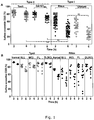
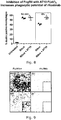
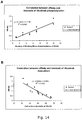
- Fc ⁇ RIIb expression level can be measured as a ratio of the Geometric Mean Fluorescent Intensity (Geo MFI) of Fc ⁇ RIIb to isotype control.
- Fc ⁇ RIIb expression levels can be measured by immunohistochemistry of tumor biopsies. A person skilled in the art would understand that there are multiple techniques and methodologies for determining Fc ⁇ RIIb expression levels.
- Also disclosed herein but not claimed is how to identify antibodies that are suitable for combination treatment with Fc ⁇ RIIb antibodies, namely those antibodies that are internalized from the target cell surface in an Fc ⁇ RIIb dependent manner. Also disclosed herein but not claimed is a means to identify patient subsets that are suitable for combination treatment with Fc ⁇ RIIb antibodies.
- an assay for identifying agents that reduce or prevent binding between the Fc domain of an antibody to a target cell surface antigen and Fc ⁇ RIIb on the target cell comprising determining the extent of binding between the Fc domain and Fc ⁇ RIIb in the presence and absence of a test agent.
- Useful agents are identified if the test agent reduces or prevents Fc domain binding to Fc ⁇ RIIb.
- Such assays are also useful for identifying which agents (for example antibody molecules) are suitable for combination therapy with anti-Fc ⁇ RIIb antibodies.
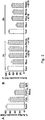
- the assay for identifying agents useful in the practice of the uses of the invention involves screening for agents that block stimulation/signaling of Fc ⁇ RIIb, as indicated by phosphorylation of tyrosine-293 in the intracellular ITIM motif as detected by Western blotting.
- Raji cells are cultured with an antibody to a cell surface antigen, e.g. the anti-CD20 mAb rituximab, in the presence or absence of the anti-Fc ⁇ RIIb test agent before immunoblotting for phosphorylated Fc ⁇ RIIb.
- the amount of phosphorylated Fc ⁇ RIIb becomes elevated in cells stimulated by rituximab, and should be inhibited by the addition of the test agents similar to that shown in Figure 4A using AT10 as the blocking agent.
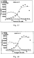
- these agents should preferably also block internalization of rituximab according to the quenching assay indicated in Figure 1A .
- Figure 2B shows a typical example of blocking by an anti- Fc ⁇ RIIb blocking entity, in this case AT10.
- Such assays are also useful for identifying which agents (for example antibody molecules) are suitable for combination therapy with anti-Fc ⁇ RIIb antibodies.
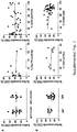
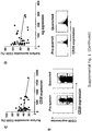
- the assay used to identify agents suitable for combination therapy with Fc ⁇ RIIb antibodies measures the percentage of agent (for example antibody molecule) internalization into Fc ⁇ RIIb-expressing cells.
- This assay method comprises determining the percentage of agent (for example antibody molecules) retained at the cell surface following incubation with agent (for example antibody molecule) target-expressing and Fc ⁇ RIIb-expressing cells, whereby decreasing percentages of agent (for example antibody molecules) accessible at the cell surface (equivalent of increasing antibody molecule internalization) is predictive of, a response to treatment with the antibody molecule.
- the above-mentioned ligands may be small organic or inorganic moieties, such as peptides or polypeptides.
- the ligand When the ligand is a small organic or organic moiety, it may have a M r of from 50 to 2000, such as from 100 to 1000, for example from 100 to 500.
- the ligand binds to Fc ⁇ RIIb with a K d of from mM to pM, such as in the range of from ⁇ M (micromolar) to nM. Generally, the ligands with the lowest Kd are preferred.
- the ligand may be a peptidomimetic, a nucleic acid, a peptide nucleic acid (PNA) or an aptamer. It may also be a lipid, or a carbohydrate.
- the ligand may be a polypeptide which binds to Fc ⁇ RIIb.
- polypeptides by which we include oligopeptides
- the polypeptide may also be a binding protein based on a modular framework, such as ankyrin repeat proteins, armadillo repeat proteins, leucine rich proteins, tetratriopeptide repeat proteins or Designed Ankyrin Repeat Proteins (DARPins) or proteins based on lipocalin or fibronectin domains or Affilin scaffolds.
- a modular framework such as ankyrin repeat proteins, armadillo repeat proteins, leucine rich proteins, tetratriopeptide repeat proteins or Designed Ankyrin Repeat Proteins (DARPins) or proteins based on lipocalin or fibronectin domains or Affilin scaffolds.
- the test agent is a library of test compounds, for example the library is any of a peptide library, a protein library, an antibody library, a recombinant combinatorial antibody library or a scFV or Fab phage display library.
- the second antibody molecule (ii) is one or more antibody molecules that specifically bind Fc ⁇ RIIb.
- the one or more antibody molecules do not include a domain capable of recruiting an effector cell. of recruiting an effector cell.
- the one or more antibody molecules are one or more monoclonal antibody molecules.
- the second antibody molecule prevents or reduces Fc ⁇ RIIb signaling. Even more preferably, the second antibody molecule prevents or reduces internalization of the first antibody molecule by the target cell.
- the SEQ ID NOs refer to the sequences indicated in clones 1-13 below.
- CDRs complementarity determining regions
- the second antibody molecule comprises a variable heavy chain (VH) comprising the following CDRs:
- the second antibody molecule comprises a variable light chain (VL) comprising the following CDRs:
- the second antibody molecule comprises a variable heavy chain (VH) amino acid sequence selected from the group consisting of: SEQ ID NO: 3; SEQ ID NO: 4; SEQ ID NO: 5, SEQ ID NO: 6; SEQ ID NO: 7; SEQ ID NO: 8; SEQ ID NO: 9; SEQ ID NO: 10; SEQ ID NO: 11; SEQ ID NO: 12; SEQ ID NO: 13; SEQ ID NO: 14; and SEQ ID NO: 15.
- VH variable heavy chain
- the second antibody molecule comprises a variable light chain (VL) amino acid sequence selected from the group consisting of: SEQ ID NO: 16; SEQ ID NO: 17; SEQ ID NO: 18; SEQ ID NO: 19; SEQ ID NO: 20; SEQ ID NO: 21; SEQ ID NO: 22; SEQ ID NO:23; SEQ ID NO: 24; SEQ ID NO: 25; SEQ ID NO: 26; SEQ ID NO: 27; and SEQ ID NO: 28.
- VL variable light chain
- the second antibody molecule comprises the following CDR amino acid sequences:
- the second antibody molecule comprises the following amino acid sequences:
- the second antibody molecule of the invention may also comprise the constant regions (CH) and (CL) of SEQ ID NO 1 and SEQ ID NO 2.
- the second antibody molecule is capable of competing with the agents described herein, for example agents comprising the amino acid sequences set out in the embodiments above (for example SEQ ID NOs: 1-106), for preventing or reducing Fc ⁇ RIIb binding to the Fc domain of the first antibody molecule.
- the tested agent is capable of inhibiting or otherwise interfering, at least in part, with the binding of an agent as defined herein to Fc ⁇ RIIb and preventing or reducing Fc ⁇ RIIb binding to the Fc domain of the first antibody molecule.
- the second antibody molecule may be capable of inhibiting the binding of an agent described herein by at least 10%, for example at least 20%, 30%, 40%, 50%, 60%, 70%, 80%, 90%, 95% or even by 100% and/or inhibiting the ability of the agent to prevent or reduce Fc ⁇ RIIb binding to the Fc domain of the first antibody molecule by at least 10%, for example at least 20%, 30%, 40%, 50%, 60%, 70%, 80%, 90%, 95% or even by 100%.
- ELISA Enzyme-linked immunosorbent assay
- ELISA assays can be used to evaluate epitope-modifying or blocking antibodies. Additional methods suitable for identifying competing antibodies are disclosed in Antibodies: A Laboratory Manual, Harlow & Lane (for example, see pages 567 to 569, 574 to 576, 583 and 590 to 612, 1988, CSHL, NY, ISBN 0-87969-314-2 ).
- the second antibody molecule of the invention may comprise the following constant regions (CH and CL):
- the second antibody molecule of the invention may comprise one or more sequences of clones 1-14:
- Target cell surface antigens disclosed herein but not claimed may be selected from the following: Thy-1 (CD90, Cluster of Differentiation 90 ( Biofactors. 2009 May-Jun;35(3):258-65 )); Ly-6 (Lymphocyte Antigen 6 ( Mol Biol Rep. 2009 Apr;36(4):697-703 )); CD59 (Complement regulatory protein ( Mol Immunol. 2007 Jan;44(1-3):73-81 )); Fas (FS7-associated cell surface antigen, CD95, APO-1 or TNFRSF6 ( Adv Exp Med Biol. 2009;647:64-93 )); EGFR (Epidermal Growth Factor Receptor ( FEBS J.
- Thy-1 CD90, Cluster of Differentiation 90 ( Biofactors. 2009 May-Jun;35(3):258-65 )
- Ly-6 Lymphocyte Antigen 6 ( Mol Biol Rep. 2009 Apr;36(4):697-703 )
- CD59 Complement regulatory protein ( Mol Immunol. 2007 Jan;
- Her2 Human epidermal growth factor receptor 2 ( Clin Breast Cancer. 2008 Oct;8(5):392-401 )); CXCR4 (Chemokine Receptor 4 ( Biochim Biophys Acta. 2007 Apr;1768(4):952-63 )); HLA Molecules (Human Leukocyte Antigen molecules ( Korean J Lab Med. 2010 Jun;30(3):203 )); GM1 (ganglioside, monosialotetrahexosylganglioside ( J Lipid Res.
- CD22 Cheson (2008) NEJM 359(6): 613-26
- CD23 Cheson, 2008
- CD80 Cheson, 2008
- CD74 CD74
- DRD Cheson, 2008
- the target cell surface antigen is selected from: CD 19 (Cluster of Differentiation 19 ( Cell Immunol. 1989 Feb;118(2):368-81 )); CD40 (Cluster of Differentiation 40 ( Basic Clin Pharmacol Toxicol. 2009 Feb;104(2):87-92 )); and CD20.
- the surface antigens CD19, CD20, or CD40 are human forms thereof.
- the first antibody molecule is a monoclonal antibody, preferably a monoclonal antibody that upon binding to the target cell is removed from the cell surface and internalized into the target cell in an Fc ⁇ RIIb-dependent manner.
- the monoclonal antibody is an anti-CD19, anti-CD20 or anti-CD40 antibody.
- the monoclonal antibody is an anti-CD20 monoclonal antibody.
- the first antibody molecule is a Type I anti-CD20 antibody. In another preferred embodiment, the first antibody molecule is not a Type II anti-CD20 antibody.
- the cell surface antigen is CD20 and the first antibody molecule Type I antibody.
- Anti-CD20 monoclonal antibodies As mentioned above, there are two types of anti-CD20 monoclonal antibodies (mAb). Anti-CD20 mAb were first defined by the inventors as falling into different groupings in 2003 (43 and 25) and then subsequently defined as Type I and II mAbs in 2004 (26). Initially the basis for this was that anti-CD20 mAb fall into two distinct types of reagents based on their ability to eradicate lymphoma xenografts: type I (e.g. Rituximab and 1F5) utilize complement; and type II (e.g. B1), do not.
- type I e.g. Rituximab and 1F5
- type II e.g. B1
- Type II mAb being able to elicit more potent homotypic adhesion and direct cell death but these could not be used alone to define a Type I or II mAb (unlike the Tx-100 raft assays; see below).
- various anti-CD20 mAb may be classified as type I (e.g. rituximab, ofatumumab) or type II (e.g. tositumomab (B1), GA101, 11B8) according to their ability to redistribute CD20 in the plasma membrane and their activity in various effector assays (25-27).
- Type I (e.g. rituximab, ofatumumab) anti-CD20 monoclonal antibodies induce CD20 to redistribute into large detergent resistant microdomains (rafts), whereas type II (tositumomab-like) anti-CD20 monoclonal antibodies do not (50).
- anti-CD20 mAbs can be designated as Type I or Type II by virtue of whether they redistribute CD20 into lipid rafts. This is done by the Tx-100 insolubility assay or by sucrose density gradient separation and western blotting. Both methods are described in Cragg et al Blood 2003 (43 ) as follows:
- Triton X-100 insoluble fraction To assess whether more antigen could be moved into the Triton X-100 insoluble fraction by additional cross-linking, cells were incubated with FITC-mAb as before, washed and then divided into four. Two of these samples were incubated with goat anti-mouse Ig F(ab')2 fragments for 15 minutes on ice. After washing, one of the cross-linked and one of the non-cross-linked samples were lysed in Triton X-100 and washed as detailed above prior to flow cytometry.
- Monoclonal Ab (1 ⁇ g/10 6 cells) was added to cells at 37°C. Following 20 minutes incubation, cells were pelleted and lysed in ice-cold 1.0 % Triton X-100 in MES-buffered saline (25 mM MES, pH 6.5, 150mM NaCl, 1mM phenylmethylsulfonyl fluoride, 5 ⁇ g/ml aprotinin, 5 ⁇ g/ml leupeptin, 10mM EDTA). Lipid raft fractions were then prepared by sucrose density gradient centrifugation.
- Lysates were mixed with an equal volume of 80% sucrose in lysis buffer, overlaid with a discontinuous 5-30 % sucrose density gradient and then centrifuged at 200, 000 x g for 16 h. Fractions (0.5ml) were collected and analysed by Western blotting.
- Anti-CD20 mAbs can require the AxP motif in the large loop of CD20. (Ofatumumab and other Genmab antibodies do not). However, (Niederfelner et al. (51)) indicates Type II mAb bind to a slightly different region of the CD20 loop compared to Type I.
- the target cell is a cancer cell.
- a cancer selected from non-Hodgkin lymphoma, including but not limited to follicular lymphoma, diffuse large B cell lymphoma, mantle cell lymphoma, or chronic lymphocytic leukaemia.
- the invention provides compositions for use and uses for treating cancer, in particular a B cell malignancy which is preferably selected from lymphomas, chronic lymphocytic leukemias, acute lymphoblastic leukemias, multiple myeloma, Hodgkin's and non-Hodgkin's disease, diffuse large B cell lymphoma, follicular lymphoma with areas of diffuse large B cell lymphoma, small lymphocytic lymphoma, mantle cell lymphoma, and diffuse small cleaved cell lymphoma or combinations thereof.
- the B cell malignancy is a lymphoma, such as non-Hodgkin's (NHL).
- the invention provides compositions for use and uses for treating an inflammatory disease.
- This may be an autoimmune disease, such as Hashimoto's thyroiditis, pernicious anemia, Adison's disease, type 1 diabetes, rheumatoid arthritis, systemic lupus erythematosus, dermatomyositis, Sjogren's syndrome, dermatomyositis, lupus erythematosus, multiple sclerosis, autoimmune inner ear disease myasthenia gravis, Reiter's syndrome, Graves disease, autoimmune hepatitis, familial adenomatous polyposis and ulcerative colitis or combinations thereof.
- the autoimmune disease is rheumatoid arthritis or systemic lupus erythematosus.
- the treatment is of diseases including, chronic lymphocytic leukemia (CLL), non-Hodgkin lymphoma (NHL), B cell malignancies, Rheumatoid arthritis, systemic lupus erythematosus, dermatomyositis, systemic sclerosis and autoimmune blistering diseases.
- CLL chronic lymphocytic leukemia
- NHL non-Hodgkin lymphoma
- B cell malignancies Rheumatoid arthritis
- systemic lupus erythematosus systemic lupus erythematosus
- dermatomyositis systemic sclerosis
- autoimmune blistering diseases including, chronic lymphocytic leukemia (CLL), non-Hodgkin lymphoma (NHL), B cell malignancies, Rheumatoid arthritis, systemic lupus erythematosus, dermatomyositis, systemic sclerosis and autoimmune blistering diseases.
- the treatment that is enhanced by use of the invention is treatment with an anti-CD20 mAb, such as rituximab.
- “elevated” we include the meaning that the cells in question express higher levels of Fc ⁇ RIIb on their surface than control or reference cells that express low or medium levels of Fc ⁇ RIIb on their surface.
- the cells in question are a type of B cell
- “elevated” levels of Fc ⁇ RIIb expression would be recognized if the level of expression was higher than the normal (preferably median) level of expression of Fc ⁇ RIIb by B cells of the same cell type.
- “elevated” levels would be recognized if the cells in question expressed Fc ⁇ RIIb at a level higher than a different cell type which expresses Fc ⁇ RIIb at low or medium level.
- the greater the degree of elevation in Fc ⁇ RIIb expression of target cells the worse the predicted response of those cells to treatment with an antibody molecule that binds specifically to a surface antigen of the target cells and that has an Fc domain capable of binding Fc ⁇ RIIb.
- the greater the elevation in Fc ⁇ RIIb expression the greater the benefit to be gained from the use of an agent that prevents or reduces binding of the Fc domain to Fc ⁇ RIIb according to the invention as demonstrated in figures 2D and 3A .
- the elevated Fc ⁇ RIIb expression level is at least 1.1 fold increased over the median expression level of cells of the same cell type in patients having the same disease, or at least 1.2, 1.3, 1.4, 1.5, 1.6, 1.7, 1.8, 1.9, 2.0, 2.1, 2.2, 2.3, 2.4, 2.5, 2.6, 2.7, 2.8, 2.9, 3.0, 3.1, 3.2, 3.3, 3.4, 3.5, 3.6, 3.7 ,3.8, 3.9, 4.0, 4.1, 4.2, 4.3, 4.4, 4.5, 4.6, 4.7, 4.8, 4.9, 5.0, 5.1, 5.2, 5.3, 5.4, 5.5, 5.6, 5.7, 5.8, 5.9, 6.0, 6.1, 6.2, 6.3, 6.4, 6.5, 6.6, 6.7, 6.8, 6.9, 7.0, 7.1, 7.2,7.3, 7.4, 7.5, 7.6, 7.7, 7.8, 7.9, 8.0, 8.1, 8.2, 8.3, 8.4, 8.5, 8.6,
- they are at least 1.8 fold increased over the median expression level of cells of the same cell type in patients having the same disease.
- the inventors have determined that the level of Fc ⁇ RIIb expression correlates with an increased modulation of mAb from the cell surface. Even higher levels of Fc ⁇ RIIb expression give an even higher modulation, i.e. there is a correlation between degree of elevation of expression and modulation as shown in figures 2D and 3A .
- Figure 2D the expression level of Fc ⁇ RIIb is plotted versus the level of modulation observed. The correlation is such that the highest levels of Fc ⁇ RIIb (e.g. >400) results in the lowest level of surface CD20 ( ⁇ 20%) i.e. the greatest effect on modulating the mAb from the cell surface.
- Fc ⁇ RIIb low (18), medium (70) and high (124) levels of Fc ⁇ RIIb were introduced into an Fc ⁇ RIIb -negative cell line (Ramos) and this was directly correlated with reducing levels of cell surface CD20 (60, 40, 30% for low medium and high) in proportion to the expression of the Fc ⁇ RIIb.
- Fc ⁇ RIIb expression levels are given as a ratio of the Geometric Mean Fluorescent Intensity (Geo MFI) of Fc ⁇ RIIb to isotype control
- Modulation reduces the amount of mAb left on the cell surface.
- mAbs require the Fc to engage with immune effector mechanisms (ADCC, ADCP, CDC) in order to delete target cells. Therefore reducing modulation with Fc ⁇ RIIb blocking mAb will improve Fc-dependent effector functions. This is shown for ADCP (phagocytosis) in Figure 8 . Phagocytosis with rituximab alone was 40% but rose to 55% when the Fc ⁇ RIIb was blocked by AT10.
- antibody molecule we include monoclonal antibodies, synthetic antibodies, recombinantly produced antibodies, multispecific antibodies, human antibodies, chimeric antibodies, camelized antibodies, single-chain Fvs (scFv), single chain antibodies, Fab fragments, F(ab') fragments, disulfide-linked Fvs (sdFv), intrabodies, or epitope-binding fragments of any of the above.
- the antibodies of the invention are monoclonal antibodies, and more preferably, humanized or human antibodies.
- WO 2008/002933 in Section 5.3 to 5.3.1 pages 74-91, describes the preparation and characterization of monoclonal antibodies that bind specifically to a target cell surface antigen such as CD20 or Fc ⁇ RIIb.
- a target cell surface antigen such as CD20 or Fc ⁇ RIIb.
- Useful antibodies that specifically bind Fc ⁇ RIIb and CD20, including monoclonal antibodies produced by hybridomas deposited under the Budapest Treaty, are also disclosed on pages 15 to 21 of WO 2008/002933 .
- binds we include agents such as antibody molecules that bind to a target antigen but do not bind (cross-react) to other antigens or bind such antigens more weakly, i.e. with a lower affinity than the target antigen.
- an antibody that specifically binds to Fc ⁇ RIIB may bind to other peptides or polypeptides with lower affinity as determined by, e.g., immunoassays, BIAcore, or other assays known in the art.
- antibodies that specifically bind to Fc ⁇ RIIB or a fragment thereof do not cross-react with other antigens.
- Antibodies that specifically bind to Fc ⁇ RIIB can be identified, for example, by immunoassays, BIAcore, or other techniques known to those of skill in the art.
- An antibody or a fragment thereof binds specifically to a Fc ⁇ RIIB when it binds to Fc ⁇ RIIB with higher affinity than to any cross-reactive antigen as determined using experimental techniques, such as western blots, radioimmunoassay (RIA) and enzyme-linked immunosorbent assays (ELISAs) (See Fundamental Immunology Second Edition, Raven Press, New York at pages 332-336 (1989 ) for a discussion regarding antibody specificity) and binds Fc ⁇ RIIB, particularly human Fc ⁇ RIIB, more particularly native human Fc ⁇ RIIB, with a greater affinity than said antibody or a fragment thereof binds Fc ⁇ RIIA, particularly human Fc ⁇ RIIA, more particularly native human Fc ⁇ RIIA.
- Representative antibodies are disclosed in US Patent Application Nos. 2004-0185045
- certain Fc ⁇ RIIB antibodies used in combination with CD20 antibodies in the compositions for use of the invention bind the extracellular domain of native human Fc ⁇ RIIB.
- the antibody or a fragment thereof binds Fc ⁇ RIIB with at least 2 times greater affinity than said antibody or a fragment thereof binds Fc ⁇ RIIA.
- the antibody or a fragment thereof binds Fc ⁇ RIIB with at least 4 times, at least 6 times, at least 8 times, at least 10 times, at least 100 times, at least 1000 times, at least 10 4 , at least 10 5 , at least 10 6 , at least 10 7 , or at least 10 8 times greater affinity than said antibody or a fragment thereof binds Fc ⁇ RIIA.
- said antibody or a fragment thereof bind Fc ⁇ RIIB with 100 times, 1000 times, 10 4 times, 10 5 times, 10 6 times, 10 7 times, 10 8 times greater affinity than said antibody or a fragment thereof binds Fc ⁇ RIIA.
- Example 1 CD20 modulation in primary CLL and other NHL samples
- Example 2 Modulation of CD20 in B-NHL is an Fc-dependent process.
- F(ab') 2 and IgG showed a similar binding profile (results not shown), but after 6 h culture the F(ab') 2 fragment had modulated significantly less (40% surface accessible CD20) than the intact IgG (20% surface accessible CD20). Since the assays were performed with highly enriched (> 95% pure) B cells, the only FcR present in abundance would be the inhibitory Fc ⁇ RIIb.
- Fc ⁇ RIIb -ve Ramos cells were transfected with an Fc ⁇ RIIb-encoding plasmid.
- the resultant Fc ⁇ RIIb +ve cells displayed variable Fc ⁇ RIIb expression levels and were subsequently sorted into sub-clones expressing low, medium and high Fc ⁇ RIIb. These cells, along with parental Fc ⁇ RIIb - Ramos cells were then assessed in the internalization assay.
- CD20 modulation rates at 6 h correlated with Fc ⁇ RIIb expression with increasing modulation in the order Fc ⁇ RIIb -ve >Fc ⁇ RIIb +ve low >Fc ⁇ RIIb +ve medium >Fc ⁇ RIIb +ve high ( Figure 3A ).
- Example 6 CD20 and Fc ⁇ RIIb cross-linking occurs predominantly in a cis fashion
- Rituximab could be co-ligated by CD20 and Fc ⁇ RIIb on either the same (cis) or adjacent cells (trans).
- CD20 and Fc ⁇ RIIb on either the same (cis) or adjacent cells (trans).
- PKH26-labeled Fc ⁇ RIIb - Ramos cells with high Fc ⁇ RIIb-expressing Ramos transfectants ( Figure 4B ), and then compared the level of modulation in each cell type, with both cell types cultured alone as a control.
- Fc ⁇ RIIb +ve cells showed greater modulation than cells lacking Fc ⁇ RIIb ( Figure 4B ).
- Example 8 Fc ⁇ RIIb inhibits Type I anti-CD20 mAb in vivo
- mice were treated with rituximab variants (250 ⁇ g, iv) harboring mouse IgG1 (m1) or mouse IgG2a (m2a) and then B cell depletion monitored by flow cytometry for 90 days through serial bleeding of the mice and staining with B220 and CD19 mAb ( Figure 6 ). These variants either bind strongly (m1) or weakly (m2a) to CD32b.
- Example 9 Fc ⁇ RIIb enhances and augments the activity of anti-CD20 mAb against human tumours in vivo
- CD32 human tumour cells
- mAbs such as rituximab
- a xenograft system In this system, only the human tumour cells express hCD32 and so any therapeutic effects derive from effects on the tumour cell, most likely by blocking modulation, not through any effects on the host effector cells.
- CD20 positive CD32 positive human tumour cells (Daudi or Raji) were innoculated into SCID mice and then treated with either rituximab, AT10 or both and survival of the mice or tumour growth monitored ( Figure 7 ). Doses of mAb used are shown in the figure legends.
- Daudi cells were inoculated subcutaneously and the tumour monitored by caliper measurements every 3-5 days.
- B and C Raji tumour cells were injected intravenously and animals monitored for survival.
- AT10 was shown to enhance and augment the activity of rituximab, demonstrating the potential of this combination in vivo.
- Example 10 Fc ⁇ RIIb levels predict clinical outcome in rituximab-treated MCL patients.
- the Rationale for the experiments in examples 10 and 11 is as follows. If cells express high levels of CD32b (Fc ⁇ RIIb), they will internalise rituximab more quickly (shown as reduced % surface accessible CD20). If there is less rituximab at the cell surface, then there will be less Fc-dependent effector activity (such as phagocytosis or ADCC) and therefore less tumour cell killing and hence less extensive therapeutic results. Therefore, we checked the Fc ⁇ RIIb expression in a cohort of patients treated with MCL and determined whether they were high or low expressors of CD32b. Clinical data was already available for this cohort and so the clinical results were then stratified according to whether they were high or low Fc ⁇ RIIb-expressing tumours.
- variable regions VH and VL
- CDR regions of the 14 antibody clones The amino acid sequences of the variable regions (VH and VL), together with the CDR regions of the 14 antibody clones are shown in figures 37 - 50 .
- the constant (CH and CL) regions are the same.
- the constant regions are shown in figure 36 .
- CD32B Selections against CD32B (Fc ⁇ RIIb) were performed using the n-CoDeR ® scFv phage display library.
- Human CD32A was used as non-target.
- the extra cellular domains of CD32A and CD32B fused to mIgG3-Fc were produced in HEK293E and purified on protein A. Three consecutive protein selections were performed. Non-target was used as competitor in all selections.
- Resulting phages were converted to scFv/Fab producing format and transformed into E.coli Top10 bacteria for screening of individual clones. Screening determined the specificity for human CD32B and CD32A and was analyzed using coated proteins in ELISA as well as through transfected CHO cells in FMAT.
- the IgGs were left to bind CD32B transfected CHO cells followed by addition of an IC in the form of IgG1 coated bovine serum albumin. Bound IC was then detected and inhibiting properties of the IgGs could be evaluated.
- Rituximab-alexa 488 was added to Ramos cells transfected with CD32B in the presence or absence of different CD32b blocking mAb (WT or 297Q mutants) and modulation assessed after 1,2,6 and 24h.
- CD32b blocking mAb WT or 297Q mutants
- As a control for the blocking ability of CD32 mAb we also included the dual CD32a and b specific mAb, AT10 (IgG and Fab2 fragments (Fab)), alongside a negative control, isotype matched irrelevant mAb (iso wt or nq).
- the data in figure 12 clearly indicate that all 3 nCoder mAb (C1, C3 and C11) are able to block the modulation of rituximab in either the wt or 297q format.
- Figure 13 shows the ability of anti-CD32b mAb to block modulation of rituximab using all 13 mAb.
- control CD32 negative Ramos cells were included to allow estimation of the maximal effect of the CD32 blocking mAb.
- the data in figure 12 clearly indicate that all the nCoder mAb were able to block the modulation of rituximab.
- the relative affinity of the mAb was determined by a dose titration experiment measuring mAb binding to CD32B transfected CHO cells. Briefly, CD32B transfected adherent CHO K1 cell were seeded into FMAT plates. IgG were titrated in 1:2 dilutions from 30nM to approximately 0,015nM and left to bind for 1h at room temperature. After washing bound IgG were detected with anti-human-IgG-APC. Finally, the plates were washed and read in the FMAT (Applied Biosystems). This gives an EC50 value for mAb binding to target expressing cells and can be translated to a relative affinity.
- the relative affinity was then correlated with the ability of ant-CD32b blocking mAb to prevent phosphorylation of CD32b after rituximab binding. This was determined by stimulating cells with rituximab in the presence or absence of anti-CD32b mAb and then performing western blotting for phospho-CD32b. The CD32b mAb were then ranked according to their ability to block the CD32 phosphorylation with 1 being the most effective.
- Figure 14A shows that there was evidently a close correlation between the affinity of the mAb and the ability to block CD32b phosphorylation.
- Figure 14B shows there was evidently a strong correlation between the affinity of the mAb and the ability to block rituximab modulation. This data confirms the central role of CD32B in accelerating the modulation of rituximab from the target cell surface.
- Example 14 Dose dependent binding to hCD32B transfected cells and dose dependent binding and inhibition of immune complex to hCD32B transfected cells.
- FIGS. 15-27 show the dose dependent binding to hCD32B transfected cells and dose dependent binding and inhibition of immune complex to hCD32B transfected cells mediated by clones 1-13, respectively. Circles show dose dependent binding to hCD32A transfected CHO K1 cells and black diamonds show dependent binding to hCD32B transfected CHO K1 cells. Crosses show dose dependent inhibition of immune complex to hCD32B transfected CHO K1 cells.
- CD32B and CD32A are very closely related molecules. However, while CD32B transmits an inhibitory signal, CD32A transmits a positive, hence it is essential that the antibody only binds CD32B for the desired effect. 2) Furthermore, to effectively block a signal through CD32B, the antibody does not only have to bind CD32B, but also to block binding of it's natural ligand, an immune complex (IC). Hence the figure shows binding to CD32A, CD32B and inhibition of 1C binding. The figures demonstrate that all mAb's are specific for CD32B and does not bind CD32A and that they all inhibit 1C binding.
- Example 15 Cell specificity of the anti-CD32B antibodies.
- PBMCs isolated from peripheral blood was prepared using Ficoll density gradient. Cells were stained with cell specific markers and evaluated for binding of the CD32B specific antibodies. As shown in figure 28 , only B cells (CD19+ cells) stained positive with clone 1-13.
- CD32B In resting PBMC's, CD32B is only expressed on B cells while the closely related CD32A is expressed on monocytes and neutrophils.
- the previous figures show specificity on transfected CHO cells.
- Figure 28 shows that the antibodies also binds B cells expressing CD32B in it's truly native form on B cells while they do not stain CD32A expressing neutrophils or monocytes. Hence this figure is a demonstration of antibody specificity when antigen is expressed in normal non-transfected PBMC's.
- Example 16 Dose dependent staining of B cells by anti-Fc ⁇ RIIb mAb clone 1-13.
- PBMCs isolated from peripheral blood was prepared using Ficoll density gradient. Cells were stained with CD19 and thereafter with 10, 1 or 0,1 mg/ml CD32B specific antibodies as indicated. Figure 29 shows how the cell staining of B cells by each clone is dose dependent.
- B cells (know to express CD32B) have been gated out using a CD19 specific mAb. This gate is called M1.
- M1 CD19 specific mAb.
- concentration of CD32B mAb is decreased, the number of B cells stained drops from nearly 100% down to much lower values, showing a specific and dose dependent staining of B cells, as expected from a CD32B specific mAb.
- Example 17 Capacity of each mAb to inhibit Fc-mediated CD32B phosphorylation.
- CD32b The ability of CD32b to precipitate the internalisation of other Type I anti-CD20 mAb is shown in figure 31 .
- Alexa-488 labelled versions of each mAb were incubated with pCDNA3-transfected Ramos or CD32B-transfected Ramos cells for 1 or 6 hr and the extent of modulation determined as before.
- mAb used were rituximab (RTX), in-house produced ofatumumab (OFA) and tositumumomab (Tos). The data clearly show that the rate of internalisation of OFA is similar to that of RTX and is accelerated by the presence of CD32b.
- Anti-CD19 mAb also internalize from the cell surface of malignant human B-cells in a manner which is partially dependent upon CD32B.
- Ramos huCD32b transfectants Internalisation with other surface antigens can also be effected by CD32b expression.
- the modulation assay was performed as before with different mAb in the presence (+) or absence (-) of CD32 BLOCKING with AT10. Ramos CD32B transfectants were used in this 6h assay. * p ⁇ 0.05.
- f3.3 MHC Class II;
- RFBP CD 19;
- RTX rituximab.
- Figure 32 clearly shows a significant reduction in surface modulation for RTX and RFB9 mAb and less for F3.3 after incubation with CD32 blocking.
- Ramos cells have no CD32b and so demonstrate the level of internalisation in the absence of CD32B. If the antigen is able to be internalised by CD32b then expressing it (on the Ramos-CD32B cells) will increase the level of internalisation.
- Figure 33 shows that internalisation with other antigens is also effected by CD32b expression.
- Fc ⁇ RIIb surface expression was determined by flow cytometry using PE-labeled AT10 (described below). Populations of Ramos Fc ⁇ RIIb transfectants expressing low, medium or high levels of Fc ⁇ RIIb were sorted using a FACS Aria flow cytometer (BD Biosciences, USA).
- CLL/SLL, FL, DLBCL and MCL samples were obtained with informed consent in accordance with the Declaration of Helsinki.
- Blood samples were collected in K 2 E or LiH with Lymphoprep and solid tissue was disaggregated through a sterile strainer and centrifuged.
- Cells were cryopreserved in RPMI supplemented with 50% human AB serum and 10% DMSO and stored in University of Southampton's Cancer Sciences Division Tumor Bank under Human Tissue Authority licensing. Ethical approval for the use of clinical samples was obtained by the Southampton University Hospitals NHS Trust from the Southampton and South West Hampshire Research Ethics Committee.
- mutation status of IgVH genes (33) and CD38 positivity (44) was determined as detailed previously.
- VH leader primer mix and a C ⁇ 100 primer were used to amplify heavy-chain genes from cDNA. All nucleotide sequences were aligned to the V-base directory, and mutational status was determined using a 98% cutoff.
- anti-CD38 PE clone HB7; BD Biosciences
- Determination of ZAP-70 status was carried out as described by Crespo et al. (30). Surface Ig expression of CLL cells was determined by flow cytometry as described previously (45, 46).
- Rituximab was gifted by Oncology Pharmacy, Southampton General Hospital.
- Rit m2a (rituximab with mouse IgG2a Fc region), and WR17 (anti-CD37), all mouse IgG2a, were produced as described previously (18).
- Anti-Fc ⁇ RII mAb (AT10) was produced in-house and has been described previously (47).
- Tositumomab was gifted by Prof Tim Illidge (Manchester, United Kingdom).
- Ofatumumab and GA101 gly glycosylated GA101 with unmodified Fc region) were produced in-house from patent published sequences.
- NB These mAb were produced in CHO or 293F cells and so may differ (for example in their carbohydrate structures) from the mAb produced for clinical use.
- Alexa-488 and anti-Alexa 488 reagents were purchased from Invitrogen. Production of F(ab') 2 fragments has previously been described (48). Fab' fragments were generated by incubation with 20 mM 2-mercaptoethanol at 25°C, for 30 min, followed by addition of excess iodoacetamide.
- Western blotting antibodies used were anti-actin (AC74, Sigma, UK) and anti-phospho-Fc ⁇ RIIb (Cell Signaling Technology, UK).
- Fluorochrome-labeled mAb were obtained from BD Biosciences or made in-house. mAb were conjugated with Alexa 488 (Invitrogen) as per the manufacturer's protocol. Flow cytometry has been described previously (49). Samples were assessed on either a FACScan or FACSCalibur and data analyzed with CellQuest Pro (all BD Biosciences) or FCS Express (DeNovo Software, USA). B cells were identified with APC-labeled anti-human CD19 (in-house) and Fc ⁇ RIIb expression determined using PE-labeled AT10 (in-house). To control for inter-experimental variation, Fc ⁇ RIIb expression was represented as the ratio of Fc ⁇ RIIb:isotype control Geo mean fluorescence intensity (MFI).
- MFI Geo mean fluorescence intensity
- the internalization assay was performed as detailed previously (28). Briefly, 2-4 x 10 5 cells per well were incubated with Alexa-488 labeled mAb at a final concentration of 5 ⁇ g/ml. Samples were harvested after 1, 2, 6 and/or 24 h, washed twice, resuspended and incubated at 4°C for 30 min with APC-labeled anti-CD19, with or without the quenching antibody, anti-Alexa-488 (Invitrogen). Samples were then washed once and analyzed on a flow cytometer.
- Ramos cells which are Fc ⁇ RIIb -ve
- PKH26 Sigma Aldrich
- the PKH26-labeled cells were then co-cultured with equal numbers (2.5 x 10 5 cells) of Ramos cells transfected with Fc ⁇ RIIb. Both cell types were cultured alone as controls.
- the internalization assay was then performed as described above, and the modulation compared on the PKH26-labeled and - unlabeled populations. Further variations of this co-culture assay are described in figure legends.
- CLL cells were incubated with appropriate Alexa 488-labeled mAb for various times as described in the figure legends and then harvested, washed and fixed with 2% paraformaldehyde.
- Fc ⁇ RIIb and LAMP-1 were then permeabilized with 0.3% saponin and incubated with Alexa-647-labeled AT10 F(ab') 2 (labeling performed with Alexa Fluor-647 labeling kit (Invitrogen) as per the manufacturer's protocol), and/or biotin conjugated anti-human CD107a (LAMP-1) (eBioscience, UK).
- Cells were then washed, streptavidin-Alexa Fluor-547 (Invitrogen) added, followed by further washing. Cells were then transferred onto slides and images captured using LAS-AF v2 software on a TCS-SP5 laser scanning confocal microscope (Leica Microsystems, UK) (10x eye piece, 100x objective lens).
- cells were seeded at 1-20 x 10 5 /ml, stimulated with various mAb for 2 and/or 6 h and then their relative proximity assessed by light microscopy.
- Cells were viewed with an Olympus CKX21 inverted microscope (Olympus, UK) using a 10x or 20x/0.25 PH lens. Images were acquired using a CCL2 digital cooled camera (Olympus) and were processed with Cell B (Olympus Soft imaging solutions) and Adobe Photoshop version CS2 software (Adobe, San Jose, CA).
- the invention provides compostiions for use or uses for treatment, prophylaxis, and amelioration of one or more symptoms associated with a cancer or inflammatory disease by administering to a subject or patient an effective amount of a pharmaceutical composition as defined in the claims.
- the subject or patient is an animal, preferably a mammal such as non-primate (e.g., cows, pigs, horses, cats, dogs, rats etc.) and a primate (e.g., monkey such as, a cynomolgous monkey and a human).
- a mammal such as non-primate (e.g., cows, pigs, horses, cats, dogs, rats etc.) and a primate (e.g., monkey such as, a cynomolgous monkey and a human).
- a primate e.g., monkey such as, a cynomolgous monkey and a human.
- the subject is a human.
- compositions for use of the invention e.g., encapsulation in liposomes, microparticles, microcapsules, recombinant cells capable of expressing the antibody etc.
- compositions for use of the invention are formulated in liposomes for targeted delivery of the antibodies of the invention.
- Liposomes are vesicles comprised of concentrically ordered phospholipid bilayess which encapsulate an aqueous phase. Liposomes typically comprise various types of lipids, phospholipids, and/or surfactants. The components of liposomes are arranged in a bilayer configuration, similar to the lipid arrangement of biological membranes. Liposomes are particularly preferred delivery vehicles due, in part, to their biocompatibility, low immunogenicity, and low toxicity.
- compositions for use of the invention include, but are not limited to, parenteral administration (e.g., introdermal, intramuscular, introperitoneal, intravenous and subcutaneous), epidural, and mucosal (e.g., intranasal and oral routes).
- parenteral administration e.g., introdermal, intramuscular, introperitoneal, intravenous and subcutaneous
- epidural e.g., epidural and mucosal and oral routes
- mucosal e.g., intranasal and oral routes
- the compositions for use of the invention are administered intramuscularly, intravenously, or subcutaneously.
- the compositions may be administered by any convenient route, for example, by infusion or bolus injection, by absorption through epithelial or mucocutaneous linings (e.g., oral mucosa, rectal and intestinal mucosa, etc). and may be administered together with other biologically active agents. Administration can be systemic or local.
- pulmonary administration can also be employed, e.g., by use of an inhaler or nebulizer, and formulation with an aerosolizing agent.
- an inhaler or nebulizer e.g., a pressurized device
- formulation with an aerosolizing agent e.g., a pressurized device
- inhaler or nebulizer e.g., a pressurized device
- formulation with an aerosolizing agent e.g., by use of an inhaler or nebulizer, and formulation with an aerosolizing agent.
- the amount of the composition for use of the invention which will be effective in the treatment, prevention or amelioration of one or more symptoms associated with a cancer or inflammatory disease can be determined by standard clinical techniques.
- the precise dose to be employed in the formulation will also depend on the route of administration, and the seriousness of the condition, and should be decided according to the judgment of the practitioner and each patient's circumstances. Effective doses may be extrapolated from dose-response curves derived from in vitro or animal model test systems.
- the dosage administered to a patient is typically 0.0001 mg/kg to 100 mg/kg of the patient's body weight independently for each antibody in the combination.
- the dosage of each antibody administered to a patient is between 0.0001 mg/kg and 20 mg/kg, 0.0001 mg/kg and 10 mg/kg, 0.0001 mg/kg and 5 mg/kg, 0.0001 and 2 mg/kg, 0.0001 and 1 mg/kh, 0.0001 mg/kg and 0.75 mg/kg, 0.0001 mg/kg and 0.5 mg/kg, 0.0001 mg/kg to 0.25 mg/kg, 0.0001 to 0.15 mg/kg, 0.0001 to 0.10 mg/kg, 0.001 to 0.5 mg/kg to 02.5 mg/kg or 0.01 to 0.10 mg/kg of the patient's body weight.
- human antibodies have a longer half-life within the human body than antibodies from other species due to the immune response to the foreign polypeptides. Thus, lower dosages of human antibodies and less frequent administration is often possible. Further, the dosage and frequency of administration of antibodies of the invention or fragments thereof may be reduced by enhancing uptake and tissue penetration of the antibodies by modifications such as, for example, lipidation.
- the dosage of each of the antibodies of the compositions for use of the invention administered to a patient are 0.01 mg to 1000 mg/day.
- compositions for use of the invention comprise a prophylactically or therapeutically effective amount of a first antibody molecule and second antibody molecule as claimed and a pharmaceutically acceptable carrier.
- the term "pharmaceutically acceptable” means approved by a regulatory agency or listed in the US Pharmacopeia or other generally recognized pharmacopeia for use in animals, and more particularly in humans.
- carrier refers to a diluent, adjuvant (e.g., Freund's adjuvant (complete and incomplete), excipient, or vehicle with which the therapeutic is administered.
- Such pharmaceutical carriers can be sterile liquids, such as water and oils, including those of petroleum, animal, vegetable or synthetic origin, such as peanut oil, soybean oil, mineral oil, sesame oil and the like. Water is a preferred carrier when the pharmaceutical composition is administered intravenously.
- Saline solutions and aqueous dextrose and glycerol solutions can also be employed a s liquid carriers, particularly for injectable solutions.
- suitable pharmaceutical excipients include starch, glucose, lactose, sucrose, gelatin, malt, rice, flour, chalk, silica gel, sodium stearate, glycerol monostearate, talc, sodium chloride, dried skim milk, glycerol, propylene, glycol, water, ethanol and the like.
- the composition if desired, can also contain minor amounts of wetting or emulsifying agents, or pH buffering agents. These compositions can take the form of solutions, suspensions, emulsion, tablets, pills, capsules, powders, sustained-release formulations and the like.
- a first antibody molecule and a second antibody molecule as claimed can be administered simultaneously or less than 1 hour apart, at about 1 hour apart, at about 1 hour to about 2 hours apart, at about 2 hours to about 3 hours apart, at about 3 hours to about 4 hours apart, at about 4 hours to about 5 hours apart, at about 5 hours to about 6 hours apart, at about 6 hours to about 7 hours apart, at about 7 hours to about 8 hours apart, at about 8 hours to about 9 hours apart, at about 9 hours to about 10 hours apart, at about 10 hours to about 100 hours apart, at about 11 hours to about 12 hours apart, no more than 24 hours apart or no more than 48 hours apart.
- two or more components are administered within the same patient visit.
- the dosage amounts and frequencies of administration provided herein are encompassed by the terms therapeutically effective and phrophylactically effective.
- the dosage and frequency further will typically vary according to factors specific for each patient depending on the specific therapeutic or prophylactic agents administered, the severity and type of disease, the route of administration, as well as age, body weight, response, and the past medical history of the patient. Suitable regimens can be selected by one skilled in the art by considering such factors and by following, for example, dosages reported in the literature and recommended in the Physician's Desk Reference (56th ed., 2002 ).
- type I mAb like rituximab modulate from the surface of human CD20 Tg mouse B cells (in vitro and in vivo) and from certain primary tumor cells derived from patients with NHL, thereby limiting their capacity to recruit effectors and deplete target cells (28).
- the present work provides a molecular rationale for CD20 modulation induced by rituximab and ofatumumab.
- Fc ⁇ RIIb expression should provide an important prognostic marker for response to type I anti-CD20 mAb.
- type I anti-CD20 mAb When primary CLL/SLL cells were cultured with type I anti-CD20 mAb, significant but heterogeneous modulation of CD20 was observed, and this heterogeneity could not be linked to known prognostic factors in CLL.
- Analysis of other B-NHL subtypes showed that MCL displayed similar heterogeneous modulation to CLL, but that FL and notably DLBCL showed significantly less modulation. Based on these results we now report a close correlation between the level of Fc ⁇ RIIb expression in these malignancies and the extent to which they modulate in a 6 hour culture.
- transformed FL cases are generally poorly responsive to therapy, and express Fc ⁇ RIIb (21), an observation consistent with our own findings that one of the high Fc ⁇ RIIb-expressing samples ( Figure 2C ) was identified as an FL and demonstrated correspondingly high rates of modulation. Although on a single case, we feel this could potentially provide an important means of resistance to rituximab.
- CD20 modulation showed a strong correlation with Fc ⁇ RIIb expression level regardless of B-NHL disease subtype. It was previously suggested that Fc ⁇ RIIb could inhibit therapeutic mAb efficacy by competing with activatory Fc receptors on effector cells, thereby inhibiting cytotoxic signaling (40).
- Fc ⁇ RIIb could inhibit therapeutic mAb efficacy by competing with activatory Fc receptors on effector cells, thereby inhibiting cytotoxic signaling (40).
- rituximab co-crosslinks CD20 and Fc ⁇ RIIb predominantly on the same cell, resulting in activation of Fc ⁇ RIIb, and the rapid paired internalization of both surface antigens together with bound mAb into lysosomes for degradation.
- the expression of Fc ⁇ RIIb appears to result in decreased effector-cell recruitment through its ability to down-regulate the surface expression of the mAb on the target cell.
- polymorphisms studied reflected efficiency of BCR inhibition in autoimmune disease, and there are no published observations indicating that these polymorphisms are relevant in lymphoma or influence Fc binding of human IgG1.
- Fc ⁇ RIIb expression, through its ability to regulate the rate of internalization will be an important prognostic indicator on the success of immunotherapy with type I mAb (including rituximab and ofatumumab). It may have a less pronounced effect upon type II mAb therapy.
Landscapes
- Health & Medical Sciences (AREA)
- Chemical & Material Sciences (AREA)
- Immunology (AREA)
- Life Sciences & Earth Sciences (AREA)
- Organic Chemistry (AREA)
- Medicinal Chemistry (AREA)
- General Health & Medical Sciences (AREA)
- Molecular Biology (AREA)
- Engineering & Computer Science (AREA)
- Biochemistry (AREA)
- Proteomics, Peptides & Aminoacids (AREA)
- Biophysics (AREA)
- Genetics & Genomics (AREA)
- Veterinary Medicine (AREA)
- Pharmacology & Pharmacy (AREA)
- Public Health (AREA)
- Animal Behavior & Ethology (AREA)
- Biomedical Technology (AREA)
- Hematology (AREA)
- Urology & Nephrology (AREA)
- Microbiology (AREA)
- General Chemical & Material Sciences (AREA)
- Nuclear Medicine, Radiotherapy & Molecular Imaging (AREA)
- Chemical Kinetics & Catalysis (AREA)
- Bioinformatics & Cheminformatics (AREA)
- Analytical Chemistry (AREA)
- Mycology (AREA)
- Cell Biology (AREA)
- Food Science & Technology (AREA)
- General Physics & Mathematics (AREA)
- Physics & Mathematics (AREA)
- Pathology (AREA)
- Epidemiology (AREA)
- Biotechnology (AREA)
- Oncology (AREA)
- Rheumatology (AREA)
- Pain & Pain Management (AREA)
- Endocrinology (AREA)
- Medicines Containing Antibodies Or Antigens For Use As Internal Diagnostic Agents (AREA)
- Peptides Or Proteins (AREA)
Priority Applications (3)
| Application Number | Priority Date | Filing Date | Title |
|---|---|---|---|
| SI201131366T SI2606068T1 (en) | 2010-08-20 | 2011-08-19 | The combined use of FC gamma RIIb (CD32B) and CD19-, CD20- or CD40-specific antibodies |
| PL11760819T PL2606068T3 (pl) | 2010-08-20 | 2011-08-19 | SKOJARZONE ZASTOSOWANIE PRZECIWCIAŁ SPECYFICZNYCH WZGLĘDEM GAMMA RIIB (CD32B) I CD19, CD20 lub CD40 |
| EP17184259.4A EP3263602A1 (en) | 2010-08-20 | 2011-08-19 | Combined use of fc gamma rub (cd32b) and cd20 specific antibodies |
Applications Claiming Priority (2)
| Application Number | Priority Date | Filing Date | Title |
|---|---|---|---|
| GBGB1013989.7A GB201013989D0 (en) | 2010-08-20 | 2010-08-20 | Biological materials and methods of using the same |
| PCT/GB2011/051572 WO2012022985A1 (en) | 2010-08-20 | 2011-08-19 | Combined use of fc gamma riib (cd32b) and cd20 - specific antibodies |
Related Child Applications (2)
| Application Number | Title | Priority Date | Filing Date |
|---|---|---|---|
| EP17184259.4A Division-Into EP3263602A1 (en) | 2010-08-20 | 2011-08-19 | Combined use of fc gamma rub (cd32b) and cd20 specific antibodies |
| EP17184259.4A Division EP3263602A1 (en) | 2010-08-20 | 2011-08-19 | Combined use of fc gamma rub (cd32b) and cd20 specific antibodies |
Publications (2)
| Publication Number | Publication Date |
|---|---|
| EP2606068A1 EP2606068A1 (en) | 2013-06-26 |
| EP2606068B1 true EP2606068B1 (en) | 2017-09-27 |
Family
ID=42984443
Family Applications (2)
| Application Number | Title | Priority Date | Filing Date |
|---|---|---|---|
| EP11760819.0A Active EP2606068B1 (en) | 2010-08-20 | 2011-08-19 | COMBINED USE OF FC GAMMA RIIB (CD32B) AND CD19-, CD20- or CD40- SPECIFIC ANTIBODIES |
| EP17184259.4A Withdrawn EP3263602A1 (en) | 2010-08-20 | 2011-08-19 | Combined use of fc gamma rub (cd32b) and cd20 specific antibodies |
Family Applications After (1)
| Application Number | Title | Priority Date | Filing Date |
|---|---|---|---|
| EP17184259.4A Withdrawn EP3263602A1 (en) | 2010-08-20 | 2011-08-19 | Combined use of fc gamma rub (cd32b) and cd20 specific antibodies |
Country Status (17)
Families Citing this family (37)
| Publication number | Priority date | Publication date | Assignee | Title |
|---|---|---|---|---|
| GB201013989D0 (en) * | 2010-08-20 | 2010-10-06 | Univ Southampton | Biological materials and methods of using the same |
| RU2015125349A (ru) * | 2012-11-29 | 2017-01-10 | Байер Хелскеа Ллк | МОНОКЛОНАЛЬНЫЕ АНТИТЕЛА ПРОТИВ АКТИВИРОВАННОГО БЕЛКА С (аРС) |
| EP3524691A1 (en) * | 2012-12-07 | 2019-08-14 | SuppreMol GmbH | Stratification and treatment of patients of idiopathic thrombocytopenic purpura |
| EP3683240A1 (en) | 2013-03-12 | 2020-07-22 | Molecular Templates, Inc. | Cd20-binding immunotoxins for inducing cellular internalization and methods using same |
| EP2833139A1 (en) * | 2013-08-01 | 2015-02-04 | SuppreMol GmbH | In vitro method for determining the stability of compositions comprising soluble Fc gamma receptor(s) |
| KR20160113158A (ko) | 2014-01-27 | 2016-09-28 | 몰레큘러 템플레이츠, 인코퍼레이션. | 폴리펩티드를 전달하는 mhc 클래스 i 항원결정기 |
| US11142584B2 (en) | 2014-03-11 | 2021-10-12 | Molecular Templates, Inc. | CD20-binding proteins comprising Shiga toxin A subunit effector regions for inducing cellular internalization and methods using same |
| GB2526139A (en) | 2014-05-15 | 2015-11-18 | Biolnvent Internat Ab | Medicaments, uses and methods |
| CA2947048C (en) | 2014-06-11 | 2023-10-17 | Molecular Templates, Inc. | Protease-cleavage resistant, shiga toxin a subunit effector polypeptides and cell-targeted molecules comprising the same |
| EP3253799B1 (en) | 2015-02-05 | 2020-12-02 | Molecular Templates, Inc. | Multivalent cd20-binding molecules comprising shiga toxin a subunit effector regions and enriched compositions thereof |
| WO2016146702A1 (en) * | 2015-03-16 | 2016-09-22 | Helmholtz Zentrum München - Deutsches Forschungszentrum für Gesundheit und Umwelt (GmbH) | Trispecific binding molecules for treating hbv infection and associated conditions |
| DK3303373T3 (da) | 2015-05-30 | 2020-06-02 | Molecular Templates Inc | Deimmuniserede Shiga toksin A-underenhedsstilladser og cellemålrettede molekyler omfattende samme |
| TW201731872A (zh) | 2015-12-18 | 2017-09-16 | 諾華公司 | 標靶CD32b之抗體及其使用方法 |
| JP7075134B2 (ja) | 2016-12-07 | 2022-05-25 | モレキュラー テンプレーツ,インク. | 部位特異的コンジュゲーションのための志賀毒素aサブユニットエフェクターポリペプチド、志賀毒素エフェクター足場、及び細胞標的化分子 |
| US20180193003A1 (en) | 2016-12-07 | 2018-07-12 | Progenity Inc. | Gastrointestinal tract detection methods, devices and systems |
| US20200024312A1 (en) | 2017-01-25 | 2020-01-23 | Molecular Templates, Inc. | Cell-targeting molecules comprising de-immunized, shiga toxin a subunit effectors and cd8+ t-cell epitopes |
| JP2020505418A (ja) * | 2017-01-30 | 2020-02-20 | オハイオ・ステイト・イノベーション・ファウンデーション | 受動抗体依存性細胞媒介活性化 |
| WO2018183929A1 (en) | 2017-03-30 | 2018-10-04 | Progenity Inc. | Treatment of a disease of the gastrointestinal tract with an immune modulatory agent released using an ingestible device |
| WO2018229715A1 (en) | 2017-06-16 | 2018-12-20 | Novartis Ag | Compositions comprising anti-cd32b antibodies and methods of use thereof |
| WO2018229706A1 (en) | 2017-06-16 | 2018-12-20 | Novartis Ag | Combination therapy for the treatment of cancer |
| WO2019081983A1 (en) | 2017-10-25 | 2019-05-02 | Novartis Ag | CD32B TARGETING ANTIBODIES AND METHODS OF USE |
| EP3717907A1 (en) | 2017-11-30 | 2020-10-07 | Novartis AG | Bcma-targeting chimeric antigen receptor, and uses thereof |
| JP7323200B2 (ja) | 2018-04-17 | 2023-08-08 | モレキュラー テンプレーツ,インク. | 脱免疫化志賀毒素aサブユニット足場を含むher2ターゲティング分子 |
| CN108823211A (zh) * | 2018-05-04 | 2018-11-16 | 中国医学科学院基础医学研究所 | 一种cd19核酸适配体及其应用 |
| US11492409B2 (en) | 2018-06-01 | 2022-11-08 | Novartis Ag | Binding molecules against BCMA and uses thereof |
| WO2019246317A1 (en) | 2018-06-20 | 2019-12-26 | Progenity, Inc. | Treatment of a disease or condition in a tissue originating from the endoderm |
| US20230041197A1 (en) | 2018-06-20 | 2023-02-09 | Progenity, Inc. | Treatment of a disease of the gastrointestinal tract with an immunomodulator |
| EP3883635A1 (en) | 2018-11-19 | 2021-09-29 | Progenity, Inc. | Methods and devices for treating a disease with biotherapeutics |
| CN114127119A (zh) * | 2019-07-17 | 2022-03-01 | 生物发明国际公司 | 用于治疗特定患者的癌症的抗体组合 |
| CN118557750A (zh) * | 2019-10-18 | 2024-08-30 | 基因泰克公司 | 使用抗CD79b免疫缀合物治疗弥漫性大B细胞淋巴瘤的方法 |
| WO2021119482A1 (en) | 2019-12-13 | 2021-06-17 | Progenity, Inc. | Ingestible device for delivery of therapeutic agent to the gastrointestinal tract |
| MX2022015229A (es) * | 2020-06-04 | 2023-02-09 | Bioinvent Int Ab | Mejora de la tolerabilidad de anticuerpos asociada con la administracion intravenosa. |
| EP3919077A1 (en) | 2020-06-04 | 2021-12-08 | BioInvent International AB | Model for prediction of tolerability issues in connection with intravenous administration of therapeutic antibodies |
| KR20230104651A (ko) | 2020-11-06 | 2023-07-10 | 노파르티스 아게 | Cd19 결합 분자 및 이의 용도 |
| TW202336033A (zh) | 2022-03-07 | 2023-09-16 | 瑞典商生物創新國際有限公司 | 抗體之新穎組合及用途 |
| US20230399375A1 (en) | 2022-04-15 | 2023-12-14 | Kyverna Therapeutics, Inc. | Methods and compositions for treating autoimmune disease |
| WO2023240064A1 (en) | 2022-06-08 | 2023-12-14 | Kyverna Therapeutics, Inc. | Methods and compositions for treating autoimmune disease |
Family Cites Families (31)
| Publication number | Priority date | Publication date | Assignee | Title |
|---|---|---|---|---|
| US4485045A (en) | 1981-07-06 | 1984-11-27 | Research Corporation | Synthetic phosphatidyl cholines useful in forming liposomes |
| US4544545A (en) | 1983-06-20 | 1985-10-01 | Trustees University Of Massachusetts | Liposomes containing modified cholesterol for organ targeting |
| US4880078A (en) | 1987-06-29 | 1989-11-14 | Honda Giken Kogyo Kabushiki Kaisha | Exhaust muffler |
| CA2109528A1 (en) | 1991-05-01 | 1992-11-02 | Gregory A. Prince | A method for treating infectious respiratory diseases |
| US5934272A (en) | 1993-01-29 | 1999-08-10 | Aradigm Corporation | Device and method of creating aerosolized mist of respiratory drug |
| US6019968A (en) | 1995-04-14 | 2000-02-01 | Inhale Therapeutic Systems, Inc. | Dispersible antibody compositions and methods for their preparation and use |
| ES2395214T3 (es) | 1996-03-04 | 2013-02-11 | The Penn State Research Foundation | Materiales y métodos para aumentar la internalización celular |
| US5855913A (en) | 1997-01-16 | 1999-01-05 | Massachusetts Instite Of Technology | Particles incorporating surfactants for pulmonary drug delivery |
| US5985309A (en) | 1996-05-24 | 1999-11-16 | Massachusetts Institute Of Technology | Preparation of particles for inhalation |
| US5874064A (en) | 1996-05-24 | 1999-02-23 | Massachusetts Institute Of Technology | Aerodynamically light particles for pulmonary drug delivery |
| WO1998031346A1 (en) | 1997-01-16 | 1998-07-23 | Massachusetts Institute Of Technology | Preparation of particles for inhalation |
| EP1089712B1 (en) | 1998-06-24 | 2003-05-02 | Advanced Inhalation Research, Inc. | Large porous particles emitted from an inhaler |
| JP2003513937A (ja) * | 1999-11-08 | 2003-04-15 | アイデック・ファーマシューティカルズ・コーポレイション | 抗cd20抗体および/または化学療法薬および放射線療法と併用して抗cd40l抗体を使用するb細胞悪性疾患の処置 |
| ATE536188T1 (de) | 2002-08-14 | 2011-12-15 | Macrogenics Inc | Fcgammariib-spezifische antikörper und verfahren zur verwendung davon |
| US20090010920A1 (en) | 2003-03-03 | 2009-01-08 | Xencor, Inc. | Fc Variants Having Decreased Affinity for FcyRIIb |
| AU2003265522A1 (en) | 2003-08-18 | 2005-03-10 | Macrogenics, Inc. | FCGammaRIIB-SPECIFIC ANTIBODIES AND METHODS OF USE THEREOF |
| PL1709073T3 (pl) | 2003-11-26 | 2012-01-31 | Max Planck Gesellschaft | Substancja wiążąca receptor IIb Fc ludzkiej IgG (FcγRIIb) |
| KR20070038453A (ko) * | 2004-04-16 | 2007-04-10 | 마크로제닉스, 인크. | FcγRIIB-특이적 항체 및 그의 사용 방법 |
| CN101001872A (zh) * | 2004-04-16 | 2007-07-18 | 宏观基因有限公司 | FcγRIIB-特异性抗体及其使用方法 |
| AU2005244058B2 (en) | 2004-05-10 | 2011-07-28 | Macrogenics, Inc. | Humanized FcgammaRIIB specific antibodies and methods of use thereof |
| JP2008513351A (ja) * | 2004-05-20 | 2008-05-01 | セントメアリーズ ホスピタル エヌエイチエス トラスト | アレルギー疾患の処置のためのIgE再標的化機能変更分子(ERFAM) |
| EP1810035A4 (en) | 2004-11-10 | 2010-03-17 | Macrogenics Inc | GENERATION OF FC ANTIBODY REGIONS FOR EFFECTOR FUNCTION |
| CA2590935C (en) * | 2004-12-15 | 2014-09-30 | Macrogenics, Inc. | Fc.gamma.riib-specific antibodies and methods of use thereof |
| EP1674479A1 (en) * | 2004-12-22 | 2006-06-28 | Memorial Sloan-Kettering Cancer Center | Modulation of Fc Gamma receptors for optimizing immunotherapy |
| EP2032159B1 (en) * | 2006-06-26 | 2015-01-07 | MacroGenics, Inc. | Combination of fcgammariib antibodies and cd20-specific antibodies and methods of use thereof |
| US8652466B2 (en) | 2006-12-08 | 2014-02-18 | Macrogenics, Inc. | Methods for the treatment of disease using immunoglobulins having Fc regions with altered affinities for FcγRactivating and FcγRinhibiting |
| US20120039871A1 (en) | 2007-11-08 | 2012-02-16 | Pikamab, Inc. | Methods and compositions for antibody therapy |
| EP2234641B1 (en) * | 2008-01-03 | 2015-08-19 | Genmab A/S | Monoclonal antibodies against cd32b |
| WO2010080994A2 (en) | 2009-01-08 | 2010-07-15 | The Uab Research Foundation | Process for regulating immune response |
| GB201013989D0 (en) | 2010-08-20 | 2010-10-06 | Univ Southampton | Biological materials and methods of using the same |
| GB2526139A (en) | 2014-05-15 | 2015-11-18 | Biolnvent Internat Ab | Medicaments, uses and methods |
-
2010
- 2010-08-20 GB GBGB1013989.7A patent/GB201013989D0/en not_active Ceased
-
2011
- 2011-08-19 CN CN202110959355.3A patent/CN114099702B/zh active Active
- 2011-08-19 RU RU2013112345/10A patent/RU2013112345A/ru unknown
- 2011-08-19 CN CN202110959354.9A patent/CN114099668A/zh active Pending
- 2011-08-19 RU RU2017143633A patent/RU2769949C2/ru active
- 2011-08-19 PT PT117608190T patent/PT2606068T/pt unknown
- 2011-08-19 JP JP2013524483A patent/JP6215704B2/ja active Active
- 2011-08-19 CN CN202110959353.4A patent/CN114099667A/zh active Pending
- 2011-08-19 CN CN202110959352.XA patent/CN114099666B/zh active Active
- 2011-08-19 AU AU2011290503A patent/AU2011290503B2/en active Active
- 2011-08-19 ES ES11760819.0T patent/ES2653685T3/es active Active
- 2011-08-19 PL PL11760819T patent/PL2606068T3/pl unknown
- 2011-08-19 CA CA2808744A patent/CA2808744C/en active Active
- 2011-08-19 WO PCT/GB2011/051572 patent/WO2012022985A1/en active Application Filing
- 2011-08-19 CN CN201180040351XA patent/CN103080131A/zh active Pending
- 2011-08-19 NO NO11760819A patent/NO2606068T3/no unknown
- 2011-08-19 EP EP11760819.0A patent/EP2606068B1/en active Active
- 2011-08-19 SI SI201131366T patent/SI2606068T1/en unknown
- 2011-08-19 DK DK11760819.0T patent/DK2606068T3/da active
- 2011-08-19 CN CN201710789909.3A patent/CN107670034B/zh active Active
- 2011-08-19 HU HUE11760819A patent/HUE037478T2/hu unknown
- 2011-08-19 CN CN202110959481.9A patent/CN114099669A/zh active Pending
- 2011-08-19 EP EP17184259.4A patent/EP3263602A1/en not_active Withdrawn
- 2011-08-19 US US13/817,744 patent/US10525129B2/en active Active
- 2011-08-19 KR KR1020137006990A patent/KR101948609B1/ko active Active
-
2016
- 2016-04-11 JP JP2016078639A patent/JP6421140B2/ja active Active
-
2019
- 2019-11-14 US US16/684,450 patent/US11623005B2/en active Active
-
2021
- 2021-06-25 US US17/359,496 patent/US20210346498A1/en not_active Abandoned
-
2023
- 2023-03-01 US US18/177,050 patent/US20230381310A1/en active Pending
Non-Patent Citations (1)
| Title |
|---|
| ANDREW T VAUGHAN ET AL: "Inhibitory FcgammaRIIb (CD32b) becomes activated by therapeutic mAb in both cis and trans and drives internalization according to antibody specificity", BLOOD, vol. 123, no. 5, 30 January 2014 (2014-01-30), pages 669 - 677, XP055203540, DOI: 10.1182/blood-2013- * |
Also Published As
Similar Documents
| Publication | Publication Date | Title |
|---|---|---|
| US11623005B2 (en) | Combined use of FcγRIIb (CD32B) and CD20 specific antibodies | |
| AU2015261422B2 (en) | Medicaments, uses and methods | |
| US20190248895A1 (en) | Treatment with anti-kir3dl2 agents | |
| HK40067401A (en) | Combined use of fcγriib(cd32b) and cd20 specific antibodies | |
| HK40074577A (en) | Medicaments, uses and methods |
Legal Events
| Date | Code | Title | Description |
|---|---|---|---|
| PUAI | Public reference made under article 153(3) epc to a published international application that has entered the european phase |
Free format text: ORIGINAL CODE: 0009012 |
|
| 17P | Request for examination filed |
Effective date: 20130206 |
|
| AK | Designated contracting states |
Kind code of ref document: A1 Designated state(s): AL AT BE BG CH CY CZ DE DK EE ES FI FR GB GR HR HU IE IS IT LI LT LU LV MC MK MT NL NO PL PT RO RS SE SI SK SM TR |
|
| DAX | Request for extension of the european patent (deleted) | ||
| 17Q | First examination report despatched |
Effective date: 20140813 |
|
| STAA | Information on the status of an ep patent application or granted ep patent |
Free format text: STATUS: EXAMINATION IS IN PROGRESS |
|
| RIC1 | Information provided on ipc code assigned before grant |
Ipc: A61K 39/00 20060101ALI20170216BHEP Ipc: G01N 33/68 20060101ALI20170216BHEP Ipc: A61P 35/02 20060101ALI20170216BHEP Ipc: A61K 39/395 20060101ALI20170216BHEP Ipc: C07K 16/28 20060101AFI20170216BHEP |
|
| GRAP | Despatch of communication of intention to grant a patent |
Free format text: ORIGINAL CODE: EPIDOSNIGR1 |
|
| STAA | Information on the status of an ep patent application or granted ep patent |
Free format text: STATUS: GRANT OF PATENT IS INTENDED |
|
| INTG | Intention to grant announced |
Effective date: 20170407 |
|
| GRAS | Grant fee paid |
Free format text: ORIGINAL CODE: EPIDOSNIGR3 |
|
| RIN1 | Information on inventor provided before grant (corrected) |
Inventor name: JOHNSON, PETER Inventor name: CRAGG, MARK Inventor name: TEIGE, INGRID Inventor name: FRENDEUS, BJOERN Inventor name: BEERS, STEPHEN Inventor name: ROGHANIAN, ALI Inventor name: GLENNIE, MARTIN Inventor name: LIM, SEAN |
|
| GRAA | (expected) grant |
Free format text: ORIGINAL CODE: 0009210 |
|
| STAA | Information on the status of an ep patent application or granted ep patent |
Free format text: STATUS: THE PATENT HAS BEEN GRANTED |
|
| AK | Designated contracting states |
Kind code of ref document: B1 Designated state(s): AL AT BE BG CH CY CZ DE DK EE ES FI FR GB GR HR HU IE IS IT LI LT LU LV MC MK MT NL NO PL PT RO RS SE SI SK SM TR |
|
| REG | Reference to a national code |
Ref country code: GB Ref legal event code: FG4D |
|
| REG | Reference to a national code |
Ref country code: CH Ref legal event code: EP |
|
| REG | Reference to a national code |
Ref country code: AT Ref legal event code: REF Ref document number: 931865 Country of ref document: AT Kind code of ref document: T Effective date: 20171015 |
|
| REG | Reference to a national code |
Ref country code: IE Ref legal event code: FG4D |
|
| REG | Reference to a national code |
Ref country code: CH Ref legal event code: NV Representative=s name: ISLER AND PEDRAZZINI AG, CH |
|
| REG | Reference to a national code |
Ref country code: DE Ref legal event code: R096 Ref document number: 602011041931 Country of ref document: DE |
|
| REG | Reference to a national code |
Ref country code: DK Ref legal event code: T3 Effective date: 20171113 |
|
| REG | Reference to a national code |
Ref country code: SE Ref legal event code: TRGR |
|
| REG | Reference to a national code |
Ref country code: PT Ref legal event code: SC4A Ref document number: 2606068 Country of ref document: PT Date of ref document: 20171229 Kind code of ref document: T Free format text: AVAILABILITY OF NATIONAL TRANSLATION Effective date: 20171219 |
|
| REG | Reference to a national code |
Ref country code: NL Ref legal event code: FP |
|
| PG25 | Lapsed in a contracting state [announced via postgrant information from national office to epo] |
Ref country code: HR Free format text: LAPSE BECAUSE OF FAILURE TO SUBMIT A TRANSLATION OF THE DESCRIPTION OR TO PAY THE FEE WITHIN THE PRESCRIBED TIME-LIMIT Effective date: 20170927 Ref country code: LT Free format text: LAPSE BECAUSE OF FAILURE TO SUBMIT A TRANSLATION OF THE DESCRIPTION OR TO PAY THE FEE WITHIN THE PRESCRIBED TIME-LIMIT Effective date: 20170927 |
|
| REG | Reference to a national code |
Ref country code: ES Ref legal event code: FG2A Ref document number: 2653685 Country of ref document: ES Kind code of ref document: T3 Effective date: 20180208 |
|
| REG | Reference to a national code |
Ref country code: LT Ref legal event code: MG4D |
|
| REG | Reference to a national code |
Ref country code: NO Ref legal event code: T2 Effective date: 20170927 |
|
| PG25 | Lapsed in a contracting state [announced via postgrant information from national office to epo] |
Ref country code: BG Free format text: LAPSE BECAUSE OF FAILURE TO SUBMIT A TRANSLATION OF THE DESCRIPTION OR TO PAY THE FEE WITHIN THE PRESCRIBED TIME-LIMIT Effective date: 20171227 Ref country code: LV Free format text: LAPSE BECAUSE OF FAILURE TO SUBMIT A TRANSLATION OF THE DESCRIPTION OR TO PAY THE FEE WITHIN THE PRESCRIBED TIME-LIMIT Effective date: 20170927 Ref country code: RS Free format text: LAPSE BECAUSE OF FAILURE TO SUBMIT A TRANSLATION OF THE DESCRIPTION OR TO PAY THE FEE WITHIN THE PRESCRIBED TIME-LIMIT Effective date: 20170927 |
|
| PG25 | Lapsed in a contracting state [announced via postgrant information from national office to epo] |
Ref country code: RO Free format text: LAPSE BECAUSE OF FAILURE TO SUBMIT A TRANSLATION OF THE DESCRIPTION OR TO PAY THE FEE WITHIN THE PRESCRIBED TIME-LIMIT Effective date: 20170927 |
|
| PG25 | Lapsed in a contracting state [announced via postgrant information from national office to epo] |
Ref country code: SM Free format text: LAPSE BECAUSE OF FAILURE TO SUBMIT A TRANSLATION OF THE DESCRIPTION OR TO PAY THE FEE WITHIN THE PRESCRIBED TIME-LIMIT Effective date: 20170927 Ref country code: IS Free format text: LAPSE BECAUSE OF FAILURE TO SUBMIT A TRANSLATION OF THE DESCRIPTION OR TO PAY THE FEE WITHIN THE PRESCRIBED TIME-LIMIT Effective date: 20180127 Ref country code: EE Free format text: LAPSE BECAUSE OF FAILURE TO SUBMIT A TRANSLATION OF THE DESCRIPTION OR TO PAY THE FEE WITHIN THE PRESCRIBED TIME-LIMIT Effective date: 20170927 |
|
| REG | Reference to a national code |
Ref country code: GR Ref legal event code: EP Ref document number: 20170403501 Country of ref document: GR Effective date: 20180518 |
|
| REG | Reference to a national code |
Ref country code: DE Ref legal event code: R097 Ref document number: 602011041931 Country of ref document: DE |
|
| REG | Reference to a national code |
Ref country code: FR Ref legal event code: PLFP Year of fee payment: 8 |
|
| PLBE | No opposition filed within time limit |
Free format text: ORIGINAL CODE: 0009261 |
|
| STAA | Information on the status of an ep patent application or granted ep patent |
Free format text: STATUS: NO OPPOSITION FILED WITHIN TIME LIMIT |
|
| REG | Reference to a national code |
Ref country code: HU Ref legal event code: AG4A Ref document number: E037478 Country of ref document: HU |
|
| 26N | No opposition filed |
Effective date: 20180628 |
|
| REG | Reference to a national code |
Ref country code: SK Ref legal event code: T3 Ref document number: E 28440 Country of ref document: SK |
|
| PG25 | Lapsed in a contracting state [announced via postgrant information from national office to epo] |
Ref country code: MC Free format text: LAPSE BECAUSE OF FAILURE TO SUBMIT A TRANSLATION OF THE DESCRIPTION OR TO PAY THE FEE WITHIN THE PRESCRIBED TIME-LIMIT Effective date: 20170927 |
|
| PG25 | Lapsed in a contracting state [announced via postgrant information from national office to epo] |
Ref country code: MT Free format text: LAPSE BECAUSE OF NON-PAYMENT OF DUE FEES Effective date: 20180819 |
|
| PG25 | Lapsed in a contracting state [announced via postgrant information from national office to epo] |
Ref country code: MK Free format text: LAPSE BECAUSE OF NON-PAYMENT OF DUE FEES Effective date: 20170927 Ref country code: CY Free format text: LAPSE BECAUSE OF FAILURE TO SUBMIT A TRANSLATION OF THE DESCRIPTION OR TO PAY THE FEE WITHIN THE PRESCRIBED TIME-LIMIT Effective date: 20170927 |
|
| PG25 | Lapsed in a contracting state [announced via postgrant information from national office to epo] |
Ref country code: AL Free format text: LAPSE BECAUSE OF FAILURE TO SUBMIT A TRANSLATION OF THE DESCRIPTION OR TO PAY THE FEE WITHIN THE PRESCRIBED TIME-LIMIT Effective date: 20170927 |
|
| REG | Reference to a national code |
Ref country code: FR Ref legal event code: PLFP Year of fee payment: 12 |
|
| P01 | Opt-out of the competence of the unified patent court (upc) registered |
Effective date: 20230517 |
|
| PGFP | Annual fee paid to national office [announced via postgrant information from national office to epo] |
Ref country code: IE Payment date: 20240611 Year of fee payment: 14 |
|
| PGFP | Annual fee paid to national office [announced via postgrant information from national office to epo] |
Ref country code: GB Payment date: 20240627 Year of fee payment: 14 |
|
| PGFP | Annual fee paid to national office [announced via postgrant information from national office to epo] |
Ref country code: FR Payment date: 20240611 Year of fee payment: 14 |
|
| PGFP | Annual fee paid to national office [announced via postgrant information from national office to epo] |
Ref country code: PL Payment date: 20240618 Year of fee payment: 14 |
|
| PGFP | Annual fee paid to national office [announced via postgrant information from national office to epo] |
Ref country code: SE Payment date: 20240611 Year of fee payment: 14 |
|
| PGFP | Annual fee paid to national office [announced via postgrant information from national office to epo] |
Ref country code: NL Payment date: 20240705 Year of fee payment: 14 |
|
| PGFP | Annual fee paid to national office [announced via postgrant information from national office to epo] |
Ref country code: LU Payment date: 20240814 Year of fee payment: 14 |
|
| PGFP | Annual fee paid to national office [announced via postgrant information from national office to epo] |
Ref country code: DE Payment date: 20240625 Year of fee payment: 14 Ref country code: FI Payment date: 20240821 Year of fee payment: 14 |
|
| PGFP | Annual fee paid to national office [announced via postgrant information from national office to epo] |
Ref country code: GR Payment date: 20240712 Year of fee payment: 14 Ref country code: DK Payment date: 20240813 Year of fee payment: 14 |
|
| PGFP | Annual fee paid to national office [announced via postgrant information from national office to epo] |
Ref country code: PT Payment date: 20240819 Year of fee payment: 14 |
|
| PGFP | Annual fee paid to national office [announced via postgrant information from national office to epo] |
Ref country code: BE Payment date: 20240703 Year of fee payment: 14 |
|
| PGFP | Annual fee paid to national office [announced via postgrant information from national office to epo] |
Ref country code: ES Payment date: 20240909 Year of fee payment: 14 Ref country code: CH Payment date: 20240901 Year of fee payment: 14 |
|
| PGFP | Annual fee paid to national office [announced via postgrant information from national office to epo] |
Ref country code: CZ Payment date: 20240731 Year of fee payment: 14 |
|
| PGFP | Annual fee paid to national office [announced via postgrant information from national office to epo] |
Ref country code: AT Payment date: 20240725 Year of fee payment: 14 |
|
| PGFP | Annual fee paid to national office [announced via postgrant information from national office to epo] |
Ref country code: SK Payment date: 20240712 Year of fee payment: 14 Ref country code: HU Payment date: 20240716 Year of fee payment: 14 Ref country code: SI Payment date: 20240627 Year of fee payment: 14 |
|
| PGFP | Annual fee paid to national office [announced via postgrant information from national office to epo] |
Ref country code: NO Payment date: 20240809 Year of fee payment: 14 Ref country code: IT Payment date: 20240710 Year of fee payment: 14 |
|
| PGFP | Annual fee paid to national office [announced via postgrant information from national office to epo] |
Ref country code: TR Payment date: 20240730 Year of fee payment: 14 |