EP2561103B1 - Nichtinvasive erkennung genetischer anomalien von föten - Google Patents
Nichtinvasive erkennung genetischer anomalien von föten Download PDFInfo
- Publication number
- EP2561103B1 EP2561103B1 EP11863253.8A EP11863253A EP2561103B1 EP 2561103 B1 EP2561103 B1 EP 2561103B1 EP 11863253 A EP11863253 A EP 11863253A EP 2561103 B1 EP2561103 B1 EP 2561103B1
- Authority
- EP
- European Patent Office
- Prior art keywords
- chromosome
- coverage depth
- content
- fetal
- relationship
- Prior art date
- Legal status (The legal status is an assumption and is not a legal conclusion. Google has not performed a legal analysis and makes no representation as to the accuracy of the status listed.)
- Active
Links
Images


Classifications
-
- G—PHYSICS
- G16—INFORMATION AND COMMUNICATION TECHNOLOGY [ICT] SPECIALLY ADAPTED FOR SPECIFIC APPLICATION FIELDS
- G16B—BIOINFORMATICS, i.e. INFORMATION AND COMMUNICATION TECHNOLOGY [ICT] SPECIALLY ADAPTED FOR GENETIC OR PROTEIN-RELATED DATA PROCESSING IN COMPUTATIONAL MOLECULAR BIOLOGY
- G16B20/00—ICT specially adapted for functional genomics or proteomics, e.g. genotype-phenotype associations
-
- C—CHEMISTRY; METALLURGY
- C12—BIOCHEMISTRY; BEER; SPIRITS; WINE; VINEGAR; MICROBIOLOGY; ENZYMOLOGY; MUTATION OR GENETIC ENGINEERING
- C12Q—MEASURING OR TESTING PROCESSES INVOLVING ENZYMES, NUCLEIC ACIDS OR MICROORGANISMS; COMPOSITIONS OR TEST PAPERS THEREFOR; PROCESSES OF PREPARING SUCH COMPOSITIONS; CONDITION-RESPONSIVE CONTROL IN MICROBIOLOGICAL OR ENZYMOLOGICAL PROCESSES
- C12Q1/00—Measuring or testing processes involving enzymes, nucleic acids or microorganisms; Compositions therefor; Processes of preparing such compositions
- C12Q1/68—Measuring or testing processes involving enzymes, nucleic acids or microorganisms; Compositions therefor; Processes of preparing such compositions involving nucleic acids
- C12Q1/6844—Nucleic acid amplification reactions
-
- C—CHEMISTRY; METALLURGY
- C12—BIOCHEMISTRY; BEER; SPIRITS; WINE; VINEGAR; MICROBIOLOGY; ENZYMOLOGY; MUTATION OR GENETIC ENGINEERING
- C12Q—MEASURING OR TESTING PROCESSES INVOLVING ENZYMES, NUCLEIC ACIDS OR MICROORGANISMS; COMPOSITIONS OR TEST PAPERS THEREFOR; PROCESSES OF PREPARING SUCH COMPOSITIONS; CONDITION-RESPONSIVE CONTROL IN MICROBIOLOGICAL OR ENZYMOLOGICAL PROCESSES
- C12Q1/00—Measuring or testing processes involving enzymes, nucleic acids or microorganisms; Compositions therefor; Processes of preparing such compositions
- C12Q1/68—Measuring or testing processes involving enzymes, nucleic acids or microorganisms; Compositions therefor; Processes of preparing such compositions involving nucleic acids
- C12Q1/6876—Nucleic acid products used in the analysis of nucleic acids, e.g. primers or probes
- C12Q1/6883—Nucleic acid products used in the analysis of nucleic acids, e.g. primers or probes for diseases caused by alterations of genetic material
-
- G—PHYSICS
- G16—INFORMATION AND COMMUNICATION TECHNOLOGY [ICT] SPECIALLY ADAPTED FOR SPECIFIC APPLICATION FIELDS
- G16B—BIOINFORMATICS, i.e. INFORMATION AND COMMUNICATION TECHNOLOGY [ICT] SPECIALLY ADAPTED FOR GENETIC OR PROTEIN-RELATED DATA PROCESSING IN COMPUTATIONAL MOLECULAR BIOLOGY
- G16B20/00—ICT specially adapted for functional genomics or proteomics, e.g. genotype-phenotype associations
- G16B20/10—Ploidy or copy number detection
-
- G—PHYSICS
- G16—INFORMATION AND COMMUNICATION TECHNOLOGY [ICT] SPECIALLY ADAPTED FOR SPECIFIC APPLICATION FIELDS
- G16B—BIOINFORMATICS, i.e. INFORMATION AND COMMUNICATION TECHNOLOGY [ICT] SPECIALLY ADAPTED FOR GENETIC OR PROTEIN-RELATED DATA PROCESSING IN COMPUTATIONAL MOLECULAR BIOLOGY
- G16B30/00—ICT specially adapted for sequence analysis involving nucleotides or amino acids
- G16B30/10—Sequence alignment; Homology search
-
- C—CHEMISTRY; METALLURGY
- C12—BIOCHEMISTRY; BEER; SPIRITS; WINE; VINEGAR; MICROBIOLOGY; ENZYMOLOGY; MUTATION OR GENETIC ENGINEERING
- C12Q—MEASURING OR TESTING PROCESSES INVOLVING ENZYMES, NUCLEIC ACIDS OR MICROORGANISMS; COMPOSITIONS OR TEST PAPERS THEREFOR; PROCESSES OF PREPARING SUCH COMPOSITIONS; CONDITION-RESPONSIVE CONTROL IN MICROBIOLOGICAL OR ENZYMOLOGICAL PROCESSES
- C12Q2600/00—Oligonucleotides characterized by their use
- C12Q2600/156—Polymorphic or mutational markers
-
- G—PHYSICS
- G16—INFORMATION AND COMMUNICATION TECHNOLOGY [ICT] SPECIALLY ADAPTED FOR SPECIFIC APPLICATION FIELDS
- G16B—BIOINFORMATICS, i.e. INFORMATION AND COMMUNICATION TECHNOLOGY [ICT] SPECIALLY ADAPTED FOR GENETIC OR PROTEIN-RELATED DATA PROCESSING IN COMPUTATIONAL MOLECULAR BIOLOGY
- G16B20/00—ICT specially adapted for functional genomics or proteomics, e.g. genotype-phenotype associations
- G16B20/20—Allele or variant detection, e.g. single nucleotide polymorphism [SNP] detection
-
- G—PHYSICS
- G16—INFORMATION AND COMMUNICATION TECHNOLOGY [ICT] SPECIALLY ADAPTED FOR SPECIFIC APPLICATION FIELDS
- G16B—BIOINFORMATICS, i.e. INFORMATION AND COMMUNICATION TECHNOLOGY [ICT] SPECIALLY ADAPTED FOR GENETIC OR PROTEIN-RELATED DATA PROCESSING IN COMPUTATIONAL MOLECULAR BIOLOGY
- G16B30/00—ICT specially adapted for sequence analysis involving nucleotides or amino acids
Definitions
- the invention relates to non-invasive methods for the detection of fetal genetic abnormalities, more particularly chromosomal aneuploidy, by DNA sequencing of peripheral blood samples from pregnant women containing both maternal and fetal DNA. More particularly, this invention relates to data analysis which takes account of GC bias introduced by amplification and sequencing of DNA samples through reliance on pre-established relationship between GC content and coverage depth for a chromosome in the absence of aneuploidy and at the selected DNA fragmentation size as discussed in more detail below. Statistical analysis may be used to compare calculated coverage depth for a chromosome in a sample to expected coverage depth according to such established relationship whereby detection of fetal chromosomal aneuploidy can be achieved.
- Noninvasive screening of fetal aneuploidy using maternal serum markers and ultrasound are available but have limited sensitivity and specificity ( Kagan, et al., Human Reproduction (2008) 23:1968-1975 ; Malone, et al., N Engl J Med (2005) 353:2001-2011 ).
- Fetal DNA has been detected and quantitated in maternal plasma and serum ( Lo, et al., Lancet (1997) 350:485 487 ; Lo, et al., Am. J. hum. Genet. (1998) 62:768-775 ).
- Multiple fetal cell types occur in the maternal circulation, including fetal granulocytes, lymphocytes, nucleated red blood cells, and trophoblast cells ( Pertl and Bianchi, Obstetrics and Gynecology (2001) 98:483-490 ).
- Fetal DNA can be detected in the serum at the seventh week of gestation, and increases with the term of the pregnancy.
- the fetal DNA present in the maternal serum and plasma is comparable to the concentration of DNA obtained from fetal cell isolation protocols.
- Circulating fetal DNA has been used to determine the sex of the fetus ( Lo, et al., Am. J. hum. Genet. (1998) 62:768-775 ). Also, fetal rhesus D genotype has been detected using fetal DNA.
- the diagnostic and clinical applications of circulating fetal DNA is limited to genes that are present in the fetus but not in the mother ( Pertl and Bianchi, Obstetrics and Gynecology (2001) 98:483-490 ). Thus, a need still exists for a non- invasive method that can determine the sequence of fetal DNA and provide definitive diagnosis of chromosomal abnormalities in a fetus.
- GC bias introduced by amplification and sequencing placed a practical limit on the sensitivity of aneuploidy detection.
- GC bias might be introduced during the sample preparation and the sequencing process, under different conditions such as reagent composition, cluster density and temperature, which leads to differential sampling of DNA molecules with different GC composition and significant variation in sequencing data for chromosomes that are GC-rich or GC-poor.
- Fan and Quake developed a method to computationally remove GC bias by applying weight to each GC density based on local genomic GC content, to ameliorate the number of reads mapped in each bin by multiplying corresponding weight ( Fan and Quake PLoS ONE (2010) 5: e10439 ).
- the method has difficulty in dealing with sex chromosome disorders especially chromosome Y relevant disorders for the reason that the process may cause slight distortion of data which will interfere with the precision of detection.
- the current invention is directed to methods for non-invasive detection of fetal chromosomal aneuploidy in which large-scale sequencing of nucleotides from maternal peripheral blood samples is carried out.
- the present invention provides a computer-implemented method to determine a fetal genetic abnormality which is a chromosomal aneuploidy or partial aneuploidy, which method comprises:
- polynucleotide fragments range from about 10 to about 1000 bp in length. In another embodiment the polynucleotide fragments range from about 15 to about 500 bp in length. In yet another embodiment the polynucleotide fragments range from about 20 to about 200 bp in length. In still another embodiment the polynucleotide fragments range from about 25 to about 100 bp in length. In a further embodiment the polynucleotide fragments are about 35 bp in length.
- the sequence information is obtained by parallel genomic sequencing.
- the assignment of the fragment to chromosomes is by comparing the sequence of the fragments with a reference human genomic sequence.
- the reference human genomic sequence may be any suitable and/or published human genome builds, such as hg18 or hg19. The fragments that assign to more than one chromosome or do not assign to any chromosome are disregarded.
- the coverage depth of a chromosome for the purpose of method of the invention is the ratio between the number of fragments that assign uniquely to the chromosome and the number of reference unique reads of the chromosome.
- the coverage depth is normalized.
- the normalization is calculated against the coverage of all other autosomes.
- the normalization is calculated against the coverage of all other chromosomes.
- f (GC i,j ) represents the function of the relationship between normalized coverage depth and the corresponding GC content of sample i, chromosome j
- ⁇ i,j represents the residual of sample i, chromosome j.
- the relationship between coverage depth and GC content is calculated by local polynomial regression.
- the relationship may be a non-strong linear relationship.
- the relationship is determined by loess algorithm.
- the GC content of a chromosome is the average GC content of all fragments that are assigned to the chromosome for the purpose of step (c) of the method of the invention as defined above.
- the GC content of a fragment may be calculated by dividing the number of G/C nucleotides in the fragment by the total number of nucleotides of the fragment.
- the GC content of a chromosome is the aggregate GC content of the reference unique reads of the chromosome.
- At least 2, 5, 10, 20, 50, 100, 200, 500 or 1000 euploid samples may be used in order to determine the relationship between coverage depth and GC content for a chromosome in the absence of aneuploidy.
- the chromosome may be any of chromosomes 1, 2,..., 22, X or Y.
- the samples for this purpose may be from pregnant female subjects.
- the samples may be from male subjects.
- the samples may be from both pregnant female subjects and male subjects.
- the samples are peripheral blood samples.
- a method of the invention to determine a fetal chromosomal aneuploidy further comprises determining the fetal gender.
- the method further comprises estimating the fetal fraction.
- the fetal aneuploidy to be detected may be a disorder for an autosome selected from the group consisting of trisomy 13, 18 and 21.
- the fetal aneuploidy to be detected may be a disorder for a sex chromosome selected from the group consisting of XO, XXX, XXY and XYY.
- the comparison of said fitted coverage depth to said coverage depth of the chromosome is conducted by a statistical hypothesis test, wherein one hypothesis is that the fetus is euploid (H0) and the other hypothesis is that the fetus is aneuploid (H1). A statistic may be calculated for both hypotheses.
- *),* D,T represents conditional probability density given a t distribution degree.
- the fetal gender is female
- >3.13 indicates the fetus may be XXX or XO.
- the fetal gender is male
- >3.13 indicates the fetus may be XXY or XYY.
- a computer readable medium comprising a plurality of instructions adapted to perform a method of the invention for prenatal diagnosis of a fetal genetic abnormality when said instructions are supplemented with sequence information of multiple polynucleotide fragments from a peripheral blood sample derived from a pregnant female and containing both maternal and fetal DNA.
- the computer readable medium will; b) assign said polynucleotide fragments to chromosomes based on said sequence information; c) determine coverage depth and GC content of a chromosome based on the sequence information for those fragments assigned uniquely to said chromosome; d) determine fitted coverage depth of said chromosome using said GC content of said chromosome and established relationship between coverage depth and GC content of said chromosome; and e) compare said fitted coverage depth to said coverage depth of said chromosome, wherein a difference between them indicates fetal chromosomal aneuploidy.
- a system comprising means adapted for carrying out a method of the invention.
- Such a system comprises: a) means for obtaining sequence information of multiple polynucleotide fragments from a sample; and b) a computer readable medium as described above.
- the current invention is directed to methods for non-invasive detection of fetal genetic abnormalities by large-scale sequencing of polynucleotide fragments from a maternal peripheral blood sample.
- methods which take account of GC bias from the sequencing results arising from the difference in GC content of a chromosome based on the relationship between the coverage depth of a chromosome and the corresponding GC content.
- described herein is a method to computationally adjust reference parameters being used in a student-t calculation with GC contents by locally weighted polynomial regression to fit the coverage depth of a chromosome in samples against the GC content of the polynucleotide fragments.
- Also provided herein is a method of determining chromosomal abnormality of a fetus as described above involving statistical analysis using a statistical hypothesis test.
- methods are described to calculate data quality control (DQC) standards useful in determining the amount of clinical samples needed for a certain statistical significance level.
- DQC data quality control
- a dimer includes one or more dimers.
- chromosomal abnormality refers to a deviation between the structure of the subject chromosome and a normal homologous chromosome.
- normal refers to the predominate karyotype or banding pattern found in healthy individuals of a particular species.
- a chromosomal abnormality can be numerical or structural, and includes but is not limited to aneuploidy, polyploidy, inversion, a trisomy, a monosomy, duplication, deletion, deletion of a part of a chromosome, addition, addition of a part of chromosome, insertion, a fragment of a chromosome, a region of a chromosome, chromosomal rearrangement, and translocation.
- a chromosomal abnormality can be correlated with presence of a pathological condition or with a predisposition to develop a pathological condition.
- a single nucleotide polymorphism is not a chromosomal abnormality.
- XO Monosomy X
- XXY syndrome is a condition in which human males have an extra X chromosome, existing in roughly 1 out of every 1000 males ( Bock, Understanding Klinefelter Syndrome: A Guide for XXY Males and Their Families. NIH Pub. No. 93-3202 (1993 )).
- XYY syndrome is an aneuploidy of the sex chromosomes in which a human male receives an extra Y chromosome, giving a total of 47 chromosomes instead of the more usual 46, affecting 1 in 1000 male births while potentially leading to male infertility ( Aksglaede, et al., J Clin Endocrinol Metab (2008) 93:169-176 ).
- Turner syndrome encompasses several conditions, of which monosomy X (XO, absence of an entire sex chromosome, the Barr body) is most common. Typical females have two X chromosomes, but in Turner syndrome, one of those sex chromosomes is missing. Occurring in 1 in 2000 to 1 in 5000 phenotypic females, the syndrome manifests itself in a number of ways. Klinefelter syndrome is a condition in which human males have an extra X chromosome. In humans, Klinefelter syndrome is the most common sex chromosome disorder and the second most common condition caused by the presence of extra chromosomes. The condition exists in roughly 1 out of every 1,000 males.
- XYY syndrome is an aneuploidy of the sex chromosomes in which a human male receives an extra Y chromosome, giving a total of 47 chromosomes instead of the more usual 46. This produces a 47, XYY karyotype. This condition is usually asymptomatic and affects 1 in 1000 male births while potentially leading to male infertility.
- Trisomy 13 (Patau syndrome), trisomy 18 (Edward syndrome) and trisomy 21 (Down syndrome) are the most clinically important autosomal trisomies and how to detect them has always been the hot topic. Detection of above fetal chromosomal aberration has great significance in prenatal diagnosis ( Ostler, Diseases of the eye and skin: a color atlas. Lippincott Williams & Wilkins. pp. 72. ISBN 9780781749992 (2004 ); Driscoll and Gross N Engl J Med (2009) 360: 2556-2562 ; Kagan, et al., Human Reproduction (2008) 23:1968-1975 ).
- reference unique reads refers to fragments of a chromosome that have a unique sequence. Therefore, such fragments can be unambiguously assigned to a single chromosomal location.
- Reference unique reads of a chromosome may be constructed based on a published reference genome sequence, such as hg18 or hg19.
- polynucleotide oligonucleotide
- nucleic acid nucleic acid molecule
- nucleic acid molecule polymeric form of nucleotides of any length, and may comprise ribonucleotides, deoxyribonucleotides, analogs thereof, or mixtures thereof. This term refers only to the primary structure of the molecule. Thus, the term includes triple-, double-and single-stranded deoxyribonucleic acid (“DNA”), as well as triple-, double- and single-stranded ribonucleic acid (“RNA").
- DNA triple-, double-and single-stranded deoxyribonucleic acid
- RNA triple-, double- and single-stranded ribonucleic acid
- polynucleotide oligonucleotide
- nucleic acid containing D-ribose
- polyribonucleotides including tRNA, rRNA, hRNA, and mRNA, whether spliced or unspliced, any other type of polynucleotide which is an N- or C-glycoside of a purine or pyrimidine base, and other polymers containing normucleotidic backbones, for example, polyamide (e.g., peptide nucleic acids (“PNAs”)) and polymorpholino (commercially available from the Anti-Virals, Inc., Corvallis, OR., as NeuGene ®
- PNAs peptide nucleic acids
- these terms include, for example, 3'-deoxy-2',5'-DNA, oligodeoxyribonucleotide N3' to P5' phosphoramidates, 2'-O-alkyl-substituted RNA, hybrids between DNA and RNA or between PNAs and DNA or RNA, and also include known types of modifications, for example, labels, alkylation, "caps," substitution of one or more of the nucleotides with an analog, intemucleotide modifications such as, for example, those with uncharged linkages (e.g., methyl phosphonates, phosphotriesters, phosphoramidates, carbamates, etc.), with negatively charged linkages (e.g., phosphorothioates, phosphorodithioates, etc.), and with positively charged linkages (e.g., aminoalkylphosphoramidates, aminoalkylphosphotriesters), those containing pendant moieties, such as, for example, proteins (including enzymes (e
- Massively parallel sequencing means techniques for sequencing millions of fragments of nucleic acids, e.g., using attachment of randomly fragmented genomic DNA to a planar, optically transparent surface and solid phase amplification to create a high density sequencing flow cell with millions of clusters, each containing ⁇ 1,000 copies of template per sq. cm. These templates are sequenced using four-color DNA sequencing-by-synthesis technology. See products offered by Illumina, Inc., San Diego, Calif. The presently used sequencing is preferably carried out without a pre-amplification or cloning step, but may be combined with amplification-based methods in a microfluidic chip having reaction chambers for both PCR and microscopic template-based sequencing.
- establishing a relationship between coverage depth and GC content of a chromosome will comprise: obtaining sequence information of multiple polynucleotide fragments covering said chromosome from more than one sample; assigning said fragments to chromosomes based on said sequence information; determining coverage depth and GC content of said chromosome based on said sequence information for each sample; and determining the relationship between the coverage depth and GC content of said chromosome.
- sequence information of polynucleotide fragments is obtained by sequencing template DNA obtained from a peripheral blood sample.
- the template DNA contains both maternal DNA and fetal DNA.
- template DNA is obtained from blood of a pregnant female. Blood may be collected using any standard technique for blood drawing including but not limited to venipuncture. For example, blood can be drawn from a vein from the inside of the elbow or the back of the hand. Blood samples can be collected from a pregnant female at any time during fetal gestation.
- blood samples can be collected from human females at 1-4, 4-8, 8-12, 12-16, 16-20, 20-24, 24-28, 28-32, 32-36, 36-40, or 40-44 weeks of fetal gestation, and preferably between 8-28 weeks of fetal gestation.
- the polynucleotide fragments are assigned to a chromosome location based on the sequence information.
- a reference genomic sequence is used to obtain the reference unique reads.
- the term "reference unique reads” refers to all the unique polynucleotide fragments that have been assigned to a specific genomic location based on a reference genomic sequence.
- the reference unique reads have the same length of, for example, about 10, 12, 15, 20, 25, 30, 35, 40, 50, 100, 200, 300, 500, or 1000 bp.
- human genome builds hg18 or hg 19 may be used as the reference genomic sequence.
- a chromosome location may be a contiguous window on a chromosome that has a length of about 10, 20, 30, 40, 50, 60, 70, 80, 90, 100, 200, 300, 400, 500, 600, 700, 800, 900, 1000, 2000, 3000, 4000, 5000, 6000, 7000, 8000, 9000, 10,000 or more kb.
- a chromosome location may also be a single chromosome.
- Polynucleotide fragments that do not assign to a single chromosome location or assign to multiple chromosome locations are discarded.
- the coverage depth is normalized, based on the coverage depth of another chromosome location, another chromosome, average of all other autosomes, average of all other chromosomes, or average of all chromosomes.
- GC content of a chromosome location can be calculated by the average GC percentage of a chromosome location based on the unique reference reads in the chromosome location, or on the sequenced polynucleotide fragments that assign to the chromosome location.
- the calculation may be based on the sequence information of polynucleotide fragments obtained from at least 1, 2, 5, 10, 20, 50, 100, 200, 500 or 1000 samples.
- the relationship between coverage depth and GC content is a non-strong linear relationship.
- Loess algorithm or locally weighted polynomial regression, may be used to assess non-linear relationships (correlations) between pairs of values, such as between coverage depth and GC content.
- a method to determine a fetal genetic abnormality comprises: a) obtaining sequence information of multiple polynucleotide fragments from a peripheral blood sample which is derived from a pregnant female and contains both maternal and fetal DNA; b) assigning said fragments to chromosomes based on said sequence information; c) determining coverage depth and GC content of a chromosome based on the sequence information for those fragments that assigned uniquely to said chromosome in step (b); d) determining fitted coverage depth of said chromosome using said GC content of said chromosome and established relationship between coverage depth and GC content for said chromosome in the absence of aneuploidy; and e) comparing said fitted coverage depth to the coverage depth of said chromosome determined in step (c), wherein a difference between them indicates a fetal genetic abnormality which is a chromosomal aneuploidy.
- the methods of the invention are especially useful for the detection of aneuploidy, polyploidy, monosomy, trisomy, trisomy 21, trisomy 13, trisomy 14, trisomy 15, trisomy 16, trisomy 18, trisomy 22, triploidy, tetraploidy, and sex chromosome abnormalities including XO, XXY, XYY, and XXX.
- the methods may involve analyzing sequence data in a defined chromosomal sliding "window," such as contiguous, nonoverlapping 50 Kb regions spread across a chromosome.
- Partial trisomies of 13q, 8p (8p23.1), 7q, distal 6p, 5p, 3q (3q25.1), 2q, 1q (1q42.1 and 1q21-qter), partial Xp and monosomy 4q35.1 have been reported, among others.
- partial duplications of the long arm of chromosome 18 can result in Edwards syndrome in the case of a duplication of 18q21.1-qter ( Mewar, et al., Am J Hum Genet. (1993) 53:1269-78 ).
- the fetal fraction is estimated based on the sequence information obtained for the polynucleotide fragments from a sample.
- the coverage depth, and GC content, of chromosome X and Y may be used for estimating the fetal fraction.
- the fetal gender is determined based on the sequence information obtained for the polynucleotide fragments from a sample. The coverage depth, and GC content, of chromosome X and Y may be used for determining the fetal gender.
- the comparison of said fitted coverage depth to said coverage depth of the chromosome is conducted by a statistical hypothesis test, wherein one hypothesis is that the fetus is euploid (H0) and the other hypothesis is that the fetus is aneuploid (H1).
- the student t-statistic is calculated for both hypotheses as t1 and t2, respectively.
- the log likelihood ratio of t1 and t2 is calculated.
- a log likelihood ratio of >1 indicates trisomy of the fetus.
- acomputer readable medium comprising a plurality of instructions adapted to perform a method of the invention for prenatal diagnosis of a fetal genetic abnormality when said instructions are supplemented with sequence information obtained as in step (a) of the method.
- a system comprising means adapted for carrying out a method of the invention.
- Such a system comprises: a) means for obtaining sequence information from suitable polynucleotide fragments; and b) a computer readable medium as described above.
- the sequencing is done using massively parallel sequencing.
- Massively parallel sequencing such as that achievable on the 454 platform (Roche) ( Margulies, et al., Nature (2005) 437:376-380 ), Illumina Genome Analyzer (or SolexaTM platform) or SOLiD System (Applied Biosystems) or the Helicos True Single Molecule DNA sequencing technology ( Harris, et al., Science (2008) 320:106-109 ), the single molecule, real-time (SMRTTM) technology of Pacific Biosciences, and nanopore sequencing ( Soni and Meller, Clin Chem (2007) 53:1996-2001 ), allow the sequencing of many nucleic acid molecules isolated from a specimen at high orders of multiplexing in a parallel fashion ( Dear, Brief Funct Genomic Proteomic (2003) 1:397-416 ). Each of these platforms sequences clonally expanded or even non-amplified single molecules of nucle
- FIG. 1 A schematic procedural framework for calculating coverage depth and GC content is illustrated in Figure 1 .
- we deleted the outlier by applying quintile outlier cutoff method to get a clear data set.
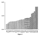
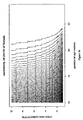
- Figure 6 shows that the standard variation ( see Formula 3) for every chromosome under a certain total number of unique reads is influenced by the participating cases number of the reference.
- the standard variation barely increases when the selected cases number was more than 150 under the condition that 1.7 million of total unique reads number were sequenced for each case.
- the standard variation was different for different chromosomes.
- our method had a moderate standard variation for chromosome 13 (0.0063), chromosome 18 (0.0066) and chromosome 21(0.0072).
- the standard variation of chromosome X is higher than above mentioned chromosomes which would require more strategies to do accurate abnormal detection.
- Figure 7 shows the Q-Q plot, wherein the residual is compiled to normal distribution which implicates the student-t calculation is reasonable.
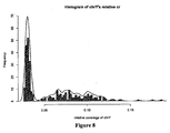
- Example 5 The distinguishing of fetal gender
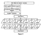
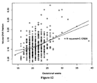
- 903 participants were recruited prospectively from Shenzhen People's Hospital and Shenzhen Maternal and child care service center with their karyotype results. Approvals were obtained from the institutional review boards of each recruitment site and all participants gave informed written consent. The maternal ages and gestational weeks at blood sampling were recorded. The 903 cases included 2 trisomy 13 cases, 15 trisomy 18 cases, 16 trisomy 21 cases, 3 XO cases, 2 XXY cases and 1 XYY cases. Their karyotype results distribution is shown in Figure 9 .
- Peripheral venous blood (5 milliliters) was collected from each participating pregnant woman in EDTA tubes and centrifuged at 1,600g for 10 min in 4 hours. Plasma was transferred to microcentrifuge tubes and recentrifuged at 16,000g for 10 min to remove residual cells. Cell-free plasma was stored at 80°C until DNA extraction. Each plasma sample was frozen and thawed only once.
- the size distribution of the sequencing libraries was analyzed with a DNA 1000 kit on the 2100 BioanalyzerTM (Agilent) and quantified with Real-time PCR.
- the sequencing libraries with different index were then pooled into one by equal quantity before cluster station on Illumina GA IITM (single-end sequencing).
- H1:the fetus is aneuploidy was that the mean coverage depth of the patient case distribution with a rough fetal fraction was equal to the mean coverage depth of the distribution of aneuploidy cases with the same fetal fraction, which means that this patient case is aneuploid if this null hypothesis were accepted.
- L i , j log p t ⁇ 1 i , j , degree
- L i,j Log likelihood ratio. If the ratio was larger than 1, we could infer the fetus might be trisomy.
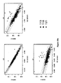
- the fetal DNA fraction was estimated first by Y and X. Meanwhile, we could extrapolate the fitted coverage depth for chromosome X with the fetal DNA fraction estimated only by the coverage depth of chromosome Y and t2 can be calculated.
- t2 i ( cr i,x - (1 - fy i / 2) ⁇ ⁇ r i,Xf )/ std Xf . If t2 is too large (larger than 5) or too small(less than -5) the fetus may be XXY or XYY.
- the gap between fetal fractions estimated by X and Y independently will provide information for detecting disorders about sex chromosomes.
- the CV value of chromosome 13 is 0.0066 with 100% sensitivity rate and 100% specificity rate.
- the CV value of chromosome 13 is 0.0063 and with 100% sensitivity rate and 100% specificity rate.
- the CV of these two approaches were 0.0062 and 0.0066, respectively, both with 100% sensitivity and specificity rates for them were 99.89% and 99.96%, respectively.
- the performance was similar when comparing the CV of these two approaches for chromosome 21: 0.0088 and 0.0072, respectively. Both resulted in the same sensitivity rate of 100% in our small cases set study and achieved the same 100% specificity rate.
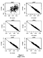
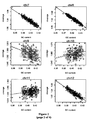
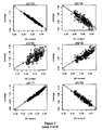
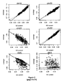
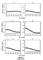
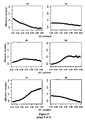
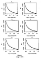
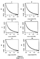
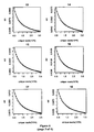
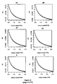
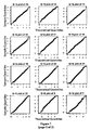
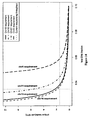
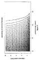
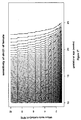
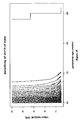
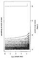
- Example 9 Theoretical performance of GC-correlation t-test approach in consideration of data size, gestational weeks and fetal DNA fraction
- Figures 15-21 show the resulting plots of our calculation.
- the student-t larger than 3 was set to identify female fetus aneuploidy while for male fetus, when computing probability density of false negative in every fraction, a logarithm likelihood larger than 1 was employed as the critical value we mentioned in Binary hypothesis which helped to achieve a higher sensitivity comparing to female ones.
Claims (14)
- Computerimplementiertes Verfahren zur Bestimmung einer genetischen Anomalie eines Fötus, bei der es sich um eine chromosomale Aneuploidie handelt, wobei das Verfahren folgendes umfasst:(a) das Erhalten von Sequenzinformationen mehrerer Polynukleotidfragmente aus einer Probe, wobei die genannte Probe eine periphere Blutprobe ist, die von einer schwangeren Frau gewonnen wird und sowohl mütterliche als auch fetale DNA enthält;(b) das Zuweisen der genannten Fragmente zu Chromosomen auf der Basis der genannten Informationssequenz durch Vergleichen der genannten Fragmente mit eindeutigen Referenzwerten für die gleiche Größe jedes der Chromosomen, wobei die eindeutigen Referenzwerte Fragmente eines Chromosoms sind, die eine eindeutige Sequenz aufweisen, die auf der Basis einer Referenz-Genomsequenz eindeutig einer einzelnen chromosomalen Position zugeordnet werden kann;(c) das Bestimmen der Testtiefe und des GC-Gehalts eines Chromosoms auf der Basis der Sequenzinformationen für diese Fragmente, die in Schritt (b) den eindeutigen Referenzwerten des genannten Chromosoms zugeordnet sind,
wobei die Testtiefe das Verhältnis zwischen der Anzahl der eindeutig dem Chromosom zugeordneten Fragmente und der Anzahl der eindeutigen Referenzwerte für das genannte Chromosom für die gleiche Fragmentgröße auf der Basis der genannten menschlichen Referenz-Genomsequenz ist;(d) das Bestimmen einer Ausgleichstesttiefe des genannten Chromosoms unter Verwendung des genannten GC-Gehalts des genannten Chromosoms und dem festgestellten Verhältnis zwischen der Testtiefe und dem GC-Gehalt für das genannte Chromosom in Abwesenheit von Aneuploidie, wobei das genannte festgestellte Verhältnis bestimmt ist durch ein Verfahren, das die folgenden Schritte umfasst:(i) das Erhalten von Sequenzinformationen mehrerer Polynukleotidfragmente für das genannte Chromosom aus einer Mehrzahl von euploiden peripheren Blutproben, die genomische DNA enthalten, wobei die Fragmentgröße der Fragmentgröße der mehreren Polynukleotidfragmente aus Schritt (a) oben entspricht;(ii) das Zuweisen der genannten Fragmente zu Chromosomen auf der Basis der genannten Sequenzinformationen wie in Schritt (b) oben;(iii) das Bestimmen der Testtiefe und des GC-Gehalts des genannten Chromosoms auf der Basis der genannten Sequenzinformationen für jede euploide Probe wie in Schritt (c) oben; und(iv) das Verwenden der in Schritt (iii) für jede Probe bestimmten Testtiefe und des GC-Gehalts zum Bestimmen des Verhältnisses zwischen der Testtiefe und dem GC-Gehalt des genannten Chromosoms in Abwesenheit von Aneuploidie; und(e) das Vergleichen der genannten Ausgleichstesttiefe mit der in Schritt (c) bestimmten Testtiefe des genannten Chromosoms, wobei eine Differenz zwischen diesen eine fetale chromosomale Aneuploidie indiziert. - Verfahren nach Anspruch 1, wobei Schritt (a) ferner das Erhalten von Sequenzinformationen mehrerer Polynukleotidfragmente von mehreren verschiedenen Proben umfasst, und wobei die Testtiefe normalisiert ist, so dass sie Differenzen der Gesamtanzahl von erhaltenen Sequenzwerten für verschiedene Proben berücksichtigt, zum Beispiel im Vergleich zu der durchschnittlichen Testtiefe eines anderen Chromosoms, vorzugsweise im Vergleich zu der durchschnittlichen Testtiefe aller anderen Autosome oder im Vergleich zu der durchschnittlichen Testtiefe aller anderen Chromosomen.
- Verfahren nach Anspruch 1 oder Anspruch 2, wobei der GC-Gehalt des Chromosoms bestimmt wird wie der durchschnittliche GC-Gehalt aller Fragmente, die dem genannten Chromosom für den Zweck aus Schritt (c) zugeordnet sind.
- Verfahren nach Anspruch 2, wobei dieses ferner das Bestimmen des fetalen Geschlechts zum Beispiel gemäß folgender Formel umfasst:

wobei cr.a i,x und cr.a i,y die entsprechende relative Testtiefe der X- und Y-Chromosomen sind. - Verfahren nach Anspruch 2, wobei dieses ferner das Schätzen der fetalen Fraktion umfasst, wobei die fetale Fraktion berechnet wird auf der Basis der Testtiefe von Chromosom X und I oder Y gemäß der Bestimmung in Schritt (c) aus Anspruch 1 gemäß einer Formel, die ausgewählt wird aus:

wobei cri,Y die Testtiefe von Chromosom Y der Probe i ist, wobei ĉri,Yf = f(GCi,Yf ) die aus dem Verhältnis der Testtiefe für Chromosom Y und dem entsprechenden GC-Gehalt von Proben schwangerer Frauen mit weiblichem Fötus berechnete Ausgleichstesttiefe ist, und ĉri,Ym = f(GCi,Ym ) die aus dem Verhältnis der Testtiefe für Chromosom Y und dem entsprechenden GC-Gehalt von Proben männlicher Probanden berechnete Ausgleichstesttiefe ist; oder
wobei cri,X die Testtiefe von Chromosom X der Probe i ist, wobei ĉri,Xf = f(GCi,Xf ) die aus dem Verhältnis der Testtiefe für Chromosom X und dem entsprechenden GC-Gehalt von Proben schwangerer Frauen mit weiblichem Fötus berechnete Ausgleichstesttiefe ist, und ĉri,Xm = f(GCi,Xm ) die aus dem Verhältnis der Testtiefe für Chromosom X und dem entsprechenden GC-Gehalt von Proben männlicher Probanden berechnete Ausgleichstesttiefe ist; oder
wobei ĉri,Xf = f(GCi,Xf ) die aus dem Verhältnis der Testtiefe für Chromosom X und dem entsprechenden GC-Gehalt von Proben schwangerer Frauen mit weiblichem Fötus berechnete Ausgleichstesttiefe ist, wobei ĉri,Yf = f(GCi,Yf ) die aus dem Verhältnis der Testtiefe für Chromosom Y und dem entsprechenden GC-Gehalt von Proben schwangerer Frauen mit weiblichem Fötus berechnete Ausgleichstesttiefe ist, wobei ĉri,Xm = f(GCi,Xm ) die aus dem Verhältnis der Testtiefe für Chromosom X und dem entsprechenden GC-Gehalt von Proben männlicher Probanden berechnete Ausgleichstesttiefe ist, und wobei ĉri,Ym = f(GCi,Ym ) die aus dem Verhältnis der Testtiefe für Chromosom Y und dem entsprechenden GC-Gehalt von Proben männlicher Probanden berechnete Ausgleichstesttiefe ist. - Verfahren nach Anspruch 2, wobei der Vergleich der genannten Ausgleichstesttiefe mit der in Schritt (c) bestimmten Testtiefe des Chromosoms durch einen statistischen Hypothesetest durchgeführt wird, wobei es eine Hypothese ist, dass der Fötus euploid (H0) ist, und wobei es die andere Hypothese ist, dass der Fötus für das genannte Chromosom Aneuploidie aufweist (H1).
- Verfahren nach Anspruch 6, wobei die studentsche t-Verteilung für beide Hypothesen berechnet wird.
- Verfahren nach Anspruch 7, wobei die studentsche t-Verteilung für H0 und H1 gemäß folgender Formeln entsprechend berechnet wird:

und
wobei fxy die fetale Fraktion ist,
i der Probenindex ist,
j die Chromosomnummer ist,
wobei f(GCi,j) die Funktion des Verhältnisses zwischen der normalisierten Testtiefe und dem entsprechenden GC-Gehalt der Probe i darstellt, wobei Chromosom j, εi,j den Rest von Probe i, Chromosom j darstellt,
und die Ausgleichstesttiefe darstellt; und
stdj die Standardabweichung gemäß folgender Formel darstellt:
wobei ns für die Anzahl der Referenzproben steht. - Verfahren nach Anspruch 8, wobei das Log-Likelihood-Verhältnis von t1 und t2 gemäß folgender Formel berechnet wird: L i,j = log(p(t1 i,j ,Grad|D))/log(p(t2i,j,Grad|T)), wobei Li,j das Log-Likelihood-Verhältnis ist, wobei Grad einen t-Verteilungsgrad bezeichnet, wobei D für Diploidie steht, wobei T für Trisomie steht, und wobei p(t1 i,j ,Grad|*), *=D,T, für die bedingte Wahrscheinlichkeitsdichte bei gegebenem t-Verteilungsgrad steht, wobei für den Fall, dass das Verhältnis größer ist als 1, daraus für den Fötus geschlossen wird, dass dieser für das genannte Chromosom Trisomie aufweist.
- Verfahren nach einem der Ansprüche 1 bis 9 zur Verwendung zur Bestimmung einer fetalen autosomalen Aneuploidie.
- Verfahren nach Anspruch 10, wobei die fetale Aneuploidie ausgewählt ist aus der Gruppe bestehend aus Trisomie 13, 18 und 21.
- Verfahren nach Anspruch 4 zum Einsatz in der Bestimmung einer Geschlechtschromosom-Aneuploidie, wie etwa einer Geschlechtschromosom-Aneuploidie, die ausgewählt ist aus der Gruppe bestehend aus XO, XXX, XXY und XYY.
- Computerlesbares Medium, das eine Mehrzahl von Anweisungen umfasst, die das Verfahren nach einem der Ansprüche 1 bis 12 ausführen, wenn die genannten Anweisungen ergänzt werden
- System, das Mittel umfasst, die sich zur Ausführung des Verfahrens nach einem der Ansprüche 1 bis 12 eignet.
Priority Applications (2)
| Application Number | Priority Date | Filing Date | Title |
|---|---|---|---|
| SI201130281T SI2561103T1 (sl) | 2011-06-29 | 2011-06-29 | Neinvazivna detekcija genetske anomalije ploda |
| PL11863253T PL2561103T3 (pl) | 2011-06-29 | 2011-06-29 | Nieinwazyjna detekcja anomalii genetycznych płodu |
Applications Claiming Priority (1)
| Application Number | Priority Date | Filing Date | Title |
|---|---|---|---|
| PCT/CN2011/001070 WO2013000100A1 (en) | 2011-06-29 | 2011-06-29 | Noninvasive detection of fetal genetic abnormality |
Publications (3)
| Publication Number | Publication Date |
|---|---|
| EP2561103A1 EP2561103A1 (de) | 2013-02-27 |
| EP2561103A4 EP2561103A4 (de) | 2013-08-07 |
| EP2561103B1 true EP2561103B1 (de) | 2014-08-27 |
Family
ID=47392194
Family Applications (1)
| Application Number | Title | Priority Date | Filing Date |
|---|---|---|---|
| EP11863253.8A Active EP2561103B1 (de) | 2011-06-29 | 2011-06-29 | Nichtinvasive erkennung genetischer anomalien von föten |
Country Status (18)
| Country | Link |
|---|---|
| US (1) | US9547748B2 (de) |
| EP (1) | EP2561103B1 (de) |
| JP (1) | JP5659319B2 (de) |
| KR (1) | KR101489568B1 (de) |
| CN (1) | CN103403183B (de) |
| AU (1) | AU2012261664B2 (de) |
| BR (1) | BR112012033760B1 (de) |
| CA (2) | CA2791118C (de) |
| DK (1) | DK2561103T3 (de) |
| ES (1) | ES2512448T3 (de) |
| HK (1) | HK1190758A1 (de) |
| MY (1) | MY172864A (de) |
| PL (1) | PL2561103T3 (de) |
| RU (1) | RU2589681C2 (de) |
| SG (1) | SG191757A1 (de) |
| SI (1) | SI2561103T1 (de) |
| WO (1) | WO2013000100A1 (de) |
| ZA (1) | ZA201209583B (de) |
Families Citing this family (59)
| Publication number | Priority date | Publication date | Assignee | Title |
|---|---|---|---|---|
| US11270781B2 (en) | 2011-01-25 | 2022-03-08 | Ariosa Diagnostics, Inc. | Statistical analysis for non-invasive sex chromosome aneuploidy determination |
| US20140235474A1 (en) | 2011-06-24 | 2014-08-21 | Sequenom, Inc. | Methods and processes for non invasive assessment of a genetic variation |
| SG191757A1 (en) | 2011-06-29 | 2013-08-30 | Bgi Health Service Co Ltd | Noninvasive detection of fetal genetic abnormality |
| US9984198B2 (en) | 2011-10-06 | 2018-05-29 | Sequenom, Inc. | Reducing sequence read count error in assessment of complex genetic variations |
| US10424394B2 (en) | 2011-10-06 | 2019-09-24 | Sequenom, Inc. | Methods and processes for non-invasive assessment of genetic variations |
| US10196681B2 (en) | 2011-10-06 | 2019-02-05 | Sequenom, Inc. | Methods and processes for non-invasive assessment of genetic variations |
| US9367663B2 (en) | 2011-10-06 | 2016-06-14 | Sequenom, Inc. | Methods and processes for non-invasive assessment of genetic variations |
| WO2013052907A2 (en) | 2011-10-06 | 2013-04-11 | Sequenom, Inc. | Methods and processes for non-invasive assessment of genetic variations |
| EP2805280B1 (de) | 2012-01-20 | 2022-10-05 | Sequenom, Inc. | Diagnostische verfahren zur evaluierung von versuchsbedingungen |
| US10504613B2 (en) | 2012-12-20 | 2019-12-10 | Sequenom, Inc. | Methods and processes for non-invasive assessment of genetic variations |
| US9920361B2 (en) | 2012-05-21 | 2018-03-20 | Sequenom, Inc. | Methods and compositions for analyzing nucleic acid |
| US10497461B2 (en) | 2012-06-22 | 2019-12-03 | Sequenom, Inc. | Methods and processes for non-invasive assessment of genetic variations |
| US10482994B2 (en) | 2012-10-04 | 2019-11-19 | Sequenom, Inc. | Methods and processes for non-invasive assessment of genetic variations |
| US20130309666A1 (en) | 2013-01-25 | 2013-11-21 | Sequenom, Inc. | Methods and processes for non-invasive assessment of genetic variations |
| EP2959015B1 (de) * | 2013-02-20 | 2020-11-04 | Bionano Genomics, Inc. | Charakterisierung von molekülen in nanofluiden |
| US10844424B2 (en) | 2013-02-20 | 2020-11-24 | Bionano Genomics, Inc. | Reduction of bias in genomic coverage measurements |
| WO2015130696A1 (en) * | 2014-02-25 | 2015-09-03 | Bionano Genomics, Inc. | Reduction of bias in genomic coverage measurements |
| WO2014133369A1 (ko) * | 2013-02-28 | 2014-09-04 | 주식회사 테라젠이텍스 | 유전체 서열분석을 이용한 태아 염색체 이수성의 진단 방법 및 장치 |
| LT2981921T (lt) * | 2013-04-03 | 2023-02-27 | Sequenom, Inc. | Neinvazinio genetinių variacijų vertinimo būdai ir procesai |
| EP3004383B1 (de) * | 2013-05-24 | 2019-04-24 | Sequenom, Inc. | Auf fläche unter der kurve (auc) analyse basiertes verfahren zur nichtinvasiven beurteilung genetischer variationen |
| CN105074011B (zh) * | 2013-06-13 | 2020-10-02 | 阿瑞奥萨诊断公司 | 用于非入侵性性染色体非整倍性确定的统计分析 |
| MX2015016911A (es) * | 2013-06-21 | 2016-06-21 | Sequenom Inc | Metodos y procesos para evaluacion no invasiva de variaciones geneticas. |
| CA3205430A1 (en) | 2013-10-04 | 2015-04-09 | Sequenom, Inc. | Methods and processes for non-invasive assessment of genetic variations |
| US10438691B2 (en) | 2013-10-07 | 2019-10-08 | Sequenom, Inc. | Non-invasive assessment of chromosome alterations using change in subsequence mappability |
| CA2928185C (en) | 2013-10-21 | 2024-01-30 | Verinata Health, Inc. | Method for improving the sensitivity of detection in determining copy number variations |
| CN103525939B (zh) * | 2013-10-28 | 2015-12-02 | 博奥生物集团有限公司 | 无创检测胎儿染色体非整倍体的方法和系统 |
| EP3149199B1 (de) * | 2014-05-30 | 2020-03-25 | Verinata Health, Inc. | Nachweis von eventuell fetalen subchromosomalen aneuploidien und kopienzahlvariationen |
| CN104156631B (zh) * | 2014-07-14 | 2017-07-18 | 天津华大基因科技有限公司 | 染色体三倍体检验方法 |
| WO2016010401A1 (ko) * | 2014-07-18 | 2016-01-21 | 에스케이텔레콘 주식회사 | 산모의 혈청 dna를 이용한 태아의 단일유전자 유전변이의 예측방법 |
| US11783911B2 (en) | 2014-07-30 | 2023-10-10 | Sequenom, Inc | Methods and processes for non-invasive assessment of genetic variations |
| WO2016045106A1 (zh) * | 2014-09-26 | 2016-03-31 | 深圳华大基因股份有限公司 | 单细胞染色体的cnv分析方法和检测装置 |
| EP3502273B1 (de) | 2014-12-12 | 2020-07-08 | Verinata Health, Inc. | Zellfreies dna-fragment |
| CN104789466B (zh) * | 2015-05-06 | 2018-03-13 | 安诺优达基因科技(北京)有限公司 | 检测染色体非整倍性的试剂盒和装置 |
| BE1022789B1 (nl) * | 2015-07-17 | 2016-09-06 | Multiplicom Nv | Werkwijze en systeem voor geslachtsinschatting van een foetus van een zwangere vrouw |
| KR101817785B1 (ko) * | 2015-08-06 | 2018-01-11 | 이원다이애그노믹스(주) | 다양한 플랫폼에서 태아의 성별과 성염색체 이상을 구분할 수 있는 새로운 방법 |
| KR101678962B1 (ko) | 2015-08-21 | 2016-12-06 | 이승재 | 대규모 병렬형 게놈서열분석 방법을 이용한 비침습적 산전검사 장치 및 방법 |
| WO2017051996A1 (ko) * | 2015-09-24 | 2017-03-30 | 에스케이텔레콤 주식회사 | 비침습적 태아 염색체 이수성 판별 방법 |
| CN105354443A (zh) * | 2015-12-14 | 2016-02-24 | 孔祥军 | 无创产前基因检测分析软件 |
| CN105483229B (zh) * | 2015-12-21 | 2018-10-16 | 广东腾飞基因科技股份有限公司 | 一种检测胎儿染色体非整倍体的方法及系统 |
| KR101817180B1 (ko) * | 2016-01-20 | 2018-01-10 | 이원다이애그노믹스(주) | 염색체 이상 판단 방법 |
| US10095831B2 (en) | 2016-02-03 | 2018-10-09 | Verinata Health, Inc. | Using cell-free DNA fragment size to determine copy number variations |
| CN106096330B (zh) * | 2016-05-31 | 2019-02-01 | 北京百迈客医学检验所有限公司 | 一种无创产前生物信息检测分析方法 |
| JP6785068B2 (ja) * | 2016-05-31 | 2020-11-18 | 富士フイルム株式会社 | 生物情報解析方法 |
| EP3491560A1 (de) | 2016-07-27 | 2019-06-05 | Sequenom, Inc. | Genkopienzahlvariationklassifizierungen |
| US11694768B2 (en) | 2017-01-24 | 2023-07-04 | Sequenom, Inc. | Methods and processes for assessment of genetic variations |
| CN110191951A (zh) | 2017-01-24 | 2019-08-30 | 深圳华大生命科学研究院 | 基于外泌体dna进行无创产前诊断的方法及其应用 |
| EP3601591A1 (de) * | 2017-03-31 | 2020-02-05 | Premaitha Limited | Verfahren zur detektion einer chromosomalen anomalie eines fötus |
| US11342047B2 (en) | 2017-04-21 | 2022-05-24 | Illumina, Inc. | Using cell-free DNA fragment size to detect tumor-associated variant |
| EP4335928A3 (de) * | 2018-01-05 | 2024-04-17 | BillionToOne, Inc. | Qualitätskontrollvorlagen zur sicherstellung der gültigkeit von tests auf sequenzierungsbasis |
| WO2019195975A1 (zh) * | 2018-04-09 | 2019-10-17 | 深圳华大生命科学研究院 | 基因文库的构建方法及其应用 |
| CN111918965A (zh) * | 2018-04-28 | 2020-11-10 | 深圳华大生命科学研究院 | 一种胎儿游离核酸的富集方法及其应用 |
| CN111373054A (zh) * | 2018-05-31 | 2020-07-03 | 深圳华大临床检验中心 | 确定男性待测样本是否存在三倍体的方法、系统和计算机可读介质 |
| CN109192243B (zh) * | 2018-08-13 | 2021-03-12 | 成都凡迪医学检验所有限公司 | 染色体比例的修正方法、装置、介质 |
| KR20200106643A (ko) | 2019-03-05 | 2020-09-15 | (주)인실리코젠 | 바코드 서열 정보 기반 고민감도 유전변이 탐지 및 레포팅 시스템 |
| WO2020226528A1 (ru) * | 2019-05-08 | 2020-11-12 | Общество с ограниченной ответственностью "ГЕНОТЕК ИТ" | Способ определения кариотипа плода беременной женщины |
| CN110211654A (zh) * | 2019-05-30 | 2019-09-06 | 湖南自兴智慧医疗科技有限公司 | 一种自动隐藏性别信息的核型检测系统及方法 |
| CN111627498B (zh) * | 2020-05-21 | 2022-10-04 | 北京吉因加医学检验实验室有限公司 | 一种测序数据gc偏向性校正的方法及其装置 |
| RU2752783C1 (ru) * | 2020-12-18 | 2021-08-03 | Федеральное государственное бюджетное учреждение "Ивановский научно-исследовательский институт материнства и детства имени В.Н. Городкова" Министерства здравоохранения Российской Федерации | Способ прогнозирования анеуплоидии эмбрионов в программе экстракорпорального оплодотворения у женщин с эндометриоз-ассоциированным бесплодием |
| WO2023031641A1 (en) * | 2021-09-03 | 2023-03-09 | Inserm ( Institut National De La Sante Et De La Recherche Medicale) | Methods and devices for non-invasive prenatal testing |
Family Cites Families (30)
| Publication number | Priority date | Publication date | Assignee | Title |
|---|---|---|---|---|
| US10021A (en) * | 1853-09-13 | Screw-eastemtito- for boots and shoes | ||
| GB9704444D0 (en) | 1997-03-04 | 1997-04-23 | Isis Innovation | Non-invasive prenatal diagnosis |
| US20010051341A1 (en) | 1997-03-04 | 2001-12-13 | Isis Innovation Limited | Non-invasive prenatal diagnosis |
| US6492144B1 (en) | 1997-05-30 | 2002-12-10 | Diagen Corporation | Methods for detection of nucleic acid sequences in urine |
| USRE39920E1 (en) | 1997-05-30 | 2007-11-13 | Xenomics, Inc. | Methods for detection of nucleic acid sequences in urine |
| ATE320505T1 (de) | 1997-05-30 | 2006-04-15 | Xenomics | Verfahren zur bestimmung von nukleinsäuresequenzen in urin |
| US20020119478A1 (en) | 1997-05-30 | 2002-08-29 | Diagen Corporation | Methods for detection of nucleic acid sequences in urine |
| US20070087345A1 (en) | 2003-07-10 | 2007-04-19 | Third Wave Technologies, Inc. | Assays for the direct measurement of gene dosage |
| DE60328193D1 (de) | 2003-10-16 | 2009-08-13 | Sequenom Inc | Nicht invasiver Nachweis fötaler genetischer Merkmale |
| US20060046258A1 (en) | 2004-02-27 | 2006-03-02 | Lapidus Stanley N | Applications of single molecule sequencing |
| US20100216153A1 (en) * | 2004-02-27 | 2010-08-26 | Helicos Biosciences Corporation | Methods for detecting fetal nucleic acids and diagnosing fetal abnormalities |
| US20100216151A1 (en) | 2004-02-27 | 2010-08-26 | Helicos Biosciences Corporation | Methods for detecting fetal nucleic acids and diagnosing fetal abnormalities |
| SI2385143T1 (sl) | 2006-02-02 | 2016-11-30 | The Board of Trustees of the Leland Stanford Junior University Office of the General Counsel | Neinvazivni genetski presejalni test ploda z digitalno analizo |
| DE602007014335D1 (de) | 2006-02-28 | 2011-06-16 | Univ Louisville Res Found | Erkennung von chromosomabnormalitäten im fötus mit hilfe der tandem-einzelnukleotid-polymorphismen |
| US20100184043A1 (en) | 2006-02-28 | 2010-07-22 | University Of Louisville Research Foundation | Detecting Genetic Abnormalities |
| US20100184044A1 (en) | 2006-02-28 | 2010-07-22 | University Of Louisville Research Foundation | Detecting Genetic Abnormalities |
| US20080038733A1 (en) | 2006-03-28 | 2008-02-14 | Baylor College Of Medicine | Screening for down syndrome |
| US8372584B2 (en) | 2006-06-14 | 2013-02-12 | The General Hospital Corporation | Rare cell analysis using sample splitting and DNA tags |
| US20080050739A1 (en) | 2006-06-14 | 2008-02-28 | Roland Stoughton | Diagnosis of fetal abnormalities using polymorphisms including short tandem repeats |
| US8137912B2 (en) | 2006-06-14 | 2012-03-20 | The General Hospital Corporation | Methods for the diagnosis of fetal abnormalities |
| US20080026390A1 (en) | 2006-06-14 | 2008-01-31 | Roland Stoughton | Diagnosis of Fetal Abnormalities by Comparative Genomic Hybridization Analysis |
| US20080070792A1 (en) | 2006-06-14 | 2008-03-20 | Roland Stoughton | Use of highly parallel snp genotyping for fetal diagnosis |
| US20080090239A1 (en) | 2006-06-14 | 2008-04-17 | Daniel Shoemaker | Rare cell analysis using sample splitting and dna tags |
| CA2655272C (en) | 2006-06-14 | 2017-04-18 | Living Microsystems, Inc. | Rare cell analysis using sample splitting and dna tags |
| US20080113358A1 (en) | 2006-07-28 | 2008-05-15 | Ravi Kapur | Selection of cells using biomarkers |
| WO2008070862A2 (en) | 2006-12-07 | 2008-06-12 | Biocept, Inc. | Non-invasive prenatal genetic screen |
| PT2183693E (pt) | 2007-07-23 | 2014-01-14 | Univ Hong Kong Chinese | Diagnóstico de aneuploidia cromossómica fetal utilizando sequenciação genómica |
| US20100112590A1 (en) | 2007-07-23 | 2010-05-06 | The Chinese University Of Hong Kong | Diagnosing Fetal Chromosomal Aneuploidy Using Genomic Sequencing With Enrichment |
| EP3378951B1 (de) | 2008-09-20 | 2020-05-13 | The Board of Trustees of the Leland Stanford Junior University | Nicht invasive diagnose von aneuploidie durch sequenzierung |
| SG191757A1 (en) | 2011-06-29 | 2013-08-30 | Bgi Health Service Co Ltd | Noninvasive detection of fetal genetic abnormality |
-
2011
- 2011-06-29 SG SG2013049317A patent/SG191757A1/en unknown
- 2011-06-29 RU RU2012158107/15A patent/RU2589681C2/ru active
- 2011-06-29 AU AU2012261664A patent/AU2012261664B2/en active Active
- 2011-06-29 CA CA2791118A patent/CA2791118C/en active Active
- 2011-06-29 WO PCT/CN2011/001070 patent/WO2013000100A1/en active Application Filing
- 2011-06-29 ES ES11863253.8T patent/ES2512448T3/es active Active
- 2011-06-29 KR KR1020127034453A patent/KR101489568B1/ko active IP Right Review Request
- 2011-06-29 SI SI201130281T patent/SI2561103T1/sl unknown
- 2011-06-29 PL PL11863253T patent/PL2561103T3/pl unknown
- 2011-06-29 US US13/641,080 patent/US9547748B2/en active Active
- 2011-06-29 CN CN201180067286.XA patent/CN103403183B/zh active Active
- 2011-06-29 DK DK11863253.8T patent/DK2561103T3/da active
- 2011-06-29 EP EP11863253.8A patent/EP2561103B1/de active Active
- 2011-06-29 CA CA2948939A patent/CA2948939C/en active Active
- 2011-06-29 BR BR112012033760-2A patent/BR112012033760B1/pt active IP Right Grant
- 2011-06-29 JP JP2014517369A patent/JP5659319B2/ja active Active
- 2011-06-29 MY MYPI2012005470A patent/MY172864A/en unknown
-
2012
- 2012-12-18 ZA ZA2012/09583A patent/ZA201209583B/en unknown
-
2014
- 2014-04-23 HK HK14103891.8A patent/HK1190758A1/xx unknown
Also Published As
| Publication number | Publication date |
|---|---|
| ES2512448T3 (es) | 2014-10-24 |
| AU2012261664B2 (en) | 2014-07-03 |
| RU2589681C2 (ru) | 2016-07-10 |
| JP5659319B2 (ja) | 2015-01-28 |
| CA2948939C (en) | 2021-02-02 |
| CA2791118A1 (en) | 2012-12-29 |
| PL2561103T3 (pl) | 2015-02-27 |
| BR112012033760B1 (pt) | 2020-11-17 |
| US20140099642A1 (en) | 2014-04-10 |
| CA2948939A1 (en) | 2012-12-29 |
| JP2014520509A (ja) | 2014-08-25 |
| EP2561103A4 (de) | 2013-08-07 |
| CA2791118C (en) | 2019-05-07 |
| KR20140023847A (ko) | 2014-02-27 |
| SG191757A1 (en) | 2013-08-30 |
| ZA201209583B (en) | 2014-01-29 |
| WO2013000100A1 (en) | 2013-01-03 |
| SI2561103T1 (sl) | 2014-11-28 |
| RU2012158107A (ru) | 2015-08-10 |
| MY172864A (en) | 2019-12-13 |
| KR101489568B1 (ko) | 2015-02-03 |
| HK1190758A1 (en) | 2014-07-11 |
| US9547748B2 (en) | 2017-01-17 |
| BR112012033760A2 (pt) | 2018-02-27 |
| EP2561103A1 (de) | 2013-02-27 |
| AU2012261664A1 (en) | 2013-01-17 |
| DK2561103T3 (da) | 2014-10-20 |
| CN103403183A (zh) | 2013-11-20 |
| CN103403183B (zh) | 2014-10-15 |
Similar Documents
| Publication | Publication Date | Title |
|---|---|---|
| EP2561103B1 (de) | Nichtinvasive erkennung genetischer anomalien von föten | |
| JP6317354B2 (ja) | 血漿による胎児または腫瘍のメチロームの非侵襲的決定 | |
| US9353414B2 (en) | Noninvasive diagnosis of fetal aneuploidy by sequencing | |
| CN104120181B (zh) | 对染色体测序结果进行gc校正的方法及装置 | |
| HUE030510T2 (hu) | Magzati kromoszómális aneuploidia diagnosztizálása genomszekvenálás alkalmazásával | |
| TW201441618A (zh) | 藉由大量平行rna定序之母體血漿轉錄體分析 | |
| EP3018213A1 (de) | Verfahren zur Bestimmung des Vorhandenseins eines biologischen Zustands durch Bestimmung sowohl der relativen als auch der totalen Menge an zwei verschiedenen Nukleinsäuren | |
| EP3662479A1 (de) | Verfahren zur nicht-invasiven pränatalen detektion von fetalen chromosomalen geschlechtsabnormitäten und fetalen geschlechtsbestimmung für einzel- und zwillingsschwangerschaften | |
| TWI489305B (zh) | 對胎兒遺傳異常的無創性檢測 | |
| Sillence | Cell-free fetal DNA (cffDNA) enrichment for non-invasive prenatal testing (NIPT): a comparison of molecular techniques |
Legal Events
| Date | Code | Title | Description |
|---|---|---|---|
| PUAI | Public reference made under article 153(3) epc to a published international application that has entered the european phase |
Free format text: ORIGINAL CODE: 0009012 |
|
| 17P | Request for examination filed |
Effective date: 20121016 |
|
| AK | Designated contracting states |
Kind code of ref document: A1 Designated state(s): AL AT BE BG CH CY CZ DE DK EE ES FI FR GB GR HR HU IE IS IT LI LT LU LV MC MK MT NL NO PL PT RO RS SE SI SK SM TR |
|
| A4 | Supplementary search report drawn up and despatched |
Effective date: 20130710 |
|
| RAP1 | Party data changed (applicant data changed or rights of an application transferred) |
Owner name: BGI HEALTH SERVICE CO., LTD |
|
| RIC1 | Information provided on ipc code assigned before grant |
Ipc: G06F 19/22 20110101ALN20130704BHEP Ipc: C12Q 1/68 20060101ALI20130704BHEP Ipc: G06F 19/18 20110101AFI20130704BHEP |
|
| 17Q | First examination report despatched |
Effective date: 20130724 |
|
| REG | Reference to a national code |
Ref country code: DE Ref legal event code: R079 Ref document number: 602011009544 Country of ref document: DE Free format text: PREVIOUS MAIN CLASS: C12Q0001680000 Ipc: G06F0019180000 |
|
| GRAP | Despatch of communication of intention to grant a patent |
Free format text: ORIGINAL CODE: EPIDOSNIGR1 |
|
| RIC1 | Information provided on ipc code assigned before grant |
Ipc: G06F 19/18 20110101AFI20140611BHEP Ipc: G06F 19/22 20110101ALN20140611BHEP Ipc: C12Q 1/68 20060101ALI20140611BHEP |
|
| GRAS | Grant fee paid |
Free format text: ORIGINAL CODE: EPIDOSNIGR3 |
|
| GRAA | (expected) grant |
Free format text: ORIGINAL CODE: 0009210 |
|
| DAX | Request for extension of the european patent (deleted) | ||
| INTG | Intention to grant announced |
Effective date: 20140703 |
|
| RIN1 | Information on inventor provided before grant (corrected) |
Inventor name: YUAN, YUYING Inventor name: CHEN, FANG Inventor name: ZHANG, XIUQING Inventor name: JIANG, FUMAN Inventor name: CHAI, XIANGHUA Inventor name: CHEN, HUIFEI |
|
| AK | Designated contracting states |
Kind code of ref document: B1 Designated state(s): AL AT BE BG CH CY CZ DE DK EE ES FI FR GB GR HR HU IE IS IT LI LT LU LV MC MK MT NL NO PL PT RO RS SE SI SK SM TR |
|
| RAP1 | Party data changed (applicant data changed or rights of an application transferred) |
Owner name: BGI DIAGNOSIS CO., LTD. |
|
| REG | Reference to a national code |
Ref country code: GB Ref legal event code: FG4D |
|
| REG | Reference to a national code |
Ref country code: CH Ref legal event code: EP |
|
| REG | Reference to a national code |
Ref country code: AT Ref legal event code: REF Ref document number: 684822 Country of ref document: AT Kind code of ref document: T Effective date: 20140915 |
|
| REG | Reference to a national code |
Ref country code: IE Ref legal event code: FG4D |
|
| REG | Reference to a national code |
Ref country code: RO Ref legal event code: EPE |
|
| REG | Reference to a national code |
Ref country code: DE Ref legal event code: R096 Ref document number: 602011009544 Country of ref document: DE Effective date: 20141009 |
|
| REG | Reference to a national code |
Ref country code: CH Ref legal event code: NV Representative=s name: MEYER AND KOLLEGEN, CH |
|
| REG | Reference to a national code |
Ref country code: DK Ref legal event code: T3 Effective date: 20141016 |
|
| REG | Reference to a national code |
Ref country code: ES Ref legal event code: FG2A Ref document number: 2512448 Country of ref document: ES Kind code of ref document: T3 Effective date: 20141024 |
|
| REG | Reference to a national code |
Ref country code: SE Ref legal event code: TRGR |
|
| REG | Reference to a national code |
Ref country code: AT Ref legal event code: MK05 Ref document number: 684822 Country of ref document: AT Kind code of ref document: T Effective date: 20140827 |
|
| REG | Reference to a national code |
Ref country code: LT Ref legal event code: MG4D |
|
| REG | Reference to a national code |
Ref country code: NL Ref legal event code: VDEP Effective date: 20140827 |
|
| PG25 | Lapsed in a contracting state [announced via postgrant information from national office to epo] |
Ref country code: NO Free format text: LAPSE BECAUSE OF FAILURE TO SUBMIT A TRANSLATION OF THE DESCRIPTION OR TO PAY THE FEE WITHIN THE PRESCRIBED TIME-LIMIT Effective date: 20141127 Ref country code: GR Free format text: LAPSE BECAUSE OF FAILURE TO SUBMIT A TRANSLATION OF THE DESCRIPTION OR TO PAY THE FEE WITHIN THE PRESCRIBED TIME-LIMIT Effective date: 20141128 Ref country code: LT Free format text: LAPSE BECAUSE OF FAILURE TO SUBMIT A TRANSLATION OF THE DESCRIPTION OR TO PAY THE FEE WITHIN THE PRESCRIBED TIME-LIMIT Effective date: 20140827 Ref country code: BG Free format text: LAPSE BECAUSE OF FAILURE TO SUBMIT A TRANSLATION OF THE DESCRIPTION OR TO PAY THE FEE WITHIN THE PRESCRIBED TIME-LIMIT Effective date: 20141127 Ref country code: FI Free format text: LAPSE BECAUSE OF FAILURE TO SUBMIT A TRANSLATION OF THE DESCRIPTION OR TO PAY THE FEE WITHIN THE PRESCRIBED TIME-LIMIT Effective date: 20140827 Ref country code: PT Free format text: LAPSE BECAUSE OF FAILURE TO SUBMIT A TRANSLATION OF THE DESCRIPTION OR TO PAY THE FEE WITHIN THE PRESCRIBED TIME-LIMIT Effective date: 20141229 |
|
| PG25 | Lapsed in a contracting state [announced via postgrant information from national office to epo] |
Ref country code: HR Free format text: LAPSE BECAUSE OF FAILURE TO SUBMIT A TRANSLATION OF THE DESCRIPTION OR TO PAY THE FEE WITHIN THE PRESCRIBED TIME-LIMIT Effective date: 20140827 Ref country code: CY Free format text: LAPSE BECAUSE OF FAILURE TO SUBMIT A TRANSLATION OF THE DESCRIPTION OR TO PAY THE FEE WITHIN THE PRESCRIBED TIME-LIMIT Effective date: 20140827 Ref country code: RS Free format text: LAPSE BECAUSE OF FAILURE TO SUBMIT A TRANSLATION OF THE DESCRIPTION OR TO PAY THE FEE WITHIN THE PRESCRIBED TIME-LIMIT Effective date: 20140827 Ref country code: LV Free format text: LAPSE BECAUSE OF FAILURE TO SUBMIT A TRANSLATION OF THE DESCRIPTION OR TO PAY THE FEE WITHIN THE PRESCRIBED TIME-LIMIT Effective date: 20140827 Ref country code: AT Free format text: LAPSE BECAUSE OF FAILURE TO SUBMIT A TRANSLATION OF THE DESCRIPTION OR TO PAY THE FEE WITHIN THE PRESCRIBED TIME-LIMIT Effective date: 20140827 Ref country code: IS Free format text: LAPSE BECAUSE OF FAILURE TO SUBMIT A TRANSLATION OF THE DESCRIPTION OR TO PAY THE FEE WITHIN THE PRESCRIBED TIME-LIMIT Effective date: 20141227 |
|
| REG | Reference to a national code |
Ref country code: PL Ref legal event code: T3 |
|
| PG25 | Lapsed in a contracting state [announced via postgrant information from national office to epo] |
Ref country code: NL Free format text: LAPSE BECAUSE OF FAILURE TO SUBMIT A TRANSLATION OF THE DESCRIPTION OR TO PAY THE FEE WITHIN THE PRESCRIBED TIME-LIMIT Effective date: 20140827 |
|
| PG25 | Lapsed in a contracting state [announced via postgrant information from national office to epo] |
Ref country code: EE Free format text: LAPSE BECAUSE OF FAILURE TO SUBMIT A TRANSLATION OF THE DESCRIPTION OR TO PAY THE FEE WITHIN THE PRESCRIBED TIME-LIMIT Effective date: 20140827 Ref country code: SK Free format text: LAPSE BECAUSE OF FAILURE TO SUBMIT A TRANSLATION OF THE DESCRIPTION OR TO PAY THE FEE WITHIN THE PRESCRIBED TIME-LIMIT Effective date: 20140827 |
|
| REG | Reference to a national code |
Ref country code: DE Ref legal event code: R097 Ref document number: 602011009544 Country of ref document: DE |
|
| PLBE | No opposition filed within time limit |
Free format text: ORIGINAL CODE: 0009261 |
|
| STAA | Information on the status of an ep patent application or granted ep patent |
Free format text: STATUS: NO OPPOSITION FILED WITHIN TIME LIMIT |
|
| 26N | No opposition filed |
Effective date: 20150528 |
|
| REG | Reference to a national code |
Ref country code: HU Ref legal event code: AG4A Ref document number: E023786 Country of ref document: HU |
|
| PG25 | Lapsed in a contracting state [announced via postgrant information from national office to epo] |
Ref country code: MC Free format text: LAPSE BECAUSE OF FAILURE TO SUBMIT A TRANSLATION OF THE DESCRIPTION OR TO PAY THE FEE WITHIN THE PRESCRIBED TIME-LIMIT Effective date: 20140827 |
|
| PG25 | Lapsed in a contracting state [announced via postgrant information from national office to epo] |
Ref country code: LU Free format text: LAPSE BECAUSE OF FAILURE TO SUBMIT A TRANSLATION OF THE DESCRIPTION OR TO PAY THE FEE WITHIN THE PRESCRIBED TIME-LIMIT Effective date: 20150629 |
|
| REG | Reference to a national code |
Ref country code: IE Ref legal event code: MM4A |
|
| PG25 | Lapsed in a contracting state [announced via postgrant information from national office to epo] |
Ref country code: IE Free format text: LAPSE BECAUSE OF NON-PAYMENT OF DUE FEES Effective date: 20150629 |
|
| REG | Reference to a national code |
Ref country code: FR Ref legal event code: PLFP Year of fee payment: 6 |
|
| PG25 | Lapsed in a contracting state [announced via postgrant information from national office to epo] |
Ref country code: MT Free format text: LAPSE BECAUSE OF FAILURE TO SUBMIT A TRANSLATION OF THE DESCRIPTION OR TO PAY THE FEE WITHIN THE PRESCRIBED TIME-LIMIT Effective date: 20140827 |
|
| PG25 | Lapsed in a contracting state [announced via postgrant information from national office to epo] |
Ref country code: SM Free format text: LAPSE BECAUSE OF FAILURE TO SUBMIT A TRANSLATION OF THE DESCRIPTION OR TO PAY THE FEE WITHIN THE PRESCRIBED TIME-LIMIT Effective date: 20140827 |
|
| REG | Reference to a national code |
Ref country code: FR Ref legal event code: PLFP Year of fee payment: 7 |
|
| REG | Reference to a national code |
Ref country code: FR Ref legal event code: PLFP Year of fee payment: 8 |
|
| PG25 | Lapsed in a contracting state [announced via postgrant information from national office to epo] |
Ref country code: MK Free format text: LAPSE BECAUSE OF FAILURE TO SUBMIT A TRANSLATION OF THE DESCRIPTION OR TO PAY THE FEE WITHIN THE PRESCRIBED TIME-LIMIT Effective date: 20140827 |
|
| PG25 | Lapsed in a contracting state [announced via postgrant information from national office to epo] |
Ref country code: AL Free format text: LAPSE BECAUSE OF FAILURE TO SUBMIT A TRANSLATION OF THE DESCRIPTION OR TO PAY THE FEE WITHIN THE PRESCRIBED TIME-LIMIT Effective date: 20140827 |
|
| REG | Reference to a national code |
Ref country code: DE Ref legal event code: R079 Ref document number: 602011009544 Country of ref document: DE Free format text: PREVIOUS MAIN CLASS: G06F0019180000 Ipc: G16B0020000000 |
|
| P01 | Opt-out of the competence of the unified patent court (upc) registered |
Effective date: 20230607 |
|
| PGFP | Annual fee paid to national office [announced via postgrant information from national office to epo] |
Ref country code: RO Payment date: 20230526 Year of fee payment: 13 Ref country code: IT Payment date: 20230608 Year of fee payment: 13 Ref country code: FR Payment date: 20230630 Year of fee payment: 13 Ref country code: DK Payment date: 20230531 Year of fee payment: 13 Ref country code: DE Payment date: 20230613 Year of fee payment: 13 Ref country code: CZ Payment date: 20230524 Year of fee payment: 13 |
|
| PGFP | Annual fee paid to national office [announced via postgrant information from national office to epo] |
Ref country code: TR Payment date: 20230615 Year of fee payment: 13 Ref country code: SI Payment date: 20230522 Year of fee payment: 13 Ref country code: SE Payment date: 20230626 Year of fee payment: 13 Ref country code: PL Payment date: 20230530 Year of fee payment: 13 Ref country code: HU Payment date: 20230525 Year of fee payment: 13 |
|
| PGFP | Annual fee paid to national office [announced via postgrant information from national office to epo] |
Ref country code: BE Payment date: 20230616 Year of fee payment: 13 |
|
| PGFP | Annual fee paid to national office [announced via postgrant information from national office to epo] |
Ref country code: GB Payment date: 20230620 Year of fee payment: 13 Ref country code: ES Payment date: 20230706 Year of fee payment: 13 Ref country code: CH Payment date: 20230702 Year of fee payment: 13 |