EP2561103B1 - Noninvasive detection of fetal genetic abnormality - Google Patents
Noninvasive detection of fetal genetic abnormality Download PDFInfo
- Publication number
- EP2561103B1 EP2561103B1 EP11863253.8A EP11863253A EP2561103B1 EP 2561103 B1 EP2561103 B1 EP 2561103B1 EP 11863253 A EP11863253 A EP 11863253A EP 2561103 B1 EP2561103 B1 EP 2561103B1
- Authority
- EP
- European Patent Office
- Prior art keywords
- chromosome
- coverage depth
- content
- fetal
- relationship
- Prior art date
- Legal status (The legal status is an assumption and is not a legal conclusion. Google has not performed a legal analysis and makes no representation as to the accuracy of the status listed.)
- Active
Links
Images


Classifications
-
- G—PHYSICS
- G16—INFORMATION AND COMMUNICATION TECHNOLOGY [ICT] SPECIALLY ADAPTED FOR SPECIFIC APPLICATION FIELDS
- G16B—BIOINFORMATICS, i.e. INFORMATION AND COMMUNICATION TECHNOLOGY [ICT] SPECIALLY ADAPTED FOR GENETIC OR PROTEIN-RELATED DATA PROCESSING IN COMPUTATIONAL MOLECULAR BIOLOGY
- G16B20/00—ICT specially adapted for functional genomics or proteomics, e.g. genotype-phenotype associations
-
- C—CHEMISTRY; METALLURGY
- C12—BIOCHEMISTRY; BEER; SPIRITS; WINE; VINEGAR; MICROBIOLOGY; ENZYMOLOGY; MUTATION OR GENETIC ENGINEERING
- C12Q—MEASURING OR TESTING PROCESSES INVOLVING ENZYMES, NUCLEIC ACIDS OR MICROORGANISMS; COMPOSITIONS OR TEST PAPERS THEREFOR; PROCESSES OF PREPARING SUCH COMPOSITIONS; CONDITION-RESPONSIVE CONTROL IN MICROBIOLOGICAL OR ENZYMOLOGICAL PROCESSES
- C12Q1/00—Measuring or testing processes involving enzymes, nucleic acids or microorganisms; Compositions therefor; Processes of preparing such compositions
- C12Q1/68—Measuring or testing processes involving enzymes, nucleic acids or microorganisms; Compositions therefor; Processes of preparing such compositions involving nucleic acids
- C12Q1/6844—Nucleic acid amplification reactions
-
- C—CHEMISTRY; METALLURGY
- C12—BIOCHEMISTRY; BEER; SPIRITS; WINE; VINEGAR; MICROBIOLOGY; ENZYMOLOGY; MUTATION OR GENETIC ENGINEERING
- C12Q—MEASURING OR TESTING PROCESSES INVOLVING ENZYMES, NUCLEIC ACIDS OR MICROORGANISMS; COMPOSITIONS OR TEST PAPERS THEREFOR; PROCESSES OF PREPARING SUCH COMPOSITIONS; CONDITION-RESPONSIVE CONTROL IN MICROBIOLOGICAL OR ENZYMOLOGICAL PROCESSES
- C12Q1/00—Measuring or testing processes involving enzymes, nucleic acids or microorganisms; Compositions therefor; Processes of preparing such compositions
- C12Q1/68—Measuring or testing processes involving enzymes, nucleic acids or microorganisms; Compositions therefor; Processes of preparing such compositions involving nucleic acids
- C12Q1/6876—Nucleic acid products used in the analysis of nucleic acids, e.g. primers or probes
- C12Q1/6883—Nucleic acid products used in the analysis of nucleic acids, e.g. primers or probes for diseases caused by alterations of genetic material
-
- G—PHYSICS
- G16—INFORMATION AND COMMUNICATION TECHNOLOGY [ICT] SPECIALLY ADAPTED FOR SPECIFIC APPLICATION FIELDS
- G16B—BIOINFORMATICS, i.e. INFORMATION AND COMMUNICATION TECHNOLOGY [ICT] SPECIALLY ADAPTED FOR GENETIC OR PROTEIN-RELATED DATA PROCESSING IN COMPUTATIONAL MOLECULAR BIOLOGY
- G16B20/00—ICT specially adapted for functional genomics or proteomics, e.g. genotype-phenotype associations
- G16B20/10—Ploidy or copy number detection
-
- G—PHYSICS
- G16—INFORMATION AND COMMUNICATION TECHNOLOGY [ICT] SPECIALLY ADAPTED FOR SPECIFIC APPLICATION FIELDS
- G16B—BIOINFORMATICS, i.e. INFORMATION AND COMMUNICATION TECHNOLOGY [ICT] SPECIALLY ADAPTED FOR GENETIC OR PROTEIN-RELATED DATA PROCESSING IN COMPUTATIONAL MOLECULAR BIOLOGY
- G16B30/00—ICT specially adapted for sequence analysis involving nucleotides or amino acids
- G16B30/10—Sequence alignment; Homology search
-
- C—CHEMISTRY; METALLURGY
- C12—BIOCHEMISTRY; BEER; SPIRITS; WINE; VINEGAR; MICROBIOLOGY; ENZYMOLOGY; MUTATION OR GENETIC ENGINEERING
- C12Q—MEASURING OR TESTING PROCESSES INVOLVING ENZYMES, NUCLEIC ACIDS OR MICROORGANISMS; COMPOSITIONS OR TEST PAPERS THEREFOR; PROCESSES OF PREPARING SUCH COMPOSITIONS; CONDITION-RESPONSIVE CONTROL IN MICROBIOLOGICAL OR ENZYMOLOGICAL PROCESSES
- C12Q2600/00—Oligonucleotides characterized by their use
- C12Q2600/156—Polymorphic or mutational markers
-
- G—PHYSICS
- G16—INFORMATION AND COMMUNICATION TECHNOLOGY [ICT] SPECIALLY ADAPTED FOR SPECIFIC APPLICATION FIELDS
- G16B—BIOINFORMATICS, i.e. INFORMATION AND COMMUNICATION TECHNOLOGY [ICT] SPECIALLY ADAPTED FOR GENETIC OR PROTEIN-RELATED DATA PROCESSING IN COMPUTATIONAL MOLECULAR BIOLOGY
- G16B20/00—ICT specially adapted for functional genomics or proteomics, e.g. genotype-phenotype associations
- G16B20/20—Allele or variant detection, e.g. single nucleotide polymorphism [SNP] detection
-
- G—PHYSICS
- G16—INFORMATION AND COMMUNICATION TECHNOLOGY [ICT] SPECIALLY ADAPTED FOR SPECIFIC APPLICATION FIELDS
- G16B—BIOINFORMATICS, i.e. INFORMATION AND COMMUNICATION TECHNOLOGY [ICT] SPECIALLY ADAPTED FOR GENETIC OR PROTEIN-RELATED DATA PROCESSING IN COMPUTATIONAL MOLECULAR BIOLOGY
- G16B30/00—ICT specially adapted for sequence analysis involving nucleotides or amino acids
Definitions
- the invention relates to non-invasive methods for the detection of fetal genetic abnormalities, more particularly chromosomal aneuploidy, by DNA sequencing of peripheral blood samples from pregnant women containing both maternal and fetal DNA. More particularly, this invention relates to data analysis which takes account of GC bias introduced by amplification and sequencing of DNA samples through reliance on pre-established relationship between GC content and coverage depth for a chromosome in the absence of aneuploidy and at the selected DNA fragmentation size as discussed in more detail below. Statistical analysis may be used to compare calculated coverage depth for a chromosome in a sample to expected coverage depth according to such established relationship whereby detection of fetal chromosomal aneuploidy can be achieved.
- Noninvasive screening of fetal aneuploidy using maternal serum markers and ultrasound are available but have limited sensitivity and specificity ( Kagan, et al., Human Reproduction (2008) 23:1968-1975 ; Malone, et al., N Engl J Med (2005) 353:2001-2011 ).
- Fetal DNA has been detected and quantitated in maternal plasma and serum ( Lo, et al., Lancet (1997) 350:485 487 ; Lo, et al., Am. J. hum. Genet. (1998) 62:768-775 ).
- Multiple fetal cell types occur in the maternal circulation, including fetal granulocytes, lymphocytes, nucleated red blood cells, and trophoblast cells ( Pertl and Bianchi, Obstetrics and Gynecology (2001) 98:483-490 ).
- Fetal DNA can be detected in the serum at the seventh week of gestation, and increases with the term of the pregnancy.
- the fetal DNA present in the maternal serum and plasma is comparable to the concentration of DNA obtained from fetal cell isolation protocols.
- Circulating fetal DNA has been used to determine the sex of the fetus ( Lo, et al., Am. J. hum. Genet. (1998) 62:768-775 ). Also, fetal rhesus D genotype has been detected using fetal DNA.
- the diagnostic and clinical applications of circulating fetal DNA is limited to genes that are present in the fetus but not in the mother ( Pertl and Bianchi, Obstetrics and Gynecology (2001) 98:483-490 ). Thus, a need still exists for a non- invasive method that can determine the sequence of fetal DNA and provide definitive diagnosis of chromosomal abnormalities in a fetus.
- GC bias introduced by amplification and sequencing placed a practical limit on the sensitivity of aneuploidy detection.
- GC bias might be introduced during the sample preparation and the sequencing process, under different conditions such as reagent composition, cluster density and temperature, which leads to differential sampling of DNA molecules with different GC composition and significant variation in sequencing data for chromosomes that are GC-rich or GC-poor.
- Fan and Quake developed a method to computationally remove GC bias by applying weight to each GC density based on local genomic GC content, to ameliorate the number of reads mapped in each bin by multiplying corresponding weight ( Fan and Quake PLoS ONE (2010) 5: e10439 ).
- the method has difficulty in dealing with sex chromosome disorders especially chromosome Y relevant disorders for the reason that the process may cause slight distortion of data which will interfere with the precision of detection.
- the current invention is directed to methods for non-invasive detection of fetal chromosomal aneuploidy in which large-scale sequencing of nucleotides from maternal peripheral blood samples is carried out.
- the present invention provides a computer-implemented method to determine a fetal genetic abnormality which is a chromosomal aneuploidy or partial aneuploidy, which method comprises:
- polynucleotide fragments range from about 10 to about 1000 bp in length. In another embodiment the polynucleotide fragments range from about 15 to about 500 bp in length. In yet another embodiment the polynucleotide fragments range from about 20 to about 200 bp in length. In still another embodiment the polynucleotide fragments range from about 25 to about 100 bp in length. In a further embodiment the polynucleotide fragments are about 35 bp in length.
- the sequence information is obtained by parallel genomic sequencing.
- the assignment of the fragment to chromosomes is by comparing the sequence of the fragments with a reference human genomic sequence.
- the reference human genomic sequence may be any suitable and/or published human genome builds, such as hg18 or hg19. The fragments that assign to more than one chromosome or do not assign to any chromosome are disregarded.
- the coverage depth of a chromosome for the purpose of method of the invention is the ratio between the number of fragments that assign uniquely to the chromosome and the number of reference unique reads of the chromosome.
- the coverage depth is normalized.
- the normalization is calculated against the coverage of all other autosomes.
- the normalization is calculated against the coverage of all other chromosomes.
- f (GC i,j ) represents the function of the relationship between normalized coverage depth and the corresponding GC content of sample i, chromosome j
- ⁇ i,j represents the residual of sample i, chromosome j.
- the relationship between coverage depth and GC content is calculated by local polynomial regression.
- the relationship may be a non-strong linear relationship.
- the relationship is determined by loess algorithm.
- the GC content of a chromosome is the average GC content of all fragments that are assigned to the chromosome for the purpose of step (c) of the method of the invention as defined above.
- the GC content of a fragment may be calculated by dividing the number of G/C nucleotides in the fragment by the total number of nucleotides of the fragment.
- the GC content of a chromosome is the aggregate GC content of the reference unique reads of the chromosome.
- At least 2, 5, 10, 20, 50, 100, 200, 500 or 1000 euploid samples may be used in order to determine the relationship between coverage depth and GC content for a chromosome in the absence of aneuploidy.
- the chromosome may be any of chromosomes 1, 2,..., 22, X or Y.
- the samples for this purpose may be from pregnant female subjects.
- the samples may be from male subjects.
- the samples may be from both pregnant female subjects and male subjects.
- the samples are peripheral blood samples.
- a method of the invention to determine a fetal chromosomal aneuploidy further comprises determining the fetal gender.
- the method further comprises estimating the fetal fraction.
- the fetal aneuploidy to be detected may be a disorder for an autosome selected from the group consisting of trisomy 13, 18 and 21.
- the fetal aneuploidy to be detected may be a disorder for a sex chromosome selected from the group consisting of XO, XXX, XXY and XYY.
- the comparison of said fitted coverage depth to said coverage depth of the chromosome is conducted by a statistical hypothesis test, wherein one hypothesis is that the fetus is euploid (H0) and the other hypothesis is that the fetus is aneuploid (H1). A statistic may be calculated for both hypotheses.
- *),* D,T represents conditional probability density given a t distribution degree.
- the fetal gender is female
- >3.13 indicates the fetus may be XXX or XO.
- the fetal gender is male
- >3.13 indicates the fetus may be XXY or XYY.
- a computer readable medium comprising a plurality of instructions adapted to perform a method of the invention for prenatal diagnosis of a fetal genetic abnormality when said instructions are supplemented with sequence information of multiple polynucleotide fragments from a peripheral blood sample derived from a pregnant female and containing both maternal and fetal DNA.
- the computer readable medium will; b) assign said polynucleotide fragments to chromosomes based on said sequence information; c) determine coverage depth and GC content of a chromosome based on the sequence information for those fragments assigned uniquely to said chromosome; d) determine fitted coverage depth of said chromosome using said GC content of said chromosome and established relationship between coverage depth and GC content of said chromosome; and e) compare said fitted coverage depth to said coverage depth of said chromosome, wherein a difference between them indicates fetal chromosomal aneuploidy.
- a system comprising means adapted for carrying out a method of the invention.
- Such a system comprises: a) means for obtaining sequence information of multiple polynucleotide fragments from a sample; and b) a computer readable medium as described above.
- the current invention is directed to methods for non-invasive detection of fetal genetic abnormalities by large-scale sequencing of polynucleotide fragments from a maternal peripheral blood sample.
- methods which take account of GC bias from the sequencing results arising from the difference in GC content of a chromosome based on the relationship between the coverage depth of a chromosome and the corresponding GC content.
- described herein is a method to computationally adjust reference parameters being used in a student-t calculation with GC contents by locally weighted polynomial regression to fit the coverage depth of a chromosome in samples against the GC content of the polynucleotide fragments.
- Also provided herein is a method of determining chromosomal abnormality of a fetus as described above involving statistical analysis using a statistical hypothesis test.
- methods are described to calculate data quality control (DQC) standards useful in determining the amount of clinical samples needed for a certain statistical significance level.
- DQC data quality control
- a dimer includes one or more dimers.
- chromosomal abnormality refers to a deviation between the structure of the subject chromosome and a normal homologous chromosome.
- normal refers to the predominate karyotype or banding pattern found in healthy individuals of a particular species.
- a chromosomal abnormality can be numerical or structural, and includes but is not limited to aneuploidy, polyploidy, inversion, a trisomy, a monosomy, duplication, deletion, deletion of a part of a chromosome, addition, addition of a part of chromosome, insertion, a fragment of a chromosome, a region of a chromosome, chromosomal rearrangement, and translocation.
- a chromosomal abnormality can be correlated with presence of a pathological condition or with a predisposition to develop a pathological condition.
- a single nucleotide polymorphism is not a chromosomal abnormality.
- XO Monosomy X
- XXY syndrome is a condition in which human males have an extra X chromosome, existing in roughly 1 out of every 1000 males ( Bock, Understanding Klinefelter Syndrome: A Guide for XXY Males and Their Families. NIH Pub. No. 93-3202 (1993 )).
- XYY syndrome is an aneuploidy of the sex chromosomes in which a human male receives an extra Y chromosome, giving a total of 47 chromosomes instead of the more usual 46, affecting 1 in 1000 male births while potentially leading to male infertility ( Aksglaede, et al., J Clin Endocrinol Metab (2008) 93:169-176 ).
- Turner syndrome encompasses several conditions, of which monosomy X (XO, absence of an entire sex chromosome, the Barr body) is most common. Typical females have two X chromosomes, but in Turner syndrome, one of those sex chromosomes is missing. Occurring in 1 in 2000 to 1 in 5000 phenotypic females, the syndrome manifests itself in a number of ways. Klinefelter syndrome is a condition in which human males have an extra X chromosome. In humans, Klinefelter syndrome is the most common sex chromosome disorder and the second most common condition caused by the presence of extra chromosomes. The condition exists in roughly 1 out of every 1,000 males.
- XYY syndrome is an aneuploidy of the sex chromosomes in which a human male receives an extra Y chromosome, giving a total of 47 chromosomes instead of the more usual 46. This produces a 47, XYY karyotype. This condition is usually asymptomatic and affects 1 in 1000 male births while potentially leading to male infertility.
- Trisomy 13 (Patau syndrome), trisomy 18 (Edward syndrome) and trisomy 21 (Down syndrome) are the most clinically important autosomal trisomies and how to detect them has always been the hot topic. Detection of above fetal chromosomal aberration has great significance in prenatal diagnosis ( Ostler, Diseases of the eye and skin: a color atlas. Lippincott Williams & Wilkins. pp. 72. ISBN 9780781749992 (2004 ); Driscoll and Gross N Engl J Med (2009) 360: 2556-2562 ; Kagan, et al., Human Reproduction (2008) 23:1968-1975 ).
- reference unique reads refers to fragments of a chromosome that have a unique sequence. Therefore, such fragments can be unambiguously assigned to a single chromosomal location.
- Reference unique reads of a chromosome may be constructed based on a published reference genome sequence, such as hg18 or hg19.
- polynucleotide oligonucleotide
- nucleic acid nucleic acid molecule
- nucleic acid molecule polymeric form of nucleotides of any length, and may comprise ribonucleotides, deoxyribonucleotides, analogs thereof, or mixtures thereof. This term refers only to the primary structure of the molecule. Thus, the term includes triple-, double-and single-stranded deoxyribonucleic acid (“DNA”), as well as triple-, double- and single-stranded ribonucleic acid (“RNA").
- DNA triple-, double-and single-stranded deoxyribonucleic acid
- RNA triple-, double- and single-stranded ribonucleic acid
- polynucleotide oligonucleotide
- nucleic acid containing D-ribose
- polyribonucleotides including tRNA, rRNA, hRNA, and mRNA, whether spliced or unspliced, any other type of polynucleotide which is an N- or C-glycoside of a purine or pyrimidine base, and other polymers containing normucleotidic backbones, for example, polyamide (e.g., peptide nucleic acids (“PNAs”)) and polymorpholino (commercially available from the Anti-Virals, Inc., Corvallis, OR., as NeuGene ®
- PNAs peptide nucleic acids
- these terms include, for example, 3'-deoxy-2',5'-DNA, oligodeoxyribonucleotide N3' to P5' phosphoramidates, 2'-O-alkyl-substituted RNA, hybrids between DNA and RNA or between PNAs and DNA or RNA, and also include known types of modifications, for example, labels, alkylation, "caps," substitution of one or more of the nucleotides with an analog, intemucleotide modifications such as, for example, those with uncharged linkages (e.g., methyl phosphonates, phosphotriesters, phosphoramidates, carbamates, etc.), with negatively charged linkages (e.g., phosphorothioates, phosphorodithioates, etc.), and with positively charged linkages (e.g., aminoalkylphosphoramidates, aminoalkylphosphotriesters), those containing pendant moieties, such as, for example, proteins (including enzymes (e
- Massively parallel sequencing means techniques for sequencing millions of fragments of nucleic acids, e.g., using attachment of randomly fragmented genomic DNA to a planar, optically transparent surface and solid phase amplification to create a high density sequencing flow cell with millions of clusters, each containing ⁇ 1,000 copies of template per sq. cm. These templates are sequenced using four-color DNA sequencing-by-synthesis technology. See products offered by Illumina, Inc., San Diego, Calif. The presently used sequencing is preferably carried out without a pre-amplification or cloning step, but may be combined with amplification-based methods in a microfluidic chip having reaction chambers for both PCR and microscopic template-based sequencing.
- establishing a relationship between coverage depth and GC content of a chromosome will comprise: obtaining sequence information of multiple polynucleotide fragments covering said chromosome from more than one sample; assigning said fragments to chromosomes based on said sequence information; determining coverage depth and GC content of said chromosome based on said sequence information for each sample; and determining the relationship between the coverage depth and GC content of said chromosome.
- sequence information of polynucleotide fragments is obtained by sequencing template DNA obtained from a peripheral blood sample.
- the template DNA contains both maternal DNA and fetal DNA.
- template DNA is obtained from blood of a pregnant female. Blood may be collected using any standard technique for blood drawing including but not limited to venipuncture. For example, blood can be drawn from a vein from the inside of the elbow or the back of the hand. Blood samples can be collected from a pregnant female at any time during fetal gestation.
- blood samples can be collected from human females at 1-4, 4-8, 8-12, 12-16, 16-20, 20-24, 24-28, 28-32, 32-36, 36-40, or 40-44 weeks of fetal gestation, and preferably between 8-28 weeks of fetal gestation.
- the polynucleotide fragments are assigned to a chromosome location based on the sequence information.
- a reference genomic sequence is used to obtain the reference unique reads.
- the term "reference unique reads” refers to all the unique polynucleotide fragments that have been assigned to a specific genomic location based on a reference genomic sequence.
- the reference unique reads have the same length of, for example, about 10, 12, 15, 20, 25, 30, 35, 40, 50, 100, 200, 300, 500, or 1000 bp.
- human genome builds hg18 or hg 19 may be used as the reference genomic sequence.
- a chromosome location may be a contiguous window on a chromosome that has a length of about 10, 20, 30, 40, 50, 60, 70, 80, 90, 100, 200, 300, 400, 500, 600, 700, 800, 900, 1000, 2000, 3000, 4000, 5000, 6000, 7000, 8000, 9000, 10,000 or more kb.
- a chromosome location may also be a single chromosome.
- Polynucleotide fragments that do not assign to a single chromosome location or assign to multiple chromosome locations are discarded.
- the coverage depth is normalized, based on the coverage depth of another chromosome location, another chromosome, average of all other autosomes, average of all other chromosomes, or average of all chromosomes.
- GC content of a chromosome location can be calculated by the average GC percentage of a chromosome location based on the unique reference reads in the chromosome location, or on the sequenced polynucleotide fragments that assign to the chromosome location.
- the calculation may be based on the sequence information of polynucleotide fragments obtained from at least 1, 2, 5, 10, 20, 50, 100, 200, 500 or 1000 samples.
- the relationship between coverage depth and GC content is a non-strong linear relationship.
- Loess algorithm or locally weighted polynomial regression, may be used to assess non-linear relationships (correlations) between pairs of values, such as between coverage depth and GC content.
- a method to determine a fetal genetic abnormality comprises: a) obtaining sequence information of multiple polynucleotide fragments from a peripheral blood sample which is derived from a pregnant female and contains both maternal and fetal DNA; b) assigning said fragments to chromosomes based on said sequence information; c) determining coverage depth and GC content of a chromosome based on the sequence information for those fragments that assigned uniquely to said chromosome in step (b); d) determining fitted coverage depth of said chromosome using said GC content of said chromosome and established relationship between coverage depth and GC content for said chromosome in the absence of aneuploidy; and e) comparing said fitted coverage depth to the coverage depth of said chromosome determined in step (c), wherein a difference between them indicates a fetal genetic abnormality which is a chromosomal aneuploidy.
- the methods of the invention are especially useful for the detection of aneuploidy, polyploidy, monosomy, trisomy, trisomy 21, trisomy 13, trisomy 14, trisomy 15, trisomy 16, trisomy 18, trisomy 22, triploidy, tetraploidy, and sex chromosome abnormalities including XO, XXY, XYY, and XXX.
- the methods may involve analyzing sequence data in a defined chromosomal sliding "window," such as contiguous, nonoverlapping 50 Kb regions spread across a chromosome.
- Partial trisomies of 13q, 8p (8p23.1), 7q, distal 6p, 5p, 3q (3q25.1), 2q, 1q (1q42.1 and 1q21-qter), partial Xp and monosomy 4q35.1 have been reported, among others.
- partial duplications of the long arm of chromosome 18 can result in Edwards syndrome in the case of a duplication of 18q21.1-qter ( Mewar, et al., Am J Hum Genet. (1993) 53:1269-78 ).
- the fetal fraction is estimated based on the sequence information obtained for the polynucleotide fragments from a sample.
- the coverage depth, and GC content, of chromosome X and Y may be used for estimating the fetal fraction.
- the fetal gender is determined based on the sequence information obtained for the polynucleotide fragments from a sample. The coverage depth, and GC content, of chromosome X and Y may be used for determining the fetal gender.
- the comparison of said fitted coverage depth to said coverage depth of the chromosome is conducted by a statistical hypothesis test, wherein one hypothesis is that the fetus is euploid (H0) and the other hypothesis is that the fetus is aneuploid (H1).
- the student t-statistic is calculated for both hypotheses as t1 and t2, respectively.
- the log likelihood ratio of t1 and t2 is calculated.
- a log likelihood ratio of >1 indicates trisomy of the fetus.
- acomputer readable medium comprising a plurality of instructions adapted to perform a method of the invention for prenatal diagnosis of a fetal genetic abnormality when said instructions are supplemented with sequence information obtained as in step (a) of the method.
- a system comprising means adapted for carrying out a method of the invention.
- Such a system comprises: a) means for obtaining sequence information from suitable polynucleotide fragments; and b) a computer readable medium as described above.
- the sequencing is done using massively parallel sequencing.
- Massively parallel sequencing such as that achievable on the 454 platform (Roche) ( Margulies, et al., Nature (2005) 437:376-380 ), Illumina Genome Analyzer (or SolexaTM platform) or SOLiD System (Applied Biosystems) or the Helicos True Single Molecule DNA sequencing technology ( Harris, et al., Science (2008) 320:106-109 ), the single molecule, real-time (SMRTTM) technology of Pacific Biosciences, and nanopore sequencing ( Soni and Meller, Clin Chem (2007) 53:1996-2001 ), allow the sequencing of many nucleic acid molecules isolated from a specimen at high orders of multiplexing in a parallel fashion ( Dear, Brief Funct Genomic Proteomic (2003) 1:397-416 ). Each of these platforms sequences clonally expanded or even non-amplified single molecules of nucle
- FIG. 1 A schematic procedural framework for calculating coverage depth and GC content is illustrated in Figure 1 .
- we deleted the outlier by applying quintile outlier cutoff method to get a clear data set.
- Figure 6 shows that the standard variation ( see Formula 3) for every chromosome under a certain total number of unique reads is influenced by the participating cases number of the reference.
- the standard variation barely increases when the selected cases number was more than 150 under the condition that 1.7 million of total unique reads number were sequenced for each case.
- the standard variation was different for different chromosomes.
- our method had a moderate standard variation for chromosome 13 (0.0063), chromosome 18 (0.0066) and chromosome 21(0.0072).
- the standard variation of chromosome X is higher than above mentioned chromosomes which would require more strategies to do accurate abnormal detection.
- Figure 7 shows the Q-Q plot, wherein the residual is compiled to normal distribution which implicates the student-t calculation is reasonable.
- Example 5 The distinguishing of fetal gender
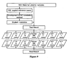
- 903 participants were recruited prospectively from Shenzhen People's Hospital and Shenzhen Maternal and child care service center with their karyotype results. Approvals were obtained from the institutional review boards of each recruitment site and all participants gave informed written consent. The maternal ages and gestational weeks at blood sampling were recorded. The 903 cases included 2 trisomy 13 cases, 15 trisomy 18 cases, 16 trisomy 21 cases, 3 XO cases, 2 XXY cases and 1 XYY cases. Their karyotype results distribution is shown in Figure 9 .
- Peripheral venous blood (5 milliliters) was collected from each participating pregnant woman in EDTA tubes and centrifuged at 1,600g for 10 min in 4 hours. Plasma was transferred to microcentrifuge tubes and recentrifuged at 16,000g for 10 min to remove residual cells. Cell-free plasma was stored at 80°C until DNA extraction. Each plasma sample was frozen and thawed only once.
- the size distribution of the sequencing libraries was analyzed with a DNA 1000 kit on the 2100 BioanalyzerTM (Agilent) and quantified with Real-time PCR.
- the sequencing libraries with different index were then pooled into one by equal quantity before cluster station on Illumina GA IITM (single-end sequencing).
- H1:the fetus is aneuploidy was that the mean coverage depth of the patient case distribution with a rough fetal fraction was equal to the mean coverage depth of the distribution of aneuploidy cases with the same fetal fraction, which means that this patient case is aneuploid if this null hypothesis were accepted.
- L i , j log p t ⁇ 1 i , j , degree
- L i,j Log likelihood ratio. If the ratio was larger than 1, we could infer the fetus might be trisomy.
- the fetal DNA fraction was estimated first by Y and X. Meanwhile, we could extrapolate the fitted coverage depth for chromosome X with the fetal DNA fraction estimated only by the coverage depth of chromosome Y and t2 can be calculated.
- t2 i ( cr i,x - (1 - fy i / 2) ⁇ ⁇ r i,Xf )/ std Xf . If t2 is too large (larger than 5) or too small(less than -5) the fetus may be XXY or XYY.
- the gap between fetal fractions estimated by X and Y independently will provide information for detecting disorders about sex chromosomes.
- the CV value of chromosome 13 is 0.0066 with 100% sensitivity rate and 100% specificity rate.
- the CV value of chromosome 13 is 0.0063 and with 100% sensitivity rate and 100% specificity rate.
- the CV of these two approaches were 0.0062 and 0.0066, respectively, both with 100% sensitivity and specificity rates for them were 99.89% and 99.96%, respectively.
- the performance was similar when comparing the CV of these two approaches for chromosome 21: 0.0088 and 0.0072, respectively. Both resulted in the same sensitivity rate of 100% in our small cases set study and achieved the same 100% specificity rate.
- Example 9 Theoretical performance of GC-correlation t-test approach in consideration of data size, gestational weeks and fetal DNA fraction
- Figures 15-21 show the resulting plots of our calculation.
- the student-t larger than 3 was set to identify female fetus aneuploidy while for male fetus, when computing probability density of false negative in every fraction, a logarithm likelihood larger than 1 was employed as the critical value we mentioned in Binary hypothesis which helped to achieve a higher sensitivity comparing to female ones.
Description
- The invention relates to non-invasive methods for the detection of fetal genetic abnormalities, more particularly chromosomal aneuploidy, by DNA sequencing of peripheral blood samples from pregnant women containing both maternal and fetal DNA. More particularly, this invention relates to data analysis which takes account of GC bias introduced by amplification and sequencing of DNA samples through reliance on pre-established relationship between GC content and coverage depth for a chromosome in the absence of aneuploidy and at the selected DNA fragmentation size as discussed in more detail below. Statistical analysis may be used to compare calculated coverage depth for a chromosome in a sample to expected coverage depth according to such established relationship whereby detection of fetal chromosomal aneuploidy can be achieved.
- Conventional prenatal diagnostic methods with invasive procedures, such as chorionic villus sampling and amniocentesis, carry potential risks for both fetuses and mothers. Noninvasive screening of fetal aneuploidy using maternal serum markers and ultrasound are available but have limited sensitivity and specificity (Kagan, et al., Human Reproduction (2008) 23:1968-1975; Malone, et al., N Engl J Med (2005) 353:2001-2011).
- Recent studies have demonstrated noninvasive detection of fetal aneuploidy by massively parallel sequencing of DNA molecules in the plasma of pregnant women is feasible. Fetal DNA has been detected and quantitated in maternal plasma and serum (Lo, et al., Lancet (1997) 350:485 487; Lo, et al., Am. J. hum. Genet. (1998) 62:768-775). Multiple fetal cell types occur in the maternal circulation, including fetal granulocytes, lymphocytes, nucleated red blood cells, and trophoblast cells (Pertl and Bianchi, Obstetrics and Gynecology (2001) 98:483-490). Fetal DNA can be detected in the serum at the seventh week of gestation, and increases with the term of the pregnancy. The fetal DNA present in the maternal serum and plasma is comparable to the concentration of DNA obtained from fetal cell isolation protocols.
- Circulating fetal DNA has been used to determine the sex of the fetus (Lo, et al., Am. J. hum. Genet. (1998) 62:768-775). Also, fetal rhesus D genotype has been detected using fetal DNA. However, the diagnostic and clinical applications of circulating fetal DNA is limited to genes that are present in the fetus but not in the mother (Pertl and Bianchi, Obstetrics and Gynecology (2001) 98:483-490). Thus, a need still exists for a non- invasive method that can determine the sequence of fetal DNA and provide definitive diagnosis of chromosomal abnormalities in a fetus.
- The discovery of fetal cells and cell-free fetal nucleic acids in maternal blood in the past few decades and the application of high-throughput shotgun sequencing of maternal plasma cell-free DNA make it is available to detect small changes in the representation of chromosomes contributed by an aneuploid fetus in a maternal plasma sample. Non-invasive detection of
trisomy - However, as some studies show, GC bias introduced by amplification and sequencing placed a practical limit on the sensitivity of aneuploidy detection. GC bias might be introduced during the sample preparation and the sequencing process, under different conditions such as reagent composition, cluster density and temperature, which leads to differential sampling of DNA molecules with different GC composition and significant variation in sequencing data for chromosomes that are GC-rich or GC-poor.
- To improve sensitivity, protocols for removal of the effect of GC-bias have been developed. Fan and Quake developed a method to computationally remove GC bias by applying weight to each GC density based on local genomic GC content, to ameliorate the number of reads mapped in each bin by multiplying corresponding weight (Fan and Quake PLoS ONE (2010) 5: e10439). However, the method has difficulty in dealing with sex chromosome disorders especially chromosome Y relevant disorders for the reason that the process may cause slight distortion of data which will interfere with the precision of detection.
- Here, we describe a method which makes use of GC-bias in order to get a high sensitivity in fetal aneuploidy detection as well as avoid data distortion. This method enables definition of parameters used for statistical test according to GC-content. In addition, the inventors introduced the estimated fetal fraction into the diagnosis by binary hypothesis which showed higher sensitivity and specificity. Our method also shows it should be possible to increase the sensitivity of non-invasive detection of fetal genetic abnormality to preset precision for maternal samples containing a low fetal DNA fraction by sequencing more polynucleotide fragments. Resampling of maternal plasma in later gestational weeks may also increase the sensitivity of diagnosis.
- The current invention is directed to methods for non-invasive detection of fetal chromosomal aneuploidy in which large-scale sequencing of nucleotides from maternal peripheral blood samples is carried out. In one aspect, the present invention provides a computer-implemented method to determine a fetal genetic abnormality which is a chromosomal aneuploidy or partial aneuploidy, which method comprises:
- (a) obtaining sequence information of multiple polynucleotide fragments from a sample, said sample being a peripheral blood sample derived from a pregnant female and containing both maternal and fetal DNA;
- (b) assigning said fragments to chromosomes based on said sequence information by comparing said fragments to the reference unique reads of the same size for each of said chromosomes, wherein reference unique reads are fragments of a chromosome that have a unique sequence which can be unambiguously assigned to a single chromosomal location based on a reference genomic sequence;
- (c) determining coverage depth and GC content of a chromosome based on the sequence information for those fragments that assigned to reference unique reads of said chromosome in step (b),
wherein coverage depth is the ratio between the number of fragments assigned uniquely to said chromosome and the number of reference unique reads for said chromosome of the same fragment size based on said reference human genomic sequence; - (d) determining fitted coverage depth of said chromosome using said GC content of said chromosome and established relationship between coverage depth and GC content for said chromosome in the absence of aneuploidy, wherein said established relationship has been determined by a method comprising the steps of:
- (i) obtaining sequence information of multiple polynucleotide fragments covering said chromosome from a plurality of euploid peripheral blood samples containing genomic DNA, wherein the fragment size is the same as the fragment size of the multiple polynucleotide fragments of step (a) above;
- (ii) assigning said fragments to chromosomes based on said sequence information as in step (b) above;
- (iii) determining coverage depth and GC content of said chromosome based on said sequence information for each euploid sample as in step (c) above; and
- (iv) using the coverage depth and GC content determined for each sample in step (iii) to determine the relationship between the coverage depth and GC content of said chromosome in the absence of aneuploidy; and
- (e) comparing said fitted coverage depth to the coverage depth of said chromosome determined in step (c), wherein a difference between them indicates fetal chromosomal aneuploidy.
- In one embodiment the polynucleotide fragments range from about 10 to about 1000 bp in length. In another embodiment the polynucleotide fragments range from about 15 to about 500 bp in length. In yet another embodiment the polynucleotide fragments range from about 20 to about 200 bp in length. In still another embodiment the polynucleotide fragments range from about 25 to about 100 bp in length. In a further embodiment the polynucleotide fragments are about 35 bp in length.
- In one embodiment, the sequence information is obtained by parallel genomic sequencing. In another embodiment the assignment of the fragment to chromosomes is by comparing the sequence of the fragments with a reference human genomic sequence. The reference human genomic sequence may be any suitable and/or published human genome builds, such as hg18 or hg19. The fragments that assign to more than one chromosome or do not assign to any chromosome are disregarded.
- As indicated above, the coverage depth of a chromosome for the purpose of method of the invention is the ratio between the number of fragments that assign uniquely to the chromosome and the number of reference unique reads of the chromosome. In another embodiment, the coverage depth is normalized. In still another embodiment, the normalization is calculated against the coverage of all other autosomes. In yet another embodiment, the normalization is calculated against the coverage of all other chromosomes.
- In one embodiment, the relationship is in the formula:

wherein f(GCi,j) represents the function of the relationship between normalized coverage depth and the corresponding GC content of sample i, chromosome j, εi,j represents the residual of sample i, chromosome j. In some embodiments, the relationship between coverage depth and GC content is calculated by local polynomial regression. In some embodiments, the relationship may be a non-strong linear relationship. In some embodiments, the relationship is determined by loess algorithm. - In some embodiments, the method further comprises calculating fitted coverage depth according to the formula:

- In some embodiments, the method further comprises calculating standard variation according to the formula:

- In some embodiments, the method further comprises calculating student t-statistic according to the formula:

- In one embodiment, the GC content of a chromosome is the average GC content of all fragments that are assigned to the chromosome for the purpose of step (c) of the method of the invention as defined above. The GC content of a fragment may be calculated by dividing the number of G/C nucleotides in the fragment by the total number of nucleotides of the fragment. In another embodiment, the GC content of a chromosome is the aggregate GC content of the reference unique reads of the chromosome.
- At least 2, 5, 10, 20, 50, 100, 200, 500 or 1000 euploid samples may be used in order to determine the relationship between coverage depth and GC content for a chromosome in the absence of aneuploidy. The chromosome may be any of
chromosomes - The samples for this purpose may be from pregnant female subjects. The samples may be from male subjects. Alternatively, the samples may be from both pregnant female subjects and male subjects. The samples are peripheral blood samples.
- In some embodiments, a method of the invention to determine a fetal chromosomal aneuploidy further comprises determining the fetal gender. The fetal gender may be determined according to the formula:

wherein cr.ai,x and cr.ai,y are normalized relative coverage depth of the X and Y chromosomes, respectively. - In some embodiments, the method further comprises estimating the fetal fraction. The fetal fraction may be calculated according to the formula:

wherein ĉri,Yf = f(GCi,Yf ) is the fitted coverage depth calculated from the relationship of the chromosome Y coverage depth and corresponding GC content of samples from pregnant women with a female fetus, ĉri,Ym = f(GCi,Ym ) refers to the fitted coverage depth calculated from the relationship of the chromosome Y coverage depth and corresponding GC content of male subjects. Alternatively, the fetal fraction may be calculated according to the formula:
wherein ĉri,Xf = f(GCi,Xf ) is the fitted coverage depth calculated from the relationship of the chromosome X coverage depth and corresponding GC content of samples from pregnant women with a female fetus, ĉr i,Ym = f(GCi,Xm ) refers to the fitted coverage depth calculated from the relationship of the chromosome X coverage depth and corresponding GC content of samples from male subjects. Further, the fetal fraction may be calculated according to the formula:
wherein ĉri,Xf = f(GCi,Xf ) is the fitted coverage depth calculated from the relationship of the chromosome X coverage depth and corresponding GC content of samples from pregnant women with a female fetus, ĉri,Yf = f(GCi,Yf ) refers to the fitted coverage depth calculated from the relationship of the chromosome Y coverage depth and corresponding GC content of samples from pregnant women with a female fetus, ĉri,Xm = f(GCi,Xm ) refers to the fitted coverage depth calculated from the relationship of the chromosome X coverage depth and corresponding GC content of samples from male subjects, ĉri,Ym = f(GCi,Ym ) refers to the fitted coverage depth calculated from the relationship of the chromosome Y coverage depth and corresponding GC content of male subjects. - The fetal aneuploidy to be detected may be a disorder for an autosome selected from the group consisting of
trisomy - In one embodiment, the fetal gender is female, and the student t-statistic is calculated according to formula: t1 i,X = (cri,X - cr̂i,Xf ) / stdXf , wherein ĉri,Xf = f(GCi,Xf ) is the fitted coverage depth calculated from the relationship of the chromosome X coverage depth and corresponding GC content of samples from pregnant women with a female fetus. In some embodiments, |t1|>3.13 indicates the fetus may be XXX or XO. In some embodiments, |t1|>5 indicates the fetus is XXX or XO.
- In another embodiment, the fetal gender is male, and the student t-statistic is calculated according to formula: t2 i =(cri,x - (1 - fyi / 2)·ĉri,Xf )/stdXf , wherein ĉri,Xf = f(GCi,Xf ) is the fitted coverage depth calculated from the relationship of the chromosome X coverage depth and corresponding GC content of samples from pregnant women with a female fetus. In some embodiments, |t2|>3.13 indicates the fetus may be XXY or XYY. In some embodiments, |t2|>5 indicates the fetus is XXY or XYY.
- In another aspect, provided herein is a computer readable medium comprising a plurality of instructions adapted to perform a method of the invention for prenatal diagnosis of a fetal genetic abnormality when said instructions are supplemented with sequence information of multiple polynucleotide fragments from a peripheral blood sample derived from a pregnant female and containing both maternal and fetal DNA. The computer readable medium will; b) assign said polynucleotide fragments to chromosomes based on said sequence information; c) determine coverage depth and GC content of a chromosome based on the sequence information for those fragments assigned uniquely to said chromosome; d) determine fitted coverage depth of said chromosome using said GC content of said chromosome and established relationship between coverage depth and GC content of said chromosome; and e) compare said fitted coverage depth to said coverage depth of said chromosome, wherein a difference between them indicates fetal chromosomal aneuploidy.
- In still another aspect, provided herein is a system comprising means adapted for carrying out a method of the invention. Such a system comprises: a) means for obtaining sequence information of multiple polynucleotide fragments from a sample; and b) a computer readable medium as described above.
-
-
Figure 1 shows a schematic process for calculating the coverage depth and GC content by using sequence information of polynucleotide fragments. -
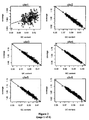
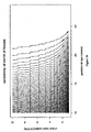
Figure 2 illustrates the normalized coverage depth-GC content correlation established by using data from 300 reference cases. The normalized coverage depth for each case is plotted against corresponding sequenced GC content. Crosses denote cases with euploid female fetus, squares denote cases with euploid male fetus. The solid line is the fitting line of the coverage depth and GC content. -
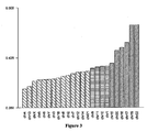
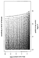
Figure 3 illustrates the tendency between normalized coverage depth and corresponding GC content by arranging chromosomes with their inherent ascending GC content. The inherent ascending GC content of each chromosome here refers to the average GC content of sequenced tags of that chromosome from 300 reference cases. -
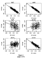
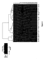
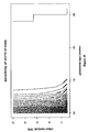
Figure 4 shows different compositions of GC class for each chromosome. The GC content of every 35 bp read of the reference unique reads was calculated for each chromosome, GC content was classified into 36 levels and the percentage of each level was calculated as the composition GC of each chromosome. The chromosomes were then graphed by the heatmap and clustered hierarchically. -
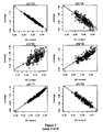
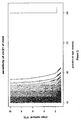
Figure 5 demonstrates sequencing bias introduces the correlation showed inFigure 2 by manual simulation of the process of sequencer preference. -
Figure 6 plots standard variation against total number of sequenced polynucleotide fragments. In 150 samples, the adjusted standard variance of every chromosome shows linear relationship with reciprocal of the square root of the number of unique reads. -
Figure 7 shows Q-Q plots of residual of every chromosome calculated byFormula 3. A linear relationship is shown with a normal distribution. -
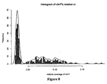
Figure 8 shows the histogram of chromosome Y coverage depth. There are two peaks which implicates that the gender of cases can be distinguished by the coverage depth of chromosome Y. The curve is distribution of relative coverage depth of chromosome Y estimated by kernel density estimation with Gaussian kernel. -
Figure 9 shows a diagram of the process for diagnosing 903 test samples for fetal chromosome abnormality. -
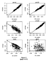
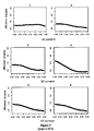
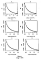
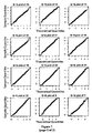
Figure 10 shows the result of aneuploidy:trisomy Figure 10A shows the plots of normalized coverage depth vs. GC content ofchromosomes Figure 10B shows the plots of chromosomes X and Y. Circles represent normal female fetuses' relative coverage depth with GC content, dots represent normal male fetuses. The solid line is fitting line of relative coverage and GC content, the dash lines are t-value absolute is 1, the dotted lines are absolute of t-value is 2 and the dot-dash lines: absolute of t-value is 3. -
Figure 11 compares the confidence value of different diagnostic approaches. -
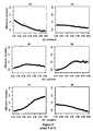
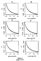
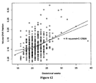
Figure 12 shows the relationship between fetal DNA fraction and gestational age. The fraction of fetal DNA in maternal plasma correlates with gestational age. Fetal DNA fraction was estimated by X and Y together. There is a statistically significant correlation between the average fetal DNA fraction and gestational age (P<0.001). Note that the R2 value represents the square of the correlation coefficient is small. The minimum fraction is 3.49%. -
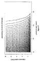
Figure 13 shows the relationship between the standard variance with the case number required for detection. The standard variances computed byFormula 5 of every chromosome vary with different number of samples. The standard variance becomes stable when the number of samples is larger than 100. -
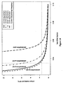
Figure 14 shows the estimated number of unique reads for the detection of fetal aneuploidy in cell-free plasma as a function of fetal DNA fraction. The estimates are based on level of confidence t-value no smaller than 3 for aneuploidy ofchromosomes trisomy 21 can be detected if 3.5% of the cell-free DNA is fetal. Aneuploidy of chromosome X was not detected easily when the fraction and unique reads number are small, such as 4% and 5 million reads. Different chromosome requires different level of fetal DNA fraction and unique reads number, which may be caused by the GC structure of the chromosome. -
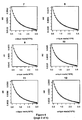
Figure 15 shows a contour graph of sensitivity mapped by data volume and gestational age (weeks) for detection of trisomy ofchromosome 13 for female fetuses, for every gestational week and every point of data volume. -
Figure 16 shows a contour graph of sensitivity mapped by data volume and gestational age (weeks) for detection of trisomy ofchromosome 18 for female fetuses, for every gestational week and every point of data volume. -
Figure 17 shows a contour graph of sensitivity mapped by data volume and gestational age (weeks) for detection of trisomy ofchromosome 21 for female fetuses, for every gestational week and every point of data volume. -
Figure 18 shows a contour graph of sensitivity mapped by data volume and gestational age (weeks) for detection of trisomy of chromosome X for female fetuses, for every gestational week and every point of data volume. -
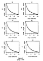
Figure 19 shows a contour graph of sensitivity mapped by data volume and gestational age (weeks) for detection of trisomy ofchromosome 13 of male. For every gestational week and every point of data volume, we compute its empirical distribution of fetal DNA fraction and standard variance for given data volume firstly, and comparing the fraction estimated by XY or Y then we compute the sensitivity of every type of aneuploidy. -
Figure 20 shows a contour graph of sensitivity mapped by data volume and gestational age (weeks) for detection of trisomy ofchromosome 18 of male. -
Figure 21 shows a contour graph of sensitivity mapped by data volume and gestational age (weeks) for detection of trisomy ofchromosome 21 of male. - The current invention is directed to methods for non-invasive detection of fetal genetic abnormalities by large-scale sequencing of polynucleotide fragments from a maternal peripheral blood sample. As indicated above, provided are methods which take account of GC bias from the sequencing results arising from the difference in GC content of a chromosome based on the relationship between the coverage depth of a chromosome and the corresponding GC content. Accordingly, described herein is a method to computationally adjust reference parameters being used in a student-t calculation with GC contents by locally weighted polynomial regression to fit the coverage depth of a chromosome in samples against the GC content of the polynucleotide fragments.
- Also provided herein is a method of determining chromosomal abnormality of a fetus as described above involving statistical analysis using a statistical hypothesis test. In addition, methods are described to calculate data quality control (DQC) standards useful in determining the amount of clinical samples needed for a certain statistical significance level.
- Unless defined otherwise, all technical and scientific terms used herein have the same meaning as is commonly understood by one of ordinary skill in the art to which this invention belongs. All patents, applications, published applications and other publications referred to herein are incorporated by reference in their entirety. If a definition set forth in this section is contrary to or otherwise inconsistent with a definition set forth in the patents, applications, published applications and other publications that are herein incorporated by reference, the definition set forth in this section prevails over the definition that is incorporated herein by reference.
- As used herein, the singular forms "a", "an", and "the" include plural references unless indicated otherwise. For example, "a" dimer includes one or more dimers.
- The term "chromosomal abnormality" refers to a deviation between the structure of the subject chromosome and a normal homologous chromosome. The term "normal" refers to the predominate karyotype or banding pattern found in healthy individuals of a particular species. A chromosomal abnormality can be numerical or structural, and includes but is not limited to aneuploidy, polyploidy, inversion, a trisomy, a monosomy, duplication, deletion, deletion of a part of a chromosome, addition, addition of a part of chromosome, insertion, a fragment of a chromosome, a region of a chromosome, chromosomal rearrangement, and translocation. A chromosomal abnormality can be correlated with presence of a pathological condition or with a predisposition to develop a pathological condition. As defined herein, a single nucleotide polymorphism ("SNP") is not a chromosomal abnormality.
- Monosomy X (XO, absence of an entire X chromosome) is the most common type of Turner syndrome, occurring in 1 in 2500 to 1 in 3000 live-born girls (Sybert and McCauley N Engl J Med (2004) 351:1227-1238). XXY syndrome is a condition in which human males have an extra X chromosome, existing in roughly 1 out of every 1000 males (Bock, Understanding Klinefelter Syndrome: A Guide for XXY Males and Their Families. NIH Pub. No. 93-3202 (1993)). XYY syndrome is an aneuploidy of the sex chromosomes in which a human male receives an extra Y chromosome, giving a total of 47 chromosomes instead of the more usual 46, affecting 1 in 1000 male births while potentially leading to male infertility (Aksglaede, et al., J Clin Endocrinol Metab (2008) 93:169-176).
- Turner syndrome encompasses several conditions, of which monosomy X (XO, absence of an entire sex chromosome, the Barr body) is most common. Typical females have two X chromosomes, but in Turner syndrome, one of those sex chromosomes is missing. Occurring in 1 in 2000 to 1 in 5000 phenotypic females, the syndrome manifests itself in a number of ways. Klinefelter syndrome is a condition in which human males have an extra X chromosome. In humans, Klinefelter syndrome is the most common sex chromosome disorder and the second most common condition caused by the presence of extra chromosomes. The condition exists in roughly 1 out of every 1,000 males. XYY syndrome is an aneuploidy of the sex chromosomes in which a human male receives an extra Y chromosome, giving a total of 47 chromosomes instead of the more usual 46. This produces a 47, XYY karyotype. This condition is usually asymptomatic and affects 1 in 1000 male births while potentially leading to male infertility.
- Trisomy 13 (Patau syndrome), trisomy 18 (Edward syndrome) and trisomy 21 (Down syndrome) are the most clinically important autosomal trisomies and how to detect them has always been the hot topic. Detection of above fetal chromosomal aberration has great significance in prenatal diagnosis (Ostler, Diseases of the eye and skin: a color atlas. Lippincott Williams & Wilkins. pp. 72. ISBN 9780781749992 (2004); Driscoll and Gross N Engl J Med (2009) 360: 2556-2562; Kagan, et al., Human Reproduction (2008) 23:1968-1975).
- The term "reference unique reads" refers to fragments of a chromosome that have a unique sequence. Therefore, such fragments can be unambiguously assigned to a single chromosomal location. Reference unique reads of a chromosome may be constructed based on a published reference genome sequence, such as hg18 or hg19.
- The terms "polynucleotide," "oligonucleotide," "nucleic acid" and "nucleic acid molecule" are used interchangeably herein to refer to a polymeric form of nucleotides of any length, and may comprise ribonucleotides, deoxyribonucleotides, analogs thereof, or mixtures thereof. This term refers only to the primary structure of the molecule. Thus, the term includes triple-, double-and single-stranded deoxyribonucleic acid ("DNA"), as well as triple-, double- and single-stranded ribonucleic acid ("RNA"). It also includes modified, for example by alkylation, and/or by capping, and unmodified forms of the polynucleotide. More particularly, the terms "polynucleotide," "oligonucleotide," "nucleic acid" and "nucleic acid molecule" include polydeoxyribonucleotides (containing 2-deoxy-D-ribose), polyribonucleotides (containing D-ribose), including tRNA, rRNA, hRNA, and mRNA, whether spliced or unspliced, any other type of polynucleotide which is an N- or C-glycoside of a purine or pyrimidine base, and other polymers containing normucleotidic backbones, for example, polyamide (e.g., peptide nucleic acids ("PNAs")) and polymorpholino (commercially available from the Anti-Virals, Inc., Corvallis, OR., as NeuGene®) polymers, and other synthetic sequence-specific nucleic acid polymers providing that the polymers contain nucleobases in a configuration which allows for base pairing and base stacking, such as is found in DNA and RNA. Thus, these terms include, for example, 3'-deoxy-2',5'-DNA, oligodeoxyribonucleotide N3' to P5' phosphoramidates, 2'-O-alkyl-substituted RNA, hybrids between DNA and RNA or between PNAs and DNA or RNA, and also include known types of modifications, for example, labels, alkylation, "caps," substitution of one or more of the nucleotides with an analog, intemucleotide modifications such as, for example, those with uncharged linkages (e.g., methyl phosphonates, phosphotriesters, phosphoramidates, carbamates, etc.), with negatively charged linkages (e.g., phosphorothioates, phosphorodithioates, etc.), and with positively charged linkages (e.g., aminoalkylphosphoramidates, aminoalkylphosphotriesters), those containing pendant moieties, such as, for example, proteins (including enzymes (e.g., nucleases), toxins, antibodies, signal peptides, poly-L-lysine, etc.), those with intercalators (e.g., acridine, psoralen, etc.), those containing chelates (of, e.g., metals, radioactive metals, boron, oxidative metals, etc.), those containing alkylators, those with modified linkages (e.g., alpha anomeric nucleic acids, etc.), as well as unmodified forms of the polynucleotide or oligonucleotide.
- "Massively parallel sequencing" means techniques for sequencing millions of fragments of nucleic acids, e.g., using attachment of randomly fragmented genomic DNA to a planar, optically transparent surface and solid phase amplification to create a high density sequencing flow cell with millions of clusters, each containing ∼1,000 copies of template per sq. cm. These templates are sequenced using four-color DNA sequencing-by-synthesis technology. See products offered by Illumina, Inc., San Diego, Calif. The presently used sequencing is preferably carried out without a pre-amplification or cloning step, but may be combined with amplification-based methods in a microfluidic chip having reaction chambers for both PCR and microscopic template-based sequencing. Only about 30 bp of random sequence information are needed to identify a sequence as belonging to a specific human chromosome. Longer sequences can uniquely identify more particular targets. In the present case, a large number of 35 bp reads were obtained. Further description of a massively parallel sequencing method is found in Rogers and Ventner, Nature (2005) 437:326-327.
- It is understood that aspects and embodiments of the invention described herein include "consisting" and/or "consisting essentially of" aspects and embodiments.
- Other objects, advantages and features of the present invention will become apparent from the following specification taken in conjunction with the accompanying drawings.
- As indicated above, establishing a relationship between coverage depth and GC content of a chromosome, will comprise: obtaining sequence information of multiple polynucleotide fragments covering said chromosome from more than one sample; assigning said fragments to chromosomes based on said sequence information; determining coverage depth and GC content of said chromosome based on said sequence information for each sample; and determining the relationship between the coverage depth and GC content of said chromosome.
- To calculate the coverage depth and GC content of a chromosome location, sequence information of polynucleotide fragments is obtained by sequencing template DNA obtained from a peripheral blood sample. In one embodiment, the template DNA contains both maternal DNA and fetal DNA. In another embodiment, template DNA is obtained from blood of a pregnant female. Blood may be collected using any standard technique for blood drawing including but not limited to venipuncture. For example, blood can be drawn from a vein from the inside of the elbow or the back of the hand. Blood samples can be collected from a pregnant female at any time during fetal gestation. For example, blood samples can be collected from human females at 1-4, 4-8, 8-12, 12-16, 16-20, 20-24, 24-28, 28-32, 32-36, 36-40, or 40-44 weeks of fetal gestation, and preferably between 8-28 weeks of fetal gestation.
- The polynucleotide fragments are assigned to a chromosome location based on the sequence information. A reference genomic sequence is used to obtain the reference unique reads. As used therein, the term "reference unique reads" refers to all the unique polynucleotide fragments that have been assigned to a specific genomic location based on a reference genomic sequence. In some embodiments, the reference unique reads have the same length of, for example, about 10, 12, 15, 20, 25, 30, 35, 40, 50, 100, 200, 300, 500, or 1000 bp. In some other embodiments, human genome builds hg18 or
hg 19 may be used as the reference genomic sequence. A chromosome location may be a contiguous window on a chromosome that has a length of about 10, 20, 30, 40, 50, 60, 70, 80, 90, 100, 200, 300, 400, 500, 600, 700, 800, 900, 1000, 2000, 3000, 4000, 5000, 6000, 7000, 8000, 9000, 10,000 or more kb. A chromosome location may also be a single chromosome. - As used herein, the term "coverage depth" refers to the ratio between the number of fragments that assigns to a chromosome location and the number of reference unique reads of the chromosome location using the following formula:

wherein n¡,j is number of unique sequence reads mapped to chromosome j in sample i; Ci,j is the coverage depth in chromosome j in sample i; Nj is number of Reference Unique Reads in chromosome j. - Polynucleotide fragments that do not assign to a single chromosome location or assign to multiple chromosome locations are discarded. In some embodiments, the coverage depth is normalized, based on the coverage depth of another chromosome location, another chromosome, average of all other autosomes, average of all other chromosomes, or average of all chromosomes. In some embodiments, the average coverage depth of 22 autosomes is used as a normalization constant to account for the differences in total number of sequence reads obtained for different samples:

wherein cri,j represents the relative coverage depth of chromosome j in sample i. From this point forward, "relative coverage depth" for each chromosome refers to the normalized value and is used for comparing different samples and for subsequent analysis. - GC content of a chromosome location can be calculated by the average GC percentage of a chromosome location based on the unique reference reads in the chromosome location, or on the sequenced polynucleotide fragments that assign to the chromosome location. GC content of a chromosome may be calculated using the following formula:

wherein i represents sample i, j represents chromosome j, NGCi,j represents the number of G and C DNA bases and BASEi.j represents the number of DNA bases on chromosome j in sample i. - To establish a relationship between the coverage depth and GC content of a chromosome location, the calculation may be based on the sequence information of polynucleotide fragments obtained from at least 1, 2, 5, 10, 20, 50, 100, 200, 500 or 1000 samples.
- In some instances, the relationship between coverage depth and GC content is a non-strong linear relationship. Loess algorithm, or locally weighted polynomial regression, may be used to assess non-linear relationships (correlations) between pairs of values, such as between coverage depth and GC content.
- As previously noted above, a method to determine a fetal genetic abnormality according to the invention,comprises: a) obtaining sequence information of multiple polynucleotide fragments from a peripheral blood sample which is derived from a pregnant female and contains both maternal and fetal DNA; b) assigning said fragments to chromosomes based on said sequence information; c) determining coverage depth and GC content of a chromosome based on the sequence information for those fragments that assigned uniquely to said chromosome in step (b); d) determining fitted coverage depth of said chromosome using said GC content of said chromosome and established relationship between coverage depth and GC content for said chromosome in the absence of aneuploidy; and e) comparing said fitted coverage depth to the coverage depth of said chromosome determined in step (c), wherein a difference between them indicates a fetal genetic abnormality which is a chromosomal aneuploidy.
- The methods of the invention are especially useful for the detection of aneuploidy, polyploidy, monosomy, trisomy,
trisomy 21,trisomy 13,trisomy 14,trisomy 15,trisomy 16,trisomy 18,trisomy 22, triploidy, tetraploidy, and sex chromosome abnormalities including XO, XXY, XYY, and XXX. One may also focus on certain regions within the human genome according to the present methods in order to identify partial monosomies and partial trisomies. For example, the methods may involve analyzing sequence data in a defined chromosomal sliding "window," such as contiguous, nonoverlapping 50 Kb regions spread across a chromosome. Partial trisomies of 13q, 8p (8p23.1), 7q, distal 6p, 5p, 3q (3q25.1), 2q, 1q (1q42.1 and 1q21-qter), partial Xp and monosomy 4q35.1 have been reported, among others. For example, partial duplications of the long arm ofchromosome 18 can result in Edwards syndrome in the case of a duplication of 18q21.1-qter (Mewar, et al., Am J Hum Genet. (1993) 53:1269-78). - In some embodiments, the fetal fraction is estimated based on the sequence information obtained for the polynucleotide fragments from a sample. The coverage depth, and GC content, of chromosome X and Y may be used for estimating the fetal fraction. In some embodiments, the fetal gender is determined based on the sequence information obtained for the polynucleotide fragments from a sample. The coverage depth, and GC content, of chromosome X and Y may be used for determining the fetal gender.
- In some embodiments, the comparison of said fitted coverage depth to said coverage depth of the chromosome is conducted by a statistical hypothesis test, wherein one hypothesis is that the fetus is euploid (H0) and the other hypothesis is that the fetus is aneuploid (H1). In some embodiments, the student t-statistic is calculated for both hypotheses as t1 and t2, respectively. In some embodiments, the log likelihood ratio of t1 and t2 is calculated. In some embodiments, a log likelihood ratio of >1 indicates trisomy of the fetus.
- In another aspect, provided herein is acomputer readable medium comprising a plurality of instructions adapted to perform a method of the invention for prenatal diagnosis of a fetal genetic abnormality when said instructions are supplemented with sequence information obtained as in step (a) of the method.
- In still another aspect, as previously indicated also provided herein is a system comprising means adapted for carrying out a method of the invention. Such a system comprises: a) means for obtaining sequence information from suitable polynucleotide fragments; and b) a computer readable medium as described above.
- It will be apparent to those skilled in the art that a number of different sequencing methods and variations can be used. In one embodiment, the sequencing is done using massively parallel sequencing. Massively parallel sequencing, such as that achievable on the 454 platform (Roche) (Margulies, et al., Nature (2005) 437:376-380), Illumina Genome Analyzer (or Solexa™ platform) or SOLiD System (Applied Biosystems) or the Helicos True Single Molecule DNA sequencing technology (Harris, et al., Science (2008) 320:106-109), the single molecule, real-time (SMRT™) technology of Pacific Biosciences, and nanopore sequencing (Soni and Meller, Clin Chem (2007) 53:1996-2001), allow the sequencing of many nucleic acid molecules isolated from a specimen at high orders of multiplexing in a parallel fashion (Dear, Brief Funct Genomic Proteomic (2003) 1:397-416). Each of these platforms sequences clonally expanded or even non-amplified single molecules of nucleic acid fragments. Commercially available sequencing equipment may be used in obtaining the sequence information of the polynucleotide fragments.
- The following examples are offered to illustrate but not to limit the invention.
- A schematic procedural framework for calculating coverage depth and GC content is illustrated in
Figure 1 . We used software to produce the reference unique reads by incising the hg18 reference sequences into 1-mer (1-mer here is a read being artificially decomposed from the human sequence reference with the same "1" length with sample sequencing reads) and collected those "unique" 1-mer as our reference unique reads. Secondly, we mapped our sequenced sample reads to the reference unique reads of each chromosome. Thirdly, we deleted the outlier by applying quintile outlier cutoff method to get a clear data set. Finally, we counted the coverage depth of each chromosome for every sample and the GC content of the sequenced unique reads mapped to each chromosome for every sample. - In order to investigate how GC content affects our data, we chose 300 euploid cases with karyotype result and scattered their coverage depth and related GC content of sequenced reads into a graph, which showed a strong correlation between them, and this phenomenon was unreported previously (
Figure 2 ). InFigure 2 , coverage depth correlated strongly with the GC-content, and showed an obviously downward trend in some chromosomes such as 4, 13, etc., while upward trend in other chromosomes such as 19, 22, etc. All chromosomes were arranged in ascending order by their inherent GC-content and a downward tendency is present in lower GC-content group chromosomes while upward tendency in higher GC-content group chromosomes as shown inFigure 3 . It can be interpreted that if the polynucleotide fragments being sequenced for one sample has a higher GC-content than the other sample, the coverage depth representing this sample would drop comparing to that of the other sample in lower GC-content group chromosomes while rise in higher GC-content group chromosomes. - The possible explanation for such a different changing tendency among different GC-content chromosomes is the differences in GC-content composition in different chromosomes shown in
Figure 4 combined with the GC-bias introduced in the sequencing process. The GC content of every 35-mer reference unique reads for each chromosome is used to classify GC content into 36 levels. The percentage of each level as the composition GC of each chromosome was calculated and then used to draw the heatmap with the Heatmap2 software. Takechromosome 13 as an example, large part of it consists of lower GC-content sequence segments but small part of it consists of higher GC-content sequence segments. If conditions during the sequencing or PCR process is in favor of sequence those segment with higher GC-content, then a relative large part ofchromosome 13 with low GC-content would be hard to be sequenced with a result that the coverage depth in this sample'schromosome 13 was becoming lower. In comparison, in a higher GC-content group such aschromosome 19, the coverage depth in this sample'schromosome 19 is becoming higher for that a large part ofchromosome 19 was of higher GC-content to which the sequencer prefers. No matter in which chromosome, GC-poor and GC-rich segments were hard to be sequenced but the influence introduced by GC-bias was different to different chromosomes with different GC-content composition. Every reference chromosome was divided into 1 KB bins, the GC content of each unique reference read in the bin was calculated. The GC content of each bin in the proper interval form [0.3, 0.6] divided by step size of 0.001, and the relative coverage in every interval is calculated.Figure 5 shows plots of relative coverage and GC content for each chromosome. - Influence of fetal gender on data was analyzed using independent two-sample t-test. No significant difference was found between autosomes except for sex chromosomes in the same GC content roughly, but there is obvious difference in UR% between female and male (Chiu et al., (2008) Proc Natl Acad Sci USA 105:20458-20463), implying that there is no need to distinguish fetal gender when detecting autosome aneuploidy, but it is needed to distinguish fetal gender firstly, when detecting sex chromosome aneuploidy such as XO, XYY etc.
- Using this phenomenon discussed above, we tried to use local polynomial to fit the relationship between coverage depth and the corresponding GC content. The coverage depth consists of a function of GC and a residual of normal distribution as following:

wherein f(GCi,j) represents the function for the relationship between coverage depth and the corresponding GC content of sample i, chromosome j, εi,j represents the residual of sample i, chromosome j. - There is non-strong linear relationship between the coverage depth and the corresponding GC content so we applied loess algorithm to fit the coverage depth with the corresponding GC content, from which we calculated a value important to our model, that is, the fitted coverage depth:

With the fitted coverage depth, the standard variance and the student t were calculated according to the flowingFormula 6 and Formula 7:

- For the reason that fetal fraction is very important for our detection so we estimated the fetal fraction before the testing procedure. As we had mentioned before, we had sequenced 19 male adults, when compared their coverage depth with that of cases carrying female fetus, we found that male's coverage depth of chromosome X is almost 1/2 times of female's, and male's coverage depth of chromosome Y is almost 0.5 larger than female's. Then we can estimate the fetal fraction depending on the coverage depth of chromosome X and Y as
Formula 8,Formula 9 andFormula 10, considering GC-correlation as well:


wherein ĉri,Xf = f(GCi,Xf ) is the fitted coverage depth by the regression correlation of the chromosome X coverage depth and corresponding GC content of cases with female fetus, ĉri,Yf = f(GCi,Yf ) refers to the fitted coverage depth by the regression correlation of the chromosome Y coverage depth and corresponding GC content of cases with female fetus, ĉri,Xm = f(GCi,Xm ) refers to the fitted coverage depth by the regression correlation of the chromosome X coverage depth and corresponding GC content of male adults ĉri,Ym = f(GCi,Ym ) refers to the fitted coverage depth by the regression correlation of the chromosome Y coverage depth and corresponding GC content of male adults. For computing simply, given σ̂X,f and σ̂X,m are equal and σ̂Y,f and σ̂Y,m are equal. -
Figure 6 shows that the standard variation (see Formula 3) for every chromosome under a certain total number of unique reads is influenced by the participating cases number of the reference. The standard variation barely increases when the selected cases number was more than 150 under the condition that 1.7 million of total unique reads number were sequenced for each case. However, the standard variation was different for different chromosomes. After considering the GC-bias, our method had a moderate standard variation for chromosome 13 (0.0063), chromosome 18 (0.0066) and chromosome 21(0.0072). The standard variation of chromosome X is higher than above mentioned chromosomes which would require more strategies to do accurate abnormal detection. -
Figure 7 shows the Q-Q plot, wherein the residual is compiled to normal distribution which implicates the student-t calculation is reasonable. - To discover the disorder of the sex chromosome, it is best to distinguish fetal gender. There existed two obvious peaks when we investigated the frequency distribution of the coverage depth of chromosome Y in our 300 cases, which gave us a hint to distinguish the gender by coverage depth of chromosome Y. Cases with coverage depth less than 0.04 can be regarded as carrying female fetus while more than 0.051 regarded as carrying male fetus, between 0.04 and 0.051 are regarded as gender uncertain as
Figure 8 . For these gender dubious and aneuploidy cases, logistic regression was used to predict their gender as Formula 11 (Fan, et al., Proc Natl Acad Sci USA (2008) 42:16266-16271):
wherein cr.ai,x and cr.ai,y are normalized relative coverage of X and Y, respectively. - Comparing with the karyotype result, our method for distinguishing fetal gender performed quite well in our 300 reference cases with 100% accuracy while mistook one case when being carried out in our 901 cases set and the chromosome Y coverage depth of this mistaken case is between 0.04 and 0.051.
- 903 participants were recruited prospectively from Shenzhen People's Hospital and Shenzhen Maternal and child care service center with their karyotype results. Approvals were obtained from the institutional review boards of each recruitment site and all participants gave informed written consent. The maternal ages and gestational weeks at blood sampling were recorded. The 903 cases included 2
trisomy 13 cases, 15trisomy 18 cases, 16trisomy 21 cases, 3 XO cases, 2 XXY cases and 1 XYY cases. Their karyotype results distribution is shown inFigure 9 . - Peripheral venous blood (5 milliliters) was collected from each participating pregnant woman in EDTA tubes and centrifuged at 1,600g for 10 min in 4 hours. Plasma was transferred to microcentrifuge tubes and recentrifuged at 16,000g for 10 min to remove residual cells. Cell-free plasma was stored at 80°C until DNA extraction. Each plasma sample was frozen and thawed only once.
- For massively parallel genomic sequencing, all extracted DNA from 600 µl maternal plasma was used for DNA library construction according to a modified protocol from Illumina. Briefly, end-repairing of maternal plasma DNA fragments was performed using T4 DNA polymerase, Klenow™ polymerase, and T4 polynucleotide kinase. Commercially available adapters (Illumina) were ligated to the DNA fragments after addition of terminal A-residues. The adapter-ligated DNA was then additionally amplified using a 17-cycle PCR with standard multiplex primers. Agencourt AMPure™ 60 ml Kit (Beckman) was used for the purification of PCR products. The size distribution of the sequencing libraries was analyzed with a DNA 1000 kit on the 2100 Bioanalyzer™ (Agilent) and quantified with Real-time PCR. The sequencing libraries with different index were then pooled into one by equal quantity before cluster station on Illumina GA II™ (single-end sequencing).
- 19 male euploid samples were sequenced for subsequent analysis for estimation of fetal DNA fraction. One new GC-correlation t-test approach has been developed by us for the diagnosis of
trisomy 13,trisomy 18,trisomy 21 and sex-chromosome abnormities. We compared this new method to two other methods mentioned below in terms of diagnostic performance. - To determine whether the copy number of a chromosome within a patient case deviated from normal, the coverage depth of a chromosome was compared to that of all other reference cases. All previous study had just one null hypothesis. We introduced binary hypotheses for the first time by using a two null hypothesis. One null hypothesis (H0: the fetus is euploidy) was the assumption that the mean coverage depth of the patient case distribution and the mean coverage depth of all normal reference distribution were equal, which means that the patient case was euploid if this null hypothesis is accepted. Using student t test, t1 can be calculated as Formula 12:

The other null hypothesis (H1:the fetus is aneuploidy) was that the mean coverage depth of the patient case distribution with a rough fetal fraction was equal to the mean coverage depth of the distribution of aneuploidy cases with the same fetal fraction, which means that this patient case is aneuploid if this null hypothesis were accepted. The student t-statistic, t2 were calculated as Formula 13:
|t1|>3 and |t2|<3 would indicate an aneuploid case in most instances especially when the distributions between euploid cases and aneuploid cases were completely discriminated, while in other condition such as the insufficient precision or insufficient fetal fraction and so on, |t1| may smaller than 3 but the fetus was abnormal. Combined t1 and t2 can help us make more accurate decision, then we employed Log likelihood ratio of t1 and t2 as Formula 14:
wherein Li,j is Log likelihood ratio. If the ratio was larger than 1, we could infer the fetus might be trisomy. - But for cases with female fetuses, it is hard for us to estimate its fetal fraction so that is impossible to compute. However, we can give a Reference Value (RV) of
fraction 7% according to the empirical distribution of fetal fraction. - 903 cases were investigated, 866 of them carried euploid fetuses amongst which 300 cases had been selected out randomly to develop the GC correlation student-t approach. Besides, 2
trisomy trisomy trisomy Figure 10A ). The sensitivity and specificity of this approach were 100% and 100% (Table 1). - For
trisomy trisomy 18 cases and 888 out of 891 non-trisomy 18 cases could be correctly identified (Figure 10A ). The sensitivity and specificity of this approach were 100% and 99.66% respectively. For trisomy, 21, 16 out of 16trisomy 21 cases and 16 out of 16 non-trisomy 21 cases could also be correctly detected (Figure 10A ). The sensitivity and specificity of this approach were 100% and 100% respectively. - Above we considered the detection of trisomy for autosomes, disorders for sex chromosome such as XO, XXX, XXY and XYY can be detect by our method, too.
- Firstly, gender was confirmed by gender distinguishing. If a testing case was confirmed to carry female fetus, student-t value t1 t1 i,X = (cri,X - cr̂i,Xf )/stdXf was needed to be calculated for XXX or XO detection, where cr̂i,Xf and stdXf are same as
Formula 10; if t1 is larger than 3.13 or less than -3.13, this case maybe XXX or XO. But considering precision was limited by the great deviation of coverage depth for chromosome X, we sampled the plasma again and repeated the experiment to make a more credible decision when |t1|<5 even though |t1|>3.13. |t1|>5 was confirmed to be aneuploidy in this case. All our detection processes were on the premise that the data satisfied the standard quality control. - If the testing sample was confirmed to carry with male fetus, the fetal DNA fraction was estimated first by Y and X. Meanwhile, we could extrapolate the fitted coverage depth for chromosome X with the fetal DNA fraction estimated only by the coverage depth of chromosome Y and t2 can be calculated. t2 i =(cri,x - (1 - fyi / 2)·ĉri,Xf )/stdXf . If t2 is too large (larger than 5) or too small(less than -5) the fetus may be XXY or XYY. In addition, the gap between fetal fractions estimated by X and Y independently will provide information for detecting disorders about sex chromosomes.
- In the XO detection, 3 out of 4 XO cases were detected, and the case failed to be identified was a chimera case (
Figure 10B ). The sensitivity and specificity of this approach were 75% (100% if we disregard the chimera case) and 99.55% respectively. For XXY cases, all the 2 cases were successfully identified, while 901 out of 901 non-XXY cases were correctly classified (Figure 10B ) with thesensitivity 100% andspecificity 100%. For the XYY case, we identified it correctly (Figure 10B ) and the sensitivity and specificity were 100% and 100% respectively. - To evaluate whether our novel approach had any advantages when compared to other two reported approaches, z-score and z-score witch GC correction, we implemented all these three approaches to analyze our 900 cases and the same 300 cases as reference set for all those approaches. The precision of a measurement was always embodied in the confidence value (CV). In our investigation, the CV of standard z-score approach is larger than the other approaches in clinically
interested chromosome 18 and 21 (Figure 11 ) leading to a lower sensitivity rate fortrisomy 18 and 21 (Table 1).Table 1 Comparison of sensitivity and specificity of different methods Standard z-score approach z-score approach with GC correction our approach with GC correlation t-test Diagnosis (# of cases) Sensitivity (%) Specificity (%) Sensitivity (%) Specificity (%) Sensitivity (%) Specificity (%) Autosome Trisomy 13 (2) 50% 99.89 100% 100% 100% 100% Trisomy 18 (12) 91.67% 100% 100% 99.89% 100% 99.96% Trisomy 21 (16) 93.75% 100% 100% 100% 100% 100% Heterosome XO (3 XO, 1 XO/XX chimera) not available not available not available not available 75% 100% XXY (1) not available not available not available not available 100% 100% XXY (2) not available not available not available not available 100% 100% Binary hypothesis not available not available available - For the z-score approach with GC correction, the CV value of
chromosome 13 is 0.0066 with 100% sensitivity rate and 100% specificity rate. For the novel GC correlation student t approach discussed herein, the CV value ofchromosome 13 is 0.0063 and with 100% sensitivity rate and 100% specificity rate. Inchromosome 18, the CV of these two approaches were 0.0062 and 0.0066, respectively, both with 100% sensitivity and specificity rates for them were 99.89% and 99.96%, respectively. The performance was similar when comparing the CV of these two approaches for chromosome 21: 0.0088 and 0.0072, respectively. Both resulted in the same sensitivity rate of 100% in our small cases set study and achieved the same 100% specificity rate. And these two methods all performed better than the standard z-score approach. Not only was our new developed approach with GC correlation comparable to the GC correction approach with good performance, but also it had another advantage in the detection of sex-chromosome abnormalities such as XO, XXY and XYY. Our data shows that when a GC correction approach is employed there would be difficulty in distinguishing gender of fetuses by deviation of data representing sex chromosomes introduced when amending the number of sequence tags by multiplying by a weight factor so that the detection of a sex chromosome disorder would seem to be hard. - Measuring aneuploidy remains challenging because of the high background of maternal DNA (Fan, et al., Proc Natl Acad Sci USA (2008) 42:16266-16271) and arbitrary small fetal DNA fraction was the most significant restraining factor for aneuploidy detection by massively parallel genome sequencing (MPGS) approach to this day. However, there was no big breakthrough in determining the minimum fetal DNA fraction clinically before MPGS detection especially for female fetuses while the only clinical clue related to implicate the fraction of fetal DNA was gestational weeks. It was reported that there is a statistically significant correlation between the fetal DNA fraction and gestational age previously (Lo, et al., Am. J. Human Genet. (1998) 62:768-775). In our study, to investigate the relationship between estimated fetal DNA fraction and gestational age, we plotted in
Figure 12 the fetal DNA fraction of all those participating cases with male fetuses (totally 427 cases) referred toestimation Formula 10. The estimated fetal DNA fraction for each sample correlates with gestational age (P smaller than 0.0001). It also showed that even though in thegestational age 20, there were 4 out of 65 cases with fetal DNA fraction less than 5%, which would adversely affect the detection accuracy. To evaluate the fetal fraction estimation method we selected some cases hierarchically distributed in estimated fetal fraction, and then Q-PCR helped to calculate another relative fetal fraction. Then we got a correlation standard curve showing a strong correlation between them which demonstrated the estimation of fetal fraction by our method is credible. - Meanwhile, the sequencing depth (the number of total unique reads) was another significant factor affecting the precision of aneuploidy detection embodying in the value of standard variation. The standard variation for each chromosome employed in our GC-correlated approach could be fixed under a certain level of sequencing depth when the reference case number reaches 150 (
Figure 13 ). To investigate how the sequencing depth influence the standard variation for each chromosome, we sequenced 150 cases not only in our 1.7 million level but in another sequencing depth level with the number of total unique reads reaching 5 million (SD=1.7 million). Depending on these two sets, we found the standard variance is linear with reciprocal of the square root of the total unique reads number (see description ofFigure 6 ). - For a given fetal DNA fraction, we could estimate the total unique reads number required in our method to detect deviation of chromosome copy number from normal at t1 equal 3 (
Figure 14 ). It showed that the less the fetal DNA fraction was, the greater sequencing depth required. In our 1.7 million unique reads set, our approach is able to detect aneuploidy fetuses forchromosome 13 and X with fetal DNA fraction more than 4.5% and aneuploidy fetuses forchromosome trisomy 18 andtrisomy 21 even with the fetal DNA fraction about 3%. If we want to identify fetuses abnormal in chromosome X such as XXX or XO with the fetal fraction about 4%, the required total unique number in those cases and corresponding reference cases should reach 5 million. If the fetal DNA is less than 3.5%, the sequencing depth requirement would beyond 20M. And if the DNA fetal fraction was lower the detection would become incredible and difficult so we proposed another strategy, that is, we should re-sampling the pregnant woman's plasma, re-do our experiment and re-analyze the data when the gestational age become larger on the great probability that fetal DNA fraction would be elevated along with the increasing of gestational age. And this strategy also can be applied to samples suspected to have small fetal DNA fraction. - Even though our approach performs well but it is not persuasive without a large set of abnormal cases. To estimate the sensitivity of this GC-correlation student t approach applying by us, we published the theoretical sensitivity considering different gestational age and different sequencing depth.
- We calculated the theoretical sensitivity of aneuploidy with following steps. Firstly, we applied regression analysis to fit fetal DNA fraction with gestational age f̂ri , = f(gsai ), where f̂ri is the fitting mean of fetal DNA fraction in ith gestational age gsai , and estimated the approximate fetal DNA fraction distribution by employing Gaussian kernel density estimation (Birke, (2008) Journal of Statistical Planning and Inference 139:2851-2862) mainly referring to estimated fetal DNA fraction distributed in 19 and 20 gestational weeks before extrapolating the fetal DNA fraction distribution in the other weeks according to the relationship between fetal DNA fraction and gestational age

Figure 12 ). Secondly, we estimated the standard variance according to total unique reads number as we mentioned before σ̂ = f(tuqn) where tuqn is total unique reads number. Finally, to calculate the sensitivity in every gestational age at a certain sequencing depth level according to the fetal DNA fraction distribution and standard variance estimated in each sequencing depth, we computed the probability density of false negative in every fetal DNA fraction (here, we supposed that the fetal DNA fraction fluctuation normally distributed) and then integrated them to get a false negative rate (FNR) in a gestational age consisting of all levels of fetal DNA fraction
Figures 15-21 show the resulting plots of our calculation. The student-t larger than 3 was set to identify female fetus aneuploidy while for male fetus, when computing probability density of false negative in every fraction, a logarithm likelihood larger than 1 was employed as the critical value we mentioned in Binary hypothesis which helped to achieve a higher sensitivity comparing to female ones. - However, our inference is relative conservative for the reason that it is hard to get a distribution infinitely approximate to real distribution of fetal DNA fraction along with gestational age especially in small gestational age in small-scale sampling.
-
- 1. Virginia P. Sybert, Elizabeth McCauley (2004). Turner's Syndrome., N Engl J Med (2004) 351:1227-1238.
- 2. Robert Bock (1993). Understanding Klinefelter Syndrome: A Guide for XXY Males and Their Families. NIH Pub. No. 93-3202 August 1993
- 3. Aksglaede, Lise; Skakkebaek, Niels E.; Juul, Anders (January 2008). "Abnormal sex chromosome constitution and longitudinal growth: serum levels of insulin-like growth factor (IGF)-I, IGF binding protein-3, luteinizing hormone, and testosterone in 109 males with 47,XXY, 47,XYY, or sex-determining region of the Y chromosome (SRY)-positive 46,XX karyotypes". J Clin Endocrinol Metab 93 (1): 169-176. doi:10.1210/jc.2007-1426.PMID 17940117.
- 4. H. Bruce Ostler (2004). Diseases of the eye and skin: a color atlas. Lippincott Williams & Wilkins. pp. 72. ISBN 9780781749992.
- 5. Driscoll DA, Gross S (2009) Clinical practice. Prenatal screening for aneuploidy. N Engl J Med 360: 2556-2562.
- 6. Karl O.Kagan, Dave Wright, Catalina Valencia etc (2008). Screening for doi:10.1093/humrep/den224
- 7. Malone FD, et al. (2005) First-trimester or second-trimester screening, or both, for Down's syndrome. N Engl J Med 353:2001-2011.
- 8. Fan HC, Quake SR (2010) Sensitivity of Noninvasive Prenatal Detection of Fetal Aneuploidy from Maternal Plasma Using Shotgun Sequencing Is Limited Only by Counting Statistics. PLoS ONE 5(5): e10439. doi:10.1371/journal.pone.0010439.
- 9. Chiu RW, Chan KC, Gao Y, Lau VY, Zheng W, et al. (2008) Noninvasive prenatal diagnosis of fetal chromosomal aneuploidy by massively parallel genomic sequencing of DNA in maternal plasma. Proc Natl Acad Sci USA 105: 20458-20463.
- 10. McCullagh, P. and Nelder, J.∼A. (1989), Generalized Linear Models, London, UK: Chapman & Hall/CRC.
- 11. Fan HC, Blumenfeld YJ, et al. (2008) Noninvasive diagnosis of fetal aneuploidy by shotgun sequencing DNA from maternal blood. Proc Natl Acad Sci USA 42:16266-16271.
- 12. Melanie Birke. (2008) Shape constrained kernel density estimation. Journal of Statistical Planning and Inference Volume 139, .
- 13. Lo et al., Lancet 350:485 487 (1997).
- 14. Lo et al., Am. J. hum. Genet. 62:768-775 (1998).
- 15. Pertl and Bianchi, Obstetrics and Gynecology 98:483-490 (2001).
- 16. Rogers and Ventner, "Genomics: Massively parallel sequencing," Nature, 437, 326-327 (15 Sep. 2005).
- 17. Mewar et al., "Clinical and molecular evaluation of four patients with partial duplications of the long arm of .
- 18. Margulies et al., (2005) Nature 437:376-380.
- 19. Harris et al., (2008) Science, 320:106-109.
- 20. Soni and Meller, (2007) Clin Chem 53:1996-2001.
- 21. Dear, (2003) Brief Funct Genomic Proteomic 1:397-416.
Claims (14)
- A computer-implemented method to determine a fetal genetic abnormality which is a chromosomal aneuploidy, which method comprises:(a) obtaining sequence information of multiple polynucleotide fragments from a sample, said sample being a peripheral blood sample derived from a pregnant female and containing both maternal and fetal DNA;(b) assigning said fragments to chromosomes based on said sequence information by comparing said fragments to the reference unique reads of the same size for each of said chromosomes, wherein reference unique reads are fragments of a chromosome that have a unique sequence which can be unambiguously assigned to a single chromosomal location based on a reference genomic sequence;(c) determining coverage depth and GC content of a chromosome based on the sequence information for those fragments that assigned to reference unique reads of said chromosome in step (b),
wherein coverage depth is the ratio between the number of fragments assigned uniquely to said chromosome and the number of reference unique reads for said chromosome of the same fragment size based on said reference human genomic sequence;(d) determining fitted coverage depth of said chromosome using said GC content of said chromosome and established relationship between coverage depth and GC content for said chromosome in the absence of aneuploidy, wherein said established relationship has been determined by a method comprising the steps of:(i) obtaining sequence information of multiple polynucleotide fragments covering said chromosome from a plurality of euploid peripheral blood samples containing genomic DNA, wherein the fragment size is the same as the fragment size of the multiple polynucleotide fragments of step (a) above;(ii) assigning said fragments to chromosomes based on said sequence information as in step (b) above;(iii) determining coverage depth and GC content of said chromosome based on said sequence information for each euploid sample as in step (c) above; and(iv) using the coverage depth and GC content determined for each sample in step (iii) to determine the relationship between the coverage depth and GC content of said chromosome in the absence of aneuploidy; and(e) comparing said fitted coverage depth to the coverage depth of said chromosome determined in step (c), wherein a difference between them indicates fetal chromosomal aneuploidy. - The method of claim 1, wherein step (a) further comprises obtaining sequence information of multiple polynucleotide fragments from multiple different samples and the coverage depth is normalised to take account of differences in total number of sequence reads obtained for different samples, for example against the average coverage depth of another chromosome, preferably against the average coverage depth of all other autosomes or against the average coverage depth of all other chromosomes.
- The method according to claim 1 or claim 2, wherein the GC content of the chromosome is determined as the average GC content of all the fragments that assign to said chromosome for the purpose of step (c).
- The method according to claim 2 further comprising determining the fetal gender, for example according to the formula

wherein cr.a.i,x and cr.ai,y are relative coverage depth of the X and Y chromosomes of sample i, respectively. - The method according to claim 2 further comprising estimating the fetal fraction, wherein the fetal fraction is calculated relying on the coverage depth of chromosome X and / or Y determined as in step (c) of claim 1 according to a formula selected from:

wherein cri,Y is the coverage depth of chromosome Y of sample i, ĉri,Yf = f(GCi,Yf ) is the fitted coverage depth calculated from the relationship of the chromosome Y coverage depth and corresponding GC content of samples from pregnant women with a female fetus, and ĉri,Ym = f(GCi,Ym ) refers to the fitted coverage depth calculated from the relationship of the chromosome Y coverage depth and corresponding GC content of male subjects; or
wherein cri,X is the coverage depth of chromosome X of sample i, ĉri,Xf = f(GCi,Xf ) is the fitted coverage depth calculated from the relationship of the chromosome X coverage depth and corresponding GC content of samples from pregnant women with a female fetus, ĉri,Xm = f(GCi,Xm ) refers to the fitted coverage depth calculated from the relationship of the chromosome X coverage depth and corresponding GC content of samples from male subjects; or

wherein ĉri,Xf = f(GCi,Xf ) is the fitted coverage depth calculated from the relationship of the chromosome X coverage depth and corresponding GC content of samples from pregnant women with a female fetus, ĉri,Yf = f(GCi,Yf ) refers to the fitted coverage depth calculated from the relationship of the chromosome Y coverage depth and corresponding GC content of samples from pregnant women with a female fetus, ĉri,Xm = f(GCi,Xm ) refers to the fitted coverage depth calculated from the relationship of the chromosome X coverage depth and corresponding GC content of samples from male subjects, and ĉri,Ym = f(GCi,Ym ) refers to the fitted coverage depth calculated from the relationship of the chromosome Y coverage depth and corresponding GC content of male subjects. - The method according to claim 2, wherein the comparison of said fitted coverage depth to the coverage depth of the chromosome determined in step (c) is conducted by a statistical hypothesis test, wherein one hypothesis is that the fetus is euploid (H0) and the other hypothesis is that the fetus exhibits aneuploidy for said chromosome (H1).
- The method of claim 6, wherein the student t-statistic is calculated for both hypotheses.
- The method of claim 7, wherein the student t-statistic is calculated for H0 and H1 according to the formulae

and
respectively, wherein fxy is the fetal fraction,
i is the sample index,
j is the chromosome number,
wherein f(GCi,j) represents the function for the relationship between normalized coverage depth and the corresponding GC content of sample i, chromosome j, εi,j represents the residual of sample i, chromosome j,
and represents the fitted coverage depth, and
stdj is the standard variation according to the formula
wherein ns represents the number of reference samples. - The method of claim 8, wherein the log likelihood ratio of t1 and t2 is calculated according to formula: Li,j = log(p(t1 i,j , degree |D))/log(p(t2 i,j , degree|T)), wherein Li,j is Log likelihood ratio, wherein degree refers to a t distribution degree, D refers to Diploidy, T refers to Trisomy, and p(t1 i,j , degree |*),*=D,T represents conditional probability density given a t distribution degree, if the ratio is larger than 1 the fetus being inferred to exhibit trisomy for said chromosome.
- The method according to any one of claims 1 to 9 for use in determining a fetal autosome aneuploidy.
- The method of claim 10, wherein the fetal aneuploidy is selected from the group consisting of trisomy 13, 18 and 21.
- The method according to claim 4 for use in determining a sex chromosome aneuploidy, such as a sex-chromosome aneuploidy selected from the group consisting of XO, XXX, XXY and XYY.
- A computer readable medium comprising a plurality of instructions adapted to perform the method of any one of claims 1 to 12 when said instructions are supplemented with sequence information obtained as in step (a) of claim 1.
- A system comprising means adapted for carrying out the method of any one of claims 1 to 12.
Priority Applications (2)
| Application Number | Priority Date | Filing Date | Title |
|---|---|---|---|
| SI201130281T SI2561103T1 (en) | 2011-06-29 | 2011-06-29 | Noninvasive detection of fetal genetic abnormality |
| PL11863253T PL2561103T3 (en) | 2011-06-29 | 2011-06-29 | Noninvasive detection of fetal genetic abnormality |
Applications Claiming Priority (1)
| Application Number | Priority Date | Filing Date | Title |
|---|---|---|---|
| PCT/CN2011/001070 WO2013000100A1 (en) | 2011-06-29 | 2011-06-29 | Noninvasive detection of fetal genetic abnormality |
Publications (3)
| Publication Number | Publication Date |
|---|---|
| EP2561103A1 EP2561103A1 (en) | 2013-02-27 |
| EP2561103A4 EP2561103A4 (en) | 2013-08-07 |
| EP2561103B1 true EP2561103B1 (en) | 2014-08-27 |
Family
ID=47392194
Family Applications (1)
| Application Number | Title | Priority Date | Filing Date |
|---|---|---|---|
| EP11863253.8A Active EP2561103B1 (en) | 2011-06-29 | 2011-06-29 | Noninvasive detection of fetal genetic abnormality |
Country Status (18)
| Country | Link |
|---|---|
| US (1) | US9547748B2 (en) |
| EP (1) | EP2561103B1 (en) |
| JP (1) | JP5659319B2 (en) |
| KR (1) | KR101489568B1 (en) |
| CN (1) | CN103403183B (en) |
| AU (1) | AU2012261664B2 (en) |
| BR (1) | BR112012033760B1 (en) |
| CA (2) | CA2791118C (en) |
| DK (1) | DK2561103T3 (en) |
| ES (1) | ES2512448T3 (en) |
| HK (1) | HK1190758A1 (en) |
| MY (1) | MY172864A (en) |
| PL (1) | PL2561103T3 (en) |
| RU (1) | RU2589681C2 (en) |
| SG (1) | SG191757A1 (en) |
| SI (1) | SI2561103T1 (en) |
| WO (1) | WO2013000100A1 (en) |
| ZA (1) | ZA201209583B (en) |
Families Citing this family (59)
| Publication number | Priority date | Publication date | Assignee | Title |
|---|---|---|---|---|
| US11270781B2 (en) | 2011-01-25 | 2022-03-08 | Ariosa Diagnostics, Inc. | Statistical analysis for non-invasive sex chromosome aneuploidy determination |
| WO2012177792A2 (en) | 2011-06-24 | 2012-12-27 | Sequenom, Inc. | Methods and processes for non-invasive assessment of a genetic variation |
| SG191757A1 (en) | 2011-06-29 | 2013-08-30 | Bgi Health Service Co Ltd | Noninvasive detection of fetal genetic abnormality |
| US9984198B2 (en) | 2011-10-06 | 2018-05-29 | Sequenom, Inc. | Reducing sequence read count error in assessment of complex genetic variations |
| US9367663B2 (en) | 2011-10-06 | 2016-06-14 | Sequenom, Inc. | Methods and processes for non-invasive assessment of genetic variations |
| US10196681B2 (en) | 2011-10-06 | 2019-02-05 | Sequenom, Inc. | Methods and processes for non-invasive assessment of genetic variations |
| US10424394B2 (en) | 2011-10-06 | 2019-09-24 | Sequenom, Inc. | Methods and processes for non-invasive assessment of genetic variations |
| US20140242588A1 (en) | 2011-10-06 | 2014-08-28 | Sequenom, Inc | Methods and processes for non-invasive assessment of genetic variations |
| HUE059847T2 (en) | 2012-01-20 | 2023-01-28 | Sequenom Inc | Diagnostic processes that factor experimental conditions |
| US9920361B2 (en) | 2012-05-21 | 2018-03-20 | Sequenom, Inc. | Methods and compositions for analyzing nucleic acid |
| US10504613B2 (en) | 2012-12-20 | 2019-12-10 | Sequenom, Inc. | Methods and processes for non-invasive assessment of genetic variations |
| US10497461B2 (en) | 2012-06-22 | 2019-12-03 | Sequenom, Inc. | Methods and processes for non-invasive assessment of genetic variations |
| US10482994B2 (en) | 2012-10-04 | 2019-11-19 | Sequenom, Inc. | Methods and processes for non-invasive assessment of genetic variations |
| US20130309666A1 (en) | 2013-01-25 | 2013-11-21 | Sequenom, Inc. | Methods and processes for non-invasive assessment of genetic variations |
| US10844424B2 (en) | 2013-02-20 | 2020-11-24 | Bionano Genomics, Inc. | Reduction of bias in genomic coverage measurements |
| WO2015130696A1 (en) * | 2014-02-25 | 2015-09-03 | Bionano Genomics, Inc. | Reduction of bias in genomic coverage measurements |
| CN105229168B (en) * | 2013-02-20 | 2020-07-17 | 生物纳米基因有限公司 | Characterization of molecules in nanofluids |
| KR101614471B1 (en) | 2013-02-28 | 2016-04-21 | 주식회사 테라젠이텍스 | Method and apparatus for diagnosing fetal chromosomal aneuploidy using genomic sequencing |
| US10930368B2 (en) | 2013-04-03 | 2021-02-23 | Sequenom, Inc. | Methods and processes for non-invasive assessment of genetic variations |
| IL300163B1 (en) * | 2013-05-24 | 2024-02-01 | Sequenom Inc | Methods and processes for non-invasive assessment of genetic variations |
| EP3008215B1 (en) * | 2013-06-13 | 2020-01-01 | Ariosa Diagnostics, Inc. | Statistical analysis for non-invasive sex chromosome aneuploidy determination |
| KR102299305B1 (en) * | 2013-06-21 | 2021-09-06 | 시쿼넘, 인코포레이티드 | Methods and processes for non-invasive assessment of genetic variations |
| EP4258269A3 (en) | 2013-10-04 | 2024-01-10 | Sequenom, Inc. | Methods and processes for non-invasive assessment of genetic variations |
| EP3055427B1 (en) | 2013-10-07 | 2018-09-12 | Sequenom, Inc. | Methods and processes for non-invasive assessment of chromosome alterations |
| KR102429186B1 (en) * | 2013-10-21 | 2022-08-03 | 베리나타 헬스, 인코포레이티드 | Method for improving the sensitivity of detection in determining copy number variations |
| CN103525939B (en) * | 2013-10-28 | 2015-12-02 | 博奥生物集团有限公司 | The method and system of Non-invasive detection foetal chromosome aneuploidy |
| JP6659672B2 (en) * | 2014-05-30 | 2020-03-04 | ベリナタ ヘルス インコーポレイテッド | Detection of fetal chromosome partial aneuploidy and copy number variation |
| CN104156631B (en) * | 2014-07-14 | 2017-07-18 | 天津华大基因科技有限公司 | The chromosome triploid method of inspection |
| WO2016010401A1 (en) * | 2014-07-18 | 2016-01-21 | 에스케이텔레콘 주식회사 | Method for expecting fetal single nucleotide polymorphisms using maternal serum dna |
| WO2016019042A1 (en) | 2014-07-30 | 2016-02-04 | Sequenom, Inc. | Methods and processes for non-invasive assessment of genetic variations |
| WO2016045106A1 (en) * | 2014-09-26 | 2016-03-31 | 深圳华大基因股份有限公司 | Cnv analysis method for chromosomes of single-cell and detection device |
| DK3230469T3 (en) | 2014-12-12 | 2019-07-15 | Verinata Health Inc | Use of cell-free DNA fragment size to determine copy number variations |
| CN104789466B (en) * | 2015-05-06 | 2018-03-13 | 安诺优达基因科技(北京)有限公司 | Detect the kit and device of chromosomal aneuploidy |
| BE1022789B1 (en) * | 2015-07-17 | 2016-09-06 | Multiplicom Nv | Method and system for gender assessment of a fetus of a pregnant woman |
| KR101817785B1 (en) * | 2015-08-06 | 2018-01-11 | 이원다이애그노믹스(주) | Novel Method for Analysing Non-Invasive Prenatal Test Results from Various Next Generation Sequencing Platforms |
| KR101678962B1 (en) | 2015-08-21 | 2016-12-06 | 이승재 | Apparatus and Method for Non-invasive Prenatal Testing(NIPT) using Massively Parallel Shot-gun Sequencing(MPSS) |
| WO2017051996A1 (en) * | 2015-09-24 | 2017-03-30 | 에스케이텔레콤 주식회사 | Non-invasive type fetal chromosomal aneuploidy determination method |
| CN105354443A (en) * | 2015-12-14 | 2016-02-24 | 孔祥军 | Noninvasive prenatal gene testing and analyzing software |
| CN105483229B (en) * | 2015-12-21 | 2018-10-16 | 广东腾飞基因科技股份有限公司 | A kind of method and system of detection foetal chromosome aneuploidy |
| KR101817180B1 (en) * | 2016-01-20 | 2018-01-10 | 이원다이애그노믹스(주) | Method of detecting chromosomal abnormalities |
| US10095831B2 (en) | 2016-02-03 | 2018-10-09 | Verinata Health, Inc. | Using cell-free DNA fragment size to determine copy number variations |
| JP6785068B2 (en) * | 2016-05-31 | 2020-11-18 | 富士フイルム株式会社 | Biological information analysis method |
| CN106096330B (en) * | 2016-05-31 | 2019-02-01 | 北京百迈客医学检验所有限公司 | A kind of noninvasive antenatal biological information determination method |
| EP3491560A1 (en) | 2016-07-27 | 2019-06-05 | Sequenom, Inc. | Genetic copy number alteration classifications |
| EP3574424A1 (en) | 2017-01-24 | 2019-12-04 | Sequenom, Inc. | Methods and processes for assessment of genetic variations |
| EP3575399A4 (en) | 2017-01-24 | 2020-01-08 | BGI Shenzhen | Exosomal dna-based method for performing non-invasive prenatal diagnosis and application thereof |
| CN110914456A (en) * | 2017-03-31 | 2020-03-24 | 普莱梅沙有限公司 | Method for detecting chromosomal abnormalities in a fetus |
| US11342047B2 (en) | 2017-04-21 | 2022-05-24 | Illumina, Inc. | Using cell-free DNA fragment size to detect tumor-associated variant |
| EP3735470B1 (en) | 2018-01-05 | 2023-11-22 | BillionToOne, Inc. | Quality control templates for ensuring validity of sequencing-based assays |
| WO2019195975A1 (en) * | 2018-04-09 | 2019-10-17 | 深圳华大生命科学研究院 | Construction method for gene library and use thereof |
| WO2019205132A1 (en) * | 2018-04-28 | 2019-10-31 | 深圳华大生命科学研究院 | Method for enriching fetal free nucleic acids and application thereof |
| CN111373054A (en) * | 2018-05-31 | 2020-07-03 | 深圳华大临床检验中心 | Method, system and computer readable medium for determining the presence of triploids in a male test sample |
| CN109192243B (en) * | 2018-08-13 | 2021-03-12 | 成都凡迪医学检验所有限公司 | Method, apparatus and medium for correcting chromosome proportion |
| KR20200106643A (en) | 2019-03-05 | 2020-09-15 | (주)인실리코젠 | High sensitive genetic variation detection and reporting system based on barcode sequence information |
| WO2020226528A1 (en) * | 2019-05-08 | 2020-11-12 | Общество с ограниченной ответственностью "ГЕНОТЕК ИТ" | Method for determining fetal karyotype in a pregnant woman |
| CN110211654A (en) * | 2019-05-30 | 2019-09-06 | 湖南自兴智慧医疗科技有限公司 | A kind of the caryogram detection system and method for automatic hiding gender information |
| CN111627498B (en) * | 2020-05-21 | 2022-10-04 | 北京吉因加医学检验实验室有限公司 | Method and device for correcting GC bias of sequencing data |
| RU2752783C1 (en) * | 2020-12-18 | 2021-08-03 | Федеральное государственное бюджетное учреждение "Ивановский научно-исследовательский институт материнства и детства имени В.Н. Городкова" Министерства здравоохранения Российской Федерации | Method for prediction of embryo aneuploidy in the extracorporal fertilization program in women with endometriosis-associated infertility |
| WO2023031641A1 (en) * | 2021-09-03 | 2023-03-09 | Inserm ( Institut National De La Sante Et De La Recherche Medicale) | Methods and devices for non-invasive prenatal testing |
Family Cites Families (30)
| Publication number | Priority date | Publication date | Assignee | Title |
|---|---|---|---|---|
| US10021A (en) * | 1853-09-13 | Screw-eastemtito- for boots and shoes | ||
| US20010051341A1 (en) | 1997-03-04 | 2001-12-13 | Isis Innovation Limited | Non-invasive prenatal diagnosis |
| GB9704444D0 (en) | 1997-03-04 | 1997-04-23 | Isis Innovation | Non-invasive prenatal diagnosis |
| US20020119478A1 (en) | 1997-05-30 | 2002-08-29 | Diagen Corporation | Methods for detection of nucleic acid sequences in urine |
| EP0920539B9 (en) | 1997-05-30 | 2006-08-02 | Xenomics | Methods for detection of nucleic acid sequences in urine |
| USRE39920E1 (en) | 1997-05-30 | 2007-11-13 | Xenomics, Inc. | Methods for detection of nucleic acid sequences in urine |
| US6492144B1 (en) | 1997-05-30 | 2002-12-10 | Diagen Corporation | Methods for detection of nucleic acid sequences in urine |
| EP1649040A4 (en) | 2003-07-10 | 2007-07-18 | Third Wave Tech Inc | Assays for the direct measurement of gene dosage |
| DE60328193D1 (en) | 2003-10-16 | 2009-08-13 | Sequenom Inc | Non-invasive detection of fetal genetic traits |
| US20100216151A1 (en) | 2004-02-27 | 2010-08-26 | Helicos Biosciences Corporation | Methods for detecting fetal nucleic acids and diagnosing fetal abnormalities |
| US20060046258A1 (en) | 2004-02-27 | 2006-03-02 | Lapidus Stanley N | Applications of single molecule sequencing |
| US20100216153A1 (en) * | 2004-02-27 | 2010-08-26 | Helicos Biosciences Corporation | Methods for detecting fetal nucleic acids and diagnosing fetal abnormalities |
| DK3002338T3 (en) | 2006-02-02 | 2019-08-05 | Univ Leland Stanford Junior | NON-INVASIVE FEET GENETIC SCREENING BY DIGITAL ANALYSIS |
| DE602007014335D1 (en) | 2006-02-28 | 2011-06-16 | Univ Louisville Res Found | DETECTION OF CHROMOSOME ABNORMALITIES IN FATUS USING TANDEM INDIVIDUAL LIQUID POLYMORPHISMS |
| US20100184044A1 (en) | 2006-02-28 | 2010-07-22 | University Of Louisville Research Foundation | Detecting Genetic Abnormalities |
| US20100184043A1 (en) | 2006-02-28 | 2010-07-22 | University Of Louisville Research Foundation | Detecting Genetic Abnormalities |
| WO2007112418A2 (en) | 2006-03-28 | 2007-10-04 | Baylor College Of Medicine | Screening for down syndrome |
| EP2589668A1 (en) | 2006-06-14 | 2013-05-08 | Verinata Health, Inc | Rare cell analysis using sample splitting and DNA tags |
| US8137912B2 (en) | 2006-06-14 | 2012-03-20 | The General Hospital Corporation | Methods for the diagnosis of fetal abnormalities |
| AU2007260676A1 (en) | 2006-06-14 | 2007-12-21 | Artemis Health, Inc. | Rare cell analysis using sample splitting and DNA tags |
| US20080070792A1 (en) | 2006-06-14 | 2008-03-20 | Roland Stoughton | Use of highly parallel snp genotyping for fetal diagnosis |
| WO2009035447A1 (en) | 2006-06-14 | 2009-03-19 | Living Microsystems, Inc. | Diagnosis of fetal abnormalities by comparative genomic hybridization analysis |
| WO2008111990A1 (en) | 2006-06-14 | 2008-09-18 | Cellpoint Diagnostics, Inc. | Rare cell analysis using sample splitting and dna tags |
| US20080050739A1 (en) | 2006-06-14 | 2008-02-28 | Roland Stoughton | Diagnosis of fetal abnormalities using polymorphisms including short tandem repeats |
| WO2008014516A2 (en) | 2006-07-28 | 2008-01-31 | Living Microsystems, Inc. | Selection of cells using biomarkers |
| ES2391212T3 (en) | 2006-12-07 | 2012-11-22 | Novartis Ag | Non-invasive prenatal genetic screening |
| US20100112590A1 (en) | 2007-07-23 | 2010-05-06 | The Chinese University Of Hong Kong | Diagnosing Fetal Chromosomal Aneuploidy Using Genomic Sequencing With Enrichment |
| CA3176319A1 (en) | 2007-07-23 | 2009-01-29 | The Chinese University Of Hong Kong | Analyzing tumor dna in a cell-free sample |
| SI2334812T1 (en) * | 2008-09-20 | 2017-05-31 | The Board of Trustees of the Leland Stanford Junior University Office of the General Counsel Building 170 | Noninvasive diagnosis of fetal aneuploidy by sequencing |
| SG191757A1 (en) | 2011-06-29 | 2013-08-30 | Bgi Health Service Co Ltd | Noninvasive detection of fetal genetic abnormality |
-
2011
- 2011-06-29 SG SG2013049317A patent/SG191757A1/en unknown
- 2011-06-29 DK DK11863253.8T patent/DK2561103T3/en active
- 2011-06-29 ES ES11863253.8T patent/ES2512448T3/en active Active
- 2011-06-29 MY MYPI2012005470A patent/MY172864A/en unknown
- 2011-06-29 CA CA2791118A patent/CA2791118C/en active Active
- 2011-06-29 WO PCT/CN2011/001070 patent/WO2013000100A1/en active Application Filing
- 2011-06-29 US US13/641,080 patent/US9547748B2/en active Active
- 2011-06-29 CN CN201180067286.XA patent/CN103403183B/en active Active
- 2011-06-29 KR KR1020127034453A patent/KR101489568B1/en active IP Right Review Request
- 2011-06-29 JP JP2014517369A patent/JP5659319B2/en active Active
- 2011-06-29 RU RU2012158107/15A patent/RU2589681C2/en active
- 2011-06-29 EP EP11863253.8A patent/EP2561103B1/en active Active
- 2011-06-29 PL PL11863253T patent/PL2561103T3/en unknown
- 2011-06-29 CA CA2948939A patent/CA2948939C/en active Active
- 2011-06-29 SI SI201130281T patent/SI2561103T1/en unknown
- 2011-06-29 BR BR112012033760-2A patent/BR112012033760B1/en active IP Right Grant
- 2011-06-29 AU AU2012261664A patent/AU2012261664B2/en active Active
-
2012
- 2012-12-18 ZA ZA2012/09583A patent/ZA201209583B/en unknown
-
2014
- 2014-04-23 HK HK14103891.8A patent/HK1190758A1/en unknown
Also Published As
| Publication number | Publication date |
|---|---|
| CA2791118C (en) | 2019-05-07 |
| SG191757A1 (en) | 2013-08-30 |
| CA2948939C (en) | 2021-02-02 |
| AU2012261664B2 (en) | 2014-07-03 |
| WO2013000100A1 (en) | 2013-01-03 |
| RU2012158107A (en) | 2015-08-10 |
| US20140099642A1 (en) | 2014-04-10 |
| PL2561103T3 (en) | 2015-02-27 |
| BR112012033760B1 (en) | 2020-11-17 |
| DK2561103T3 (en) | 2014-10-20 |
| CN103403183A (en) | 2013-11-20 |
| KR20140023847A (en) | 2014-02-27 |
| BR112012033760A2 (en) | 2018-02-27 |
| CN103403183B (en) | 2014-10-15 |
| HK1190758A1 (en) | 2014-07-11 |
| ZA201209583B (en) | 2014-01-29 |
| KR101489568B1 (en) | 2015-02-03 |
| SI2561103T1 (en) | 2014-11-28 |
| EP2561103A4 (en) | 2013-08-07 |
| CA2948939A1 (en) | 2012-12-29 |
| JP5659319B2 (en) | 2015-01-28 |
| ES2512448T3 (en) | 2014-10-24 |
| CA2791118A1 (en) | 2012-12-29 |
| EP2561103A1 (en) | 2013-02-27 |
| RU2589681C2 (en) | 2016-07-10 |
| MY172864A (en) | 2019-12-13 |
| AU2012261664A1 (en) | 2013-01-17 |
| US9547748B2 (en) | 2017-01-17 |
| JP2014520509A (en) | 2014-08-25 |
Similar Documents
| Publication | Publication Date | Title |
|---|---|---|
| EP2561103B1 (en) | Noninvasive detection of fetal genetic abnormality | |
| JP6317354B2 (en) | Non-invasive determination of fetal or tumor methylomes by plasma | |
| US9353414B2 (en) | Noninvasive diagnosis of fetal aneuploidy by sequencing | |
| CN104120181B (en) | The method and device of GC corrections is carried out to chromosome sequencing result | |
| HUE030510T2 (en) | Diagnosing fetal chromosomal aneuploidy using genomic sequencing | |
| TW201441618A (en) | Maternal plasma transcriptome analysis by massively parallel RNA sequencing | |
| EP3018213A1 (en) | Method for determining the presence of a biological condition by determining total and relative amounts of two different nucleic acids | |
| EP3662479A1 (en) | A method for non-invasive prenatal detection of fetal sex chromosomal abnormalities and fetal sex determination for singleton and twin pregnancies | |
| TWI489305B (en) | Non-invasive detection of fetus genetic abnormality | |
| Sillence | Cell-free fetal DNA (cffDNA) enrichment for non-invasive prenatal testing (NIPT): a comparison of molecular techniques |
Legal Events
| Date | Code | Title | Description |
|---|---|---|---|
| PUAI | Public reference made under article 153(3) epc to a published international application that has entered the european phase |
Free format text: ORIGINAL CODE: 0009012 |
|
| 17P | Request for examination filed |
Effective date: 20121016 |
|
| AK | Designated contracting states |
Kind code of ref document: A1 Designated state(s): AL AT BE BG CH CY CZ DE DK EE ES FI FR GB GR HR HU IE IS IT LI LT LU LV MC MK MT NL NO PL PT RO RS SE SI SK SM TR |
|
| A4 | Supplementary search report drawn up and despatched |
Effective date: 20130710 |
|
| RAP1 | Party data changed (applicant data changed or rights of an application transferred) |
Owner name: BGI HEALTH SERVICE CO., LTD |
|
| RIC1 | Information provided on ipc code assigned before grant |
Ipc: G06F 19/22 20110101ALN20130704BHEP Ipc: C12Q 1/68 20060101ALI20130704BHEP Ipc: G06F 19/18 20110101AFI20130704BHEP |
|
| 17Q | First examination report despatched |
Effective date: 20130724 |
|
| REG | Reference to a national code |
Ref country code: DE Ref legal event code: R079 Ref document number: 602011009544 Country of ref document: DE Free format text: PREVIOUS MAIN CLASS: C12Q0001680000 Ipc: G06F0019180000 |
|
| GRAP | Despatch of communication of intention to grant a patent |
Free format text: ORIGINAL CODE: EPIDOSNIGR1 |
|
| RIC1 | Information provided on ipc code assigned before grant |
Ipc: G06F 19/18 20110101AFI20140611BHEP Ipc: G06F 19/22 20110101ALN20140611BHEP Ipc: C12Q 1/68 20060101ALI20140611BHEP |
|
| GRAS | Grant fee paid |
Free format text: ORIGINAL CODE: EPIDOSNIGR3 |
|
| GRAA | (expected) grant |
Free format text: ORIGINAL CODE: 0009210 |
|
| DAX | Request for extension of the european patent (deleted) | ||
| INTG | Intention to grant announced |
Effective date: 20140703 |
|
| RIN1 | Information on inventor provided before grant (corrected) |
Inventor name: YUAN, YUYING Inventor name: CHEN, FANG Inventor name: ZHANG, XIUQING Inventor name: JIANG, FUMAN Inventor name: CHAI, XIANGHUA Inventor name: CHEN, HUIFEI |
|
| AK | Designated contracting states |
Kind code of ref document: B1 Designated state(s): AL AT BE BG CH CY CZ DE DK EE ES FI FR GB GR HR HU IE IS IT LI LT LU LV MC MK MT NL NO PL PT RO RS SE SI SK SM TR |
|
| RAP1 | Party data changed (applicant data changed or rights of an application transferred) |
Owner name: BGI DIAGNOSIS CO., LTD. |
|
| REG | Reference to a national code |
Ref country code: GB Ref legal event code: FG4D |
|
| REG | Reference to a national code |
Ref country code: CH Ref legal event code: EP |
|
| REG | Reference to a national code |
Ref country code: AT Ref legal event code: REF Ref document number: 684822 Country of ref document: AT Kind code of ref document: T Effective date: 20140915 |
|
| REG | Reference to a national code |
Ref country code: IE Ref legal event code: FG4D |
|
| REG | Reference to a national code |
Ref country code: RO Ref legal event code: EPE |
|
| REG | Reference to a national code |
Ref country code: DE Ref legal event code: R096 Ref document number: 602011009544 Country of ref document: DE Effective date: 20141009 |
|
| REG | Reference to a national code |
Ref country code: CH Ref legal event code: NV Representative=s name: MEYER AND KOLLEGEN, CH |
|
| REG | Reference to a national code |
Ref country code: DK Ref legal event code: T3 Effective date: 20141016 |
|
| REG | Reference to a national code |
Ref country code: ES Ref legal event code: FG2A Ref document number: 2512448 Country of ref document: ES Kind code of ref document: T3 Effective date: 20141024 |
|
| REG | Reference to a national code |
Ref country code: SE Ref legal event code: TRGR |
|
| REG | Reference to a national code |
Ref country code: AT Ref legal event code: MK05 Ref document number: 684822 Country of ref document: AT Kind code of ref document: T Effective date: 20140827 |
|
| REG | Reference to a national code |
Ref country code: LT Ref legal event code: MG4D |
|
| REG | Reference to a national code |
Ref country code: NL Ref legal event code: VDEP Effective date: 20140827 |
|
| PG25 | Lapsed in a contracting state [announced via postgrant information from national office to epo] |
Ref country code: NO Free format text: LAPSE BECAUSE OF FAILURE TO SUBMIT A TRANSLATION OF THE DESCRIPTION OR TO PAY THE FEE WITHIN THE PRESCRIBED TIME-LIMIT Effective date: 20141127 Ref country code: GR Free format text: LAPSE BECAUSE OF FAILURE TO SUBMIT A TRANSLATION OF THE DESCRIPTION OR TO PAY THE FEE WITHIN THE PRESCRIBED TIME-LIMIT Effective date: 20141128 Ref country code: LT Free format text: LAPSE BECAUSE OF FAILURE TO SUBMIT A TRANSLATION OF THE DESCRIPTION OR TO PAY THE FEE WITHIN THE PRESCRIBED TIME-LIMIT Effective date: 20140827 Ref country code: BG Free format text: LAPSE BECAUSE OF FAILURE TO SUBMIT A TRANSLATION OF THE DESCRIPTION OR TO PAY THE FEE WITHIN THE PRESCRIBED TIME-LIMIT Effective date: 20141127 Ref country code: FI Free format text: LAPSE BECAUSE OF FAILURE TO SUBMIT A TRANSLATION OF THE DESCRIPTION OR TO PAY THE FEE WITHIN THE PRESCRIBED TIME-LIMIT Effective date: 20140827 Ref country code: PT Free format text: LAPSE BECAUSE OF FAILURE TO SUBMIT A TRANSLATION OF THE DESCRIPTION OR TO PAY THE FEE WITHIN THE PRESCRIBED TIME-LIMIT Effective date: 20141229 |
|
| PG25 | Lapsed in a contracting state [announced via postgrant information from national office to epo] |
Ref country code: HR Free format text: LAPSE BECAUSE OF FAILURE TO SUBMIT A TRANSLATION OF THE DESCRIPTION OR TO PAY THE FEE WITHIN THE PRESCRIBED TIME-LIMIT Effective date: 20140827 Ref country code: CY Free format text: LAPSE BECAUSE OF FAILURE TO SUBMIT A TRANSLATION OF THE DESCRIPTION OR TO PAY THE FEE WITHIN THE PRESCRIBED TIME-LIMIT Effective date: 20140827 Ref country code: RS Free format text: LAPSE BECAUSE OF FAILURE TO SUBMIT A TRANSLATION OF THE DESCRIPTION OR TO PAY THE FEE WITHIN THE PRESCRIBED TIME-LIMIT Effective date: 20140827 Ref country code: LV Free format text: LAPSE BECAUSE OF FAILURE TO SUBMIT A TRANSLATION OF THE DESCRIPTION OR TO PAY THE FEE WITHIN THE PRESCRIBED TIME-LIMIT Effective date: 20140827 Ref country code: AT Free format text: LAPSE BECAUSE OF FAILURE TO SUBMIT A TRANSLATION OF THE DESCRIPTION OR TO PAY THE FEE WITHIN THE PRESCRIBED TIME-LIMIT Effective date: 20140827 Ref country code: IS Free format text: LAPSE BECAUSE OF FAILURE TO SUBMIT A TRANSLATION OF THE DESCRIPTION OR TO PAY THE FEE WITHIN THE PRESCRIBED TIME-LIMIT Effective date: 20141227 |
|
| REG | Reference to a national code |
Ref country code: PL Ref legal event code: T3 |
|
| PG25 | Lapsed in a contracting state [announced via postgrant information from national office to epo] |
Ref country code: NL Free format text: LAPSE BECAUSE OF FAILURE TO SUBMIT A TRANSLATION OF THE DESCRIPTION OR TO PAY THE FEE WITHIN THE PRESCRIBED TIME-LIMIT Effective date: 20140827 |
|
| PG25 | Lapsed in a contracting state [announced via postgrant information from national office to epo] |
Ref country code: EE Free format text: LAPSE BECAUSE OF FAILURE TO SUBMIT A TRANSLATION OF THE DESCRIPTION OR TO PAY THE FEE WITHIN THE PRESCRIBED TIME-LIMIT Effective date: 20140827 Ref country code: SK Free format text: LAPSE BECAUSE OF FAILURE TO SUBMIT A TRANSLATION OF THE DESCRIPTION OR TO PAY THE FEE WITHIN THE PRESCRIBED TIME-LIMIT Effective date: 20140827 |
|
| REG | Reference to a national code |
Ref country code: DE Ref legal event code: R097 Ref document number: 602011009544 Country of ref document: DE |
|
| PLBE | No opposition filed within time limit |
Free format text: ORIGINAL CODE: 0009261 |
|
| STAA | Information on the status of an ep patent application or granted ep patent |
Free format text: STATUS: NO OPPOSITION FILED WITHIN TIME LIMIT |
|
| 26N | No opposition filed |
Effective date: 20150528 |
|
| REG | Reference to a national code |
Ref country code: HU Ref legal event code: AG4A Ref document number: E023786 Country of ref document: HU |
|
| PG25 | Lapsed in a contracting state [announced via postgrant information from national office to epo] |
Ref country code: MC Free format text: LAPSE BECAUSE OF FAILURE TO SUBMIT A TRANSLATION OF THE DESCRIPTION OR TO PAY THE FEE WITHIN THE PRESCRIBED TIME-LIMIT Effective date: 20140827 |
|
| PG25 | Lapsed in a contracting state [announced via postgrant information from national office to epo] |
Ref country code: LU Free format text: LAPSE BECAUSE OF FAILURE TO SUBMIT A TRANSLATION OF THE DESCRIPTION OR TO PAY THE FEE WITHIN THE PRESCRIBED TIME-LIMIT Effective date: 20150629 |
|
| REG | Reference to a national code |
Ref country code: IE Ref legal event code: MM4A |
|
| PG25 | Lapsed in a contracting state [announced via postgrant information from national office to epo] |
Ref country code: IE Free format text: LAPSE BECAUSE OF NON-PAYMENT OF DUE FEES Effective date: 20150629 |
|
| REG | Reference to a national code |
Ref country code: FR Ref legal event code: PLFP Year of fee payment: 6 |
|
| PG25 | Lapsed in a contracting state [announced via postgrant information from national office to epo] |
Ref country code: MT Free format text: LAPSE BECAUSE OF FAILURE TO SUBMIT A TRANSLATION OF THE DESCRIPTION OR TO PAY THE FEE WITHIN THE PRESCRIBED TIME-LIMIT Effective date: 20140827 |
|
| PG25 | Lapsed in a contracting state [announced via postgrant information from national office to epo] |
Ref country code: SM Free format text: LAPSE BECAUSE OF FAILURE TO SUBMIT A TRANSLATION OF THE DESCRIPTION OR TO PAY THE FEE WITHIN THE PRESCRIBED TIME-LIMIT Effective date: 20140827 |
|
| REG | Reference to a national code |
Ref country code: FR Ref legal event code: PLFP Year of fee payment: 7 |
|
| REG | Reference to a national code |
Ref country code: FR Ref legal event code: PLFP Year of fee payment: 8 |
|
| PG25 | Lapsed in a contracting state [announced via postgrant information from national office to epo] |
Ref country code: MK Free format text: LAPSE BECAUSE OF FAILURE TO SUBMIT A TRANSLATION OF THE DESCRIPTION OR TO PAY THE FEE WITHIN THE PRESCRIBED TIME-LIMIT Effective date: 20140827 |
|
| PG25 | Lapsed in a contracting state [announced via postgrant information from national office to epo] |
Ref country code: AL Free format text: LAPSE BECAUSE OF FAILURE TO SUBMIT A TRANSLATION OF THE DESCRIPTION OR TO PAY THE FEE WITHIN THE PRESCRIBED TIME-LIMIT Effective date: 20140827 |
|
| REG | Reference to a national code |
Ref country code: DE Ref legal event code: R079 Ref document number: 602011009544 Country of ref document: DE Free format text: PREVIOUS MAIN CLASS: G06F0019180000 Ipc: G16B0020000000 |
|
| P01 | Opt-out of the competence of the unified patent court (upc) registered |
Effective date: 20230607 |
|
| PGFP | Annual fee paid to national office [announced via postgrant information from national office to epo] |
Ref country code: RO Payment date: 20230526 Year of fee payment: 13 Ref country code: IT Payment date: 20230608 Year of fee payment: 13 Ref country code: FR Payment date: 20230630 Year of fee payment: 13 Ref country code: DK Payment date: 20230531 Year of fee payment: 13 Ref country code: DE Payment date: 20230613 Year of fee payment: 13 Ref country code: CZ Payment date: 20230524 Year of fee payment: 13 |
|
| PGFP | Annual fee paid to national office [announced via postgrant information from national office to epo] |
Ref country code: TR Payment date: 20230615 Year of fee payment: 13 Ref country code: SI Payment date: 20230522 Year of fee payment: 13 Ref country code: SE Payment date: 20230626 Year of fee payment: 13 Ref country code: PL Payment date: 20230530 Year of fee payment: 13 Ref country code: HU Payment date: 20230525 Year of fee payment: 13 |
|
| PGFP | Annual fee paid to national office [announced via postgrant information from national office to epo] |
Ref country code: BE Payment date: 20230616 Year of fee payment: 13 |
|
| PGFP | Annual fee paid to national office [announced via postgrant information from national office to epo] |
Ref country code: GB Payment date: 20230620 Year of fee payment: 13 Ref country code: ES Payment date: 20230706 Year of fee payment: 13 Ref country code: CH Payment date: 20230702 Year of fee payment: 13 |