EP2500436B1 - Procédé, sonde et kit pour l'hybridation d'ADN in situ et son utilisation - Google Patents
Procédé, sonde et kit pour l'hybridation d'ADN in situ et son utilisation Download PDFInfo
- Publication number
- EP2500436B1 EP2500436B1 EP11290139.2A EP11290139A EP2500436B1 EP 2500436 B1 EP2500436 B1 EP 2500436B1 EP 11290139 A EP11290139 A EP 11290139A EP 2500436 B1 EP2500436 B1 EP 2500436B1
- Authority
- EP
- European Patent Office
- Prior art keywords
- mitochondrial
- dna
- probe
- nucleic acid
- seq
- Prior art date
- Legal status (The legal status is an assumption and is not a legal conclusion. Google has not performed a legal analysis and makes no representation as to the accuracy of the status listed.)
- Not-in-force
Links
- 239000000523 sample Substances 0.000 title claims description 400
- 238000000034 method Methods 0.000 title claims description 104
- 238000011065 in-situ storage Methods 0.000 title description 2
- 210000004027 cell Anatomy 0.000 claims description 235
- 108020004414 DNA Proteins 0.000 claims description 200
- 230000002438 mitochondrial effect Effects 0.000 claims description 161
- 108020005196 Mitochondrial DNA Proteins 0.000 claims description 154
- 108091032973 (ribonucleotides)n+m Proteins 0.000 claims description 123
- 150000007523 nucleic acids Chemical group 0.000 claims description 122
- 230000010076 replication Effects 0.000 claims description 121
- 238000002372 labelling Methods 0.000 claims description 107
- 125000003729 nucleotide group Chemical group 0.000 claims description 96
- 239000002773 nucleotide Substances 0.000 claims description 87
- 102000039446 nucleic acids Human genes 0.000 claims description 72
- 108020004707 nucleic acids Proteins 0.000 claims description 72
- 230000000977 initiatory effect Effects 0.000 claims description 63
- 238000001514 detection method Methods 0.000 claims description 57
- 238000013518 transcription Methods 0.000 claims description 55
- 230000035897 transcription Effects 0.000 claims description 55
- 108091028043 Nucleic acid sequence Proteins 0.000 claims description 52
- 108090000623 proteins and genes Proteins 0.000 claims description 47
- 230000000295 complement effect Effects 0.000 claims description 38
- 238000009396 hybridization Methods 0.000 claims description 37
- ZHNUHDYFZUAESO-UHFFFAOYSA-N Formamide Chemical compound NC=O ZHNUHDYFZUAESO-UHFFFAOYSA-N 0.000 claims description 34
- 102000004169 proteins and genes Human genes 0.000 claims description 32
- 210000003527 eukaryotic cell Anatomy 0.000 claims description 30
- 102000053602 DNA Human genes 0.000 claims description 24
- 238000004925 denaturation Methods 0.000 claims description 24
- 230000036425 denaturation Effects 0.000 claims description 24
- 201000010099 disease Diseases 0.000 claims description 23
- 208000037265 diseases, disorders, signs and symptoms Diseases 0.000 claims description 23
- 239000012634 fragment Substances 0.000 claims description 23
- 238000004458 analytical method Methods 0.000 claims description 22
- 229930040373 Paraformaldehyde Natural products 0.000 claims description 20
- 229920002866 paraformaldehyde Polymers 0.000 claims description 20
- 206010028980 Neoplasm Diseases 0.000 claims description 19
- 238000007901 in situ hybridization Methods 0.000 claims description 17
- 108091064355 mitochondrial RNA Proteins 0.000 claims description 17
- 238000000137 annealing Methods 0.000 claims description 16
- 239000000872 buffer Substances 0.000 claims description 16
- 238000012360 testing method Methods 0.000 claims description 16
- 208000012268 mitochondrial disease Diseases 0.000 claims description 15
- 201000011510 cancer Diseases 0.000 claims description 14
- 238000005406 washing Methods 0.000 claims description 14
- 230000008823 permeabilization Effects 0.000 claims description 12
- 238000010438 heat treatment Methods 0.000 claims description 11
- 229920004890 Triton X-100 Polymers 0.000 claims description 10
- 239000000463 material Substances 0.000 claims description 10
- 239000013043 chemical agent Substances 0.000 claims description 9
- 230000002914 neoplasic effect Effects 0.000 claims description 8
- 230000008685 targeting Effects 0.000 claims description 8
- 241000972773 Aulopiformes Species 0.000 claims description 6
- 230000003013 cytotoxicity Effects 0.000 claims description 6
- 231100000135 cytotoxicity Toxicity 0.000 claims description 6
- 235000019515 salmon Nutrition 0.000 claims description 6
- 229940079593 drug Drugs 0.000 claims description 5
- 239000003814 drug Substances 0.000 claims description 5
- 108020004682 Single-Stranded DNA Proteins 0.000 claims description 4
- 150000001875 compounds Chemical class 0.000 claims description 4
- 150000002894 organic compounds Chemical class 0.000 claims description 3
- 238000004513 sizing Methods 0.000 claims description 3
- 108091092584 GDNA Proteins 0.000 claims 2
- 230000003297 denaturating effect Effects 0.000 claims 1
- 238000002509 fluorescent in situ hybridization Methods 0.000 description 117
- 210000003470 mitochondria Anatomy 0.000 description 101
- 108091060210 Heavy strand Proteins 0.000 description 53
- LFQSCWFLJHTTHZ-UHFFFAOYSA-N Ethanol Chemical compound CCO LFQSCWFLJHTTHZ-UHFFFAOYSA-N 0.000 description 48
- 238000002474 experimental method Methods 0.000 description 43
- 239000002953 phosphate buffered saline Substances 0.000 description 43
- 210000001519 tissue Anatomy 0.000 description 36
- 230000004543 DNA replication Effects 0.000 description 35
- 238000011282 treatment Methods 0.000 description 35
- OKKJLVBELUTLKV-UHFFFAOYSA-N Methanol Chemical compound OC OKKJLVBELUTLKV-UHFFFAOYSA-N 0.000 description 34
- 230000000875 corresponding effect Effects 0.000 description 32
- 241000282414 Homo sapiens Species 0.000 description 29
- 238000010166 immunofluorescence Methods 0.000 description 27
- 239000000203 mixture Substances 0.000 description 26
- 102100038625 NADH-ubiquinone oxidoreductase chain 1 Human genes 0.000 description 21
- 210000002950 fibroblast Anatomy 0.000 description 21
- 101710163270 Nuclease Proteins 0.000 description 20
- 238000011529 RT qPCR Methods 0.000 description 19
- 238000012545 processing Methods 0.000 description 19
- WOVKYSAHUYNSMH-RRKCRQDMSA-N 5-bromodeoxyuridine Chemical compound C1[C@H](O)[C@@H](CO)O[C@H]1N1C(=O)NC(=O)C(Br)=C1 WOVKYSAHUYNSMH-RRKCRQDMSA-N 0.000 description 18
- 238000011002 quantification Methods 0.000 description 17
- 108020004465 16S ribosomal RNA Proteins 0.000 description 16
- 102100037173 Mitochondrial-derived peptide MOTS-c Human genes 0.000 description 16
- 230000008569 process Effects 0.000 description 16
- CSCPPACGZOOCGX-UHFFFAOYSA-N Acetone Chemical compound CC(C)=O CSCPPACGZOOCGX-UHFFFAOYSA-N 0.000 description 15
- 101001028702 Homo sapiens Mitochondrial-derived peptide MOTS-c Proteins 0.000 description 15
- 230000015572 biosynthetic process Effects 0.000 description 15
- 230000008045 co-localization Effects 0.000 description 14
- PRDFBSVERLRRMY-UHFFFAOYSA-N 2'-(4-ethoxyphenyl)-5-(4-methylpiperazin-1-yl)-2,5'-bibenzimidazole Chemical compound C1=CC(OCC)=CC=C1C1=NC2=CC=C(C=3NC4=CC(=CC=C4N=3)N3CCN(C)CC3)C=C2N1 PRDFBSVERLRRMY-UHFFFAOYSA-N 0.000 description 12
- 239000003298 DNA probe Substances 0.000 description 12
- 230000001413 cellular effect Effects 0.000 description 12
- 230000003993 interaction Effects 0.000 description 12
- 108020004999 messenger RNA Proteins 0.000 description 12
- 102000040650 (ribonucleotides)n+m Human genes 0.000 description 11
- 108700036248 MT-RNR1 Proteins 0.000 description 11
- 230000000694 effects Effects 0.000 description 11
- YBJHBAHKTGYVGT-ZKWXMUAHSA-N (+)-Biotin Chemical compound N1C(=O)N[C@@H]2[C@H](CCCCC(=O)O)SC[C@@H]21 YBJHBAHKTGYVGT-ZKWXMUAHSA-N 0.000 description 10
- 238000003762 quantitative reverse transcription PCR Methods 0.000 description 10
- 238000013459 approach Methods 0.000 description 9
- 238000003556 assay Methods 0.000 description 9
- 230000014509 gene expression Effects 0.000 description 9
- 210000003463 organelle Anatomy 0.000 description 9
- 239000000243 solution Substances 0.000 description 9
- 108020005091 Replication Origin Proteins 0.000 description 8
- 238000009826 distribution Methods 0.000 description 8
- 210000005260 human cell Anatomy 0.000 description 8
- 239000002096 quantum dot Substances 0.000 description 8
- 101000632623 Homo sapiens NADH-ubiquinone oxidoreductase chain 6 Proteins 0.000 description 7
- 102100028386 NADH-ubiquinone oxidoreductase chain 6 Human genes 0.000 description 7
- 238000003745 diagnosis Methods 0.000 description 7
- 239000007850 fluorescent dye Substances 0.000 description 7
- 238000011534 incubation Methods 0.000 description 7
- 230000004807 localization Effects 0.000 description 7
- 230000004065 mitochondrial dysfunction Effects 0.000 description 7
- 238000003786 synthesis reaction Methods 0.000 description 7
- 102100029344 ATP synthase protein 8 Human genes 0.000 description 6
- 108020004635 Complementary DNA Proteins 0.000 description 6
- WSFSSNUMVMOOMR-UHFFFAOYSA-N Formaldehyde Chemical compound O=C WSFSSNUMVMOOMR-UHFFFAOYSA-N 0.000 description 6
- 101000700892 Homo sapiens ATP synthase protein 8 Proteins 0.000 description 6
- 101000801530 Homo sapiens Mitochondrial import receptor subunit TOM22 homolog Proteins 0.000 description 6
- 108020004566 Transfer RNA Proteins 0.000 description 6
- 239000012620 biological material Substances 0.000 description 6
- 230000006735 deficit Effects 0.000 description 6
- 238000013461 design Methods 0.000 description 6
- SHIBSTMRCDJXLN-UHFFFAOYSA-N Digoxigenin Natural products C1CC(C2C(C3(C)CCC(O)CC3CC2)CC2O)(O)C2(C)C1C1=CC(=O)OC1 SHIBSTMRCDJXLN-UHFFFAOYSA-N 0.000 description 5
- 101000835023 Homo sapiens Transcription factor A, mitochondrial Proteins 0.000 description 5
- 108091093105 Nuclear DNA Proteins 0.000 description 5
- 102100026155 Transcription factor A, mitochondrial Human genes 0.000 description 5
- 239000000427 antigen Substances 0.000 description 5
- 229960002685 biotin Drugs 0.000 description 5
- 235000020958 biotin Nutrition 0.000 description 5
- 239000011616 biotin Substances 0.000 description 5
- 239000003795 chemical substances by application Substances 0.000 description 5
- QONQRTHLHBTMGP-UHFFFAOYSA-N digitoxigenin Natural products CC12CCC(C3(CCC(O)CC3CC3)C)C3C11OC1CC2C1=CC(=O)OC1 QONQRTHLHBTMGP-UHFFFAOYSA-N 0.000 description 5
- SHIBSTMRCDJXLN-KCZCNTNESA-N digoxigenin Chemical compound C1([C@@H]2[C@@]3([C@@](CC2)(O)[C@H]2[C@@H]([C@@]4(C)CC[C@H](O)C[C@H]4CC2)C[C@H]3O)C)=CC(=O)OC1 SHIBSTMRCDJXLN-KCZCNTNESA-N 0.000 description 5
- 238000003384 imaging method Methods 0.000 description 5
- 230000001771 impaired effect Effects 0.000 description 5
- 238000010348 incorporation Methods 0.000 description 5
- 239000003550 marker Substances 0.000 description 5
- 230000004898 mitochondrial function Effects 0.000 description 5
- 210000004940 nucleus Anatomy 0.000 description 5
- 238000013519 translation Methods 0.000 description 5
- 101150001086 COB gene Proteins 0.000 description 4
- 102000004190 Enzymes Human genes 0.000 description 4
- 108090000790 Enzymes Proteins 0.000 description 4
- 101150053771 MT-CYB gene Proteins 0.000 description 4
- 102000006382 Ribonucleases Human genes 0.000 description 4
- 108010083644 Ribonucleases Proteins 0.000 description 4
- 238000001574 biopsy Methods 0.000 description 4
- 239000003153 chemical reaction reagent Substances 0.000 description 4
- 101150006264 ctb-1 gene Proteins 0.000 description 4
- 230000007423 decrease Effects 0.000 description 4
- 229940088598 enzyme Drugs 0.000 description 4
- 238000000799 fluorescence microscopy Methods 0.000 description 4
- 230000006870 function Effects 0.000 description 4
- 229910052739 hydrogen Inorganic materials 0.000 description 4
- 239000001257 hydrogen Substances 0.000 description 4
- 238000010191 image analysis Methods 0.000 description 4
- 210000004962 mammalian cell Anatomy 0.000 description 4
- 101150088166 mt:Cyt-b gene Proteins 0.000 description 4
- 239000013612 plasmid Substances 0.000 description 4
- 239000013615 primer Substances 0.000 description 4
- 108020004394 Complementary RNA Proteins 0.000 description 3
- 230000006820 DNA synthesis Effects 0.000 description 3
- 208000005156 Dehydration Diseases 0.000 description 3
- 102000016911 Deoxyribonucleases Human genes 0.000 description 3
- 108010053770 Deoxyribonucleases Proteins 0.000 description 3
- 241000196324 Embryophyta Species 0.000 description 3
- PEDCQBHIVMGVHV-UHFFFAOYSA-N Glycerine Chemical compound OCC(O)CO PEDCQBHIVMGVHV-UHFFFAOYSA-N 0.000 description 3
- 101000598279 Homo sapiens NADH-ubiquinone oxidoreductase chain 5 Proteins 0.000 description 3
- 108010058682 Mitochondrial Proteins Proteins 0.000 description 3
- 102100033590 Mitochondrial import receptor subunit TOM22 homolog Human genes 0.000 description 3
- 102100036971 NADH-ubiquinone oxidoreductase chain 5 Human genes 0.000 description 3
- 102000057297 Pepsin A Human genes 0.000 description 3
- 108090000284 Pepsin A Proteins 0.000 description 3
- 238000000692 Student's t-test Methods 0.000 description 3
- 230000004075 alteration Effects 0.000 description 3
- 150000001413 amino acids Chemical class 0.000 description 3
- 108091007433 antigens Proteins 0.000 description 3
- 102000036639 antigens Human genes 0.000 description 3
- 238000010804 cDNA synthesis Methods 0.000 description 3
- 238000012512 characterization method Methods 0.000 description 3
- 210000000349 chromosome Anatomy 0.000 description 3
- 239000002299 complementary DNA Substances 0.000 description 3
- 239000003184 complementary RNA Substances 0.000 description 3
- 210000000805 cytoplasm Anatomy 0.000 description 3
- 230000006378 damage Effects 0.000 description 3
- 230000007547 defect Effects 0.000 description 3
- 229960000633 dextran sulfate Drugs 0.000 description 3
- 239000000975 dye Substances 0.000 description 3
- 238000001215 fluorescent labelling Methods 0.000 description 3
- 239000012909 foetal bovine serum Substances 0.000 description 3
- 230000002068 genetic effect Effects 0.000 description 3
- 239000011521 glass Substances 0.000 description 3
- 238000012744 immunostaining Methods 0.000 description 3
- 238000004519 manufacturing process Methods 0.000 description 3
- 239000012528 membrane Substances 0.000 description 3
- 230000002503 metabolic effect Effects 0.000 description 3
- 238000012544 monitoring process Methods 0.000 description 3
- 238000001964 muscle biopsy Methods 0.000 description 3
- 229940111202 pepsin Drugs 0.000 description 3
- 230000035790 physiological processes and functions Effects 0.000 description 3
- 238000002360 preparation method Methods 0.000 description 3
- 230000001105 regulatory effect Effects 0.000 description 3
- 238000012216 screening Methods 0.000 description 3
- 241000894007 species Species 0.000 description 3
- 238000009987 spinning Methods 0.000 description 3
- 238000010186 staining Methods 0.000 description 3
- 210000002700 urine Anatomy 0.000 description 3
- 238000012800 visualization Methods 0.000 description 3
- WNDDWSAHNYBXKY-UHFFFAOYSA-N ATTO 425-2 Chemical compound CC1CC(C)(C)N(CCCC(O)=O)C2=C1C=C1C=C(C(=O)OCC)C(=O)OC1=C2 WNDDWSAHNYBXKY-UHFFFAOYSA-N 0.000 description 2
- QTBSBXVTEAMEQO-UHFFFAOYSA-N Acetic acid Chemical compound CC(O)=O QTBSBXVTEAMEQO-UHFFFAOYSA-N 0.000 description 2
- QGZKDVFQNNGYKY-UHFFFAOYSA-N Ammonia Chemical compound N QGZKDVFQNNGYKY-UHFFFAOYSA-N 0.000 description 2
- 241000282461 Canis lupus Species 0.000 description 2
- 241000283707 Capra Species 0.000 description 2
- 241000283705 Capra hircus Species 0.000 description 2
- 241000499489 Castor canadensis Species 0.000 description 2
- 241000282670 Cebus albifrons Species 0.000 description 2
- 108010077544 Chromatin Proteins 0.000 description 2
- 102100028203 Cytochrome c oxidase subunit 3 Human genes 0.000 description 2
- 108020003215 DNA Probes Proteins 0.000 description 2
- 241000252212 Danio rerio Species 0.000 description 2
- 102000007260 Deoxyribonuclease I Human genes 0.000 description 2
- 108010008532 Deoxyribonuclease I Proteins 0.000 description 2
- 241000206602 Eukaryota Species 0.000 description 2
- 241000282326 Felis catus Species 0.000 description 2
- 241000287828 Gallus gallus Species 0.000 description 2
- 241000282575 Gorilla Species 0.000 description 2
- 101000861034 Homo sapiens Cytochrome c oxidase subunit 3 Proteins 0.000 description 2
- JVTAAEKCZFNVCJ-UHFFFAOYSA-M Lactate Chemical compound CC(O)C([O-])=O JVTAAEKCZFNVCJ-UHFFFAOYSA-M 0.000 description 2
- TWRXJAOTZQYOKJ-UHFFFAOYSA-L Magnesium chloride Chemical compound [Mg+2].[Cl-].[Cl-] TWRXJAOTZQYOKJ-UHFFFAOYSA-L 0.000 description 2
- 241000699666 Mus <mouse, genus> Species 0.000 description 2
- 241000699660 Mus musculus Species 0.000 description 2
- 208000021642 Muscular disease Diseases 0.000 description 2
- 201000009623 Myopathy Diseases 0.000 description 2
- 101150090932 ND6 gene Proteins 0.000 description 2
- 101100384865 Neurospora crassa (strain ATCC 24698 / 74-OR23-1A / CBS 708.71 / DSM 1257 / FGSC 987) cot-1 gene Proteins 0.000 description 2
- 241000283973 Oryctolagus cuniculus Species 0.000 description 2
- 241000282576 Pan paniscus Species 0.000 description 2
- 108020002230 Pancreatic Ribonuclease Proteins 0.000 description 2
- 102000005891 Pancreatic ribonuclease Human genes 0.000 description 2
- 208000037273 Pathologic Processes Diseases 0.000 description 2
- 241000282405 Pongo abelii Species 0.000 description 2
- LCTONWCANYUPML-UHFFFAOYSA-M Pyruvate Chemical compound CC(=O)C([O-])=O LCTONWCANYUPML-UHFFFAOYSA-M 0.000 description 2
- 108020004518 RNA Probes Proteins 0.000 description 2
- 239000003391 RNA probe Substances 0.000 description 2
- 241000700184 Rattus sordidus Species 0.000 description 2
- FAPWRFPIFSIZLT-UHFFFAOYSA-M Sodium chloride Chemical compound [Na+].[Cl-] FAPWRFPIFSIZLT-UHFFFAOYSA-M 0.000 description 2
- 238000002105 Southern blotting Methods 0.000 description 2
- 239000013504 Triton X-100 Substances 0.000 description 2
- DRTQHJPVMGBUCF-XVFCMESISA-N Uridine Chemical compound O[C@@H]1[C@H](O)[C@@H](CO)O[C@H]1N1C(=O)NC(=O)C=C1 DRTQHJPVMGBUCF-XVFCMESISA-N 0.000 description 2
- 230000032683 aging Effects 0.000 description 2
- -1 anti-TOM22 Atto488 Chemical compound 0.000 description 2
- 230000036436 anti-hiv Effects 0.000 description 2
- 238000000376 autoradiography Methods 0.000 description 2
- 230000008901 benefit Effects 0.000 description 2
- 230000000903 blocking effect Effects 0.000 description 2
- 239000008280 blood Substances 0.000 description 2
- 210000004369 blood Anatomy 0.000 description 2
- 230000003915 cell function Effects 0.000 description 2
- 210000003483 chromatin Anatomy 0.000 description 2
- 239000013611 chromosomal DNA Substances 0.000 description 2
- 230000008878 coupling Effects 0.000 description 2
- 238000010168 coupling process Methods 0.000 description 2
- 238000005859 coupling reaction Methods 0.000 description 2
- 230000003247 decreasing effect Effects 0.000 description 2
- 238000011161 development Methods 0.000 description 2
- 230000018109 developmental process Effects 0.000 description 2
- 238000010494 dissociation reaction Methods 0.000 description 2
- 230000005593 dissociations Effects 0.000 description 2
- 238000011156 evaluation Methods 0.000 description 2
- 229940093915 gynecological organic acid Drugs 0.000 description 2
- 230000006872 improvement Effects 0.000 description 2
- 238000000338 in vitro Methods 0.000 description 2
- 208000015181 infectious disease Diseases 0.000 description 2
- JVTAAEKCZFNVCJ-UHFFFAOYSA-N lactic acid Chemical compound CC(O)C(O)=O JVTAAEKCZFNVCJ-UHFFFAOYSA-N 0.000 description 2
- 210000004185 liver Anatomy 0.000 description 2
- 210000005229 liver cell Anatomy 0.000 description 2
- 238000005259 measurement Methods 0.000 description 2
- 239000002609 medium Substances 0.000 description 2
- 239000002207 metabolite Substances 0.000 description 2
- 229910052751 metal Inorganic materials 0.000 description 2
- 239000002184 metal Substances 0.000 description 2
- NIQQIJXGUZVEBB-UHFFFAOYSA-N methanol;propan-2-one Chemical compound OC.CC(C)=O NIQQIJXGUZVEBB-UHFFFAOYSA-N 0.000 description 2
- 230000008437 mitochondrial biogenesis Effects 0.000 description 2
- 230000030544 mitochondrion distribution Effects 0.000 description 2
- 230000025608 mitochondrion localization Effects 0.000 description 2
- 210000000663 muscle cell Anatomy 0.000 description 2
- 230000007171 neuropathology Effects 0.000 description 2
- 229940127073 nucleoside analogue Drugs 0.000 description 2
- 210000000056 organ Anatomy 0.000 description 2
- 150000007524 organic acids Chemical class 0.000 description 2
- 235000005985 organic acids Nutrition 0.000 description 2
- 230000008520 organization Effects 0.000 description 2
- 230000009054 pathological process Effects 0.000 description 2
- 239000002243 precursor Substances 0.000 description 2
- 229940076788 pyruvate Drugs 0.000 description 2
- 238000004445 quantitative analysis Methods 0.000 description 2
- 239000003642 reactive oxygen metabolite Substances 0.000 description 2
- 238000003753 real-time PCR Methods 0.000 description 2
- 238000009877 rendering Methods 0.000 description 2
- 238000012163 sequencing technique Methods 0.000 description 2
- 210000002027 skeletal muscle Anatomy 0.000 description 2
- DAEPDZWVDSPTHF-UHFFFAOYSA-M sodium pyruvate Chemical compound [Na+].CC(=O)C([O-])=O DAEPDZWVDSPTHF-UHFFFAOYSA-M 0.000 description 2
- 238000011895 specific detection Methods 0.000 description 2
- 238000003860 storage Methods 0.000 description 2
- 230000035882 stress Effects 0.000 description 2
- 239000000126 substance Substances 0.000 description 2
- 230000000946 synaptic effect Effects 0.000 description 2
- 238000012353 t test Methods 0.000 description 2
- 230000001173 tumoral effect Effects 0.000 description 2
- 238000011144 upstream manufacturing Methods 0.000 description 2
- PHIQHXFUZVPYII-ZCFIWIBFSA-N (R)-carnitine Chemical compound C[N+](C)(C)C[C@H](O)CC([O-])=O PHIQHXFUZVPYII-ZCFIWIBFSA-N 0.000 description 1
- IHPYMWDTONKSCO-UHFFFAOYSA-N 2,2'-piperazine-1,4-diylbisethanesulfonic acid Chemical compound OS(=O)(=O)CCN1CCN(CCS(O)(=O)=O)CC1 IHPYMWDTONKSCO-UHFFFAOYSA-N 0.000 description 1
- PENWAFASUFITRC-UHFFFAOYSA-N 2-(4-chlorophenyl)imidazo[2,1-a]isoquinoline Chemical compound C1=CC(Cl)=CC=C1C1=CN(C=CC=2C3=CC=CC=2)C3=N1 PENWAFASUFITRC-UHFFFAOYSA-N 0.000 description 1
- QKNYBSVHEMOAJP-UHFFFAOYSA-N 2-amino-2-(hydroxymethyl)propane-1,3-diol;hydron;chloride Chemical compound Cl.OCC(N)(CO)CO QKNYBSVHEMOAJP-UHFFFAOYSA-N 0.000 description 1
- 102100021921 ATP synthase subunit a Human genes 0.000 description 1
- 241000251468 Actinopterygii Species 0.000 description 1
- 102000002260 Alkaline Phosphatase Human genes 0.000 description 1
- 108020004774 Alkaline Phosphatase Proteins 0.000 description 1
- 108091023043 Alu Element Proteins 0.000 description 1
- 101100233567 Arabidopsis thaliana ISPG gene Proteins 0.000 description 1
- 206010003591 Ataxia Diseases 0.000 description 1
- 108090001008 Avidin Proteins 0.000 description 1
- 241000894006 Bacteria Species 0.000 description 1
- 201000005943 Barth syndrome Diseases 0.000 description 1
- 208000031229 Cardiomyopathies Diseases 0.000 description 1
- 108020004638 Circular DNA Proteins 0.000 description 1
- 108010014303 DNA-directed DNA polymerase Proteins 0.000 description 1
- 102000016928 DNA-directed DNA polymerase Human genes 0.000 description 1
- LTMHDMANZUZIPE-AMTYYWEZSA-N Digoxin Natural products O([C@H]1[C@H](C)O[C@H](O[C@@H]2C[C@@H]3[C@@](C)([C@@H]4[C@H]([C@]5(O)[C@](C)([C@H](O)C4)[C@H](C4=CC(=O)OC4)CC5)CC3)CC2)C[C@@H]1O)[C@H]1O[C@H](C)[C@@H](O[C@H]2O[C@@H](C)[C@H](O)[C@@H](O)C2)[C@@H](O)C1 LTMHDMANZUZIPE-AMTYYWEZSA-N 0.000 description 1
- 239000006144 Dulbecco’s modified Eagle's medium Substances 0.000 description 1
- 108010067770 Endopeptidase K Proteins 0.000 description 1
- 208000037149 Facioscapulohumeral dystrophy Diseases 0.000 description 1
- 102000016359 Fibronectins Human genes 0.000 description 1
- 108010067306 Fibronectins Proteins 0.000 description 1
- WQZGKKKJIJFFOK-GASJEMHNSA-N Glucose Natural products OC[C@H]1OC(O)[C@H](O)[C@@H](O)[C@@H]1O WQZGKKKJIJFFOK-GASJEMHNSA-N 0.000 description 1
- 241000238631 Hexapoda Species 0.000 description 1
- 101000753741 Homo sapiens ATP synthase subunit a Proteins 0.000 description 1
- 101001109060 Homo sapiens NADH-ubiquinone oxidoreductase chain 4L Proteins 0.000 description 1
- 206010021118 Hypotonia Diseases 0.000 description 1
- 208000026350 Inborn Genetic disease Diseases 0.000 description 1
- 102100034343 Integrase Human genes 0.000 description 1
- 206010048804 Kearns-Sayre syndrome Diseases 0.000 description 1
- 208000006136 Leigh Disease Diseases 0.000 description 1
- 208000017507 Leigh syndrome Diseases 0.000 description 1
- 206010052641 Mitochondrial DNA mutation Diseases 0.000 description 1
- 102000006404 Mitochondrial Proteins Human genes 0.000 description 1
- 206010058799 Mitochondrial encephalomyopathy Diseases 0.000 description 1
- 101100113087 Mus musculus Cgnl1 gene Proteins 0.000 description 1
- 208000007379 Muscle Hypotonia Diseases 0.000 description 1
- 102100021452 NADH-ubiquinone oxidoreductase chain 4L Human genes 0.000 description 1
- 101150006407 NRF1 gene Proteins 0.000 description 1
- 101100168115 Neurospora crassa (strain ATCC 24698 / 74-OR23-1A / CBS 708.71 / DSM 1257 / FGSC 987) con-6 gene Proteins 0.000 description 1
- 102000050267 Neurotensin Human genes 0.000 description 1
- 101800001814 Neurotensin Proteins 0.000 description 1
- 241000283283 Orcinus orca Species 0.000 description 1
- 238000012408 PCR amplification Methods 0.000 description 1
- 101150012011 PIANP gene Proteins 0.000 description 1
- 239000007990 PIPES buffer Substances 0.000 description 1
- 241000282577 Pan troglodytes Species 0.000 description 1
- 241000251482 Protopterus annectens Species 0.000 description 1
- 239000013616 RNA primer Substances 0.000 description 1
- 108010092799 RNA-directed DNA polymerase Proteins 0.000 description 1
- 238000010240 RT-PCR analysis Methods 0.000 description 1
- 208000007014 Retinitis pigmentosa Diseases 0.000 description 1
- 108020001027 Ribosomal DNA Proteins 0.000 description 1
- 238000010818 SYBR green PCR Master Mix Methods 0.000 description 1
- 240000004808 Saccharomyces cerevisiae Species 0.000 description 1
- 108010090804 Streptavidin Proteins 0.000 description 1
- 208000006011 Stroke Diseases 0.000 description 1
- 229930006000 Sucrose Natural products 0.000 description 1
- CZMRCDWAGMRECN-UGDNZRGBSA-N Sucrose Chemical compound O[C@H]1[C@H](O)[C@@H](CO)O[C@@]1(CO)O[C@@H]1[C@H](O)[C@@H](O)[C@H](O)[C@@H](CO)O1 CZMRCDWAGMRECN-UGDNZRGBSA-N 0.000 description 1
- 241001504501 Troglodytes Species 0.000 description 1
- 230000006682 Warburg effect Effects 0.000 description 1
- 238000009825 accumulation Methods 0.000 description 1
- 229960000583 acetic acid Drugs 0.000 description 1
- 230000002378 acidificating effect Effects 0.000 description 1
- 238000007605 air drying Methods 0.000 description 1
- 230000001476 alcoholic effect Effects 0.000 description 1
- 229910021529 ammonia Inorganic materials 0.000 description 1
- 230000003321 amplification Effects 0.000 description 1
- 230000000798 anti-retroviral effect Effects 0.000 description 1
- 230000006907 apoptotic process Effects 0.000 description 1
- 230000003143 atherosclerotic effect Effects 0.000 description 1
- 238000004630 atomic force microscopy Methods 0.000 description 1
- 238000013142 basic testing Methods 0.000 description 1
- ZYGHJZDHTFUPRJ-UHFFFAOYSA-N benzo-alpha-pyrone Natural products C1=CC=C2OC(=O)C=CC2=C1 ZYGHJZDHTFUPRJ-UHFFFAOYSA-N 0.000 description 1
- DRTQHJPVMGBUCF-PSQAKQOGSA-N beta-L-uridine Natural products O[C@H]1[C@@H](O)[C@H](CO)O[C@@H]1N1C(=O)NC(=O)C=C1 DRTQHJPVMGBUCF-PSQAKQOGSA-N 0.000 description 1
- 238000002306 biochemical method Methods 0.000 description 1
- 238000004820 blood count Methods 0.000 description 1
- 229910021538 borax Inorganic materials 0.000 description 1
- 210000004556 brain Anatomy 0.000 description 1
- 230000003139 buffering effect Effects 0.000 description 1
- 230000000747 cardiac effect Effects 0.000 description 1
- 229960004203 carnitine Drugs 0.000 description 1
- 230000022131 cell cycle Effects 0.000 description 1
- 230000030833 cell death Effects 0.000 description 1
- 230000024245 cell differentiation Effects 0.000 description 1
- 230000010261 cell growth Effects 0.000 description 1
- 210000000170 cell membrane Anatomy 0.000 description 1
- 230000023549 cell-cell signaling Effects 0.000 description 1
- 230000030570 cellular localization Effects 0.000 description 1
- 230000002490 cerebral effect Effects 0.000 description 1
- 238000003759 clinical diagnosis Methods 0.000 description 1
- 238000010367 cloning Methods 0.000 description 1
- XLJKHNWPARRRJB-UHFFFAOYSA-N cobalt(2+) Chemical compound [Co+2] XLJKHNWPARRRJB-UHFFFAOYSA-N 0.000 description 1
- 230000002596 correlated effect Effects 0.000 description 1
- 235000001671 coumarin Nutrition 0.000 description 1
- 150000004775 coumarins Chemical class 0.000 description 1
- 230000037029 cross reaction Effects 0.000 description 1
- 210000004748 cultured cell Anatomy 0.000 description 1
- 230000001955 cumulated effect Effects 0.000 description 1
- 231100000433 cytotoxic Toxicity 0.000 description 1
- 229940127089 cytotoxic agent Drugs 0.000 description 1
- 239000002254 cytotoxic agent Substances 0.000 description 1
- 231100000599 cytotoxic agent Toxicity 0.000 description 1
- 230000001472 cytotoxic effect Effects 0.000 description 1
- 238000012217 deletion Methods 0.000 description 1
- 230000037430 deletion Effects 0.000 description 1
- 230000001419 dependent effect Effects 0.000 description 1
- 230000002074 deregulated effect Effects 0.000 description 1
- 239000002274 desiccant Substances 0.000 description 1
- 239000003599 detergent Substances 0.000 description 1
- 206010012601 diabetes mellitus Diseases 0.000 description 1
- LTMHDMANZUZIPE-PUGKRICDSA-N digoxin Chemical compound C1[C@H](O)[C@H](O)[C@@H](C)O[C@H]1O[C@@H]1[C@@H](C)O[C@@H](O[C@@H]2[C@H](O[C@@H](O[C@@H]3C[C@@H]4[C@]([C@@H]5[C@H]([C@]6(CC[C@@H]([C@@]6(C)[C@H](O)C5)C=5COC(=O)C=5)O)CC4)(C)CC3)C[C@@H]2O)C)C[C@@H]1O LTMHDMANZUZIPE-PUGKRICDSA-N 0.000 description 1
- 229960005156 digoxin Drugs 0.000 description 1
- LTMHDMANZUZIPE-UHFFFAOYSA-N digoxine Natural products C1C(O)C(O)C(C)OC1OC1C(C)OC(OC2C(OC(OC3CC4C(C5C(C6(CCC(C6(C)C(O)C5)C=5COC(=O)C=5)O)CC4)(C)CC3)CC2O)C)CC1O LTMHDMANZUZIPE-UHFFFAOYSA-N 0.000 description 1
- LOKCTEFSRHRXRJ-UHFFFAOYSA-I dipotassium trisodium dihydrogen phosphate hydrogen phosphate dichloride Chemical compound P(=O)(O)(O)[O-].[K+].P(=O)(O)([O-])[O-].[Na+].[Na+].[Cl-].[K+].[Cl-].[Na+] LOKCTEFSRHRXRJ-UHFFFAOYSA-I 0.000 description 1
- 238000006073 displacement reaction Methods 0.000 description 1
- 230000004064 dysfunction Effects 0.000 description 1
- 238000001493 electron microscopy Methods 0.000 description 1
- 230000002255 enzymatic effect Effects 0.000 description 1
- ZYBWTEQKHIADDQ-UHFFFAOYSA-N ethanol;methanol Chemical compound OC.CCO ZYBWTEQKHIADDQ-UHFFFAOYSA-N 0.000 description 1
- 230000007717 exclusion Effects 0.000 description 1
- 208000008570 facioscapulohumeral muscular dystrophy Diseases 0.000 description 1
- 235000019688 fish Nutrition 0.000 description 1
- 239000012530 fluid Substances 0.000 description 1
- GNBHRKFJIUUOQI-UHFFFAOYSA-N fluorescein Chemical compound O1C(=O)C2=CC=CC=C2C21C1=CC=C(O)C=C1OC1=CC(O)=CC=C21 GNBHRKFJIUUOQI-UHFFFAOYSA-N 0.000 description 1
- MHMNJMPURVTYEJ-UHFFFAOYSA-N fluorescein-5-isothiocyanate Chemical compound O1C(=O)C2=CC(N=C=S)=CC=C2C21C1=CC=C(O)C=C1OC1=CC(O)=CC=C21 MHMNJMPURVTYEJ-UHFFFAOYSA-N 0.000 description 1
- 238000001917 fluorescence detection Methods 0.000 description 1
- 230000009395 genetic defect Effects 0.000 description 1
- 208000016361 genetic disease Diseases 0.000 description 1
- 239000012362 glacial acetic acid Substances 0.000 description 1
- 239000008103 glucose Substances 0.000 description 1
- 208000019622 heart disease Diseases 0.000 description 1
- 230000001900 immune effect Effects 0.000 description 1
- 238000000099 in vitro assay Methods 0.000 description 1
- 230000006698 induction Effects 0.000 description 1
- 230000008595 infiltration Effects 0.000 description 1
- 238000001764 infiltration Methods 0.000 description 1
- 239000000543 intermediate Substances 0.000 description 1
- 238000011835 investigation Methods 0.000 description 1
- 238000009533 lab test Methods 0.000 description 1
- 239000004310 lactic acid Substances 0.000 description 1
- 235000014655 lactic acid Nutrition 0.000 description 1
- 229960000448 lactic acid Drugs 0.000 description 1
- 231100000518 lethal Toxicity 0.000 description 1
- 230000001665 lethal effect Effects 0.000 description 1
- 208000036546 leukodystrophy Diseases 0.000 description 1
- 208000019423 liver disease Diseases 0.000 description 1
- 238000011866 long-term treatment Methods 0.000 description 1
- 230000007774 longterm Effects 0.000 description 1
- 229910001629 magnesium chloride Inorganic materials 0.000 description 1
- 230000007246 mechanism Effects 0.000 description 1
- 230000007102 metabolic function Effects 0.000 description 1
- 230000004060 metabolic process Effects 0.000 description 1
- 238000000386 microscopy Methods 0.000 description 1
- 210000004944 mitochondria-rich cell Anatomy 0.000 description 1
- 230000007631 mitochondrial deficit Effects 0.000 description 1
- 238000012986 modification Methods 0.000 description 1
- 230000004048 modification Effects 0.000 description 1
- 230000000877 morphologic effect Effects 0.000 description 1
- 210000004165 myocardium Anatomy 0.000 description 1
- 101150015830 nd1 gene Proteins 0.000 description 1
- 230000004770 neurodegeneration Effects 0.000 description 1
- 208000015122 neurodegenerative disease Diseases 0.000 description 1
- 230000002232 neuromuscular Effects 0.000 description 1
- 210000002569 neuron Anatomy 0.000 description 1
- 201000001119 neuropathy Diseases 0.000 description 1
- 230000007823 neuropathy Effects 0.000 description 1
- PCJGZPGTCUMMOT-ISULXFBGSA-N neurotensin Chemical compound C([C@@H](C(=O)N[C@@H]([C@@H](C)CC)C(=O)N[C@@H](CC(C)C)C(O)=O)NC(=O)[C@H]1N(CCC1)C(=O)[C@H](CCCN=C(N)N)NC(=O)[C@H](CCCN=C(N)N)NC(=O)[C@H]1N(CCC1)C(=O)[C@H](CCCCN)NC(=O)[C@H](CC(N)=O)NC(=O)[C@H](CCC(O)=O)NC(=O)[C@H](CC=1C=CC(O)=CC=1)NC(=O)[C@H](CC(C)C)NC(=O)[C@H]1NC(=O)CC1)C1=CC=C(O)C=C1 PCJGZPGTCUMMOT-ISULXFBGSA-N 0.000 description 1
- 230000007935 neutral effect Effects 0.000 description 1
- 231100001160 nonlethal Toxicity 0.000 description 1
- 238000010606 normalization Methods 0.000 description 1
- 230000008689 nuclear function Effects 0.000 description 1
- 238000003199 nucleic acid amplification method Methods 0.000 description 1
- 208000001749 optic atrophy Diseases 0.000 description 1
- 230000003287 optical effect Effects 0.000 description 1
- 238000005457 optimization Methods 0.000 description 1
- 150000004893 oxazines Chemical class 0.000 description 1
- 230000010627 oxidative phosphorylation Effects 0.000 description 1
- 230000036542 oxidative stress Effects 0.000 description 1
- 239000012188 paraffin wax Substances 0.000 description 1
- 230000001575 pathological effect Effects 0.000 description 1
- 208000033808 peripheral neuropathy Diseases 0.000 description 1
- 230000035479 physiological effects, processes and functions Effects 0.000 description 1
- 229920003023 plastic Polymers 0.000 description 1
- 102000054765 polymorphisms of proteins Human genes 0.000 description 1
- 230000002035 prolonged effect Effects 0.000 description 1
- 238000000746 purification Methods 0.000 description 1
- 230000009467 reduction Effects 0.000 description 1
- 230000003362 replicative effect Effects 0.000 description 1
- 238000011160 research Methods 0.000 description 1
- 238000003757 reverse transcription PCR Methods 0.000 description 1
- PYWVYCXTNDRMGF-UHFFFAOYSA-N rhodamine B Chemical compound [Cl-].C=12C=CC(=[N+](CC)CC)C=C2OC2=CC(N(CC)CC)=CC=C2C=1C1=CC=CC=C1C(O)=O PYWVYCXTNDRMGF-UHFFFAOYSA-N 0.000 description 1
- 108020004418 ribosomal RNA Proteins 0.000 description 1
- 238000009738 saturating Methods 0.000 description 1
- 238000012764 semi-quantitative analysis Methods 0.000 description 1
- 239000011780 sodium chloride Substances 0.000 description 1
- 239000001509 sodium citrate Substances 0.000 description 1
- 229940054269 sodium pyruvate Drugs 0.000 description 1
- 235000010339 sodium tetraborate Nutrition 0.000 description 1
- 238000007619 statistical method Methods 0.000 description 1
- 238000006467 substitution reaction Methods 0.000 description 1
- 239000005720 sucrose Substances 0.000 description 1
- 238000001356 surgical procedure Methods 0.000 description 1
- 230000004083 survival effect Effects 0.000 description 1
- 208000024891 symptom Diseases 0.000 description 1
- 230000009885 systemic effect Effects 0.000 description 1
- MPLHNVLQVRSVEE-UHFFFAOYSA-N texas red Chemical compound [O-]S(=O)(=O)C1=CC(S(Cl)(=O)=O)=CC=C1C(C1=CC=2CCCN3CCCC(C=23)=C1O1)=C2C1=C(CCC1)C3=[N+]1CCCC3=C2 MPLHNVLQVRSVEE-UHFFFAOYSA-N 0.000 description 1
- 230000005030 transcription termination Effects 0.000 description 1
- 230000001052 transient effect Effects 0.000 description 1
- BSVBQGMMJUBVOD-UHFFFAOYSA-N trisodium borate Chemical compound [Na+].[Na+].[Na+].[O-]B([O-])[O-] BSVBQGMMJUBVOD-UHFFFAOYSA-N 0.000 description 1
- GPRLSGONYQIRFK-MNYXATJNSA-N triton Chemical compound [3H+] GPRLSGONYQIRFK-MNYXATJNSA-N 0.000 description 1
- 208000001072 type 2 diabetes mellitus Diseases 0.000 description 1
- DRTQHJPVMGBUCF-UHFFFAOYSA-N uracil arabinoside Natural products OC1C(O)C(CO)OC1N1C(=O)NC(=O)C=C1 DRTQHJPVMGBUCF-UHFFFAOYSA-N 0.000 description 1
- 229940045145 uridine Drugs 0.000 description 1
- 238000001262 western blot Methods 0.000 description 1
Images


Classifications
-
- C—CHEMISTRY; METALLURGY
- C12—BIOCHEMISTRY; BEER; SPIRITS; WINE; VINEGAR; MICROBIOLOGY; ENZYMOLOGY; MUTATION OR GENETIC ENGINEERING
- C12Q—MEASURING OR TESTING PROCESSES INVOLVING ENZYMES, NUCLEIC ACIDS OR MICROORGANISMS; COMPOSITIONS OR TEST PAPERS THEREFOR; PROCESSES OF PREPARING SUCH COMPOSITIONS; CONDITION-RESPONSIVE CONTROL IN MICROBIOLOGICAL OR ENZYMOLOGICAL PROCESSES
- C12Q1/00—Measuring or testing processes involving enzymes, nucleic acids or microorganisms; Compositions therefor; Processes of preparing such compositions
- C12Q1/68—Measuring or testing processes involving enzymes, nucleic acids or microorganisms; Compositions therefor; Processes of preparing such compositions involving nucleic acids
- C12Q1/6813—Hybridisation assays
- C12Q1/6841—In situ hybridisation
-
- C—CHEMISTRY; METALLURGY
- C12—BIOCHEMISTRY; BEER; SPIRITS; WINE; VINEGAR; MICROBIOLOGY; ENZYMOLOGY; MUTATION OR GENETIC ENGINEERING
- C12Q—MEASURING OR TESTING PROCESSES INVOLVING ENZYMES, NUCLEIC ACIDS OR MICROORGANISMS; COMPOSITIONS OR TEST PAPERS THEREFOR; PROCESSES OF PREPARING SUCH COMPOSITIONS; CONDITION-RESPONSIVE CONTROL IN MICROBIOLOGICAL OR ENZYMOLOGICAL PROCESSES
- C12Q1/00—Measuring or testing processes involving enzymes, nucleic acids or microorganisms; Compositions therefor; Processes of preparing such compositions
- C12Q1/68—Measuring or testing processes involving enzymes, nucleic acids or microorganisms; Compositions therefor; Processes of preparing such compositions involving nucleic acids
- C12Q1/6844—Nucleic acid amplification reactions
- C12Q1/6851—Quantitative amplification
-
- C—CHEMISTRY; METALLURGY
- C12—BIOCHEMISTRY; BEER; SPIRITS; WINE; VINEGAR; MICROBIOLOGY; ENZYMOLOGY; MUTATION OR GENETIC ENGINEERING
- C12Q—MEASURING OR TESTING PROCESSES INVOLVING ENZYMES, NUCLEIC ACIDS OR MICROORGANISMS; COMPOSITIONS OR TEST PAPERS THEREFOR; PROCESSES OF PREPARING SUCH COMPOSITIONS; CONDITION-RESPONSIVE CONTROL IN MICROBIOLOGICAL OR ENZYMOLOGICAL PROCESSES
- C12Q1/00—Measuring or testing processes involving enzymes, nucleic acids or microorganisms; Compositions therefor; Processes of preparing such compositions
- C12Q1/68—Measuring or testing processes involving enzymes, nucleic acids or microorganisms; Compositions therefor; Processes of preparing such compositions involving nucleic acids
- C12Q1/6876—Nucleic acid products used in the analysis of nucleic acids, e.g. primers or probes
-
- G—PHYSICS
- G01—MEASURING; TESTING
- G01N—INVESTIGATING OR ANALYSING MATERIALS BY DETERMINING THEIR CHEMICAL OR PHYSICAL PROPERTIES
- G01N33/00—Investigating or analysing materials by specific methods not covered by groups G01N1/00 - G01N31/00
- G01N33/48—Biological material, e.g. blood, urine; Haemocytometers
- G01N33/50—Chemical analysis of biological material, e.g. blood, urine; Testing involving biospecific ligand binding methods; Immunological testing
-
- C—CHEMISTRY; METALLURGY
- C12—BIOCHEMISTRY; BEER; SPIRITS; WINE; VINEGAR; MICROBIOLOGY; ENZYMOLOGY; MUTATION OR GENETIC ENGINEERING
- C12Q—MEASURING OR TESTING PROCESSES INVOLVING ENZYMES, NUCLEIC ACIDS OR MICROORGANISMS; COMPOSITIONS OR TEST PAPERS THEREFOR; PROCESSES OF PREPARING SUCH COMPOSITIONS; CONDITION-RESPONSIVE CONTROL IN MICROBIOLOGICAL OR ENZYMOLOGICAL PROCESSES
- C12Q2563/00—Nucleic acid detection characterized by the use of physical, structural and functional properties
- C12Q2563/107—Nucleic acid detection characterized by the use of physical, structural and functional properties fluorescence
-
- C—CHEMISTRY; METALLURGY
- C12—BIOCHEMISTRY; BEER; SPIRITS; WINE; VINEGAR; MICROBIOLOGY; ENZYMOLOGY; MUTATION OR GENETIC ENGINEERING
- C12Q—MEASURING OR TESTING PROCESSES INVOLVING ENZYMES, NUCLEIC ACIDS OR MICROORGANISMS; COMPOSITIONS OR TEST PAPERS THEREFOR; PROCESSES OF PREPARING SUCH COMPOSITIONS; CONDITION-RESPONSIVE CONTROL IN MICROBIOLOGICAL OR ENZYMOLOGICAL PROCESSES
- C12Q2600/00—Oligonucleotides characterized by their use
- C12Q2600/158—Expression markers
Definitions
- the invention is directed to a method for analyzing events associated with replication in the genomic DNA in a eukaryotic cell, especially a mammalian cell, using in situ hybridization techniques. Is disclosed herein a method especially directed to the detection of the occurrence of initiation of replication events in genomic DNA, in which the co-detection of DNA and RNA molecules in a single cell is enabled. According to a further particular aspect, the co-detection and co-visualisation of DNA and optionally RNA and protein(s) in a single cell is enabled.
- genomic DNA molecule is a nucleic acid molecule that belongs to the genome of a cell, and replicates, especially in an autonomous or independent manner, in particular under the control of cellular regulatory elements, in metabolically active cell(s), and whose replication can therefore be observed in situ.
- the genome of a cell is considered to be the DNA of an organism, that carries all the information for all the proteins the organism will ever synthesize, and more generally the genome contains all the information necessary for the survival of a cell.
- Genomic DNA can be chromosomal DNA or plasmidic DNA, with the proviso that said plasmidic DNA belongs to the genome of a cell, as defined herein.
- genomic DNA (gDNA) molecule(s) is either mitochondrial gDNA or nuclear gDNA or both.
- a plasmid is a DNA molecule that is separate from, and can replicate independently of, the chromosomal DNA. Plasmids are double stranded and, in many cases, in particular in most cases, circular. Plasmids usually occur naturally in bacteria, but are sometimes found in eukaryotic organisms. By contrast, plasmidic DNA not belonging to a genome as functionally defined is not considered to be genomic DNA in the context of the present disclosure.
- the invention also relates to specific probes, in particular nucleotide probes, which are particularly devised for the detection of the occurrence of initiation of replication events in genomic DNA in a eukaryotic cell.
- the invention encompasses means useful for detecting the occurrence of initiation of replication events in genomic DNA, in particular kits comprising such probes and processes for carrying out the invention.
- the methods, probes and kits of the invention are suitable for analyzing initiation of replication of genomic DNA at the single cell level, and therefore provide means for detecting impaired replication of gDNA, and in particular means useful for detecting diseases associated with such impairment, including mitochondrial disease(s), neoplasic diseases(s) or cancer(s).
- the invention especially relies on the results obtained in experiments designed to observe the occurrence of initiation of replication events in mitochondrial genomic DNA in human cells.
- Mitochondria are energy-producing organelles whose function is directed not only by the nuclear genome but also by their own genome. Each mitochondrion carries several copies of a genomic DNA, i.e. a circular double-stranded DNA that is replicated and transcribed autonomously in the organelle.
- Human mtDNA a 16.5 kbp molecule, is organized in 13 protein-coding, 2 rRNA, and 22 tRNAs genes that are transcribed from the (heavy) H-strand (12 mRNA, 2 rRNA and 14 tRNAs) and from the (light) L-strand (1 mRNA for ND6 gene, and 8 tRNA) with production of polycistronic precursor RNAs.
- Nuclear DNA is generally compacted in chromosome(s), and its replication generally begins at specific location(s) in the genome, called “origin(s)” or “replication origin(s)", which is/are the positions at which the DNA helix is first opened, giving rise to a "replication bubble". Unwinding of DNA at the origin, and synthesis of new strands, forms a replication fork, which has an asymmetric structure.
- the DNA daughter strand that is synthesized continuously is known as the leading strand, whose synthesis slightly precedes the synthesis of the daughter strand that is synthesized discontinuously, known as the lagging strand.
- Eukaryotic chromosomes generally contain multiple origins of replication. The different replication origins in eukaryotic chromosomes can be activated in a sequence, determined in part by the structure of the chromatin, with the most condensed regions of chromatin beginning their replication last.
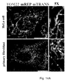
- RNA and DNA labelling 8 as well as labelling of either RNA or DNA, and proteins 9,10 have been performed
- the techniques used namely immunofluorescence and Fluorescent In Situ Hybridization (FISH)
- FISH Fluorescent In Situ Hybridization
- D. JACKSON 1996 relates to a study investigating the sequences found in attaching loops of nuclear and mitochondrial DNA to underlying structures in human cells. A library of such sequences has been generated. However, all cloned fragments generated in this study bear the property of being transcribed within a cell. This study does not disclose nucleic acid molecules that remain RNA-free during transcription and replication of the nuclear and mitochondrial genome.
- SOLOVEI IRINA et al., 2010 discloses protocols for a 3D-FISH method applied on cultured cells combined with immunostaining.
- ALINE V PROBST ET AL, 20 April 2007 discloses using 3D-FISH for visualizing the spatial organization of the nucleic DNA and to follow the dynamic unfolding of the centromeric regions by staining the nucleic DNA in a cell.
- P.S. MASNY, 30 June 2004 discloses the use of 3D-FISH for studying the nuclear organization of the FSHD genomic region.
- ZARDOYA ET AL 1 April 1996 discusses the determination of the complete nucleotide sequence of the African lungfish ( Portopterus dolloi) mitochondrial genome through cloning said genome into plasmids and subsequent sequencing.
- ZARDOYA ET AL does not disclose any nucleic acid molecule suitable for use as a probe for detecting the occurrences of initiation of replication events in mitochondrial DNA.
- WO2005/003766 discloses a method of regulating metabolism and mitochondrial functions in a cell, by contacting the cell with particular agents.
- a list of mitochondrial proteins is provided, with reference to databases access numbers.
- ALAN L ET AL 1 July 2010 discloses an experiment of co-localization on mitochondrial DNA using labelled probes targeting either ND5 mRNA or the complementary mRNA permanently attached to the mitochondrial D-loop. D7 does not disclose a staining with probes targeting mitochondrial DNA,
- XING Y ET AL 26 February 1993 relates to the visualization of fibronectin and neurotensin messenger RNAs within mammalian cells by flurorescence hybridization with genomic complementary DNA and intron-specific probes.
- the visualization of the spatial repartition of different categories of mRNAs within said cells is sought, distinguishing RNA free to diffuse, or RNA localizing to the nuclear interior, in order to conclude to a nuclear function closely integrated with the cell structure.
- the present invention addresses the need to obtain an outstanding tool for studying DNA replication, allowing a deeper comprehension of the events associated with DNA replication, and including the comprehension of the coordination of these events with other events such as RNA transcription and even protein(s) distribution in cell(s) or tissue(s) by tracking and monitoring these distinct molecular subpopulations in a concomitant or even simultaneous manner. Consequently, the invention proposes a new way to further explore the complex cellular dynamics and to use such exploration in detection of pathological states.
- novel information can be provided on the dynamics of mitochondrial gDNA processing during physiological and pathological processes. These findings have implications in diagnostic tools of diseases, especially mitochondrial diseases or diseases associated with mitochondrial dysfunction(s) or impairment, in particular those where mtDNA depletion and mtDNA loss can be observed.
- mtDNA depletion can originate from genetic defects, or be acquired, i.e by clinical treatments, as for prolonged administration of anti-HIV nucleoside analogues.
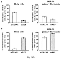
- qPCR analysis on mtDNA is currently used to detect alterations of the mtDNA content in a given cell population.
- the invention relates to a method for both the detection of the occurrence of initiation of replication events in genomic DNA, which is mitochondrial genomic DNA, and the detection of mitochondrial DNA transcription in a eukaryotic cell, comprising the steps of:
- the target region or the nucleic acid sequence of the target region that has an ability to remain RNA-free is located in a naturally transiently open structure of two complementary single strands of gDNA in a metabolically active cell.
- such a naturally transiently open structure is a replication bubble originating around the locus of a replication origin.
- such a naturally transiently open structure is the so-called DNA encompassed by the D-loop region of the mitochondrial genome, which is located between coordinates 16024 to 576 in the human mitochondrial genome (NCBI or Genbank or MITOMAP sequence reference NC_012920.1).
- the sequence of the target region that has an ability to remain RNA-free is located proximal to, or includes, or overlaps known origin(s) of replication, in particular a mitochondrial origin of replication, or is within a distance of less than 10 nucleotides, in particular 1, 2, 3, 4, 5, 6, 7, 8, 9, especially 6 nucleotides, from a known origin of replication, in particular a mitochondrial origin of replication.
- an origin of replication can be the O H origin of replication, located between coordinates 110 to 441 of the mitochondrial genome (NCBI or Genbank or MITOMAP sequence reference C_012920.1).
- said target region or said nucleic acid sequence of the target region encompasses nucleotides within a distance of less than 10, in particular 1, 2, 3, 4, 5, 6, 7, 8 or 9 nucleotides upstream or downstream from a naturally transiently open structure of two complementary single strands of gDNA in a metabolically active cell.
- hybridization relates to the fact of obtaining a close interaction of the nucleotide probe and the target region that is expected to be revealed by the detection of the nucleotide probe. Such an interaction can be achieved by the formation of hydrogen bonds between the nucleotide probe and the target sequence, which is typical of the interactions between complementary nucleotide molecules capable of base pairing. Hydrogen bonds can be found, for example, in the annealing of two complementary strands of DNA.
- the nucleic acid sequence of the probe should be at least partly complementary to the sequence of the target region of the genomic DNA, i.e. should be complementary over a region sufficient to enable stable base pairing.
- a first nucleotide probe designed for hybridizing to a target region of genomic DNA is a labelled nucleic sequence fragment complementary to the target region of the genomic DNA and having substantially or in particular exactly, the same length as said target.
- the first nucleotide probe designed for hybridizing to a target region of genomic DNA is a labelled nucleic sequence fragment complementary to the targeted DNA fragment and having substantially, or in particular exactly, the same length than the nucleic acid sequence which has no identified corresponding annealing RNA in a metabolically active cell and therefore remains RNA-free during transcription and replication of said DNA genome.
- a first nucleotide probe designed for hybridizing to a target region of genomic DNA is a labelled nucleic sequence fragment comprising a nucleic acid sequence that is complementary to the targeted DNA fragment, said nucleic acid sequence having substantially, or in particular exactly, the same length than the nucleic acid sequence which has no identified corresponding annealing RNA in a metabolically active cell and therefore remains RNA-free during transcription and replication of said DNA genome.
- the interaction of the nucleotide probe and the target region can also involve van der Waals interactions, ionic bonds or covalent linkages. Such interaction(s) might imply that the nucleotide probe contains modified nucleotides or bear specific moieties generally not present in nucleotidic molecules.
- In situ hybridization refers to the fact that the hybridization is carried out on the assayed biological material.
- Said biological material can be single cell(s) or tissue(s), or a sample comprising the same.
- the integrity of the structure and/or content of the biological material is maintained. Therefore, in order to carry out the invention, the biological material is preferably fixed.
- the method of the invention is carried out on fixed cell(s) or tissue(s).
- the method of the invention further permits to maintain the integrity of the cell(s) volume and thus the analysis of fixed sample(s) in three-dimension.
- the cell(s) or tissue(s) are eukaryotic cell(s) or tissue(s), in particular human cell(s) or tissue(s).
- human cell(s) or tissue(s) derived from human cell lines such as HeLa, HCT116, HT29, AGS cell lines, and/or human primary cells, i.e. IMR-90, BJ human fibroblasts obtained from ATCC, are used.
- genomic DNA is mitochondrial gDNA.
- the genomic DNA may be nuclear gDNA.
- genomic DNA may be both nuclear gDNA and mitochondrial gDNA.
- genomic DNA refers to a group of genomic DNA molecules, i.e. refers to more than one genomic DNA molecule, said group of genomic DNA molecules consisting of more than one copy of genomic DNA molecules (as found in a single mitochondrion) and/or more than one genomic DNA molecules that are different from each other (such as mitochondrial gDNA and nuclear gDNA).
- a "probe” is aimed at revealing the target region of interest, and is therefore generally, but non-exclusively, labelled.
- Labelling of the probe aimed at revealing the target region in the DNA genome is preferably achieved with either radio- or antibody-discoverable- or fluorescent- or biotinylated- tags or quantum dots, especially fluorescent quantum dots.
- Said tags or quantum dots are directly or indirectly associated, including coupled, to the probe.
- the probe can be localized or visualized or measured on the biological material after hybridization with its target using appropriate techniques, such as autoradiography or fluorescence microscopy.
- discoverable tag is digoxigenin, biotin, or hapten for example revealed by a labelled antibody or a labelled reagent, such as a fluorescent antibody raised against digoxigenin or a labelled biotin binding molecule such as avidin or streptavidin.
- the probe is rendered discoverable, especially through fluorescence detection methods, by introducing an antigen in said probe or by coupling said probe with an antigen that will be further revealed by a secondary anti-antigen antibody, especially a fluorescent anti-antigen antibody.
- a secondary anti-antigen antibody especially a fluorescent anti-antigen antibody.
- One advantage of using antibodies might be an increase of the intensity of the resulting fluorescent signal.
- probe(s) are directly labelled with fluorescent moieties (tags).
- fluorescent moieties tags.
- One advantage of such an embodiment might be to bypass the use of an antibody for the detection of the probe in order, for example, to increase the specificity or the practicability of the labelling/detection method.
- Probe(s) is/are preferably nucleotide probe(s), and are especially short sequences of single stranded DNA capable of base pairing with their complementary DNAs. Probe(s) encompass(es) probe(s) containing nucleotide(s) coupled or linked to other molecule(s) or moiety(ies).
- probe(s) is/are DNA probe(s) such as PCR product(s) or DNA fragment(s), including plasmidic probe(s) or probe(s) comprising such elements.
- they can be double-stranded DNA probes that require being denaturated as single stands prior to their use.
- the nucleotide probe may contain modified nucleotides or bear specific moieties generally not present in nucleotidic molecules.
- Locked Nucleic Acids are modified nucleotides and a class of RNA analogs that have an exceptionally high affinity towards complementary DNA and RNA. They can substitute natural nucleotides in DNA probes.
- a "target region" in the DNA genome is a genomic DNA region comprising a nucleic acid sequence which has no identified corresponding annealing RNA in the metabolically active cell under assay and therefore remains RNA-free during transcription and replication of the DNA genome to which the nucleic acid sequence belongs.
- a target region is a genomic DNA region that has ability to remain RNA free, in particular RNA-transcript(s) free, during the transcription and replication of the DNA genome to which it belongs (mtDNA or nDNA) in the cell that is tested.
- the target region consists of a nucleic acid region in a genomic DNA which has no identified corresponding annealing RNA in a metabolically active cell.
- occurrence it is meant that the method of the invention enables to qualitatively detect initiation of the replication of genomic DNA and, according to a particular embodiment, to quantitatively detect such initiation event(s) of the replication process.
- “Initiation of replication events” can be, for example, the formation of replication bubble(s) on the analyzed genomic DNA, or in a particular embodiment where the mitochondrial gDNA is assayed for initiation of replication, the formation of a D-loop structure, including the formation of three-stranded D-loop structure.
- Such events may precede the entire replication of the analyzed gDNA, meaning that such events may precede replication over the complete analyzed gDNA.
- Such events may alternatively be followed by interrupted synthesis of the nascent strand of DNA.
- DNA replication usually begins at specific location(s) in the genome, called “origin(s)” or “replication origin(s)”. Once polymerases have opened the double stranded genomic DNA molecule, an area known as a "replication bubble” forms (usually initiated at a certain set of nucleotides, the origin of replication).
- D-loop region is typical of the human and other mt DNAs, such as Mammalian, Avian, Fish or Plant mtDNAs.
- the coordinates of said D-loop regions vary according to the considered organisms but can be found in the literature 31 . However D-Loop regions are not found in all mtDNA.
- the D-loop region is roughly located between the coordinates 16024 and 576 of the L-strand on the mitochondrial genome (according to the data released to date on databases, in particular under accession number NC_012920.1 (NCBI, GenBank or MITOMAP sequence reference), see in particular MITOMAP: http://www.mitomap.org/MITOMAP/HumanMitoSeq).
- the target region or the RNA-free nucleic acid sequence comprised in said target region is located in a naturally transiently open structure of two complementary single strands of gDNA in a metabolically active cell, as disclosed above and in the following embodiments.
- the target region or the RNA-free nucleic acid sequence comprised in said target region is located upstream from the major H-strand promoter on the mitochondrial genome (PH1), which coordinates are given in Table 2 (coordinates and direction are given herein with respect to the L-strand of the mtDNA).
- the target region or the RNA-free nucleic acid sequence comprised in said target region encompasses the sequences found on either the L-strand or the H strand at the specific location mentioned herein. Reference is made to the L-strand to indicate the position of the major H-strand promoter only.
- probe(s) is/are double-stranded DNA probe(s). They are denaturated as single stands prior to their use. When used simultaneously after denaturation, such a mix of complementary single-stranded probe(s) results in annealing both the L and H strands of the target region of a mtDNA.
- the target region or the RNA-free nucleic acid sequence comprised in said target region is located downstream from the L-strand promoter on the mitochondrial genome (LP or LSP), which coordinates are given in Table 2 (coordinates and direction are given herein with respect to the L-strand of the mtDNA).
- LP or LSP mitochondrial genome
- the target region or the RNA-free nucleic acid sequence comprised in said target region encompasses the sequences found on either the L-strand or the H strand at the specific location mentioned herein. Reference is made to the L-strand to indicate the position of the L-strand promoter only.
- Naturally transiently open structure of two complementary single strands of gDNA it is meant a gDNA structure formed by the dissociation of the two DNA strands constituting the gDNA as a result of processing by replication machinery and mechanism(s) inherent to a metabolically active cell, during its life cycle.
- the target region is located near or in a region of the genomic DNA that is involved in the early events of the replication of said genomic DNA, such as a region found in a replication bubble or a region at least partly encompassed by a replication bubble.
- a region will generally be localized in the vicinity of a replication origin of a genomic DNA of an eukaryotic cell, in particular in the close vicinity or near, i.e. no farther than 10 nucleotides from a replication origin of a genomic DNA of an eukaryotic cell.
- the target region is located in the vicinity of a replication bubble or at a locus encompassed by a replication bubble (where a replication bubble can be found), in particular no farther than 10 nucleotides from such a bubble or locus encompassed by such a bubble.
- Replication bubbles initiate at the locus of replication origins.
- the target region is located at 5 nucleotides of the O H replication origin in the human mtDNA.
- a nucleic acid sequence having "no identified corresponding annealing RNA in a metabolically active cell” is a sequence having no strictly corresponding, i.e. complementary or matching, RNA, especially no RNA transcript(s) resulting from the transcription process occurring naturally in the living eukaryotic cell under assay.
- corresponding it is understood a substantial, in particular a strict, complementarity of nucleic acid sequences which are aligned and whose similarity is calculated over the entire length of the aligned sequence by alignment algorithm such as the Needelman and Wunsch algorithm (a substantial similarity or perfect match is expected).
- nucleic acid sequence has an ability to remain RNA-free, in particular RNA-transcript(s) free, within the analyzed cell, meaning that such a nucleic acid sequence will not give rise to any identified RNA molecule, especially a RNA molecule that would have been transcribed from genomic DNA in the analyzed cell, nor hybridize with RNA primers involved in replication process in a metabolically active cell containing said nucleic acid sequence.
- a nucleic acid sequence cannot be detected by a probe aimed at detecting the result of transcription events occurring in a cell.
- a first nucleotide probe whose sequence would strictly match the sequence of a nucleic acid sequence as discussed above, would not hybridize with any RNA molecule naturally expressed within a metabolically active cell.
- nucleic acid sequence is thus characterized in that it does not bear any coding information that would be reflected at the transcription level of the DNA processing in a cell.
- condition enabling in situ hybridization it is meant that the target region is rendered physically accessible to the probe in order to enable the hybridization of said probe to the target region.
- the hybridization of the probe to the target region may be only partial along the entire length of the probe or the target region, but sufficient to be specific and stable during washing step(s) following the hybridization.
- the hybridization of the first nucleotide probe to the target region occurs over the length of the probe and/or over the length of the target region.
- said target region has to be available under the form of an accessible single stranded of gDNA even transiently during the replication process.
- it is the nucleic acid sequence which has no identified corresponding annealing RNA that has to be available under the form of an accessible single stranded of gDNA even transiently during the replication process.
- the first nucleotide probe strictly anneals to the above-mentioned nucleic acid sequence comprised in the target region that has no identified corresponding annealing RNA in a metabolically active cell.
- the first probe when hybridizing to the nucleic acid sequence comprised in the target region which has no identified corresponding annealing RNA, the first probe does not overflow the boundaries of said nucleic acid sequence.
- the D-loop region of said DNA can be found as a specific structure involving a three-stranded DNA structure that is formed when a newly synthesized single DNA strand remains bound to one of the parental DNA strand of the gDNA and displaces one of the duplex parental strand.
- such a three-stranded DNA structure might help rendering a target region located in the D-loop structure or in the vicinity of this structure accessible to the probe.
- the target region is, in this configuration, either located in a naturally transiently open segment of two complementary single strands of gDNA when the mitochondrial genomic DNA is entering replication, or in a region which is impacted by the presence of the third DNA strand that might help to push aside proteins or other elements that might render the target region crowded and/or hinder the target region with the result of rendering said region inaccessible to the probe for subsequent hybridization of said probe to the target region.
- initiation of replication is considered an event to be detected in mitochondrial gDNA even when the presence of a third DNA strand in a D-loop does not further give rise to the replication of the whole mitochondrial DNA strand.
- the initiation of the replication of the mitochondrial gDNA is considered to happen with the formation of a replication bubble, including the formation of a D-loop around the locus of a replication origin.
- the method of the invention can permit the specific detection of the D-loop region opening by labeling the mitochondrial genomic DNA with a probe as defined in the claims hybridizing at least partly the target region located in the vicinity of said D-loop or at a locus included in said D-loop.
- the first nucleotide probe strictly anneals to the above-mentioned nucleic acid sequence comprised in the target region that has no identified corresponding annealing RNA in a metabolically active cell.
- the accessibility of the target region to the first probe as defined in the claims can be improved by performing a step aimed at partially denaturing the genomic DNA molecule comprising the target region, for example by heating the eukaryotic cell comprising said genomic DNA at a temperature in the range of 72 to 78°C, preferably 75°C, for 2 to 8 minutes, preferably 4 to 5 minutes, in particular 5 minutes, prior to the hybridization step.
- said partial denaturation is performed without using any chemical agent resulting in a complete denaturation of nucleic acids. Consequently, treatments with HCl or Pepsin, alkaline agents or ethanol are prohibited. Conversely, the use of chemical agents and/or temperature conditions enabling or assisting a partial denaturation of nucleic acids is possible.
- An example of chemical agent that can be used is formamide. Combinations between the proposed treatments disclosed herein are encompassed by the present invention.
- partial denaturation it is meant that the two strands constituting a double stranded nucleic acid are not found completely separated i.e. under the form of single strands, after such a denaturation.
- said partial denaturation results in increasing the size of opening(s) or bubble(s) that could be found on the double-stranded nucleic acid of gDNA prior to eliciting its partial denaturation.
- said agent(s) enable(s) the partial denaturation by performing or assisting the increase in size of opening(s) or bubble(s) on the double-stranded target nucleic acid, to the exclusion of the result consisting in the dissociation of the strands of the double-stranded target nucleic acid, on their whole length.
- both the initiation of genomic DNA replication in a cell or tissue and produced RNA molecules being for example RNA molecules corresponding to transcription products of genomic DNA fragments concomitantly transcribed in said cell or tissue, are tracked.
- the method of the invention is used for the further detection of at least one RNA molecule corresponding to a transcribed region of a DNA molecule in an eukaryotic cell, which comprises the step of contacting said eukaryotic cell expressing said RNA molecule with at least a second nucleotide probe, and detecting said second nucleotide probe after hybridization with said RNA molecule.
- the DNA molecule giving rise to the RNA transcript molecule detected by the second nucleotide probe is a genomic DNA inside said cell (the analyzed cell).
- the labelling of the nucleic acid sequence of the target region on the DNA genome and the RNA molecule is achieved in one step, in particular simultaneously.
- the detection of the nucleic acid sequence of the target region on the DNA genome and the RNA molecule is achieved in one step, in particular simultaneously.
- the hybridization of the second nucleotide probe to a RNA molecule corresponding to a transcribed region of a DNA molecule is achieved by obtaining a close interaction of the nucleotide probe and the RNA molecule that is expected to be revealed by the detection of the nucleotide probe.
- Such an interaction can be achieved by the formation of hydrogen bonds between the nucleotide probe and RNA molecule, which is a typical example of the interactions between complementary nucleotide molecules. Hydrogen bonds can be found, for example, in the annealing of two complementary strands of DNA.
- a nucleotide probe designed for hybridizing to a RNA fragment is a labelled nucleic sequence fragment complementary to the RNA fragment to detect.
- the nucleic acid sequence of the probe should be at least partly complementary to at least a part of the RNA molecule to detect, i.e. should be complementary over a region sufficient to enable stable base pairing.
- the interaction of the nucleotide probe and the RNA molecule can also involve van der Waals interactions, ionic bonds or covalent linkages. Such interaction(s) might imply that the nucleotide probe contains modified nucleotides or bear specific moieties generally not present in nucleotidic molecules.
- probe(s) are directly labelled with fluorescent moieties (tags).
- Probe(s) is/are preferably nucleotide probe(s), and are especially short sequences of DNA or RNA (cRNA probes or riboprobes) that binds to their complementary RNAs. Probe(s) encompass(es) probe(s) containing nucleotide(s) coupled or linked to other molecule(s) or moiety(ies).
- the nucleotide probe may contain modified nucleotides or bears specific moieties generally not present in nucleotidic molecules.
- Locked Nucleic Acids are modified nucleotides and a class of RNA analogs that have an exceptionally high affinity towards complementary DNA and RNA. They can substitute natural nucleotides in DNA or RNA probes.
- the invention encompasses the use of probes suitable for revealing several distinct RNA molecules or fragments thereof, or the use of a single probe targeting distinct RNA molecules or fragments thereof (specific of a pool of RNA molecules or fragments thereof), or the use of a single probe specific of the sequence of a unique RNA molecule or fragment thereof within a cell or tissue.
- probe encompasses a plurality of molecular entities used together to reveal one or many RNA molecules, said RNA molecules being distinct or different.
- such a probe suitable for revealing RNA molecule(s) is generally, but non-exclusively, labelled.
- Labelling of the probe can be achieved with either radio- or antibody-discoverable- or fluorescent- or biotinylated- tags or quantum dots, especially fluorescent quantum dots. Said tags or quantum dots are directly or indirectly associated, including coupled, to the probe.
- the probe can be localized in the biological material using appropriate techniques, such as autoradiography or fluorescence microscopy, respectively.
- RNA molecule(s) can be polycistronic RNA, RNA corresponding to transcribed fragments of genomic DNA (nuclear and/or mitochondrial genomic DNA), processed or unprocessed RNA(s) in said cell.
- the first probe aimed at revealing a gDNA target region is a single stranded DNA fragment ranging in size from 80 bp to 3000 bp, or from 90 to 150 bp, in particular from 95 to 110 bp, preferably sizing 99 bp.
- said second probe aims at revealing RNA molecule(s)
- said second probe is a single stranded nucleotidic DNA fragment ranging in size from 100 bp to 3000 bp, and preferably sizing between 100bp and 2000bp when aimed at detecting mitochondrial transcripts.
- the size and/or sequence of second probe(s) aimed at revealing RNA molecule(s) is particularly adapted to enable the detection of transcription products resulting from the transcription of coding segments of genomic DNA, especially segments corresponding to genes.
- the nucleic acid molecule suitable for use as first nucleotide probe is specific for a segment of a non transcribed mitochondrial gDNA, especially an entirely non-transcribed mitochondrial gDNA, according to the definition provided herein.
- the first nucleotide probe or molecule is complementary of a genomic DNA region that has no corresponding RNA transcript at a cellular level, and therefore remains RNA-free at the cellular level.
- the first nucleotide probe or molecule targets the genomic DNA sequence localized between the two promoters PH1 (or HSP- Heavy Strand Promoter) and LSP (Light Strand Promoter) of the mitochondrial genome of a eukaryotic cell.
- PH1 HSP- Heavy Strand Promoter
- LSP Light Strand Promoter
- Both the HSP and LSP promoters are found in all eukaryotic mtDNA although their name might differ depending on the species to which the considered eukaryotic mtDNA belongs. The corresponding names can be identified through the literature.
- mREP In the human mitochondrial genome, such a first probe or nucleic acid molecule sequence has been designated mREP by the inventors. Coordinates of this sequence are given in Table 1 (NCBI, GenBank or MITOMAP sequence NC_012920.1, used as reference). mREP (SEQ ID No 1) is also disclosed in Fig. 13a .
- said first probe comprises the mREP sequence (SEQ ID No 1).
- the first nucleotide probe or molecule is complementary of a nucleic acid sequence that has at least 80% identity with the target DNA region that is localized between the two promoters PH1 (or HSP) and LSP of the mitochondrial genome of a eukaryotic cell, or has at least 80 % identity with mREP.
- At least 80 % homology it is meant that their sequence of nucleotides differ from less than 20%, calculated over the entire length of the considered sequence (global alignment calculated for example by the Needleman and Wunsch algorithm).
- Fig 13b discloses polymorphism variations known to date with respect to the human mREP sequence (SEQ ID No 1) part of the invention. However, very few of these variations can be found simultaneously in an individual, i.e. only 1, 2, 3 or 4 of these variations can be found simultaneously. Said variations are generally linked to a subpopulation type (e.g. Caucasian, African). These polymorphism variations are a basis for the design of variants of mREP.
- Table 3 discloses the percentages of identity between the sequences corresponding to the mREP probe (SEQ ID No 1) in several organisms and species, with respect to human mREP probe, along with coordinates of said sequences on the corresponding mitochondrial genomes (SEQ ID No 2 to 16).
- Table 3 Percentages of identity between the sequences (SEQ ID No 2 to 16) corresponding to the mREP (SEQ ID No 1) probe in several organisms and species. Refseq are NCBI or GenBank reference numbers.
- the first nucleotide probe comprises a nucleic acid molecule having the sequence of any one of SEQ ID N°1, SEQ ID N°2, SEQ ID N°3, SEQ ID N°4, SEQ ID N°5, SEQ ID N°6, SEQ ID N°7, SEQ ID N°8, SEQ ID N°9, SEQ ID N°10, SEQ ID N°11, SEQ ID N°12, SEQ ID N°13, SEQ ID N°14, SEQ ID N°15, SEQ ID N°16, or the sequence that is complementary of any one of SEQ ID N°1, SEQ ID N°2, SEQ ID N°3, SEQ ID N°4, SEQ ID N°5, SEQ ID N°6, SEQ ID N°7, SEQ ID N°8, SEQ ID N°9, SEQ ID N°10, SEQ ID N°11, SEQ ID N°12, SEQ ID N°13, SEQ ID N°14, SEQ ID N°15, SEQ ID N°16, or
- the first nucleotide probe comprises a nucleic acid molecule encompassing fragments of the nucleic acid sequence of any one of SEQ ID N°1, SEQ ID N°2, SEQ ID N°3, SEQ ID N°4, SEQ ID N°5, SEQ ID N°6, SEQ ID N°7, SEQ ID N°8, SEQ ID N°9, SEQ ID N°10, SEQ ID N°11, SEQ ID N°12, SEQ ID N°13, SEQ ID N°14, SEQ ID N°15, SEQ ID N°16, or the nucleic acid sequence that is complementary of any one of SEQ ID N°1, SEQ ID N°2, SEQ ID N°3, SEQ ID N°4, SEQ ID N°5, SEQ ID N°6, SEQ ID N°7, SEQ ID N°8, SEQ ID N°9, SEQ ID N°10, SEQ ID N°11, SEQ ID N°12, SEQ ID N°13, SEQ ID N°14, SEQ ID ID N°15, SEQ ID N
- the first nucleotide probe consists of a nucleic acid molecule that has the nucleic acid sequence of any one of SEQ ID N°1, SEQ ID N°2, SEQ ID N°3, SEQ ID N°4, SEQ ID N°5, SEQ ID N°6, SEQ ID N°7, SEQ ID N°8, SEQ ID N°9, SEQ ID N°10, SEQ ID N°11, SEQ ID N°12, SEQ ID N°13, SEQ ID N°14, SEQ ID N°15, SEQ ID N°16, or the nucleic acid sequence that is complementary of any one of SEQ ID N°1, SEQ ID N°2, SEQ ID N°3, SEQ ID N°4, SEQ ID N°5, SEQ ID N°6, SEQ ID N°7, SEQ ID N°8, SEQ ID N°9, SEQ ID N°10, SEQ ID N°11, SEQ ID N°12, SEQ ID N°13, SEQ ID N°14, SEQ ID N°
- the first nucleotide probe is a nucleic acid that has at least 80% identity with the nucleic acid sequence of any one of SEQ ID N°1, SEQ ID N°2, SEQ ID N°3, SEQ ID N°4, SEQ ID N°5, SEQ ID N°6, SEQ ID N°7, SEQ ID N°8, SEQ ID N°9, SEQ ID N°10, SEQ ID N°11, SEQ ID N°12, SEQ ID N°13, SEQ ID N°14, SEQ ID N°15, SEQ ID N°16, or the nucleic acid sequence that is complementary of any one of SEQ ID N°1, SEQ ID N°2, SEQ ID N°3, SEQ ID N°4, SEQ ID N°5, SEQ ID N°6, SEQ ID N°7, SEQ ID N°8, SEQ ID N°9, SEQ ID N°10, SEQ ID N°11, SEQ ID N°12, SEQ ID N°13, SEQ ID N°14, SEQ ID N°5, SEQ ID N°
- the first nucleotide probe may encompass a mix of more than one, but distinct probes, especially probes comprising nucleic acid molecules having distinct sequences from each other, chosen among the sequences disclosed above.
- the first nucleotide probe may encompass a mix of two nucleic acid molecules having sequences that are complementary to each other. Such a mix might have to be denaturated prior to its use.
- Fig 13b discloses polymorphism variations known to date with respect to the mREP sequence. These polymorphism variations may be used to design probes alternative to mREP corresponding to SEQ ID No 1.
- Fig 13c discloses alignments between the human mREP sequence and the corresponding sequences in different organisms, which are also disclosed herein under SED ID No 2 to 16.
- the first nucleotide probe is directly labelled, in particular with a fluorescent group.
- Labelling can be achieved with groups or labels such as Biotin, digoxin and digoxigenin (DIG), alkaline phosphatase and fluorescent groups or labels such as fluorescein (FITC), Texas Red and rhodamine or derivatives thereof, or dyes including coumarins, rhodamines, carbopyronins, oxazines or derivatives thereof or quantum dots, especially fluorescent quantum dots, or derivatives thereof.
- groups or labels such as Biotin, digoxin and digoxigenin (DIG), alkaline phosphatase and fluorescent groups or labels such as fluorescein (FITC), Texas Red and rhodamine or derivatives thereof, or dyes including coumarins, rhodamines, carbopyronins, oxazines or derivatives thereof or quantum dots, especially fluorescent quantum dots, or derivatives thereof.
- groups or labels such as Biotin, digoxin and digoxigenin (DIG), alkaline phosphatase and fluorescent groups
- directly labelled it is meant that the detection of the label does not require the intervention of another, i.e. secondary, chemical agent or compound, including an antibody, to be achieved. Such a direct labelling might improve the specificity of the labelling.
- Labelling can be achieved through a commercial kit, for example according to a nick translation procedure.
- the first nucleotide probe comprises modified nucleotides, as disclosed herein.
- the invention also relates to a method for the in situ hybridization and detection of mitochondrial nucleotidic material within at least one eukaryotic cell, which comprises the steps of:
- the denaturation step of said method can further be carried out in an appropriate buffer and/or with a chemical agent aimed at partially denaturing nucleic acids, as disclosed herein.
- the denaturation step of the nucleic acid contents of a fixed cell is carried out by heating at a temperature in a range of 72 to 78 °C, preferably 75°C, for 2 to 8 minutes, preferably 4 to 5 minutes, especially 5 minutes, in the presence of a chemical agent such as formamide, especially a solution of 70% formamide or 70% formamide/2xSSC.
- a chemical agent such as formamide, especially a solution of 70% formamide or 70% formamide/2xSSC.
- Said method can further comprise a step of washing the cell(s) contacted with the nucleotide probe(s) with an appropriate buffer prior to the detection step.
- the hybridization step is carried out during about 15 hours at 37°C.
- the method of the invention further comprises steps enabling labelling and detection of at least one protein of interest within the eukaryotic cell.
- the cell(s) or tissue(s) under assay can be contacted with antibodies specific for the protein(s) of which detection is sought.
- the detection of nucleotidic material and of the protein of interest is achieved in one step, in particular simultaneously.
- a step of analysis of the result(s) of the detection(s) might be subsequently performed.
- Fixation of cells can alternatively be performed with agents such as paraffin, acetone, methanol, ethanol, a combination of methanol and acetone a combination of methanol and ethanol, formalin, a combination of paraformaldehyde and methanol, or any combination(s) of the agents disclosed herein.
- agents such as paraffin, acetone, methanol, ethanol, a combination of methanol and acetone a combination of methanol and ethanol, formalin, a combination of paraformaldehyde and methanol, or any combination(s) of the agents disclosed herein.
- the fixation of cell(s) or tissue(s) is performed on glass slide(s) with a solution of 1 to 4% paraformaldehyde (PFA), preferably 2% PFA for about 20 to 30 minutes, especially 30 minutes.
- PFA paraformaldehyde
- the fixation is carried out with a solution of 2% PFA for about 30 minutes. Said fixation can be carried out at room temperature, RT.
- fixation can be carried out according to any one of the following protocols:
- RNA FISH procedures require the fixation of cell(s) or tissue(s) with 4% PFA for 10 minutes minimum.
- the permeabilization step might be required to allow a good infiltration of the probes, especially through cell membranes, in order for the probes to reach their target sequence.
- Known chemical reagents used for permeabilizing cells in the prior art are HCl, detergents such as Triton or SDS or Proteinase K.
- fixed cell(s) or tissue(s) are permeabilized using a 0.5 % to 1% Triton X100 solution in an appropriate buffer, such as PBS 1X, for example during 5 min. Said permeabilization can be carried out at 4°C.
- fixed cell(s) or tissue(s) can be washed up to 3 times in PBS 1X prior to the permeabilization, and up to 4 times in PBS 1X after the permeabilization.
- the permeabilization can be followed by incubation of the cell(s) or tissue(s) in 50% Formamide/2X SSC (saline-sodium citrate buffer) in PBS 1X at RT during 30 min, and further switching to 70% Formamide/2X SSC just before the denaturation.
- Formamide/2X SSC saline-sodium citrate buffer
- a control assay for the probe specificity can be performed by incubating the samples during 1 hour at 37°C in RNase or DNase solution (100 ug/ml), with an additional washing up to 3 times in PBS 1X prior to the incubation of the cell(s) or tissue(s) in Formamide.
- Standard DNA FISH procedures generally involve a heat treatment of the glass slides (for example during 90°C 1h30 or at 37°C overnight) to remove all enzymes that could interfere with the experiment, and the permeabilization is achieved by incubation with 0.005% Pepsin/0.001 M HCl at 37°C for 15 min, washing in (4%) paraformaldehyde/PBS, then incubating in ethanol series: 70%, 90%, 100%, further followed by RNase treatment that is performed to remove primary transcripts and subsequent washing and dehydratation in ethanol series. The samples are finally air dried.
- Standard RNA FISH procedures generally require permeabilization with Triton X100 0.5% in buffer (the composition of which might contain PIPES, MgCl2, sucrose or NaCl%), washing in 4% PFA/PBS for 10 min on ice and further washings (twice) in 70% ethanol and subsequent dehydratation in ethanol series: 80%, 95%, 100% ethanol and air drying of the samples on a heating plate at 42°C.
- the method of the invention in conducted in absence of enzymatic (i.e. pepsin), acidic (i.e. HCl) or alkaline treatment, alcoholic treatment (i.e. ethanol) or drying agent or treatment (i.e. air dry).
- enzymatic i.e. pepsin
- acidic i.e. HCl
- alkaline treatment i.e. ethanol
- drying agent or treatment i.e. air dry.
- the denaturation step is performed by heating the samples comprising the assayed cell(s) or tissue(s) at a temperature in a range of 72 to 78 °C, preferably 75°C, for 2 to 8 minutes, preferably during 4 to 5 minutes, especially 5 minutes. According to a specific embodiment, the samples are heated at 75°C during 5 minutes. The samples can then be kept on ice until the probe(s) are ready.
- RNA FISH procedures generally involve heating the samples at temperatures close to 80°C, and the assistance of other treatments such as chemical or physical treatments usually used to denaturate nucleic acids (e.g. washing with 70% ethanol on ice or dehydratation with ethanol series) in order to obtain single stranded DNA molecules.
- other treatments such as chemical or physical treatments usually used to denaturate nucleic acids (e.g. washing with 70% ethanol on ice or dehydratation with ethanol series) in order to obtain single stranded DNA molecules.
- the denaturation step according to the invention may be carried out in the presence of chemical agent(s) aimed at partially denaturing nucleic acids, or in an appropriate buffer, such as formamide.
- probe(s) are preferably small in size (3 kb or shorter, preferably 1kb or shorter) and the direct use of fluorescence probe(s) for labelling DNA(s) and RNA(s) is preferred.
- a kit such as the Jena Nick Translation kit can be used to label the probe(s) (Atto fluorescence) and the quantity of DNA recovered after purification should be estimated (about 10% loss compared to input).
- a pre-hybridization step can be performed, comprising the following steps:
- pre-hybridized slides can be put to pre cool 30 min at 37°C in the dark before incubating slides.
- Standard DNA and RNA FISH procedures generally require a higher concentration of fluorescent or tagged probe (final concentration of about 10 ng/ ⁇ l) and the addition of Cot-1 DNA and Salmon Sperm DNA at a final concentration of more than 0.5 ⁇ g/ ⁇ L. The mix is then incubated (5 to 10 min) at 74-85°C in the dark.
- the pre-hybridisation and/or hybridisation step is carried out in absence of Cot-1 DNA.
- the hybridization step is carried out during about 15 hours at 37°C.
- the hybridization step can be done by incubating the samples about 15 hours at 37°C on a heating metal block in the dark.
- Standard DNA and RNA FISH procedures generally require overnight hybridization.
- the method of the invention further comprises a step of washing the cell contacted with the nucleotide probe(s) with an appropriate buffer prior to the detection step.
- washing can be performed up to 2 times for 2 min in 3 mL 2X SSC at RT (cover with black top to protect from light), then up to 2 times in 1X SSC at RT, then up to 2 times in 0.1X SSC at RT. Finally, an ultimate washing can be performed up to 2 times in PBS 1X at RT (all in the dark).
- the procedure allows the labelling of genomic DNA, especially mtDNA and RNAs. Further steps are required for additional labelling of mitochondrial or cellular proteins.
- the method of the invention further comprises steps enabling labelling and detection of at least one protein of interest within the eukaryotic cell treated for in situ hybridization and detection of nucleotidic material.
- immunofluorescence can be used and the cell can be contacted with antibodies specific for the protein(s) of which detection is sought.
- Antibodies raised against a particular protein or epitope can be obtained according to usual techniques commonly used in the immunological field.
- the method of the invention enables to achieve the detection of the nucleotidic material and the protein of interest in one step, in particular simultaneously.
- the following procedure is suitable for the detection of the occurrence of initiation of replication events in the mitochondrial genomic DNA of human cell lines and human primary fibroblasts as well as in cells of other eukaryote organisms.
- mtDNA probe(s) used for tracking the occurrence of initiation of replication events has to be adapted to the sequence of the corresponding mitochondrial genomes (see Table 3 that gives correspondence of the mREP probe in other organisms).
- the following protocol is optimized for 3D Z-scanning of mammalian cells using a confocal spinning disk microscope. Z-stacks of 200nm. 3D reconstruction using the IMARIS (Bitplane software).
- the following protocol is optimized for 3D Z-scanning of mammalian cells using a confocal spinning disk microscope. Z-stacks of 200nm. 3D reconstruction using the IMARIS (Bitplane software).
- nucleic acid molecule(s) suitable for use as probe(s) as defined herein and especially designated as the "first probe” for use in the process of the present disclosure or invention are also disclosed nucleic acid molecule(s) suitable for use as probe(s) as defined herein and especially designated as the "first probe” for use in the process of the present disclosure or invention.
- such a nucleic acid molecule is suitable for use as a probe suitable for in situ hybridization targeting the genomic DNA.
- nucleic acid molecule suitable for use as a probe hybridizing with a target region in a eukaryotic genomic DNA, wherein said target region comprises a nucleic acid sequence which has no identified corresponding annealing RNA in the metabolically active cell containing said eukaryotic genomic DNA and therefore remains RNA-free during transcription and replication of said DNA genome.
- a nucleic acid molecule hybridizes with said RNA-free nucleic acid sequence, and has the same length as said RNA-free nucleic acid sequence or is longer.
- Nucleic acid molecule(s) or probe(s) disclosed above and herein with respect to a method according to the present disclosure are themselves part of the object of the present disclosure. They are prepared and/or used as either a single stand molecule or a double strand molecule of complementary sequences.
- nucleic acid molecules that are specific for a segment of non transcribed mitochondrial gDNA and comprises or consists of :
- a nucleic acid molecule of the invention is as defined in claim 13 or 14.
- kits for carrying out in situ hybridization on fixed cells comprising a so-called first probe, consisting of a nucleic acid molecule, of the present disclosure, and comprising optionally a so-called second probe, consisting of a nucleic acid molecule, of the present disclosure, hybridizing with a RNA molecule.
- kits comprising a probe, consisting of a nucleic acid molecule, suitable for in situ hybridization targeting the genomic DNA as disclosed herein, and comprising optionally a so-called second probe, consisting of a nucleic acid molecule, of the present disclosure, hybridizing with a RNA molecule.
- a kit of the invention is as defined in claim 15.
- kits further comprise probes, consisting of nucleic acid molecule(s), and/or antibody(ies) for additionally detecting protein(s).
- Kits to label and detect in a same cell or tissue DNA and optionally RNA and/or proteins are useful for the detection of the occurrence of initiation events in the genomic DNA replication in eukaryote cell(s) or tissue(s).
- kits can further comprise instructions for use in a process for detecting the occurrence of initiation of replication events in genomic DNA in a eukaryotic cell, according to a methods as disclosed herein.
- kits can further comprise reagents necessary for carrying out such a process, as disclosed herein.
- kits can further comprise material, e.g measurement material, data carrier(s), recording support(s), to collect or analyze the data measured by a process according to the present disclosure or invention.
- material e.g measurement material, data carrier(s), recording support(s), to collect or analyze the data measured by a process according to the present disclosure or invention.
- the present invention is of particular interest for analyzing the processing of DNA, RNA or metabolites in cell(s) or tissue(s), and/or analyzing the dynamics of said cell(s) or tissue(s), and/or detecting specific diseases.
- the invention is of particular interest for providing means useful for the analysis, detection and optionally subsequent diagnosis of mitochondrial disease(s) or neoplasic diseases(s) or cancer(s).
- the invention is also of particular interest for analyzing, detecting and optionally subsequently diagnosing mitochondrial disease(s) or neoplasic diseases(s) or cancer(s).
- mitochondrial misfunction is associated with a variety of diseases (cancers, myopathies, neuropathologies, infections), and with the ageing process.
- Mitochondrial diseases encompass cardiomyopathy, neuropathy, Retinitis pigmentosa, encephalomyopathy, hepatopathy, hypotonia, Renal tubulopathy, Leigh syndrome, Barth syndrome, optic atrophy and Ataxia, Leukodystrophy, Diabetes, Kearns-Sayre syndrome.
- Mitochondrial dysfunctions can lead to or are involved in type 2 diabetes, Parkinson, Alzheimer, Atherosclerotic heart disease, stroke or cancers.
- the method of the invention, the probes described herein and kits encompassing said probes or permitting to carry out the method of the invention are useful in clinical diagnosis protocols by contributing to means necessary to identify and/or to class diseases associated with mitochondrial dysfunctions according to the default in mitochondrial mtDNA processing, e.g. transcription and replication, or in mtDNA content.
- mtDNA depletion diseases mtDNA depletion diseases
- cancers can be also used to identify mtDNA depletion induced by clinical treatment (i.e., long term treatment with anti-HIV nucleoside analogues deplete mtDNA) or impaired or abolished initiation of mtDNA replication.
- the method of the invention can be used in the analysis and detection of mitochondrial disease(s) or disease(s) resulting from mitochondrial dysfunction(s) or impairment, or disease(s) resulting in mitochondrial dysfunction(s) or impairment.
- the method of the invention is applicable to all eukaryotic cells, including human, mouse, insect, yeast, fish or plant cell as a diagnosis tool or a biotechnological tool for exploring the functions of said cells.
- the method of the invention, the probes or nucleic acid molecules described herein and kits encompassing said probes or nucleic acid molecules or permitting to carry out the method of the invention can also be used in the analysis and detection of neoplasic diseases(s) or cancer(s).
- the invention provides means useful for the detection and diagnosis of neoplasic or tumoral cell(s) or tissue(s), and especially to distinguish said cell(s) or tissue(s) among healthy cell(s) or tissue(s).
- the present invention is also of particular interest for testing the cytotoxicity of organic and chemical compounds, especially drugs.
- Human HeLa cells and IMR90 primary fibroblasts (purchased from ATCC) were grown in MEM medium with 10% foetal bovine serum (FBS), HeLa rho° cells in DMEM medium with 10% FBS 1 mM sodium pyruvate and 0.2 mM uridine, at 37°C and in the presence of 5% CO2. Cells cultures were split at regular intervals for different experiments as required. IMR-90 cells were at passage 15. Culture under low oxidative stress were treated with 50 uM H2O2 for the time indicated.
- FBS foetal bovine serum
- BrdU, anti-TOM22 Atto488, and Hoechst 33342 were purchased from Sigma; anti-BrdU antibody from BD Biosciences; MitoTracker® Green FM, and secondary antibodies (Goat anti-mouse antibodies and Goat anti-rabbit antibodies Alexa® Fluor 555 or Alexa® Fluor 488 conjugated) were purchased from Invitrogen.
- IF Immunofluorescence
- Probe labeling and denaturation The DNA probes for FISH were labeled by nick translation of PCR products, incorporating Atto425-dUTP, or Atto488-dUTP, or Atto550-dUTP, using commercial kit (Atto425/Atto488/Atto550 NT Labeling kit, Jena Bioscience). 40 ng of labeled probes were mixed with 400 ng of sonicated salmon sperm DNA (Sigma) and hybridization buffer (50% formamide, 10% dextran sulfate, in 2x SSC pH 7.0). The hybridization mix was denatured at 80°C for 10 min then kept at 37°C for 30 min.
- RNAseA 100 ⁇ g/ml, Roche
- RNAseH 100 U/ml, NEB
- DNAsel 100 U/ml, Invitrogen
- the enzymes were either added simultaneously or the second nuclease was added after incubation with the first nuclease, followed by three washes with PBS, and further incubation for 1 hr at 37°C.
- the immunofluorescence procedure was applied for 3D-FISH coupled IF, after hybridizaton and 0.1xSSC wash.
- BrdU incorporation Cells plated on slides were incubated for 10 min in the presence of 100 ⁇ M BrdU, then immediately fixed in 2% PFA (10 min), treated for 10 min with 4N HCl and 0.5% Triton X-100, and neutralized for 30 min by 100 mM sodium borate. Cells were blocked in 5% BSA in PBS and permeabilized with 0.5% Triton X100 26 . BrdU was detected by immunostaining with anti-BrdU antibody. The DNA was stained with 10 ⁇ g/ml Hoechst 33342, and the image analysis was carried out using spinning-disk Perkin Elmer confocal microscope.
- FISH coupled BrdU Cells plated on slides were fixed with 2% PFA and permeabilized with 0.5% Triton X100. Denaturation was performed using buffer containing 10 mM Tris HCl pH 8.0, 50 mM KCI, 5% glycerol at 95°C for 8 min. The slides were washed in 0.1xSSC and series dehydrated in 70%, 90%, and 100% ethanol and finally air-dried 27 . Hybridization was done overnight at 37°C. After washing the slides in 2xSSC then 0.1xSSC, the slides were incubated in blocking buffer (BSA 5%; PBS 1x) for 1 hr, then incubated with mouse anti BrdU antibody for 1 hr.
- BSA blocking buffer
- a secondary anti-mouse antibody Alexa® Fluor 555 or Alexa® Fluor 488 conjugated was applied.
- the DNA was stained with 10 ⁇ g/ml Hoechst 33342 and the image analysis was carried out using spinning-disk Perkin Elmer confocal microscope.
- Confocal acquisition, 3D-reconstruction and quantification Confocal acquisitions were performed using a spinning-disk Perkin-Elmer Ultraview RS Nipkow Disk, an inverted laser-scanning confocal microscope Zeiss Axiovert 200M with an Apochromat 63x/1.4 oil objective and a Hamamatsu ORCA II ER camera (Imagopole, PFID, Institut Pasteur). Optical slices were taken every 200-nm interval along the z-axis covering the whole depth of the cell, at resolution of 1.024/1.024 pixels. Three-dimensional reconstruction was achieved using the IMARIS software (Bitplane). Fluorescence quantification was done using a single-imaging frame collection and ImageJ 1.34-s software (post-acquisition analysis).
- the perinuclear location of FISH-labelled organelles corresponds to mitochondria located within 2
- the percentage of perinuclear 3D-FISH mitochondria was calculated on the total 3D-FISH labelling. Quantification of mREP-positive and mREP-negative mitochondria was performed on either Poly or TFAM immunolabeled areas. For each condition, 300 samples of identical surface were analysed. Co-localization studies were done with ImageJ JACoP plug-in 28
- RNA was isolated from HeLa cells and IMR90 primary fibroblasts using a RNAeasy Mini kit (Qiagen) and a RNAeasy Micro kit (Qiagen), respectively. The total RNA was treated with DNasel (Qiagen), then reverse-transcribed using Superscript® III Reverse transcriptase (Invitrogen). Real-time quantitative PCR was performed using Power Sybr Green PCR Master Mix (Applied Biosystems) and the rate of dye incorporation was monitored using the StepOne T M Plus RealTime PCR system (Applied Biosystems). Three biological replicates were used for each condition. Data were analyzed by StepOne Plus RT PCR software v2.1 and Microsoft excel. TBP transcript levels were used for normalisation of each target ( ⁇ CT).
- Real-time PCR CT values were analyzed using the 2 - ⁇ Ct method to calculate the fold expression (all (A 2 CT)method) 29 .
- Custom primers were designed using the Primer3Plus online software (http://www.bioinformatics.nl/cgi-bin/primer3plus.cgi). Primers used for amplification are available upon request.
- the inventors have developed a novel approach that labels simultaneously mtDNA and mtRNA in human cells by improving fluorescence in situ hybridization (FISH), and performed 3D confocal acquisitions (3D-FISH). Since proteins are not destroyed during this treatment, and in contrast to existing protocols, 3D-FISH have been coupled to immunofluorescence. Hence, it was possible for the first time to monitor mitochondrial DNA, RNA and proteins simultaneously. Moreover, the intensity of fluorescence could be quantified, thereby permitting a relative assessment of these nucleic acids with single-cell resolution.
- FISH fluorescence in situ hybridization
- 3D-FISH 3D confocal acquisitions
- probes used for FISH experiments are given on the mitochondrial DNA (NC_012920.1, NCBI or GenBank or MITOMAP accession number, was used as reference). Individual probes are indicated in the upper panel. Mix of more than one probe and their composition are indicated in individual panels below. All probes are oriented in the direction of transcription of the H strand, with the exception of probe ND6 that is in the inverse orientation (transcription on the L strand).
- each probe recognized a specific subset of mitochondria and not the entire mitochondrial network ( Fig. 2a ), indicating that only a subpopulation of mitochondria carries detectable amounts of the target nucleic acid, and that mitochondria may not be functionally alike.
- Saturation experiments indicated that labelling of a subset of mitochondria was not due to limited concentrations of probe ( Fig. 5 ).
- Treatment with nucleases showed that all of the probes recognized essentially RNA targets, with the exception of probes 4, 8 and 13, which also recognized DNA (decrease in fluorescence following treatment with DNAsel of 65%, 97% and 47%, respectively).
- Figure 2a also shows that in HeLa cells the distribution of labelled mitochondria varies as a function of the mtDNA region tested. Indeed, 16S rRNA is present mainly in mitochondria located in the perinuclear region and in tubular, filamentous mitochondria, whereas transcripts of the last third of the H-strand appear in fragmented mitochondrial entities, distributed more randomly in the cytoplasm. Thus, mitochondria cluster around the nucleus during processing of the 16SRNA, and they spread to the cellular periphery as RNA processing on the H-strand terminates.
- the perinuclear localization of mitochondria labelled by 3D-FISH is high in HeLa cells (>50% of the total mitochondrial population). Although a different localization of mitochondrial populations according to the detected type of transcript was also observed in IMR-90 primary fibroblasts, no prevalent perinuclear distribution of mitochondria appears in these cells (30-47%, all probes included; Fig. 2c and Fig. 9 ). Moreover, mitochondria labelled for various RNAs and DNAs appeared to be more fragmented in IMR-90 fibroblasts than in HeLa cells.
- Mitochondrial transcripts exist as processed transcripts of single genes, and unprocessed polycistronic transcripts 1 .
- the RNA labelling which we observed with 3D-FISH may represent one or both of these types of transcripts.
- Experiments involving co-labelling with one or more mtDNA probes helped to distinguish between unprocessed RNA molecules and individual transcripts ( Fig. 10 ).
- Early transcripts on the H-strand appeared largely as unprocessed molecules, whereas late H-strand transcripts appeared frequently as processed molecules.
- FISH signal was not limited by probes concentration.
- the inventors have then co-labelled cells with mTOT and with a probe that targets nuclear Alu sequences 34 (probe Hs Alu) and found that labelling of mitochondrial nucleic acids did not preclude the labelling of nuclear nucleic acids ( Fig. 5 ), indicating that the accumulation of mtDNA labelling in the perinuclear region was not due to inaccessibility of the probe to the nucleus.
- This experiment also confirmed that labelling with mtDNA probes was specific to mitochondrial nucleic acids.
- HeLa cells were also labelled with a further mix of probes, called mTOT ⁇ r, that includes all probes present in mTOT with the exception of probes 1, 2 and 14 that cover the rDNA portion of the mt genome.
- Fig. 5 shows that the intensity of labelling in the absence of rDNA probes was at least as high as with the probe mTOT, indicating that 3D-FISH labels mitochondrial mRNAs even in the presence of large amounts of rRNAs.
- the inventors have also observed that mTOT ⁇ r-labelled mitochondria were not mostly located in the perinuclear region as it was the case with the mTOT mix. This experiment suggested that different combinations of mt DNA probes label distinct mitochondrial populations.
- 16S but not 12S rRNAs was present in larger amounts than the other transcripts and was produced by a larger proportion of mitochondria
- transcripts containing 16S rRNA are present in larger amounts than the other transcripts, as expected, but surprisingly this was not the case for transcripts containing 12S rRNA (probe 14, Fig. 2 ). This finding was confirmed by quantitative RT-PCR (qRT-PCR) analysis of the 16S and 12S rRNAs ( Fig.
- probe 3 that essentially covers the ND1 gene localised downstream of 16S on the H-strand.
- the signal for probe 3 was lower than for 16S rRNA (probes 1 and 2), as expected 35 , it was at least twofold higher than for the other genes located downstream of rRNA transcription terminators ( Figure 2B ).
- RNA production of large amounts of a given RNA may originate from elevated transcription by individual mitochondria or from a large number of mitochondria implicated in transcription, or both.
- the inventors have coupled 3D-FISH of each of the 14 mtDNA probes to immunofluorescence with anti-TOM22, a mitochondrial outer membrane marker that identifies the entire mitochondrial population.
- p the percentage of co-localization between anti-TOM22 and each probe was assessed.
- the inventors have found that a large proportion of mitochondria (49-69%) was labelled with probes 1 to 3, and with probes 6-7, while only 19-38% of mitochondria are labelled with the remaining probes ( Fig. 7 a-b ).
- RNA labelling for probe 3 that essentially covers ND1 (see above).
- High levels of ND1 labelling may results from PH1 transcription of rRNAs that did not stop at terminators or, alternatively, from a particularly long-lived RNA, although it was not reported that the ND1 transcript was more long-lived than the other mRNA in HeLa cells 37 .
- the levels of ND1 labelling (probe 3) were close to those of 16S rRNA (probes 1-2). The inventors have reasoned that large amounts of ND1 RNA may result from leaky termination of transcription from PH1.
- the inventors have performed co-labelling with additional pairs of probes to verify the simultaneous presence of transcripts in mitochondria. It was found that probe 14, that labelled 12S rRNA present at the beginning of the H-strand transcript colocalized with probe 12, that labelled CytB present at the end of the same transcript, in 55.6 ⁇ 7% of cases, indicating that mitochondrial entities showing co-localization either contained the 5' and the 3' end of the PH2-directed transcript, i.e. the complete H-strand transcript, or that 12S and CytB processed transcripts were present in equimolar amounts on the same mitochondrial entities ( Fig. 10 ).
- transcripts labelled by 3H-FISH probes represented both unprocessed RNA molecules, in particular the early transcripts on the H-strand, and processed individual transcripts. This proof of principle showed that processed and unprocessed RNA molecules could be identified for any mitochondrial gene of interest using, appropriate pairs of probes in co-localization experiments.
- probes 6-7 and 8-9 were weakly labelled by 3D-FISH (probes 6-7 and 8-9, respectively).
- the low labelling of probes 6-7 and 8-9 might result from the levels of additional transcripts other than ATP8 and ND4, also identified by these probes (see Table 2).
- Table 2 Position of the probe on the human mitochondrial genome.
- the coordinates of the genetic element present at a given position of the mitochondrial genome are indicated in column 1 (data from MITOMAP: http://www.mitomap.org/MITOMAP/HumanMitoSeq).
- ATP8 and ND4 were being transcribed they might not be labelled by 3D-FISH. Indeed ongoing transcripts formed DNA-RNA hybrids with the template that might not be detected as RNA molecules by 3D-FISH (see Fig. 1c ). It is also possible that probes 6-7 and 8-9 had impaired accessibility to RNA molecules. Finally, probe 3 showed high 3D-FISH labelling but ND1, its main target, was not expressed at high levels. This apparent discrepancy may be due to the fact that 3D-FISH identified not only full transcripts but also, differently from RT-qPCR, unprocessed and truncated transcripts, and this seemed to be the case for ND1 RNA (see above).
- 3D-FISH labelled a variety of RNA molecules (including processed, unprocessed and truncated transcripts), whereas RT-qPCR data were based essentially on full-length transcripts.
- 3D-FISH labelled a subset of mitochondria in single cells, while RT-qPCR detected the transcripts levels of entire mitochondrial and cellular populations.
- 3D-FISH revealed RNA molecules that were not necessarily detected by RT-qPCR.
- 3D-FISH revealed mtDNA initiation of replication
- probes 13 and 4 included the regions of initiation of replication of the H- and the L-strand, respectively, suggesting that these probes detected DNA regions engaged in the initiation of replication.
- Probe 8 included the ND4 region, where an additional origin of replication for the L-strand has been observed using atomic force microscopy 13 and which is expected to be activated less frequently than the two major ones. These three origins are located almost symmetrically on the mt genome, as schematized in Figure 3a .
- probe 13 which is unique within the entire mitochondrial genome, as it contains a sequence that is not transcribed, and therefore it should be present only in its DNA form ( Fig. 3b ).
- the inventors generated a second probe (13-1) which covered a shorter region than probe 13 and which did not contain genes, and a third probe (mREP) that covered only non-transcribed DNA ( Fig. 3b ).
- 3D-FISH with each of the three probes showed that only mREP resulted in fully DNasel-sensitive and fully RNaseA-resistant labelling ( Fig. 3c , Fig. 11 ) indicating that this probe specifically labelled DNA.
- mTRANS a combination of three probes (1, 6, and 11) that together covered early, middle and late transcribed regions of the H-strand of the mitochondrial genome. Probes in mTRANS are located almost symmetrically in the circular genome, and do not recognize regions involved in the initiation of DNA replication. 3D-FISH experiments of co-labelling with mREP and mTRANS probes, in the absence and in the presence of nucleases showed that mTRANS recognizes only RNA ( Fig. 3c ).
- TFAM is a protein implicated both in transcription of mtDNA and in binding to the mtDNA, and whose levels are correlated with increased mtDNA 14 .
- mREP labelled only a subset of BrdU-positive mitochondria, indicating that mREP did not detect ongoing replication of the complete mt genome but rather a special event of DNA replication. All together these data support the notion that mREP is as a marker of the initiation of mtDNA replication. Whether DNA synthesis proceeds from O H until the end of the H-strand, or terminates before (i.e. formation of the D-loop) was not defined by this labelling.
- mREP labelling is an indicator of mtDNA initiation of replication, it should anticipate the increase in mtDNA content.
- HeLa cells were treated in culture with low doses (50 ⁇ M) of H 2 O 2 , known to increase the mtDNA copy number and the mitochondrial mass 38 .
- treatment with H2O2 resulted in an increase of about 30% of the mitochondrial mass, measured by the intensity of fluorescence of the mitochondrial protein TOM22, and in the increased expression of the mitochondrial biogenesis marker Nrf1 ( Fig. 12 a,b).
- mREP labelling increased by about 70% 1 h after treatment, when the DNA content was low probably due to the stress of the treatment, and returned to control values 3h after treatment when the original mitochondrial DNA content was restored.
- mREP labelling was as low as in untreated cells.
- mREP labelling increased when the mtDNA content was low and returned to original values when the mtDNA content was elevated.
- mREP labelling was followed by a rise in the mtDNA content within 2 hours, compatibly with the time necessary to replicate the mt genome (about 92 minutes for total mtDNA replication 39 ), supporting the notion that mREP detected the initiation of mtDNA replication.
- H 2 O 2 we also observed an increase of mitochondrial but not nuclear transcription, measured by labelling with the mTRANS probe and CytC expression, respectively.
- BrdU incorporation an indicator of DNA replication, in mitochondria.
- a large number of mitochondrial entities were labelled with BrdU, corresponding to mitochondria that had replicated or were replicating their DNA.
- BrdU-positive mitochondria were also mREP-positive, indicating that mREP labelling targeted only a subset of mitochondria engaged in DNA replication and not the mitochondria whose mtDNA replication was achieved.
- 92.8% ⁇ 3% of mREP labelled mitochondria were BrdU-positive, confirming that the large majority of mREP labelling indeed targeted mitochondria engaged in DNA replication (the other 7.2% of positive-mREP mitochondria could contain BrdU below the limits of the detection).
- 3D-FISH revealed mitochondria lacking transcription and initiation of DNA replication activity
- Defects in mtDNA have been linked to human diseases 17 .
- the detection of the mtDNA status has relied on classical molecular biology approaches as PCR, Southern blot and sequencing, and follows the morphological and biochemical diagnosis which suggest defects in mitochondrial function.
- the inventors have investigated whether 3D-FISH would provide more in-depth information of the mtDNA status in cells with impaired mtDNA content. To do this, the inventors have labelled with 3D-FISH HeLa rho0 cells, whose mitochondria do not contain DNA 19 .
- the 3D-FISH experiments carried out provided direct information of the mtDNA activity, namely transcription and initiation of replication, and this for the entire mitochondrial population within the cell.
- Mitochondria appear to be homogeneous as a population within single cells, although functional differences have been described for synaptic and non-synaptic mitochondria in neurons 20 .
- the inventors have devised here a novel 3D-FISH approach which identifies a variety of mitochondrial populations in single-cells. These populations differ in the intracellular localization, in the relative amount of transcript that they carry and in their engagement in initiation of DNA replication, indicating that mitochondria are more heterogeneous than previously thought in DNA processing activities.
- RNAseH treatment revealed the presence of another class of transcripts (accounting for 38% of additional signal) that were still bound to the DNA template and likely resulted from ongoing transcription.
- RNA levels detected with the two approaches discrepancies between the two tests exposed the presence of alternative mitochondrial transcripts that were not detected by classical methods (e.g., ND1 RNA, see below).
- 3D-FISH method described herein permitted the detailed investigation of mtDNA dynamics, since it labelled relevant mitochondria in single-cells, whereas RT-qPCR only assessed the transcript levels of entire mitochondrial and cellular populations.
- mitochondria carrying abundant transcripts (16S, ND1 and ATP8) were mainly located in the perinuclear region, whereas the less abundant transcripts of the last third of the H-strand appeared progressively distributed in the cytoplasm and in more fragmented mitochondrial entities.
- the perinuclear localisation of mitochondria may be required for the nuclear uptake of molecules necessary for intensive mitochondrial transcription and/or DNA replication, or for buffering Ca 2+ fluctuations from the cytoplasm 23 .
- perinuclear localization of mitochondria has been also described in cells of patients with myopathic and neurodegenerative diseases characterized by mitochondrial dysfunctions 24,25 .
- the combination of mtDNA transcription and initiation of replication labelling can provide information on mitochondrial dynamics in a variety of physiological processes. (e.g., the dynamics of mitochondrial DNA transcription and replication during the cell cycle, Chatre & Ricchetti, in preparation).
- the 3D-FISH method which is described herein can provide novel information on alterations of mtDNA dynamics and represents a novel tool which can impact on disease screening related to the mitochondrial function.
- Mitochondrial DNA (mtDNA) replication and transcription are crucial for cell function, but these processes are poorly understood at the single-cell level.
- 3D-FISH modified fluorescence in situ hybridization
- the inventors have identified mitochondria engaged in initiation of DNA replication in human cells. Mitochondria were also distinctly marked according to transcription profiles. Thus, the inventors have documented the existence of mitochondrial subpopulations in single cells according to the prevalent mtDNA processing activity, indicating that mitochondria may not be functionally alike.
- the inventors have proposed an in situ hybridization procedure, and more particularly a 3D-FISH protocol that can be coupled to immunofluorescence, and they were thus able for the first time to monitor mtDNA, mtRNA and proteins simultaneously in single cells and demonstrate significant heterogeneities that have been previously missed.
- novel information can be provided on the dynamics of mtDNA processing during physiological and pathological processes.
- the present invention is of particular interest for analyzing the processing of DNA, RNA or metabolites in cell(s) or tissue(s), and/or analyzing the dynamics of said cell(s) or tissue(s), and/or detecting specific diseases.
- mitochondrial misfunction is associated with a variety of diseases (cancers, myopathies, neuropathologies, infections), and with the ageing process, and can be found in a number of mitochondrial diseases.
- Mitochondrial diseases are diagnosed in 11.5/100 000 adults and children per year in the world ( ⁇ 800 000 patients/year), and 1/4 000 (25/100 000) USA children.
- Mitochondrial diseases are difficult to diagnose. Referral to an appropriate research center is critical. If experienced physicians are involved, however, diagnoses can be made through a combination of clinical observations, laboratory evaluation, cerebral imaging, and muscle biopsies. Despite these advances, many cases do not receive a specific diagnosis.
- Metabolic Screening in Blood and Urine complete blood count, lactate, pyruvate, plasma amino acids, liver enzymes, ammonia, urine organic acids
- Metabolic Screening in Spinal fluid lactate, pyruvate, amino acids, cell count, glucose, protein
- Characterization of Systemic Involvement echocardiogram, ophthalmologic exam, brain MRI, electrocardiogram, audiology testing
- Clinical Neurogenetics Evaluation karyotype, child neurology consultation, fragile X test, genetics consultation.
- fluorescence imaging tracks separately mitochondrial DNA (mtDNA), mitochondrial RNA (mtRNA), by fluorescence in situ hybridization (FISH), and proteins, by immunofluorescence (IF), in fixed cells.
- mtDNA mitochondrial DNA
- mtRNA mitochondrial RNA
- FISH fluorescence in situ hybridization
- IF immunofluorescence
- the development of a novel FISH labeling approach of cells that allows the tracking of mitochondrial DNA initiation of replication at the single-cell resolution is of particular interest to reveal dysfunctions at this level.
- the present invention further allows the simultaneous detection of mitochondrial RNA, and thus the monitoring of transcription events. Bi-dimensional or three-dimensional imaging can also be performed.
- the developed FISH procedure does not damage the epitope/antigen, it permits also the simultaneous analysis of mitochondrial and/or cellular proteins.
- 3D-Fluorescence In Situ Hybridization coupled ImmunoFluorescence 3D-FISH coupled IF
- 3D-FISH coupled IF 3D-Fluorescence In Situ Hybridization coupled ImmunoFluorescence
- the present invention enables to detect i) alterations in mitochondrial DNA transcription and replication in any single type of cell, including cells extracted from a buccal sample, which avoids biopsies; ii) the impairment in mtDNA replication and transcription (which are the outcome of the mitochondrial DNA molecule) in a portion or in the totality of mitochondria; iii) the impairment in mtDNA replication and transcription in a specific number or in the totality of tested cells.
- mitochondrial depletion disease cells are equally or differently affected in their mitochondrial DNA content and activity.
- the present invention enables to reveal the proportion of mitochondria that display at the same time mitochondrial DNA transcription and replication signal, which indicates efficient cell activity, see figure 14 . These data might be of interest or directly useful to follow the progression or even anticipate the progression of mitochondrial diseases.
- the method of the invention, the probes described herein and kits encompassing said probes or permitting to carry out the method of the invention can also be used in the analysis and detection of neoplasic diseases(s) or cancer(s).
- the invention provides means useful for the detection and diagnosis of neoplasic or tumoral cell(s) or tissue(s), and especially to distinguish said cell(s) or tissue(s) among healthy cell(s) or tissue(s).
- mitochondria that are active in DNA replication were also rich in transcripts (a sign of efficient mitochondrial activity) while in cancer cell lines this occurred only in a small fraction of mitochondria.
- the method of the invention could therefore be used as an indicator of reduced mitochondrial activity, characteristic of cancer cells.
- the present invention is also of particular interest for testing the cytotoxicity of organic or chemical compounds, especially drugs.
- the present invention can be used in particular to assay tissues and organs whose cells are rich in mitochondria, as it is the case for cardiac and skeletal muscle, as well as liver. Therefore the induction of cytotoxicity by drugs or treatments affecting directly or indirectly these tissues/organs, can be identified and measured by checking mitochondrial DNA transcription and replication. Although lethal cytotoxicity can be evaluated with a number of available tests, the present invention provides for the detection and quantification of non-lethal and transitory cytotoxicity (the one which can have effect on the long term).
- the inventors have shown in HeLa cells that a mild cytotoxic agent (50 ⁇ M of H 2 0 2- ) known to reduce the mtDNA content 32 results in increase of mREP and mTRANS after a few hours of treatment, and that these events were associated with increase of the mitochondrial mass as well as of the transcription of a mitochondrial biogenesis factor ( Fig12B ). Since the inventors have used HeLa cells, which are not particularly rich in mitochondria as muscle and liver cells are, this experiment indicates that the invention enables the detection of cytotoxic effects in any type of cells. A more pronounced detection in mitochondria-rich cells is therefore expected.
Landscapes
- Chemical & Material Sciences (AREA)
- Life Sciences & Earth Sciences (AREA)
- Health & Medical Sciences (AREA)
- Engineering & Computer Science (AREA)
- Organic Chemistry (AREA)
- Proteomics, Peptides & Aminoacids (AREA)
- Zoology (AREA)
- Wood Science & Technology (AREA)
- Analytical Chemistry (AREA)
- Immunology (AREA)
- Molecular Biology (AREA)
- General Health & Medical Sciences (AREA)
- Biotechnology (AREA)
- Microbiology (AREA)
- Physics & Mathematics (AREA)
- Biochemistry (AREA)
- General Engineering & Computer Science (AREA)
- Biophysics (AREA)
- Bioinformatics & Cheminformatics (AREA)
- Genetics & Genomics (AREA)
- Urology & Nephrology (AREA)
- Hematology (AREA)
- Biomedical Technology (AREA)
- Cell Biology (AREA)
- Food Science & Technology (AREA)
- Medicinal Chemistry (AREA)
- General Physics & Mathematics (AREA)
- Pathology (AREA)
- Chemical Kinetics & Catalysis (AREA)
- Measuring Or Testing Involving Enzymes Or Micro-Organisms (AREA)
Claims (16)
- Procédé pour à la fois la détection de l'occurrence d'événements d'initiation de la réplication dans l'ADN génomique, qui est l'ADN génomique mitochondrial, et la détection de la transcription de l'ADN mitochondrial dans une cellule eucaryote, comprenant les étapes de :- mise en contact de ladite cellule eucaryote comprenant de l'ADN génomique mitochondrial avec une première sonde nucléotidique, dans des conditions permettant l'hybridation in situ de ladite première sonde nucléotidique avec une région cible dans le génome de l'ADN mitochondrial, dans laquelle ladite région cible comprend une séquence d'acide nucléique qui n'a pas d'ARN correspondant identifié s'hybridant dans une cellule métaboliquement active et reste donc exempte d'ARN pendant la transcription et la réplication dudit génome d'ADN mitochondrial, ladite première sonde nucléotidique étant spécifique d'un segment d'ADNg mitochondrial non transcrit et comprend ou est constituée par :i. l'acide nucléique ayant la séquence de l'une quelconque des SEQ ID N° 1 à SEQ ID N°16 ou un complément de celles-ci, ouii. un acide nucléique qui a au moins 80 % d'identité avec la séquence d'acide nucléique de l'une quelconque des SEQ ID N° 1 à SEQ ID N°16 ou un complément de celles-ci,ladite molécule d'acide nucléique étant soit une molécule simple brin soit une molécule double brin, et,- mise en contact de ladite cellule eucaryote avec au moins une deuxième sonde nucléotidique ciblant au moins une molécule d'ARN mitochondrial correspondant à une région transcrite d'une molécule d'ADN mitochondrial dans la cellule, et- détection de ladite première sonde nucléotidique hybridée audit ADN mitochondrial et de ladite deuxième sonde nucléotidique hybridée à ladite molécule d'ARN mitochondrial.
- Procédé selon la revendication 1, caractérisé en ce qu'il comprend une étape de dénaturation partielle de la molécule d'ADN génomique comprenant la région cible par l'une des étapes suivantes :i. par chauffage de la cellule eucaryote comprenant ledit ADN génomique à une température dans la plage de 72 à 78 °C, de préférence 75 °C, pendant 2 à 8 minutes, de préférence 4 à 5 minutes, en particulier 5 minutes,ii. ou en présence d'un agent chimique tel que le formamide, par exemple une solution de formamide à 70 %,ou par les deux étapes.
- Procédé selon l'une quelconque des revendications 1 à 2, caractérisé en ce que la première sonde nucléotidique est un fragment d'ADN simple brin d'une taille allant de 80 pb à 3 000 pb, ou de 90 à 150 pb, en particulier de 95 à 110 pb, de préférence d'une taille de 99 pb.
- Procédé selon l'une quelconque des revendications 1 à 3, caractérisé en ce que la première sonde nucléotidique cible la séquence d'ADN génomique mitochondrial localisée entre les deux promoteurs PH1 (HSP) et LSP du génome mitochondrial d'une cellule eucaryote, ou est complémentaire d'une séquence d'acide nucléique qui a au moins 80 % d'identité avec la région d'ADN cible qui est localisée entre les deux promoteurs PH1 (HSP) et LSP du génome mitochondrial d'une cellule eucaryote.
- Procédé selon l'une quelconque des revendications 1 à 4, caractérisé en ce que la première sonde nucléotidique, et éventuellement la deuxième sonde nucléotidique, est marquée directement avec un groupe fluorescent, et/ou comprend des nucléotides modifiés.
- Procédé selon l'une quelconque des revendications 1 à 5, caractérisé en ce que la détection de la séquence d'acide nucléique de la région cible sur le génome d'ADN mitochondrial et la molécule d'ARN mitochondrial est réalisée en une seule étape, en particulier simultanément.
- Procédé selon l'une quelconque des revendications 1 à 6, caractérisé en ce qu'il est réalisé sur une(des) cellule(s) ou un(des) tissu (s) fixé(s).
- Procédé selon l'une quelconque des revendications 1 à 7, pour l'hybridation in situ et la détection de matériel nucléotidique mitochondrial au sein d'au moins une cellule eucaryote, comprenant les étapes de :a. fixation de ladite cellule dans du paraformaldéhyde (PFA) à 1 à 4 %, de préférence du PFA à 2 % pendant environ 20 à 30 minutes, en particulier 30 minutes,b. perméabilisation de ladite cellule fixée avec 0,5 % à 1 % de Triton X100 dans du PBS 1X, pendant environ 5 à 10 minutes à 4 °C, en particulier 5 minutes,c. dénaturation du contenu acide nucléique de ladite cellule perméabilisée fixée par chauffage à une température dans une plage de 72 à 78 °C, de préférence 75 °C, pendant 2 à 8 minutes, de préférence 4 à 5 minutes, en particulier 5 minutes,d. mise en contact de l'acide(des acides) nucléique(s) dans la cellule traitée selon l'étape (c) avec une(des) sonde(s) nucléotidique(s) mitochondriale(s) pour permettre l'hybridation de l'acide(des acides) nucléique(s) mitochondrial(mitochondriaux) avec ladite(lesdites) sonde(s), dans laquelle la(les) sonde(s) a(ont) une taille allant de 80 à 3 000 nucléotides, ou de 90 à 1 000 nucléotides, en particulier de 95 à 110 nucléotides, ladite(lesdites) sonde(s) nucléotidique(s) étant contenue(s) dans une solution d'hybridation comprenant de 100 ng/µl à 10 ng/µl d'ADN de sperme de saumon,e. détection de l'acide(des acides) nucléique(s) hybridé(s) à la(aux) sonde(s) ajoutée(s) à l'étape (d).
- Procédé selon la revendication 8, comprenant en outre une étape de lavage de la cellule mise en contact avec la(les) sonde(s) nucléotidique(s) mitochondriale(s) avec un tampon approprié avant l'étape de détection.
- Procédé selon l'une quelconque des revendications 8 ou 9, dans lequel l'étape d'hybridation est réalisée pendant environ 15 heures à 37 °C.
- Procédé selon l'une quelconque des revendications 1 à 10, comprenant en outre des étapes permettant le marquage et la détection d'au moins une protéine d'intérêt au sein de la cellule eucaryote, dans lequel optionnellement la détection d'un matériel nucléotidique mitochondrial et de la protéine d'intérêt est réalisée en une étape, en particulier simultanément.
- Procédé selon l'une quelconque des revendications 1 à 11, comprenant en outre une étape d'analyse du(des) résultat(s) de la(des) détection(s) réalisée(s).
- Molécule d'acide nucléique s'hybridant avec une région cible dans un ADN génomique mitochondrial eucaryote, dans laquelle ladite région cible comprend une séquence d'acide nucléique qui n'a pas d'ARN correspondant identifié s'hybridant dans la cellule métaboliquement active contenant ledit ADN génomique mitochondrial eucaryote et reste donc exempte d'ARN pendant la transcription et la réplication dudit génome d'ADN mitochondrial, dans laquelle ladite molécule d'acide nucléique est spécifique d'un segment d'ADNg mitochondrial non transcrit, constituée par :i. l'acide nucléique ayant la séquence de l'une quelconque de SEQ ID N° 1 à SEQ ID N°16 ou un complément de celles-ci, ou,ii. un acide nucléique qui a au moins 80 % d'identité avec la séquence d'acide nucléique de l'une quelconque des SEQ ID N° 1 à SEQ ID N°16 ou un complément de celles-ci,ladite molécule d'acide nucléique étant soit une molécule simple brin soit une molécule double brin.
- Molécule d'acide nucléique selon la revendication 13, qui est marquée.
- Kit pour réaliser une hybridation in situ sur des cellules fixées, comprenant une première sonde nucléotidique constituée par une molécule d'acide nucléique détectable selon la revendication 13 ou la revendication 14, et comprenant une deuxième sonde nucléotidique s'hybridant avec une molécule d'ARN mitochondrial.
- Utilisation du procédé, de la molécule d'acide nucléique ou du kit selon l'une quelconque des revendications 1 à 15 dans la détection de maladie(s) mitochondriale(s), ou dans la détection de maladies(s) néoplasique(s) ou de cancer(s), ou dans le test de la cytotoxicité de composés organiques ou chimiques, en particulier de médicaments, sur des cellules eucaryotes.
Priority Applications (7)
| Application Number | Priority Date | Filing Date | Title |
|---|---|---|---|
| DK11290139.2T DK2500436T3 (en) | 2011-03-17 | 2011-03-17 | A method, probe and kit for in situ hybridization of DNA and use thereof |
| EP11290139.2A EP2500436B1 (fr) | 2011-03-17 | 2011-03-17 | Procédé, sonde et kit pour l'hybridation d'ADN in situ et son utilisation |
| PCT/EP2012/054739 WO2012123588A1 (fr) | 2011-03-17 | 2012-03-16 | Procédé, sonde et trousse pour l'hybridation in situ d'adn et leur utilisation |
| JP2013558467A JP6262533B2 (ja) | 2011-03-17 | 2012-03-16 | Dnaインサイチュハイブリダイゼーションのための方法、プローブ及びキット、並びにそれらの使用 |
| EP12708876.3A EP2686440B1 (fr) | 2011-03-17 | 2012-03-16 | Procédé, sonde et trousse pour l'hybridation in situ d'adn dans les mitochondries et leur utilisation |
| US14/005,364 US9957555B2 (en) | 2011-03-17 | 2012-03-16 | Method, probe and kit for DNA in situ hybridization and use thereof |
| US15/965,681 US11566280B2 (en) | 2011-03-17 | 2018-04-27 | Method, probe and kit for DNA in situ hybridization and use thereof |
Applications Claiming Priority (1)
| Application Number | Priority Date | Filing Date | Title |
|---|---|---|---|
| EP11290139.2A EP2500436B1 (fr) | 2011-03-17 | 2011-03-17 | Procédé, sonde et kit pour l'hybridation d'ADN in situ et son utilisation |
Publications (2)
| Publication Number | Publication Date |
|---|---|
| EP2500436A1 EP2500436A1 (fr) | 2012-09-19 |
| EP2500436B1 true EP2500436B1 (fr) | 2016-05-25 |
Family
ID=45833435
Family Applications (2)
| Application Number | Title | Priority Date | Filing Date |
|---|---|---|---|
| EP11290139.2A Not-in-force EP2500436B1 (fr) | 2011-03-17 | 2011-03-17 | Procédé, sonde et kit pour l'hybridation d'ADN in situ et son utilisation |
| EP12708876.3A Not-in-force EP2686440B1 (fr) | 2011-03-17 | 2012-03-16 | Procédé, sonde et trousse pour l'hybridation in situ d'adn dans les mitochondries et leur utilisation |
Family Applications After (1)
| Application Number | Title | Priority Date | Filing Date |
|---|---|---|---|
| EP12708876.3A Not-in-force EP2686440B1 (fr) | 2011-03-17 | 2012-03-16 | Procédé, sonde et trousse pour l'hybridation in situ d'adn dans les mitochondries et leur utilisation |
Country Status (5)
| Country | Link |
|---|---|
| US (2) | US9957555B2 (fr) |
| EP (2) | EP2500436B1 (fr) |
| JP (1) | JP6262533B2 (fr) |
| DK (1) | DK2500436T3 (fr) |
| WO (1) | WO2012123588A1 (fr) |
Families Citing this family (16)
| Publication number | Priority date | Publication date | Assignee | Title |
|---|---|---|---|---|
| EP2766498B1 (fr) | 2011-10-14 | 2019-06-19 | President and Fellows of Harvard College | Séquençage par assemblage structurel |
| WO2014163886A1 (fr) | 2013-03-12 | 2014-10-09 | President And Fellows Of Harvard College | Procédé de génération d'une matrice tridimensionnelle contenant des acides nucléiques |
| EP2794928B1 (fr) | 2011-12-22 | 2019-02-20 | President and Fellows of Harvard College | Compositions et procédés pour la détection d'analytes |
| US11021737B2 (en) | 2011-12-22 | 2021-06-01 | President And Fellows Of Harvard College | Compositions and methods for analyte detection |
| WO2013184754A2 (fr) | 2012-06-05 | 2013-12-12 | President And Fellows Of Harvard College | Séquençage spatial d'acides nucléiques à l'aide de sondes d'origami d'adn |
| EP3603679B1 (fr) | 2013-06-04 | 2022-08-10 | President and Fellows of Harvard College | Régulation transcriptionnelle guidée par arn |
| US10179932B2 (en) | 2014-07-11 | 2019-01-15 | President And Fellows Of Harvard College | Methods for high-throughput labelling and detection of biological features in situ using microscopy |
| EP3371329A4 (fr) | 2015-11-03 | 2019-06-19 | President and Fellows of Harvard College | Procédé et appareil pour imagerie volumétrique d'une matrice tridimensionnelle contenant des acides nucléiques |
| CA3006781A1 (fr) * | 2015-12-07 | 2017-06-15 | Arc Bio, Llc | Procedes et compositions pour la fabrication et l'utilisation d'acides nucleiques de guidage |
| CN109415761B (zh) | 2016-04-25 | 2022-09-20 | 哈佛学院董事及会员团体 | 用于原位分子检测的杂交链反应方法 |
| GB2569252A (en) | 2016-08-31 | 2019-06-12 | Harvard College | Methods of combining the detection of biomolecules into a single assay using fluorescent in situ sequencing |
| JP7239465B2 (ja) | 2016-08-31 | 2023-03-14 | プレジデント アンド フェローズ オブ ハーバード カレッジ | 蛍光in situ配列決定による検出のための核酸配列ライブラリの作製法 |
| CN109789167A (zh) | 2017-04-14 | 2019-05-21 | 哈佛学院董事及会员团体 | 用于产生细胞衍生的微丝网络的方法 |
| US11680283B2 (en) * | 2017-09-22 | 2023-06-20 | University Of Washington | In situ combinatorial labeling of cellular molecules |
| CN112770776A (zh) | 2018-07-30 | 2021-05-07 | 瑞德库尔有限责任公司 | 用于样品处理或分析的方法和系统 |
| CN114891861A (zh) * | 2022-05-05 | 2022-08-12 | 江苏省食品药品监督检验研究院 | 一种检测抗体中cho细胞残留dna的raa试剂盒及其检测方法 |
Family Cites Families (6)
| Publication number | Priority date | Publication date | Assignee | Title |
|---|---|---|---|---|
| US5541308A (en) * | 1986-11-24 | 1996-07-30 | Gen-Probe Incorporated | Nucleic acid probes for detection and/or quantitation of non-viral organisms |
| WO2000061819A2 (fr) * | 1999-04-14 | 2000-10-19 | California Institute Of Technology | Procedes de detection et de modulation de mutations liees a l'age |
| US20070190534A1 (en) | 2001-06-11 | 2007-08-16 | Genesis Genomics Inc. | Mitochondrial sites and genes associated with prostate cancer |
| USH2220H1 (en) * | 2001-08-10 | 2008-07-01 | Snp Consortium | Identification and mapping of single nucleotide polymorphisms in the human genome |
| US20070203083A1 (en) * | 2003-06-13 | 2007-08-30 | Mootha Vamsi K | Methods Of Regulating Metabolism And Mitochondrial Function |
| JP2005107404A (ja) | 2003-10-01 | 2005-04-21 | Matsushita Electric Ind Co Ltd | 広角撮像光学系、並びにそれを備えた広角撮像装置、監視用撮像装置、車載用撮像装置及び投写装置 |
-
2011
- 2011-03-17 DK DK11290139.2T patent/DK2500436T3/en active
- 2011-03-17 EP EP11290139.2A patent/EP2500436B1/fr not_active Not-in-force
-
2012
- 2012-03-16 EP EP12708876.3A patent/EP2686440B1/fr not_active Not-in-force
- 2012-03-16 US US14/005,364 patent/US9957555B2/en active Active
- 2012-03-16 JP JP2013558467A patent/JP6262533B2/ja active Active
- 2012-03-16 WO PCT/EP2012/054739 patent/WO2012123588A1/fr active Application Filing
-
2018
- 2018-04-27 US US15/965,681 patent/US11566280B2/en active Active
Also Published As
| Publication number | Publication date |
|---|---|
| JP6262533B2 (ja) | 2018-01-17 |
| EP2686440A1 (fr) | 2014-01-22 |
| US9957555B2 (en) | 2018-05-01 |
| EP2686440B1 (fr) | 2017-12-20 |
| US20190330691A1 (en) | 2019-10-31 |
| DK2500436T3 (en) | 2016-08-29 |
| WO2012123588A1 (fr) | 2012-09-20 |
| US11566280B2 (en) | 2023-01-31 |
| JP2014509862A (ja) | 2014-04-24 |
| EP2500436A1 (fr) | 2012-09-19 |
| US20140087378A1 (en) | 2014-03-27 |
Similar Documents
| Publication | Publication Date | Title |
|---|---|---|
| EP2500436B1 (fr) | Procédé, sonde et kit pour l'hybridation d'ADN in situ et son utilisation | |
| US20230212658A1 (en) | Multiplex labeling of molecules by sequential hybridization barcoding | |
| US20210115504A1 (en) | Multiplex labeling of molecules by sequential hybridization barcoding with rapid switching and rehybridization of probes | |
| Nuovo et al. | A methodology for the combined in situ analyses of the precursor and mature forms of microRNAs and correlation with their putative targets | |
| US20220026433A1 (en) | Cleavable fluorescent tyramide for sensitive and multiplexed analysis of biological samples | |
| US20160369329A1 (en) | Multiplex labeling of molecules by sequential hybridization barcoding using probes with cleavable linkers | |
| AU2019200289A1 (en) | Transposition into native chromatin for personal epigenomics | |
| CN115244185A (zh) | 使用探针对连接的原位rna分析 | |
| EP3885448B1 (fr) | Analyse de la chromatine au moyen d'une enzyme de coupure | |
| EP2944647A1 (fr) | Sondes spécifiques des chromosomes hautement visibles et procédés associés | |
| EP2971111B1 (fr) | Procede pour la detection des des points de rupture dans des rearrangements chromosomiques | |
| US20180340235A1 (en) | Methods for the detection, visualization and high resolution physical mapping of genomic rearrangements in breast and ovarian cancer genes and loci brca1 and brca2 using genomic morse code in conjunction with molecular combing | |
| US20140155289A1 (en) | Nucleic acid analysis method | |
| Sabino et al. | Epigenetic reprogramming by TET enzymes impacts co-transcriptional R-loops | |
| Lin et al. | Imaging of individual transcripts by amplification-based single-molecule fluorescence in situ hybridization | |
| CN109988842B (zh) | 一种寡核苷酸标记探针原位检测pdl1的试剂组与应用 | |
| Iyer et al. | Barcoded oligonucleotides ligated on RNA amplified for multiplex and parallel in-situ analyses | |
| CN113025690B (zh) | 一种核酸探针组及其应用 | |
| US10036071B2 (en) | Methods for the detection of sequence amplification in the BRCA1 locus | |
| JPH07505777A (ja) | 一般的な生産の染色体異数体の検出用プローブ | |
| KR20140013389A (ko) | 나노입자의 독성 여부 진단용 바이오마커 조성물 | |
| US20160145686A1 (en) | Biomarker for senescence and use thereof | |
| US20080181894A1 (en) | Identification of Human Gene Sequences of Cancer Antigens Expressed in Metastatic Carcinoma Involved in Metastasis Formation, and Their Use in Cancer Diagnosis, Prognosis and Therapy | |
| Selvarajah et al. | Characterization of trisomy 8 in pediatric undifferentiated sarcomas using advanced molecular cytogenetic techniques | |
| CN114945681A (zh) | 用于连续核酸检测的方法 |
Legal Events
| Date | Code | Title | Description |
|---|---|---|---|
| PUAI | Public reference made under article 153(3) epc to a published international application that has entered the european phase |
Free format text: ORIGINAL CODE: 0009012 |
|
| AK | Designated contracting states |
Kind code of ref document: A1 Designated state(s): AL AT BE BG CH CY CZ DE DK EE ES FI FR GB GR HR HU IE IS IT LI LT LU LV MC MK MT NL NO PL PT RO RS SE SI SK SM TR |
|
| AX | Request for extension of the european patent |
Extension state: BA ME |
|
| 17P | Request for examination filed |
Effective date: 20130301 |
|
| 17Q | First examination report despatched |
Effective date: 20130410 |
|
| RAP1 | Party data changed (applicant data changed or rights of an application transferred) |
Owner name: INSTITUT PASTEUR Owner name: CENTRE NATIONAL DE LA RECHERCHE SCIENTIFIQUE |
|
| GRAP | Despatch of communication of intention to grant a patent |
Free format text: ORIGINAL CODE: EPIDOSNIGR1 |
|
| INTG | Intention to grant announced |
Effective date: 20151218 |
|
| GRAS | Grant fee paid |
Free format text: ORIGINAL CODE: EPIDOSNIGR3 |
|
| GRAA | (expected) grant |
Free format text: ORIGINAL CODE: 0009210 |
|
| AK | Designated contracting states |
Kind code of ref document: B1 Designated state(s): AL AT BE BG CH CY CZ DE DK EE ES FI FR GB GR HR HU IE IS IT LI LT LU LV MC MK MT NL NO PL PT RO RS SE SI SK SM TR |
|
| REG | Reference to a national code |
Ref country code: GB Ref legal event code: FG4D |
|
| REG | Reference to a national code |
Ref country code: CH Ref legal event code: EP |
|
| REG | Reference to a national code |
Ref country code: IE Ref legal event code: FG4D Ref country code: AT Ref legal event code: REF Ref document number: 802373 Country of ref document: AT Kind code of ref document: T Effective date: 20160615 |
|
| REG | Reference to a national code |
Ref country code: DE Ref legal event code: R096 Ref document number: 602011026864 Country of ref document: DE |
|
| REG | Reference to a national code |
Ref country code: DK Ref legal event code: T3 Effective date: 20160825 |
|
| REG | Reference to a national code |
Ref country code: NL Ref legal event code: FP |
|
| REG | Reference to a national code |
Ref country code: SE Ref legal event code: TRGR |
|
| REG | Reference to a national code |
Ref country code: LT Ref legal event code: MG4D |
|
| REG | Reference to a national code |
Ref country code: CH Ref legal event code: NV Representative=s name: BOHEST AG, CH |
|
| PG25 | Lapsed in a contracting state [announced via postgrant information from national office to epo] |
Ref country code: LT Free format text: LAPSE BECAUSE OF FAILURE TO SUBMIT A TRANSLATION OF THE DESCRIPTION OR TO PAY THE FEE WITHIN THE PRESCRIBED TIME-LIMIT Effective date: 20160525 Ref country code: NO Free format text: LAPSE BECAUSE OF FAILURE TO SUBMIT A TRANSLATION OF THE DESCRIPTION OR TO PAY THE FEE WITHIN THE PRESCRIBED TIME-LIMIT Effective date: 20160825 Ref country code: FI Free format text: LAPSE BECAUSE OF FAILURE TO SUBMIT A TRANSLATION OF THE DESCRIPTION OR TO PAY THE FEE WITHIN THE PRESCRIBED TIME-LIMIT Effective date: 20160525 |
|
| REG | Reference to a national code |
Ref country code: AT Ref legal event code: MK05 Ref document number: 802373 Country of ref document: AT Kind code of ref document: T Effective date: 20160525 |
|
| PG25 | Lapsed in a contracting state [announced via postgrant information from national office to epo] |
Ref country code: GR Free format text: LAPSE BECAUSE OF FAILURE TO SUBMIT A TRANSLATION OF THE DESCRIPTION OR TO PAY THE FEE WITHIN THE PRESCRIBED TIME-LIMIT Effective date: 20160826 Ref country code: ES Free format text: LAPSE BECAUSE OF FAILURE TO SUBMIT A TRANSLATION OF THE DESCRIPTION OR TO PAY THE FEE WITHIN THE PRESCRIBED TIME-LIMIT Effective date: 20160525 Ref country code: LV Free format text: LAPSE BECAUSE OF FAILURE TO SUBMIT A TRANSLATION OF THE DESCRIPTION OR TO PAY THE FEE WITHIN THE PRESCRIBED TIME-LIMIT Effective date: 20160525 Ref country code: PT Free format text: LAPSE BECAUSE OF FAILURE TO SUBMIT A TRANSLATION OF THE DESCRIPTION OR TO PAY THE FEE WITHIN THE PRESCRIBED TIME-LIMIT Effective date: 20160926 Ref country code: RS Free format text: LAPSE BECAUSE OF FAILURE TO SUBMIT A TRANSLATION OF THE DESCRIPTION OR TO PAY THE FEE WITHIN THE PRESCRIBED TIME-LIMIT Effective date: 20160525 Ref country code: HR Free format text: LAPSE BECAUSE OF FAILURE TO SUBMIT A TRANSLATION OF THE DESCRIPTION OR TO PAY THE FEE WITHIN THE PRESCRIBED TIME-LIMIT Effective date: 20160525 |
|
| PG25 | Lapsed in a contracting state [announced via postgrant information from national office to epo] |
Ref country code: IT Free format text: LAPSE BECAUSE OF FAILURE TO SUBMIT A TRANSLATION OF THE DESCRIPTION OR TO PAY THE FEE WITHIN THE PRESCRIBED TIME-LIMIT Effective date: 20160525 |
|
| PG25 | Lapsed in a contracting state [announced via postgrant information from national office to epo] |
Ref country code: RO Free format text: LAPSE BECAUSE OF FAILURE TO SUBMIT A TRANSLATION OF THE DESCRIPTION OR TO PAY THE FEE WITHIN THE PRESCRIBED TIME-LIMIT Effective date: 20160525 Ref country code: CZ Free format text: LAPSE BECAUSE OF FAILURE TO SUBMIT A TRANSLATION OF THE DESCRIPTION OR TO PAY THE FEE WITHIN THE PRESCRIBED TIME-LIMIT Effective date: 20160525 Ref country code: EE Free format text: LAPSE BECAUSE OF FAILURE TO SUBMIT A TRANSLATION OF THE DESCRIPTION OR TO PAY THE FEE WITHIN THE PRESCRIBED TIME-LIMIT Effective date: 20160525 Ref country code: SK Free format text: LAPSE BECAUSE OF FAILURE TO SUBMIT A TRANSLATION OF THE DESCRIPTION OR TO PAY THE FEE WITHIN THE PRESCRIBED TIME-LIMIT Effective date: 20160525 |
|
| PG25 | Lapsed in a contracting state [announced via postgrant information from national office to epo] |
Ref country code: SM Free format text: LAPSE BECAUSE OF FAILURE TO SUBMIT A TRANSLATION OF THE DESCRIPTION OR TO PAY THE FEE WITHIN THE PRESCRIBED TIME-LIMIT Effective date: 20160525 Ref country code: AT Free format text: LAPSE BECAUSE OF FAILURE TO SUBMIT A TRANSLATION OF THE DESCRIPTION OR TO PAY THE FEE WITHIN THE PRESCRIBED TIME-LIMIT Effective date: 20160525 Ref country code: BE Free format text: LAPSE BECAUSE OF FAILURE TO SUBMIT A TRANSLATION OF THE DESCRIPTION OR TO PAY THE FEE WITHIN THE PRESCRIBED TIME-LIMIT Effective date: 20160525 Ref country code: PL Free format text: LAPSE BECAUSE OF FAILURE TO SUBMIT A TRANSLATION OF THE DESCRIPTION OR TO PAY THE FEE WITHIN THE PRESCRIBED TIME-LIMIT Effective date: 20160525 |
|
| REG | Reference to a national code |
Ref country code: DE Ref legal event code: R097 Ref document number: 602011026864 Country of ref document: DE |
|
| PLBE | No opposition filed within time limit |
Free format text: ORIGINAL CODE: 0009261 |
|
| REG | Reference to a national code |
Ref country code: FR Ref legal event code: PLFP Year of fee payment: 7 |
|
| STAA | Information on the status of an ep patent application or granted ep patent |
Free format text: STATUS: NO OPPOSITION FILED WITHIN TIME LIMIT |
|
| PGFP | Annual fee paid to national office [announced via postgrant information from national office to epo] |
Ref country code: SE Payment date: 20170320 Year of fee payment: 7 |
|
| 26N | No opposition filed |
Effective date: 20170228 |
|
| PG25 | Lapsed in a contracting state [announced via postgrant information from national office to epo] |
Ref country code: SI Free format text: LAPSE BECAUSE OF FAILURE TO SUBMIT A TRANSLATION OF THE DESCRIPTION OR TO PAY THE FEE WITHIN THE PRESCRIBED TIME-LIMIT Effective date: 20160525 |
|
| PGFP | Annual fee paid to national office [announced via postgrant information from national office to epo] |
Ref country code: IE Payment date: 20170220 Year of fee payment: 7 Ref country code: DK Payment date: 20170223 Year of fee payment: 7 Ref country code: LU Payment date: 20170224 Year of fee payment: 7 |
|
| PG25 | Lapsed in a contracting state [announced via postgrant information from national office to epo] |
Ref country code: MC Free format text: LAPSE BECAUSE OF FAILURE TO SUBMIT A TRANSLATION OF THE DESCRIPTION OR TO PAY THE FEE WITHIN THE PRESCRIBED TIME-LIMIT Effective date: 20160525 |
|
| REG | Reference to a national code |
Ref country code: FR Ref legal event code: PLFP Year of fee payment: 8 |
|
| PG25 | Lapsed in a contracting state [announced via postgrant information from national office to epo] |
Ref country code: MT Free format text: LAPSE BECAUSE OF NON-PAYMENT OF DUE FEES Effective date: 20170317 |
|
| REG | Reference to a national code |
Ref country code: DK Ref legal event code: EBP Effective date: 20180331 |
|
| PG25 | Lapsed in a contracting state [announced via postgrant information from national office to epo] |
Ref country code: SE Free format text: LAPSE BECAUSE OF NON-PAYMENT OF DUE FEES Effective date: 20180318 Ref country code: AL Free format text: LAPSE BECAUSE OF FAILURE TO SUBMIT A TRANSLATION OF THE DESCRIPTION OR TO PAY THE FEE WITHIN THE PRESCRIBED TIME-LIMIT Effective date: 20160525 |
|
| REG | Reference to a national code |
Ref country code: IE Ref legal event code: MM4A |
|
| PG25 | Lapsed in a contracting state [announced via postgrant information from national office to epo] |
Ref country code: LU Free format text: LAPSE BECAUSE OF NON-PAYMENT OF DUE FEES Effective date: 20180317 |
|
| PG25 | Lapsed in a contracting state [announced via postgrant information from national office to epo] |
Ref country code: IE Free format text: LAPSE BECAUSE OF NON-PAYMENT OF DUE FEES Effective date: 20180317 |
|
| PG25 | Lapsed in a contracting state [announced via postgrant information from national office to epo] |
Ref country code: DK Free format text: LAPSE BECAUSE OF NON-PAYMENT OF DUE FEES Effective date: 20180331 |
|
| PG25 | Lapsed in a contracting state [announced via postgrant information from national office to epo] |
Ref country code: HU Free format text: LAPSE BECAUSE OF FAILURE TO SUBMIT A TRANSLATION OF THE DESCRIPTION OR TO PAY THE FEE WITHIN THE PRESCRIBED TIME-LIMIT; INVALID AB INITIO Effective date: 20110317 |
|
| PG25 | Lapsed in a contracting state [announced via postgrant information from national office to epo] |
Ref country code: BG Free format text: LAPSE BECAUSE OF FAILURE TO SUBMIT A TRANSLATION OF THE DESCRIPTION OR TO PAY THE FEE WITHIN THE PRESCRIBED TIME-LIMIT Effective date: 20160525 |
|
| PG25 | Lapsed in a contracting state [announced via postgrant information from national office to epo] |
Ref country code: CY Free format text: LAPSE BECAUSE OF NON-PAYMENT OF DUE FEES Effective date: 20160525 |
|
| PG25 | Lapsed in a contracting state [announced via postgrant information from national office to epo] |
Ref country code: MK Free format text: LAPSE BECAUSE OF FAILURE TO SUBMIT A TRANSLATION OF THE DESCRIPTION OR TO PAY THE FEE WITHIN THE PRESCRIBED TIME-LIMIT Effective date: 20160525 |
|
| PG25 | Lapsed in a contracting state [announced via postgrant information from national office to epo] |
Ref country code: TR Free format text: LAPSE BECAUSE OF FAILURE TO SUBMIT A TRANSLATION OF THE DESCRIPTION OR TO PAY THE FEE WITHIN THE PRESCRIBED TIME-LIMIT Effective date: 20160525 |
|
| PG25 | Lapsed in a contracting state [announced via postgrant information from national office to epo] |
Ref country code: IS Free format text: LAPSE BECAUSE OF FAILURE TO SUBMIT A TRANSLATION OF THE DESCRIPTION OR TO PAY THE FEE WITHIN THE PRESCRIBED TIME-LIMIT Effective date: 20160925 |
|
| PGFP | Annual fee paid to national office [announced via postgrant information from national office to epo] |
Ref country code: GB Payment date: 20220316 Year of fee payment: 12 Ref country code: DE Payment date: 20220307 Year of fee payment: 12 Ref country code: CH Payment date: 20220321 Year of fee payment: 12 |
|
| PGFP | Annual fee paid to national office [announced via postgrant information from national office to epo] |
Ref country code: NL Payment date: 20220225 Year of fee payment: 12 Ref country code: FR Payment date: 20220331 Year of fee payment: 12 |
|
| REG | Reference to a national code |
Ref country code: DE Ref legal event code: R119 Ref document number: 602011026864 Country of ref document: DE |
|
| REG | Reference to a national code |
Ref country code: CH Ref legal event code: PL |
|
| REG | Reference to a national code |
Ref country code: NL Ref legal event code: MM Effective date: 20230401 |
|
| GBPC | Gb: european patent ceased through non-payment of renewal fee |
Effective date: 20230317 |
|
| PG25 | Lapsed in a contracting state [announced via postgrant information from national office to epo] |
Ref country code: NL Free format text: LAPSE BECAUSE OF NON-PAYMENT OF DUE FEES Effective date: 20230401 |
|
| PG25 | Lapsed in a contracting state [announced via postgrant information from national office to epo] |
Ref country code: GB Free format text: LAPSE BECAUSE OF NON-PAYMENT OF DUE FEES Effective date: 20230317 |
|
| PG25 | Lapsed in a contracting state [announced via postgrant information from national office to epo] |
Ref country code: LI Free format text: LAPSE BECAUSE OF NON-PAYMENT OF DUE FEES Effective date: 20230331 Ref country code: GB Free format text: LAPSE BECAUSE OF NON-PAYMENT OF DUE FEES Effective date: 20230317 Ref country code: FR Free format text: LAPSE BECAUSE OF NON-PAYMENT OF DUE FEES Effective date: 20230331 Ref country code: DE Free format text: LAPSE BECAUSE OF NON-PAYMENT OF DUE FEES Effective date: 20231003 Ref country code: CH Free format text: LAPSE BECAUSE OF NON-PAYMENT OF DUE FEES Effective date: 20230331 |