EP2465510B1 - Verfahren und Zusammensetzungen zur Erzeugung einer Immunreaktion mittels CD40-Induktion sowie Strukturerkennungsrezeptoren und Adapter davon - Google Patents
Verfahren und Zusammensetzungen zur Erzeugung einer Immunreaktion mittels CD40-Induktion sowie Strukturerkennungsrezeptoren und Adapter davon Download PDFInfo
- Publication number
- EP2465510B1 EP2465510B1 EP11193762.9A EP11193762A EP2465510B1 EP 2465510 B1 EP2465510 B1 EP 2465510B1 EP 11193762 A EP11193762 A EP 11193762A EP 2465510 B1 EP2465510 B1 EP 2465510B1
- Authority
- EP
- European Patent Office
- Prior art keywords
- cells
- dcs
- cell
- antigen
- icd40
- Prior art date
- Legal status (The legal status is an assumption and is not a legal conclusion. Google has not performed a legal analysis and makes no representation as to the accuracy of the status listed.)
- Not-in-force
Links
Images


Classifications
-
- C—CHEMISTRY; METALLURGY
- C07—ORGANIC CHEMISTRY
- C07K—PEPTIDES
- C07K14/00—Peptides having more than 20 amino acids; Gastrins; Somatostatins; Melanotropins; Derivatives thereof
- C07K14/435—Peptides having more than 20 amino acids; Gastrins; Somatostatins; Melanotropins; Derivatives thereof from animals; from humans
- C07K14/705—Receptors; Cell surface antigens; Cell surface determinants
- C07K14/70578—NGF-receptor/TNF-receptor superfamily, e.g. CD27, CD30, CD40, CD95
-
- A—HUMAN NECESSITIES
- A61—MEDICAL OR VETERINARY SCIENCE; HYGIENE
- A61P—SPECIFIC THERAPEUTIC ACTIVITY OF CHEMICAL COMPOUNDS OR MEDICINAL PREPARATIONS
- A61P35/00—Antineoplastic agents
-
- A—HUMAN NECESSITIES
- A61—MEDICAL OR VETERINARY SCIENCE; HYGIENE
- A61P—SPECIFIC THERAPEUTIC ACTIVITY OF CHEMICAL COMPOUNDS OR MEDICINAL PREPARATIONS
- A61P37/00—Drugs for immunological or allergic disorders
- A61P37/02—Immunomodulators
- A61P37/04—Immunostimulants
-
- C—CHEMISTRY; METALLURGY
- C07—ORGANIC CHEMISTRY
- C07K—PEPTIDES
- C07K14/00—Peptides having more than 20 amino acids; Gastrins; Somatostatins; Melanotropins; Derivatives thereof
- C07K14/435—Peptides having more than 20 amino acids; Gastrins; Somatostatins; Melanotropins; Derivatives thereof from animals; from humans
- C07K14/705—Receptors; Cell surface antigens; Cell surface determinants
-
- C—CHEMISTRY; METALLURGY
- C12—BIOCHEMISTRY; BEER; SPIRITS; WINE; VINEGAR; MICROBIOLOGY; ENZYMOLOGY; MUTATION OR GENETIC ENGINEERING
- C12N—MICROORGANISMS OR ENZYMES; COMPOSITIONS THEREOF; PROPAGATING, PRESERVING, OR MAINTAINING MICROORGANISMS; MUTATION OR GENETIC ENGINEERING; CULTURE MEDIA
- C12N9/00—Enzymes; Proenzymes; Compositions thereof; Processes for preparing, activating, inhibiting, separating or purifying enzymes
- C12N9/14—Hydrolases (3)
-
- A—HUMAN NECESSITIES
- A61—MEDICAL OR VETERINARY SCIENCE; HYGIENE
- A61K—PREPARATIONS FOR MEDICAL, DENTAL OR TOILETRY PURPOSES
- A61K39/00—Medicinal preparations containing antigens or antibodies
- A61K2039/57—Medicinal preparations containing antigens or antibodies characterised by the type of response, e.g. Th1, Th2
-
- C—CHEMISTRY; METALLURGY
- C12—BIOCHEMISTRY; BEER; SPIRITS; WINE; VINEGAR; MICROBIOLOGY; ENZYMOLOGY; MUTATION OR GENETIC ENGINEERING
- C12N—MICROORGANISMS OR ENZYMES; COMPOSITIONS THEREOF; PROPAGATING, PRESERVING, OR MAINTAINING MICROORGANISMS; MUTATION OR GENETIC ENGINEERING; CULTURE MEDIA
- C12N2510/00—Genetically modified cells
-
- Y—GENERAL TAGGING OF NEW TECHNOLOGICAL DEVELOPMENTS; GENERAL TAGGING OF CROSS-SECTIONAL TECHNOLOGIES SPANNING OVER SEVERAL SECTIONS OF THE IPC; TECHNICAL SUBJECTS COVERED BY FORMER USPC CROSS-REFERENCE ART COLLECTIONS [XRACs] AND DIGESTS
- Y02—TECHNOLOGIES OR APPLICATIONS FOR MITIGATION OR ADAPTATION AGAINST CLIMATE CHANGE
- Y02A—TECHNOLOGIES FOR ADAPTATION TO CLIMATE CHANGE
- Y02A50/00—TECHNOLOGIES FOR ADAPTATION TO CLIMATE CHANGE in human health protection, e.g. against extreme weather
- Y02A50/30—Against vector-borne diseases, e.g. mosquito-borne, fly-borne, tick-borne or waterborne diseases whose impact is exacerbated by climate change
Definitions
- the disclosure relates generally to the field of immunology, and in particular, methods and compositions for activating antigen-presenting cells and for inducing immune responses.
- the present invention relates to an ex vivo method for activating a dendritic cell, which comprises:
- DC dendritic cells
- APCs effective antigen-presenting cells
- a parameter in the optimization of DC-based cancer vaccines is the interaction of DCs with immune effector cells, such as CD4+, CD8+ T cells and T regulatory (Treg) cells.
- immune effector cells such as CD4+, CD8+ T cells and T regulatory (Treg) cells.
- T regulatory (Treg) cells T regulatory cells 4 .
- the maturation state of the DCs is a key factor in determining the resulting effector functions 4 .
- DCs need to be fully mature, expressing high levels of co-stimulatory molecules, (like CD40, CD80, and CD86), and pro-inflammatory cytokines, like IL-12p70 and IL-6. Equally important, the DCs must be able to migrate efficiently from the site of vaccination to draining lymph nodes to initiate T cell interactions 5 .
- MC maturation cytokine cocktail
- PGE 2 prostaglandin E2
- PGE 2 has also been reported to have numerous properties that are potentially deleterious to the stimulation of an immune response, including suppression of T-cell proliferation, 8,9 inhibition of pro-inflammatory cytokine production (e.g., IL-12p70 and TNF-alpha 10,11 ), and down-regulation of major histocompatibility complex (MHC) II surface expression 12 . Therefore, maturation protocols that can avoid PGE 2 while promoting migration are likely to improve the therapeutic efficacy of DC-based vaccines.
- MHC major histocompatibility complex
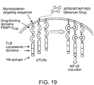
- a DC activation system based on targeted temporal control of the CD40 signaling pathway has been developed to extend the pro-stimulatory state of DCs within lymphoid tissues.
- DC functionality was improved by increasing both the amplitude and the duration of CD40 signaling 13 .
- the CD40 receptor was re-engineered so that the cytoplasmic domain of CD40 was fused to synthetic ligand-binding domains along with a membrane-targeting sequence.
- Administration of a lipid-permeable, dimerizing drug, AP20187 (AP), called a chemical inducer of dimerization (CID) 14 led to the in vivo induction of CD40-dependent signaling cascades in murine DCs.
- Pattern recognition receptor (PRR) signaling an example of which is Toll-like receptor (TLR) signaling also plays a critical role in the induction of DC maturation and activation, and human DCs express, multiple distinct TLRs 15 .
- the eleven mammalian TLRs respond to various pathogen-derived macromolecules, contributing to the activation of innate immune responses along with initiation of adaptive immunity.
- LPS Lipopolysaccharide
- MPL monophosphoryl lipid A
- NF-kappaB and IRF3 mitogen-activated protein kinases
- JNK c-Jun kinase 1718 .
- DCs mature, and partially upregulate pro-inflammatory cytokines, like IL-6, IL-12, and Type I interferons 19 .
- LPS-induced maturation has been shown to enhance the ability of DCs to stimulate antigen-specific T cell responses in vitro and in vivo 20 .
- iCD40 inducible CD40
- DCs dendritic cells
- PRR Pattern recognition receptor
- inducing CD40 synergistically activates antigen-presenting cells and induces an immune response against an antigen. It also has been discovered that antigen-presenting cells can be activated and immune responses can be generated against an antigen by inducing CD40.
- a method for activating an antigen-presenting cell which comprises: (a) transducing an antigen-presenting cell with a nucleic acid having a nucleotide sequence that encodes a chimeric protein, wherein the chimeric protein comprises a membrane targeting region, a ligand-binding region and a CD40 cytoplasmic polypeptide region lacking the CD40 extracellular domain; (b) contacting the antigen-presenting cell with a non-protein multimeric ligand that binds to the ligand-binding region; and (c) contacting the antigen-presenting cell with a PRR ligand, for example, a TLR ligand whereby the antigen-presenting cell is activated.
- a PRR ligand for example, a TLR ligand
- antigen-presenting cell is not contacted with prostaglandin E 2 (PGE 2 ) when contacted with the multimeric ligand, and in particular embodiments, the antigen-presenting cell is not contacted with a composition comprising prostaglandin E 2 (PGE 2 ) and one or more components selected from the group consisting of IL-1beta, IL-6 and TNF alpha.
- a method for activating an antigen-presenting cell which comprises: (a) transducing an antigen-presenting cell with a nucleic acid having a nucleotide sequence that encodes a chimeric protein, wherein the chimeric protein comprises a membrane targeting region, a ligand-binding region and a CD40 cytoplasmic polypeptide region lacking the CD40 extracellular domain; and (b) contacting the antigen-presenting cell with a non-protein multimeric ligand that binds to the ligand-binding region, wherein the antigen-presenting cell is not contacted with prostaglandin E2 (PGE 2 ) when contacted with the multimeric ligand, whereby the antigen-presenting cell is activated.
- the method further comprises contacting the antigen-presenting cell with a PRR ligand, for example, a Toll-like receptor (TLR) ligand.
- PRR ligand for example, a Toll-like receptor (TLR) ligand.
- a method for inducing a cytotoxic T lymphocyte (CTL) immune response against an antigen comprises: contacting an antigen-presenting cell sensitized with an antigen with: (a) a multimeric ligand that binds to a chimeric protein in the cell, wherein the chimeric protein comprises a membrane targeting region, a ligand-binding region and a CD40 cytoplasmic polypeptide region lacking the CD40 extracellular domain, and (b) a PRR ligand, for example, a Toll-like receptor (TLR) ligand; whereby a CTL immune response is induced against the antigen.
- TLR Toll-like receptor
- antigen-presenting cell is not contacted with prostaglandin E 2 (PGE 2 ) when contacted with the multimeric ligand, and in particular embodiments, the antigen-presenting cell is not contacted with a composition comprising prostaglandin E 2 (PGE 2 ) and one or more components selected from the group consisting of IL-1beta, IL-6 and TNF alpha.
- a method for inducing an immune response against an antigen which comprises: contacting an antigen-presenting cell sensitized with an antigen with a multmeric ligand that binds to a chimeric protein in the cell, wherein: (a) the chimeric protein comprises a membrane targeting region, a ligand-binding region and a CD40 cytoplasmic polypeptide region lacking the CD40 extracellular domain, and (b) the antigen-presenting cell is not contacted with prostaglandin E 2 (PGE 2 ) when contacted with the multimeric ligand; whereby an immune response against the antigen is induced.
- the method can further comprise contacting the antigen-presenting cell with a PRR ligand, for example, a TLR ligand.
- a method for inducing a cytotoxic T lymphocyte (CTL) immune response against an antigen comprises: contacting a human antigen-presenting cell sensitized with an antigen with: (a) a multimeric molecule having two or more regions that bind to and multimerize native CD40, and (b) a PRR ligand, for example, a TLR ligand ligand; whereby a CTL immune response is induced against the antigen.
- CTL cytotoxic T lymphocyte
- the multimeric molecule can be an antibody that binds to an epitope in the CD40 extracellular domain (e.g., humanized anti-CD40 antibody; Tai et al., Cancer Research 64, 2846-2852 (2004 )), can be a CD40 ligand (e.g., U.S. Patent No. 6,497,876 (Maraskovsky et al. )) or may be another co-stimulatory molecule (e.g., B7/CD28).
- a CD40 ligand e.g., U.S. Patent No. 6,497,876 (Maraskovsky et al. )
- another co-stimulatory molecule e.g., B7/CD28.
- the antigen-presenting cell can be transduced ex vivo or in vivo with a nucleic acid that encodes the chimeric protein.
- the antigen-presenting cell may be sensitized to the antigen at the same time the antigen-presenting cell is contacted with the multimeric ligand, or the antigen-presenting cell can be pre-sensitized to the antigen before the antigen-presenting cell is contacted with the multimerization ligand.
- the antigen-presenting cell is contacted with the antigen ex vivo.
- the antigen-presenting cell is transduced with the nucleic acid ex vivo and administered to the subject by intradermal administration, and sometimes the antigen-presenting cell is transduced with the nucleic acid ex vivo and administered to the subject by subcutaneous administration.
- the antigen may be a tumor antigen, and the CTL immune response can induced by migration of the antigen-presenting cell to a draining lymph node.
- compositions comprising an antigen-presenting cell and a PRR ligand, for example, a TLR ligand, wherein: the antigen-presenting cell is transduced with a nucleic acid having a nucleotide sequence that encodes a chimeric protein, and the chimeric protein comprises a membrane targeting region, a ligand-binding region and a CD40 cytoplasmic polypeptide region lacking the CD40 extracellular domain.
- the composition may further comprise a non-protein multimeric ligand that binds to the ligand-binding region.
- the membrane targeting region can be a myristoylation targeting region.
- the CD40 cytoplasmic polypeptide region is encoded by a polynucleotide sequence in SEQ ID NO: 1.
- the multimeric ligand often is a small molecule and it sometimes is dimeric, such as a dimeric FK506 or a dimeric FK506 analog (e.g., AP1903).
- Any suitable PRR ligand for example, any suitable TLR ligand can be utilized, and can be selected by the person of ordinary skill in the art (e.g., Napolitani et al., Nature Immunology, Advanced Online Publication doi:10.1038/ni1223 (2005 )).
- the TLR ligand in certain embodiments is selected from the group consisting of lipopolysaccharide (LPS), monophosphoryl lipid A (MPL), FSL-1, Pam3, CSK4, poly(I:C), synthetic imidazoquinoline resiquimod (R848; U.S. Patent No. 6,558,951 to Tomai et al. ) and CpG, and the TLR ligand sometimes is a TLR4 ligand such as LPS or MPL.
- the nucleic acid can be contained within a viral vector, such as an adenoviral vector, for example.
- the antigen-presenting cell is transduced with the nucleic acid ex vivo or in vivo, and sometimes the antigen-presenting cell is a dendritic cell, such as a human dendritic cell, for example.
- Also discussed herein is a method for assessing migration of an antigen-presenting cell to a lymph node, which comprises: (a) injecting into a subject an antigen-presenting cell that produces a detectable protein, and (b) determining the amount of the detectable protein in the lymph node of the animal, whereby migration of the antigen-presenting cell to the lymph node is assessed from the amount of the detectable protein in the lymph node.
- the animal can be a rodent, such as a rat or a mouse (e.g., irradiated mouse).
- the detectable protein is a luciferase protein, such as a chick beetle (e.g., Pyrophorus plagiophalamus) red-shifted luciferase protein.
- the antigen-presenting cell has been transduced with a nucleic acid having a polynucleotide sequence that encodes the detectable protein.
- the lymph node is the popliteal lymph node or inguinal lymph node.
- the antigen-presenting cell can be a dendritic cell, such as a human dendritic cell.
- the lymph node is removed from the animal before the amount of detectable protein is determined, and sometimes the D-Luciferin is administered to the removed lymph node.
- the amount of the detectable protein may be qualitative (e.g., relative amounts compared across different samples) and can be quantitative (e.g., a concentration).
- the amount of the detectable protein may be determined by directly detecting the protein.
- the protein may be fluorescent (e.g., green fluorescent protein or a red-shifted or blue-shifted version) or can be bound to a fluorescent label (e.g., an antibody linked to a fluorophore).
- the amount of the detectable protein can determined indirectly by administering a substrate to the animal that is converted into a detectable product by the protein and detecting the detectable product.
- the amount of a luciferase protein can be determined by administering D-Luciferin to the animal and detecting the D-Luciferin product generated by the luciferase produced in the antigen-presenting cell.
- ex-vivo methods for activating an antigen-presenting cell like dendritic cells which comprise: contacting an antigen-presenting cell with a non-protein multimeric ligand that binds to a ligand-binding region in the cell, which cell has been transduced (or transfected) with a nucleic acid that encodes a chimeric protein, wherein the chimeric protein comprises (i) a membrane-targeting region, (ii) a ligand-binding region, (iii) a CD40 cytoplasmic polypeptide region lacking the CD40 extracelluar domain, and (iv) a signaling region and/or cytoplasmic region of a pattern recognition receptor (PRR) whereby the antigen-presenting cell is activated.
- the CD40 cytoplasmic polypeptide region in certain embodiments is encoded by a polynucleotide sequence in SEQ ID NO: 1.
- the chimeric protein comprises a signaling region and/or cytoplasmic region of a PRR such as a NOD2 PRR.
- the PRR in some embodiments is a RIG-like helicase (RLH), such as RIG-1 PRR.
- RIG-like helicase RIG-1 PRR.
- the membrane targeting region is a myristoylation-targeting region, although the membrane-targeting region can be selected from other types of transmembrane-targeting regions, such as regions described hereafter.
- the ligand is a small molecule, and sometimes the molecule is dimeric. Examples of dimeric molecules are dimeric FK506 and dimeric FK506 analogs.
- the ligand is AP1903 or AP20187.
- the chimeric protein includes one or more ligand-binding regions, such as two or three ligand-binding regions, for example. The ligand-binding regions often are tandem.
- the nucleic acid in certain embodiments is contained within a viral vector, such as an adenoviral vector for example.
- the antigen-presenting cell in some embodiments is contacted with an antigen, sometimes ex vivo.
- the antigen-presenting cell is in a subject and an immune response is generated against the antigen, such as a cytotoxic T-lymphocyte (CTL) immune response.
- CTL cytotoxic T-lymphocyte
- an immune response is generated against a tumor antigen (e.g., PSMA).
- the nucleic acid is prepared ex vivo and administered to the subject by intradermal administration or by subcutaneous administration, for example. Sometimes the antigen-presenting cell is transduced or transfected with the nucleic acid ex vivo or in vivo.
- the nucleic acid comprises a promoter sequence operably linked to the polynucleotide sequence.
- the nucleic acid comprises an ex vivo-transcribed RNA, containing the protein-coding region of the chimeric protein.
- compositions which comprises a nucleic acid having a polynucleotide sequence that encodes a chimeric protein, wherein the chimeric protein comprises (i) a membrane targeting region, (ii) a ligand-binding region that binds to a multimeric non-protein ligand, and (iii-a) a signaling region and/or cytoplasmic region of a pattern recognition receptor (PRR) or (iii-b) an adapter of a PRR.
- PRR pattern recognition receptor
- allogeneic refers to cell types or tissues that are antigenically distinct. Thus, cells or tissue transferred from the same species can be antigenically distinct.
- antigen as used herein is defined as a molecule that provokes an immune response. This immune response may involve either antibody production, or the activation of specific immunologically competent cells, or both.
- An antigen can be derived from organisms, subunits of proteins/antigens, killed or inactivated whole cells or lysates.
- Exemplary organisms include but are not limited to, Helicobacters, Campylobacters, Clostridia, Corynebacterium diphtheriae, Bordetella pertussis, influenza virus, parainfluenza viruses, respiratory syncytial virus, Borrelia burgdorfei, Plasmodium, herpes simplex viruses, human immunodeficiency virus, papillomavirus, Vibrio cholera, E. coli, measles virus, rotavirus, shigella, Salmonella typhi, Neisseria gonorrhea. Therefore, a skilled artisan realizes that any macromolecule, including virtually all proteins or peptides, can serve as antigens.
- antigens can be derived from recombinant or genomic DNA.
- any DNA which contains nucleotide sequences or partial nucleotide sequences of a pathogenic genome or a gene or a fragment of a gene for a protein that elicits an immune response results in synthesis of an antigen.
- the present disclosure is not limited to the use of the entire nucleic acid sequence of a gene or genome. It is readily inherent that the present invention includes, but is not limited to, the use of partial nucleic acid sequences of more than one gene or genome and that these nucleic acid sequences are arranged in various combinations to elicit the desired immune response.
- antigen-presenting cell is any of a variety of cells capable of displaying, acquiring, or presenting at least one antigen or antigenic fragment on (or at) its cell surface.
- the term “antigen-presenting cell” can be any cell that accomplishes the goal of the invention by aiding the enhancement of an immune response (i.e., from the T-cell or -B-cell arms of the immune system) against an antigen or antigenic composition.
- Such cells can be defined by those of skill in the art, using methods disclosed herein and in the art.
- a cell that displays or presents an antigen normally or preferentially with a class II major histocompatibility molecule or complex to an immune cell is an "antigen-presenting cell.”
- a cell e.g., an APC cell
- another cell such as a recombinant cell or a tumor cell that expresses the desired antigen.
- the immune cell to which an antigen-presenting cell displays or presents an antigen to is a CD4+TH cell.
- Additional molecules expressed on the APC or other immune cells may aid or improve the enhancement of an immune response.
- Secreted or soluble molecules, such as for example, cytokines and adjuvants, may also aid or enhance the immune response against an antigen.
- cytokines and adjuvants may also aid or enhance the immune response against an antigen.
- cancer as used herein is defined as a hyperproliferation of cells whose unique trait-loss of normal controls-results in unregulated growth, lack of differentiation, local tissue invasion, and metastasis. Examples include but are not limited to, melanoma, non-small cell lung, small-cell lung, lung, hepatocarcinoma, leukemia, retinoblastoma, astrocytoma, glioblastoma, gum, tongue, neuroblastoma, head, neck, breast, pancreatic, prostate, renal, bone, testicular, ovarian, mesothelioma, cervical, gastrointestinal, lymphoma, brain, colon, sarcoma or bladder.
- cell may be used interchangeably. All of these terms also include their progeny, which are any and all subsequent generations. It is understood that all progeny may not be identical due to deliberate or inadvertent mutations.
- iCD40 molecule is defined as an inducible CD40. This iCD40 can bypass mechanisms that extinguish endogenous CD40 signaling.
- iCD40 embraces "iCD40 nucleic acids”, “iCD40 polypeptides” and/or iCD40 expression vectors. Yet further, it is understood the activity of iCD40 as used herein is driven by CID.
- cDNA is intended to refer to DNA prepared using messenger RNA (mRNA) as template.
- mRNA messenger RNA
- dendritic cell is an antigen-presenting cell existing in vivo, in vitro, ex vivo, or in a host or subject, or which can be derived from a hematopoietic stem cell or a monocyte.
- Dendritic cells and their precursors can be isolated from a variety of lymphoid organs, e.g., spleen, lymph nodes, as well as from bone marrow and peripheral blood.
- the DC has a characteristic morphology with thin sheets (lamellipodia) extending in multiple directions away from the dendritic cell body.
- dendritic cells express high levels of MHC and costimulatory (e.g., B7-1 and B7-2) molecules. Dendritic cells can induce antigen specific differentiation of T cells in vitro, and are able to initiate primary T cell responses in vitro and in vivo.
- expression construct or "transgene” is defined as any type of genetic construct containing a nucleic acid coding for gene products in which part or all of the nucleic acid encoding sequence is capable of being transcribed can be inserted into the vector.
- the transcript is translated into a protein, but it need not be.
- expression includes both transcription of a gene and translation of mRNA into a gene product.
- expression only includes transcription of the nucleic acid ncoding genes of interest.
- therapeutic construct may also be used to refer to the expression construct or transgene.
- the expression construct or transgene is a therapeutic construct or a prophylactic construct.
- expression vector refers to a vector containing a nucleic acid sequence coding for at least part of a gene product capable of being transcribed. In some cases, RNA molecules are then translated into a protein, polypeptide, or peptide. In other cases, these sequences are not translated, for example, in the production of antisense molecules or ribozymes.
- Expression vectors can contain a variety of control sequences, which refer to nucleic acid sequences necessary for the transcription and possibly translation of an operatively linked coding sequence in a particular host organism. In addition to control sequences that govern transcription and translation, vectors and expression vectors may contain nucleic acid sequences that serve other functions as well and are described infra.
- ex vivo refers to "outside" the body.
- ex vivo and in vitro can be used interchangeably.
- the term “functionally equivalent”, as used herein, refers to a CD40 nucleic acid fragment, variant, or analog, refers to a nucleic acid that codes for a CD40 polypeptide, or a CD40 polypeptide, that stimulates an immune response to destroy tumors or hyperproliferative disease.
- “functionally equivalent” refers to a CD40 polypeptide that is lacking the extracellular domain, but is capable of amplifying the T cell-mediated tumor killing response by upregulating dendritic cell expression of antigen presentation molecules.
- hyperproliferative disease is defined as a disease that results from a hyperproliferation of cells.
- exemplary hyperproliferative diseases include, but are not limited to cancer or autoimmune diseases.
- Other hyperproliferative diseases may include vascular occulsion, restenosis, atherosclerosis, or inflammatory bowel disease.
- the term "gene” is defined as a functional protein, polypeptide, or peptide-encoding unit. As will be understood by those in the art, this functional term includes genomic sequences, cDNA sequences, and smaller engineered gene segments that express, or is adapted to express, proteins, polypeptides, domains, peptides, fusion proteins, and mutants.
- immunogen refers to a substance that is capable of provoking an immune response.
- immunogens include, e.g., antigens, autoantigens that play a role in induction of autoimmune diseases, and tumor-associated antigens expressed on cancer cells.
- immunocompromised as used herein is defined as a subject that has reduced or weakened immune system.
- the immunocompromised condition may be due to a defect or dysfunction of the immune system or to other factors that heighten susceptibility to infection and/or disease.
- a defect or dysfunction of the immune system or to other factors that heighten susceptibility to infection and/or disease.
- immunocompromised individuals often do not fit completely into one group or the other. More than one defect in the body's defense mechanisms may be affected. For example, individuals with a specific T-lymphocyte defect caused by HIV may also have neutropenia caused by drugs used for antiviral therapy or be immunocompromised because of a breach of the integrity of the skin and mucous membranes.
- An immunocompromised state can result from indwelling central lines or other types of impairment due to intravenous drug abuse; or be caused by secondary malignancy, malnutrition, or having been infected with other infectious agents such as tuberculosis or sexually transmitted diseases, e.g., syphilis or hepatitis.
- the term "pharmaceutically or pharmacologically acceptable” refers to molecular entities and compositions that do not produce adverse, allergic, or other untoward reactions when administered to an animal or a human.
- pharmaceutically acceptable carrier includes any and all solvents, dispersion media, coatings, antibacterial and antifungal agents, isotonic and absorption delaying agents and the like.
- the use of such media and agents for pharmaceutically active substances is well known in the art. Except insofar as any conventional media or agent is incompatible with the vectors or cells described herein, its use in therapeutic compositions is contemplated. Supplementary active ingredients also can be incorporated into the compositions.
- nucleotide is defined as a chain of nucleotides.
- nucleic acids are polymers of nucleotides.
- nucleic acids and polynucleotides as used herein are interchangeable.
- nucleic acids are polynucleotides, which can be hydrolyzed into the monomeric “nucleotides.” The monomeric nucleotides can be hydrolyzed into nucleosides.
- polynucleotides include, but are not limited to, all nucleic acid sequences which are obtained by any means available in the art, including, without limitation, recombinant means, i.e., the cloning of nucleic acid sequences from a recombinant library or a cell genome, using ordinary cloning technology and PCRTM, and the like, and by synthetic means.
- recombinant means i.e., the cloning of nucleic acid sequences from a recombinant library or a cell genome, using ordinary cloning technology and PCRTM, and the like, and by synthetic means.
- polynucleotides include mutations of the polynucleotides, include but are not limited to, mutation of the nucleotides, or nucleosides by methods well known in the art.
- polypeptide is defined as a chain of amino acid residues, usually having a defined sequence.
- polypeptide is interchangeable with the terms “peptides” and “proteins”.
- promoter is defined as a DNA sequence recognized by the synthetic machinery of the cell, or introduced synthetic machinery, required to initiate the specific transcription of a gene.
- the term “regulate an immune response” or “modulate an immune response” refers to the ability to modify the immune response.
- the composition described herein is capable of enhancing and/or activating the immune response.
- the composition described herein is also capable of inhibiting the immune response.
- the form of regulation is determined by the ligand that is used with the composition described herein. For example, a dimeric analog of the chemical results in dimerization of the co-stimulatory polypeptide leading to activation of the DCs, however, a monomeric analog of the chemical does not result in dimerization of the co-stimulatory polypeptide, which would not activate the DCs.
- transfection and “transduction” are interchangeable and refer to the process by which an exogenous DNA sequence is introduced into a eukaryotic host cell.
- Transfection can be achieved by any one of a number of means including electroporation, microinjection, gene gun delivery, retroviral infection, lipofection, superfection and the like.
- genotypeic refers to cells, tissues or animals that have genotypes. For example, identical twins or animals of the same inbred strain. Syngeneic and isogeneic can be used interchangeable.
- subject includes, but is not limited to, an organism or animal; a mammal, including, e.g., a human, non-human primate (e.g., monkey), mouse, pig, cow, goat, rabbit, rat, guinea pig, hamster, horse, monkey, sheep, or other non-human mammal; a non-mammal, including, e.g., a non-mammalian vertebrate, such as a bird (e.g., a chicken or duck) or a fish, and a non-mammalian invertebrate.
- a mammal including, e.g., a human, non-human primate (e.g., monkey), mouse, pig, cow, goat, rabbit, rat, guinea pig, hamster, horse, monkey, sheep, or other non-human mammal
- a non-mammal including, e.g., a non-mammalian vertebrate, such
- under transcriptional control or "operatively linked” is defined as the promoter is in the correct location and orientation in relation to the nucleic acid to control RNA polymerase initiation and expression of the gene.
- treatment refers to prophylaxis and/or therapy.
- the term refers to a prophylactic treatment which increases the resistance of a subject to infection with a pathogen or, in other words, decreases the likelihood that the subject will become infected with the pathogen or will show signs of illness attributable to the infection, as well as a treatment after the subject has become infected in order to fight the infection, e. g., reduce or eliminate the infection or prevent it from becoming worse.
- the term "vaccine” refers to a formulation which contains the composition of the present disclosure and which is in a form that is capable of being administered to an animal.
- the vaccine comprises a conventional saline or buffered aqueous solution medium in which the composition of the present invention is suspended or dissolved.
- the composition of the present invention can be used conveniently to prevent, ameliorate, or otherwise treat a condition.
- the vaccine Upon introduction into a subject, the vaccine is able to provoke an immune response including, but not limited to, the production of antibodies, cytokines and/or other cellular responses.
- the innate immune system uses a set of germline-encoded receptors for the recognition of conserved molecular patterns present in microorganisms. These molecular patterns occur in certain constituents of microorganisms including: lipopolysaccharides, peptidoglycans, lipoteichoic acids, phosphatidyl cholines, bacteria-specific proteins, including lipoproteins, bacterial DNAs, viral single and double-stranded RNAs, unmethylated CpG-DNAs, mannans and a variety of other bacterial and fungal cell wall components. Such molecular patterns can also occur in other molecules such as plant alkaloids.
- PAMPs Pathogen Associated Molecular Patterns
- PRRs Pattern Recognition Receptors
- CD14, DEC205, collectins Some of these receptors recognize PAMPs directly (e.g., CD14, DEC205, collectins), while others (e.g., complement receptors) recognize the products generated by PAMP recognition.
- Cellular PRRs are expressed on effector cells of the innate immune system, including cells that function as professional antigen-presenting cells (APC) in adaptive immunity.
- effector cells include, but are not limited to, macrophages, dendritic cells, B lymphocytes and surface epithelia.
- APC professional antigen-presenting cells
- This expression profile allows PRRs to directly induce innate effector mechanisms, and also to alert the host organism to the presence of infectious agents by inducing the expression of a set of endogenous signals, such as inflammatory cytokines and chemokines, as discussed below. This latter function allows efficient mobilization of effector forces to combat the invaders.
- DCs dendritic cells
- the primary function of dendritic cells is to acquire antigen in the peripheral tissues, travel to secondary lymphoid tissue, and present antigen to effector T cells of the immune system (Banchereau, et al., 2000; Banchereau, et al., 1998).
- DCs carry out their crucial role in the immune response, they undergo maturational changes allowing them to perform the appropriate function for each environment (Termeer, C.C. et al., 2000).
- MHC major histocompatibility complex
- DCs In addition, other genetic alterations permit the DCs to home to the T cell-rich paracortex of draining lymph nodes and to express T-cell chemokines that attract naive and memory T cells and prime antigen-specific naive TH0 cells (Adema, G.J. et al., 1997). During this stage, mature DCs present antigen via their MHC II molecules to CD4+ T helper cells, inducing the upregulation of T cell CD40 ligand (CD40L) that, in turn, engages the DC CD40 receptor.
- CD40L T cell CD40 ligand
- This DC:T cell interaction induces rapid expression of additional DC molecules that are crucial for the initiation of a potent CD8+ cytotoxic T lymphocyte (CTL) response, including further upregulation of MHC I and II molecules, adhesion molecules, costimulatory molecules (e.g., B7.1,B7.2), cytokines (e.g., IL-12) and anti-apoptotic proteins (e.g., Bcl-2) (Anderson, D.M., et al., 1997; Caux, C., et al., 1997; Ohshima, Y., et al., 1997; Sallusto, F., et al., 1998).
- CD8+ T cells exit lymph nodes, reenter circulation and home to the original site of inflammation to destroy pathogens or malignant cells.
- CD40 receptor serving as the "on switch" for DCs
- CD40 is a 48-kDa transmembrane member of the TNF receptor superfamily (McWhirter, S.M., et al., 1999).
- CD40-CD40L interaction induces CD40 trimerization, necessary for initiating signaling cascades involving TNF receptor associated factors (TRAFs) (Ni, C.Z., et al., 2000; Pullen, S.S. et al., 1999).
- CD40 uses these signaling molecules to activate several transcription factors in DCs, including NF-kappa B, AP-1, STAT3, and p38MAPK (McWhirter, S.M., et al., 1999).
- the present disclosure contemplates a novel DC activation system based on recruiting signaling molecules or co-stimulatory polypeptides to the plasmid membrane of the DCs resulting in prolonged/increased activation and/or survival in the DCs.
- Co-stimulatory polypeptides include any molecule or polypeptide that activates the NFkappaB pathway, Akt pathway, and/or p38 pathway.
- the DC activation system is based upon utilizing a recombinant signaling molecule fused to a ligand-binding domains (i.e., a small molecule binding domain) in which the co-stimulatory polypeptide is activated and/or regulated with a ligand resulting in oligomerization (i.e., a lipid-permeable, organic, dimerizing drug).
- a ligand-binding domains i.e., a small molecule binding domain
- a ligand resulting in oligomerization i.e., a lipid-permeable, organic, dimerizing drug.
- Other systems that may be used to crosslink or oligomerization of co-stimulatory polypeptides include antibodies, natural ligands, and/or artificial cross-reacting or synthetic ligands.
- dimerization systems contemplated include the coumermycin/DNA gyrase B system.
- Co-stimulatory polypeptides that can be used herein include those that activate NFkappaB and other variable signaling cascades for example the p38 pathway and/or Akt pathway.
- Such co-stimulatory polypeptides include, but are not limited to Pattern Recognition Receptors, C-reactive protein receptors (i.e., Nod1, Nod2, PtX3-R), TNF receptors (i.e., CD40, RANK/TRANCE-R, OX40, 4-1BB), and HSP receptors (Lox-1 and CD-91).
- Pattern Recognition Receptors include, but are not limited to endocytic pattern-recognition receptors (i.e., mannose receptors, scavenger receptors (i.e., Mac-1, LRP, peptidoglycan, teichoic acids, toxins, CD11c/CR4)); external signal pattern-recognition receptors (Toll-like receptors (TLR1, TLR2, TLR3, TLR4, TLR5, TLR6, TLR7, TLR8, TLR9, TLR10), peptidoglycan recognition protein, (PGRPs bind bacterial peptidoglycan, and CD14); internal signal pattern-recognition receptors (i.e., NOD-receptors 1 & 2), RIG1, and PRRs shown in Figure 28 .
- endocytic pattern-recognition receptors i.e., mannose receptors, scavenger receptors (i.e., Mac-1, LRP, peptidoglycan,
- Pattern Recognition Receptors suitable for the present invention, including those discussed in, for example, Werts C., et al., Cell Death and Differentiation (2006) 13:798-815 ; Meylan, E., et al., Nature (2006) 442:39-44 ; and Strober, W., et al., Nature Reviews (2006) 6:9-20 .
- the present disclosure involves an expression construct encoding a co-stimulatory polypeptide and a ligand-binding domain, all operatively linked. More particularly, more than one ligand-binding domain is used in the expression construct. Yet further, the expression construct contains a membrane-targeting sequence.
- appropriate expression constructs may include a co-stimulatory polypeptide element on either side of the above FKBP ligand-binding elements.
- the expression construct described herein may be inserted into a vector, for example a viral vector or plasmid.
- co-stimulatory polypeptide molecules are capable of amplifying the T-cell-mediated response by upregulating dendritic cell expression of antigen presentation molecules.
- Co-stimulatory proteins that are contemplated herein include, for example, but are not limited to the members of tumor necrosis factor (TNF) family (i.e., CD40, RANK/TRANCE-R, OX40, 4-1B), Toll-like receptors, C-reactive protein receptors, Pattern Recognition Receptors, and HSP receptors.
- TNF tumor necrosis factor
- cytoplasmic domains from these co-stimulatory polypeptides are used in the expression vector.
- the co-stimulatory polypeptide molecule is CD40.
- the CD40 molecule comprises a nucleic acid molecule which: (1) hybridizes under stringent conditions to a nucleic acid having the sequence of a known CD40 gene and (2) codes for a CD40 polypeptide.
- the CD40 polypeptide is lacking the extracellular domain.
- Exemplary polynucleotide sequences that encode CD40 polypeptides include, but are not limited to SEQ.ID.NO: 1 and CD40 isoforms from other species. It is contemplated that other normal or mutant variants of CD40 can be used herein.
- a CD40 region can have an amino acid sequence that differs from the native sequence by one or more amino acid substitutions, deletions and/or insertions.
- one or more TNF receptor associated factor (TRAF) binding regions may be eliminated or effectively eliminated (e.g., a CD40 amino acid sequence is deleted or altered such that a TRAF protein does not bind or binds with lower affinity than it binds to the native CD40 sequence).
- a TRAF 3 binding region is deleted or altered such that it is eliminated or effectively eliminated (e.g., amino acids 250-254 may be altered or deleted; Hauer et al., PNAS 102(8): 2874-2879 (2005 )).
- the present disclosure involves the manipulation of genetic material to produce expression constructs that encode an inducible form of CD40 (iCD40).
- Such methods involve the generation of expression constructs containing, for example, a heterologous nucleic acid sequence encoding CD40 cytoplasmic domain and a means for its expression, replicating the vector in an appropriate helper cell, obtaining viral particles produced therefrom, and infecting cells with the recombinant virus particles.
- the preferable CD40 molecule of the invention lacks the extracellular domain.
- the extracellular domain is truncated or removed. It is also contemplated that the extracellular domain can be mutated using standard mutagenesis, insertions, deletions, or substitutions to produce an CD40 molecule that does not have a functional extracellular domain.
- a CD40 nucleic acid may have the nucleic acid sequence of SEQ.ID.NO: 1.
- the CD40 nucleic acids of the disclosure also include homologs and alleles of a nucleic acid having the sequence of SEQ.ID.NO: 1, as well as, functionally equivalent fragments, variants, and analogs of the foregoing nucleic acids.
- the gene will be a heterologous polynucleotide sequence derived from a source other than the viral genome, which provides the backbone of the vector.
- the gene is derived from a prokaryotic or eukaryotic source such as a bacterium, a virus, yeast, a parasite, a plant, or even an animal.
- the heterologous DNA also is derived from more than one source, i.e., a multigene construct or a fusion protein.
- the heterologous DNA also may include a regulatory sequence, which is derived from one source and the gene from a different source.
- the ligand-binding ("dimerization") domain of the expression construct of this disclosure can be any convenient domain that will allow for induction using a natural or unnatural ligand, preferably an unnatural synthetic ligand.
- the ligand-binding domain can be internal or external to the cellular membrane, depending upon the nature of the construct and the choice of ligand.
- a wide variety of ligand-binding proteins, including receptors, are known, including ligand-binding proteins associated with the cytoplasmic regions indicated above.
- the term "ligand-binding domain” can be interchangeable with the term "receptor".
- ligand-binding domains or receptors include the FKBPs and cyclophilin receptors, the steroid receptors, the tetracycline receptor, the other receptors indicated above, and the like, as well as "unnatural" receptors, which can be obtained from antibodies, particularly the heavy or light chain subunit, mutated sequences thereof, random amino acid sequences obtained by stochastic procedures, combinatorial syntheses, and the like.
- the ligand-binding domains or receptor domains will be at least about 50 amino acids, and fewer than about 350 amino acids, usually fewer than 200 amino acids, either as the natural domain or truncated active portion thereof.
- the binding domain will be small ( ⁇ 25 kDa, to allow efficient transfection in viral vectors), monomeric (this rules out the avidin-biotin system), nonimmunogenic, and should have synthetically accessible, cell permeable, nontoxic ligands that can be configured for dimerization.
- the receptor domain can be intracellular or extracellular depending upon the design of the expression construct and the availability of an appropriate ligand.
- the binding domain can be on either side of the membrane, but for hydrophilic ligands, particularly protein ligands, the binding domain will usually be external to the cell membrane, unless there is a transport system for internalizing the ligand in a form in which it is available for binding.
- the construct can encode a signal peptide and transmembrane domain 5' or 3' of the receptor domain sequence or by having a lipid attachment signal sequence 5' of the receptor domain sequence. Where the receptor domain is between the signal peptide and the transmembrane domain, the receptor domain will be extracellular.
- the portion of the expression construct encoding the receptor can be subjected to mutagenesis for a variety of reasons.
- the mutagenized protein can provide for higher binding affinity, allow for discrimination by the ligand of the naturally occurring receptor and the mutagenized receptor, provide opportunities to design a receptor-ligand pair, or the like.
- the change in the receptor can involve changes in amino acids known to be at the binding site, random mutagenesis using combinatorial techniques, where the codons for the amino acids associated with the binding site or other amino acids associated with conformational changes can be subject to mutagenesis by changing the codon(s) for the particular amino acid, either with known changes or randomly, expressing the resulting proteins in an appropriate prokaryotic host and then screening the resulting proteins for binding.
- Antibodies and antibody subunits e.g., heavy or light chain, particularly fragments, more particularly all or part of the variable region, or fusions of heavy and light chain to create high-affinity binding, can be used as the binding domain.
- Antibodies that are contemplated herein include ones that are an ectopically expressed human product, such as an extracellular domain that would not trigger an immune response and generally not expressed in the periphery (i.e., outside the CNS/brain area). Such examples, include, but are not limited to low affinity nerve growth factor receptor (LNGFR), and embryonic surface proteins (i.e., carcinoembryonic antigen).
- LNGFR low affinity nerve growth factor receptor
- embryonic surface proteins i.e., carcinoembryonic antigen
- antibodies can be prepared against haptenic molecules, which are physiologically acceptable, and the individual antibody subunits screened for binding affinity.
- the cDNA encoding the subunits can be isolated and modified by deletion of the constant region, portions of the variable region, mutagenesis of the variable region, or the like, to obtain a binding protein domain that has the appropriate affinity for the ligand.
- almost any physiologically acceptable haptenic compound can be employed as the ligand or to provide an epitope for the ligand.
- natural receptors can be employed, where the binding domain is known and there is a useful ligand for binding.
- the transduced signal will normally result from ligand-mediated oligomerization of the chimeric protein molecules, i.e., as a result of oligomerization following ligand-binding, although other binding events, for example allosteric activation, can be employed to initiate a signal.
- the construct of the chimeric protein will vary as to the order of the various domains and the number of repeats of an individual domain.
- the ligand for the ligand-binding domains/receptor domains of the chimeric surface membrane proteins will usually be multimeric in the sense that it will have at least two binding sites, with each of the binding sites capable of binding to the receptor domain.
- the subject ligands will be a dimer or higher order oligomer, usually not greater than about tetrameric, of small synthetic organic molecules, the individual molecules typically being at least about 150 D and fewer than about 5 kDa, usually fewer than about 3 kDa.
- a variety of pairs of synthetic ligands and receptors can be employed.
- dimeric FK506 can be used with an FKBP receptor
- dimerized cyclosporin A can be used with the cyclophilin receptor
- dimerized estrogen with an estrogen receptor
- dimerized glucocorticoids with a glucocorticoid receptor
- dimerized tetracycline with the tetracycline receptor
- dimerized vitamin D with the vitamin D receptor
- higher orders of the ligands e.g., trimeric can be used.
- unnatural receptors e.g., antibody subunits, modified antibody subunits or modified receptors and the like
- any of a large variety of compounds can be used. A significant characteristic of these ligand units is that they bind the receptor with high affinity and are able to be dimerized chemically.
- the present invention utilizes the technique of chemically induced dimerization (CID) to produce a conditionally controlled protein or polypeptide.
- CID chemically induced dimerization
- this technique is inducible, it also is reversible, due to the degradation of the labile dimerizing agent or administration of a monomeric competitive inhibitor.
- CID system uses synthetic bivalent ligands to rapidly crosslink signaling molecules that are fused to ligand-binding domains CID. This system has been used to trigger the oligomerization and activation of cell surface (Spencer et al., 1993; Spencer et al., 1996; Blau et al., 1997), or cytosolic proteins (Luo et al., 1996; MacCorkle et al., 1998), the recruitment of transcription factors to DNA elements to modulate transcription (Ho et al., 1996; Rivera et al., 1996) or the recruitment of signaling molecules to the plasma membrane to stimulate signaling (Spencer et al., 1995; Holsinger et al., 1995).
- the CID system is based upon the notion that surface receptor aggregation effectively activates downstream signaling cascades.
- the CID system uses a dimeric analog of the lipid permeable immunosuppressant drug, FK506, which loses its normal bioactivity while gaining the ability to crosslink molecules genetically fused to the FK506-binding protein, FKBP12.
- FK506 lipid permeable immunosuppressant drug
- FKBP12 FKBP12
- FKBP12 third-generation AP20187/AP1903 CIDs for their binding domain, FKBP12 permits specific activation of the recombinant receptor in vivo without the induction of non-specific side effects through endogenous FKBP12.
- synthetic ligands are resistant to protease degradation, making them more efficient at activating receptors in vivo than most delivered protein agents.
- the ligands used herein are capable of binding to two or more of the ligand-binding domains.
- One skilled in the art realizes that the chimeric proteins may be able to bind to more than one ligand when they contain more than one ligand-binding domain.
- the ligand is typically a non-protein or a chemical.
- Exemplary ligands include, but are not limited to dimeric FK506 (e.g., FK1012).
- CD40 activation Since the mechanism of CD40 activation is fundamentally based on trimerization, this receptor is particularly amenable to the CID system. CID regulation provides the system with 1) temporal control, 2) reversibility by addition of a non-active monomer upon signs of an autoimmune reaction, and 3) limited potential for non-specific side effects. In addition, inducible in vivo DC CD40 activation would circumvent the requirement of a second "danger" signal normally required for complete induction of CD40 signaling and would potentially promote DC survival in situ allowing for enhanced T cell priming. Thus, engineering DC vaccines to express iCD40 amplifies the T cell-mediated killing response by upregulating DC expression of antigen presentation molecules, adhesion molecules; TH1 promoting cytokines, and pro-survival factors.
- dimerization systems contemplated include the coumermycin/DNA gyrase B system.
- Coumermycin-induced dimerization activates a modified Raf protein and stimulates the MAP kinase cascade. See Farrar et al., 1996.
- a membrane-targeting sequence provides for transport of the chimeric protein to the cell surface membrane, where the same or other sequences can encode binding of the chimeric protein to the cell surface membrane.
- Molecules in association with cell membranes contain certain regions that facilitate the membrane association, and such regions can be incorporated into a chimeric protein molecule to generate membrane-targeted molecules.
- some proteins contain sequences at the N-terminus or C-terminus that are acylated, and these acyl moieties facilitate membrane association.
- Such sequences are recognized by acyltransferases and often conform to a particular sequence motif.
- Certain acylation motifs are capable of being modified with a single acyl moiety and others are capable of being modified with multiple acyl moieties.
- the N-terminal sequence of the protein kinase Src can comprise a single myristoyl moiety.
- Dual acylation regions are located within the N-terminal regions of certain protein kinases (e.g., Yes, Fyn, Lck) and G-protein alpha subunits.
- Such dual acylation regions often are located within the first eighteen amino acids of such proteins, and conform to the sequence motif Met-Gly-Cys-Xaa-Cys, where the Met is cleaved, the Gly is N-acylated and one of the Cys residues is S-acylated.
- the Gly often is myristoylated and a Cys can be palmitoylated.
- These and other acylation motifs are known to the person of ordinary skill in the art (e.g., Gauthier-Campbell et al., Molecular Biology of the Cell 15: 2205-2217 (2004 ); Glabati et al., Biochem. J.
- a native sequence from a protein containing an acylation motif is incorporated into a chimeric protein.
- an N-terminal portion of Lck, Fyn or Yes or a G-protein alpha subunit such as the first twenty-five N-terminal amino acids or fewer from such proteins (e.g., about 5 to about 20 amino acids, about 10 to about 19 amino acids, or about 15 to about 19 amino acids of the native sequence with optional mutations), may be incorporated within the N-terminus of a chimeric protein.
- a C-terminal sequence of about 25 amino acids or less from a G-protein gamma subunit containing a CAAX box motif sequence (e.g., about 5 to about 20 amino acids, about 10 to about 18 amino acids, or about 15 to about 18 amino acids of the native sequence with optional mutations) can be linked to the C-terminus of a chimeric protein.
- an acyl moiety has a log p value of +1 to +6, and sometimes has a log p value of +3 to +4.5.
- Log p values are a measure of hydrophobicity and often are derived from octanol/water partitioning studies, in which molecules with higher hydrophobicity partition into octanol with higher frequency and are characterized as having a higher log p value.
- Log p values are published for a number of lipophilic molecules and log p values can be calculated using known partitioning processes (e.g., Chemical Reviews, Vol. 71, Issue 6, page 599 , where entry 4493 shows lauric acid having a log p value of 4.2).
- acyl moiety can be linked to a peptide composition described above and tested for antimicrobial activity using known methods and those described hereafter.
- the acyl moiety sometimes is a C1-C20 alkyl, C2-C20 alkenyl, C2-C20 alkynyl, C3-C6 cycloalkyl, C1-C4 haloalkyl, C4-C12 cyclalkylalkyl, aryl, substituted aryl, or aryl (C1-C4) alkyl, for example.
- Any acyl-containing moiety sometimes is a fatty acid, and examples of fatty acid moieties are propyl (C3), butyl (C4), pentyl (C5), hexyl (C6), heptyl (C7), octyl (C8), nonyl (C9), decyl (C10), undecyl (C11), lauryl (C12), myristyl (C14), palmityl (C16), stearyl (C18), arachidyl (C20), behenyl (C22) and lignoceryl moieties (C24), and each moiety can contain 0, 1, 2, 3, 4, 5, 6, 7 or 8 unsaturations (i.e., double bonds).
- An acyl moiety sometimes is a lipid molecule, such as a phosphatidyl lipid (e.g., phosphatidyl serine, phosphatidyl inositol, phosphatidyl ethanolamine, phosphatidyl choline), sphingolipid (e.g., shingomyelin, sphigosine, ceramide, ganglioside, cerebroside), or modified versions thereof.
- a phosphatidyl lipid e.g., phosphatidyl serine, phosphatidyl inositol, phosphatidyl ethanolamine, phosphatidyl choline
- sphingolipid e.g., shingomyelin, sphigosine, ceramide, ganglioside, cerebroside
- one, two, three, four or five or more acyl moieties are linked to a membrane association region.
- a chimeric protein herein also may include a single-pass or multiple pass transmembrane sequence (e.g., at the N-terminus or C-terminus of the chimeric protein).
- Single pass transmembrane regions are found in certain CD molecules, tyrosine kinase receptors, serine/threonine kinase receptors, TGFbeta, BMP, activin and phosphatases.
- Single pass transmembrane regions often include a signal peptide region and a transmembrane region of about 20 to about 25 amino acids, many of which are hydrophobic amino acids and can form an alpha helix.
- a short track of positively charged amino acids often follows the transmembrane span.
- Multiple pass proteins include ion pumps, ion channels, and transporters, and include two or more helices that span the membrane multiple times. All or substantially all of a multiple pass protein sometimes is incorporated in a chimeric protein. Sequences for single pass and multiple pass transmembrane regions are known and can be selected for incorporation into a chimeric protein molecule by the person of ordinary skill in the art.
- membrane-targeting sequence can be employed that is functional in the host and may, or may not, be associated with one of the other domains of the chimeric protein.
- such sequences include, but are not limited to myristoylation-targeting sequence, palmitoylation targeting sequence, prenylation sequences (i.e., farnesylation, geranyl-geranylation, CAAX Box) or transmembrane sequences (utilizing signal peptides) from receptors.
- the expression constructs described herein contain nucleic acid constructs whose expression is identified in vitro or in vivo by including a marker in the expression construct.
- markers would confer an identifiable change to the cell permitting easy identification of cells containing the expression construct.
- a drug selection marker aids in cloning and in the selection of transfonnants.
- genes that confer resistance to neomycin, puromycin, hygromycin, DHFR, GPT, zeocin and histidinol are useful selectable markers.
- enzymes such as herpes simplex virus thymidine kinase (tk) are employed. Immunologic markers also can be employed.
- selectable marker employed is not believed to be important, so long as it is capable of being expressed simultaneously with the nucleic acid encoding a gene product.
- selectable markers include reporters such as EGFP, beta-gal or chloramphenicol acetyltransferase (CAT).
- the particular promoter employed to control the expression of a polynucleotide sequence of interest is not believed to be important, so long as it is capable of directing the expression of the polynucleotide in the targeted cell.
- a human cell it is preferable to position the polynucleotide sequence-coding region adjacent to and under the control of a promoter that is capable of being expressed in a human cell.
- a promoter might include either a human or viral promoter.
- the human cytomegalovirus (CMV) immediate early gene promoter can be used to obtain high-level expression of the coding sequence of interest.
- CMV cytomegalovirus
- the use of other viral or mammalian cellular or bacterial phage promoters which are well known in the art to achieve expression of a coding sequence of interest is contemplated as well, provided that the levels of expression are sufficient for a given purpose.
- a promoter with well-known properties, the level and pattern of expression of the protein of interest following transfection or transformation can be optimized.
- Selection of a promoter that is regulated in response to specific physiologic or synthetic signals can permit inducible expression of the gene product.
- a transgene or transgenes when a multicistronic vector is utilized, is toxic to the cells in which the vector is produced in, it is desirable to prohibit or reduce expression of one or more of the transgenes.
- transgenes that are toxic to the producer cell line are pro-apoptotic and cytokine genes.
- inducible promoter systems are available for production of viral vectors where the transgene products are toxic (add in more inducible promoters).
- the ecdysone system (Invitrogen, Carlsbad, CA) is one such system. This system is designed to allow regulated expression of a gene of interest in mammalian cells. It consists of a tightly regulated expression mechanism that allows virtually no basal level expression of the transgene, but over 200-fold inducibility.
- the system is based on the heterodimeric ecdysone receptor of Drosophila, and when ecdysone or an analog such as muristerone A binds to the receptor, the receptor activates a promoter to turn on expression of the downstream transgene high levels of mRNA transcripts are attained.
- both monomers of the heterodimeric receptor are constitutively expressed from one vector, whereas the ecdysone-responsive promoter, which drives expression of the gene of interest is on another plasmid.
- Engineering of this type of system into the gene transfer vector of interest would therefore be useful.
- Cotransfection of plasmids containing the gene of interest and the receptor monomers in the producer cell line would then allow for the production of the gene transfer vector without expression of a potentially toxic transgene.
- expression of the transgene could be activated with ecdysone or muristeron A.
- Tet-OffTM or Tet-OnTM system (Clontech, Palo Alto, CA) originally developed by Gossen and Bujard (Gossen and Bujard, 1992; Gossen et al., 1995).
- This system also allows high levels of gene expression to be regulated in response to tetracycline or tetracycline derivatives such as doxycycline.
- Tet-OnTM system gene expression is turned on in the presence of doxycycline
- Tet-OffTM system gene expression is turned on in the absence of doxycycline.
- the tetracycline operator sequence to which the tetracycline repressor binds, and the tetracycline repressor protein is cloned into a plasmid behind a promoter that has tetracycline-responsive elements present in it.
- a second plasmid contains a regulatory element called the tetracycline-controlled transactivator, which is composed, in the Tet-OffTM system, of the VP16 domain from the herpes simplex virus and the wild-type tertracycline repressor.
- the tetracycline-controlled transactivator which is composed, in the Tet-OffTM system, of the VP16 domain from the herpes simplex virus and the wild-type tertracycline repressor.
- the tetracycline repressor is not wild type and in the presence of doxycycline activates transcription.
- the Tet-OffTM system would be preferable so that the producer cells could be grown in the presence of tetracycline or doxycycline and prevent expression of a potentially toxic transgene, but when the vector is introduced to the patient, the gene expression would be constitutively on.
- a transgene in a gene therapy vector.
- different viral promoters with varying strengths of activity are utilized depending on the level of expression desired.
- the CMV immediate early promoter if often used to provide strong transcriptional activation.
- Modified versions of the CMV promoter that are less potent have also been used when reduced levels of expression of the transgene are desired.
- retroviral promoters such as the LTRs from MLV or MMTV are often used.
- viral promoters that are used depending on the desired effect include SV40, RSV LTR, HIV-1 and HIV-2 LTR, adenovirus promoters such as from the E1A, E2A, or MLP region, AAV LTR, HSV-TK, and avian sarcoma virus.
- tissue specific promoters are used to effect transcription in specific tissues or cells so as to reduce potential toxicity or undesirable effects to non-targeted tissues.
- promoters such as the PSA associated promoter or prostate-specific glandular kallikrein.
- Cytokine and inflammatory protein responsive promoters that can be used include K and T kininogen (Kageyama et al., 1987), c-fos, TNF-alpha, C-reactive protein (Arcone et al., 1988), haptoglobin (Oliviero et al., 1987), serum amyloid A2, C/EBP alpha, IL-1, IL-6 (Poli and Cortese, 1989), Complement C3 (Wilson et al., 1990), IL-8, alpha-1 acid glycoprotein (Prowse and Baumann, 1988), alpha-1 antitrypsin, lipoprotein lipase (Zechner et al., 1988), angiotensinogen (Ron et al., 1991), fibrinogen, c-jun (inducible by phor
- Enhancers are genetic elements that increase transcription from a promoter located at a distant position on the same molecule of DNA. Enhancers are organized much like promoters. That is, they are composed of many individual elements, each of which binds to one or more transcriptional proteins. The basic distinction between enhancers and promoters is operational. An enhancer region as a whole must be able to stimulate transcription at a distance; this need not be true of a promoter region or its component elements. On the other hand, a promoter must have one or more elements that direct initiation of RNA synthesis at a particular site and in a particular orientation, whereas enhancers lack these specificities. Promoters and enhancers are often overlapping and contiguous, often seeming to have a very similar modular organization.
- Eukaryotic Promoter Data Base EPDB Any promoter/enhancer combination (as per the Eukaryotic Promoter Data Base EPDB) can be used to drive expression of the gene.
- Eukaryotic cells can support cytoplasmic transcription from certain bacterial promoters if the appropriate bacterial polymerase is provided, either as part of the delivery complex or as an additional genetic expression construct.
- a cDNA insert where a cDNA insert is employed, one will typically desire to include a polyadenylation signal to effect proper polyadenylation of the gene transcript.
- the nature of the polyadenylation signal is not believed to be crucial to the successful practice of the disclosure, and any such sequence is employed such as human or bovine growth hormone and SV40 polyadenylation signals.
- a terminator Also contemplated as an element of the expression cassette is a terminator. These elements can serve to enhance message levels and to minimize read through from the cassette into other sequences.
- a specific initiation signal also may be required for efficient translation of coding sequences. These signals include the ATG initiation codon or adjacent sequences. Exogenous translational control signals, including the ATG initiation codon, may need to be provided. One of ordinary skill in the art would readily be capable of determining this and providing the necessary signals. It is well known that the initiation codon must be in-frame with the reading frame of the desired coding sequence to ensure translation of the entire insert. The exogenous translational control signals and initiation codons can be either natural or synthetic. The efficiency of expression may be enhanced by the inclusion of appropriate transcription enhancer elements.
- IRES internal ribosome entry sites
- IRES elements are able to bypass the ribosome-scanning model of 5' methylated cap-dependent translation and begin translation at internal sites (Pelletier and Sonenberg, 1988).
- IRES elements from two members of the picornavirus family polio and encephalomyocarditis have been described (Pelletier and Sonenberg, 1988), as well an IRES from a mammalian message (Macejak and Sarnow, 1991).
- IRES elements can be linked to heterologous open reading frames. Multiple open reading frames can be transcribed together, each separated by an IRES, creating polycistronic messages.
- each open reading frame is accessible to ribosomes for efficient translation.
- Multiple genes can be efficiently expressed using a single promoter/enhancer to transcribe a single message (see U.S. Patent Nos. 5,925,565 and 5,935,819 ).
- a transformed cell comprising an expression vector is generated by introducing into the cell the expression vector.
- Suitable methods for polynucleotide delivery for transformation of an organelle, a cell, a tissue or an organism for use with the current invention include virtually any method by which a polynucleotide (e.g., DNA) can be introduced into an organelle, a cell, a tissue or an organism, as described herein or as would be known to one of ordinary skill in the art.
- a host cell can, and has been, used as a recipient for vectors.
- Host cells may be derived from prokaryotes or eukaryotes, depending upon whether the desired result is replication of the vector or expression of part or all of the vector-encoded polynucleotide sequences. Numerous cell lines and cultures are available for use as a host cell, and they can be obtained through the American Type Culture Collection (ATCC), which is an organization that serves as an archive for living cultures and genetic materials.
- ATCC American Type Culture Collection
- the host cell is a dendritic cell, which is an antigen-presenting cell.
- a plasmid or cosmid can be introduced into a prokaryote host cell for replication of many vectors.
- Bacterial cells used as host cells for vector replication and/or expression include DH5alpha, JM109, and KC8, as well as a number of commercially available bacterial hosts such as SURE® Competent Cells and SOLOPACK Gold Cells (STRATAGENE®, La Jolla, CA).
- bacterial cells such as E. coli LE392 could be used as host cells for phage viruses.
- Eukaryotic cells that can be used as host cells include, but are not limited to yeast, insects and mammals.
- Examples of mammalian eukaryotic host cells for replication and/or expression of a vector include, but are not limited to, HeLa, NIH3T3, Jurkat, 293, COS, CHO, Saos, and PC12.
- Examples of yeast strains include, but are not limited to, YPH499, YPH500 and YPH501.
- transfecting vascular cells and tissues removed from an organism in an ex vivo setting are known to those of skill in the art.
- canine endothelial cells have been genetically altered by retroviral gene transfer in vitro and transplanted into a canine (Wilson et al., 1989).
- Yucatan minipig endothelial cells were transfected by retrovirus in vitro and transplanted into an artery using a double-balloon catheter (Nabel et al., 1989).
- cells or tissues may be removed and transfected ex vivo using the polynucleotides described herein.
- the transplanted cells or tissues may be placed into an organism.
- a polynucleotide may be delivered to an organelle, a cell, a tissue or an organism via one or more injections (i.e., a needle injection), such as, for example, subcutaneously, intradermally, intramuscularly, intravenously, intraperitoneally, etc.
- injections i.e., a needle injection
- Methods of injection of vaccines are well known to those of ordinary skill in the art (e.g., injection of a composition comprising a saline solution).
- Further embodiments described herein include the introduction of a polynucleotide by direct microinjection.
- the amount of the expression vector used may vary upon the nature of the antigen as well as the organelle, cell, tissue or organism used.
- Intradermal, intranodal, or intralymphatic injections are some of the more commonly used methods of DC administration. Intradermal injection is characterized by a low rate of absorption into the bloodstream but rapid uptake into the lymphatic system. The presence of large numbers of Langerhans dendritic cells in the dermis will transport intact as well as processed antigen to draining lymph nodes. Proper site preparation is necessary to perform this correctly (i.e., hair must be clipped in order to observe proper needle placement). Intranodal injection allows for direct delivery of antigen to lymphoid tissues. Intralymphatic injection allows direct administration of DCs.
- a polynucleotide is introduced into an organelle, a cell, a tissue or an organism via electroporation. Electroporation involves the exposure of a suspension of cells and DNA to a high-voltage electric discharge. In some variants of this method, certain cell wall-degrading enzymes, such as pectin-degrading enzymes, are employed to render the target recipient cells more susceptible to transformation by electroporation than untreated cells ( U.S. Patent No. 5,384,253 ).
- a polynucleotide is introduced to the cells using calcium phosphate precipitation.
- Human KB cells have been transfected with adenovirus 5 DNA (Graham and Van Der Eb, 1973) using this technique.
- mouse L(A9), mouse C127, CHO, CV-1, BHK, NIH3T3 and HeLa cells were transfected with a neomycin marker gene (Chen and Okayama, 1987), and rat hepatocytes were transfected with a variety of marker genes (Rippe et al., 1990).
- a polynucleotide is delivered into a cell using DEAE-dextran followed by polyethylene glycol.
- reporter plasmids were introduced into mouse myeloma and erythroleukemia cells (Gopal, 1985).
- Additional embodiments described herein include the introduction of a polynucleotide by direct sonic loading.
- LTK- fibroblasts have been transfected with the thymidine kinase gene by sonication loading (Fechheimer et al., 1987).
- a polynucleotide may be entrapped in a lipid complex such as, for example, a liposome.
- Liposomes are vesicular structures characterized by a phospholipid bilayer membrane and an inner aqueous medium. Multilamellar liposomes have multiple lipid layers separated by aqueous medium. They form spontaneously when phospholipids are suspended in an excess of aqueous solution. The lipid components undergo self-rearrangement before the formation of closed structures and entrap water and dissolved solutes between the lipid bilayers (Ghosh and Bachhawat, 1991). Also contemplated is a polynucleotide complexed with Lipofectamine (Gibco BRL) or Superfect (Qiagen).
- a polynucleotide may be delivered to a target cell via receptor-mediated delivery vehicles.
- receptor-mediated delivery vehicles take advantage of the selective uptake of macromolecules by receptor-mediated endocytosis that will be occurring in a target cell.
- this delivery method adds another degree of specificity to the present disclosure.
- Certain receptor-mediated gene targeting vehicles comprise a cell receptor-specific ligand and a polynucleotide-binding agent. Others comprise a cell receptor-specific ligand to which the polynucleotide to be delivered has been operatively attached.
- Several ligands have been used for receptor-mediated gene transfer (Wu and Wu, 1987; Wagner et al., 1990; Perales et al., 1994; Myers, EPO 0273085 ), which establishes the operability of the technique. Specific delivery in the context of another mammalian cell type has been described (Wu and Wu, 1993).
- a ligand is chosen to correspond to a receptor specifically expressed on the target cell population.
- a polynucleotide delivery vehicle component of a cell-specific polynucleotide-targeting vehicle may comprise a specific binding ligand in combination with a liposome.
- the polynucleotide(s) to be delivered are housed within the liposome and the specific binding ligand is functionally incorporated into the liposome membrane.
- the liposome will thus specifically bind to the receptor(s) of a target cell and deliver the contents to a cell.
- Such systems have been shown to be functional using systems in which, for example, epidermal growth factor (EGF) is used in the receptor-mediated delivery of a polynucleotide to cells that exhibit upregulation of the EGF receptor.
- EGF epidermal growth factor
- the polynucleotide delivery vehicle component of a targeted delivery vehicle may be a liposome itself, which will preferably comprise one or more lipids or glycoproteins that direct cell-specific binding.
- lipids or glycoproteins that direct cell-specific binding.
- lactosyl-ceramide, a galactose-terminal asialoganglioside have been incorporated into liposomes and observed an increase in the uptake of the insulin gene by hepatocytes (Nicolau et al., 1987). It is contemplated that the tissue-specific transforming constructs described herein can be specifically delivered into a target cell in a similar manner.
- Microprojectile bombardment techniques can be used to introduce a polynucleotide into at least one, organelle, cell, tissue or organism ( U.S. Patent No. 5,550,318 ; U.S. Patent No. 5,538,880 ; U.S. Patent No. 5,610,042 ; and PCT Application WO 94/09699 ). This method depends on the ability to accelerate DNA-coated microprojectiles to a high velocity allowing them to pierce cell membranes and enter cells without killing them (Klein et al., 1987). There are a wide variety of microprojectile bombardment techniques known in the art, many of which are applicable to the disclosure.
- one or more particles may be coated with at least one polynucleotide and delivered into cells by a propelling force.
- Several devices for accelerating small particles have been developed. One such device relies on a high voltage discharge to generate an electrical current, which in turn provides the motive force (Yang et al., 1990).
- the microprojectiles used have consisted of biologically inert substances such as tungsten or gold particles or beads. Exemplary particles include those comprised of tungsten, platinum, and preferably, gold. It is contemplated that in some instances DNA precipitation onto metal particles would not be necessary for DNA delivery to a recipient cell using microprojectile bombardment. However, it is contemplated that particles may contain DNA rather than be coated with DNA. DNA-coated particles may increase the level of DNA delivery via particle bombardment but are not, in and of themselves, necessary.
- transgene is incorporated into a viral particle to mediate gene transfer to a cell.
- the virus simply will be exposed to the appropriate host cell under physiologic conditions, permitting uptake of the virus.
- the present methods are advantageously employed using a variety of viral vectors, as discussed below.
- Adenovirus is particularly suitable for use as a gene transfer vector because of its midsized DNA genome, ease of manipulation, high titer, wide target-cell range, and high infectivity.
- the roughly 36 kb viral genome is bounded by 100-200 base pair (bp) inverted terminal repeats (ITR), in which are contained cis-acting elements necessary for viral DNA replication and packaging.
- ITR inverted terminal repeats
- the early (E) and late (L) regions of the genome that contain different transcription units are divided by the onset of viral DNA replication.
- the E1 region encodes proteins responsible for the regulation of transcription of the viral genome and a few cellular genes.
- the expression of the E2 region (E2A and E2B) results in the synthesis of the proteins for viral DNA replication. These proteins are involved in DNA replication, late gene expression, and host cell shut off (Renan, 1990).
- the products of the late genes (L1, L2, L3, L4 and L5), including the majority of the viral capsid proteins, are expressed only after significant processing of a single primary transcript issued by the major late promoter (MLP).
- MLP located at 16.8 map units
- TL tripartite leader
- adenovirus In order for adenovirus to be optimized for gene therapy, it is necessary to maximize the carrying capacity so that large segments of DNA can be included. It also is very desirable to reduce the toxicity and immunologic reaction associated with certain adenoviral products.
- the two goals are, to an extent, coterminous in that elimination of adenoviral genes serves both ends. By practice of the present disclosure, it is possible to achieve both these goals while retaining the ability to manipulate the therapeutic constructs with relative ease.
- ITR inverted terminal repeats
- the packaging signal for viral encapsulation is localized between 194-385 bp (0.5-1.1 map units) at the left end of the viral genome (Hearing et al., 1987).
- This signal mimics the protein recognition site in bacteriophage lambda DNA where a specific sequence close to the left end, but outside the cohesive end sequence, mediates the binding to proteins that are required for insertion of the DNA into the head structure.
- E1 substitution vectors of Ad have demonstrated that a 450 bp (0-1.25 map units) fragment at the left nd of the viral genome could direct packaging in 293 cells (Levrero et al., 1991).
- adenoviral genome can be incorporated into the genome of mammalian cells and the genes encoded thereby expressed. These cell lines are capable of supporting the replication of an adenoviral vector that is deficient in the adenoviral function encoded by the cell line. There also have been reports of complementation of replication deficient adenoviral vectors by "helping" vectors, e.g., wild-type virus or conditionally defective mutants.
- Replication-deficient adenoviral vectors can be complemented, in trans, by helper virus. This observation alone does not permit isolation of the replication-deficient vectors, however, since the presence of helper virus, needed to provide replicative functions, would contaminate any preparation. Thus, an additional element was needed that would add specificity to the replication and/or packaging of the replication-deficient vector. That element, as provided for in the invention, derives from the packaging function of adenovirus.
- helper viruses that are packaged with varying efficiencies.
- the mutations are point mutations or deletions.
- helper viruses with low efficiency packaging are grown in helper cells, the virus is packaged, albeit at reduced rates compared to wild-type virus, thereby permitting propagation of the helper.
- helper viruses are grown in cells along with virus that contains wild-type packaging signals, however, the wild-type packaging signals are recognized preferentially over the mutated versions.
- the virus containing the wild-type signals is packaged selectively when compared to the helpers. If the preference is great enough, stocks approaching homogeneity should be achieved.
- the retroviruses are a group of single-stranded RNA viruses characterized by an ability to convert their RNA to double-stranded DNA in infected cells by a process of reverse-transcription (Coffin, 1990).
- the resulting DNA then stably integrates into cellular chromosomes as a provirus and directs synthesis of viral proteins.
- the integration results in the retention of the viral gene sequences in the recipient cell and its descendants.
- the retroviral genome contains three genes - gag, pol and env - that code for capsid proteins, polymerase enzyme, and envelope components, respectively.
- a sequence found upstream from the gag gene, termed psi functions as a signal for packaging of the genome into virions.
- Two long terminal repeat (LTR) sequences are present at the 5' and 3' ends of the viral genome. These contain strong promoter and enhancer sequences and also are required for integration in the host cell genome (Coffin, 1990).
- a nucleic acid encoding a promoter is inserted into the viral genome in the place of certain viral sequences to produce a virus that is replication-defective.
- a packaging cell line containing the gag, pol and env genes but without the LTR and psi components is constructed (Mann et al., 1983).
- Retroviral vectors are able to infect a broad variety of cell types. However, integration and stable expression of many types of retroviruses require the division of host cells (Paskind et al., 1975).
- AAV utilizes a linear, single-stranded DNA of about 4700 base pairs. Inverted terminal repeats flank the genome. Two genes are present within the genome, giving rise to a number of distinct gene products. The first, the cap gene, produces three different virion proteins (VP), designated VP-1, VP-2 and VP-3. The second, the rep gene, encodes four non-structural proteins (NS). One or more of these rep gene products is responsible for transactivating AAV transcription.
- the three promoters in AAV are designated by their location, in map units, in the genome. These are, from left to right, p5, p19 and p40. Transcription gives rise to six transcripts, two initiated at each of three promoters, with one of each pair being spliced.
- the splice site derived from map units 42-46, is the same for each transcript.
- the four non-structural proteins apparently are derived from the longer of the transcripts, and three virion proteins all arise from the smallest transcript.
- AAV is not associated with any pathologic state in humans.
- AAV requires "helping" functions from viruses such as herpes simplex virus I and II, cytomegalovirus, pseudorabies virus and, of course, adenovirus.
- the best characterized of the helpers is adenovirus, and many "early" functions for this virus have been shown to assist with AAV replication.
- Low-level expression of AAV rep proteins is believed to hold AAV structural expression in check, and helper virus infection is thought to remove this block.
- the terminal repeats of the AAV vector can be obtained by restriction endonuclease digestion of AAV or a plasmid such as p201, which contains a modified AAV genome (Samulski et al., 1987), or by other methods known to the skilled artisan, including but not limited to chemical or enzymatic synthesis of the terminal repeats based upon the published sequence of AAV.
- the ordinarily skilled artisan can determine, by well-known methods such as deletion analysis, the minimum sequence or part of the AAV ITRs which is required to allow function, i.e., stable and site-specific integration. The ordinarily skilled artisan also can determine which minor modifications of the sequence can be tolerated while maintaining the ability of the terminal repeats to direct stable, site-specific integration.
- AAV-based vectors have proven to be safe and effective vehicles for gene delivery in vitro, and these vectors are being developed and tested in pre-clinical and clinical stages for a wide range of applications in potential gene therapy, both ex vivo and in vivo (Carter and Flotte, 1995 ; Chatterjee et al., 1995; Ferrari et al., 1996; Fisher et al., 1996; Flotte et al., 1993; Goodman et al., 1994; Kaplitt et al., 1994; 1996, Kessler et al., 1996; Koeberl et al., 1997; Mizukami et al., 1996).
- AAV-mediated efficient gene transfer and expression in the lung has led to clinical trials for the treatment of cystic fibrosis (Carter and Flotte, 1995; Flotte et al., 1993).
- the prospects for treatment of muscular dystrophy by AAV-mediated gene delivery of the dystrophin gene to skeletal muscle, of Parkinson's disease by tyrosine hydroxylase gene delivery to the brain, of hemophilia B by Factor IX gene delivery to the liver, and potentially of myocardial infarction by vascular endothelial growth factor gene to the heart appear promising since AAV-mediated transgene expression in these organs has recently been shown to be highly efficient (Fisher et al., 1996; Flotte et al., 1993; Kaplitt et al., 1994; 1996; Koeberl et al., 1997; McCown et al., 1996; Ping et al., 1996; Xiao et al., 1996).
- viral vectors are employed as expression constructs in the present disclosure.
- Vectors derived from viruses such as vaccinia virus (Ridgeway, 1988; Baichwal and Sugden, 1986; Coupar et al., 1988) canary pox virus, and herpes viruses are employed. These viruses offer several features for use in gene transfer into various mammalian cells.
- the nucleic acid encoding the transgene are positioned and expressed at different sites.
- the nucleic acid encoding the transgene is stably integrated into the genome of the cell. This integration is in the cognate location and orientation via homologous recombination (gene replacement) or it is integrated in a random, non-specific location (gene augmentation).
- the nucleic acid is stably maintained in the cell as a separate, episomal segment of DNA. Such nucleic acid segments or "episomes" encode sequences sufficient to permit maintenance and replication independent of or in synchronization with the host cell cycle. How the expression construct is delivered to a cell and where in the cell the nucleic acid remains is dependent on the type of expression construct employed.
- the present disclosure contemplates a novel DC activation strategy that incorporates the manipulation of signaling co-stimulatory polypeptides that activate NF ⁇ B pathways, Akt pathways, and/or p38 pathways.
- This DC activation system can be used in conjunction with or without standard vaccines to enhance the immune response since it replaces the requirement for CD4+ T cell help during APC activation (Bennett S.R. et al., 1998; Ridge, J.P. et al., 1998; Schoenberger, S.P., et al., 1998).
- the DC activation system of the present invention enhances immune responses by circumventing the need for the generation of MHC class II-specific peptides.
- the DC activation is via CD40 activation.
- DC activation via endogenous CD40/CD40L interactions may be subject to downregulation due to negative feedback, leading rapidly to the "IL-12 burn-out effect”.
- an alternatively spliced isoform of CD40 (type II) is produced as a secretable factor (Tone, M., et al., 2001).
- Type II CD40 may act as a dominant negative receptor, downregulating signaling through CD40L and potentially limiting the potency of the immune response generated.
- the present disclosure co-opts the natural regulation of CD40 by creating an inducible form of CD40 (iCD40), lacking the extracellular domain and activated instead by synthetic dimerizing ligands (Spencer, D.M. et al., 1993) through a technology termed chemically induced dimerization (CID).
- iCD40 inducible form of CD40
- CID chemically induced dimerization
- the present disclosure comprises a method of enhancing the immune response in a subject comprising the step of administering either the expression vector, expression construct or transduced antigen-presenting cells described herein to the subject.
- the expression vector described herein encodes a co-stimulatory polypeptide, such as iCD40.
- the antigen-presenting cells are comprised in an animal, such as human, non-human primate, cow, horse, pig, sheep, goat, dog, cat, or rodent.
- the subject is a human, more preferably, a patient suffering from an infectious disease, and/or a subject that is immunocompromised, or is suffering from a hyperproliferative disease.
- the expression construct and/or expression vector can be utilized as a composition or substance that activates antigen-presenting cells.
- a composition that "activates antigen-presenting cells” or “enhances the activity antigen-presenting cells” refers to the ability to stimulate one or more activities associated with antigen-presenting cells. Such activities are well known by those of skill in the art.
- a composition, such as the expression construct or vector described herein can stimulate upregulation of co-stimulatory molecules on antigen-presenting cells, induce nuclear translocation of NF- ⁇ B in antigen-presenting cells, activate toll- like receptors in antigen-presenting cells, or other activities involving cytokines or chemokines.
- an amount of a composition that activates antigen-presenting cells which "enhances" an immune response refers to an amount in which an immune response is observed that is greater or intensified or deviated in any way with the addition of the composition when compared to the same immune response measured without the addition of the composition.
- the lytic activity of cytotoxic T cells can be measured, e.g., using a 51Cr release assay, with and without the composition.
- the amount of the substance at which the CTL lytic activity is enhanced as compared to the CTL lytic activity without the composition is said to be an amount sufficient to enhance the immune response of the animal to the antigen.
- the immune response in enhanced by a factor of at least about 2, more preferably by a factor of about 3 or more.
- the amount of cytokines secreted may also be altered.
- the enhanced immune response may be an active or a passive immune response.
- the response may be part of an adaptive immunotherapy approach in which antigen-presenting cells are obtained with from a subject (e.g., a patient), then transduced with a composition comprising the expression vector or construct described herein.
- the antigen-presenting cells may be obtained from the blood of the subject or bone marrow of the subject.
- the antigen-presenting cells are isolated from the bone marrow.
- the antigen-presenting cells are administered to the same or different animal (e.g., same or different donors).
- the subject e.g., a patient
- a cancer such as for example, prostate cancer
- the method of enhancing the immune response is practiced in conjunction with a known cancer therapy or any known therapy to treat the infectious disease.
- the expression construct, expression vector and/or transduced antigen-presenting cells can enhance or contribute to the effectiveness of a vaccine by, for example, enhancing the immunogenicity of weaker antigens such as highly purified or recombinant antigens, reducing the amount of antigen required for an immune response, reducing the frequency of immunization required to provide protective immunity, improve the efficacy of vaccines in subjects with reduced or weakened immune responses, such as newborns, the aged, and immunocompromised individuals, and enhance the immunity at a target tissue, such as mucosal immunity, or promote cell-mediated or humoral immunity by eliciting a particular cytokine profile.
- a target tissue such as mucosal immunity, or promote cell-mediated or humoral immunity by eliciting a particular cytokine profile.
- an immunocompromised individual or subject is a subject that has a reduced or weakened immune response.
- Such individuals may also include a subject that has undergone chemotherapy or any other therapy resulting in a weakened immune system, a transplant recipient, a subject currently taking immunosuppressants, an aging individual, or any individual that has a reduced and/or impaired CD4 T helper cells. It is contemplated that the present disclosure can be utilized to enhance the amount and/or activity of CD4 T helper cells in an immunocompromised subject
- the cells prior to administering the transduced antigen-presenting cell, are challenged with antigens (also referred herein as "target antigens").
- antigens also referred herein as "target antigens”
- the transduced, loaded antigen-presenting cells are administered to the subject parenterally, intradermally, intranodally, or intralymphatically. Additional parenteral routes include, but are not limited to subcutaneous, intramuscular, intraperitoneal, intravenous, intraarterial, intramyocardial, transendocardial, transepicardial, intrathecal, and infusion techniques.
- the target antigen is an antigen or immunological epitope on the antigen, which is crucial in immune recognition and ultimate elimination or control of the disease-causing agent or disease state in a mammal.
- the immune recognition may be cellular and/or humoral. In the case of intracellular pathogens and cancer, immune recognition is preferably a T lymphocyte response.
- the target antigen may be derived or isolated from a pathogenic microorganism such as viruses including HIV, (Korber et al, 1977) influenza, Herpes simplex, human papilloma virus ( U.S. Pat. No. 5,719,054 ), Hepatitis B ( U.S. Pat. No. 5,780,036 ), Hepatitis C ( U.S. Pat. No. 5,709,995 ), EBV, Cytomegalovirus (CMV) and the like.
- Target antigen may be derived or isolated from pathogenic bacteria such as from Chlamydia ( U.S. Pat. No.
- Target antigen may be derived or isolated from pathogenic yeast including Aspergillus, invasive Candida ( U.S. Pat. No. 5,645,992 ), Nocardia, Histoplasmosis, Cryptosporidia and the like.
- Target antigen may be derived or isolated from a pathogenic protozoan and pathogenic parasites including but not limited to Pneumocystis carinii, Trypanosoma, Leishmania ( U.S. Pat. No. 5,965,242 ), Plasmodium ( U.S. Pat. No. 5,589,343 ) and Toxoplasma gondii.
- Target antigen includes an antigen associated with a preneoplastic or hyperplastic state.
- Target antigen may also be associated with, or causative of cancer.
- Such target antigen may be tumor specific antigen, tumor associated antigen (TAA) or tissue specific antigen, epitope thereof, and epitope agonist thereof.
- TAA tumor associated antigen
- target antigens include but are not limited to carcinoembryonic antigen (CEA) and epitopes thereof such as CAP-1, CAP-1-bD (46) and the like (GenBank Accession No. M29540), MART-1 (Kawakami et al, 1994), MAGE-1 ( U.S. Pat. No. 5,750,395 ), MAGE-3, GAGE ( U.S. Pat. No.
- TAAs may be identified, isolated and cloned by methods known in the art such as those disclosed in U.S. Pat. No. 4,514,506 .
- Target antigen may also include one or more growth factors and splice variants of each.
- An antigen may be expressed more frequently in cancer cells than in non-cancer cells.
- the antigen may result from contacting the modified dendritic cell with prostate specific membrane antigen (PSMA) or fragment thereof.
- PSMA prostate specific membrane antigen
- the modified dendritic cell is contacted with a PSMA fragment having the amino acid sequence of SEQ ID NO: 4 (e.g., encoded by the nucleotide sequence of SEQ ID NO: 3).
- a gene encoding a target antigen or immunological epitope thereof of interest is isolated from the genomic DNA.
- the desired gene may be isolated from cDNA copies of the genome. If restriction maps of the genome are available, the DNA fragment that contains the gene of interest is cleaved by restriction endonuclease digestion by methods routine in the art. In instances where the desired gene has been previously cloned, the genes may be readily obtained from the available clones. Alternatively, if the DNA sequence of the gene is known, the gene can be synthesized by any of the conventional techniques for synthesis of deoxyribonucleic acids.
- Genes encoding an antigen of interest can be amplified by cloning the gene into a bacterial host.
- various prokaryotic cloning vectors can be used. Examples are plasmids pBR322, pUC and pEMBL.
- the genes encoding at least one target antigen or immunological epitope thereof can be prepared for insertion into the plasmid vectors designed for recombination with a virus by standard techniques.
- the cloned genes can be excised from the prokaryotic cloning vector by restriction enzyme digestion. In most cases, the excised fragment will contain the entire coding region of the gene.
- the DNA fragment carrying the cloned gene can be modified as needed, for example, to make the ends of the fragment compatible with the insertion sites of the DNA vectors used for recombination with a virus, then purified prior to insertion into the vectors at restriction endonuclease cleavage sites (cloning sites).
- Antigen loading of dendritic cells with antigens may be achieved by incubating dendritic cells or progenitor cells with the polypeptide, DNA (naked or within a plasmid vector) or RNA; or with antigen-expressing recombinant bacterium or viruses (e.g., vaccinia, fowlpox, adenovirus or lentivirus vectors). Prior to loading, the polypeptide may be covalently conjugated to an immunological partner that provides T cell help (e.g., a carrier molecule). Alternatively, a dendritic cell may be pulsed with a non-conjugated immunological partner, separately or in the presence of the polypeptide. Antigens from cells or MHC molecules may be obtained by acid-elution or other methods known in the art (see Zitvogel et al., 1996).
- the ligand is a non-protein. More specifically, the ligand is a dimeric FK506 or dimeric FK506 analogs, which result in enhancement or positive regulation of the immune response. The use of monomeric FK506 or monomeric FK506 analogs results in inhibition or reduction in the immune response negatively.
- T-lymphocytes are activated by contact with the antigen-presenting cell that comprises the expression vector described herein and has been challenged, transfected, pulsed, or electrofused with an antigen.
- Electrofusing in the present disclosure is a method of generating hybrid cells.
- fusion parameters can be easily and accurately electronically controlled to conditions depending on the cells to be fused.
- electrofusion of cells has shown to the ability to increase fusion efficiency over that of fusion by chemical means or via biological fusogens.
- Electrofusion is performed by applying electric pulses to cells in suspension. By exposing cells to an alternating, electric field, cells are brought close to each other in forming pearl chains in a process termed dielectrophoresis alignment. Subsequent higher voltage pulses cause cells to come into closer contact, reversible electropores are formed in reversibly permeabilizing and mechanically breaking down cell membranes, resulting in fusion.
- T cells express a unique antigen binding receptor on their membrane (T-cell receptor), which can only recognize antigen in association with major histocompatibility complex (MHC) molecules on the surface of other cells.
- MHC major histocompatibility complex
- T helper cells and T cytotoxic cells are primarily distinguished by their display of the membrane bound glycoproteins CD4 and CD8, respectively.
- T helper cells secret various lymphokines, that are crucial for the activation of B cells, T cytotoxic cells, macrophages and other cells of the immune system.
- a naive CD8 T cell that recognizes an antigen-MHC complex proliferates and differentiates into an effector cell called a cytotoxic CD8 T lymphocyte (CTL).
- CTLs eliminate cells of the body displaying antigen, such as virus-infected cells and tumor cells, by producing substances that result in cell lysis.
- CTL activity can be assessed by methods described herein or as would be known to one of skill in the art.
- CTLs may be assessed in freshly isolated peripheral blood mononuclear cells (PBMC), in a phytohaemaglutinin-stimulated 1L-2 expanded cell line established from PBMC (Bernard et al., 1998) or by T cells isolated from a previously immunized subject and restimulated for 6 days with DC infected with an adenovirus vector containing antigen using standard 4 h 51Cr release microtoxicity assays.
- PBMC peripheral blood mononuclear cells
- T cells isolated from a previously immunized subject and restimulated for 6 days with DC infected with an adenovirus vector containing antigen using standard 4 h 51Cr release microtoxicity assays One type of assay uses cloned T-cells.
- the fluorophore used is the non-toxic molecule AlamarBlue (Nociari et al., 1998).
- the AlamarBlue is fluorescently quenched (i.e., low quantum yield) until mitochondrial reduction occurs, which then results in a dramatic increase in the AlamarBlue fluorescence intensity (i.e., increase in the quantum yield).
- This assay is reported to be extremely sensitive, specific and requires a significantly lower number of effector cells than the standard 51 Cr release assay.
- NKs natural killer cells
- NKs are lymphoid cells that lack antigen-specific receptors and are part of the innate immune system.
- infected cells are usually destroyed by T cells alerted by foreign particles bound the cell surface MHC.
- virus-infected cells signal infection by expressing viral proteins that are recognized by antibodies. These cells can be killed by NKs.
- tumor cells if the tumor cells lose expression of MHC I molecules, then it may be susceptible to NKs.
- the transduced antigen-presenting cell is transfected with tumor cell mRNA.
- the transduced transfected antigen-presenting cell is administered to an animal to effect cytotoxic T lymphocytes and natural killer cell anti-tumor antigen immune response and regulated using dimeric FK506 and dimeric FK506 analogs.
- the tumor cell mRNA is mRNA from a prostate tumor cell.
- the transduced antigen-presenting cell is pulsed with tumor cell lysates.
- the pulsed transduced antigen-presenting cells are administered to an animal to effect cytotoxic T lymphocytes and natural killer cell anti-tumor antigen immune response and regulated using dimeric FK506 and dimeric FK506 analogs.
- the tumor cell lysates is a prostate tumor cell lysate.
- compositions-expression constructs expression vectors, fused proteins, transduced cells, activated DCs, transduced and loaded DCs--in a form appropriate for the intended application.
- this will entail preparing compositions that are essentially free of pyrogens, as well as other impurities that could be harmful to humans or animals.
- Aqueous compositions described herein comprise an effective amount of the vector to cells, dissolved or dispersed in a pharmaceutically acceptable carrier or aqueous medium. Such compositions also are referred to as inocula.
- pharmaceutically acceptable carrier includes any and all solvents, dispersion media, coatings, antibacterial and antifungal agents, isotonic and absorption delaying agents and the like.
- compositions described herein may include classic pharmaceutical preparations. Administration of these compositions according to the present disclosure will be via any common route so long as the target tissue is available via that route. This includes oral, nasal, buccal, rectal, vaginal or topical. Alternatively, administration may be by orthotopic, intradermal, subcutaneous, intramuscular, intraperitoneal or intravenous injection. Such compositions would normally be administered as pharmaceutically acceptable compositions, described supra.
- the pharmaceutical forms suitable for injectable use include sterile aqueous solutions or dispersions and sterile powders for the extemporaneous preparation of sterile injectable solutions or dispersions.
- the form must be sterile and must be fluid to the extent that easy syringability exists. It must be stable under the conditions of manufacture and storage and must be preserved against the contaminating action of microorganisms, such as bacteria and fungi.
- the carrier can be a solvent or dispersion medium containing, for example, water, ethanol, polyol (for example, glycerol, propylene glycol, and liquid polyethylene glycol, and the like), suitable mixtures thereof, and vegetable oils.
- the proper fluidity can be maintained, for example, by the use of a coating, such as lecithin, by the maintenance of the required particle size in the case of dispersion and by the use of surfactants.
- a coating such as lecithin
- surfactants for example, sodium sulfate, sodium sulfate, sodium sulfate, sodium sulfate, sodium sulfate, sodium sulfate, sodium sulfate, sodium sorbic acid, thimerosal, and the like.
- isotonic agents for example, sugars or sodium chloride.
- Prolonged absorption of the injectable compositions can be brought about by the use in the compositions of agents delaying absorption, for example, aluminum monostearate and gelatin.
- compositions described herein may be incorporated with excipients and used in the form of non-ingestible mouthwashes and dentifrices.
- a mouthwash may be prepared incorporating the active ingredient in the required amount in an appropriate solvent, such as a sodium borate solution (Dobell's Solution).
- the active ingredient may be incorporated into an antiseptic wash containing sodium borate, glycerin and potassium bicarbonate.
- the active ingredient also may be dispersed in dentifrices, including: gels, pastes, powders and slurries.
- the active ingredient may be added in a therapeutically effective amount to a paste dentifrice that may include water, binders, abrasives, flavoring agents, foaming agents, and humectants.
- compositions described herein may be formulated in a neutral or salt form.
- Pharmaceutically-acceptable salts include the acid addition salts (formed with the free amino groups of the protein) and which are formed with inorganic acids such as, for example, hydrochloric or phosphoric acids, or such organic acids as acetic, oxalic, tartaric, mandelic, and the like. Salts formed with the free carboxyl groups can also be derived from inorganic bases such as, for example, sodium, potassium, ammonium, calcium, or ferric hydroxides, and such organic bases as isopropylamine, trimethylamine, histidine, procaine and the like.
- solutions Upon formulation, solutions will be administered in a manner compatible with the dosage formulation and in such amount as is therapeutically effective.
- the formulations are easily administered in a variety of dosage forms such as injectable solutions, drug release capsules and the like.
- the solution For parenteral administration in an aqueous solution, for example, the solution should be suitably buffered if necessary and the liquid diluent first rendered isotonic with sufficient saline or glucose.
- aqueous solutions are especially suitable for intravenous, intramuscular, subcutaneous and intraperitoneal administration.
- sterile aqueous media which can be employed, will be known to those of skill in the art in light of the present disclosure.
- one dosage could be dissolved in 1 ml of isotonic NaCl solution and either added to 1000 ml of hypodermoclysis fluid or injected at the proposed site of infusion, (see for example, " Remington's Pharmaceutical Sciences” 15th Edition, pages 1035-1038 and 1570-1580 ). Some variation in dosage will necessarily occur depending on the condition of the subject being treated. The person responsible for administration will, in any event, determine the appropriate dose for the individual subject. Moreover, for human administration, preparations should meet sterility, pyrogenicity, and general safety and purity standards as required by FDA Office of Biologics standards.
- the present disclosure also describes methods of treatment or prevention of a disease caused by pathogenic microorganisms and/or a hyperproliferative disease.
- Diseases which may be treated or prevented by use of the present disclosure include diseases caused by viruses, bacteria, yeast, parasites, protozoa, cancer cells and the like.
- the pharmaceutical composition described herein may be used as a generalized immune enhancer (DC activating composition or system) and as such has utility in treating diseases.
- Exemplary disease that can be treated and/or prevented utilizing the pharmaceutical composition described herein include, but are not limited to infections of viral etiology such as HIV, influenza, Herpes, viral hepatitis, Epstein Bar, polio, viral encephalitis, measles, chicken pox, Papilloma virus etc.; or infections of bacterial etiology such as pneumonia, tuberculosis, syphilis, etc.; or infections of parasitic etiology such as malaria, trypanosomiasis, leishmaniasis, trichomoniasis, amoebiasis, etc.
- viral etiology such as HIV, influenza, Herpes, viral hepatitis, Epstein Bar, polio, viral encephalitis, measles, chicken pox, Papilloma virus etc.
- infections of bacterial etiology such as pneumonia, tuberculosis, syphilis, etc.
- Preneoplastic or hyperplastic states which may be treated or prevented using the pharmaceutical composition described herein (transduced DCs, expression vector, expression construct, etc.) include but are not limited to preneoplastic or hyperplastic states such as colon polyps, Crohn's disease, ulcerative colitis, breast lesions and the like.
- Cancers which may be treated using the pharmaceutical composition described herein include, but are not limited to primary or metastatic melanoma, adenocarcinoma, squamous cell carcinoma, adenosquamous cell carcinoma, thymoma, lymphoma, sarcoma, lung cancer, liver cancer, non-Hodgkin's lymphoma, Hodgkin's lymphoma, leukemias, uterine cancer, breast cancer, prostate cancer, ovarian cancer, pancreatic cancer, colon cancer, multiple myeloma, neuroblastoma, NPC, bladder cancer, cervical cancer and the like.
- hyperproliferative diseases that may be treated using DC activation system described herein include, but are no limited to rheumatoid arthritis, inflammatory bowel disease, osteoarthritis, leiomyomas, adenomas, lipomas, hemangiomas, fibromas, vascular occlusion, restenosis, atherosclerosis, pre-neoplastic lesions (such as adenomatous hyperplasia and prostatic intraepithelial neoplasia), carcinoma in situ, oral hairy leukoplakia, or psoriasis.
- rheumatoid arthritis inflammatory bowel disease, osteoarthritis, leiomyomas, adenomas, lipomas, hemangiomas, fibromas, vascular occlusion, restenosis, atherosclerosis, pre-neoplastic lesions (such as adenomatous hyperplasia and prostatic intraepithelial neoplasia), carcinoma in situ, oral hairy
- the administration of the pharmaceutical composition (expression construct, expression vector, fused protein, transduced cells, activated DCs, transduced and loaded DCs) of the disclosure may be for either "prophylactic" or "therapeutic” purpose.
- the pharmaceutical composition described herein is provided in advance of any symptom.
- the prophylactic administration of pharmaceutical composition serves to prevent or ameliorate any subsequent infection or disease.
- the pharmaceutical composition is provided at or after the onset of a symptom of infection or disease.
- the present disclosure may be provided either prior to the anticipated exposure to a disease-causing agent or disease state or after the initiation of the infection or disease.
- unit dose refers to physically discrete units suitable as unitary dosages for mammals, each unit containing a predetermined quantity of pharmaceutical composition calculated to produce the desired immunogenic effect in association with the required diluent.
- the specifications for the novel unit dose of an inoculum of this disclosure are dictated by and are dependent upon the unique characteristics of the pharmaceutical composition and the particular immunologic effect to be achieved.
- an effective amount of the pharmaceutical composition would be the amount that achieves this selected result of enhancing the immune response, and such an amount could be determined as a matter of routine by a person skilled in the art.
- an effective amount of for treating an immune system deficiency could be that amount necessary to cause activation of the immune system, resulting in the development of an antigen specific immune response upon exposure to antigen.
- the term is also synonymous with "sufficient amount.”
- the effective amount for any particular application can vary depending on such factors as the disease or condition being treated, the particular composition being administered, the size of the subject, and/or the severity of the disease or condition.
- One of ordinary skill in the art can empirically determine the effective amount of a particular composition described herein without necessitating undue experimentation.
- a cell an expression construct capable of providing a co-stimulatory polypeptide, such as CD40 to the cell, such as an antigen-presenting cell and activating CD40 are described herein.
- a co-stimulatory polypeptide such as CD40
- the genetic elements employed therein is incorporated into this section by reference.
- Particularly preferred expression vectors are viral vectors such as adenovirus, adeno-associated virus, herpes virus, vaccinia virus and retrovirus. Also preferred is lysosomal-encapsulated expression vector.
- transduced antigen-presenting cells Another therapy that is contemplated is the administration of transduced antigen-presenting cells.
- the antigen-presenting cells may be transduced in vitro. Formulation as a pharmaceutically acceptable composition is discussed above.
- the transduced antigen-presenting cells may be transfected with target antigen nucleic acids, such as mRNA or DNA or proteins; pulsed with cell lysates, proteins or nucleic acids; or electrofused with cells.
- target antigen nucleic acids such as mRNA or DNA or proteins
- the cells, proteins, cell lysates, or nucleic acid may derive from cells, such as tumor cells or other pathogenic microorganism, for example, viruses, bacteria, protozoa, etc.
- compositions and methods described herein may be desirable to combine these compositions and methods described herein with an agent effective in the treatment of the disease.
- anti-cancer agents may be used in combination with the present disclosure.
- An "anti-cancer" agent is capable of negatively affecting cancer in a subject, for example, by killing one or more cancer cells, inducing apoptosis in one or more cancer cells, reducing the growth rate of one or more cancer cells, reducing the incidence or number of metastases, reducing a tumor's size, inhibiting a tumor's growth, reducing the blood supply to a tumor or one or more cancer cells, promoting an immune response against one or more cancer cells or a tumor, preventing or inhibiting the progression of a cancer, or increasing the lifespan of a subject with a cancer.
- Anti-cancer agents include, for example, chemotherapy agents (chemotherapy), radiotherapy agents (radiotherapy), a surgical procedure (surgery), immune therapy agents (immunotherapy), genetic therapy agents (gene therapy), hormonal therapy, other biological agents (biotherapy) and/or alternative therapies.
- antibiotics can be used in combination with the pharmaceutical composition described herein to treat and/or prevent an infectious disease.
- antibiotics include, but are not limited to, amikacin, aminoglycosides (e.g., gentamycin), amoxicillin, amphotericin B, ampicillin, antimonials, atovaquone sodium stibogluconate, azithromycin, capreomycin, cefotaxime, cefoxitin, ceftriaxone, chloramphenicol, clarithromycin, clindamycin, clofazimine, cycloserine, dapsone, doxycycline, ethambutol, ethionamide, fluconazole, fluoroquinolones, isoniazid, itraconazole, kanamycin, ketoconazole, minocycline, ofloxacin), para-aminosalicylic acid, pentamidine, polymixin definsins, prothionamide, pyrazinamide,
- Such an agent would be provided in a combined amount with the expression vector effective to kill or inhibit proliferation of a cancer cell and/or microorganism.
- This process may involve contacting the cell(s) with an agent(s) and the pharmaceutical composition described herein at the same time or within a period of time wherein separate administration of the pharmaceutical composition described herein and an agent to a cell, tissue or organism produces a desired therapeutic benefit.
- This may be achieved by contacting the cell, tissue or organism with a single composition or pharmacological formulation that includes both the pharmaceutical composition described herein and one or more agents, or by contacting the cell with two or more distinct compositions or formulations, wherein one composition includes the pharmaceutical composition described herein and the other includes one or more agents.
- contacted and “exposed,” when applied to a cell, tissue or organism are used herein to describe the process by which the pharmaceutical composition and/or another agent, such as for example a chemotherapeutic or radiotherapeutic agent, are delivered to a target cell, tissue or organism or are placed in direct juxtaposition with the target cell, tissue or organism.
- the pharmaceutical composition and/or additional agent(s) are delivered to one or more cells in a combined amount effective to kill the cell(s) or prevent them from dividing.
- the administration of the pharmaceutical composition may precede, be co-current with and/or follow the other agent(s) by intervals ranging from minutes to weeks.
- the pharmaceutical composition described herein, and other agent(s) are applied separately to a cell, tissue or organism, one would generally ensure that a significant period of time did not expire between the times of each delivery, such that the pharmaceutical composition described herein and agent(s) would still be able to exert an advantageously combined effect on the cell, tissue or organism.
- one or more agents may be administered within of from substantially simultaneously, about 1 minute, to about 24 hours to about 7 days to about 1 to about 8 weeks or more, and any range derivable therein, prior to and/or after administering the expression vector. Yet further, various combination regimens of the pharmaceutical composition described herein and one or more agents may be employed.
- NA-6-Mel, T2, SK-Mel-37 and LNCaP cell lines were purchased from ATCC (Manassas, VA).
- HLA-A2-restricted peptides MAGE-3 p271-279 FLWGPRALV
- influenza matrix IM
- GILGFVFTL influenza matrix
- HIV-1 gag p77-85 SLYNTVATL
- T helper cell polarization experiments tetanus toxoid peptide TTp30 FNNFTVSFWLRVPKVSASHLE was used. All peptides were synthesized by Genemed Synthesis Inc (San Francisco, CA), with an HPLC-determined purity of > 95%.
- the human CD40 cytoplasmic domain was Pfu I polymerase (Stratagene, La Jolla, California) amplified from human monocyte-derived DC cDNA using an Xho I-flanked 5' primer (5hCD40X), 5'-atatactcgagaaaaggtggccaagaagccaacc-3', and a Sal 1-flanked 3' primer (3hCD40S), 5'-atatagtcgactcactgtctctcctgcactgagatg-3'.
- the PCR fragment was subcloned into Sal 1-digested pSH1/M-FvFvls-E15 and sequenced to create pSH1/M-FvFvls-CD40-E.
- Inducible CD40 was subsequently subcloned into a non-replicating E1, E3-deleted Ad5/f35-based vector expressing the transgene under a cytomegalovirus early/immediate promoter.
- the iCD40-encoding sequence was confirmed by restriction digest and sequencing. Amplification, purification, and titration of all adenoviruses were carried out in the Viral Vector Core Facility of Baylor College of Medicine.
- Total cellular extracts were prepared with RIPA buffer containing a protease inhibitor cocktail (Sigma-Aldrich, St. Louis, MO) and quantitated using a detergent-compatible protein concentration assay (Bio-Rad, Hercules, CA). 10-15 micrograms of total protein were routinely separated on 12% SDS-PAGE gels, and proteins were transferred to nitrocellulose membranes (Bio-Rad). Blots were hybridized with goat anti-CD40 (T-20, Santa Cruz Biotechnology, Santa Cruz, CA) and mouse anti-alpha-tubulin (Santa Cruz Biotechnology) Abs followed by donkey anti-goat and goat anti-mouse IgG-HRP (Santa Cruz Biotechnology), respectively. Blots were developed using the SuperSignal West Dura Stable substrate system (Pierce, Rockford, Illinois).
- PBMCs Peripheral blood mononuclear cells from healthy donors were isolated by density centrifugation of heparinized blood on Lymphoprep (Nycomed, Oslo, Norway). PBMCs were washed with PBS, resuspended in CellGenix DC medium (Freiburg, Germany) and allowed to adhere in culture plates for 2 h at 37 °C and 5% CO 2 . Nonadherent cells were removed by extensive washings, and adherent monocytes were cultured for 5 days in the presence of 500 U/ml hIL-4 and 800 U/ml hGM-CSF (R&D Systems, Minneapolis, MN).
- imDCs were MHC-class I, IIhi, and expressed CD40lo, CD80lo, CD83lo, CD86lo.
- the imDCs were CD14neg and contained ⁇ 3% of contaminating CD3+ T, CD19+ B, and CD16+ NK cells.
- adenoviruses 10,000 viral particle (vp)/cell ( ⁇ 160 MOI) for 90 min at 37 °C and 5% CO 2 .
- DCs were stimulated with MPL, FSL-1, Pam3CSK4 (InvivoGen, San Diego, CA), LPS (Sigma-Aldrich, St.
- AP20187 kind gift from ARIAD Pharmaceuticals, Cambridge, MA
- maturation cocktail containing 10 ng/ml TNF-alpha, 10 ng/ml IL-1beta, 150 ng/ml IL-6 (R&D Systems, Minneapolis, MN) and 1 micrograms/ml of PGE 2 (Cayman Chemicals, Ann Arbor, MI).
- DCs were pulsed with 50 micrograms/ml of PSMA or MAGE 3 peptide 24 hours before and after adenoviral transduction.
- Chemotaxis of DCs was measured by migration through a polycarbonate filter with 8 micrometer pore size in 96-Multiwell HTS Fluoroblok plates (BD Biosciences). Assay medium (250 ⁇ L) containing 100 ng/ml CCL19 (R&D Systems) or assay medium alone (as a control for spontaneous migration) were loaded into the lower chamber. DCs (50,000) were labeled with Green-CMFDA cell tracker (Invitrogen), unstimulated or stimulated for 48 h with the indicated reagents, and were added to the upper chamber in a total volume of 50 ⁇ L for I hour at 37 °C and 5% CO 2 .
- Green-CMFDA cell tracker Invitrogen
- DCs from HLA-A2-positive healthy volunteers were pulsed with MAGE-3 A2.1 peptide (residues 271-279; FLWGPRALV) on day 4 of culture, followed by transduction with Ad-iCD40 and stimulation with various stimuli on day 5.
- Autologous T cells were purified from PBMCs by negative selection (Miltenyi Biotec, Auburn, CA) and mixed with DCs at DC:T cell ratio 1:3. Cells were incubated in complete RPMI with 20 U/ml hIL-2 (R&D Systems) and 25 micrograms/ml of MAGE 3 A2.1 peptide. T cells were restimulated at day 7 and assayed at day 14 of culture.
- Plates were then washed thoroughly with PBS containing 0.05% Tween 20, and anti-IFN-mAb (0.2 ⁇ g/ml, 7-B6-1-biotin; Mabtech) was added to each well. After incubation for 2 h at 37 °C, plates were washed and developed with streptavidin-alkaline phosphatase (1 ⁇ g/ml; Mabtech) for 1h at room temperature. After washing, substrate (3-amino-9-ethylcarbazole; Sigma-Aldrich) was added and incubated for 5 min. Plate membranes displayed dark-pink spots that were scanned and analyzed by ZellNet Consulting Inc. (Fort Lee, NJ).
- Antigen recognition was assessed using target cells labeled with 51 Chromium (Amersham) for 1 h at 37 °C and washed three times. Labeled target cells (5000 cells in 50 ⁇ l) were then added to effector cells (100 ⁇ l) at the indicated effector:target cell ratios in V-bottom microwell plates at the indicated concentrations. Supernatants were harvested after 6-h incubation at 37 °C, and chromium release was measured using MicroBeta Trilux counter (Perkin-Elmer Inc, Torrance CA). Assays involving LNCaP cells were run for 18 hours. The percentage of specific lysis was calculated as: 100 * [(experimental - spontaneous release)/(maximum - spontaneous release)].
- HLA-A2 tetramers assembled with MAGE-3.A2 peptide were obtained from Baylor College of Medicine Tetramer Core Facility (Houston, TX). Presensitized CD8+ T cells in 50 ⁇ l of PBS containing 0.5% FCS were stained with PE-labeled tetramer for 15 min on ice before addition of FITC-CD8 mAb (BD Biosciences). After washing, results were analyzed by flow cytometry.
- Naive CD4+CD45RA+ T-cells from HLA-DR11.5-positive donors were isolated by negative selection using na ⁇ ve CD4+ T cell isolation kit (Miltenyi Biotec, Auburn, CA). T cells were stimulated with autologous DCs pulsed with tetanus toxoid (5 FU/ml) and stimulated with various stimuli at a stimulator to responder ratio of 1:10.
- T cells were restimulated with autologous DCs pulsed with the HLA-DR11.5-restricted helper peptide TTp30 and transduced with adenovector Ad-iCD40.
- Cells were stained with PE-anti-CD4 Ab (BD Biosciences), fixed and permeabilized using BD Cytofix/Cytoperm kit (BD Biosciences), then stained with bIFN-gamma mAb (eBioscience, San Diego, CA) and analyzed by flow cytometry.
- Supernatants were analyzed using human TH1/TH2 BD Cytometric Bead Array Flex Set on BD FACSArray Bioanalyzer (BD Biosciences).
- the baculovirus transfer vector, pAcGP67A (BD Biosciences) containing the cDNA of extracellular portion of PSMA (residues 44-750) was kindly provided by Dr Pamela J. Bjorkman (Howard Hughes Medical Institute, California Institute of Technology, Pasadena, CA). PSMA was fused with a hydrophobic secretion signal, Factor Xa cleavage site, and N-terminal 6x-His affinity tag. High titer baculovirus was produced by the Baculovirus/Monoclonal antibody core facility of Baylor College of Medicine.
- PSMA protein was produced in High 5 cells infected with recombinant virus, and protein was purified from cell supernatants using Ni-NTA affinity columns (Qiagen, Chatsworth, CA) as previously described 24. After purification the ⁇ 100 kDa solitary band of PSMA protein was detected by silver staining of acrylamide gels.
- Ad5-CBR adenovector
- Human DCs were transduced with ⁇ 50 MOI of Ad5-CBR, and 160 MOI of Ad5f35-iCD40.
- DCs were then matured with MC or 1 micrograms/ml LPS (Sigma-Aldrich, St. Louis, MO).
- Mouse bone-marrow derived DCs were obtained as described before 13 and were matured with 1 micrograms/ml LPS.
- IVISTM 100 imaging system Xenogen Corp., Alameda, CA
- Results are expressed as the mean ⁇ standard error. Sample size was determined with a power of 0.8, with a one-sided alpha-level of 0.05. Differences between experimental groups were determined by the Student t test.
- adenovirus Ad5/f35-ihCD40 (simplified to Ad-iCD40) was generated, expressing inducible human CD40 receptor, based on the previously described mouse iCD40 vector13 ( Figure 1A ).
- the human CD40 cytoplasmic signaling domain was cloned downstream of a myristoylation-targeting domain and two tandem domains (from human FKBP12(V36), designated as "Fv"), which bind dimerizing drug AP2018722.
- Fv human FKBP12(V36)
- Ad-iCD40 Transduction of Ad-iCD40 led to expression of the distinctly sized iCD40, which did not interfere with endogenous CD40 expression. Interestingly, the expression of iCD40 was also significantly enhanced by LPS stimulation, likely due to inducibility of ubiquitous transcription factors binding the "constitutive" CMV promoter.
- DC-based vaccines One of the issues for the design of DC-based vaccines is to obtain fully matured and activated DCs, as maturation status is linked to the transition from a tolerogenic to an activating, immunogenic state 4,13,23 . It has been shown that expression of mouse variant Ad-iCD40 can induce murine bone marrow-derived DC maturation 13 . To determine whether humanized iCD40 affects the expression of maturation markers in DCs, DCs were transduced with Ad-iCD40 and the expression of maturation markers CD40, CD80, CD83, and CD86 were evaluated. TLR-4 signaling mediated by LPS or its derivative MPL is a potent inducer of DC maturation 18,24,26 .
- CD40 signaling specifically up-regulates CD83 expression in human DCs27. Consistent with previous reports 27 , the expression levels of CD83 were upregulated upon Ad-iCD40 transduction, and CD83 expression was further upregulated following LPS or MPL addition ( Figure 2 and data not shown). As shown in Figure 2 the expression of CD40, CD80, and CD86 maturation markers were also highly induced by iCD40 with the addition of the dimerizer, AP20187, and further induced by the addition of LPS or MPL. In contrast, control adenovirus expressing renilla luciferase, Ad-Luc, only provided incidental activation.
- Example 3 Inducible CD40 signaling and TLR4 ligation synergize for IL-12p70 and L-6 production in human DCs
- Interleukin-12 activates T and NK cell responses, and induces IFN-gamma production. It also favors the differentiation of TH1 cells and is a vital link between innate and adaptive immunity 1.28 . Therefore, induction of biologically active IL-12p70 heterodimer is likely critical for optimum DC-based vaccines. Nonetheless, current DC vaccination protocols that include PGE 2 produce only limited IL-1229.
- IL-12 is a heterodimeric cytokine consisting of p40 and p35 chains. Previously, it was reported that inducible CD40 signaling promotes the expression of the p35 subunit of IL-12p70 in mouse bone marrow-derived DCs 13 . It was also reported that TLR-4 ligation can promote p40 expression 30 . Therefore, iCD40-DCs was cultured in the presence of LPS or MPL and assayed supernatants by ELISA for production of IL-12p70.
- iCD40-DCs did not produce detectable IL-12p70 heterodimer ( Figure 3A ). If PGE 2 was withheld from the MC, DCs produced detectable but low levels of IL-I2p70 ( Figure 3B ), consistent with a potentially deleterious role for PGE 2 . Furthermore, DCs cultured for 12 h in the presence of LPS or MPL alone also failed to produce IL-12 ( ⁇ 30 pg/ml). However, when Ad-iCD40-transduced DCs were cultured in the presence of either MPL or LPS they produced very high levels of IL-12p70 (16.4 ⁇ 7.8 ng/ml for MPL).
- IL-12p70 production in iCD40-DCs was also dose-dependent as IL-12 levels correlated with viral particles dose (Supplementary Figure 1 ).
- TLR 1,2,4, and 6 were tested for IL12 production.
- FSL-1 ligand for TLR2/TLR6
- Pam3 CSK4 ligand for TLR1/TLR2
- TLR4 ligation with MPL or LPS synergized with CD40 signaling.
- SOCS1 is negative feedback inhibitor of DC activation, that can attenuate 33 responsiveness to LPS and cytokine stimulation 34 .
- Figure 3D shows that LPS stimulation up-regulated SOCS1 expression in DCs, as previously reported 33 .
- iCD40-DCs expressed 3-fold lower levels of SOCS1 than CD40L-stimulated DCs.
- iCD40 did not induce SOCS1 by itself, unlike CD40L.
- IL-6 plays an important role in cell survival and resistance to T regulatory cells 19,35 . It was observed that upon transfection with Ad-iCD40, IL-6 expression was significantly enhanced and further upregulated when iCD40-DCs were stimulated with dimerizer drug and TLR-4 ligands ( Figure 3E ). Thus, iCD40 signaling is sufficient for production of some pro-inflammatory cytokines, but requires additional TLR signaling for production of the key TH1 cytokine, IL-12.
- Example 4 iCD40 and TLR-4-stimulated DCs enhance antigen-specific TH1 polarization
- iCD40-DCs matured with TLR-4 ligands can effectively prime CD4+ T helper (TH) cells.
- TH T helper
- Naive CD4+CD45RA+ T cells were stimulated for 7 days in the presence of autologous Ad-iCD40 DCs pulsed with the model antigen, tetanus toxoid.
- T cells were stimulated with the MHC class II-restricted tetanus toxoid epitope, TTp30.
- FIG 4A shows that the production of IFN-gamma was significantly increased in the CD4+ T cells co-cultured with iCD40-DCs and iCD40-DCs stimulated with either MPL or MC.
- IFN-gamma production was iCD40-specific, as it was not induced by control virus Ad-GFP-transduced DCs or by MPL or MC stimulation alone ( Figure 4A and data not shown).
- T cell polarization was analyzed by assessing TH1/TH2 cytokine levels in the supernatants of T cells using a cytometric bead array ( Figure 4B ).
- the levels of IFN-gamma, TNF-alpha, IL-4, and IL-5 secreted cytokines were increased in helper T cells stimulated by iCD40-DCs, indicating the expansion of both TH1 and TH2-polarized T cells.
- the levels of TH1 cytokines were significantly higher than TH2-associated cytokines, indicating a predominant expansion of TH1 cells.
- induction of TT-specific CD4+ T-helper cells from naive CD4+CD45RA+ cells, using MC-matured DCs led to only a modest bias in TT epitope-specific TH1 differentiation.
- Example 5 MPL-stimulated iCD40-DCs induce strong tumor antigen-specific CTL responses
- iCD40 and MPL could enhance cytotoxic T lymphocyte (CTL) responses to poorly immunogenic melanoma self-antigen MAGE-3.
- CTL cytotoxic T lymphocyte
- iCD40-DCs from HLA-A2-positive donors were pulsed with class-I HLA-A2.1-restricted MAGE3-derived immunodominant peptide, FLWGPRALV, and co-cultured with autologous T cells. After a series of stimulations, the frequency of antigen-specific T cells was assessed by IFN-gamma-specific ELISPOT assay ( Figure 5A ).
- iCD40-DCs stimulated with MPL led to a 50% increase in MAGE-3-specific T cells relative to iCD40-DCs stimulated with MC and about a five-fold increase in antigen-specific T cells compared to control non-transduced (WT) DCs.
- Immature DCs from HLA-A2-positive volunteers were transfected with Ad-iCD40, pulsed with MAGE-3 protein, and used as stimulators to generate CTLs in vitro.
- SK-MEL-37 cells (HLA-A2+, MAGE-3+) and T2 cells pulsed with MAGE-3 A2.1 peptide were utilized as targets.
- the CTLs induced by iCD40-DCs were capable of efficiently recognizing and lysing their cognate targets (SK-MEL-37, left top panel), and also T2 cells pulsed with MAGE-3 A2.1 peptide (lower left panel), indicating the presence of MAGE-3-specific CTLs.
- control targets were lysed at significantly lower levels (right panels).
- Improved lytic activity was consistently observed when iCD40-DCs treated with MPL or MC were used as stimulators compared with non-transduced DCs treated with MPL or MC alone.
- a significant expansion of MAGE-3/HLA-A2-specific tetramer positive CD8+CTLs by iCD40-DCs that were treated with MPL ( Figure 5C ) was observed.
- PSMA prostate-specific membrane antigen
- SK-Mel-37 cells (MAGE-3+) were used as targets when DCs of the same donor were pulsed with MAGE-3, and LNCaP cells (MAGE-3-) were used here as negative controls ( Figure 6B ).
- Example 6 Inducible CD40 enhances CCR7 expression and migratory abilities of DCs without PGE 2
- CCR7 is up-regulated on DCs upon maturation and is responsible for directing their migration to draining lymph nodes 37 .
- CCR7 also affects DC "cytoarchitecture", the rate of endocytosis, survival, migratory speed, and maturation 38 .
- iCD40 specifically up-regulates CCR7 expression in human DCs ( Figure 7A ).
- CCR7 expression correlated with Ad-iCD40 viral dose-escalation (Supplementary Figure 2 ).
- Chemokines and chemokine receptors share a high degree of sequence identity within a species and between species 39 .
- a novel xenograft model was developed for monitoring the migration of human DCs in vivo.
- Human DCs were transduced with iCD40 and matured with LPS or MC, and mouse DCs were matured with LPS. Since DCs were co-transduced with Ad5-CBR, bioluminescence was immediately visible ( Figure 7C and not shown). As expected, immature DCs did not migrate to the draining popliteal lymph nodes.
- iCD40-DCs matured with LPS or MC were detectable in the xenogeneic popliteal lymph nodes within 2 days post-inoculation ( Figure 7C ).
- the iCD40-DCs were detected in inguinal LNs while MC-stimulated DCs were undetectable, suggesting higher migratory abilities of iCD40-DCs than stimulated with MC.
- iCD40 signaling in DCs plays a critical role in controlling CCR7 expression and is sufficient for DC migration to lymph nodes.
- the migration of iCD40-DCs is further enhanced when the cells are stimulated with LPS, correlating with enhanced CCR7 expression.
- Example 7 Analysis of Results Presented in Example 2 to Example 6 and Documents Cited in Specification Through Example 7
- Dendritic cell efficacy depends on many variables, especially maturation status and efficient migration to lymph nodes.
- Several clinical trials in cancer patients showed the potency of DCs to induce adaptive immunity to tumor-specific antigens 40,41,42 .
- clinical responses were transient, and warrant further improvement in DC vaccine design 43,44 .
- Limitation of current DC-based vaccines are the transient activation state within lymphoid tissues, low induction of CD4+ T cell immunity, and impaired ability to migrate to the draining lymph nodes 45 .
- Less than 24 hours following exposure to LPS DCs terminate synthesis of the TH1-polarizing cytokine, IL-12, and become refractory to further stimuli 46 , limiting their ability to activate T helper cells and CTLs.
- a method for promoting mouse DC function in vivo was developed by manipulation of a chimeric inducible CD40 receptor 13 . It has been observed that the inducible CD40 approach is also effective in enhancing the immunostimulatory function of human DCs . Consistent with previous reports of the synergistic activity of combining TLR and CD40 signaling for IL-12p70 secretion, iCD40 plus TLR4 signaling induced high level IL-12 secretion, DC maturation, T cell stimulatory functions, and extensive migratory capacities 20,47 .
- IL-6 is implicated in the survival of many different cell types by activation of anti-apoptotic pathways, such as p38 MAPK, ERK1, 2 47 and PI3-kinase 48 .
- the induction of IL-6 expression by iCD40 and TLR-4 signalings in DCs also was identified. This finding could partly explain the prolonged survival of DCs described previously 13 .
- IL-6 expression is critical in the ability of DCs to inhibit the generation of CD4+CD25+T regulatory cells 35 .
- an iCD40-DCs-based vaccine could potentially suppress negative regulators in vivo, inhibiting peripheral tolerance to targeting antigens.
- CD4+ T cells also play a critical role in antitumor immunity, as they contribute to the induction, persistence and expansion of CD8+ T cells 49 .
- iCD40-DCs could effectively prime na ⁇ ve T cells and effectively expand antigen-specific cells representing both arms of the immune response (i.e. MAGE-3 and PSMA specific CTLs and TT-specific CD4+ T cells).
- TH1 IFN-gamma and TNF-alpha
- cytokines were produced predominantly in the milieu of iCD40-DC-stimulated CD4+ T cells, indicating expansion of TH1 cells.
- these cytokines were not detected when T cells were stimulated with MC-treated DCs, because PGE 2 (a key MC component) is a powerful suppressor of TH1 responses 50,51,52 .
- DC migration directly correlates with T cell proliferation 53 . Therefore, the increase in migration should enhance efficacy of DC-based vaccines 45 .
- Current DC vaccine protocols include pre-conditioning the vaccine injection site with inflammatory cytokines or ex vivo stimulation of DCs with TLR ligands and pro-inflammatory cytokines, consisting primarily of MC constituents53,54.
- PGE 2 has been used for the past few years as an indispensible component of the DC maturation cocktail because it stimulates the migratory capacity of DCs by up-regulating CCR7 and sensitization to its ligands.
- Alternative approaches enhancing DCs migration without PGE 2 should be beneficial for DC-based vaccine improvement.
- iCD40 stimulation of DCs was capable of inducing a potent cytotoxic T cell response to the prostate-specific antigen, PSMA, which was capable of significantly increased killing of target LNCaP cells.
- PSMA prostate-specific antigen
- a clinical vaccine trial is planned using iCD40-transduced, PSMA protein-loaded DCs, in combination with AP20187, in patients with advanced, hormone refractory prostate cancer.
- the DC-based vaccine approach could compliment recently described techniques that are based on the expansion of tumor-derived T cells or the genetic modification of polyclonal endogenous T cells to antigen-specificity 59,60 .
- Example 8 Inducible Pattern Recognition Receptors
- the innate immune system uses several families of pattern recognition receptors PRRs to sense pathological infection or injury.
- PRRs One family of PRRs is the Toll-like receptors (TLRs) that now include about 11 members in mammals. These typically bind to multi-valent ligands through a leucine-rich motif (LRM).
- LRM leucine-rich motif
- the ligands can come from bacteria, viruses, fungi, or host cells and can bind to TLRs either on the cell surface or within endocytic vesicles (especially TLR 3, 7, 8 and 9).
- TIR conserved TIR
- TIR-containing adapter molecules such as MyD88 and TRIF/T1CAM-1, and adapters TIRAM/TICAM-2 and MAL/TIRAP.
- Additional PRRs include the NOD-like receptors (e.g. NOD1 and NOD2) and the RIG-like helicases, RIG-I and Mda-5.
- Many PRRs bind to ligands through flexible LRMs and couple to downstream signaling molecules through protein-protein binding motifs, such as TIR or CARD (caspase recruitment domain) domains.
- Stimulation through TLR-4 in conjunction with signaling through the costimulatory molecule CD40 can promote high-level maturation and migratory properties in human monocyte-derived dendritic cells (MoDCs).
- MoDCs monocyte-derived dendritic cells
- this prolonged and enhanced activation state of human MoDCs in vitro and/or in vivo may both promote the activation and expansion of autologous tumor-specific T cells for adoptive immunotherapy and overcome the problems of self-limiting ex vivo -matured DCs for vaccination.
- TLR-4 ligand monophosphoryl lipid A (MPL).
- MPL monophosphoryl lipid A
- a unified chimeric receptor may be developed containing all components necessary for potent, inducible activation of MoDCs in vitro and in vivo within the context of an immunological synapse.
- iTLRs representing multiple TLR subfamilies are developed and their efficacy alone and when fused to iCD40 compared.
- the most potent iCD40-TLR is compared with previously identified most potent methods for enhancing DC activation.
- synergistic effects are appraised in a spectrum of in vitro assays on human DCs.
- iTLR4 CID-inducible versions of toll-like receptor 4
- iTLR3 CID-inducible versions of toll-like receptor 4
- iTLR3 CID-inducible versions of toll-like receptor 4
- iTLR3 i.e. TLR3, 7, 8, and 9
- Efficacy is based on induction of transcription factors NF-kB and IRF3/7s, and phosphorylation of p38 and JNK in the DC line, 2DSC/1.
- the most potent inducible receptor is subcloned into an adenovector for efficient transduction of MoDCs.
- iTLR-CD40 Myr F - ⁇ Akt, SOCS-1 shRNA
- the murine DC-vaccine models are extended to a preclinical human model for optimizing DC maturation and activation.
- DC maturation e.g. upregulated CD83, CCR7
- survival is compared in the absence of growth factors (i.e. GM-CSF), chemotactic response to CCL19/21 in a 2-chamber assay and in vivo in non-myoablatively irradiated scid mice using optical imaging.
- GM-CSF growth factors
- eDCs enhanced DCs
- Th1 polarization determined via multiplex cytokine assays (e.g. IL-4, IFN-gamma, IL-12, IL-23), and delta-4 mRNA)
- IL-4, IFN-gamma, IL-12, IL-23, and delta-4 mRNA the activation of autologous T cells in healthy donors by various eDCs presenting two distinct cocktails of HLA-A2-restricted antigens, one strong and one weak is compared.
- SOCS-1 depletion is assayed for synergy with iTLR-CD40, iCD40 or Myr F - ⁇ Akt signaling to produce a further optimized eDC. If synergy is found, a bicistronic adenovector containing both genetic elements will be developed and characterized. These 2 aims should lead to development of an extremely potent DC vaccine platform.
- Dendritic cells play a critical role in initiating and regulating adaptive immunity 7,8 .
- DCs Upon detection of "danger signals", DCs physiologically adapt to their microenvironment by undergoing a genetic maturation program 6 .
- DCs are capable of potently activating naive antigen-specific T lymphocytes and regulating their subsequent phenotype and function 9 .
- CTL cytotoxic T lymphocyte
- This signal is comprised of both soluble cytokines, such as IL-2, as well as CD40L-mediated stimulation of the surface CD40 receptor on the DC 11-13 .
- soluble cytokines such as IL-2
- CD40L-mediated stimulation of the surface CD40 receptor on the DC 11-13 A member of the tumor necrosis factor receptor (TNFR) superfamily, CD40 triggers various pathways within the DC resulting in the upregulation of several antigen presentation, costimulatory, cytokine, and pro-survival genes, which collectively enable the DC to induce CTL activation 14,15 .
- TNFR tumor necrosis factor receptor
- DCs in immunotherapy Given the pre-eminent role of DCs as antigen-presenting cells (APCs), their exploitation as natural adjuvants in vaccination protocols for the treatment of various malignancies is not surprising 16,17 .
- Typical applications include harvesting peripheral blood monocytes via leukapheresis, differentiation in culture in GM-CSF and IL-4, and loading immature monocyte (or CD34 + precursor cell)-derived DCs (MoDC) with tumor antigens by one of several methods, such as pulsing immature DCs with unfractionated tumor lysates, MHC-eluted peptides, tumor-derived heat shock proteins (HSPs), tumor associated antigens (TAAs (peptides or proteins)), or transfecting DCs with bulk tumor mRNA, or mRNA coding for TAAs (reviewed in 18,19).
- HSPs tumor-derived heat shock proteins
- TAAs tumor associated antigens
- Antigen-loaded DCs are then typically matured ex vivo with inflammatory cytokines (e.g. TNFalpha, IL1beta, IL6, and PGE 2 ) or other adjuvants (e.g. LPS, CpG oligonucleotides) and injected into patients.
- inflammatory cytokines e.g. TNFalpha, IL1beta, IL6, and PGE 2
- other adjuvants e.g. LPS, CpG oligonucleotides
- DC-based vaccines Two key limitations of DC-based vaccines are the short lifespan of matured DCs and their transient activation state within lymphoid tissues. Less than 24 hours following exposure to lipopolysaccharide (LPS), DCs terminate synthesis of the T H 1-polarizing cytokine, IL-12, and become refractory to further stimuli 22 , limiting their ability to activate cytotoxic T lymphocytes (CTLs). Other studies indicate that the survival of antigen-pulsed DCs within the draining lymph node (LN) is limited to only 48 hours following their delivery, due primarily to elimination by antigen-specific CTLs 23 .
- LPS lipopolysaccharide
- CTLs cytotoxic T lymphocytes
- DC survival DC survival is regulated, at least partly, by pathogen-derived molecules acting through one or more conserved Toll-like receptors (TLRs) and T cell-expressed costimulatory molecules (e.g. CD40L and TRANCE), which are partly dependent on Bcl-2 and Bcl-x L for anti-apoptotic activity 3,24-27 .
- TLRs Toll-like receptors
- CD40L and TRANCE T cell-expressed costimulatory molecules
- TLR signals include receptor desensitization or downregulation 4,28,29 , expression of negative regulators for TLR/IL-1Rs, like IRAK-M 30 and SOCS-1 5 , and induction of pro-apoptotic molecules, like Bim 31 , resulting in the neutralization of anti-apoptotic molecules by TLR signals.
- negative regulators for TLR/IL-1Rs like IRAK-M 30 and SOCS-1 5
- pro-apoptotic molecules like Bim 31
- Akt/PKB family proteins major downstream effectors of PI3 Kinases (PI3K) have been reported as critical components in the regulation of various biological processes, including growth, survival, transformation, and others (reviewed in 32).
- PI3K PI3 Kinases
- CD40 TNF family receptor
- CD40-specific monoclonal antibodies or of trimerized CD40L 40
- CD40-stimulated, antigen-loaded DC-based vaccines 41 the utilization of CD40-stimulated, antigen-loaded DC-based vaccines 41
- CD40L genetically modified CD40 ligand
- CD40L-induced cleavage by matrix metalloproteinase enzymes 29 include CD40L-induced cleavage by matrix metalloproteinase enzymes 29 , negative feedback degradation by an alternatively spliced CD40 isoform 28 , and CD40L-mediated endocytosis of CD40.
- novel DC activation system was developed based on the CD40 signaling pathway to extend the pro-stimulatory state of DCs within lymphoid tissues by providing DC-targeted functionality, temporal control, and resistance to CD40 regulatory mechanisms.
- This engineered recombinant receptor was comprised of the cytoplasmic domain of CD40 fused to ligand binding domains and a membrane-targeting sequence ( Fig.10 ).
- the chimeric CD40 was named inducible CD40 (iCD40).
- iCD40 inducible CD40
- TLRs Role of TLRs in DC survival and activation TLRs binds to a variety of viral and bacterial-derived molecules, which trigger activation of target cells, such as T cells, macrophages and dendritic cells.
- target cells such as T cells, macrophages and dendritic cells.
- TLR3 relies instead on the adapter TRIF, leading to IRF3 and Type I interferon induction. Together, these signaling pathways can synergize to produce high levels of the Th1 cytokine, IL-12 43 .
- TLR-4 can utilize both pathways following binding of the potent mitogen, LPS, or derivatives.
- TLR-4 Stimulation through TLR-4 in conjunction with signaling through the costimulatory molecule CD40 can promote high-level maturation and migratory properties in human MoDCs (preliminary data).
- TLR-4 ligand is monophosphoryl lipid A (MPL).
- TLRs Like many cell-surface receptors that make a single pass through the plasma membrane, TLRs are likely to all be activated by homo or heterodimerization or oligomerization. Over the past few years there have been several citations showing homodimerization-mediated activation of TLR-4 and heterodimerization-mediated activation of TLR2 with TLR1 and TLR6 44-48 . Moreover, in a recent article, lan Wilson and colleagues crystallized TLR-3 and identified dimerization regions within the extracellular domain, suggesting that it signals as a homodimer followed dsRNA binding 49 . Therefore, it is extremely likely that chemically induced dimerization of TLRs, especially TLR-4, will lead to their induction.
- iCD40 functionality in primary DCs Characterization of iCD40 functionality in primary DCs and development of an iCD40-expressing DC-based prostate cancer vaccine.
- BMDCs primary bone marrow-derived DCs
- Ad-iCD40-GFP helper-dependent, ⁇ E1, ⁇ E3-type 5 adenoviral vector, named Ad-iCD40-GFP, was engineered to express both iCD40 and EGFP under the control of the CMV early/immediate promoter/enhancer.
- Ad-iCD40-GFP successfully transduced and expressed the iCD40 transgene, as well as the EGFP marker, in purified BMDCs ( Fig. 11A ,B ).
- Titrating Ad-iCD40-GFP while measuring iCD40-induced upregulation of B7.2 (CD86) showed that maximum drug-mediated iCD40 activation occurred at around 100 moi and proceeded asymptotically to plateau at higher viral titers (data not shown).
- AP20187 induced the surface expression of MHC class I K b , B7.2, as well as endogenous CD40 on iCD40-expressing BMDCs at 100 moi but not on non-transduced DCs ( Fig. 11C and not shown).
- DC longevity is another critical variable that influences the generation of T cell-dependent immunity.
- CTL-mediated killing of DCs is considered to be a significant mechanism for modulating immune responses while protecting the host from autoimmune pathologies 55,56 .
- Other work has established that CD40 stimulation of DCs prolongs their survival by a variety of mechanisms, including upregulation of the anti-apoptotic protein bcl-X L and the granzyme B inhibitor, Spi-6 57,58 .
- the effects of iCD40 relative to CD40L on DC survival were compared in an in vitro serum-starvation culture assay ( Fig. 11E ).
- iCD40 expressing-BMDCs were found to exhibit greater longevity under these conditions relative to non-transduced DCs treated with CD40L. This effect was iCD40-dependent since Ad-GFP-transduced DCs failed to reflect improved survival under these conditions. This work also showed that exposure of iCD40 BMDCs to the AP20187 dimerizer drug even further enhanced this survival effect relative to untreated BMDCs. Moreover, when Ad-iCD40 transduced DCs were CFSE-stained and injected into footpads, significantly increased numbers of DCs were found in popliteal lymph nodes following i.p. injections of AP20187 versus in vitro stimulated iCD40 DCs or LPS/CD40L-treated DCs ( Fig. 11F ).
- PI blood iodide
- the iCD40 Activation Switch Functions as a Potent Adjuvant for Anti-Tumor DNA Vaccines.
- Vaccination with wt BMDCs failed to slow the overall tumor growth rate ( FIG 12 a ).
- drug-mediated iCD40 activation of BMDC vaccines resulted in sustained decreases in tumor size ( Fig 12 b ).
- the response rate to in vivo activated iCD40-expressing BMDC vaccines was significantly higher than the response rates to wild type BMDCs under all other vaccination conditions (70% vs 30%).
- H-2K b OVA 257-264 tetramer analysis was performed on peripheral blood CD8 + T cells. This analysis verified the presence of a expanded population of K b OVA 257-264 -specific CD8 + T cells exclusively in mice vaccinated with in vivo activated iCD40 BMDCs ( Fig 12 c , data not shown).
- Akt-1 is found to be essential for DC survival, especially following growth factor withdrawal. Moreover, overexpression of Akt-1 leads to enhanced DCs (eDCs) with greater apoptosis resistance, increased maturation, and improved immunogenicity against highly immunogenic tumors. Following is a summary of these results: Rapid down-regulation of Akt following cytokine withdrawal is prevented by innate and acquired immune signals. To investigate pathways involved in DC survival following inflammatory stimuli, the initial focus was on those signaling proteins that were known to be induced by the well-characterized TLR ligand, LPS, which was previously implicated in cell survival.
- PI3 kinase PI3K
- Src kinase inhibitors significantly antagonized LPS-mediated survival, whereas JAK and MAPK inhibitors had almost no effect even after 48 hr incubation (data not shown).
- PI3K and Akt are common regulators for innate and acquired immune signal-mediated DC survival
- various concentrations (0.05 - 5 ⁇ M) of PI3K inhibitor, wortmannin, in BMDCs treated with LPS or anti-CD40 were tested. Even low wortmannin concentration (0.05 ⁇ M), which has very little effect on other types of cells (data not shown), rapidly induced DC death in both LPS and anti-CD40 treatment, suggesting that PI3K is a common mediator of DC survival by inflammatory stimuli.
- Akt constitutively active Akt
- M-Akt constitutively active Akt
- c-Src which is only myristoylated
- c-Src is excluded from lipid rafts in fibroblasts because dual acylation, such as palmitoylation and myristoylation, is important for lipid raft localization of Src family kinases 63 .
- this construct was compared with several distinct deregulated Akt alleles, containing myristoylation-targeting sequences from Src family kinases Src, Fyn and Lck fused to ⁇ Akt.
- the pleckstrin homology (PH) domain was removed to further improve Akt activity when PI3K is limiting 64 .
- the Fyn myristoylation-targeting sequence (M F ) showed the most efficient lipid raft localization, 2-3-fold NF- ⁇ B induction, ⁇ 6-fold induced Akt-S473 and GSK3 phosphorylation, and enhanced viability of Jurkat cells following treatment with PI3K inhibitors (data not shown). Therefore, M F - ⁇ Akt was used in subsequent experiments. Moreover, for improved expression in BMDCs, replication-defective adenovirus, Ad-M F - ⁇ Akt, expressing functionally optimized Akt was generated.
- Ad-M F - ⁇ Akt led to higher GSK3 ⁇ / ⁇ phosphorylation in BMDCs, compared with Ad-M-Akt 62 .
- Ad-M F - ⁇ Akt more efficiently protected DCs than Ad-M-Akt.
- functionally optimized Akt almost completely suppresses induction of DC death by PI3K (but not Src) inhibition.
- Akt transduced DCs show prolonged longevity in vitro and in vivo. Previous reports demonstrated a correlation between prolonging DC lifespan by overcoming various death signals in lymphoid tissues and the adjuvant potency of DC-based vaccines and T cell dependent immunity 4,25,26 . Therefore, induction of Akt activity promotes the survival of DCs under various conditions was assayed. First, the effects of Ad-M-Akt and LPS on DC survival following growth factor depravation was tested. As shown in Fig.
- DCs preincubated with LPS (1 ⁇ g/ml) or infected with Ad-M-Akt maintained viability at least 5 days after GM-CSF withdrawal, whereas DCs untreated or transduced with Ad-GFP underwent significant cell death by day 4, suggesting that the induction of Akt inhibits cell death signals mediated by GM-CSF withdrawal in vitro.
- Akt improves DC ability to induce T cell functions.
- optimal maturation and DC activation accompanied by IL-12 production, is important for naive T cell priming, leading to cell proliferation and IFN- ⁇ production 65 .
- M F - ⁇ Akt-DCs proliferative response of allogeneic (BALB/c) and syngeneic OT-1 T cells (expressing transgenic TCRs specific for K b -restricted OVA 257-264 peptide (SIINFEKL)) to peptide-pulsed, Akt-transduced DCs was examined.
- Ad-M F - ⁇ Akt transduced DCs induced T cell proliferation comparable to LPS-treated DCs, but higher than Ad-GFP-transduced DCs.
- M F - ⁇ Akt-DCs induced about two-fold higher T cell proliferation than DCs activated with LPS.
- Ad-M-Akt-transduced DCs also consistently revealed 5-7-fold higher allogeneic T cell proliferation responses than DCs pulsed with LPS or Ad-GFP at low DC:effector ratios after 72-hr incubations.
- M F - ⁇ Akt-DCs produced at least 7-fold higher IFN-gamma + OVA peptide (SIINFEKL)-specific splenocytes than DCs treated with LPS or Ad-GFP in our ELISpot assay.
- SIINFEKL IFN-gamma + OVA peptide
- M F - ⁇ Akt enhances the DC vaccine ability to eradicate a pre-established tumor.
- antitumor efficacy of M F - ⁇ Akt-DCs was measured.
- the induction of immunity by Ad-M F - ⁇ Akt-transduced DC vaccines was monitored after immunization of C57BL/6 mice bearing large ( ⁇ 0.4 cm 3 ) s.c. EG.7-OVA tumors. While control SIINFEKL-pulsed DC vaccines showed no significant inhibition of tumor growth or increased survival, a single i.p. dose of peptide-pulsed M F - ⁇ Akt-DCs led to significant tumor growth inhibition ( P ⁇ 0.05) ( Fig. 14a, b , and data not shown).
- iTLRs CID-inducible TLRs
- the TLR cytoplasmic signaling domains were PCR-amplified with flanking Xho I and Sal I restriction sites for subcloning on the 5' or 3' side of two chemical inducers of dimerization (CID) binding domains (CBD), FKBP12 V36 1 .
- CBD-TLRs were localized to the plasma membrane using myristoylation-targeting motifs ( Fig. 15 ).
- TLRs initial testing of TLRs involved co-transfection of expression vectors into Jurkat-TAg or 293 cells along with an NF-kB-responsive SEAP (secreted alkaline phosphatase) reporter plasmid 66 .
- SEAP secreted alkaline phosphatase reporter plasmid 66 .
- iTLR7 and iTLR8 functioned in Jurkat-TAg cells, but not iTLR3, 4, and 9, regardless of the relative position of the CBDs and TLRs ( Fig.16 and not shown). Additional transfections in a panel of cells will be required to determine whether this reflects physiological tissue-specific signaling differences or other idiosyncrasies of these chimeric constructs.
- cytoplasmic Xho I- Sal I-flanked TLR signaling domains from above are combined in various arrangements upstream and downstream of CBDs.
- the two most potent constructs are modified to contain the cytoplasmic domain of CD40, previously demonstrated to be activated by CID ( Fig. 20 ).
- MoDCs are prepared by standard incubation of adherent peripheral blood DC precursors in GM-CSF and IL-4. Immature DCs are transduced with the developed icTLR/CD40 vector and control vectors (e.g. Ad5/f35-iCD40 and Ad5/f35-EGFP). Standard MoDC assays for maturation and activity are described herein and also include, for example, flow cytometry analysis of maturation markers (e.g. CD40, CD80, CD86, HLA class I and II, CCR7), IL-12 production, migration, and activation of antigen-specific T cells.
- maturation markers e.g. CD40, CD80, CD86, HLA class I and II, CCR7
- iTLR4 will likely synergize with iCD40; however, due to the vagaries of protein engineering, placing CD40 and TLR signaling domains in tandem may interfere with the signaling pathways activated by isolated domains. Therefore, if tandem domains have unanticipated signaling, the constructs will be coexpressed in viral vectors using alternative strategies, such as use of bicistronic expression cassettes or cloning into the E3 region of ⁇ E1 ⁇ E3 adenovectors. Also, chimeric receptors may not signal identical to the endogenous proteins.
- TLR4 is thought to be the most potent TLR for activation of myeloid DCs
- an alternative TLR(s) may function better when converted to a CID-activated receptor.
- synergism between iTLRs and constitutive Akt, M F - ⁇ Akt, or siRNA SOCS-1 may be found to be more potent than iCD40 and iTLR.
- other combinations of immune regulatory genes may be combined in multicistronic adenovectors.
- Inducible CD40 can be activated in vivo within the context of an immunological synapse and lacks its extracellular domain, bypassing several negative feedback mechanisms that target this domain.
- Optimized constitutively active Akt, M F - ⁇ Akt, is based on lipid-raft targeting of a truncated Akt1 allele. Reducing the inhibitor SOCS-1 with siRNA technology increases toll-receptor signaling and Type I interferon production. Thus, all three methods have the capacity to enhance MoDCs.
- monocyte-derived DCs are differentiated and enriched from peripheral blood mononuclear cells obtained from the Blood Bank or healthy volunteers. Briefly, DC precursors are isolated by buoyant density techniques (Histopaque: Sigma-Aldrich) and then adherent (and semi-adherent) cells are cultured for 5 days in serum free X-VIVO 15 DC medium (Cambrex Bio Science) in the presence of cytokines GM-CSF (800 U/ml) and IL-4 (500 U/ml) (R&D Systems, Minneapolis, MN).
- eDCs enhanced DCs
- immature DCs are incubated for an additional 24 hours in the presence of adenovectors expressing iCD40 (i.e. Ad5/f35-iCD40), constitutive Akt (Ad5/f35-MF- ⁇ Akt), shRNA SOCS1 (Ad5-shSOCS1), or Ad5-iTLR/CD40 at 10,000 viral particles (vp) per cell.
- Ad5 vectors may be added at 20,000 vp to compensate partly for somewhat reduced transduction efficiency.
- additional TLR4 ligand monophosphoryl lipid A (MPL; 1 mg/ml) or dimerizer AP20817 100 nM; iCD40-DCs only) will be added for complete maturation.
- Determination of maturation state of MoDCs A number of surface proteins ("markers") are induced during MoDC activation, including CD25, CD40, CD80, CD83, CD86, HLA class I and class II, CCR7 and others. Preliminary studies demonstrated that iCD40 signaling alone is sufficient to upregulate CD83 and CCR7 on MoDCs (not shown). Additional TLR4 signaling (via MPL) leads to additive (or synergistic) activation of all maturation markers ( Fig. 21 and not shown). Therefore, at a fixed vp number, induction of maturation markers (determined by flow cytometry) by all four viral vectors either alone or in combination with MPL is evaluated.
- markers surface proteins
- Maturation by the previous "gold standard” maturation cocktail (MC), comprised of IL-1a, IL-6, TNFa, and PGE 2 acts as positive control and non-treated (mock) immature DCs serve as negative controls in these and the following experiments.
- MC maturation cocktail
- production of IL-12 and other T H 1-polarizing cytokines e.g. IL-23, TNFa
- iCD40 is not sufficient for IL-12 production
- combinations of MPL and iCD40 lead to potent synergistic production of IL-12 ( Fig. 22 ). Therefore, DC culture supernatants, stimulated as above, are harvested 24 and 48 hours after transduction and maturation.
- IL-12 p70 levels, IL-12/IL-23 p40 dimers and TNFa concentrations are determined by colorimetric sandwich ELISA assays (BD Biosciences).
- BD Biosciences colorimetric sandwich ELISA assays
- multiplex beads developed by BD to simultaneously assay multiple additional cytokines e.g. IL-1, IL-6, IFNa, etc.
- additional cytokines e.g. IL-1, IL-6, IFNa, etc.
- BMDCs murine bone marrow-derived DCs
- immature MoDCs are deficient in this crucial function. While PGE 2 is typically used to upregulate CCR7 and migratory capacity, the utility of PGE 2 is tempered by potential deleterious effects, which include down regulation of CD40 signaling and IL-12 production and upregulation of IL-10 50,52,69 . Moreover, even in the presence of PGE 2 , migration to LNs is modest and around 1-2% of injected cells 70 .
- CCR7 expression is likely a prerequisite for migration to lymph nodes
- chemotactic responsiveness to the LN-derived CCR7 chemokines, CCL19 and CCL21 is a more direct measure of likely migration to lymph nodes. Therefore, migration to CCL19/MIP3b may be compared in a modified 2-chamber assay.
- migration in vivo may be compared by injecting eDCs into the lower leg of non-myeloablatively irradiated immunodeficient SCID mice.
- Minimal radiation ⁇ 250 rad
- NK natural killer
- human MoDCs can respond to murine chemokines and migrate to draining LNs 71 .
- cells are labeled with the fluorescent dye, Green-CMFDA cell tracker, which is quantitated by flow cytometry.
- MoDCs are transduced with adenovector, Ad5/f35-CBR, expressing red-shifted (510 nm excitation peak) click beetle ( Pyrophorus plagiophthalamus ) luciferase (Promega).
- CBR luciferase allele should more easily allow detection of bioluminescent DCs (using our IVIS® Imaging System (Xenogen Corp, Alameda, CA)) both within the draining popliteal LN and at more distant and membrane-distal sites.
- Activation and polarization of autologous T cells In addition to maturation and migration, ability to activate a T H 1-biased antigen-specific immune response in vivo is the sine qua non of DC vaccination against solid tumors. Therefore, the ability of eDCs to stimulate both T helper and cytotoxic function may be evaluated. Initially, stimulation of proliferation of allogeneic CD4 + T cells may be assayed. Enhanced DCs are matured and activated using the conditions described above, irradiated (3000 rad) and cultured 1:10 with allogeneic magnetic bead-purified (Miltenyi Biotec, Auburn, CA) CD4 + T cells. Proliferation is assessed 4 days later after 16-hour incubation with [ 3 H]-thymidine.
- T H 1 polarization determined by ELISpot assays to IL-4 and IFNg
- ability to stimulate naive CTL function is determined using HLA-A2-restricted tetramer analysis and CTL assays.
- HLA-A2-restricted tetramer analysis Activation of autologous T cells in healthy donors by various eDCs presenting 2 distinct cocktails of HLA-A2-restricted antigens, one strong and one weak is compared.
- CTL assays will be based on antigen-specific lytic activity of T cells stimulated with standard or eDCs as above. These 4 T cell assays should provide a balanced preclinical analysis of enhanced DCs along with a functional analysis of the various approaches.
- SOCS-1 depletion will be assessed for synergy with iCD40-TLR, iCD40 or Myr F - ⁇ Akt signaling to produce a further optimized eDC, and if synergy is found, a bicistronic adenovector containing iCD40 (or Myr F - ⁇ Akt) and SOCS-1 shRNA may be developed and characterized.
- TLRs 3,4, 7, 8 and 9 were initially selected to construct inducible chimeric proteins as they represent TLRs from the different subfamilies that are know to trigger the Th1 cytokine, IL-12, in monocyte-derived DCs. Further, TLR4 has been shown to trigger signaling following cross linking of chimeric TLR4 alleles via heterologous extracellular domains.
- the cytoplasmic domains of each (including TIRs) were PCR-amplified and placed adjacent (5' and 3') to two (2) FKBP12(V36) (F v and F v' (wobbled)) genes, which were attached to the plasma membrane using a myristoylation-targeting sequence from c-Src. Chimeric proteins having a third FKBP gene have been developed to improve oligomerization.
- SEAP Secreted alkaline phosphatase assays: Reporters assays were conducted in human Jurkat-TAg (T cells) or 293 (kidney embryonic epithelial) cells or murine RAW264.7 (macrophage) cells.
- Jurkat-TAg cells (10 7 ) in log-phase growth were electroporated (950 mF, 250 V) with 2 mg expression plasmid and 2 mg of reporter plasmid NF-kB-SEAP 3 or IFNbeta-TA-SEAP (see above).
- 293 or RAW264.7 cells ⁇ 2 x 10 5 cells per 35-mm dish
- transformed cells were stimulated with CID. After an additional 20 h, supernatants were assayed for SEAP activity as described previously 3 .
- Jurkat-TAg and RAW264.7 cells were grown in RPM11640 medium, 10% fetal bovine serum (FBS), 10 mM HEPES (pH 7.14), penicillin (100 U/ml) and streptomycin (100 mg/ml). 293 cells were grown in Dulbecco's modified Eagle's medium, 10% FBS, and pen-strep.
- Protein expression was determined by western blot using antibodies to the common hemagluttinin (HA) epitope (E) tag.
- Chimeric iTLR4 with the PIP2 membrane targeting motif is activated 2-fold.
- the construct encoded two ligand binding domains.
- the rest of the iTLRs are not induced at robust levels by CID in 293, RAW or D2SC1 cells, as observed in reporter assays ( Figures 25A and 25B ). This might be attributed to the varied membrane targeting requirements of the iTLRs. Therefore, inducible Nod2 and RIG-1 were developed, which are cytoplasmic PRRs that do not need targeting to the plasma membrane. While iNod2 was activated 2 fold by the dimerizer drug in 293 cells, no such effect is observed in RAW 264.7 cells.
- iNod2 activity decreases in RAW cells.
- iNod2 and iCD40 together, on NFkappaB activation is additive in 293 cells ( Figure 26 ).
- iRIG-1 is activated by 2.5 fold ( Figure 27 ).
- Inducible versions of the full-length adaptor molecules MyD88 and TRIP that are the primary mediators of signaling downstream of TLRs are in the screening process.
- Example 10 Drug-dependent Induction of NF-Kappa B activityin Cells Transfected with iRIG-I, iCD40, and iNOD2
- 293 cells were transfected with 1 microgram NF-KappaB-SEAP reporter construct + 1 microgram inducible PRR construct using Fugene 6 transfection reagent. The transfections were performed in a 6-well plate at 1*10 6 cells/well or transfection.
- Jurkat TAg cells were transfected with 2 micrograms NF-kappa B-SEAP reporter construct and 3 micrograms inducible PRR construct using electroporation at 950 microF and 0.25 kV. The cells were transfected at 10*10 6 cells/transfection.
- the cells were plated in a 96-well plate with 2 different concentrations of AP20187 (100 nM and 1000 nM). After a further 24 hour incubation at 37°C, 5% CO 2 , supernatants were collected and analyzed for SEAP activity by incubation with SEAP substrate, 4-methylumbilliferyl phosphate (MUP). Fluorescence was determined at excitation 355 nm and emission 460 nm using a FLUOstar Optima plate reader (BMG Labtech).
- transfections were normalized for total DNA using an "empty" expression vector, pSHI/S-Fv'-Fvls-E.
- Figures 31-34 are graphs that show drug-dependent induction of NF-kappaB activity and SEAP reporter counts. Each graph is representative of a separate individual experiment.
- Example 12 Examples of Particular Nucleic Acid and Amino Acid Sequences
Claims (13)
- Ex vivo-Verfahren zur Aktivierung einer dendritischen Zelle, umfassend:Inkontaktbringen einer dendritischen Zelle mit einem nicht-proteinogenen multimeren Liganden, der an eine Ligand-Bindungsregion eines chimären Proteins in der Zelle bindet, wobei die Zelle mit einer Nucleinsäure, die das chimäre Protein codiert, transduziert oder transfiziert wurde, wobei das chimäre Protein (i) eine Membran-Zielregion, (ii) eine Ligand-Bindungsregion, (iii) eine cytoplasmatische CD40-Polypeptidregion, der die extrazelluläre Domäne von CD40 fehlt, und (iv) eine Signalregion und/oder cytoplasmatische Region eines Pattern-Recognition-Rezeptors umfasst, wobei der Pattern-Recognition-Rezeptor NOD2 oder RIG-1 ist, wobei das Binden des nicht-proteinogenen multimeren Liganden an die Ligand-Bindungsregion in einer Oligomerisierung des chimären Proteins resultiert,wodurch die dendritische Zelle aktiviert wird.
- Verfahren nach Anspruch 1, wobei die dendritische Zelle mit einem Antigen in Kontakt gebracht wird.
- Verfahren nach Anspruch 2, wobei das Antigen ein prostataspezifisches Membran-Antigen ist.
- Verfahren nach Anspruch 2 oder 3, des Weiteren umfassend das Inkontaktbringen der dendritischen Zelle mit einem T-Lymphocyten.
- Verfahren nach Anspruch 4, wobei eine cytotoxische T-Lymphocyt (CTL)-Immunantwort gegen das Antigen induziert wird.
- Verfahren nach einem der Ansprüche 1 bis 5, wobei die dendritische Zelle ohne die Hinzugabe eines synthetischen Adjuvans aktiviert wird.
- Aktivierte dendritische Zelle, die durch das Verfahren nach einem der Ansprüche 1 bis 6 erhalten werden kann.
- Arzneimittel, das eine aktivierte dendritische Zelle umfasst, die durch das in einem der Ansprüche 1 bis 6 beanspruchte Verfahren erhalten werden kann.
- Arzneimittel, das eine aktivierte dendritische Zelle umfasst, die durch das in einem der Ansprüche 1 bis 6 beanspruchte Verfahren erhalten werden kann, zur Verwendung bei der Behandlung und/oder Vorbeugung einer Erkrankung, die durch pathogene Mikroorganismen verursacht wurde, und/oder einer hyperproliferativen Erkrankung.
- Verwendung einer aktivierten dendritischen Zelle, die durch das in einem der Ansprüche 1 bis 6 beanspruchte Verfahren erhalten werden kann, zur Herstellung eines Arzneimittels zur Behandlung und/oder Vorbeugung einer Erkrankung, die durch pathogene Mikroorganismen verursacht wurde, und/oder einer hyperproliferativen Erkrankung.
- Arzneimittel, das eine aktivierte dendritische Zelle umfasst, die durch das in einem der Ansprüche 1 bis 6 beanspruchte Verfahren erhalten werden kann, zur Verwendung bei der Behandlung und/oder Vorbeugung von Krebs.
- Verwendung einer aktivierten dendritischen Zelle, die durch das in einem der Ansprüche 1 bis 6 beanspruchte Verfahren erhalten werden kann, zur Herstellung eines Arzneimittels zur Behandlung und/oder Vorbeugung von Krebs.
- Arzneimittel nach Anspruch 11 zur Verwendung bei der Behandlung und/oder Vorbeugung von Krebs, oder Verwendung nach Anspruch 12, zur Herstellung eines Arzneimittels zur Behandlung und/oder Vorbeugung von Krebs, dadurch gekennzeichnet, dass der Krebs Prostatakrebs ist.
Applications Claiming Priority (4)
| Application Number | Priority Date | Filing Date | Title |
|---|---|---|---|
| US86221106P | 2006-10-19 | 2006-10-19 | |
| US89508807P | 2007-03-15 | 2007-03-15 | |
| EP07844466.8A EP2076272B1 (de) | 2006-10-19 | 2007-10-19 | Erzeugung einer immunreaktion mittels cd40-induktion sowie strukturerkennungsrezeptoren |
| PCT/US2007/081963 WO2008049113A2 (en) | 2006-10-19 | 2007-10-19 | Generating an immune response by inducing cd40 and pattern recognition receptors |
Related Parent Applications (3)
| Application Number | Title | Priority Date | Filing Date |
|---|---|---|---|
| EP07844466.8A Division EP2076272B1 (de) | 2006-10-19 | 2007-10-19 | Erzeugung einer immunreaktion mittels cd40-induktion sowie strukturerkennungsrezeptoren |
| EP07844466.8A Division-Into EP2076272B1 (de) | 2006-10-19 | 2007-10-19 | Erzeugung einer immunreaktion mittels cd40-induktion sowie strukturerkennungsrezeptoren |
| EP07844466.8 Division | 2007-10-19 |
Publications (3)
| Publication Number | Publication Date |
|---|---|
| EP2465510A2 EP2465510A2 (de) | 2012-06-20 |
| EP2465510A3 EP2465510A3 (de) | 2012-10-03 |
| EP2465510B1 true EP2465510B1 (de) | 2018-11-28 |
Family
ID=39314865
Family Applications (3)
| Application Number | Title | Priority Date | Filing Date |
|---|---|---|---|
| EP11193768.6A Not-in-force EP2465511B1 (de) | 2006-10-19 | 2007-10-19 | Verfahren und Zusammensetzungen zur Erzeugung einer Immunreaktion mittels CD40-Induktion sowie Strukturerkennungsrezeptoren und Adapter davon |
| EP11193762.9A Not-in-force EP2465510B1 (de) | 2006-10-19 | 2007-10-19 | Verfahren und Zusammensetzungen zur Erzeugung einer Immunreaktion mittels CD40-Induktion sowie Strukturerkennungsrezeptoren und Adapter davon |
| EP07844466.8A Not-in-force EP2076272B1 (de) | 2006-10-19 | 2007-10-19 | Erzeugung einer immunreaktion mittels cd40-induktion sowie strukturerkennungsrezeptoren |
Family Applications Before (1)
| Application Number | Title | Priority Date | Filing Date |
|---|---|---|---|
| EP11193768.6A Not-in-force EP2465511B1 (de) | 2006-10-19 | 2007-10-19 | Verfahren und Zusammensetzungen zur Erzeugung einer Immunreaktion mittels CD40-Induktion sowie Strukturerkennungsrezeptoren und Adapter davon |
Family Applications After (1)
| Application Number | Title | Priority Date | Filing Date |
|---|---|---|---|
| EP07844466.8A Not-in-force EP2076272B1 (de) | 2006-10-19 | 2007-10-19 | Erzeugung einer immunreaktion mittels cd40-induktion sowie strukturerkennungsrezeptoren |
Country Status (6)
| Country | Link |
|---|---|
| US (5) | US8691210B2 (de) |
| EP (3) | EP2465511B1 (de) |
| AU (3) | AU2007310946B2 (de) |
| CA (2) | CA2666667C (de) |
| HK (1) | HK1133391A1 (de) |
| WO (1) | WO2008049113A2 (de) |
Families Citing this family (25)
| Publication number | Priority date | Publication date | Assignee | Title |
|---|---|---|---|---|
| US8981061B2 (en) | 2001-03-20 | 2015-03-17 | Novo Nordisk A/S | Receptor TREM (triggering receptor expressed on myeloid cells) and uses thereof |
| US7404950B2 (en) | 2003-02-18 | 2008-07-29 | Baylor College Of Medicine | Induced activation in dendritic cell |
| WO2008049113A2 (en) * | 2006-10-19 | 2008-04-24 | Baylor College Of Medicine | Generating an immune response by inducing cd40 and pattern recognition receptors |
| WO2010033949A1 (en) | 2008-09-22 | 2010-03-25 | Baylor College Of Medicine | Methods and compositions for generating an immune response by inducing cd40 and pattern recognition receptor adapters |
| US20110287038A1 (en) * | 2010-04-16 | 2011-11-24 | Kevin Slawin | Method for treating solid tumors |
| US9089520B2 (en) | 2010-05-21 | 2015-07-28 | Baylor College Of Medicine | Methods for inducing selective apoptosis |
| SI2814842T1 (sl) | 2012-02-15 | 2018-10-30 | Novo Nordisk A/S | Protitelesa, ki vežejo peptidoglikal prepoznan protein 1 |
| CN104220456B (zh) | 2012-02-15 | 2018-02-23 | 诺和诺德股份有限公司 | 结合并阻断髓样细胞表达的触发受体‑1 (trem‑1)的抗体 |
| US9550830B2 (en) | 2012-02-15 | 2017-01-24 | Novo Nordisk A/S | Antibodies that bind and block triggering receptor expressed on myeloid cells-1 (TREM-1) |
| US10093701B2 (en) | 2012-09-07 | 2018-10-09 | University Of Miami | Fusion proteins for promoting an immune response, nucleic acids encoding same, and methods of making and use thereof |
| US9434935B2 (en) | 2013-03-10 | 2016-09-06 | Bellicum Pharmaceuticals, Inc. | Modified caspase polypeptides and uses thereof |
| JP6541639B2 (ja) * | 2013-03-14 | 2019-07-10 | ベリカム ファーマシューティカルズ, インコーポレイテッド | T細胞増殖をコントロールするための方法 |
| AU2014274916B2 (en) | 2013-06-05 | 2019-10-31 | Bellicum Pharmaceuticals, Inc. | Methods for inducing partial apoptosis using caspase polypeptides |
| AU2015218396A1 (en) | 2014-02-14 | 2016-08-11 | Bellicum Pharmaceuticals, Inc. | Methods for activating T cells using an inducible chimeric polypeptide |
| AU2015289054B2 (en) | 2014-07-17 | 2021-04-08 | Novo Nordisk A/S | Site directed mutagenesis of trem-1 antibodies for decreasing viscosity |
| CA2959168A1 (en) | 2014-09-02 | 2016-03-10 | Bellicum Pharmaceuticals, Inc. | Costimulation of chimeric antigen receptors by myd88 and cd40 polypeptides |
| EP3215522B1 (de) | 2014-11-03 | 2021-12-01 | Academisch Ziekenhuis Leiden H.O.D.N. Leids Universitair Medisch Centrum | Gegen bob1 gerichtete t-zell-rezeptoren und verwendungen davon |
| AU2017353852A1 (en) * | 2016-11-02 | 2019-05-23 | Apexigen, Inc. | Anti-CD40 antibodies in combination and methods of use |
| EP3621988A1 (de) | 2017-05-09 | 2020-03-18 | Bellicum Pharmaceuticals, Inc. | Verfahren zur verstärkung oder änderung der signaltransduktion |
| US20230140802A1 (en) * | 2017-09-27 | 2023-05-04 | University Of Southern California | Novel platforms for co-stimulation, novel car designs and other enhancements for adoptive cellular therapy |
| JP2021505139A (ja) | 2017-12-08 | 2021-02-18 | ベリカム ファーマシューティカルズ, インコーポレイテッド | Car−t細胞の効力を増強及び維持するための方法 |
| WO2019126344A1 (en) | 2017-12-20 | 2019-06-27 | Bellicum Pharmaceuticals, Inc. | Multimeric piperidine derivatives |
| CA3092387A1 (en) | 2018-04-02 | 2019-10-10 | Bristol-Myers Squibb Company | Anti-trem-1 antibodies and uses thereof |
| WO2021168274A1 (en) | 2020-02-21 | 2021-08-26 | Silverback Therapeutics, Inc. | Nectin-4 antibody conjugates and uses thereof |
| TW202216211A (zh) | 2020-07-01 | 2022-05-01 | 美商希沃爾拜克治療公司 | 抗asgr1抗體共軛物及其用途 |
Citations (1)
| Publication number | Priority date | Publication date | Assignee | Title |
|---|---|---|---|---|
| WO2004073641A2 (en) * | 2003-02-18 | 2004-09-02 | Kevin Slawin | Induced activation in dendritic cells |
Family Cites Families (55)
| Publication number | Priority date | Publication date | Assignee | Title |
|---|---|---|---|---|
| US4514506A (en) | 1982-02-23 | 1985-04-30 | The Government Of The United States As Represented By The Secretary Of The Department Of Health And Human Services | Method for the identification and purification of human lung tumor-associated antigens (hLTAA) and clinical detection and determination of these antigens |
| EP0273085A1 (de) | 1986-12-29 | 1988-07-06 | IntraCel Corporation | Verfahren zur Einführung fremder Nukleinsäuren in eukaryotische Zellen |
| FR2610631B1 (fr) | 1987-02-09 | 1989-11-24 | Pasteur Institut | Molecules comportant au moins une sequence peptidique porteuse d'un ou plusieurs, epitope caracteristique d'une proteine produite par p. falciparum dans les hepatocytes, et leurs utilisations, notamment pour le diagnostic du paludisme ou dans des compositions de vaccins contre le paludisme |
| US5869608A (en) | 1989-03-17 | 1999-02-09 | The United States Of America As Represented By The Department Of Health And Human Services | Nucleotide and amino acid sequences of the four variable domains of the major outer membrane proteins of Chlamydia trachomatis |
| US5550318A (en) | 1990-04-17 | 1996-08-27 | Dekalb Genetics Corporation | Methods and compositions for the production of stably transformed, fertile monocot plants and cells thereof |
| US5484956A (en) | 1990-01-22 | 1996-01-16 | Dekalb Genetics Corporation | Fertile transgenic Zea mays plant comprising heterologous DNA encoding Bacillus thuringiensis endotoxin |
| US5384253A (en) | 1990-12-28 | 1995-01-24 | Dekalb Genetics Corporation | Genetic transformation of maize cells by electroporation of cells pretreated with pectin degrading enzymes |
| GB9105383D0 (en) | 1991-03-14 | 1991-05-01 | Immunology Ltd | An immunotherapeutic for cervical cancer |
| EP0510691B1 (de) | 1991-04-26 | 2004-11-03 | Osaka Bioscience Institute | Für ein menschliches Zelloberflächen-Antigen kodierende DNS |
| US5780036A (en) | 1991-08-26 | 1998-07-14 | The Scripps Research Institute | Peptides for inducing cytotoxic T lymphocyte responses to hepattis B virus |
| US5610042A (en) | 1991-10-07 | 1997-03-11 | Ciba-Geigy Corporation | Methods for stable transformation of wheat |
| DE4228457A1 (de) | 1992-08-27 | 1994-04-28 | Beiersdorf Ag | Herstellung von heterodimerem PDGF-AB mit Hilfe eines bicistronischen Vektorsystems in Säugerzellen |
| GB9222888D0 (en) | 1992-10-30 | 1992-12-16 | British Tech Group | Tomography |
| US5348253A (en) | 1993-02-01 | 1994-09-20 | Gratzer Louis B | Blended winglet |
| US5830462A (en) | 1993-02-12 | 1998-11-03 | President & Fellows Of Harvard College | Regulated transcription of targeted genes and other biological events |
| US5834266A (en) | 1993-02-12 | 1998-11-10 | President & Fellows Of Harvard College | Regulated apoptosis |
| US5869337A (en) | 1993-02-12 | 1999-02-09 | President And Fellows Of Harvard College | Regulated transcription of targeted genes and other biological events |
| CA2155728C (en) | 1993-02-12 | 2000-04-25 | Gerald R. Crabtree | Regulated transcription of targeted genes and other biological events |
| US5426027A (en) | 1993-05-20 | 1995-06-20 | The Government Of The United States Of America As Represented By The Secretary | Nucleic acid probes and methods for detecting Candida DNA cells in blood |
| US5648226A (en) | 1993-07-22 | 1997-07-15 | Ludwig Institute For Cancer Research | Isolated peptides derived from tumor rejection antigens, and their use |
| WO1995004542A1 (en) | 1993-08-06 | 1995-02-16 | Cytel Corporation | Cloning and characterization of the complete mage-1 gene |
| US5550214A (en) | 1994-02-10 | 1996-08-27 | Brigham And Women's Hospital | Isolated antigenic oncogene peptide fragments and uses |
| US5709995A (en) | 1994-03-17 | 1998-01-20 | The Scripps Research Institute | Hepatitis C virus-derived peptides capable of inducing cytotoxic T lymphocyte responses |
| FR2722208B1 (fr) | 1994-07-05 | 1996-10-04 | Inst Nat Sante Rech Med | Nouveau site interne d'entree des ribosomes, vecteur le contenant et utilisation therapeutique |
| WO1996012796A1 (en) | 1994-10-25 | 1996-05-02 | The Board Of Trustees Of The Leland Stanford Junior University | Conditional transformation of genetically engineered cells |
| US5837544A (en) | 1995-02-02 | 1998-11-17 | Cell Genesys, Inc. | Method of inducing a cell to proliferate using a chimeric receptor comprising janus kinase |
| US5600042A (en) | 1995-04-27 | 1997-02-04 | Chen; Wu-Chi | Process for the production of vinyl chloride |
| US5840839A (en) | 1996-02-09 | 1998-11-24 | The United States Of America As Represented By The Secretary Of The Department Of Health And Human Services | Alternative open reading frame DNA of a normal gene and a novel human cancer antigen encoded therein |
| US5853719A (en) | 1996-04-30 | 1998-12-29 | Duke University | Methods for treating cancers and pathogen infections using antigen-presenting cells loaded with RNA |
| JP2002514047A (ja) | 1996-07-10 | 2002-05-14 | イミュネックス・コーポレーション | 樹状細胞を活性化する方法 |
| US5955596A (en) | 1996-07-26 | 1999-09-21 | American Cyanamid Company | NucA protein of Haemophilus influenzae and the gene encoding that protein |
| US6046158A (en) | 1996-12-20 | 2000-04-04 | Board Of Regents The University Of Texas Systems | Unique dendritic cell-associated C-type lectins, dectin-1 and dectin-2; compositions and uses thereof |
| US5965242A (en) | 1997-02-19 | 1999-10-12 | Eastman Kodak Company | Glow-in-the-dark medium and method of making |
| US6969609B1 (en) | 1998-12-09 | 2005-11-29 | The United States Of America As Represented By The Department Of Health And Human Serivces | Recombinant vector expressing multiple costimulatory molecules and uses thereof |
| US6558951B1 (en) | 1999-02-11 | 2003-05-06 | 3M Innovative Properties Company | Maturation of dendritic cells with immune response modifying compounds |
| GB9930616D0 (en) | 1999-12-24 | 2000-02-16 | Mathilda & Terence Kennedy Ins | Activation and inhibition of the immune system |
| US20030108527A1 (en) | 1999-12-28 | 2003-06-12 | Tsukasa Seya | Maturation-promoting agent for immature dendrtic cells |
| US7435585B2 (en) | 2000-01-03 | 2008-10-14 | University Of Pennsylvania | Auto-stimulating cells and methods for making and using the same |
| SE0001642D0 (sv) | 2000-05-04 | 2000-05-04 | Sahltech Ab | Reagent for detection of biomolecules, and use thereof |
| US20030232055A1 (en) | 2000-07-31 | 2003-12-18 | Ruslan Medzhitov | Innate immune system-directed vaccines |
| WO2002012285A2 (en) | 2000-08-10 | 2002-02-14 | Board Of Regents, The University Of Texas System | The tumor suppressor car-1 |
| WO2002036769A2 (en) | 2000-10-31 | 2002-05-10 | F. Hoffmann-La Roche Ag | Nucleic acids encoding cd40/cd40l chimeric polypeptides, methods for their production and uses thereof |
| WO2003006606A2 (en) | 2001-07-09 | 2003-01-23 | Oklahoma Medical Research Foundation | Targeted fusion proteins and methods for the characterization of cellular membrane domains |
| EP1425040A2 (de) | 2001-09-14 | 2004-06-09 | Cytos Biotechnology AG | Steigerung der durch virusähnliche partikel hervorgerufenen immunantwort durch in-vivo aktivierung von antigen-präsentierenden zellen |
| US20050215472A1 (en) | 2001-10-23 | 2005-09-29 | Psma Development Company, Llc | PSMA formulations and uses thereof |
| WO2003035003A2 (en) | 2001-10-26 | 2003-05-01 | Cleveland Clinic Foundation | Fused cells, methods of forming same, and therapie utilizing same |
| WO2003078575A2 (en) * | 2002-03-11 | 2003-09-25 | The Johns Hopkins University | Molecular switches and methods for making and using the same |
| WO2007050095A2 (en) | 2004-11-19 | 2007-05-03 | The Trustees Of The University Of Pennsylvania | Improved vaccines and methods for using the same |
| KR100705981B1 (ko) | 2005-10-12 | 2007-04-10 | 주식회사 리제론 | 인간 성장호르몬을 포함하는 탈모방지 또는 발모촉진용조성물 |
| US20100196336A1 (en) | 2006-05-23 | 2010-08-05 | Dongsu Park | Modified dendritic cells having enhanced survival and immunogenicity and related compositions and methods |
| WO2008049113A2 (en) | 2006-10-19 | 2008-04-24 | Baylor College Of Medicine | Generating an immune response by inducing cd40 and pattern recognition receptors |
| WO2009061996A2 (en) | 2007-11-07 | 2009-05-14 | Celldex Therapeutics Inc. | Antibodies that bind human dendritic and epithelial cell 205 (dec-205) |
| AU2009221915A1 (en) | 2008-03-03 | 2009-09-11 | Dyax Corp. | Metalloproteinase 12 binding proteins |
| WO2010033949A1 (en) * | 2008-09-22 | 2010-03-25 | Baylor College Of Medicine | Methods and compositions for generating an immune response by inducing cd40 and pattern recognition receptor adapters |
| US20110287038A1 (en) | 2010-04-16 | 2011-11-24 | Kevin Slawin | Method for treating solid tumors |
-
2007
- 2007-10-19 WO PCT/US2007/081963 patent/WO2008049113A2/en active Application Filing
- 2007-10-19 EP EP11193768.6A patent/EP2465511B1/de not_active Not-in-force
- 2007-10-19 EP EP11193762.9A patent/EP2465510B1/de not_active Not-in-force
- 2007-10-19 CA CA2666667A patent/CA2666667C/en active Active
- 2007-10-19 CA CA 3058450 patent/CA3058450A1/en not_active Abandoned
- 2007-10-19 AU AU2007310946A patent/AU2007310946B2/en not_active Ceased
- 2007-10-19 EP EP07844466.8A patent/EP2076272B1/de not_active Not-in-force
- 2007-10-19 US US12/445,939 patent/US8691210B2/en active Active
-
2010
- 2010-01-08 HK HK10100140.7A patent/HK1133391A1/xx not_active IP Right Cessation
-
2014
- 2014-02-26 US US14/191,167 patent/US20140287490A1/en not_active Abandoned
- 2014-05-09 AU AU2014202528A patent/AU2014202528B2/en not_active Ceased
-
2016
- 2016-12-14 AU AU2016273887A patent/AU2016273887A1/en not_active Abandoned
-
2017
- 2017-12-28 US US15/857,265 patent/US20180201663A1/en not_active Abandoned
-
2020
- 2020-02-19 US US16/795,154 patent/US20210002347A1/en not_active Abandoned
-
2022
- 2022-10-28 US US18/050,947 patent/US20240034771A1/en active Pending
Patent Citations (1)
| Publication number | Priority date | Publication date | Assignee | Title |
|---|---|---|---|---|
| WO2004073641A2 (en) * | 2003-02-18 | 2004-09-02 | Kevin Slawin | Induced activation in dendritic cells |
Also Published As
| Publication number | Publication date |
|---|---|
| EP2076272A2 (de) | 2009-07-08 |
| US20140287490A1 (en) | 2014-09-25 |
| CA2666667A1 (en) | 2008-04-24 |
| US20180201663A1 (en) | 2018-07-19 |
| US20240034771A1 (en) | 2024-02-01 |
| EP2465511A3 (de) | 2012-10-10 |
| WO2008049113A2 (en) | 2008-04-24 |
| AU2014202528A1 (en) | 2014-05-29 |
| EP2465511B1 (de) | 2019-05-22 |
| EP2076272B1 (de) | 2015-07-29 |
| AU2007310946B2 (en) | 2014-06-05 |
| US8691210B2 (en) | 2014-04-08 |
| HK1133391A1 (en) | 2010-03-26 |
| AU2016273887A1 (en) | 2017-01-05 |
| EP2076272A4 (de) | 2009-09-16 |
| EP2465510A2 (de) | 2012-06-20 |
| EP2465510A3 (de) | 2012-10-03 |
| CA3058450A1 (en) | 2008-04-24 |
| CA2666667C (en) | 2023-06-20 |
| US20210002347A1 (en) | 2021-01-07 |
| AU2007310946A1 (en) | 2008-04-24 |
| US20110033383A1 (en) | 2011-02-10 |
| AU2014202528B2 (en) | 2016-09-15 |
| WO2008049113A3 (en) | 2009-01-08 |
| EP2465511A2 (de) | 2012-06-20 |
Similar Documents
| Publication | Publication Date | Title |
|---|---|---|
| US20240034771A1 (en) | Methods and compositions for generating an immune response by inducing cd40 and pattern recognition receptors and adaptors thereof | |
| US20240124552A1 (en) | Methods and compositions for generating an immune response by inducing cd40 and pattern recognition receptor adapters | |
| JP2016117770A (ja) | 固形腫瘍を処置するための方法 |
Legal Events
| Date | Code | Title | Description |
|---|---|---|---|
| PUAI | Public reference made under article 153(3) epc to a published international application that has entered the european phase |
Free format text: ORIGINAL CODE: 0009012 |
|
| AC | Divisional application: reference to earlier application |
Ref document number: 2076272 Country of ref document: EP Kind code of ref document: P |
|
| AK | Designated contracting states |
Kind code of ref document: A2 Designated state(s): AT BE BG CH CY CZ DE DK EE ES FI FR GB GR HU IE IS IT LI LT LU LV MC MT NL PL PT RO SE SI SK TR |
|
| RIN1 | Information on inventor provided before grant (corrected) |
Inventor name: LAPTEVA, NATALIA Inventor name: SPENCER, DAVID |
|
| PUAL | Search report despatched |
Free format text: ORIGINAL CODE: 0009013 |
|
| AK | Designated contracting states |
Kind code of ref document: A3 Designated state(s): AT BE BG CH CY CZ DE DK EE ES FI FR GB GR HU IE IS IT LI LT LU LV MC MT NL PL PT RO SE SI SK TR |
|
| RIC1 | Information provided on ipc code assigned before grant |
Ipc: A61K 31/713 20060101AFI20120830BHEP Ipc: C07K 14/00 20060101ALI20120830BHEP Ipc: C07H 21/00 20060101ALI20120830BHEP Ipc: A61K 38/16 20060101ALI20120830BHEP |
|
| 17P | Request for examination filed |
Effective date: 20130403 |
|
| 17Q | First examination report despatched |
Effective date: 20160715 |
|
| STAA | Information on the status of an ep patent application or granted ep patent |
Free format text: STATUS: EXAMINATION IS IN PROGRESS |
|
| GRAP | Despatch of communication of intention to grant a patent |
Free format text: ORIGINAL CODE: EPIDOSNIGR1 |
|
| STAA | Information on the status of an ep patent application or granted ep patent |
Free format text: STATUS: GRANT OF PATENT IS INTENDED |
|
| INTG | Intention to grant announced |
Effective date: 20180608 |
|
| GRAS | Grant fee paid |
Free format text: ORIGINAL CODE: EPIDOSNIGR3 |
|
| GRAA | (expected) grant |
Free format text: ORIGINAL CODE: 0009210 |
|
| STAA | Information on the status of an ep patent application or granted ep patent |
Free format text: STATUS: THE PATENT HAS BEEN GRANTED |
|
| AC | Divisional application: reference to earlier application |
Ref document number: 2076272 Country of ref document: EP Kind code of ref document: P |
|
| AK | Designated contracting states |
Kind code of ref document: B1 Designated state(s): AT BE BG CH CY CZ DE DK EE ES FI FR GB GR HU IE IS IT LI LT LU LV MC MT NL PL PT RO SE SI SK TR |
|
| REG | Reference to a national code |
Ref country code: GB Ref legal event code: FG4D |
|
| REG | Reference to a national code |
Ref country code: CH Ref legal event code: EP |
|
| REG | Reference to a national code |
Ref country code: AT Ref legal event code: REF Ref document number: 1069441 Country of ref document: AT Kind code of ref document: T Effective date: 20181215 |
|
| REG | Reference to a national code |
Ref country code: DE Ref legal event code: R096 Ref document number: 602007057009 Country of ref document: DE |
|
| REG | Reference to a national code |
Ref country code: IE Ref legal event code: FG4D |
|
| REG | Reference to a national code |
Ref country code: NL Ref legal event code: MP Effective date: 20181128 |
|
| REG | Reference to a national code |
Ref country code: LT Ref legal event code: MG4D |
|
| REG | Reference to a national code |
Ref country code: AT Ref legal event code: MK05 Ref document number: 1069441 Country of ref document: AT Kind code of ref document: T Effective date: 20181128 |
|
| PG25 | Lapsed in a contracting state [announced via postgrant information from national office to epo] |
Ref country code: AT Free format text: LAPSE BECAUSE OF FAILURE TO SUBMIT A TRANSLATION OF THE DESCRIPTION OR TO PAY THE FEE WITHIN THE PRESCRIBED TIME-LIMIT Effective date: 20181128 Ref country code: LV Free format text: LAPSE BECAUSE OF FAILURE TO SUBMIT A TRANSLATION OF THE DESCRIPTION OR TO PAY THE FEE WITHIN THE PRESCRIBED TIME-LIMIT Effective date: 20181128 Ref country code: FI Free format text: LAPSE BECAUSE OF FAILURE TO SUBMIT A TRANSLATION OF THE DESCRIPTION OR TO PAY THE FEE WITHIN THE PRESCRIBED TIME-LIMIT Effective date: 20181128 Ref country code: BG Free format text: LAPSE BECAUSE OF FAILURE TO SUBMIT A TRANSLATION OF THE DESCRIPTION OR TO PAY THE FEE WITHIN THE PRESCRIBED TIME-LIMIT Effective date: 20190228 Ref country code: IS Free format text: LAPSE BECAUSE OF FAILURE TO SUBMIT A TRANSLATION OF THE DESCRIPTION OR TO PAY THE FEE WITHIN THE PRESCRIBED TIME-LIMIT Effective date: 20190328 Ref country code: LT Free format text: LAPSE BECAUSE OF FAILURE TO SUBMIT A TRANSLATION OF THE DESCRIPTION OR TO PAY THE FEE WITHIN THE PRESCRIBED TIME-LIMIT Effective date: 20181128 Ref country code: ES Free format text: LAPSE BECAUSE OF FAILURE TO SUBMIT A TRANSLATION OF THE DESCRIPTION OR TO PAY THE FEE WITHIN THE PRESCRIBED TIME-LIMIT Effective date: 20181128 |
|
| PG25 | Lapsed in a contracting state [announced via postgrant information from national office to epo] |
Ref country code: SE Free format text: LAPSE BECAUSE OF FAILURE TO SUBMIT A TRANSLATION OF THE DESCRIPTION OR TO PAY THE FEE WITHIN THE PRESCRIBED TIME-LIMIT Effective date: 20181128 Ref country code: GR Free format text: LAPSE BECAUSE OF FAILURE TO SUBMIT A TRANSLATION OF THE DESCRIPTION OR TO PAY THE FEE WITHIN THE PRESCRIBED TIME-LIMIT Effective date: 20190301 Ref country code: PT Free format text: LAPSE BECAUSE OF FAILURE TO SUBMIT A TRANSLATION OF THE DESCRIPTION OR TO PAY THE FEE WITHIN THE PRESCRIBED TIME-LIMIT Effective date: 20190328 |
|
| PG25 | Lapsed in a contracting state [announced via postgrant information from national office to epo] |
Ref country code: NL Free format text: LAPSE BECAUSE OF FAILURE TO SUBMIT A TRANSLATION OF THE DESCRIPTION OR TO PAY THE FEE WITHIN THE PRESCRIBED TIME-LIMIT Effective date: 20181128 |
|
| PG25 | Lapsed in a contracting state [announced via postgrant information from national office to epo] |
Ref country code: CZ Free format text: LAPSE BECAUSE OF FAILURE TO SUBMIT A TRANSLATION OF THE DESCRIPTION OR TO PAY THE FEE WITHIN THE PRESCRIBED TIME-LIMIT Effective date: 20181128 Ref country code: PL Free format text: LAPSE BECAUSE OF FAILURE TO SUBMIT A TRANSLATION OF THE DESCRIPTION OR TO PAY THE FEE WITHIN THE PRESCRIBED TIME-LIMIT Effective date: 20181128 Ref country code: IT Free format text: LAPSE BECAUSE OF FAILURE TO SUBMIT A TRANSLATION OF THE DESCRIPTION OR TO PAY THE FEE WITHIN THE PRESCRIBED TIME-LIMIT Effective date: 20181128 Ref country code: DK Free format text: LAPSE BECAUSE OF FAILURE TO SUBMIT A TRANSLATION OF THE DESCRIPTION OR TO PAY THE FEE WITHIN THE PRESCRIBED TIME-LIMIT Effective date: 20181128 |
|
| REG | Reference to a national code |
Ref country code: DE Ref legal event code: R097 Ref document number: 602007057009 Country of ref document: DE |
|
| PG25 | Lapsed in a contracting state [announced via postgrant information from national office to epo] |
Ref country code: RO Free format text: LAPSE BECAUSE OF FAILURE TO SUBMIT A TRANSLATION OF THE DESCRIPTION OR TO PAY THE FEE WITHIN THE PRESCRIBED TIME-LIMIT Effective date: 20181128 Ref country code: SK Free format text: LAPSE BECAUSE OF FAILURE TO SUBMIT A TRANSLATION OF THE DESCRIPTION OR TO PAY THE FEE WITHIN THE PRESCRIBED TIME-LIMIT Effective date: 20181128 Ref country code: EE Free format text: LAPSE BECAUSE OF FAILURE TO SUBMIT A TRANSLATION OF THE DESCRIPTION OR TO PAY THE FEE WITHIN THE PRESCRIBED TIME-LIMIT Effective date: 20181128 |
|
| PLBE | No opposition filed within time limit |
Free format text: ORIGINAL CODE: 0009261 |
|
| STAA | Information on the status of an ep patent application or granted ep patent |
Free format text: STATUS: NO OPPOSITION FILED WITHIN TIME LIMIT |
|
| PG25 | Lapsed in a contracting state [announced via postgrant information from national office to epo] |
Ref country code: SI Free format text: LAPSE BECAUSE OF FAILURE TO SUBMIT A TRANSLATION OF THE DESCRIPTION OR TO PAY THE FEE WITHIN THE PRESCRIBED TIME-LIMIT Effective date: 20181128 |
|
| 26N | No opposition filed |
Effective date: 20190829 |
|
| PG25 | Lapsed in a contracting state [announced via postgrant information from national office to epo] |
Ref country code: TR Free format text: LAPSE BECAUSE OF FAILURE TO SUBMIT A TRANSLATION OF THE DESCRIPTION OR TO PAY THE FEE WITHIN THE PRESCRIBED TIME-LIMIT Effective date: 20181128 |
|
| REG | Reference to a national code |
Ref country code: DE Ref legal event code: R119 Ref document number: 602007057009 Country of ref document: DE |
|
| PG25 | Lapsed in a contracting state [announced via postgrant information from national office to epo] |
Ref country code: MC Free format text: LAPSE BECAUSE OF FAILURE TO SUBMIT A TRANSLATION OF THE DESCRIPTION OR TO PAY THE FEE WITHIN THE PRESCRIBED TIME-LIMIT Effective date: 20181128 |
|
| REG | Reference to a national code |
Ref country code: CH Ref legal event code: PL |
|
| PG25 | Lapsed in a contracting state [announced via postgrant information from national office to epo] |
Ref country code: LU Free format text: LAPSE BECAUSE OF NON-PAYMENT OF DUE FEES Effective date: 20191019 Ref country code: LI Free format text: LAPSE BECAUSE OF NON-PAYMENT OF DUE FEES Effective date: 20191031 Ref country code: CH Free format text: LAPSE BECAUSE OF NON-PAYMENT OF DUE FEES Effective date: 20191031 Ref country code: DE Free format text: LAPSE BECAUSE OF NON-PAYMENT OF DUE FEES Effective date: 20200501 |
|
| REG | Reference to a national code |
Ref country code: BE Ref legal event code: MM Effective date: 20191031 |
|
| PG25 | Lapsed in a contracting state [announced via postgrant information from national office to epo] |
Ref country code: BE Free format text: LAPSE BECAUSE OF NON-PAYMENT OF DUE FEES Effective date: 20191031 |
|
| GBPC | Gb: european patent ceased through non-payment of renewal fee |
Effective date: 20191019 |
|
| PG25 | Lapsed in a contracting state [announced via postgrant information from national office to epo] |
Ref country code: GB Free format text: LAPSE BECAUSE OF NON-PAYMENT OF DUE FEES Effective date: 20191019 Ref country code: FR Free format text: LAPSE BECAUSE OF NON-PAYMENT OF DUE FEES Effective date: 20191031 Ref country code: IE Free format text: LAPSE BECAUSE OF NON-PAYMENT OF DUE FEES Effective date: 20191019 |
|
| PG25 | Lapsed in a contracting state [announced via postgrant information from national office to epo] |
Ref country code: CY Free format text: LAPSE BECAUSE OF FAILURE TO SUBMIT A TRANSLATION OF THE DESCRIPTION OR TO PAY THE FEE WITHIN THE PRESCRIBED TIME-LIMIT Effective date: 20181128 |
|
| PG25 | Lapsed in a contracting state [announced via postgrant information from national office to epo] |
Ref country code: HU Free format text: LAPSE BECAUSE OF FAILURE TO SUBMIT A TRANSLATION OF THE DESCRIPTION OR TO PAY THE FEE WITHIN THE PRESCRIBED TIME-LIMIT; INVALID AB INITIO Effective date: 20071019 Ref country code: MT Free format text: LAPSE BECAUSE OF FAILURE TO SUBMIT A TRANSLATION OF THE DESCRIPTION OR TO PAY THE FEE WITHIN THE PRESCRIBED TIME-LIMIT Effective date: 20181128 |