EP2452691A2 - Verfahren und Zusammensetzungen zur Vorbeugung von diabetischer und alternder Haut - Google Patents
Verfahren und Zusammensetzungen zur Vorbeugung von diabetischer und alternder Haut Download PDFInfo
- Publication number
- EP2452691A2 EP2452691A2 EP11189717A EP11189717A EP2452691A2 EP 2452691 A2 EP2452691 A2 EP 2452691A2 EP 11189717 A EP11189717 A EP 11189717A EP 11189717 A EP11189717 A EP 11189717A EP 2452691 A2 EP2452691 A2 EP 2452691A2
- Authority
- EP
- European Patent Office
- Prior art keywords
- skin
- diabetic
- agent
- modulator
- inhibitor
- Prior art date
- Legal status (The legal status is an assumption and is not a legal conclusion. Google has not performed a legal analysis and makes no representation as to the accuracy of the status listed.)
- Withdrawn
Links
- 206010012601 diabetes mellitus Diseases 0.000 title claims abstract description 308
- 239000000203 mixture Substances 0.000 title claims abstract description 62
- 230000002265 prevention Effects 0.000 title claims abstract description 23
- 238000000034 method Methods 0.000 title abstract description 78
- 230000007170 pathology Effects 0.000 claims abstract description 104
- 208000037265 diseases, disorders, signs and symptoms Diseases 0.000 claims abstract description 74
- 230000032683 aging Effects 0.000 claims abstract description 62
- 230000001771 impaired effect Effects 0.000 claims abstract description 35
- 230000004962 physiological condition Effects 0.000 claims abstract description 23
- 210000003491 skin Anatomy 0.000 claims description 415
- 239000003795 chemical substances by application Substances 0.000 claims description 127
- 239000003112 inhibitor Substances 0.000 claims description 74
- 208000035475 disorder Diseases 0.000 claims description 70
- 239000012190 activator Substances 0.000 claims description 49
- 239000002537 cosmetic Substances 0.000 claims description 46
- 230000002829 reductive effect Effects 0.000 claims description 43
- 210000002615 epidermis Anatomy 0.000 claims description 38
- 230000014509 gene expression Effects 0.000 claims description 38
- 108091008611 Protein Kinase B Proteins 0.000 claims description 36
- 230000004069 differentiation Effects 0.000 claims description 36
- 102000043136 MAP kinase family Human genes 0.000 claims description 34
- 108091054455 MAP kinase family Proteins 0.000 claims description 34
- 230000035755 proliferation Effects 0.000 claims description 34
- 230000000694 effects Effects 0.000 claims description 33
- 239000011159 matrix material Substances 0.000 claims description 26
- 210000004003 subcutaneous fat Anatomy 0.000 claims description 26
- 230000000699 topical effect Effects 0.000 claims description 26
- 102000003746 Insulin Receptor Human genes 0.000 claims description 25
- 108010001127 Insulin Receptor Proteins 0.000 claims description 25
- 108090000430 Phosphatidylinositol 3-kinases Proteins 0.000 claims description 22
- 102000003993 Phosphatidylinositol 3-kinases Human genes 0.000 claims description 22
- 108010035532 Collagen Proteins 0.000 claims description 20
- 102000008186 Collagen Human genes 0.000 claims description 20
- 108091007960 PI3Ks Proteins 0.000 claims description 20
- 229920001436 collagen Polymers 0.000 claims description 20
- 208000017520 skin disease Diseases 0.000 claims description 19
- 206010061218 Inflammation Diseases 0.000 claims description 18
- 230000004054 inflammatory process Effects 0.000 claims description 18
- 102000014777 Adipokines Human genes 0.000 claims description 17
- 108010078606 Adipokines Proteins 0.000 claims description 17
- 108010014905 Glycogen Synthase Kinase 3 Proteins 0.000 claims description 17
- 239000000478 adipokine Substances 0.000 claims description 17
- 102000016942 Elastin Human genes 0.000 claims description 16
- 108010014258 Elastin Proteins 0.000 claims description 16
- 229920002549 elastin Polymers 0.000 claims description 16
- 239000000835 fiber Substances 0.000 claims description 15
- 230000004155 insulin signaling pathway Effects 0.000 claims description 15
- 230000009467 reduction Effects 0.000 claims description 15
- -1 IL-1β Proteins 0.000 claims description 14
- 206010040954 Skin wrinkling Diseases 0.000 claims description 14
- 230000001965 increasing effect Effects 0.000 claims description 13
- 150000001875 compounds Chemical class 0.000 claims description 12
- 238000007665 sagging Methods 0.000 claims description 12
- 102000011690 Adiponectin Human genes 0.000 claims description 11
- 108010076365 Adiponectin Proteins 0.000 claims description 11
- 210000004177 elastic tissue Anatomy 0.000 claims description 11
- 206010033675 panniculitis Diseases 0.000 claims description 11
- 239000008194 pharmaceutical composition Substances 0.000 claims description 11
- 206010053695 Diabetic dermopathy Diseases 0.000 claims description 10
- 206010056969 Necrobiosis lipoidica diabeticorum Diseases 0.000 claims description 10
- 230000002159 abnormal effect Effects 0.000 claims description 10
- 201000008043 necrobiosis lipoidica Diseases 0.000 claims description 10
- 102000004232 Mitogen-Activated Protein Kinase Kinases Human genes 0.000 claims description 9
- 108090000744 Mitogen-Activated Protein Kinase Kinases Proteins 0.000 claims description 9
- 102000013530 TOR Serine-Threonine Kinases Human genes 0.000 claims description 9
- 108010065917 TOR Serine-Threonine Kinases Proteins 0.000 claims description 9
- 201000010272 acanthosis nigricans Diseases 0.000 claims description 9
- 210000002374 sebum Anatomy 0.000 claims description 9
- 230000037303 wrinkles Effects 0.000 claims description 9
- QJYNZEYHSMRWBK-NIKIMHBISA-N 1,2,3,4,6-pentakis-O-galloyl-beta-D-glucose Chemical compound OC1=C(O)C(O)=CC(C(=O)OC[C@@H]2[C@H]([C@H](OC(=O)C=3C=C(O)C(O)=C(O)C=3)[C@@H](OC(=O)C=3C=C(O)C(O)=C(O)C=3)[C@H](OC(=O)C=3C=C(O)C(O)=C(O)C=3)O2)OC(=O)C=2C=C(O)C(O)=C(O)C=2)=C1 QJYNZEYHSMRWBK-NIKIMHBISA-N 0.000 claims description 8
- 101800000415 Acylation stimulating protein Proteins 0.000 claims description 8
- 102400000630 Acylation stimulating protein Human genes 0.000 claims description 8
- MLDQJTXFUGDVEO-UHFFFAOYSA-N BAY-43-9006 Chemical compound C1=NC(C(=O)NC)=CC(OC=2C=CC(NC(=O)NC=3C=C(C(Cl)=CC=3)C(F)(F)F)=CC=2)=C1 MLDQJTXFUGDVEO-UHFFFAOYSA-N 0.000 claims description 8
- 206010062356 Diabetic bullosis Diseases 0.000 claims description 8
- 108010022233 Plasminogen Activator Inhibitor 1 Proteins 0.000 claims description 8
- 102100039418 Plasminogen activator inhibitor 1 Human genes 0.000 claims description 8
- 238000002845 discoloration Methods 0.000 claims description 8
- 230000028327 secretion Effects 0.000 claims description 8
- 229960003787 sorafenib Drugs 0.000 claims description 8
- 230000036572 transepidermal water loss Effects 0.000 claims description 8
- 102000003706 Complement factor D Human genes 0.000 claims description 7
- 108090000059 Complement factor D Proteins 0.000 claims description 7
- 206010048768 Dermatosis Diseases 0.000 claims description 7
- 108010007457 Extracellular Signal-Regulated MAP Kinases Proteins 0.000 claims description 7
- 201000005708 Granuloma Annulare Diseases 0.000 claims description 7
- 108010055717 JNK Mitogen-Activated Protein Kinases Proteins 0.000 claims description 7
- 102100024193 Mitogen-activated protein kinase 1 Human genes 0.000 claims description 7
- 238000004519 manufacturing process Methods 0.000 claims description 7
- 206010040872 skin infection Diseases 0.000 claims description 7
- 206010013700 Drug hypersensitivity Diseases 0.000 claims description 6
- 206010040865 Skin hyperpigmentation Diseases 0.000 claims description 6
- 206010064127 Solar lentigo Diseases 0.000 claims description 6
- 230000003712 anti-aging effect Effects 0.000 claims description 6
- 208000007754 scleredema adultorum Diseases 0.000 claims description 6
- 230000036548 skin texture Effects 0.000 claims description 6
- 108010013563 Lipoprotein Lipase Proteins 0.000 claims description 5
- 206010040829 Skin discolouration Diseases 0.000 claims description 5
- 108060008682 Tumor Necrosis Factor Proteins 0.000 claims description 5
- 102000000852 Tumor Necrosis Factor-alpha Human genes 0.000 claims description 5
- 206010020718 hyperplasia Diseases 0.000 claims description 5
- 230000007794 irritation Effects 0.000 claims description 5
- RIEABXYBQSLTFR-UHFFFAOYSA-N monobutyrin Chemical compound CCCC(=O)OCC(O)CO RIEABXYBQSLTFR-UHFFFAOYSA-N 0.000 claims description 5
- 230000037307 sensitive skin Effects 0.000 claims description 5
- 210000000106 sweat gland Anatomy 0.000 claims description 5
- MZOFCQQQCNRIBI-VMXHOPILSA-N (3s)-4-[[(2s)-1-[[(2s)-1-[[(1s)-1-carboxy-2-hydroxyethyl]amino]-4-methyl-1-oxopentan-2-yl]amino]-5-(diaminomethylideneamino)-1-oxopentan-2-yl]amino]-3-[[2-[[(2s)-2,6-diaminohexanoyl]amino]acetyl]amino]-4-oxobutanoic acid Chemical compound OC[C@@H](C(O)=O)NC(=O)[C@H](CC(C)C)NC(=O)[C@H](CCCN=C(N)N)NC(=O)[C@H](CC(O)=O)NC(=O)CNC(=O)[C@@H](N)CCCCN MZOFCQQQCNRIBI-VMXHOPILSA-N 0.000 claims description 4
- ACJHLQAJKHNUGE-UHFFFAOYSA-N 2,5-dihydroxy-3-(1-methylindol-3-yl)-6-phenylcyclohexa-2,5-diene-1,4-dione Chemical compound C12=CC=CC=C2N(C)C=C1C(C(C=1O)=O)=C(O)C(=O)C=1C1=CC=CC=C1 ACJHLQAJKHNUGE-UHFFFAOYSA-N 0.000 claims description 4
- ARQIUFDEIJRCRU-UHFFFAOYSA-N 2,5-dihydroxy-3-(4-methoxyphenyl)-6-phenylcyclohexa-2,5-diene-1,4-dione Chemical compound C1=CC(OC)=CC=C1C1=C(O)C(=O)C(C=2C=CC=CC=2)=C(O)C1=O ARQIUFDEIJRCRU-UHFFFAOYSA-N 0.000 claims description 4
- SJIOPXRFHRQRRS-UHFFFAOYSA-N 2-chloro-5-[[7-[[7-[(4-chloro-3-sulfophenyl)sulfamoyl]-5-hydroxynaphthalen-2-yl]carbamoylamino]-4-hydroxynaphthalen-2-yl]sulfonylamino]benzenesulfonic acid Chemical compound C=1C=C2C(O)=CC(S(=O)(=O)NC=3C=C(C(Cl)=CC=3)S(O)(=O)=O)=CC2=CC=1NC(=O)NC(C=C1C=2)=CC=C1C(O)=CC=2S(=O)(=O)NC1=CC=C(Cl)C(S(O)(=O)=O)=C1 SJIOPXRFHRQRRS-UHFFFAOYSA-N 0.000 claims description 4
- 206010068388 Actinic elastosis Diseases 0.000 claims description 4
- 229940126638 Akt inhibitor Drugs 0.000 claims description 4
- QSBGWDDCOJYQGY-KOQODJNWSA-N Angiotensin IV Chemical compound C([C@@H](C(=O)N[C@@H]([C@@H](C)CC)C(=O)N[C@@H](CC=1NC=NC=1)C(=O)N1[C@@H](CCC1)C(=O)N[C@@H](CC=1C=CC=CC=1)C(O)=O)NC(=O)[C@@H](N)C(C)C)C1=CC=C(O)C=C1 QSBGWDDCOJYQGY-KOQODJNWSA-N 0.000 claims description 4
- 102400000349 Angiotensin-4 Human genes 0.000 claims description 4
- 101800000737 Angiotensin-4 Proteins 0.000 claims description 4
- 102000004881 Angiotensinogen Human genes 0.000 claims description 4
- 108090001067 Angiotensinogen Proteins 0.000 claims description 4
- 102000018746 Apelin Human genes 0.000 claims description 4
- 108010052412 Apelin Proteins 0.000 claims description 4
- MDZCSIDIPDZWKL-UHFFFAOYSA-N CHIR-98014 Chemical compound C1=C([N+]([O-])=O)C(N)=NC(NCCNC=2N=C(C(=CN=2)N2C=NC=C2)C=2C(=CC(Cl)=CC=2)Cl)=C1 MDZCSIDIPDZWKL-UHFFFAOYSA-N 0.000 claims description 4
- AQGNHMOJWBZFQQ-UHFFFAOYSA-N CT 99021 Chemical compound CC1=CNC(C=2C(=NC(NCCNC=3N=CC(=CC=3)C#N)=NC=2)C=2C(=CC(Cl)=CC=2)Cl)=N1 AQGNHMOJWBZFQQ-UHFFFAOYSA-N 0.000 claims description 4
- 239000012824 ERK inhibitor Substances 0.000 claims description 4
- 101001050473 Homo sapiens Intelectin-1 Proteins 0.000 claims description 4
- 102100023353 Intelectin-1 Human genes 0.000 claims description 4
- 108090000978 Interleukin-4 Proteins 0.000 claims description 4
- 108090001005 Interleukin-6 Proteins 0.000 claims description 4
- 108090001007 Interleukin-8 Proteins 0.000 claims description 4
- 239000012825 JNK inhibitor Substances 0.000 claims description 4
- 229940118135 JNK inhibitor Drugs 0.000 claims description 4
- 239000005511 L01XE05 - Sorafenib Substances 0.000 claims description 4
- 102000016267 Leptin Human genes 0.000 claims description 4
- 108010092277 Leptin Proteins 0.000 claims description 4
- 229940126560 MAPK inhibitor Drugs 0.000 claims description 4
- 229940124647 MEK inhibitor Drugs 0.000 claims description 4
- 102000000424 Matrix Metalloproteinase 2 Human genes 0.000 claims description 4
- 108010016165 Matrix Metalloproteinase 2 Proteins 0.000 claims description 4
- 102000001776 Matrix metalloproteinase-9 Human genes 0.000 claims description 4
- 108010015302 Matrix metalloproteinase-9 Proteins 0.000 claims description 4
- 101710151321 Melanostatin Proteins 0.000 claims description 4
- 102400000064 Neuropeptide Y Human genes 0.000 claims description 4
- 108010064862 Nicotinamide phosphoribosyltransferase Proteins 0.000 claims description 4
- 102000015532 Nicotinamide phosphoribosyltransferase Human genes 0.000 claims description 4
- 239000012828 PI3K inhibitor Substances 0.000 claims description 4
- 108010011536 PTEN Phosphohydrolase Proteins 0.000 claims description 4
- 102000014160 PTEN Phosphohydrolase Human genes 0.000 claims description 4
- 102000007156 Resistin Human genes 0.000 claims description 4
- 108010047909 Resistin Proteins 0.000 claims description 4
- 102100036400 Serpin A12 Human genes 0.000 claims description 4
- 101710168285 Serpin A12 Proteins 0.000 claims description 4
- 206010048214 Xanthoma Diseases 0.000 claims description 4
- 206010048215 Xanthomatosis Diseases 0.000 claims description 4
- BWVPHIKGXQBZPV-QKFDDRBGSA-N apelin Chemical compound NCC(=O)N[C@@H](CO)C(=O)N[C@@H](CC(C)C)C(=O)N[C@@H](CCSC)C(=O)N1CCC[C@H]1C(=O)N[C@@H](CC(C)C)C(=O)N1[C@H](C(=O)N[C@@H](CC(O)=O)C(=O)NCC(=O)N[C@@H](CC(N)=O)C(=O)NCC(=O)N[C@@H](CC(C)C)C(=O)N[C@@H](CCC(O)=O)C(=O)N[C@@H](CC(O)=O)C(=O)NCC(=O)N[C@@H](CC(N)=O)C(=O)N[C@@H](C(C)C)C(=O)N[C@@H](CCCNC(N)=N)C(=O)N[C@@H](CC=2NC=NC=2)C(=O)N[C@@H](CC(C)C)C(=O)N[C@@H](C(C)C)C(=O)N[C@@H](CCC(N)=O)C(=O)N2[C@@H](CCC2)C(=O)N[C@@H](CCCNC(N)=N)C(=O)NCC(=O)N[C@@H](CO)C(=O)N[C@@H](CCCNC(N)=N)C(=O)N[C@@H](CC(N)=O)C(=O)NCC(=O)N2[C@@H](CCC2)C(=O)NCC(=O)N2[C@@H](CCC2)C(=O)N[C@@H](CC=2C3=CC=CC=C3NC=2)C(=O)N[C@@H](CCC(N)=O)C(=O)NCC(=O)NCC(=O)N[C@@H](CCCNC(N)=N)C(=O)N[C@@H](CCCNC(N)=N)C(=O)N[C@@H](CCCCN)C(=O)N[C@@H](CC=2C=CC=CC=2)C(=O)N[C@@H](CCCNC(N)=N)C(=O)N[C@@H](CCCNC(N)=N)C(=O)N[C@@H](CCC(N)=O)C(=O)N[C@@H](CCCNC(N)=N)C(=O)N2[C@@H](CCC2)C(=O)N[C@@H](CCCNC(N)=N)C(=O)N[C@@H](CC(C)C)C(=O)N[C@@H](CO)C(=O)N[C@@H](CC=2NC=NC=2)C(=O)N[C@@H](CCCCN)C(=O)NCC(=O)N2[C@@H](CCC2)C(=O)N[C@@H](CCSC)C(=O)N2[C@@H](CCC2)C(=O)N[C@@H](CC=2C=CC=CC=2)C(O)=O)CCC1 BWVPHIKGXQBZPV-QKFDDRBGSA-N 0.000 claims description 4
- 230000002390 hyperplastic effect Effects 0.000 claims description 4
- NRYBAZVQPHGZNS-ZSOCWYAHSA-N leptin Chemical compound O=C([C@H](CO)NC(=O)[C@H](CC(C)C)NC(=O)[C@H](CC(O)=O)NC(=O)[C@H](CC(C)C)NC(=O)[C@H](CCC(N)=O)NC(=O)[C@H](CC=1C2=CC=CC=C2NC=1)NC(=O)[C@H](CC(C)C)NC(=O)[C@@H](NC(=O)[C@H](CC(O)=O)NC(=O)[C@H](CCC(N)=O)NC(=O)[C@H](CC(C)C)NC(=O)[C@H](CO)NC(=O)CNC(=O)[C@H](CCC(N)=O)NC(=O)[C@@H](N)CC(C)C)CCSC)N1CCC[C@H]1C(=O)NCC(=O)N[C@@H](CS)C(O)=O NRYBAZVQPHGZNS-ZSOCWYAHSA-N 0.000 claims description 4
- 229940039781 leptin Drugs 0.000 claims description 4
- 239000002829 mitogen activated protein kinase inhibitor Substances 0.000 claims description 4
- URPYMXQQVHTUDU-OFGSCBOVSA-N nucleopeptide y Chemical compound C([C@@H](C(=O)N[C@@H]([C@@H](C)CC)C(=O)N[C@@H](CC(N)=O)C(=O)N[C@@H](CC(C)C)C(=O)N[C@@H]([C@@H](C)CC)C(=O)N[C@@H]([C@@H](C)O)C(=O)N[C@@H](CCCNC(N)=N)C(=O)N[C@@H](CCC(N)=O)C(=O)N[C@@H](CCCNC(N)=N)C(=O)N[C@@H](CC=1C=CC(O)=CC=1)C(N)=O)NC(=O)[C@H](CC=1NC=NC=1)NC(=O)[C@H](CCCNC(N)=N)NC(=O)[C@H](CC(C)C)NC(=O)[C@H](C)NC(=O)[C@H](CO)NC(=O)[C@H](CC=1C=CC(O)=CC=1)NC(=O)[C@H](CC=1C=CC(O)=CC=1)NC(=O)[C@H](CCCNC(N)=N)NC(=O)[C@H](C)NC(=O)[C@H](CC(C)C)NC(=O)[C@H](CC(O)=O)NC(=O)[C@H](CCC(O)=O)NC(=O)[C@H](C)NC(=O)[C@H]1N(CCC1)C(=O)[C@H](C)NC(=O)[C@H](CC(O)=O)NC(=O)[C@H](CCC(O)=O)NC(=O)CNC(=O)[C@H]1N(CCC1)C(=O)[C@H](CC(N)=O)NC(=O)[C@H](CC(O)=O)NC(=O)[C@H]1N(CCC1)C(=O)[C@H](CCCCN)NC(=O)[C@H](CO)NC(=O)[C@H]1N(CCC1)C(=O)[C@@H](N)CC=1C=CC(O)=CC=1)C1=CC=C(O)C=C1 URPYMXQQVHTUDU-OFGSCBOVSA-N 0.000 claims description 4
- 102000002574 p38 Mitogen-Activated Protein Kinases Human genes 0.000 claims description 4
- 108010068338 p38 Mitogen-Activated Protein Kinases Proteins 0.000 claims description 4
- 229940043441 phosphoinositide 3-kinase inhibitor Drugs 0.000 claims description 4
- 239000003197 protein kinase B inhibitor Substances 0.000 claims description 4
- 230000036454 renin-angiotensin system Effects 0.000 claims description 4
- 230000036620 skin dryness Effects 0.000 claims description 4
- QDLHCMPXEPAAMD-QAIWCSMKSA-N wortmannin Chemical compound C1([C@]2(C)C3=C(C4=O)OC=C3C(=O)O[C@@H]2COC)=C4[C@@H]2CCC(=O)[C@@]2(C)C[C@H]1OC(C)=O QDLHCMPXEPAAMD-QAIWCSMKSA-N 0.000 claims description 4
- QDLHCMPXEPAAMD-UHFFFAOYSA-N wortmannin Natural products COCC1OC(=O)C2=COC(C3=O)=C2C1(C)C1=C3C2CCC(=O)C2(C)CC1OC(C)=O QDLHCMPXEPAAMD-UHFFFAOYSA-N 0.000 claims description 4
- 102400000344 Angiotensin-1 Human genes 0.000 claims description 3
- 101800000734 Angiotensin-1 Proteins 0.000 claims description 3
- ORWYRWWVDCYOMK-HBZPZAIKSA-N angiotensin I Chemical compound C([C@@H](C(=O)N[C@@H]([C@@H](C)CC)C(=O)N[C@@H](CC=1NC=NC=1)C(=O)N1[C@@H](CCC1)C(=O)N[C@@H](CC=1C=CC=CC=1)C(=O)N[C@@H](CC=1NC=NC=1)C(=O)N[C@@H](CC(C)C)C(O)=O)NC(=O)[C@@H](NC(=O)[C@H](CCCN=C(N)N)NC(=O)[C@@H](N)CC(O)=O)C(C)C)C1=CC=C(O)C=C1 ORWYRWWVDCYOMK-HBZPZAIKSA-N 0.000 claims description 3
- 102000001267 GSK3 Human genes 0.000 claims 4
- 102100022119 Lipoprotein lipase Human genes 0.000 claims 1
- 102000005765 Proto-Oncogene Proteins c-akt Human genes 0.000 claims 1
- 238000011282 treatment Methods 0.000 abstract description 37
- NOESYZHRGYRDHS-UHFFFAOYSA-N insulin Chemical compound N1C(=O)C(NC(=O)C(CCC(N)=O)NC(=O)C(CCC(O)=O)NC(=O)C(C(C)C)NC(=O)C(NC(=O)CN)C(C)CC)CSSCC(C(NC(CO)C(=O)NC(CC(C)C)C(=O)NC(CC=2C=CC(O)=CC=2)C(=O)NC(CCC(N)=O)C(=O)NC(CC(C)C)C(=O)NC(CCC(O)=O)C(=O)NC(CC(N)=O)C(=O)NC(CC=2C=CC(O)=CC=2)C(=O)NC(CSSCC(NC(=O)C(C(C)C)NC(=O)C(CC(C)C)NC(=O)C(CC=2C=CC(O)=CC=2)NC(=O)C(CC(C)C)NC(=O)C(C)NC(=O)C(CCC(O)=O)NC(=O)C(C(C)C)NC(=O)C(CC(C)C)NC(=O)C(CC=2NC=NC=2)NC(=O)C(CO)NC(=O)CNC2=O)C(=O)NCC(=O)NC(CCC(O)=O)C(=O)NC(CCCNC(N)=N)C(=O)NCC(=O)NC(CC=3C=CC=CC=3)C(=O)NC(CC=3C=CC=CC=3)C(=O)NC(CC=3C=CC(O)=CC=3)C(=O)NC(C(C)O)C(=O)N3C(CCC3)C(=O)NC(CCCCN)C(=O)NC(C)C(O)=O)C(=O)NC(CC(N)=O)C(O)=O)=O)NC(=O)C(C(C)CC)NC(=O)C(CO)NC(=O)C(C(C)O)NC(=O)C1CSSCC2NC(=O)C(CC(C)C)NC(=O)C(NC(=O)C(CCC(N)=O)NC(=O)C(CC(N)=O)NC(=O)C(NC(=O)C(N)CC=1C=CC=CC=1)C(C)C)CC1=CN=CN1 NOESYZHRGYRDHS-UHFFFAOYSA-N 0.000 description 201
- 108090000765 processed proteins & peptides Proteins 0.000 description 112
- 101001051777 Homo sapiens Protein kinase C alpha type Proteins 0.000 description 102
- 102100024924 Protein kinase C alpha type Human genes 0.000 description 102
- 108090001061 Insulin Proteins 0.000 description 100
- 102000004877 Insulin Human genes 0.000 description 100
- 229940125396 insulin Drugs 0.000 description 100
- 108090000315 Protein Kinase C Proteins 0.000 description 69
- 102000003923 Protein Kinase C Human genes 0.000 description 66
- 108010029485 Protein Isoforms Proteins 0.000 description 51
- 102000001708 Protein Isoforms Human genes 0.000 description 51
- 241000699670 Mus sp. Species 0.000 description 37
- 210000004027 cell Anatomy 0.000 description 37
- 210000004207 dermis Anatomy 0.000 description 36
- 241000699666 Mus <mouse, genus> Species 0.000 description 31
- 241001465754 Metazoa Species 0.000 description 30
- 238000007390 skin biopsy Methods 0.000 description 27
- 230000011664 signaling Effects 0.000 description 26
- 230000015572 biosynthetic process Effects 0.000 description 25
- 239000000758 substrate Substances 0.000 description 25
- 230000004913 activation Effects 0.000 description 24
- 102000004196 processed proteins & peptides Human genes 0.000 description 24
- 210000002510 keratinocyte Anatomy 0.000 description 20
- 239000012188 paraffin wax Substances 0.000 description 20
- ZSJLQEPLLKMAKR-GKHCUFPYSA-N streptozocin Chemical compound O=NN(C)C(=O)N[C@H]1[C@@H](O)O[C@H](CO)[C@@H](O)[C@@H]1O ZSJLQEPLLKMAKR-GKHCUFPYSA-N 0.000 description 20
- ZSJLQEPLLKMAKR-UHFFFAOYSA-N Streptozotocin Natural products O=NN(C)C(=O)NC1C(O)OC(CO)C(O)C1O ZSJLQEPLLKMAKR-UHFFFAOYSA-N 0.000 description 19
- 206010052428 Wound Diseases 0.000 description 19
- 208000027418 Wounds and injury Diseases 0.000 description 19
- 229960001052 streptozocin Drugs 0.000 description 19
- 238000011746 C57BL/6J (JAX™ mouse strain) Methods 0.000 description 18
- 238000004458 analytical method Methods 0.000 description 18
- 230000006735 deficit Effects 0.000 description 18
- 210000001789 adipocyte Anatomy 0.000 description 17
- 238000010186 staining Methods 0.000 description 17
- YASAKCUCGLMORW-UHFFFAOYSA-N Rosiglitazone Chemical compound C=1C=CC=NC=1N(C)CCOC(C=C1)=CC=C1CC1SC(=O)NC1=O YASAKCUCGLMORW-UHFFFAOYSA-N 0.000 description 16
- 210000002950 fibroblast Anatomy 0.000 description 16
- 239000004480 active ingredient Substances 0.000 description 15
- 238000009826 distribution Methods 0.000 description 15
- 230000002500 effect on skin Effects 0.000 description 15
- 238000001262 western blot Methods 0.000 description 15
- 230000037361 pathway Effects 0.000 description 14
- 102000002254 Glycogen Synthase Kinase 3 Human genes 0.000 description 13
- 230000003902 lesion Effects 0.000 description 13
- 238000002360 preparation method Methods 0.000 description 13
- 102000004169 proteins and genes Human genes 0.000 description 13
- 108090000623 proteins and genes Proteins 0.000 description 13
- 239000003921 oil Substances 0.000 description 12
- 230000026731 phosphorylation Effects 0.000 description 12
- 238000006366 phosphorylation reaction Methods 0.000 description 12
- 230000019491 signal transduction Effects 0.000 description 12
- 210000001519 tissue Anatomy 0.000 description 12
- 230000007547 defect Effects 0.000 description 11
- 230000002950 deficient Effects 0.000 description 11
- 230000001747 exhibiting effect Effects 0.000 description 11
- 230000006698 induction Effects 0.000 description 11
- 239000006166 lysate Substances 0.000 description 11
- 235000018102 proteins Nutrition 0.000 description 11
- 230000033228 biological regulation Effects 0.000 description 10
- 210000000577 adipose tissue Anatomy 0.000 description 9
- 230000009172 bursting Effects 0.000 description 9
- 239000007788 liquid Substances 0.000 description 9
- 230000001019 normoglycemic effect Effects 0.000 description 9
- 235000019198 oils Nutrition 0.000 description 9
- 108050006400 Cyclin Proteins 0.000 description 8
- 101001051767 Homo sapiens Protein kinase C beta type Proteins 0.000 description 8
- 229940080774 Peroxisome proliferator-activated receptor gamma agonist Drugs 0.000 description 8
- 102000009339 Proliferating Cell Nuclear Antigen Human genes 0.000 description 8
- 102100024923 Protein kinase C beta type Human genes 0.000 description 8
- IQFYYKKMVGJFEH-XLPZGREQSA-N Thymidine Chemical compound O=C1NC(=O)C(C)=CN1[C@@H]1O[C@H](CO)[C@@H](O)C1 IQFYYKKMVGJFEH-XLPZGREQSA-N 0.000 description 8
- 206010000269 abscess Diseases 0.000 description 8
- 230000001010 compromised effect Effects 0.000 description 8
- 239000000839 emulsion Substances 0.000 description 8
- 230000028709 inflammatory response Effects 0.000 description 8
- 239000006210 lotion Substances 0.000 description 8
- 239000003883 ointment base Substances 0.000 description 8
- YBYRMVIVWMBXKQ-UHFFFAOYSA-N phenylmethanesulfonyl fluoride Chemical compound FS(=O)(=O)CC1=CC=CC=C1 YBYRMVIVWMBXKQ-UHFFFAOYSA-N 0.000 description 8
- 230000001105 regulatory effect Effects 0.000 description 8
- 229960004586 rosiglitazone Drugs 0.000 description 8
- PEDCQBHIVMGVHV-UHFFFAOYSA-N Glycerine Chemical compound OCC(O)CO PEDCQBHIVMGVHV-UHFFFAOYSA-N 0.000 description 7
- 102000015695 Myristoylated Alanine-Rich C Kinase Substrate Human genes 0.000 description 7
- 108010063737 Myristoylated Alanine-Rich C Kinase Substrate Proteins 0.000 description 7
- 238000011161 development Methods 0.000 description 7
- 230000018109 developmental process Effects 0.000 description 7
- 239000003925 fat Substances 0.000 description 7
- 238000009472 formulation Methods 0.000 description 7
- 239000000499 gel Substances 0.000 description 7
- 230000005764 inhibitory process Effects 0.000 description 7
- 239000000463 material Substances 0.000 description 7
- 230000008569 process Effects 0.000 description 7
- 230000004044 response Effects 0.000 description 7
- 230000037204 skin physiology Effects 0.000 description 7
- XLYOFNOQVPJJNP-UHFFFAOYSA-N water Substances O XLYOFNOQVPJJNP-UHFFFAOYSA-N 0.000 description 7
- 101710103106 Guanine nucleotide-binding protein subunit beta-2-like 1 Proteins 0.000 description 6
- OUYCCCASQSFEME-QMMMGPOBSA-N L-tyrosine Chemical compound OC(=O)[C@@H](N)CC1=CC=C(O)C=C1 OUYCCCASQSFEME-QMMMGPOBSA-N 0.000 description 6
- 239000000556 agonist Substances 0.000 description 6
- 238000010171 animal model Methods 0.000 description 6
- 230000004888 barrier function Effects 0.000 description 6
- 239000000969 carrier Substances 0.000 description 6
- 239000006071 cream Substances 0.000 description 6
- 230000008021 deposition Effects 0.000 description 6
- 230000006870 function Effects 0.000 description 6
- 238000012423 maintenance Methods 0.000 description 6
- 239000000546 pharmaceutical excipient Substances 0.000 description 6
- 239000012071 phase Substances 0.000 description 6
- 231100000241 scar Toxicity 0.000 description 6
- 210000004927 skin cell Anatomy 0.000 description 6
- 230000037394 skin elasticity Effects 0.000 description 6
- 239000006228 supernatant Substances 0.000 description 6
- 239000000725 suspension Substances 0.000 description 6
- 230000001225 therapeutic effect Effects 0.000 description 6
- GXPHKUHSUJUWKP-UHFFFAOYSA-N troglitazone Chemical compound C1CC=2C(C)=C(O)C(C)=C(C)C=2OC1(C)COC(C=C1)=CC=C1CC1SC(=O)NC1=O GXPHKUHSUJUWKP-UHFFFAOYSA-N 0.000 description 6
- 229960001641 troglitazone Drugs 0.000 description 6
- GXPHKUHSUJUWKP-NTKDMRAZSA-N troglitazone Natural products C([C@@]1(OC=2C(C)=C(C(=C(C)C=2CC1)O)C)C)OC(C=C1)=CC=C1C[C@H]1SC(=O)NC1=O GXPHKUHSUJUWKP-NTKDMRAZSA-N 0.000 description 6
- 208000001072 type 2 diabetes mellitus Diseases 0.000 description 6
- 102000002110 C2 domains Human genes 0.000 description 5
- 108050009459 C2 domains Proteins 0.000 description 5
- 208000002249 Diabetes Complications Diseases 0.000 description 5
- 229940043436 PKC-ζ inhibitor Drugs 0.000 description 5
- 208000002352 blister Diseases 0.000 description 5
- 210000004204 blood vessel Anatomy 0.000 description 5
- 230000001413 cellular effect Effects 0.000 description 5
- 230000002124 endocrine Effects 0.000 description 5
- 230000002401 inhibitory effect Effects 0.000 description 5
- 206010024378 leukocytosis Diseases 0.000 description 5
- 230000007246 mechanism Effects 0.000 description 5
- 230000001404 mediated effect Effects 0.000 description 5
- 239000002674 ointment Substances 0.000 description 5
- 239000008188 pellet Substances 0.000 description 5
- 239000002508 peroxisome proliferator activated receptor antagonist Substances 0.000 description 5
- 239000000047 product Substances 0.000 description 5
- 230000002062 proliferating effect Effects 0.000 description 5
- 230000036573 scar formation Effects 0.000 description 5
- 239000000126 substance Substances 0.000 description 5
- 150000001467 thiazolidinediones Chemical class 0.000 description 5
- 229960004441 tyrosine Drugs 0.000 description 5
- DWRXFEITVBNRMK-UHFFFAOYSA-N Beta-D-1-Arabinofuranosylthymine Natural products O=C1NC(=O)C(C)=CN1C1C(O)C(O)C(CO)O1 DWRXFEITVBNRMK-UHFFFAOYSA-N 0.000 description 4
- VTYYLEPIZMXCLO-UHFFFAOYSA-L Calcium carbonate Chemical compound [Ca+2].[O-]C([O-])=O VTYYLEPIZMXCLO-UHFFFAOYSA-L 0.000 description 4
- DNTSIBUQMRRYIU-UHFFFAOYSA-N GW 9662 Chemical compound [O-][N+](=O)C1=CC=C(Cl)C(C(=O)NC=2C=CC=CC=2)=C1 DNTSIBUQMRRYIU-UHFFFAOYSA-N 0.000 description 4
- 206010022489 Insulin Resistance Diseases 0.000 description 4
- 102000043296 Lipoprotein lipases Human genes 0.000 description 4
- 239000012654 PKC-ζ inhibitor Substances 0.000 description 4
- 229940126033 PPAR agonist Drugs 0.000 description 4
- 102100033810 RAC-alpha serine/threonine-protein kinase Human genes 0.000 description 4
- 238000009825 accumulation Methods 0.000 description 4
- 239000013543 active substance Substances 0.000 description 4
- 210000002469 basement membrane Anatomy 0.000 description 4
- 230000008901 benefit Effects 0.000 description 4
- IQFYYKKMVGJFEH-UHFFFAOYSA-N beta-L-thymidine Natural products O=C1NC(=O)C(C)=CN1C1OC(CO)C(O)C1 IQFYYKKMVGJFEH-UHFFFAOYSA-N 0.000 description 4
- 230000003436 cytoskeletal effect Effects 0.000 description 4
- 230000001419 dependent effect Effects 0.000 description 4
- 238000002474 experimental method Methods 0.000 description 4
- 230000028993 immune response Effects 0.000 description 4
- 238000001114 immunoprecipitation Methods 0.000 description 4
- 238000000338 in vitro Methods 0.000 description 4
- 238000010348 incorporation Methods 0.000 description 4
- 230000005012 migration Effects 0.000 description 4
- 238000013508 migration Methods 0.000 description 4
- 230000000877 morphologic effect Effects 0.000 description 4
- 230000037311 normal skin Effects 0.000 description 4
- 239000002307 peroxisome proliferator activated receptor agonist Substances 0.000 description 4
- 235000019271 petrolatum Nutrition 0.000 description 4
- 230000035790 physiological processes and functions Effects 0.000 description 4
- 229940104230 thymidine Drugs 0.000 description 4
- 239000012049 topical pharmaceutical composition Substances 0.000 description 4
- OUYCCCASQSFEME-UHFFFAOYSA-N tyrosine Natural products OC(=O)C(N)CC1=CC=C(O)C=C1 OUYCCCASQSFEME-UHFFFAOYSA-N 0.000 description 4
- KIWODJBCHRADND-UHFFFAOYSA-N 3-anilino-4-[1-[3-(1-imidazolyl)propyl]-3-indolyl]pyrrole-2,5-dione Chemical compound O=C1NC(=O)C(C=2C3=CC=CC=C3N(CCCN3C=NC=C3)C=2)=C1NC1=CC=CC=C1 KIWODJBCHRADND-UHFFFAOYSA-N 0.000 description 3
- 108010039627 Aprotinin Proteins 0.000 description 3
- 101100015729 Drosophila melanogaster drk gene Proteins 0.000 description 3
- KCXVZYZYPLLWCC-UHFFFAOYSA-N EDTA Chemical compound OC(=O)CN(CC(O)=O)CCN(CC(O)=O)CC(O)=O KCXVZYZYPLLWCC-UHFFFAOYSA-N 0.000 description 3
- 102000004190 Enzymes Human genes 0.000 description 3
- 108090000790 Enzymes Proteins 0.000 description 3
- 102100028314 Filaggrin Human genes 0.000 description 3
- 101710088660 Filaggrin Proteins 0.000 description 3
- 108010072051 Glatiramer Acetate Proteins 0.000 description 3
- 102100025087 Insulin receptor substrate 1 Human genes 0.000 description 3
- 101710201824 Insulin receptor substrate 1 Proteins 0.000 description 3
- 102100022905 Keratin, type II cytoskeletal 1 Human genes 0.000 description 3
- 108010070514 Keratin-1 Proteins 0.000 description 3
- GDBQQVLCIARPGH-UHFFFAOYSA-N Leupeptin Natural products CC(C)CC(NC(C)=O)C(=O)NC(CC(C)C)C(=O)NC(C=O)CCCN=C(N)N GDBQQVLCIARPGH-UHFFFAOYSA-N 0.000 description 3
- 241001529936 Murinae Species 0.000 description 3
- 102000023984 PPAR alpha Human genes 0.000 description 3
- 102000003728 Peroxisome Proliferator-Activated Receptors Human genes 0.000 description 3
- 108090000029 Peroxisome Proliferator-Activated Receptors Proteins 0.000 description 3
- 229940122054 Peroxisome proliferator-activated receptor delta agonist Drugs 0.000 description 3
- 239000004264 Petrolatum Substances 0.000 description 3
- 108091000080 Phosphotransferase Proteins 0.000 description 3
- 102100037314 Protein kinase C gamma type Human genes 0.000 description 3
- 208000003251 Pruritus Diseases 0.000 description 3
- 241000212916 Psammomys obesus Species 0.000 description 3
- 241000700159 Rattus Species 0.000 description 3
- 102000014400 SH2 domains Human genes 0.000 description 3
- 108050003452 SH2 domains Proteins 0.000 description 3
- 241000282887 Suidae Species 0.000 description 3
- 102000040945 Transcription factor Human genes 0.000 description 3
- 108091023040 Transcription factor Proteins 0.000 description 3
- 229960004405 aprotinin Drugs 0.000 description 3
- 210000000270 basal cell Anatomy 0.000 description 3
- 229960005520 bryostatin Drugs 0.000 description 3
- MJQUEDHRCUIRLF-TVIXENOKSA-N bryostatin 1 Chemical compound C([C@@H]1CC(/[C@@H]([C@@](C(C)(C)/C=C/2)(O)O1)OC(=O)/C=C/C=C/CCC)=C\C(=O)OC)[C@H]([C@@H](C)O)OC(=O)C[C@H](O)C[C@@H](O1)C[C@H](OC(C)=O)C(C)(C)[C@]1(O)C[C@@H]1C\C(=C\C(=O)OC)C[C@H]\2O1 MJQUEDHRCUIRLF-TVIXENOKSA-N 0.000 description 3
- MUIWQCKLQMOUAT-AKUNNTHJSA-N bryostatin 20 Natural products COC(=O)C=C1C[C@@]2(C)C[C@]3(O)O[C@](C)(C[C@@H](O)CC(=O)O[C@](C)(C[C@@]4(C)O[C@](O)(CC5=CC(=O)O[C@]45C)C(C)(C)C=C[C@@](C)(C1)O2)[C@@H](C)O)C[C@H](OC(=O)C(C)(C)C)C3(C)C MUIWQCKLQMOUAT-AKUNNTHJSA-N 0.000 description 3
- 239000013592 cell lysate Substances 0.000 description 3
- 230000000875 corresponding effect Effects 0.000 description 3
- 230000006378 damage Effects 0.000 description 3
- 230000007423 decrease Effects 0.000 description 3
- 230000003247 decreasing effect Effects 0.000 description 3
- VHJLVAABSRFDPM-QWWZWVQMSA-N dithiothreitol Chemical compound SC[C@@H](O)[C@H](O)CS VHJLVAABSRFDPM-QWWZWVQMSA-N 0.000 description 3
- 230000007783 downstream signaling Effects 0.000 description 3
- 239000003995 emulsifying agent Substances 0.000 description 3
- 229940088598 enzyme Drugs 0.000 description 3
- 239000012530 fluid Substances 0.000 description 3
- 235000011187 glycerol Nutrition 0.000 description 3
- 101150098203 grb2 gene Proteins 0.000 description 3
- 210000004209 hair Anatomy 0.000 description 3
- BXWNKGSJHAJOGX-UHFFFAOYSA-N hexadecan-1-ol Chemical compound CCCCCCCCCCCCCCCCO BXWNKGSJHAJOGX-UHFFFAOYSA-N 0.000 description 3
- 201000001421 hyperglycemia Diseases 0.000 description 3
- 230000001976 improved effect Effects 0.000 description 3
- 208000015181 infectious disease Diseases 0.000 description 3
- ZPNFWUPYTFPOJU-LPYSRVMUSA-N iniprol Chemical compound C([C@H]1C(=O)NCC(=O)NCC(=O)N[C@H]2CSSC[C@H]3C(=O)N[C@@H](CCCCN)C(=O)N[C@@H](C)C(=O)N[C@@H](CCCNC(N)=N)C(=O)N[C@H](C(N[C@H](C(=O)N[C@@H](CCCNC(N)=N)C(=O)N[C@@H](CC=4C=CC(O)=CC=4)C(=O)N[C@@H](CC=4C=CC=CC=4)C(=O)N[C@@H](CC=4C=CC(O)=CC=4)C(=O)N[C@@H](CC(N)=O)C(=O)N[C@@H](C)C(=O)N[C@@H](CCCCN)C(=O)N[C@@H](C)C(=O)NCC(=O)N[C@@H](CC(C)C)C(=O)N[C@@H](CSSC[C@H](NC(=O)[C@H](CC(O)=O)NC(=O)[C@H](CCC(O)=O)NC(=O)[C@H](C)NC(=O)[C@H](CO)NC(=O)[C@H](CCCCN)NC(=O)[C@H](CC=4C=CC=CC=4)NC(=O)[C@H](CC(N)=O)NC(=O)[C@H](CC(N)=O)NC(=O)[C@H](CCCNC(N)=N)NC(=O)[C@H](CCCCN)NC(=O)[C@H](C)NC(=O)[C@H](CCCNC(N)=N)NC2=O)C(=O)N[C@@H](CCSC)C(=O)N[C@@H](CCCNC(N)=N)C(=O)N[C@@H]([C@@H](C)O)C(=O)N[C@@H](CSSC[C@H](NC(=O)[C@H](CC=2C=CC=CC=2)NC(=O)[C@H](CC(O)=O)NC(=O)[C@H]2N(CCC2)C(=O)[C@@H](N)CCCNC(N)=N)C(=O)N[C@@H](CC(C)C)C(=O)N[C@@H](CCC(O)=O)C(=O)N2[C@@H](CCC2)C(=O)N2[C@@H](CCC2)C(=O)N[C@@H](CC=2C=CC(O)=CC=2)C(=O)N[C@@H]([C@@H](C)O)C(=O)NCC(=O)N2[C@@H](CCC2)C(=O)N3)C(=O)NCC(=O)NCC(=O)N[C@@H](C)C(O)=O)C(=O)N[C@@H](CCC(N)=O)C(=O)N[C@H](C(=O)N[C@@H](CC=2C=CC=CC=2)C(=O)N[C@H](C(=O)N1)C(C)C)[C@@H](C)O)[C@@H](C)CC)=O)[C@@H](C)CC)C1=CC=C(O)C=C1 ZPNFWUPYTFPOJU-LPYSRVMUSA-N 0.000 description 3
- 238000002347 injection Methods 0.000 description 3
- 239000007924 injection Substances 0.000 description 3
- 230000007803 itching Effects 0.000 description 3
- GDBQQVLCIARPGH-ULQDDVLXSA-N leupeptin Chemical compound CC(C)C[C@H](NC(C)=O)C(=O)N[C@@H](CC(C)C)C(=O)N[C@H](C=O)CCCN=C(N)N GDBQQVLCIARPGH-ULQDDVLXSA-N 0.000 description 3
- 108010052968 leupeptin Proteins 0.000 description 3
- 210000004379 membrane Anatomy 0.000 description 3
- 239000012528 membrane Substances 0.000 description 3
- GLDOVTGHNKAZLK-UHFFFAOYSA-N octadecan-1-ol Chemical compound CCCCCCCCCCCCCCCCCCO GLDOVTGHNKAZLK-UHFFFAOYSA-N 0.000 description 3
- 230000008520 organization Effects 0.000 description 3
- 230000036961 partial effect Effects 0.000 description 3
- 229950000964 pepstatin Drugs 0.000 description 3
- 108010091212 pepstatin Proteins 0.000 description 3
- FAXGPCHRFPCXOO-LXTPJMTPSA-N pepstatin A Chemical compound OC(=O)C[C@H](O)[C@H](CC(C)C)NC(=O)[C@H](C)NC(=O)C[C@H](O)[C@H](CC(C)C)NC(=O)[C@H](C(C)C)NC(=O)[C@H](C(C)C)NC(=O)CC(C)C FAXGPCHRFPCXOO-LXTPJMTPSA-N 0.000 description 3
- 239000003614 peroxisome proliferator Substances 0.000 description 3
- 239000003873 peroxisome proliferator activated receptor gamma antagonist Substances 0.000 description 3
- 108091008725 peroxisome proliferator-activated receptors alpha Proteins 0.000 description 3
- 229940066842 petrolatum Drugs 0.000 description 3
- PHEDXBVPIONUQT-RGYGYFBISA-N phorbol 13-acetate 12-myristate Chemical compound C([C@]1(O)C(=O)C(C)=C[C@H]1[C@@]1(O)[C@H](C)[C@H]2OC(=O)CCCCCCCCCCCCC)C(CO)=C[C@H]1[C@H]1[C@]2(OC(C)=O)C1(C)C PHEDXBVPIONUQT-RGYGYFBISA-N 0.000 description 3
- 102000020233 phosphotransferase Human genes 0.000 description 3
- DCWXELXMIBXGTH-UHFFFAOYSA-N phosphotyrosine Chemical compound OC(=O)C(N)CC1=CC=C(OP(O)(O)=O)C=C1 DCWXELXMIBXGTH-UHFFFAOYSA-N 0.000 description 3
- 230000035479 physiological effects, processes and functions Effects 0.000 description 3
- 229920001223 polyethylene glycol Polymers 0.000 description 3
- 230000003716 rejuvenation Effects 0.000 description 3
- 210000002966 serum Anatomy 0.000 description 3
- 230000009759 skin aging Effects 0.000 description 3
- 230000008591 skin barrier function Effects 0.000 description 3
- 238000002415 sodium dodecyl sulfate polyacrylamide gel electrophoresis Methods 0.000 description 3
- 239000007787 solid Substances 0.000 description 3
- 210000000130 stem cell Anatomy 0.000 description 3
- 230000008093 supporting effect Effects 0.000 description 3
- 230000004083 survival effect Effects 0.000 description 3
- 235000015112 vegetable and seed oil Nutrition 0.000 description 3
- 239000008158 vegetable oil Substances 0.000 description 3
- VBICKXHEKHSIBG-UHFFFAOYSA-N 1-monostearoylglycerol Chemical compound CCCCCCCCCCCCCCCCCC(=O)OCC(O)CO VBICKXHEKHSIBG-UHFFFAOYSA-N 0.000 description 2
- 208000020053 Abnormal inflammatory response Diseases 0.000 description 2
- 102100021569 Apoptosis regulator Bcl-2 Human genes 0.000 description 2
- PHEDXBVPIONUQT-UHFFFAOYSA-N Cocarcinogen A1 Natural products CCCCCCCCCCCCCC(=O)OC1C(C)C2(O)C3C=C(C)C(=O)C3(O)CC(CO)=CC2C2C1(OC(C)=O)C2(C)C PHEDXBVPIONUQT-UHFFFAOYSA-N 0.000 description 2
- 102000004127 Cytokines Human genes 0.000 description 2
- 108090000695 Cytokines Proteins 0.000 description 2
- 201000004624 Dermatitis Diseases 0.000 description 2
- 206010012655 Diabetic complications Diseases 0.000 description 2
- 239000006144 Dulbecco’s modified Eagle's medium Substances 0.000 description 2
- LFQSCWFLJHTTHZ-UHFFFAOYSA-N Ethanol Chemical compound CCO LFQSCWFLJHTTHZ-UHFFFAOYSA-N 0.000 description 2
- 108010010803 Gelatin Proteins 0.000 description 2
- 101000971171 Homo sapiens Apoptosis regulator Bcl-2 Proteins 0.000 description 2
- 101000976075 Homo sapiens Insulin Proteins 0.000 description 2
- 101001026864 Homo sapiens Protein kinase C gamma type Proteins 0.000 description 2
- 101001059454 Homo sapiens Serine/threonine-protein kinase MARK2 Proteins 0.000 description 2
- 102000001284 I-kappa-B kinase Human genes 0.000 description 2
- 108060006678 I-kappa-B kinase Proteins 0.000 description 2
- QNAYBMKLOCPYGJ-REOHCLBHSA-N L-alanine Chemical compound C[C@H](N)C(O)=O QNAYBMKLOCPYGJ-REOHCLBHSA-N 0.000 description 2
- WHUUTDBJXJRKMK-VKHMYHEASA-N L-glutamic acid Chemical compound OC(=O)[C@@H](N)CCC(O)=O WHUUTDBJXJRKMK-VKHMYHEASA-N 0.000 description 2
- KDXKERNSBIXSRK-YFKPBYRVSA-N L-lysine Chemical compound NCCCC[C@H](N)C(O)=O KDXKERNSBIXSRK-YFKPBYRVSA-N 0.000 description 2
- 239000004166 Lanolin Substances 0.000 description 2
- 102000035195 Peptidases Human genes 0.000 description 2
- 108091005804 Peptidases Proteins 0.000 description 2
- 102000004160 Phosphoric Monoester Hydrolases Human genes 0.000 description 2
- 108090000608 Phosphoric Monoester Hydrolases Proteins 0.000 description 2
- 241000255969 Pieris brassicae Species 0.000 description 2
- 201000010273 Porphyria Cutanea Tarda Diseases 0.000 description 2
- 206010036186 Porphyria non-acute Diseases 0.000 description 2
- 239000004365 Protease Substances 0.000 description 2
- 102000001253 Protein Kinase Human genes 0.000 description 2
- 108091006300 SLC2A4 Proteins 0.000 description 2
- 229920002684 Sepharose Polymers 0.000 description 2
- 102100028904 Serine/threonine-protein kinase MARK2 Human genes 0.000 description 2
- 206010040844 Skin exfoliation Diseases 0.000 description 2
- 206010072170 Skin wound Diseases 0.000 description 2
- FAPWRFPIFSIZLT-UHFFFAOYSA-M Sodium chloride Chemical compound [Na+].[Cl-] FAPWRFPIFSIZLT-UHFFFAOYSA-M 0.000 description 2
- 229920002472 Starch Polymers 0.000 description 2
- 208000002847 Surgical Wound Diseases 0.000 description 2
- 229940123464 Thiazolidinedione Drugs 0.000 description 2
- 239000013504 Triton X-100 Substances 0.000 description 2
- 229920004890 Triton X-100 Polymers 0.000 description 2
- 206010067584 Type 1 diabetes mellitus Diseases 0.000 description 2
- 102100033019 Tyrosine-protein phosphatase non-receptor type 11 Human genes 0.000 description 2
- 101710116241 Tyrosine-protein phosphatase non-receptor type 11 Proteins 0.000 description 2
- FHEAIOHRHQGZPC-KIWGSFCNSA-N acetic acid;(2s)-2-amino-3-(4-hydroxyphenyl)propanoic acid;(2s)-2-aminopentanedioic acid;(2s)-2-aminopropanoic acid;(2s)-2,6-diaminohexanoic acid Chemical compound CC(O)=O.C[C@H](N)C(O)=O.NCCCC[C@H](N)C(O)=O.OC(=O)[C@@H](N)CCC(O)=O.OC(=O)[C@@H](N)CC1=CC=C(O)C=C1 FHEAIOHRHQGZPC-KIWGSFCNSA-N 0.000 description 2
- 206010000496 acne Diseases 0.000 description 2
- 230000009471 action Effects 0.000 description 2
- 239000000654 additive Substances 0.000 description 2
- 229960003767 alanine Drugs 0.000 description 2
- 230000004075 alteration Effects 0.000 description 2
- 239000005557 antagonist Substances 0.000 description 2
- 239000003472 antidiabetic agent Substances 0.000 description 2
- 239000003963 antioxidant agent Substances 0.000 description 2
- 235000006708 antioxidants Nutrition 0.000 description 2
- 238000013459 approach Methods 0.000 description 2
- 239000008346 aqueous phase Substances 0.000 description 2
- 238000003556 assay Methods 0.000 description 2
- 230000001580 bacterial effect Effects 0.000 description 2
- 230000004071 biological effect Effects 0.000 description 2
- 230000036760 body temperature Effects 0.000 description 2
- 229910000019 calcium carbonate Inorganic materials 0.000 description 2
- 239000001506 calcium phosphate Substances 0.000 description 2
- 229910000389 calcium phosphate Inorganic materials 0.000 description 2
- 235000011010 calcium phosphates Nutrition 0.000 description 2
- 230000015556 catabolic process Effects 0.000 description 2
- 230000030833 cell death Effects 0.000 description 2
- 210000000170 cell membrane Anatomy 0.000 description 2
- 230000004663 cell proliferation Effects 0.000 description 2
- 239000001913 cellulose Substances 0.000 description 2
- 229920002678 cellulose Polymers 0.000 description 2
- 235000010980 cellulose Nutrition 0.000 description 2
- 229960000541 cetyl alcohol Drugs 0.000 description 2
- 229920001577 copolymer Polymers 0.000 description 2
- 230000007812 deficiency Effects 0.000 description 2
- 238000006731 degradation reaction Methods 0.000 description 2
- 230000006866 deterioration Effects 0.000 description 2
- 235000014113 dietary fatty acids Nutrition 0.000 description 2
- 239000003085 diluting agent Substances 0.000 description 2
- 201000010099 disease Diseases 0.000 description 2
- 239000006185 dispersion Substances 0.000 description 2
- 230000003828 downregulation Effects 0.000 description 2
- 229940079593 drug Drugs 0.000 description 2
- 239000003814 drug Substances 0.000 description 2
- 239000000194 fatty acid Substances 0.000 description 2
- 229930195729 fatty acid Natural products 0.000 description 2
- 230000002538 fungal effect Effects 0.000 description 2
- 229920000159 gelatin Polymers 0.000 description 2
- 239000008273 gelatin Substances 0.000 description 2
- 235000019322 gelatine Nutrition 0.000 description 2
- 235000011852 gelatine desserts Nutrition 0.000 description 2
- 229960003776 glatiramer acetate Drugs 0.000 description 2
- 230000012010 growth Effects 0.000 description 2
- 239000003102 growth factor Substances 0.000 description 2
- 229940088597 hormone Drugs 0.000 description 2
- 239000005556 hormone Substances 0.000 description 2
- 238000011532 immunohistochemical staining Methods 0.000 description 2
- 238000010874 in vitro model Methods 0.000 description 2
- 238000001727 in vivo Methods 0.000 description 2
- 230000001939 inductive effect Effects 0.000 description 2
- 238000009413 insulation Methods 0.000 description 2
- PBGKTOXHQIOBKM-FHFVDXKLSA-N insulin (human) Chemical compound C([C@@H](C(=O)N[C@@H](CC(C)C)C(=O)N[C@H]1CSSC[C@H]2C(=O)N[C@H](C(=O)N[C@@H](CO)C(=O)N[C@H](C(=O)N[C@H](C(N[C@@H](CO)C(=O)N[C@@H](CC(C)C)C(=O)N[C@@H](CC=3C=CC(O)=CC=3)C(=O)N[C@@H](CCC(N)=O)C(=O)N[C@@H](CC(C)C)C(=O)N[C@@H](CCC(O)=O)C(=O)N[C@@H](CC(N)=O)C(=O)N[C@@H](CC=3C=CC(O)=CC=3)C(=O)N[C@@H](CSSC[C@H](NC(=O)[C@H](C(C)C)NC(=O)[C@H](CC(C)C)NC(=O)[C@H](CC=3C=CC(O)=CC=3)NC(=O)[C@H](CC(C)C)NC(=O)[C@H](C)NC(=O)[C@H](CCC(O)=O)NC(=O)[C@H](C(C)C)NC(=O)[C@H](CC(C)C)NC(=O)[C@H](CC=3NC=NC=3)NC(=O)[C@H](CO)NC(=O)CNC1=O)C(=O)NCC(=O)N[C@@H](CCC(O)=O)C(=O)N[C@@H](CCCNC(N)=N)C(=O)NCC(=O)N[C@@H](CC=1C=CC=CC=1)C(=O)N[C@@H](CC=1C=CC=CC=1)C(=O)N[C@@H](CC=1C=CC(O)=CC=1)C(=O)N[C@@H]([C@@H](C)O)C(=O)N1[C@@H](CCC1)C(=O)N[C@@H](CCCCN)C(=O)N[C@@H]([C@@H](C)O)C(O)=O)C(=O)N[C@@H](CC(N)=O)C(O)=O)=O)CSSC[C@@H](C(N2)=O)NC(=O)[C@H](CCC(N)=O)NC(=O)[C@H](CCC(O)=O)NC(=O)[C@H](C(C)C)NC(=O)[C@@H](NC(=O)CN)[C@@H](C)CC)[C@@H](C)CC)[C@@H](C)O)NC(=O)[C@H](CCC(N)=O)NC(=O)[C@H](CC(N)=O)NC(=O)[C@@H](NC(=O)[C@@H](N)CC=1C=CC=CC=1)C(C)C)C1=CN=CN1 PBGKTOXHQIOBKM-FHFVDXKLSA-N 0.000 description 2
- 230000003834 intracellular effect Effects 0.000 description 2
- 238000002955 isolation Methods 0.000 description 2
- 235000019388 lanolin Nutrition 0.000 description 2
- 229940039717 lanolin Drugs 0.000 description 2
- 230000000670 limiting effect Effects 0.000 description 2
- 230000004807 localization Effects 0.000 description 2
- 230000007774 longterm Effects 0.000 description 2
- 230000002503 metabolic effect Effects 0.000 description 2
- LXCFILQKKLGQFO-UHFFFAOYSA-N methylparaben Chemical compound COC(=O)C1=CC=C(O)C=C1 LXCFILQKKLGQFO-UHFFFAOYSA-N 0.000 description 2
- 238000002156 mixing Methods 0.000 description 2
- 230000004660 morphological change Effects 0.000 description 2
- 238000010172 mouse model Methods 0.000 description 2
- 210000000056 organ Anatomy 0.000 description 2
- 244000052769 pathogen Species 0.000 description 2
- 230000001717 pathogenic effect Effects 0.000 description 2
- 230000000144 pharmacologic effect Effects 0.000 description 2
- 239000000843 powder Substances 0.000 description 2
- QELSKZZBTMNZEB-UHFFFAOYSA-N propylparaben Chemical compound CCCOC(=O)C1=CC=C(O)C=C1 QELSKZZBTMNZEB-UHFFFAOYSA-N 0.000 description 2
- 108060006633 protein kinase Proteins 0.000 description 2
- 238000003156 radioimmunoprecipitation Methods 0.000 description 2
- 102000005962 receptors Human genes 0.000 description 2
- 108020003175 receptors Proteins 0.000 description 2
- 238000011084 recovery Methods 0.000 description 2
- 238000011069 regeneration method Methods 0.000 description 2
- 238000009877 rendering Methods 0.000 description 2
- 210000004761 scalp Anatomy 0.000 description 2
- 238000012216 screening Methods 0.000 description 2
- 210000001732 sebaceous gland Anatomy 0.000 description 2
- 125000003607 serino group Chemical group [H]N([H])[C@]([H])(C(=O)[*])C(O[H])([H])[H] 0.000 description 2
- 230000037380 skin damage Effects 0.000 description 2
- 230000005808 skin problem Effects 0.000 description 2
- 230000036560 skin regeneration Effects 0.000 description 2
- 239000011734 sodium Substances 0.000 description 2
- 239000002904 solvent Substances 0.000 description 2
- 239000007921 spray Substances 0.000 description 2
- 235000019698 starch Nutrition 0.000 description 2
- 230000035882 stress Effects 0.000 description 2
- 210000003207 subcutaneous adipocyte Anatomy 0.000 description 2
- 238000007920 subcutaneous administration Methods 0.000 description 2
- 235000000346 sugar Nutrition 0.000 description 2
- 150000008163 sugars Chemical class 0.000 description 2
- 238000003786 synthesis reaction Methods 0.000 description 2
- 239000002562 thickening agent Substances 0.000 description 2
- 230000009772 tissue formation Effects 0.000 description 2
- 230000014616 translation Effects 0.000 description 2
- 230000005945 translocation Effects 0.000 description 2
- QORWJWZARLRLPR-UHFFFAOYSA-H tricalcium bis(phosphate) Chemical compound [Ca+2].[Ca+2].[Ca+2].[O-]P([O-])([O-])=O.[O-]P([O-])([O-])=O QORWJWZARLRLPR-UHFFFAOYSA-H 0.000 description 2
- 230000003827 upregulation Effects 0.000 description 2
- LSGOVYNHVSXFFJ-UHFFFAOYSA-N vanadate(3-) Chemical compound [O-][V]([O-])([O-])=O LSGOVYNHVSXFFJ-UHFFFAOYSA-N 0.000 description 2
- LOGFVTREOLYCPF-KXNHARMFSA-N (2s,3r)-2-[[(2r)-1-[(2s)-2,6-diaminohexanoyl]pyrrolidine-2-carbonyl]amino]-3-hydroxybutanoic acid Chemical compound C[C@@H](O)[C@@H](C(O)=O)NC(=O)[C@H]1CCCN1C(=O)[C@@H](N)CCCCN LOGFVTREOLYCPF-KXNHARMFSA-N 0.000 description 1
- HKWJHKSHEWVOSS-OMDJCSNQSA-N 1,2-dihexadecanoyl-sn-glycero-3-phospho-(1D-myo-inositol-3,4-bisphosphate) Chemical compound CCCCCCCCCCCCCCCC(=O)OC[C@@H](OC(=O)CCCCCCCCCCCCCCC)COP(O)(=O)O[C@H]1[C@H](O)[C@@H](O)[C@H](OP(O)(O)=O)[C@@H](OP(O)(O)=O)[C@H]1O HKWJHKSHEWVOSS-OMDJCSNQSA-N 0.000 description 1
- IIZPXYDJLKNOIY-JXPKJXOSSA-N 1-palmitoyl-2-arachidonoyl-sn-glycero-3-phosphocholine Chemical compound CCCCCCCCCCCCCCCC(=O)OC[C@H](COP([O-])(=O)OCC[N+](C)(C)C)OC(=O)CCC\C=C/C\C=C/C\C=C/C\C=C/CCCCC IIZPXYDJLKNOIY-JXPKJXOSSA-N 0.000 description 1
- FLPJVCMIKUWSDR-UHFFFAOYSA-N 2-(4-formylphenoxy)acetamide Chemical compound NC(=O)COC1=CC=C(C=O)C=C1 FLPJVCMIKUWSDR-UHFFFAOYSA-N 0.000 description 1
- KISWVXRQTGLFGD-UHFFFAOYSA-N 2-[[2-[[6-amino-2-[[2-[[2-[[5-amino-2-[[2-[[1-[2-[[6-amino-2-[(2,5-diamino-5-oxopentanoyl)amino]hexanoyl]amino]-5-(diaminomethylideneamino)pentanoyl]pyrrolidine-2-carbonyl]amino]-3-hydroxypropanoyl]amino]-5-oxopentanoyl]amino]-5-(diaminomethylideneamino)p Chemical compound C1CCN(C(=O)C(CCCN=C(N)N)NC(=O)C(CCCCN)NC(=O)C(N)CCC(N)=O)C1C(=O)NC(CO)C(=O)NC(CCC(N)=O)C(=O)NC(CCCN=C(N)N)C(=O)NC(CO)C(=O)NC(CCCCN)C(=O)NC(C(=O)NC(CC(C)C)C(O)=O)CC1=CC=C(O)C=C1 KISWVXRQTGLFGD-UHFFFAOYSA-N 0.000 description 1
- QKNYBSVHEMOAJP-UHFFFAOYSA-N 2-amino-2-(hydroxymethyl)propane-1,3-diol;hydron;chloride Chemical compound Cl.OCC(N)(CO)CO QKNYBSVHEMOAJP-UHFFFAOYSA-N 0.000 description 1
- ICIDSZQHPUZUHC-UHFFFAOYSA-N 2-octadecoxyethanol Chemical compound CCCCCCCCCCCCCCCCCCOCCO ICIDSZQHPUZUHC-UHFFFAOYSA-N 0.000 description 1
- LXAHHHIGZXPRKQ-UHFFFAOYSA-N 5-fluoro-2-methylpyridine Chemical compound CC1=CC=C(F)C=N1 LXAHHHIGZXPRKQ-UHFFFAOYSA-N 0.000 description 1
- 206010000591 Acrochordon Diseases 0.000 description 1
- 108010005094 Advanced Glycation End Products Proteins 0.000 description 1
- 102000004400 Aminopeptidases Human genes 0.000 description 1
- 108090000915 Aminopeptidases Proteins 0.000 description 1
- 102400000345 Angiotensin-2 Human genes 0.000 description 1
- 101800000733 Angiotensin-2 Proteins 0.000 description 1
- 102000015427 Angiotensins Human genes 0.000 description 1
- 108010064733 Angiotensins Proteins 0.000 description 1
- 101710082924 Atypical protein kinase C Proteins 0.000 description 1
- 208000035143 Bacterial infection Diseases 0.000 description 1
- 108091003079 Bovine Serum Albumin Proteins 0.000 description 1
- 101100065885 Caenorhabditis elegans sec-15 gene Proteins 0.000 description 1
- OYPRJOBELJOOCE-UHFFFAOYSA-N Calcium Chemical compound [Ca] OYPRJOBELJOOCE-UHFFFAOYSA-N 0.000 description 1
- 241000282472 Canis lupus familiaris Species 0.000 description 1
- 241001155430 Centrarchus Species 0.000 description 1
- 206010009900 Colitis ulcerative Diseases 0.000 description 1
- 102000029816 Collagenase Human genes 0.000 description 1
- 108060005980 Collagenase Proteins 0.000 description 1
- 108091035707 Consensus sequence Proteins 0.000 description 1
- 208000011231 Crohn disease Diseases 0.000 description 1
- 102100031051 Cysteine and glycine-rich protein 1 Human genes 0.000 description 1
- 102000010831 Cytoskeletal Proteins Human genes 0.000 description 1
- 108010037414 Cytoskeletal Proteins Proteins 0.000 description 1
- QNAYBMKLOCPYGJ-UHFFFAOYSA-N D-alpha-Ala Natural products CC([NH3+])C([O-])=O QNAYBMKLOCPYGJ-UHFFFAOYSA-N 0.000 description 1
- SNPLKNRPJHDVJA-ZETCQYMHSA-N D-panthenol Chemical compound OCC(C)(C)[C@@H](O)C(=O)NCCCO SNPLKNRPJHDVJA-ZETCQYMHSA-N 0.000 description 1
- 230000006820 DNA synthesis Effects 0.000 description 1
- 206010012441 Dermatitis bullous Diseases 0.000 description 1
- 101100181139 Drosophila melanogaster Pkcdelta gene Proteins 0.000 description 1
- 206010013786 Dry skin Diseases 0.000 description 1
- 108030004793 Dual-specificity kinases Proteins 0.000 description 1
- 239000006145 Eagle's minimal essential medium Substances 0.000 description 1
- 241000611421 Elia Species 0.000 description 1
- 208000017701 Endocrine disease Diseases 0.000 description 1
- 206010053177 Epidermolysis Diseases 0.000 description 1
- 241000283086 Equidae Species 0.000 description 1
- 206010015150 Erythema Diseases 0.000 description 1
- 206010063560 Excessive granulation tissue Diseases 0.000 description 1
- 102000010834 Extracellular Matrix Proteins Human genes 0.000 description 1
- 108010037362 Extracellular Matrix Proteins Proteins 0.000 description 1
- 206010017533 Fungal infection Diseases 0.000 description 1
- WQZGKKKJIJFFOK-GASJEMHNSA-N Glucose Natural products OC[C@H]1OC(O)[C@H](O)[C@@H](O)[C@@H]1O WQZGKKKJIJFFOK-GASJEMHNSA-N 0.000 description 1
- 102000058061 Glucose Transporter Type 4 Human genes 0.000 description 1
- 206010018429 Glucose tolerance impaired Diseases 0.000 description 1
- 229920002527 Glycogen Polymers 0.000 description 1
- 108010001483 Glycogen Synthase Proteins 0.000 description 1
- 102000016285 Guanine Nucleotide Exchange Factors Human genes 0.000 description 1
- 108010067218 Guanine Nucleotide Exchange Factors Proteins 0.000 description 1
- 101000599951 Homo sapiens Insulin-like growth factor I Proteins 0.000 description 1
- 108010001336 Horseradish Peroxidase Proteins 0.000 description 1
- 206010060378 Hyperinsulinaemia Diseases 0.000 description 1
- 206010020649 Hyperkeratosis Diseases 0.000 description 1
- CZGUSIXMZVURDU-JZXHSEFVSA-N Ile(5)-angiotensin II Chemical group C([C@@H](C(=O)N[C@@H]([C@@H](C)CC)C(=O)N[C@@H](CC=1NC=NC=1)C(=O)N1[C@@H](CCC1)C(=O)N[C@@H](CC=1C=CC=CC=1)C([O-])=O)NC(=O)[C@@H](NC(=O)[C@H](CCCNC(N)=[NH2+])NC(=O)[C@@H]([NH3+])CC([O-])=O)C(C)C)C1=CC=C(O)C=C1 CZGUSIXMZVURDU-JZXHSEFVSA-N 0.000 description 1
- 208000020060 Increased inflammatory response Diseases 0.000 description 1
- 102100025092 Insulin receptor substrate 2 Human genes 0.000 description 1
- 101710201820 Insulin receptor substrate 2 Proteins 0.000 description 1
- 108090000723 Insulin-Like Growth Factor I Proteins 0.000 description 1
- 102000004218 Insulin-Like Growth Factor I Human genes 0.000 description 1
- 102100037852 Insulin-like growth factor I Human genes 0.000 description 1
- 102000003777 Interleukin-1 beta Human genes 0.000 description 1
- 108090000193 Interleukin-1 beta Proteins 0.000 description 1
- 108010044467 Isoenzymes Proteins 0.000 description 1
- 206010023201 Joint contracture Diseases 0.000 description 1
- 102100040445 Keratin, type I cytoskeletal 14 Human genes 0.000 description 1
- 108010066321 Keratin-14 Proteins 0.000 description 1
- 102000011782 Keratins Human genes 0.000 description 1
- 108010076876 Keratins Proteins 0.000 description 1
- QNAYBMKLOCPYGJ-UWTATZPHSA-N L-Alanine Natural products C[C@@H](N)C(O)=O QNAYBMKLOCPYGJ-UWTATZPHSA-N 0.000 description 1
- 235000019766 L-Lysine Nutrition 0.000 description 1
- ONIBWKKTOPOVIA-BYPYZUCNSA-N L-Proline Chemical compound OC(=O)[C@@H]1CCCN1 ONIBWKKTOPOVIA-BYPYZUCNSA-N 0.000 description 1
- AYFVYJQAPQTCCC-GBXIJSLDSA-N L-threonine Chemical compound C[C@@H](O)[C@H](N)C(O)=O AYFVYJQAPQTCCC-GBXIJSLDSA-N 0.000 description 1
- 208000034693 Laceration Diseases 0.000 description 1
- 239000004472 Lysine Substances 0.000 description 1
- 208000001145 Metabolic Syndrome Diseases 0.000 description 1
- 206010028116 Mucosal inflammation Diseases 0.000 description 1
- 201000010927 Mucositis Diseases 0.000 description 1
- 208000031888 Mycoses Diseases 0.000 description 1
- 108010057466 NF-kappa B Proteins 0.000 description 1
- 102000003945 NF-kappa B Human genes 0.000 description 1
- 206010028980 Neoplasm Diseases 0.000 description 1
- 239000004677 Nylon Substances 0.000 description 1
- BZQFBWGGLXLEPQ-UHFFFAOYSA-N O-phosphoryl-L-serine Natural products OC(=O)C(N)COP(O)(O)=O BZQFBWGGLXLEPQ-UHFFFAOYSA-N 0.000 description 1
- 208000008589 Obesity Diseases 0.000 description 1
- 241000283973 Oryctolagus cuniculus Species 0.000 description 1
- 101000971435 Oryctolagus cuniculus Protein kinase C gamma type Proteins 0.000 description 1
- 102000042846 PKC family Human genes 0.000 description 1
- 108091082203 PKC family Proteins 0.000 description 1
- 229910019142 PO4 Inorganic materials 0.000 description 1
- 102000018546 Paxillin Human genes 0.000 description 1
- ACNHBCIZLNNLRS-UHFFFAOYSA-N Paxilline 1 Natural products N1C2=CC=CC=C2C2=C1C1(C)C3(C)CCC4OC(C(C)(O)C)C(=O)C=C4C3(O)CCC1C2 ACNHBCIZLNNLRS-UHFFFAOYSA-N 0.000 description 1
- 229920001213 Polysorbate 20 Polymers 0.000 description 1
- 108010005991 Pork Regular Insulin Proteins 0.000 description 1
- 208000001280 Prediabetic State Diseases 0.000 description 1
- 108010050808 Procollagen Proteins 0.000 description 1
- ONIBWKKTOPOVIA-UHFFFAOYSA-N Proline Natural products OC(=O)C1CCCN1 ONIBWKKTOPOVIA-UHFFFAOYSA-N 0.000 description 1
- 108010024526 Protein Kinase C beta Proteins 0.000 description 1
- 102000015766 Protein Kinase C beta Human genes 0.000 description 1
- 108010078137 Protein Kinase C-epsilon Proteins 0.000 description 1
- 102000014458 Protein Kinase C-epsilon Human genes 0.000 description 1
- 108010015499 Protein Kinase C-theta Proteins 0.000 description 1
- 102000001892 Protein Kinase C-theta Human genes 0.000 description 1
- 101710109947 Protein kinase C alpha type Proteins 0.000 description 1
- 101710144823 Protein kinase C gamma type Proteins 0.000 description 1
- 102000004022 Protein-Tyrosine Kinases Human genes 0.000 description 1
- 108090000412 Protein-Tyrosine Kinases Proteins 0.000 description 1
- 201000004681 Psoriasis Diseases 0.000 description 1
- 208000006311 Pyoderma Diseases 0.000 description 1
- 239000012083 RIPA buffer Substances 0.000 description 1
- 206010037868 Rash maculo-papular Diseases 0.000 description 1
- 208000017442 Retinal disease Diseases 0.000 description 1
- 206010038923 Retinopathy Diseases 0.000 description 1
- 108050008861 SH3 domains Proteins 0.000 description 1
- 102000000395 SH3 domains Human genes 0.000 description 1
- 108010017324 STAT3 Transcription Factor Proteins 0.000 description 1
- 206010039710 Scleroderma Diseases 0.000 description 1
- MTCFGRXMJLQNBG-UHFFFAOYSA-N Serine Natural products OCC(N)C(O)=O MTCFGRXMJLQNBG-UHFFFAOYSA-N 0.000 description 1
- 102100024040 Signal transducer and activator of transcription 3 Human genes 0.000 description 1
- 208000028990 Skin injury Diseases 0.000 description 1
- 235000021355 Stearic acid Nutrition 0.000 description 1
- 101710172711 Structural protein Proteins 0.000 description 1
- QAOWNCQODCNURD-UHFFFAOYSA-L Sulfate Chemical compound [O-]S([O-])(=O)=O QAOWNCQODCNURD-UHFFFAOYSA-L 0.000 description 1
- 206010042496 Sunburn Diseases 0.000 description 1
- 108700031954 Tgfb1i1/Leupaxin/TGFB1I1 Proteins 0.000 description 1
- AYFVYJQAPQTCCC-UHFFFAOYSA-N Threonine Natural products CC(O)C(N)C(O)=O AYFVYJQAPQTCCC-UHFFFAOYSA-N 0.000 description 1
- 239000004473 Threonine Substances 0.000 description 1
- 208000031737 Tissue Adhesions Diseases 0.000 description 1
- 239000007983 Tris buffer Substances 0.000 description 1
- 102000004142 Trypsin Human genes 0.000 description 1
- 108090000631 Trypsin Proteins 0.000 description 1
- 206010053613 Type IV hypersensitivity reaction Diseases 0.000 description 1
- 208000025865 Ulcer Diseases 0.000 description 1
- 201000006704 Ulcerative Colitis Diseases 0.000 description 1
- 206010064996 Ulcerative keratitis Diseases 0.000 description 1
- 208000032159 Vaginal inflammation Diseases 0.000 description 1
- 201000000690 abdominal obesity-metabolic syndrome Diseases 0.000 description 1
- 238000002679 ablation Methods 0.000 description 1
- 230000005856 abnormality Effects 0.000 description 1
- 230000002745 absorbent Effects 0.000 description 1
- 239000002250 absorbent Substances 0.000 description 1
- 238000010521 absorption reaction Methods 0.000 description 1
- 159000000021 acetate salts Chemical class 0.000 description 1
- DPXJVFZANSGRMM-UHFFFAOYSA-N acetic acid;2,3,4,5,6-pentahydroxyhexanal;sodium Chemical compound [Na].CC(O)=O.OCC(O)C(O)C(O)C(O)C=O DPXJVFZANSGRMM-UHFFFAOYSA-N 0.000 description 1
- 230000003213 activating effect Effects 0.000 description 1
- 125000002252 acyl group Chemical group 0.000 description 1
- 239000012082 adaptor molecule Substances 0.000 description 1
- 102000035181 adaptor proteins Human genes 0.000 description 1
- 108091005764 adaptor proteins Proteins 0.000 description 1
- 210000000593 adipose tissue white Anatomy 0.000 description 1
- 230000002411 adverse Effects 0.000 description 1
- 230000006570 age-dependent alteration Effects 0.000 description 1
- 238000013019 agitation Methods 0.000 description 1
- 235000004279 alanine Nutrition 0.000 description 1
- 150000001298 alcohols Chemical class 0.000 description 1
- 229940024606 amino acid Drugs 0.000 description 1
- 235000001014 amino acid Nutrition 0.000 description 1
- 150000001413 amino acids Chemical class 0.000 description 1
- 239000002280 amphoteric surfactant Substances 0.000 description 1
- APKFDSVGJQXUKY-INPOYWNPSA-N amphotericin B Chemical compound O[C@H]1[C@@H](N)[C@H](O)[C@@H](C)O[C@H]1O[C@H]1/C=C/C=C/C=C/C=C/C=C/C=C/C=C/[C@H](C)[C@@H](O)[C@@H](C)[C@H](C)OC(=O)C[C@H](O)C[C@H](O)CC[C@@H](O)[C@H](O)C[C@H](O)C[C@](O)(C[C@H](O)[C@H]2C(O)=O)O[C@H]2C1 APKFDSVGJQXUKY-INPOYWNPSA-N 0.000 description 1
- 229950006323 angiotensin ii Drugs 0.000 description 1
- 229940031955 anhydrous lanolin Drugs 0.000 description 1
- 125000000129 anionic group Chemical group 0.000 description 1
- 239000003945 anionic surfactant Substances 0.000 description 1
- 239000003242 anti bacterial agent Substances 0.000 description 1
- 229940127003 anti-diabetic drug Drugs 0.000 description 1
- 229940121363 anti-inflammatory agent Drugs 0.000 description 1
- 239000002260 anti-inflammatory agent Substances 0.000 description 1
- 230000001153 anti-wrinkle effect Effects 0.000 description 1
- 229940088710 antibiotic agent Drugs 0.000 description 1
- 229940121375 antifungal agent Drugs 0.000 description 1
- 239000003429 antifungal agent Substances 0.000 description 1
- 239000000427 antigen Substances 0.000 description 1
- 102000036639 antigens Human genes 0.000 description 1
- 108091007433 antigens Proteins 0.000 description 1
- 239000004599 antimicrobial Substances 0.000 description 1
- 239000007864 aqueous solution Substances 0.000 description 1
- 230000003143 atherosclerotic effect Effects 0.000 description 1
- 208000010668 atopic eczema Diseases 0.000 description 1
- 230000001908 autoinhibitory effect Effects 0.000 description 1
- 230000035578 autophosphorylation Effects 0.000 description 1
- 210000001099 axilla Anatomy 0.000 description 1
- 208000022362 bacterial infectious disease Diseases 0.000 description 1
- 230000000975 bioactive effect Effects 0.000 description 1
- 238000012742 biochemical analysis Methods 0.000 description 1
- 230000003851 biochemical process Effects 0.000 description 1
- 238000001574 biopsy Methods 0.000 description 1
- GEHJBWKLJVFKPS-UHFFFAOYSA-N bromochloroacetic acid Chemical compound OC(=O)C(Cl)Br GEHJBWKLJVFKPS-UHFFFAOYSA-N 0.000 description 1
- UDSAIICHUKSCKT-UHFFFAOYSA-N bromophenol blue Chemical compound C1=C(Br)C(O)=C(Br)C=C1C1(C=2C=C(Br)C(O)=C(Br)C=2)C2=CC=CC=C2S(=O)(=O)O1 UDSAIICHUKSCKT-UHFFFAOYSA-N 0.000 description 1
- 239000000872 buffer Substances 0.000 description 1
- 239000011575 calcium Substances 0.000 description 1
- 229910052791 calcium Inorganic materials 0.000 description 1
- 201000011510 cancer Diseases 0.000 description 1
- 201000003984 candidiasis Diseases 0.000 description 1
- 230000023852 carbohydrate metabolic process Effects 0.000 description 1
- 235000021256 carbohydrate metabolism Nutrition 0.000 description 1
- 239000001768 carboxy methyl cellulose Substances 0.000 description 1
- 230000003197 catalytic effect Effects 0.000 description 1
- 125000002091 cationic group Chemical group 0.000 description 1
- 239000003093 cationic surfactant Substances 0.000 description 1
- 238000004113 cell culture Methods 0.000 description 1
- 230000011712 cell development Effects 0.000 description 1
- 230000036755 cellular response Effects 0.000 description 1
- 229940073669 ceteareth 20 Drugs 0.000 description 1
- 229940081733 cetearyl alcohol Drugs 0.000 description 1
- 229940074979 cetyl palmitate Drugs 0.000 description 1
- 238000012512 characterization method Methods 0.000 description 1
- 208000037976 chronic inflammation Diseases 0.000 description 1
- 208000037893 chronic inflammatory disorder Diseases 0.000 description 1
- 239000007979 citrate buffer Substances 0.000 description 1
- 230000036570 collagen biosynthesis Effects 0.000 description 1
- 230000037319 collagen production Effects 0.000 description 1
- 229960002424 collagenase Drugs 0.000 description 1
- 230000001447 compensatory effect Effects 0.000 description 1
- 230000000295 complement effect Effects 0.000 description 1
- 238000010276 construction Methods 0.000 description 1
- 230000001276 controlling effect Effects 0.000 description 1
- 229940038717 copaxone Drugs 0.000 description 1
- 238000012937 correction Methods 0.000 description 1
- 230000002596 correlated effect Effects 0.000 description 1
- 238000004132 cross linking Methods 0.000 description 1
- 210000004748 cultured cell Anatomy 0.000 description 1
- 210000000172 cytosol Anatomy 0.000 description 1
- 230000032459 dedifferentiation Effects 0.000 description 1
- 230000007850 degeneration Effects 0.000 description 1
- 229960003964 deoxycholic acid Drugs 0.000 description 1
- 230000000994 depressogenic effect Effects 0.000 description 1
- 230000035618 desquamation Effects 0.000 description 1
- 238000001514 detection method Methods 0.000 description 1
- 229950006137 dexfosfoserine Drugs 0.000 description 1
- 235000005911 diet Nutrition 0.000 description 1
- 230000037213 diet Effects 0.000 description 1
- 238000010790 dilution Methods 0.000 description 1
- 239000012895 dilution Substances 0.000 description 1
- 230000008034 disappearance Effects 0.000 description 1
- 238000002224 dissection Methods 0.000 description 1
- 239000002552 dosage form Substances 0.000 description 1
- 239000003937 drug carrier Substances 0.000 description 1
- 230000037336 dry skin Effects 0.000 description 1
- 230000004064 dysfunction Effects 0.000 description 1
- 230000002526 effect on cardiovascular system Effects 0.000 description 1
- 230000003028 elevating effect Effects 0.000 description 1
- 230000008030 elimination Effects 0.000 description 1
- 238000003379 elimination reaction Methods 0.000 description 1
- 239000003974 emollient agent Substances 0.000 description 1
- 230000001804 emulsifying effect Effects 0.000 description 1
- 239000003623 enhancer Substances 0.000 description 1
- 230000002708 enhancing effect Effects 0.000 description 1
- 210000005175 epidermal keratinocyte Anatomy 0.000 description 1
- 210000000981 epithelium Anatomy 0.000 description 1
- 231100000321 erythema Toxicity 0.000 description 1
- 238000011156 evaluation Methods 0.000 description 1
- 238000010195 expression analysis Methods 0.000 description 1
- 210000002744 extracellular matrix Anatomy 0.000 description 1
- 239000000284 extract Substances 0.000 description 1
- 210000003414 extremity Anatomy 0.000 description 1
- 150000004665 fatty acids Chemical class 0.000 description 1
- 150000002191 fatty alcohols Chemical class 0.000 description 1
- 239000012091 fetal bovine serum Substances 0.000 description 1
- 230000003176 fibrotic effect Effects 0.000 description 1
- 239000000706 filtrate Substances 0.000 description 1
- 238000005188 flotation Methods 0.000 description 1
- 239000006260 foam Substances 0.000 description 1
- 210000000497 foam cell Anatomy 0.000 description 1
- 239000012634 fragment Substances 0.000 description 1
- 210000001035 gastrointestinal tract Anatomy 0.000 description 1
- 239000003349 gelling agent Substances 0.000 description 1
- 208000024693 gingival disease Diseases 0.000 description 1
- 239000008103 glucose Substances 0.000 description 1
- 230000004190 glucose uptake Effects 0.000 description 1
- 229960002989 glutamic acid Drugs 0.000 description 1
- 229940075507 glyceryl monostearate Drugs 0.000 description 1
- 229940096919 glycogen Drugs 0.000 description 1
- 239000001963 growth medium Substances 0.000 description 1
- 210000003780 hair follicle Anatomy 0.000 description 1
- 230000003779 hair growth Effects 0.000 description 1
- 210000000301 hemidesmosome Anatomy 0.000 description 1
- 208000014617 hemorrhoid Diseases 0.000 description 1
- PXDJXZJSCPSGGI-UHFFFAOYSA-N hexadecanoic acid hexadecyl ester Natural products CCCCCCCCCCCCCCCCOC(=O)CCCCCCCCCCCCCCC PXDJXZJSCPSGGI-UHFFFAOYSA-N 0.000 description 1
- 230000003284 homeostatic effect Effects 0.000 description 1
- 230000013632 homeostatic process Effects 0.000 description 1
- 238000000265 homogenisation Methods 0.000 description 1
- 239000003906 humectant Substances 0.000 description 1
- 229930195733 hydrocarbon Natural products 0.000 description 1
- 150000002430 hydrocarbons Chemical class 0.000 description 1
- 230000003451 hyperinsulinaemic effect Effects 0.000 description 1
- 201000008980 hyperinsulinism Diseases 0.000 description 1
- 229940126904 hypoglycaemic agent Drugs 0.000 description 1
- 210000002865 immune cell Anatomy 0.000 description 1
- 210000000987 immune system Anatomy 0.000 description 1
- 238000003119 immunoblot Methods 0.000 description 1
- 238000002991 immunohistochemical analysis Methods 0.000 description 1
- 230000002055 immunohistochemical effect Effects 0.000 description 1
- 238000003364 immunohistochemistry Methods 0.000 description 1
- 230000006872 improvement Effects 0.000 description 1
- 238000005462 in vivo assay Methods 0.000 description 1
- 239000000411 inducer Substances 0.000 description 1
- 208000027866 inflammatory disease Diseases 0.000 description 1
- 230000002757 inflammatory effect Effects 0.000 description 1
- 239000004615 ingredient Substances 0.000 description 1
- 230000000977 initiatory effect Effects 0.000 description 1
- CDAISMWEOUEBRE-GPIVLXJGSA-N inositol Chemical group O[C@H]1[C@H](O)[C@@H](O)[C@H](O)[C@H](O)[C@@H]1O CDAISMWEOUEBRE-GPIVLXJGSA-N 0.000 description 1
- 102000006495 integrins Human genes 0.000 description 1
- 108010044426 integrins Proteins 0.000 description 1
- 210000001596 intra-abdominal fat Anatomy 0.000 description 1
- 230000007728 intracellular signaling mechanism Effects 0.000 description 1
- 102000025702 laminin binding proteins Human genes 0.000 description 1
- 108091014736 laminin binding proteins Proteins 0.000 description 1
- 239000000787 lecithin Substances 0.000 description 1
- 235000010445 lecithin Nutrition 0.000 description 1
- 229940067606 lecithin Drugs 0.000 description 1
- 206010024217 lentigo Diseases 0.000 description 1
- 210000003141 lower extremity Anatomy 0.000 description 1
- 238000004020 luminiscence type Methods 0.000 description 1
- 229960003646 lysine Drugs 0.000 description 1
- 229920002521 macromolecule Polymers 0.000 description 1
- 208000012965 maculopapular rash Diseases 0.000 description 1
- 239000003550 marker Substances 0.000 description 1
- 230000035800 maturation Effects 0.000 description 1
- 238000005259 measurement Methods 0.000 description 1
- 230000010534 mechanism of action Effects 0.000 description 1
- 239000002609 medium Substances 0.000 description 1
- 210000002752 melanocyte Anatomy 0.000 description 1
- 108020004999 messenger RNA Proteins 0.000 description 1
- 229920000609 methyl cellulose Polymers 0.000 description 1
- 235000010270 methyl p-hydroxybenzoate Nutrition 0.000 description 1
- 239000004292 methyl p-hydroxybenzoate Substances 0.000 description 1
- 239000001923 methylcellulose Substances 0.000 description 1
- 229960002216 methylparaben Drugs 0.000 description 1
- 230000000813 microbial effect Effects 0.000 description 1
- 230000001617 migratory effect Effects 0.000 description 1
- 239000002480 mineral oil Substances 0.000 description 1
- 235000010446 mineral oil Nutrition 0.000 description 1
- 239000007758 minimum essential medium Substances 0.000 description 1
- 238000007479 molecular analysis Methods 0.000 description 1
- 230000003990 molecular pathway Effects 0.000 description 1
- 239000001788 mono and diglycerides of fatty acids Substances 0.000 description 1
- 210000004400 mucous membrane Anatomy 0.000 description 1
- 201000006417 multiple sclerosis Diseases 0.000 description 1
- 210000003205 muscle Anatomy 0.000 description 1
- 210000003007 myelin sheath Anatomy 0.000 description 1
- 125000001419 myristoyl group Chemical group O=C([*])C([H])([H])C([H])([H])C([H])([H])C([H])([H])C([H])([H])C([H])([H])C([H])([H])C([H])([H])C([H])([H])C([H])([H])C([H])([H])C([H])([H])C([H])([H])[H] 0.000 description 1
- GOQYKNQRPGWPLP-UHFFFAOYSA-N n-heptadecyl alcohol Natural products CCCCCCCCCCCCCCCCCO GOQYKNQRPGWPLP-UHFFFAOYSA-N 0.000 description 1
- 230000012666 negative regulation of transcription by glucose Effects 0.000 description 1
- 210000005036 nerve Anatomy 0.000 description 1
- 231100000344 non-irritating Toxicity 0.000 description 1
- 239000002736 nonionic surfactant Substances 0.000 description 1
- 231100000252 nontoxic Toxicity 0.000 description 1
- 230000003000 nontoxic effect Effects 0.000 description 1
- 238000010606 normalization Methods 0.000 description 1
- 235000015097 nutrients Nutrition 0.000 description 1
- 230000035764 nutrition Effects 0.000 description 1
- 235000016709 nutrition Nutrition 0.000 description 1
- 229920001778 nylon Polymers 0.000 description 1
- 235000020824 obesity Nutrition 0.000 description 1
- QIQXTHQIDYTFRH-UHFFFAOYSA-N octadecanoic acid Chemical compound CCCCCCCCCCCCCCCCCC(O)=O QIQXTHQIDYTFRH-UHFFFAOYSA-N 0.000 description 1
- OQCDKBAXFALNLD-UHFFFAOYSA-N octadecanoic acid Natural products CCCCCCCC(C)CCCCCCCCC(O)=O OQCDKBAXFALNLD-UHFFFAOYSA-N 0.000 description 1
- IIGMITQLXAGZTL-UHFFFAOYSA-N octyl octadecanoate Chemical compound CCCCCCCCCCCCCCCCCC(=O)OCCCCCCCC IIGMITQLXAGZTL-UHFFFAOYSA-N 0.000 description 1
- 230000009965 odorless effect Effects 0.000 description 1
- 239000003605 opacifier Substances 0.000 description 1
- 239000003960 organic solvent Substances 0.000 description 1
- 230000002018 overexpression Effects 0.000 description 1
- 238000012856 packing Methods 0.000 description 1
- 125000000913 palmityl group Chemical group [H]C([*])([H])C([H])([H])C([H])([H])C([H])([H])C([H])([H])C([H])([H])C([H])([H])C([H])([H])C([H])([H])C([H])([H])C([H])([H])C([H])([H])C([H])([H])C([H])([H])C([H])([H])C([H])([H])[H] 0.000 description 1
- 229940101267 panthenol Drugs 0.000 description 1
- 235000020957 pantothenol Nutrition 0.000 description 1
- 239000011619 pantothenol Substances 0.000 description 1
- 230000000803 paradoxical effect Effects 0.000 description 1
- 239000002245 particle Substances 0.000 description 1
- 239000006072 paste Substances 0.000 description 1
- 230000007119 pathological manifestation Effects 0.000 description 1
- ACNHBCIZLNNLRS-UBGQALKQSA-N paxilline Chemical compound N1C2=CC=CC=C2C2=C1[C@]1(C)[C@@]3(C)CC[C@@H]4O[C@H](C(C)(O)C)C(=O)C=C4[C@]3(O)CC[C@H]1C2 ACNHBCIZLNNLRS-UBGQALKQSA-N 0.000 description 1
- 230000035699 permeability Effects 0.000 description 1
- 239000003208 petroleum Substances 0.000 description 1
- 239000003209 petroleum derivative Substances 0.000 description 1
- 239000002831 pharmacologic agent Substances 0.000 description 1
- 230000009038 pharmacological inhibition Effects 0.000 description 1
- 239000002644 phorbol ester Substances 0.000 description 1
- 150000003905 phosphatidylinositols Chemical class 0.000 description 1
- 150000003906 phosphoinositides Chemical class 0.000 description 1
- LFGREXWGYUGZLY-UHFFFAOYSA-N phosphoryl Chemical group [P]=O LFGREXWGYUGZLY-UHFFFAOYSA-N 0.000 description 1
- BZQFBWGGLXLEPQ-REOHCLBHSA-N phosphoserine Chemical compound OC(=O)[C@@H](N)COP(O)(O)=O BZQFBWGGLXLEPQ-REOHCLBHSA-N 0.000 description 1
- 230000006461 physiological response Effects 0.000 description 1
- 239000000049 pigment Substances 0.000 description 1
- 238000007747 plating Methods 0.000 description 1
- 229920000642 polymer Polymers 0.000 description 1
- 239000000256 polyoxyethylene sorbitan monolaurate Substances 0.000 description 1
- 235000010486 polyoxyethylene sorbitan monolaurate Nutrition 0.000 description 1
- 229920001184 polypeptide Polymers 0.000 description 1
- 229920001296 polysiloxane Polymers 0.000 description 1
- 239000011148 porous material Substances 0.000 description 1
- 230000003389 potentiating effect Effects 0.000 description 1
- 201000009104 prediabetes syndrome Diseases 0.000 description 1
- 239000003755 preservative agent Substances 0.000 description 1
- 230000009862 primary prevention Effects 0.000 description 1
- 239000000651 prodrug Substances 0.000 description 1
- 229940002612 prodrug Drugs 0.000 description 1
- 230000002035 prolonged effect Effects 0.000 description 1
- 230000001737 promoting effect Effects 0.000 description 1
- 235000010232 propyl p-hydroxybenzoate Nutrition 0.000 description 1
- 239000004405 propyl p-hydroxybenzoate Substances 0.000 description 1
- 229960003415 propylparaben Drugs 0.000 description 1
- 230000001681 protective effect Effects 0.000 description 1
- 238000002731 protein assay Methods 0.000 description 1
- 108010027883 protein kinase C eta Proteins 0.000 description 1
- 108010008359 protein kinase C lambda Proteins 0.000 description 1
- 238000001243 protein synthesis Methods 0.000 description 1
- 238000011002 quantification Methods 0.000 description 1
- 229920005604 random copolymer Polymers 0.000 description 1
- 230000008929 regeneration Effects 0.000 description 1
- 230000026304 regulation of glucose transport Effects 0.000 description 1
- 230000022532 regulation of transcription, DNA-dependent Effects 0.000 description 1
- 230000009712 regulation of translation Effects 0.000 description 1
- 238000007634 remodeling Methods 0.000 description 1
- 230000004043 responsiveness Effects 0.000 description 1
- 238000012552 review Methods 0.000 description 1
- 150000003839 salts Chemical class 0.000 description 1
- 239000012723 sample buffer Substances 0.000 description 1
- 238000007790 scraping Methods 0.000 description 1
- 230000035945 sensitivity Effects 0.000 description 1
- 230000001953 sensory effect Effects 0.000 description 1
- 101150012554 shc gene Proteins 0.000 description 1
- 108091006024 signal transducing proteins Proteins 0.000 description 1
- 102000034285 signal transducing proteins Human genes 0.000 description 1
- 230000037370 skin discoloration Effects 0.000 description 1
- 230000037393 skin firmness Effects 0.000 description 1
- 230000004215 skin function Effects 0.000 description 1
- 150000003384 small molecules Chemical class 0.000 description 1
- 235000019812 sodium carboxymethyl cellulose Nutrition 0.000 description 1
- 229920001027 sodium carboxymethylcellulose Polymers 0.000 description 1
- 239000011780 sodium chloride Substances 0.000 description 1
- FHHPUSMSKHSNKW-SMOYURAASA-M sodium deoxycholate Chemical compound [Na+].C([C@H]1CC2)[C@H](O)CC[C@]1(C)[C@@H]1[C@@H]2[C@@H]2CC[C@H]([C@@H](CCC([O-])=O)C)[C@@]2(C)[C@@H](O)C1 FHHPUSMSKHSNKW-SMOYURAASA-M 0.000 description 1
- 239000003381 stabilizer Substances 0.000 description 1
- 239000008107 starch Substances 0.000 description 1
- 229940100459 steareth-20 Drugs 0.000 description 1
- 239000008117 stearic acid Substances 0.000 description 1
- 229960004274 stearic acid Drugs 0.000 description 1
- 230000000638 stimulation Effects 0.000 description 1
- 230000000475 sunscreen effect Effects 0.000 description 1
- 239000000516 sunscreening agent Substances 0.000 description 1
- 239000000375 suspending agent Substances 0.000 description 1
- 208000024891 symptom Diseases 0.000 description 1
- 230000008685 targeting Effects 0.000 description 1
- 238000012360 testing method Methods 0.000 description 1
- 238000002560 therapeutic procedure Methods 0.000 description 1
- 230000008719 thickening Effects 0.000 description 1
- 239000003104 tissue culture media Substances 0.000 description 1
- 210000000515 tooth Anatomy 0.000 description 1
- 230000002103 transcriptional effect Effects 0.000 description 1
- 230000026683 transduction Effects 0.000 description 1
- 238000010361 transduction Methods 0.000 description 1
- 238000012546 transfer Methods 0.000 description 1
- 238000013519 translation Methods 0.000 description 1
- 230000001960 triggered effect Effects 0.000 description 1
- LENZDBCJOHFCAS-UHFFFAOYSA-N tris Chemical compound OCC(N)(CO)CO LENZDBCJOHFCAS-UHFFFAOYSA-N 0.000 description 1
- 239000003656 tris buffered saline Substances 0.000 description 1
- 239000012588 trypsin Substances 0.000 description 1
- 230000007306 turnover Effects 0.000 description 1
- 230000005951 type IV hypersensitivity Effects 0.000 description 1
- 208000027930 type IV hypersensitivity disease Diseases 0.000 description 1
- 125000001493 tyrosinyl group Chemical group [H]OC1=C([H])C([H])=C(C([H])=C1[H])C([H])([H])C([H])(N([H])[H])C(*)=O 0.000 description 1
- 231100000397 ulcer Toxicity 0.000 description 1
- 238000011144 upstream manufacturing Methods 0.000 description 1
- 239000003981 vehicle Substances 0.000 description 1
- 230000028973 vesicle-mediated transport Effects 0.000 description 1
- 235000019155 vitamin A Nutrition 0.000 description 1
- 239000011719 vitamin A Substances 0.000 description 1
- 235000019156 vitamin B Nutrition 0.000 description 1
- 239000011720 vitamin B Substances 0.000 description 1
- 235000019154 vitamin C Nutrition 0.000 description 1
- 239000011718 vitamin C Substances 0.000 description 1
- 235000019165 vitamin E Nutrition 0.000 description 1
- 239000011709 vitamin E Substances 0.000 description 1
- 239000001993 wax Substances 0.000 description 1
- 239000012130 whole-cell lysate Substances 0.000 description 1
Images


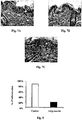


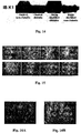
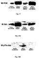


Classifications
-
- A—HUMAN NECESSITIES
- A61—MEDICAL OR VETERINARY SCIENCE; HYGIENE
- A61K—PREPARATIONS FOR MEDICAL, DENTAL OR TOILETRY PURPOSES
- A61K38/00—Medicinal preparations containing peptides
- A61K38/16—Peptides having more than 20 amino acids; Gastrins; Somatostatins; Melanotropins; Derivatives thereof
- A61K38/17—Peptides having more than 20 amino acids; Gastrins; Somatostatins; Melanotropins; Derivatives thereof from animals; from humans
- A61K38/22—Hormones
- A61K38/28—Insulins
-
- A—HUMAN NECESSITIES
- A61—MEDICAL OR VETERINARY SCIENCE; HYGIENE
- A61K—PREPARATIONS FOR MEDICAL, DENTAL OR TOILETRY PURPOSES
- A61K31/00—Medicinal preparations containing organic active ingredients
- A61K31/33—Heterocyclic compounds
- A61K31/395—Heterocyclic compounds having nitrogen as a ring hetero atom, e.g. guanethidine or rifamycins
- A61K31/41—Heterocyclic compounds having nitrogen as a ring hetero atom, e.g. guanethidine or rifamycins having five-membered rings with two or more ring hetero atoms, at least one of which being nitrogen, e.g. tetrazole
- A61K31/425—Thiazoles
- A61K31/427—Thiazoles not condensed and containing further heterocyclic rings
-
- A—HUMAN NECESSITIES
- A61—MEDICAL OR VETERINARY SCIENCE; HYGIENE
- A61K—PREPARATIONS FOR MEDICAL, DENTAL OR TOILETRY PURPOSES
- A61K31/00—Medicinal preparations containing organic active ingredients
- A61K31/33—Heterocyclic compounds
- A61K31/395—Heterocyclic compounds having nitrogen as a ring hetero atom, e.g. guanethidine or rifamycins
- A61K31/435—Heterocyclic compounds having nitrogen as a ring hetero atom, e.g. guanethidine or rifamycins having six-membered rings with one nitrogen as the only ring hetero atom
- A61K31/44—Non condensed pyridines; Hydrogenated derivatives thereof
- A61K31/445—Non condensed piperidines, e.g. piperocaine
-
- A—HUMAN NECESSITIES
- A61—MEDICAL OR VETERINARY SCIENCE; HYGIENE
- A61K—PREPARATIONS FOR MEDICAL, DENTAL OR TOILETRY PURPOSES
- A61K38/00—Medicinal preparations containing peptides
- A61K38/02—Peptides of undefined number of amino acids; Derivatives thereof
-
- A—HUMAN NECESSITIES
- A61—MEDICAL OR VETERINARY SCIENCE; HYGIENE
- A61K—PREPARATIONS FOR MEDICAL, DENTAL OR TOILETRY PURPOSES
- A61K38/00—Medicinal preparations containing peptides
- A61K38/04—Peptides having up to 20 amino acids in a fully defined sequence; Derivatives thereof
- A61K38/08—Peptides having 5 to 11 amino acids
- A61K38/085—Angiotensins
-
- A—HUMAN NECESSITIES
- A61—MEDICAL OR VETERINARY SCIENCE; HYGIENE
- A61K—PREPARATIONS FOR MEDICAL, DENTAL OR TOILETRY PURPOSES
- A61K38/00—Medicinal preparations containing peptides
- A61K38/16—Peptides having more than 20 amino acids; Gastrins; Somatostatins; Melanotropins; Derivatives thereof
- A61K38/17—Peptides having more than 20 amino acids; Gastrins; Somatostatins; Melanotropins; Derivatives thereof from animals; from humans
- A61K38/1703—Peptides having more than 20 amino acids; Gastrins; Somatostatins; Melanotropins; Derivatives thereof from animals; from humans from vertebrates
- A61K38/1709—Peptides having more than 20 amino acids; Gastrins; Somatostatins; Melanotropins; Derivatives thereof from animals; from humans from vertebrates from mammals
-
- A—HUMAN NECESSITIES
- A61—MEDICAL OR VETERINARY SCIENCE; HYGIENE
- A61K—PREPARATIONS FOR MEDICAL, DENTAL OR TOILETRY PURPOSES
- A61K38/00—Medicinal preparations containing peptides
- A61K38/16—Peptides having more than 20 amino acids; Gastrins; Somatostatins; Melanotropins; Derivatives thereof
- A61K38/17—Peptides having more than 20 amino acids; Gastrins; Somatostatins; Melanotropins; Derivatives thereof from animals; from humans
- A61K38/19—Cytokines; Lymphokines; Interferons
- A61K38/191—Tumor necrosis factors [TNF], e.g. lymphotoxin [LT], i.e. TNF-beta
-
- A—HUMAN NECESSITIES
- A61—MEDICAL OR VETERINARY SCIENCE; HYGIENE
- A61K—PREPARATIONS FOR MEDICAL, DENTAL OR TOILETRY PURPOSES
- A61K38/00—Medicinal preparations containing peptides
- A61K38/16—Peptides having more than 20 amino acids; Gastrins; Somatostatins; Melanotropins; Derivatives thereof
- A61K38/17—Peptides having more than 20 amino acids; Gastrins; Somatostatins; Melanotropins; Derivatives thereof from animals; from humans
- A61K38/19—Cytokines; Lymphokines; Interferons
- A61K38/20—Interleukins [IL]
- A61K38/2006—IL-1
-
- A—HUMAN NECESSITIES
- A61—MEDICAL OR VETERINARY SCIENCE; HYGIENE
- A61K—PREPARATIONS FOR MEDICAL, DENTAL OR TOILETRY PURPOSES
- A61K38/00—Medicinal preparations containing peptides
- A61K38/16—Peptides having more than 20 amino acids; Gastrins; Somatostatins; Melanotropins; Derivatives thereof
- A61K38/17—Peptides having more than 20 amino acids; Gastrins; Somatostatins; Melanotropins; Derivatives thereof from animals; from humans
- A61K38/19—Cytokines; Lymphokines; Interferons
- A61K38/20—Interleukins [IL]
- A61K38/2026—IL-4
-
- A—HUMAN NECESSITIES
- A61—MEDICAL OR VETERINARY SCIENCE; HYGIENE
- A61K—PREPARATIONS FOR MEDICAL, DENTAL OR TOILETRY PURPOSES
- A61K38/00—Medicinal preparations containing peptides
- A61K38/16—Peptides having more than 20 amino acids; Gastrins; Somatostatins; Melanotropins; Derivatives thereof
- A61K38/17—Peptides having more than 20 amino acids; Gastrins; Somatostatins; Melanotropins; Derivatives thereof from animals; from humans
- A61K38/19—Cytokines; Lymphokines; Interferons
- A61K38/20—Interleukins [IL]
- A61K38/204—IL-6
-
- A—HUMAN NECESSITIES
- A61—MEDICAL OR VETERINARY SCIENCE; HYGIENE
- A61K—PREPARATIONS FOR MEDICAL, DENTAL OR TOILETRY PURPOSES
- A61K38/00—Medicinal preparations containing peptides
- A61K38/16—Peptides having more than 20 amino acids; Gastrins; Somatostatins; Melanotropins; Derivatives thereof
- A61K38/17—Peptides having more than 20 amino acids; Gastrins; Somatostatins; Melanotropins; Derivatives thereof from animals; from humans
- A61K38/19—Cytokines; Lymphokines; Interferons
- A61K38/20—Interleukins [IL]
- A61K38/2053—IL-8
-
- A—HUMAN NECESSITIES
- A61—MEDICAL OR VETERINARY SCIENCE; HYGIENE
- A61K—PREPARATIONS FOR MEDICAL, DENTAL OR TOILETRY PURPOSES
- A61K38/00—Medicinal preparations containing peptides
- A61K38/16—Peptides having more than 20 amino acids; Gastrins; Somatostatins; Melanotropins; Derivatives thereof
- A61K38/17—Peptides having more than 20 amino acids; Gastrins; Somatostatins; Melanotropins; Derivatives thereof from animals; from humans
- A61K38/22—Hormones
- A61K38/2264—Obesity-gene products, e.g. leptin
-
- A—HUMAN NECESSITIES
- A61—MEDICAL OR VETERINARY SCIENCE; HYGIENE
- A61K—PREPARATIONS FOR MEDICAL, DENTAL OR TOILETRY PURPOSES
- A61K38/00—Medicinal preparations containing peptides
- A61K38/16—Peptides having more than 20 amino acids; Gastrins; Somatostatins; Melanotropins; Derivatives thereof
- A61K38/17—Peptides having more than 20 amino acids; Gastrins; Somatostatins; Melanotropins; Derivatives thereof from animals; from humans
- A61K38/22—Hormones
- A61K38/2271—Neuropeptide Y
-
- A—HUMAN NECESSITIES
- A61—MEDICAL OR VETERINARY SCIENCE; HYGIENE
- A61K—PREPARATIONS FOR MEDICAL, DENTAL OR TOILETRY PURPOSES
- A61K38/00—Medicinal preparations containing peptides
- A61K38/16—Peptides having more than 20 amino acids; Gastrins; Somatostatins; Melanotropins; Derivatives thereof
- A61K38/43—Enzymes; Proenzymes; Derivatives thereof
- A61K38/45—Transferases (2)
-
- A—HUMAN NECESSITIES
- A61—MEDICAL OR VETERINARY SCIENCE; HYGIENE
- A61K—PREPARATIONS FOR MEDICAL, DENTAL OR TOILETRY PURPOSES
- A61K38/00—Medicinal preparations containing peptides
- A61K38/16—Peptides having more than 20 amino acids; Gastrins; Somatostatins; Melanotropins; Derivatives thereof
- A61K38/43—Enzymes; Proenzymes; Derivatives thereof
- A61K38/46—Hydrolases (3)
- A61K38/48—Hydrolases (3) acting on peptide bonds (3.4)
- A61K38/4886—Metalloendopeptidases (3.4.24), e.g. collagenase
-
- A—HUMAN NECESSITIES
- A61—MEDICAL OR VETERINARY SCIENCE; HYGIENE
- A61K—PREPARATIONS FOR MEDICAL, DENTAL OR TOILETRY PURPOSES
- A61K38/00—Medicinal preparations containing peptides
- A61K38/16—Peptides having more than 20 amino acids; Gastrins; Somatostatins; Melanotropins; Derivatives thereof
- A61K38/55—Protease inhibitors
- A61K38/57—Protease inhibitors from animals; from humans
-
- A—HUMAN NECESSITIES
- A61—MEDICAL OR VETERINARY SCIENCE; HYGIENE
- A61P—SPECIFIC THERAPEUTIC ACTIVITY OF CHEMICAL COMPOUNDS OR MEDICINAL PREPARATIONS
- A61P17/00—Drugs for dermatological disorders
-
- A—HUMAN NECESSITIES
- A61—MEDICAL OR VETERINARY SCIENCE; HYGIENE
- A61P—SPECIFIC THERAPEUTIC ACTIVITY OF CHEMICAL COMPOUNDS OR MEDICINAL PREPARATIONS
- A61P17/00—Drugs for dermatological disorders
- A61P17/16—Emollients or protectives, e.g. against radiation
-
- A—HUMAN NECESSITIES
- A61—MEDICAL OR VETERINARY SCIENCE; HYGIENE
- A61P—SPECIFIC THERAPEUTIC ACTIVITY OF CHEMICAL COMPOUNDS OR MEDICINAL PREPARATIONS
- A61P17/00—Drugs for dermatological disorders
- A61P17/18—Antioxidants, e.g. antiradicals
-
- A—HUMAN NECESSITIES
- A61—MEDICAL OR VETERINARY SCIENCE; HYGIENE
- A61P—SPECIFIC THERAPEUTIC ACTIVITY OF CHEMICAL COMPOUNDS OR MEDICINAL PREPARATIONS
- A61P3/00—Drugs for disorders of the metabolism
- A61P3/08—Drugs for disorders of the metabolism for glucose homeostasis
- A61P3/10—Drugs for disorders of the metabolism for glucose homeostasis for hyperglycaemia, e.g. antidiabetics
-
- A—HUMAN NECESSITIES
- A61—MEDICAL OR VETERINARY SCIENCE; HYGIENE
- A61P—SPECIFIC THERAPEUTIC ACTIVITY OF CHEMICAL COMPOUNDS OR MEDICINAL PREPARATIONS
- A61P43/00—Drugs for specific purposes, not provided for in groups A61P1/00-A61P41/00
Definitions
- the present invention relates to methods and compositions for prevention and/or treatment of skin pathologies and disorders associated with diabetes and/or aging. More particularly, the present invention relates to the use of various agents capable of restoring impaired physiological conditions of the skin associated with skin pathologies or disorders.
- Skin is composed of a myriad of cell types, acting together to maintain overall tissue function. Normal skin physiology and architecture is dependent on the cross talk between these cells, encompassing even more intricate intracellular signaling mechanisms. The delicate balance between biochemical processes is the focus point of all physiological outcomes. This balance is the basis by which the skin exerts its physiological functions including the formation of a pathogenic barrier, modulation of the immune response, serving as an homeostatic barrier (i.e. fluid regulation), and controlling body temperature. Skin pathologies transpire when this balance is compromised and thus non desired physiological routes are expressed.
- Skin is a complex tissue organized in distinct layers, namely, the epidermis, dermis and hypodermis, each possessing a different cell characterization and physiological significance (Schaefer and Redelmeier, 1996).
- the epidermis is stratified squamous epithelium in which cells undergoing growth and differentiation are strictly compartmentalized (Schaefer and Redelmeier, 1996). In a normal physiologic state, proliferation is confined to the basal cells that adhere to the basement membrane. Differentiation is a spatial process in which basal cells lose their adhesion to the basement membrane, cease DNA synthesis and undergo a series of morphological and biochemical changes. The ultimate maturation step is the production of the cornified layer forming the protective barrier of the skin (Tennenbaum et al., 1991; Wysocki, 1999).
- the dermis is mainly composed of matrix fibers and contains various cell types.
- all skin appendages namely, microvasculature, sweat and sebaceous glands, sensory nerves and hair follicles, are localized in the dermis.
- the dermis has been attributed the supporting role of skin nourishment, maintaining the epidermis and the route by which signals from other parts of the body reach the outer layer (Tennenbaum et al., 1991; Wysocki, 1999).
- the hypodermis is the deepest layer of the skin, mainly consisting of adipose cells, also known as the subcutaneous fat layer. Until recently, this layer has been thought to have the role of insulation from the external temperature changes and mechanical support to the upper layers of the skin (Jackson et al., 1993). Only recently, the endocrine significance of fat tissue (Pantanetti et al., 2004; Fliers et al., 2003), specifically the visceral fat tissue, has been acknowledged and identified as playing a role in glucose regulation in diabetes development and progression (Laviola et al., 2006; Maianu, 2001). Moreover, recent publications have identified the potential impact of subcutaneous adipocytes, which secrete several cytokines and growth factors that can affect skin physiology and regeneration (Nakagami et al., 2006).
- the insulin receptor is an insulin-regulated tyrosine kinase. Insulin binding to its receptor results in receptor activation via autophosphorylation of tyrosine residues on several regions of the intracellular ⁇ -subunit. Subsequently insulin receptor substrate (IRS) proteins are tyrosine phosphorylated and activated. Tyrosine-phosphorylated IRS-proteins generate downstream signals by the direct binding to the SH2 domains of various signaling proteins.
- PI 3-kinase phosphatidylinositol 3-kinase
- SHP2 phosphotyrosine phosphatase SHP2, Grb2, Nck, and Crk.
- the products of the shc gene are also substrates of the insulin receptor since they contain SH2 domains and are tyrosine phosphorylated in response to insulin (White, 1997).
- PI 3-kinase One of the earliest steps in the insulin signaling pathway is the activation of PI 3-kinase. Once activated, the catalytic subunit phosphorylates phosphoinositides at the 3' position of the inositol ring or proteins at serine residues. PI3K activates downstream molecules such as PtdIns(3,4)P2/ PtdIns(3,4,5)P3-dependent kinase 1 (PDK1), which activates serine kinase Akt. Akt in turn deactivates glycogen synthase kinase 3 (GSK-3), leading to activation of glycogen synthase and thus glycogen synthesis.
- PtdIns(3,4)P2/ PtdIns(3,4,5)P3-dependent kinase 1 Akt
- Akt in turn deactivates glycogen synthase kinase 3 (GSK-3), leading to activation of glycogen synthase and thus glycogen synthesis.
- Akt Akt-induced Akt
- Other targets of Akt include mTOR-mediated activation of protein synthesis by PHAS/elf4 and p70 s6k and cell survival mechanisms represented by BAD (Bcl-2/Bcl-X L antagonist) and IKK (I- ⁇ B Kinase).
- Grb2 and SHP2 Other signal transduction proteins which interact with IRS molecules include Grb2 and SHP2. Flanking its SH2-domain, Grb2 contains two SH3-domains that associate constitutively with proline rich regions in mSOS, a guanine nucleotide exchange factor that stimulates GDP/GTP exchange on Ras. Activated Ras recruits Raf, a serine/threonine kinase to the plasma membrane. Raf activation results in the activation of MEK by phosphorylation of two serine residues. MEK is a dual specificity kinase that activates MAPK (mitogen-activated protein kinase) by both tyrosine and threonine phosphorylation.
- MAPK mitogen-activated protein kinase
- MAPK acts as an activator of some transcriptional factors (Myc, NF-kB, AP-1), kinases (Rsk), cell survival proteins (Bcl-2, cPL-2) and structural proteins (paxillin) (Taha and Klip, 1999) .
- insulin stimulates the activation of PKC isoforms in several tissues and cell types. It has also been reported that PKC isoforms may form complexes with the IR and phosphorylate several molecules involved in IR-initiated signaling, inhibiting their function.
- serine/threonine kinases a crucial role in modulating insulin signals is played by the PKC family members (Farese et al., 2005).
- PKC ⁇ has been reported to inhibit insulin action in both cellular and animal models.
- Overexpression of PKC ⁇ has been shown to inhibit insulin signaling in cultured cell systems.
- Murine models of PKC ⁇ gene ablation also exhibit increased sensitivity to insulin, further supporting the concept of the PKC ⁇ negative role in insulin signaling by targeting insulin toward degradation.
- Insulin down stream signaling also involves the specific activation of PKC ⁇ and PKC ⁇ (Formisano et al., 2000).
- insulin was shown to regulate skin proliferation via PKC ⁇ activation and transcriptional regulation of STAT3 (Gartsbein et al., 2006).
- Insulin also stimulates the formation of a multimolecular complex, including IRS-1 and PKC ⁇ ; PKC ⁇ inhibits IR/IRS-1 signaling and regulates insulin degradation. Insulin plays an important role in the overall regulation of protein synthesis. Some of the effects of insulin involve changes in mRNAs abundance, but insulin also has important effects on the translation process itself (Patel et al, 2006). Indeed, several initiation and elongation factors are regulated by this hormone, often as a consequence of changes in their states of phosphorylation.
- thinned skin exhibits a reduction in rete ridges, which undermines the skins' ability to control temperature and fluid homeostasis. Furthermore, it has been demonstrated that the rete ridges serve as the enriched stem cell population of human skin. Therefore, it is clear that the rete ridges attenuation in the structure of diabetic skin results in depletion of the skin stem cell population and leads to severe impairment in skin function and remodeling (Wertheimer, 2004).
- NLD necrobiosis lipoidica diabeticorum
- diabetic skin exhibits an impaired immune response to changes in the skin architecture brought by the metabolic complications. Such lesions result from delayed type hypersensitivity, accumulation of activated histocytes (foam cells) in the skin layers or an immune mediated response contributing to the changes in the dermis (Wertheimer, 2004).
- diabetic skin has been found to be constantly infected with either microbial or fungal sources due to its reduced ability to effectively react to exogenic infections. These opportunistic agents take advantage of the impaired immune response and pose a constant aggravation to patients (Wang and Margolis, 2006; Muller et al., 2005).
- Skin changes are among the most visible signs of aging.
- Evidence of increasing age include wrinkles and sagging skin.
- Aging changes in the skin are a group of common conditions and developments that occur as people grow older.
- the outer skin layer epidermis
- the number of pigment-containing cells decreases. Aging skin thus appears thinner, more pale, and translucent.
- the present invention provides a method of treatment or prevention of a skin pathology, disorder or condition associated with diabetes and/or aging, the method comprising topically administering to the skin of a diabetic and/or aging individual a therapeutically or cosmetically effective amount of at least one agent capable of restoring an impaired physiological condition of the skin associated with said pathology, disorder or condition.
- the present invention provides the use of at least one agent capable of restoring an impaired physiological condition of the skin associated with a skin pathology, disorder or condition associated with diabetes and/or aging, for the preparation of a topical pharmaceutical or cosmetic composition for treatment or prevention of said skin pathology, disorder or condition in diabetic and/or aging individuals.
- the present invention provides a topical pharmaceutical or cosmetic composition, when used for treating or preventing a skin pathology, disorder or condition associated with diabetes and/or aging, comprising a therapeutically or cosmetically effective amount of at least one agent capable of restoring an impaired physiological condition of the skin associated with said skin pathology, disorder or condition, optionally with a pharmaceutically or cosmeticaly acceptable carrier.
- compositions of the invention can be used for prevention of recurrence of said skin pathology, disorder or condition in a diabetic patient.
- the agents used according to the present invention include a PKC isoform modulator, an adipokine, PPAR ⁇ , a PPAR agonist or antagonist, and an agent capable of modulating the expression and/or activity of a factor involved in the insulin signaling pathway in the skin.
- These agents are useful for revitalization/renewal/rejuvenation/restoration of the hypodermis/subcutaneous fat tissue/layer in diabetic and/or aging individuals and can thus be used as effective anti-aging agents.
- the invention further provides a cosmetic anti-aging composition
- a cosmetic anti-aging composition comprising a cosmetically effective amount of at least one agent capable of restoring an impaired physiological condition of the skin associated with an aging skin pathology, disorder or condition, optionally with a cosmeticaly acceptable carrier.
- the present invention relates to methods, pharmaceutical and cosmetical compositions for treatment and/or prevention of skin pathologies, disorders and conditions associated with the diabetic and/or aging skin.
- the terms "skin pathology” and "skin disorder” do not include wounds and, in particular, do not include the types of wounds defined in the previous patent applications US 09/629,970 , US 10/169,801 , US 11/332,774 , US 11/348,527 , WO 02/09639 , WO 2005/007072 and WO/2005/013885 .
- the terms "skin pathology" and "skin disorder” do not include an ulcer, a diabetes-related wound, a burn, a sun burn, an aging skin wound, a corneal ulceration wound, an inflammatory gastrointestinal tract disease wound, a bowel inflammatory disease wound, a Crohn's disease wound, an ulcerative colitis, a hemorrhoid, an epidermolysis bulosa wound, a skin blistering wound, a psoriasis wound, an animal skin wound, a proud flesh wound, an animal diabetic wound, a retinopathy wound, an oral wound (mucositis), a vaginal mucositis wound, a gum disease wound, a laceration, a surgical incision wound and a post surgical adhesions wound
- diabetes skin and “skin pathology, disorder or condition associated with diabetes” are used interchangeably to define a skin disorder that is caused by diabetes or is affected by diabetes.
- Some of these disorders are conditions that anyone can have but to which people with diabetes are particularly prone, such as atherosclerotic skin changes, bacterial and fungal infections of the skin, and itching.
- Other diabetic skin diseases are seen mainly or exclusively in people with diabetes and include those pathologies summarized in Table 1, in which the compartmentalization of the different pathologies to affected cells/tissues, enables better understanding of the disorders and treatment of the common impairments associated with diabetic skin.
- Table 1 Skin pathologies related to diabetes and cell/tissue associated therewith Pathology Cells/tissue associated with Clinical and histological manifestation NLD matrix/dermis Thickening of collagen Granuloma annulare matrix/dermis Focal degeneration of collagen Diabetic dermopathy matrix/dermis Atrophic dermis with fibroblast proliferation, fibrotic collagen Sclerederma diabeticorum matrix/dermis/fat Enlarged dermis through the replacement of subcutaneous fat by collagen fibers APD matrix/dermis Transversing collagen/elastin to epidermis Bullosis diabeticorum epidermis Reduced threshold to blister formation Acanthosis nigricans epidermis Thick epidermis and altered dermal papills APD epidermis Primary epidermal defect, thick epidermis Cutaneous infections epidermis Breach in pathogenic barrier, reduction in immune cells in the epidermis
- epidermal-related defects can be associated either to impaired proliferation/differentiation ratio or breach of cell-cell or cell-matrix (basement membrane) contact (Wertheimer et al., 2001; Alt et al., 2001).
- Dermal/matrix-related defects may be influenced by the differentiation capacity of fibroblast inhabiting the dermal layer. Fibroblasts, the most common cell type in the dermis, are known to be responsible for the collagen and elastin fiber network synthesis and maintenance. In the dermis, fibroblasts are found in all differentiation stages from stem cells through intermediate stages and the fully differentiated committed fibroblast. It is the mature cell that is responsible for matrix formation (Freinkel and Woodley, 2001); thus, any disturbance in fibroblast differentiation may contribute to matrix imperfections. In addition, fibroblast depletion by reduced proliferation might also contribute to impaired matrix formation and /or maintenance.
- adipose cell in the hypodermis are also dependent on their dedifferentiation state in order to exert their endocrine role in skin physiology (Gregoire, 2001).
- the inventors of the present invention have developed a reliable technique for quantitatively identifying the critical parameters in skin physiology. These parameters are specifically linked to signaling pathways leading to physiological processes including proliferation, migration, differentiation, matrix formation and maintenance. Establishing the link between the parameters is of great importance in primary prevention and treatment of skin complications of diabetic patients. Thus, by underlying the biochemical regulation of these processes, we are able to control specific signaling pathways leading to the desired physiological endpoints. It is important to take into consideration that many of these pathways interact and influence each other, implying that only a combined regulation will be able to effectively direct the desired outcome.
- Advanced glycation endproduct formation caused by the consistent hyperglycemia, have been thought to affect directly various skin structures such as the microvasculature, dermal collagen fibers and basement membrane (Yang et al., 2003). Hyperglycemia has also been shown to have a direct effect on the epidermal keratinocytes, modifying their ability to proliferate and differentiate (Spravchikov et al., 2000).
- the defects found in diabetic patients in general can be corrected in accordance with the present invention specifically by renewal of the various cell layers of skin, particularly the hypodermis layer.
- This is a novel approach to correct esthetic defects, restore elasticity and rejuvenate skin in diabetic and non-diabetic individuals and to prevent appearance or recurrence of a pathology associated with diabetic skin.
- the present invention provides a method for treating or preventing a skin pathology, disorder or condition associated with diabetes and/or aging, comprising topically administering to the skin of a diabetic and/or aging individual a therapeutically or cosmetically effective amount of at least one agent capable of restoring an impaired physiological condition of the skin associated with said skin pathology, disorder or condition.
- the term "impaired physiological condition of the skin associated with said skin pathology or disorder" when related to the diabetic skin refers to all pathologies that impair biochemical balance and structure of the skin, such as disappearance of the hypodermis adipose layer; impaired integrity of the epidermis layer through the disrupted regulation of the differentiation process and proliferation capacity, which is frequent in diabetic skin; dermis exhibition of disseminated matrix deposition affecting collagen distribution, elastic fibers destruction and enhanced inflammatory response throughout all skin layers, which are characteristic of diabetic skin.
- distinct skin pathologies specific to the diabetic state are induced, as specified herein in the specification.
- the term also refers to a unique impairment in the hypodermis layer, which we have characterized in the present invention, which leads to the impairment of aging skin including impaired elasticity, thinning, wrinkles, etc.
- the term also refers to the biochemical impairments in the insulin signaling pathway, PKC pathways and adipokine secretion in diabetic and aged skin that specifically cause structural pathological manifestations including changes in epidermis, dermis and uniquely in the hypodermis.
- the at least one agent is a PKC isoform modulator capable of modulating the expression and/or activity of at least one PKC isoform.
- PKC isoform encompasses all PKC isoforms including PKC ⁇ , PKC ⁇ (including both PKC ⁇ 1 and PKC ⁇ 2), PKC- ⁇ , PKC- ⁇ , PKC- ⁇ , PKC- ⁇ , PKC- ⁇ , PKC- ⁇ and PKC- ⁇ .
- the PKC isoform is PKC ⁇ , PKC ⁇ , PKC ⁇ or PKC ⁇ .
- modulating expression and/or activity of a PKC isoform relates to an increased or reduced expression and/or activity of a PKC isoform. Increase of the expression leads to increased production of the PKC isoform.
- PKC activator is used herein interchangeably with the term “PKC isoform activator” to describe a molecule that enhances expression and/or activity of a PKC isoform.
- PKC inhibitor is used herein interchangeably with the term “PKC isoform inhibitor” to describe a molecule that inhibits expression and/or activity of a PKC isoform.
- the phosphoryl transfer region, the pseudosubstrate domain, the phorbolester binding sequences, and the phosphorylation sites may be targets for modulation of isoenzyme-specific PKC activity (Hofmann, 1997). It should be understood that many modulators are not specific and sometimes the modulator may be both inhibitor and activator.
- the at least one agent is a PKC isoform inhibitor, which may be a PKC isoform pseudosubstrate inhibitor, a peptide binding to the PKC isoform substrate region, a peptide binding to the ATP-binding site of a PKC isoform, or Copolymer-1.
- a PKC isoform inhibitor which may be a PKC isoform pseudosubstrate inhibitor, a peptide binding to the PKC isoform substrate region, a peptide binding to the ATP-binding site of a PKC isoform, or Copolymer-1.
- the "pseudosubstrate region" or autoinhibitory domain of a PKC isoform is defined as a consensus sequence of substrates for the kinase with no phosphorylatable residue.
- the pseudosubstrate domain is based in the regulatory region, closely resembling the substrate recognition motif, which blocks the recognition site and prevents phosphorylation.
- inhibitory peptides are obtained by replacing a phosphorylatable residue of serine (S) or tyrosine (T) by alanine (A).
- S serine
- T tyrosine
- A alanine
- PKC ⁇ is the only PKC isoform known to have additional binding site enabling the isoform's activation on the C2 domain, the conserved domain 2 of PKC ⁇ (Benes et al., 2005).
- the ATP-binding sites of all PKC isoforms are known from the literature. Any peptide that binds to the ATP-binding site will inhibit the activity of the PKC isoform.
- the PKC isoform inhibitor is preferably a PKC ⁇ , PKC ⁇ , PKC ⁇ or PKC ⁇ inhibitor, more preferably a PKC ⁇ or a PKC ⁇ inhibitor.
- PKC ⁇ inhibitors examples include, without being limited to, a PKC ⁇ pseudosubstrate inhibitor such as the peptides of SEQ IDS NO: 1-7, and a peptide binding to the substrate region such as the peptides of SEQ ID NO: 8 to NO. 24.
- the peptides of SEQ ID NO: 1 to NO: 24 may be N-acylated, preferably by an acyl group derived from a C12-C20 fatty acid, more preferably C 14 -acyl (myristoyl).
- the PKC ⁇ inhibitor is the N-myristoylated PKC- ⁇ pseudosubstrate peptide of SEQ ID NO: 1.
- It may be administered in a therapeutically or cosmetically effective concentration ranging from 0.01 ⁇ M to 10 ⁇ M, preferably 0.1-5 ⁇ M, 0.5-2 ⁇ M, or 1-2 ⁇ M, more preferably 1 ⁇ M for therapeutic purposes, and at a lower concentration for cosmetic purposes, more preferably 0.1 ⁇ M.
- PKC ⁇ inhibitors examples include, without being limited to, a PKC ⁇ pseudosubstrate inhibitor such as the N-myristoylated peptide of SEQ ID NO: 25, a PKC ⁇ inhibitor that binds to the substrate region such as the peptides of SEQ ID NO: 26 and NO: 27, and Copolymer 1 (the active ingredient of the drug glatiramer acetate/Copaxone®, of Teva Pharmaceutical Industries Ltd., Petach Tikva, Israel), clinically used for treating multiple sclerosis.
- Copolymer-1 is a synthetic polypeptide analog of myelin basic protein (MBP), which is a natural component of the myelin sheath.
- MBP myelin basic protein
- Copolymer-1 is a random copolymer of the 4 amino acids L-glutamic acid, L-alanine, L-lysine and L-tyrosine.
- L-glutamic acid L-alanine
- L-lysine L-tyrosine
- Copolymer-1 molecules of higher molecular weight are also known (MW 15,000-18,000) and can be used according to the present invention.
- Copolymer-1 was discovered to be a specific and very effective PKC ⁇ inhibitor.
- PKC ⁇ inhibitors examples include, without being limited to, a PKC ⁇ inhibitor that binds to the substrate region such as the peptides of SEQ ID NO: 28 to NO: 38.
- PKC ⁇ inhibitors examples include, without being limited to, a PKC ⁇ inhibitor that binds to the substrate region such as the peptides of SEQ ID NO: 39 to NO: 43.
- said PKC isoform modulator used in the methods and compositions of the present invention is a PKC isoform activator, preferably a PKC ⁇ , PKC ⁇ or PKC ⁇ activator, more preferably a PKC ⁇ activator.
- the PKC isoform activator may be, without being limited to, a peptide binding to a PKC isoform substrate region, a peptide acting on a PKC isoform phosphorylation site, insulin, bryostatin, a PKC isoform RACK peptide or a MARCKS (myristoylated alanine-rich C kinase substrate)-derived peptide.
- PKC ⁇ activators examples include, without being limited to, insulin, a peptide binding to the PKC ⁇ substrate region such as those of SEQ ID NO: 44 to NO: 51; a peptide acting on the PKC ⁇ phosphorylation site such as those of SEQ ID NO: 52 to NO: 54; a PKC ⁇ RACK peptide, and peptides corresponding to the C2 domain (disclosed in Benes et al., 2005).
- the PKC ⁇ activator is insulin, administered in a pharmaceutical or cosmetic composition in a concentration ranging from 0.01 ⁇ M to 10 ⁇ M, preferably 0.01-2 ⁇ M, more preferably 7x10 -7 M (0.1 unit) for therapeutic purpose and 7x10 -8 M (0.01 unit) for cosmetic purpose.
- PKC ⁇ activators that can be used according to the present invention include, without being limited to, the PKC- ⁇ MARCKS-derived peptide of SEQ ID NO: 55.
- more than one agent preferably two or three different PKC isoforms modulators, can be used in the methods/compositions of the present invention.
- two agents are used and the agents may be: (i) a PKC ⁇ inhibitor, preferably the N-myristoylated peptide of SEQ ID NO: 1, and a PKC ⁇ activator, preferably insulin; (ii) a PKC ⁇ inhibitor, preferably Copolymer-1 or the N-myristoylated peptide of SEQ ID NO: 25, and a PKC ⁇ activator, preferably insulin; and (iii) a PKC ⁇ inhibitor, preferably the N-myristoylated peptide of SEQ ID NO: 1, and a PKC ⁇ inhibitor, preferably Copolymer-1 or the N-myristoylated peptide of SEQ ID NO: 25.
- the concentrations of the peptide of SEQ ID NO:1 and of insulin are as defined above for therapeutic and cosmetic purposes.
- the concentration of Copolymer 1 is in the range of 0.5-60 ⁇ g/ml, preferably 10-55 ⁇ g/ml or 50-55 ⁇ g/ml, more preferably about 55 ⁇ g/ml.
- three agents are used, for instance, a PKC ⁇ inhibitor, preferably the N-myristoylated peptide of SEQ ID NO: 1, a PKC ⁇ inhibitor, preferably Copolymer-1, and a PKC ⁇ activator, preferably insulin.
- a PKC ⁇ inhibitor preferably the N-myristoylated peptide of SEQ ID NO: 1
- a PKC ⁇ inhibitor preferably Copolymer-1
- a PKC ⁇ activator preferably insulin.
- the insulin for use in the methods and compositions of the present invention may be recombinant or from a natural source such as human insulin or a non-human mammal insulin that is suitable for human use such as porcine insulin.
- the at least one agent is an adipokine, a bioactive molecule secreted by adipocytes selected from adipocyte-secreted enzymes, growth factors, cytokines and hormones.
- adipokines include, but are not limited to, complement factors D (adipsin), C3 and B, adiponectin (Acrp30), apelin, visfatin, resistin, leptin, lipoprotein lipase (LPL), plasminogen activator inhibitor-1 (PAI-1), tumor necrosis factor- ⁇ (TNF- ⁇ ), interleukin-1 ⁇ (IL-1 ⁇ ), IL-4, IL-6, IL-8, angiotensin I-IV (angiotensin IV is an active angiotensin II fragment) and cycloanalogues thereof, angiotensinogen, 1-butyrylglycerol (monobutyrin), matrix metalloproteinase 2, matrix metalloproteinase 9,
- the adipokine is adiponectin that can be used in a concentration of 0.01-1 ⁇ g/ml, preferably 0.1 ⁇ g/ml.
- the at least one agent is PPAR ⁇ (peroxisome proliferators-activated receptor- ⁇ ), a transcription factor expressed in adipocytes, keratinocytes and sebaceous glands, or it is a PPAR agonist or antagonist.
- PPAR ⁇ peroxisome proliferators-activated receptor- ⁇
- a transcription factor expressed in adipocytes, keratinocytes and sebaceous glands or it is a PPAR agonist or antagonist.
- the agent is a PPAR ⁇ antagonist such as GW9662 (2-chloro-5-nitrobenzanilide) or a PPAR ⁇ agonist such as a thiazolidinedione derivative, e.g., troglitazone [5-(4-(6-hydroxy-2,5,7,8-tetramethylchroman-2-yl-methoxy)benzyl)-2,4-thiazolidinedione)], rosiglitazone [5-(4-(2-(methyl-2-pyridinylamino) ethoxy) phenyl)methyl)-2,4-thiazolidinedione), or the partial PPAR ⁇ agonist GW0072, a potent antagonist of adipocyte differentiation.
- a PPAR ⁇ antagonist such as GW9662 (2-chloro-5-nitrobenzanilide) or a PPAR ⁇ agonist
- a thiazolidinedione derivative e.g., troglitazone [5-(
- the agent is a PPAR ⁇ agonist such as GW7647 and BM-17.0744, a PPAR ⁇ agonist such as GW0742,or a PPAR non-subtype selective agonist such as GW2433, GW4148, GW2331 and L165041.
- the PPAR ⁇ agonist is troglitazone or rosiglitazone that can be used in a concentration of 2-300 nM, preferably 10-200 nM, 20-150 nM, 20-100 nM, or 20-50 nM, preferably 50nM or 20 nM for cosmetic purposes.
- the agent is a PPAR antagonist that can be used in a concentration of 1-100nM.
- the at least one agent is capable of modulating the expression and/or activity of a factor involved in the insulin signaling pathway in the skin.
- the agent directly activates the expression and/or activity of the insulin receptor (IR).
- IR activators include, without being limited to, TLK16998; TLK19781; 2,5-dihydroxy-3-(4-methoxyphenyl)-6-phenyl-2,5-cyclohexadiene-1,4-dione, 2,5-dihydroxy-3-(1-methyl-1H-indol-3-yl)-6-phenyl-2,5-cyclohexadiene-1,4-dione, and 1,2,3,4,6-penta-O-galloyl-D-glucopyranose.
- the agent acts downstream to the IR modulating specific target pathways downstream to the IR.
- agents include, without being limited to, a phosphatidylinositol 3-kinase (PI3K) modulator, a protein kinase B/Akt modulator, a glycogen synthase kinase-3 (GSK3) modulator, a Raf modulator, a renin-angiotensin system (Ras) modulator, a mitogen-activated protein kinase (MAPK) modulator, an ERK modulator, a JNK modulator, or a MEK modulator.
- PI3K phosphatidylinositol 3-kinase
- GSK3 glycogen synthase kinase-3
- Raf modulator a renin-angiotensin system
- MEK mitogen-activated protein kinase
- modulators include, but are not limited to, a PI3K inhibitor such as PTEN, Wortmannin and LY 294002; an Akt inhibitor such as NL-71-101 or an Akt activator such as the mammalian target of rapamycin (mTOR); a GSK3 inhibitor such as the compounds CT 20026, CHIR 98014 and CHIR 99021; a Raf inhibitor such as BAY 43-9006 (sorafenib); a p38-MAPK inhibitor such as SB203580; a ERK inhibitor such as PD09859; a JNK inhibitor such as SP600125; and a MEK inhibitor such as PD98059 or UO126.
- a PI3K inhibitor such as PTEN, Wortmannin and LY 294002
- an Akt inhibitor such as NL-71-101 or an Akt activator such as the mammalian target of rapamycin (mTOR)
- mTOR mammalian target of rapamycin
- GSK3 inhibitor
- Any skin pathology or disorder associated with diabetes can be treated and/or prevented by the method of the present invention including, but not limited to, skin thinning, epidermal thinning, skin dryness, abnormal differentiation pattern in epidermis, reduction in proliferation in all skin layers, absence/irregularity of the hypodermis subcutaneous adipose tissue, fragility, reduced skin tone, loss of skin moisture, sagging of skin; skin hyperpigmentation and discoloration, irritated and sensitive skin; increased inflammation in skin, loss of skin texture, disruption of matrix fiber (collagen and elastin) structure, loss of skin smoothness and firmness, loss of elasticity, limited oil/sebum secretion, necrobiosis lipoidica diabeticorum (NLD), granuloma annulare, diabetic dermopathy, bullosis diabeticorum, limited joint mobility, scleredema diabeticorum, acanthosis nigricans, acquired perforating dermatosis, insulin allergy, cutaneous
- treatment of all skin disorders or conditions associated with aging as described below is applicable for diabetic aging individuals.
- the method of the invention is applied when the skin pathology or disorder is associated with diabetes and said at least one agent is administered in a pharmaceutical composition comprising a therapeutically effective amount of said at least one agent.
- the pharmaceutical composition comprises a pharmaceutically acceptable carrier and the N-myristoylated PKC ⁇ pseudosubstrate peptide of SEQ ID NO: 1 in a concentration ranging from 0.01 ⁇ g/ml to 10 ⁇ g/ml, preferably 0.1-5 ⁇ g/ml, 0.5-2 ⁇ g/ml, or 1-2 ⁇ g/ml, more preferably 1 ⁇ g/ml, optionally in combination with insulin in a concentration ranging from 0.01 ⁇ g/ml to 10 ⁇ g/ml, preferably 0.1-2 ⁇ g/ml, more preferably 0.1 ⁇ g/ml.
- the method of the invention is applied when the skin pathology, disorder or condition is associated with diabetes and said at least one agent is administered in a cosmetic composition comprising a cosmetically effective amount of said at least one agent.
- the pharmaceutical composition comprises a cosmetically acceptable carrier and the N-myristoylated PKC ⁇ pseudosubstrate peptide of SEQ ID NO: 1 in a concentration ranging from 0.01 ⁇ g/ml to 10 ⁇ g/ml, preferably 0.1-5 ⁇ g/ml, 0.5-2 ⁇ g/ml, or 1-2 ⁇ g/ml, more preferably 0.1 ⁇ g/ml, optionally in combination with insulin in a concentration ranging from 0.01 ⁇ g/ml to 10 ⁇ g/ml, preferably 0.1-2 ⁇ g/ml, more preferably 0.1 ⁇ g/ml.
- the method of the invention is applied when the skin disorder or condition is associated with aging and said at least one agent is administered in a cosmetic composition comprising a cosmetically effective amount of said at least one agent.
- the pharmaceutical composition comprises a cosmetically acceptable carrier and the N-myristoylated PKC ⁇ pseudosubstrate peptide of SEQ ID NO: 1 in a concentration ranging from 0.01 ⁇ g/ml to 10 ⁇ g/ml, preferably 0.1-5 ⁇ g/ml, 0.5-2 ⁇ g/ml, or 1-2 ⁇ g/ml, more preferably 0.1 ⁇ g/ml, optionally in combination with insulin in a concentration ranging from 0.01 ⁇ g/ml to 10 ⁇ g/ml, preferably 0.1-2 ⁇ g/ml, more preferably 0.1 ⁇ g/ml.
- the disorders or conditions associated with aging that can be treated by the method of the invention include, but are not limited to, dry and flaky skin, skin thinning, flattening of rete ridges, actinic elastosis, wrinkles, crow's feet, liver spots, age spots, skin hyerpigmentation, discoloration, facilitated blistering, fragility, reduced skin tone, reduced threshhold for irritation, loss of tautness and sagging, loss of elasticity, laxity, hyperplastic and disorganized elastic fiber system, reduced skin flexibility, reduced acral and sweat glands, reduced sebum production, trans epidermal water loss (TEWL) resulting in loss of moisture.
- TEWL trans epidermal water loss
- the invention also provides a method for prevention of recurrence of a skin pathology or disorder in a diabetic patient, comprising topically administering to the skin of said diabetic patient a therapeutically or cosmetically effective amount of at least one agent as defined above capable of restoring an impaired physiological condition of the skin associated with said skin pathology or disorder.
- the invention further relates to a method for revitalization/rejuvenation/renewal of the subcutaneous fat layer of a diabetic or aging individual, which comprises topically administering to the skin of said individual a cosmetically effective amount of at least one agent as defined above capable of causing renewal of the hypodermis layer.
- the invention still further relates to the use of at least one agent capable of restoring an impaired physiological condition of the skin associated with a skin pathology, disorder or condition associated with diabetes and/or aging, for the preparation of a topical pharmaceutical or cosmetic composition for treatment or prevention of said skin pathology or disorder in diabetic and/or aging individuals.
- compositions for topical application for treatment or prevention of skin pathologies, disorders or conditions in diabetic and/or aging individuals comprising a pharmaceutically or cosmetically acceptable carrier and at least one agent as defined above capable of restoring an impaired physiological condition of the skin associated with said skin pathology, disorder or condition associated with diabetes and/or aging.
- the therapeutically/pharmaceutically/cosmetically active ingredients used in the present invention can be administered to the diabetic or aging skin per se , or in a pharmaceutical or cosmetic composition mixed with suitable carriers and/or excipients.
- Pharmaceutical compositions suitable for use in context of the present invention include those compositions in which the active ingredients are contained in an amount effective to achieve an intended therapeutic effect.
- Cosmetic compositions suitable for use in context of the present invention include those compositions in which the active ingredients are contained in an amount effective to achieve an intended cosmetic effect.
- compositions of the invention are intended for topical application and may be in the form of an aqueous solution, a gel, a cream, a paste, a lotion, a spray, a suspension, a powder, a dispersion, a salve, an ointment, a serum, an anhydrous stick, oil based sprays, oil-in-water emulsions or water-in-oil emulsions.
- topical application refers to external application to the skin, mucous membranes, teeth, hair, scalp.
- compositions of the present invention may be manufactured by processes well known in the art, e.g., by means of conventional mixing, dissolving, granulating, dragee-making, levigating, emulsifying, encapsulating, entrapping or lyophilizing processes.
- the phrases “pharmaceutically suitable carrier” and “cosmetically or dermatologically acceptable carrier” refer to a carrier or a diluent that does not cause significant irritation to an organism and does not abrogate the biological activity and properties of the administered ingredients.
- inert substances are added to further facilitate processes and administration of the active ingredients and these excipients include, without limitation, calcium carbonate, calcium phosphate, various sugars and types of starch, cellulose derivatives, gelatin, vegetable oils and polyethylene glycols. Techniques for formulation and administration of active ingredients may be found in " Remington's Pharmaceutical Sciences,” Mack Publishing Co., Easton, PA, latest editi on, which is incorporated herein by reference.
- the topical route is preferred, and is assisted by a topical carrier.
- the topical carrier is one, which is generally suited for topical active ingredients administration and includes any such materials known in the art.
- the topical carrier is selected so as to provide the composition in the desired form, e.g., as a liquid or non-liquid carrier, lotion, cream, paste, gel, powder, ointment, solvent, liquid diluent, drops and the like, and may be comprised of a material of either naturally occurring or synthetic origin. It is essential, clearly, that the selected carrier does not adversely affect the active agent or other components of the topical formulation, and which is stable with respect to all components of the topical formulation.
- suitable topical carriers include water, alcohols and other nontoxic organic solvents, glycerin, mineral oil, silicone, petroleum jelly, lanolin, fatty acids, vegetable oils, parabens, waxes, and the like.
- Preferred formulations herein are colorless, odorless ointments, liquids, lotions, creams and gels.
- Ointments are semisolid preparations, which are typically based on petrolatum or other petroleum derivatives.
- the specific ointment base to be used is one that will provide for optimum active ingredients delivery, and, preferably, will provide for other desired characteristics as well, e.g., emolliency or the like.
- an ointment base should be inert, stable, nonirritating and nonsensitizing. As explained in Remington: The Science and Practice of Pharmacy, 19th Ed.
- ointment bases may be grouped in four classes: oleaginous bases; emulsifiable bases; emulsion bases; and water-soluble bases.
- Oleaginous ointment bases include, for example, vegetable oils, fats obtained from animals, and semisolid hydrocarbons obtained from petroleum.
- Emulsifiable ointment bases also known as absorbent ointment bases, contain little or no water and include, for example, hydroxystearin sulfate, anhydrous lanolin and hydrophilic petrolatum.
- Emulsion ointment bases are either water-in-oil (W/O) emulsions or oil-in-water (O/W) emulsions, and include, for example, cetyl alcohol, glyceryl monostearate, lanolin and stearic acid.
- W/O water-in-oil
- O/W oil-in-water
- Preferred water-soluble ointment bases are prepared from polyethylene glycols of varying molecular weight; again, reference may be made to Remington: The Science and Practice of Pharmacy for further information.
- Lotions are preparations to be applied to the skin surface without friction, and are typically liquid or semi liquid preparations, in which solid particles, including the active agent, are present in a water or alcohol base. Lotions are usually suspensions of solids, and may comprise a liquid oily emulsion of the oil-in-water type. Lotions are preferred formulations herein for treating large body areas, because of the ease of applying a more fluid composition. It is generally necessary that the insoluble matter in a lotion be finely divided. Lotions will typically contain suspending agents to produce better dispersions as well as compounds useful for localizing and holding the active agent in contact with the skin, e.g., methylcellulose, sodium carboxymethylcellulose, or the like.
- Creams containing the selected active ingredients are, as known in the art, viscous liquid or semisolid emulsions, either oil-in-water or water-in-oil.
- Cream bases are water-washable, and contain an oil phase, an emulsifier and an aqueous phase.
- the oil phase also sometimes called the "internal" phase, is generally comprised of petrolatum and a fatty alcohol such as cetyl or stearyl alcohol; the aqueous phase usually, although not necessarily, exceeds the oil phase in volume, and generally contains a humectant.
- the emulsifier in a cream formulation is generally a nonionic, anionic, cationic or amphoteric surfactant.
- Gel formulations are preferred for application to the scalp.
- gels are semisolid, suspension-type systems.
- Single-phase gels contain organic macromolecules distributed substantially uniformly throughout the carrier liquid, which is typically aqueous, but also, preferably, contains an alcohol and, optionally, an oil.
- additives may be included in the topical formulations of the invention.
- solvents may be used to solubilize certain active ingredients substances.
- Other optional additives include skin permeation enhancers, opacifiers, antioxidants, gelling agents, thickening agents, stabilizers, and the like.
- the cosmetic compositions may contain one or more emulsifiers, preservatives, thickeners, sunscreens, antioxidants, emollients, skin protectants, hair protectants.
- excipients usually used in the cosmetic art may be added such as, but not limited to, methylparaben, glycerin, EDTA disodium, cetearyl alcohol, ceteareth 20, propylparaben, cetyl palmitate, steareth-20, octyl stearate, polyolprepolymer-2, lecithin, and the like.
- the topical pharmaceutical compositions for the treatment of diabetic skin pathologies and disorders may contain other pharmaceutically active agents or ingredients, used for the treatment of such pathologies and disorders.
- Other agents may also be added, such as antimicrobial agents, antifungal agents, antibiotics and anti-inflammatory agents.
- the topical cosmetic compositions for the prevention and/or treatment of diabetic and aging skin pathologies, disorders and conditions may contain additional nutrients used in anti-aging and anti-wrinkle compositions including, without limitation, Vitamins A, B, C, E and Panthenol.
- compositions herein described may also comprise suitable solid or gel phase carriers or excipients.
- suitable solid or gel phase carriers or excipients include, but are not limited to, calcium carbonate, calcium phosphate, various sugars, starches, cellulose derivatives, gelatin and polymers such as polyethylene glycols.
- Dosing is dependent on the type, the severity and manifestation of the pathology or disorder and on the responsiveness of the subject to the active ingredients, as well as the dosage form employed and the potency of the particular agent(s).
- the doses for cosmetic use, particularly for prevention purposes, will be usually lower than the doses for pharmaceutical/therapeutic purpose.
- This manifestation is also characteristic of known diabetic skin pathologies such as Scleroderma Diabeticorum and leads to altered skin structure including skin thinning, flaky and dry skin, which also affect barrier function and skin elasticity.
- the significance of restoring the subcutaneous adipose tissue can indeed overcome many of the diabetic skin pathologies. This can be achieved according to the invention by treating diabetic skin with PKC modulating agents, various adipokines and insulin signaling related molecules, known to affect skin and adipose tissue differentiation and physiology.
- diabetes skin In addition to the abrogation of the hypodermis subcutaneous adipose tissue, abnormal structure of the epidermis and the dermis layers is observed in diabetes skin.
- the diabetic epidermis is thin, exhibits fewer layers and portrays a disruption in differentiation pattern demonstrated by the increased cornified envelope formation.
- the impaired differentiation by itself is a common base for diabetic related dermopathies.
- Several skin pathologies of diabetic skin are related to the interference with normal epidermal structure such as Bullosis Diabeticorum, APD and others.
- insulin, PKC and MAPK we can restore the normal differentiation pattern overcoming the epidermis related diabetic skin pathologies as demonstrated in Examples 10 and 13 hereinafter.
- the dermis appears to be disorganized and markedly reduced in size. Elastic fibers are reduced and matrix deposition is altered. These manifestations resemble human diabetic pathologies such as diabetic dermopathy and NLD.
- the proliferation capacity in diabetic skin is compromised underling these pathologies.
- Restoration of the dermal layer, improved matrix deposition and skin strength can be achieved by induction of PKC modulating factors, insulin signaling related factors and adipokines as demonstrated in Example 8 hereinafter. This can also be achieved by induction of hypodermis adipose tissue formation.
- the present invention further provides a method of providing at least one skin care benefit selected from the group consisting of: treating, delaying or preventing the impairments and pathologies which are characteristic of the diabetes state and improve the appearance and condition of skin. This includes: treating sensitive, dry or flaky skin, to induce skin elasticity and flexibility.
- Treating delaying or preventing sagging of skin imparting a youthful appearance to skin, normalizing skin color by lightening or darkening skin; soothing irritated or sensitive skin; improving skin texture, enhancing skin smoothness and inducing skin firmness and strength as well as regulating oil/sebum secretion and moisture; Furthermore, these compositions are also indicated for delay, treat or prevent the distinct pathologies which can appear in diabetes skin including: necrobiosis lipoidica diabeticorum, granuloma annulare, diabetic dermopathy, bullosis diabeticorum, limited joint mobility, scleredema diabeticorum, acanthosis nigricans, acquired perforating dermatosis, insulin allergy or diabetes related cutaneous skin infections.
- diabetes skin has unique characteristics.
- Some of these compositions were shown to be effective in correcting unique defects correlated to the aging process as well. Therefore, they can be utilized as a new line of cosmetic care products treating the cosmetic appearance of aging skin.
- specific molecules related to the insulin signaling pathway, MAPK signaling pathway, adipocyte secreted molecules and PKC modulating agents can treat, prevent or delay the impairments and pathologies of diabetic and aging skin as demonstrated in the presented examples.
- the condition of human skin is affected by different factors such as humidity, ultraviolet rays, cosmetic compositions, aging, diseases, stress and nutrition.
- Several features are associated with skin aging. This includes loss of resilience and skin thinning which are associated with reduction in the number of cells and blood vessels that furnish the skin.
- aged skin is associated with changes in skin esthetics and appearance such as wrinkles and crow's feet, stains and age spots, sagging, loss of tautness and gloss, loss of flexibility and elasticity.
- Decline in the fibroblasts and other dermal cells proliferation is associated with loss of collagen biosynthesis and irregularities of various components of the extracellular matrix in the skin thus, leading to loss of elasticity and induction of wrinkles and sagging which is characteristic of the skin aging process. Therefore, a variety of skin related products for cosmetic use in aging skin includes components known to affect dermal structure, dermal cell proliferation and the induction of collagen and matrix production.
- a new mechanism for the induction of skin aging which is characterized by the loss of the subcutaneous fat tissue and mediates irregularities in the adipocyte tissue components within the skin, including the impairment in the secretion of various adipokines.
- the cosmetic compositions and methods described herein result in the prevention, reduction or delay in the formation of wrinkles, the prevention, reduction or delay in loss of skin tone, and the prevention, reduction or delay in the formation of pimples and blackheads. They also moderate skin discolorations such as brown spots, age spots or liver spots, rejuvenate dry, abused, or irritated skin, close or tighten pores, improve skin texture, smoothness or firmness, and create smooth and supple skin with improved elasticity. A general improvement in the appearance, texture and condition, in particular with respect to the radiance, clarity, and general appearance of skin is achieved. The present invention therefore provides a wide range of results that are collectively described as anti-aging benefits.
- Tissue culture media and serum were purchased from Biological Industries (Beit HaEmek, Israel). Enhanced Chemical Luminescence (ECL) was performed with a kit purchased from BioRad (Israel). Monoclonal anti phosphotyrosine antibody was purchased from Upstate Biotechnology Inc. (Lake Placid, NY, USA). Polyclonal and monoclonal antibodies to PKC isoforms were purchased from Santa Cruz (California, USA) and Transduction Laboratories (Lexington, KY). Rat mAB to phosphotyrosine was purchased from Sigma (St. Louis, MO) and rabbit anti phosphoserine was purchased from Zymed (San Francisco, CA).
- Horseradish peroxidase anti-rabbit and anti-mouse IgG were obtained from Bio-Rad (Israel).
- Leupeptin, aprotinin, phenylmethanesulfonyl fluoride (PMSF), dithiothreitol (DTT), Na orthovanadate and pepstatin were purchased from Sigma Chemicals (St. Louis, MO).
- Insulin Human insulin
- IGF1 was a gift from Cytolab (Israel). Keratin 1 and 14, and filaggrin antibodies were purchased from Babco-Convance (Richmond, CA).
- mice and Psammomys obesus animal models of diabetes type I and diabetes type II were studied skin biopsies obtained from mice and Psammomys obesus animal models of diabetes type I and diabetes type II. Different mouse strains were used for the emphasis of specific signaling pathways.
- a wild type strain 129 brown mouse
- Mice were maintained normoglycemic or were diabetes-induced by a single injection of STZ (185 mg/kg in citrate buffer).
- STZ 185 mg/kg in citrate buffer
- Psammomys obesus were obtained from both diabetic prone and diabetic resistant strains and placed on high energy diet, which is the inducer of diabetes in this animal model.
- Skin biopsies were either embedded in paraffin or frozen for protein analysis via Western blotting. Assessment of specific parameters was performed by histological analysis using H&E, Elastin and Masson staining and immunohistochemistry specifically indicating epidermal differentiation state. Immunohistochemical markers included specific protein expression of keratins 14 and proliferating cell nuclear antigen (PCNA) characteristic of basal proliferating cells, Keratin 1 (specific of spinous differentiating cells) and filaggrin distribution (characteristic of the cornified layer-terminal differentiation state).
- PCNA proliferating cell nuclear antigen
- mice that persisted to overt diabetes, assessed by the prolonged time with very high levels of hyperglycemia (>450 mg/dl for a period of 7 days or more), were taken into consideration and compared to their non-injected litter mates.
- Additional animal models included: a mouse with a defected insulin signaling pathway due to PKC ⁇ depletion and a mouse from 129 origin with defective MAPK signaling route in skin due to PKC ⁇ depletion in skin.
- PKC downregulation can be achieved by either treating the skin with long term phorbol 12-myristate 13-acetate (PMA) exposure or the specific modulation of skin cells utilizing PKC isoform inhibitors. These mice were also injected with STZ and maintained in an overt diabetic state. Skin from these models was subjected to both paraffin embedding and histological analysis as well as protein expression analysis by immunoprecipitation and Western blot.
- PMA phorbol 12-myristate 13-acetate
- the in vitro model systems were based on primary skin cell cultures, either keratinocytes or fibroblasts. The cells were treated and tested for morphological changes as well as migratory and proliferative capacity. Migration was studied utilizing the scratch assay technique and proliferation was tested by proliferating cell nuclear antigen (PCNA) expression or by thymidine incorporation, as described by Shen et al. (2001).
- PCNA proliferating cell nuclear antigen
- keratinocytes Primary keratinocytes were isolated from newborn skin as previously described (Alt et al., 2001). Keratinocytes were cultured in Eagle's Minimal Essential Medium (EMEM) containing 8% Chelex (Chelex-100, BioRad) treated fetal bovine serum. In order to maintain a proliferative basal cell phenotype, the final Ca 2+ concentration was adjusted to 0.05 mM. Experiments were performed 5-7 days after plating.
- EMEM Eagle's Minimal Essential Medium
- Chelex Chelex-100, BioRad
- fibroblasts Primary dermal fibroblasts were isolated from newborn skin. For each newborn skin, dermis was separated from epidermis after overnight flotation on trypsin. Dermises were then incubated with collagenase (25 ml per 10 dermises) for 30 min with agitation at 37°C. Next, 3 volumes of HiCa, namely, Dulbecco's Modified Eagle's Medium (DMEM) with serum and fungisome, were added to the filtrate and suspension was filtered through sterile nylon mesh and centrifuged at 1200 rpm for 7 min at RT (pellet contains fibroblasts and follicles).
- DMEM Dulbecco's Modified Eagle's Medium
- Pellet was resuspended in HiCa, each mouse equivalent to 1 ml medium and centrifuged at 500 rpm for 3 min (pellet contains follicles; supernatant contains fibroblasts). Appropriate volumes of HiCa were added to the supernatant and plated in equivalent 1 mouse per 10 cm culture dish. The experiments were performed on early cell passages (passages 2-6).
- whole cell lysates were prepared by scraping cells into PBS containing 10 ⁇ g/ml aprotinin, 10 ⁇ g/ml leupeptin, 2 ⁇ g/ml pepstatin, 1 mM PMSF, 10 mM ethylenediaminetetraacetic acid (EDTA), 200 ⁇ M NaVO4 and 10 mM NaF. After homogenization and 4 freeze/thaw cycles, lysates were spun down at 4°C for 20 min in a microcentrifuge at maximal speed. The supernatant containing the soluble cytosol protein fraction was transferred to another tube.
- PBS containing 10 ⁇ g/ml aprotinin, 10 ⁇ g/ml leupeptin, 2 ⁇ g/ml pepstatin, 1 mM PMSF, 10 mM ethylenediaminetetraacetic acid (EDTA), 200 ⁇ M NaVO4 and 10 mM NaF.
- EDTA ethylenediaminetetra
- the pellet was resuspended in 250 ⁇ l PBS containing 1% Triton X-100 with protease and phosphatase inhibitors, incubated for 30 min at 4°C and spun down in a microcentrifuge at maximal speed at 4°C. The supernatant contains the membrane fraction. Protein concentrations were measured using a modified Lowery assay (Bio-Rad DC Protein Assay Kit). Western blot analysis of cellular protein fractions was carried out as described (Alt et al., 2001).
- the lysate was precleared by mixing 300 ⁇ g of cell lysate with 25 ⁇ l of Protein A/G Sepharose (Santa Cruz, CA, USA), and the suspension was rotated continuously for 30 min at 4°C. The preparation was then centrifuged at maximal speed at 4°C for 10 min, and 30 ⁇ l of A/G Sepharose was added to the supernatant along with specific polyclonal or monoclonal antibodies to the individual antigens (dilution 1:100). The samples were rotated overnight at 4°C. The suspension was then centrifuged at maximal speed for 10 min at 4°C, and the pellet was washed with RIPA buffer.
- Example 1 Diabetes skin exhibits excessive inflammatory response
- Fig. 1A-1D show the inflammation response in skin of diabetic animals (1A-1B), in which abscess formation is marked by yellow arrows, vs. non-diabetic animals (1C-1D).
- skin of diabetic animals is indeed prone to severe inflammation response, indicated by facilitated abscess formation in different parts of the skin. Therefore, it is important to prevent this manifestation in order to overcome the diabetic inflamed skin problem.
- the integrity of diabetic skin structure is severally compromised. This severe impairment includes the abrogation of the hypodermis subcutaneous adipose tissue, the hyper differentiation and abnormal structure of the epidermis, and the disruption of dermal structure.
- Figs. 2A-2D diabetes has a profound effect on skin architecture.
- Fig. 2A shows the structure of normoglycemic mouse skin in which the hypodermis fat layer, indicated by an arrow, underlies the dermis. Contrary to that, diabetic mouse skin exhibits an absolute absence of hypodermis fat tissue, and instead, the underlying muscle is "pushed" under the dermis rendering the appearance of thinner skin ( Fig. 2B ).
- NOD nonobese diabetic
- Psammomys obesus type 2 diabetic model diabetic rats and also in large animal models.
- Figs. 2C shows the structure of normal mouse skin, demonstrating normal structure of the epidermis underlined by a yellow speckled line.
- Fig. 2D shows diabetic mouse skin demonstrating abnormal structure of the epidermis underlined by a yellow speckled line.
- the diabetic epidermis is thin, exhibits fewer layers and portrays a disruption in differentiation pattern demonstrated by an increased cornified envelope formation.
- Figs. 3A-3B illustrate the direct effect of the insulin pathway on skin structure as demonstrated in the insulin resistant skin model (3B), exhibiting a defective dermal structure compared to the normal skin (3A).
- the dermis in the insulin resistant skin model appears to be markedly reduced as well as having a disorganized structure.
- the disorganized structure of the dermis is visible in whole animal observations as well: mice seem fragile with very thin skin. These pathologies resemble human diabetic pathologies such as diabetic dermopathy.
- adipokine secretion affecting other skin layers.
- Another aspect of diabetes induced architectural impairment is demonstrated by the epidermal differentiation scheme: by influencing the regulation of specific signaling pathways involved, such as insulin and MAPK, we can restore the normal differentiation pattern which is a common base for diabetic related dermopathies.
- Fig. 4 shows the percent of mice with normal hypodermis in each group. As can be seen, old mice also exhibit depletion of the subcutaneous hypodermal layer, indicating that, as in diabetes, restoration of this layer will overcome the pathologies which result from its depletion.
- Diabetic skin has been described as frail and thin. Many of the pathologies related to the diabetic state have shown histological evidence of changes in matrix structure and physiology.
- Non-Diabetic Mice 0.85, 1.10, 1.25, 1.26, 1.28, 1.30, 1.40, 1.50, 1.52, 1.7 Diabetic Mice 0.10, 0.20, 0.25, 0.48, 0.55, 0.62, 0.64, 0.75, 0.82, 0.92, 1.2
- skin strength is reduced by half in diabetic animals, further exemplifying the diabetic influence on skin structure. It is understood that skin strength is based on its collagen and other matrix fibers content and organization. Furthermore, as shown in Example 2 hereinabove, diabetic skin hypodermis layer is reduced promoting skin thinning and loss of skin strength.
- the restoration of skin strength can be achieved by influencing collagen and matrix fibers formation in the dermis, induction of hypodermis adipose tissue formation and distribution, as well as maintenance of adequate epidermal organization (proliferation and differentiation patterns).
- Figs 6A-6C show the elastin distribution in the various skin biopsies illustrated as thin black fibers outlined by yellow arrows: Fig. 6A shows the elastin distribution in diabetic mouse skin, Fig 6B shows diabetic mouse skin following manipulation by induction of MAPK pathway, exhibiting restoration of elastic fibers, and Fig. 6C shows diabetic mouse skin in which insulin signaling was impaired by inhibition of PKC ⁇ activation, exhibiting a similar pattern of proliferation as the diabetic control (6A).
- elastin fibers seem to be decreased under diabetic condition as compared to wild type normoglycemic controls (not shown), indicating that insulin signaling has a direct influence on elastin content.
- MAPK regulated pathway model shown in Fig. 6B , restores elastin content to normoglycemic levels (not shown), indicating that by overriding the insulin non-responsive pathway by activating downstream elements in various ways we can treat diabetes related skin impairments.
- Example 5 Diabetes influences proliferation capacity of skin
- PCNA proliferating cell nuclear antigen
- Figs 7A-7C show the proliferation capacity of the various skin biopsies illustrated as dark brown stained nuclei: Fig. 7A shows the proliferation capacity of normoglycemic mouse skin illustrated as dark brown stained nuclei outlined by yellow arrows; Fig. 7B shows diabetic mouse skin exhibiting marked reduction in the skin proliferating capacity (less brown staining) both in dermis and epidermis; and Fig. 7C shows diabetic mouse skin in which MAPK and PKC ⁇ pathways were inhibited, exhibiting restoration of normal proliferation capacity outlined by yellow arrows.
- Fig. 8 shows that the hyperinflammation phenomenon in diabetic skin can be prevented by introducing adipokines such as adiponectin.
- adiponectin at 0.1 ⁇ g/ml dramatically reduced the inflammation response in skin where only 20% of the animals exhibited severe inflammation of the skin in comparison to 90% in the non-treated control group. Similar results were obtained when different adiponectin concentrations (0.01-1 ⁇ g/ml) were tested (not shown).
- Fig. 9 shows that the hyperinflammation can be reduced when treating with either specific peptides such as PKC ⁇ inhibitors or peptide combinations such as the combination of a PKC ⁇ inhibitor and insulin which is a PKC ⁇ activator.
- a markedly reduced inflammation was achieved when treating with either Cop-1 or the combination of insulin and the N-myristoylated PKC ⁇ pseudosubstrate peptide of SEQ ID NO: 1.
- Damaged skin from diabetic (STZ injected) C57BL/6J mice was treated with either PBS (control); a mixture of insulin (0.1 unit) and the N-myristoylated PKC ⁇ pseudosubstrate peptide of SEQ ID NO: 1 (1 ⁇ M); or adipsin (1 ⁇ M) for 7-9 days.
- Skin biopsies were then collected and embedded in paraffin, and histological sections were performed and stained with H&E. The sections were analyzed for presence of the hypodermis layer presented as a continuous layer of adipocytes, and the percent of total animals per each group with fully reconstituted hypodermis is shown in Figs. 11A-11B .
- Keratinocytes Primary mouse keratinocytes were cultured in 0.05 mM Ca +2 MEM and differentiation was induced by elevating Ca +2 concentrations to 0.12 mM. Keratinocytes were treated with either PBS (control) or different concentrations (50, 150 and 300 nM) of troglitazone, a PPAR ⁇ activator. The cells were lysed and the cytoskeletal fraction was subjected to SDS-PAGE and blotted with anti-filaggrin antibody.
- PPAR ⁇ is an insulin regulated transcription factor mainly secreted by adipocytes, and a crucial regulator of diabetes progression.
- PPAR ⁇ can affect keratinocytes underlined by subcutaneous adipose tissue.
- activation of PPAR ⁇ can induce the pattern and structure of normal skin by upregulation of filaggrin expression which signifies induction of terminal differentiation and cornified layer formation ( Fig. 12 ).
- Fig. 13 shows the effect of rosiglitazone on keratinocyte proliferation capacity. The results show the profound increase in keratinocyte proliferation by rosiglitazone, further acknowledging the role of adipocyte secreted factors on skin physiology.
- Keratin 1 belongs to a large family of cytoskeletal proteins whose expression in skin keratinocytes varies depending on commitment of the cell to differentiate into spinous epidermal layer. Reduced K1 expression leads to skin hyperplasia, callus formation and disruption of skin barrier functions.
- Example 10 The combination of insulin and a PKC ⁇ inhibitor prevents scar formation and improves esthetics of a damaged skin in vivo
- the scar sites were treated with either PBS (control) or with the combination of insulin (0.1 unit) and the N-myristoylated PKC ⁇ pseudosubstrate peptide of SEQ ID NO: 1 (1 ⁇ M) for 21 days. Thereafter, the scar sites were photographed for the assessment of esthetics and scar formation.
- Fig. 15 shows the 4 scar sites treated with PBS (upper panel) and the 4 scar sites treated with the combination described above (lower panel).
- Fig. 16 shows the treated area on days 0 (16A) and 14 (16B).
- the treatment with the combination of insulin and the N-myristoylated PKC ⁇ pseudosubstrate peptide of SEQ ID NO: 1, affecting the insulin-MAPK signaling pathway can prevent scar formation and improve the overall esthetics of a damaged skin.
- treatment also induced the growth and development of new hair in replacement of scar tissue implying the ability of treatment to induce hair growth, which further improves skin aesthetics, indicating that the same effect may also be achieved by other small molecules affecting the insulin-MAPK signaling pathway.
- Example 11 Mediators of the insulin signaling pathway can be modulated to prevent and/or overcome diabetes skin pathologies
- Phosphotidylinositol-3-Kinase is known as one of the keystones of insulin signaling. It functions upstream of Akt and delivers the signal from insulin receptor via phosphotidylinositol-3-phosphates.
- Fig. 17 shows the analysis of Triton-soluble fractions of whole skin total lysates from (i) a control non-diabetic; (ii) a control diabetic; (iii) a PKC ⁇ depleted diabetic; and (iv) a PKC ⁇ depleted non-diabetic 129 brown mice, subjected to Western blot analysis utilizing anti-PI3K antibody.
- Akt As a result of insulin induced signaling, key enzyme Akt becomes activated and initiates various cellular responses that include differentiation, migration and survival. Akt represents one of the hallmarks of PI3K-mediated insulin signaling. Its regulation by altering its cellular expression may contribute to insulin sensitivity of the cell.
- Fig. 18A shows the analysis of Triton-soluble fractions of whole skin total lysates from (i) a control non-diabetic; (ii) a control diabetic; (iii) a PKC ⁇ depleted non-diabetic; and (iv) a PKC ⁇ depleted diabetic 129 brown mice, subjected to Western blot analysis utilizing anti-Akt antibody.
- Akt significantly increases in skin of diabetic mice as compared to non-diabetic animals. This may be attributed to the loss of this enzyme activation due to lack of insulin in diabetic animals and accumulation of unused Akt potential as a compensation mechanism by the cell.
- PKC ⁇ deficient non-diabetic mice exhibit Akt levels similar to control; however, induction of diabetes in these animals eventually elevates Akt expression levels, though by significantly lower intensity. As shown in Fig. 18B , this can be explained by basal elevation of Akt activity in PKC ⁇ deficient animals, which in turn downregulates Akt expression by inducing Akt inhibiting proteins.
- Akt another signaling component of insulin-induced pathways, MAPK
- Fig. 19 shows the analysis of Triton-soluble fractions of whole skin total lysates from (i) a control non-diabetic; (ii) a control diabetic; (iii) a PKC ⁇ depleted non-diabetic; (iv) a PKC ⁇ depleted diabetic; (v) a PKC ⁇ depleted non-diabetic; and (vi) a PKC ⁇ depleted 129 brown diabetic mice, subjected to Western Blot analysis utilizing anti-pMAPK and anti-MAPK antibodies.
- induction of diabetes elevates both MAPK activation and cellular expression levels.
- activation and expression of MAPK show only a minor increase.
- inhibition of PKC ⁇ prevents MAPK accumulation and activation and therefore balancing MAPK distribution in diabetes patients.
- PKC ⁇ deficient mice show relatively high levels of MAPK activation and expression even at non-diabetic conditions, thus predisposing the animal to diabetes development.
- the data presented hereinabove indicate that by inhibiting PKC ⁇ and activation of PKC ⁇ , together with pharmacological inhibition of MAPK activation and/or expression, one may overcome diabetes symptoms in skin.
- Example 12 The combination of insulin with a PKC ⁇ inhibitor promotes complete recovery of diabetes related skin disorders
- FIGs. 20A-20B show the palm of the hand of this patient on days 0 (20A) and 14 (20B).
- FIGs. 21A-21B show the back of the hand of this patient on days 0 (21A) and 14 (21B).
- Fig. 22A-22B show the treated area on days 0 (22A) and 14 (22B). The results presented hereinabove indicate that in all these cases, a two weeks treatment with the aforesaid combination promoted complete recovery of the skin.
Landscapes
- Health & Medical Sciences (AREA)
- Life Sciences & Earth Sciences (AREA)
- Chemical & Material Sciences (AREA)
- Pharmacology & Pharmacy (AREA)
- Veterinary Medicine (AREA)
- Public Health (AREA)
- General Health & Medical Sciences (AREA)
- Medicinal Chemistry (AREA)
- Animal Behavior & Ethology (AREA)
- Engineering & Computer Science (AREA)
- Bioinformatics & Cheminformatics (AREA)
- Epidemiology (AREA)
- Immunology (AREA)
- Proteomics, Peptides & Aminoacids (AREA)
- Gastroenterology & Hepatology (AREA)
- Zoology (AREA)
- Endocrinology (AREA)
- Organic Chemistry (AREA)
- Diabetes (AREA)
- Chemical Kinetics & Catalysis (AREA)
- General Chemical & Material Sciences (AREA)
- Nuclear Medicine, Radiotherapy & Molecular Imaging (AREA)
- Dermatology (AREA)
- Obesity (AREA)
- Marine Sciences & Fisheries (AREA)
- Vascular Medicine (AREA)
- Genetics & Genomics (AREA)
- Emergency Medicine (AREA)
- Hematology (AREA)
- Biochemistry (AREA)
- Toxicology (AREA)
- Medicines That Contain Protein Lipid Enzymes And Other Medicines (AREA)
- Pharmaceuticals Containing Other Organic And Inorganic Compounds (AREA)
- Cosmetics (AREA)
- Peptides Or Proteins (AREA)
Applications Claiming Priority (2)
| Application Number | Priority Date | Filing Date | Title |
|---|---|---|---|
| US71166605P | 2005-08-29 | 2005-08-29 | |
| EP06780442A EP1940440A4 (de) | 2005-08-29 | 2006-08-29 | Verfahren und zusammensetzungen zur prävention und behandlung von diabetischer und gealterter haut |
Related Parent Applications (1)
| Application Number | Title | Priority Date | Filing Date |
|---|---|---|---|
| EP06780442.7 Division | 2006-08-29 |
Publications (2)
| Publication Number | Publication Date |
|---|---|
| EP2452691A2 true EP2452691A2 (de) | 2012-05-16 |
| EP2452691A3 EP2452691A3 (de) | 2012-09-05 |
Family
ID=37809280
Family Applications (5)
| Application Number | Title | Priority Date | Filing Date |
|---|---|---|---|
| EP06780442A Ceased EP1940440A4 (de) | 2005-08-29 | 2006-08-29 | Verfahren und zusammensetzungen zur prävention und behandlung von diabetischer und gealterter haut |
| EP11189750A Withdrawn EP2452687A3 (de) | 2005-08-29 | 2006-08-29 | Verfahren und Zusammensetzungen zur Vorbeugung und Behandlung von diabetischer und alternder Haut |
| EP11189717A Withdrawn EP2452691A3 (de) | 2005-08-29 | 2006-08-29 | Verfahren und Zusammensetzungen zur Vorbeugung von diabetischer und alternder Haut |
| EP11189741A Withdrawn EP2452686A3 (de) | 2005-08-29 | 2006-08-29 | Verfahren und Zusammensetzungen zur Vorbeugung und Behandlung von diabetischer und alternder Haut |
| EP11189760A Withdrawn EP2462944A3 (de) | 2005-08-29 | 2006-08-29 | Verfahren und Zusammensetzungen zur Vorbeugung und Behandlung von diabetischer und alternder Haut |
Family Applications Before (2)
| Application Number | Title | Priority Date | Filing Date |
|---|---|---|---|
| EP06780442A Ceased EP1940440A4 (de) | 2005-08-29 | 2006-08-29 | Verfahren und zusammensetzungen zur prävention und behandlung von diabetischer und gealterter haut |
| EP11189750A Withdrawn EP2452687A3 (de) | 2005-08-29 | 2006-08-29 | Verfahren und Zusammensetzungen zur Vorbeugung und Behandlung von diabetischer und alternder Haut |
Family Applications After (2)
| Application Number | Title | Priority Date | Filing Date |
|---|---|---|---|
| EP11189741A Withdrawn EP2452686A3 (de) | 2005-08-29 | 2006-08-29 | Verfahren und Zusammensetzungen zur Vorbeugung und Behandlung von diabetischer und alternder Haut |
| EP11189760A Withdrawn EP2462944A3 (de) | 2005-08-29 | 2006-08-29 | Verfahren und Zusammensetzungen zur Vorbeugung und Behandlung von diabetischer und alternder Haut |
Country Status (9)
| Country | Link |
|---|---|
| US (1) | US8367606B2 (de) |
| EP (5) | EP1940440A4 (de) |
| JP (1) | JP5688496B2 (de) |
| AU (1) | AU2006286152B2 (de) |
| CA (1) | CA2625674C (de) |
| IL (1) | IL189844A0 (de) |
| NZ (1) | NZ566083A (de) |
| WO (1) | WO2007026356A2 (de) |
| ZA (1) | ZA200802546B (de) |
Families Citing this family (36)
| Publication number | Priority date | Publication date | Assignee | Title |
|---|---|---|---|---|
| US20040037828A1 (en) | 2002-07-09 | 2004-02-26 | Bar-Ilan University | Methods and pharmaceutical compositions for healing wounds |
| AU2004263009B2 (en) | 2003-08-07 | 2009-12-24 | Healor Ltd. | Pharmaceutical compositions and methods for accelerating wound healing |
| ZA200802546B (en) | 2005-08-29 | 2009-10-28 | Healor Ltd | Methods and compositions for prvention and treatment of diabetic and aged skin |
| KR20100040912A (ko) * | 2007-07-30 | 2010-04-21 | 힐로 리미티드 | 약제학적 조성물 및 관련 방법 |
| US20090291986A1 (en) | 2008-05-22 | 2009-11-26 | Apostolos Pappas | Composition and method of treating facial skin defect |
| US20090291066A1 (en) * | 2008-05-22 | 2009-11-26 | Apostolos Pappas | composition and method of treating facial skin defect |
| US20120059021A1 (en) * | 2008-11-11 | 2012-03-08 | University Of Washington | Compositions and methods for treating cancer and methods for predicting a response to such treatments |
| WO2010097788A2 (en) * | 2009-02-24 | 2010-09-02 | Healor Ltd. | Visfatin therapeutic agents for the treatment of acne and other conditions |
| WO2010117055A1 (ja) * | 2009-04-10 | 2010-10-14 | 国立大学法人九州大学 | プロテインキナーゼCα又はη選択的阻害活性を有するポリペプチド |
| US20110150798A1 (en) * | 2009-12-21 | 2011-06-23 | Sarah Bacus | Compositions and methods for increasing cellular far and bleaching skin |
| US20110150856A1 (en) | 2009-12-21 | 2011-06-23 | Sarah Bacus | Compositions and methods for treatment of vitiligo |
| EP2523677A2 (de) * | 2010-01-11 | 2012-11-21 | Healor Ltd. | Verfahren zur behandlung von entzündungskrankheiten und -erkrankungen |
| PL2588593T3 (pl) | 2010-06-30 | 2018-01-31 | Avon Prod Inc | Kompozycje i sposoby stymulowania MAGP-1 dla poprawy wyglądu skóry |
| US8815265B2 (en) | 2010-06-30 | 2014-08-26 | Avon Products, Inc. | Cosmetic use of N-substituted sulfonyloxybenzylamines and related compounds |
| AU2011327785A1 (en) * | 2010-11-08 | 2013-03-07 | Healor Ltd. | Buffered ophthalmic compositions and methods of use thereof |
| CA2825568A1 (en) * | 2011-03-01 | 2012-09-07 | Heiko Barg | Composition comprising peroxisome proliferator-activated receptor-gamma (ppar) |
| AU2013203164B8 (en) * | 2011-03-15 | 2014-11-20 | Cell Ideas Pty Ltd | Therapeutics for skin conditions |
| AU2015202776B2 (en) * | 2011-03-15 | 2017-08-17 | Cell Ideas Pty Ltd | Pharmaceutical compositions and topical use thereof |
| EP3156060B1 (de) * | 2011-03-15 | 2019-11-06 | Cell Ideas Pty Ltd. | Pharmazeutische zusammensetzungen und topische verwendung davon |
| WO2012142083A2 (en) * | 2011-04-11 | 2012-10-18 | The Johns Hopkins University | C1q/tnf-related protein 12 and compositions and methods of using same |
| HUE037564T2 (hu) * | 2011-07-21 | 2018-09-28 | Peter Takacs | Hüvelyszövet fiatalító készítmények és módszerek |
| JP6082402B2 (ja) | 2011-11-28 | 2017-02-15 | アンスティチュ ナショナル ドゥ ラ サンテ エ ドゥ ラ ルシェルシュ メディカル | 加齢に伴う機能障害の処置に使用するための薬学的組成物 |
| US8426471B1 (en) | 2011-12-19 | 2013-04-23 | Topokine Therapeutics, Inc. | Methods and compositions for reducing body fat and adipocytes |
| EP2922542A4 (de) | 2012-11-21 | 2016-10-05 | Topokine Therapeutics Inc | Verfahren und zusammensetzungen zur lokalen körperfettvermehrung |
| EP2948134B1 (de) | 2013-01-24 | 2020-03-04 | Palvella Therapeutics, Inc. | Zusammensetzungen zur transdermalen verabreichung von mtor-inhibitoren |
| NO2753788T3 (de) | 2013-05-10 | 2018-06-16 | ||
| WO2014186504A1 (en) | 2013-05-15 | 2014-11-20 | Topokine Therapeutics, Inc. | Methods and compositions for topical delivery of prostaglandins to subcutaneous fat |
| US10195245B2 (en) | 2013-06-14 | 2019-02-05 | Helix Biomedix, Inc. | Tetrapeptides derived from human C-X-C chemokines useful for treatment of various skin conditions |
| WO2015095789A2 (en) * | 2013-12-20 | 2015-06-25 | The Regents Of The University Of California | Suppression of allergic lung inflammation and hyperreactivity |
| TW201545965A (zh) * | 2014-03-24 | 2015-12-16 | Access Business Group Int Llc | 飲料分配器 |
| US10188661B2 (en) | 2014-06-27 | 2019-01-29 | Topokine Therapeutics, Inc. | Topical dosage regimen |
| EP3268051A4 (de) * | 2015-03-13 | 2018-08-15 | Joslin Diabetes Center, Inc. | Verfahren zur beschleunigung der wundheilung bei diabetischen patienten |
| WO2017053659A1 (en) * | 2015-09-23 | 2017-03-30 | Khan Tapan K | Methods for survival and rejuvenation of dermal fibroblasts using pkc activators |
| US10722499B2 (en) | 2017-01-06 | 2020-07-28 | Palvella Therapeutics, Inc. | Anyhydrous compositions of mTOR inhibitors and methods of use |
| EP3817743A4 (de) | 2018-07-02 | 2022-07-06 | Palvella Therapeutics, Inc. | Wasserfreie zusammensetzungen von mtor-hemmern und verfahren zur verwendung |
| KR20220057542A (ko) * | 2019-09-05 | 2022-05-09 | 아지노모토 가부시키가이샤 | 펩타이드 내포 페리틴 |
Citations (3)
| Publication number | Priority date | Publication date | Assignee | Title |
|---|---|---|---|---|
| WO2002009639A2 (en) | 2000-07-31 | 2002-02-07 | Bar Ilan University | Methods and pharmaceutical compositions for healing wounds |
| WO2005007072A2 (en) | 2003-07-15 | 2005-01-27 | Bar-Ilan University | Methods and pharmaceutical compositions for healing wounds |
| WO2005013885A2 (en) | 2003-08-07 | 2005-02-17 | Healor Ltd. | Pharmaceutical compositions and methods for accelerating wound healing |
Family Cites Families (98)
| Publication number | Priority date | Publication date | Assignee | Title |
|---|---|---|---|---|
| US3549747A (en) | 1968-02-20 | 1970-12-22 | Flow Pharma Inc | Contact lens wetting solution and method of using same |
| US3856919A (en) | 1970-06-08 | 1974-12-24 | Burton Parsons Chemicals Inc | Ophthalmic solution |
| US3947573A (en) | 1969-12-01 | 1976-03-30 | Burton, Parsons And Company, Inc. | Opthalmic solution |
| US3767788A (en) | 1970-06-08 | 1973-10-23 | Burton Parsons Chemicals Inc | Ophthalmic solution |
| US3767789A (en) | 1971-06-21 | 1973-10-23 | Burton Parsons & Co Inc | Method of providing a synthetic mucus in vivo |
| US3907985A (en) | 1973-07-27 | 1975-09-23 | Burton Parsons And Company Inc | Polystyrene sulfonate containing opthalmic solutions |
| US3987163A (en) | 1973-07-27 | 1976-10-19 | Burton, Parsons And Company, Inc. | Polystyrene sulfonate containing opthalmic solutions |
| US4029817A (en) | 1973-09-24 | 1977-06-14 | Allergan Pharmaceuticals | Soft contact lens preserving solutions |
| US3920810A (en) | 1974-04-23 | 1975-11-18 | Burton Parsons And Company Inc | Polyacrylamide containing ophthalmic solutions |
| US4120949A (en) | 1977-10-05 | 1978-10-17 | Cooper Laboratories, Inc. | Ophthalmic solution |
| US4131651A (en) | 1977-10-25 | 1978-12-26 | Barnes-Hind Pharmaceuticals, Inc. | Treatment of dry eye |
| US4409205A (en) | 1979-03-05 | 1983-10-11 | Cooper Laboratories, Inc. | Ophthalmic solution |
| US4558033A (en) | 1983-06-06 | 1985-12-10 | Amgen | Potentiation of the effects of insulin by peptides |
| US4673649A (en) | 1983-07-15 | 1987-06-16 | University Patents, Inc. | Process and defined medium for growth of human epidermal keratinocyte cells |
| US4573649A (en) * | 1983-08-01 | 1986-03-04 | The Boeing Company | Integrated alternate gear extension and ground-crew door opening/closing system for an aircraft |
| US4940660A (en) | 1985-11-25 | 1990-07-10 | Wako Pure Chemical Industries | Color developing method in clinical examinations |
| US4833257A (en) | 1986-07-28 | 1989-05-23 | Arizona Board Of Regents | Compositions of matter and methods of using same |
| IL80298A (en) | 1986-10-14 | 1993-01-31 | Res & Dev Co Ltd | Eye drops |
| JPH0739508B2 (ja) | 1986-11-11 | 1995-05-01 | 株式会社林原生物化学研究所 | プルラン・ポリエチレングリコ−ル会合物とその製造方法並びに用途 |
| US4885163A (en) | 1987-02-24 | 1989-12-05 | Eli Lilly And Company | Topical use of IGF-II for wound healing |
| US4808402A (en) | 1987-05-29 | 1989-02-28 | Northwestern University | Method and compositions for modulating neovascularization |
| JPS63303929A (ja) | 1987-06-04 | 1988-12-12 | Kanji Izumi | 創傷治療剤 |
| ZA892928B (en) | 1988-04-25 | 1991-01-30 | Pro Neuron Inc | Pharmaceutical compositions containing deoxyribonucleosides for wound healing |
| US5137734A (en) | 1989-03-22 | 1992-08-11 | Dana Farber Cancer Institute | Angiogenic monoglycerides |
| US5019400A (en) | 1989-05-01 | 1991-05-28 | Enzytech, Inc. | Very low temperature casting of controlled release microspheres |
| US5158935A (en) | 1989-05-12 | 1992-10-27 | Chiron Corporation | Human epidermal growth factor having substitution at position 11 |
| US5145679A (en) | 1989-10-05 | 1992-09-08 | Hinson Joan B | Topical emollient for prevention and treatment of circulatory induced lesions |
| US5423778A (en) | 1989-12-14 | 1995-06-13 | Elof Eriksson | System and method for transplantation of cells |
| US5981606A (en) | 1991-03-01 | 1999-11-09 | Warner-Lambert Company | Therapeutic TGF-beta-wound healing compositions and methods for preparing and using same |
| MX9201643A (es) | 1991-04-11 | 1992-10-01 | Schering Corp | Agentes contra tumores y anti-psoriasicos. |
| WO1992018147A1 (en) | 1991-04-19 | 1992-10-29 | Affinity Biotech, Inc. | Convertible microemulsion formulations |
| JPH0543453A (ja) | 1991-08-20 | 1993-02-23 | Sumitomo Pharmaceut Co Ltd | 創傷治癒促進用局所用徐放性製剤 |
| HUT67319A (en) | 1991-08-30 | 1995-03-28 | Life Medical Sciences Inc | Compositions for treating wounds |
| US5591709A (en) | 1991-08-30 | 1997-01-07 | Life Medical Sciences, Inc. | Compositions and methods for treating wounds |
| AU3062392A (en) | 1991-11-04 | 1993-06-07 | Novo Nordisk A/S | Pdgf gel formulation |
| US6537973B1 (en) | 1992-03-16 | 2003-03-25 | Isis Pharmaceuticals, Inc. | Oligonucleotide inhibition of protein kinase C |
| DE4208552A1 (de) | 1992-03-17 | 1993-09-23 | Liedtke Pharmed Gmbh | Topische arzneiformen mit insulin |
| GB9210574D0 (en) | 1992-05-18 | 1992-07-01 | Ca Nat Research Council | Biotherapeutic cell-coated microspheres for wound/burn and prothesis implant applications |
| CA2125060C (en) | 1993-07-02 | 1999-03-30 | Henry P. Dabrowski | Ophthalmic solution for artificial tears |
| US6485721B1 (en) | 1994-04-25 | 2002-11-26 | Research Development Corporation Of Japan | Biologically active substance-secreting hybrid gel |
| US6028118A (en) * | 1996-08-08 | 2000-02-22 | Les Laboratoires Aeterna Inc. | Methods of using extracts of shark cartilage |
| JP3414539B2 (ja) | 1994-05-11 | 2003-06-09 | 有限会社ドット | 経鼻吸収用組成物 |
| RU2161489C2 (ru) | 1994-05-24 | 2001-01-10 | Еда Рисерч энд Дивелопмент Ко. Лтд. | Усовершенствованный сополимер-1 в сополимерных композициях |
| JPH07316066A (ja) | 1994-05-26 | 1995-12-05 | Mochida Pharmaceut Co Ltd | 創傷治癒剤 |
| US5474978A (en) | 1994-06-16 | 1995-12-12 | Eli Lilly And Company | Insulin analog formulations |
| US5631245A (en) * | 1995-06-06 | 1997-05-20 | Biodynamics Pharmaceuticals, Inc. | Method for medicating the inflammatory controlling system and adverse inflammatory reactions and for making compounds for treating the pathology of adverse inflammatory reactions |
| CA2244222A1 (en) | 1996-01-23 | 1997-07-31 | The Board Of Trustees Of The Leland Stanford Junior University | Methods for screening for transdominant effector peptides and rna molecules |
| US5869037A (en) | 1996-06-26 | 1999-02-09 | Cornell Research Foundation, Inc. | Adenoviral-mediated gene transfer to adipocytes |
| JPH10158188A (ja) | 1996-11-29 | 1998-06-16 | Senju Pharmaceut Co Ltd | 角膜治療用組成物 |
| GB9702943D0 (en) | 1997-02-13 | 1997-04-02 | Univ Manchester | Wound healing |
| JPH10265405A (ja) | 1997-03-19 | 1998-10-06 | Masaaki Nanbara | インスリン含有の皮膚外用製剤 |
| US6274712B1 (en) | 1997-12-23 | 2001-08-14 | 3-Dimensional Pharmaceuticals, Inc. | Analogs of human basic fibroblast growth factor mutated at one or more of the positions glutamute 89, aspartate 101 or leucine 137 |
| FR2773075B1 (fr) | 1997-12-31 | 2000-05-05 | Cird Galderma | Utilisation d'activateurs de ppar-gamma en dermatologie |
| EP1044016B1 (de) | 1998-01-09 | 2005-03-16 | Novo Nordisk A/S | Stabilisierte insulin-zubereitungen |
| US6489306B2 (en) | 1998-02-23 | 2002-12-03 | University Of South Florida | Method of intranasal gene transfer for protection against respiratory infection |
| US6335201B1 (en) | 1998-03-06 | 2002-01-01 | The Regents Of The University Of California | Method and apparatus for detecting enzymatic activity using molecules that change electrophoretic mobility |
| US6376467B1 (en) | 1998-10-09 | 2002-04-23 | The Regents Of The University Of California | Use of inhibitors of protein kinase C epsilon to treat pain |
| US6096288A (en) | 1998-10-12 | 2000-08-01 | Mobil Oil Corporation | Synthesis of the cubic mesoporous molecular sieve MCM-48 |
| CA2350599A1 (en) | 1998-11-20 | 2000-06-02 | Genentech, Inc. | Method of inhibiting angiogenesis |
| US7261881B1 (en) | 1999-05-20 | 2007-08-28 | Yale University | Modulation of angiogenesis and wound healing |
| US6541447B1 (en) | 1999-09-01 | 2003-04-01 | B & M Healthcare Technologies, Inc. | Wound healing composition and method for use thereof |
| NZ521437A (en) | 2000-02-25 | 2004-04-30 | Immunex Corp | Integrin antagonists suitable as inhibitors of angiogenesis |
| RU2271222C2 (ru) | 2000-03-24 | 2006-03-10 | Новартис Аг | Улучшенный способ лечения неоваскуляризации |
| WO2001076650A1 (en) | 2000-04-06 | 2001-10-18 | University Technology Corporation | Compositions and methods for promoting wound healing |
| US20030148924A1 (en) * | 2002-07-09 | 2003-08-07 | Tamar Tennenbaum | Methods and pharmaceutical compositions of healing wounds |
| US20060258562A1 (en) | 2000-07-31 | 2006-11-16 | Healor Ltd. | Methods and pharmaceutical compositions for healing wounds |
| US20100129332A1 (en) * | 2000-07-31 | 2010-05-27 | Tamar Tennenbaum | Methods and pharmaceutical compositions for healing wounds |
| US6673603B2 (en) | 2000-09-01 | 2004-01-06 | Modex Therapeutiques, S.A. | Cell paste comprising keratinocytes and fibroblasts |
| GB2369572A (en) | 2000-11-29 | 2002-06-05 | Raft Trustees Ltd | Wound treatment composition comprising insulin |
| US20020119914A1 (en) | 2000-12-26 | 2002-08-29 | Deguang Zhu | New uses of insulin and pancreatin |
| JP2002198443A (ja) | 2000-12-26 | 2002-07-12 | Nec Corp | 半導体装置及びその製造方法 |
| GB0103877D0 (en) * | 2001-02-16 | 2001-04-04 | King S College London | Novel Drug Delivery system |
| DE10109280A1 (de) | 2001-02-26 | 2002-09-05 | Peter Mayser | Indolderivate mit inhibitorischer Wirkung auf Proteinkinasen |
| ATE409037T1 (de) | 2001-03-08 | 2008-10-15 | Univ Kentucky Res Found | Verfahren zur erhöhung der leptinspiegel unter verwendung von nikotinsäureverbindungen |
| KR100404072B1 (ko) | 2001-03-12 | 2003-11-03 | 주식회사 두산 | 광범위 피부질환 치료용 조성물 |
| JP2002272831A (ja) | 2001-03-14 | 2002-09-24 | Fumio Kamiyama | 創傷被覆用粘着性シート |
| JP2004532230A (ja) | 2001-04-30 | 2004-10-21 | アラクノーバ・セラピューティックス・リミテッド | Ppar−ガンマアクチベーターを使用する瘢痕化および関連状態の処置 |
| WO2002094877A2 (en) | 2001-05-23 | 2002-11-28 | Institut National De La Sante Et De La Recherche Medicale (Inserm) | New nuclear receptor cofactors and related modulators |
| ES2184623B1 (es) | 2001-06-29 | 2004-09-16 | Centro De Investigaciones Energeticas, Medioambientales Y Tecnologicas (C.I.E.M.A.T.) | Piel artificial autologa secretora de leptina y metodo de obtencion. |
| US20030124503A1 (en) | 2001-12-28 | 2003-07-03 | Olivencia-Yurvati Albert H. | Pyruvate cardioplegia solutions for administration to the heart during cardiopulmonary surgery and methods of use thereof |
| US6967018B2 (en) | 2002-01-11 | 2005-11-22 | Applied Genetic Technologies Corporation | Adiponectin gene therapy |
| RU2249467C2 (ru) | 2002-11-25 | 2005-04-10 | ООО Научно-производственное предприятие "ЭРЛОН", Лтд. | Медицинский материал и изделия на его основе |
| US20040175384A1 (en) | 2003-12-12 | 2004-09-09 | Mohapatra Shyam S. | Protein kinase C as a target for the treatment of respiratory syncytial virus |
| SE0300207D0 (sv) | 2003-01-29 | 2003-01-29 | Karolinska Innovations Ab | New use and composition |
| AU2004259000A1 (en) * | 2003-07-21 | 2005-02-03 | Bethesda Pharmaceuticals, Inc. | Design and synthesis of optimized ligands for PPAR |
| EP1670497A4 (de) | 2003-09-16 | 2009-07-15 | Garvan Inst Med Res | Verfahren zur identifizierung von modulatoren von proteinkinase c epsilon (pkcϵ) und verfahren zur behandlung von damit zusammenhängendem abweichendem glucose-stoffwechsel |
| MXPA06007094A (es) | 2003-12-24 | 2006-08-23 | Wyeth Corp | Metodos para tratar asma. |
| WO2006086681A2 (en) | 2005-02-09 | 2006-08-17 | Beth Israel Deaconess Medical Center, Inc. | Methods of inhibiting smooth muscle cell migration and proliferation |
| WO2006108270A1 (en) | 2005-04-11 | 2006-10-19 | Pharmagap Inc. | Inhibitors of protein kinases and uses thereof |
| EP1922086A4 (de) | 2005-08-05 | 2011-08-03 | Pharmagap Inc | Gezielte proteinkinase-c-hemmer und ihre verwendungen |
| ZA200802546B (en) | 2005-08-29 | 2009-10-28 | Healor Ltd | Methods and compositions for prvention and treatment of diabetic and aged skin |
| WO2007075911A2 (en) | 2005-12-22 | 2007-07-05 | Yale University | Inhibition of glycogen synthase kinase and methods of treating autoimmune or immune inflammatory disease |
| US20080292726A1 (en) | 2007-01-23 | 2008-11-27 | Bernstein Lawrence R | Ophthalmic gallium compositions and methods of their use |
| KR20100040912A (ko) | 2007-07-30 | 2010-04-21 | 힐로 리미티드 | 약제학적 조성물 및 관련 방법 |
| US8506944B2 (en) | 2008-05-07 | 2013-08-13 | The Regents Of The University Of California | Replenishment and enrichment of ocular surface lubrication |
| US20110223177A1 (en) | 2008-09-09 | 2011-09-15 | University Of East Anglia | Treatment of fibrotic eye disorders |
| WO2010097788A2 (en) | 2009-02-24 | 2010-09-02 | Healor Ltd. | Visfatin therapeutic agents for the treatment of acne and other conditions |
| EP2523677A2 (de) | 2010-01-11 | 2012-11-21 | Healor Ltd. | Verfahren zur behandlung von entzündungskrankheiten und -erkrankungen |
-
2006
- 2006-08-29 ZA ZA200802546A patent/ZA200802546B/xx unknown
- 2006-08-29 EP EP06780442A patent/EP1940440A4/de not_active Ceased
- 2006-08-29 EP EP11189750A patent/EP2452687A3/de not_active Withdrawn
- 2006-08-29 EP EP11189717A patent/EP2452691A3/de not_active Withdrawn
- 2006-08-29 NZ NZ566083A patent/NZ566083A/xx not_active IP Right Cessation
- 2006-08-29 US US11/990,583 patent/US8367606B2/en active Active
- 2006-08-29 EP EP11189741A patent/EP2452686A3/de not_active Withdrawn
- 2006-08-29 WO PCT/IL2006/001001 patent/WO2007026356A2/en not_active Ceased
- 2006-08-29 AU AU2006286152A patent/AU2006286152B2/en not_active Ceased
- 2006-08-29 CA CA2625674A patent/CA2625674C/en not_active Expired - Fee Related
- 2006-08-29 EP EP11189760A patent/EP2462944A3/de not_active Withdrawn
- 2006-08-29 JP JP2008528653A patent/JP5688496B2/ja not_active Expired - Fee Related
-
2008
- 2008-02-28 IL IL189844A patent/IL189844A0/en unknown
Patent Citations (3)
| Publication number | Priority date | Publication date | Assignee | Title |
|---|---|---|---|---|
| WO2002009639A2 (en) | 2000-07-31 | 2002-02-07 | Bar Ilan University | Methods and pharmaceutical compositions for healing wounds |
| WO2005007072A2 (en) | 2003-07-15 | 2005-01-27 | Bar-Ilan University | Methods and pharmaceutical compositions for healing wounds |
| WO2005013885A2 (en) | 2003-08-07 | 2005-02-17 | Healor Ltd. | Pharmaceutical compositions and methods for accelerating wound healing |
Non-Patent Citations (41)
| Title |
|---|
| "Remington: The Science and Practice of Pharmacy, 19th Ed.", 1995, MACK PUBLISHING CO., pages: 1399 - 1404 |
| "Remington's Pharmaceutical Sciences", MACK PUBLISHING CO |
| ALT, A. OHBA; M. LI; L. GARTSBEIN; M. BELANGER; A. DENNING; M.F. KUROKI; T. YUSPA; S.H. TENNENBAUM, T: "Protein kinase Cdelta-mediated phosphorylation of alpha6beta4 is associated with reduced integrin localization to the hemidesmosome and decreased keratinocyte attachment", CANCER RES., vol. 61, 2001, pages 4591 - 4598 |
| BENES, C.H. WU; N. ELIA; A.E.H. DHARIA; T. CANTLEY; L.C.SOLTOFF, S.P.: "The C2 domain of PKC6 is a phosphotyrosine binding domain", CELL, vol. 121, 2005, pages 271 - 280 |
| CHANG, L. CHIANG; S.H. SALTIEL, A.R.: "Insulin signaling and the regulation of glucose transport", MOL MED., vol. 10, 2004, pages 65 - 71, XP002490816 |
| CONNERY, L.E.; COURSIN, D.B.: "Assessment and therapy of selected endocrine disorders", ANESTHESIOL. CLIN. NORTH AMERICA, vol. 22, 2004, pages 93 - 123 |
| ELIAS, P.M.; GHADIALLY, R.: "The aged epidermal permeability barrier: basis for functional abnormalities", CLIN GERIATRMED., vol. 18, 2002, pages 103 - 120, XP009114232, DOI: doi:10.1016/S0749-0690(03)00037-5 |
| FARESE, R.V.; SAJAN, M.P.; STANDAERT, M.L.: "Insulin-sensitive protein kinases (atypical protein kinase C and protein kinase B/Akt): actions and defects in obesity and type II diabetes", EXP BIOI MED, vol. 230, 2005, pages 593 - 605 |
| FLIERS, E. KREIER; F. VOSHOL; P.J. HAVEKES; L.M. SAUERWEIN; H.P. KALSBEEK; A. BUIJS; R.M. ROMIJN, J.A.: "White adipose tissue: getting nervous", J NEUROENDOCRINOL., vol. 15, 2003, pages 1005 - 1010 |
| FORMISANO, P. ORIENTY; F. FIORY; F. CARUSO; M. MIELE; C. MAITAN; M.A. ANDREOZZI; F. VIGLIOTTA; G. CONDORELLI; G. BEGUINOT, F.: "Insulin-activated protein kinase Cbeta bypasses Ras and stimulates mitogen-activated protein kinase activity and cell proliferation in muscle cells", MOL CELL BIOL., vol. 20, 2000, pages 6323 - 6333 |
| FREINKEL, R.K.; WOODLEY, D.T.: "The biology of the skin", 2001, PARTHENON PUBLISHING GROUP |
| GARTSBEIN, M. ALT; A. HASHIMOTO; K. NAKAJIMA; K. KUROKI; T. TENNENBAUM, T.: "The role of protein kinase C delta activation and STAT3 Ser727 phosphorylation in insulin-induced keratinocyte proliferation", J CELL SCI., vol. 119, 2006, pages 470 - 481 |
| GREGOIRE, F.M.: "Adipocyte differentiation: from fibroblast to endocrine cell", EXP BIOL MED, vol. 226, 2001, pages 997 - 1002 |
| HOFMANN, J.: "The potential for isoenzyme-selective modulation of protein kinase C", THE FASEB J, vol. 11, 1997, pages 649 - 669 |
| ISHIKI, M.; KLIP, A.: "Minireview: recent developments in the regulation of glucose transporter-4 traffic: new signals, locations, and partners", ENDOCRINOLOGY, vol. 146, 2005, pages 5071 - 5078 |
| JACKSON, S.M. WILLIAMS; M.L. FEINGOLD; K.R. ELIAS, P.M.: "Pathobiology of the stratum corneum", WEST JMED., vol. 158, 1993, pages 279 - 285 |
| LAVIOLA, L. PERRINI; S. CIGNARELLI; A. GIORGINO, F.: "Insulin signalling in human adipose tissue", ARCH PHYSIOL BIOCHEM., vol. 112, 2006, pages 82 - 88 |
| MAIANU, L. KELLER; S.R. GARVEY, W.T.: "Adipocytes exhibit abnormal subcellular distribution and translocation of vesicles containing glucose transporter 4 and insulin-regulated aminopeptidase in type 2 diabetes mellitus: implications regarding defects in vesicle trafficking", J CLIN ENDOCRINOL METAB., vol. 86, 2001, pages 5450 - 5456 |
| MONTAGNA, W.; CARLISLE, K.: "Structural changes in aging human skin", J INVEST DERMATOL., vol. 73, 1979, pages 47 - 53 |
| MULLER, L.M. GORTER; K.J. HAK; E. GOUDZWAARD; W.L. SCHELLEVIS; F.G. HOEPELMAN; A.I. RUTTEN, G.E.: "Increased risk of common infections in patients with type 1 and type 2 diabetes mellitus", CLIN INFECT DIS., vol. 41, 2005, pages 281 - 288 |
| NAKAGAMI, H. ET AL., J ATHEROSCLER. THROMB., 2006 |
| NG, T. PARSONS; M. HUGHES; W.E. MONYPENNY; J. ZICHA; D. GAUTREAU; A. ARPIN; M. GSCHMEISSNER; S. VERVEER; P.J. BASTIAENS; P.I. PARK: "Ezrin is a downstream effector of trafficking PKC-integrin complexes involved in the control of cell motility", EMBO J., vol. 20, 2001, pages 2723 - 2741 |
| PANTANETTI, P. GARRAPA; G.G. MANTERO; F. BOSCARO; M. FALOIA; E. VENARUCCI, D.: "Adipose tissue as an endocrine organ? A review of recent data related to cardiovascular complications of endocrine dysfunctions", CLIN EXP HYPERTENS., vol. 26, 2004, pages 387 - 398 |
| PATEL, N. HUANG; C. KLIP, A.: "Cellular location of insulin-triggered signals and implications for glucose uptake", PFLUGERS ARCH., vol. 451, 2006, pages 499 - 510, XP019344109, DOI: doi:10.1007/s00424-005-1475-6 |
| SCHAEFER, H.; REDELMEIER, T.E.: "Skin barrier: principles of percutaneous absorption", 1996, KARGER, article "Structure and dynamics of the skin barrier", pages: 1 - 42 |
| SHEN, S. ALT; A. WERTHEIMER; E. GARTSBEIN; M. KUROKI; T. OHBA; M. BRAIMAN; L. SAMPSON; S.R. TENNENBAUM, T.: "PKCdelta activation: a divergence point in the signaling of insulin and IGF-1-induced proliferation of skin keratinocytes", DIABETES, vol. 50, 2001, pages 255 - 264, XP002223026, DOI: doi:10.2337/diabetes.50.2.255 |
| SPRAVCHIKOV, N. SIZYAKOV; G. GARTSBEIN; M. ACCILI; D. TENNENBAUM; T. WERTHEIMER, E.: "Glucose effects on skin keratinocytes: implications for diabetes skin complications", DIABETES, vol. 50, 2000, pages 1627 - 1635, XP002223027, DOI: doi:10.2337/diabetes.50.7.1627 |
| TAHA, C.; KLIP, A.: "The insulin signaling pathway", J MEMBRANE BIOL., vol. 169, 1999, pages 1 - 12 |
| TENNENBAUM, T. YUSPA; S.H. KNOX; B. SOBEL; M.E. CASTRONOVO; V. YAMADA; Y. DE LUCA, L.M.: "Alterations in attachment to laminin and localization of laminin binding proteins during differentiation of primary mouse keratinocytes in vitro (Abstract", J INVEST. DERMATOL., 1991, pages 96 |
| TINGO, X.T. DIMG; S.Y. HANSEN, B.C.: "Paradoxical increase in dermal microvascular flow in pre-diabetes associated with elevated levels of CRP", CLIN HEMORHEOL MICROCIRC., vol. 34, 2006, pages 273 - 282 |
| VARANI, J. DAME; M.K. RITTIE; L. FLIGIEL; S.E. KANG; S.FISHER; G.J. VOORHEES, J.J.: "Decreased collagen production in chronologically aged skin: roles of age- dependent alteration in fibroblast function and defective mechanical stimulation", AM J PATHOL., vol. 168, 2006, pages 1861 - 1868 |
| WANG, Y.R.; MARGOLIS, D.: "The prevalence of diagnosed cutaneous manifestations during ambulatory diabetes visits in the United States, 1998-2002", DERMATOLOGY, vol. 212, 2006, pages 229 - 234 |
| WERTHEIMER, E. SPRAVCHIKOV; N. TREBICZ; M. GARTSBEIN; M. ACCILI; D. AVINOAH; I. NOFEH-MOSES; S. SIZYAKOV; G. TENNENBAUM, T.: "The regulation of skin proliferation and differentiation in the IR null mouse: implications for skin complications of diabetes", ENDOCRINOLOGY, vol. 142, 2001, pages 1234 - 1241, XP002223028, DOI: doi:10.1210/en.142.3.1234 |
| WERTHEIMER, E.: "Diabetic skin complications: a need for reorganizing the categories of diabetes-associated complications", ISR. MED ASSOC. J, vol. 6, 2004, pages 287 - 289 |
| WERTHEIMER, E.; ENK, C.D.: "Diabetes Mellitus: a fundamental and clinical text", 2001, article "Skin and diabetes mellitus" |
| WEST, M.D.: "The cellular and molecular biology of skin aging", ARCH DERMATOL., vol. 130, 1994, pages 87 - 95 |
| WHITE, M.F.: "The insulin signalling system and the IRS proteins", DIABETOLOGIA, vol. 40, no. 2, 1997, pages 2 - 17 |
| WYSOCKI, A.B.: "Skin, anatomy, physiology, and pathophysiology", NURS. CLIN. NORTH AM., vol. 34, 1999, pages 777 - 797 |
| YANG, S. LITCHFIELD; J.E. BAYNES, J.W.: "AGE-breakers cleave model compounds, but do not break Maillard crosslinks in skin and tail collagen from diabetic rats", ARCH BIOCHEM BIOPHYS., vol. 412, 2003, pages 42 - 46 |
| YOON, H.S. BAIK; S.H. OH, C.H.: "Quantitative measurement of desquamation and skin elasticity in diabetic patients", SKIN RES TECHNOL., vol. 8, 2002, pages 250 - 254 |
| YUSPA, S.H. KILKENNY; A.E. STEINERT; P.M. ROOP, D.R.: "Expression of murine epidermal differentiation markers is tightly regulated by restricted extracellular calcium concentrations in vitro", J CELL BIOL., vol. 109, 1989, pages 1207 - 1217, XP002520093, DOI: doi:10.1083/jcb.109.3.1207 |
Also Published As
| Publication number | Publication date |
|---|---|
| EP1940440A4 (de) | 2009-11-11 |
| WO2007026356A2 (en) | 2007-03-08 |
| IL189844A0 (en) | 2008-11-03 |
| JP5688496B2 (ja) | 2015-03-25 |
| EP2452686A3 (de) | 2012-09-05 |
| CA2625674A1 (en) | 2007-03-08 |
| EP1940440A2 (de) | 2008-07-09 |
| AU2006286152B2 (en) | 2012-05-31 |
| ZA200802546B (en) | 2009-10-28 |
| WO2007026356A3 (en) | 2008-11-13 |
| EP2452686A2 (de) | 2012-05-16 |
| EP2452687A3 (de) | 2012-09-05 |
| AU2006286152A1 (en) | 2007-03-08 |
| US20110021422A1 (en) | 2011-01-27 |
| NZ566083A (en) | 2012-11-30 |
| EP2462944A2 (de) | 2012-06-13 |
| US8367606B2 (en) | 2013-02-05 |
| EP2452691A3 (de) | 2012-09-05 |
| JP2009506107A (ja) | 2009-02-12 |
| EP2462944A3 (de) | 2012-09-12 |
| CA2625674C (en) | 2015-04-14 |
| EP2452687A2 (de) | 2012-05-16 |
Similar Documents
| Publication | Publication Date | Title |
|---|---|---|
| US8367606B2 (en) | Method and compositions for prevention and treatment of diabetic and aged skin | |
| US8507431B2 (en) | Methods for accelerating wound healing by administration of a preadipocyte modulator or an adipocyte modulator | |
| CN101835486B (zh) | 用于治疗创伤的药物组合物以及相关方法 | |
| JP2009506107A5 (de) | ||
| JP6846063B2 (ja) | 異常な皮膚瘢痕化の治療 | |
| El-Kebbi et al. | Regulation of glucose transport by pioglitazone in cultured muscle cells | |
| Toblli et al. | ACE inhibitor and angiotensin type I receptor antagonist in combination reduce renal damage in obese Zucker rats | |
| Zorina et al. | Prospects for the use of intranasally administered insulin and insulin-like growth factor-1 in cerebral ischemia | |
| Shen et al. | Pioglitazone prevents hyperglycemia induced decrease of AdipoR1 and AdipoR2 in coronary arteries and coronary VSMCs | |
| Jia et al. | Inducible beige adipocytes improve impaired glucose metabolism in interscapular BAT-removal mice | |
| Bloomgarden | Clinical diabetic neuropathy | |
| WO2010022262A2 (en) | Methods for treating type i diabetes with leptin and leptin agonists | |
| AU2024288687A1 (en) | Treatment of hyperpigmentation conditions by botulinum toxin | |
| Urme et al. | From White to Beige: Role of DPP-4 in Adipose Remodeling and Obesity Treatment | |
| White et al. | Articles in PresS. Am J Physiol Endocrinol Metab (November 18, 2008). doi: 10.1152/ajpendo. 90513.2008 | |
| HK1235306B (zh) | 治疗异常皮肤瘢痕形成 | |
| Li et al. | Cardiac Muscle Protein Catabolism in db/db Obese Mice, a Type II Diabetes Mellitus Model | |
| US20020147134A1 (en) | Method of preventing and treating the complications of insulin dependent diabetes mellitus | |
| HK1172249A (en) | Pharmaceutical composition for treating wounds and related methods | |
| CA2823372A1 (en) | Pharmaceutical composition for treating wounds and related methods |
Legal Events
| Date | Code | Title | Description |
|---|---|---|---|
| PUAI | Public reference made under article 153(3) epc to a published international application that has entered the european phase |
Free format text: ORIGINAL CODE: 0009012 |
|
| 17P | Request for examination filed |
Effective date: 20111118 |
|
| AC | Divisional application: reference to earlier application |
Ref document number: 1940440 Country of ref document: EP Kind code of ref document: P |
|
| AK | Designated contracting states |
Kind code of ref document: A2 Designated state(s): AT BE BG CH CY CZ DE DK EE ES FI FR GB GR HU IE IS IT LI LT LU LV MC NL PL PT RO SE SI SK TR |
|
| AX | Request for extension of the european patent |
Extension state: AL BA HR MK RS |
|
| PUAL | Search report despatched |
Free format text: ORIGINAL CODE: 0009013 |
|
| AK | Designated contracting states |
Kind code of ref document: A3 Designated state(s): AT BE BG CH CY CZ DE DK EE ES FI FR GB GR HU IE IS IT LI LT LU LV MC NL PL PT RO SE SI SK TR |
|
| AX | Request for extension of the european patent |
Extension state: AL BA HR MK RS |
|
| RIC1 | Information provided on ipc code assigned before grant |
Ipc: A61K 38/17 20060101AFI20120727BHEP |
|
| 17Q | First examination report despatched |
Effective date: 20130507 |
|
| RAP1 | Party data changed (applicant data changed or rights of an application transferred) |
Owner name: HARDUF BIO-TECH LTD. |
|
| STAA | Information on the status of an ep patent application or granted ep patent |
Free format text: STATUS: THE APPLICATION IS DEEMED TO BE WITHDRAWN |
|
| 18D | Application deemed to be withdrawn |
Effective date: 20140919 |