EP2437065A2 - Biomarker für Leberentzündung - Google Patents
Biomarker für Leberentzündung Download PDFInfo
- Publication number
- EP2437065A2 EP2437065A2 EP11187578A EP11187578A EP2437065A2 EP 2437065 A2 EP2437065 A2 EP 2437065A2 EP 11187578 A EP11187578 A EP 11187578A EP 11187578 A EP11187578 A EP 11187578A EP 2437065 A2 EP2437065 A2 EP 2437065A2
- Authority
- EP
- European Patent Office
- Prior art keywords
- tropomyosin
- liver
- proteins
- cirrhosis
- protein
- Prior art date
- Legal status (The legal status is an assumption and is not a legal conclusion. Google has not performed a legal analysis and makes no representation as to the accuracy of the status listed.)
- Withdrawn
Links
- 208000006454 hepatitis Diseases 0.000 title claims description 16
- 208000018191 liver inflammation Diseases 0.000 title claims description 9
- 239000000090 biomarker Substances 0.000 title description 11
- 102000004169 proteins and genes Human genes 0.000 claims abstract description 63
- 108090000623 proteins and genes Proteins 0.000 claims abstract description 63
- 102000005937 Tropomyosin Human genes 0.000 claims abstract description 37
- 108010030743 Tropomyosin Proteins 0.000 claims abstract description 37
- 102000003932 Transgelin Human genes 0.000 claims abstract description 15
- 108090000333 Transgelin Proteins 0.000 claims abstract description 15
- 108010065472 Vimentin Proteins 0.000 claims abstract description 9
- 102000013127 Vimentin Human genes 0.000 claims abstract description 9
- -1 hMFAP 4 Proteins 0.000 claims abstract description 9
- 210000005048 vimentin Anatomy 0.000 claims abstract description 9
- 101710170648 Actin, alpha cardiac muscle 1 Proteins 0.000 claims abstract description 8
- 101710092112 Calponin-1 Proteins 0.000 claims abstract description 8
- 102100033620 Calponin-1 Human genes 0.000 claims abstract description 8
- 102000008934 Muscle Proteins Human genes 0.000 claims abstract description 7
- 108010074084 Muscle Proteins Proteins 0.000 claims abstract description 7
- 210000002027 skeletal muscle Anatomy 0.000 claims abstract description 7
- 241000282414 Homo sapiens Species 0.000 claims abstract description 6
- 208000019423 liver disease Diseases 0.000 claims abstract description 4
- 208000019425 cirrhosis of liver Diseases 0.000 claims description 38
- 230000007882 cirrhosis Effects 0.000 claims description 29
- 238000000034 method Methods 0.000 claims description 20
- 206010019668 Hepatic fibrosis Diseases 0.000 claims description 19
- 239000000523 sample Substances 0.000 claims description 18
- 238000001502 gel electrophoresis Methods 0.000 claims description 13
- 210000004185 liver Anatomy 0.000 claims description 13
- 239000003550 marker Substances 0.000 claims description 11
- 238000001514 detection method Methods 0.000 claims description 8
- 238000006243 chemical reaction Methods 0.000 claims description 7
- 230000002440 hepatic effect Effects 0.000 claims description 7
- 231100000283 hepatitis Toxicity 0.000 claims description 7
- 230000036961 partial effect Effects 0.000 claims description 6
- 101710193115 Tropomyosin alpha-4 chain Proteins 0.000 claims description 5
- 102100024944 Tropomyosin alpha-4 chain Human genes 0.000 claims description 5
- 238000001155 isoelectric focusing Methods 0.000 claims description 5
- 238000004949 mass spectrometry Methods 0.000 claims description 5
- 210000002235 sarcomere Anatomy 0.000 claims description 5
- 239000012472 biological sample Substances 0.000 claims description 4
- 238000011002 quantification Methods 0.000 claims description 4
- 206010061218 Inflammation Diseases 0.000 claims description 3
- 101100425896 Mus musculus Tpm1 gene Proteins 0.000 claims description 3
- 238000004638 bioanalytical method Methods 0.000 claims description 3
- 210000004369 blood Anatomy 0.000 claims description 3
- 239000008280 blood Substances 0.000 claims description 3
- 238000003745 diagnosis Methods 0.000 claims description 3
- 238000000338 in vitro Methods 0.000 claims description 3
- 230000004054 inflammatory process Effects 0.000 claims description 3
- 238000002965 ELISA Methods 0.000 claims description 2
- 238000003491 array Methods 0.000 claims description 2
- 239000003153 chemical reaction reagent Substances 0.000 claims description 2
- 238000003748 differential diagnosis Methods 0.000 claims description 2
- 238000010166 immunofluorescence Methods 0.000 claims description 2
- 238000003364 immunohistochemistry Methods 0.000 claims description 2
- 238000012317 liver biopsy Methods 0.000 claims description 2
- 238000012544 monitoring process Methods 0.000 claims description 2
- 210000002381 plasma Anatomy 0.000 claims description 2
- 238000004393 prognosis Methods 0.000 claims description 2
- 238000003127 radioimmunoassay Methods 0.000 claims description 2
- 210000002966 serum Anatomy 0.000 claims description 2
- 238000002415 sodium dodecyl sulfate polyacrylamide gel electrophoresis Methods 0.000 claims description 2
- 238000000539 two dimensional gel electrophoresis Methods 0.000 claims 1
- 230000002438 mitochondrial effect Effects 0.000 abstract description 11
- 108010006229 Acetyl-CoA C-acetyltransferase Proteins 0.000 abstract description 6
- 108010010685 Methenyltetrahydrofolate cyclohydrolase Proteins 0.000 abstract description 6
- 230000003247 decreasing effect Effects 0.000 abstract description 6
- 102000007469 Actins Human genes 0.000 abstract description 5
- 108010085238 Actins Proteins 0.000 abstract description 5
- 102000052030 Aldehyde Dehydrogenase 1 Family Human genes 0.000 abstract description 4
- 101710196131 Aldehyde dehydrogenase 1 Proteins 0.000 abstract description 4
- 108700024106 Argininosuccinate synthases Proteins 0.000 abstract description 4
- 102000053640 Argininosuccinate synthases Human genes 0.000 abstract description 4
- 101710137044 Fibrinogen alpha chain Proteins 0.000 abstract description 4
- 102100022272 Fructose-bisphosphate aldolase B Human genes 0.000 abstract description 4
- 101710123710 Fructose-bisphosphate aldolase B Proteins 0.000 abstract description 4
- 102000013460 Malate Dehydrogenase Human genes 0.000 abstract description 4
- 108010026217 Malate Dehydrogenase Proteins 0.000 abstract description 4
- 102000004079 Prolyl Hydroxylases Human genes 0.000 abstract description 4
- 108010043005 Prolyl Hydroxylases Proteins 0.000 abstract description 4
- 102000017955 Regucalcin Human genes 0.000 abstract description 4
- 108050007056 Regucalcin Proteins 0.000 abstract description 4
- 102000007562 Serum Albumin Human genes 0.000 abstract description 4
- 108010071390 Serum Albumin Proteins 0.000 abstract description 4
- 101001135391 Homo sapiens Prostaglandin E synthase Proteins 0.000 abstract description 3
- 102100033076 Prostaglandin E synthase Human genes 0.000 abstract description 3
- 102100027211 Albumin Human genes 0.000 abstract description 2
- 101150049307 EEF1A2 gene Proteins 0.000 abstract description 2
- 101100001390 Homo sapiens ALB gene Proteins 0.000 abstract description 2
- 102100027573 ATP synthase subunit alpha, mitochondrial Human genes 0.000 abstract 1
- 102100037768 Acetyl-CoA acetyltransferase, mitochondrial Human genes 0.000 abstract 1
- 101710118769 Cap-associated protein CAF20 Proteins 0.000 abstract 1
- 102100031752 Fibrinogen alpha chain Human genes 0.000 abstract 1
- 101000936262 Homo sapiens ATP synthase subunit alpha, mitochondrial Proteins 0.000 abstract 1
- 102000015654 Methylenetetrahydrofolate Dehydrogenase (NADP) Human genes 0.000 abstract 1
- 102000016349 Myosin Light Chains Human genes 0.000 abstract 1
- 108010067385 Myosin Light Chains Proteins 0.000 abstract 1
- 102100022567 Tubulin polymerization-promoting protein family member 3 Human genes 0.000 abstract 1
- 235000018102 proteins Nutrition 0.000 description 49
- 230000003176 fibrotic effect Effects 0.000 description 29
- 206010016654 Fibrosis Diseases 0.000 description 22
- 208000005176 Hepatitis C Diseases 0.000 description 17
- 210000004027 cell Anatomy 0.000 description 15
- 230000008859 change Effects 0.000 description 15
- 239000000975 dye Substances 0.000 description 14
- 230000004761 fibrosis Effects 0.000 description 8
- 230000002829 reductive effect Effects 0.000 description 8
- 210000001519 tissue Anatomy 0.000 description 7
- PBVAJRFEEOIAGW-UHFFFAOYSA-N 3-[bis(2-carboxyethyl)phosphanyl]propanoic acid;hydrochloride Chemical compound Cl.OC(=O)CCP(CCC(O)=O)CCC(O)=O PBVAJRFEEOIAGW-UHFFFAOYSA-N 0.000 description 6
- 102000004190 Enzymes Human genes 0.000 description 6
- 108090000790 Enzymes Proteins 0.000 description 6
- 230000014509 gene expression Effects 0.000 description 6
- 210000005229 liver cell Anatomy 0.000 description 6
- 102100025413 Formyltetrahydrofolate synthetase Human genes 0.000 description 5
- 102000002274 Matrix Metalloproteinases Human genes 0.000 description 5
- 108010000684 Matrix Metalloproteinases Proteins 0.000 description 5
- 102000005345 Acetyl-CoA C-acetyltransferase Human genes 0.000 description 4
- 241000711549 Hepacivirus C Species 0.000 description 4
- 101000801701 Homo sapiens Tropomyosin alpha-1 chain Proteins 0.000 description 4
- 102100033632 Tropomyosin alpha-1 chain Human genes 0.000 description 4
- 238000004458 analytical method Methods 0.000 description 4
- 230000000840 anti-viral effect Effects 0.000 description 4
- 102000006783 calponin Human genes 0.000 description 4
- 108010086826 calponin Proteins 0.000 description 4
- 210000004024 hepatic stellate cell Anatomy 0.000 description 4
- 102400000524 Fibrinogen alpha chain Human genes 0.000 description 3
- PEDCQBHIVMGVHV-UHFFFAOYSA-N Glycerine Chemical compound OCC(O)CO PEDCQBHIVMGVHV-UHFFFAOYSA-N 0.000 description 3
- 101000911596 Homo sapiens Myelin-associated neurite-outgrowth inhibitor Proteins 0.000 description 3
- 101000679406 Homo sapiens Tubulin polymerization-promoting protein family member 3 Proteins 0.000 description 3
- 102100031829 Myosin light polypeptide 6 Human genes 0.000 description 3
- 101710101143 Myosin light polypeptide 6 Proteins 0.000 description 3
- 108010045517 Serum Amyloid P-Component Proteins 0.000 description 3
- XSQUKJJJFZCRTK-UHFFFAOYSA-N Urea Natural products NC(N)=O XSQUKJJJFZCRTK-UHFFFAOYSA-N 0.000 description 3
- 238000011161 development Methods 0.000 description 3
- 230000018109 developmental process Effects 0.000 description 3
- 230000001771 impaired effect Effects 0.000 description 3
- 238000011835 investigation Methods 0.000 description 3
- 210000005228 liver tissue Anatomy 0.000 description 3
- 230000002018 overexpression Effects 0.000 description 3
- 239000002243 precursor Substances 0.000 description 3
- 108010035532 Collagen Proteins 0.000 description 2
- 102000008186 Collagen Human genes 0.000 description 2
- 102000001390 Fructose-Bisphosphate Aldolase Human genes 0.000 description 2
- 108010068561 Fructose-Bisphosphate Aldolase Proteins 0.000 description 2
- WZUVPPKBWHMQCE-UHFFFAOYSA-N Haematoxylin Chemical compound C12=CC(O)=C(O)C=C2CC2(O)C1C1=CC=C(O)C(O)=C1OC2 WZUVPPKBWHMQCE-UHFFFAOYSA-N 0.000 description 2
- 108700039791 Hepatitis C virus nucleocapsid Proteins 0.000 description 2
- SIKJAQJRHWYJAI-UHFFFAOYSA-N Indole Chemical compound C1=CC=C2NC=CC2=C1 SIKJAQJRHWYJAI-UHFFFAOYSA-N 0.000 description 2
- 102100039742 Malate dehydrogenase, mitochondrial Human genes 0.000 description 2
- 101710096076 Malate dehydrogenase, mitochondrial Proteins 0.000 description 2
- 108010029485 Protein Isoforms Proteins 0.000 description 2
- 102000001708 Protein Isoforms Human genes 0.000 description 2
- 108010026552 Proteome Proteins 0.000 description 2
- 102000004142 Trypsin Human genes 0.000 description 2
- 108090000631 Trypsin Proteins 0.000 description 2
- 108010062497 VLDL Lipoproteins Proteins 0.000 description 2
- 230000015572 biosynthetic process Effects 0.000 description 2
- 230000001413 cellular effect Effects 0.000 description 2
- 229920001436 collagen Polymers 0.000 description 2
- 235000018417 cysteine Nutrition 0.000 description 2
- XUJNEKJLAYXESH-UHFFFAOYSA-N cysteine Natural products SCC(N)C(O)=O XUJNEKJLAYXESH-UHFFFAOYSA-N 0.000 description 2
- 230000000694 effects Effects 0.000 description 2
- 238000002330 electrospray ionisation mass spectrometry Methods 0.000 description 2
- 208000002672 hepatitis B Diseases 0.000 description 2
- 210000003494 hepatocyte Anatomy 0.000 description 2
- 238000011534 incubation Methods 0.000 description 2
- 239000003112 inhibitor Substances 0.000 description 2
- 230000003993 interaction Effects 0.000 description 2
- 230000003834 intracellular effect Effects 0.000 description 2
- 239000006166 lysate Substances 0.000 description 2
- 239000012139 lysis buffer Substances 0.000 description 2
- 238000004519 manufacturing process Methods 0.000 description 2
- 239000000463 material Substances 0.000 description 2
- 230000002503 metabolic effect Effects 0.000 description 2
- 210000003470 mitochondria Anatomy 0.000 description 2
- 238000002156 mixing Methods 0.000 description 2
- 210000000663 muscle cell Anatomy 0.000 description 2
- 238000002264 polyacrylamide gel electrophoresis Methods 0.000 description 2
- 230000008569 process Effects 0.000 description 2
- 108090000765 processed proteins & peptides Proteins 0.000 description 2
- 239000003642 reactive oxygen metabolite Substances 0.000 description 2
- 230000001105 regulatory effect Effects 0.000 description 2
- 238000000926 separation method Methods 0.000 description 2
- 230000004083 survival effect Effects 0.000 description 2
- 238000003786 synthesis reaction Methods 0.000 description 2
- UMGDCJDMYOKAJW-UHFFFAOYSA-N thiourea Chemical compound NC(N)=S UMGDCJDMYOKAJW-UHFFFAOYSA-N 0.000 description 2
- 230000009452 underexpressoin Effects 0.000 description 2
- 238000001262 western blot Methods 0.000 description 2
- KIUKXJAPPMFGSW-DNGZLQJQSA-N (2S,3S,4S,5R,6R)-6-[(2S,3R,4R,5S,6R)-3-Acetamido-2-[(2S,3S,4R,5R,6R)-6-[(2R,3R,4R,5S,6R)-3-acetamido-2,5-dihydroxy-6-(hydroxymethyl)oxan-4-yl]oxy-2-carboxy-4,5-dihydroxyoxan-3-yl]oxy-5-hydroxy-6-(hydroxymethyl)oxan-4-yl]oxy-3,4,5-trihydroxyoxane-2-carboxylic acid Chemical compound CC(=O)N[C@H]1[C@H](O)O[C@H](CO)[C@@H](O)[C@@H]1O[C@H]1[C@H](O)[C@@H](O)[C@H](O[C@H]2[C@@H]([C@@H](O[C@H]3[C@@H]([C@@H](O)[C@H](O)[C@H](O3)C(O)=O)O)[C@H](O)[C@@H](CO)O2)NC(C)=O)[C@@H](C(O)=O)O1 KIUKXJAPPMFGSW-DNGZLQJQSA-N 0.000 description 1
- FPIPGXGPPPQFEQ-UHFFFAOYSA-N 13-cis retinol Natural products OCC=C(C)C=CC=C(C)C=CC1=C(C)CCCC1(C)C FPIPGXGPPPQFEQ-UHFFFAOYSA-N 0.000 description 1
- QKNYBSVHEMOAJP-UHFFFAOYSA-N 2-amino-2-(hydroxymethyl)propane-1,3-diol;hydron;chloride Chemical compound Cl.OCC(N)(CO)CO QKNYBSVHEMOAJP-UHFFFAOYSA-N 0.000 description 1
- UMCMPZBLKLEWAF-BCTGSCMUSA-N 3-[(3-cholamidopropyl)dimethylammonio]propane-1-sulfonate Chemical compound C([C@H]1C[C@H]2O)[C@H](O)CC[C@]1(C)[C@@H]1[C@@H]2[C@@H]2CC[C@H]([C@@H](CCC(=O)NCCC[N+](C)(C)CCCS([O-])(=O)=O)C)[C@@]2(C)[C@@H](O)C1 UMCMPZBLKLEWAF-BCTGSCMUSA-N 0.000 description 1
- 102100026105 3-ketoacyl-CoA thiolase, mitochondrial Human genes 0.000 description 1
- 101710093560 34 kDa protein Proteins 0.000 description 1
- 230000002407 ATP formation Effects 0.000 description 1
- 102100039819 Actin, alpha cardiac muscle 1 Human genes 0.000 description 1
- 208000007848 Alcoholism Diseases 0.000 description 1
- 102100038910 Alpha-enolase Human genes 0.000 description 1
- 101710165425 Alpha-enolase Proteins 0.000 description 1
- ATRRKUHOCOJYRX-UHFFFAOYSA-N Ammonium bicarbonate Chemical compound [NH4+].OC([O-])=O ATRRKUHOCOJYRX-UHFFFAOYSA-N 0.000 description 1
- 229910000013 Ammonium bicarbonate Inorganic materials 0.000 description 1
- 102000005666 Apolipoprotein A-I Human genes 0.000 description 1
- 108010059886 Apolipoprotein A-I Proteins 0.000 description 1
- 102000000584 Calmodulin Human genes 0.000 description 1
- 108010041952 Calmodulin Proteins 0.000 description 1
- OKTJSMMVPCPJKN-UHFFFAOYSA-N Carbon Chemical group [C] OKTJSMMVPCPJKN-UHFFFAOYSA-N 0.000 description 1
- 208000006154 Chronic hepatitis C Diseases 0.000 description 1
- 208000003322 Coinfection Diseases 0.000 description 1
- 102000010831 Cytoskeletal Proteins Human genes 0.000 description 1
- 108010037414 Cytoskeletal Proteins Proteins 0.000 description 1
- 101710184673 Enolase 1 Proteins 0.000 description 1
- 208000034826 Genetic Predisposition to Disease Diseases 0.000 description 1
- 108090000288 Glycoproteins Proteins 0.000 description 1
- 102000003886 Glycoproteins Human genes 0.000 description 1
- 101000947699 Homo sapiens Microfibril-associated glycoprotein 4 Proteins 0.000 description 1
- 101000582767 Homo sapiens Regucalcin Proteins 0.000 description 1
- 102000004157 Hydrolases Human genes 0.000 description 1
- 108090000604 Hydrolases Proteins 0.000 description 1
- 102000006992 Interferon-alpha Human genes 0.000 description 1
- 108010047761 Interferon-alpha Proteins 0.000 description 1
- KDXKERNSBIXSRK-UHFFFAOYSA-N Lysine Natural products NCCCCC(N)C(O)=O KDXKERNSBIXSRK-UHFFFAOYSA-N 0.000 description 1
- 239000004472 Lysine Substances 0.000 description 1
- 101800001014 Non-structural protein 5A Proteins 0.000 description 1
- 206010053159 Organ failure Diseases 0.000 description 1
- 108010033276 Peptide Fragments Proteins 0.000 description 1
- 102000007079 Peptide Fragments Human genes 0.000 description 1
- 108010050808 Procollagen Proteins 0.000 description 1
- 102100030484 Prostaglandin E synthase 2 Human genes 0.000 description 1
- 108090000748 Prostaglandin-E Synthases Proteins 0.000 description 1
- 102100030262 Regucalcin Human genes 0.000 description 1
- IWUCXVSUMQZMFG-AFCXAGJDSA-N Ribavirin Chemical compound N1=C(C(=O)N)N=CN1[C@H]1[C@H](O)[C@H](O)[C@@H](CO)O1 IWUCXVSUMQZMFG-AFCXAGJDSA-N 0.000 description 1
- 241000242680 Schistosoma mansoni Species 0.000 description 1
- 102100036202 Serum amyloid P-component Human genes 0.000 description 1
- 102100021941 Sorcin Human genes 0.000 description 1
- 101710089292 Sorcin Proteins 0.000 description 1
- 102000046299 Transforming Growth Factor beta1 Human genes 0.000 description 1
- 101800002279 Transforming growth factor beta-1 Proteins 0.000 description 1
- 239000007983 Tris buffer Substances 0.000 description 1
- 101710128188 Tropomyosin alpha-1 chain Proteins 0.000 description 1
- 101710186379 Tropomyosin-1 Proteins 0.000 description 1
- FPIPGXGPPPQFEQ-BOOMUCAASA-N Vitamin A Natural products OC/C=C(/C)\C=C\C=C(\C)/C=C/C1=C(C)CCCC1(C)C FPIPGXGPPPQFEQ-BOOMUCAASA-N 0.000 description 1
- 230000002159 abnormal effect Effects 0.000 description 1
- XBJFCYDKBDVADW-UHFFFAOYSA-N acetonitrile;formic acid Chemical compound CC#N.OC=O XBJFCYDKBDVADW-UHFFFAOYSA-N 0.000 description 1
- 239000002253 acid Substances 0.000 description 1
- 230000003213 activating effect Effects 0.000 description 1
- 206010001584 alcohol abuse Diseases 0.000 description 1
- 208000025746 alcohol use disease Diseases 0.000 description 1
- FPIPGXGPPPQFEQ-OVSJKPMPSA-N all-trans-retinol Chemical compound OC\C=C(/C)\C=C\C=C(/C)\C=C\C1=C(C)CCCC1(C)C FPIPGXGPPPQFEQ-OVSJKPMPSA-N 0.000 description 1
- 235000001014 amino acid Nutrition 0.000 description 1
- 125000000539 amino acid group Chemical group 0.000 description 1
- 150000001413 amino acids Chemical group 0.000 description 1
- 235000012538 ammonium bicarbonate Nutrition 0.000 description 1
- 239000001099 ammonium carbonate Substances 0.000 description 1
- 230000006907 apoptotic process Effects 0.000 description 1
- KDZOASGQNOPSCU-UHFFFAOYSA-N argininosuccinate Chemical compound OC(=O)C(N)CCCN=C(N)NC(C(O)=O)CC(O)=O KDZOASGQNOPSCU-UHFFFAOYSA-N 0.000 description 1
- 230000002238 attenuated effect Effects 0.000 description 1
- 238000001574 biopsy Methods 0.000 description 1
- 239000000872 buffer Substances 0.000 description 1
- 238000004850 capillary HPLC Methods 0.000 description 1
- 239000004202 carbamide Substances 0.000 description 1
- 230000003915 cell function Effects 0.000 description 1
- 230000010261 cell growth Effects 0.000 description 1
- 210000000170 cell membrane Anatomy 0.000 description 1
- 230000006790 cellular biosynthetic process Effects 0.000 description 1
- 230000033077 cellular process Effects 0.000 description 1
- 238000005119 centrifugation Methods 0.000 description 1
- 239000003638 chemical reducing agent Substances 0.000 description 1
- 230000001447 compensatory effect Effects 0.000 description 1
- 239000012141 concentrate Substances 0.000 description 1
- 239000013068 control sample Substances 0.000 description 1
- 230000008878 coupling Effects 0.000 description 1
- 238000010168 coupling process Methods 0.000 description 1
- 238000005859 coupling reaction Methods 0.000 description 1
- 210000004292 cytoskeleton Anatomy 0.000 description 1
- 230000032459 dedifferentiation Effects 0.000 description 1
- 238000002405 diagnostic procedure Methods 0.000 description 1
- 235000014113 dietary fatty acids Nutrition 0.000 description 1
- 201000010099 disease Diseases 0.000 description 1
- 208000037265 diseases, disorders, signs and symptoms Diseases 0.000 description 1
- 230000002222 downregulating effect Effects 0.000 description 1
- 230000003828 downregulation Effects 0.000 description 1
- 210000002472 endoplasmic reticulum Anatomy 0.000 description 1
- 230000009088 enzymatic function Effects 0.000 description 1
- 239000006167 equilibration buffer Substances 0.000 description 1
- 238000011156 evaluation Methods 0.000 description 1
- 210000003722 extracellular fluid Anatomy 0.000 description 1
- 229930195729 fatty acid Natural products 0.000 description 1
- 239000000194 fatty acid Substances 0.000 description 1
- 150000004665 fatty acids Chemical class 0.000 description 1
- GNBHRKFJIUUOQI-UHFFFAOYSA-N fluorescein Chemical compound O1C(=O)C2=CC=CC=C2C21C1=CC=C(O)C=C1OC1=CC(O)=CC=C21 GNBHRKFJIUUOQI-UHFFFAOYSA-N 0.000 description 1
- 239000007850 fluorescent dye Substances 0.000 description 1
- 239000012634 fragment Substances 0.000 description 1
- 230000034659 glycolysis Effects 0.000 description 1
- 210000002288 golgi apparatus Anatomy 0.000 description 1
- 208000010710 hepatitis C virus infection Diseases 0.000 description 1
- 238000000589 high-performance liquid chromatography-mass spectrometry Methods 0.000 description 1
- 102000044791 human MFAP4 Human genes 0.000 description 1
- 102000051079 human TPM1 Human genes 0.000 description 1
- 229920002674 hyaluronan Polymers 0.000 description 1
- 229960003160 hyaluronic acid Drugs 0.000 description 1
- PZOUSPYUWWUPPK-UHFFFAOYSA-N indole Natural products CC1=CC=CC2=C1C=CN2 PZOUSPYUWWUPPK-UHFFFAOYSA-N 0.000 description 1
- RKJUIXBNRJVNHR-UHFFFAOYSA-N indolenine Natural products C1=CC=C2CC=NC2=C1 RKJUIXBNRJVNHR-UHFFFAOYSA-N 0.000 description 1
- 208000015181 infectious disease Diseases 0.000 description 1
- 229950000038 interferon alfa Drugs 0.000 description 1
- 238000002372 labelling Methods 0.000 description 1
- 230000000670 limiting effect Effects 0.000 description 1
- 230000006372 lipid accumulation Effects 0.000 description 1
- 108010053156 lipid transfer protein Proteins 0.000 description 1
- 150000002632 lipids Chemical class 0.000 description 1
- 239000007788 liquid Substances 0.000 description 1
- 238000001840 matrix-assisted laser desorption--ionisation time-of-flight mass spectrometry Methods 0.000 description 1
- 210000004379 membrane Anatomy 0.000 description 1
- 239000012528 membrane Substances 0.000 description 1
- 208000030159 metabolic disease Diseases 0.000 description 1
- 230000003818 metabolic dysfunction Effects 0.000 description 1
- 230000004060 metabolic process Effects 0.000 description 1
- 238000001531 micro-dissection Methods 0.000 description 1
- 210000001589 microsome Anatomy 0.000 description 1
- 239000000203 mixture Substances 0.000 description 1
- 210000000651 myofibroblast Anatomy 0.000 description 1
- 238000001186 nanoelectrospray ionisation mass spectrometry Methods 0.000 description 1
- 230000010627 oxidative phosphorylation Effects 0.000 description 1
- 230000036542 oxidative stress Effects 0.000 description 1
- 238000002331 protein detection Methods 0.000 description 1
- 238000001243 protein synthesis Methods 0.000 description 1
- 230000006950 reactive oxygen species formation Effects 0.000 description 1
- 230000008929 regeneration Effects 0.000 description 1
- 238000011069 regeneration method Methods 0.000 description 1
- 230000002787 reinforcement Effects 0.000 description 1
- 238000004007 reversed phase HPLC Methods 0.000 description 1
- 229960000329 ribavirin Drugs 0.000 description 1
- HZCAHMRRMINHDJ-DBRKOABJSA-N ribavirin Natural products O[C@@H]1[C@H](O)[C@@H](CO)O[C@H]1N1N=CN=C1 HZCAHMRRMINHDJ-DBRKOABJSA-N 0.000 description 1
- 230000035945 sensitivity Effects 0.000 description 1
- 210000000329 smooth muscle myocyte Anatomy 0.000 description 1
- 241000894007 species Species 0.000 description 1
- 150000003431 steroids Chemical class 0.000 description 1
- 238000012360 testing method Methods 0.000 description 1
- 239000005460 tetrahydrofolate Substances 0.000 description 1
- WGTODYJZXSJIAG-UHFFFAOYSA-N tetramethylrhodamine chloride Chemical compound [Cl-].C=12C=CC(N(C)C)=CC2=[O+]C2=CC(N(C)C)=CC=C2C=1C1=CC=CC=C1C(O)=O WGTODYJZXSJIAG-UHFFFAOYSA-N 0.000 description 1
- 238000002560 therapeutic procedure Methods 0.000 description 1
- 125000003396 thiol group Chemical group [H]S* 0.000 description 1
- 239000005495 thyroid hormone Substances 0.000 description 1
- 229940036555 thyroid hormone Drugs 0.000 description 1
- 230000014616 translation Effects 0.000 description 1
- 238000002054 transplantation Methods 0.000 description 1
- LENZDBCJOHFCAS-UHFFFAOYSA-N tris Chemical compound OCC(N)(CO)CO LENZDBCJOHFCAS-UHFFFAOYSA-N 0.000 description 1
- 239000012588 trypsin Substances 0.000 description 1
- 238000002604 ultrasonography Methods 0.000 description 1
- 235000019155 vitamin A Nutrition 0.000 description 1
- 239000011719 vitamin A Substances 0.000 description 1
- 229940045997 vitamin a Drugs 0.000 description 1
Images
Classifications
-
- G—PHYSICS
- G01—MEASURING; TESTING
- G01N—INVESTIGATING OR ANALYSING MATERIALS BY DETERMINING THEIR CHEMICAL OR PHYSICAL PROPERTIES
- G01N33/00—Investigating or analysing materials by specific methods not covered by groups G01N1/00 - G01N31/00
- G01N33/48—Biological material, e.g. blood, urine; Haemocytometers
- G01N33/50—Chemical analysis of biological material, e.g. blood, urine; Testing involving biospecific ligand binding methods; Immunological testing
- G01N33/68—Chemical analysis of biological material, e.g. blood, urine; Testing involving biospecific ligand binding methods; Immunological testing involving proteins, peptides or amino acids
- G01N33/6893—Chemical analysis of biological material, e.g. blood, urine; Testing involving biospecific ligand binding methods; Immunological testing involving proteins, peptides or amino acids related to diseases not provided for elsewhere
-
- G—PHYSICS
- G01—MEASURING; TESTING
- G01N—INVESTIGATING OR ANALYSING MATERIALS BY DETERMINING THEIR CHEMICAL OR PHYSICAL PROPERTIES
- G01N2800/00—Detection or diagnosis of diseases
- G01N2800/08—Hepato-biliairy disorders other than hepatitis
- G01N2800/085—Liver diseases, e.g. portal hypertension, fibrosis, cirrhosis, bilirubin
Definitions
- the invention relates to a method for the diagnostic examination of biological samples of a person for liver inflammation, in particular hepatic fibrosis and / or cirrhosis, wherein the sample is examined for one or more proteins as markers for liver inflammation, in particular hepatic fibrosis and / or cirrhosis, one opposite In the healthy state, increased or decreased protein concentration indicates the presence of hepatic inflammation, especially hepatic fibrosis and / or cirrhosis.
- HCV hepatitis C virus
- liver fibrosis and / or cirrhosis are responsible for the development of liver fibrosis and / or cirrhosis, especially the hepatic Stelatzellen (HSC), which are responsible in a normal liver at rest, in particular for the storage of vitamin A.
- HSC hepatic Stelatzellen
- a fibrotic liver however, they become activated, proliferate, and develop into myofibroblast-like cells.
- MMPs matrix metalloproteinases
- TMPs physiological inhibitors of MMPs
- peg interferon alfa and ribavirin are used for the antiviral treatment of chronic hepatitis C.
- therapy remains unsuccessful in at least 50% of patients infected with HCV genotype 1, which is the most prevalent in the Western world. The same applies to patients who are infected with HCV genotype 4, which is common in Egypt.
- the cost of permanent antiviral treatment is immense and treatment is associated with significant side effects.
- antiviral treatment no longer leads to the desired outcome. There is therefore a need to be able to better diagnose fibrosis in hepatitis patients and thus the development of liver cirrhosis in order to give the attending physician the opportunity to decide whether an antiviral treatment makes sense and promises success.
- liver fibrosis a number of non-invasive markers have been used to detect liver fibrosis, including the so-called Acti or Fibro test, procollagen III peptide (PIIIP), hyaluronic acid, matrix metalloproteinases (MMPs) and their inhibitors (TIMPs) ( T. Poynard et al., Expert Rev Mol. 2005, 5 (1): 15 - 21 ; V. Leroy et al., J. Hep 2001, 35 (1): 26 ).
- PIIIP procollagen III peptide
- MMPs matrix metalloproteinases
- the object is to provide an improved method for examining biological samples for liver inflammation and / or hepatic fibrosis and / or cirrhosis of the liver, in which novel markers are used.
- the object is achieved by a method for studying biological samples of a person for liver inflammation, in particular hepatic fibrosis and / or cirrhosis, wherein the sample is examined for one or more proteins as markers for hepatitis, in particular hepatic fibrosis and / or cirrhosis, and wherein an elevated level of the proteins indicates the presence of hepatitis, especially hepatic fibrosis and / or cirrhosis, wherein the proteins are selected from a group consisting of: ER60, vimentin, actin alpha 1 skeletal muscle protein, hMFAP4, tropomyosin, PTGES2, amyloid P-component, transglin, calponin 1, homo sapiens p20 protein, 17 kDa myosin light chain, H chain H IgG B12, prolyl 4-hydroxylase, beta subunit.
- the proteins are selected from a group consisting of: ER60, vimentin, actin alpha 1 skeletal muscle protein
- the invention also relates to a corresponding method in which a reduced level of the proteins indicates the presence of hepatitis, in particular hepatic fibrosis and / or cirrhosis, in which case the proteins are selected from a group: methylenetetrahydrofolate dehydrogenase 1, PRO2619, aldehyde dehydrogenase 1 , Fibrinogen alpha-chain preproprotein, Fructose bisphosphate aldolase B, argininosuccinate synthetase, Eef1a2, ATP5A1, alpha-2-actin, regucalcine, serum albumin, mitochondrial malate dehydrogenase, mitochondrial acetoacetyl-CoA thiolase.
- the proteins are selected from a group: methylenetetrahydrofolate dehydrogenase 1, PRO2619, aldehyde dehydrogenase 1 , Fibrinogen alpha-chain preproprotein, Fructo
- partial sequences of the biomarkers according to the invention are preferably the 60% of the amino acid sequence of a biomarker according to the invention, in particular 70% and more, 80% and more, in particular 90 to 95% comprise.
- hepatitis includes any form of hepatitis, but in particular hepatic fibrosis, including cirrhosis of the liver (see, for example, Pschyrembel, Clinical Dictionary, 260th Edition 2004, Berlin). Hepatic fibrosis and liver cirrhosis are preferred according to the invention.
- the invention also relates to the diagnosis of liver inflammation, in particular hepatic fibrosis and / or cirrhosis, wherein a determination of at least one protein selected from the group consisting of: ER60, vimentin, actin alpha 1 skeletal muscle protein, hMFAP 4, tropomyosin, PTGES 2, amyloid P-component, transgulin, calponin 1, homo sapiens p20 protein, 17 kDa myosin light chain, H chain H IgG B12, prolyl 4-hydroxylase, beta subunit methylene tetrahydrofolate dehydrogenase 1, PR02619, aldehyde dehydrogenase 1, fibrinogen alpha chain preproprotein, fructose bisphosphate Aldolase B, argininosuccinate synthetase, Eefla2, ATP5A1, alpha-2-actin, regucalcine, serum albumin, mitochondrial malate dehydrogenase, mitochondrial Aceto
- biomarkers or marker proteins according to the invention for the diagnosis according to the invention is possible.
- liver biopsy specimens were taken from hepatitis C infected patients.
- the samples were homogenized in a hand homogenizer with lysis buffer and freed of DNA and other cell material to obtain a protein concentrate.
- the proteins were labeled with a dye and subjected to 2D polyacrylamide gel electrophoresis with isoelectric focusing in the first and SDS gel electrophoresis in the second dimension.
- the results were compared for fibrotic and non-fibrotic cells by means of software suitable for detecting and quantifying the spots which were increased or decreased in the fibrotic sample compared to the non-fibrotic sample.
- Software such as GE Healthcare's ImageQuant TM software can be used with the same company's DeCyder software.
- the emission of the dyes with which the proteins were labeled is measured and evaluated.
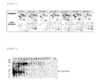
- FIG. 1 Figure 2 shows images of proteins separated by 2D gel electrophoresis using fibrotic and non-fibrotic cells from cirrhotic liver parenchyma from a total of 7 patients suffering from hepatitis C-associated liver cirrhosis.
- the circular marker shows tropomyosin (beta), the oval tag hMFAP4.
- the similarity of the images underlines the reproducibility of the results.
- the tropomyosin may also be sarcomere tropomyosin kappa, beta tropomyosin, TPM1 human tropomyosin or tropomyosin 4.
- the aforementioned hMFAP4 is the human microfibrillar associated protein 4.
- downregulated metabolic enzymes may also be used as markers, particularly argininosuccinate synthetase, methylenetetrahydrofolate dehydrogenase, fructose 1,6-bisphosphate aldolase, mitochondrial malate dehydrogenase, and mitochondrial acetoacetyl-CoA thiolase.
- Regucalcin also known as senescent marker protein-30 (SMP-30) plays an important role in maintaining the intracellular Ca2 + level by activating Ca2 + enzymes in the plasma membrane, microsomes and mitochondria. Regucalcin was 13-fold downregulated in fibrotic versus healthy liver cells. This may indicate that the compensatory effects of regucalcin on oxidative stress in diseased liver tissue are reduced.
- ROS reactive oxygen species
- ATP5A1 ATP synthase alpha subunit
- hepatitis C virus core protein and the NS5A protein are associated with membranes of the endoplasmic reticulum, the Golgi apparatus and an increase in intracellular lipid accumulation by interaction with apolipoprotein A1 (apoA1) or A2 (apoA2).
- the lipid transfer protein is correspondingly inhibited or the synthesis of VLDL (very low density lipoproteins) is disturbed.
- VLDL very low density lipoproteins
- Metabolic disorders in fibrotic liver are also manifested in decreased expression of glycollic enzymes such as fructose 1,6-bisphosphate aldolase (fold change: -16.6), enolase-1 (2-phosphodiglycerate hydrolase) or glyoxylase.
- glycollic enzymes such as fructose 1,6-bisphosphate aldolase (fold change: -16.6), enolase-1 (2-phosphodiglycerate hydrolase) or glyoxylase.
- serum albumin fold change: -13.2
- liver regeneration in fibrotic liver cells is apparently affected by a weaker expression of methylenetetrahydrofolate dehydrogenase (fold change: -9.4), which catalyzes three consecutive reactions in the conversion of C-1 derivatives of tetrahydrofolate.
- An undiminished enzymatic function is important for normal cellular function, growth and dedifferentiation.
- ROS reactive organic species
- the proteins actin alpha (fold change: 6.5) and actin gamma (fold change: 9.1) could be identified. These are major components of the thin filaments of muscle cells and the cytoskeleton of non-muscle cells. Actin is apparently a product of HSC cells and therefore occurs more frequently in hepatitis C-induced fibrosis.
- vimentin fold change: 4,6
- It is believed to be a product of hepatic stellate cells of mesenchyme.
- the 34 kDa protein calponin is also normally expressed specifically in smooth muscle cells and binds calmodulin, actin and tropomyosin. In view of the myofibroblast-like activated hepatic stellate cells, calponin was also up-regulated in fibrotic cells (fold change: 18.5), according to the proteome analysis.
- transgelin (fold change: 15) is believed to be a product of hepatic stellate cells.
- Transgelin is a 22 kDa protein, also referred to as SM22-alpha, and has structural similarities to calponin.
- the amyloid component P a glycoprotein composed of a pair of non-covalently bound pentamers, the subunits having a size of 23 to 25 kDa, was also more expressed in fibrosis than in healthy ones Tissue (fold change: 7.3).
- the physiological role in hepatic fibrogenesis is unknown, but overexpression indicates an abnormal cellular process.
- over- or under-expression of the proteins useful as biomarkers is due to impaired cellular balance, impaired mitochondrial and metabolic enzymes, decreased cellular synthesis, and enhanced expression of cytoskeletal proteins in the process of apoptosis in fibrotic liver cells.
- the gel electrophoresis is preferably an SDS-polyacrylamide gel electrophoresis.
- Corresponding gel analysis software is available from GE Healthcare, DeCyder Software, for example.
- the samples are preferably labeled with a dye before carrying out the 2D gel electrophoresis.
- the dyes are preferably fluorescent dyes. Particularly preferred is the use of Cy2, Cy3 and / or Cy5. These dyes are available, for example, from GE Healthcare, Freiburg, Germany. It is about Carbomethylindocyaninfarbstoffe, wherein two indole molecules are connected via a carbon chain with conjugated double bonds.
- the appropriately functionalized dyes can be reacted with the thiol groups of the side chains of the cysteine to covalently link the proteins to the dyes.
- the sample is first reduced with the aid of a suitable reducing agent, for example with the aid of tris (2-carboxyethyl) phosphine hydrochloride (TCEP). It is then reacted with appropriately functionalized dye until finally the reaction is stopped by the addition of DTT.
- a suitable reducing agent for example with the aid of tris (2-carboxyethyl) phosphine hydrochloride (TCEP).
- TCEP tris (2-carboxyethyl) phosphine hydrochloride
- DTT 2-carboxyethyl phosphine hydrochloride
- Cy3, Cy5 dye system is particularly advantageous in that, in addition to the actual sample, an internal standard can also be used in 2D gel electrophoresis, with the actual sample and the internal standard being provided with different dyes (Cy3 or Cy5).
- the detection and quantification of the proteins serving as markers can also be carried out with the aid of further protein diagnostic methods familiar to the person skilled in the art, in particular using radioactively or fluorescently labeled antibodies.
- suitable bioanalytical methods such as, for example, immunohistochemistry, Antibody arrays, Luminex, ELISA, immunofluorescence, radioimmunoassays.
- the detection and quantification of the proteins serving as markers can also be carried out with other suitable bioanalytical methods, such as mass spectrometric methods, for example MRM (Multi Reaction Monitoring) or AQUA (Absolute Quantification), with the aid of which the marker proteins can be quantitatively measured become.
- the sample used to detect the proteins may be a sample of liver tissue obtained by biopsy. However, it is also possible to use (full) blood, serum or plasma samples, which of course are easier to obtain.
- the invention also relates to the use of said proteins as biomarkers for detecting liver inflammation, in particular hepatic fibrosis.
- the aforementioned marker proteins according to the invention can also be used in other embodiments for differential diagnosis, in particular differential diagnostic early detection, prognosis of liver disease, assessment of severity, therapy-accompanying course assessment, aetiology and in-vitro diagnostics.
- the invention relates to a kit or diagnostic device for carrying out the method according to the invention, wherein the kit contains at least one biomarker according to the invention (also: marker protein) together with detection reagents and further auxiliaries.
- biomarker according to the invention also: marker protein
- liver parenchyma was harvested, immediately chilled on ice and stored at -86 ° C. Tissue samples were separated under the microscope between fibrotic tissue and healthy sections. The fibrotic material was taken along the fibrotic septum. The layers for 2D gel electrophoresis were stained with hematoxylin and stored at -20 ° C. The isolated cells from the microdissection were taken up in 100 ⁇ l lysis buffer (Tris HCl 30 mM, thiourea 2 M, urea 7 M, CHAPS 4%, pH 8.0) and then disrupted by use of ultrasound (6 ⁇ 10 s pulses).
- Centrifugation (12,000 g for 5 min) was used to remove DNA and other cell debris. The protein concentration of the lysate was determined.
- the cysteine amino acids of the proteins to be tested (3,500 fibrotic cells, 2,500 non-fibrotic cells) were also reduced by incubation with 2 nmol TCEP at 37 ° C in the dark for one hour before adding 4 nmol Cy5. After thorough mixing, the samples were reacted at 37 ° C in the dark for 30 minutes. The reaction was stopped by adding 10 ⁇ l of DTT.
- a 2D gel electrophoresis was carried out using a 21.25 h voltage gradient for isoelectric focusing.
- the equilibration buffer used was 125 mM Tris, 40% (w / v) glycerol, 3% (w / v) SDS, 65 mM DTT, pH 6.8 for 10 min.
- polyacrylamide gel electrophoresis was performed in the second dimension.
- the tropomyosin protein could be reproducibly identified in patient sera from liver cirrhosis patients of various origins ( FIG. 2 ).
- FIG. 2 Tropomyosin Western Blot of the following patient sera: 1: (Patient 1) Hepatitis C (HepC) Cirrhosis CHILD C 2: (Patient 2) HepC cirrhosis CHILD C 3: (Patient 3) HepC cirrhosis CHILD C 4: (Patient 4) HepC cirrhosis CHILD C 5: (Patient 5) HepC cirrhosis CHILD C 6: (Patient 6) HepC cirrhosis CHILD A 7: (Patient 7) HepC cirrhosis CHILD A 8th: (Patient 8) normal control 9: (Patient 9) normal control 10: (Patient 10) normal control 11: (Patient 11) normal control 12: (Patient 12) Ethytotoxic cirrhosis CHILD C 13: (Patient 13) Ethytotoxic cirrhosis CHILD C 14: (Patient
- the patients were hepatitis C infected cirrhotics and fibrosis patients, and patients with cystic acid liver disease and hepatitis B patients with hepatic fibrosis.
- Patients with cirrhosis of the liver were further subdivided into different CHILD classes, which provide information about the severity of cirrhosis by including various blood parameters.
- the classification is done by CHILD A with a survival rate of about 100% for the next year to CHILD C with a survival of about 30%.
- a tropomyosin band could not be detected in patient sera of healthy normal controls ( FIG. 2 , Vol 8-11).
- tropomyosin was detected in hepatitis C patients with a CHILD C classification ( FIG.
Landscapes
- Life Sciences & Earth Sciences (AREA)
- Health & Medical Sciences (AREA)
- Engineering & Computer Science (AREA)
- Molecular Biology (AREA)
- Chemical & Material Sciences (AREA)
- Biomedical Technology (AREA)
- Urology & Nephrology (AREA)
- Hematology (AREA)
- Immunology (AREA)
- Cell Biology (AREA)
- General Health & Medical Sciences (AREA)
- Biotechnology (AREA)
- Proteomics, Peptides & Aminoacids (AREA)
- Food Science & Technology (AREA)
- Medicinal Chemistry (AREA)
- Physics & Mathematics (AREA)
- Analytical Chemistry (AREA)
- Biochemistry (AREA)
- Microbiology (AREA)
- General Physics & Mathematics (AREA)
- Pathology (AREA)
- Measuring Or Testing Involving Enzymes Or Micro-Organisms (AREA)
- Investigating Or Analysing Biological Materials (AREA)
- Medicines That Contain Protein Lipid Enzymes And Other Medicines (AREA)
- Peptides Or Proteins (AREA)
- Medicines Containing Material From Animals Or Micro-Organisms (AREA)
Abstract
Description
- Die Erfindung betrifft ein Verfahren zur diagnostischen Untersuchung biologischer Proben eines Menschen auf Leberentzündung, insbesondere hepatische Fibrose und / oder Leberzirrhose, wobei die Probe auf eines oder mehrere Proteine als Marker für Leberentzündung, insbesondere hepatische Fibrose und / oder Leberzirrhose, untersucht wird, wobei eine gegenüber dem gesunden Zustand erhöhte oder verringerte Konzentration der Proteine das Vorliegen einer Leberentzündung, insbesondere einer hepatischen Fibrose und / oder Leberzirrhose, anzeigt.
- Weltweit sind ca. 170 Mio. Menschen chronisch mit dem Hepatitis C-Virus (HCV) infiziert. Der Verlauf der Krankheit variiert dabei zwischen den Patienten erheblich; während ca. 20 % der Patienten innerhalb von 20 Jahren eine Leberzirrhose entwickeln, ist bei anderen Patienten eine derartige Entwicklung selbst nach noch längeren Zeiträumen nicht zu beobachten. Es konnten eine Reihe von Faktoren identifiziert werden, die die Wahrscheinlichkeit einer hepatischen Fibrose und / oder Leberzirrhose erhöhen, u.a. männliches Geschlecht, Alkoholmissbrauch, Co-Infektion mit HIV oder Schistosoma mansoni, genetische Prädisposition und erhöhtes Alter bei der Infektion.
- Als Vorläuferzellen sind für die Entwicklung einer Leberfibrose und / oder Leberzirrhose vor allem die hepatischen Stelatzellen (HSC) verantwortlich, die in einer normalen Leber im Ruhezustand insbesondere für die Speicherung von Vitamin A verantwortlich sind. In einer fibrotischen Leber hingegen werden sie aktiviert, proliferieren und entwickeln sich zu myofibroblastartigen Zellen. Diese Myofibroblasten produzieren große Mengen Kollagen, regulieren die Produktion von Matrixmetalloproteinasen (MMPs) herab und zeigen eine erhöhte Expression der physiologischen Inhibitoren der MMPs (TIMPs). Mit zunehmender Kollagenakkumulation entwickelt sich die Fibrose der Leber weiter, was schließlich bis zum Organversagen führen kann.
- Zur antiviralen Behandlung einer chronischen Hepatitis C werden insbesondere Peg-Interferon Alfa und Ribavirin eingesetzt. Auch wenn auf diese Weise viele Patienten erfolgreich behandelt werden können, bleibt die Therapie bei mindestens 50 % der Patienten, die mit dem HCV Genotyp 1 infiziert sind, der in der westlichen Welt am weitesten verbreitet ist, erfolglos. Ähnliches gilt für Patienten, die mit dem HCV Genotyp 4 infiziert sind, der in Ägypten häufig vorkommt. Darüber hinaus sind die Kosten der dauerhaften antiviralen Behandlung immens und die Behandlung ist mit erheblichen Nebenwirkungen verbunden. Bei Patienten, die sich in einem weit fortgeschrittenen Stadium befinden, führt die antivirale Behandlung wiederum nicht mehr zum gewünschten Erfolg. Es besteht daher Bedarf, eine Fibrose bei Hepatitis-Patienten und damit die Entstehung einer Leberzirrhose besser diagnostizieren zu können, um dem behandelnden Arzt die Möglichkeit zu verschaffen, zu entscheiden, ob eine antivirale Behandlung sinnvoll und Erfolg versprechend ist.
- Bereits in der Vergangenheit wurde eine Reihe nicht-invasiver Marker zum Nachweis von Leberfibrose verwendet, darunter der sog. Acti- oder Fibro-Test, Prokollagen III-Peptid (PIIIP), Hyaluronsäure, Matrixmetalloproteinasen (MMPs) und ihre Inhibitoren (TIMPs) (T. Poynard et al., Expert Rev Mol Diagn. 2005, 5 (1): 15 - 21; V. Leroy et al., J Hep 2001, 35 (1):26). All diese Marker zeigen jedoch nur eine limitierte Sensibilität und Spezifität, weshalb weiterhin Bedarf nach geeigneteren Biomarkern besteht.
- Ausgehend vom beschriebenen Stand der Technik stellt sich daher die Aufgabe ein verbessertes Verfahren zur Untersuchung biologischer Proben auf Leberentzündung und / oder hepatische Fibrose und / oder Leberzirrhose zur Verfügung zu stellen, bei dem neuartige Marker verwendet werden.
- Die Aufgabe wird erfindungsgemäß gelöst durch ein Verfahren zur Untersuchung biologischer Proben eines Menschen auf Leberentzündung, insbesondere hepatische Fibrose und / oder Leberzirrhose, wobei die Probe auf eines oder mehrere Proteine als Marker für eine Leberentzündung, insbesondere hepatische Fibrose und / oder Leberzirrhose, untersucht wird und wobei ein erhöhter Level der Proteine das Vorliegen einer Leberentzündung, insbesondere hepatische Fibrose und / oder Leberzirrhose, anzeigt, wobei die Proteine ausgewählt sind aus einer Gruppe bestehend aus: ER60, Vimentin, Actin alpha 1 skeletales Muskelprotein, hMFAP4, Tropomyosin, PTGES2, Amyloid-P-Komponente, Transgelin, Calponin 1, Homo sapiens p20 protein, 17 kDa Myosin leichte Kette, H Chain H Igg B12, Prolyl 4-hydroxylase, beta subunit.
- Des Weiteren betrifft die Erfindung auch ein entsprechendes Verfahren bei dem ein verringerter Level der Proteine das Vorliegen einer Leberentzündung, insbesondere hepatische Fibrose und / oder Leberzirrhose, anzeigt, wobei in diesem Fall die Proteine ausgewählt sind aus einer Gruppe: Methylentetrahydrofolatdehydrogenase 1, PRO2619, Aldehyddehydrogenase 1, Fibrinogen alpha-Kette Preproprotein, Fructose-Bisphosphat-Aldolase B, Argininosuccinatsynthetase, Eef1a2, ATP5A1, alpha-2-Actin, Regucalcin, Serumalbumin, mitochondriale Malatdehydrogenase, mitochondriale Acetoacetyl-CoA-Thiolase.
- Sowohl bei den herauf als auch bei herunterregulierenden Proteinen kann die Untersuchung ebenfalls über die Bestimmung von Teilsequenzen der erfindungsgemäßen Biomarker (auch: Markerproteine) erfolgen. Insbesondere sind solche Teilsequenzen bevorzugt die 60 % der Aminosäuresequenz eines erfindungsgemäßen Biomarker, insbesondere 70 % und mehr, 80 % und mehr, insbesondere 90 bis 95 % umfassen.
- Im Rahmen dieser Erfindung umfasst der Begriff der Leberentzündung jede Form der Hepatitis, jedoch insbesondere die hepatische Fibrose bis hin zur Leberzirrhose (siehe zu dem Begriffen z.B. einschlägig Pschyrembel, Klinisches Wörterbuch, 260. Auflage 2004, Berlin). Die hepatische Fibrose und Leberzirrhose ist erfindungsgemäß bevorzugt.
- Ferner betrifft die Erfindung ebenfalls die Diagnose von Leberentzündungen, insbesondere hepatische Fibrose und / oder Leberzirrhose, wobei eine Bestimmung mindestens eines Proteins ausgewählt aus der Gruppe bestehend aus: ER60, Vimentin, Actin alpha 1 skeletales Muskelprotein, hMFAP 4, Tropomyosin, PTGES 2, Amyloid P-Komponente, Transgelin, Calponin 1, Homo sapiens p20 Protein, 17 kDa Myosin leichte Kette, H Chain H Igg B12, Prolyl 4-hydroxylase, beta subunit Methylentetrahydrofolatdehydrogenase 1, PR02619, Aldehyddehydrogenase 1, Fibrinogen alpha-Kette Preproprotein, Fructose-Bisphosphat-Aldolase B, Argininosuccinatsynthetase, Eefla2, ATP5A1, alpha-2-Actin, Regucalcin, Serumalbumin, mitochondriale Malatdehydrogenase, mitochondrialer Acetoacetyl-CoA-Thiolase oder jeweils eine Teilsequenz davon an einem zu untersuchenden Patienten durchgeführt wird.
- Des Weiteren ist eine Kombination solcher erfindungsgemäßen Biomarker bzw. Markerproteine zur erfindungsgemäßen Diagnose möglich.
- Die genannten Proteine konnten bei einer Proteomanalyse von fibrotischem Gewebe im Vergleich zu nicht-fibrotischem Gewebe als potentielle Biomarker identifiziert werden. Hierzu wurden mit Hepatitis C infizierten Patienten Leber-Biopsieproben entnommen. Die Proben wurden in einem Handhomogenisator mit Lysispuffer homogenisiert und von DNA und sonstigem Zellmaterial befreit, um ein Proteinkonzentrat zu erhalten. Die Proteine wurden mit einem Farbstoff gelabelt und einer 2D-Polyacrylamid-Gelelektrophorese unterworfen mit einer isoelektrischen Fokussierung in der ersten und einer SDS-Gelelektrophorese in der zweiten Dimension. Die Ergebnisse wurden für fibrotische und nicht-fibrotische Zellen mit Hilfe einer dafür geeigneten Software verglichen, um die Spots zu detektieren und zu quantifizieren, die bei der fibrotischen Probe im Vergleich zur nicht-fibrotischen Probe verstärkt oder verringert waren. Als Software kann beispielsweise die ImageQuant™-Software der Firma GE Healthcare in Verbindung mit der DeCyder-Software derselben Firma durchgeführt werden. Dabei wird die Emission der Farbstoffe, mit denen die Proteine gelabelt wurden, gemessen und ausgewertet.
- Die weitere Auswertung erfolgte mit Hilfe von LC-ESI-MS (Flüssigchomatographie-Elektrosprayionisations-Massenspektrometrie). Dabei wurden zunächst die Proteine im Gel, in dem die Proben zuvor aufgetrennt wurden, mit Hilfe von Trypsin in einzelne Peptidfragmente zerlegt. Diese wurden mit Hilfe von Reversed-Phase HPLC voneinander separiert und massenspektrometrisch untersucht, um die einzelnen Proteine zu identifizieren. Selbstverständlich können hierbei auch andere geeignete massenspektrometrische Verfahren angewandt werden, beispielsweise MALDI-TOF-MS.
- Bei den Untersuchungen konnten folgende Proteine identifiziert werden, die in fibrotischen Zellen gegenüber nicht-fibrotischen Zellen herauf- (fold change positiv) bzw. herunterreguliert (fold change negativ) wurden:
-
NCBI-Accession Identifiziertes Protein Fold change IPI00025252.1 ER60 Protein 26,9 IPI00418471.5 Vimentin 5,6 IPI00448938.1 H Chain H Igg B12 14.7 IPI00697648.1 Actin alpha 1 skeletales Muskelprotein 6,5 IPI00022792.3 hMFAP 4 45,1 IPI00455050.1 sarkomeres Tropomyosin kappa 87,1 IPI00014581.1 TPM1 humanes Tropomyosin alpha-Kette 28,7 IPI00220709.3 beta Tropomyosin 52,4 IPI00010779.3 Tropomyosin 4 20,6 IPI00303568.3 PTGES 2 6,1 IPI00010796.1 Prolyl 4-hydroxylase, beta subunit 4.6 IPI00022391.1 Amyloid P-Komponente, Serum 7,3 IPI00216138.5 Transgelin 15 IPI00021264.1 Calponin 1 21,1 IPI00022433.5 Homo sapiens p20 Protein [pir B53814] 16,9 IPI00718271.2 17 kDa Myosin leichte Kette 8,4 -
NCBI-Accession Identifiziertes Protein Fold change IPI00218342.9 Methylentetrahydrofolatdehydrogenase 1 - 9,37 IPI00745872.1 PR02619 - 3,2 IPI00218914.4 Aldehyddehydrogenase 1 - 7,7 IPI00029717.1 Fibrinogen alpha-Kette Preprotein - 6,4 IPI00218407.5 Fructose-Bisphosphat-Aldolase B - 16,6 IPI00020632.4 Argininosuccinatsynthetase - 11,0 IPI00014424.1 Eefla2 - 6,4 IPI00440493.2 ATP5A1 Protein - 5,8 IPI00708487.1 alpha-2-Actin; alpha cardiac Actin - 8,6 IPI00017551.1 Regucalcin (Seneszenzmarkerprotein 30) - 13,3 IPI00708398.1 ABBOS-Serumalbuminprecursor -13,3 IPI00291006.1 mitochondrialer Malatdehydrogenaseprecursor - 5,9 IPI00030363.1 mitochondrialer Acetoacetyl-CoA-Thiolaseprecursor - 5,9 NCBI: National Centre for Biotechnology Information - Im Folgenden werden die Sequenzinformationen zu einigen der identifizierten Proteine angegeben. Dabei sind die Peptidsequenzen dunkel unterlegt, die zum einen zur Identifikation der Proteine führten und zum anderen eine Unterscheidung der Proteinisoformen zulassen (vgl. auch Sequenzprotokoll (sequence listing) als Anhang zur Patentanmeldung).
-


-

- SEQ ID NO 1: sarkomeres Tropomyosin kappa, TPM1-kappa; NCBI Accession: IPI00455050.1
- SEQ ID NO 2: sarkomeres Tropomyosin kappa; NCBI Accession: IPI00455050.1
- SEQ ID NO 3: beta Tropomyosin; NCBI Accession: IPI00220709.3
- SEQ ID NO 4: TPM 1 humanes Tropomyosin 1 alpha - Kette; NCBI Accession: IPI00014581.1
- SEQ ID NO 5: Tropomyosin 4; NCBI Accession: IPI00010779.3
- SEQ ID NO 6: Transgelin; NCBI Accession: IPI00216138.5
- SEQ ID NO 7: Transgelinvariante; NCBI Accession: IPI00216138.5
- In
Figur 1 sind Aufnahmen der mittels 2D-Gelelektrophorese aufgetrennten Proteine gezeigt, wobei fibrotische und nicht-fibrotische Zellen aus zirrhotischem Leberparenchym von insgesamt 7 Patienten verwendet wurden, die unter mit Hepatitis C verbundener Leberzirrhose litten. Die kreisrunde Markierung zeigt Tropomyosin (beta), die ovale Markierung hMFAP4. Die Ähnlichkeit der Aufnahmen unterstreicht die Reproduzierbarkeit der Ergebnisse. - Als besonders erfolgversprechende und bevorzugte erfindungsgemäße Biomarker haben sich Tropomyosin, Transgelin, Calponin, hMFAP4 und Vimentin erwiesen. Bei dem Tropomyosin kann es sich weiterhin um sarkomeres Tropomyosin kappa, beta Tropomyosin, TPM1 humanes Tropomyosin oder Tropomyosin 4 handeln.
- Bei dem erwähnten hMFAP4 handelt es sich um das humane mikrofibrilar assoziierte Protein 4. Neben den zuvor erwähnten hochregulierten Proteinen, können jedoch auch herunterregulierte metabolische Enzyme als Marker verwendet werden, insbesondere Argininosuccinatsynthetase, Methylentetrahydrofolatdehydrogenase, Fructose-1,6-Bisphosphataldolase, mitochondriale Malatdehydrogenase und mitochondriale Acetoacetyl-CoA-Thiolase.
- Auf einige der identifizierten Proteine wird im folgenden kurz eingegangen, wobei es sich bei den dort angegebenen Erläuterungen lediglich um Erklärungsversuche handelt, die hinsichtlich des Patentbegehrens in keiner Weise beschränkend zu verstehen sind.
- Regucalcin, auch als Seneszensmarkerprotein-30 (SMP-30) bekannt, spielt eine bedeutende Rolle bei der Aufrechterhaltung des intrazellulären Ca2+-Niveaus durch Aktivierung von Ca2+-Enzymen in der Plasmamembran, den Mikrosomen und Mitochondrien. Regucalcin wurde 13-fach herabreguliert in fibrotischen im Vergleich zu gesunden Leberzellen. Dies könnte darauf hindeuten, dass die kompensatorischen Effekte von Regucalcin gegenüber oxidativem Stress in erkranktem Lebergewebe vermindert werden.
- Mitochondriale Malatdehydrogenase und mitochondriale Acetoacetyltransferase wurden beide um das 5,9-fache im fibrotischen Lebergewebe herunterreguliert. Es konnte gezeigt werden, dass die Wechselwirkung zwischen Hepatitis C-Virus-Coreprotein und den Proteinen NS3 und NS5 mit Mitochondrien zur Ausbildung reaktiver Sauerstoffspezies (ROS) führt, was eine Erklärung für die schwächere Expression der genannten Enzyme in fibrotischen Zellen sein könnte.
- Ein weiteres Anzeichen für gestörte mitochondriale Enzyme in fibrotischen Leberzellen ist die verringerte Expression der ATP-Synthase alpha-Untereinheit (ATP5A1), die die ATP-Synthese während der oxidativen Phosphorylierung katalysiert.
- Weitere Effekte des Hepatitis C-Virus-Coreproteins und des NS5A-Proteines sind die Assoziierung mit Membranen des endoplasmatischen Reticulums, des Golgi-Apparates und eine Verstärkung der intrazellulären Lipidakkumulation durch Wechselwirkung mit Apolipoprotein A1 (apoA1) oder A2 (apoA2). Das Lipidtransferprotein wird entsprechend inhibiert oder die Synthese von VLDL (very low density lipoproteins) wird gestört. Es konnte eine signifikante Herabregulierung der Acetyl-CoA-Acetyltransferase in fibrotischen Leberzellen beobachtet werden (fold change: -5,88).
- Metabolische Störungen in fibrotischer Leber manifestieren sich auch in einer herabgesetzten Expression von glykolitischen Enzymen wie Fructose-1,6-Bisphosphataldolase (fold change: -16,6), Enolase-1 (2-Phosphodiglycerathydrolase) oder Glyoxylase.
- Aufgrund dieser metabolischen Disfunktionen ist die Proteinsynthese in fibrotischen Leberzellen teilweise deutlich reduziert: Serumalbumin (fold change: -13,2), welches als Carrier für Fettsäuren, Steroide und Thyroidhormone fungiert und das extrazelluläre Flüssigkeitsvolumen stabilisiert, wurde nur noch vermindert exprimiert.
- Der Prozess der Leberregeneration in fibrotischen Leberzellen wird offenbar durch eine schwächere Expression von Methylentetrahydrofolatdehydrogenase beeinträchtigt (fold change: -9,4), welche drei aufeinanderfolgende Reaktionen bei der Umwandlung von C-1-Derivaten von Tetrahydrofolat katalysiert. Eine unverminderte enzymatische Funktion ist für die normale zelluläre Funktion, das Wachstum und die Dedifferenzierung bedeutsam.
- Insgesamt scheint eine Störung des zellulären Gleichgewichtes, der glykolytischen Reaktionswege und der Lipidkompartimentierung und des Metabolismus in fibrotischen Zellen vorzuliegen, was die Produktion von reaktiven organischen Spezies (ROS) begünstigt. Diese wiederum induzieren die Synthese von TGF-β1 in Hepatozyten und hepatischen Stelatzellen, den stärksten Promotor von hepatischer Fibrogenese.
- Im Bereich der verstärkt nachweisbaren Proteine in fibrotischem Gewebe konnten die Proteine Actin alpha (fold change: 6,5) und Actin gamma (fold change: 9,1) identifiziert werden. Es handelt sich dabei um Hauptkomponenten der dünnen Filamente von Muskelzellen und des Zytoskeletons von Nicht-Muskelzellen. Actin ist offenbar ein Produkt der HSC-Zellen und tritt daher bei Hepatitis C-induzierter Fibrose verstärkt auf.
- Ein weiteres identifiziertes Protein ist das Vimentin (fold change: 4,6). Es handelt sich hierbei vermutlich um ein Produkt der hepatischer Stelatzellen aus Mesenchym.
- Verschiedene Tropomyosin-Isoformen, die offenbar nicht durch Hepatozyten aber durch myofibroblastartige hepatische Stelatzellen produziert werden, weisen z. T. die höchsten Raten an Verstärkung auf (fold change: 9,7 bis 83,1).
- Das 34 kDa Protein Calponin wird normalerweise ebenfalls spezifisch in glatten Muskelzellen exprimiert und bindet Calmodulin, Actin und Tropomyosin. Angesichts der myofibroblastartigen aktivierten hepatischen Stelatzellen wurde gemäß Ergebnis der Proteomanalyse auch Calponin in fibrotischen Zellen heraufreguliert (fold change: 18,5).
- Auch Transgelin (fold change: 15) ist vermutlich ein Produkt von hepatischen Stelatzellen. Transgelin ist ein 22 kDa Protein, das auch als SM22-alpha bezeichnet wird, und weist strukturelle Ähnlichkeiten mit Calponin auf. Die Amyloidkomponente P, ein Glykoprotein, das sich aus einem Paar nicht kovalent gebundener Pentamere zusammensetzt, wobei die Untereinheiten eine Größe von 23 bis 25 kDa aufweisen, wurde ebenfalls bei Fibrose stärker exprimiert als in gesundem Gewebe (fold change: 7,3). Die physiologische Funktion in hepatischer Fibrogenese ist bislang unbekannt, aber die Überexpression deutet auf einen abnormalen zellulären Prozess hin.
- Zusammengefasst läßt sich sagen, dass die Über- bzw. Unterexpression der als Biomarker verwendbaren Proteine auf ein gestörtes zelluläres Gleichgewicht, beeinträchtigte mitochondriale und metabolische Enzyme, verminderte zelluläre Synthese und eine verstärkte Expression zytoskeletaler Proteine beim Prozess der Apoptose in fibrotischen Leberzellen zurückzuführen ist.
- Bei der Untersuchung einer Probe auf die als Biomarker dienenden Proteine kann, wie oben beschrieben, so vorgegangen werden, dass mit Hilfe einer 2D-Gelelektrophorese, bestehend aus isoelektrischer Fokussierung in der ersten Dimension und Gelelektrophorese in der zweiten Dimension eine Auftrennung der Proteine erfolgt und durch Vergleich des Proteinmusters mit einer nicht-fibrotischen Kontrollprobe die Über- bzw. Unterexpression bestimmter Proteine nachgewiesen wird. Bei der Gelelektrophorese handelt es sich bevorzugt um eine SDS-Polyacrylamid-Gelelektrophorese. Entsprechende Software zur Auswertung der Gele ist beispielsweise von GE Healthcare in Form der DeCyder Software erhältlich.
- Zum Nachweise der Proteine werden die Proben vor Durchführung der 2D-Gelelektrophorese vorzugsweise mit einem Farbstoff gelabelt. Bei den Farbstoffen handelt es sich vorzugsweise um Fluoreszenzfarbstoffe. Besonders bevorzugt ist der Einsatz von Cy2, Cy3 und/oder Cy5. Diese Farbstoffe sind beispielsweise von der Firma GE Healthcare, Freiburg, Deutschland, erhältlich. Es handelt sich dabei um Carbomethylindocyaninfarbstoffe, wobei zwei Indolmoleküle über eine Kohlenstoffkette mit konjugierten Doppelbindungen verbunden sind. Die entsprechend funktionalisierten Farbstoffe können beispielsweise mit den Thiolgruppen der Seitenketten des Cysteins zur Reaktion gebracht werden, um die Proteine mit den Farbstoffen kovalent zu verknüpfen. Zwecks Reduktion von Disulfidbrücken wird dabei die Probe zunächst mit Hilfe eines geeigneten Reduktionsmittels reduziert, beispielsweise mit Hilfe von Tris(2-Carboxyethyl)phosphinhydrochlorid (TCEP). Anschließend wird mit entsprechend funktionalisiertem Farbstoff umgesetzt, bis schließlich die Reaktion durch Zugabe von DTT gestoppt wird. Denkbar ist jedoch auch die Funktionalisierung anderer Aminosäurereste mit Hilfe der Farbstoffe, beispielsweise der Seitenkette des Lysins.
- Die Verwendung des Cy3, Cy5-Farbstoffsystems ist insofern besonders vorteilhaft als neben der eigentlichen Probe auch ein interner Standard bei der 2D-Gelelektrophorese eingesetzt werden kann, wobei die eigentliche Probe und der interne Standard mit unterschiedlichen Farbstoffen (Cy3 bzw. Cy5) versehen werden.
- Selbstverständlich sind neben den genannten Farbstoffen auch andere (Fluoreszenz)farbstoffe einsetzbar, die aus dem Stand der Technik sind, beispielsweise Fluorescein oder Tetramethylrhodamin.
- Der Nachweis und die Quantifizierung der als Marker dienenden Proteine kann auch mit Hilfe weiterer, dem Fachmann geläufiger Protein-Diagnoseverfahren durchgeführt werden, insbesondere unter Verwendung radioaktiv oder fluoreszenzmarkierter Antikörper. Zu nennen sind hier insbesondere dazu geeignete bioanalytische Verfahren, wie zum Beispiel Immunhistochemie, Antikörperarrays, Luminex, ELISA, Immunfluoreszenz, Radioimmunoassays. Der Nachweis und die Quantifizierung der als Marker dienenden Proteine kann auch mit weiteren dazu geeigneten bioanalytischen Verfahren, wie zum Beispiel massenspektrometrischen Verfahren, z.B. MRM (Multi reaction monitoring) oder AQUA (absolute Quantification), mit deren Hilfe die Markerproteine quantitativ gemessen werden können, durchgeführt werden.
- Bei der zum Nachweis der Proteine verwendeten Probe kann es sich um eine Probe von Lebergewebe handeln, die mit Hilfe einer Biopsie entnommen wurde. Möglich ist jedoch auch die Verwendung von (Voll-)Blut-, Serum- oder Plasmaproben, welche selbstverständlich leichter zu gewinnen sind.
- Neben den beschriebenen Verfahren betrifft die Erfindung auch die Verwendung der genannten Proteine als Biomarker zum Nachweis einer Leberentzündung insbesondere einer hepatischen Fibrose.
- Die genannten erfindungsgemäßen Markerproteine können in weiteren Ausführungsformen ebenfalls zur Differentialdiagnose, insbesondere differentialdiagnostischen Früherkennung, Verlaufsprognose der Lebererkrankung, Beurteilung des Schweregrades, therapiebegleitenden Verlaufsbeurteilung, Ätiologie und in-vitro Diagnostik eingesetzt werden.
- In einer weiteren Ausführungsform betrifft die Erfindung einen Kit oder diagnostische Vorrichtung zur Durchführung des erfindungsgemäßen Verfahrens, wobei der Kit mindestens einen erfindungsgemäßen Biomarker (auch: Markerprotein) samt Nachweisreagenzien und weitere Hilfsmittel enthält. Nachfolgende Beispiele dienen zur Erläuterung der Erfindung, ohne die Erfindung auf diese Beispiele zu beschränken.
- Von insgesamt sieben Patienten mit einer Hepatitis C-Infektion, Genotyp 1, die sich einer Lebertransplantation unterzogen, wurde Leberparenchym entnommen, sofort auf Eis gekühlt und bei -86°C gelagert. Unter dem Mikroskop wurden Gewebeproben aufgetrennt zwischen fibrotischem Gewebe und gesunden Abschnitten. Das fibrotische Material wurde dabei entlang des fibrotischen Septums entnommen. Die Schichten für die 2D-Gelelektrophorese wurden mit Haematoxylin angefärbt und bei -20°C gelagert. Die isolierten Zellen aus der Mikrodissektion wurden in 100 µl Lysispuffer (Tris HCl 30 mM; Thioharnstoff 2 M; Harnstoff 7 M, CHAPS 4%; pH 8,0) aufgenommen und anschließend durch Anwendung von Ultraschall aufgebrochen (6 x 10 s Pulse).
- Mit einer Zentrifugation (12.000 g für 5 min) wurden DNA und sonstige Zellreste entfernt. Die Proteinkonzentration des Lysats wurde ermittelt.
- Zur Anfertigung eines internen Standards wurden 3 µg des Gewebelysates durch Anwendung von 2 nmol Tris-(2-Carboxyethyl)-phosphinhydrochlorid (TCEP) bei 37°C im Dunkeln über eine Zeitspanne von einer Stunde reduziert. Cy-Farbstoffe (GE Healthcare, Freiburg, Deutschland) wurden mit wasserfreiem DMF p. a. (2 nmol/pl) verdünnt und 4 nmol Cy3 wurden den mit Hilfe von TCEP reduzierten Proben zugegeben. Nach einer Inkubationszeit von 30 min bei 37°C wurde die Labeling-Reaktion durch Zugabe von 4 µl DTT (1,08 g/ml) gestoppt.
- Die Cystein-Aminosäuren der zu untersuchenden Proteine (3.500 fibrotische Zellen, 2.500 nicht-fibrotische Zellen) wurden ebenfalls durch Inkubation mit 2 nmol TCEP bei 37°C im Dunkeln für eine Stunde reduziert, bevor 4 nmol Cy5 hinzugegeben wurden. Nach gründlicher Durchmischung wurden die Proben 30 min lang bei 37°C im Dunkeln umgesetzt. Die Reaktion wurde gestoppt durch Zugabe von 10 µl DTT.
- Zur Vorbereitung der isoelektrischen Fokussierung wurden 10 µl Ampholine 2-4 (GE Healthcare) hinzugefügt. Die Cy3- und Cy5-markierten Zellen wurden nach Durchmischung gleichzeitig weiter verarbeitet.
- Anschließend wurde eine 2D-Gelelektrophorese durchgeführt, wobei zur isoelektrischen Fokussierung ein 21,25 h Spannungsgradient verwendet wurde. Als Äquilibrierungspuffer wurde 125 mM Tris, 40 % (w/v) Glycerol, 3 % (w/v) SDS, 65 mM DTT, pH 6,8 für 10 min verwendet. Anschließend wurde in der zweiten Dimension eine Polyacrylamidgelektrophorese durchgeführt.
- Mit Hilfe eines geeigneten Scanners (Typhoon 9400, GE Healthcare) wurden nach beendeter Gelelektrophorese Bilder aufgenommen und mit Hilfe der ImageQuant-Software und der DeCyder-Software (GE Healthcare) ausgewertet. Auf diese Weise konnte eine differenzielle Analyse im Gel (DIA) durchgeführt werden, um die einzelnen Spots zu detektieren und zu quantifizieren.
- Mit Hilfe des Enzyms Trypsin in 10 mM Ammoniumbicarbonatpuffer (pH 7,8) wurden die Proteine bei 37°C über Nacht gespalten. Die auf diese Weise generierten Fragmente wurden zweifach mit einem Acetonitril-Ameisensäuregemisch extrahiert zur weiteren Auftrennung und massenspektrometrischen Untersuchung. Dies erfolgte mit Hilfe von Online-RP-Kapillar-HPLC, gekoppelt mit nano-ESI-MS (Elektrosprayionisations-Massenspektrometrie). Mit Hilfe der HPLC-MS-Kopplung und unter Heranziehung geeigneter Proteindatenbanken (NCBI, National Centre for Biotechnology Information) konnte eine vollständige Identifizierung der Proteinspots herbeigeführt werden.
- Hierfür wurde eine Proteindetektion mittels Western Blot-Analyse durchgeführt. Das Tropomyosin-Protein konnte in Patientenseren von Leberzirrhose- Patienten unterschiedlicher Genese reproduzierbar identifiziert werden (
Figur 2 ). -
Figur 2 : Tropomyosin Western Blot von folgenden Patientenseren:1: (Patient 1) Hepatitis C (HepC) Zirrhose CHILD C 2: (Patient 2) HepC Zirrhose CHILD C 3: (Patient 3) HepC Zirrhose CHILD C 4: (Patient 4) HepC Zirrhose CHILD C 5: (Patient 5) HepC Zirrhose CHILD C 6: (Patient 6) HepC Zirrhose CHILD A 7: (Patient 7) HepC Zirrhose CHILD A 8: (Patient 8) Normalkontrolle 9: (Patient 9) Normalkontrolle 10: (Patient 10) Normalkontrolle 11: (Patient 11) Normalkontrolle 12: (Patient 12) Äthyltoxische Zirrhose CHILD C 13: (Patient 13) Äthyltoxische Zirrhose CHILD C 14: (Patient 14) HepB Fibrose 15: (Patient 15) HepC Fibrose 16: Tropomyosin 0,005µg - Bei den Patienten handelte es sich zum einen um Hepatitis C infizierte Patienten mit Leberzirrhose bzw. Fibrose, zum anderen um Patienten mit äthyltoxischer Leberzirrhose sowie einem Hepatitis B Patienten mit Leberfibrose. Patienten mit Leberzirrhose wurden weiter in verschiedene CHILD-Klassen unterteilt, welche Aufschluss über den Schweregrad der Zirrhose, durch Einbezug verschiedener Blutparameter, geben. Die Klassifizierung erfolgt von CHILD A mit einer Überlebensrate von ca. 100% für das nächste Jahr bis CHILD C mit einer Überlebensrate von ca. 30%. Eine Tropomyosinbande konnte in Patientenseren gesunder Normalkontrollen nicht detektiert werden (
Figur 2 , Bande 8-11). Interessanterweise konnte Tropomyosin in Hepatitis C Patienten mit einer CHILD C Klassifizierung nachgewiesen werden (Figur 2 , Bande 1-5), während bei Zirrhotikern mit einer CHILD A Zirrhose keine Proteinbande detektierbar war (Figur 2 , Bande 6-7). Zusätzlich wurden äthyltoxische Leberzirrhosen untersucht (CHILD C Klassifizierung), die ebenfalls ein Signal für Tropomyosin in abgeschwächter Form zeigten und eine CHILD C Klassifizierung aufwiesen (Figur 2 , Bande 12-13). Weiterhin konnte im Hepatitis B Fibrosepatienten eine Tropomyosinbande detektiert werden, was im Hepatitis C Fibrosepatienten nicht möglich war.


Claims (15)
- In-vitro Verfahren zur Untersuchung biologischer Proben eines Menschen auf Leberentzündung insbesondere hepatische Fibrose und / oder Leberzirrhose,
wobei die Probe auf eines oder mehrere Proteine als Marker für Leberentzündung, insbesondere hepatische Fibrose und / oder Leberzirrhose, untersucht wird, wobei eine gegenüber dem gesunden Zustand erhöhte Konzentration der Proteine das Vorliegen einer Leberentzündung, insbesondere einer hepatischen Fibrose und / oder Leberzirrhose, anzeigt,
dadurch gekennzeichnet,
dass die Proteine ausgewählt sind aus der Gruppe bestehend aus: Transgelin, hMFAP 4, Tropomyosin, Vimentin, Actin alpha 1 skeletales Muskelprotein, Calponin 1 oder jeweils eine Teilsequenz davon. - Verfahren nach Anspruch 1, wobei die Proteine eine Kombination aus Transgelin und Tropomyosin oder Transgelin und Tropomyosin und hMFAP 4 oder / und Calponin 1 ist.
- Verfahren nach Anspruch 1 oder 2, dadurch gekennzeichnet, dass das Tropomyosin: sarkomeres Tropomyosin kappa, beta Tropomyosin, TPM 1 humanes Tropomyosin oder Tropomyosin 4 oder eine Kombination davon ist.
- Verfahren zur Diagnose von Leberentzündungen, insbesondere hepatische Fibrose und / oder Leberzirrhose,
dadurch gekennzeichnet,
dass eine Bestimmung mindestens eines Proteins ausgewählt aus der Gruppe bestehend aus: Transgelin, hMFAP 4, Tropomyosin, Vimentin, Actin alpha 1 skeletales Muskelprotein, Calponin 1 oder jeweils eine Teilsequenz davon oder jeweils eine Teilsequenz davon von einem zu untersuchenden Patienten durchgeführt wird. - Verfahren nach einem der Ansprüche 1 bis 4, dadurch gekennzeichnet, dass die Untersuchung der Probe mit Hilfe eines Diagnostikums durchgeführt wird, das dazu geeignete bioanalytische Verfahren, wie zum Beispiel Immunhistochemie, Antikörperarrays, Luminex, ELISA, Immunfluoreszenz, Radioimmunoassays verwendet.
- Verfahren nach einem der Ansprüche 1 bis 5, dadurch gekennzeichnet, dass die Untersuchung der Probe mittels massenspektrometrischer Verfahren wie MRM (Multi reaction monitoring) oder AQUA (absolute Quantification) erfolgt, mit deren Hilfe die Markerproteine quantitativ bestimmbar sind.
- Verfahren nach einem der Ansprüche 1 bis 6, dadurch gekennzeichnet, dass die Untersuchung der Probe mit Hilfe einer 2D-Elektrophorese erfolgt, wobei in der ersten Dimension eine isoelektrische Fokussierung, in der zweiten Dimension eine Gelelektrophorese durchgeführt wird.
- Verfahren nach Anspruch 7, dadurch gekennzeichnet, dass die Gelelektrophorese eine SDS-Polyacrylamid-Gelelektrophorese ist.
- Verfahren nach Anspruch 7 oder 8, dadurch gekennzeichnet, dass die Proben vor Durchführung der 2D-Gelelektrophorese mit einem Farbstoff gelabelt werden.
- Verfahren nach Anspruch 9, dadurch gekennzeichnet, dass der Farbstoff Cy2, Cy3 und/oder Cy5 ist.
- Verfahren nach einem der Ansprüche 1 bis 10, dadurch gekennzeichnet, dass die Probe eine Leberbiopsieprobe ist.
- Verfahren nach einem der Ansprüche 1 bis 11, dadurch gekennzeichnet, dass die Probe aus Blutserum, Blutplasma oder (Voll)Blut erhalten wird.
- Verwendung von Transgelin, hMFAP 4, Tropomyosin, Vimentin, Actin alpha 1 skeletales Muskelprotein, Calponin 1 als Marker zum Nachweis einer Leberentzündung, insbesondere einer hepatischen Fibrose und / oder Leberzirrhose.
- Verwendung mindestens eines der in Anspruch 13 genannten Markerproteine zur Differentialdiagnose, insbesondere differentialdiagnostischer Früherkennung, Verlaufsprognose der Lebererkrankung, Beurteilung des Schweregrades, therapiebegleitende Verlaufsbeurteilung, Ätiologie und in-vitro Diagnostik.
- Kit oder diagnostische Vorrichtung enthaltend mindestens ein Markerprotein nach Anspruch 13 samt Nachweisreagenzien und weitere Hilfsmittel.
Applications Claiming Priority (4)
| Application Number | Priority Date | Filing Date | Title |
|---|---|---|---|
| DE102006037613 | 2006-08-10 | ||
| DE102006048249A DE102006048249A1 (de) | 2006-08-10 | 2006-10-12 | Biomarker für Leberentzündung |
| EP07801230A EP2052254B1 (de) | 2006-08-10 | 2007-08-10 | Biomarker für leberentzündung |
| EP11175127A EP2388595A3 (de) | 2006-08-10 | 2007-08-10 | Biomarker für Leberentzündung |
Related Parent Applications (2)
| Application Number | Title | Priority Date | Filing Date |
|---|---|---|---|
| EP07801230.9 Division | 2007-08-10 | ||
| EP11175127.7 Division | 2011-07-24 |
Publications (2)
| Publication Number | Publication Date |
|---|---|
| EP2437065A2 true EP2437065A2 (de) | 2012-04-04 |
| EP2437065A3 EP2437065A3 (de) | 2012-08-29 |
Family
ID=38922186
Family Applications (5)
| Application Number | Title | Priority Date | Filing Date |
|---|---|---|---|
| EP11187569.6A Not-in-force EP2437064B1 (de) | 2006-08-10 | 2007-08-10 | Biomarker für Leberentzündung |
| EP11187588A Withdrawn EP2437066A3 (de) | 2006-08-10 | 2007-08-10 | Biomarker für Leberentzündung |
| EP11175127A Withdrawn EP2388595A3 (de) | 2006-08-10 | 2007-08-10 | Biomarker für Leberentzündung |
| EP07801230A Not-in-force EP2052254B1 (de) | 2006-08-10 | 2007-08-10 | Biomarker für leberentzündung |
| EP11187578A Withdrawn EP2437065A3 (de) | 2006-08-10 | 2007-08-10 | Biomarker für Leberentzündung |
Family Applications Before (4)
| Application Number | Title | Priority Date | Filing Date |
|---|---|---|---|
| EP11187569.6A Not-in-force EP2437064B1 (de) | 2006-08-10 | 2007-08-10 | Biomarker für Leberentzündung |
| EP11187588A Withdrawn EP2437066A3 (de) | 2006-08-10 | 2007-08-10 | Biomarker für Leberentzündung |
| EP11175127A Withdrawn EP2388595A3 (de) | 2006-08-10 | 2007-08-10 | Biomarker für Leberentzündung |
| EP07801230A Not-in-force EP2052254B1 (de) | 2006-08-10 | 2007-08-10 | Biomarker für leberentzündung |
Country Status (9)
| Country | Link |
|---|---|
| US (2) | US8535896B2 (de) |
| EP (5) | EP2437064B1 (de) |
| CN (2) | CN103630693A (de) |
| AT (1) | ATE518143T1 (de) |
| AU (1) | AU2007283312B2 (de) |
| CA (1) | CA2660257A1 (de) |
| DE (1) | DE102006048249A1 (de) |
| HK (1) | HK1135184A1 (de) |
| WO (1) | WO2008017303A2 (de) |
Families Citing this family (9)
| Publication number | Priority date | Publication date | Assignee | Title |
|---|---|---|---|---|
| DE102006048249A1 (de) * | 2006-08-10 | 2008-02-14 | Wolff Prof. Dr. Schmiegel | Biomarker für Leberentzündung |
| EP2302359A1 (de) | 2009-09-24 | 2011-03-30 | Université De Reims Champagne-Ardenne | Serum-Infrarotspektroskopie zur nicht-invasiven Beurteilung von Leberfibrose in Patienten mit chronischer Lebererkrankung |
| EP2405271A1 (de) * | 2010-07-06 | 2012-01-11 | Bio-Rad Innovations | Markers der Verletzlichkeit von Atherosklerotischem Plaque |
| CN102507936B (zh) * | 2011-11-09 | 2013-10-23 | 北京正旦国际科技有限责任公司 | 一种肝癌标志物多抗免疫质谱试剂盒 |
| CN103808944B (zh) * | 2014-03-07 | 2016-04-20 | 高平 | 生物标志物vwf和adamts13及其在肝硬化诊断试剂中的用途 |
| CN105785039B (zh) * | 2016-04-01 | 2017-08-04 | 北京理工大学 | 阶梯式定量检测血浆中血清白蛋白及纤维蛋白原的方法 |
| CN108990420B (zh) * | 2016-05-29 | 2022-06-24 | 深圳市绘云生物科技有限公司 | 肝病相关生物标志物和其使用方法 |
| GB201614455D0 (en) * | 2016-08-24 | 2016-10-05 | Univ Oxford Innovation Ltd | Biomarkers |
| CN108572251B (zh) * | 2018-05-31 | 2020-01-17 | 中国科学院昆明动物研究所 | 肝硬化早期小分子标志物及其应用 |
Family Cites Families (7)
| Publication number | Priority date | Publication date | Assignee | Title |
|---|---|---|---|---|
| WO2003022987A2 (en) * | 2001-07-26 | 2003-03-20 | Eos Biotechnology, Inc. | Methods of diagnosis of hepatitis c infection, compositions and methods of screening for modulators of hepatitis c infection |
| US7247426B2 (en) * | 2001-08-02 | 2007-07-24 | Agilent Technologies, Inc. | Classifying cancers |
| CA2515096A1 (en) * | 2003-02-06 | 2004-08-26 | Genomic Health, Inc. | Gene expression markers for response to egfr inhibitor drugs |
| TWI281473B (en) * | 2003-12-19 | 2007-05-21 | Ind Tech Res Inst | Biomarkers for liver diseases and method for using same |
| JP2007315752A (ja) * | 2004-08-16 | 2007-12-06 | Ajinomoto Co Inc | 肝線維化ステージの判定方法 |
| TW200745556A (en) * | 2006-01-24 | 2007-12-16 | Ind Tech Res Inst | Biomarkers for liver fibrotic injury |
| DE102006048249A1 (de) * | 2006-08-10 | 2008-02-14 | Wolff Prof. Dr. Schmiegel | Biomarker für Leberentzündung |
-
2006
- 2006-10-12 DE DE102006048249A patent/DE102006048249A1/de not_active Withdrawn
-
2007
- 2007-08-10 EP EP11187569.6A patent/EP2437064B1/de not_active Not-in-force
- 2007-08-10 EP EP11187588A patent/EP2437066A3/de not_active Withdrawn
- 2007-08-10 CN CN201310424171.2A patent/CN103630693A/zh active Pending
- 2007-08-10 EP EP11175127A patent/EP2388595A3/de not_active Withdrawn
- 2007-08-10 AU AU2007283312A patent/AU2007283312B2/en not_active Ceased
- 2007-08-10 AT AT07801230T patent/ATE518143T1/de active
- 2007-08-10 US US12/377,058 patent/US8535896B2/en not_active Expired - Fee Related
- 2007-08-10 CA CA002660257A patent/CA2660257A1/en not_active Abandoned
- 2007-08-10 EP EP07801230A patent/EP2052254B1/de not_active Not-in-force
- 2007-08-10 WO PCT/DE2007/001427 patent/WO2008017303A2/de active Application Filing
- 2007-08-10 EP EP11187578A patent/EP2437065A3/de not_active Withdrawn
- 2007-08-10 CN CN200780029721.3A patent/CN101611317B/zh not_active Expired - Fee Related
-
2010
- 2010-02-12 HK HK10101636.6A patent/HK1135184A1/xx not_active IP Right Cessation
-
2013
- 2013-09-10 US US14/022,993 patent/US20140045718A1/en not_active Abandoned
Non-Patent Citations (3)
| Title |
|---|
| PSCHYREMBEL: "Klinisches Wörterbuch", vol. 260, 2004 |
| T. POYNARD ET AL., EXPERT REV MOL DIAGN, vol. 5, no. 1, 2005, pages 15 - 21 |
| V. LEROY ET AL., J HEP, vol. 35, no. 1, 2001, pages 26 |
Also Published As
| Publication number | Publication date |
|---|---|
| EP2437064A3 (de) | 2012-08-29 |
| AU2007283312B2 (en) | 2013-09-12 |
| WO2008017303A9 (de) | 2009-06-04 |
| US20140045718A1 (en) | 2014-02-13 |
| ATE518143T1 (de) | 2011-08-15 |
| WO2008017303A2 (de) | 2008-02-14 |
| EP2052254B1 (de) | 2011-07-27 |
| CN103630693A (zh) | 2014-03-12 |
| EP2437064A2 (de) | 2012-04-04 |
| CN101611317B (zh) | 2013-09-18 |
| CN101611317A (zh) | 2009-12-23 |
| WO2008017303A3 (de) | 2008-06-26 |
| AU2007283312A1 (en) | 2008-02-14 |
| EP2388595A2 (de) | 2011-11-23 |
| DE102006048249A1 (de) | 2008-02-14 |
| US20100240545A1 (en) | 2010-09-23 |
| EP2388595A3 (de) | 2012-08-29 |
| EP2437065A3 (de) | 2012-08-29 |
| EP2437066A3 (de) | 2012-08-29 |
| HK1135184A1 (en) | 2010-05-28 |
| EP2437066A2 (de) | 2012-04-04 |
| CA2660257A1 (en) | 2008-02-14 |
| US8535896B2 (en) | 2013-09-17 |
| EP2437064B1 (de) | 2015-02-25 |
| EP2052254A2 (de) | 2009-04-29 |
Similar Documents
| Publication | Publication Date | Title |
|---|---|---|
| EP2052254B1 (de) | Biomarker für leberentzündung | |
| EP3260866B1 (de) | Neuartige biomarker zur kognitiven beeinträchtigung und verfahren zur erkennung von kognitiver beeinträchtigung mithilfe solcher biomarker | |
| US20190064191A1 (en) | Means and methods for differentiating between heart failure and pulmonary disease in a subject | |
| US20070087448A1 (en) | Biological profiles and methods of use | |
| Gul et al. | Prognostic utility of neuroinjury biomarkers in post out-of-hospital cardiac arrest (OHCA) patient management | |
| JP2009500641A (ja) | 多発性硬化症の診断方法 | |
| EP2444814B9 (de) | Biomarker für psychische erkrankungen einschliesslich kognitiver störungen und verfahren unter verwendung solcher biomarker zur erkennung psychischer erkrankungen einschliesslich kognitiver störungen | |
| O'Connell et al. | Proteomic profiling reveals a severely perturbed protein expression pattern in aged skeletal muscle | |
| US20210270847A1 (en) | Protein and peptide biomarkers for traumatic injury to the central nervous system | |
| EP1865067B1 (de) | Untersuchungs-, bestimmungs- oder auswertungsverfahren | |
| EP2756311B1 (de) | Verfahren zur diagnose von morbus alzheimer | |
| US11193947B2 (en) | B-type natriuretic peptide proteolytic assay for cardiovascular disease risk assessment | |
| CN102481319A (zh) | 糖尿病前期和2型糖尿病中的载脂蛋白ciii | |
| JP2010507093A (ja) | バイオマーカー | |
| WO2014006224A1 (en) | Tropomyosin isoforms related to alzheimers disease and mild cognitive impairment | |
| EP1373898B1 (de) | Nachweis von entzündungen | |
| Kiyosawa et al. | Cerebrospinal fluid 28-kDa calbindin-D as a possible marker for Purkinje cell damage | |
| EP1780287A1 (de) | In vitro Verfahren zur Diagnose von neurodegenerativen Erkrankungen | |
| EP3149485B1 (de) | Verfahren zur diagnose einer durch den alternativen weg des komplementsystems vermittelten krankheit oder eines risikos hierfür | |
| AT500564B1 (de) | Verfahren zur tumordiagnose | |
| US20230417774A1 (en) | Method for detecting mild cognitive impairment and mild alzheimer's disease | |
| WO2003056341A2 (de) | Verfahren zur diagnose von brustkrebserkrankungen, zugehörige peptide und deren verwendungen | |
| Gam | Urinary Profiling with Liquid Chromatography-Mass Spectrometry | |
| Gam | Urinary Profiling with Liquid Chromatography-Mass Spectrometry: Applications to Prediabetes in Animal Models | |
| DE102006041644A1 (de) | Verfahren zur Detektion von Modifikationen in einem Protein oder Peptid |
Legal Events
| Date | Code | Title | Description |
|---|---|---|---|
| PUAI | Public reference made under article 153(3) epc to a published international application that has entered the european phase |
Free format text: ORIGINAL CODE: 0009012 |
|
| AC | Divisional application: reference to earlier application |
Ref document number: 2388595 Country of ref document: EP Kind code of ref document: P Ref document number: 2052254 Country of ref document: EP Kind code of ref document: P |
|
| AK | Designated contracting states |
Kind code of ref document: A2 Designated state(s): AT BE BG CH CY CZ DE DK EE ES FI FR GB GR HU IE IS IT LI LT LU LV MC MT NL PL PT RO SE SI SK TR |
|
| RIC1 | Information provided on ipc code assigned before grant |
Ipc: G01N 33/68 20060101AFI20120410BHEP |
|
| RAP1 | Party data changed (applicant data changed or rights of an application transferred) |
Owner name: SIPOS, BENCE Owner name: SITEK, BARBARA Owner name: MEYER, HELMUT E. Owner name: STUEHLER, KAI Owner name: SCHMIEGEL, WOLFF |
|
| RIN1 | Information on inventor provided before grant (corrected) |
Inventor name: MOELLEKEN , CHRISTIAN Inventor name: KLOEPPEL, GUENTER Inventor name: SCHMIEGEL, WOLFF Inventor name: SITEK, BARBARA Inventor name: SIPOS, BENCE Inventor name: MEYER, HELMUT, E. Inventor name: STUEHLER, KAI |
|
| PUAL | Search report despatched |
Free format text: ORIGINAL CODE: 0009013 |
|
| AK | Designated contracting states |
Kind code of ref document: A3 Designated state(s): AT BE BG CH CY CZ DE DK EE ES FI FR GB GR HU IE IS IT LI LT LU LV MC MT NL PL PT RO SE SI SK TR |
|
| RIC1 | Information provided on ipc code assigned before grant |
Ipc: G01N 33/68 20060101AFI20120726BHEP |
|
| STAA | Information on the status of an ep patent application or granted ep patent |
Free format text: STATUS: THE APPLICATION IS DEEMED TO BE WITHDRAWN |
|
| 18D | Application deemed to be withdrawn |
Effective date: 20130301 |