EP2358915B1 - A genetic marker for detection of human papillomavirus - Google Patents
A genetic marker for detection of human papillomavirus Download PDFInfo
- Publication number
- EP2358915B1 EP2358915B1 EP09797216.0A EP09797216A EP2358915B1 EP 2358915 B1 EP2358915 B1 EP 2358915B1 EP 09797216 A EP09797216 A EP 09797216A EP 2358915 B1 EP2358915 B1 EP 2358915B1
- Authority
- EP
- European Patent Office
- Prior art keywords
- hpv
- dna
- pcr
- seq
- urine
- Prior art date
- Legal status (The legal status is an assumption and is not a legal conclusion. Google has not performed a legal analysis and makes no representation as to the accuracy of the status listed.)
- Not-in-force
Links
- 241000701806 Human papillomavirus Species 0.000 title claims description 174
- 238000001514 detection method Methods 0.000 title claims description 50
- 239000003550 marker Substances 0.000 title description 11
- 230000002068 genetic effect Effects 0.000 title description 3
- 239000013615 primer Substances 0.000 claims description 142
- 150000007523 nucleic acids Chemical class 0.000 claims description 100
- 210000002700 urine Anatomy 0.000 claims description 99
- 238000000034 method Methods 0.000 claims description 93
- 238000003752 polymerase chain reaction Methods 0.000 claims description 80
- 102000039446 nucleic acids Human genes 0.000 claims description 73
- 108020004707 nucleic acids Proteins 0.000 claims description 73
- 108091034117 Oligonucleotide Proteins 0.000 claims description 39
- 239000002773 nucleotide Substances 0.000 claims description 26
- 125000003729 nucleotide group Chemical group 0.000 claims description 26
- 101150029662 E1 gene Proteins 0.000 claims description 25
- KCXVZYZYPLLWCC-UHFFFAOYSA-N EDTA Chemical compound OC(=O)CN(CC(O)=O)CCN(CC(O)=O)CC(O)=O KCXVZYZYPLLWCC-UHFFFAOYSA-N 0.000 claims description 9
- 208000009608 Papillomavirus Infections Diseases 0.000 claims description 9
- 230000015556 catabolic process Effects 0.000 claims description 9
- 238000006731 degradation reaction Methods 0.000 claims description 9
- 238000001914 filtration Methods 0.000 claims description 9
- 239000003155 DNA primer Substances 0.000 claims description 8
- 239000003463 adsorbent Substances 0.000 claims description 8
- 230000000694 effects Effects 0.000 claims description 8
- 208000015181 infectious disease Diseases 0.000 claims description 6
- 239000007787 solid Substances 0.000 claims description 6
- 238000003753 real-time PCR Methods 0.000 claims description 5
- BACYUWVYYTXETD-UHFFFAOYSA-N N-Lauroylsarcosine Chemical compound CCCCCCCCCCCC(=O)N(C)CC(O)=O BACYUWVYYTXETD-UHFFFAOYSA-N 0.000 claims description 4
- 150000001875 compounds Chemical class 0.000 claims description 4
- 239000000356 contaminant Substances 0.000 claims description 4
- 230000002401 inhibitory effect Effects 0.000 claims description 4
- 238000001556 precipitation Methods 0.000 claims description 4
- 150000003839 salts Chemical class 0.000 claims description 4
- 108700004121 sarkosyl Proteins 0.000 claims description 4
- DBMJMQXJHONAFJ-UHFFFAOYSA-M Sodium laurylsulphate Chemical compound [Na+].CCCCCCCCCCCCOS([O-])(=O)=O DBMJMQXJHONAFJ-UHFFFAOYSA-M 0.000 claims description 3
- 229960004198 guanidine Drugs 0.000 claims description 3
- YQOKLYTXVFAUCW-UHFFFAOYSA-N guanidine;isothiocyanic acid Chemical compound N=C=S.NC(N)=N YQOKLYTXVFAUCW-UHFFFAOYSA-N 0.000 claims description 3
- PJJJBBJSCAKJQF-UHFFFAOYSA-N guanidinium chloride Chemical compound [Cl-].NC(N)=[NH2+] PJJJBBJSCAKJQF-UHFFFAOYSA-N 0.000 claims description 3
- 230000002779 inactivation Effects 0.000 claims description 3
- 239000000463 material Substances 0.000 claims description 3
- 230000002829 reductive effect Effects 0.000 claims description 3
- 235000019333 sodium laurylsulphate Nutrition 0.000 claims description 3
- 101710163270 Nuclease Proteins 0.000 claims description 2
- 229960001484 edetic acid Drugs 0.000 claims description 2
- 238000000338 in vitro Methods 0.000 claims description 2
- 108020004414 DNA Proteins 0.000 description 98
- 239000000523 sample Substances 0.000 description 78
- 238000012360 testing method Methods 0.000 description 58
- 230000002441 reversible effect Effects 0.000 description 50
- 238000005251 capillar electrophoresis Methods 0.000 description 34
- 238000006243 chemical reaction Methods 0.000 description 34
- 108091028043 Nucleic acid sequence Proteins 0.000 description 33
- 230000000295 complement effect Effects 0.000 description 33
- 238000009396 hybridization Methods 0.000 description 31
- 230000003321 amplification Effects 0.000 description 30
- 238000003199 nucleic acid amplification method Methods 0.000 description 30
- 239000000047 product Substances 0.000 description 30
- 238000003556 assay Methods 0.000 description 28
- 108090000623 proteins and genes Proteins 0.000 description 23
- 238000012163 sequencing technique Methods 0.000 description 23
- 210000004027 cell Anatomy 0.000 description 22
- 239000012634 fragment Substances 0.000 description 19
- 239000000203 mixture Substances 0.000 description 19
- 238000004458 analytical method Methods 0.000 description 17
- 238000009595 pap smear Methods 0.000 description 17
- 230000035945 sensitivity Effects 0.000 description 17
- TWRXJAOTZQYOKJ-UHFFFAOYSA-L Magnesium chloride Chemical compound [Mg+2].[Cl-].[Cl-] TWRXJAOTZQYOKJ-UHFFFAOYSA-L 0.000 description 16
- 208000006105 Uterine Cervical Neoplasms Diseases 0.000 description 16
- 206010008263 Cervical dysplasia Diseases 0.000 description 15
- 206010008342 Cervix carcinoma Diseases 0.000 description 15
- 238000001712 DNA sequencing Methods 0.000 description 14
- 201000010881 cervical cancer Diseases 0.000 description 14
- 208000007879 Atypical Squamous Cells of the Cervix Diseases 0.000 description 13
- LFQSCWFLJHTTHZ-UHFFFAOYSA-N Ethanol Chemical compound CCO LFQSCWFLJHTTHZ-UHFFFAOYSA-N 0.000 description 12
- 238000012216 screening Methods 0.000 description 12
- 102000004190 Enzymes Human genes 0.000 description 10
- 108090000790 Enzymes Proteins 0.000 description 10
- JLCPHMBAVCMARE-UHFFFAOYSA-N [3-[[3-[[3-[[3-[[3-[[3-[[3-[[3-[[3-[[3-[[3-[[5-(2-amino-6-oxo-1H-purin-9-yl)-3-[[3-[[3-[[3-[[3-[[3-[[5-(2-amino-6-oxo-1H-purin-9-yl)-3-[[5-(2-amino-6-oxo-1H-purin-9-yl)-3-hydroxyoxolan-2-yl]methoxy-hydroxyphosphoryl]oxyoxolan-2-yl]methoxy-hydroxyphosphoryl]oxy-5-(5-methyl-2,4-dioxopyrimidin-1-yl)oxolan-2-yl]methoxy-hydroxyphosphoryl]oxy-5-(6-aminopurin-9-yl)oxolan-2-yl]methoxy-hydroxyphosphoryl]oxy-5-(6-aminopurin-9-yl)oxolan-2-yl]methoxy-hydroxyphosphoryl]oxy-5-(6-aminopurin-9-yl)oxolan-2-yl]methoxy-hydroxyphosphoryl]oxy-5-(6-aminopurin-9-yl)oxolan-2-yl]methoxy-hydroxyphosphoryl]oxyoxolan-2-yl]methoxy-hydroxyphosphoryl]oxy-5-(5-methyl-2,4-dioxopyrimidin-1-yl)oxolan-2-yl]methoxy-hydroxyphosphoryl]oxy-5-(4-amino-2-oxopyrimidin-1-yl)oxolan-2-yl]methoxy-hydroxyphosphoryl]oxy-5-(5-methyl-2,4-dioxopyrimidin-1-yl)oxolan-2-yl]methoxy-hydroxyphosphoryl]oxy-5-(5-methyl-2,4-dioxopyrimidin-1-yl)oxolan-2-yl]methoxy-hydroxyphosphoryl]oxy-5-(6-aminopurin-9-yl)oxolan-2-yl]methoxy-hydroxyphosphoryl]oxy-5-(6-aminopurin-9-yl)oxolan-2-yl]methoxy-hydroxyphosphoryl]oxy-5-(4-amino-2-oxopyrimidin-1-yl)oxolan-2-yl]methoxy-hydroxyphosphoryl]oxy-5-(4-amino-2-oxopyrimidin-1-yl)oxolan-2-yl]methoxy-hydroxyphosphoryl]oxy-5-(4-amino-2-oxopyrimidin-1-yl)oxolan-2-yl]methoxy-hydroxyphosphoryl]oxy-5-(6-aminopurin-9-yl)oxolan-2-yl]methoxy-hydroxyphosphoryl]oxy-5-(4-amino-2-oxopyrimidin-1-yl)oxolan-2-yl]methyl [5-(6-aminopurin-9-yl)-2-(hydroxymethyl)oxolan-3-yl] hydrogen phosphate Polymers Cc1cn(C2CC(OP(O)(=O)OCC3OC(CC3OP(O)(=O)OCC3OC(CC3O)n3cnc4c3nc(N)[nH]c4=O)n3cnc4c3nc(N)[nH]c4=O)C(COP(O)(=O)OC3CC(OC3COP(O)(=O)OC3CC(OC3COP(O)(=O)OC3CC(OC3COP(O)(=O)OC3CC(OC3COP(O)(=O)OC3CC(OC3COP(O)(=O)OC3CC(OC3COP(O)(=O)OC3CC(OC3COP(O)(=O)OC3CC(OC3COP(O)(=O)OC3CC(OC3COP(O)(=O)OC3CC(OC3COP(O)(=O)OC3CC(OC3COP(O)(=O)OC3CC(OC3COP(O)(=O)OC3CC(OC3COP(O)(=O)OC3CC(OC3COP(O)(=O)OC3CC(OC3COP(O)(=O)OC3CC(OC3COP(O)(=O)OC3CC(OC3CO)n3cnc4c(N)ncnc34)n3ccc(N)nc3=O)n3cnc4c(N)ncnc34)n3ccc(N)nc3=O)n3ccc(N)nc3=O)n3ccc(N)nc3=O)n3cnc4c(N)ncnc34)n3cnc4c(N)ncnc34)n3cc(C)c(=O)[nH]c3=O)n3cc(C)c(=O)[nH]c3=O)n3ccc(N)nc3=O)n3cc(C)c(=O)[nH]c3=O)n3cnc4c3nc(N)[nH]c4=O)n3cnc4c(N)ncnc34)n3cnc4c(N)ncnc34)n3cnc4c(N)ncnc34)n3cnc4c(N)ncnc34)O2)c(=O)[nH]c1=O JLCPHMBAVCMARE-UHFFFAOYSA-N 0.000 description 10
- KWGKDLIKAYFUFQ-UHFFFAOYSA-M lithium chloride Chemical compound [Li+].[Cl-] KWGKDLIKAYFUFQ-UHFFFAOYSA-M 0.000 description 10
- 241000341655 Human papillomavirus type 16 Species 0.000 description 9
- 230000027455 binding Effects 0.000 description 9
- 239000011347 resin Substances 0.000 description 9
- 229920005989 resin Polymers 0.000 description 9
- 108010014303 DNA-directed DNA polymerase Proteins 0.000 description 8
- 102000016928 DNA-directed DNA polymerase Human genes 0.000 description 8
- 206010028980 Neoplasm Diseases 0.000 description 8
- VYPSYNLAJGMNEJ-UHFFFAOYSA-N Silicium dioxide Chemical compound O=[Si]=O VYPSYNLAJGMNEJ-UHFFFAOYSA-N 0.000 description 8
- 241000700605 Viruses Species 0.000 description 8
- 238000012217 deletion Methods 0.000 description 8
- 229910001629 magnesium chloride Inorganic materials 0.000 description 8
- 108091033319 polynucleotide Proteins 0.000 description 8
- 102000040430 polynucleotide Human genes 0.000 description 8
- 239000002157 polynucleotide Substances 0.000 description 8
- 239000000243 solution Substances 0.000 description 8
- 108091032973 (ribonucleotides)n+m Proteins 0.000 description 7
- 230000015572 biosynthetic process Effects 0.000 description 7
- 201000011510 cancer Diseases 0.000 description 7
- 238000001502 gel electrophoresis Methods 0.000 description 7
- 230000008569 process Effects 0.000 description 7
- 102000016911 Deoxyribonucleases Human genes 0.000 description 6
- 108010053770 Deoxyribonucleases Proteins 0.000 description 6
- 239000012614 Q-Sepharose Substances 0.000 description 6
- 230000004913 activation Effects 0.000 description 6
- 230000037430 deletion Effects 0.000 description 6
- 238000005516 engineering process Methods 0.000 description 6
- 238000002955 isolation Methods 0.000 description 6
- 230000036961 partial effect Effects 0.000 description 6
- 238000003786 synthesis reaction Methods 0.000 description 6
- 108091093088 Amplicon Proteins 0.000 description 5
- 108010006785 Taq Polymerase Proteins 0.000 description 5
- 238000000137 annealing Methods 0.000 description 5
- 230000004888 barrier function Effects 0.000 description 5
- 230000003993 interaction Effects 0.000 description 5
- 210000003734 kidney Anatomy 0.000 description 5
- 230000000670 limiting effect Effects 0.000 description 5
- 239000002987 primer (paints) Substances 0.000 description 5
- 238000000746 purification Methods 0.000 description 5
- 230000008685 targeting Effects 0.000 description 5
- 230000003612 virological effect Effects 0.000 description 5
- 230000000692 anti-sense effect Effects 0.000 description 4
- 230000008859 change Effects 0.000 description 4
- 238000003780 insertion Methods 0.000 description 4
- 230000037431 insertion Effects 0.000 description 4
- 238000007857 nested PCR Methods 0.000 description 4
- 108091008146 restriction endonucleases Proteins 0.000 description 4
- 239000000377 silicon dioxide Substances 0.000 description 4
- -1 ELISA Proteins 0.000 description 3
- 241000282414 Homo sapiens Species 0.000 description 3
- 238000012408 PCR amplification Methods 0.000 description 3
- 229910019142 PO4 Inorganic materials 0.000 description 3
- 238000002105 Southern blotting Methods 0.000 description 3
- QVGXLLKOCUKJST-UHFFFAOYSA-N atomic oxygen Chemical compound [O] QVGXLLKOCUKJST-UHFFFAOYSA-N 0.000 description 3
- 238000011109 contamination Methods 0.000 description 3
- 238000004925 denaturation Methods 0.000 description 3
- 230000036425 denaturation Effects 0.000 description 3
- 238000013461 design Methods 0.000 description 3
- 238000011161 development Methods 0.000 description 3
- 201000010099 disease Diseases 0.000 description 3
- 208000037265 diseases, disorders, signs and symptoms Diseases 0.000 description 3
- 238000002474 experimental method Methods 0.000 description 3
- 239000000499 gel Substances 0.000 description 3
- 239000001257 hydrogen Substances 0.000 description 3
- 229910052739 hydrogen Inorganic materials 0.000 description 3
- 238000002844 melting Methods 0.000 description 3
- 230000008018 melting Effects 0.000 description 3
- 108020004999 messenger RNA Proteins 0.000 description 3
- 230000035772 mutation Effects 0.000 description 3
- 230000009871 nonspecific binding Effects 0.000 description 3
- 239000001301 oxygen Substances 0.000 description 3
- 229910052760 oxygen Inorganic materials 0.000 description 3
- HMFHBZSHGGEWLO-UHFFFAOYSA-N pentofuranose Chemical group OCC1OC(O)C(O)C1O HMFHBZSHGGEWLO-UHFFFAOYSA-N 0.000 description 3
- NBIIXXVUZAFLBC-UHFFFAOYSA-K phosphate Chemical compound [O-]P([O-])([O-])=O NBIIXXVUZAFLBC-UHFFFAOYSA-K 0.000 description 3
- 239000010452 phosphate Substances 0.000 description 3
- 238000007894 restriction fragment length polymorphism technique Methods 0.000 description 3
- 238000005070 sampling Methods 0.000 description 3
- 238000007790 scraping Methods 0.000 description 3
- 238000011895 specific detection Methods 0.000 description 3
- 238000011282 treatment Methods 0.000 description 3
- 108700028369 Alleles Proteins 0.000 description 2
- 238000007399 DNA isolation Methods 0.000 description 2
- ZHNUHDYFZUAESO-UHFFFAOYSA-N Formamide Chemical compound NC=O ZHNUHDYFZUAESO-UHFFFAOYSA-N 0.000 description 2
- 108091092878 Microsatellite Proteins 0.000 description 2
- LRHPLDYGYMQRHN-UHFFFAOYSA-N N-Butanol Chemical compound CCCCO LRHPLDYGYMQRHN-UHFFFAOYSA-N 0.000 description 2
- 238000000636 Northern blotting Methods 0.000 description 2
- 108700026244 Open Reading Frames Proteins 0.000 description 2
- 241001631646 Papillomaviridae Species 0.000 description 2
- 208000006994 Precancerous Conditions Diseases 0.000 description 2
- 229920005654 Sephadex Polymers 0.000 description 2
- 239000012507 Sephadex™ Substances 0.000 description 2
- VMHLLURERBWHNL-UHFFFAOYSA-M Sodium acetate Chemical compound [Na+].CC([O-])=O VMHLLURERBWHNL-UHFFFAOYSA-M 0.000 description 2
- FAPWRFPIFSIZLT-UHFFFAOYSA-M Sodium chloride Chemical compound [Na+].[Cl-] FAPWRFPIFSIZLT-UHFFFAOYSA-M 0.000 description 2
- 238000002441 X-ray diffraction Methods 0.000 description 2
- 125000003275 alpha amino acid group Chemical group 0.000 description 2
- 230000004075 alteration Effects 0.000 description 2
- 125000000539 amino acid group Chemical group 0.000 description 2
- 150000001413 amino acids Chemical class 0.000 description 2
- 239000012472 biological sample Substances 0.000 description 2
- 239000008280 blood Substances 0.000 description 2
- 210000004369 blood Anatomy 0.000 description 2
- 239000000872 buffer Substances 0.000 description 2
- 230000001413 cellular effect Effects 0.000 description 2
- 238000005119 centrifugation Methods 0.000 description 2
- 210000003679 cervix uteri Anatomy 0.000 description 2
- 239000003153 chemical reaction reagent Substances 0.000 description 2
- 238000003759 clinical diagnosis Methods 0.000 description 2
- 238000010367 cloning Methods 0.000 description 2
- 238000004737 colorimetric analysis Methods 0.000 description 2
- 230000009918 complex formation Effects 0.000 description 2
- 230000001351 cycling effect Effects 0.000 description 2
- SUYVUBYJARFZHO-RRKCRQDMSA-N dATP Chemical compound C1=NC=2C(N)=NC=NC=2N1[C@H]1C[C@H](O)[C@@H](COP(O)(=O)OP(O)(=O)OP(O)(O)=O)O1 SUYVUBYJARFZHO-RRKCRQDMSA-N 0.000 description 2
- SUYVUBYJARFZHO-UHFFFAOYSA-N dATP Natural products C1=NC=2C(N)=NC=NC=2N1C1CC(O)C(COP(O)(=O)OP(O)(=O)OP(O)(O)=O)O1 SUYVUBYJARFZHO-UHFFFAOYSA-N 0.000 description 2
- RGWHQCVHVJXOKC-SHYZEUOFSA-J dCTP(4-) Chemical compound O=C1N=C(N)C=CN1[C@@H]1O[C@H](COP([O-])(=O)OP([O-])(=O)OP([O-])([O-])=O)[C@@H](O)C1 RGWHQCVHVJXOKC-SHYZEUOFSA-J 0.000 description 2
- 238000006073 displacement reaction Methods 0.000 description 2
- 238000001962 electrophoresis Methods 0.000 description 2
- 239000012149 elution buffer Substances 0.000 description 2
- 230000002255 enzymatic effect Effects 0.000 description 2
- 238000002509 fluorescent in situ hybridization Methods 0.000 description 2
- 238000013467 fragmentation Methods 0.000 description 2
- 238000006062 fragmentation reaction Methods 0.000 description 2
- 230000037433 frameshift Effects 0.000 description 2
- 239000011521 glass Substances 0.000 description 2
- 238000000892 gravimetry Methods 0.000 description 2
- 230000036541 health Effects 0.000 description 2
- 238000010348 incorporation Methods 0.000 description 2
- 238000007834 ligase chain reaction Methods 0.000 description 2
- 230000005389 magnetism Effects 0.000 description 2
- 230000003211 malignant effect Effects 0.000 description 2
- 230000007246 mechanism Effects 0.000 description 2
- 239000012528 membrane Substances 0.000 description 2
- 108091070501 miRNA Proteins 0.000 description 2
- 239000002679 microRNA Substances 0.000 description 2
- 210000004080 milk Anatomy 0.000 description 2
- 239000008267 milk Substances 0.000 description 2
- 235000013336 milk Nutrition 0.000 description 2
- 238000012986 modification Methods 0.000 description 2
- 230000004048 modification Effects 0.000 description 2
- 230000000737 periodic effect Effects 0.000 description 2
- 239000013641 positive control Substances 0.000 description 2
- SCVFZCLFOSHCOH-UHFFFAOYSA-M potassium acetate Chemical compound [K+].CC([O-])=O SCVFZCLFOSHCOH-UHFFFAOYSA-M 0.000 description 2
- 238000003757 reverse transcription PCR Methods 0.000 description 2
- 230000001568 sexual effect Effects 0.000 description 2
- 239000002002 slurry Substances 0.000 description 2
- 238000006467 substitution reaction Methods 0.000 description 2
- 238000013518 transcription Methods 0.000 description 2
- 230000035897 transcription Effects 0.000 description 2
- 230000002485 urinary effect Effects 0.000 description 2
- 210000001635 urinary tract Anatomy 0.000 description 2
- XLYOFNOQVPJJNP-UHFFFAOYSA-N water Substances O XLYOFNOQVPJJNP-UHFFFAOYSA-N 0.000 description 2
- 101150084750 1 gene Proteins 0.000 description 1
- QKNYBSVHEMOAJP-UHFFFAOYSA-N 2-amino-2-(hydroxymethyl)propane-1,3-diol;hydron;chloride Chemical compound Cl.OCC(N)(CO)CO QKNYBSVHEMOAJP-UHFFFAOYSA-N 0.000 description 1
- BFSVOASYOCHEOV-UHFFFAOYSA-N 2-diethylaminoethanol Chemical compound CCN(CC)CCO BFSVOASYOCHEOV-UHFFFAOYSA-N 0.000 description 1
- LOSIULRWFAEMFL-UHFFFAOYSA-N 7-deazaguanine Chemical compound O=C1NC(N)=NC2=C1CC=N2 LOSIULRWFAEMFL-UHFFFAOYSA-N 0.000 description 1
- 108020005544 Antisense RNA Proteins 0.000 description 1
- 241000894006 Bacteria Species 0.000 description 1
- 101150061050 CIN1 gene Proteins 0.000 description 1
- CURLTUGMZLYLDI-UHFFFAOYSA-N Carbon dioxide Chemical compound O=C=O CURLTUGMZLYLDI-UHFFFAOYSA-N 0.000 description 1
- 108010077544 Chromatin Proteins 0.000 description 1
- 108091033380 Coding strand Proteins 0.000 description 1
- 208000003322 Coinfection Diseases 0.000 description 1
- 206010009944 Colon cancer Diseases 0.000 description 1
- 208000001333 Colorectal Neoplasms Diseases 0.000 description 1
- 108020004635 Complementary DNA Proteins 0.000 description 1
- 102000053602 DNA Human genes 0.000 description 1
- 230000004544 DNA amplification Effects 0.000 description 1
- 230000004543 DNA replication Effects 0.000 description 1
- AHCYMLUZIRLXAA-SHYZEUOFSA-N Deoxyuridine 5'-triphosphate Chemical compound O1[C@H](COP(O)(=O)OP(O)(=O)OP(O)(O)=O)[C@@H](O)C[C@@H]1N1C(=O)NC(=O)C=C1 AHCYMLUZIRLXAA-SHYZEUOFSA-N 0.000 description 1
- 238000009007 Diagnostic Kit Methods 0.000 description 1
- 101150071673 E6 gene Proteins 0.000 description 1
- 238000002965 ELISA Methods 0.000 description 1
- 241000206602 Eukaryota Species 0.000 description 1
- 241000233866 Fungi Species 0.000 description 1
- 101710113436 GTPase KRas Proteins 0.000 description 1
- 206010071602 Genetic polymorphism Diseases 0.000 description 1
- 241000341657 Human papillomavirus type 18 Species 0.000 description 1
- UGQMRVRMYYASKQ-KQYNXXCUSA-N Inosine Chemical compound O[C@@H]1[C@H](O)[C@@H](CO)O[C@H]1N1C2=NC=NC(O)=C2N=C1 UGQMRVRMYYASKQ-KQYNXXCUSA-N 0.000 description 1
- 229930010555 Inosine Natural products 0.000 description 1
- 108091092195 Intron Proteins 0.000 description 1
- 238000001347 McNemar's test Methods 0.000 description 1
- 241000187479 Mycobacterium tuberculosis Species 0.000 description 1
- 241000204031 Mycoplasma Species 0.000 description 1
- HFWMKLYYLLCKID-UHFFFAOYSA-N NC(N)=N.NC(N)=N.N=C=S.Cl Chemical compound NC(N)=N.NC(N)=N.N=C=S.Cl HFWMKLYYLLCKID-UHFFFAOYSA-N 0.000 description 1
- 239000000020 Nitrocellulose Substances 0.000 description 1
- 108091005461 Nucleic proteins Proteins 0.000 description 1
- 239000004677 Nylon Substances 0.000 description 1
- 238000002944 PCR assay Methods 0.000 description 1
- 239000012807 PCR reagent Substances 0.000 description 1
- ZYFVNVRFVHJEIU-UHFFFAOYSA-N PicoGreen Chemical compound CN(C)CCCN(CCCN(C)C)C1=CC(=CC2=[N+](C3=CC=CC=C3S2)C)C2=CC=CC=C2N1C1=CC=CC=C1 ZYFVNVRFVHJEIU-UHFFFAOYSA-N 0.000 description 1
- 239000002202 Polyethylene glycol Substances 0.000 description 1
- 208000032236 Predisposition to disease Diseases 0.000 description 1
- 206010036790 Productive cough Diseases 0.000 description 1
- 108091028664 Ribonucleotide Proteins 0.000 description 1
- 241000589500 Thermus aquaticus Species 0.000 description 1
- 108020005202 Viral DNA Proteins 0.000 description 1
- 210000002593 Y chromosome Anatomy 0.000 description 1
- 230000002159 abnormal effect Effects 0.000 description 1
- 230000009102 absorption Effects 0.000 description 1
- 238000010521 absorption reaction Methods 0.000 description 1
- 230000009471 action Effects 0.000 description 1
- 239000008186 active pharmaceutical agent Substances 0.000 description 1
- 230000001093 anti-cancer Effects 0.000 description 1
- 230000001640 apoptogenic effect Effects 0.000 description 1
- 230000006907 apoptotic process Effects 0.000 description 1
- 238000013459 approach Methods 0.000 description 1
- 239000007864 aqueous solution Substances 0.000 description 1
- 238000013096 assay test Methods 0.000 description 1
- 230000001580 bacterial effect Effects 0.000 description 1
- 230000037429 base substitution Effects 0.000 description 1
- 230000009286 beneficial effect Effects 0.000 description 1
- SESFRYSPDFLNCH-UHFFFAOYSA-N benzyl benzoate Chemical compound C=1C=CC=CC=1C(=O)OCC1=CC=CC=C1 SESFRYSPDFLNCH-UHFFFAOYSA-N 0.000 description 1
- 210000001124 body fluid Anatomy 0.000 description 1
- 210000000481 breast Anatomy 0.000 description 1
- 239000007853 buffer solution Substances 0.000 description 1
- 238000004422 calculation algorithm Methods 0.000 description 1
- 238000004364 calculation method Methods 0.000 description 1
- 235000011089 carbon dioxide Nutrition 0.000 description 1
- 210000003756 cervix mucus Anatomy 0.000 description 1
- 201000003565 cervix uteri carcinoma in situ Diseases 0.000 description 1
- 239000007795 chemical reaction product Substances 0.000 description 1
- 210000003483 chromatin Anatomy 0.000 description 1
- 210000000349 chromosome Anatomy 0.000 description 1
- 239000002299 complementary DNA Substances 0.000 description 1
- 239000003184 complementary RNA Substances 0.000 description 1
- 239000012141 concentrate Substances 0.000 description 1
- 230000009260 cross reactivity Effects 0.000 description 1
- 125000004122 cyclic group Chemical group 0.000 description 1
- 230000002380 cytological effect Effects 0.000 description 1
- HAAZLUGHYHWQIW-KVQBGUIXSA-N dGTP Chemical compound C1=NC=2C(=O)NC(N)=NC=2N1[C@H]1C[C@H](O)[C@@H](COP(O)(=O)OP(O)(=O)OP(O)(O)=O)O1 HAAZLUGHYHWQIW-KVQBGUIXSA-N 0.000 description 1
- 239000005547 deoxyribonucleotide Substances 0.000 description 1
- 125000002637 deoxyribonucleotide group Chemical group 0.000 description 1
- 230000001419 dependent effect Effects 0.000 description 1
- 229960000633 dextran sulfate Drugs 0.000 description 1
- 238000003745 diagnosis Methods 0.000 description 1
- 238000010586 diagram Methods 0.000 description 1
- 238000003748 differential diagnosis Methods 0.000 description 1
- 230000004069 differentiation Effects 0.000 description 1
- 238000010790 dilution Methods 0.000 description 1
- 239000012895 dilution Substances 0.000 description 1
- 239000003814 drug Substances 0.000 description 1
- 230000008030 elimination Effects 0.000 description 1
- 238000003379 elimination reaction Methods 0.000 description 1
- 238000009585 enzyme analysis Methods 0.000 description 1
- 238000000605 extraction Methods 0.000 description 1
- 210000003754 fetus Anatomy 0.000 description 1
- 239000012530 fluid Substances 0.000 description 1
- 210000004392 genitalia Anatomy 0.000 description 1
- PCHJSUWPFVWCPO-UHFFFAOYSA-N gold Chemical compound [Au] PCHJSUWPFVWCPO-UHFFFAOYSA-N 0.000 description 1
- 230000001744 histochemical effect Effects 0.000 description 1
- 238000007901 in situ hybridization Methods 0.000 description 1
- 238000012296 in situ hybridization assay Methods 0.000 description 1
- 210000004969 inflammatory cell Anatomy 0.000 description 1
- 230000005764 inhibitory process Effects 0.000 description 1
- 230000000977 initiatory effect Effects 0.000 description 1
- 229960003786 inosine Drugs 0.000 description 1
- 208000020082 intraepithelial neoplasia Diseases 0.000 description 1
- 238000012729 kappa analysis Methods 0.000 description 1
- 238000009533 lab test Methods 0.000 description 1
- 238000002372 labelling Methods 0.000 description 1
- 230000003902 lesion Effects 0.000 description 1
- 239000007788 liquid Substances 0.000 description 1
- ADKOXSOCTOWDOP-UHFFFAOYSA-L magnesium;aluminum;dihydroxide;trihydrate Chemical compound O.O.O.[OH-].[OH-].[Mg+2].[Al] ADKOXSOCTOWDOP-UHFFFAOYSA-L 0.000 description 1
- 238000004519 manufacturing process Methods 0.000 description 1
- 239000011159 matrix material Substances 0.000 description 1
- 230000011987 methylation Effects 0.000 description 1
- 238000007069 methylation reaction Methods 0.000 description 1
- 230000027939 micturition Effects 0.000 description 1
- 238000010369 molecular cloning Methods 0.000 description 1
- 238000011310 molecular screening test Methods 0.000 description 1
- 238000012544 monitoring process Methods 0.000 description 1
- 230000001338 necrotic effect Effects 0.000 description 1
- 229920001220 nitrocellulos Polymers 0.000 description 1
- 229920001778 nylon Polymers 0.000 description 1
- 210000000056 organ Anatomy 0.000 description 1
- 244000052769 pathogen Species 0.000 description 1
- 230000001717 pathogenic effect Effects 0.000 description 1
- 239000013610 patient sample Substances 0.000 description 1
- 239000008188 pellet Substances 0.000 description 1
- 150000004713 phosphodiesters Chemical class 0.000 description 1
- 229920002401 polyacrylamide Polymers 0.000 description 1
- 229920001223 polyethylene glycol Polymers 0.000 description 1
- 229920000642 polymer Polymers 0.000 description 1
- 102000054765 polymorphisms of proteins Human genes 0.000 description 1
- 238000002360 preparation method Methods 0.000 description 1
- 238000012545 processing Methods 0.000 description 1
- 102000004169 proteins and genes Human genes 0.000 description 1
- 230000005180 public health Effects 0.000 description 1
- 208000008128 pulmonary tuberculosis Diseases 0.000 description 1
- 238000011002 quantification Methods 0.000 description 1
- 230000002285 radioactive effect Effects 0.000 description 1
- 239000011541 reaction mixture Substances 0.000 description 1
- 238000011084 recovery Methods 0.000 description 1
- 230000009467 reduction Effects 0.000 description 1
- 238000011160 research Methods 0.000 description 1
- 230000000241 respiratory effect Effects 0.000 description 1
- 239000011369 resultant mixture Substances 0.000 description 1
- 238000010839 reverse transcription Methods 0.000 description 1
- 239000002336 ribonucleotide Substances 0.000 description 1
- 125000002652 ribonucleotide group Chemical group 0.000 description 1
- 210000003296 saliva Anatomy 0.000 description 1
- 230000028327 secretion Effects 0.000 description 1
- 210000000582 semen Anatomy 0.000 description 1
- 238000011896 sensitive detection Methods 0.000 description 1
- 238000000926 separation method Methods 0.000 description 1
- 230000000405 serological effect Effects 0.000 description 1
- 239000011780 sodium chloride Substances 0.000 description 1
- 239000011343 solid material Substances 0.000 description 1
- 238000001179 sorption measurement Methods 0.000 description 1
- 210000003802 sputum Anatomy 0.000 description 1
- 208000024794 sputum Diseases 0.000 description 1
- 208000022159 squamous carcinoma in situ Diseases 0.000 description 1
- 238000010186 staining Methods 0.000 description 1
- 238000007619 statistical method Methods 0.000 description 1
- 239000006228 supernatant Substances 0.000 description 1
- 210000001138 tear Anatomy 0.000 description 1
- 238000005382 thermal cycling Methods 0.000 description 1
- 210000001519 tissue Anatomy 0.000 description 1
- 230000007704 transition Effects 0.000 description 1
- 238000013519 translation Methods 0.000 description 1
- 238000011269 treatment regimen Methods 0.000 description 1
- 235000011178 triphosphate Nutrition 0.000 description 1
- 239000001226 triphosphate Substances 0.000 description 1
- 125000002264 triphosphate group Chemical class [H]OP(=O)(O[H])OP(=O)(O[H])OP(=O)(O[H])O* 0.000 description 1
- 238000011144 upstream manufacturing Methods 0.000 description 1
- 210000003708 urethra Anatomy 0.000 description 1
- 208000022625 uterine cervix carcinoma in situ Diseases 0.000 description 1
- 239000013598 vector Substances 0.000 description 1
Images
Classifications
-
- C—CHEMISTRY; METALLURGY
- C12—BIOCHEMISTRY; BEER; SPIRITS; WINE; VINEGAR; MICROBIOLOGY; ENZYMOLOGY; MUTATION OR GENETIC ENGINEERING
- C12Q—MEASURING OR TESTING PROCESSES INVOLVING ENZYMES, NUCLEIC ACIDS OR MICROORGANISMS; COMPOSITIONS OR TEST PAPERS THEREFOR; PROCESSES OF PREPARING SUCH COMPOSITIONS; CONDITION-RESPONSIVE CONTROL IN MICROBIOLOGICAL OR ENZYMOLOGICAL PROCESSES
- C12Q1/00—Measuring or testing processes involving enzymes, nucleic acids or microorganisms; Compositions therefor; Processes of preparing such compositions
- C12Q1/70—Measuring or testing processes involving enzymes, nucleic acids or microorganisms; Compositions therefor; Processes of preparing such compositions involving virus or bacteriophage
- C12Q1/701—Specific hybridization probes
- C12Q1/708—Specific hybridization probes for papilloma
-
- C—CHEMISTRY; METALLURGY
- C12—BIOCHEMISTRY; BEER; SPIRITS; WINE; VINEGAR; MICROBIOLOGY; ENZYMOLOGY; MUTATION OR GENETIC ENGINEERING
- C12Q—MEASURING OR TESTING PROCESSES INVOLVING ENZYMES, NUCLEIC ACIDS OR MICROORGANISMS; COMPOSITIONS OR TEST PAPERS THEREFOR; PROCESSES OF PREPARING SUCH COMPOSITIONS; CONDITION-RESPONSIVE CONTROL IN MICROBIOLOGICAL OR ENZYMOLOGICAL PROCESSES
- C12Q2600/00—Oligonucleotides characterized by their use
- C12Q2600/112—Disease subtyping, staging or classification
-
- C—CHEMISTRY; METALLURGY
- C12—BIOCHEMISTRY; BEER; SPIRITS; WINE; VINEGAR; MICROBIOLOGY; ENZYMOLOGY; MUTATION OR GENETIC ENGINEERING
- C12Q—MEASURING OR TESTING PROCESSES INVOLVING ENZYMES, NUCLEIC ACIDS OR MICROORGANISMS; COMPOSITIONS OR TEST PAPERS THEREFOR; PROCESSES OF PREPARING SUCH COMPOSITIONS; CONDITION-RESPONSIVE CONTROL IN MICROBIOLOGICAL OR ENZYMOLOGICAL PROCESSES
- C12Q2600/00—Oligonucleotides characterized by their use
- C12Q2600/158—Expression markers
-
- C—CHEMISTRY; METALLURGY
- C12—BIOCHEMISTRY; BEER; SPIRITS; WINE; VINEGAR; MICROBIOLOGY; ENZYMOLOGY; MUTATION OR GENETIC ENGINEERING
- C12Q—MEASURING OR TESTING PROCESSES INVOLVING ENZYMES, NUCLEIC ACIDS OR MICROORGANISMS; COMPOSITIONS OR TEST PAPERS THEREFOR; PROCESSES OF PREPARING SUCH COMPOSITIONS; CONDITION-RESPONSIVE CONTROL IN MICROBIOLOGICAL OR ENZYMOLOGICAL PROCESSES
- C12Q2600/00—Oligonucleotides characterized by their use
- C12Q2600/16—Primer sets for multiplex assays
Definitions
- This invention relates to the fields of medicine and molecular biology. More specifically, the invention relates to use of the E1 gene fragment of papillomavirus genome as a specific marker for differential diagnosis by detection of most common high risk HPV genotypes against low risk counterparts
- Pap smear a laboratory test developed in the 1940s to detect abnormal cervical cells.
- the test has achieved tremendous success in industrialized countries that offer periodic, high-quality screening.
- Pap smear programs are complex and costly to run and have failed to reach a significant proportion of women in developing countries where health systems and infrastructure are weak.
- women do not perform or consent to the Pap smear procedure due to cultural restrictions.
- the current acceptable rate for false negatives for a test that guides physician to make a medical recommendation is approximately 5-10% but recent studies suggest that the actual rate of Pap smear may be much higher ( Nanda K. et al., 2000, Ann Intern Med. 132:810-819 ; Kulasingam S. et al, 2002, JAMA. 288: 1749-1757 ).
- the Pap smear defines approximately 7-8% of cases as atypical squamous cells of undetermined significance (ASCUS). In an additional 20-30% of cases, the Pap smear may be insufficient for interpretation due to the presence of inflammatory cells.
- DAPONTE A ET AL "Use of real-time PCR to detect human papillomavirus-16 viral loads in vaginal and urine self-sampled specimens.”
- CLINICAL MICROBIOLOGY AND INFECTION, vol. 14(6), June 2008, pages 619-621 discloses a real-time PCR method for detecting HPV-16 viral load in urine self-sampled specimens.
- the primers used for detecting HPV-16 viral load are specific for the E1 or the E6 gene.
- the primers binding to the E1 gene of the HPV-16 genome bind at residues 1112 - 1131 and 1263 - 1283, respectively.
- the invention provides compositions and methods for differential detection of the high risk type HPV. Specifically, a newly identified fragment of HPV genome is used as a marker for this differential detection of the high risk type viruses. Oligonucleotide primer and probe compositions that target this marker fragment are used to detect high risk HPV in clinical samples such as urine.
- the invention provides a method of diagnosing a human papillomavirus (HPV) infection in a patient, including the steps of: (a) obtaining a urine sample from said patient; and (b) detecting one or more sequences of the E1 gene of HPV in said urine sample, the detecting comprising use of primer pair SEQ ID NO:41 and 42 in the polymerase chain reaction (PCR), wherein detecting one or more sequences of the E1 gene of HPV indicates the presence of at least one human papillomavirus, thereby diagnosing an HPV infection in a patient.
- the nucleic acids are DNA or RNA.
- the DNA is transrenal DNA. This method detects HPV DNA that comprises transrenal DNA. Alternatively, this method detect transrenal DNA, exclusively.
- the detecting step includes a technique selected from nested primer PCR or Real Time PCR.
- the detecting step includes a polymerase chain reaction that uses primer pairs sufficiently complementary to hybridize with a sequence in the E1 gene of HPV. Moreover, the detecting step includes a polymerase chain reaction that uses primer pairs sufficiently complementary to hybridize with a sequence encoded by SEQ ID NO: 69, 70, 71, 72, 73, 74, 75, 76, 77, 78, 79, 80, 81, 82, 83, 84, 85, 86, 87, 88, 89, 90, 91, 92, 93, 94, 95, 96, or 97.
- the detecting step includes a polymerase chain reaction that uses primer pairs sufficiently complementary to hybridize with a sequence encoded by SEQ ID NO: 1, 2, 3, 4, 5, 6, 7, 8, 9, 10, 11, 12, 13, 14, or 15, or a complementary sequence thereof.
- the polymerase chain reaction uses the primer pair of SEQ ID NO: 41 and 42.
- the primer pair of SEQ ID NO: 41 and 42 differentially detects high-risk forms of HPV.
- multiple pairs of primers are added to a PCR reaction contained in single tube.
- Group PCR reactions include 1-5, 5-10, 10-15, 15-20, 20-25 primers, or any number in between.
- Group PCR reactions are used to identify all possible forms of HPV that are present in a biological or clinical urine sample. For example, the primers listed in Table 3, Table 4, or Table 5 are applied to any given sample in the context of a single PCR reaction.
- nucleic acid degradation in said urine sample is reduced.
- Reducing nucleic acid degradation includes inhibiting nuclease activity by increased pH, increased salt concentration, heat inactivation, or by treating said urine sample with a compound selected from the group consisting of ethylenediaminetetraacetic acid, guanidine-HCl guanidine isothiocyanate, N-lauroylsarcosine, and sodium dodecylsulphate.
- the detecting step of this method further includes substantially isolating said nucleic acids in said urine sample. Isolation is performed by precipitation or by using a solid adsorbent material.
- This method further comprises filtering the urine sample to remove contaminants.
- filtering removes nucleic acids comprising more than about 1000 nucleotides. In another aspect, filtering removes nucleic acids comprising more than about 300 nucleotides.
- this method further includes the step of quantifying said nucleic acids. Quantification is accomplished by methods known in the art.
- HPVs Human papillomaviruses
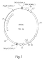
- Fig.1 Human papillomaviruses
- the disclosure provides primers and probes that detect HPV in all modalities: (i) direct detection of the most frequent high risk types only using nucleic acid (NA) amplification or other analytical methods, (ii) direct detection of the most frequent high risk types using a two step process (NA amplification with a subsequent analysis of the product by hybridization) and (iii) amplification and analysis of high and low risk HPV types in a single reaction.
- the invention further provides methods for the design and use of oligonucleotide primers specific for E1 gene region of HPV.
- the compositions and methods of the invention address a long-felt need for detection, screening and monitoring of diseases associated with HPV infection.
- cervical cells Collection of cervical cells from a patient requires a visit to a doctor's office and at least a trained technician, but more likely, a certified physician, to perform the specimen collection. Moreover, the procedure is invasive and uncomfortable for the patient. It is suggested that the preceding obstacles to collection of cervical cells could be the reason that around 30% of women in United States do not have Pap smear examinations on a regular basis ( Ackermann SP, et al., 1992, MMWR CDC Surveill Summ, 41: 17-25 ; Anderson, LM, May DS.1995, Am J Public Health, 85: 840-2 ). Further, there are religious and other cultural reasons limiting women's visit to the gynecologist office for cervical sampling and general vaginal examination.
- the invention provides a solution to address above-mentioned obstacles to cervical cell collection.
- the methods of the invention use a different source of HPV DNA, which does not require cervical scrapings. Rather, the compositions and methods of the invention detect HPV DNA in a urine sample obtained from a patient.
- HPV DNA is detected in the cellular pellet of centrifuged urine ( Payan C, et al., 2007, J Clin Microbiol., 45(3):897-901 ; Forslund O, et al.,1993, J Clin Microbiol., 31(8):1975-9 ; Song ES, et al., 2007, J Korean Med Sci., 22(1):99-104 ) or whole urine (50, 51, 52 Brinkman JA, et al., 2002, J Clin Microbiol., 40(9):3155-61 ; Sellors JW, et al., 2000, CMAJ. 163(5):513-8 ; Smits PH, et al., 2005, J Clin Microbiol. 2005, 43(12):5936-9 ).
- the preceding published tests are PCR based. Clinical sensitivity in these reports is not satisfactory due to the size of amplimers that ranged from 100 to 500 base pairs (bp).
- NA appear in urine from two sources, (i): cells shed into urine from genitourinary tract, of NAs of which are usually high molecular weight, and (ii): transrenal NAs (Tr-DNA) that cross the kidney barrier from the bloodstream into urine, which are usually low molecular weight fragments.
- Low molecular weight transrenal NA sizes range from about 20 to 150 bp ( Chan KC, et al., 2008, Clin Cancer Res., 14(15):4809-13 ; Su YH, et al.,2004, Ann N Y Acad Sci., 1022:81-9 ; Umansky SR, Tomei LD.
- oligonucleotide primer compositions of the invention effectively detect both HPV DNA released from cells that are shed as well as Tr-DNA in urine.
- Oligonucleotide primer compositions of the invention target a newly identified highly specific genetic marker in the E1 gene of HPV (Table 1). Targeting this marker within the E1 gene allows the design of PCR primers and probes for specific detection of high risk HPV genotypes in a clinical or biological sample.
- the invention also provides high risk HPV specific primers mapped to an area of the E1 gene that amplify very short DNA fragments to detect HPV genome fragments present in the Tr-DNA fraction of urine.
- Oligonucleotides selected from the regions of the E1 gene of HPV specified in Table 1, or complementary sequences are used for HPV detection in a biological or clinical sample. Moreover, an oligonucleotide or complementary sequence with at least 50%, 55%, 60%, 65%, 70%, 75%, 80%, 85%, 90%, 95%, 100% homology or identity, or any percentage point in between is used for HPV detection in biological or clinical sample.
- HPV is detected from a biological or clinical sample using the following exemplary techniques, including, but not limited to, polymerase chain reaction (PCR) and all variants of this method; Real Time PCR; NA hybridization; Cyclic Probe Reaction; Single-Strand Conformation Polymorphism (SSCP); Strand Displacement Amplification (STA); Restriction Fragment Length Polymorphism (RFLP), and techniques of NA analysis involving nanotechnology.
- Primers may hybridize to binding sites which are either immediately adjacent to each other on the target sequence or slightly overlapping (having no intervening sequences between the primer binding sites).
- oligonucleotides selected from regions of the E1 gene of HPV detect specific RNA transcripts of E1 gene by a reverse transcription PCR reaction.
- Biological and clinical samples include, but are not limited to, any fluid in the body including blood, urine, saliva, sputum, tears, semen, milk, or vaginal secretions.
- the biological or clinical sample is urine.
- a diagnostic kit for detecting HPV comprising: reagents to facilitate the isolation of DNA of 20-500 nucleotides in length from urine; reagents to facilitate amplification of DNA of 20-500 nucleotides in length by the polymerase chain reaction; a heat stable DNA polymerase; and an oligonucleotide specific for a marker sequence only occurring in the E1 gene of HPV.
- DNA is subject to degradation by DNases present in bodily fluids, such as urine.
- the present invention encompasses several methods for preventing or reducing the degradation of DNA while in urine so that sufficiently large sequences are available for detection by known methods of DNA detection such as those described below.
- samples of urine are taken when the urine has been held in the bladder for less than 12 hours, in a specific embodiment the urine is held in the bladder for less than 5 hours, more preferable for less than 2 hours. Collecting and analyzing a urine sample before it has been held in the bladder for a long period of time reduces the exposure of DNA to the any DNase present in the urine.
- the urine sample is treated using one or more methods of inhibiting DNase activity.
- Methods of inhibiting DNase activity include, but are not limited to, the use of ethylenediaminetetraacetic acid (EDTA), guanidine-HCl, GITC (Guanidine isothiocyanate), N-lauroylsarcosine, Na-dodecylsulphate (SDS), high salt concentration and heat inactivation of DNase.
- the urine sample is treated with an adsorbent that traps DNA, after which the adsorbent is removed from the sample, rinsed and treated to release the trapped DNA for detection and analysis.
- an adsorbent that traps DNA after which the adsorbent is removed from the sample, rinsed and treated to release the trapped DNA for detection and analysis.
- This method not only isolates DNA from the urine sample, but, when used with some adsorbents, including, but not limited to Hybond N membranes (Amersham Pharmacia Biotech Ltd., Piscataway, N.J.) protects the DNA from degradation by DNase activity.
- the present invention encompasses embodiments wherein sensitivity of detection is increased by any method(s) known in the art, including, without limitation, one or more of the following methods.
- DNA is present in minute amounts in the urine
- larger urine samples can be collected and thereafter concentrated by any means that does not effect the detection of DNA present in the sample.
- Some examples include, without limiting the breadth of the invention, reducing liquid present in the sample by butanol concentration or concentration using Sephadex G-25 (Pharmacia Biotech, Inc., Piscataway N.J.).
- Nested PCR can be used to improve sensitivity by several orders of magnitude. Because of the vulnerability of nested PCR to inaccurate results due to DNA contamination, in one embodiment of the present invention, precautions are taken to avoid DNA contamination of the sample. For example, without limiting the present invention, one can treat PCR reagents with restriction endonuclease(s) that cleave within the target sequence, prior to adding them to the test DNA sample.
- the present invention encompasses substantially purifying or isolating nucleic acids from a sample prior to detection.
- Nucleic acid molecules can be isolated from urine using any of a number of procedures, which are well-known in the art. Any method for isolation that facilitates the detection of target nucleic acid is acceptable.
- DNA can be isolated by precipitation, as described by Ishizawa et al., Nucleic Acids Res. 19, 5972 (1991 ). Where a large volume sample contains a low concentration of DNA, as with urine, a preferred method of isolating DNA is encompassed. In this method, a sample is treated with an adsorbent that acts to concentrate the DNA.
- a sample can be treated with a solid material that will adsorb DNA, such as, without limitation, DEAE Sephadex A-25 (Pharmacia Biotech, Inc., Piscataway N.J.), a DNA filter, and/or glass milk. Sample DNA is eluted from the adsorbent after other compositions are washed away.
- a solid material that will adsorb DNA such as, without limitation, DEAE Sephadex A-25 (Pharmacia Biotech, Inc., Piscataway N.J.), a DNA filter, and/or glass milk.
- the present invention also encompasses methods of reducing the presence of contaminating nucleic acids in the urine sample.
- Contamination of urine samples by nucleic acid sequences that have not crossed the kidney barrier can be introduced by cells shedding from the urinary tract lining, by sexual intercourse, or during processing of the urine sample prior to detection of the DNA sequence of interest.
- DNA passing the kidney barrier and appearing in urine is likely to have on average a shorter length than DNA introduced from contaminating sources because of the fragmentation that occurs in apoptotic cells and necrotic cells in the body, combined with the action of DNase in the blood and urine.
- Filtration can be used to reduce the level of contaminating DNA in a urine sample prior to detection, by selecting for shorter sequences of DNA.
- nucleic acids containing more than about 1000 base pairs, or 1000 nucleotides when denatured are removed from the sample prior to detection.
- urine samples are filtered prior to amplification by PCR to remove substantially all DNA comprising greater than 300 base pairs, or 300 nucleotides when denatured. Without limiting the invention to a specific mechanism, it is proposed that such a filtration removes contaminating DNA from cells shed from the urethral/bladder wall or introduced into the urethra during sexual intercourse.
- Nucleic acid molecules can also be isolated by gel electrophoresis, whereby fragments of nucleic acid are separated according to molecular weight.
- the technique of restriction fragments length polymorphisms (RFLP) applies the methods of electrophoresis separation, followed by nucleic acid detection enabling comparison by molecular weight of fragments from two or more alleles of a specific gene sequence.
- the present invention further encompasses methods having the step of reducing DNA degradation in said urine sample, which in one embodiment encompasses treatment with a compound selected from the group comprising: ethylenediaminetetraacetic acid, guanidine-HCl, Guanidine isothiocyanate, N-lauroylsarcosine, and Na-dodecylsulphate.
- DNA degradation can further be reduced by taking a urine sample that has been held in the bladder less than 12 hours.
- the nucleic acid sequence is substantially isolated by precipitation or by treatment with a solid adsorbent material.
- the urine sample is filtered to remove contaminants, and, in a specific embodiment, the filtering removes DNA comprising more than about 1000 nucleotides. Preferably, the filtering removes DNA comprising more than about 300 nucleotides.
- detect and “analyze” in relation to a nucleic acid sequence refer to the use of any method of observing, ascertaining or quantifying signals indicating the presence of the target nucleic acid sequence in a sample or the absolute or relative quantity of that target nucleic acid sequence in a sample.
- Methods can be combined with nucleic acid labeling methods to provide a signal by, for example: fluorescence, radioactivity, colorimetry, gravimetry, X-ray diffraction or adsorption, magnetism, enzymatic activity and the like.
- the signal can then be detected and/or quantified, by methods appropriate to the type of signal, to determine the presence or absence, of the specific DNA sequence of interest.
- nucleic acid sequence refers to the use of any method to study the amount of a particular nucleic acid sequence, including, without limitation, methods to determine the number of copies of a nucleic acid sequence or to determine the change in quantity of copies of the nucleic acid sequence over time, or to determine the relative concentration of a sequence when compared to another sequence.
- DNA sequences can be "amplified” in a number of ways, including, but not limited to cycling probe reaction ( Bekkaoui, F. et al, BioTechniques 20,240-248 (1996 ), polymerase chain reaction (PCR), nested PCR, PCR-SSCP (single strand conformation polymorphism), ligase chain reaction (LCR) ( F. Barany Proc. Natl. Acad. Sci USA 88:189-93 (1991 )), cloning, strand displacement amplification (SDA) ( G. K. Terrance Walker et al., Nucleic Acids Res. 22:2670-77 (1994 ), and variations such as allele-specific amplification (ASA).
- cycling probe reaction Bekkaoui, F. et al, BioTechniques 20,240-248 (1996 )
- PCR polymerase chain reaction
- LCR ligase chain reaction
- SDA strand displacement amplification
- gene refers to a DNA sequence that comprises control and coding sequences necessary for the transcription of an RNA sequence.
- the term “genome” refers to the complete gene complement of an organism, contained in a set of chromosomes in eukaryotes.
- wild-type gene or gene sequence is that which is most frequently observed in a population and is thus arbitrarily designed the "normal” or “wild-type” form of the gene.
- modified refers to a gene, sequence or gene product which displays modifications in sequence and or functional properties (i.e., altered characteristics) when compared to the wild-type gene, sequence or gene product. It is noted that naturally-occurring mutants can be isolated; these are identified by the fact that they have altered characteristics when compared to the wild-type gene or gene product.
- mutations can take many forms, including addition, addition-deletion, deletion, frame-shift, missense, point, reading frame shift, reverse, transition and transversion mutations as well as microsatellite alterations.
- oligonucleotide and “polynucleotide” and “polymeric” nucleic acid are interchangeable and are defined as a molecule comprised of two or more deoxyribonucleotides or ribonucleotides, preferably more than three, and usually more than ten. The exact size will depend on many factors, which in turn depends on the ultimate function or use of the oligonucleotide.
- the oligonucleotide can be generated in any manner, including chemical synthesis, DNA replication, reverse transcription, or a combination thereof.
- an end of an oligonucleotide is referred to as the "5' end” if its 5' phosphate is not linked to the 3' oxygen of a mononucleotide pentose ring and as the "3' end” if its 3' oxygen is not linked to a 5' phosphate of a subsequent mononucleotide pentose ring.
- a nucleic acid sequence even if internal to a larger oligonucleotide, also can be said to have 5' and 3' ends.
- the former can be called the "upstream” oligonucleotide and the latter the "downstream” oligonucleotide.
- primer refers to an oligonucleotide which is capable of acting as a point of initiation of synthesis when placed under conditions in which primer extension is initiated.
- An oligonucleotide “primer” can occur naturally, as in a purified restriction digest or be produced synthetically.
- a primer is selected to be "substantially" complementary to a strand of specific sequence of the template.
- a primer must be sufficiently complementary to hybridize with a template strand for primer elongation to occur.
- a primer sequence need not reflect the exact sequence of the template.
- a non-complementary nucleotide fragment can be attached to the 5' end of the primer, with the remainder of the primer sequence being substantially complementary to the strand.
- Non-complementary bases or longer sequences can be interspersed into the primer, provided that the primer sequence has sufficient complementarity with the sequence of the template to hybridize and thereby form a template primer complex for synthesis of the extension product of the primer.
- a "target" nucleic acid is a nucleic acid sequence to be evaluated by hybridization, amplification or any other means of analyzing a nucleic acid sequence, including a combination of analysis methods.
- Hybridization methods involve the annealing of a complementary sequence to the target nucleic acid (the sequence to be analyzed). The ability of two polymers of nucleic acid containing complementary sequences to find each other and anneal through base pairing interaction is a well-recognized phenomenon.
- the initial observations of the "hybridization” process by Marmur and Lane, Proc. Natl. Acad. Sci. USA 46:453 (1960 ) and Doty et al., Proc. Natl. Acad. Sci. USA 46:461 (1960 ) have been followed by the refinement of this process into an essential tool of modern biology.
- Hybridization encompasses, but is not limited to, slot, dot and blot hybridization techniques.
- hybridization method it is important for some diagnostic applications to determine whether the hybridization represents complete or partial complementarity. For example, where it is desired to detect simply the presence or absence of pathogen DNA (such as from a virus, bacterium, fungi, mycoplasma, protozoan) it is only important that the hybridization method ensures hybridization when the relevant sequence is present; conditions can be selected where both partially complementary probes and completely complementary probes will hybridize. Other diagnostic applications, however, could require that the hybridization method distinguish between partial and complete complementarity. It may be of interest to detect genetic polymorphisms.
- hybridization regardless of the method used, requires some degree of complementarity between the sequence being analyzed (the target sequence) and the fragment of DNA used to perform the test (the probe). (Of course, one can obtain binding without any complementarity but this binding is nonspecific and to be avoided.)
- nucleic acid sequence refers to an oligonucleotide which, when aligned with the nucleic acid sequence such that the 5' end of one sequence is paired with the 3' end of the other, is in "antiparallel association.”
- Specific bases not commonly found in natural nucleic acids can be included in the nucleic acids of the present invention and include, for example, inosine and 7-deazaguanine. Complementarity need not be perfect; stable duplexes can contain mismatched base pairs or unmatched bases.
- nucleic acid technology can determine duplex stability empirically considering a number of variables including, for example, the length of the oligonucleotide, base composition and sequence of the oligonucleotide, ionic strength and incidence of mismatched base pairs.
- Tm is used in reference to the "melting temperature.”
- the melting temperature is the temperature at which a population of double-stranded nucleic acid molecules becomes half dissociated into single strands.
- probe refers to an oligonucleotide (i.e., a sequence of nucleotides), whether occurring naturally as in a purified restriction digest or produced synthetically, which forms a duplex structure or other complex with a sequence in another nucleic acid, due to complementarity or other means of reproducible attractive interaction, of at least one sequence in the probe with a sequence in the other nucleic acid. Probes are useful in the detection, identification and isolation of particular gene sequences.
- any probe will be labeled with any "reporter molecule,” so that it is detectable in any detection system, including, but not limited to, enzyme (e.g., ELISA, as well as enzyme-based histochemical assays), fluorescent, radioactive, and luminescent systems. It is further contemplated that the oligonucleotide of interest (i.e., to be detected) will be labeled with a reporter molecule. It is also contemplated that both the probe and oligonucleotide of interest will be labeled. It is not intended that the present disclosure be limited to any particular detection system or label.
- label refers to any atom or molecule which can be used to provide a detectable (preferably quantifiable) signal, and which can be attached to a nucleic acid or protein. Labels provide signals detectable by any number of methods, including, but not limited to, fluorescence, radioactivity, colorimetry, gravimetry, X-ray diffraction or absorption, magnetism, and enzymatic activity.
- substantially single-stranded when used in reference to a nucleic acid target means that the target molecule exists primarily as a single strand of nucleic acid in contrast to a double-stranded target which exists as two strands of nucleic acid which are held together by inter-strand base pairing interactions.
- sequence variation refers to differences in nucleic acid sequence between two nucleic acid templates.
- a wild-type structural gene and a mutant form of this wild-type structural gene can vary in sequence by the presence of single base substitutions and/or deletions or insertions of one or more nucleotides. These two forms of the structural gene are said to vary in sequence from one another.
- a second mutant form of the structural gene can exit. This second mutant form is said to vary in sequence from both the wild-type gene and the first mutant form of the gene.
- structure probing signature means the measured level of complex formation between a target nucleic acid and a probe or set of probes, such measured levels being characteristic of the target nucleic acid when compared to levels of complex formation involving reference targets or probes.
- Oligonucleotide primers matching or complementary to a gene sequence refers to oligonucleotide primers capable of facilitating the template-dependent synthesis of single or double-stranded nucleic acids. Oligonucleotide primers matching or complementary to a gene sequence can be used in PCRs, RT-PCRs and the like.
- Nucleic acid sequence refers to an oligonucleotide, nucleotide or polynucleotide, and fragments or portions thereof, and to DNA or RNA of genomic or synthetic origin which can be single- or double-stranded, and represent the sense or antisense strand.
- a “deletion” is defined as a change in either nucleotide or amino acid sequence in which one or more nucleotides or amino acid residues, respectively, are absent.
- insertion or “addition” is that change in a nucleotide or amino acid sequence which has resulted in the addition of one or more nucleotides or amino acid residues, respectively, as compared to, naturally occurring sequences.
- substitution results from the replacement of one or more nucleotides or amino acids by different nucleotides or amino acids, respectively.
- a "modification" in a nucleic acid sequence refers to any change to a nucleic acid sequence, including, but not limited to a deletion, an addition, an addition-deletion, a substitution, an insertion, a reversion, a transversion, a point mutation, a microsatellite alteration, methylation or nucleotide adduct formation.
- purified As used herein, the terms “purified”, “decontaminated” and “sterilized” refer to the removal of contaminant(s) from a sample.
- the terms “substantially purified” and “substantially isolated” refer to nucleic acid sequences that are removed from their natural environment, isolated or separated, and are preferably 60% free, more preferably 75% free, and most preferably 90% free from other components with which they are naturally associated.
- An "isolated polynucleotide” is therefore a substantially purified polynucleotide. It is contemplated that to practice the methods of the present invention polynucleotides can be, but need not be substantially purified. A variety of methods for the detection of nucleic acid sequences in unpurified form are known in the art.
- PCR polymerase chain reaction
- This process for amplifying the target sequence consists of introducing a large excess of two oligonucleotide primers to the DNA mixture containing the desired target sequence, followed by a precise sequence of thermal cycling in the presence of a DNA polymerase.
- the two primers are complementary to their respective strands of the double stranded target sequence.
- the mixture is denatured and the primers then annealed to their complementary sequences within the target molecule.
- the primers are extended with a polymerase so as to form a new pair of complementary strands.
- the steps of denaturation, primer annealing and polymerase extension can be repeated many times (i.e., denaturation, annealing and extension constitute one "cycle”; there can be numerous “cycles”) to obtain a high concentration of an amplified segment of the desired target sequence.
- the length of the amplified segment of the desired target sequence is determined by the relative positions of the primers with respect to each other, and therefore, this length is a controllable parameter.
- the method is referred to as the “polymerase chain reaction” (hereinafter "PCR”). Because the desired amplified segments of the target sequence become the predominant sequences (in terms of concentration) in the mixture, they are said to be "PCR amplified”.
- polymerase refers to any enzyme suitable for use in the amplification of nucleic acids of interest. It is intended that the term encompass such DNA polymerases as Taq DNA polymerase obtained from Thermus aquaticus, although other polymerases, both thermostable and thermolabile are also encompassed by this definition.
- PCR it is possible to amplify a single copy of a specific target sequence in genomic DNA to a level that can be detected by several different methodologies (e.g., staining, hybridization with a labeled probe; incorporation of biotinylated primers followed by avidin-enzyme conjugate detection; incorporation of 32P-labeled deoxynucleotide triphosphates, such as dCTP or dATP, into the amplified segment).
- any oligonucleotide sequence can be amplified with the appropriate set of primer molecules.
- the amplified segments created by the PCR process itself are, themselves, efficient templates for subsequent PCR amplifications.
- Amplified target sequences can be used to obtain segments of DNA (e.g., genes) for insertion into recombinant vectors.
- PCR product and “amplification product” refer to the resultant mixture of compounds after two or more cycles of the PCR steps of denaturation, annealing and extension are complete. These terms encompass the case where there has been amplification of one or more segments of one or more target sequences.
- restriction endonucleases and “restriction enzymes” refer to bacterial enzymes, each of which cut double-stranded DNA at or near a specific nucleotide sequence.
- complementarity are used in reference to polynucleotides (i.e., a sequence of nucleotides) related by the base-pairing rules. For example, for the sequence “A-G-T,” is complementary to the sequence “T-C-A.” Complementarity can be “partial,” in which only some of the nucleic acids' bases are matched according to the base pairing rules. Or, there can be “complete” or “total” complementarity between the nucleic acids. The degree of complementarity between nucleic acid strands has significant effects on the efficiency and strength of hybridization between nucleic acid strands. This is of particular importance in amplification reactions, as well as detection methods that depend upon binding between nucleic acids.
- the term "homology" refers to a degree of complementarity. There can be partial homology or complete homology (i.e., identity).
- a partially complementary sequence is one that at least partially inhibits a completely complementary sequence from hybridizing to a target nucleic acid is referred to using the functional term "substantially homologous.”
- the inhibition of hybridization of the completely complementary sequence to the target sequence can be examined using a hybridization assay (Southern or Northern blot, solution hybridization and the like) under conditions of low stringency.
- a substantially homologous sequence or probe will compete for and inhibit the binding (i.e., the hybridization) of a completely homologous to a target under conditions of low stringency.
- low stringency conditions are such that are non-specific binding is permitted; low stringency conditions require that the binding of two sequences to one another be a specific (i.e., selective) interaction.
- the absence of non-specific binding can be tested by the use of a second target which lacks even a partial degree of complementarity (e.g., less than about 30% identity); in the absence of non-specific binding the probe will not hybridize to the second non-complementary target.
- Numerous equivalent conditions can be employed to comprise either low or high stringency conditions; factors such as the length and nature (DNA, RNA, base composition) of the probe and nature of the target (DNA, RNA, base composition, present in solution or immobilized, etc.) and the concentration of the salts and other components (e.g., the presence or absence of formamide, dextran sulfate, polyethylene glycol) are considered and the hybridization solution can be varied to generate conditions of either low or high stringency hybridization different from, but equivalent to, the above listed conditions.
- factors such as the length and nature (DNA, RNA, base composition) of the probe and nature of the target (DNA, RNA, base composition, present in solution or immobilized, etc.) and the concentration of the salts and other components (e.g., the presence or absence of formamide, dextran sulfate, polyethylene glycol) are considered and the hybridization solution can be varied to generate conditions of either low or high stringency hybridization different from, but equivalent to, the above listed conditions.
- hybridization includes "any process by which a strand of nucleic acid joins with a complementary strand through base pairing" ( Coombs, Dictionary of Biotechnology, Stockton Press, New York N.Y. [1994 ].
- Stringency typically occurs in a range from about Tm-5° C. (5° C. below the Tm of the probe) to about 20° C. to 25° C. below Tm. As will be understood by those of skill in the art, a stringent hybridization can be used to identify or detect identical polynucleotide sequences or to identify or detect similar or related polynucleotide sequences.
- hybridization complex refers to a complex formed between two nucleic acid sequences by virtue of the formation of hydrogen bonds between complementary G and C bases and between complementary A and T bases; these hydrogen bonds can be further stabilized by base stacking interactions.
- the two complementary nucleic acid sequences hydrogen bond in an antiparallel configuration.
- a hybridization complex can be formed in solution (e.g., C0t or R0t analysis) or between one nucleic acid sequence present in solution and another nucleic acid sequence immobilized to a solid support (e.g., a nylon membrane or a nitrocellulose filter as employed in Southern and Northern blotting, dot blotting or a glass slide as employed in situ hybridization, including FISH [fluorescent in situ hybridization]).
- antisense is used in reference to RNA sequences which are complementary to a specific RNA (e.g., mRNA) or DNA sequence.
- Antisense RNA can be produced by any method, including synthesis by splicing the gene(s) of interest in a reverse orientation to a viral promoter which permits the synthesis of a coding strand. Once introduced into a cell, this transcribed strand combines with natural mRNA produced by the cell to form duplexes. These duplexes then block either further transcription of the mRNA or its translation. In this manner, mutant phenotypes can be generated.
- antisense strand is used in reference to a nucleic acid strand that is complementary to the "sense” strand.
- the designation (-) i.e., “negative” is sometimes used in reference to the antisense strand, with the designation (+) sometimes used in reference to the sense (i.e., "positive") strand.
- sample as used herein is used in its broadest sense.
- a biological sample suspected of containing nucleic acid can comprise, but is not limited to, genomic DNA (in solution or bound to a solid support such as for Southern blot analysis), cDNA (in solution or bound to a solid support), and the like.
- urinary tract refers to the organs and ducts which participate in the secretion and elimination of urine from the body.
- transrenal DNA and "transrenal nucleic acid” as used herein refer to nucleic acids that have crossed the kidney barrier.
- Transrenal DNA as used herein differs from miRNA. Specifically, transrenal DNA comprises randomness in the 3' and 5' ends, which is not present in miRNA.
- Urine specimens were collected in containers, which are capable of holding a volume of at least 100 ml.
- Q-Sepharose slurry For a standard batch size of 10 ml urine (prior to dilution) 1.0 ml of Q-Sepharose slurry (Q-Sepharose stock: 25 ml size from GE Healthcare; 250 ⁇ L resin) was used.
- Binding of urinary NA to Q-Sepharose was performed for 30 min at room temperature (20-25°C) with rotation in a 50 mL tube.
- the resin was collected by centrifugation at room temperature (800-1000 x g for 5 min) and transferred into an empty disposable column.
- the resin was washed twice with at least 1 mL of 0.3 M LiCl/10 mM NaOAc (pH 5).
- NA was eluted with 750 ⁇ L of 2 M LiCl/10 mM NaOAc.
- DNA Eluted from Q-sepharose NA in 750 ⁇ l buffer was supplemented with 2.25 mL of 95% EtOH and applied to a silica column (Qiagen or equivalent). If column extension was used one load took the whole mixture, otherwise several loads were performed. The column was centrifuged for 1 minute in a table top microcentrifuge (Eppendorf). Alternatively, a vacuum manifold was used.
- Example 2 Use of specific PCR primers mapped to the E1 gene for amplification of HPV individual genotypes.
- the predicted size of the PCR product was 47 base pairs (bp).
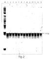
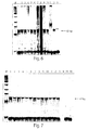
- Example 3 Use of single pair of PCR primers for detection of all 13 high risk HPV genotypes
- the purpose of this experiment was to use a single pair of primers mapped in the fragment of interest of the HPV E1 gene for specific detection of all or most of the 13 high-risk HPV strain that do not react with low risk counterparts.
- SEQ ID 41: 5'-CAGGCAGAATTAGAGRCAGC-3' was used as the forward primer and SEQ ID 42: 5'-tccaccacaWACTTTCGTTTTA-3' was used as the reverse primer.
- Lowercase nucleotides in the reverse primer are the randomly selected tail to adjust the melting temperature (Tm) of the primer.
- Expected size of the specific product was 97 bp.
- the forward primer was paired with the reverse primer at a concentration of 800 nM.
- the final concentration of MgCl 2 was 3mM and the final concentration of the JumpStart Taq DNA polymerase was 1.25 U/reaction.
- Individual oligonucleotides corresponding to high and low risk HPV types were used as templates at1000 copies per reaction (see, Table 2).
- lane numbers from 1 to 13 correspond to the following high risk HPV genotypes: 16; 18; 31; 33; 35; 39; 45; 51; 52; 56; 58; 59; 68, respectively, and lanes 14 and 15 correspond to low risk genotypes 6 and 11, respectively.
- the molecular weight marker (“M”) is a 25 bp ladder.
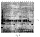
- Example 4 Use of single pair of PCR primers to analyze urine samples from patients with cervical cancer.
- a single pair of primers that mapped in the fragment of interest of HPV E1 gene were used for specific detection of DNA of high risk HPV genotypes.
- SEQ ID 41: 5'-CAGGCAGAATTAGAGRCAGC-3' was used as the forward primer
- SEQ ID 42: 5'-tccaccacaWACTTTCGTTTTA-3' was used as the reverse primer.
- Lowercase nucleotides in the reverse primer are the randomly selected tail used to adjust the Tm of the primer.
- the forward primer was paired with the reverse primer at a concentration of 800 nM.
- final concentration of MgCl 2 was 3mM and the final concentration of the JumpStart Taq DNA polymerase was 1.25 U/reaction.
- DNA from urine samples were extracted according to the protocol described in Example 1. Patients were asked to donate two urine samples: a first sample that was self-collected in the morning and a second sample that was collected at doctor's office later the same day (within a 24 hour period). Cervical samples were taken for the Digene tests. DNA from 10 ml of urine was extracted in 100 ⁇ l of elution buffer, of which 5 ⁇ l was used for PCR.
- lane numbers from 1 to 18 represent urine samples of patients with cancer of the cervix. Odd lane numbers represent self-collected morning urine samples, whereas even lane numbers represent urine samples donated by the patients at the doctor's office later the same day.
- lane 19 contained a urine sample from a healthy volunteer.
- Lane 20 contained water as a control for urine DNA purification.
- Lane 21 contained HPV 16 genomic DNA as a positive control.
- Lane 22 contained human genomic DNA (20,000 genome equivalent).
- Lane 23 contained an equivocal mix of low risk HPV 6 and 11 templates.
- Lane 24 was a reaction control that contained no oligonucleotide or DNA template.
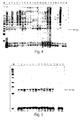
- Example 5 Use of all HPV high risk specific PCR primer pairs in a single tube PCR for detection of the virus:
- Oligonucleotide templates representing high risk genotypes were amplified by PCR with the mixture of all high risk specific primer pairs. In each reaction, a total of 25 PCR primers were included (see, Table 4). In the PCR, the forward primers, each used at a concentration of 200 nM, were each were combined with reverse primers, each used at a concentration of 300 nM. In this reaction, the final concentration of MgCl 2 was 2mM and the final concentration of AmpliTaq DNA polymerase was 1.25 U/reaction. Individual oligonucleotides corresponding to high and low risk HPV types were used as templates at 1000 copies per reaction (Table 3).
- Expected size of the product was 62 bp.
- the footprint of the target was 51 bp.
- Results are depicted in Figure 5 , wherein lane numbers from 1 to 13 correspond to the following high risk HPV genotypes: 16; 18; 31; 33; 35; 39; 45; 51; 52; 56; 58; 59; 68, respectively, and lanes 15 and 16 correspond to low risk genotypes 6 and 11, respectively.
- the molecular weight marker (“M”) is a 25 bp ladder.
- Example 6 Use of all HPV high risk specific PCR primer pairs in a single tube PCR for detection of the virus:
- Urine samples from patients with cervical cancer were tested using a mixture of all high risk specific primer pairs. Therefore, the PCR contained a total of 25 PCR primers in each reaction (see, Table 4). In the PCR, the forward primers, each used at a concentration of 200 nM, were combined with reverse primers, each used at a concentration of 300 nM. In this reaction, the final concentration of MgCl 2 was 2mM and the final concentration of AmpliTaq DNA polymerase was 1.25 U/reaction. DNA from urine samples were extracted according to the protocol described in the Example 1. Urine samples collected at the visit to doctors office were used (see Example 5). DNA from 10 ml of urine was extracted in 100 ⁇ l of elution buffer, of which 5 ⁇ l was used for PCR.
- Lane 11 contained human genomic DNA (20,000 genome equivalent).
- Lane 12 contained HPV 18 genomic DNA as a positive control.
- Lane 13 represented a reaction control that contained no oligonucleotide or DNA template.
- Example 7 Use of subset of HPV high risk specific primers in a single tube PCR for detection of the virus.
- Oligonucleotides representing high risk HPV genotypes were amplified by PCR using a subset of high risk HP, the forward primers, each used at a concentration of 200 nM, were each were combined with a reverse primer, used at a concentration of 300 nM. In this reaction a total of 20 primers were used (Table 5). In this reaction, the final concentration of MgCl 2 was 2mM and the final concentration of AmpliTaq DNA polymerase was 1.25 U/reaction. Individual oligonucleotides corresponding to high and low risk HPV types were used at 1000 copies per reaction (Table 2).
- Expected size of the product was 62 bp.
- the footprint of the target was 51 bp.
- Results are depicted in Figure 7 , wherein lane numbers from 1 to 13 correspond to the following high risk HPV genotypes: 16; 18; 31; 33; 35; 39; 45; 51; 52; 56; 58; 59; 68, respectively, and lanes 15 and 16 correspond to low risk genotypes 6 and 11, respectively.
- the molecular weight marker (“M”) is a 25 bp ladder.
- Example 8 An improved molecular screening test for the detection of High Risk HPV in urine of high and low risk populations in India
- the Xenomics Transrenal DNA (Tr-DNA) technology is based on DNA fragments from cells dying throughout the body. This DNA appears in the bloodstream and is excreted into the urine. Analysis of urine samples was applied to detection of Y chromosome-specific DNA sequences from women with male fetuses, mutant K-ras in colorectal cancer patients, and Mycobacterium tuberculosis in patients with pulmonary tuberculosis.
- HPV DNA test used in this study involves isolation of DNA from urine and specific amplification of the HPV E1 region to detect the presence of high risk HPV types that have been associated with cervical cancer. These high-risk types include HPV 16, 18, 31, 33, 35, 39, 45, 51, 52, 56, 58, 59 and 68.
- DNA is amplified by the Polymerase Chain Reaction (PCR) using a FAM-labeled forward primer and an unlabelled reverse primer. These primers generate a 93-96 base pair (bp) amplicon as determined by capillary electrophoresis (CE). No cross-reactivity was observed with the low risk HPV types 6 and 11. Data obtained demonstrated that sensitivity and specificity of this test were equivalent to or better than those of a current assay in clinical use based on cervical scraping.
- Pap smears and hc2 tests were performed by Simbiosys Biowares Inc. and Metropolis Inc. A portion of the collected cervical sample was immediately used to make a smear for Pap testing and the remainder was transferred to buffer solution for HPV testing by QIAGEN hc2. hc2 tests were performed and analyzed as per manufacturer's instructions using the HR HPV Probe cocktail. The recommended positivity threshold of 1 pg/ml was used as a cutoff, and all samples with a relative light units/control (RLU/CO) ratio of 1.00 or greater were considered positive.
- RLU/CO relative light units/control
- Urine samples stored at -80°C were thawed and mixed by gentle inversion. DNA isolation was carried out as per the protocol described previously ( Shekhtman E. M. et al. Clin Chem 2009; 55:723-729 ). Briefly a 1:1 urine:water sample was incubated with Q-sepharose resin slurry (GE Healthcare, Piscataway, NJ). The resin was pelleted and the supernatant was discarded. Pelleted resin was resuspended and transferred to a spin column. The resuspension buffer was removed and the resin was washed. DNA was eluted from the resin by 2M LiCl.
- the eluate was brought to 70% ethanol and applied to a QIAquick column (QIAGEN, Hilden, Germany). The column was washed with 2M LiCl/70% EtOH followed by 75 mM KOAc (pH 5.0)/ 80% EtOH. DNA was eluted with EB Buffer (QIAGEN, Hilden, Germany) and stored at - 20°C. The isolated DNA samples were quantitated by the Picogreen assay (Life Technologies).
- Primers XEN-HPV-FAM-F and XEN-HPV-R were used in PCR assays. Following a 10 min treatment with AmpErase® UNG (Life Technologies), amplifications were carried out for 40 cycles in 25 ⁇ L with 600 nM each primer, 3 mM MgCl 2 , 1.25 Units AmpliTaq Gold DNA Polymerase (Life Technologies), 200 ⁇ M each of dATP, dCTP, dGTP; and 400 ⁇ M dUTP. Each cycle was 15 seconds (s) at 95°C and 60 s at 50°C. Reaction products were subjected to capillary electrophoresis by GENEWIZ (South Plainfield, NJ).
- PCR amplifications were performed using different primer sets (Table 6) with JumpStart TM Taq DNA polymerase (Sigma-Aldrich) and various MgCl 2 concentrations (2 mM for MY09/MY11 and GP5+/GP6+, 3 mM for XEN-HPV-F/-R).
- Reaction mixtures were subjected to various cycling steps for each primer pair: 95°C for 30 seconds, 55°C for 45 seconds, 72°C for 20 seconds, 37 cycles of amplification (MY09/MY11); 95°C for 15 seconds, 40°C for 30 seconds, 72°C for 10 seconds, 50 cycles of amplification (GP5+/GP6+); 95°C for 15 seconds, 50°C for 60 seconds, 45 cycles of amplification (XEN-HPV-F/-R).
- PCR products were analyzed by electrophoresis on 10% polyacrylamide gels (Bio-Rad). Gel slices were excised and purified according to QIAEX II Gel Extraction Kit instructions (QIAGEN) and sequenced with one of the primers used for PCR amplification. DNA sequencing was performed by GENEWIZ Inc. (South Plainfield, New Jersey). Raw PCR product sequences were analyzed by NCBI Blastn algorithm to match specific human papillomavirus strains.
- XEN-HPV-F/-R primers were used to amplify an 88 bp footprint of the E1 gene that could provide evidence of high risk HPV DNA sequence if any were present in the sample.
- High Risk HPV types were demonstrated by DNA sequencing of the L1 region (MY09/MY11 and/or GP5+/GP6+ primers) in four samples.
- Six additional samples were shown to have high risk HPV types by using XEN-HPV-F/-R primers. These latter primers failed to detect the sample by CE but amplified a product for sequencing possibly because of low titer of HPV. This may be because PCR using XEN-HPV-F/-R primers for detection was carried out for 40 cycles vs. 45 cycles for sequencing.
- the hc2 assay did not detect 13 samples with HPV16, six HPV18, one HPV31, one HPV35, two HPV45, one HPV52, one HPV58, and two HPV59 and four containing multiple high risk HPV types (16+45, 16+33, 16+56 and 18+31) (Table 10).
- Table 11 lists the 13 of the 31 samples which required using XEN-HPV-F/-R primers for high risk types to be revealed. Secondary sequencing with the L1 region primers (MY09/MY11 and/or GP5+/GP6+ primers) yielded either no HPV detected or low risk types.
- the CE assay detected more High Risk HPV positive women, including many flagged as ASCUS and Normal, than the hc2 assay (Table 10).
- Some samples provided by Simbiosys were categorized as Control with a normal Pap result but no available hc2 result. Others were flagged as Normal by Pap smear and were Nonreactive by hc2.
- Samples from these groups were obtained from high-risk populations including STD clinics and brothels.
- the feasibility of using urine as a sample matrix for detecting High Risk HPV was examined.
- the Xenomics HPV test could therefore be proposed as a qualitative screening test thereby eliminating the need of Pap test or other molecular tests for screening.
- the Xenomics HPV test was unable to detect High Risk HPV types in 10 hc2-Reactive samples confirmed by DNA sequencing (Table 9). Six out of the 10 samples were staged CIN 2 and higher. One possible explanation is the deletion of the HPV E1 region ( Arias-Pulido H. et al. J Clin Microbiol 2006; 44:1755-1762 ). In 3 of the 6 samples, only XEN-HPV-F/-R primers generated a PCR product that, upon sequencing, contained High Risk HPV. XEN-HPV-F/-R primers confirmed the presence of High Risk HPV33 in a fourth sample. This indicates that the E1 region was hence not deleted.
- the PCR conditions used for sequencing as opposed to detection may provide a more sensitive amplification system.
- the HPV DNA CE assay balances sensitivity and specificity such that some low titer HPV samples may not be detected. The result is an assay with fewer false positive and false negative results compared to a commercially available assay.
- Table 12 considers the assay comparison when the hc2 result is combined with DNA sequencing using both literature primers (MY09/MY11 and/or GP5+/GP6+ primers) and XEN-HPV-F/-R primers to resolve discordant samples.
- the assay Sensitivity is 93.0% (133/143); Specificity is 96.0% (170/177). Positive and Negative Predictive Values are 95.0% (133/140) and 94.4% (170/180), respectively.
- the Kappa value of 0.89 indicates excellent agreement between the two methods.
- sensitivity of urine-based HPV testing is similar to or better than the currently used hc2 test based on analysis of cervical cells.
- a higher sensitivity of the CE test compared to other urine DNA-based studies can be explained by several factors.
- DNA was isolated from whole urine, not a cellular fraction. This is critical in the overall recovery of HPV DNA.
- HPV DNA sequences can also be contributed by transrenal DNA (Tr-DNA) ( Melkonyan H.S. et al. Ann N Y Acad Sci 2008; 1137:73-81 ).
Landscapes
- Chemical & Material Sciences (AREA)
- Life Sciences & Earth Sciences (AREA)
- Health & Medical Sciences (AREA)
- Organic Chemistry (AREA)
- Zoology (AREA)
- Engineering & Computer Science (AREA)
- Immunology (AREA)
- Wood Science & Technology (AREA)
- Proteomics, Peptides & Aminoacids (AREA)
- Virology (AREA)
- Biochemistry (AREA)
- Microbiology (AREA)
- Molecular Biology (AREA)
- Physics & Mathematics (AREA)
- Biophysics (AREA)
- Analytical Chemistry (AREA)
- Biotechnology (AREA)
- Bioinformatics & Cheminformatics (AREA)
- General Engineering & Computer Science (AREA)
- General Health & Medical Sciences (AREA)
- Genetics & Genomics (AREA)
- Measuring Or Testing Involving Enzymes Or Micro-Organisms (AREA)
- Investigating Or Analysing Biological Materials (AREA)
Applications Claiming Priority (2)
| Application Number | Priority Date | Filing Date | Title |
|---|---|---|---|
| US19785008P | 2008-10-31 | 2008-10-31 | |
| PCT/US2009/062114 WO2010051261A2 (en) | 2008-10-31 | 2009-10-26 | A genetic marker for detection of human papillomavirus |
Publications (2)
| Publication Number | Publication Date |
|---|---|
| EP2358915A2 EP2358915A2 (en) | 2011-08-24 |
| EP2358915B1 true EP2358915B1 (en) | 2016-08-10 |
Family
ID=42045205
Family Applications (1)
| Application Number | Title | Priority Date | Filing Date |
|---|---|---|---|
| EP09797216.0A Not-in-force EP2358915B1 (en) | 2008-10-31 | 2009-10-26 | A genetic marker for detection of human papillomavirus |
Country Status (9)
| Country | Link |
|---|---|
| US (3) | US8642261B2 (enExample) |
| EP (1) | EP2358915B1 (enExample) |
| JP (2) | JP2012507293A (enExample) |
| CN (2) | CN102333892B (enExample) |
| AU (1) | AU2009308943A1 (enExample) |
| CA (1) | CA2741762C (enExample) |
| ES (1) | ES2588251T3 (enExample) |
| HK (1) | HK1221749A1 (enExample) |
| WO (1) | WO2010051261A2 (enExample) |
Families Citing this family (8)
| Publication number | Priority date | Publication date | Assignee | Title |
|---|---|---|---|---|
| WO2013144344A2 (en) | 2012-03-30 | 2013-10-03 | Istituto Europeo Di Oncologia S.R.L. | Biomarkers for hpv infections, related diagnostic methods and kits |
| US20150376725A1 (en) * | 2013-09-04 | 2015-12-31 | Trovagene, Inc. | HPV Detection in Urine |
| EP3169813B1 (en) | 2014-07-18 | 2019-06-12 | The Chinese University Of Hong Kong | Methylation pattern analysis of tissues in dna mixture |
| US10428391B2 (en) | 2015-07-27 | 2019-10-01 | The Johns Hopkins University | Method to use viral and host methylation markers for cervical cancer screening and triage in liquid prep, serum/plasma, and urine: PCR and sequencing based process methods |
| WO2018099418A1 (en) | 2016-11-30 | 2018-06-07 | The Chinese University Of Hong Kong | Analysis of cell-free dna in urine and other samples |
| WO2019013613A2 (es) | 2017-07-09 | 2019-01-17 | Hakken Enterprise Sa De Cv | Métodos y kits para determinar el riesgo de cáncer |
| TW202233846A (zh) * | 2019-01-03 | 2022-09-01 | 中國大陸商杭州諾輝健康科技有限公司 | 用於尿液樣本儲存及dna提取之組合物及方法 |
| KR102286565B1 (ko) * | 2020-01-13 | 2021-08-05 | 대한민국 | 소변에서 결핵균 trDNA 추출용 올리고머 |
Family Cites Families (9)
| Publication number | Priority date | Publication date | Assignee | Title |
|---|---|---|---|---|
| FR2629458B2 (fr) * | 1987-07-31 | 1991-08-09 | Ire Celltarg Sa | Nouvelles sondes d'acides nucleiques specifiques de differents types de virus de papillome humain |
| US5447839A (en) * | 1988-09-09 | 1995-09-05 | Hoffmann-La Roche Inc. | Detection of human papillomavirus by the polymerase chain reaction |
| AUPN443995A0 (en) * | 1995-07-27 | 1995-08-17 | Csl Limited | Papillomavirus polyprotein |
| US20020119478A1 (en) * | 1997-05-30 | 2002-08-29 | Diagen Corporation | Methods for detection of nucleic acid sequences in urine |
| CA2592078A1 (en) * | 2004-12-23 | 2006-06-29 | Human Genetic Signatures Pty Ltd | Detection of human papilloma virus |
| KR20080084809A (ko) * | 2005-11-15 | 2008-09-19 | 게노이드 케이에프티. | 병원체를 검출하는 방법 |
| CN1814796A (zh) * | 2005-12-02 | 2006-08-09 | 浙江大学 | 一种用于26种人乳头瘤病毒的检测与分型的方法 |
| DE102006041970A1 (de) | 2006-08-28 | 2008-03-06 | Genome Identification Diagnostics Gmbh | Verfahren und Mittel zum Nachweis von humanen Papillomaviren |
| EP2184368A1 (en) * | 2008-11-07 | 2010-05-12 | DKFZ Deutsches Krebsforschungszentrum | Diagnostic transcript and splice patterns of HPV16 in different cervical lesions |
-
2009
- 2009-10-26 JP JP2011534663A patent/JP2012507293A/ja active Pending
- 2009-10-26 CN CN200980153183.8A patent/CN102333892B/zh not_active Expired - Fee Related
- 2009-10-26 CN CN201510671553.4A patent/CN105385784A/zh active Pending
- 2009-10-26 AU AU2009308943A patent/AU2009308943A1/en not_active Abandoned
- 2009-10-26 EP EP09797216.0A patent/EP2358915B1/en not_active Not-in-force
- 2009-10-26 WO PCT/US2009/062114 patent/WO2010051261A2/en not_active Ceased
- 2009-10-26 US US13/126,081 patent/US8642261B2/en not_active Expired - Fee Related
- 2009-10-26 CA CA2741762A patent/CA2741762C/en not_active Expired - Fee Related
- 2009-10-26 ES ES09797216.0T patent/ES2588251T3/es active Active
-
2014
- 2014-02-04 US US14/172,793 patent/US9453265B2/en not_active Expired - Fee Related
-
2015
- 2015-01-27 JP JP2015013489A patent/JP6092906B2/ja not_active Expired - Fee Related
-
2016
- 2016-07-25 US US15/218,177 patent/US20170016078A1/en not_active Abandoned
- 2016-08-19 HK HK16109959.2A patent/HK1221749A1/zh unknown
Also Published As
| Publication number | Publication date |
|---|---|
| WO2010051261A3 (en) | 2010-12-16 |
| JP6092906B2 (ja) | 2017-03-08 |
| EP2358915A2 (en) | 2011-08-24 |
| CN102333892B (zh) | 2015-11-25 |
| US8642261B2 (en) | 2014-02-04 |
| CA2741762A1 (en) | 2010-05-06 |
| ES2588251T3 (es) | 2016-10-31 |
| JP2015130863A (ja) | 2015-07-23 |
| WO2010051261A2 (en) | 2010-05-06 |
| HK1221749A1 (zh) | 2017-06-09 |
| US9453265B2 (en) | 2016-09-27 |
| CN102333892A (zh) | 2012-01-25 |
| US20140363807A1 (en) | 2014-12-11 |
| US20170016078A1 (en) | 2017-01-19 |
| US20110318727A1 (en) | 2011-12-29 |
| CA2741762C (en) | 2018-04-03 |
| JP2012507293A (ja) | 2012-03-29 |
| HK1164940A1 (zh) | 2012-09-28 |
| AU2009308943A1 (en) | 2010-05-06 |
| CN105385784A (zh) | 2016-03-09 |
Similar Documents
| Publication | Publication Date | Title |
|---|---|---|
| US9453265B2 (en) | Method for detection of high risk human papillomavirus | |
| Gregoire et al. | Amplification of human papillomavirus DNA sequences by using conserved primers | |
| Melchers et al. | Human papillomavirus detection in urine samples from male patients by the polymerase chain reaction | |
| EP0539461B1 (en) | Method for the diagnosis of human pappiloma virus type 16 using the polymerase chain reaction | |
| US20140051066A1 (en) | Hpv detection and quantification by real-time multiplex amplification | |
| Tham et al. | Diagnostic Sensitivity of Polymerase Chain Reaction and Southern Blot Hybridization for the Detection df Human Papillomavirus DNA in Biopsy Specimens from Cervical Lesions | |
| CA2697156A1 (en) | Composition comprising an oligonucleotide mixture for improved detection of human papillomavirus genotypes | |
| US8597889B2 (en) | Method for detecting cancer cell caused by HPV, method for determining whether or not tissue is at stage of high-grade dysplasia or more severe stage, and primer set and kit used therefor | |
| US20150376725A1 (en) | HPV Detection in Urine | |
| US20030129585A1 (en) | Detection of human papillomaviruses | |
| HK1164940B (en) | A genetic marker for detection of human papillomavirus | |
| AU2003267861A2 (en) | Method for detecting and typing of cutaneous HPV and primers and probes for use therein | |
| WO2014029851A1 (en) | Diagnosis of hpv-induced cancer | |
| Walboomers et al. | Detection of Genital Human Papilloma Virus Infections by the Polymerase Chain Reaction | |
| Soltanahmadi et al. | MOLECULAR DIAGNOSIS OF HUMAN PAPILLOMAVIRUS (HPV) GENOTYPES IN TEHRAN CITY | |
| Gissmann | The Discovery of HPV and its Clinical Implications |
Legal Events
| Date | Code | Title | Description |
|---|---|---|---|
| PUAI | Public reference made under article 153(3) epc to a published international application that has entered the european phase |
Free format text: ORIGINAL CODE: 0009012 |
|
| 17P | Request for examination filed |
Effective date: 20110525 |
|
| AK | Designated contracting states |
Kind code of ref document: A2 Designated state(s): AT BE BG CH CY CZ DE DK EE ES FI FR GB GR HR HU IE IS IT LI LT LU LV MC MK MT NL NO PL PT RO SE SI SK SM TR |
|
| DAX | Request for extension of the european patent (deleted) | ||
| 17Q | First examination report despatched |
Effective date: 20140116 |
|
| GRAP | Despatch of communication of intention to grant a patent |
Free format text: ORIGINAL CODE: EPIDOSNIGR1 |
|
| INTG | Intention to grant announced |
Effective date: 20160316 |
|
| GRAS | Grant fee paid |
Free format text: ORIGINAL CODE: EPIDOSNIGR3 |
|
| GRAA | (expected) grant |
Free format text: ORIGINAL CODE: 0009210 |
|
| AK | Designated contracting states |
Kind code of ref document: B1 Designated state(s): AT BE BG CH CY CZ DE DK EE ES FI FR GB GR HR HU IE IS IT LI LT LU LV MC MK MT NL NO PL PT RO SE SI SK SM TR |
|
| REG | Reference to a national code |
Ref country code: GB Ref legal event code: FG4D |
|
| REG | Reference to a national code |
Ref country code: CH Ref legal event code: EP Ref country code: AT Ref legal event code: REF Ref document number: 819123 Country of ref document: AT Kind code of ref document: T Effective date: 20160815 |
|
| REG | Reference to a national code |
Ref country code: IE Ref legal event code: FG4D |
|
| REG | Reference to a national code |
Ref country code: DE Ref legal event code: R096 Ref document number: 602009040319 Country of ref document: DE |
|
| REG | Reference to a national code |
Ref country code: FR Ref legal event code: PLFP Year of fee payment: 8 |
|
| REG | Reference to a national code |
Ref country code: ES Ref legal event code: FG2A Ref document number: 2588251 Country of ref document: ES Kind code of ref document: T3 Effective date: 20161031 |
|
| REG | Reference to a national code |
Ref country code: LT Ref legal event code: MG4D |
|
| REG | Reference to a national code |
Ref country code: NL Ref legal event code: MP Effective date: 20160810 |
|
| REG | Reference to a national code |
Ref country code: AT Ref legal event code: MK05 Ref document number: 819123 Country of ref document: AT Kind code of ref document: T Effective date: 20160810 |
|
| PG25 | Lapsed in a contracting state [announced via postgrant information from national office to epo] |
Ref country code: NO Free format text: LAPSE BECAUSE OF FAILURE TO SUBMIT A TRANSLATION OF THE DESCRIPTION OR TO PAY THE FEE WITHIN THE PRESCRIBED TIME-LIMIT Effective date: 20161110 Ref country code: IS Free format text: LAPSE BECAUSE OF FAILURE TO SUBMIT A TRANSLATION OF THE DESCRIPTION OR TO PAY THE FEE WITHIN THE PRESCRIBED TIME-LIMIT Effective date: 20161210 Ref country code: HR Free format text: LAPSE BECAUSE OF FAILURE TO SUBMIT A TRANSLATION OF THE DESCRIPTION OR TO PAY THE FEE WITHIN THE PRESCRIBED TIME-LIMIT Effective date: 20160810 Ref country code: LT Free format text: LAPSE BECAUSE OF FAILURE TO SUBMIT A TRANSLATION OF THE DESCRIPTION OR TO PAY THE FEE WITHIN THE PRESCRIBED TIME-LIMIT Effective date: 20160810 Ref country code: NL Free format text: LAPSE BECAUSE OF FAILURE TO SUBMIT A TRANSLATION OF THE DESCRIPTION OR TO PAY THE FEE WITHIN THE PRESCRIBED TIME-LIMIT Effective date: 20160810 Ref country code: FI Free format text: LAPSE BECAUSE OF FAILURE TO SUBMIT A TRANSLATION OF THE DESCRIPTION OR TO PAY THE FEE WITHIN THE PRESCRIBED TIME-LIMIT Effective date: 20160810 |
|
| PG25 | Lapsed in a contracting state [announced via postgrant information from national office to epo] |
Ref country code: GR Free format text: LAPSE BECAUSE OF FAILURE TO SUBMIT A TRANSLATION OF THE DESCRIPTION OR TO PAY THE FEE WITHIN THE PRESCRIBED TIME-LIMIT Effective date: 20161111 Ref country code: LV Free format text: LAPSE BECAUSE OF FAILURE TO SUBMIT A TRANSLATION OF THE DESCRIPTION OR TO PAY THE FEE WITHIN THE PRESCRIBED TIME-LIMIT Effective date: 20160810 Ref country code: SE Free format text: LAPSE BECAUSE OF FAILURE TO SUBMIT A TRANSLATION OF THE DESCRIPTION OR TO PAY THE FEE WITHIN THE PRESCRIBED TIME-LIMIT Effective date: 20160810 Ref country code: AT Free format text: LAPSE BECAUSE OF FAILURE TO SUBMIT A TRANSLATION OF THE DESCRIPTION OR TO PAY THE FEE WITHIN THE PRESCRIBED TIME-LIMIT Effective date: 20160810 Ref country code: BE Free format text: LAPSE BECAUSE OF NON-PAYMENT OF DUE FEES Effective date: 20161031 Ref country code: PT Free format text: LAPSE BECAUSE OF FAILURE TO SUBMIT A TRANSLATION OF THE DESCRIPTION OR TO PAY THE FEE WITHIN THE PRESCRIBED TIME-LIMIT Effective date: 20161212 Ref country code: PL Free format text: LAPSE BECAUSE OF FAILURE TO SUBMIT A TRANSLATION OF THE DESCRIPTION OR TO PAY THE FEE WITHIN THE PRESCRIBED TIME-LIMIT Effective date: 20160810 |
|
| PG25 | Lapsed in a contracting state [announced via postgrant information from national office to epo] |
Ref country code: RO Free format text: LAPSE BECAUSE OF FAILURE TO SUBMIT A TRANSLATION OF THE DESCRIPTION OR TO PAY THE FEE WITHIN THE PRESCRIBED TIME-LIMIT Effective date: 20160810 Ref country code: EE Free format text: LAPSE BECAUSE OF FAILURE TO SUBMIT A TRANSLATION OF THE DESCRIPTION OR TO PAY THE FEE WITHIN THE PRESCRIBED TIME-LIMIT Effective date: 20160810 |
|
| REG | Reference to a national code |
Ref country code: DE Ref legal event code: R097 Ref document number: 602009040319 Country of ref document: DE |
|
| PG25 | Lapsed in a contracting state [announced via postgrant information from national office to epo] |
Ref country code: BE Free format text: LAPSE BECAUSE OF FAILURE TO SUBMIT A TRANSLATION OF THE DESCRIPTION OR TO PAY THE FEE WITHIN THE PRESCRIBED TIME-LIMIT Effective date: 20160810 Ref country code: DK Free format text: LAPSE BECAUSE OF FAILURE TO SUBMIT A TRANSLATION OF THE DESCRIPTION OR TO PAY THE FEE WITHIN THE PRESCRIBED TIME-LIMIT Effective date: 20160810 Ref country code: SK Free format text: LAPSE BECAUSE OF FAILURE TO SUBMIT A TRANSLATION OF THE DESCRIPTION OR TO PAY THE FEE WITHIN THE PRESCRIBED TIME-LIMIT Effective date: 20160810 Ref country code: BG Free format text: LAPSE BECAUSE OF FAILURE TO SUBMIT A TRANSLATION OF THE DESCRIPTION OR TO PAY THE FEE WITHIN THE PRESCRIBED TIME-LIMIT Effective date: 20161110 Ref country code: SM Free format text: LAPSE BECAUSE OF FAILURE TO SUBMIT A TRANSLATION OF THE DESCRIPTION OR TO PAY THE FEE WITHIN THE PRESCRIBED TIME-LIMIT Effective date: 20160810 Ref country code: CZ Free format text: LAPSE BECAUSE OF FAILURE TO SUBMIT A TRANSLATION OF THE DESCRIPTION OR TO PAY THE FEE WITHIN THE PRESCRIBED TIME-LIMIT Effective date: 20160810 |
|
| REG | Reference to a national code |
Ref country code: CH Ref legal event code: PL |
|
| PLBE | No opposition filed within time limit |
Free format text: ORIGINAL CODE: 0009261 |
|
| STAA | Information on the status of an ep patent application or granted ep patent |
Free format text: STATUS: NO OPPOSITION FILED WITHIN TIME LIMIT |
|
| 26N | No opposition filed |
Effective date: 20170511 |
|
| REG | Reference to a national code |
Ref country code: IE Ref legal event code: MM4A |
|
| PG25 | Lapsed in a contracting state [announced via postgrant information from national office to epo] |
Ref country code: LI Free format text: LAPSE BECAUSE OF NON-PAYMENT OF DUE FEES Effective date: 20161031 Ref country code: CH Free format text: LAPSE BECAUSE OF NON-PAYMENT OF DUE FEES Effective date: 20161031 |
|
| PG25 | Lapsed in a contracting state [announced via postgrant information from national office to epo] |
Ref country code: SI Free format text: LAPSE BECAUSE OF FAILURE TO SUBMIT A TRANSLATION OF THE DESCRIPTION OR TO PAY THE FEE WITHIN THE PRESCRIBED TIME-LIMIT Effective date: 20160810 Ref country code: LU Free format text: LAPSE BECAUSE OF NON-PAYMENT OF DUE FEES Effective date: 20161026 |
|
| REG | Reference to a national code |
Ref country code: FR Ref legal event code: PLFP Year of fee payment: 9 |
|
| PG25 | Lapsed in a contracting state [announced via postgrant information from national office to epo] |
Ref country code: IE Free format text: LAPSE BECAUSE OF NON-PAYMENT OF DUE FEES Effective date: 20161026 |
|
| PGFP | Annual fee paid to national office [announced via postgrant information from national office to epo] |
Ref country code: DE Payment date: 20171018 Year of fee payment: 9 Ref country code: FR Payment date: 20171026 Year of fee payment: 9 |
|
| PGFP | Annual fee paid to national office [announced via postgrant information from national office to epo] |
Ref country code: GB Payment date: 20171025 Year of fee payment: 9 Ref country code: ES Payment date: 20171102 Year of fee payment: 9 Ref country code: IT Payment date: 20171024 Year of fee payment: 9 |
|
| PG25 | Lapsed in a contracting state [announced via postgrant information from national office to epo] |
Ref country code: CY Free format text: LAPSE BECAUSE OF FAILURE TO SUBMIT A TRANSLATION OF THE DESCRIPTION OR TO PAY THE FEE WITHIN THE PRESCRIBED TIME-LIMIT Effective date: 20160810 Ref country code: HU Free format text: LAPSE BECAUSE OF FAILURE TO SUBMIT A TRANSLATION OF THE DESCRIPTION OR TO PAY THE FEE WITHIN THE PRESCRIBED TIME-LIMIT; INVALID AB INITIO Effective date: 20091026 |
|
| PG25 | Lapsed in a contracting state [announced via postgrant information from national office to epo] |
Ref country code: MC Free format text: LAPSE BECAUSE OF FAILURE TO SUBMIT A TRANSLATION OF THE DESCRIPTION OR TO PAY THE FEE WITHIN THE PRESCRIBED TIME-LIMIT Effective date: 20160810 Ref country code: MK Free format text: LAPSE BECAUSE OF FAILURE TO SUBMIT A TRANSLATION OF THE DESCRIPTION OR TO PAY THE FEE WITHIN THE PRESCRIBED TIME-LIMIT Effective date: 20160810 Ref country code: MT Free format text: LAPSE BECAUSE OF NON-PAYMENT OF DUE FEES Effective date: 20161031 Ref country code: TR Free format text: LAPSE BECAUSE OF FAILURE TO SUBMIT A TRANSLATION OF THE DESCRIPTION OR TO PAY THE FEE WITHIN THE PRESCRIBED TIME-LIMIT Effective date: 20160810 |
|
| REG | Reference to a national code |
Ref country code: DE Ref legal event code: R119 Ref document number: 602009040319 Country of ref document: DE |
|
| GBPC | Gb: european patent ceased through non-payment of renewal fee |
Effective date: 20181026 |
|
| PG25 | Lapsed in a contracting state [announced via postgrant information from national office to epo] |
Ref country code: DE Free format text: LAPSE BECAUSE OF NON-PAYMENT OF DUE FEES Effective date: 20190501 |
|
| PG25 | Lapsed in a contracting state [announced via postgrant information from national office to epo] |
Ref country code: FR Free format text: LAPSE BECAUSE OF NON-PAYMENT OF DUE FEES Effective date: 20181031 |
|
| PG25 | Lapsed in a contracting state [announced via postgrant information from national office to epo] |
Ref country code: IT Free format text: LAPSE BECAUSE OF NON-PAYMENT OF DUE FEES Effective date: 20181026 Ref country code: GB Free format text: LAPSE BECAUSE OF NON-PAYMENT OF DUE FEES Effective date: 20181026 |
|
| REG | Reference to a national code |
Ref country code: ES Ref legal event code: FD2A Effective date: 20191203 |
|
| PG25 | Lapsed in a contracting state [announced via postgrant information from national office to epo] |
Ref country code: ES Free format text: LAPSE BECAUSE OF NON-PAYMENT OF DUE FEES Effective date: 20181027 |