EP2344665B1 - Use of eef1a as biomarker for screening of metap2 inhibitors - Google Patents
Use of eef1a as biomarker for screening of metap2 inhibitors Download PDFInfo
- Publication number
- EP2344665B1 EP2344665B1 EP09778819A EP09778819A EP2344665B1 EP 2344665 B1 EP2344665 B1 EP 2344665B1 EP 09778819 A EP09778819 A EP 09778819A EP 09778819 A EP09778819 A EP 09778819A EP 2344665 B1 EP2344665 B1 EP 2344665B1
- Authority
- EP
- European Patent Office
- Prior art keywords
- metap2
- meteef1a
- activity
- compounds
- concentration
- Prior art date
- Legal status (The legal status is an assumption and is not a legal conclusion. Google has not performed a legal analysis and makes no representation as to the accuracy of the status listed.)
- Active
Links
Images


Classifications
-
- G—PHYSICS
- G01—MEASURING; TESTING
- G01N—INVESTIGATING OR ANALYSING MATERIALS BY DETERMINING THEIR CHEMICAL OR PHYSICAL PROPERTIES
- G01N33/00—Investigating or analysing materials by specific methods not covered by groups G01N1/00 - G01N31/00
- G01N33/48—Biological material, e.g. blood, urine; Haemocytometers
- G01N33/50—Chemical analysis of biological material, e.g. blood, urine; Testing involving biospecific ligand binding methods; Immunological testing
- G01N33/5005—Chemical analysis of biological material, e.g. blood, urine; Testing involving biospecific ligand binding methods; Immunological testing involving human or animal cells
- G01N33/5008—Chemical analysis of biological material, e.g. blood, urine; Testing involving biospecific ligand binding methods; Immunological testing involving human or animal cells for testing or evaluating the effect of chemical or biological compounds, e.g. drugs, cosmetics
- G01N33/5014—Chemical analysis of biological material, e.g. blood, urine; Testing involving biospecific ligand binding methods; Immunological testing involving human or animal cells for testing or evaluating the effect of chemical or biological compounds, e.g. drugs, cosmetics for testing toxicity
- G01N33/5017—Chemical analysis of biological material, e.g. blood, urine; Testing involving biospecific ligand binding methods; Immunological testing involving human or animal cells for testing or evaluating the effect of chemical or biological compounds, e.g. drugs, cosmetics for testing toxicity for testing neoplastic activity
-
- C—CHEMISTRY; METALLURGY
- C12—BIOCHEMISTRY; BEER; SPIRITS; WINE; VINEGAR; MICROBIOLOGY; ENZYMOLOGY; MUTATION OR GENETIC ENGINEERING
- C12Q—MEASURING OR TESTING PROCESSES INVOLVING ENZYMES, NUCLEIC ACIDS OR MICROORGANISMS; COMPOSITIONS OR TEST PAPERS THEREFOR; PROCESSES OF PREPARING SUCH COMPOSITIONS; CONDITION-RESPONSIVE CONTROL IN MICROBIOLOGICAL OR ENZYMOLOGICAL PROCESSES
- C12Q1/00—Measuring or testing processes involving enzymes, nucleic acids or microorganisms; Compositions therefor; Processes of preparing such compositions
- C12Q1/34—Measuring or testing processes involving enzymes, nucleic acids or microorganisms; Compositions therefor; Processes of preparing such compositions involving hydrolase
- C12Q1/37—Measuring or testing processes involving enzymes, nucleic acids or microorganisms; Compositions therefor; Processes of preparing such compositions involving hydrolase involving peptidase or proteinase
-
- G—PHYSICS
- G01—MEASURING; TESTING
- G01N—INVESTIGATING OR ANALYSING MATERIALS BY DETERMINING THEIR CHEMICAL OR PHYSICAL PROPERTIES
- G01N2333/00—Assays involving biological materials from specific organisms or of a specific nature
- G01N2333/90—Enzymes; Proenzymes
- G01N2333/914—Hydrolases (3)
- G01N2333/948—Hydrolases (3) acting on peptide bonds (3.4)
-
- G—PHYSICS
- G01—MEASURING; TESTING
- G01N—INVESTIGATING OR ANALYSING MATERIALS BY DETERMINING THEIR CHEMICAL OR PHYSICAL PROPERTIES
- G01N2800/00—Detection or diagnosis of diseases
- G01N2800/52—Predicting or monitoring the response to treatment, e.g. for selection of therapy based on assay results in personalised medicine; Prognosis
Definitions
- the invention relates to a method for screening compounds, which inhibit MetAP2 activity, by providing a cellular system or a sample thereof expressing MetAP2 and/or EEF1A. incubating at least a portion of the system with compounds to be screened, and detecting MetAP2 inhibition by determining EEF1A with N-terminal methionine residue (MetEEF1A).
- Another object of the invention concerns a method for monitoring the likehood of response to a treatment of cancerous diseases, which are caused, mediated and/or propagated by MetAP2 activity, by determining MetEEF1A in a biological sample withdrawn from the mammal in used of such treatment with at least one Met AP2 inhibitor, administered to said mammal.
- the application also relates to the use of EEF1A as biomarker.
- N-terminal methionine processing is accomplished by the action of two intracellular metalloproteases: methionine aminopeptidase 1 and 2 (MetAP1 and MetAP2).
- MetAP1 and MetAP2 are dissimilar in a number of key respects. These include substrate specificity and expression control. For example, MetAP2 is able to process a limited set of proteins untouched by MetAP1. In addition, induction of MetAP2 expression is associated with cell proliferation, whereas MetAP1 is constitutively expressed. More recently. MetAP1 has been shown to play a role in the G2/M phase of cell cycle, whereas MetAP2 inhibition leads to G1 arrest.
- MetAP2 The biological role of MetAP2 is not well understood, however, the enzyme plays an important role in embryonic development and vasculogenesis, and the inhibition of enzymatic activity is regarded as an attractive approach for cancer therapy.
- the natural product fumagillin and its analog TNP-470 selectively block MetAP2 activity and inhibit endothelial cell proliferation.
- TNP470 inhibits MetAP2 potently and irreversibly, its therapeutic use is limited due to significant toxicity. Further knowledge on substrate interactions may help to explain anti-angiogenic and antitumoral effects of MetAP2 inhibitors.
- the housekeeping protein glyceraldehyde-3-phosphate dehydrogenase (GAPDH) was detected after selective inhibition of methionine processing nearly a decade ago.
- GAP2 enzyme The housekeeping protein glyceraldehyde-3-phosphate dehydrogenase (GAPDH) was detected after selective inhibition of methionine processing nearly a decade ago.
- An initial assay for the active cellular MetAP2 enzyme is known from Turk et al. (1999) Chem & Biol 6: 823-833 , which is based on the 2D gel mobility shift of the enzyme GAPDH from cells treated with TNP-470. The assay can only be used for irreversible MetAP2 inhibitors. Wang et al.
- MetAP2-specific substrates included thioredoxin-1 (Trx-1), SH3 binding glutamic acid rich-like protein (SH3BGRL) and eukaryotic elongation factor-2 (eEF2).
- Trx-1 thioredoxin-1
- SH3BGRL SH3 binding glutamic acid rich-like protein
- eEF2 eukaryotic elongation factor-2
- GPDH glyceraldehye 3-phosphate dehydrogenase
- CypA cyclophillin A
- the technical problem forming the basis of the present invention is to provide a method for screening compounds, which effectively inhibit the proteolytic activity of MetAP2. It is another problem of the invention to provide a biomarker, which allows the identification and characterization of the MetAP2 inhibiting properties of such compounds in-vitro and in-vivo. It is still another problem to provide substances for the detection of altered MetAP2 activity, which makes a simple and fast monitoring of proliferative-dependent diseases possible.
- the present invention solves the problem by providing a method for screening compounds, which inhibit methionine aminopeptidase 2 (MetAP2) activity, comprising the steps of:
- the protein EEF1A is a substrate for MetAP2, but it appears not to be processed by MetAP1. Consequently, the aforementioned EEF1A protein represents a novel biomarker that is well suited for monitoring the level of MetAP2 activity.
- the underlying biomarker EEF1A of the invention has been unexpectedly found to be differentially processed after cell-based studies using MetAP2 inhibitors. Since the initiator methionine of EEF1A is not cleaved off after pharmacological MetAP2 inhibition, the protein carries an N-terminal methionine instead of an acetylated glycine residue.
- the protein EEF1A has already been described in the state of the art by sequence and other features; however, a link to MetAP2 has not been identified previously.
- the eukaryotic translation elongation factor 1 alpha (EEF1A) is a 50 kD cytoplasmic protein of the GTP-binding elongation factor family (EF-Tu/EF-1A subfamily). The factor is involved in protein biosynthesis and especially found in a nuclear export complex that is composed of Exportin-5 (XPO5), EEF1A1, Ran and aminoacylated tRNA. The interaction with XPO5 promotes the GTP-dependent binding of aminoacyl- tRNA to the A-site of ribosomes.
- XPO5 Exportin-5
- Ran aminoacylated tRNA
- EEF1A may be named in another way, such as eEF1A, EF1A, EF1 ⁇ , EF1alpha, EF1a, leukocyte receptor cluster member 7 (LENG7) and elongation factor Tu (EF-Tu). It shall be considered that EF-Tu is actually a protein of another structure and function.
- EEF1A comprises a group of different proteins EEF1A1, EEF1A2, etc., which are subsumed under the scope of protection. Both explicitly mentioned isoforms have identical N-termini and are especially applicable in the method of the invention. Reference to a specified EEF1A isoform shall not be understood to limit the scope of protection since the protein members of the group represented by EEF1A may be replaced by each other. The teaching of the present specification concerning a specific EEF1A isoform, such as EEF1A1, is considered as valid and applicable without restrictions to other members of the EEF1A group if expedient therein.
- EEF1A isoforms will be characterized, which also exhibit a high homology to some or even all members of the EEF1A group, particularly at the N-terminus, as well as a strong substrate affinity to MetAP2. Therefore, the teaching of the present invention is not restricted to the currently known EEF1A isoforms, but shall cover each EEF1A isoform of high homology to them and strong substrate affinity to MetAP2.
- the isoform of EEF1A can be easily assigned by the accession numbers (e.g. P68104, Q05639), which are generally accepted and fixed in numerous data bases, such as the GenBank, SwissProt and the like.
- a cellular system is provided.
- the cellular system is defined to be any subject provided that the subject comprises cells.
- the cellular system can be selected from the group of single cells, cell cultures, tissues, organs and mammals.
- the scope of the cellular system also comprises parts of such biological entities, i.e. samples of tissues, organs and mammals. It shall be understood that each cellular system in the aforementioned order represents a sample of the respective following system.
- the cellular sample is taken in-vivo or in-situ from a mammal to be tested.
- the withdrawal of the cellular sample follows good medical practice.
- Biological samples may be taken from any kind of biological species, but the sample is especially taken from a laboratory animal or a human, more preferably a rat, mouse or human, most preferably a human.
- the cellular system may also comprise a biological fluid, wherein the sample of body fluid preferably consists of blood, serum, plasma, saliva or urine. It is also preferred to gather a tissue sample by biopsy, especially taken dose to the location of ailment.
- the biological samples can be originated from any tissue, such as the uterus, pituitary gland, liver, brain, colon, breast, adipose tissue, etc.
- the sample may be purified to remove disturbing substances, such as inhibitors for the formation of hydrogen bonds.
- the cell sample refers to any type of primary cells or genetically engineered cells, either in the isolated status, in culture or as cell line, provided that they are capable of expressing MetAP2 and/or EEF1A, preferably MetAP2 and EEF1A.
- variants, mutants, parts or homologous protein sequences of MetAP2 having the same function are included in the scope of definition as well as protection.
- the degree of alteration between the original sequence and its derivatives is inevitably limited by the requirement of substrate recognition and methionine cleavage.
- the homology amounts to at least 85 %, more preferably at least 95 %, most preferably at least 98 %.
- Possible alterations comprise deletion, insertion, substitution, modification and addition of at least one amino acid, or the fusion with another protein acid.
- the engineered cells are capable of expressing the MetAP2 protein by transfection with appropriate vectors harboring the corresponding gene or parts thereof.
- the recombinant cells are of eukaryotic origin.
- Artificial fragments preferably encompass a peptide produced synthetically or by recombinant techniques, which at least comprises the N-terminus of diagnostic interest consisting of at least the N-terminal 20 contiguous amino acids as derived from the sequence disclosed in SEQ ID NOs: 1 or 2, preferably at least the N-terminal 40 contiguous amino acids, more preferably at least the N-terminal 60 contiguous amino acids, highly preferably at least the N-terminal 82 contiguous amino acids.
- N-terminal fragments or the N-terminus of full-length proteins exhibit a complete identity to the corresponding N-termini of the proteins according to SEQ ID NOs: 1 and 2 in order to ensure substrate recognition at all.
- a fragment may be advantageously used as a standard in an immunoassay.
- full-length EEF1A or a physiological variant of this marker is detected in a method according to the present invention.
- a cell line is provided in step (a) of the screening method.
- Most preferred cell lines of the present invention are mouse brain endothelioma cells (bEND.3), human colon carcinoma cells (HCT116), human umbilical vein endothelial cells (HUVEC) and/or human fibrosarcoma cells (HT1080).
- the cell sample is stored, such as frozen, cultivated for a certain period or immediately subjected to step (b). Before incubating it with compounds to be screened, the cell sample is divided into multiple portions. At least two portions are provided; one is used for screening while the other one serves as control. Preferably, the number of portions for screening exceeds the number of control portions. Usually, numerous portions are subjected to a high-throughput screening.
- a "compound with MetAP2 inhibiting activity” is an agent that blocks at least some of the biological effects of MetAP2, which refers to any factor, agent, compound whether endogenous or exogenous in origin, which is capable of binding and interacting with MetAP2 and thereby stopping certain biological effects of MetAP2.
- the skilled artisan would know that, for instance, one of the biological effects of MetAP2 is to promote cell proliferation.
- the compounds are composed of biological and/or chemical structures capable to interact with a target molecule.
- target molecule any component of MetAP2 signaling shall be considered as "target molecule", which is not limited to the MetAP2 protein target, but may also comprise the coding gene or a gene product thereof, or a regulator protein, or a component of a signal transduction pathway comprising said gene or gene products thereof. Consequently, the specific interaction of compounds may involve either the mere targeting or the induction of alterations in cell function, or it may even include both effects simultaneously.
- the compounds to be screened in the inventive method are not restricted anyway.
- the compounds are selected from the group of nucleic acids, peptides, carbohydrates, polymers, small molecules having a molecular weight between 50 and 1.000 Da and proteins. These compounds are often available in libraries. It is preferred to incubate a single compound within a distinct portion of the cell sample. However, it is also possible to investigate the cooperative effect of compounds by incubating at least two compounds within one portion. A further portion of cells is simultaneously incubated in the absence of the compounds.
- incubation denotes the contacting of the compounds with the cells for a distinct period, which depends on the kind of compounds and/or target.
- the incubation process also depends on various other parameters, e.g. the cell type and the sensitivity of detection, which optimization follows routine procedures known to those skilled in the art.
- the incubation procedure can be realized without a chemical conversion or may involve a biochemical reaction. Adding chemical solutions and/or applying physical procedures, e.g. impact of heat, can improve the accessibility of the target structures in the sample. Specific incubation products are formed as result of the incubation.
- step (c) the identification of effective compounds in the meaning of the invention is indirectly performed by determining the presence of EEF1A with N-terminal methionine. The determination is performed at a specified moment and correlated to the signal strength at the beginning of the experiment and the positive/negative control. Either the control system is not incubated with the compounds (negative control) or the control system is incubated with a standard compound having no MetAP2 inhibiting activity (negative control) or a standard compound having MetAP2 inhibiting activity (positive control). The activity is revealed by a change in protein processing, i.e. the initiator methionine is not cleaved off after treatment with a potentially inhibiting compound. Pair-wise comparisons are made between each of the treatments. A pair-wise comparison involves the protein processing data for the given biomarker EEF1A under a given treatment condition compared to the protein processing data for this protein under a second treatment condition. The comparison is performed using suitable statistical technique with the assistance of known and commercially available programs.
- the detection may be performed by applying the intact cell to a detection method of choice. It is preferred, however, to provide cellular extracts first.
- Cell lysis can be performed in suitable, well-known lysis buffers, which may cause an osmotic shock and perforate the cell membrane.
- the stability of the cell structure can also be destroyed by mechanical forces, such as ball mill, French press, ultrasonic, etc., by enzymatic degradation of cell wall and cell membrane, respectively, and/or by the action of tensides.
- the biomarker may be further purified to remove disturbing substances or the biomarker EEF1A can be concentrated in the sample.
- Downstream-processing and/or concentrating are preferably performed by the method of precipitation, dialysis, gel filtration, gel elution, or chromatography, such as HPLC or ion exchange chromatography. It is recommended to combine several methods for better yields.
- MetEEF1A is preferably determined by means of substances specifically interacting with MetEEF1A.
- the term "specific substances" as used herein comprises molecules with high affinity to at MetEEF1A in order to ensure a reliable binding.
- the substances are preferably specific to parts of the protein. Such parts represent a restriction to those regions which are sufficient for the expression of a specific function, i.e. the provision of a structural determinant for recognition. All truncations are inevitably limited by the requirement of preserving the unique recognition. However, the parts of the gene products can be very small.
- the substances are mono-specific in order to guarantee an exclusive and directed interaction with the chosen single target. It is particularly required that the specific substances are capable of discriminating between EEF1A (e.g. a protein with an acetylated glycine residue at the N-terminus) and MetEEF1A (i.e. protein with a methionine residue at the N-terminus).
- the recognition of the protein or N-terminal parts thereof according to the invention can be realized by a specific interaction with substances on the primary, secondary and/or tertiary structure level of an amino acid sequence.
- the single amino acid modification at the N-terminus favors the primary structure recognition.
- the term "recognition" - without being limited thereto - relates to any type of interaction between the specific substances and the target, particularly covalent or non-covalent binding or association, such as a covalent bond, hydrophobic/ hydrophilic interactions, van der Waals forces, ion pairs, hydrogen bonds, ligand-receptor interactions, interactions between epitope and antibody binding site, nucleotide base pairing, and the like. Such association may also encompass the presence of other molecules such as peptides, proteins or other nucleotide sequences.
- the specific substances are composed of biological and/or chemical structures capable to interact with the target molecule in such a manner that makes a recognition, binding and interaction possible.
- the substances are selected from the group of nucleic acids, peptides, carbohydrates, polymers, small molecules having a molecular weight between 50 and 1.000 Da and proteins, preferably nucleic acids and proteins.
- the specific substances express a sufficient sensitivity and specificity in order to ensure a reliable detection.
- a specific substance has at least an affinity of 10 -7 M for its corresponding target molecule.
- the specific substance preferably has an affinity of 10 -8 M or even more preferred of 10 -9 M for its target molecule.
- the term specific is used to indicate that other biomolecules present in the sample do not significantly bind to the substance specific for MetEEF1A.
- the level of binding to a biomolecule other than the target molecule results in a binding affinity of only 10 % of the affinity of the target molecule, more preferably only 5 % or less.
- a most preferred specific substance will fulfill both the above minimum criteria for affinity as well as for specificity.
- the proteins or peptides are preferably selected from the group consisting of antibodies, cytokines, lipocalins, receptors, lectins, avidins, lipoproteins, glycoproteins, oligopeptides, peptide ligands and peptide hormones. More preferably, antibodies are used as specific substance.
- Antibody denotes a polypeptide essentially encoded by an immunoglobulin gene or fragments thereof. According to the invention, antibodies are present as intact immunoglobulins or a number of well-characterized fragments.
- Fragments are preferably selected from the group consisting of F ab fragments, F c fragments, single chain antibodies (scFv), variable regions, constant regions, H chain (V H ), and L chain (V L ), more preferably F ab fragments and scFv. Fragments, such as F ab fragments and F c fragments, can be produced by cleavage using various peptidases. Furthermore, fragments can be engineered and recombinantly expressed, preferably scFv.
- nucleic acid refers to a natural or synthetic polymer of single- or doublestranded DNA or RNA alternatively including synthetic, non-natural or modified nucleotides, which can be incorporated in DNA or RNA polymers. Each nucleotide consists of a sugar moiety, a phosphate moiety, and either a purine or pyrimidine residue.
- the nucleic acids are preferably single or double stranded DNA or RNA, primers, antisense oligonucleotides, ribozymes, DNA enzymes, aptamers and/or siRNA, or parts thereof.
- the nucleic acids can be optionally modified as phosphorothioate DNA, locked nucleic acid (LNA), peptide nucleic acid (PNA) or aptamers.
- LNA locked nucleic acid
- PNA peptide nucleic acid
- spiegelmer particularly preferred nucleic acid probes to be used as MetAP2-specific substances are aptamers.
- RNA aptamers and RNA aptamers have been found to express a high affinity for a wide variety of target molecules.
- Target structures may comprise proteins, peptides and small molecules, such as organic dyes, nucleotides, amino acids, vitamins, alkaloids, etc. More preferred are RNA aptamers since the 2'-hydroxyl group available in RNA promotes a couple of intra- and intermolecular contacts, the latter being between molecules of the same sequence, different sequences, or between RNA and any other molecule which is not composed of RNA.
- These nucleic acid ligands can be identified by an efficient in-vitro selection procedure - the so-called SELEX process (systematic evolution of ligands by exponential enrichment).
- RNA aptamers should be chemically modified using phosphorothioates, locked nucleic acids, or Spiegelmers, for instance.
- L-RNA versions of aptamers called Spiegelmers are especially long-lived as they are essentially impervious to natural degradation processes. Because of their high affinity for a broad spectrum of structural targets, aptamers act very similar to antibodies.
- RNA aptamers can be synthesized using standard phosphoramidite chemistry.
- RNA aptamers having more than approximately 30 nucleotides can be favorably synthesized in large amounts by in-vitro transcription. Selection, synthesis, and purification of aptamers are well-known to those skilled in the art.
- the specific substances can be labeled, in doing so the labeling depends on their inherent features and the detection method to be applied, i.e. the required sensitivity, ease of conjugation, stability requirements, and available instrumentation and disposal provisions.
- the applied methods depend on the specific incubation products to be monitored and are well-known to the skilled artisan.
- Preferred examples of suitable detection methods according to the present invention are luminescence, particularly fluorescence, furthermore VIS coloring and/or radioactive emission.
- Luminescence concerns the emission of light as a result of chemiluminescence, bioluminescence or photoluminescence. Chemiluminescence involves the emission of visible light as a result of a chemical reaction, whereas bioluminescence requires the activity of luciferase.
- the presently preferred photoluminescence which is also known as fluorescence stimulation, is caused by the absorption of photons, preferably provided by radiation, which is released again as photon with a shift in wavelength of 30 to 50 nm and within a period of approximately 10 -8 seconds.
- the instruments for fluorescence detection include, but are not limited to typical benchtop fluorometers, fluorescence multi-well plate readers, fiber optic fluorometers, fluorescence microscopes and microchips/microfluidics systems coupled with fluorescence detection.
- VIS coloring denotes the visualization of any achromatic substance in order to be visible to the naked eye.
- the intensity of coloring is measured by a photometer.
- Radioactive radiation of isotopes is measured by scintillation.
- the process of liquid scintillation involves the detection of beta decay within a sample via capture of beta emissions in a system of organic solvents and solutes referred to as the scintillation cocktail.
- the beta decay electron emitted by radioactive isotopes such as 3 H, 14 C, 32 P, 33 P and 35 S in the sample excites the solvent molecule, which in turn transfers the energy to the solute.
- the energy emission of the solute (the light photon) is converted into an electrical signal by a photo-multiplier tube within a scintillation counter.
- the cocktail must also act as a solubilizing agent keeping a uniform suspension of the sample.
- Gamma ray photons often arise as a result of other decay processes (series decay) to rid the newly formed nucleus of excess energy. They have no mass and produce little if any direct ionization by collision along their path. Gamma photons are absorbed for detection and quantization by one or more of three mechanisms: The Compton effect, the photoelectric effect and pair production. A favorable gamma decay isotope of the present invention is 125 I.
- a labeling method is not particularly limited as long as a label is easily detected.
- a "labeled specific substance” is one that is bound, either covalently through a linker or a chemical bond, or non-covalently through ionic, van der Waals, electrostatic, hydrophobic interactions or hydrogen bonds, to a label such that the presence of the MetEEF1A protein may be detected by detecting the presence of the label bound to the biomarker.
- the covalent linkage of an anti-MetEEF1A antibody to an enzyme may be performed by different methods, such as the coupling with glutaraldehyde. Both, the enzyme and the antibody are interlinked with glutaraldehyde via free amino groups, and the by-products of networked enzymes and antibodies are removed.
- the enzyme is coupled to the antibody via sugar residues if it is a glycoprotein, such as the peroxidase. The enzyme is oxidized by sodium periodate and directly interlinked with amino groups of the antibody.
- Other enzyme containing carbohydrates can also be coupled to the antibody in this manner, however sometimes a loss in activity is observed due to the oxidation, e.g. a diminished activity of alkaline phosphatase.
- Enzyme coupling may also be performed by interlinking the amino groups of the antibody with free thiol groups of an enzyme, such as ⁇ -galactosidase, using a heterobifunctional linker, such as succinimidyl 6-(N-maleimido) hexanoate.
- an enzyme such as ⁇ -galactosidase
- a heterobifunctional linker such as succinimidyl 6-(N-maleimido) hexanoate.
- Direct labels include fluorescent or luminescent tags, metals, dyes, radionuclides, and the like, attached to the antibody.
- An antibody labeled with iodine-125 ( 125 I) can be used.
- a chemiluminescence assay using a chemilumineseent antibody specific for the protein marker is suitable for sensitive, non-radioactive detection of protein levels.
- An antibody labeled with fluorochrome is also suitable.
- fluorochromes examples include, without limitation, DAPI, fluorescein, Hoechst 33258, R-phycocyanin, B-phycoerythrin, R- phycoerythrin, rhodamine, Texas red, and lissamine.
- Indirect labels include various enzymes well known in the art, such as horseradish peroxidase (HRP), alkaline phosphatase (AP), ⁇ -galactosidase, urease and the like.
- HRP horseradish peroxidase
- AP alkaline phosphatase
- ⁇ -galactosidase urease and the like.
- a horseradish-peroxidase detection system can be used, for example, with the chromogenic substrate tetramethylbenzidine (TMB), which yields a soluble product in the presence of hydrogen peroxide that is detectable at 450 nm.
- TMB chromogenic substrate tetramethylbenzidine
- An alkaline phosphatase detection system can be used with the chromogenic substrate p-nitrophenyl phosphate, for example, which yields a soluble product readily detectable at 405 nm.
- a ß-galactosidase detection system can be used with the chromogenic substrate o-nitrophenyl-ß-D-galactopyranosxde (ONPG), which yields a soluble product detectable at 410 nm.
- a urease detection system can be used with a substrate, such as urea-bromocresol purple.
- the antibodies are labeled with detectable moieties, which include, but are not limited to, radionuclides, fluorescent dyes, e.g. fluorescein, fluorescein isothiocyanate (FITC), Oregon GreenTM, rhodamine, Texas red, tetrarhodimine isothiocynate (TRITC), Cy3, Cy5, etc., fluorescent markers, e.g. green fluorescent protein (GFP), phycoerythrin, etc., auto-quenched fluorescent compounds that are activated by tumor-associated proteases, enzymes, e.g. luciferase, HRP, AP, etc., nanoparticles, biotin, digoxigenin and the like.
- detectable moieties include, but are not limited to, radionuclides, fluorescent dyes, e.g. fluorescein, fluorescein isothiocyanate (FITC), Oregon GreenTM, rhodamine, Texas red, tetrarh
- the nucleic acids are labelled with digoxigenin, biotin, chemiluminescence substances, fluorescence dyes, magnetic beads, metallic beads, colloidal particles, electron-dense reagents, enzymes, all of them are well-known in the art, or radioactive isotopes.
- Preferred isotopes for labeling nucleic acids in the scope of the invention are 3 H, 14 C, 32 P, 33 P, 35 S, or 125 I, more preferred 32 P, 33 P, or 125 I.
- immunoassay encompasses techniques including, without limitation, enzyme immunoassays (EIA), such as enzyme multiplied immunoassay technique (EMIT), enzyme-linked immunosorbent assay (ELISA), IgM antibody capture ELISA (MAC ELISA) and microparticle enzyme immunoassay (MEIA), furthermore capillary electrophoresis immunoassays (CEIA), radio-immunoassays (RIA); immunoradiometric assays (IRMA), fluorescence polarization immunoassays (FPIA) and chemiluminescence assays (CL).
- EIA enzyme immunoassays
- EMIT enzyme multiplied immunoassay technique
- ELISA enzyme-linked immunosorbent assay
- MAC ELISA IgM antibody capture ELISA
- MEIA microparticle enzyme immunoassay
- CEIA furthermore capillary electrophoresis immunoassays
- RIA radio-immunoassays
- Immunoassays can be automated. Immunoassays can also be used in conjunction with laser induced fluorescence. Liposome immunoassays, such as flow-injection liposome immunoassays and liposome immunosensors, are also suitable for use in the present invention. In addition, nephelometry assays, in which the formation of protein/antibody complexes results in increased light scatter that is converted to a peak rate signal as a function of the marker concentration, are suitable for use in the methods of the present invention.
- antibodies are used as specific substances to MetEEF1A and the incubation products are detected by the labeling of the antibodies, preferably by ELISA, RIA, fluoro immunoassay (FIA), soluble particle immune assay (SPIA) or western blotting.
- ELISA ELISA
- RIA fluoro immunoassay
- SPIA soluble particle immune assay
- Component of ELISAs are enzymes which are bound to one partner of the immunological reaction.
- the tracer antigen (analyte derivative) of MetEEF1A is preferably labeled in the competitive ELISA using a single capture antibody (herein after referred to as primary), whereas the antibody is preferably labeled in the non-competitive ELISA preferably comprising the precipitation of the antigen-antibody complex by a second antibody (herein after referred to as secondary) which is directed to another epitope of MetEEF1A than the primary antibody.
- Complexes consisting of antigen and two antibodies are also called sandwich complexes.
- the detection comprises the subsequent enzymatic conversion of a substrate to a product, preferably a colored product, which is recognized by visual coloring, bioluminescence, fluorescence or the measurement of electrical signals (enzyme electrode).
- a product preferably a colored product, which is recognized by visual coloring, bioluminescence, fluorescence or the measurement of electrical signals (enzyme electrode).
- enzymes for labeling in the present invention are known to the skilled artisan, such as peroxidase (e.g. HRP), chloramphenicol acetyl transferase (CAT), green fluorescent protein (GFP), glutathione S-transferase (GST), luciferase, ⁇ -galactosidase and AP.
- radioactive immunoassays utilizing radioactive isotopes which are either incorporated into an immune reagent during synthesis, preferably into tracer MetEEF1A, or subsequently coupled to an immune reagent of the assay, preferably to an antibody.
- Preferred radioactive isotopes in the inventive method are 3 H, 14 C 32 P, 33 P, 35 S, and 125 I, and more preferred 14 C, 35 S, and 125 I.
- a favorite method follows the competitive principle of binding. A constant amount of radioactive MetEEF1A and a variable amount of MetEEF1A marker of the sample to be analyzed compete for a defined amount of antibody which is present in excess. The displacement of tracer is directly proportional to the marker concentration which can be evaluated by a calibration curve.
- Antigens or antibodies, respectively, which are favorably labeled with fluorophores, are used in FIAs.
- SPIA utilizes the color change of silver particle as result of agglutination. Neither a secondary antibody nor an indicator reaction are required making it particularly useful in the scope of the present invention. Similarly favorably is the latex agglutination test using antibodies which are bound to colored latex particles. However, it requires a strong immobilization of MetEEF1A to remove unbound and/or non-specifically bound antigens in previous washing steps.
- all methods for detection include intensive washing steps to separate unbound and/or non-specifically bound antigens from the MetEEF1A/antibody complex.
- the experimental procedure of any detection method is well-known to those skilled in the art.
- Another favorite detection method for specific incubation products of the invention is western blotting.
- a gel is mixed and cast, samples previously prepared are loaded onto the gel and fractionated by electrophoresis.
- the proteins present in the polyacrylamide gel are blotted onto a nitrocellulose membrane to which antibodies may be applied to detect the specific protein of interest, MetEEF1A. Because the blotting process is not 100 % efficient, residual MetEEF1A in the gel may be non-selectively stained using Coomassie Blue.
- Western blotting is simply performed and advantageously when an exact determination of the concentration is dispensable.
- Antibodies are usually produced in mammal organisms when an immune response is caused by antigens being strange to the organism and having a molecular weight which exceeds 3.000 g/mol.
- polyclonal antibodies are known which are directed to the human EEF1A1 antigen.
- Favorable host species for antibody production comprise goat, rabbit, and mouse.
- Further polyclonal and monoclonal antibodies can be selected against EEF1A originated form different species and fragments thereof.
- Popular techniques, such as the hybridoma technology are well-known to the skilled artisan.
- the antibodies directed against EEF1A are applied as specific substances in the inventive method.
- a signal from the direct or indirect label can be analyzed, for example, using a spectrophotometer to detect color from a chromogenic substrate, using a radiation counter to detect radiation, such as a gamma counter for detection of 125 I, or using a fluorometer to detect fluorescence in the presence of light of a certain wavelength.
- a quantitative analysis can be made using a spectrophotometer, such as an EMAX Microplate Reader (Molecular Devices; Menlo Park, CA) in accordance with the manufacturer's instructions.
- the assays of the present invention can be automated or performed robotically, and the signal from multiple samples can be detected simultaneously.
- the antibodies can be immobilized onto a variety of solid supports, such as magnetic or chromatographic matrix particles, the surface of an assay plate ⁇ e.g. microtiter wells), pieces of a solid substrate material or membrane ⁇ e.g. plastic, nylon, paper) and the like.
- An assay strip can be prepared by coating the antibody or a plurality of antibodies in an array on a solid support. This strip can then be dipped into the test sample and processed quickly through washes and detection steps to generate a measurable signal, such as a colored spot.
- Useful physical formats comprise surfaces having a plurality of discrete, addressable locations for the detection of a plurality of different biomarkers.
- Such formats include protein microarrays or protein chips.
- each discrete surface location may comprise antibodies to immobilize one or more protein markers for detection at each location.
- Surfaces may alternatively comprise one or more discrete particles (e.g. microparticles or nanoparticles) immobilized at discrete locations of a surface, where the microparticles comprise antibodies to immobilize one or more protein markers for detection.
- the analysis can be carried out in a variety of physical formats.
- the use of microtiter plates or automation could be used to facilitate the processing of large numbers of test samples.
- single sample formats could be developed to facilitate diagnosis or prognosis in a timely fashion.
- Optical images viewed and optionally recorded by a camera or other recording device are optionally further processed in any of the embodiments herein, e.g. by digitizing the image and storing and analyzing the image on a computer.
- a variety of commercially available peripheral equipment and software is available for digitizing, storing and analyzing a digitized video or digitized optical image.
- One conventional system carries light from the specimen field to a cooled charge-coupled device (CCD) camera, in common use in the art.
- a CCD camera includes an array of picture elements (pixels). The light from the specimen is imaged on the CCD. Particular pixels corresponding to regions of the specimen are sampled to obtain light intensity readings for each position. Multiple pixels are processed in parallel to increase speed.
- the apparatus and methods of the invention are easily used for viewing any sample, e.g. by fluorescent or dark field microscopic techniques.
- Analysis of the biomarker can be achieved, for example by high pressure liquid chromatography (HPLC) and/or mass spectrometry, e.g. matrix-assisted laser desorption/ionization mass spectrometry (MALDI-MS), matrix-assisted laser desorption/ionization time-of-flight mass spectrometry (MALDI-TOF/MS), tandem MS, etc., preferably mass spectrometry, more preferably MALDI-TOF/MS or tandem MS.
- the analysis of the biomarker comprises the determination of the complete protein mass and/or the determination of protein fragments. Preferably, at least the complete mass is determined, which is optionally followed by the determination of fragment masses.
- the biomarker is adsorbed in a suited matrix showing a high absorption at the excited laser wave length, thereby preventing the generation of fragment ions.
- the biomarker remains stable by the gentle generation of ions enabling the separation from other cellular components.
- the detection of time-of-flight mass spectrometers is based on a mass separation principle in high vacuum. To be considered are ionized species starting from the same position at the same time, being accelerated by means of a constant homogeneous electrostatic field. Their velocities are unambiguously related to their mass-to-charge ratio, and times of arrival at a detector directly indicate their masses.
- the biomarker determination can be performed by sequencing or electrophoresis.
- sequence analysis include Edman sequencing, capillary array sequencing, thermal cycle sequencing, solid-phase sequencing, sequencing with mass spectrometry, such as MALDI-TOF/MS and sequencing by hybridization, preferably Edman sequencing.
- electrophoretic analysis include slab gel electrophoresis, such as agarose or polyacrylamide gel electrophoresis, capillary electrophoresis, denaturing gradient gel electrophoresis and isoelectric focusing electrophoresis (IEF), preferably 1D IEF. IEF analysis is especially stained by Coomassie Blue or silver.
- Electrochemical processes and probes are also well-established and described in WO 2003/060464 A2 , for example.
- the induction and accumulation of MetEEF1A is detected by IEF along a first dimension followed by at least one method that is selected from the group of western blotting, mass spectrometry and N-terminal sequencing.
- MetEEF1A bearing the methionine terminal residue is more positively charged at neutral pH in comparison with the constitutive acetylated isoform EEF1A, which allows the two isoforms to be separated by pl. Consequently, the antibody for western blotting does not have to be mono-specific to MetEEF1A, but any specific antibody to EEF1A in general can be applied, i.e.
- step (c) comprises the further sub-steps of:
- the cellular system of the invention is incubated with various concentrations of an identified MetAP2 inhibitor.
- the amount of emitted signal or change in signal observed in the presence of the inhibitor is indicative of the change in activity experienced by the compound.
- the change in signal is a change in the signal intensity and/or the signal lifetime. It does not matter whether the change in signal results in a decrease or increase of the signal. Even the loss of any signal is regarded as change in signal.
- the signal amount or change, respectively, can be then related to the concentration of the inhibitor in the sample, i.e. the calibration curve enables the meter-reading of a matching concentration.
- the calibration curve is based on the Lambert-Beer equation if using UV/VIS coloring or luminescence.
- the concentration of the biomarker is subsequently calculated by considering the molar part of MetEEF1A within the product complex if present.
- the molar ratio of specific substance and MetEEF1A is 1:1, which is present in antibody/MetEEF1A complexes for instance, so that the molar concentration of the incubation products corresponds to the molar concentration of MetEEF1A.
- Efficacy of compounds is diagnosed by comparing the concentration of MetEEF1A in the sample with known MetEEF1A concentration levels of either non-treated cells and/or treated cells. It shall be understood that the known concentrations are statistically proven, therefore representing a certain level or range, respectively. Any measured concentration, which differs from the MetEEF1A concentration level of untreated cells, indicates an abnormality of the tested cell sample, whereas a compound cannot be classified as inhibitor at a MetEEF1A concentration that is comparable to the concentration level of untreated cells. It is preferred to measure concentrations, which are higher than the gene product concentration level of untreated cells, for detecting MetAP2 inhibition. Using this method, the inventors demonstrated sensitivity to submicromolar or even nanomolar concentrations. The calibration plot reveals that the method can be applied in a dynamic range that spans over a couple of magnitude.
- the present screening method is performed such that furthermore in step (c) EEF1A with acetylated N-terminal glycine residue (acGlyEEF1A) is determined, in step (c') an amount of signal, or change in signal of acGlyEEF1A is correlated with an acGlyEEF1A concentration in the system and optionally a ratio of MetEEF1A to acGlyEEF1A is determined, and in step (c") the acGlyEEF1A concentration and/or the ratio of MetEEF1A to acGlyEEF1A is compared with another acGlyEEF1A concentration and/or another ratio of MetEEF1A to acGlyEEF1A in a cellular system being not incubated with the compounds and/or in a cellular system being incubated with a standard compound having MetAP2 inhibiting activity.
- step (c) EEF1A with acetylated N-terminal gly
- the screening method involves another step (d), which comprises the detection of the specific binding of compounds to the MetAP2 protein target.
- the compounds showing the greatest discrepancy to the control are chosen. They are analyzed for specificity to exclude another signal transduction, which is not initiated by the binding to the MetAP2 protein target of the invention or associated molecules thereof, and additionally tested for such cross-reactivity in order to prevent adverse reactions or other effects by linked pathways if simultaneous docking to further target structures occurs.
- the direction and strength of MetEEF1A expression can also been figured out by the differential protein processing analysis of the biomarker of the invention such that either a distinct up-regulation or down-regulation of activity can be recognized, which forms the basis of compound election.
- the screening of compounds, which inhibit MetAP2 activity is preferred in the scope of the present invention, the method can also address the screening of MetAP2 activators.
- the invention also relates to a method for screening compounds, which activate MetAP2 activity, comprising the steps of:
- the MetAP2 activation refers to any observable or measurable decrease in the levels of MetEEF1A expression in comparison to a control system.
- the measurement of levels of expression may be carried out using any techniques that are capable of protein biomarkers in a biological sample. Examples of these techniques are discussed above.
- MetEEF1A concentration under-runs at least twice the MetEEF1A concentration in the control system, preferably at least 10 times, more preferably at least 25 times, most preferably at least 40 times.
- MetAP2 activation results in the complete conversion of MetEEF1A into acGlyEEF1A (i.e. lack of MetEEF1A).
- the method of the invention is preferably applied for screening compounds that provide anti-proliferative activity.
- a proliferative cell culture is applied, such as a small cell lung carcinoma, a non-small cell lung carcinoma, an osteosarcoma, a human breast carcinoma or a contact inhibited mouse fibroblast cell line in particular. Consequently, it is particularly preferred to screen compounds with angiogenic and/or anti-tumor activity. Such activities are associated with physiological and/or pathological conditions, which are caused, mediated and/or propagated by MetAP2 activity.
- the MetAP2 inhibitors are highly useful as cytotoxic agents for treating proliferative diseases, preferably cancer including tumors and metastasis.
- MetAP2 inhibitors are particularly suitable for inhibiting the growth of various lymphomas, sarcomas, carcinomas and myelomas.
- MetAP2 inhibitors are suitable for treating angiogenesis-dependent diseases, e.g. various ocular neovascular diseases.
- the application also teaches an embodiment of the method for screening therapeutic compounds for an anti-proliferative indication, wherein in step (a) a mammal is provided, in step (b) the compound to be screened are administered to the mammal, and in step (c) a therapeutic effect is detected by determining MetEEF1A in a biological sample withdrawn from the mammal.
- a mammal is provided
- step (b) the compound to be screened are administered to the mammal
- a therapeutic effect is detected by determining MetEEF1A in a biological sample withdrawn from the mammal.
- the mammal of step (a) is preferably a non-human organism, more preferably a laboratory animal, most preferably species such as mice or rats that may be genetically modified.
- the mammal suffers from any proliferative clinical picture that is associated with a minor or even absent MetEEF1A level.
- This expression level of the biomarker MetEEF1A on protein basis is measured in a biopsy sample, such as a tissue sample from tumor tissue or plasma of said mammalian patient, and set as base-line.
- step (b) it is possible to contact mice or rats, for example, with the compound candidates by injection, infusion, oral or rectal intake. It is preferred to incubate a single compound within a distinct portion of the non-human organisms.
- Step b) can also be performed in-vitro by exposing ex-vivo a sample, such as a tissue sample from tumor or plasma of the mammalian patient to said anti-MetAP2 drug. A human patient is preferred if performing step (b) in-vitro.
- step (c) the MetEEF1A expression level is measured again in an identical manner to step a), and differences in the expression levels measured in step a) and c) are calculated. Any difference is inherently based on a change to the initial base-line, thereby confirming an interaction of a defined screening compound with MetAP2.
- the desired increase in levels of MetEEF1A expression indicates a successful MetAP2 inhibition, which is to be correlated to the therapeutic effect.
- Such a relationship can be established by monitoring the chronological sequence of the effect against a single or multiple dose of the defined screening compound by using the treated mammal only. The effect is determined either by means of qualitative parameters, e.g. decreasing severity of symptoms, or quantitative parameters, e.g. reduced rate of cell growth rate, diminished tumor size, etc.
- step (c) An increase in the expression level of the biomarkers MetEEF1A obtained in step (c) compared to step (a) indicates an increased likelihood that said mammal responds therapeutically to the treatment with said screening compound.
- This increase may approach a threshold under the proviso that the comparison is performed with a healthy control mammal (i.e. lacking any proliferative abnormality), which inherently exhibits a down-regulated MetAP2 activity.
- the determination of step (c) is favorably performed in comparison with another mammal showing non-proliferative and/or proliferative effects.
- the comparative mammal is not exposed to compounds to be screened, but treated in an identical manner to measure MetEEF1A levels.
- Step (c) preferably comprises the further sub-steps of:
- At least two subjects of a non-human organism suffering of a proliferative disorder are provided as sample, a subset of them the compounds are administered, and the protein processing pattern is correlated to the symptoms of the disorder in subjects to which compounds have been administered and subjects to which no compounds have been administered.
- step (c) A "therapeutically relevant effect” relieves to some extent one or more symptoms of a disease or returns to normality, either partially or completely, one or more physiological or biochemical parameters associated with or causative of the disease or pathological conditions.
- therapeutically effective amount denotes an amount which, compared with a corresponding subject who has not received this amount, has the following consequence: improved treatment, healing, prevention or elimination of a disease, syndrome, condition, complaint, disorder or side-effects or also the reduction in the advance of a disease, complaint or disorder.
- the expression “therapeutically effective amount” also encompasses the amounts which are effective for increasing normal physiological function.
- the in-vivo dose rate of the chosen compound is advantageously pre-adjusted to the proliferation of the specific cells with regard to their in-vitro data. Therefore, the therapeutic efficacy is remarkably enhanced.
- compositions can be adapted for administration via any desired suitable method, for example by oral (including buccal or sublingual), rectal, nasal, topical (including buccal, sublingual or transdermal), vaginal or parenteral (including subcutaneous, intramuscular, intravenous or intradermal) methods.
- Such formulations can be prepared using all processes known in the pharmaceutical art by, for example, combining the active ingredient with the excipient(s) or adjuvant(s).
- the invention also relates to a method for monitoring the likelihood or response to treatment of cancerous diseases which are caused, mediated and/or propagated by MetAP2 activity, wherein MetEEF1A is determined in a biological sample withdrawn from the mammal.
- MetAP2 inhibitor or a physiologically acceptable salt thereof, administered to said mammal.
- An increase in the expression level of said biomarker over the period of monitoring indicates a decreased MetAP2 activity, which is associated with an increased likelihood that said mammal responds to the treatment with said compound.
- the compound is preferably obtained by the screening method of the invention as set forth above.
- EEF1A provides a powerful tool for assessing the progression of a state, condition or treatment.
- the present invention can be used as a clinical marker to monitor efficacy of a MetAP2 inhibitor compound on each patient individually.
- EEF1A can be identified in a patient prior to an event, such as menopause, surgery, the onset of a therapeutic regime, or the completion of a therapeutic regime, to provide a base line result. This base-line can then be compared with the result obtained using identical methods either during or after such event.
- This information can be used for both diagnostic and prognostic purposes.
- the information about the clinical marker can be additionally used to optimize the dosage and the regimen of an active compound by monitoring the induction and accumulation of MetEEF1A in the subject's biological sample.
- the method of the present invention can be used to find a therapeutically effective compound and/or a therapeutically effective amount or regimen for the selected compound, thereby individually selecting and optimizing a therapy for a patient.
- the inventive method of monitoring can be employed in human and veterinary medicine.
- the mammal is preferably a laboratory animal and/or a non-human organism.
- the compounds can be administered before or following an onset of disease once or several times acting as therapy.
- the terms "effective amount” or “effective dose” or “dose” are interchangeably used herein and denote an amount of the pharmaceutical compound having a prophylactically or therapeutically relevant effect on a disease or pathological conditions, i.e. which causes in a tissue, system, animal or human a biological or medical response which is sought or desired, for example, by a researcher or physician.
- the aforementioned medical products of the inventive use are particularly used for the therapeutic treatment.
- Monitoring is considered as a kind of treatment, wherein the compounds are preferably administered in distinct intervals, e.g. in order to booster the response and eradicate the pathogens and/or symptoms of the MetAP2-related disease completely. Either the identical compound or different compounds can be applied.
- the medicament can also be used to reduce the likelihood of developing a disease or even prevent the initiation of diseases associated with MetAP2 activity in advance or to treat the arising and continuing symptoms.
- prophylactic treatment is advisable if the subject possesses any preconditions for the aforementioned physiological or pathological conditions, such as a familial disposition, a genetic defect, or a previously passed disease.
- the diseases as concerned by the invention are cancerous diseases, which are selected from the group of cancerous diseases of the ear-nose-throat region, the lungs, mediastinum, gastrointestinal tract, urogenital system, gynecological system, breast, endocrine system, skin and bone, furthermore soft-tissue sarcomas, mesotheliomas, melanomas, neoplasms of the central nervous system, cancerous diseases during infancy, lymphomas, leukemias, paraneoplastic syndromes, metastases with unknown primary tumor (CUP syndrome), peritoneal carcinomatoses, immunosuppression-related malignancies and/or tumor metastases.
- cancerous diseases which are selected from the group of cancerous diseases of the ear-nose-throat region, the lungs, mediastinum, gastrointestinal tract, urogenital system, gynecological system, breast, endocrine system, skin and bone, furthermore soft-tissue sarcomas, mesot
- the tumors may be designated as the following type of cancer: adenocarcinoma of breast, prostate and colon; all forms of lung cancer starting in the bronchial tube; bone marrow cancer, melanoma, hepatoma, neuroblastoma, papilloma; apudoma, choristoma, branchioma, malignant carcinoid syndrome, carcinoid heart disease, carcinoma (e.g.
- Walker carcinoma basal cell carcinoma, squamobasal carcinoma, Brown-Pearce carcinoma, ductal carcinoma, Ehrlich tumor, in-situ carcinoma, cancer-2 carcinoma, Merkel cell carcinoma, mucous cancer, non-parvicellular bronchial carcinoma, oat-cell carcinoma, papillary carcinoma, scirrhus carcinoma, bronchio-alveolar carcinoma, bronchial carcinoma, squamous cell carcinoma and transitional cell carcinoma), histiocytic functional disorder, leukemia (e.g.
- B cell leukemia in connection with B cell leukemia, mixed-cell leukemia, null cell leukemia, T cell leukemia, chronic T cell leukemia, HTLV-II-associated leukemia, acute lymphocytic leukemia, chronic lymphocytic leukemia, mast cell leukemia, and myeloid leukemia), malignant histiocytosis, Hodgkin disease, non-Hodgkin lymphoma, solitary plasma cell tumor; reticuloendotheliosis, chondroblastoma; chondroma, chondrosarcoma; fibroma; fibrosarcoma; giant cell tumors, histiocytoma, lipoma, liposarcoma, leukosarcoma, mesothelioma, myxoma, myxosarcoma, osteoma, osteosarcoma, Ewing sarcoma, synovioma, adenofibroma,
- Ewing sarcoma experimentally, Kaposi sarcoma and mast cell sarcoma
- neoplasms e.g. bone neoplasms, breast neoplasms, neoplasms of the digestive system, colorectal neoplasms, liver neoplasms, pancreas neoplasms, hypophysis neoplasms, testicle neoplasms, orbital neoplasms, neoplasms of the head and neck, the central nervous system, neoplasms of the hearing organ, pelvis, respiratory tract and urogenital tract), neurofibromatosis and cervical squamous cell dysplasia.
- neoplasms e.g. bone neoplasms, breast neoplasms, neoplasms of the digestive system, colorectal neoplasms, liver neoplasms, pancreas neoplasms,
- the tumor is preferably selected from the group of tumors of the squamous epithelium, the bladder, the stomach, the kidneys, the head, the neck, the oesophagus, the cervix, the thyroid, the intestine, the liver, the brain, the prostate, the urogenital tract, the lymphatic system, the stomach, the larynx and/or the lung.
- the tumor is furthermore preferably selected from the group of lung adenocarcinoma, small-cell lung carcinomas, pancreatic cancer, glioblastomas, colon carcinoma and breast carcinoma.
- preference is given to the treatment and/or monitoring of a tumor of the blood and immune system, more preferably for the treatment and/or monitoring of a tumor selected from the group of acute myeloid leukemia, chronic myeloid leukemia, acute lymphatic leukemia and/or chronic lymphatic leukemia.
- Such tumors can also be designated as cancers in the meaning of the invention.
- oral tongue squamous cell carcinoma, head squamous cell carcinoma, neck squamous cell carcinoma, pancreas cancer and breast cancer are disclaimed from the subject-matter which is sought by the method for monitoring.
- the invention also relates to a method for determining MetAP2 activity comprising the steps of:
- any partial activity between complete inhibition or maximal activity can be set or even the aforementioned thresholds can be achieved.
- the inverse proportionality can follow either a linear or a non-linear function.
- the prior teaching concerning the methods for screening compounds as well as monitoring physiological and/or pathological conditions is valid and applicable without restrictions to the method for determining MetAP2 activity.
- Another in-vitro method for predicting the likelihood that a patient suffering from a cancer, will respond therapeutically to the treatment with an anti-MetAP2 drug comprises the steps of (a) measuring in a biopsy tissue sample from tumor tissue or plasma of said patient the expression level of MetEEF1A biomarker on protein basis, (b) exposing ex-vivo a tissue sample from tumor or plasma of said patient to said anti-MetAP2 drug, and (c) measuring in said exposed tissue sample of step (b) the expression level of said biomarker specified in step (a) along with calculating the differences in expression levels measured in steps (b) and (c), wherein an increase in the expression level of said biomarker obtained in this step (c) compared to step (a) indicates an increased likelihood that said patient responds therapeutically to the treatment with said anti-MetAP2 drug.
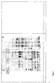
- Object of the invention is also the use of EEF1A1 comprising the amino acid sequence of SEQ ID NO: 1 and/or EEF1A2 comprising the amino acid sequence of SEQ ID NO: 2 (cf. Figure 11 ), or variants, mutants, parts of the amino acid sequence or at least 85 % homologous sequences having the same function, or a nucleic acid encoding EEF1A1 and/or a nucleic acid sequence encoding EEF1A2, as biomarker for a reduction of MetAP2 activity.
- the biomarker can be used for monitoring, determining and/or predicting the reduction of MetAP2 activity, the status of cell proliferation and/or the reduction of likelihood of developing a tumor and/or a progressive tumor growth.
- the different uses can be subsumed under the general term "assessing”. It goes without saying that data are monitored over a specific period, while data are determined at a particular time. Both the period and the time can be easily designed by the skilled artisan depending on the experimental trials conducted.
- EEF1A can be used as biomarker for predicting in-vitro the pharmaceutical efficacy and/or clinical response of a mammal suffering from cancer to a MetAP2-inhibiting drug, which is intended to be administered and/or is administered in cancer treatment.
- the underlying treatment is particularly a first-line treatment, and the inhibitory drug with which the mammalian patient is to be treated, is administered in mono-therapy.
- said drug is combined with a chemotherapeutic agent, and said patient has developed chemo-refractory cancer.
- a method for screening MetAP2 inhibitors or modulators by applying the unique biomarker EEF1A is provided for the first time.
- the present invention teaches the induction and accumulation of MetEEF1A in cells, whose enzymatic MetAP2 activity is interrupted.
- EEF1A as a biomarker for MetAP2 inhibitors has several advantages. It is an abundant cellular protein so it can be easily detected in tumor samples. The N-terminal status of EEF1A reflects the MetAP2 enzyme activity over a period, thus truly representing the consequence of MetAP2 inhibition and better correlating with efficacy of the testing compound.
- MetEEF1A readout is on MetAP2-specfic substrate, but not MetAP2 itself, so it can be used for all types of MetAP2 inhibitors.
- the substrate is processed by MetAP2, but not by MetAP1, which makes it possible to prove that an anti-angiogenic and anti-proliferative activity is specifically due to inhibition of the proteolytic activity of MetAP2.
- MetAP2 Inhibition of methionine processing by MetAP2 blocks tumor cell growth in-vitro. MetAP2 inhibitors even induce MetEEF1A in a dose-dependent fashion similar to that observed for their antiproliferative activity.
- the robust marker has the potential to monitor MetAP2 inhibition not only in cells but also in animals and human subjects treated with MetAP2 inhibitors.
- Induction of MetEEF1A also correlated with the in-vivo anti-angiogenic and anti-tumor activity.
- EEF1A N-terminal status is an outstanding biomarker for cellular MetAP2 inhibition in-vitro and in-vivo. EEF1A processing is of benefit to monitor MetAP2 inhibition in preclinical and clinical testing.
- the analysis of the differential processed EEF1A isoform is very suitable for large-scale screening tests.
- the novel marker allows the identification of novel MetAP2 inhibitors.
- Compounds can be identified and evaluated with a specific cellular mechanism of action and additionally, their potential to exert anti-angiogenic and/or anti-proliferative effects can be favorably proved.
- the characterization of EEF1A, particularly MetEEF1A, critically involved in substrate recognition by MetAP2 results in the provision of pharmaceutical compositions for the diagnosis, prophylactic or therapeutic treatment and/or monitoring of conditions, which are caused, mediated and/or propagated by MetAP2 activity.
- Their use is a promising, novel approach for a broad spectrum of therapies causing a direct and immediate reduction of symptoms that are clearly connected with MetAP2-dependent diseases.
- the compounds are of special benefit as anti-cancer agents in mammals.
- EEF1A is qualified as biomarker for detecting and characterizing MetAP2 activity.
- the detection method as well as arising monitoring method of the invention can be performed in a simple and fast manner.
- the appropriate kit is cost-efficiently produced.
- Targeting EEF1A isoforms is highly specific for the MetAP2 activity and driven medical disorders therefrom. All detecting substances are characterized by a high affinity, specificity and stability; low manufacturing costs and convenient handling.
- Mouse brain endothelioma cells (bEND3, ECACC Nr.9609129) were cultured at 37°C and 10 % CO 2 in D-MEM (Invitrogen, #41965) supplemented with 10 % (v/v) heat-inactivated fetal bovine serum (Pan Biotech, #3302), 1 mM sodium pyruvate (Invitrogen, #11360) and 1 x Nonessential Amino Acid Solution (Sigma, M7145).
- Human colon carcinoma cells (HCT116, ATCC CCL 225) were cultured at 37°C and 10 % CO 2 in MEM alpha (Invitrogen, #22571) supplemented with 10 % (v/v) heat-inactivated fetal bovine serum (Pan Biotech, #3302), 2 mM glutamine (Invitrogen, #25030) and 1 mM sodium pyruvate (Invitrogen, #11360).
- Human umbilical vein endothelial cells (HUVEC, PromoCell, C-12200) are cultivated at 37°C and 5 % CO 2 in endothelial cell growth medium (PromoCell, C-22020) with supplement mix (PromoCell, C-39215).
- Human fibrosarcoma cells (HT1080, ATCC CCL-121) were cultured at 37°C and 10 % CO 2 in D-MEM (Invitrogen, #41965) supplemented with 10% (v/v) heat-inactivated fetal bovine serum (Pan Biotech, #3302).
- cells were re-suspended in IEF-lysis buffer (7 M Urea, 2M thiourea, 4 % CHAPS, 1 % DTT, 1 % Pharmalyte pH 3-10 (GE Healthcare) at a density of 2*10 7 cells per ml and incubated for 30 min with gentle agitation. The lysates were centrifuged for 5 min at 10.000*g, supernatants were collected in fresh eppendorf micro test tubes and finally frozen at - 70°C.
- IEF-lysis buffer 7 M Urea, 2M thiourea, 4 % CHAPS, 1 % DTT, 1 % Pharmalyte pH 3-10 (GE Healthcare) at a density of 2*10 7 cells per ml and incubated for 30 min with gentle agitation. The lysates were centrifuged for 5 min at 10.000*g, supernatants were collected in fresh eppendorf micro test tubes and finally frozen at - 70°C.
- IEF gels (CleanGel IEF ultra ETC1001-52 from ETC, Germany) were re-hydrated in 7 M urea, 2 M thiourea, 1 % DTE, 4 % CHAPS, 3 %, Servalyte 7-9 for 120 min, and assembled on a Multiphor II electrophoresis unit (GE Healthcare). Samples (5 ⁇ l) were applied close to the anode and run with anode buffer (6 M glycine) and cathode fluid 10 (Serva, Germany).
- Coomassie Blue stained acrylamide gel bands were excised after IEF separation, washed twice with 50 % acetonitrile and rinsed with pure acetonitrile.
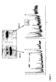
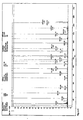
- In gel digest was performed by adding 0.1 ⁇ g trypsin dissolved in 10 ⁇ l ammonium bicarbonate pH 7.4 to the gel band. Incubation was achieved at 37°C overnight. Aliquots of the digest mix were applied onto a target plate of a MALDI-TOF MS (Ultraflex, Bruker, Germany) and ⁇ -cyano-4-hydroxycinnamic acid was added as a matrix. Mass analysis was performed and peptide fragments were detected applying standard protocols.
- the achieved peptide mass lists were searched against the SwissProt database using the Mascot search algorithm (Matrix Science, UK) to identify the protein. Furthermore, distinct peptides from a mass map were used for subsequent MS-fragmentation studies in order to determine the primary sequence of those individual peptides and for unambiguous protein annotation.
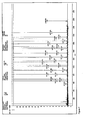
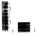
- Table 1 contains explanatory information for Figure 10 .
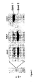
- Compounds were incubated with bEND3 cells for 48 h at the indicated concentrations, lysed and electrophoretic mobility shift of EEF1A1 was assessed in an IEF gel.
- the compounds used were characterized as inhibitors of MetAP2 and inhibitors of HUVEC cell proliferation with the indicated potencies (IC50).
- Inhibition of MetAP2 was measured via cleavage of the tripeptide Met-Ala-Ser and subsequent detection of cleaved methionine using an enzyme-coupling reaction composed of L-amino oxidase and peroxidase ( Wang et al. (2003) Biochemistry 42(17): 5035-5042 ).
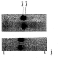
- EXAMPLE 1 Treatment-related pattern shows a new band with lower pl after IEF
- the EEF1A1 mobility shift could be reliably detected in all tested cell types with two structurally distinct MetAP2 inhibitors (TNP470) as it is shown in Figure 9 (shown: bEND3 cell extract). No additional band occurred in the control sample even after 48 h, whereas a second band (Band 2) with increasing intensity was already to be seen after 2 h ( Figure 9 ). Negative controls were unequivocally distinguished from active compounds ( Figure 10 ; Table 1).
- the two isoforms EEF1A1 and EEF1A2 comprise a sequence identity of 92.7 % ( Figure 11 ). Both isoforms are identical within the first 82 amino acids and, therefore, are substrates of MetAP2.
Landscapes
- Health & Medical Sciences (AREA)
- Life Sciences & Earth Sciences (AREA)
- Engineering & Computer Science (AREA)
- Chemical & Material Sciences (AREA)
- Immunology (AREA)
- Molecular Biology (AREA)
- Organic Chemistry (AREA)
- Biomedical Technology (AREA)
- Proteomics, Peptides & Aminoacids (AREA)
- Bioinformatics & Cheminformatics (AREA)
- Hematology (AREA)
- Microbiology (AREA)
- Biotechnology (AREA)
- Urology & Nephrology (AREA)
- Zoology (AREA)
- Wood Science & Technology (AREA)
- Toxicology (AREA)
- Physics & Mathematics (AREA)
- Analytical Chemistry (AREA)
- Biochemistry (AREA)
- General Health & Medical Sciences (AREA)
- General Physics & Mathematics (AREA)
- Pathology (AREA)
- Biophysics (AREA)
- Cell Biology (AREA)
- Medicinal Chemistry (AREA)
- Food Science & Technology (AREA)
- Tropical Medicine & Parasitology (AREA)
- General Engineering & Computer Science (AREA)
- Genetics & Genomics (AREA)
- Investigating Or Analysing Biological Materials (AREA)
- Measuring Or Testing Involving Enzymes Or Micro-Organisms (AREA)
Priority Applications (1)
| Application Number | Priority Date | Filing Date | Title |
|---|---|---|---|
| EP09778819A EP2344665B1 (en) | 2008-11-06 | 2009-10-05 | Use of eef1a as biomarker for screening of metap2 inhibitors |
Applications Claiming Priority (3)
| Application Number | Priority Date | Filing Date | Title |
|---|---|---|---|
| EP08019432 | 2008-11-06 | ||
| EP09778819A EP2344665B1 (en) | 2008-11-06 | 2009-10-05 | Use of eef1a as biomarker for screening of metap2 inhibitors |
| PCT/EP2009/007102 WO2010051882A1 (en) | 2008-11-06 | 2009-10-05 | Use of eef1a as biomarker and a method of screening metap2 inhibitors |
Publications (2)
| Publication Number | Publication Date |
|---|---|
| EP2344665A1 EP2344665A1 (en) | 2011-07-20 |
| EP2344665B1 true EP2344665B1 (en) | 2013-01-23 |
Family
ID=41409225
Family Applications (1)
| Application Number | Title | Priority Date | Filing Date |
|---|---|---|---|
| EP09778819A Active EP2344665B1 (en) | 2008-11-06 | 2009-10-05 | Use of eef1a as biomarker for screening of metap2 inhibitors |
Country Status (8)
| Country | Link |
|---|---|
| US (1) | US9151743B2 (enExample) |
| EP (1) | EP2344665B1 (enExample) |
| JP (1) | JP5347028B2 (enExample) |
| AU (1) | AU2009313167C1 (enExample) |
| CA (1) | CA2742737C (enExample) |
| ES (1) | ES2402564T3 (enExample) |
| IL (1) | IL212664A0 (enExample) |
| WO (1) | WO2010051882A1 (enExample) |
Cited By (1)
| Publication number | Priority date | Publication date | Assignee | Title |
|---|---|---|---|---|
| CN108611352A (zh) * | 2018-04-20 | 2018-10-02 | 华南农业大学 | 一种拟禾本科根结线虫翻译延长因子Mg-eEF1A及其防治植物病害的应用 |
Families Citing this family (5)
| Publication number | Priority date | Publication date | Assignee | Title |
|---|---|---|---|---|
| WO2015069489A1 (en) | 2013-11-06 | 2015-05-14 | Merck Patent Gmbh | Predictive biomarker for hypoxia-activated prodrug therapy |
| KR101712982B1 (ko) * | 2015-07-31 | 2017-03-07 | 고려대학교 산학협력단 | 비알콜성지방간 조절인자 14-3-3 단백질 |
| US20200030278A1 (en) * | 2017-02-10 | 2020-01-30 | Zafgen, Inc. | Pharmaceutical compositions of metap-2 inhibitors |
| CN115461054A (zh) * | 2020-02-06 | 2022-12-09 | 加利福尼亚大学董事会 | 延伸因子1-α抑制剂及其用途 |
| CN116395718B (zh) * | 2023-04-12 | 2024-12-17 | 中国科学院长春应用化学研究所 | 一种弱碱性钠盐纳米药物、其制备方法及其应用 |
Family Cites Families (4)
| Publication number | Priority date | Publication date | Assignee | Title |
|---|---|---|---|---|
| WO1998056372A1 (en) * | 1997-06-09 | 1998-12-17 | Massachusetts Institute Of Technology | TYPE 2 METHIONINE AMINOPEPTIDASE (MetAP2) INHIBITORS AND USES THEROF |
| WO2002039990A2 (en) * | 2000-11-14 | 2002-05-23 | Novartis Ag | Method for screening anti-proliferative compounds and inhibiting tumor growth |
| US20020182701A1 (en) * | 2001-08-30 | 2002-12-05 | Saint Louis University | Dominant negative variants of methionine aminopeptidase 2 (MetAP2) and clinical uses thereof |
| US20030232383A1 (en) | 2001-11-02 | 2003-12-18 | Sylvia Daunert | Novel reagentless sensing system for measuring carbohydrates based on the galactose/glucose binding protein |
-
2009
- 2009-10-05 ES ES09778819T patent/ES2402564T3/es active Active
- 2009-10-05 CA CA2742737A patent/CA2742737C/en active Active
- 2009-10-05 AU AU2009313167A patent/AU2009313167C1/en active Active
- 2009-10-05 WO PCT/EP2009/007102 patent/WO2010051882A1/en not_active Ceased
- 2009-10-05 US US13/127,834 patent/US9151743B2/en active Active
- 2009-10-05 EP EP09778819A patent/EP2344665B1/en active Active
- 2009-10-05 JP JP2011533565A patent/JP5347028B2/ja active Active
-
2011
- 2011-05-03 IL IL212664A patent/IL212664A0/en active IP Right Grant
Cited By (2)
| Publication number | Priority date | Publication date | Assignee | Title |
|---|---|---|---|---|
| CN108611352A (zh) * | 2018-04-20 | 2018-10-02 | 华南农业大学 | 一种拟禾本科根结线虫翻译延长因子Mg-eEF1A及其防治植物病害的应用 |
| CN108611352B (zh) * | 2018-04-20 | 2020-08-07 | 华南农业大学 | 一种拟禾本科根结线虫翻译延长因子Mg-eEF1A及其防治植物病害的应用 |
Also Published As
| Publication number | Publication date |
|---|---|
| CA2742737A1 (en) | 2010-05-14 |
| WO2010051882A1 (en) | 2010-05-14 |
| EP2344665A1 (en) | 2011-07-20 |
| ES2402564T3 (es) | 2013-05-06 |
| JP2012507268A (ja) | 2012-03-29 |
| IL212664A0 (en) | 2011-07-31 |
| JP5347028B2 (ja) | 2013-11-20 |
| US20110275088A1 (en) | 2011-11-10 |
| CA2742737C (en) | 2018-05-29 |
| AU2009313167A1 (en) | 2011-06-23 |
| AU2009313167C1 (en) | 2014-06-12 |
| US9151743B2 (en) | 2015-10-06 |
Similar Documents
| Publication | Publication Date | Title |
|---|---|---|
| US8119355B2 (en) | Biomarkers for cancer sensitivity and uses thereof | |
| US20070292869A1 (en) | Compositions and Methods for Analyzing Renal Cancer | |
| EP2344665B1 (en) | Use of eef1a as biomarker for screening of metap2 inhibitors | |
| EP2620770B1 (en) | Novel testing method for angiitis | |
| JP6826543B2 (ja) | アルツハイマー病に関係する生体分子 | |
| KR20160045547A (ko) | 췌장암 진단용 조성물 및 이를 이용한 췌장암 진단방법 | |
| EP2998740B1 (en) | Method for predicting clinical effect of immunotherapy | |
| AU2009313167B2 (en) | Use of EEF1A as biomarker for screening of MetAP2 inhibitors | |
| CN104364649B (zh) | 用于鉴定具有基因毒性的化合物的蛋白表达分析 | |
| US20130287801A1 (en) | Biomarkers, uses of biomarkers and a method of identifying biomarkers | |
| EP2270510A1 (en) | Diagnostic method for predicting the risk of cancer recurrence based on Histone macroH2A isoforms | |
| JP4515099B2 (ja) | Lasp−1免疫反応性の決定によって炎症性疾患および感染を診断するための方法 | |
| JP6361943B2 (ja) | 補体因子bタンパク質に特異的に結合する抗体及び糖鎖抗原19−9タンパク質に特異的に結合する抗体を含む膵臓癌診断用キット | |
| WO2006091861A2 (en) | Compositions and methods relating to cns lymphoma | |
| WO2011009523A1 (en) | Eg5 as biomarker in rare thoracic cancer | |
| EP2307561A2 (en) | Interferon epsilon (ifne1) as a marker for targeted cancer therapy | |
| EP4075141A2 (en) | Method for predicting reactivity to drug targeting bcl2 family protein | |
| US9977026B2 (en) | Detection of elevated levels of phosphorylated MCM and method of increasing MCM phosphorylation capacity | |
| US9903871B2 (en) | Stabilized peptide fragments from nucleoredoxin X1 and uses thereof | |
| WO2015073509A2 (en) | Biomarker for melk activity and methods of using same | |
| CN119548517A (zh) | 一种m2巨噬细胞的诱导剂及应用 | |
| WO2007037532A9 (ja) | Srms遺伝子の治療的又は診断的用途 | |
| JP2017515108A (ja) | 肉腫の転移を検出するための方法およびバイオマーカー |
Legal Events
| Date | Code | Title | Description |
|---|---|---|---|
| PUAI | Public reference made under article 153(3) epc to a published international application that has entered the european phase |
Free format text: ORIGINAL CODE: 0009012 |
|
| 17P | Request for examination filed |
Effective date: 20110202 |
|
| AK | Designated contracting states |
Kind code of ref document: A1 Designated state(s): AT BE BG CH CY CZ DE DK EE ES FI FR GB GR HR HU IE IS IT LI LT LU LV MC MK MT NL NO PL PT RO SE SI SK SM TR |
|
| AX | Request for extension of the european patent |
Extension state: AL BA RS |
|
| DAX | Request for extension of the european patent (deleted) | ||
| 17Q | First examination report despatched |
Effective date: 20120217 |
|
| GRAP | Despatch of communication of intention to grant a patent |
Free format text: ORIGINAL CODE: EPIDOSNIGR1 |
|
| GRAS | Grant fee paid |
Free format text: ORIGINAL CODE: EPIDOSNIGR3 |
|
| GRAA | (expected) grant |
Free format text: ORIGINAL CODE: 0009210 |
|
| AK | Designated contracting states |
Kind code of ref document: B1 Designated state(s): AT BE BG CH CY CZ DE DK EE ES FI FR GB GR HR HU IE IS IT LI LT LU LV MC MK MT NL NO PL PT RO SE SI SK SM TR |
|
| REG | Reference to a national code |
Ref country code: GB Ref legal event code: FG4D |
|
| REG | Reference to a national code |
Ref country code: CH Ref legal event code: EP |
|
| REG | Reference to a national code |
Ref country code: AT Ref legal event code: REF Ref document number: 595015 Country of ref document: AT Kind code of ref document: T Effective date: 20130215 Ref country code: CH Ref legal event code: EP |
|
| REG | Reference to a national code |
Ref country code: IE Ref legal event code: FG4D |
|
| REG | Reference to a national code |
Ref country code: DE Ref legal event code: R096 Ref document number: 602009013054 Country of ref document: DE Effective date: 20130321 |
|
| REG | Reference to a national code |
Ref country code: ES Ref legal event code: FG2A Ref document number: 2402564 Country of ref document: ES Kind code of ref document: T3 Effective date: 20130506 |
|
| REG | Reference to a national code |
Ref country code: AT Ref legal event code: MK05 Ref document number: 595015 Country of ref document: AT Kind code of ref document: T Effective date: 20130123 |
|
| REG | Reference to a national code |
Ref country code: LT Ref legal event code: MG4D |
|
| REG | Reference to a national code |
Ref country code: NL Ref legal event code: VDEP Effective date: 20130123 |
|
| PG25 | Lapsed in a contracting state [announced via postgrant information from national office to epo] |
Ref country code: SE Free format text: LAPSE BECAUSE OF FAILURE TO SUBMIT A TRANSLATION OF THE DESCRIPTION OR TO PAY THE FEE WITHIN THE PRESCRIBED TIME-LIMIT Effective date: 20130123 Ref country code: BE Free format text: LAPSE BECAUSE OF FAILURE TO SUBMIT A TRANSLATION OF THE DESCRIPTION OR TO PAY THE FEE WITHIN THE PRESCRIBED TIME-LIMIT Effective date: 20130123 Ref country code: NO Free format text: LAPSE BECAUSE OF FAILURE TO SUBMIT A TRANSLATION OF THE DESCRIPTION OR TO PAY THE FEE WITHIN THE PRESCRIBED TIME-LIMIT Effective date: 20130423 Ref country code: BG Free format text: LAPSE BECAUSE OF FAILURE TO SUBMIT A TRANSLATION OF THE DESCRIPTION OR TO PAY THE FEE WITHIN THE PRESCRIBED TIME-LIMIT Effective date: 20130423 Ref country code: AT Free format text: LAPSE BECAUSE OF FAILURE TO SUBMIT A TRANSLATION OF THE DESCRIPTION OR TO PAY THE FEE WITHIN THE PRESCRIBED TIME-LIMIT Effective date: 20130123 Ref country code: IS Free format text: LAPSE BECAUSE OF FAILURE TO SUBMIT A TRANSLATION OF THE DESCRIPTION OR TO PAY THE FEE WITHIN THE PRESCRIBED TIME-LIMIT Effective date: 20130523 Ref country code: LT Free format text: LAPSE BECAUSE OF FAILURE TO SUBMIT A TRANSLATION OF THE DESCRIPTION OR TO PAY THE FEE WITHIN THE PRESCRIBED TIME-LIMIT Effective date: 20130123 |
|
| PG25 | Lapsed in a contracting state [announced via postgrant information from national office to epo] |
Ref country code: GR Free format text: LAPSE BECAUSE OF FAILURE TO SUBMIT A TRANSLATION OF THE DESCRIPTION OR TO PAY THE FEE WITHIN THE PRESCRIBED TIME-LIMIT Effective date: 20130424 Ref country code: PT Free format text: LAPSE BECAUSE OF FAILURE TO SUBMIT A TRANSLATION OF THE DESCRIPTION OR TO PAY THE FEE WITHIN THE PRESCRIBED TIME-LIMIT Effective date: 20130523 Ref country code: LV Free format text: LAPSE BECAUSE OF FAILURE TO SUBMIT A TRANSLATION OF THE DESCRIPTION OR TO PAY THE FEE WITHIN THE PRESCRIBED TIME-LIMIT Effective date: 20130123 Ref country code: PL Free format text: LAPSE BECAUSE OF FAILURE TO SUBMIT A TRANSLATION OF THE DESCRIPTION OR TO PAY THE FEE WITHIN THE PRESCRIBED TIME-LIMIT Effective date: 20130123 Ref country code: SI Free format text: LAPSE BECAUSE OF FAILURE TO SUBMIT A TRANSLATION OF THE DESCRIPTION OR TO PAY THE FEE WITHIN THE PRESCRIBED TIME-LIMIT Effective date: 20130123 Ref country code: FI Free format text: LAPSE BECAUSE OF FAILURE TO SUBMIT A TRANSLATION OF THE DESCRIPTION OR TO PAY THE FEE WITHIN THE PRESCRIBED TIME-LIMIT Effective date: 20130123 Ref country code: NL Free format text: LAPSE BECAUSE OF FAILURE TO SUBMIT A TRANSLATION OF THE DESCRIPTION OR TO PAY THE FEE WITHIN THE PRESCRIBED TIME-LIMIT Effective date: 20130123 |
|
| PG25 | Lapsed in a contracting state [announced via postgrant information from national office to epo] |
Ref country code: HR Free format text: LAPSE BECAUSE OF FAILURE TO SUBMIT A TRANSLATION OF THE DESCRIPTION OR TO PAY THE FEE WITHIN THE PRESCRIBED TIME-LIMIT Effective date: 20130123 |
|
| PG25 | Lapsed in a contracting state [announced via postgrant information from national office to epo] |
Ref country code: SK Free format text: LAPSE BECAUSE OF FAILURE TO SUBMIT A TRANSLATION OF THE DESCRIPTION OR TO PAY THE FEE WITHIN THE PRESCRIBED TIME-LIMIT Effective date: 20130123 Ref country code: CZ Free format text: LAPSE BECAUSE OF FAILURE TO SUBMIT A TRANSLATION OF THE DESCRIPTION OR TO PAY THE FEE WITHIN THE PRESCRIBED TIME-LIMIT Effective date: 20130123 Ref country code: DK Free format text: LAPSE BECAUSE OF FAILURE TO SUBMIT A TRANSLATION OF THE DESCRIPTION OR TO PAY THE FEE WITHIN THE PRESCRIBED TIME-LIMIT Effective date: 20130123 Ref country code: EE Free format text: LAPSE BECAUSE OF FAILURE TO SUBMIT A TRANSLATION OF THE DESCRIPTION OR TO PAY THE FEE WITHIN THE PRESCRIBED TIME-LIMIT Effective date: 20130123 Ref country code: RO Free format text: LAPSE BECAUSE OF FAILURE TO SUBMIT A TRANSLATION OF THE DESCRIPTION OR TO PAY THE FEE WITHIN THE PRESCRIBED TIME-LIMIT Effective date: 20130123 |
|
| PG25 | Lapsed in a contracting state [announced via postgrant information from national office to epo] |
Ref country code: CY Free format text: LAPSE BECAUSE OF FAILURE TO SUBMIT A TRANSLATION OF THE DESCRIPTION OR TO PAY THE FEE WITHIN THE PRESCRIBED TIME-LIMIT Effective date: 20130123 |
|
| PLBE | No opposition filed within time limit |
Free format text: ORIGINAL CODE: 0009261 |
|
| STAA | Information on the status of an ep patent application or granted ep patent |
Free format text: STATUS: NO OPPOSITION FILED WITHIN TIME LIMIT |
|
| 26N | No opposition filed |
Effective date: 20131024 |
|
| REG | Reference to a national code |
Ref country code: DE Ref legal event code: R097 Ref document number: 602009013054 Country of ref document: DE Effective date: 20131024 |
|
| PG25 | Lapsed in a contracting state [announced via postgrant information from national office to epo] |
Ref country code: MC Free format text: LAPSE BECAUSE OF FAILURE TO SUBMIT A TRANSLATION OF THE DESCRIPTION OR TO PAY THE FEE WITHIN THE PRESCRIBED TIME-LIMIT Effective date: 20130123 |
|
| REG | Reference to a national code |
Ref country code: IE Ref legal event code: MM4A |
|
| PG25 | Lapsed in a contracting state [announced via postgrant information from national office to epo] |
Ref country code: IE Free format text: LAPSE BECAUSE OF NON-PAYMENT OF DUE FEES Effective date: 20131005 |
|
| PG25 | Lapsed in a contracting state [announced via postgrant information from national office to epo] |
Ref country code: SM Free format text: LAPSE BECAUSE OF FAILURE TO SUBMIT A TRANSLATION OF THE DESCRIPTION OR TO PAY THE FEE WITHIN THE PRESCRIBED TIME-LIMIT Effective date: 20130123 |
|
| PG25 | Lapsed in a contracting state [announced via postgrant information from national office to epo] |
Ref country code: TR Free format text: LAPSE BECAUSE OF FAILURE TO SUBMIT A TRANSLATION OF THE DESCRIPTION OR TO PAY THE FEE WITHIN THE PRESCRIBED TIME-LIMIT Effective date: 20130123 |
|
| PG25 | Lapsed in a contracting state [announced via postgrant information from national office to epo] |
Ref country code: LU Free format text: LAPSE BECAUSE OF NON-PAYMENT OF DUE FEES Effective date: 20131005 Ref country code: HU Free format text: LAPSE BECAUSE OF FAILURE TO SUBMIT A TRANSLATION OF THE DESCRIPTION OR TO PAY THE FEE WITHIN THE PRESCRIBED TIME-LIMIT; INVALID AB INITIO Effective date: 20091005 Ref country code: MK Free format text: LAPSE BECAUSE OF FAILURE TO SUBMIT A TRANSLATION OF THE DESCRIPTION OR TO PAY THE FEE WITHIN THE PRESCRIBED TIME-LIMIT Effective date: 20130123 |
|
| PG25 | Lapsed in a contracting state [announced via postgrant information from national office to epo] |
Ref country code: MT Free format text: LAPSE BECAUSE OF FAILURE TO SUBMIT A TRANSLATION OF THE DESCRIPTION OR TO PAY THE FEE WITHIN THE PRESCRIBED TIME-LIMIT Effective date: 20130123 |
|
| REG | Reference to a national code |
Ref country code: FR Ref legal event code: PLFP Year of fee payment: 8 |
|
| REG | Reference to a national code |
Ref country code: FR Ref legal event code: PLFP Year of fee payment: 9 |
|
| REG | Reference to a national code |
Ref country code: FR Ref legal event code: PLFP Year of fee payment: 10 |
|
| P01 | Opt-out of the competence of the unified patent court (upc) registered |
Effective date: 20230518 |
|
| PGFP | Annual fee paid to national office [announced via postgrant information from national office to epo] |
Ref country code: IT Payment date: 20250923 Year of fee payment: 17 |
|
| PGFP | Annual fee paid to national office [announced via postgrant information from national office to epo] |
Ref country code: GB Payment date: 20250923 Year of fee payment: 17 |
|
| PGFP | Annual fee paid to national office [announced via postgrant information from national office to epo] |
Ref country code: FR Payment date: 20250923 Year of fee payment: 17 |
|
| REG | Reference to a national code |
Ref country code: CH Ref legal event code: U11 Free format text: ST27 STATUS EVENT CODE: U-0-0-U10-U11 (AS PROVIDED BY THE NATIONAL OFFICE) Effective date: 20251101 |
|
| PGFP | Annual fee paid to national office [announced via postgrant information from national office to epo] |
Ref country code: DE Payment date: 20250923 Year of fee payment: 17 |
|
| PGFP | Annual fee paid to national office [announced via postgrant information from national office to epo] |
Ref country code: CH Payment date: 20251101 Year of fee payment: 17 |
|
| PGFP | Annual fee paid to national office [announced via postgrant information from national office to epo] |
Ref country code: ES Payment date: 20251103 Year of fee payment: 17 |