EP2321752B1 - Bilddatenverwaltungssysteme - Google Patents
Bilddatenverwaltungssysteme Download PDFInfo
- Publication number
- EP2321752B1 EP2321752B1 EP09785393.1A EP09785393A EP2321752B1 EP 2321752 B1 EP2321752 B1 EP 2321752B1 EP 09785393 A EP09785393 A EP 09785393A EP 2321752 B1 EP2321752 B1 EP 2321752B1
- Authority
- EP
- European Patent Office
- Prior art keywords
- data
- consistency
- imaging
- image
- imaging data
- Prior art date
- Legal status (The legal status is an assumption and is not a legal conclusion. Google has not performed a legal analysis and makes no representation as to the accuracy of the status listed.)
- Active
Links
Images


Classifications
-
- G—PHYSICS
- G16—INFORMATION AND COMMUNICATION TECHNOLOGY [ICT] SPECIALLY ADAPTED FOR SPECIFIC APPLICATION FIELDS
- G16H—HEALTHCARE INFORMATICS, i.e. INFORMATION AND COMMUNICATION TECHNOLOGY [ICT] SPECIALLY ADAPTED FOR THE HANDLING OR PROCESSING OF MEDICAL OR HEALTHCARE DATA
- G16H30/00—ICT specially adapted for the handling or processing of medical images
- G16H30/20—ICT specially adapted for the handling or processing of medical images for handling medical images, e.g. DICOM, HL7 or PACS
-
- G—PHYSICS
- G16—INFORMATION AND COMMUNICATION TECHNOLOGY [ICT] SPECIALLY ADAPTED FOR SPECIFIC APPLICATION FIELDS
- G16H—HEALTHCARE INFORMATICS, i.e. INFORMATION AND COMMUNICATION TECHNOLOGY [ICT] SPECIALLY ADAPTED FOR THE HANDLING OR PROCESSING OF MEDICAL OR HEALTHCARE DATA
- G16H30/00—ICT specially adapted for the handling or processing of medical images
- G16H30/40—ICT specially adapted for the handling or processing of medical images for processing medical images, e.g. editing
Definitions
- the present invention relates to data management systems and in particular to systems for managing data, such as image data (radiological, histological etc.), collected and/or analyzed as part of clinical trials or in healthcare.
- data such as image data (radiological, histological etc.), collected and/or analyzed as part of clinical trials or in healthcare.
- the DICOM image format includes the image, and also a header containing metadata that relates to the subject and the acquired images. For clinical trial use, it is accompanied by a paper or electronic form that contains associated information which is not included in the DICOM metadata (e.g. the trial identifier, time-point in the trial, any comments made by the collecting site), and may also be accompanied by other data files.
- DICOM provides a standard format for image transfer and storage, it does not standardize many of the components of the DICOM header (which includes a number of tags), which are entered by the person operating the scanner. Such tags include the Series Description, which defines the type of scan (e.g.
- T1 weighted vs T2 weighted MRI scan will invariably depend on the language spoken in the country where the data is collected, procedures at that site, and is additionally prone to human error.
- the DICOM tags do not contain certain relevant information required in clinical trials, such as a precise description of the anatomy imaged (so that checks can be made that the correct anatomy was imaged), the compound being used or its mode of action, and many types of data required for the quantitative analysis of image data.
- DICOM is widely used, it is not a universally supported format - especially for analysed results - and so other formats need to be handled also, and these have different ways of storing metadata, sometimes primarily in the file name and folder names that contain these files.
- US2007/0025606 discloses an automated method and system for indexing, storing and searching medical images.
- the present invention provides a system according to claim 1.
- the system may be arranged to admit the data for at least one of storage and analysis.
- the present invention makes use of the fact that, for all clinical trials, and for properly protocoled patient management in healthcare, the expected types of images, the part of the subject to be imaged, the type of associated data or associated files, the timing of acquisition, and the analysis results to be obtained from these images, is pre-defined by the protocol that is fixed before the data collection begins, with any revisions being properly documented. Similarly for histological studies there is, in appropriate cases, a protocol which defines the times and types of the images.. This provides important prior information about what is expected. Similarly, example images of each type (with the same physical properties and of the correct anatomy) are likely to be available e.g. from a previous subjects being imaged with a similar protocol (E.g. a previous clinical trial), collected as dedicated reference data, or collected during a preparatory period (e.g: site qualification) before patients are recruited.
- a protocol which defines the times and types of the images.
- the present invention provides, in some embodiments, a system that can import DICOM images with accompanying non-DICOM metadata and analysis results, automatically clean it, and correctly populate an image database or repository.
- Any problem data needs to be identified, any mis-labelling or ambiguous labelling resolved, and the images and associated data correctly assigned to scan type, subject, site, time point, trial etc.
- the data might need to be imported all in one go (e.g. at the end of a trial), or as the trial proceeds.
- the identification of missing or mis-labelled data can be performed in real-time to enable any errors to be resolved automatically, or by the means of user-interaction, at the time of import.
- an imaging data management system is arranged to manage data on a central server 2 at a central site.
- Data can be uploaded from a number of sites, for example from a number of scanners 3 (only one of which is shown) via computers 4 at each scanner site.
- Associated data may be loaded up both from the same computer as the images and/or from separate computers.
- Tasks can be scheduled to computers 5, if they can be performed automatically, and to workstations 6 if they are to be performed manually. The results of the trial can be reported to a client system 7.
- the central server 2 is arranged to receive imaging data, in the form of actual image data, and associated metadata. This may be in the form of DICOM files, and comes from computers 4 at the scanner sites.
- the central server 2 is further arranged to perform a number of data checking and cleaning tasks, and to output the cleaned data for example to provide reports to the client 7.
- the central server 2 includes a first memory area that serves as a quarantine 10 in which incoming data is held, a matching system 11, typically comprising a processor, arranged to perform the various checking and cleaning tasks that will be described in more detail below, and a further memory area 12 for holding the cleaned data ready for exporting (e.g.: for subsequent analysis).
- the central server 2 is therefore arranged to use prior information about the images and associated data (metadata) that is expected which typically includes any one or more of the following:
- this reference data is stored in a trial or clinical configuration file, which is derived from the trial protocol or clinical protocol, or histology protocol 15.
- the protocol is generated when the trial is planned and, the derived trial configuration file is stored on the central server 2.
- a protocol is defined for the treatment of patients that have similar symptoms or diagnoses or are undergoing similar treatments, and these protocols are also known in advance, though they may be more flexible than clinical trial protocols.
- the server 2 may obtain aspects of the trial configuration or clinical protocol by accessing a further computer system such as a Clinical Trial Management System (CTMS) or Clinical Data Management System (CDMS) or some other hospital or radiological information system.
- CTMS Clinical Trial Management System
- CDMS Clinical Data Management System
- some of the data such as example images, may be stored in a separate file or files 16.
- the central server 2 specifically the matching system 11 is arranged to match in-coming imaging data comprising the image data and metadata (normally a large number of files either arriving in bulk after the trial is completed, or arriving gradually during the course of the trial) against the prior knowledge about what is expected in order to allocate correct labels to the required image attributes.
- the matching involves analysis of the metadata, and of the image data itself (by comparison with reference data and previously collected data). In some circumstances, the expected Subject or Patient IDs will be incorporated in the trial configuration file.
- the MIME types of the files, the names and properties of data files and the names and properties of the folders containing them can be used in the matching alongside the contents of the files themselves.
- the addresses of the computers being used and the identities of the users uploading the data can also be used in the matching process.
- Data is quarantined while this matching is going on, and any data for which the match confidence is below a defined threshold is left in quarantine for inspection by a user.
- the results of the match are presented to a user via the user interface 6 immediately following their calculation, to enable the user to appreciate whether the data uploaded matches the criterion, or to prompt for clarification or additional data.
- the matching is performed in a combined multidimensional classifier space.
- the dimensions correspond to different properties of the imaging data, such as values of parameters defined in the metadata or properties of the image data.
- the matching includes: matching metadata values in the incoming data against the reference metadata values. These metadata values can therefore define one or more dimensions of the classifier space.
- Other dimensions can relate to image derived measures such as intensity histograms.
- the image intensity histograms can be quite distinct for a particular type of image acquisition, and can be calculated by plotting voxel intensity (brightness) on the horizontal axis, and frequency of occurrence of this brightness on the vertical axis.
- Another type of image-derived measure is the degree of matching of the anatomical region or organs imaged, which can be determined by aligning the uploaded images against reference images using an image registration algorithm and ascertaining whether the overlap between the acquired and reference anatomy is sufficiently close to meet the requirements of the analysis.
- the reference image could be from a different subject, the same subject at an earlier time or from one or multiple atlases, or feature databases.
- Incoming data can therefore be matched to expected data or reference data by determining the proximity between the two in the multi-dimensional classification space. Where there are more than one set of reference or expected data, the one which is closest to the incoming data in the classification space is identified as the closest match. Once the closest match has been found a confidence parameter dependent on the proximity is associated with the match, and the incoming data can be corrected if appropriate.
- a further image-derived measure is the detection and quality checking of any contrast agent used during the examination, such as oral iodinated contrast in CT or intravenous Gd-DTPA contrast in MRI.
- the matching of an individual image to a reference image can also be used to determine whether a contrast agent has been used correctly during the examination (both whether the contrast agent has been injected at all, and optionally also whether the timing of the injection and image collection is as prescribed).
- the reference image can be pre-labelled with one or more known vascular regions (e.g.: major vessels or vascular tissue), and following alignment of the current image to the reference image using an image registration algorithm, the intensity properties of the image in the region(s) of interest are compared between the current and reference image (e.g: using a comparison of the mean intensity or intensity histogram for the two images) to determine whether the pattern of intensity enhancement is consistent with the use of contrast agent in the examination, and whether the image has been acquired correctly following injection of that agent (eg: venous phase vs arterial phase).
- the change in intensity of this region with time can also be compared between the current and reference image in order to assess whether the dynamic change in contrast is consistent with correct use of the contrast agent during the examination.
- the classifiers which can be adaptive algorithms run on the matching system 11, can learn more about the expected study data to increase the confidence of the labelling.
- one part of the checking of incoming image data files includes checking at step 30 the site label which indicates the site at which the scan was performed. If the site label is unambiguous (e.g. it is in the correct format and corresponds to a site for which data collected at that timepoint is expected), then the process moves to step 34 where it checks the subject ID against other IDs associated with that site. If the site label is ambiguous, or if step 34 fails to produce a candidate match then the process moves to step 36 where it checks the subject ID against all possible subject IDs from all sites to identify the closest match. From the closest, or identical, subject ID the correct site with which that subject is associated can be identified. From either of steps 34 or 36 the server can generate at step 38 a candidate site and subject ID with a confidence level indicating a probability that the candidate data is correct.
- the site label is unambiguous (e.g. it is in the correct format and corresponds to a site for which data collected at that timepoint is expected)
- step 34 checks the subject ID against other IDs associated with that site. If
- the acquisition times indicated in the data files are checked at step 40 against the expected acquisition times as defined in the trial configuration file, or as calculated from the date and time of previous data from the same subject along with between-visit intervals in the trial or clinical configuration file.
- This comparison identifies the most likely acquisition time point for each image, which is used as a candidate time point 42, and those candidate time points with associated confidence levels are also output.
- the server 2 checks the labels in the images (eg: DICOM tags) and any associated data files against the labels in the trial configuration file, and identifies the most likely data labels together with a confidence level.
- the check can be against expected labels for the site identified in the image file, if that is matched to a valid site, and against expected labels from all sites if there is no valid site match.
- various properties of the images in the image files are determined, by analysis of the image data, and compared with corresponding properties of the appropriate reference images 16. Properties which may be used for this comparison include the noise spectrum of the image, i.e. the level of noise as a function of spatial frequency, the signal to noise ratio of the image, image histogram metrics including skewness or similarity of match with a reference image histogram, or image features, i.e.
- the various image properties of the received images are compared with the corresponding properties of clean images from the same site, or earlier images from scans of the same subject at earlier time points.
- each of the properties which is compared has one or more labels or parameters associated with it.
- the noise spectrum of the image may be unique to a particular scanner, or a particular scanner type, and therefore a reference noise spectrum may be indicative of the image having been acquired on a particular scanner or type of scanner. Therefore the degree of similarity between the noise spectrum of the received image and those of a number of reference images may identify the scanner on which the received image was acquired, with an associated confidence determined by the degree of similarity.
- Reference image features may be associated with particular scan parameters, for example the part of anatomy being scanned, or the orientation of the subject during the scan. These parameters will generally be defined in the trial or clinical protocol file by means of associated metadata or labels.
- a further aspect of the checking relates to the metadata that describes in words the type of image collected (eg: the DICOM series description, but could be other metadata fields within the file or file or folder names). Such a description is often entered by a user and is particularly prone to error.. That data is compared with other metadata to confirm that it is consistent with that, and also with properties of the image data, which depend on the type of scanner used or the type of scan performed, to check that it is consistent with that data.
- the metadata that describes in words the type of image collected (eg: the DICOM series description, but could be other metadata fields within the file or file or folder names). Such a description is often entered by a user and is particularly prone to error.. That data is compared with other metadata to confirm that it is consistent with that, and also with properties of the image data, which depend on the type of scanner used or the type of scan performed, to check that it is consistent with that data.
- the trial configuration file will define what type of files are expected (eg: by their MIME type or filename extension) at each time point, and the system can search a specified disk to find files of the expected type that contain relevant identifiers in the filename, directory name or within the file contents.
- the system can search relevant portions of the computer disk from which the data is being uploaded to find any files of this type that contain the required subject ID in the file name or the directory name (matching strings using methods such as regular expressions and Levenshtein Distance). For each candidate match, a match metric is provided. If there is only one match above a pre-determined threshold, this file can be uploaded and associated with the relevant subject visit automatically. If there are multiple possible matches, then the different possible matches can be presented to the user via the user interface 6 for them to select the match that is correct.
- a complete candidate set of metadata, (or labels) with associated confidence levels can be defined for all image files and associated files.
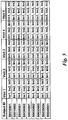
- an example of a trial protocol defines a number of subject identities, a number of visits that each subject is expected to make to the scanner site including the identity of the site and an acceptable range of times for each of the visits, and a number of different scan images that need to be acquired at each visit, including the type of scanner and the type of image to be acquired with the scanner, and also the types of associated data that may be collected along with the images, and may also include the analysis results expected from these data.
- This associated data or analysis results could for example be in the form of simple tables of numbers entered by a user, or computer files generated by the scanner or by physiological monitoring devices.
- the acceptable range of times may be defined in absolute terms, but are more likely to be in terms of time intervals between each visit and the previous one, or each visit and a reference time point, such as a start date of the trial or initial diagnosis or commencement of a treatment protocol.
- These definitions of the different expected images and associated files therefore include data defining labels, characteristics and features of the expected data. In the example shown three images are required of each subject at each visit.
- the timing of the visits is also defined, typically in terms of an acceptable range of time intervals between each consecutive pair of visits. Referring back to Figure 2 , once the data cleaning has been performed on an individual image file, the matching system is arranged to identify a candidate image definition, i.e. a candidate location in the table of Figure 5 , for that image file.
- the confidence level is checked. If it is above a predetermined limit, which can be set independently for each parameter, then the image data, or the metadata, of the received imaging data file can be corrected or left as appropriate so that it corresponds to the reference data. For example, if the subject ID is very close to, but slightly different from, a valid ID, then it can be corrected automatically. If all the data in the imaging file suggest that it came from one particular scanner, apart from the scanner ID in the file metadata, then assuming the confidence is high enough the scanner ID can be corrected automatically. In other cases, where an exact match, or a match with sufficiently high confidence, cannot be found, then this can be flagged to a user for review.
- a predetermined limit which can be set independently for each parameter
- this can be done by displaying the image, and any associated metadata that is not in doubt on the display of the user interface 6, together with a list of the most likely options for the data or label that is in doubt. A user can then make the appropriate selection to correct the data by providing a suitable input at the user interface 6.
- Example 1 a trial involves 100 subjects scanned at 20 hospitals. Each subject is required to receive multiple types of imaging at each of four time points: X-ray, T1 MRI, T2 MRI, and contrast enhanced MRI.
- the clinical trial protocol for this trial will include a schedule of visits, which defines which sort of image should be collected from a subject at which hospital visit
- the data could be distributed across multiple computer systems or stored on several hundred CDs, and it is desirable to load it onto a single system, and then perform queries such as "retrieve the T1 MR scans from all subjects at time point 2" or "show all subjects for which the contrast MRI scan is missing from any time point"
- the DICOM file does not contain any reference to the "time point", and just ordering the files by date does not deal with the possibility that a subject might have had an unexpected repeat scan (E.g. if they moved and were invited back a week later for another), or that a scan is missing (e.g. if the patient did not attend, or the data got lost in the post). Furthermore there is the possibility of subject or site mislabelling.
- Example 2 is a research study involving functional MRI data collected from 30 subjects from 3 sites, with each subject attending for two visits. At each time point, a dynamic series of functional MRI experiments are performed while the subject is performing a task in accordance with a stimulus paradigm. In order to perform the analysis, additional image and non-image data is required, including a spreadsheet file describing the temporal stimulus, and a volumetric MRI scan. The study protocol describes the image and associated data required at each time point, and also the analysis results expected for each time point. The analysis is performed at each site individually, and the analysis of the output is additional images (in a different format to the format of the original images) and a spreadsheet.
- the task is to upload the original data (image and associated data), along with the results into a single system, such that all the files are grouped together and correctly labelled according to the type of scan, the type of data, the subject, the site and the visit.
- the system can present users with a graphical interface that allows them to see, at a glance, which subjects have been upload completely with all results attached, and which are incomplete or do not have all results available, and to easily retrieve any images or results of interest.
- Example 3 involves a large volume of legacy data from 20 different multi-site trials.
- Each trial has its own trial configuration file that describes the image data and associated data required for all subjects and lists all the subject IDs enrolled in that trial.
- the task is to upload all that legacy data onto a central system such that each image is correctly labelled with the subject ID, the site ID, the trial ID the molecule under test, and the pathology being treated.
- the use of the technology described in this embodiment of the invention uploads the data to enable the user to perform searches such as "find all subjects with pathology X tested with molecule Y that completed imaging from 2 or more time points", and will get back results that meet the search criteria regardless of which trial the subjects were enrolled in.
- Example 4 In a clinical trial in which 1000 subjects are being imaged at 100 hospitals, it is desirable to check whether the sites have collected data that is suitable for subsequent central analysis should that analysis become necessary.
- the trial protocol requires that the images are correctly de-identified, and cover each subject's chest, abdomen and pelvis, that they have a slice thickness of between 2mm and 4mm, and that they have a contrast agent present.
- the system checks that the patient ID and other personally identifiable information in the image headers is in a de-identified format (and alters this automatically or through user intervention if required), checks the DICOM metadata tags to ensure that the slice thickness is in the desired range, then aligns each image with reference data (which might be data from a typical subject, or of the same subject at a previous time point) using an image registration algorithm, and uses image histogram analysis in a known vascular region to check for the presence of the contrast agents, and an image over-lap measure to determine whether the correct anatomy has been imaged. Feedback is given back to the trial organizer at each of the sites in real time as to whether the requirements of the protocol have been met.
- Example 5 In a hospital all examinations of patients being treated for a particular cancer are to be reviewed for the purposes of clinical audit. Images and other clinical data from examinations of patients within the selected category are identified both using an external list of subjects and using image files and other data directly to identify all examinations that are likely to be relevant. The total uploaded data can then be ordered and searched, verified against an external list of the subjects to identify discrepancies, and then for all verified subjects checks can be made against the established protocol for treatment monitoring. The system can then provide information about errors, deviations from protocol and missed or inappropriate examinations and can provide information to link to subsequent follow-up assessment.
- the embodiments described provide a much more automated solution to data checking and cleaning compared to previous solutions, reducing the likelihood of mislabelled or wrongly-acquired data being analysed, and providing at-a-glance information about protocol compliance from all study/clinical sites. In some cases it may be possible to avoid the option of human interaction altogether, but where some cases are referred to a user for review, checking and correction as described above, the rate at which the user will have to intervene will be considerably less than in previous more manual methods and the system provides an audit trail so that the manual steps can be subsequently confirmed should the need arise.
- the system can be used prospectively, in that the matching system on the server 2 is arranged to monitor the expected time of arrival of image data as defined in the trial protocol, and detect non-arrival of expected data at an expected arrival time. This can be referred to an operator to check or flagged in the report data as an error. This prompting can take place in real-time, potentially while the user is performing the upload, or even while the subject is still in the scanner.
- a full audit trail of all data cleaning activity and other data amendments is recorded, for example in the file containing the imaging data. This allows the audit trail to be kept and checked as required.
- the embodiment focuses especially on clinical images stored in DICOM format, but images in other formats including propriety radiological imaging formats, research-data formats, image analysis formats (eg: niftii) immuno-histochemistry image formats, MR spectroscopy and other complex data types (eg physiological data from EEG, ECG or actigraphy) can be handled by some embodiments of the invention.
- image analysis formats eg: niftii
- immuno-histochemistry image formats eg: MR spectroscopy
- other complex data types eg physiological data from EEG, ECG or actigraphy
Landscapes
- Health & Medical Sciences (AREA)
- Nuclear Medicine, Radiotherapy & Molecular Imaging (AREA)
- Radiology & Medical Imaging (AREA)
- Engineering & Computer Science (AREA)
- Epidemiology (AREA)
- General Health & Medical Sciences (AREA)
- Medical Informatics (AREA)
- Primary Health Care (AREA)
- Public Health (AREA)
- Medical Treatment And Welfare Office Work (AREA)
- Measuring And Recording Apparatus For Diagnosis (AREA)
Claims (15)
- System zum Aufnehmen von medizinischen Bilddaten in klinischen Versuchen oder Gesundheitswesen, wobei die Daten Bilddaten und zugehörige Metadaten umfassen, das System ein Eingabemittel, das eingerichtet ist zum Empfangen von Bilddaten von mindestens einer Quelle, einen Speicher, aufweisend darin gespeicherte Konsistenzdaten, die mindestens ein Konsistenzkriterium definieren, wobei die Konsistenzdaten Referenzdaten beinhalten und das mindestens eine Konsistenzkriterium einen Ähnlichkeitsgrad zwischen den Abbildungsdaten und den Referenzdaten umfasst, die Referenzdaten Referenzbilddaten und/oder Referenzmetadaten umfassen, und ein Verarbeitungsmittel, das eingerichtet ist zum Analysieren der Abbildungsdaten, um zu bestimmen, ob sie das Konsistenzkriterium erfüllen, wobei das Analysieren der Abbildungsdaten beinhaltet, den Ähnlichkeitsgrad zwischen den Abbildungsdaten und den Referenzdaten unter Verwendung einer Vielzahl von Eigenschaften der Daten zu bestimmen, wobei der Ähnlichkeitsgrad unter Verwendung eines Abstands in einem multidimensionalen Klassifizierungsraum gemessen wird, in welchem jede Dimension einer Eigenschaft der Daten entspricht, umfasst,
dadurch gekennzeichnet, dass das Verarbeitungsmittel ferner eingerichtet ist zum Korrigieren der Abbildungsdaten, so dass sie den Referenzdaten entsprechen, falls der Ähnlichkeitsgrad über einer vorbestimmten Grenze liegt. - System nach Anspruch 1, wobei die Konsistenzdaten mindestens ein Charakteristikum der Abbildungsdaten definieren und das mindestens eine Konsistenzkriterium beinhaltet, dass die Abbildungsdaten dieses Charakteristikum aufweisen.
- System nach Anspruch 1 oder Anspruch 2, ferner umfassend eine Benutzerschnittstelle, wobei, falls der Ähnlichkeitsgrad unter einer vorbestimmten Grenze liegt, das Verarbeitungsmittel eingerichtet ist zum Bereitstellen einer Anzeige davon über die Benutzerschnittstelle.
- System nach Anspruch 3, wobei, falls der Ähnlichkeitsgrad unter einer vorbestimmten Schwelle liegt, das Verarbeitungsmittel eingerichtet ist zum Anzeigen der Daten auf der Benutzerschnittstelle zwecks Prüfung durch einen Benutzer.
- System nach einem vorhergehenden Anspruch, wobei die Konsistenzdaten ein Protokoll definieren, dass Referenzabbildungsdatensätze umfasst, die eine Vielzahl von Referenzbildern betreffen, und wobei das Verarbeitungsmittel eingerichtet ist zum Vergleichen empfangener Abbildungsdaten mit jedem aus den Referenzabbildungsdatensätzen und zum Identifizieren, mit welchem von ihnen es am stärksten konsistent ist.
- System nach Anspruch 5, wobei jeder der Referenzabbildungsdatensätze mindestens eines der Folgenden beinhaltet: eine Zeit der Aufnahme; eine Scanneridentität und eine Patientenidentität.
- System nach einem vorhergehenden Anspruch, wobei die Konsistenzdaten Referenzbilddaten enthalten und das Verarbeitungsmittel eingerichtet ist zum Vergleichen von Bilddaten innerhalb der empfangenen Abbildungsdaten mit den Referenzbilddaten, um einen Übereinstimmungsgrad zwischen einem Teil einer Anatomie einer Testperson, abgebildet in den empfangenen Abbildungsdaten, und einem Teil von in den Referenzbilddaten abgebildeter Anatomie zu bestimmen.
- System nach einem vorhergehenden Anspruch, wobei das Verarbeitungsmittel eingerichtet ist zum Vergleichen von intensitätsbezogenen Daten der empfangenen Abbildungsdaten mit entsprechenden Daten der Referenzabbildungsdaten, um zu bestimmen, ob die Beziehung zwischen den zwei Sätzen von intensitätsbezogenen Daten mit einer erwarteten Verwendung eines Kontrastmittels konsistent ist.
- System nach Anspruch 8, wobei die Referenzbilddaten von einem zuvor aufgenommenen Bild von derselben Testperson wie die empfangenen Daten stammen.
- System nach einem vorhergehenden Anspruch, wobei die Abbildungsdaten Serienbeschreibungsdaten beinhalten und das Konsistenzkriterium die mindestens eine Konsistenz der Serienbeschreibungsdaten mit den Referenzdaten betrifft.
- System nach einem vorhergehenden Anspruch, wobei das mindestens eine Konsistenzkriterium Konsistenz zwischen verschiedenen Teilen der Abbildungsdaten betrifft.
- System nach Anspruch 11, wobei das mindestens eine Konsistenzkriterium Konsistenz zwischen den Bilddaten und den Metadaten betrifft.
- System nach einem vorhergehenden Anspruch, wobei die Konsistenzdaten ein Protokoll aufweisen, das mindestens eines der Folgenden beinhaltet: Testpersonenidentität, akzeptable Bereiche von Scanzeitpunkten, Art des Abbildungsgeräts und Art des Abbildungsverfahrens.
- System nach Anspruch 13, wobei das Protokoll ein klinisches Versuchsprotokoll ist.
- Computerimplementiertes Verfahren zum Aufnehmen von Daten in einen klinischen Versuch oder Gesundheitswesen, wobei die Daten Bilddaten und zugehörige Metadaten zur Analyse umfassen, wobei das Verfahren Definieren mindestens eines durch Konsistenzdaten definierten Konsistenzkriteriums, Analysieren der Abbildungsdaten, um zu bestimmen, ob sie das mindestens eine Konsistenzkriterium erfüllen, umfasst, wobei die Konsistenzdaten Referenzdaten beinhalten und das Konsistenzkriterium einen Ähnlichkeitsgrad zwischen den Abbildungsdaten und den Referenzdaten umfasst, die Referenzdaten Referenzbilddaten und/oder Referenzmetadaten umfassen, wobei das Analysieren der Abbildungsdaten Bestimmen eines Ähnlichkeitsgrads zwischen den Abbildungsdaten und den Referenzdaten unter Verwendung einer Vielzahl von Eigenschaften der Daten umfasst, und wobei der Schritt des Bestimmens eines Ähnlichkeitsgrads Messen eines Abstands in einem multidimensionalen Klassifizierungsraum, in welchem jede Dimension einer Eigenschaft der Daten entspricht, umfasst, gekennzeichnet durch Korrigieren der Abbildungsdaten, so dass sie den Referenzdaten entsprechen, falls der Ähnlichkeitsgrad über einer vorbestimmten Grenze liegt.
Applications Claiming Priority (2)
| Application Number | Priority Date | Filing Date | Title |
|---|---|---|---|
| GBGB0813666.5A GB0813666D0 (en) | 2008-07-25 | 2008-07-25 | Image data management systems |
| PCT/GB2009/050919 WO2010010403A1 (en) | 2008-07-25 | 2009-07-24 | Image data management systems |
Publications (2)
| Publication Number | Publication Date |
|---|---|
| EP2321752A1 EP2321752A1 (de) | 2011-05-18 |
| EP2321752B1 true EP2321752B1 (de) | 2019-05-01 |
Family
ID=39746953
Family Applications (1)
| Application Number | Title | Priority Date | Filing Date |
|---|---|---|---|
| EP09785393.1A Active EP2321752B1 (de) | 2008-07-25 | 2009-07-24 | Bilddatenverwaltungssysteme |
Country Status (4)
| Country | Link |
|---|---|
| US (1) | US8625866B2 (de) |
| EP (1) | EP2321752B1 (de) |
| GB (1) | GB0813666D0 (de) |
| WO (1) | WO2010010403A1 (de) |
Families Citing this family (35)
| Publication number | Priority date | Publication date | Assignee | Title |
|---|---|---|---|---|
| US9818157B2 (en) * | 2008-10-07 | 2017-11-14 | State Farm Mutual Automobile Insurance Company | Method for using electronic metadata to verify insurance claims |
| US10332225B2 (en) * | 2011-01-28 | 2019-06-25 | Varian Medical Systems International Ag | Radiation therapy knowledge exchange |
| US20120271840A1 (en) * | 2011-04-25 | 2012-10-25 | General Electric Company | Systems and methods for storing and providing scan protocol information |
| GB2492450B (en) * | 2011-06-27 | 2015-03-04 | Ibm | A method for identifying pairs of derivative and original images |
| US9449380B2 (en) * | 2012-03-20 | 2016-09-20 | Siemens Medical Solutions Usa, Inc. | Medical image quality monitoring and improvement system |
| US20140317109A1 (en) * | 2013-04-23 | 2014-10-23 | Lexmark International Technology Sa | Metadata Templates for Electronic Healthcare Documents |
| US9740728B2 (en) * | 2013-10-14 | 2017-08-22 | Nanoark Corporation | System and method for tracking the conversion of non-destructive evaluation (NDE) data to electronic format |
| MY177942A (en) | 2013-12-06 | 2020-09-28 | Exxonmobil Upstream Res Co | Method and system for separating a feed stream with a feed stream distribution mechanism |
| US9779505B2 (en) * | 2014-09-30 | 2017-10-03 | Toshiba Medical Systems Corporation | Medical data processing apparatus and method |
| JP6827920B2 (ja) | 2014-11-07 | 2021-02-10 | コーニンクレッカ フィリップス エヌ ヴェKoninklijke Philips N.V. | 最適化された解剖学的関心構造ラベリング |
| US10650267B2 (en) * | 2015-01-20 | 2020-05-12 | Canon Medical Systems Corporation | Medical image processing apparatus |
| JP6460814B2 (ja) * | 2015-01-30 | 2019-01-30 | キヤノンメディカルシステムズ株式会社 | 医用画像通信装置 |
| US10402967B2 (en) * | 2015-12-21 | 2019-09-03 | Koninklijke Philips N.V. | Device, system and method for quality assessment of medical images |
| RU2728900C2 (ru) * | 2016-02-29 | 2020-08-03 | Конинклейке Филипс Н.В. | Устройство, система и способ верификации информации, связанной с медицинским изображением |
| JP6700867B2 (ja) * | 2016-03-03 | 2020-05-27 | キヤノン株式会社 | 情報処理装置及びその方法、コンピュータプログラム |
| US11232548B2 (en) * | 2016-03-22 | 2022-01-25 | Digital Diagnostics Inc. | System and methods for qualifying medical images |
| US12008086B2 (en) | 2017-02-13 | 2024-06-11 | Tunego, Inc. | Media composition using non-fungible token (NFT) configurable pieces |
| US11250111B2 (en) | 2017-02-13 | 2022-02-15 | Tunego, Inc. | Tokenized media content management |
| US11983253B2 (en) | 2017-02-13 | 2024-05-14 | Tunego, Inc. | Non-fungible token (NFT) content identifier with split tracking |
| US11256788B2 (en) | 2017-02-13 | 2022-02-22 | Tunego, Inc. | Tokenized media content management |
| US11604858B2 (en) | 2017-02-13 | 2023-03-14 | Tunego, Inc. | Media content management |
| US11687628B2 (en) | 2017-02-13 | 2023-06-27 | Tunego, Inc. | Non-fungible token (NFT) authenticity protocol with fraud deterrent |
| EP3451344A1 (de) * | 2017-09-01 | 2019-03-06 | Koninklijke Philips N.V. | Automatisierte übereinstimmungsprüfung für medizinische bildgebung |
| US10269376B1 (en) * | 2018-06-28 | 2019-04-23 | Invoca, Inc. | Desired signal spotting in noisy, flawed environments |
| US11011257B2 (en) * | 2018-11-21 | 2021-05-18 | Enlitic, Inc. | Multi-label heat map display system |
| US10984894B2 (en) * | 2018-12-27 | 2021-04-20 | Ge Healthcare Limited | Automated image quality control apparatus and methods |
| EP3754666A1 (de) * | 2019-06-20 | 2020-12-23 | Koninklijke Philips N.V. | Vorrichtung zur prüfung medizinischer bilder |
| US11392637B2 (en) | 2019-07-10 | 2022-07-19 | Tunego, Inc. | Systems and methods for content metadata management |
| CN110489577B (zh) * | 2019-08-06 | 2024-01-26 | 腾讯医疗健康(深圳)有限公司 | 医疗影像管理方法及装置、眼底影像处理方法、电子设备 |
| US12633393B2 (en) | 2019-11-26 | 2026-05-19 | Blackford Analysis Ltd. | Systems and methods for processing medical images using relevancy rules |
| US11113577B1 (en) | 2020-02-27 | 2021-09-07 | GE Precision Healthcare LLC | Systems and methods for detecting laterality of a medical image |
| US12216620B2 (en) * | 2021-02-26 | 2025-02-04 | Intellix.AI Inc. | System and methods for massive data management and tagging |
| US12136484B2 (en) | 2021-11-05 | 2024-11-05 | Altis Labs, Inc. | Method and apparatus utilizing image-based modeling in healthcare |
| JP2023162757A (ja) * | 2022-04-27 | 2023-11-09 | キヤノン株式会社 | 情報処理装置、情報処理方法及びプログラム |
| US20230377719A1 (en) * | 2022-05-20 | 2023-11-23 | Blackford Analysis Ltd. | Systems and Methods for Routing Medical Images Using Order Data |
Citations (4)
| Publication number | Priority date | Publication date | Assignee | Title |
|---|---|---|---|---|
| US6240424B1 (en) * | 1998-04-22 | 2001-05-29 | Nbc Usa, Inc. | Method and system for similarity-based image classification |
| US20050084154A1 (en) * | 2003-10-20 | 2005-04-21 | Mingjing Li | Integrated solution to digital image similarity searching |
| US20070025606A1 (en) * | 2005-07-27 | 2007-02-01 | Bioimagene, Inc. | Method and system for storing, indexing and searching medical images using anatomical structures of interest |
| WO2007018755A1 (en) * | 2005-07-22 | 2007-02-15 | Carestream Health, Inc. | Abnormality detection in medical images |
Family Cites Families (22)
| Publication number | Priority date | Publication date | Assignee | Title |
|---|---|---|---|---|
| DE69733689T2 (de) * | 1997-12-01 | 2006-05-18 | Agfa-Gevaert | Verfahren und Vorrichtung zur Aufzeichnung eines Strahlungsbildes von einem länglichen Körper |
| US6330685B1 (en) * | 1999-03-22 | 2001-12-11 | Ming C. Hao | Non-invasive mechanism to automatically ensure 3D-graphical consistency among plurality applications |
| US7158692B2 (en) * | 2001-10-15 | 2007-01-02 | Insightful Corporation | System and method for mining quantitive information from medical images |
| US7260249B2 (en) * | 2002-09-27 | 2007-08-21 | Confirma Incorporated | Rules-based approach for processing medical images |
| JP2004290259A (ja) | 2003-03-25 | 2004-10-21 | Fuji Photo Film Co Ltd | 医用画像情報処理装置及び医用画像情報処理方法 |
| WO2004112580A2 (en) * | 2003-06-19 | 2004-12-29 | Compumed, Inc. | Method and system for analyzing bone conditions using dicom compliant bone radiographic image |
| US20050063575A1 (en) * | 2003-09-22 | 2005-03-24 | Ge Medical Systems Global Technology, Llc | System and method for enabling a software developer to introduce informational attributes for selective inclusion within image headers for medical imaging apparatus applications |
| EP1657681B1 (de) | 2004-11-10 | 2009-01-21 | Agfa HealthCare NV | Verfahren zum Durchführen von Messungen auf digitalen Bildern |
| JP2006192057A (ja) * | 2005-01-13 | 2006-07-27 | Konica Minolta Medical & Graphic Inc | 画像情報処理装置、画像情報処理方法及び医用画像情報処理システム |
| US7639852B2 (en) * | 2005-03-16 | 2009-12-29 | Siemens Aktiengesellschaft | Method for associating data with medical image datasets |
| US7979522B2 (en) * | 2005-05-27 | 2011-07-12 | L-Cubed Medical Informatics, Llc | System and method for monitoring and displaying radiology image traffic |
| WO2007116899A1 (ja) | 2006-04-06 | 2007-10-18 | Konica Minolta Medical & Graphic, Inc. | 医用情報処理装置 |
| US7860287B2 (en) * | 2006-06-16 | 2010-12-28 | Siemens Medical Solutions Usa, Inc. | Clinical trial data processing system |
| US7853063B2 (en) * | 2006-08-04 | 2010-12-14 | Carestream Health, Inc. | Method for producing a cropped medical image |
| JP2008059071A (ja) * | 2006-08-29 | 2008-03-13 | Toshiba Corp | 医用画像処理装置 |
| JPWO2008038614A1 (ja) | 2006-09-26 | 2010-01-28 | コニカミノルタエムジー株式会社 | 医用画像管理装置及びプログラム |
| US7996060B2 (en) * | 2006-10-09 | 2011-08-09 | Biosense Webster, Inc. | Apparatus, method, and computer software product for registration of images of an organ using anatomical features outside the organ |
| TWI358652B (en) * | 2008-05-30 | 2012-02-21 | Univ Yuan Ze | Dicom-based 12-lead ecg gateway and browser under |
| GB0813668D0 (en) * | 2008-07-25 | 2008-09-03 | Ixico Ltd | Image data fraud detection systems |
| US8369585B2 (en) * | 2008-10-17 | 2013-02-05 | Siemens Aktiengesellschaft | Automatic classification of information in images |
| DE102009018875B4 (de) * | 2009-04-24 | 2012-01-19 | Siemens Aktiengesellschaft | Verfahren zur unterstützten Aufnahme und/oder Auswertung von mit einer Bildaufnahmeeinrichtung aufzunehmenden und/oder aufgenommenen Bildern, Bildaufnahmeeinrichtung sowie Datenträger |
| US8520920B2 (en) * | 2009-11-11 | 2013-08-27 | Siemens Corporation | System for dynamically improving medical image acquisition quality |
-
2008
- 2008-07-25 GB GBGB0813666.5A patent/GB0813666D0/en not_active Ceased
-
2009
- 2009-07-24 EP EP09785393.1A patent/EP2321752B1/de active Active
- 2009-07-24 WO PCT/GB2009/050919 patent/WO2010010403A1/en not_active Ceased
- 2009-07-24 US US13/055,838 patent/US8625866B2/en active Active
Patent Citations (4)
| Publication number | Priority date | Publication date | Assignee | Title |
|---|---|---|---|---|
| US6240424B1 (en) * | 1998-04-22 | 2001-05-29 | Nbc Usa, Inc. | Method and system for similarity-based image classification |
| US20050084154A1 (en) * | 2003-10-20 | 2005-04-21 | Mingjing Li | Integrated solution to digital image similarity searching |
| WO2007018755A1 (en) * | 2005-07-22 | 2007-02-15 | Carestream Health, Inc. | Abnormality detection in medical images |
| US20070025606A1 (en) * | 2005-07-27 | 2007-02-01 | Bioimagene, Inc. | Method and system for storing, indexing and searching medical images using anatomical structures of interest |
Also Published As
| Publication number | Publication date |
|---|---|
| US8625866B2 (en) | 2014-01-07 |
| EP2321752A1 (de) | 2011-05-18 |
| WO2010010403A1 (en) | 2010-01-28 |
| US20110188718A1 (en) | 2011-08-04 |
| GB0813666D0 (en) | 2008-09-03 |
Similar Documents
| Publication | Publication Date | Title |
|---|---|---|
| US8625866B2 (en) | Image data management systems | |
| US9298877B2 (en) | Image data fraud detection systems | |
| US12198085B2 (en) | Artifact detection via a medical scan quality assurance system | |
| US7860287B2 (en) | Clinical trial data processing system | |
| JP5952835B2 (ja) | 撮像プロトコルの更新及び/又はリコメンダ | |
| US20080052112A1 (en) | Clinical Trial Data Processing and Monitoring System | |
| US20200211678A1 (en) | Automated image quality control apparatus and methods | |
| US7418120B2 (en) | Method and system for structuring dynamic data | |
| US20100260399A1 (en) | Scanner data collection | |
| US8612258B2 (en) | Methods and system to manage patient information | |
| US20120215561A1 (en) | Online integrating system for anamnesis | |
| CN117098496A (zh) | 用于协调包括生物标志物的成像数据集的系统、装置和方法 | |
| TWI793391B (zh) | 醫療影像辨識系統及醫療影像辨識方法 | |
| CN101836211A (zh) | 用于将连续ecg分析和ecg医嘱相组合的系统和方法 | |
| CN111279424A (zh) | 用于优化图像采集工作流的设备、系统和方法 | |
| CN108630287A (zh) | 数据整合方法 | |
| US20140195267A1 (en) | Techniques to improve accuracy of a medical report relating to a medical imaging study | |
| CN114694780A (zh) | 用于数据处理的方法、设备和介质 | |
| EP2755156A2 (de) | Techniken zur Verbesserung der Genauigkeit eines medizinischen Berichts in Bezug auf eine bildgebende medizinische Studie | |
| CN121641490A (zh) | 一种多模态医学数据校验方法 | |
| CN101196962A (zh) | 一种医学成像系统的图像信息显示方法 | |
| CN121033950A (zh) | 识别无效医学图像数据的方法及系统 | |
| CN118824444A (zh) | 一种基于大数据的远程医疗信息智能管理方法及系统 | |
| CN114119988A (zh) | 一种mr扫描数据存储方法、dicom路由器和系统 | |
| CN117854657A (zh) | 临床试验数据查询和推送方法、装置及计算机设备 |
Legal Events
| Date | Code | Title | Description |
|---|---|---|---|
| PUAI | Public reference made under article 153(3) epc to a published international application that has entered the european phase |
Free format text: ORIGINAL CODE: 0009012 |
|
| 17P | Request for examination filed |
Effective date: 20110125 |
|
| AK | Designated contracting states |
Kind code of ref document: A1 Designated state(s): AT BE BG CH CY CZ DE DK EE ES FI FR GB GR HR HU IE IS IT LI LT LU LV MC MK MT NL NO PL PT RO SE SI SK SM TR |
|
| AX | Request for extension of the european patent |
Extension state: AL BA RS |
|
| RIN1 | Information on inventor provided before grant (corrected) |
Inventor name: MCLEISH, KATE Inventor name: HILL, DEREK, LIONEL, GLENDON Inventor name: JAMIL, SHAHID Inventor name: AUSTIN, MARK, ROBERT Inventor name: HAJNAL, JOSEPH, VILMOS |
|
| DAX | Request for extension of the european patent (deleted) | ||
| RAP1 | Party data changed (applicant data changed or rights of an application transferred) |
Owner name: IXICO TECHNOLOGIES LIMITED |
|
| 17Q | First examination report despatched |
Effective date: 20150305 |
|
| GRAP | Despatch of communication of intention to grant a patent |
Free format text: ORIGINAL CODE: EPIDOSNIGR1 |
|
| STAA | Information on the status of an ep patent application or granted ep patent |
Free format text: STATUS: GRANT OF PATENT IS INTENDED |
|
| INTG | Intention to grant announced |
Effective date: 20180508 |
|
| GRAJ | Information related to disapproval of communication of intention to grant by the applicant or resumption of examination proceedings by the epo deleted |
Free format text: ORIGINAL CODE: EPIDOSDIGR1 |
|
| STAA | Information on the status of an ep patent application or granted ep patent |
Free format text: STATUS: EXAMINATION IS IN PROGRESS |
|
| REG | Reference to a national code |
Ref country code: DE Ref legal event code: R079 Ref document number: 602009058139 Country of ref document: DE Free format text: PREVIOUS MAIN CLASS: G06F0019000000 Ipc: G16H0030200000 |
|
| GRAS | Grant fee paid |
Free format text: ORIGINAL CODE: EPIDOSNIGR3 |
|
| STAA | Information on the status of an ep patent application or granted ep patent |
Free format text: STATUS: GRANT OF PATENT IS INTENDED |
|
| GRAP | Despatch of communication of intention to grant a patent |
Free format text: ORIGINAL CODE: EPIDOSNIGR1 |
|
| INTC | Intention to grant announced (deleted) | ||
| RIC1 | Information provided on ipc code assigned before grant |
Ipc: G16H 30/20 20180101AFI20180919BHEP |
|
| INTG | Intention to grant announced |
Effective date: 20181018 |
|
| GRAJ | Information related to disapproval of communication of intention to grant by the applicant or resumption of examination proceedings by the epo deleted |
Free format text: ORIGINAL CODE: EPIDOSDIGR1 |
|
| STAA | Information on the status of an ep patent application or granted ep patent |
Free format text: STATUS: EXAMINATION IS IN PROGRESS |
|
| GRAJ | Information related to disapproval of communication of intention to grant by the applicant or resumption of examination proceedings by the epo deleted |
Free format text: ORIGINAL CODE: EPIDOSDIGR1 |
|
| GRAL | Information related to payment of fee for publishing/printing deleted |
Free format text: ORIGINAL CODE: EPIDOSDIGR3 |
|
| GRAP | Despatch of communication of intention to grant a patent |
Free format text: ORIGINAL CODE: EPIDOSNIGR1 |
|
| GRAS | Grant fee paid |
Free format text: ORIGINAL CODE: EPIDOSNIGR3 |
|
| INTC | Intention to grant announced (deleted) | ||
| GRAP | Despatch of communication of intention to grant a patent |
Free format text: ORIGINAL CODE: EPIDOSNIGR1 |
|
| STAA | Information on the status of an ep patent application or granted ep patent |
Free format text: STATUS: GRANT OF PATENT IS INTENDED |
|
| RIC1 | Information provided on ipc code assigned before grant |
Ipc: G16H 30/20 20180101AFI20180919BHEP |
|
| INTG | Intention to grant announced |
Effective date: 20190122 |
|
| GRAJ | Information related to disapproval of communication of intention to grant by the applicant or resumption of examination proceedings by the epo deleted |
Free format text: ORIGINAL CODE: EPIDOSDIGR1 |
|
| GRAL | Information related to payment of fee for publishing/printing deleted |
Free format text: ORIGINAL CODE: EPIDOSDIGR3 |
|
| STAA | Information on the status of an ep patent application or granted ep patent |
Free format text: STATUS: EXAMINATION IS IN PROGRESS |
|
| GRAR | Information related to intention to grant a patent recorded |
Free format text: ORIGINAL CODE: EPIDOSNIGR71 |
|
| STAA | Information on the status of an ep patent application or granted ep patent |
Free format text: STATUS: GRANT OF PATENT IS INTENDED |
|
| GRAA | (expected) grant |
Free format text: ORIGINAL CODE: 0009210 |
|
| STAA | Information on the status of an ep patent application or granted ep patent |
Free format text: STATUS: THE PATENT HAS BEEN GRANTED |
|
| INTC | Intention to grant announced (deleted) | ||
| INTG | Intention to grant announced |
Effective date: 20190320 |
|
| AK | Designated contracting states |
Kind code of ref document: B1 Designated state(s): AT BE BG CH CY CZ DE DK EE ES FI FR GB GR HR HU IE IS IT LI LT LU LV MC MK MT NL NO PL PT RO SE SI SK SM TR |
|
| REG | Reference to a national code |
Ref country code: GB Ref legal event code: FG4D |
|
| REG | Reference to a national code |
Ref country code: CH Ref legal event code: EP Ref country code: AT Ref legal event code: REF Ref document number: 1128004 Country of ref document: AT Kind code of ref document: T Effective date: 20190515 |
|
| REG | Reference to a national code |
Ref country code: DE Ref legal event code: R096 Ref document number: 602009058139 Country of ref document: DE |
|
| REG | Reference to a national code |
Ref country code: IE Ref legal event code: FG4D |
|
| REG | Reference to a national code |
Ref country code: CH Ref legal event code: NV Representative=s name: VALIPAT S.A. C/O BOVARD SA NEUCHATEL, CH |
|
| REG | Reference to a national code |
Ref country code: NL Ref legal event code: MP Effective date: 20190501 |
|
| REG | Reference to a national code |
Ref country code: LT Ref legal event code: MG4D |
|
| PG25 | Lapsed in a contracting state [announced via postgrant information from national office to epo] |
Ref country code: FI Free format text: LAPSE BECAUSE OF FAILURE TO SUBMIT A TRANSLATION OF THE DESCRIPTION OR TO PAY THE FEE WITHIN THE PRESCRIBED TIME-LIMIT Effective date: 20190501 Ref country code: NO Free format text: LAPSE BECAUSE OF FAILURE TO SUBMIT A TRANSLATION OF THE DESCRIPTION OR TO PAY THE FEE WITHIN THE PRESCRIBED TIME-LIMIT Effective date: 20190801 Ref country code: LT Free format text: LAPSE BECAUSE OF FAILURE TO SUBMIT A TRANSLATION OF THE DESCRIPTION OR TO PAY THE FEE WITHIN THE PRESCRIBED TIME-LIMIT Effective date: 20190501 Ref country code: HR Free format text: LAPSE BECAUSE OF FAILURE TO SUBMIT A TRANSLATION OF THE DESCRIPTION OR TO PAY THE FEE WITHIN THE PRESCRIBED TIME-LIMIT Effective date: 20190501 Ref country code: PT Free format text: LAPSE BECAUSE OF FAILURE TO SUBMIT A TRANSLATION OF THE DESCRIPTION OR TO PAY THE FEE WITHIN THE PRESCRIBED TIME-LIMIT Effective date: 20190901 Ref country code: ES Free format text: LAPSE BECAUSE OF FAILURE TO SUBMIT A TRANSLATION OF THE DESCRIPTION OR TO PAY THE FEE WITHIN THE PRESCRIBED TIME-LIMIT Effective date: 20190501 Ref country code: SE Free format text: LAPSE BECAUSE OF FAILURE TO SUBMIT A TRANSLATION OF THE DESCRIPTION OR TO PAY THE FEE WITHIN THE PRESCRIBED TIME-LIMIT Effective date: 20190501 Ref country code: NL Free format text: LAPSE BECAUSE OF FAILURE TO SUBMIT A TRANSLATION OF THE DESCRIPTION OR TO PAY THE FEE WITHIN THE PRESCRIBED TIME-LIMIT Effective date: 20190501 |
|
| PG25 | Lapsed in a contracting state [announced via postgrant information from national office to epo] |
Ref country code: LV Free format text: LAPSE BECAUSE OF FAILURE TO SUBMIT A TRANSLATION OF THE DESCRIPTION OR TO PAY THE FEE WITHIN THE PRESCRIBED TIME-LIMIT Effective date: 20190501 Ref country code: BG Free format text: LAPSE BECAUSE OF FAILURE TO SUBMIT A TRANSLATION OF THE DESCRIPTION OR TO PAY THE FEE WITHIN THE PRESCRIBED TIME-LIMIT Effective date: 20190801 Ref country code: GR Free format text: LAPSE BECAUSE OF FAILURE TO SUBMIT A TRANSLATION OF THE DESCRIPTION OR TO PAY THE FEE WITHIN THE PRESCRIBED TIME-LIMIT Effective date: 20190802 |
|
| REG | Reference to a national code |
Ref country code: AT Ref legal event code: MK05 Ref document number: 1128004 Country of ref document: AT Kind code of ref document: T Effective date: 20190501 |
|
| PG25 | Lapsed in a contracting state [announced via postgrant information from national office to epo] |
Ref country code: IS Free format text: LAPSE BECAUSE OF FAILURE TO SUBMIT A TRANSLATION OF THE DESCRIPTION OR TO PAY THE FEE WITHIN THE PRESCRIBED TIME-LIMIT Effective date: 20190901 |
|
| PG25 | Lapsed in a contracting state [announced via postgrant information from national office to epo] |
Ref country code: RO Free format text: LAPSE BECAUSE OF FAILURE TO SUBMIT A TRANSLATION OF THE DESCRIPTION OR TO PAY THE FEE WITHIN THE PRESCRIBED TIME-LIMIT Effective date: 20190501 Ref country code: SK Free format text: LAPSE BECAUSE OF FAILURE TO SUBMIT A TRANSLATION OF THE DESCRIPTION OR TO PAY THE FEE WITHIN THE PRESCRIBED TIME-LIMIT Effective date: 20190501 Ref country code: AT Free format text: LAPSE BECAUSE OF FAILURE TO SUBMIT A TRANSLATION OF THE DESCRIPTION OR TO PAY THE FEE WITHIN THE PRESCRIBED TIME-LIMIT Effective date: 20190501 Ref country code: DK Free format text: LAPSE BECAUSE OF FAILURE TO SUBMIT A TRANSLATION OF THE DESCRIPTION OR TO PAY THE FEE WITHIN THE PRESCRIBED TIME-LIMIT Effective date: 20190501 Ref country code: EE Free format text: LAPSE BECAUSE OF FAILURE TO SUBMIT A TRANSLATION OF THE DESCRIPTION OR TO PAY THE FEE WITHIN THE PRESCRIBED TIME-LIMIT Effective date: 20190501 Ref country code: CZ Free format text: LAPSE BECAUSE OF FAILURE TO SUBMIT A TRANSLATION OF THE DESCRIPTION OR TO PAY THE FEE WITHIN THE PRESCRIBED TIME-LIMIT Effective date: 20190501 |
|
| REG | Reference to a national code |
Ref country code: DE Ref legal event code: R097 Ref document number: 602009058139 Country of ref document: DE |
|
| PG25 | Lapsed in a contracting state [announced via postgrant information from national office to epo] |
Ref country code: MC Free format text: LAPSE BECAUSE OF FAILURE TO SUBMIT A TRANSLATION OF THE DESCRIPTION OR TO PAY THE FEE WITHIN THE PRESCRIBED TIME-LIMIT Effective date: 20190501 Ref country code: IT Free format text: LAPSE BECAUSE OF FAILURE TO SUBMIT A TRANSLATION OF THE DESCRIPTION OR TO PAY THE FEE WITHIN THE PRESCRIBED TIME-LIMIT Effective date: 20190501 Ref country code: SM Free format text: LAPSE BECAUSE OF FAILURE TO SUBMIT A TRANSLATION OF THE DESCRIPTION OR TO PAY THE FEE WITHIN THE PRESCRIBED TIME-LIMIT Effective date: 20190501 |
|
| PLBE | No opposition filed within time limit |
Free format text: ORIGINAL CODE: 0009261 |
|
| STAA | Information on the status of an ep patent application or granted ep patent |
Free format text: STATUS: NO OPPOSITION FILED WITHIN TIME LIMIT |
|
| PG25 | Lapsed in a contracting state [announced via postgrant information from national office to epo] |
Ref country code: TR Free format text: LAPSE BECAUSE OF FAILURE TO SUBMIT A TRANSLATION OF THE DESCRIPTION OR TO PAY THE FEE WITHIN THE PRESCRIBED TIME-LIMIT Effective date: 20190501 |
|
| 26N | No opposition filed |
Effective date: 20200204 |
|
| REG | Reference to a national code |
Ref country code: BE Ref legal event code: MM Effective date: 20190731 |
|
| PG25 | Lapsed in a contracting state [announced via postgrant information from national office to epo] |
Ref country code: PL Free format text: LAPSE BECAUSE OF FAILURE TO SUBMIT A TRANSLATION OF THE DESCRIPTION OR TO PAY THE FEE WITHIN THE PRESCRIBED TIME-LIMIT Effective date: 20190501 |
|
| PG25 | Lapsed in a contracting state [announced via postgrant information from national office to epo] |
Ref country code: BE Free format text: LAPSE BECAUSE OF NON-PAYMENT OF DUE FEES Effective date: 20190731 Ref country code: SI Free format text: LAPSE BECAUSE OF FAILURE TO SUBMIT A TRANSLATION OF THE DESCRIPTION OR TO PAY THE FEE WITHIN THE PRESCRIBED TIME-LIMIT Effective date: 20190501 Ref country code: LU Free format text: LAPSE BECAUSE OF NON-PAYMENT OF DUE FEES Effective date: 20190724 |
|
| PG25 | Lapsed in a contracting state [announced via postgrant information from national office to epo] |
Ref country code: IE Free format text: LAPSE BECAUSE OF NON-PAYMENT OF DUE FEES Effective date: 20190724 |
|
| PG25 | Lapsed in a contracting state [announced via postgrant information from national office to epo] |
Ref country code: CY Free format text: LAPSE BECAUSE OF FAILURE TO SUBMIT A TRANSLATION OF THE DESCRIPTION OR TO PAY THE FEE WITHIN THE PRESCRIBED TIME-LIMIT Effective date: 20190501 |
|
| PG25 | Lapsed in a contracting state [announced via postgrant information from national office to epo] |
Ref country code: MT Free format text: LAPSE BECAUSE OF FAILURE TO SUBMIT A TRANSLATION OF THE DESCRIPTION OR TO PAY THE FEE WITHIN THE PRESCRIBED TIME-LIMIT Effective date: 20190501 Ref country code: HU Free format text: LAPSE BECAUSE OF FAILURE TO SUBMIT A TRANSLATION OF THE DESCRIPTION OR TO PAY THE FEE WITHIN THE PRESCRIBED TIME-LIMIT; INVALID AB INITIO Effective date: 20090724 |
|
| PG25 | Lapsed in a contracting state [announced via postgrant information from national office to epo] |
Ref country code: MK Free format text: LAPSE BECAUSE OF FAILURE TO SUBMIT A TRANSLATION OF THE DESCRIPTION OR TO PAY THE FEE WITHIN THE PRESCRIBED TIME-LIMIT Effective date: 20190501 |
|
| PGFP | Annual fee paid to national office [announced via postgrant information from national office to epo] |
Ref country code: DE Payment date: 20250728 Year of fee payment: 17 |
|
| PGFP | Annual fee paid to national office [announced via postgrant information from national office to epo] |
Ref country code: FR Payment date: 20250725 Year of fee payment: 17 |
|
| PGFP | Annual fee paid to national office [announced via postgrant information from national office to epo] |
Ref country code: CH Payment date: 20250801 Year of fee payment: 17 |
|
| PGFP | Annual fee paid to national office [announced via postgrant information from national office to epo] |
Ref country code: GB Payment date: 20260505 Year of fee payment: 18 |
|
| REG | Reference to a national code |
Ref country code: CH Ref legal event code: U11 Free format text: ST27 STATUS EVENT CODE: U-0-0-U10-U11 (AS PROVIDED BY THE NATIONAL OFFICE) Effective date: 20260801 |