EP2318034B1 - Pc milieu conditionné pour l'utilisation dans la cicatrisation - Google Patents
Pc milieu conditionné pour l'utilisation dans la cicatrisation Download PDFInfo
- Publication number
- EP2318034B1 EP2318034B1 EP09798310.0A EP09798310A EP2318034B1 EP 2318034 B1 EP2318034 B1 EP 2318034B1 EP 09798310 A EP09798310 A EP 09798310A EP 2318034 B1 EP2318034 B1 EP 2318034B1
- Authority
- EP
- European Patent Office
- Prior art keywords
- protein
- wound
- tissue
- wound healing
- cells
- Prior art date
- Legal status (The legal status is an assumption and is not a legal conclusion. Google has not performed a legal analysis and makes no representation as to the accuracy of the status listed.)
- Active
Links
- 230000029663 wound healing Effects 0.000 title claims description 95
- 239000003636 conditioned culture medium Substances 0.000 title claims description 88
- 210000002987 choroid plexus Anatomy 0.000 claims description 305
- 206010052428 Wound Diseases 0.000 claims description 162
- 208000027418 Wounds and injury Diseases 0.000 claims description 148
- 108090000623 proteins and genes Proteins 0.000 claims description 142
- 102000004169 proteins and genes Human genes 0.000 claims description 122
- 210000004027 cell Anatomy 0.000 claims description 80
- 238000000034 method Methods 0.000 claims description 63
- 239000000203 mixture Substances 0.000 claims description 46
- 210000002950 fibroblast Anatomy 0.000 claims description 34
- 238000000338 in vitro Methods 0.000 claims description 32
- 230000001225 therapeutic effect Effects 0.000 claims description 26
- 102000008186 Collagen Human genes 0.000 claims description 22
- 108010035532 Collagen Proteins 0.000 claims description 22
- 229920001436 collagen Polymers 0.000 claims description 22
- 230000015572 biosynthetic process Effects 0.000 claims description 16
- 239000001963 growth medium Substances 0.000 claims description 16
- 230000003247 decreasing effect Effects 0.000 claims description 11
- 238000009825 accumulation Methods 0.000 claims description 10
- 238000012258 culturing Methods 0.000 claims description 7
- 210000004739 secretory vesicle Anatomy 0.000 claims description 6
- 108060003393 Granulin Proteins 0.000 claims description 5
- 230000001413 cellular effect Effects 0.000 claims description 3
- 235000018102 proteins Nutrition 0.000 description 119
- 210000001519 tissue Anatomy 0.000 description 68
- 102000005789 Vascular Endothelial Growth Factors Human genes 0.000 description 67
- 108010019530 Vascular Endothelial Growth Factors Proteins 0.000 description 67
- 108010073929 Vascular Endothelial Growth Factor A Proteins 0.000 description 66
- 238000011282 treatment Methods 0.000 description 40
- 210000003491 skin Anatomy 0.000 description 39
- 230000035876 healing Effects 0.000 description 32
- 108020004999 messenger RNA Proteins 0.000 description 30
- 230000008569 process Effects 0.000 description 28
- 241001465754 Metazoa Species 0.000 description 25
- 230000014509 gene expression Effects 0.000 description 23
- 230000000694 effects Effects 0.000 description 21
- 239000002674 ointment Substances 0.000 description 21
- 102000009137 Tropomodulin-3 Human genes 0.000 description 19
- 108050000002 Tropomodulin-3 Proteins 0.000 description 19
- 230000001684 chronic effect Effects 0.000 description 19
- 239000002537 cosmetic Substances 0.000 description 19
- 230000033115 angiogenesis Effects 0.000 description 18
- 239000003102 growth factor Substances 0.000 description 18
- 108010087408 alpha-beta T-Cell Antigen Receptors Proteins 0.000 description 16
- 102000006707 alpha-beta T-Cell Antigen Receptors Human genes 0.000 description 15
- 238000004519 manufacturing process Methods 0.000 description 15
- 230000028327 secretion Effects 0.000 description 15
- 108010001478 Bacitracin Proteins 0.000 description 14
- 102100031611 Collagen alpha-1(III) chain Human genes 0.000 description 14
- 101000993285 Homo sapiens Collagen alpha-1(III) chain Proteins 0.000 description 14
- 229960003071 bacitracin Drugs 0.000 description 14
- 229930184125 bacitracin Natural products 0.000 description 14
- CLKOFPXJLQSYAH-ABRJDSQDSA-N bacitracin A Chemical compound C1SC([C@@H](N)[C@@H](C)CC)=N[C@@H]1C(=O)N[C@@H](CC(C)C)C(=O)N[C@H](CCC(O)=O)C(=O)N[C@@H]([C@@H](C)CC)C(=O)N[C@@H]1C(=O)N[C@H](CCCN)C(=O)N[C@@H]([C@@H](C)CC)C(=O)N[C@H](CC=2C=CC=CC=2)C(=O)N[C@@H](CC=2N=CNC=2)C(=O)N[C@H](CC(O)=O)C(=O)N[C@@H](CC(N)=O)C(=O)NCCCC1 CLKOFPXJLQSYAH-ABRJDSQDSA-N 0.000 description 14
- 239000000853 adhesive Substances 0.000 description 13
- 230000001070 adhesive effect Effects 0.000 description 13
- 239000002775 capsule Substances 0.000 description 13
- 150000001875 compounds Chemical class 0.000 description 13
- 239000003814 drug Substances 0.000 description 13
- 238000001727 in vivo Methods 0.000 description 13
- 210000002510 keratinocyte Anatomy 0.000 description 13
- 210000002540 macrophage Anatomy 0.000 description 13
- 230000035755 proliferation Effects 0.000 description 13
- 230000037390 scarring Effects 0.000 description 13
- XEKOWRVHYACXOJ-UHFFFAOYSA-N Ethyl acetate Chemical compound CCOC(C)=O XEKOWRVHYACXOJ-UHFFFAOYSA-N 0.000 description 12
- 102100025947 Insulin-like growth factor II Human genes 0.000 description 12
- 108010038512 Platelet-Derived Growth Factor Proteins 0.000 description 12
- 102000010780 Platelet-Derived Growth Factor Human genes 0.000 description 12
- 230000007547 defect Effects 0.000 description 12
- 102100031168 CCN family member 2 Human genes 0.000 description 11
- 230000001965 increasing effect Effects 0.000 description 11
- 239000011159 matrix material Substances 0.000 description 11
- 230000000699 topical effect Effects 0.000 description 11
- 102100021523 BPI fold-containing family A member 1 Human genes 0.000 description 10
- 101000899089 Homo sapiens BPI fold-containing family A member 1 Proteins 0.000 description 10
- 108090001117 Insulin-Like Growth Factor II Proteins 0.000 description 10
- 108010013563 Lipoprotein Lipase Proteins 0.000 description 10
- 102100022119 Lipoprotein lipase Human genes 0.000 description 10
- 102000004243 Tubulin Human genes 0.000 description 10
- 108090000704 Tubulin Proteins 0.000 description 10
- 230000006378 damage Effects 0.000 description 10
- 210000000981 epithelium Anatomy 0.000 description 10
- 230000001976 improved effect Effects 0.000 description 10
- 239000000463 material Substances 0.000 description 10
- 230000002829 reductive effect Effects 0.000 description 10
- 230000001172 regenerating effect Effects 0.000 description 10
- 108090000379 Fibroblast growth factor 2 Proteins 0.000 description 9
- 102100031822 Optineurin Human genes 0.000 description 9
- 101710131459 Optineurin Proteins 0.000 description 9
- 102000004887 Transforming Growth Factor beta Human genes 0.000 description 9
- 108090001012 Transforming Growth Factor beta Proteins 0.000 description 9
- 210000001175 cerebrospinal fluid Anatomy 0.000 description 9
- 230000008859 change Effects 0.000 description 9
- 239000008194 pharmaceutical composition Substances 0.000 description 9
- 230000009467 reduction Effects 0.000 description 9
- 231100000241 scar Toxicity 0.000 description 9
- ZRKFYGHZFMAOKI-QMGMOQQFSA-N tgfbeta Chemical compound C([C@H](NC(=O)[C@H](C(C)C)NC(=O)CNC(=O)[C@H](CCC(O)=O)NC(=O)[C@H](CCCNC(N)=N)NC(=O)[C@H](CC(N)=O)NC(=O)[C@H](CC(C)C)NC(=O)[C@H]([C@@H](C)O)NC(=O)[C@H](CCC(O)=O)NC(=O)[C@H]([C@@H](C)O)NC(=O)[C@H](CC(C)C)NC(=O)CNC(=O)[C@H](C)NC(=O)[C@H](CO)NC(=O)[C@H](CCC(N)=O)NC(=O)[C@@H](NC(=O)[C@H](C)NC(=O)[C@H](C)NC(=O)[C@@H](NC(=O)[C@H](CC(C)C)NC(=O)[C@@H](N)CCSC)C(C)C)[C@@H](C)CC)C(=O)N[C@@H]([C@@H](C)O)C(=O)N[C@@H](C(C)C)C(=O)N[C@@H](CC=1C=CC=CC=1)C(=O)N[C@@H](C)C(=O)N1[C@@H](CCC1)C(=O)N[C@@H]([C@@H](C)O)C(=O)N[C@@H](CC(N)=O)C(=O)N[C@@H](CCC(O)=O)C(=O)N[C@@H](C)C(=O)N[C@@H](CC=1C=CC=CC=1)C(=O)N[C@@H](CCCNC(N)=N)C(=O)N[C@@H](C)C(=O)N[C@@H](CC(C)C)C(=O)N1[C@@H](CCC1)C(=O)N1[C@@H](CCC1)C(=O)N[C@@H](CCCNC(N)=N)C(=O)N[C@@H](CCC(O)=O)C(=O)N[C@@H](CCCNC(N)=N)C(=O)N[C@@H](CO)C(=O)N[C@@H](CCCNC(N)=N)C(=O)N[C@@H](CC(C)C)C(=O)N[C@@H](CC(C)C)C(O)=O)C1=CC=C(O)C=C1 ZRKFYGHZFMAOKI-QMGMOQQFSA-N 0.000 description 9
- XLYOFNOQVPJJNP-UHFFFAOYSA-N water Substances O XLYOFNOQVPJJNP-UHFFFAOYSA-N 0.000 description 9
- 108091032973 (ribonucleotides)n+m Proteins 0.000 description 8
- 102000003727 Caveolin 1 Human genes 0.000 description 8
- 108090000026 Caveolin 1 Proteins 0.000 description 8
- 238000002965 ELISA Methods 0.000 description 8
- 102100023362 Elongation factor 1-gamma Human genes 0.000 description 8
- 102000005720 Glutathione transferase Human genes 0.000 description 8
- 108010070675 Glutathione transferase Proteins 0.000 description 8
- 101001050451 Homo sapiens Elongation factor 1-gamma Proteins 0.000 description 8
- 230000006872 improvement Effects 0.000 description 8
- 239000010410 layer Substances 0.000 description 8
- 239000004005 microsphere Substances 0.000 description 8
- 230000005012 migration Effects 0.000 description 8
- 238000013508 migration Methods 0.000 description 8
- 238000002560 therapeutic procedure Methods 0.000 description 8
- 108010032088 Calpain Proteins 0.000 description 7
- 102000007590 Calpain Human genes 0.000 description 7
- CURLTUGMZLYLDI-UHFFFAOYSA-N Carbon dioxide Chemical compound O=C=O CURLTUGMZLYLDI-UHFFFAOYSA-N 0.000 description 7
- 101000777550 Homo sapiens CCN family member 2 Proteins 0.000 description 7
- 101001076292 Homo sapiens Insulin-like growth factor II Proteins 0.000 description 7
- 238000004458 analytical method Methods 0.000 description 7
- -1 carrier Substances 0.000 description 7
- 230000004069 differentiation Effects 0.000 description 7
- 210000003780 hair follicle Anatomy 0.000 description 7
- 230000002757 inflammatory effect Effects 0.000 description 7
- 210000003205 muscle Anatomy 0.000 description 7
- 238000012545 processing Methods 0.000 description 7
- VBEQCZHXXJYVRD-GACYYNSASA-N uroanthelone Chemical compound C([C@@H](C(=O)N[C@H](C(=O)N[C@@H](CS)C(=O)N[C@@H](CC(N)=O)C(=O)N[C@@H](CS)C(=O)N[C@H](C(=O)N[C@@H]([C@@H](C)CC)C(=O)NCC(=O)N[C@@H](CC=1C=CC(O)=CC=1)C(=O)N[C@@H](CO)C(=O)NCC(=O)N[C@@H](CC(O)=O)C(=O)N[C@@H](CCCNC(N)=N)C(=O)N[C@@H](CS)C(=O)N[C@@H](CCC(N)=O)C(=O)N[C@@H]([C@@H](C)O)C(=O)N[C@@H](CCCNC(N)=N)C(=O)N[C@@H](CC(O)=O)C(=O)N[C@@H](CC(C)C)C(=O)N[C@@H](CCCNC(N)=N)C(=O)N[C@@H](CC=1C2=CC=CC=C2NC=1)C(=O)N[C@@H](CC=1C2=CC=CC=C2NC=1)C(=O)N[C@@H](CCC(O)=O)C(=O)N[C@@H](CC(C)C)C(=O)N[C@@H](CCCNC(N)=N)C(O)=O)C(C)C)[C@@H](C)O)NC(=O)[C@H](CO)NC(=O)[C@H](CC(O)=O)NC(=O)[C@H](CC(C)C)NC(=O)[C@H](CO)NC(=O)[C@H](CCC(O)=O)NC(=O)[C@@H](NC(=O)[C@H](CC=1NC=NC=1)NC(=O)[C@H](CCSC)NC(=O)[C@H](CS)NC(=O)[C@@H](NC(=O)CNC(=O)CNC(=O)[C@H](CC(N)=O)NC(=O)[C@H](CC(C)C)NC(=O)[C@H](CS)NC(=O)[C@H](CC=1C=CC(O)=CC=1)NC(=O)CNC(=O)[C@H](CC(O)=O)NC(=O)[C@H](CC=1C=CC(O)=CC=1)NC(=O)[C@H](CO)NC(=O)[C@H](CO)NC(=O)[C@H]1N(CCC1)C(=O)[C@H](CS)NC(=O)CNC(=O)[C@H]1N(CCC1)C(=O)[C@H](CC=1C=CC(O)=CC=1)NC(=O)[C@H](CO)NC(=O)[C@@H](N)CC(N)=O)C(C)C)[C@@H](C)CC)C1=CC=C(O)C=C1 VBEQCZHXXJYVRD-GACYYNSASA-N 0.000 description 7
- 108010049870 Bone Morphogenetic Protein 7 Proteins 0.000 description 6
- 102100022544 Bone morphogenetic protein 7 Human genes 0.000 description 6
- 102000019034 Chemokines Human genes 0.000 description 6
- 108010012236 Chemokines Proteins 0.000 description 6
- 102000004127 Cytokines Human genes 0.000 description 6
- 108090000695 Cytokines Proteins 0.000 description 6
- 206010056340 Diabetic ulcer Diseases 0.000 description 6
- 101800003838 Epidermal growth factor Proteins 0.000 description 6
- 102400001368 Epidermal growth factor Human genes 0.000 description 6
- 108010066687 Epithelial Cell Adhesion Molecule Proteins 0.000 description 6
- 102100031940 Epithelial cell adhesion molecule Human genes 0.000 description 6
- 102000010834 Extracellular Matrix Proteins Human genes 0.000 description 6
- 108010037362 Extracellular Matrix Proteins Proteins 0.000 description 6
- 102000003974 Fibroblast growth factor 2 Human genes 0.000 description 6
- 206010016654 Fibrosis Diseases 0.000 description 6
- 108090000723 Insulin-Like Growth Factor I Proteins 0.000 description 6
- 108010071690 Prealbumin Proteins 0.000 description 6
- 102000009190 Transthyretin Human genes 0.000 description 6
- LFTYTUAZOPRMMI-CFRASDGPSA-N UDP-N-acetyl-alpha-D-glucosamine Chemical compound O1[C@H](CO)[C@@H](O)[C@H](O)[C@@H](NC(=O)C)[C@H]1OP(O)(=O)OP(O)(=O)OC[C@@H]1[C@@H](O)[C@@H](O)[C@H](N2C(NC(=O)C=C2)=O)O1 LFTYTUAZOPRMMI-CFRASDGPSA-N 0.000 description 6
- LFTYTUAZOPRMMI-UHFFFAOYSA-N UNPD164450 Natural products O1C(CO)C(O)C(O)C(NC(=O)C)C1OP(O)(=O)OP(O)(=O)OCC1C(O)C(O)C(N2C(NC(=O)C=C2)=O)O1 LFTYTUAZOPRMMI-UHFFFAOYSA-N 0.000 description 6
- 238000003556 assay Methods 0.000 description 6
- 102000015736 beta 2-Microglobulin Human genes 0.000 description 6
- 108010081355 beta 2-Microglobulin Proteins 0.000 description 6
- 210000004556 brain Anatomy 0.000 description 6
- 229940116977 epidermal growth factor Drugs 0.000 description 6
- 210000002919 epithelial cell Anatomy 0.000 description 6
- 210000002744 extracellular matrix Anatomy 0.000 description 6
- 230000004761 fibrosis Effects 0.000 description 6
- 239000007924 injection Substances 0.000 description 6
- 238000002347 injection Methods 0.000 description 6
- NOESYZHRGYRDHS-UHFFFAOYSA-N insulin Chemical compound N1C(=O)C(NC(=O)C(CCC(N)=O)NC(=O)C(CCC(O)=O)NC(=O)C(C(C)C)NC(=O)C(NC(=O)CN)C(C)CC)CSSCC(C(NC(CO)C(=O)NC(CC(C)C)C(=O)NC(CC=2C=CC(O)=CC=2)C(=O)NC(CCC(N)=O)C(=O)NC(CC(C)C)C(=O)NC(CCC(O)=O)C(=O)NC(CC(N)=O)C(=O)NC(CC=2C=CC(O)=CC=2)C(=O)NC(CSSCC(NC(=O)C(C(C)C)NC(=O)C(CC(C)C)NC(=O)C(CC=2C=CC(O)=CC=2)NC(=O)C(CC(C)C)NC(=O)C(C)NC(=O)C(CCC(O)=O)NC(=O)C(C(C)C)NC(=O)C(CC(C)C)NC(=O)C(CC=2NC=NC=2)NC(=O)C(CO)NC(=O)CNC2=O)C(=O)NCC(=O)NC(CCC(O)=O)C(=O)NC(CCCNC(N)=N)C(=O)NCC(=O)NC(CC=3C=CC=CC=3)C(=O)NC(CC=3C=CC=CC=3)C(=O)NC(CC=3C=CC(O)=CC=3)C(=O)NC(C(C)O)C(=O)N3C(CCC3)C(=O)NC(CCCCN)C(=O)NC(C)C(O)=O)C(=O)NC(CC(N)=O)C(O)=O)=O)NC(=O)C(C(C)CC)NC(=O)C(CO)NC(=O)C(C(C)O)NC(=O)C1CSSCC2NC(=O)C(CC(C)C)NC(=O)C(NC(=O)C(CCC(N)=O)NC(=O)C(CC(N)=O)NC(=O)C(NC(=O)C(N)CC=1C=CC=CC=1)C(C)C)CC1=CN=CN1 NOESYZHRGYRDHS-UHFFFAOYSA-N 0.000 description 6
- 229940068935 insulin-like growth factor 2 Drugs 0.000 description 6
- 108010082117 matrigel Proteins 0.000 description 6
- 102000004196 processed proteins & peptides Human genes 0.000 description 6
- 108090000765 processed proteins & peptides Proteins 0.000 description 6
- 238000007920 subcutaneous administration Methods 0.000 description 6
- 230000009772 tissue formation Effects 0.000 description 6
- 238000011200 topical administration Methods 0.000 description 6
- 102000008198 Coxsackie and Adenovirus Receptor Like Membrane Protein Human genes 0.000 description 5
- 108010035601 Coxsackie and Adenovirus Receptor Like Membrane Protein Proteins 0.000 description 5
- 102100030421 Fatty acid-binding protein 5 Human genes 0.000 description 5
- 101710083187 Fatty acid-binding protein 5 Proteins 0.000 description 5
- 239000012981 Hank's balanced salt solution Substances 0.000 description 5
- 101001054732 Homo sapiens Inhibin beta A chain Proteins 0.000 description 5
- 101001076680 Homo sapiens Insulin-induced gene 1 protein Proteins 0.000 description 5
- 101000643391 Homo sapiens Serine/arginine-rich splicing factor 11 Proteins 0.000 description 5
- 102100027004 Inhibin beta A chain Human genes 0.000 description 5
- 102000048143 Insulin-Like Growth Factor II Human genes 0.000 description 5
- 102100025887 Insulin-induced gene 1 protein Human genes 0.000 description 5
- 241000699666 Mus <mouse, genus> Species 0.000 description 5
- 102000012547 Olfactory receptors Human genes 0.000 description 5
- 108050002069 Olfactory receptors Proteins 0.000 description 5
- 108010029485 Protein Isoforms Proteins 0.000 description 5
- 102000001708 Protein Isoforms Human genes 0.000 description 5
- 102100035719 Serine/arginine-rich splicing factor 11 Human genes 0.000 description 5
- 102000013275 Somatomedins Human genes 0.000 description 5
- 102000005157 Somatostatin Human genes 0.000 description 5
- 108010056088 Somatostatin Proteins 0.000 description 5
- 241000282887 Suidae Species 0.000 description 5
- 102000011409 Transcobalamins Human genes 0.000 description 5
- 108010023603 Transcobalamins Proteins 0.000 description 5
- 208000025865 Ulcer Diseases 0.000 description 5
- 239000004480 active ingredient Substances 0.000 description 5
- 229910002092 carbon dioxide Inorganic materials 0.000 description 5
- 230000035605 chemotaxis Effects 0.000 description 5
- 210000000349 chromosome Anatomy 0.000 description 5
- 208000037265 diseases, disorders, signs and symptoms Diseases 0.000 description 5
- 208000014674 injury Diseases 0.000 description 5
- 238000002955 isolation Methods 0.000 description 5
- 239000007788 liquid Substances 0.000 description 5
- 230000007246 mechanism Effects 0.000 description 5
- 239000002609 medium Substances 0.000 description 5
- 238000002493 microarray Methods 0.000 description 5
- 230000008520 organization Effects 0.000 description 5
- 239000002243 precursor Substances 0.000 description 5
- 238000000575 proteomic method Methods 0.000 description 5
- NHXLMOGPVYXJNR-ATOGVRKGSA-N somatostatin Chemical compound C([C@H]1C(=O)N[C@H](C(N[C@@H](CO)C(=O)N[C@@H](CSSC[C@@H](C(=O)N[C@@H](CCCCN)C(=O)N[C@@H](CC(N)=O)C(=O)N[C@@H](CC=2C=CC=CC=2)C(=O)N[C@@H](CC=2C=CC=CC=2)C(=O)N[C@@H](CC=2C3=CC=CC=C3NC=2)C(=O)N[C@@H](CCCCN)C(=O)N[C@H](C(=O)N1)[C@@H](C)O)NC(=O)CNC(=O)[C@H](C)N)C(O)=O)=O)[C@H](O)C)C1=CC=CC=C1 NHXLMOGPVYXJNR-ATOGVRKGSA-N 0.000 description 5
- 229960000553 somatostatin Drugs 0.000 description 5
- 230000002195 synergetic effect Effects 0.000 description 5
- 231100000397 ulcer Toxicity 0.000 description 5
- 102100024092 Aldo-keto reductase family 1 member C4 Human genes 0.000 description 4
- 101710117290 Aldo-keto reductase family 1 member C4 Proteins 0.000 description 4
- 102000004411 Antithrombin III Human genes 0.000 description 4
- 108090000935 Antithrombin III Proteins 0.000 description 4
- IJGRMHOSHXDMSA-UHFFFAOYSA-N Atomic nitrogen Chemical compound N#N IJGRMHOSHXDMSA-UHFFFAOYSA-N 0.000 description 4
- 108010039419 Connective Tissue Growth Factor Proteins 0.000 description 4
- 102000015833 Cystatin Human genes 0.000 description 4
- 108020004414 DNA Proteins 0.000 description 4
- 102100030801 Elongation factor 1-alpha 1 Human genes 0.000 description 4
- 206010063560 Excessive granulation tissue Diseases 0.000 description 4
- 102100037738 Fatty acid-binding protein, heart Human genes 0.000 description 4
- 102100038647 Fibroleukin Human genes 0.000 description 4
- 101000920078 Homo sapiens Elongation factor 1-alpha 1 Proteins 0.000 description 4
- 101001031613 Homo sapiens Fibroleukin Proteins 0.000 description 4
- 101000780208 Homo sapiens Long-chain-fatty-acid-CoA ligase 4 Proteins 0.000 description 4
- 101001135738 Homo sapiens Parathyroid hormone-related protein Proteins 0.000 description 4
- 101000992235 Homo sapiens UDP-N-acetylglucosamine-peptide N-acetylglucosaminyltransferase 110 kDa subunit Proteins 0.000 description 4
- 102100034319 Long-chain-fatty-acid-CoA ligase 4 Human genes 0.000 description 4
- 102000003940 Occludin Human genes 0.000 description 4
- 108090000304 Occludin Proteins 0.000 description 4
- 102100036899 Parathyroid hormone-related protein Human genes 0.000 description 4
- 208000037062 Polyps Diseases 0.000 description 4
- 241000282898 Sus scrofa Species 0.000 description 4
- 101001102328 Sus scrofa Pleiotrophin Proteins 0.000 description 4
- 108010009583 Transforming Growth Factors Proteins 0.000 description 4
- 102000009618 Transforming Growth Factors Human genes 0.000 description 4
- 101800004564 Transforming growth factor alpha Proteins 0.000 description 4
- 102400001320 Transforming growth factor alpha Human genes 0.000 description 4
- 108060008682 Tumor Necrosis Factor Proteins 0.000 description 4
- 102000000852 Tumor Necrosis Factor-alpha Human genes 0.000 description 4
- 102100031929 UDP-N-acetylglucosamine-peptide N-acetylglucosaminyltransferase 110 kDa subunit Human genes 0.000 description 4
- 230000001133 acceleration Effects 0.000 description 4
- 238000010171 animal model Methods 0.000 description 4
- 229960005348 antithrombin iii Drugs 0.000 description 4
- 108050004038 cystatin Proteins 0.000 description 4
- 230000001419 dependent effect Effects 0.000 description 4
- 201000010099 disease Diseases 0.000 description 4
- 230000002500 effect on skin Effects 0.000 description 4
- 238000002474 experimental method Methods 0.000 description 4
- 238000000605 extraction Methods 0.000 description 4
- 230000003176 fibrotic effect Effects 0.000 description 4
- 210000001126 granulation tissue Anatomy 0.000 description 4
- 210000004209 hair Anatomy 0.000 description 4
- 230000008595 infiltration Effects 0.000 description 4
- 238000001764 infiltration Methods 0.000 description 4
- 210000004379 membrane Anatomy 0.000 description 4
- 239000012528 membrane Substances 0.000 description 4
- 229940021182 non-steroidal anti-inflammatory drug Drugs 0.000 description 4
- 235000015097 nutrients Nutrition 0.000 description 4
- 239000000546 pharmaceutical excipient Substances 0.000 description 4
- 229920001606 poly(lactic acid-co-glycolic acid) Polymers 0.000 description 4
- 229920001184 polypeptide Polymers 0.000 description 4
- 238000007634 remodeling Methods 0.000 description 4
- 150000003839 salts Chemical class 0.000 description 4
- 230000036573 scar formation Effects 0.000 description 4
- 230000003248 secreting effect Effects 0.000 description 4
- 239000002356 single layer Substances 0.000 description 4
- 239000007787 solid Substances 0.000 description 4
- 239000000243 solution Substances 0.000 description 4
- 239000000126 substance Substances 0.000 description 4
- 238000001966 tensiometry Methods 0.000 description 4
- 239000003104 tissue culture media Substances 0.000 description 4
- 230000017423 tissue regeneration Effects 0.000 description 4
- 230000007704 transition Effects 0.000 description 4
- 230000001228 trophic effect Effects 0.000 description 4
- 102100030310 5,6-dihydroxyindole-2-carboxylic acid oxidase Human genes 0.000 description 3
- 239000004382 Amylase Substances 0.000 description 3
- 102000013142 Amylases Human genes 0.000 description 3
- 108010065511 Amylases Proteins 0.000 description 3
- 108050006947 CXC Chemokine Proteins 0.000 description 3
- 102000019388 CXC chemokine Human genes 0.000 description 3
- 102100037085 Complement C1q subcomponent subunit B Human genes 0.000 description 3
- 102000001045 Connexin 43 Human genes 0.000 description 3
- 108010069241 Connexin 43 Proteins 0.000 description 3
- RYGMFSIKBFXOCR-UHFFFAOYSA-N Copper Chemical compound [Cu] RYGMFSIKBFXOCR-UHFFFAOYSA-N 0.000 description 3
- 102100030431 Fatty acid-binding protein, adipocyte Human genes 0.000 description 3
- 102100024785 Fibroblast growth factor 2 Human genes 0.000 description 3
- 102100037362 Fibronectin Human genes 0.000 description 3
- 108010067306 Fibronectins Proteins 0.000 description 3
- 102100033429 Glutamine-fructose-6-phosphate aminotransferase [isomerizing] 1 Human genes 0.000 description 3
- 101710165608 Glutamine-fructose-6-phosphate aminotransferase [isomerizing] 1 Proteins 0.000 description 3
- 101000740680 Homo sapiens Complement C1q subcomponent subunit B Proteins 0.000 description 3
- 101001003135 Homo sapiens Interleukin-13 receptor subunit alpha-1 Proteins 0.000 description 3
- 101001000061 Homo sapiens Protein phosphatase 1 regulatory subunit 12A Proteins 0.000 description 3
- 101000597778 Homo sapiens Tropomodulin-3 Proteins 0.000 description 3
- 101000808011 Homo sapiens Vascular endothelial growth factor A Proteins 0.000 description 3
- 108091058560 IL8 Proteins 0.000 description 3
- 102000004877 Insulin Human genes 0.000 description 3
- 108090001061 Insulin Proteins 0.000 description 3
- 102000000589 Interleukin-1 Human genes 0.000 description 3
- 108010002352 Interleukin-1 Proteins 0.000 description 3
- 102100020791 Interleukin-13 receptor subunit alpha-1 Human genes 0.000 description 3
- 102000004890 Interleukin-8 Human genes 0.000 description 3
- 108090001007 Interleukin-8 Proteins 0.000 description 3
- 108010085895 Laminin Proteins 0.000 description 3
- 102100024640 Low-density lipoprotein receptor Human genes 0.000 description 3
- 241000713862 Moloney murine sarcoma virus Species 0.000 description 3
- 108010025020 Nerve Growth Factor Proteins 0.000 description 3
- 101150056950 Ntrk2 gene Proteins 0.000 description 3
- 102000004264 Osteopontin Human genes 0.000 description 3
- 108010081689 Osteopontin Proteins 0.000 description 3
- 229930040373 Paraformaldehyde Natural products 0.000 description 3
- 102100035846 Pigment epithelium-derived factor Human genes 0.000 description 3
- 108010059000 Protein Phosphatase 1 Proteins 0.000 description 3
- 102000005569 Protein Phosphatase 1 Human genes 0.000 description 3
- 102100036547 Protein phosphatase 1 regulatory subunit 12A Human genes 0.000 description 3
- 239000012980 RPMI-1640 medium Substances 0.000 description 3
- 241000700159 Rattus Species 0.000 description 3
- 102100039640 Rho-related GTP-binding protein RhoE Human genes 0.000 description 3
- 108050007494 Rho-related GTP-binding protein RhoE Proteins 0.000 description 3
- 206010039509 Scab Diseases 0.000 description 3
- 102100029710 Serine/arginine-rich splicing factor 6 Human genes 0.000 description 3
- 101710123515 Serine/arginine-rich splicing factor 6 Proteins 0.000 description 3
- 102100031344 Thioredoxin-interacting protein Human genes 0.000 description 3
- 108010000499 Thromboplastin Proteins 0.000 description 3
- 102100030859 Tissue factor Human genes 0.000 description 3
- 102100030246 Transcription factor Sp1 Human genes 0.000 description 3
- 102100035287 Tropomodulin-3 Human genes 0.000 description 3
- 230000001594 aberrant effect Effects 0.000 description 3
- 230000001154 acute effect Effects 0.000 description 3
- 239000000654 additive Substances 0.000 description 3
- 230000000996 additive effect Effects 0.000 description 3
- 210000001789 adipocyte Anatomy 0.000 description 3
- 229940072056 alginate Drugs 0.000 description 3
- 229920000615 alginic acid Polymers 0.000 description 3
- 235000019418 amylase Nutrition 0.000 description 3
- 230000002491 angiogenic effect Effects 0.000 description 3
- 230000008901 benefit Effects 0.000 description 3
- 230000033228 biological regulation Effects 0.000 description 3
- 238000004113 cell culture Methods 0.000 description 3
- 230000003399 chemotactic effect Effects 0.000 description 3
- 230000000295 complement effect Effects 0.000 description 3
- 239000002299 complementary DNA Substances 0.000 description 3
- 230000001010 compromised effect Effects 0.000 description 3
- 230000001143 conditioned effect Effects 0.000 description 3
- 229910052802 copper Inorganic materials 0.000 description 3
- 239000010949 copper Substances 0.000 description 3
- 230000006735 deficit Effects 0.000 description 3
- 230000008021 deposition Effects 0.000 description 3
- 238000011161 development Methods 0.000 description 3
- 230000018109 developmental process Effects 0.000 description 3
- 238000000502 dialysis Methods 0.000 description 3
- 239000012153 distilled water Substances 0.000 description 3
- 210000002889 endothelial cell Anatomy 0.000 description 3
- 230000003511 endothelial effect Effects 0.000 description 3
- 230000002708 enhancing effect Effects 0.000 description 3
- 210000002615 epidermis Anatomy 0.000 description 3
- 239000012530 fluid Substances 0.000 description 3
- 238000009472 formulation Methods 0.000 description 3
- 239000012737 fresh medium Substances 0.000 description 3
- 239000007789 gas Substances 0.000 description 3
- 238000003500 gene array Methods 0.000 description 3
- 230000012010 growth Effects 0.000 description 3
- 238000003306 harvesting Methods 0.000 description 3
- 102000058223 human VEGFA Human genes 0.000 description 3
- 238000010191 image analysis Methods 0.000 description 3
- 239000007943 implant Substances 0.000 description 3
- 238000011534 incubation Methods 0.000 description 3
- 210000004969 inflammatory cell Anatomy 0.000 description 3
- 230000028709 inflammatory response Effects 0.000 description 3
- 239000003112 inhibitor Substances 0.000 description 3
- 229940125396 insulin Drugs 0.000 description 3
- 238000007918 intramuscular administration Methods 0.000 description 3
- 238000001990 intravenous administration Methods 0.000 description 3
- 210000000265 leukocyte Anatomy 0.000 description 3
- 238000011670 long-evans rat Methods 0.000 description 3
- 230000007774 longterm Effects 0.000 description 3
- 238000013425 morphometry Methods 0.000 description 3
- 230000001537 neural effect Effects 0.000 description 3
- 230000004112 neuroprotection Effects 0.000 description 3
- 230000037311 normal skin Effects 0.000 description 3
- 206010033675 panniculitis Diseases 0.000 description 3
- 229920002866 paraformaldehyde Polymers 0.000 description 3
- 108090000102 pigment epithelium-derived factor Proteins 0.000 description 3
- 229920002714 polyornithine Polymers 0.000 description 3
- 230000003389 potentiating effect Effects 0.000 description 3
- 238000002360 preparation method Methods 0.000 description 3
- 239000000047 product Substances 0.000 description 3
- 230000001737 promoting effect Effects 0.000 description 3
- 238000011084 recovery Methods 0.000 description 3
- 230000004044 response Effects 0.000 description 3
- 210000000329 smooth muscle myocyte Anatomy 0.000 description 3
- 210000004304 subcutaneous tissue Anatomy 0.000 description 3
- 230000000451 tissue damage Effects 0.000 description 3
- 231100000827 tissue damage Toxicity 0.000 description 3
- 230000007838 tissue remodeling Effects 0.000 description 3
- 238000013518 transcription Methods 0.000 description 3
- 230000035897 transcription Effects 0.000 description 3
- 230000008733 trauma Effects 0.000 description 3
- 239000001993 wax Substances 0.000 description 3
- 230000037314 wound repair Effects 0.000 description 3
- 108020005345 3' Untranslated Regions Proteins 0.000 description 2
- 102100029077 3-hydroxy-3-methylglutaryl-coenzyme A reductase Human genes 0.000 description 2
- 102100039882 40S ribosomal protein S17 Human genes 0.000 description 2
- FHVDTGUDJYJELY-UHFFFAOYSA-N 6-{[2-carboxy-4,5-dihydroxy-6-(phosphanyloxy)oxan-3-yl]oxy}-4,5-dihydroxy-3-phosphanyloxane-2-carboxylic acid Chemical compound O1C(C(O)=O)C(P)C(O)C(O)C1OC1C(C(O)=O)OC(OP)C(O)C1O FHVDTGUDJYJELY-UHFFFAOYSA-N 0.000 description 2
- 102100033416 60S acidic ribosomal protein P1 Human genes 0.000 description 2
- 102100021546 60S ribosomal protein L10 Human genes 0.000 description 2
- 102000003741 Actin-related protein 3 Human genes 0.000 description 2
- 108090000104 Actin-related protein 3 Proteins 0.000 description 2
- 108010085238 Actins Proteins 0.000 description 2
- 102000007469 Actins Human genes 0.000 description 2
- 108010075348 Activated-Leukocyte Cell Adhesion Molecule Proteins 0.000 description 2
- 102000004379 Adrenomedullin Human genes 0.000 description 2
- 101800004616 Adrenomedullin Proteins 0.000 description 2
- 108700028369 Alleles Proteins 0.000 description 2
- 108010033760 Amphiregulin Proteins 0.000 description 2
- 102000007299 Amphiregulin Human genes 0.000 description 2
- 206010002091 Anaesthesia Diseases 0.000 description 2
- 102100021253 Antileukoproteinase Human genes 0.000 description 2
- 101710170876 Antileukoproteinase Proteins 0.000 description 2
- 102000004888 Aquaporin 1 Human genes 0.000 description 2
- 108090001004 Aquaporin 1 Proteins 0.000 description 2
- 241000271566 Aves Species 0.000 description 2
- 108010081589 Becaplermin Proteins 0.000 description 2
- 102100031172 C-C chemokine receptor type 1 Human genes 0.000 description 2
- 102100024210 CD166 antigen Human genes 0.000 description 2
- 102000010910 CD28 Antigens Human genes 0.000 description 2
- 108010062433 CD28 Antigens Proteins 0.000 description 2
- 101150053973 COX7C gene Proteins 0.000 description 2
- 102000000496 Carboxypeptidases A Human genes 0.000 description 2
- 108010080937 Carboxypeptidases A Proteins 0.000 description 2
- 241000282693 Cercopithecidae Species 0.000 description 2
- 102100033601 Collagen alpha-1(I) chain Human genes 0.000 description 2
- 102100037077 Complement C1q subcomponent subunit A Human genes 0.000 description 2
- 208000034656 Contusions Diseases 0.000 description 2
- 108010015742 Cytochrome P-450 Enzyme System Proteins 0.000 description 2
- 102000003849 Cytochrome P450 Human genes 0.000 description 2
- FMGSKLZLMKYGDP-UHFFFAOYSA-N Dehydroepiandrosterone Natural products C1C(O)CCC2(C)C3CCC(C)(C(CC4)=O)C4C3CC=C21 FMGSKLZLMKYGDP-UHFFFAOYSA-N 0.000 description 2
- 102000020086 Ephrin-A1 Human genes 0.000 description 2
- 108010043945 Ephrin-A1 Proteins 0.000 description 2
- 206010015548 Euthanasia Diseases 0.000 description 2
- 108050003772 Fatty acid-binding protein 4 Proteins 0.000 description 2
- 101710136552 Fatty acid-binding protein, heart Proteins 0.000 description 2
- 108091006027 G proteins Proteins 0.000 description 2
- 102100025626 GTP-binding protein GEM Human genes 0.000 description 2
- 102100039835 Galactoside alpha-(1,2)-fucosyltransferase 1 Human genes 0.000 description 2
- 108010073324 Glutaminase Proteins 0.000 description 2
- 102000009127 Glutaminase Human genes 0.000 description 2
- 206010018852 Haematoma Diseases 0.000 description 2
- WZUVPPKBWHMQCE-UHFFFAOYSA-N Haematoxylin Chemical compound C12=CC(O)=C(O)C=C2CC2(O)C1C1=CC=C(O)C(O)=C1OC2 WZUVPPKBWHMQCE-UHFFFAOYSA-N 0.000 description 2
- 206010019909 Hernia Diseases 0.000 description 2
- 108010033040 Histones Proteins 0.000 description 2
- 101000712357 Homo sapiens 60S acidic ribosomal protein P1 Proteins 0.000 description 2
- 101000777564 Homo sapiens C-C chemokine receptor type 1 Proteins 0.000 description 2
- 101000945357 Homo sapiens Collagen alpha-1(I) chain Proteins 0.000 description 2
- 101000740726 Homo sapiens Complement C1q subcomponent subunit A Proteins 0.000 description 2
- 101001027663 Homo sapiens Fatty acid-binding protein, heart Proteins 0.000 description 2
- 101000856606 Homo sapiens GTP-binding protein GEM Proteins 0.000 description 2
- 101000968916 Homo sapiens Methylsterol monooxygenase 1 Proteins 0.000 description 2
- 101000995096 Homo sapiens Nuclear factor of activated T-cells, cytoplasmic 1 Proteins 0.000 description 2
- 101001071230 Homo sapiens PHD finger protein 20 Proteins 0.000 description 2
- 101000605827 Homo sapiens Pinin Proteins 0.000 description 2
- 101001124792 Homo sapiens Proteasome subunit beta type-10 Proteins 0.000 description 2
- 101000585703 Homo sapiens Protein L-Myc Proteins 0.000 description 2
- 101000584908 Homo sapiens Ras-related protein Rab-1B Proteins 0.000 description 2
- 101001130441 Homo sapiens Ras-related protein Rap-2a Proteins 0.000 description 2
- 101001090935 Homo sapiens Regulator of nonsense transcripts 3A Proteins 0.000 description 2
- 101000729945 Homo sapiens Serine/threonine-protein kinase PLK2 Proteins 0.000 description 2
- 101000868456 Homo sapiens Solute carrier organic anion transporter family member 1A2 Proteins 0.000 description 2
- 101000831011 Homo sapiens Tigger transposable element-derived protein 4 Proteins 0.000 description 2
- 101000679343 Homo sapiens Transformer-2 protein homolog beta Proteins 0.000 description 2
- 101000635958 Homo sapiens Transforming growth factor beta-2 proprotein Proteins 0.000 description 2
- 101000944219 Homo sapiens cAMP-dependent protein kinase catalytic subunit beta Proteins 0.000 description 2
- 108010042653 IgA receptor Proteins 0.000 description 2
- 108060003951 Immunoglobulin Proteins 0.000 description 2
- 206010061218 Inflammation Diseases 0.000 description 2
- 102100026019 Interleukin-6 Human genes 0.000 description 2
- PIWKPBJCKXDKJR-UHFFFAOYSA-N Isoflurane Chemical compound FC(F)OC(Cl)C(F)(F)F PIWKPBJCKXDKJR-UHFFFAOYSA-N 0.000 description 2
- 108010001831 LDL receptors Proteins 0.000 description 2
- 102100030417 Matrilysin Human genes 0.000 description 2
- 102000000380 Matrix Metalloproteinase 1 Human genes 0.000 description 2
- 102000018697 Membrane Proteins Human genes 0.000 description 2
- 108010052285 Membrane Proteins Proteins 0.000 description 2
- 108700021154 Metallothionein 3 Proteins 0.000 description 2
- 102100028708 Metallothionein-3 Human genes 0.000 description 2
- 102100021091 Methylsterol monooxygenase 1 Human genes 0.000 description 2
- 102000016943 Muramidase Human genes 0.000 description 2
- 108010014251 Muramidase Proteins 0.000 description 2
- 108010062010 N-Acetylmuramoyl-L-alanine Amidase Proteins 0.000 description 2
- IMNFDUFMRHMDMM-UHFFFAOYSA-N N-Heptane Chemical compound CCCCCCC IMNFDUFMRHMDMM-UHFFFAOYSA-N 0.000 description 2
- 206010029113 Neovascularisation Diseases 0.000 description 2
- 102000007072 Nerve Growth Factors Human genes 0.000 description 2
- 208000012902 Nervous system disease Diseases 0.000 description 2
- 208000025966 Neurological disease Diseases 0.000 description 2
- 102100034404 Nuclear factor of activated T-cells, cytoplasmic 1 Human genes 0.000 description 2
- 108091034117 Oligonucleotide Proteins 0.000 description 2
- 102100036878 PHD finger protein 20 Human genes 0.000 description 2
- 108091082799 PRA1 family Proteins 0.000 description 2
- 102000042830 PRA1 family Human genes 0.000 description 2
- 108091000080 Phosphotransferase Proteins 0.000 description 2
- 102100038374 Pinin Human genes 0.000 description 2
- 102100033721 Pro-MCH Human genes 0.000 description 2
- 102100034014 Prolyl 3-hydroxylase 3 Human genes 0.000 description 2
- 102100029081 Proteasome subunit beta type-10 Human genes 0.000 description 2
- 102100030128 Protein L-Myc Human genes 0.000 description 2
- 229940096437 Protein S Drugs 0.000 description 2
- 102100022127 Radixin Human genes 0.000 description 2
- 102100029979 Ras-related protein Rab-1B Human genes 0.000 description 2
- 102100031420 Ras-related protein Rap-2a Human genes 0.000 description 2
- 102100035026 Regulator of nonsense transcripts 3A Human genes 0.000 description 2
- 108090000986 Ribosomal protein L10 Proteins 0.000 description 2
- 239000006146 Roswell Park Memorial Institute medium Substances 0.000 description 2
- 108091006207 SLC-Transporter Proteins 0.000 description 2
- 102000037054 SLC-Transporter Human genes 0.000 description 2
- 108091058545 Secretory proteins Proteins 0.000 description 2
- 102000040739 Secretory proteins Human genes 0.000 description 2
- 102100037044 Serine/arginine-rich splicing factor 1 Human genes 0.000 description 2
- 102100031462 Serine/threonine-protein kinase PLK2 Human genes 0.000 description 2
- 206010072170 Skin wound Diseases 0.000 description 2
- 102100032846 Solute carrier organic anion transporter family member 1A2 Human genes 0.000 description 2
- 108010053551 Sp1 Transcription Factor Proteins 0.000 description 2
- 238000000692 Student's t-test Methods 0.000 description 2
- 102100029862 Sulfotransferase 1E1 Human genes 0.000 description 2
- 208000002847 Surgical Wound Diseases 0.000 description 2
- 101000979255 Sus scrofa Neurolysin, mitochondrial Proteins 0.000 description 2
- 108091008874 T cell receptors Proteins 0.000 description 2
- 102000016266 T-Cell Antigen Receptors Human genes 0.000 description 2
- 210000001744 T-lymphocyte Anatomy 0.000 description 2
- 102000002933 Thioredoxin Human genes 0.000 description 2
- 101710114149 Thioredoxin-interacting protein Proteins 0.000 description 2
- 102100024832 Tigger transposable element-derived protein 4 Human genes 0.000 description 2
- 102000040945 Transcription factor Human genes 0.000 description 2
- 108091023040 Transcription factor Proteins 0.000 description 2
- 102100027654 Transcription factor PU.1 Human genes 0.000 description 2
- 102100022572 Transformer-2 protein homolog beta Human genes 0.000 description 2
- 102000011117 Transforming Growth Factor beta2 Human genes 0.000 description 2
- 101800000304 Transforming growth factor beta-2 Proteins 0.000 description 2
- 102100030737 Transforming growth factor beta-2 proprotein Human genes 0.000 description 2
- 102100022075 Transmembrane protein 59 Human genes 0.000 description 2
- 101710106908 Transmembrane protein 59 Proteins 0.000 description 2
- 102100033080 Tropomyosin alpha-3 chain Human genes 0.000 description 2
- 101710091952 Tropomyosin alpha-3 chain Proteins 0.000 description 2
- 208000000558 Varicose Ulcer Diseases 0.000 description 2
- 108010000134 Vascular Cell Adhesion Molecule-1 Proteins 0.000 description 2
- JLCPHMBAVCMARE-UHFFFAOYSA-N [3-[[3-[[3-[[3-[[3-[[3-[[3-[[3-[[3-[[3-[[3-[[5-(2-amino-6-oxo-1H-purin-9-yl)-3-[[3-[[3-[[3-[[3-[[3-[[5-(2-amino-6-oxo-1H-purin-9-yl)-3-[[5-(2-amino-6-oxo-1H-purin-9-yl)-3-hydroxyoxolan-2-yl]methoxy-hydroxyphosphoryl]oxyoxolan-2-yl]methoxy-hydroxyphosphoryl]oxy-5-(5-methyl-2,4-dioxopyrimidin-1-yl)oxolan-2-yl]methoxy-hydroxyphosphoryl]oxy-5-(6-aminopurin-9-yl)oxolan-2-yl]methoxy-hydroxyphosphoryl]oxy-5-(6-aminopurin-9-yl)oxolan-2-yl]methoxy-hydroxyphosphoryl]oxy-5-(6-aminopurin-9-yl)oxolan-2-yl]methoxy-hydroxyphosphoryl]oxy-5-(6-aminopurin-9-yl)oxolan-2-yl]methoxy-hydroxyphosphoryl]oxyoxolan-2-yl]methoxy-hydroxyphosphoryl]oxy-5-(5-methyl-2,4-dioxopyrimidin-1-yl)oxolan-2-yl]methoxy-hydroxyphosphoryl]oxy-5-(4-amino-2-oxopyrimidin-1-yl)oxolan-2-yl]methoxy-hydroxyphosphoryl]oxy-5-(5-methyl-2,4-dioxopyrimidin-1-yl)oxolan-2-yl]methoxy-hydroxyphosphoryl]oxy-5-(5-methyl-2,4-dioxopyrimidin-1-yl)oxolan-2-yl]methoxy-hydroxyphosphoryl]oxy-5-(6-aminopurin-9-yl)oxolan-2-yl]methoxy-hydroxyphosphoryl]oxy-5-(6-aminopurin-9-yl)oxolan-2-yl]methoxy-hydroxyphosphoryl]oxy-5-(4-amino-2-oxopyrimidin-1-yl)oxolan-2-yl]methoxy-hydroxyphosphoryl]oxy-5-(4-amino-2-oxopyrimidin-1-yl)oxolan-2-yl]methoxy-hydroxyphosphoryl]oxy-5-(4-amino-2-oxopyrimidin-1-yl)oxolan-2-yl]methoxy-hydroxyphosphoryl]oxy-5-(6-aminopurin-9-yl)oxolan-2-yl]methoxy-hydroxyphosphoryl]oxy-5-(4-amino-2-oxopyrimidin-1-yl)oxolan-2-yl]methyl [5-(6-aminopurin-9-yl)-2-(hydroxymethyl)oxolan-3-yl] hydrogen phosphate Polymers Cc1cn(C2CC(OP(O)(=O)OCC3OC(CC3OP(O)(=O)OCC3OC(CC3O)n3cnc4c3nc(N)[nH]c4=O)n3cnc4c3nc(N)[nH]c4=O)C(COP(O)(=O)OC3CC(OC3COP(O)(=O)OC3CC(OC3COP(O)(=O)OC3CC(OC3COP(O)(=O)OC3CC(OC3COP(O)(=O)OC3CC(OC3COP(O)(=O)OC3CC(OC3COP(O)(=O)OC3CC(OC3COP(O)(=O)OC3CC(OC3COP(O)(=O)OC3CC(OC3COP(O)(=O)OC3CC(OC3COP(O)(=O)OC3CC(OC3COP(O)(=O)OC3CC(OC3COP(O)(=O)OC3CC(OC3COP(O)(=O)OC3CC(OC3COP(O)(=O)OC3CC(OC3COP(O)(=O)OC3CC(OC3COP(O)(=O)OC3CC(OC3CO)n3cnc4c(N)ncnc34)n3ccc(N)nc3=O)n3cnc4c(N)ncnc34)n3ccc(N)nc3=O)n3ccc(N)nc3=O)n3ccc(N)nc3=O)n3cnc4c(N)ncnc34)n3cnc4c(N)ncnc34)n3cc(C)c(=O)[nH]c3=O)n3cc(C)c(=O)[nH]c3=O)n3ccc(N)nc3=O)n3cc(C)c(=O)[nH]c3=O)n3cnc4c3nc(N)[nH]c4=O)n3cnc4c(N)ncnc34)n3cnc4c(N)ncnc34)n3cnc4c(N)ncnc34)n3cnc4c(N)ncnc34)O2)c(=O)[nH]c1=O JLCPHMBAVCMARE-UHFFFAOYSA-N 0.000 description 2
- 210000003815 abdominal wall Anatomy 0.000 description 2
- 230000001464 adherent effect Effects 0.000 description 2
- ULCUCJFASIJEOE-NPECTJMMSA-N adrenomedullin Chemical compound C([C@@H](C(=O)N[C@@H](CCC(N)=O)C(=O)NCC(=O)N[C@@H]([C@@H](C)CC)C(=O)N[C@@H](CCCNC(N)=N)C(=O)N[C@@H](CO)C(=O)N[C@@H](CC=1C=CC=CC=1)C(=O)NCC(=O)N[C@@H]1C(N[C@@H](CCCNC(N)=N)C(=O)N[C@@H](CC=2C=CC=CC=2)C(=O)NCC(=O)N[C@H](C(=O)N[C@@H](CSSC1)C(=O)N[C@@H]([C@@H](C)O)C(=O)N[C@@H](C(C)C)C(=O)N[C@@H](CCC(N)=O)C(=O)N[C@@H](CCCCN)C(=O)N[C@@H](CC(C)C)C(=O)N[C@@H](C)C(=O)N[C@@H](CC=1NC=NC=1)C(=O)N[C@@H](CCC(N)=O)C(=O)N[C@@H]([C@@H](C)CC)C(=O)N[C@@H](CC=1C=CC(O)=CC=1)C(=O)N[C@@H](CCC(N)=O)C(=O)N[C@@H](CC=1C=CC=CC=1)C(=O)N[C@@H]([C@@H](C)O)C(=O)N[C@@H](CC(O)=O)C(=O)N[C@@H](CCCCN)C(=O)N[C@@H](CC(O)=O)C(=O)N[C@@H](CCCCN)C(=O)N[C@@H](CC(O)=O)C(=O)N[C@@H](CC(N)=O)C(=O)N[C@@H](C(C)C)C(=O)N[C@@H](C)C(=O)N1[C@@H](CCC1)C(=O)N[C@@H](CCCNC(N)=N)C(=O)N[C@@H](CO)C(=O)N[C@@H](CCCCN)C(=O)N[C@@H]([C@@H](C)CC)C(=O)N[C@@H](CO)C(=O)N1[C@@H](CCC1)C(=O)N[C@@H](CCC(N)=O)C(=O)NCC(=O)N[C@@H](CC=1C=CC(O)=CC=1)C(N)=O)[C@@H](C)O)=O)NC(=O)[C@H](CC(N)=O)NC(=O)[C@H](CC(N)=O)NC(=O)[C@H](CCSC)NC(=O)[C@H](CO)NC(=O)[C@H](CCC(N)=O)NC(=O)[C@H](CCCNC(N)=N)NC(=O)[C@@H](N)CC=1C=CC(O)=CC=1)C1=CC=CC=C1 ULCUCJFASIJEOE-NPECTJMMSA-N 0.000 description 2
- 235000010443 alginic acid Nutrition 0.000 description 2
- SHGAZHPCJJPHSC-YCNIQYBTSA-N all-trans-retinoic acid Chemical compound OC(=O)\C=C(/C)\C=C\C=C(/C)\C=C\C1=C(C)CCCC1(C)C SHGAZHPCJJPHSC-YCNIQYBTSA-N 0.000 description 2
- XAGFODPZIPBFFR-UHFFFAOYSA-N aluminium Chemical compound [Al] XAGFODPZIPBFFR-UHFFFAOYSA-N 0.000 description 2
- 229910052782 aluminium Inorganic materials 0.000 description 2
- 210000001132 alveolar macrophage Anatomy 0.000 description 2
- 230000037005 anaesthesia Effects 0.000 description 2
- 239000003242 anti bacterial agent Substances 0.000 description 2
- 229940088710 antibiotic agent Drugs 0.000 description 2
- 239000007640 basal medium Substances 0.000 description 2
- 238000001574 biopsy Methods 0.000 description 2
- 239000008280 blood Substances 0.000 description 2
- 210000004204 blood vessel Anatomy 0.000 description 2
- 239000000872 buffer Substances 0.000 description 2
- RMRJXGBAOAMLHD-IHFGGWKQSA-N buprenorphine Chemical compound C([C@]12[C@H]3OC=4C(O)=CC=C(C2=4)C[C@@H]2[C@]11CC[C@]3([C@H](C1)[C@](C)(O)C(C)(C)C)OC)CN2CC1CC1 RMRJXGBAOAMLHD-IHFGGWKQSA-N 0.000 description 2
- 102100033065 cAMP-dependent protein kinase catalytic subunit beta Human genes 0.000 description 2
- 239000001569 carbon dioxide Substances 0.000 description 2
- 239000006143 cell culture medium Substances 0.000 description 2
- 239000001913 cellulose Substances 0.000 description 2
- 229920002678 cellulose Polymers 0.000 description 2
- 210000003169 central nervous system Anatomy 0.000 description 2
- 238000012512 characterization method Methods 0.000 description 2
- 239000003795 chemical substances by application Substances 0.000 description 2
- 210000004081 cilia Anatomy 0.000 description 2
- 210000002808 connective tissue Anatomy 0.000 description 2
- 230000000875 corresponding effect Effects 0.000 description 2
- 239000006071 cream Substances 0.000 description 2
- 238000005520 cutting process Methods 0.000 description 2
- FMGSKLZLMKYGDP-USOAJAOKSA-N dehydroepiandrosterone Chemical compound C1[C@@H](O)CC[C@]2(C)[C@H]3CC[C@](C)(C(CC4)=O)[C@@H]4[C@@H]3CC=C21 FMGSKLZLMKYGDP-USOAJAOKSA-N 0.000 description 2
- 238000001514 detection method Methods 0.000 description 2
- 206010012601 diabetes mellitus Diseases 0.000 description 2
- 231100000673 dose–response relationship Toxicity 0.000 description 2
- 239000003937 drug carrier Substances 0.000 description 2
- 230000009786 epithelial differentiation Effects 0.000 description 2
- 108010017360 estrone sulfotransferase Proteins 0.000 description 2
- 239000004060 excitotoxin Substances 0.000 description 2
- 210000003195 fascia Anatomy 0.000 description 2
- 239000012091 fetal bovine serum Substances 0.000 description 2
- 238000004108 freeze drying Methods 0.000 description 2
- 108010079306 galactoside 2-alpha-L-fucosyltransferase Proteins 0.000 description 2
- BTCSSZJGUNDROE-UHFFFAOYSA-N gamma-aminobutyric acid Chemical compound NCCCC(O)=O BTCSSZJGUNDROE-UHFFFAOYSA-N 0.000 description 2
- 239000000499 gel Substances 0.000 description 2
- 230000002518 glial effect Effects 0.000 description 2
- 108020002326 glutamine synthetase Proteins 0.000 description 2
- 102000005396 glutamine synthetase Human genes 0.000 description 2
- 230000037313 granulation tissue formation Effects 0.000 description 2
- 230000036541 health Effects 0.000 description 2
- JYGXADMDTFJGBT-VWUMJDOOSA-N hydrocortisone Chemical compound O=C1CC[C@]2(C)[C@H]3[C@@H](O)C[C@](C)([C@@](CC4)(O)C(=O)CO)[C@@H]4[C@@H]3CCC2=C1 JYGXADMDTFJGBT-VWUMJDOOSA-N 0.000 description 2
- 239000000017 hydrogel Substances 0.000 description 2
- 102000018358 immunoglobulin Human genes 0.000 description 2
- 208000015181 infectious disease Diseases 0.000 description 2
- 230000004054 inflammatory process Effects 0.000 description 2
- 230000002401 inhibitory effect Effects 0.000 description 2
- 230000005764 inhibitory process Effects 0.000 description 2
- 238000007912 intraperitoneal administration Methods 0.000 description 2
- 108010028930 invariant chain Proteins 0.000 description 2
- 208000028867 ischemia Diseases 0.000 description 2
- 229960002725 isoflurane Drugs 0.000 description 2
- 238000011068 loading method Methods 0.000 description 2
- 229960000274 lysozyme Drugs 0.000 description 2
- 239000004325 lysozyme Substances 0.000 description 2
- 235000010335 lysozyme Nutrition 0.000 description 2
- 239000003550 marker Substances 0.000 description 2
- 102000043253 matrix Gla protein Human genes 0.000 description 2
- 108010057546 matrix Gla protein Proteins 0.000 description 2
- 238000005259 measurement Methods 0.000 description 2
- 230000001404 mediated effect Effects 0.000 description 2
- 239000013028 medium composition Substances 0.000 description 2
- 108010039299 melanin-concentrating hormone precursors Proteins 0.000 description 2
- 108091008568 membrane steroid hormone receptors Proteins 0.000 description 2
- 229960001810 meprednisone Drugs 0.000 description 2
- PIDANAQULIKBQS-RNUIGHNZSA-N meprednisone Chemical compound C1CC2=CC(=O)C=C[C@]2(C)[C@@H]2[C@@H]1[C@@H]1C[C@H](C)[C@@](C(=O)CO)(O)[C@@]1(C)CC2=O PIDANAQULIKBQS-RNUIGHNZSA-N 0.000 description 2
- 238000010208 microarray analysis Methods 0.000 description 2
- 238000002156 mixing Methods 0.000 description 2
- 230000004220 muscle function Effects 0.000 description 2
- 210000003098 myoblast Anatomy 0.000 description 2
- 210000002850 nasal mucosa Anatomy 0.000 description 2
- 230000001338 necrotic effect Effects 0.000 description 2
- 210000000440 neutrophil Anatomy 0.000 description 2
- 229910052757 nitrogen Inorganic materials 0.000 description 2
- 238000001543 one-way ANOVA Methods 0.000 description 2
- 230000036961 partial effect Effects 0.000 description 2
- 238000003921 particle size analysis Methods 0.000 description 2
- 230000001575 pathological effect Effects 0.000 description 2
- 102000020233 phosphotransferase Human genes 0.000 description 2
- 239000004033 plastic Substances 0.000 description 2
- 239000000843 powder Substances 0.000 description 2
- 229960002847 prasterone Drugs 0.000 description 2
- 229960004618 prednisone Drugs 0.000 description 2
- XOFYZVNMUHMLCC-ZPOLXVRWSA-N prednisone Chemical compound O=C1C=C[C@]2(C)[C@H]3C(=O)C[C@](C)([C@@](CC4)(O)C(=O)CO)[C@@H]4[C@@H]3CCC2=C1 XOFYZVNMUHMLCC-ZPOLXVRWSA-N 0.000 description 2
- 230000035752 proliferative phase Effects 0.000 description 2
- 108010008929 proto-oncogene protein Spi-1 Proteins 0.000 description 2
- 230000002685 pulmonary effect Effects 0.000 description 2
- 108010048484 radixin Proteins 0.000 description 2
- 230000007115 recruitment Effects 0.000 description 2
- 230000008929 regeneration Effects 0.000 description 2
- 238000011069 regeneration method Methods 0.000 description 2
- 230000001105 regulatory effect Effects 0.000 description 2
- 230000008439 repair process Effects 0.000 description 2
- 229930002330 retinoic acid Natural products 0.000 description 2
- 238000005070 sampling Methods 0.000 description 2
- 238000004626 scanning electron microscopy Methods 0.000 description 2
- 210000001732 sebaceous gland Anatomy 0.000 description 2
- 238000000926 separation method Methods 0.000 description 2
- 210000002966 serum Anatomy 0.000 description 2
- 210000002027 skeletal muscle Anatomy 0.000 description 2
- 239000011734 sodium Substances 0.000 description 2
- 210000004872 soft tissue Anatomy 0.000 description 2
- 210000000278 spinal cord Anatomy 0.000 description 2
- 238000001356 surgical procedure Methods 0.000 description 2
- 208000024891 symptom Diseases 0.000 description 2
- 238000007910 systemic administration Methods 0.000 description 2
- 238000012353 t test Methods 0.000 description 2
- 239000003826 tablet Substances 0.000 description 2
- 230000002123 temporal effect Effects 0.000 description 2
- 108060008226 thioredoxin Proteins 0.000 description 2
- 229940094937 thioredoxin Drugs 0.000 description 2
- 210000001578 tight junction Anatomy 0.000 description 2
- 229960001727 tretinoin Drugs 0.000 description 2
- 229940046728 tumor necrosis factor alpha inhibitor Drugs 0.000 description 2
- 239000002452 tumor necrosis factor alpha inhibitor Substances 0.000 description 2
- 108010014402 tyrosinase-related protein-1 Proteins 0.000 description 2
- 210000003556 vascular endothelial cell Anatomy 0.000 description 2
- 239000003981 vehicle Substances 0.000 description 2
- DIGQNXIGRZPYDK-WKSCXVIASA-N (2R)-6-amino-2-[[2-[[(2S)-2-[[2-[[(2R)-2-[[(2S)-2-[[(2R,3S)-2-[[2-[[(2S)-2-[[2-[[(2S)-2-[[(2S)-2-[[(2R)-2-[[(2S,3S)-2-[[(2R)-2-[[(2S)-2-[[(2S)-2-[[(2S)-2-[[2-[[(2S)-2-[[(2R)-2-[[2-[[2-[[2-[(2-amino-1-hydroxyethylidene)amino]-3-carboxy-1-hydroxypropylidene]amino]-1-hydroxy-3-sulfanylpropylidene]amino]-1-hydroxyethylidene]amino]-1-hydroxy-3-sulfanylpropylidene]amino]-1,3-dihydroxypropylidene]amino]-1-hydroxyethylidene]amino]-1-hydroxypropylidene]amino]-1,3-dihydroxypropylidene]amino]-1,3-dihydroxypropylidene]amino]-1-hydroxy-3-sulfanylpropylidene]amino]-1,3-dihydroxybutylidene]amino]-1-hydroxy-3-sulfanylpropylidene]amino]-1-hydroxypropylidene]amino]-1,3-dihydroxypropylidene]amino]-1-hydroxyethylidene]amino]-1,5-dihydroxy-5-iminopentylidene]amino]-1-hydroxy-3-sulfanylpropylidene]amino]-1,3-dihydroxybutylidene]amino]-1-hydroxy-3-sulfanylpropylidene]amino]-1,3-dihydroxypropylidene]amino]-1-hydroxyethylidene]amino]-1-hydroxy-3-sulfanylpropylidene]amino]-1-hydroxyethylidene]amino]hexanoic acid Chemical compound C[C@@H]([C@@H](C(=N[C@@H](CS)C(=N[C@@H](C)C(=N[C@@H](CO)C(=NCC(=N[C@@H](CCC(=N)O)C(=NC(CS)C(=N[C@H]([C@H](C)O)C(=N[C@H](CS)C(=N[C@H](CO)C(=NCC(=N[C@H](CS)C(=NCC(=N[C@H](CCCCN)C(=O)O)O)O)O)O)O)O)O)O)O)O)O)O)O)N=C([C@H](CS)N=C([C@H](CO)N=C([C@H](CO)N=C([C@H](C)N=C(CN=C([C@H](CO)N=C([C@H](CS)N=C(CN=C(C(CS)N=C(C(CC(=O)O)N=C(CN)O)O)O)O)O)O)O)O)O)O)O)O DIGQNXIGRZPYDK-WKSCXVIASA-N 0.000 description 1
- NMWKYTGJWUAZPZ-WWHBDHEGSA-N (4S)-4-[[(4R,7S,10S,16S,19S,25S,28S,31R)-31-[[(2S)-2-[[(1R,6R,9S,12S,18S,21S,24S,27S,30S,33S,36S,39S,42R,47R,53S,56S,59S,62S,65S,68S,71S,76S,79S,85S)-47-[[(2S)-2-[[(2S)-4-amino-2-[[(2S)-2-[[(2S)-2-[[(2S)-2-[[(2S)-2-[[(2S)-2-amino-3-methylbutanoyl]amino]-3-methylbutanoyl]amino]-3-hydroxypropanoyl]amino]-3-(1H-imidazol-4-yl)propanoyl]amino]-3-phenylpropanoyl]amino]-4-oxobutanoyl]amino]-3-carboxypropanoyl]amino]-18-(4-aminobutyl)-27,68-bis(3-amino-3-oxopropyl)-36,71,76-tribenzyl-39-(3-carbamimidamidopropyl)-24-(2-carboxyethyl)-21,56-bis(carboxymethyl)-65,85-bis[(1R)-1-hydroxyethyl]-59-(hydroxymethyl)-62,79-bis(1H-imidazol-4-ylmethyl)-9-methyl-33-(2-methylpropyl)-8,11,17,20,23,26,29,32,35,38,41,48,54,57,60,63,66,69,72,74,77,80,83,86-tetracosaoxo-30-propan-2-yl-3,4,44,45-tetrathia-7,10,16,19,22,25,28,31,34,37,40,49,55,58,61,64,67,70,73,75,78,81,84,87-tetracosazatetracyclo[40.31.14.012,16.049,53]heptaoctacontane-6-carbonyl]amino]-3-methylbutanoyl]amino]-7-(3-carbamimidamidopropyl)-25-(hydroxymethyl)-19-[(4-hydroxyphenyl)methyl]-28-(1H-imidazol-4-ylmethyl)-10-methyl-6,9,12,15,18,21,24,27,30-nonaoxo-16-propan-2-yl-1,2-dithia-5,8,11,14,17,20,23,26,29-nonazacyclodotriacontane-4-carbonyl]amino]-5-[[(2S)-1-[[(2S)-1-[[(2S)-3-carboxy-1-[[(2S)-1-[[(2S)-1-[[(1S)-1-carboxyethyl]amino]-4-methyl-1-oxopentan-2-yl]amino]-4-methyl-1-oxopentan-2-yl]amino]-1-oxopropan-2-yl]amino]-1-oxopropan-2-yl]amino]-3-(1H-imidazol-4-yl)-1-oxopropan-2-yl]amino]-5-oxopentanoic acid Chemical compound CC(C)C[C@H](NC(=O)[C@H](CC(C)C)NC(=O)[C@H](CC(O)=O)NC(=O)[C@H](C)NC(=O)[C@H](Cc1c[nH]cn1)NC(=O)[C@H](CCC(O)=O)NC(=O)[C@@H]1CSSC[C@H](NC(=O)[C@@H](NC(=O)[C@@H]2CSSC[C@@H]3NC(=O)[C@H](Cc4ccccc4)NC(=O)[C@H](CCC(N)=O)NC(=O)[C@@H](NC(=O)[C@H](Cc4c[nH]cn4)NC(=O)[C@H](CO)NC(=O)[C@H](CC(O)=O)NC(=O)[C@@H]4CCCN4C(=O)[C@H](CSSC[C@H](NC(=O)[C@@H](NC(=O)CNC(=O)[C@H](Cc4c[nH]cn4)NC(=O)[C@H](Cc4ccccc4)NC3=O)[C@@H](C)O)C(=O)N[C@@H](CCCNC(N)=N)C(=O)N[C@@H](Cc3ccccc3)C(=O)N[C@@H](CC(C)C)C(=O)N[C@@H](C(C)C)C(=O)N[C@@H](CCC(N)=O)C(=O)N[C@@H](CCC(O)=O)C(=O)N[C@@H](CC(O)=O)C(=O)N[C@@H](CCCCN)C(=O)N3CCC[C@H]3C(=O)N[C@@H](C)C(=O)N2)NC(=O)[C@H](CC(O)=O)NC(=O)[C@H](CC(N)=O)NC(=O)[C@H](Cc2ccccc2)NC(=O)[C@H](Cc2c[nH]cn2)NC(=O)[C@H](CO)NC(=O)[C@@H](NC(=O)[C@@H](N)C(C)C)C(C)C)[C@@H](C)O)C(C)C)C(=O)N[C@@H](Cc2c[nH]cn2)C(=O)N[C@@H](CO)C(=O)NCC(=O)N[C@@H](Cc2ccc(O)cc2)C(=O)N[C@@H](C(C)C)C(=O)NCC(=O)N[C@@H](C)C(=O)N[C@@H](CCCNC(N)=N)C(=O)N1)C(=O)N[C@@H](C)C(O)=O NMWKYTGJWUAZPZ-WWHBDHEGSA-N 0.000 description 1
- 102000040650 (ribonucleotides)n+m Human genes 0.000 description 1
- 102100035905 1-acylglycerol-3-phosphate O-acyltransferase ABHD5 Human genes 0.000 description 1
- CPKVUHPKYQGHMW-UHFFFAOYSA-N 1-ethenylpyrrolidin-2-one;molecular iodine Chemical compound II.C=CN1CCCC1=O CPKVUHPKYQGHMW-UHFFFAOYSA-N 0.000 description 1
- 101150028074 2 gene Proteins 0.000 description 1
- 102000007445 2',5'-Oligoadenylate Synthetase Human genes 0.000 description 1
- 108010086241 2',5'-Oligoadenylate Synthetase Proteins 0.000 description 1
- 101150090724 3 gene Proteins 0.000 description 1
- 101710158485 3-hydroxy-3-methylglutaryl-coenzyme A reductase Proteins 0.000 description 1
- 101150033839 4 gene Proteins 0.000 description 1
- SYTRJRUSWMMZLV-UHFFFAOYSA-N 4-epimatricin Natural products C1=CC(O)(C)C2C1=C(C)CC(OC(C)=O)C1C2OC(=O)C1C SYTRJRUSWMMZLV-UHFFFAOYSA-N 0.000 description 1
- 102100036311 5-hydroxytryptamine receptor 1F Human genes 0.000 description 1
- 102100024959 5-hydroxytryptamine receptor 2C Human genes 0.000 description 1
- LVRVABPNVHYXRT-BQWXUCBYSA-N 52906-92-0 Chemical compound C([C@H](N)C(=O)N[C@H](C(=O)N1CCC[C@H]1C(=O)N[C@@H]([C@@H](C)CC)C(=O)N[C@@H](CC=1C=CC=CC=1)C(=O)N[C@@H]([C@@H](C)O)C(=O)N[C@@H](CC=1C=CC(O)=CC=1)C(=O)NCC(=O)N[C@@H](CCC(O)=O)C(=O)N[C@@H](CC(C)C)C(=O)N[C@@H](CCC(N)=O)C(=O)N[C@@H](CCCNC(N)=N)C(=O)N[C@@H](CCSC)C(=O)N[C@@H](CCC(N)=O)C(=O)N[C@@H](CCC(O)=O)C(=O)N[C@@H](CCCCN)C(=O)N[C@@H](CCC(O)=O)C(=O)N[C@@H](CCCNC(N)=N)C(=O)N[C@@H](CC(N)=O)C(=O)N[C@@H](CCCCN)C(=O)NCC(=O)N[C@@H](CCC(N)=O)C(O)=O)C(C)C)C1=CC=CC=C1 LVRVABPNVHYXRT-BQWXUCBYSA-N 0.000 description 1
- 102000004567 6-phosphogluconate dehydrogenase Human genes 0.000 description 1
- 108020001657 6-phosphogluconate dehydrogenase Proteins 0.000 description 1
- 102100025643 60S ribosomal protein L12 Human genes 0.000 description 1
- 102100021308 60S ribosomal protein L23 Human genes 0.000 description 1
- ZKRFOXLVOKTUTA-KQYNXXCUSA-N 9-(5-phosphoribofuranosyl)-6-mercaptopurine Chemical compound O[C@@H]1[C@H](O)[C@@H](COP(O)(O)=O)O[C@H]1N1C(NC=NC2=S)=C2N=C1 ZKRFOXLVOKTUTA-KQYNXXCUSA-N 0.000 description 1
- 108010011376 AMP-Activated Protein Kinases Proteins 0.000 description 1
- 102000014156 AMP-Activated Protein Kinases Human genes 0.000 description 1
- 102100027787 ATP synthase subunit g, mitochondrial Human genes 0.000 description 1
- 101000983256 Acanthamoeba polyphaga mimivirus Putative prolyl 4-hydroxylase Proteins 0.000 description 1
- 102000015693 Actin Depolymerizing Factors Human genes 0.000 description 1
- 108010038798 Actin Depolymerizing Factors Proteins 0.000 description 1
- 102100034544 Acyl-CoA 6-desaturase Human genes 0.000 description 1
- 101710200145 Acyl-CoA 6-desaturase Proteins 0.000 description 1
- 102000004539 Acyl-CoA Oxidase Human genes 0.000 description 1
- 108020001558 Acyl-CoA oxidase Proteins 0.000 description 1
- 102100022089 Acyl-[acyl-carrier-protein] hydrolase Human genes 0.000 description 1
- 102100040440 Adenylate kinase isoenzyme 5 Human genes 0.000 description 1
- 102000009027 Albumins Human genes 0.000 description 1
- 108010088751 Albumins Proteins 0.000 description 1
- 102000035485 Allograft inflammatory factor 1 Human genes 0.000 description 1
- 108091010877 Allograft inflammatory factor 1 Proteins 0.000 description 1
- 102100022463 Alpha-1-acid glycoprotein 1 Human genes 0.000 description 1
- 101710186701 Alpha-1-acid glycoprotein 1 Proteins 0.000 description 1
- 102100033312 Alpha-2-macroglobulin Human genes 0.000 description 1
- 102100025981 Aminoacylase-1 Human genes 0.000 description 1
- 101710143180 Aminoacylase-1 Proteins 0.000 description 1
- 102100025674 Angiopoietin-related protein 4 Human genes 0.000 description 1
- 102000004145 Annexin A1 Human genes 0.000 description 1
- 108090000663 Annexin A1 Proteins 0.000 description 1
- 102000009515 Arachidonate 15-Lipoxygenase Human genes 0.000 description 1
- 108010048907 Arachidonate 15-lipoxygenase Proteins 0.000 description 1
- 108010014885 Arginine-tRNA ligase Proteins 0.000 description 1
- 102100029361 Aromatase Human genes 0.000 description 1
- 108010078554 Aromatase Proteins 0.000 description 1
- 102100038238 Aromatic-L-amino-acid decarboxylase Human genes 0.000 description 1
- 208000025978 Athletic injury Diseases 0.000 description 1
- 208000023275 Autoimmune disease Diseases 0.000 description 1
- 238000012935 Averaging Methods 0.000 description 1
- 102100030009 Azurocidin Human genes 0.000 description 1
- 101710154607 Azurocidin Proteins 0.000 description 1
- 102000012304 Bestrophin Human genes 0.000 description 1
- 108050002823 Bestrophin Proteins 0.000 description 1
- 108050003623 Bestrophin-1 Proteins 0.000 description 1
- 102000003928 Bone morphogenetic protein 15 Human genes 0.000 description 1
- 108090000349 Bone morphogenetic protein 15 Proteins 0.000 description 1
- 241000283690 Bos taurus Species 0.000 description 1
- 108091003079 Bovine Serum Albumin Proteins 0.000 description 1
- 102000004219 Brain-derived neurotrophic factor Human genes 0.000 description 1
- 108090000715 Brain-derived neurotrophic factor Proteins 0.000 description 1
- 102100024167 C-C chemokine receptor type 3 Human genes 0.000 description 1
- 102100025618 C-X-C chemokine receptor type 6 Human genes 0.000 description 1
- 102100039396 C-X-C motif chemokine 16 Human genes 0.000 description 1
- 101710098303 C-X-C motif chemokine 16 Proteins 0.000 description 1
- 102100031173 CCN family member 4 Human genes 0.000 description 1
- 101710137353 CCN family member 4 Proteins 0.000 description 1
- 108010025714 CD146 Antigen Proteins 0.000 description 1
- 102100022002 CD59 glycoprotein Human genes 0.000 description 1
- 108010013423 CMPacetylneuraminate monooxygenase Proteins 0.000 description 1
- 101150083464 CP gene Proteins 0.000 description 1
- 108010061300 CXCR3 Receptors Proteins 0.000 description 1
- 102000004631 Calcineurin Human genes 0.000 description 1
- 108010042955 Calcineurin Proteins 0.000 description 1
- 102000056906 Calcitonin Receptor-Like Human genes 0.000 description 1
- 108700010390 Calcitonin Receptor-Like Proteins 0.000 description 1
- 101710177890 Calcitonin receptor-stimulating peptide 2 Proteins 0.000 description 1
- 101710177888 Calcitonin receptor-stimulating peptide 3 Proteins 0.000 description 1
- BHPQYMZQTOCNFJ-UHFFFAOYSA-N Calcium cation Chemical compound [Ca+2] BHPQYMZQTOCNFJ-UHFFFAOYSA-N 0.000 description 1
- 108010017954 Calcium-Transporting ATPases Proteins 0.000 description 1
- 102000004612 Calcium-Transporting ATPases Human genes 0.000 description 1
- 102100025228 Calcium/calmodulin-dependent protein kinase type II subunit delta Human genes 0.000 description 1
- 102000000584 Calmodulin Human genes 0.000 description 1
- 108010041952 Calmodulin Proteins 0.000 description 1
- 102100035037 Calpastatin Human genes 0.000 description 1
- 102100033620 Calponin-1 Human genes 0.000 description 1
- 102100033591 Calponin-2 Human genes 0.000 description 1
- 108050006169 Calponin-2 Proteins 0.000 description 1
- 108010080437 Calsequestrin Proteins 0.000 description 1
- 102000000161 Calsequestrin Human genes 0.000 description 1
- 102100021848 Calumenin Human genes 0.000 description 1
- 101710191075 Calumenin Proteins 0.000 description 1
- 241000283707 Capra Species 0.000 description 1
- 102000034565 Carbonyl Reductase (NADPH) Human genes 0.000 description 1
- 108090000492 Carbonyl Reductase (NADPH) Proteins 0.000 description 1
- 201000009030 Carcinoma Diseases 0.000 description 1
- 108010078791 Carrier Proteins Proteins 0.000 description 1
- 102100029855 Caspase-3 Human genes 0.000 description 1
- 102000003908 Cathepsin D Human genes 0.000 description 1
- 108090000258 Cathepsin D Proteins 0.000 description 1
- 102000004171 Cathepsin K Human genes 0.000 description 1
- 108090000625 Cathepsin K Proteins 0.000 description 1
- 102400001321 Cathepsin L Human genes 0.000 description 1
- 108090000624 Cathepsin L Proteins 0.000 description 1
- 102000046169 Cationic Amino Acid Transporter 1 Human genes 0.000 description 1
- 101150059225 Cd200 gene Proteins 0.000 description 1
- 102100025051 Cell division control protein 42 homolog Human genes 0.000 description 1
- 102100023126 Cell surface glycoprotein MUC18 Human genes 0.000 description 1
- 108010021988 Cellular Retinol-Binding Proteins Proteins 0.000 description 1
- 102000016951 Chemokine CXCL2 Human genes 0.000 description 1
- 108010014414 Chemokine CXCL2 Proteins 0.000 description 1
- 108010022102 Cholestanetriol 26-monooxygenase Proteins 0.000 description 1
- 102000012272 Cholestanetriol 26-monooxygenase Human genes 0.000 description 1
- 108010009685 Cholinergic Receptors Proteins 0.000 description 1
- 108090000746 Chymosin Proteins 0.000 description 1
- 102100040836 Claudin-1 Human genes 0.000 description 1
- 108090000600 Claudin-1 Proteins 0.000 description 1
- 102100038445 Claudin-2 Human genes 0.000 description 1
- 108090000580 Claudin-2 Proteins 0.000 description 1
- 102000003780 Clusterin Human genes 0.000 description 1
- 108090000197 Clusterin Proteins 0.000 description 1
- 108010023936 Cofilin 2 Proteins 0.000 description 1
- 102000011424 Cofilin 2 Human genes 0.000 description 1
- 102000012422 Collagen Type I Human genes 0.000 description 1
- 108010022452 Collagen Type I Proteins 0.000 description 1
- 108010069502 Collagen Type III Proteins 0.000 description 1
- 102000001187 Collagen Type III Human genes 0.000 description 1
- 102000012432 Collagen Type V Human genes 0.000 description 1
- 108010022514 Collagen Type V Proteins 0.000 description 1
- 102000001191 Collagen Type VIII Human genes 0.000 description 1
- 108010069526 Collagen Type VIII Proteins 0.000 description 1
- 102100033780 Collagen alpha-3(IV) chain Human genes 0.000 description 1
- 108060005980 Collagenase Proteins 0.000 description 1
- 102000029816 Collagenase Human genes 0.000 description 1
- 102100030149 Complement C1r subcomponent Human genes 0.000 description 1
- 102100025406 Complement C1s subcomponent Human genes 0.000 description 1
- 102100022133 Complement C3 Human genes 0.000 description 1
- 108010028780 Complement C3 Proteins 0.000 description 1
- 102100031506 Complement C5 Human genes 0.000 description 1
- 108010028773 Complement C5 Proteins 0.000 description 1
- 108010002947 Connectin Proteins 0.000 description 1
- 206010055665 Corneal neovascularisation Diseases 0.000 description 1
- 102100022786 Creatine kinase M-type Human genes 0.000 description 1
- 108050006400 Cyclin Proteins 0.000 description 1
- 102100033233 Cyclin-dependent kinase inhibitor 1B Human genes 0.000 description 1
- 101710157591 Cyclin-dependent kinase inhibitor 3 Proteins 0.000 description 1
- 102100034770 Cyclin-dependent kinase inhibitor 3 Human genes 0.000 description 1
- 102000012192 Cystatin C Human genes 0.000 description 1
- 108010061642 Cystatin C Proteins 0.000 description 1
- 102100031476 Cytochrome P450 1A1 Human genes 0.000 description 1
- 101710104049 Cytochrome P450 1A1 Proteins 0.000 description 1
- FBPFZTCFMRRESA-KVTDHHQDSA-N D-Mannitol Chemical compound OC[C@@H](O)[C@@H](O)[C@H](O)[C@H](O)CO FBPFZTCFMRRESA-KVTDHHQDSA-N 0.000 description 1
- 102000053602 DNA Human genes 0.000 description 1
- 102000016928 DNA-directed DNA polymerase Human genes 0.000 description 1
- 108010014303 DNA-directed DNA polymerase Proteins 0.000 description 1
- 101710088194 Dehydrogenase Proteins 0.000 description 1
- 102100022872 Deoxyribonuclease-1-like 1 Human genes 0.000 description 1
- 101710094552 Deoxyribonuclease-1-like 1 Proteins 0.000 description 1
- 102100038194 Destrin Human genes 0.000 description 1
- 108090000082 Destrin Proteins 0.000 description 1
- 102100036910 Deubiquitinase DESI2 Human genes 0.000 description 1
- 208000008960 Diabetic foot Diseases 0.000 description 1
- 102100023147 E3 ubiquitin-protein ligase MARCHF7 Human genes 0.000 description 1
- 101710168832 E3 ubiquitin-protein ligase MARCHF7 Proteins 0.000 description 1
- 102100028090 E3 ubiquitin-protein ligase RNF114 Human genes 0.000 description 1
- 101710162464 E3 ubiquitin-protein ligase RNF114 Proteins 0.000 description 1
- 102100025137 Early activation antigen CD69 Human genes 0.000 description 1
- 102100036448 Endothelial PAS domain-containing protein 1 Human genes 0.000 description 1
- 102000004190 Enzymes Human genes 0.000 description 1
- 108090000790 Enzymes Proteins 0.000 description 1
- 102000005486 Epoxide hydrolase Human genes 0.000 description 1
- 108020002908 Epoxide hydrolase Proteins 0.000 description 1
- 102100029095 Exportin-1 Human genes 0.000 description 1
- 206010015719 Exsanguination Diseases 0.000 description 1
- 102000036355 FBXOs Human genes 0.000 description 1
- 108091007024 FBXOs Proteins 0.000 description 1
- 108091008794 FGF receptors Proteins 0.000 description 1
- 108010039731 Fatty Acid Synthases Proteins 0.000 description 1
- 101710177999 Fatty acid desaturase 2 Proteins 0.000 description 1
- 108010087894 Fatty acid desaturases Proteins 0.000 description 1
- 102100037733 Fatty acid-binding protein, brain Human genes 0.000 description 1
- 101000941893 Felis catus Leucine-rich repeat and calponin homology domain-containing protein 1 Proteins 0.000 description 1
- 102100031509 Fibrillin-1 Human genes 0.000 description 1
- 108010030229 Fibrillin-1 Proteins 0.000 description 1
- 102000003971 Fibroblast Growth Factor 1 Human genes 0.000 description 1
- 108090000386 Fibroblast Growth Factor 1 Proteins 0.000 description 1
- 102000044168 Fibroblast Growth Factor Receptor Human genes 0.000 description 1
- 102100023600 Fibroblast growth factor receptor 2 Human genes 0.000 description 1
- 102000017177 Fibromodulin Human genes 0.000 description 1
- 108010013996 Fibromodulin Proteins 0.000 description 1
- 208000003790 Foot Ulcer Diseases 0.000 description 1
- 102000034286 G proteins Human genes 0.000 description 1
- 102100032340 G2/mitotic-specific cyclin-B1 Human genes 0.000 description 1
- 102000030782 GTP binding Human genes 0.000 description 1
- 108091000058 GTP-Binding Proteins 0.000 description 1
- 102100022887 GTP-binding nuclear protein Ran Human genes 0.000 description 1
- 108010001498 Galectin 1 Proteins 0.000 description 1
- 102100021736 Galectin-1 Human genes 0.000 description 1
- 239000001828 Gelatine Substances 0.000 description 1
- 102000004878 Gelsolin Human genes 0.000 description 1
- 108090001064 Gelsolin Proteins 0.000 description 1
- 102000034615 Glial cell line-derived neurotrophic factor Human genes 0.000 description 1
- 108091010837 Glial cell line-derived neurotrophic factor Proteins 0.000 description 1
- PEDCQBHIVMGVHV-UHFFFAOYSA-N Glycerol Natural products OCC(O)CO PEDCQBHIVMGVHV-UHFFFAOYSA-N 0.000 description 1
- 102000000587 Glycerolphosphate Dehydrogenase Human genes 0.000 description 1
- 108010041921 Glycerolphosphate Dehydrogenase Proteins 0.000 description 1
- 108010043428 Glycine hydroxymethyltransferase Proteins 0.000 description 1
- 102100033851 Gonadotropin-releasing hormone receptor Human genes 0.000 description 1
- 108010041834 Growth Differentiation Factor 15 Proteins 0.000 description 1
- 102100031150 Growth arrest and DNA damage-inducible protein GADD45 alpha Human genes 0.000 description 1
- 102100040896 Growth/differentiation factor 15 Human genes 0.000 description 1
- 102100040739 Guanylate cyclase soluble subunit beta-1 Human genes 0.000 description 1
- 102100030595 HLA class II histocompatibility antigen gamma chain Human genes 0.000 description 1
- 102100036242 HLA class II histocompatibility antigen, DQ alpha 2 chain Human genes 0.000 description 1
- 108010055039 HSP47 Heat-Shock Proteins Proteins 0.000 description 1
- 102000018932 HSP70 Heat-Shock Proteins Human genes 0.000 description 1
- 108010027992 HSP70 Heat-Shock Proteins Proteins 0.000 description 1
- 108050006318 Haem oxygenases Proteins 0.000 description 1
- 102000016761 Haem oxygenases Human genes 0.000 description 1
- 102000002812 Heat-Shock Proteins Human genes 0.000 description 1
- 108010004889 Heat-Shock Proteins Proteins 0.000 description 1
- 208000032843 Hemorrhage Diseases 0.000 description 1
- 102100022132 High affinity immunoglobulin epsilon receptor subunit gamma Human genes 0.000 description 1
- 108700005087 Homeobox Genes Proteins 0.000 description 1
- 102000009331 Homeodomain Proteins Human genes 0.000 description 1
- 108010048671 Homeodomain Proteins Proteins 0.000 description 1
- 241000282412 Homo Species 0.000 description 1
- 101000929840 Homo sapiens 1-acylglycerol-3-phosphate O-acyltransferase ABHD5 Proteins 0.000 description 1
- 101000783605 Homo sapiens 5-hydroxytryptamine receptor 1F Proteins 0.000 description 1
- 101000761348 Homo sapiens 5-hydroxytryptamine receptor 2C Proteins 0.000 description 1
- 101000936950 Homo sapiens ATP synthase subunit g, mitochondrial Proteins 0.000 description 1
- 101000693076 Homo sapiens Angiopoietin-related protein 4 Proteins 0.000 description 1
- 101000856683 Homo sapiens C-X-C chemokine receptor type 6 Proteins 0.000 description 1
- 101000897400 Homo sapiens CD59 glycoprotein Proteins 0.000 description 1
- 101001077338 Homo sapiens Calcium/calmodulin-dependent protein kinase type II subunit delta Proteins 0.000 description 1
- 101000919194 Homo sapiens Calpain small subunit 1 Proteins 0.000 description 1
- 101000945318 Homo sapiens Calponin-1 Proteins 0.000 description 1
- 101000793880 Homo sapiens Caspase-3 Proteins 0.000 description 1
- 101000710873 Homo sapiens Collagen alpha-3(IV) chain Proteins 0.000 description 1
- 101000794279 Homo sapiens Complement C1r subcomponent Proteins 0.000 description 1
- 101000934958 Homo sapiens Complement C1s subcomponent Proteins 0.000 description 1
- 101000944361 Homo sapiens Cyclin-dependent kinase inhibitor 1B Proteins 0.000 description 1
- 101000928060 Homo sapiens Deubiquitinase DESI2 Proteins 0.000 description 1
- 101000934374 Homo sapiens Early activation antigen CD69 Proteins 0.000 description 1
- 101000851937 Homo sapiens Endothelial PAS domain-containing protein 1 Proteins 0.000 description 1
- 101000770943 Homo sapiens Exportin-1 Proteins 0.000 description 1
- 101001062864 Homo sapiens Fatty acid-binding protein, adipocyte Proteins 0.000 description 1
- 101001027674 Homo sapiens Fatty acid-binding protein, brain Proteins 0.000 description 1
- 101000868643 Homo sapiens G2/mitotic-specific cyclin-B1 Proteins 0.000 description 1
- 101000620756 Homo sapiens GTP-binding nuclear protein Ran Proteins 0.000 description 1
- 101001066158 Homo sapiens Growth arrest and DNA damage-inducible protein GADD45 alpha Proteins 0.000 description 1
- 101001038731 Homo sapiens Guanylate cyclase soluble subunit beta-1 Proteins 0.000 description 1
- 101001082627 Homo sapiens HLA class II histocompatibility antigen gamma chain Proteins 0.000 description 1
- 101000930801 Homo sapiens HLA class II histocompatibility antigen, DQ alpha 2 chain Proteins 0.000 description 1
- 101000824104 Homo sapiens High affinity immunoglobulin epsilon receptor subunit gamma Proteins 0.000 description 1
- 101001001473 Homo sapiens Importin subunit alpha-4 Proteins 0.000 description 1
- 101001001429 Homo sapiens Inositol monophosphatase 1 Proteins 0.000 description 1
- 101000599629 Homo sapiens Insulin-induced gene 2 protein Proteins 0.000 description 1
- 101000598002 Homo sapiens Interferon regulatory factor 1 Proteins 0.000 description 1
- 101000593378 Homo sapiens Interferon-induced GTP-binding protein Mx2 Proteins 0.000 description 1
- 101001076408 Homo sapiens Interleukin-6 Proteins 0.000 description 1
- 101001013150 Homo sapiens Interstitial collagenase Proteins 0.000 description 1
- 101000599886 Homo sapiens Isocitrate dehydrogenase [NADP], mitochondrial Proteins 0.000 description 1
- 101001139134 Homo sapiens Krueppel-like factor 4 Proteins 0.000 description 1
- 101001008568 Homo sapiens Laminin subunit beta-1 Proteins 0.000 description 1
- 101000917839 Homo sapiens Low affinity immunoglobulin gamma Fc region receptor III-B Proteins 0.000 description 1
- 101001051093 Homo sapiens Low-density lipoprotein receptor Proteins 0.000 description 1
- 101000990912 Homo sapiens Matrilysin Proteins 0.000 description 1
- 101000669513 Homo sapiens Metalloproteinase inhibitor 1 Proteins 0.000 description 1
- 101000978949 Homo sapiens NADP-dependent malic enzyme Proteins 0.000 description 1
- 101000841744 Homo sapiens Netrin receptor UNC5C Proteins 0.000 description 1
- 101000985296 Homo sapiens Neuron-specific calcium-binding protein hippocalcin Proteins 0.000 description 1
- 101000603386 Homo sapiens Neuropeptide Y receptor type 1 Proteins 0.000 description 1
- 101000603245 Homo sapiens Neuropeptide Y receptor type 2 Proteins 0.000 description 1
- 101000851058 Homo sapiens Neutrophil elastase Proteins 0.000 description 1
- 101001131829 Homo sapiens P protein Proteins 0.000 description 1
- 101000613490 Homo sapiens Paired box protein Pax-3 Proteins 0.000 description 1
- 101000733743 Homo sapiens Phorbol-12-myristate-13-acetate-induced protein 1 Proteins 0.000 description 1
- 101000728095 Homo sapiens Plasma membrane calcium-transporting ATPase 1 Proteins 0.000 description 1
- 101000609255 Homo sapiens Plasminogen activator inhibitor 1 Proteins 0.000 description 1
- 101000830411 Homo sapiens Probable ATP-dependent RNA helicase DDX4 Proteins 0.000 description 1
- 101001098982 Homo sapiens Propionyl-CoA carboxylase beta chain, mitochondrial Proteins 0.000 description 1
- 101000705766 Homo sapiens Proteasome activator complex subunit 3 Proteins 0.000 description 1
- 101001051767 Homo sapiens Protein kinase C beta type Proteins 0.000 description 1
- 101001100767 Homo sapiens Protein quaking Proteins 0.000 description 1
- 101001069810 Homo sapiens Psoriasis susceptibility 1 candidate gene 2 protein Proteins 0.000 description 1
- 101001099586 Homo sapiens Pyridoxal kinase Proteins 0.000 description 1
- 101000620576 Homo sapiens Ras-related protein Rab-14 Proteins 0.000 description 1
- 101001106672 Homo sapiens Regulator of G-protein signaling 2 Proteins 0.000 description 1
- 101000983891 Homo sapiens Scavenger receptor cysteine-rich type 1 protein M130 Proteins 0.000 description 1
- 101000663222 Homo sapiens Serine/arginine-rich splicing factor 1 Proteins 0.000 description 1
- 101000691614 Homo sapiens Serine/threonine-protein kinase PLK3 Proteins 0.000 description 1
- 101001068027 Homo sapiens Serine/threonine-protein phosphatase 2A catalytic subunit alpha isoform Proteins 0.000 description 1
- 101001095368 Homo sapiens Serine/threonine-protein phosphatase PP1-gamma catalytic subunit Proteins 0.000 description 1
- 101000637821 Homo sapiens Serum amyloid A-2 protein Proteins 0.000 description 1
- 101000621057 Homo sapiens Serum paraoxonase/lactonase 3 Proteins 0.000 description 1
- 101000684826 Homo sapiens Sodium channel protein type 2 subunit alpha Proteins 0.000 description 1
- 101000753197 Homo sapiens Sodium/potassium-transporting ATPase subunit alpha-2 Proteins 0.000 description 1
- 101000789523 Homo sapiens Sodium/potassium-transporting ATPase subunit beta-1 Proteins 0.000 description 1
- 101000908580 Homo sapiens Spliceosome RNA helicase DDX39B Proteins 0.000 description 1
- 101000831940 Homo sapiens Stathmin Proteins 0.000 description 1
- 101000685323 Homo sapiens Succinate dehydrogenase [ubiquinone] flavoprotein subunit, mitochondrial Proteins 0.000 description 1
- 101000800571 Homo sapiens T-box transcription factor T Proteins 0.000 description 1
- 101000946863 Homo sapiens T-cell surface glycoprotein CD3 delta chain Proteins 0.000 description 1
- 101000809875 Homo sapiens TYRO protein tyrosine kinase-binding protein Proteins 0.000 description 1
- 101000796022 Homo sapiens Thioredoxin-interacting protein Proteins 0.000 description 1
- 101000819074 Homo sapiens Transcription factor GATA-4 Proteins 0.000 description 1
- 101000712663 Homo sapiens Transforming growth factor beta-3 proprotein Proteins 0.000 description 1
- 101000983947 Homo sapiens Voltage-dependent L-type calcium channel subunit beta-4 Proteins 0.000 description 1
- 101000935117 Homo sapiens Voltage-dependent P/Q-type calcium channel subunit alpha-1A Proteins 0.000 description 1
- 101000708573 Homo sapiens Y+L amino acid transporter 2 Proteins 0.000 description 1
- 101000614798 Homo sapiens cAMP-dependent protein kinase type II-alpha regulatory subunit Proteins 0.000 description 1
- 101001098805 Homo sapiens cAMP-specific 3',5'-cyclic phosphodiesterase 4A Proteins 0.000 description 1
- 108091006905 Human Serum Albumin Proteins 0.000 description 1
- 102000008100 Human Serum Albumin Human genes 0.000 description 1
- 208000023105 Huntington disease Diseases 0.000 description 1
- UFHFLCQGNIYNRP-UHFFFAOYSA-N Hydrogen Chemical compound [H][H] UFHFLCQGNIYNRP-UHFFFAOYSA-N 0.000 description 1
- 108090000895 Hydroxymethylglutaryl CoA Reductases Proteins 0.000 description 1
- 206010021143 Hypoxia Diseases 0.000 description 1
- DGAQECJNVWCQMB-PUAWFVPOSA-M Ilexoside XXIX Chemical compound C[C@@H]1CC[C@@]2(CC[C@@]3(C(=CC[C@H]4[C@]3(CC[C@@H]5[C@@]4(CC[C@@H](C5(C)C)OS(=O)(=O)[O-])C)C)[C@@H]2[C@]1(C)O)C)C(=O)O[C@H]6[C@@H]([C@H]([C@@H]([C@H](O6)CO)O)O)O.[Na+] DGAQECJNVWCQMB-PUAWFVPOSA-M 0.000 description 1
- 206010061598 Immunodeficiency Diseases 0.000 description 1
- 208000029462 Immunodeficiency disease Diseases 0.000 description 1
- 102000018071 Immunoglobulin Fc Fragments Human genes 0.000 description 1
- 108010091135 Immunoglobulin Fc Fragments Proteins 0.000 description 1
- 102100035692 Importin subunit alpha-1 Human genes 0.000 description 1
- 102100036187 Importin subunit alpha-4 Human genes 0.000 description 1
- 108050004689 Inhibitor of carbonic anhydrases Proteins 0.000 description 1
- 102100035679 Inositol monophosphatase 1 Human genes 0.000 description 1
- 102000004218 Insulin-Like Growth Factor I Human genes 0.000 description 1
- 102100037970 Insulin-induced gene 2 protein Human genes 0.000 description 1
- 102100027636 Insulin-like growth factor-binding protein 1 Human genes 0.000 description 1
- 108090000957 Insulin-like growth factor-binding protein 1 Proteins 0.000 description 1
- 102100034343 Integrase Human genes 0.000 description 1
- 101710203526 Integrase Proteins 0.000 description 1
- 102100022337 Integrin alpha-V Human genes 0.000 description 1
- 108010040765 Integrin alphaV Proteins 0.000 description 1
- 108050004144 Integrin beta-1 subunit Proteins 0.000 description 1
- 108010064593 Intercellular Adhesion Molecule-1 Proteins 0.000 description 1
- 102100037877 Intercellular adhesion molecule 1 Human genes 0.000 description 1
- 108010032038 Interferon Regulatory Factor-3 Proteins 0.000 description 1
- 102000002227 Interferon Type I Human genes 0.000 description 1
- 108010014726 Interferon Type I Proteins 0.000 description 1
- 102100036981 Interferon regulatory factor 1 Human genes 0.000 description 1
- 102100029843 Interferon regulatory factor 3 Human genes 0.000 description 1
- 102000008070 Interferon-gamma Human genes 0.000 description 1
- 108010074328 Interferon-gamma Proteins 0.000 description 1
- 102100034717 Interferon-induced GTP-binding protein Mx2 Human genes 0.000 description 1
- 102100036527 Interferon-related developmental regulator 1 Human genes 0.000 description 1
- 101710120229 Interferon-related developmental regulator 1 Proteins 0.000 description 1
- 102000003812 Interleukin-15 Human genes 0.000 description 1
- 108090000172 Interleukin-15 Proteins 0.000 description 1
- 102000013691 Interleukin-17 Human genes 0.000 description 1
- 108050003558 Interleukin-17 Proteins 0.000 description 1
- 108090001005 Interleukin-6 Proteins 0.000 description 1
- 102000015696 Interleukins Human genes 0.000 description 1
- 108010063738 Interleukins Proteins 0.000 description 1
- 102000011845 Iodide peroxidase Human genes 0.000 description 1
- 108010036012 Iodide peroxidase Proteins 0.000 description 1
- 208000035901 Ischaemic ulcer Diseases 0.000 description 1
- 102100037845 Isocitrate dehydrogenase [NADP], mitochondrial Human genes 0.000 description 1
- YQEZLKZALYSWHR-UHFFFAOYSA-N Ketamine Chemical compound C=1C=CC=C(Cl)C=1C1(NC)CCCCC1=O YQEZLKZALYSWHR-UHFFFAOYSA-N 0.000 description 1
- 102100020677 Krueppel-like factor 4 Human genes 0.000 description 1
- ONIBWKKTOPOVIA-BYPYZUCNSA-N L-Proline Chemical compound OC(=O)[C@@H]1CCCN1 ONIBWKKTOPOVIA-BYPYZUCNSA-N 0.000 description 1
- 108010021290 LHRH Receptors Proteins 0.000 description 1
- 108010063045 Lactoferrin Proteins 0.000 description 1
- 102100035655 Lactosylceramide 1,3-N-acetyl-beta-D-glucosaminyltransferase Human genes 0.000 description 1
- 108010029991 Lactosylceramide 1,3-N-acetyl-beta-D-glucosaminyltransferase Proteins 0.000 description 1
- 102100032241 Lactotransferrin Human genes 0.000 description 1
- 102100027448 Laminin subunit beta-1 Human genes 0.000 description 1
- 108700014400 Leydig insulin-like Proteins 0.000 description 1
- 108010052014 Liberase Proteins 0.000 description 1
- 102100029185 Low affinity immunoglobulin gamma Fc region receptor III-B Human genes 0.000 description 1
- 102100037611 Lysophospholipase Human genes 0.000 description 1
- 102100020983 Lysosome membrane protein 2 Human genes 0.000 description 1
- 102000043136 MAP kinase family Human genes 0.000 description 1
- 108010059343 MM Form Creatine Kinase Proteins 0.000 description 1
- 241000282560 Macaca mulatta Species 0.000 description 1
- 208000002720 Malnutrition Diseases 0.000 description 1
- 241000124008 Mammalia Species 0.000 description 1
- 229930195725 Mannitol Natural products 0.000 description 1
- SYTRJRUSWMMZLV-VQGWEXQJSA-N Matricin Chemical compound [C@@H]1([C@H](CC(C)=C2[C@@H]3[C@](C=C2)(C)O)OC(C)=O)[C@@H]3OC(=O)[C@H]1C SYTRJRUSWMMZLV-VQGWEXQJSA-N 0.000 description 1
- SYTRJRUSWMMZLV-AHWDLOTJSA-N Matricin Natural products O=C(O[C@@H]1[C@H]2[C@H](C)C(=O)O[C@@H]2[C@H]2[C@](O)(C)C=CC2=C(C)C1)C SYTRJRUSWMMZLV-AHWDLOTJSA-N 0.000 description 1
- 108090000855 Matrilysin Proteins 0.000 description 1
- 108010016113 Matrix Metalloproteinase 1 Proteins 0.000 description 1
- 108010093163 Member 3 Group A Nuclear Receptor Subfamily 4 Proteins 0.000 description 1
- 102100039373 Membrane cofactor protein Human genes 0.000 description 1
- 101710146216 Membrane cofactor protein Proteins 0.000 description 1
- 101710169959 Membrane protein 2 Proteins 0.000 description 1
- 102100039364 Metalloproteinase inhibitor 1 Human genes 0.000 description 1
- 102100026262 Metalloproteinase inhibitor 2 Human genes 0.000 description 1
- 102100024289 Metalloproteinase inhibitor 4 Human genes 0.000 description 1
- 102000003792 Metallothionein Human genes 0.000 description 1
- 108090000157 Metallothionein Proteins 0.000 description 1
- 108010050345 Microphthalmia-Associated Transcription Factor Proteins 0.000 description 1
- 102100030157 Microphthalmia-associated transcription factor Human genes 0.000 description 1
- 102100026741 Microsomal glutathione S-transferase 1 Human genes 0.000 description 1
- 108700027653 Mitogen-Activated Protein Kinase 9 Proteins 0.000 description 1
- 108090000744 Mitogen-Activated Protein Kinase Kinases Proteins 0.000 description 1
- 102100037809 Mitogen-activated protein kinase 9 Human genes 0.000 description 1
- 102000010909 Monoamine Oxidase Human genes 0.000 description 1
- 108010062431 Monoamine oxidase Proteins 0.000 description 1
- 102100025744 Mothers against decapentaplegic homolog 1 Human genes 0.000 description 1
- 102000002419 Motilin Human genes 0.000 description 1
- 101800002372 Motilin Proteins 0.000 description 1
- 101100166834 Mus musculus Cp gene Proteins 0.000 description 1
- 101100485095 Mus musculus Wnt10b gene Proteins 0.000 description 1
- 241000699670 Mus sp. Species 0.000 description 1
- 206010028289 Muscle atrophy Diseases 0.000 description 1
- 102100030856 Myoglobin Human genes 0.000 description 1
- 108010062374 Myoglobin Proteins 0.000 description 1
- 102100023175 NADP-dependent malic enzyme Human genes 0.000 description 1
- 108010082739 NADPH Oxidase 2 Proteins 0.000 description 1
- 108010001621 NK Cell Lectin-Like Receptor Subfamily A Proteins 0.000 description 1
- 206010028851 Necrosis Diseases 0.000 description 1
- 102000015336 Nerve Growth Factor Human genes 0.000 description 1
- 102100029514 Netrin receptor UNC5C Human genes 0.000 description 1
- 108090000812 Neurolysin Proteins 0.000 description 1
- 102100023072 Neurolysin, mitochondrial Human genes 0.000 description 1
- 102100023206 Neuromodulin Human genes 0.000 description 1
- 101710144282 Neuromodulin Proteins 0.000 description 1
- 102100038878 Neuropeptide Y receptor type 1 Human genes 0.000 description 1
- 102100038991 Neuropeptide Y receptor type 2 Human genes 0.000 description 1
- 102000028435 Neuropeptide Y4 receptor Human genes 0.000 description 1
- 108010002245 Neuropeptide Y4 receptor Proteins 0.000 description 1
- 108090000742 Neurotrophin 3 Proteins 0.000 description 1
- 102100029268 Neurotrophin-3 Human genes 0.000 description 1
- 102000003683 Neurotrophin-4 Human genes 0.000 description 1
- 108090000099 Neurotrophin-4 Proteins 0.000 description 1
- 102100033174 Neutrophil elastase Human genes 0.000 description 1
- 108090001146 Nuclear Receptor Coactivator 1 Proteins 0.000 description 1
- 102100037223 Nuclear receptor coactivator 1 Human genes 0.000 description 1
- 102100022673 Nuclear receptor subfamily 4 group A member 3 Human genes 0.000 description 1
- 241000283973 Oryctolagus cuniculus Species 0.000 description 1
- 108010077077 Osteonectin Proteins 0.000 description 1
- 102000009890 Osteonectin Human genes 0.000 description 1
- 102100025386 Oxidized low-density lipoprotein receptor 1 Human genes 0.000 description 1
- 101710199789 Oxidized low-density lipoprotein receptor 1 Proteins 0.000 description 1
- 102100028139 Oxytocin receptor Human genes 0.000 description 1
- 108090000876 Oxytocin receptors Proteins 0.000 description 1
- 102100034574 P protein Human genes 0.000 description 1
- 102100029177 PDZ and LIM domain protein 3 Human genes 0.000 description 1
- 101710121671 PDZ and LIM domain protein 3 Proteins 0.000 description 1
- 108091007960 PI3Ks Proteins 0.000 description 1
- 229910019142 PO4 Inorganic materials 0.000 description 1
- 102100040891 Paired box protein Pax-3 Human genes 0.000 description 1
- 108030001694 Pappalysin-1 Proteins 0.000 description 1
- 241001494479 Pecora Species 0.000 description 1
- 108010077524 Peptide Elongation Factor 1 Proteins 0.000 description 1
- 102000010292 Peptide Elongation Factor 1 Human genes 0.000 description 1
- 102000052544 Peptidoglycan recognition protein Human genes 0.000 description 1
- 108010009051 Peptidoglycan recognition protein Proteins 0.000 description 1
- 108090000029 Peroxisome Proliferator-Activated Receptors Proteins 0.000 description 1
- 102000003728 Peroxisome Proliferator-Activated Receptors Human genes 0.000 description 1
- 206010057249 Phagocytosis Diseases 0.000 description 1
- 102100033716 Phorbol-12-myristate-13-acetate-induced protein 1 Human genes 0.000 description 1
- 108090000430 Phosphatidylinositol 3-kinases Proteins 0.000 description 1
- 102000003993 Phosphatidylinositol 3-kinases Human genes 0.000 description 1
- 101710105361 Phosphoglucomutase 1 Proteins 0.000 description 1
- 102100030999 Phosphoglucomutase-1 Human genes 0.000 description 1
- 108010058864 Phospholipases A2 Proteins 0.000 description 1
- 102000007982 Phosphoproteins Human genes 0.000 description 1
- 108010089430 Phosphoproteins Proteins 0.000 description 1
- 108090001050 Phosphoric Diester Hydrolases Proteins 0.000 description 1
- 102000004861 Phosphoric Diester Hydrolases Human genes 0.000 description 1
- 102100029751 Plasma membrane calcium-transporting ATPase 1 Human genes 0.000 description 1
- 102000010752 Plasminogen Inactivators Human genes 0.000 description 1
- 108010077971 Plasminogen Inactivators Proteins 0.000 description 1
- 102100031243 Polypyrimidine tract-binding protein 3 Human genes 0.000 description 1
- 101710132760 Polypyrimidine tract-binding protein 3 Proteins 0.000 description 1
- 102100037935 Polyubiquitin-C Human genes 0.000 description 1
- 102000005819 Pregnancy-Associated Plasma Protein-A Human genes 0.000 description 1
- 108010015078 Pregnancy-Associated alpha 2-Macroglobulins Proteins 0.000 description 1
- 101710194500 Pregnancy-associated glycoprotein 6 Proteins 0.000 description 1
- 208000004210 Pressure Ulcer Diseases 0.000 description 1
- 241000288906 Primates Species 0.000 description 1
- 102100026651 Pro-adrenomedullin Human genes 0.000 description 1
- 102100024770 Probable ATP-dependent RNA helicase DDX4 Human genes 0.000 description 1
- 102100036134 Probable arginine-tRNA ligase, mitochondrial Human genes 0.000 description 1
- 238000012356 Product development Methods 0.000 description 1
- 102100036691 Proliferating cell nuclear antigen Human genes 0.000 description 1
- ONIBWKKTOPOVIA-UHFFFAOYSA-N Proline Natural products OC(=O)C1CCCN1 ONIBWKKTOPOVIA-UHFFFAOYSA-N 0.000 description 1
- 102100039025 Propionyl-CoA carboxylase beta chain, mitochondrial Human genes 0.000 description 1
- 102100038277 Prostaglandin G/H synthase 1 Human genes 0.000 description 1
- 108050003243 Prostaglandin G/H synthase 1 Proteins 0.000 description 1
- 102100031298 Proteasome activator complex subunit 3 Human genes 0.000 description 1
- 102000001253 Protein Kinase Human genes 0.000 description 1
- 102000029301 Protein S Human genes 0.000 description 1
- 108010066124 Protein S Proteins 0.000 description 1
- 102100024923 Protein kinase C beta type Human genes 0.000 description 1
- 102100038669 Protein quaking Human genes 0.000 description 1
- 108010016131 Proto-Oncogene Proteins c-jun Proteins 0.000 description 1
- 102000000427 Proto-Oncogene Proteins c-jun Human genes 0.000 description 1
- 102100034249 Psoriasis susceptibility 1 candidate gene 2 protein Human genes 0.000 description 1
- 101710161123 Putative inhibitor of apoptosis Proteins 0.000 description 1
- 102100038517 Pyridoxal kinase Human genes 0.000 description 1
- 108090000944 RNA Helicases Proteins 0.000 description 1
- 102000004409 RNA Helicases Human genes 0.000 description 1
- 239000012979 RPMI medium Substances 0.000 description 1
- 108010055078 Rab geranylgeranyltransferase Proteins 0.000 description 1
- 102100022873 Ras-related protein Rab-11A Human genes 0.000 description 1
- 101710136851 Ras-related protein Rab-11A Proteins 0.000 description 1
- 102100022288 Ras-related protein Rab-14 Human genes 0.000 description 1
- 101001039269 Rattus norvegicus Glycine N-methyltransferase Proteins 0.000 description 1
- 102100021258 Regulator of G-protein signaling 2 Human genes 0.000 description 1
- 102100022941 Retinol-binding protein 1 Human genes 0.000 description 1
- 108050001924 Ribosomal protein L23 Proteins 0.000 description 1
- 108091005488 SCARB2 Proteins 0.000 description 1
- 108091006229 SLC7A1 Proteins 0.000 description 1
- 101700032040 SMAD1 Proteins 0.000 description 1
- 102100025831 Scavenger receptor cysteine-rich type 1 protein M130 Human genes 0.000 description 1
- 102100037230 Secretory carrier-associated membrane protein 1 Human genes 0.000 description 1
- 101710153936 Secretory carrier-associated membrane protein 1 Proteins 0.000 description 1
- 102000019394 Serine hydroxymethyltransferases Human genes 0.000 description 1
- 101710123510 Serine/arginine-rich splicing factor 1 Proteins 0.000 description 1
- 102100026209 Serine/threonine-protein kinase PLK3 Human genes 0.000 description 1
- 102100034464 Serine/threonine-protein phosphatase 2A catalytic subunit alpha isoform Human genes 0.000 description 1
- 102100037761 Serine/threonine-protein phosphatase PP1-gamma catalytic subunit Human genes 0.000 description 1
- 102100027287 Serpin H1 Human genes 0.000 description 1
- 102100032007 Serum amyloid A-2 protein Human genes 0.000 description 1
- 102100022833 Serum paraoxonase/lactonase 3 Human genes 0.000 description 1
- 101710081711 Small ubiquitin-related modifier 2 Proteins 0.000 description 1
- 102100023150 Sodium channel protein type 2 subunit alpha Human genes 0.000 description 1
- FAPWRFPIFSIZLT-UHFFFAOYSA-M Sodium chloride Chemical compound [Na+].[Cl-] FAPWRFPIFSIZLT-UHFFFAOYSA-M 0.000 description 1
- 102100021955 Sodium/potassium-transporting ATPase subunit alpha-2 Human genes 0.000 description 1
- 102100028844 Sodium/potassium-transporting ATPase subunit beta-1 Human genes 0.000 description 1
- 102100035227 Solute carrier family 22 member 8 Human genes 0.000 description 1
- 108050001286 Somatostatin Receptor Proteins 0.000 description 1
- 102000011096 Somatostatin receptor Human genes 0.000 description 1
- 102100024690 Spliceosome RNA helicase DDX39B Human genes 0.000 description 1
- 206010041738 Sports injury Diseases 0.000 description 1
- 229920002472 Starch Polymers 0.000 description 1
- 102100024237 Stathmin Human genes 0.000 description 1
- 102100028897 Stearoyl-CoA desaturase Human genes 0.000 description 1
- 101710119798 Stearoyl-CoA desaturase 2 Proteins 0.000 description 1
- 102100021719 Steroid 17-alpha-hydroxylase/17,20 lyase Human genes 0.000 description 1
- 101710163849 Steroid 17-alpha-hydroxylase/17,20 lyase Proteins 0.000 description 1
- 108010011732 Steroid 21-Hydroxylase Proteins 0.000 description 1
- 102000014169 Steroid 21-Hydroxylase Human genes 0.000 description 1
- 108010074438 Sterol Regulatory Element Binding Protein 2 Proteins 0.000 description 1
- 102100026841 Sterol regulatory element-binding protein 2 Human genes 0.000 description 1
- 102100023155 Succinate dehydrogenase [ubiquinone] flavoprotein subunit, mitochondrial Human genes 0.000 description 1
- 102000004896 Sulfotransferases Human genes 0.000 description 1
- 108090001033 Sulfotransferases Proteins 0.000 description 1
- 102100032891 Superoxide dismutase [Mn], mitochondrial Human genes 0.000 description 1
- 101710202572 Superoxide dismutase [Mn], mitochondrial Proteins 0.000 description 1
- 101000800379 Sus scrofa Broad substrate specificity ATP-binding cassette transporter ABCG2 Proteins 0.000 description 1
- 101000883513 Sus scrofa Chitinase-3-like protein 1 Proteins 0.000 description 1
- 102100026967 T cell receptor beta chain MC.7.G5 Human genes 0.000 description 1
- 102100033130 T-box transcription factor T Human genes 0.000 description 1
- 102100035891 T-cell surface glycoprotein CD3 delta chain Human genes 0.000 description 1
- 102100027222 T-lymphocyte activation antigen CD80 Human genes 0.000 description 1
- 101710180188 T-lymphocyte activation antigen CD80 Proteins 0.000 description 1
- 102100024817 TNF receptor-associated factor 6 Human genes 0.000 description 1
- 108090000009 TNF receptor-associated factor 6 Proteins 0.000 description 1
- 102100038717 TYRO protein tyrosine kinase-binding protein Human genes 0.000 description 1
- 102000007000 Tenascin Human genes 0.000 description 1
- 108010008125 Tenascin Proteins 0.000 description 1
- UGPMCIBIHRSCBV-XNBOLLIBSA-N Thymosin beta 4 Chemical compound N([C@@H](CC(O)=O)C(=O)N[C@@H](CCSC)C(=O)N[C@@H](C)C(=O)N[C@@H](CCC(O)=O)C(=O)N[C@@H]([C@@H](C)CC)C(=O)N[C@@H](CCC(O)=O)C(=O)N[C@@H](CCCCN)C(=O)N[C@@H](CC=1C=CC=CC=1)C(=O)N[C@@H](CC(O)=O)C(=O)N[C@@H](CCCCN)C(=O)N[C@@H](CO)C(=O)N[C@@H](CCCCN)C(=O)N[C@@H](CC(C)C)C(=O)N[C@@H](CCCCN)C(=O)N[C@@H](CCCCN)C(=O)N[C@@H]([C@@H](C)O)C(=O)N[C@@H](CCC(O)=O)C(=O)N[C@@H]([C@@H](C)O)C(=O)N[C@@H](CCC(N)=O)C(=O)N[C@@H](CCC(O)=O)C(=O)N[C@@H](CCCCN)C(=O)N[C@@H](CC(N)=O)C(=O)N1[C@@H](CCC1)C(=O)N[C@@H](CC(C)C)C(=O)N1[C@@H](CCC1)C(=O)N[C@@H](CO)C(=O)N[C@@H](CCCCN)C(=O)N[C@@H](CCC(O)=O)C(=O)N[C@@H]([C@@H](C)O)C(=O)N[C@@H]([C@@H](C)CC)C(=O)N[C@@H](CCC(O)=O)C(=O)N[C@@H](CCC(N)=O)C(=O)N[C@@H](CCC(O)=O)C(=O)N[C@@H](CCCCN)C(=O)N[C@@H](CCC(N)=O)C(=O)N[C@@H](C)C(=O)NCC(=O)N[C@@H](CCC(O)=O)C(=O)N[C@@H](CO)C(O)=O)C(=O)[C@@H]1CCCN1C(=O)[C@H](CCCCN)NC(=O)[C@H](CC(O)=O)NC(=O)[C@H](CO)NC(C)=O UGPMCIBIHRSCBV-XNBOLLIBSA-N 0.000 description 1
- 102100035000 Thymosin beta-4 Human genes 0.000 description 1
- 108010031372 Tissue Inhibitor of Metalloproteinase-2 Proteins 0.000 description 1
- 108010031429 Tissue Inhibitor of Metalloproteinase-3 Proteins 0.000 description 1
- 102000005406 Tissue Inhibitor of Metalloproteinase-3 Human genes 0.000 description 1
- 102100026260 Titin Human genes 0.000 description 1
- 102100021380 Transcription factor GATA-4 Human genes 0.000 description 1
- 101710085924 Transcription factor Sp1 Proteins 0.000 description 1
- 102000004357 Transferases Human genes 0.000 description 1
- 108090000992 Transferases Proteins 0.000 description 1
- 102000046299 Transforming Growth Factor beta1 Human genes 0.000 description 1
- 101800002279 Transforming growth factor beta-1 Proteins 0.000 description 1
- 102100033460 Transforming growth factor beta-3 proprotein Human genes 0.000 description 1
- 108090000333 Transgelin Proteins 0.000 description 1
- 102100031013 Transgelin Human genes 0.000 description 1
- 102100033632 Tropomyosin alpha-1 chain Human genes 0.000 description 1
- 101710128188 Tropomyosin alpha-1 chain Proteins 0.000 description 1
- 102000004271 Tryptophan 5-monooxygenases Human genes 0.000 description 1
- 108090000885 Tryptophan 5-monooxygenases Proteins 0.000 description 1
- 108091000117 Tyrosine 3-Monooxygenase Proteins 0.000 description 1
- 102000048218 Tyrosine 3-monooxygenases Human genes 0.000 description 1
- 108010035075 Tyrosine decarboxylase Proteins 0.000 description 1
- 102100027960 UDP-GalNAc:beta-1,3-N-acetylgalactosaminyltransferase 1 Human genes 0.000 description 1
- 101710177123 UDP-GalNAc:beta-1,3-N-acetylgalactosaminyltransferase 1 Proteins 0.000 description 1
- HSCJRCZFDFQWRP-ABVWGUQPSA-N UDP-alpha-D-galactose Chemical compound O[C@@H]1[C@@H](O)[C@@H](O)[C@@H](CO)O[C@@H]1OP(O)(=O)OP(O)(=O)OC[C@@H]1[C@@H](O)[C@@H](O)[C@H](N2C(NC(=O)C=C2)=O)O1 HSCJRCZFDFQWRP-ABVWGUQPSA-N 0.000 description 1
- 108010056354 Ubiquitin C Proteins 0.000 description 1
- 102000018478 Ubiquitin-Activating Enzymes Human genes 0.000 description 1
- 108010091546 Ubiquitin-Activating Enzymes Proteins 0.000 description 1
- 102100031834 Unconventional myosin-VI Human genes 0.000 description 1
- HSCJRCZFDFQWRP-UHFFFAOYSA-N Uridindiphosphoglukose Natural products OC1C(O)C(O)C(CO)OC1OP(O)(=O)OP(O)(=O)OCC1C(O)C(O)C(N2C(NC(=O)C=C2)=O)O1 HSCJRCZFDFQWRP-UHFFFAOYSA-N 0.000 description 1
- 102000015979 Uroplakin-3 Human genes 0.000 description 1
- 108050004262 Uroplakin-3 Proteins 0.000 description 1
- 108010022133 Voltage-Dependent Anion Channel 1 Proteins 0.000 description 1
- 108010022109 Voltage-Dependent Anion Channel 2 Proteins 0.000 description 1
- 102100025836 Voltage-dependent L-type calcium channel subunit beta-4 Human genes 0.000 description 1
- 102100025330 Voltage-dependent P/Q-type calcium channel subunit alpha-1A Human genes 0.000 description 1
- 102100037820 Voltage-dependent anion-selective channel protein 1 Human genes 0.000 description 1
- 102100037803 Voltage-dependent anion-selective channel protein 2 Human genes 0.000 description 1
- 206010048038 Wound infection Diseases 0.000 description 1
- 102100032803 Y+L amino acid transporter 2 Human genes 0.000 description 1
- 102100028956 Zinc finger Ran-binding domain-containing protein 2 Human genes 0.000 description 1
- 101710102922 Zinc finger Ran-binding domain-containing protein 2 Proteins 0.000 description 1
- 238000005299 abrasion Methods 0.000 description 1
- 102000034337 acetylcholine receptors Human genes 0.000 description 1
- 230000009471 action Effects 0.000 description 1
- 230000004913 activation Effects 0.000 description 1
- 108010066751 adenylate kinase 5 Proteins 0.000 description 1
- 239000000443 aerosol Substances 0.000 description 1
- 230000002776 aggregation Effects 0.000 description 1
- 238000004220 aggregation Methods 0.000 description 1
- 239000000556 agonist Substances 0.000 description 1
- PPQRONHOSHZGFQ-LMVFSUKVSA-N aldehydo-D-ribose 5-phosphate Chemical compound OP(=O)(O)OC[C@@H](O)[C@@H](O)[C@@H](O)C=O PPQRONHOSHZGFQ-LMVFSUKVSA-N 0.000 description 1
- 102000009120 alpha Subunit Hypoxia-Inducible Factor 1 Human genes 0.000 description 1
- 108010048418 alpha Subunit Hypoxia-Inducible Factor 1 Proteins 0.000 description 1
- 150000004716 alpha keto acids Chemical class 0.000 description 1
- 230000001668 ameliorated effect Effects 0.000 description 1
- 238000002266 amputation Methods 0.000 description 1
- 230000000202 analgesic effect Effects 0.000 description 1
- 239000002870 angiogenesis inducing agent Substances 0.000 description 1
- 239000010775 animal oil Substances 0.000 description 1
- 239000005557 antagonist Substances 0.000 description 1
- 230000003466 anti-cipated effect Effects 0.000 description 1
- 230000001153 anti-wrinkle effect Effects 0.000 description 1
- 239000000427 antigen Substances 0.000 description 1
- 108091007433 antigens Proteins 0.000 description 1
- 102000036639 antigens Human genes 0.000 description 1
- 239000012062 aqueous buffer Substances 0.000 description 1
- 238000003491 array Methods 0.000 description 1
- QVGXLLKOCUKJST-UHFFFAOYSA-N atomic oxygen Chemical compound [O] QVGXLLKOCUKJST-UHFFFAOYSA-N 0.000 description 1
- 230000004888 barrier function Effects 0.000 description 1
- 230000009286 beneficial effect Effects 0.000 description 1
- WQZGKKKJIJFFOK-FPRJBGLDSA-N beta-D-galactose Chemical compound OC[C@H]1O[C@@H](O)[C@H](O)[C@@H](O)[C@H]1O WQZGKKKJIJFFOK-FPRJBGLDSA-N 0.000 description 1
- 108010005774 beta-Galactosidase Proteins 0.000 description 1
- 229940064804 betadine Drugs 0.000 description 1
- 230000002146 bilateral effect Effects 0.000 description 1
- 230000027455 binding Effects 0.000 description 1
- 230000003115 biocidal effect Effects 0.000 description 1
- 230000004071 biological effect Effects 0.000 description 1
- 210000004369 blood Anatomy 0.000 description 1
- 210000001772 blood platelet Anatomy 0.000 description 1
- 230000036770 blood supply Effects 0.000 description 1
- 229940087828 buprenex Drugs 0.000 description 1
- 229960001736 buprenorphine Drugs 0.000 description 1
- 102100021204 cAMP-dependent protein kinase type II-alpha regulatory subunit Human genes 0.000 description 1
- 102100037092 cAMP-specific 3',5'-cyclic phosphodiesterase 4A Human genes 0.000 description 1
- 108010044208 calpastatin Proteins 0.000 description 1
- ZXJCOYBPXOBJMU-HSQGJUDPSA-N calpastatin peptide Ac 184-210 Chemical compound C([C@@H](C(=O)N[C@@H]([C@@H](C)CC)C(=O)N[C@@H](CCC(O)=O)C(=O)N[C@@H](CCC(O)=O)C(=O)N[C@@H](CC(C)C)C(=O)NCC(=O)N[C@@H](CCCCN)C(=O)N[C@@H](CCCNC(N)=N)C(=O)N[C@@H](CCC(O)=O)C(=O)N[C@@H](C(C)C)C(=O)N[C@@H]([C@@H](C)O)C(=O)N[C@@H]([C@@H](C)CC)C(=O)N1[C@@H](CCC1)C(=O)N1[C@@H](CCC1)C(=O)N[C@@H](CCCCN)C(=O)N[C@@H](CC=1C=CC(O)=CC=1)C(=O)N[C@@H](CCCNC(N)=N)C(=O)N[C@@H](CCC(O)=O)C(=O)N[C@@H](CC(C)C)C(=O)N[C@@H](CC(C)C)C(=O)N[C@@H](C)C(N)=O)NC(=O)[C@@H](NC(=O)[C@H](CO)NC(=O)[C@H](CO)NC(=O)[C@H](CCSC)NC(=O)[C@H]1N(CCC1)C(=O)[C@H](CC(O)=O)NC(C)=O)[C@@H](C)O)C1=CC=C(O)C=C1 ZXJCOYBPXOBJMU-HSQGJUDPSA-N 0.000 description 1
- 210000000748 cardiovascular system Anatomy 0.000 description 1
- 210000000845 cartilage Anatomy 0.000 description 1
- 230000015556 catabolic process Effects 0.000 description 1
- 230000003197 catalytic effect Effects 0.000 description 1
- 108010051348 cdc42 GTP-Binding Protein Proteins 0.000 description 1
- 239000013592 cell lysate Substances 0.000 description 1
- 210000000170 cell membrane Anatomy 0.000 description 1
- 230000012292 cell migration Effects 0.000 description 1
- 239000013553 cell monolayer Substances 0.000 description 1
- 230000004663 cell proliferation Effects 0.000 description 1
- 210000004289 cerebral ventricle Anatomy 0.000 description 1
- 239000002975 chemoattractant Substances 0.000 description 1
- 230000000973 chemotherapeutic effect Effects 0.000 description 1
- HEDRZPFGACZZDS-UHFFFAOYSA-N chloroform Substances ClC(Cl)Cl HEDRZPFGACZZDS-UHFFFAOYSA-N 0.000 description 1
- 229940080701 chymosin Drugs 0.000 description 1
- 238000003501 co-culture Methods 0.000 description 1
- 230000003081 coactivator Effects 0.000 description 1
- 238000004581 coalescence Methods 0.000 description 1
- RGJOEKWQDUBAIZ-UHFFFAOYSA-N coenzime A Natural products OC1C(OP(O)(O)=O)C(COP(O)(=O)OP(O)(=O)OCC(C)(C)C(O)C(=O)NCCC(=O)NCCS)OC1N1C2=NC=NC(N)=C2N=C1 RGJOEKWQDUBAIZ-UHFFFAOYSA-N 0.000 description 1
- 239000005516 coenzyme A Substances 0.000 description 1
- 229940093530 coenzyme a Drugs 0.000 description 1
- 239000003086 colorant Substances 0.000 description 1
- 239000013065 commercial product Substances 0.000 description 1
- 239000002131 composite material Substances 0.000 description 1
- 230000008602 contraction Effects 0.000 description 1
- 238000011443 conventional therapy Methods 0.000 description 1
- 229920001577 copolymer Polymers 0.000 description 1
- 201000000159 corneal neovascularization Diseases 0.000 description 1
- 230000002596 correlated effect Effects 0.000 description 1
- 238000002316 cosmetic surgery Methods 0.000 description 1
- 238000007428 craniotomy Methods 0.000 description 1
- 239000012228 culture supernatant Substances 0.000 description 1
- 210000004748 cultured cell Anatomy 0.000 description 1
- 230000001086 cytosolic effect Effects 0.000 description 1
- 230000034994 death Effects 0.000 description 1
- 230000003412 degenerative effect Effects 0.000 description 1
- 238000006731 degradation reaction Methods 0.000 description 1
- KDTSHFARGAKYJN-UHFFFAOYSA-N dephosphocoenzyme A Natural products OC1C(O)C(COP(O)(=O)OP(O)(=O)OCC(C)(C)C(O)C(=O)NCCC(=O)NCCS)OC1N1C2=NC=NC(N)=C2N=C1 KDTSHFARGAKYJN-UHFFFAOYSA-N 0.000 description 1
- 210000004207 dermis Anatomy 0.000 description 1
- 238000002405 diagnostic procedure Methods 0.000 description 1
- 238000010586 diagram Methods 0.000 description 1
- 238000009792 diffusion process Methods 0.000 description 1
- 239000003085 diluting agent Substances 0.000 description 1
- 208000035475 disorder Diseases 0.000 description 1
- 239000006185 dispersion Substances 0.000 description 1
- 238000009826 distribution Methods 0.000 description 1
- 239000006196 drop Substances 0.000 description 1
- 229940079593 drug Drugs 0.000 description 1
- 238000001962 electrophoresis Methods 0.000 description 1
- 238000004520 electroporation Methods 0.000 description 1
- 230000013020 embryo development Effects 0.000 description 1
- 239000003974 emollient agent Substances 0.000 description 1
- 239000000839 emulsion Substances 0.000 description 1
- 238000005538 encapsulation Methods 0.000 description 1
- 230000002124 endocrine Effects 0.000 description 1
- 210000002472 endoplasmic reticulum Anatomy 0.000 description 1
- 230000010595 endothelial cell migration Effects 0.000 description 1
- 238000005516 engineering process Methods 0.000 description 1
- 230000006862 enzymatic digestion Effects 0.000 description 1
- 229940088598 enzyme Drugs 0.000 description 1
- YQGOJNYOYNNSMM-UHFFFAOYSA-N eosin Chemical compound [Na+].OC(=O)C1=CC=CC=C1C1=C2C=C(Br)C(=O)C(Br)=C2OC2=C(Br)C(O)=C(Br)C=C21 YQGOJNYOYNNSMM-UHFFFAOYSA-N 0.000 description 1
- 210000001339 epidermal cell Anatomy 0.000 description 1
- 210000005175 epidermal keratinocyte Anatomy 0.000 description 1
- 235000020774 essential nutrients Nutrition 0.000 description 1
- 238000011156 evaluation Methods 0.000 description 1
- 210000003722 extracellular fluid Anatomy 0.000 description 1
- 239000004744 fabric Substances 0.000 description 1
- 102000013361 fetuin Human genes 0.000 description 1
- 108060002885 fetuin Proteins 0.000 description 1
- 108090000062 ficolin Proteins 0.000 description 1
- 239000012467 final product Substances 0.000 description 1
- 238000007710 freezing Methods 0.000 description 1
- 230000008014 freezing Effects 0.000 description 1
- 102000011778 gamma-delta T-Cell Antigen Receptors Human genes 0.000 description 1
- 108010062214 gamma-delta T-Cell Antigen Receptors Proteins 0.000 description 1
- 229920000159 gelatin Polymers 0.000 description 1
- 235000019322 gelatine Nutrition 0.000 description 1
- 238000012239 gene modification Methods 0.000 description 1
- 230000005017 genetic modification Effects 0.000 description 1
- 235000013617 genetically modified food Nutrition 0.000 description 1
- 210000001707 glomerular endothelial cell Anatomy 0.000 description 1
- 235000011187 glycerol Nutrition 0.000 description 1
- 239000008187 granular material Substances 0.000 description 1
- 230000005484 gravity Effects 0.000 description 1
- 230000003661 hair follicle regeneration Effects 0.000 description 1
- 230000003779 hair growth Effects 0.000 description 1
- 230000003659 hair regrowth Effects 0.000 description 1
- 210000004919 hair shaft Anatomy 0.000 description 1
- 230000009442 healing mechanism Effects 0.000 description 1
- 230000023597 hemostasis Effects 0.000 description 1
- 230000002440 hepatic effect Effects 0.000 description 1
- 210000003630 histaminocyte Anatomy 0.000 description 1
- 210000003701 histiocyte Anatomy 0.000 description 1
- 230000002962 histologic effect Effects 0.000 description 1
- 238000012735 histological processing Methods 0.000 description 1
- 229940088597 hormone Drugs 0.000 description 1
- 239000005556 hormone Substances 0.000 description 1
- 229960000890 hydrocortisone Drugs 0.000 description 1
- 239000001257 hydrogen Substances 0.000 description 1
- 229910052739 hydrogen Inorganic materials 0.000 description 1
- 230000002209 hydrophobic effect Effects 0.000 description 1
- 230000007954 hypoxia Effects 0.000 description 1
- 238000007654 immersion Methods 0.000 description 1
- 230000007813 immunodeficiency Effects 0.000 description 1
- 238000010874 in vitro model Methods 0.000 description 1
- 230000000977 initiatory effect Effects 0.000 description 1
- 238000003780 insertion Methods 0.000 description 1
- 230000037431 insertion Effects 0.000 description 1
- 102000006495 integrins Human genes 0.000 description 1
- 108010044426 integrins Proteins 0.000 description 1
- 229960003130 interferon gamma Drugs 0.000 description 1
- 229940100601 interleukin-6 Drugs 0.000 description 1
- 229940047122 interleukins Drugs 0.000 description 1
- 239000007928 intraperitoneal injection Substances 0.000 description 1
- 238000011835 investigation Methods 0.000 description 1
- 102000007236 involucrin Human genes 0.000 description 1
- 108010033564 involucrin Proteins 0.000 description 1
- 210000001847 jaw Anatomy 0.000 description 1
- 108010011989 karyopherin alpha 2 Proteins 0.000 description 1
- 108010029560 keratinocyte growth factor receptor Proteins 0.000 description 1
- 229960003299 ketamine Drugs 0.000 description 1
- 210000003140 lateral ventricle Anatomy 0.000 description 1
- 239000006210 lotion Substances 0.000 description 1
- 239000007937 lozenge Substances 0.000 description 1
- 210000004072 lung Anatomy 0.000 description 1
- 210000004698 lymphocyte Anatomy 0.000 description 1
- 229920002521 macromolecule Polymers 0.000 description 1
- 238000012423 maintenance Methods 0.000 description 1
- 238000013289 male long evans rat Methods 0.000 description 1
- 230000001071 malnutrition Effects 0.000 description 1
- 235000000824 malnutrition Nutrition 0.000 description 1
- 239000000594 mannitol Substances 0.000 description 1
- 235000010355 mannitol Nutrition 0.000 description 1
- 230000035800 maturation Effects 0.000 description 1
- 238000002483 medication Methods 0.000 description 1
- 101150029117 meox2 gene Proteins 0.000 description 1
- 239000004530 micro-emulsion Substances 0.000 description 1
- 239000003094 microcapsule Substances 0.000 description 1
- 238000000386 microscopy Methods 0.000 description 1
- 108010074917 microsomal glutathione S-transferase-I Proteins 0.000 description 1
- 239000002480 mineral oil Substances 0.000 description 1
- 235000010446 mineral oil Nutrition 0.000 description 1
- 210000001616 monocyte Anatomy 0.000 description 1
- 210000005087 mononuclear cell Anatomy 0.000 description 1
- 230000003551 muscarinic effect Effects 0.000 description 1
- 210000000663 muscle cell Anatomy 0.000 description 1
- 201000000585 muscular atrophy Diseases 0.000 description 1
- 108010049787 myosin VI Proteins 0.000 description 1
- GNOLWGAJQVLBSM-UHFFFAOYSA-N n,n,5,7-tetramethyl-1,2,3,4-tetrahydronaphthalen-1-amine Chemical compound C1=C(C)C=C2C(N(C)C)CCCC2=C1C GNOLWGAJQVLBSM-UHFFFAOYSA-N 0.000 description 1
- 239000002077 nanosphere Substances 0.000 description 1
- 239000007923 nasal drop Substances 0.000 description 1
- 229940100662 nasal drops Drugs 0.000 description 1
- 239000007922 nasal spray Substances 0.000 description 1
- 229940097496 nasal spray Drugs 0.000 description 1
- 230000017074 necrotic cell death Effects 0.000 description 1
- 230000009707 neogenesis Effects 0.000 description 1
- 230000004770 neurodegeneration Effects 0.000 description 1
- 208000015122 neurodegenerative disease Diseases 0.000 description 1
- 231100000252 nontoxic Toxicity 0.000 description 1
- 230000003000 nontoxic effect Effects 0.000 description 1
- 210000001331 nose Anatomy 0.000 description 1
- 208000015380 nutritional deficiency disease Diseases 0.000 description 1
- 239000003921 oil Substances 0.000 description 1
- 235000019198 oils Nutrition 0.000 description 1
- 230000003287 optical effect Effects 0.000 description 1
- 239000006186 oral dosage form Substances 0.000 description 1
- 210000000056 organ Anatomy 0.000 description 1
- 108010078815 organic anion transport protein 3 Proteins 0.000 description 1
- 108010027635 osteoclast stimulating factor Proteins 0.000 description 1
- 239000001301 oxygen Substances 0.000 description 1
- 229910052760 oxygen Inorganic materials 0.000 description 1
- 238000002640 oxygen therapy Methods 0.000 description 1
- 239000012188 paraffin wax Substances 0.000 description 1
- 239000002245 particle Substances 0.000 description 1
- 239000006072 paste Substances 0.000 description 1
- 239000003208 petroleum Substances 0.000 description 1
- 230000008782 phagocytosis Effects 0.000 description 1
- 239000010452 phosphate Substances 0.000 description 1
- 239000013612 plasmid Substances 0.000 description 1
- 239000002797 plasminogen activator inhibitor Substances 0.000 description 1
- 210000004180 plasmocyte Anatomy 0.000 description 1
- 102000015585 poly-pyrimidine tract binding protein Human genes 0.000 description 1
- 108010063723 poly-pyrimidine tract binding protein Proteins 0.000 description 1
- 229920000642 polymer Polymers 0.000 description 1
- 239000011148 porous material Substances 0.000 description 1
- 230000008092 positive effect Effects 0.000 description 1
- 238000001556 precipitation Methods 0.000 description 1
- 238000004321 preservation Methods 0.000 description 1
- 230000037452 priming Effects 0.000 description 1
- 230000002035 prolonged effect Effects 0.000 description 1
- 239000011241 protective layer Substances 0.000 description 1
- 108060006633 protein kinase Proteins 0.000 description 1
- 238000000746 purification Methods 0.000 description 1
- 108700042226 ras Genes Proteins 0.000 description 1
- 102000005962 receptors Human genes 0.000 description 1
- 108020003175 receptors Proteins 0.000 description 1
- 229940116157 regranex Drugs 0.000 description 1
- 230000000284 resting effect Effects 0.000 description 1
- 230000000717 retained effect Effects 0.000 description 1
- 210000003660 reticulum Anatomy 0.000 description 1
- 108010037046 ribosomal protein L7-L12 Proteins 0.000 description 1
- 108010093121 ribosomal protein S17 Proteins 0.000 description 1
- 238000011808 rodent model Methods 0.000 description 1
- 102200118166 rs16951438 Human genes 0.000 description 1
- 210000001908 sarcoplasmic reticulum Anatomy 0.000 description 1
- 239000012047 saturated solution Substances 0.000 description 1
- 239000013049 sediment Substances 0.000 description 1
- 239000004017 serum-free culture medium Substances 0.000 description 1
- 230000011664 signaling Effects 0.000 description 1
- 238000004088 simulation Methods 0.000 description 1
- 231100000075 skin burn Toxicity 0.000 description 1
- 210000001626 skin fibroblast Anatomy 0.000 description 1
- 210000001057 smooth muscle myoblast Anatomy 0.000 description 1
- 229910052708 sodium Inorganic materials 0.000 description 1
- 239000011780 sodium chloride Substances 0.000 description 1
- 239000002904 solvent Substances 0.000 description 1
- 108010064556 somatostatin receptor subtype-4 Proteins 0.000 description 1
- 238000012358 sourcing Methods 0.000 description 1
- 239000007921 spray Substances 0.000 description 1
- 239000003381 stabilizer Substances 0.000 description 1
- 239000008107 starch Substances 0.000 description 1
- 235000019698 starch Nutrition 0.000 description 1
- 230000004936 stimulating effect Effects 0.000 description 1
- VNOYUJKHFWYWIR-ITIYDSSPSA-N succinyl-CoA Chemical compound O[C@@H]1[C@H](OP(O)(O)=O)[C@@H](COP(O)(=O)OP(O)(=O)OCC(C)(C)[C@@H](O)C(=O)NCCC(=O)NCCSC(=O)CCC(O)=O)O[C@H]1N1C2=NC=NC(N)=C2N=C1 VNOYUJKHFWYWIR-ITIYDSSPSA-N 0.000 description 1
- 239000006228 supernatant Substances 0.000 description 1
- 239000013589 supplement Substances 0.000 description 1
- 239000000725 suspension Substances 0.000 description 1
- 238000003786 synthesis reaction Methods 0.000 description 1
- 239000006188 syrup Substances 0.000 description 1
- 235000020357 syrup Nutrition 0.000 description 1
- 230000009885 systemic effect Effects 0.000 description 1
- 230000008685 targeting Effects 0.000 description 1
- 210000002435 tendon Anatomy 0.000 description 1
- 238000010998 test method Methods 0.000 description 1
- 238000012360 testing method Methods 0.000 description 1
- 238000011287 therapeutic dose Methods 0.000 description 1
- 238000009210 therapy by ultrasound Methods 0.000 description 1
- DSNBHJFQCNUKMA-SCKDECHMSA-N thromboxane A2 Chemical compound OC(=O)CCC\C=C/C[C@@H]1[C@@H](/C=C/[C@@H](O)CCCCC)O[C@@H]2O[C@H]1C2 DSNBHJFQCNUKMA-SCKDECHMSA-N 0.000 description 1
- 108010079996 thymosin beta(4) Proteins 0.000 description 1
- 238000012090 tissue culture technique Methods 0.000 description 1
- 230000008467 tissue growth Effects 0.000 description 1
- 108010008883 tissue inhibitor of metalloproteinase-4 Proteins 0.000 description 1
- 238000012546 transfer Methods 0.000 description 1
- 238000004627 transmission electron microscopy Methods 0.000 description 1
- 230000000472 traumatic effect Effects 0.000 description 1
- 230000001960 triggered effect Effects 0.000 description 1
- 241001430294 unidentified retrovirus Species 0.000 description 1
- 230000002792 vascular Effects 0.000 description 1
- 239000005526 vasoconstrictor agent Substances 0.000 description 1
- 230000024883 vasodilation Effects 0.000 description 1
- 235000015112 vegetable and seed oil Nutrition 0.000 description 1
- 239000008158 vegetable oil Substances 0.000 description 1
- 230000007332 vesicle formation Effects 0.000 description 1
- 230000035899 viability Effects 0.000 description 1
- 239000011800 void material Substances 0.000 description 1
- 238000005303 weighing Methods 0.000 description 1
- 230000010388 wound contraction Effects 0.000 description 1
- BPICBUSOMSTKRF-UHFFFAOYSA-N xylazine Chemical compound CC1=CC=CC(C)=C1NC1=NCCCS1 BPICBUSOMSTKRF-UHFFFAOYSA-N 0.000 description 1
- 229960001600 xylazine Drugs 0.000 description 1
Images
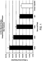


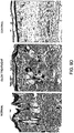
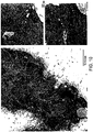


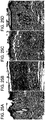
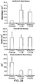


Classifications
-
- A—HUMAN NECESSITIES
- A61—MEDICAL OR VETERINARY SCIENCE; HYGIENE
- A61K—PREPARATIONS FOR MEDICAL, DENTAL OR TOILETRY PURPOSES
- A61K35/00—Medicinal preparations containing materials or reaction products thereof with undetermined constitution
- A61K35/12—Materials from mammals; Compositions comprising non-specified tissues or cells; Compositions comprising non-embryonic stem cells; Genetically modified cells
- A61K35/30—Nerves; Brain; Eyes; Corneal cells; Cerebrospinal fluid; Neuronal stem cells; Neuronal precursor cells; Glial cells; Oligodendrocytes; Schwann cells; Astroglia; Astrocytes; Choroid plexus; Spinal cord tissue
-
- A—HUMAN NECESSITIES
- A61—MEDICAL OR VETERINARY SCIENCE; HYGIENE
- A61K—PREPARATIONS FOR MEDICAL, DENTAL OR TOILETRY PURPOSES
- A61K31/00—Medicinal preparations containing organic active ingredients
- A61K31/185—Acids; Anhydrides, halides or salts thereof, e.g. sulfur acids, imidic, hydrazonic or hydroximic acids
- A61K31/19—Carboxylic acids, e.g. valproic acid
- A61K31/20—Carboxylic acids, e.g. valproic acid having a carboxyl group bound to a chain of seven or more carbon atoms, e.g. stearic, palmitic, arachidic acids
- A61K31/203—Retinoic acids ; Salts thereof
-
- A—HUMAN NECESSITIES
- A61—MEDICAL OR VETERINARY SCIENCE; HYGIENE
- A61K—PREPARATIONS FOR MEDICAL, DENTAL OR TOILETRY PURPOSES
- A61K38/00—Medicinal preparations containing peptides
- A61K38/16—Peptides having more than 20 amino acids; Gastrins; Somatostatins; Melanotropins; Derivatives thereof
- A61K38/17—Peptides having more than 20 amino acids; Gastrins; Somatostatins; Melanotropins; Derivatives thereof from animals; from humans
- A61K38/18—Growth factors; Growth regulators
-
- A—HUMAN NECESSITIES
- A61—MEDICAL OR VETERINARY SCIENCE; HYGIENE
- A61K—PREPARATIONS FOR MEDICAL, DENTAL OR TOILETRY PURPOSES
- A61K38/00—Medicinal preparations containing peptides
- A61K38/16—Peptides having more than 20 amino acids; Gastrins; Somatostatins; Melanotropins; Derivatives thereof
- A61K38/17—Peptides having more than 20 amino acids; Gastrins; Somatostatins; Melanotropins; Derivatives thereof from animals; from humans
- A61K38/19—Cytokines; Lymphokines; Interferons
-
- A—HUMAN NECESSITIES
- A61—MEDICAL OR VETERINARY SCIENCE; HYGIENE
- A61K—PREPARATIONS FOR MEDICAL, DENTAL OR TOILETRY PURPOSES
- A61K38/00—Medicinal preparations containing peptides
- A61K38/16—Peptides having more than 20 amino acids; Gastrins; Somatostatins; Melanotropins; Derivatives thereof
- A61K38/17—Peptides having more than 20 amino acids; Gastrins; Somatostatins; Melanotropins; Derivatives thereof from animals; from humans
- A61K38/22—Hormones
- A61K38/31—Somatostatins
-
- A—HUMAN NECESSITIES
- A61—MEDICAL OR VETERINARY SCIENCE; HYGIENE
- A61K—PREPARATIONS FOR MEDICAL, DENTAL OR TOILETRY PURPOSES
- A61K45/00—Medicinal preparations containing active ingredients not provided for in groups A61K31/00 - A61K41/00
- A61K45/06—Mixtures of active ingredients without chemical characterisation, e.g. antiphlogistics and cardiaca
-
- A—HUMAN NECESSITIES
- A61—MEDICAL OR VETERINARY SCIENCE; HYGIENE
- A61K—PREPARATIONS FOR MEDICAL, DENTAL OR TOILETRY PURPOSES
- A61K8/00—Cosmetics or similar toiletry preparations
- A61K8/02—Cosmetics or similar toiletry preparations characterised by special physical form
- A61K8/11—Encapsulated compositions
-
- A—HUMAN NECESSITIES
- A61—MEDICAL OR VETERINARY SCIENCE; HYGIENE
- A61K—PREPARATIONS FOR MEDICAL, DENTAL OR TOILETRY PURPOSES
- A61K8/00—Cosmetics or similar toiletry preparations
- A61K8/18—Cosmetics or similar toiletry preparations characterised by the composition
- A61K8/30—Cosmetics or similar toiletry preparations characterised by the composition containing organic compounds
- A61K8/64—Proteins; Peptides; Derivatives or degradation products thereof
-
- A—HUMAN NECESSITIES
- A61—MEDICAL OR VETERINARY SCIENCE; HYGIENE
- A61K—PREPARATIONS FOR MEDICAL, DENTAL OR TOILETRY PURPOSES
- A61K8/00—Cosmetics or similar toiletry preparations
- A61K8/18—Cosmetics or similar toiletry preparations characterised by the composition
- A61K8/72—Cosmetics or similar toiletry preparations characterised by the composition containing organic macromolecular compounds
- A61K8/73—Polysaccharides
- A61K8/733—Alginic acid; Salts thereof
-
- A—HUMAN NECESSITIES
- A61—MEDICAL OR VETERINARY SCIENCE; HYGIENE
- A61K—PREPARATIONS FOR MEDICAL, DENTAL OR TOILETRY PURPOSES
- A61K8/00—Cosmetics or similar toiletry preparations
- A61K8/18—Cosmetics or similar toiletry preparations characterised by the composition
- A61K8/96—Cosmetics or similar toiletry preparations characterised by the composition containing materials, or derivatives thereof of undetermined constitution
- A61K8/98—Cosmetics or similar toiletry preparations characterised by the composition containing materials, or derivatives thereof of undetermined constitution of animal origin
- A61K8/981—Cosmetics or similar toiletry preparations characterised by the composition containing materials, or derivatives thereof of undetermined constitution of animal origin of mammals or bird
-
- A—HUMAN NECESSITIES
- A61—MEDICAL OR VETERINARY SCIENCE; HYGIENE
- A61K—PREPARATIONS FOR MEDICAL, DENTAL OR TOILETRY PURPOSES
- A61K9/00—Medicinal preparations characterised by special physical form
- A61K9/0012—Galenical forms characterised by the site of application
- A61K9/0014—Skin, i.e. galenical aspects of topical compositions
-
- A—HUMAN NECESSITIES
- A61—MEDICAL OR VETERINARY SCIENCE; HYGIENE
- A61K—PREPARATIONS FOR MEDICAL, DENTAL OR TOILETRY PURPOSES
- A61K9/00—Medicinal preparations characterised by special physical form
- A61K9/0012—Galenical forms characterised by the site of application
- A61K9/0019—Injectable compositions; Intramuscular, intravenous, arterial, subcutaneous administration; Compositions to be administered through the skin in an invasive manner
-
- A—HUMAN NECESSITIES
- A61—MEDICAL OR VETERINARY SCIENCE; HYGIENE
- A61K—PREPARATIONS FOR MEDICAL, DENTAL OR TOILETRY PURPOSES
- A61K9/00—Medicinal preparations characterised by special physical form
- A61K9/14—Particulate form, e.g. powders, Processes for size reducing of pure drugs or the resulting products, Pure drug nanoparticles
- A61K9/16—Agglomerates; Granulates; Microbeadlets ; Microspheres; Pellets; Solid products obtained by spray drying, spray freeze drying, spray congealing,(multiple) emulsion solvent evaporation or extraction
- A61K9/1605—Excipients; Inactive ingredients
- A61K9/1629—Organic macromolecular compounds
- A61K9/1641—Organic macromolecular compounds obtained otherwise than by reactions only involving carbon-to-carbon unsaturated bonds, e.g. polyethylene glycol, poloxamers
- A61K9/1647—Polyesters, e.g. poly(lactide-co-glycolide)
-
- A—HUMAN NECESSITIES
- A61—MEDICAL OR VETERINARY SCIENCE; HYGIENE
- A61K—PREPARATIONS FOR MEDICAL, DENTAL OR TOILETRY PURPOSES
- A61K9/00—Medicinal preparations characterised by special physical form
- A61K9/14—Particulate form, e.g. powders, Processes for size reducing of pure drugs or the resulting products, Pure drug nanoparticles
- A61K9/19—Particulate form, e.g. powders, Processes for size reducing of pure drugs or the resulting products, Pure drug nanoparticles lyophilised, i.e. freeze-dried, solutions or dispersions
-
- A—HUMAN NECESSITIES
- A61—MEDICAL OR VETERINARY SCIENCE; HYGIENE
- A61K—PREPARATIONS FOR MEDICAL, DENTAL OR TOILETRY PURPOSES
- A61K9/00—Medicinal preparations characterised by special physical form
- A61K9/48—Preparations in capsules, e.g. of gelatin, of chocolate
- A61K9/50—Microcapsules having a gas, liquid or semi-solid filling; Solid microparticles or pellets surrounded by a distinct coating layer, e.g. coated microspheres, coated drug crystals
- A61K9/5005—Wall or coating material
- A61K9/5021—Organic macromolecular compounds
- A61K9/5036—Polysaccharides, e.g. gums, alginate; Cyclodextrin
-
- A—HUMAN NECESSITIES
- A61—MEDICAL OR VETERINARY SCIENCE; HYGIENE
- A61P—SPECIFIC THERAPEUTIC ACTIVITY OF CHEMICAL COMPOUNDS OR MEDICINAL PREPARATIONS
- A61P17/00—Drugs for dermatological disorders
- A61P17/02—Drugs for dermatological disorders for treating wounds, ulcers, burns, scars, keloids, or the like
-
- A—HUMAN NECESSITIES
- A61—MEDICAL OR VETERINARY SCIENCE; HYGIENE
- A61P—SPECIFIC THERAPEUTIC ACTIVITY OF CHEMICAL COMPOUNDS OR MEDICINAL PREPARATIONS
- A61P3/00—Drugs for disorders of the metabolism
- A61P3/08—Drugs for disorders of the metabolism for glucose homeostasis
- A61P3/10—Drugs for disorders of the metabolism for glucose homeostasis for hyperglycaemia, e.g. antidiabetics
-
- A—HUMAN NECESSITIES
- A61—MEDICAL OR VETERINARY SCIENCE; HYGIENE
- A61P—SPECIFIC THERAPEUTIC ACTIVITY OF CHEMICAL COMPOUNDS OR MEDICINAL PREPARATIONS
- A61P43/00—Drugs for specific purposes, not provided for in groups A61P1/00-A61P41/00
-
- A—HUMAN NECESSITIES
- A61—MEDICAL OR VETERINARY SCIENCE; HYGIENE
- A61Q—SPECIFIC USE OF COSMETICS OR SIMILAR TOILETRY PREPARATIONS
- A61Q19/00—Preparations for care of the skin
-
- A—HUMAN NECESSITIES
- A61—MEDICAL OR VETERINARY SCIENCE; HYGIENE
- A61Q—SPECIFIC USE OF COSMETICS OR SIMILAR TOILETRY PREPARATIONS
- A61Q7/00—Preparations for affecting hair growth
-
- A—HUMAN NECESSITIES
- A61—MEDICAL OR VETERINARY SCIENCE; HYGIENE
- A61K—PREPARATIONS FOR MEDICAL, DENTAL OR TOILETRY PURPOSES
- A61K2800/00—Properties of cosmetic compositions or active ingredients thereof or formulation aids used therein and process related aspects
- A61K2800/40—Chemical, physico-chemical or functional or structural properties of particular ingredients
- A61K2800/41—Particular ingredients further characterized by their size
- A61K2800/412—Microsized, i.e. having sizes between 0.1 and 100 microns
Definitions
- the invention relates to compositions for improving wound healing in particular for preventing scar formation and thus loss of function that can occur in injured tissues during the natural wound healing process.
- the invention relates to the healing of chronic wounds such as diabetic ulcers.
- a wound is a disruption of tissue integrity that is typically associated with a degenerative or traumatic loss of biological substance.
- Simple wounds include cuts and scrapes to the skin whilst deeper injuries to the muscle tissue, skeletal system or the inner organs are defined as complicated wounds 1 .
- Three distinct phases of wound healing are recognised. Firstly the inflammatory or exudative phase for the detachment of deteriorated tissue and for wound cleansing; secondly a proliferative phase for the development of granulation tissue; and thirdly a differentiation or regeneration phase for maturation and scar formation 1 .
- the inflammatory phase is characterised by hemostasis and inflammation.
- the cell membranes damaged from the wound formation, release thromboxane A 2 and prostoglandin 2-alpha, potent vasoconstrictors. This initial response helps to limit haemorrhage.
- Capillary vasodilation then occurs and inflammatory cells (platelets, neutrophils, leukocytes, macrophages, and T lymphocytes), migrate to the wound site.
- neutrophil granulocytes play a central role in wound cleansing via phagocytosis.
- the next cells present in the wound are the leukocytes and macrophages.
- the macrophages in particular, are essential for wound healing.
- cytokines are secreted by the macrophage, including collagenases, which debride the wound; interleukins and tumor necrosis factor (TNF), which stimulate fibroblasts (to produce collagen) and promote angiogenesis; and transforming growth factor (TGF), which stimulates keratinocytes 2 .
- TNF tumor necrosis factor
- TGF transforming growth factor
- the proliferation phase is characterised by epithelialisation, angiogenesis, granulation tissue formation, and collagen deposition.
- Angiogenesis stimulated by multiple factors, such as TNF alpha is essential to deliver nutrients into and around the wound site and is critical for efficient wound healing.
- Granulation tissue formation is a complex event involving leukocytes, histiocytes, plasma cells, mast cells, and in particular fibroblasts, that promote tissue growth through the production of collagen.
- cytokines involved include platelet derived growth factor (PDGF), insulin like growth factor (IGF) and epidermal growth factor (EGF). All are necessary for collagen formation 2 .
- PDGF platelet derived growth factor
- IGF insulin like growth factor
- EGF epidermal growth factor
- the final phase of the classical wound healing cascade is the differentiation phase.
- the wound undergoes contraction and the granulation tissue becomes increasingly depleted of fluids and blood vessels, begins to strengthen, and undergoes remodelling to form scar tissue.
- the final stage in wound healing is epithelialisation, whereby epidermal cells migrate to resurface the denuded area.
- new muscle cells are laid down (in addition to granulation tissue in the proliferative phase) via satellite cells which differentiate to form myoblasts 4 .
- myoblasts differentiate to form myotubes which mature and are incorporated into muscle fibres. Whilst this process results in the gain of some muscle function at the wound site, muscle wounds invariably result in loss of muscle tissue, scarring and loss of original muscle function.
- diabetic ulcers are the most common cause of foot and leg amputation.
- the incidence rate of developing foot ulcers is approximately 2% per year and as such ulcers are prone to infection and notoriously difficult to heal.
- TNF agonists and antagonists may also be useful in modifying angiogenesis, thus providing significant potential to improve the healing process directly.
- growth factors have had a limited role in clinical practice.
- the only currently available commercial product is PDGF (Regranex) which has been shown in some but not all studies to provide a modest improvement in healing time, but which has not been successful in improving the cosmetic or functional properties of healed tissue 2 .
- proteins secreted by choroid plexus (CP) cells have been shown for the first time to be useful in accelerating the wound healing process, particularly in reducing scarring in full thickness wounds and in the healing of chronic wounds.
- the present disclosure provides a therapeutic composition comprising isolated or purified CP secreted proteins, wherein the CP secreted proteins are isolated or purified from CP conditioned media.
- the present disclosure also provides a therapeutic composition comprising encapsulated CP cells secreting proteins useful for wound healing.
- the present invention also provides a therapeutic composition comprising CP conditioned media produced by the method according to claim 1.
- the present disclosure also provides a cosmetic composition comprising CP conditioned media.
- the present disclosure also provides a cosmetic composition comprising encapsulated CP cells.
- compositions of the present disclosure comprising the invention may be combined with a pharmaceutically acceptable carrier or excipient to provide pharmaceutical compositions.
- compositions of the invention may additionally comprise one or more compounds to give an additive or synergistic effect, wherein the compounds are selected from the group consisting of glucocorticosteroids, such as prednisone and methylprednisone, nonsteroidal anti-inflammatory drugs (NSAIDS), growth factors (PDGF, EGF, IGF, VEGF, TGF-Beta, and others), antibiotics, trophic or tropic compounds, peptides, mimetics, oligonucleotides, antibodies, inhibitors, as well as first and second generation anti-TNF ⁇ agents.
- glucocorticosteroids such as prednisone and methylprednisone
- NSAIDS nonsteroidal anti-inflammatory drugs
- growth factors PDGF, EGF, IGF, VEGF, TGF-Beta
- antibiotics trophic or tropic compounds
- peptides peptides
- mimetics oligonucleotides
- antibodies inhibitors, as well as first and second generation anti
- compositions of the invention may be formulated for topical, cutaneous, subcutaneous, intradermal, oral, intranasal, intraperitoneal, intramuscular, intraosseous, intraocular, or intravenous administration.
- the composition is formulated for topical administration.
- the present invention also provides a method of improving tissue wound healing comprising the step of administering an effective amount of the compositions of the invention to a patient in need thereof.
- the present disclosure comprising the invention also provides a method of treating a wound in a patient comprising the step of administering an effective amount of the compositions of the invention to a patient in need thereof.
- the present disclosure comprising the invention also provides a method of promoting wound healing comprising the step of administering an effective amount of the compositions of the invention to a patient in need thereof.
- the present disclosure comprising the invention also provides a method of modulating the accumulation of fibroblasts at a wound site in a patient comprising the step of administering an effective amount of the compositions of the invention to a patient in need thereof.
- the present disclosure comprising the invention also provides a method of modulating the accumulation of collagen at a wound site in a patient comprising the step of administering an effective amount of the compositions of the invention to a patient in need thereof
- the invention may be useful in both animal and human wound healing.
- Wound healing is improved in a human or animal patient via one or more of the following mechanisms:
- the proteins expressed by the CP of the invention comprise a mixture (e.g., two or more; any combination thereof) of VEGF, CTGF, PDGF, BMP-7, FGF-2, TGF- ⁇ , TGF- ⁇ , IL8/CXC chemokines, IGF-2, Cystatin, beta-2-microglobulin, Pleiotrophic factor ⁇ , PTHLH and other growth factors and extracellular matrix factors.
- the present disclosure also provides for the use of CP secreted proteins isolated or purified from CP conditioned media, in the manufacture of a medicament for improving wound healing in a patient in need thereof.
- the present disclosure also provides the use of CP secreted proteins isolated or purified from CP conditioned media, in the manufacture of a medicament for a cosmetic treatment of skin.
- the present invention provides CP conditioned produced by the method of claim 1, for use in improving wound healing in a patient in need thereof.
- the present disclosure also provides for the use of CP conditioned for a cosmetic treatment of skin.
- the present disclosure also provides for the use of encapsulated CP cells, in the manufacture of a medicament for improving wound healing in a patient in need thereof.
- the disclosure further provides for the use of encapsulated CP cells, in the manufacture of a medicament for a cosmetic treatment of skin.
- the medicament may be formulated for local or systemic administration, for example, the medicament may be formulated for topical administration to an external wound site, or may be formulated for injection to an internal wound site.
- the medicament is preferably formulated for topical administration.
- compositions comprising two or more isolated or purified choroid plexus (CP) secreted proteins (e.g., those delineated herein), and a clinically acceptable superficial wound treatment article (e.g., negative pressure therapy devices, bandages, films, adhesives, and the like).
- CP choroid plexus
- a clinically acceptable superficial wound treatment article e.g., negative pressure therapy devices, bandages, films, adhesives, and the like.
- the present disclosure further provides isolated or purified CP secreted proteins, CP conditioned media or encapsulated CP cells for use in a method of improving wound healing, or for cosmetic skin treatment, in a patient in need thereof.
- the present disclosure also provides a method of manufacturing a composition comprising CP secreted proteins, comprising the steps:
- the lyophilized (and optionally dialysed) CP conditioned media may be combined with a suitable pharmaceutical carrier or excipient to produce a pharmaceutical composition.
- the present disclosure also provides a composition manufactured by the method of the invention (e.g., those delineated herein).
- the present disclosure comprising the invention provides a method of modulating the profile of the CP secreted proteins from by CP cells in vitro to increase the amount of proteins that are effective at increasing wound healing, said method comprising the steps:
- the CP conditioned media extracted by this method contains a higher amount of VEGF and other CP secreted proteins useful in accelerating wound healing.
- compositions comprising CP proteins secreted by CP capsuloids containing modulated levels of secreted proteins, said capsuloids prepared by the methods of the invention.
- wound as used throughout the specification and claims means damage to one or more tissues, including open wounds such as cuts, scrapes, surgical incisions and the like, both acute and chronic, as well as internal wounds, for example, bruises, haematomas, fascia or soft tissue damage, hernia, abdominal wall damage, and the like as well as burns. "Wounds” may refer to tissue damage that occurs by trauma, or damage to tissue resulting from pathological sequelae, for example ischemia, that is exhibited due to overlying disease. The present invention is particularly useful in the healing of chronic open wounds such as ulcers, and diabetic ulcers in particular.
- Isolated or purified refers to secreted CP proteins that have been extracted from the CP conditioned media. Such extraction may include dialysis to remove salts from the media before lyophilising, or the CP conditioned media can be lyophilised directly.
- CP conditioned media comprises a cocktail of proteins that can be used in accelerating wound healing.
- this protein cocktail appears to be useful in all of the three characteristic phases of wound healing, i.e. the inflammatory phase, the proliferation phase, and the differentiation phase.
- CP secreted proteins appear to be a powerful regulator of the wound healing process and can be manipulated to prevent scar formation and resulting loss of function that would normally occur in injured tissue during the natural wound healing process. Lack of scarring is also important for cosmetic purposes, especially when the wound affects external portions of the body which are easily seen, such as the face, neck, hands etc.
- the choroid plexus are lobulated structures comprising a single continuous layer of cells derived from the ependymal layer of the cerebral ventricles.
- One function of the choroid plexus is the secretion of cerebrospinal fluid (CSF). Cerebrospinal fluid fills the four ventricles of the brain and circulates around the spinal cord and over the convexity of the brain.
- CSF cerebrospinal fluid
- the CSF is continuous with the brain interstitial (extracellular) fluid, and solutes, including macromolecules, are exchanged freely between CSF and interstitial fluid.
- the choroid plexus has been associated with the formation of the CSF-blood barrier 6 .
- CP secreted factors have been previously described for use in the treatment of neurological disorders ( WO 00/66188 ) and in the treatment of autoimmune disorders ( WO 2006/132548 ), however, there has been no disclosure or suggestion of CP secreted proteins being useful in the treatment of wounds, and in particular for treating chronic wounds, such as ulcers.
- VEGF 8 which is known to be involved in angiogenesis, ie in the proliferation stage of wound healing, as the CP also secrets a myriad of additional factors, mainly involved in CSF integrity
- a composition including all of the CP secreted proteins would be highly effective at improving wound healing, and especially in improving the healing of chronic wounds.
- the present disclosure is thus directed to a therapeutic composition
- a therapeutic composition comprising purified or isolated CP secreted proteins for use in wound healing.
- the CP secreted proteins are isolated or purified from CP conditioned media.
- the therapeutic composition comprises a mixture (e.g., two or more; any combination thereof) of CP secreted proteins including VEGF, CTGF, PDGF, BMP-7, FGF-2, TGF- ⁇ , TGF- ⁇ , IL8/CXC chemokines, IGF-2, Cystatin, beta-2-microglobulin, Pleiotrophic factor ⁇ , PTHLH and other growth factors and extracellular matrix factors.
- CP secreted proteins extracted from CP conditioned media and which are useful in the method of the present invention, may be tested for biological activity in an animal model or in vitro model of wound healing, as discussed below, and suitably active compositions formulated into pharmaceutical compositions.
- the pharmaceutical compositions of the present invention may comprise, in addition to CP secreted proteins described herein, a pharmaceutically acceptable excipient, carrier, buffer, stabiliser or other material well known in the art. Such materials should be non-toxic and should not interfere with the efficacy of the active ingredient.
- the precise nature of the carrier or other material will be dependent upon the desired nature of the pharmaceutical composition, and the route of administration e.g. oral, intravenous, cutaneous, subcutaneous, intradermal, topical, nasal, pulmonary, intramuscular or intraperitoneal.
- compositions for oral administration may be in tablet, lozenge, capsule, powder, granule or liquid form.
- a tablet or other solid oral dosage form will usually include a solid carrier such as gelatine, starch, mannitol, crystalline cellulose, or other inert materials generally used in pharmaceutical manufacture.
- liquid pharmaceutical compositions such as a syrup or emulsion, will generally include a liquid carrier such as water, petroleum, animal or vegetable oils, mineral oil or synthetic oil.
- the active ingredient will be in the form of a parenterally acceptable solution which is pyrogen-free and has suitable pH, isotonicity and stability.
- the active ingredient will be dissolved or suspended in a suitable emollient and may be formulated in the form of a cream, roll-on, lotion, stick, spray, ointment, paste, or gel, and can be applied directly to the wound site or via a intermediary such as a pad, patch or the like.
- the active ingredients will be in the form of a fine powder or a solution or suspension suitable for inhalation.
- the active ingredients may be in a form suitable for direct application to the nasal mucosa such as an ointment or cream, nasal spray, nasal drops or an aerosol.
- Therapeutic compositions can include isolated or purified choroid plexus (CP) secreted proteins (e.g., two or more of those delineated herein), and a clinically acceptable superficial wound treatment article (e.g., negative pressure therapy devices, bandages, films, adhesives, and the like). Such devices, bandages, films, adhesives, and the like are known in the art, and include for example, Also within the invention is a patch to deliver active chemotherapeutic combinations herein.
- a patch includes a material layer (e.g., polymeric, cloth, gauze, bandage) and the compound of the formulae herein as delineated herein. One side of the material layer can have a protective layer adhered to it to resist passage of the compounds or compositions.
- the patch can additionally include an adhesive to hold the patch in place on a subject.
- An adhesive is a composition, including those of either natural or synthetic origin, that when contacted with the skin of a subject, temporarily adheres to the skin. It can be water resistant.
- the adhesive can be placed on the patch to hold it in contact with the skin of the subject for an extended period of time.
- the adhesive can be made of a tackiness, or adhesive strength, such that it holds the device in place subject to incidental contract, however, upon an affirmative act (e.g., ripping, peeling, or other intentional removal) the adhesive gives way to the external pressure placed on the device or the adhesive itself, and allows for breaking of the adhesion contact.
- the adhesive can be pressure sensitive, that is, it can allow for positioning of the adhesive (and the device to be adhered to the skin) against the skin by the application of pressure (e.g., pushing, rubbing,) on the adhesive or device.
- the disclosure contemplates the use of one or more additional compounds co-administered with the pharmaceutical composition of the present invention to give an additive or synergistic effect to the treatment regime.
- Such compounds will generally be substances aimed at enhancing the rate and quality of wound healing.
- examples of such substances include glucocorticosteroids, such as prednisone and methylprednisone, nonsteroidal anti-inflammatory drugs (NSAIDS), growth factors (PDGF, EGF, IGF, VEGF, TGF-Beta, and others), antibiotics, trophic or tropic compounds, peptides, mimetics, oligonucleotides, antibodies, inhibitors, as well as first and second generation anti-TNF ⁇ agents.
- glucocorticosteroids such as prednisone and methylprednisone
- NSAIDS nonsteroidal anti-inflammatory drugs
- PDGF growth factors
- EGF EGF
- IGF IGF
- VEGF vascular endothelial growth factor
- the present invention also provides improving tissue wound healing comprising the step of administering an effective amount of the therapeutic composition of the invention to a patient in need thereof.
- the patient is preferably a human patient, but the method of the present invention may also be used to improve wound healing in non-human animals.
- Methods delineated herein include those wherein the subject is identified as in need of a particular stated treatment. Identifying a subject in need of such treatment can be in the judgment of a subject or a health care professional and can be subjective (e.g. opinion) or objective (e.g. measurable by a test or diagnostic method). Methods delineated herein also include those wherein the subject is successfully treated, their condition/symptom ameliorated or their condition/symptom is improved.
- Wound healing is improved in a human or animal patient via one or more of the following mechanisms:
- compositions comprising CP secreted proteins described herein are in the treatment of chronic wounds such as diabetic ulcers.
- Another preferred application of the present disclosure is in the treatment of skin wounds.
- compositions of the invention comprising CP secreted proteins to treat superficial or deep skin wounds can be demonstrated according to known methods 9 , and as exemplified in example 5, below.
- Another preferred application in the present disclosure is in the treatment of bums.
- the ability of CP secreted proteins to treat burn wounds can be demonstrated in known animal models. For example as described in Yang et al 10 .
- Administration of the pharmaceutical composition of the invention is preferably in a "prophylactically effective amount” or a “therapeutically effective amount", this being sufficient to show benefit to the individual.
- the actual amount administered, and rate and time-course of administration, will depend on the nature and severity of the type of wound that is being treated. Prescription of treatment, e.g. decisions on dosage etc., is within the responsibility of general practitioners and other medical doctors, and typically takes account of the disorder to be treated, the condition of the individual patient, the site of delivery, the method of administration and other factors known to practitioners. Examples of the techniques and protocols mentioned above can be found in Remington's Pharmaceutical Sciences, 16th edition, Oslo, A. (ed), 1980 .
- the present disclosure is also directed to the use of CP secreted proteins in the manufacture of a medicament for improving wound healing in a patient in need thereof.
- the CP secreted proteins may be isolated or purified from CP conditioned media.
- the present invention is directed to CP conditioned media for use in improving wound healing in a patient in need thereof.
- the medicament may be formulated for local or systemic administration, for example, the medicament may be formulated for topical administration to an external or open wound site, or may be formulated for injection into an internal or deep wound site, as described above.
- the medicament may further comprise one or more additional immuno-responsive compounds to give an additive or synergistic effect on wound healing, selected from the group of immuno-responsive compounds described above.
- the medicament may be formulated for separate, sequential or simultaneous administration of the CP secreted proteins/CP conditioned media and the one or more immuno-responsive compounds.
- the present disclosure is also directed to a method of manufacturing the therapeutic compositions comprising CP secreted proteins, said method comprising the steps:
- the CP cells may comprise a purified population of CP cells or they may alternatively comprise CP-derived cells selected from the group comprising glial or glial-derived cells, epithelial cells, multipotent neuronal precursor cells, proginator cells and cells positive for neuronal precursor cell markers (such as Neu-N).
- CP-derived cells selected from the group comprising glial or glial-derived cells, epithelial cells, multipotent neuronal precursor cells, proginator cells and cells positive for neuronal precursor cell markers (such as Neu-N).
- the CP cells may be obtained directly from a suitable mammal donor or may be obtained from primary or secondary CP cell culture or from a CP cell line including immortalised CP cell lines, or from a combination of any of the above sources.
- the CP cells in culture may have been genetically modified to produce one or more desired proteins useful in promoting wound healing, using genetic modification techniques well known in the art.
- the CP cells obtained directly from a donor may comprise cerebro spinal fluid containing one or more CP cells.
- Suitable CP cell donors include pigs, sheep, cows, goats, rabbits, mice and primates including rhesus monkeys and humans.
- the CP, cells may be isolated from donors using known methods in the art, for example, as described in WO 00/66188 .
- the CP cells may comprise isolated cells or clusters of cells and may be "naked” or encapsulated, for example, in alginate. Where CP cells are “naked", they may be “free” to make direct contact with one another or they may be separated by a biocompatible separation means which allows the diffusion of secreted proteins from the CP cells. The encapsulation of the CP cells may function as such a biocompatible separation means. CP cells may be encapsulated using methods known in the art (see for example WO 00/66188 and US 6322804 ).
- the CP cells are cultured in suitable culture media for a period of time and under conditions that allow secretion of the numerous secretion proteins including growth factors etc, from the CP cells into the media.
- the conditioned media is then separated from the cells to provide a CP secretory protein rich, cell-free CP preparation.
- the CP conditioned media can be prepared by a series of known steps to produce a therapeutic composition, including the optional step of dialysis (to remove salts), before or after lyophilisation, as set out in figure 5 .
- the processing steps resulted in a 6-9 times concentration of VEGF in the therapeutic composition, as described in example 4. It is also expected that other CP secretory proteins useful in promoting wound healing would also be concentrated in the lyophilised CP conditioned media.
- the lyophilised CP conditioned media may be combined with a pharmaceutically acceptable excipient and used in the methods of the invention to promote wound healing, as described above.
- the present invention provides a method of manipulating the profile of the proteins secreted by CP cells in vitro, said method comprising the steps:
- the media containing matrix proteins is a media rich in collagen IV, laminin and other ECM components and results in the formation of CP capsuloids, as described in example 6.
- Capsuloid formation results in an increase in the number of protein secretory vesicles and a corresponding increase in the level of secreted proteins, particular VEGF. This increase in protein secretion is the result of increased protein expression brought about by the media composition and capsuloid formation. It is contemplated in the present invention that the protein cocktail secreted by the CP capsuloids can be manipulated to maximise the therapeutic potential.
- CP secreted proteins are effective in improving wound healing by acting at all three recognised phases of wound healing, i.e. the inflammatory phase, the proliferation phase and the differentiation phase described above. Whilst the exact mechanism is unknown, it is likely a result of the combination of growth factors secreted by the CP, some of which have been shown previously to have been involved in wound healing. Such factors include VEGF, CTGF, PDGF, BMP-7, FGF-2, TGF- ⁇ , TGF- ⁇ , IL8/CXC chemokines, IGF-2, Cystatin, beta-2-microglobulin, Pleiotrophic factor ⁇ , PTHLH and other growth factors and extracellular matrix factors. Such a combination has not previously been used in wound healing.
- NT neurotrophin
- transthyretin a number of other factors secreted by CP, such as neurotrophin (NT) factors and transthyretin are not known to be actively involved in wound healing and yet other factors, such as retinoic acid, secreted by the CP are known to inhibit wound healing ( US 5973007 ).
- CP secreted proteins e.g., comprising those delineated in Tables 4 and 5
- retinoic acid and possibly other proteins that do not promote wound healing are extremely effective in improving wound healing, and in particular, in enhancing the healing of chronic wounds.
- CP secreted proteins in CP conditioned media has a direct effect on macrophage recruitment.
- both the number of macrophages and, presumably, the migration time to the wound site are increased when animals are treated with CP secreted proteins (results not shown).
- the first phase of wound healing, the inflammatory phase is significantly improved and it is expected that this will result in faster and more efficient wound cleansing and angiogenesis.
- CP secreted proteins in CP conditioned media also result in less collagen being deposited in the proliferation phase (results not shown).
- the protein cocktail secreted by the CP is thought to be a chemo-attractant for inflammatory cells, fibroblasts, endothelial cells, and epithelial cells,.
- use of CP secreted proteins is thought to result in a proportionate decrease in the relative recruitment of fibroblasts to the wound site and thus less overall production of collagen by the reduced population of fibroblasts.
- CP secreted proteins are further involved in inhibiting scar tissue formation in the differentiation phase of wound healing. Specifically, use of CP secreted proteins results in a significant reduction in scar tissue formation in a recovered wounded tissue. In addition, there was also a significant reduction in loss of functional tissue, i.e. use of CP secreted proteins also resulted in improved tissue regeneration, so that the recovered tissue was replaced without scarring and thus had little functional or cosmetic impairment. This may be particularly beneficial in cosmetic surgery or in treating wounds to portions of the body that are clearly visible, such as face, neck, hands etc.
- the present invention will be useful to treat all types of wounds such as skin cuts and abrasions, deep wounds extending through the skin and muscle (including surgical incisions) as well as internal wounds (for example wounds to muscle and tendon caused by sports injury or trauma), bruises, hematomas, , fascia, soft tissue damage, hernia, abdominal wall damage, as well as bums and chronic wounds such as ulcers, in particular, diabetic ulcers which are very difficult to treat using conventional therapy.
- wounds such as skin cuts and abrasions, deep wounds extending through the skin and muscle (including surgical incisions) as well as internal wounds (for example wounds to muscle and tendon caused by sports injury or trauma), bruises, hematomas, , fascia, soft tissue damage, hernia, abdominal wall damage, as well as bums and chronic wounds such as ulcers, in particular, diabetic ulcers which are very difficult to treat using conventional therapy.
- the described microarray was used to investigate the transcriptome of porcine CP epithelium, and then assess the in vitro and in vivo regenerative capability of secreted CP products in cell monolayers and full thickness cutaneous wounds.
- CP reduced the void area of fibroblast and keratinocyte scratch cultures by 70% and 33%, respectively, compared to empty capsule controls, which reduced the area by only 35% and 6% respectively.
- CP conditioned medium lyophilate dispersed in antibiotic ointment produced a 2-fold improvement in incision tensile strength compared to ointment alone, and an increase in the regeneration of epidermal appendages from roughly 50 to 150 features per wound.
- Neonatal Yorkshire pigs aged 7-10 days were induced with ketamine (500 mg/kg) and xylazine (0.15mg/kg), and then maintained on 3% isoflurane during controlled exsanguination.
- the brain was removed and hemisected along the midline to expose the CP in the lateral ventricles.
- the CP were extricated and placed in Hanks Balanced Salt Solution (HBSS, 0-4°C) supplemented with ciproflaxocin (Sigma, USA, 3 ⁇ g/ml), 2% human serum albumin and digested as previously described ( Borlongan, C. V.; et al.
- RPMI-CPN ciproflaxocin
- RNA was isolated from cultured CP clusters after 3 or 16 days in ENDO-SFM tissue culture medium (N 3). Cell clusters were disrupted and homogenized in buffer RLT using a QIAshredder (Qiagen, USA). Briefly, 700 ⁇ l of cell lysate was loaded onto the QIAshredder, centrifuged at 30,000 x g, and RNA was purified using an RNeasy Mini spin column (Qiagen, USA). Purified, high-quality RNA was eluted in RNase-free water (Gibco, USA).
- Single-stranded cDNA was synthesized by priming with a T7 Oligo(dT) primer, and then converted into double-stranded DNA templates for transcription by employing DNA polymerase and RNase H for simultaneous degradation of RNA and synthesis of the second cDNA strand.
- the cDNA was purified and cRNA was subsequently synthesized and amplified using in vitro transcription. After final purification of the cRNA, sample integrity was confirmed on electropherograms acquired from a Bioanalyzer 2100 (Agilent Technologies, USA). Once samples were confirmed to be suitable for array analysis, a whole-genome porcine microarray (Affymetrix, USA) was used to characterize the entire pig genome.
- CP clusters were encapsulated in alginate-polyomithine-alginate (APA) microcapsules after 10 days in culture as previously described at 50,000 clusters/mL using a coaxial air jet system ( Emerich, D. F.; et al. Extensive neuroprotection by choroid plexus transplants in excitotoxin lesioned monkeys. Neurobiol. Dis. 23:471-480; 2006 ) 600 ⁇ m diameter capsules were produced and were incubated in RPMI-CPN for 5 days prior to transfer to fibroblast or keratinocyte cultures. A sample was pulsed in fresh RPMI-CPN for 24 hours to quantify secreted VEGF.
- APA alginate-polyomithine-alginate
- Normal human dermal fibroblasts (NHDF, Cambrex, USA) or normal human epidermal keratinocytes (NHEK, Cambrex, USA) were expanded in T75 flasks in supplemented cell culture medium.
- NHEK cells were grown in Keratinocyte Growth Medium (KGM, Cambrex, USA) supplemented with epidermal growth factor, insulin, and hydrocortisone.
- the culture medium was removed, the cells were washed with HBSS three times, and a defect (approximately 9 mm 2 ) was created with the tip of a 20 ⁇ L pipette.
- the HBSS was aspirated several times to loosen the cells surrounding the defect, and then replaced with basal culture medium.
- the defects were then photographed at 4X magnification.
- Transwell inserts with a pore size of 10 ⁇ m were placed on top of the wells, and encapsulated CP were added for an equivalent dose of 500, 1500, 2500, or 5000 CP clusters per well.
- CM CP conditioned medium
- Lyophilized CM (LCM), or dialyzed lyophilized CM (DLCM) was prepared for topical administration immediately prior to application by mixing the lyophilate in bacitracin ointment (Johnson & Johnson, USA) at roughly 50 mg LCM / 350 mg ointment for the high dose, and roughly 17 mg LCM / 350 mg for the low dose.
- the actual amounts were calculated based on the amount of VEGF measured during the media collection period, with the final doses normalized to a VEGF dose of 6 ng at the low dose, and 20 ng at the high dose.
- VEGF Vascular Endothelial Growth Factor
- VEGF was measured in culture supernatants as a surrogate marker of potency as previously described ( Thanos, C. G.; et al., The in vitro expression and secretion of vascular endothelial growth factor from free and alginate-polyornithine encapsulated choroid plexus epithelium. Tissue Eng. 13:747-756; 2007 ). After 96 hours of incubation (37°C, 5% CO 2 ) the supernatant was removed and frozen for subsequent analysis. Thawed samples were analyzed in triplicate using a human VEGF ELISA (Quantikine ® , R&D Systems, USA). Sequence homology between porcine and human VEGF is greater than 90% (24).
- Samples of conditioned media (200 ⁇ L) were combined with assay diluent (50 ⁇ L) and the optical density was measured at 450 nm on a Beckman Coulter DTX-880 spectrophotometer after development. The value obtained was compared to a standard curve of recombinant human VEGF using a logarithmic curve fit. Values were expressed as pg/mL.
- CPCM was evaluated in full-thickness wounds in male Long-Evans rats approximately 3 months old and weighing between 250-350 g housed in pairs in a temperature controlled environment with a 12:12 hour light-dark cycle. All study groups consisted of 8 animals. All procedures met or exceeded NIH guidelines and were approved in advance by the Brown University IACUC governing body.
- rats were anesthetized with isoflurane (3-4%) and placed in a nose cone in a prone position. Prior to the creation of the skin defect, animals were clipped and the skin around the area was disinfected with betadine. Following the surgery, animals were treated with an analgesic (buprenorphine, 0.02 mg/kg subcutaneous) and observed until fully recovered from anesthesia. Animals were euthanized using carbon dioxide gas, and explanted tissue was removed as indicated for histological processing.
- an analgesic buprenorphine, 0.02 mg/kg subcutaneous
- tissue segments were transversely embedded in paraffin for cutting 4 ⁇ m-thick sections, which were subsequently stained with either hematoxylin and eosin (open wound tissue), or Masson's trichrome stain (incision wounds). Morphometry on healing wounds was carried out using image analysis software (Motic, China) to measure the healing area within the margin of hair growth, and statistics were calculated with Prism software (GraphPad, USA), using a one-way ANOVA with a 95% confidence interval and post-hoc Dunnet's test for comparison against the control group.
- Topical CM ointment was applied for 10 days following the procedure, and animals were sacrificed at day 14 for tissue harvesting.
- a 6 cm linear full thickness incision was made between the shoulder blades with a fresh size 15 scalpel through a plastic template designed for uniformity of straightness and length. The incision was immediately closed with 8 interrupted stitches using 6-0 silk equipped with a PC-3 cutting needle. Based on prior experience within our group, silk was chosen to minimize interference in the healing of the wounds caused by irritancy of other bristle-like materials.
- a press was used to exert force on an aluminum block resting on top of the blades, creating an evenly distributed force completely perpendicular to the plane of tissue, and resulting in precise 1-cm wide tissue segments for histological and physical analysis.
- the central 1-cm segment was processed for histology, while the outer 2 on each side were analyzed for tensile strength at break using an Instron tensiometer (Instron, USA). Skin samples were immediately transported on ice on their wax backing material to the instrument. Samples were removed from the wax, any residual panniculus camosus was moved from the underside of the wound, and the tissue was placed in the serrated jaws of the instrument under maximum clamping pressure.
- Neonatal porcine choroid plexus isolated from Yorkshire swine, was isolated and maintained in culture as previously described in Thanos et al 8 . After 7-10 days, choroid plexus clusters were encapsulated in alginate and held for another 7 days.
- Monolayers of cell types relevant to wound healing were created in 6-well plates using standard tissue culture techniques to create previously described in vitro wound models.
- Adult human dermal fibroblasts or kertinocytes were grown to 95% confluency, whereupon the monolayer was physically disrupted with a specialised tool to create a defect on the order or 10-20 mm 2 .
- cultures were washed with HBSS, photos were taken for image analysis, and the appropriate basal medium was returned to the plate.
- transwells were used to suspend CP-containing capsules (500-5000 eCP equivalents/well) so that the respective cell type would have direct access to secreted proteins.
- CP conditioned media was collected separately and added at various concentrations along with other media types in an attempt to titrate the effect. After the proper exposure time, treatment was removed and photos were again taken for image analysis. Wound area was measured using Motic software, and % healing was calculated by (area initial - area initial ).
- Encapsulated choroid plexus when separated by a transwell membrane insert, showed a dose dependent increase in healing of both fibroblast ( figure 1 ) and keratinocyte ( figure 2 ) compromised monolayers.
- the keratinocyte model revealed that CP at a dose of 2500 eCP/mL improved healing relative to the empty capsule control by almost 30%.
- CP conditioned medium was collected for 96 hours after 7 days of culture of CP cell culture and then added to fibroblast culture medium.
- the therapeutic effect of CP conditioned media and fibroblast wound model is shown in figure 3 .
- Each group containing conditioned media (CM) along with nutrients showed an improvement compared to the basal medium control.
- CM is likely depleted of essential nutrients required to maintain healthy cell cultures, especially compared to a fresh fibroblast basal growth medium (FBM).
- FBM fresh fibroblast basal growth medium
- CM provided benefit in each condition, particularly those that supplement a minimal amount of nutrients.
- Example 3 Assessment and optimisation of the long-term production of VEGF (indicator of wound healing potential) in cultures with daily or 2/week media changes
- Example 4 Preparation of a therapeutic composition of CP secreted proteins
- CP conditioned medium was formulated for therapeutic use by either dialyzing out salts and lyophilizing, or lyophilizing directly.
- the extracted CP proteins were subjected to processing and analysed for VEGF using ELISA. The following specific processing groups tested are set out in figure 5 .
- Encapsulated choroid plexus clusters (50K / mL) or empty capsules were loaded into Matrigel HC (BD Biosciences), diluted by 50% with saline, at 0.1:1.0 capsules:matrigel for a total injection volume of 1.1 mL.
- the injectate was kept on ice prior to injection, which occurred through a 16-gauge needle into the subcutaneous space between the shoulder blades of long-evans rats (Male, 250-300 g) under isolflurane anesthesia at 2-3%. 2 weeks later, subcutaneous gel slabs were explanted, fixed in 4% paraformaldehyde, and processed for H&E histology.
- FIG. 6C a high magnification view of the encapsulated CP group, shows further evidence of enhanced angiogenesis around the periphery of the capsule with loss of implanted matrigel and stratified cellular organization.
- Choroid plexus conditioned media was collected as described in example 2, above, and lyophilized as described in group II of figure 5 , discussed in example 4, above.
- the lyophilised CP conditioned media was reconstituted in distilled water for treatment of full thickness wounds or dispersed in hydrophobic ointment for topical application in 6 cm incision wounds.
- CM 25 mg lyophilised CM was injected daily for 14 days in 200 ⁇ L dH 2 0 for a total of roughly 6 ng VEGF/day, administered directly to the wound site. Macro photography was used daily to assess and measure wound area. After 4 weeks, animals were euthanized and wound areas were collected for histology.
- Example 7 Topical treatment of a full thickness critical defect using a purified CPCM
- Choroid plexus conditioned media was collected as described in example 2, above, purified by dialysis against a 1000 Da MWCO cellulose membrane, and lyophilized as described in group II of figure 5 , discussed in example 4, above.
- a critical defect was created using an 8-mm biopsy punch down to the level of subcutaneous tissue without bandage or autograft.
- 2 groups containing 10 animals included a treatment (dialyzed conditioned media) and control (lyophilised ENDO-SFM). On each animal, defects were made between the shoulder blades and the excised skin was removed. Animals received a perioperative injection of buprenex and were monitored during recovery. Both groups received topical applications of either dialyzed, lyophilized ENDO-SFM tissue culture medium, or dialyzed lyophilized CP-conditioned medium (DLCM) with dose based on a VEGF loading of approximately 20 ng VEGF. Treatments were applied daily for 10 days, and wounds were explanted on day 14 for histological characterization. Macro photography was used daily to assess and measure wound area. After 4 weeks, animals were euthanized and wound areas were collected for histology.
- the left panel shows a section of normal skin adjacent to a control wound, the middle a section of DLCM treated skin, and the right panel shows the control.
- One obvious aspect of the extent of healing is the extent of organization and infiltration of advanced remodeling cell types in the middle panel caused by treatment, compared to the far right panel, which contains high levels of fresh collagen and fibroblasts, along with a modicum of neovascularization.
- the treated group differs mainly from normal tissue in that the hair has not yet emerged through the epidermis, and exists in earlier stages of follicle development.
- the keratinocyte layer in the extreme top of each section also shows advanced healing, as the control exists with a very shallow, flat layer, compared to the thicker ridges morphology in the treated and normal groups.
- Example 8 Modulation of the cocktail of proteins produced by the choroid plexus, by manipulating culture media composition
- Choroid plexus clusters were isolated and cultured with RPMI-CPN, ENDO-SFM or in RPMI-CPN within a matrix material rich in Collagen IV, laminin and other ECM components.
- CP clusters normally appear as balls of epithelium with outwardly oriented cilia and attached internally to a matrix composed of connective tissue and other cell types ( Figure 10 ). Cells around the perimeter are rich in tight junctions and show a modicum of secretory vesicles associated with protein release.
- this morphology was manipulated such that hollow vesicles, or capsuloids, were formed with inward oriented cilia and mirovilli surrounding a fluid-filled internal pocket ( Figure 11 ). These capsuloids varied in size in accordance with the size of the original CP clusters, and enlarged over a 6-10 day timecourse ( Figures 12a and 12b ). In this process, the epithelium reorganised to form a distinct layer replete with tight junctions, while contaminating cells such as fibroblasts were separated within the matrix.
- Table 1 shows the fold changes in genes from capsuloids grown for 3 days in RPMI-CPN media compared to CP clusters grown in the same media
- Table 2 shows the fold changes in genes in CP clusters grown for 3 days in ENDO-SFM media compared to CP clusters grown in RPMI-CPN media.
- Table 3 highlights some of the genes upregulated in the presence of ENDO-SFM that are implicated in wound healing.
- Example 9 Nanoencapsulation of lyophilised choroid plexus conditioned media and demonstration of protein integrity as assessed by ELISA
- Poly(lactide-co-glycolide) was used to encapsulate micronised, lyophilised Choroid Plexus conditioned media (LCPCM).
- LCPCM described previously was reconstituted in a super saturated solution of distilled water (236.72 ing in 2 mL). Separately, 25 mL ethyl acetate was added to 413 mg PLGA (Resomer® RG503H, Boehringer Ingelheim), a biodegradable polyster with 50:50 copolymer ratio and a weight-average molecular weight of 9 kDa, considered relatively fast-degrading.
- PLGA Resomer® RG503H, Boehringer Ingelheim
- This micronisation technique was employed to improve the homogeneity of the formulation, to reduce the overall size of the microspheres, and to enhance solubility due to the larger surface area to volume ratio of the resulting therapeutic particulates.
- This polymer-CPCM combination was then dissolved in 40 mL ethyl acetate, which thoroughly dissolved the PLGA component but retained the micronised CPCM as insoluble microparticlates. The solution was rapidly dispersed into 2 L heptane, causing rapid precipitation of polymer around the micronised CPCM.
- the resultant therapeutic microspheres consisted of 36.5% (W/W) loading of CPCM, or roughly 127 pg VEGF/mg microsphere formulation.
- Microspheres were characterised using scanning electron microscopy for surface morphology, Coulter particle size analysis for size distribution, and organic extraction to determine if one of the CPCM proteins, vascular endothelial growth factor (VEGF) remained detectable and potentially active following exposure to the solvents employed.
- VEGF vascular endothelial growth factor
- VEGF vascular endothelial growth factor
- Coulter particle size analysis and scanning electron microscopy revealed that microspheres were roughly 300 nm in size, with aggregation occurring within the dispersion to create clusters with mean diameters of approximately 4 and 10 microns respectively, ( Figure 16 and 17 ).
- Example 10 Analysis of the Transcriptome of CP in cultures generating condition media
- HUVEC were then trypsinized and added to the wells of 3-D choroid plexus cultures at a density of 2.5 x 10 3 cells / cm 2 ) in RPMI 1640 / FBS, or to wells containing the matrix alone.
- HUVEC co-cultured with CP capsuloids demonstrated a change in phenotype from ovoid, epithelial cells to tubular structures as shown in Figure 19A and 19B below.
- the network formed by the endothelial tubes appears to be guided by the placement of CP capsuloids, as nearly each cell mass serves as a node in the vascular network.
- HUVEC cultured under the same conditions but without choroid plexus remained undifferentiated ( Figure 20 ). This behavior is indicative of exposure to an angiogenic stimulus, which is secreted in the cocktail of growth factors provided by the choroid plexus.
- CP was successfully isolated from neonatal pigs and digested into CP clusters ranging in size from approximately 50-250 ⁇ m. After culturing the clusters in non-adherent flasks in RPMI-CPN for 8 days, manual counting revealed that the yield per piglet decreased from 128,854 ⁇ 16,159 to 50,962 ⁇ 5,690 CP clusters. This is consistent with the temporal loss of non-epithelial clusters of cells due to attachment or death, in addition to gradual coalescence of small aggregates into larger clusters ( Figure 22 ). After the transition to serum-free media after 10 days, CM was collected in parallel with fresh medium held under the same conditions.
- VEGF levels were measured in aliquots taken from CM generated throughout the experiment ( Figure 23 ) as well as control medium. 10 days after isolation, daily levels of VEGF secreted per CP increased from approximately 41 ng to around 70 ng at 2 weeks, and remained relatively stable thereafter. Following processing of the conditioned medium to generate a pooled purified lyophilate, VEGF levels were verified to ensure that the molecule was conserved throughout the process.
- CP cohorts from the same animal group were cultured, after 10 days in RPMI-CPN, in ENDO-SFM for an additional 3 or 16 days specifically for microarray analysis.
- Whole genome porcine arrays were performed to characterize the CP expression pattern as well as to evaluate the plasticity of the CP transcriptome over the first 16 days following media transition.
- the microarray data were selected and sorted based on the potential of mRNAs to transcribe for secreted molecules, relevance in tissue regeneration, expression levels, and statistical significance. Shown in Table 5 is a list of those factors that could play a role in wound healing, along with a descriptive matrix of their potential roles. As expected ( Thanos, C.
- the regenerative genes listed encode for growth factors, angiogenic factors, extracellular matrix remodeling components, and other factors with regenerative properties.
- the regenerative factor compliment expressed by the CP clusters in ENDO-SFM was significant, and included several relevant factors (see table for abbreviations): CTGF (14.01), VEGF (11.66), TGF- ⁇ (11.39), IGF-2 (11.26), PEDF (11.07), BMP-7 (10.96), PDGF (10.59), Somatostatin (10.03), I1-6 (9.67), Tissue Factor (9.50), WISP-1 (8.85), FGF-2 (8.79), TGF- ⁇ (8.51), Wnt-10B (8.27), EGF (7.57) and others.
- Figure 21 shows explanted skin containing the wounded area, with a control treatment ( Figure 21 A) and LCM treatment ( Figure 21B ), which contains more follicles within the wounded area. This observation is more apparent histologically, as shown in Figure 25 .
- FIG. 25A normal skin is shown, which contains epidermal appendages including hair follicles associated with sebaceous glands, dense collagen, and distinctly formed epidermal rete pegs.
- a wound treated with bacitracin alone shown in Figure 25B , contains densely packed fibroblasts within the dermis, replete with newly formed vessels, and an epidermis of a thin, flat layer of keratinocytes.
- Wounds treated with LCM contain tissue with a higher level of organization, containing forming epidermal appendages, a lower cell density, and a more ridged profile suggestive of epidermal rete pegs.
- DLCM-treated wounds ( Figure 25D ) were rich in epidermal appendages, including sebaceous glands, which occasionally extended through the epidermis manifesting as visible hair. These wounds also resembled the morphology of normal tissue in the density and ratio of cells to collagen.
- wounds treated with CP products contained a significantly higher density of epidermal appendages (5- and 5.8-fold higher for DLCM and LCM respectively) and an overall smaller cross-sectional area.
- central cell density was morphometrically indistinguishable between groups.
- the current results demonstrate that CP secreted proteins are also involved in regulating the inflammatory response and in improving wound healing and reducing scar tissue formation.
- macrophages infiltrate the wound site soon after wounding and by release of chemokines contribute to key processes in healing such as regulation of epitheliasation, tissue remodeling and angiogenesis in skin 11 and other tissues.
- a schematic diagram of the wound healing process is set out in Figure 18 .
- histological data demonstrates that there is increased and accelerated infiltration of macrophages and fibroblasts into the wound area resulting in the laying down of fibrotic scar tissue ( Figures 7b and 7c - control).
- Figures 7b and 7c - control the accumulation of fibrotic connective tissue is reduced ( Figure 7b and 7c - treated) and tissue function maintained as evidenced by improved break strength ( Figure 8 ).
- Figure 7b and 7c - treated tissue function maintained as evidenced by improved break strength
- CP secreted proteins also comprise numerous cytokines and growth factors and appear to accelerate tissue repair by enhancing the wound healing process leading to accelerated healing. Perhaps this is due to a synergistic effect of the endogenous growth factors and the exogenous growth factors supplied by the administration of CP secreted proteins. The result seen in the present examples is not due to the presence of VEGF alone. A composition comprising CP secreted proteins was more effective at accelerating wound healing than recombinant VEGF alone (results not shown).
- Fibrosis is a part of the wound healing processes but excess fibrosis leads to scarring and reduced function of tissues. Fibroblasts play a major role in deposition of collagen and thus scar formation in wounds. Studies have previously correlated the extent of fibroblast accumulation with scarring in skin burn wounds 10 . It is anticipated that CP secreted proteins acts to reduce scarring by decreasing the accumulation of fibroblasts at a wound site with a consequent decrease in scarring in the healed wound. Importantly, the data presented here shows that the administration of CP secreted proteins to a wound site in vivo leads to decreased collagen accumulation and scarring in tissue that has undergone wound healing. Evidence for this is already available in example 7 above and figure 9 .
- Collagen has been found to be the major pathological finding in a number of fibrotic diseases 12 , but at the same time is an extremely important early component of non-fibrotic tissue regeneration. This fine balance of extracellular matrix production, chemotaxis, and mitogenesis in wound healing therapy can only be achieved by concomitant or staged delivery of a multitude of individual factor therapies, whereas in this work it is apparent that the CP-derived cocktail provides all aspects.
- the transcriptome of CP contains a number of genes encoding for mediators of the natural wound healing response. While it is unknown if the full compliment of these factors is secreted from CP in the tissue culture system described here, proteomic analysis of mouse CP supports an abundance of secreted proteins ( Thouvenot, E.; et al. The proteomic analysis of mouse choroid plexus secretome reveals a high protein secretion capacity of choroidal epithelial cells. Proteomics 6:5941-5952; 2006 ).
- VEGF vascular endothelial growth factor
- TGF- ⁇ 1 Li, Z.-D.; et al. VEGF induces proliferation, migration, and TGF- ⁇ 1 expression in mouse glomerular endothelial cells via mitogen-activated protein kinase and phosphatidylinositol 3-kinase. Biochem. Biophys. Res. Commun. 334:1049-1060; 2005 ), and others that are involved in wound healing. Still, it is likely that the overall effect of the CP cocktail extends beyond VEGF alone, as there are a number of other highly expressed factors contained within the CP transcriptome that act at similarly high levels.
- CTGF for example, is expressed by CP at levels nearly as high as transthyretin, the factor with the highest expression level in gene array ( Thanos, C. G.; et al., The in vitro expression and secretion of vascular endothelial growth factor from free and alginate-polyomithine encapsulated choroid plexus epithelium. Tissue Eng. 13:747-756; 2007 ) and proteomic analysis ( Thouvenot, E.; et al. The proteomic analysis of mouse choroid plexus secretome reveals a high protein secretion capacity of choroidal epithelial cells. Proteomics 6:5941-5952; 2006 ).
- CTGF promotes chemotaxis and proliferation of fibroblasts, the formation of granulation tissue, re-epithelialization, and matrix remodeling ( Igarashi, A.; et al. Regulation of connective tissue growth factor gene expression in human skin fibroblasts and during wound repair. Mol. Biol. Cell 4:637-645; 1993 ). While the primary role of CTGF in wound healing is to promote tissue reconstruction through fibrosis, it has also been shown to regulate VEGF-mediated angiogenesis through negative feedback ( Jang, H. S.; et al. A novel ex vivo angiogenesis assay based on electroporation-mediated delivery of naked plasmid DNA to skeletal muscle. Mol. Ther.
- TGF- ⁇ also highly expressed by the CP, is one of the most well known mediators of cutaneous wound healing, and is involved from the inflammatory phase through tissue remodeling and re-epithelialization ( Barrientos, S.; et al. Growth factors and cytokines in wound healing. Wound Repair Regen. 16:585-601; 2008 ).
- Wnt-dependent de novo hair follicle regeneration in adult mouse skin after wounding has demonstrated positive effects on hair shaft growth and follicle formation in vitro ( Ouji, Y.; et al. Wnt-10b, uniquely amount Wnts, promotes epithelial differentiation and shaft growth. Biochem. Biophys. Res. Commun. 367:299-304; 2008 ).
- Wnt10b works in concert with factors that are involved in the earlier stages of wound healing, e.g. PDGF, VEGF, CTGF, TGF- ⁇ , and others, to guide epithelial differentiation concurrent with angiogenesis and migration of fibroblasts and keratinocytes, effectively providing developmental cues as in embryogenesis.
- JARCET 2(2); 2002 JARCET 2(2); 2002 ), and remains a fertile area of product development.
- the application of these and other factors simultaneously, to be used as required by the healing tissue, would address issues related to targeting just one aspect of the wound healing cascade, and decrease the impact of delivery kinetics and the regulation of factors via feedback in the wound.
- Such a combination of factors could also prove useful in more severe wounds, such as deep ischemic ulcers, that would require a more coordinated rebuilding of tissue beginning with a restoration of blood supply and ending with superficial reconstruction.
- Factors from the CP could be used to improve the healing of cutaneous wounds. Because of the potentially widespread effect imparted by the CP cocktail, ranging from angiogenesis to tissue rebuilding, there may be other indications that are also suitable for investigation. A therapeutic benefit has already been described in animal models of neurodegenerative disease, and other potential indications include diseases where staged intervention is required to treat tissues suffering from ischemia and necrosis, such as diseases of the cardiovascular system, muscle wasting and repair, hair regrowth, antiwrinkle treatment, cartilage repair, and other similar areas.
- compositions comprising CP secreted proteins, have been shown here to increase the rate of wound healing by acceleration and enhancement of several key processes.
- the application of CP secreted proteins has also been shown to result in decreased deposition of collagen at the final healed wound site which prevents loss of tissue function (strength) or cosmetic damage due to scarring.
- the present invention provides compounds according to the claims for use in improving wound healing by administering thereof either systemically or locally wherein the compound comprises CP secreted proteins.
Landscapes
- Health & Medical Sciences (AREA)
- Life Sciences & Earth Sciences (AREA)
- Veterinary Medicine (AREA)
- Public Health (AREA)
- General Health & Medical Sciences (AREA)
- Animal Behavior & Ethology (AREA)
- Epidemiology (AREA)
- Chemical & Material Sciences (AREA)
- Pharmacology & Pharmacy (AREA)
- Medicinal Chemistry (AREA)
- Engineering & Computer Science (AREA)
- Bioinformatics & Cheminformatics (AREA)
- Zoology (AREA)
- Dermatology (AREA)
- Immunology (AREA)
- Birds (AREA)
- Proteomics, Peptides & Aminoacids (AREA)
- Gastroenterology & Hepatology (AREA)
- Cell Biology (AREA)
- Developmental Biology & Embryology (AREA)
- Biomedical Technology (AREA)
- Endocrinology (AREA)
- Diabetes (AREA)
- Chemical Kinetics & Catalysis (AREA)
- Organic Chemistry (AREA)
- General Chemical & Material Sciences (AREA)
- Nuclear Medicine, Radiotherapy & Molecular Imaging (AREA)
- Neurosurgery (AREA)
- Ophthalmology & Optometry (AREA)
- Virology (AREA)
- Neurology (AREA)
- Biotechnology (AREA)
- Obesity (AREA)
- Emergency Medicine (AREA)
- Hematology (AREA)
- Medicines That Contain Protein Lipid Enzymes And Other Medicines (AREA)
- Medicinal Preparation (AREA)
- Cosmetics (AREA)
- Pharmaceuticals Containing Other Organic And Inorganic Compounds (AREA)
- Micro-Organisms Or Cultivation Processes Thereof (AREA)
Claims (4)
- Méthode de manipulation du profil des protéines secrétées par les plexus choroïdes (PC) à partir de cellules de PC in vitro selon les étapes suivantes :a. culture de cellules de PC dans un milieu de culture basal in vitro ;b. ajout de protéines matricielles cellulaires au milieu de culture pour initier la formation de capsuloïdes de PC ;c. culture des capsuloïdes pendant le temps nécessaire à l'augmentation du nombre des vésicules sécrétoires dans les capsuloïdes ; etd. production de milieux conditionnés par les PC qui contiennent un profil altéré de protéines secrétées par les PC par rapport aux cellules PC cultivées uniquement dans un milieu de culture basal.
- Milieux conditionnés par les PC produits selon la méthode décrite dans la revendication 1.
- Composition thérapeutique comprenant les milieux conditionnés par les PC décrits dans la revendication 2 à utiliser pour améliorer la cicatrisation par une réduction de l'accumulation soit du collagène soit des fibroblastes dans la plaie chez un patient humain ou non-humain.
- Composition thérapeutique comprenant ses milieux conditionnés par des PC tels que décrits dans la revendication 2 à utiliser pour améliorer la cicatrisation chez un patient humain ou non-humain en réduisant l'accumulation soit du collagène soit des fibroblastes dans la plaie chez un patient humain ou non-humain.
Applications Claiming Priority (2)
| Application Number | Priority Date | Filing Date | Title |
|---|---|---|---|
| US13502608P | 2008-07-15 | 2008-07-15 | |
| PCT/US2009/004129 WO2010008573A2 (fr) | 2008-07-15 | 2009-07-15 | Guérison des plaies |
Publications (3)
| Publication Number | Publication Date |
|---|---|
| EP2318034A2 EP2318034A2 (fr) | 2011-05-11 |
| EP2318034A4 EP2318034A4 (fr) | 2012-08-22 |
| EP2318034B1 true EP2318034B1 (fr) | 2017-03-22 |
Family
ID=41550919
Family Applications (1)
| Application Number | Title | Priority Date | Filing Date |
|---|---|---|---|
| EP09798310.0A Active EP2318034B1 (fr) | 2008-07-15 | 2009-07-15 | Pc milieu conditionné pour l'utilisation dans la cicatrisation |
Country Status (6)
| Country | Link |
|---|---|
| US (2) | US20110177025A1 (fr) |
| EP (1) | EP2318034B1 (fr) |
| JP (2) | JP2011528352A (fr) |
| AU (3) | AU2009271598A1 (fr) |
| NZ (1) | NZ591037A (fr) |
| WO (1) | WO2010008573A2 (fr) |
Families Citing this family (10)
| Publication number | Priority date | Publication date | Assignee | Title |
|---|---|---|---|---|
| WO2010091253A1 (fr) * | 2009-02-06 | 2010-08-12 | Women & Infants' Hospital Of Rhode Island | Composition, formulation et méthode de traitement des troubles de la grossesse de type pré-éclampsie |
| WO2012126369A1 (fr) * | 2011-03-22 | 2012-09-27 | 北京三诺佳邑生物技术有限责任公司 | Adn, vecteur recombinant en contenant, cellule, composition et leur utilisation |
| US20130123647A1 (en) | 2011-05-03 | 2013-05-16 | The Research Foundation Of State University Of New York | Methods Useful in Optimizing the Treatment of Neuropathies and Targeting Tissues with Cosmetic Botulinum Injections |
| JP6286689B2 (ja) * | 2012-02-10 | 2018-03-14 | 株式会社ジャパニック | 非ヒト幹細胞の培養上清を原材料とする化粧品又は皮膚再生促進剤、及びタンパク質のイオン導入方法 |
| EP2687224A1 (fr) * | 2012-07-19 | 2014-01-22 | Tissue Med Biosciences Forschungs- und Entwicklungsgesellschaft mbH | Médicament pour le traitement des plaies |
| US20140088402A1 (en) | 2012-09-25 | 2014-03-27 | Innovative Therapies, Inc. | Wound measurement on smart phones |
| CN106906180A (zh) * | 2016-12-28 | 2017-06-30 | 里程 | 一种具有生物活化作用的复合添加剂及其制备方法和用途 |
| MX2020001764A (es) | 2017-12-26 | 2020-07-29 | Hirotaro Fukuoka | Composicion farmaceutica para usarse en el aumento del cabello, la modificacion del cuero cabelludo o la piel, la cicatrizacion de una herida, la estimulacion de osteogenesis o la modificacion del cabello. |
| CN113274538B (zh) * | 2021-05-28 | 2022-09-06 | 中国科学院过程工程研究所 | 一种具有创面主动修复功能的bFGF缓释纳米敷料及其制备方法和应用 |
| CN114381421B (zh) * | 2022-02-09 | 2023-08-15 | 河北医科大学 | 溶菌酶诱导的小鼠血管平滑肌细胞炎症模型及其建立方法和应用 |
Family Cites Families (13)
| Publication number | Priority date | Publication date | Assignee | Title |
|---|---|---|---|---|
| US4021A (en) * | 1845-05-01 | Isaac l | ||
| US8011A (en) * | 1851-04-01 | Improvement in blasti ng rocks | ||
| US20020127594A1 (en) * | 2000-06-22 | 2002-09-12 | Gearing David P. | Don-1 gene and polypeptides and uses therefor |
| US20050265977A1 (en) * | 1999-04-30 | 2005-12-01 | Elliott Robert B | Xenotransplant for CNS therapy |
| US20040213768A1 (en) * | 1999-04-30 | 2004-10-28 | Elliott Robert Bartlett | Preparation for biotransplantation and xenotransplantion and uses thereof |
| AU4440000A (en) * | 1999-04-30 | 2000-11-17 | Diatranz Limited | Xenotransplant for cns therapy |
| US7416885B2 (en) * | 2002-02-08 | 2008-08-26 | University Of South Florida | Proliferated cell lines and uses thereof |
| US20040009915A1 (en) * | 2002-03-06 | 2004-01-15 | Han Chang | Polynucleotides encoding a novel intracellular chloride channel-related polypeptide |
| NZ536009A (en) * | 2005-04-18 | 2007-01-26 | Neurotrophincell Pty Ltd | Choroid plexus preparation and uses thereof |
| NZ540597A (en) * | 2005-06-08 | 2007-02-23 | Neurotrophincell Pty Ltd | A method for preventing the onset of type I diabetes comprising administering an implantable composition comprising living choroid plexus cells |
| WO2007028053A2 (fr) * | 2005-09-02 | 2007-03-08 | X-Cell Medical Incorporated | Methodes de traitement et de prevention de troubles cardiaques |
| CA2580907A1 (fr) * | 2006-11-21 | 2008-05-21 | The Bionic Ear Institute | Implantation de cellules de plexus choroide pour prevenir ou traiter la perte d'audition |
| ES2324192B1 (es) * | 2008-01-30 | 2010-06-17 | PUIG BEAUTY & FASHION GROUP, S.L. | Derivados peptidicos utiles en el tratamiento, cuidado o limpieza de la piel, mucosa, cuero cabelludo o uñas. |
-
2009
- 2009-07-15 AU AU2009271598A patent/AU2009271598A1/en not_active Abandoned
- 2009-07-15 JP JP2011518733A patent/JP2011528352A/ja active Pending
- 2009-07-15 US US13/054,428 patent/US20110177025A1/en not_active Abandoned
- 2009-07-15 WO PCT/US2009/004129 patent/WO2010008573A2/fr active Application Filing
- 2009-07-15 NZ NZ591037A patent/NZ591037A/xx unknown
- 2009-07-15 EP EP09798310.0A patent/EP2318034B1/fr active Active
-
2015
- 2015-09-10 US US14/850,755 patent/US20180008644A1/en not_active Abandoned
-
2016
- 2016-01-08 JP JP2016002681A patent/JP6309033B2/ja active Active
- 2016-05-05 AU AU2016202913A patent/AU2016202913A1/en not_active Abandoned
-
2018
- 2018-03-16 AU AU2018201925A patent/AU2018201925C1/en active Active
Non-Patent Citations (1)
| Title |
|---|
| None * |
Also Published As
| Publication number | Publication date |
|---|---|
| AU2018201925A1 (en) | 2018-04-12 |
| NZ591037A (en) | 2013-02-22 |
| US20180008644A1 (en) | 2018-01-11 |
| AU2016202913A1 (en) | 2016-05-26 |
| EP2318034A4 (fr) | 2012-08-22 |
| JP6309033B2 (ja) | 2018-04-11 |
| JP2011528352A (ja) | 2011-11-17 |
| US20110177025A1 (en) | 2011-07-21 |
| EP2318034A2 (fr) | 2011-05-11 |
| AU2018201925B2 (en) | 2020-05-28 |
| AU2018201925C1 (en) | 2020-09-17 |
| WO2010008573A2 (fr) | 2010-01-21 |
| JP2016121158A (ja) | 2016-07-07 |
| AU2009271598A1 (en) | 2010-01-21 |
| WO2010008573A3 (fr) | 2010-05-27 |
Similar Documents
| Publication | Publication Date | Title |
|---|---|---|
| AU2018201925B2 (en) | Wound healing | |
| US8197804B2 (en) | Methods for treating nervous system injury and disease | |
| He et al. | Carboxymethylated chitosan stimulates proliferation of Schwann cells in vitro via the activation of the ERK and Akt signaling pathways | |
| Xu et al. | miR-126-3p-loaded small extracellular vesicles secreted by urine-derived stem cells released from a phototriggered imine crosslink hydrogel could enhance vaginal epithelization after vaginoplasty | |
| JP2008514633A (ja) | 線条体または黒質緻密部における神経学的欠損の治療 | |
| Chen et al. | Emerging role of dermal white adipose tissue in modulating hair follicle development during aging | |
| KR102128003B1 (ko) | 분리된 미토콘드리아를 포함하는 건병증 예방 또는 치료용 약학 조성물 | |
| Correia et al. | Nanotechnology-based techniques for hair follicle regeneration | |
| Zhang et al. | Mechanisms and applications of the regenerative capacity of platelets-based therapy in knee osteoarthritis | |
| WO2022081557A1 (fr) | Compositions et procédés favorisant la croissance de tissu nerveux périphérique | |
| US20230241121A1 (en) | Compositions and methods relating to exosomes derived from human dermal papilla cells | |
| CN112807273B (zh) | 一种治疗炎症性皮肤病的基因编辑微针及其应用 | |
| WO2014025974A1 (fr) | Thérapie à base de cellules autologues pour traiter l'obésité | |
| TWI846077B (zh) | 用以促進脂肪新生的組合物及其用途 | |
| Wijaya et al. | Effects of Platelet-Rich Plasma (Prp) and Stromal Vascular Fraction (Svf) Addition on Epidermal Growth Factor (Egf) Serum Levels in Full-Thickness Burn Healing in Rats | |
| Dong et al. | Poly-l-lactic acid microspheres delay aging of epidermal stem cells in rat skin | |
| La Padula | Striae distensae: assessment of a combined treatment on fibroblasts and an in vivo study | |
| KR20160047111A (ko) | 실리콘 보형물 주변에 보툴리눔 독소 타입 에이를 투여하는 것을 포함하는 피막 구축 예방 방법 | |
| Li et al. | Decreased Astrocytic CCL5 by MiR-324-5p Ameliorates Ischemic Stroke Injury via CCR5/ERK/CREB Pathway | |
| Nasr et al. | Localized delivery of Roxadustat via cubosomes-based thermosensitive in-situ hydrogel enhances diabetic wound healing by stabilizing HIF-1α in rats | |
| KR20220039630A (ko) | 고용량 트리암시놀론 아세토나이드 제형 및 상기 제형이 로딩된 마이크로니들 구조체 | |
| Kadir et al. | Fibrous Scaffolds Enhanced the Paracrine Signaling of Mesenchymal Stem Cells for Cartilage Regeneration | |
| US20100310544A1 (en) | Composition comprising calcium ions and at least one proteolytic enzyme for use in the in vitro and in vivo regeneration of cutaneous and connective tissues |
Legal Events
| Date | Code | Title | Description |
|---|---|---|---|
| PUAI | Public reference made under article 153(3) epc to a published international application that has entered the european phase |
Free format text: ORIGINAL CODE: 0009012 |
|
| 17P | Request for examination filed |
Effective date: 20110214 |
|
| AK | Designated contracting states |
Kind code of ref document: A2 Designated state(s): AT BE BG CH CY CZ DE DK EE ES FI FR GB GR HR HU IE IS IT LI LT LU LV MC MK MT NL NO PL PT RO SE SI SK SM TR |
|
| AX | Request for extension of the european patent |
Extension state: AL BA RS |
|
| A4 | Supplementary search report drawn up and despatched |
Effective date: 20120719 |
|
| RIC1 | Information provided on ipc code assigned before grant |
Ipc: A61Q 19/00 20060101ALI20120713BHEP Ipc: A61K 8/64 20060101ALI20120713BHEP Ipc: A61K 38/20 20060101ALI20120713BHEP Ipc: A61P 17/02 20060101ALI20120713BHEP Ipc: A61K 38/18 20060101AFI20120713BHEP |
|
| 17Q | First examination report despatched |
Effective date: 20150211 |
|
| GRAP | Despatch of communication of intention to grant a patent |
Free format text: ORIGINAL CODE: EPIDOSNIGR1 |
|
| INTG | Intention to grant announced |
Effective date: 20161007 |
|
| GRAS | Grant fee paid |
Free format text: ORIGINAL CODE: EPIDOSNIGR3 |
|
| GRAA | (expected) grant |
Free format text: ORIGINAL CODE: 0009210 |
|
| AK | Designated contracting states |
Kind code of ref document: B1 Designated state(s): AT BE BG CH CY CZ DE DK EE ES FI FR GB GR HR HU IE IS IT LI LT LU LV MC MK MT NL NO PL PT RO SE SI SK SM TR |
|
| AX | Request for extension of the european patent |
Extension state: AL BA RS |
|
| REG | Reference to a national code |
Ref country code: GB Ref legal event code: FG4D |
|
| REG | Reference to a national code |
Ref country code: CH Ref legal event code: EP |
|
| REG | Reference to a national code |
Ref country code: AT Ref legal event code: REF Ref document number: 877051 Country of ref document: AT Kind code of ref document: T Effective date: 20170415 |
|
| REG | Reference to a national code |
Ref country code: IE Ref legal event code: FG4D |
|
| REG | Reference to a national code |
Ref country code: DE Ref legal event code: R096 Ref document number: 602009044981 Country of ref document: DE |
|
| REG | Reference to a national code |
Ref country code: NL Ref legal event code: MP Effective date: 20170322 |
|
| PG25 | Lapsed in a contracting state [announced via postgrant information from national office to epo] |
Ref country code: LT Free format text: LAPSE BECAUSE OF FAILURE TO SUBMIT A TRANSLATION OF THE DESCRIPTION OR TO PAY THE FEE WITHIN THE PRESCRIBED TIME-LIMIT Effective date: 20170322 Ref country code: FI Free format text: LAPSE BECAUSE OF FAILURE TO SUBMIT A TRANSLATION OF THE DESCRIPTION OR TO PAY THE FEE WITHIN THE PRESCRIBED TIME-LIMIT Effective date: 20170322 Ref country code: NO Free format text: LAPSE BECAUSE OF FAILURE TO SUBMIT A TRANSLATION OF THE DESCRIPTION OR TO PAY THE FEE WITHIN THE PRESCRIBED TIME-LIMIT Effective date: 20170622 Ref country code: HR Free format text: LAPSE BECAUSE OF FAILURE TO SUBMIT A TRANSLATION OF THE DESCRIPTION OR TO PAY THE FEE WITHIN THE PRESCRIBED TIME-LIMIT Effective date: 20170322 Ref country code: GR Free format text: LAPSE BECAUSE OF FAILURE TO SUBMIT A TRANSLATION OF THE DESCRIPTION OR TO PAY THE FEE WITHIN THE PRESCRIBED TIME-LIMIT Effective date: 20170623 |
|
| REG | Reference to a national code |
Ref country code: LT Ref legal event code: MG4D |
|
| REG | Reference to a national code |
Ref country code: AT Ref legal event code: MK05 Ref document number: 877051 Country of ref document: AT Kind code of ref document: T Effective date: 20170322 |
|
| PG25 | Lapsed in a contracting state [announced via postgrant information from national office to epo] |
Ref country code: SE Free format text: LAPSE BECAUSE OF FAILURE TO SUBMIT A TRANSLATION OF THE DESCRIPTION OR TO PAY THE FEE WITHIN THE PRESCRIBED TIME-LIMIT Effective date: 20170322 Ref country code: LV Free format text: LAPSE BECAUSE OF FAILURE TO SUBMIT A TRANSLATION OF THE DESCRIPTION OR TO PAY THE FEE WITHIN THE PRESCRIBED TIME-LIMIT Effective date: 20170322 Ref country code: BG Free format text: LAPSE BECAUSE OF FAILURE TO SUBMIT A TRANSLATION OF THE DESCRIPTION OR TO PAY THE FEE WITHIN THE PRESCRIBED TIME-LIMIT Effective date: 20170622 |
|
| REG | Reference to a national code |
Ref country code: FR Ref legal event code: PLFP Year of fee payment: 9 |
|
| PG25 | Lapsed in a contracting state [announced via postgrant information from national office to epo] |
Ref country code: NL Free format text: LAPSE BECAUSE OF FAILURE TO SUBMIT A TRANSLATION OF THE DESCRIPTION OR TO PAY THE FEE WITHIN THE PRESCRIBED TIME-LIMIT Effective date: 20170322 |
|
| PG25 | Lapsed in a contracting state [announced via postgrant information from national office to epo] |
Ref country code: ES Free format text: LAPSE BECAUSE OF FAILURE TO SUBMIT A TRANSLATION OF THE DESCRIPTION OR TO PAY THE FEE WITHIN THE PRESCRIBED TIME-LIMIT Effective date: 20170322 Ref country code: IT Free format text: LAPSE BECAUSE OF FAILURE TO SUBMIT A TRANSLATION OF THE DESCRIPTION OR TO PAY THE FEE WITHIN THE PRESCRIBED TIME-LIMIT Effective date: 20170322 Ref country code: EE Free format text: LAPSE BECAUSE OF FAILURE TO SUBMIT A TRANSLATION OF THE DESCRIPTION OR TO PAY THE FEE WITHIN THE PRESCRIBED TIME-LIMIT Effective date: 20170322 Ref country code: AT Free format text: LAPSE BECAUSE OF FAILURE TO SUBMIT A TRANSLATION OF THE DESCRIPTION OR TO PAY THE FEE WITHIN THE PRESCRIBED TIME-LIMIT Effective date: 20170322 Ref country code: CZ Free format text: LAPSE BECAUSE OF FAILURE TO SUBMIT A TRANSLATION OF THE DESCRIPTION OR TO PAY THE FEE WITHIN THE PRESCRIBED TIME-LIMIT Effective date: 20170322 Ref country code: SK Free format text: LAPSE BECAUSE OF FAILURE TO SUBMIT A TRANSLATION OF THE DESCRIPTION OR TO PAY THE FEE WITHIN THE PRESCRIBED TIME-LIMIT Effective date: 20170322 Ref country code: RO Free format text: LAPSE BECAUSE OF FAILURE TO SUBMIT A TRANSLATION OF THE DESCRIPTION OR TO PAY THE FEE WITHIN THE PRESCRIBED TIME-LIMIT Effective date: 20170322 |
|
| PG25 | Lapsed in a contracting state [announced via postgrant information from national office to epo] |
Ref country code: IS Free format text: LAPSE BECAUSE OF FAILURE TO SUBMIT A TRANSLATION OF THE DESCRIPTION OR TO PAY THE FEE WITHIN THE PRESCRIBED TIME-LIMIT Effective date: 20170722 Ref country code: SM Free format text: LAPSE BECAUSE OF FAILURE TO SUBMIT A TRANSLATION OF THE DESCRIPTION OR TO PAY THE FEE WITHIN THE PRESCRIBED TIME-LIMIT Effective date: 20170322 Ref country code: PT Free format text: LAPSE BECAUSE OF FAILURE TO SUBMIT A TRANSLATION OF THE DESCRIPTION OR TO PAY THE FEE WITHIN THE PRESCRIBED TIME-LIMIT Effective date: 20170724 Ref country code: PL Free format text: LAPSE BECAUSE OF FAILURE TO SUBMIT A TRANSLATION OF THE DESCRIPTION OR TO PAY THE FEE WITHIN THE PRESCRIBED TIME-LIMIT Effective date: 20170322 |
|
| REG | Reference to a national code |
Ref country code: DE Ref legal event code: R097 Ref document number: 602009044981 Country of ref document: DE |
|
| PLBE | No opposition filed within time limit |
Free format text: ORIGINAL CODE: 0009261 |
|
| STAA | Information on the status of an ep patent application or granted ep patent |
Free format text: STATUS: NO OPPOSITION FILED WITHIN TIME LIMIT |
|
| PG25 | Lapsed in a contracting state [announced via postgrant information from national office to epo] |
Ref country code: DK Free format text: LAPSE BECAUSE OF FAILURE TO SUBMIT A TRANSLATION OF THE DESCRIPTION OR TO PAY THE FEE WITHIN THE PRESCRIBED TIME-LIMIT Effective date: 20170322 |
|
| 26N | No opposition filed |
Effective date: 20180102 |
|
| PG25 | Lapsed in a contracting state [announced via postgrant information from national office to epo] |
Ref country code: SI Free format text: LAPSE BECAUSE OF FAILURE TO SUBMIT A TRANSLATION OF THE DESCRIPTION OR TO PAY THE FEE WITHIN THE PRESCRIBED TIME-LIMIT Effective date: 20170322 |
|
| REG | Reference to a national code |
Ref country code: CH Ref legal event code: PL |
|
| REG | Reference to a national code |
Ref country code: IE Ref legal event code: MM4A |
|
| PG25 | Lapsed in a contracting state [announced via postgrant information from national office to epo] |
Ref country code: LI Free format text: LAPSE BECAUSE OF NON-PAYMENT OF DUE FEES Effective date: 20170731 Ref country code: IE Free format text: LAPSE BECAUSE OF NON-PAYMENT OF DUE FEES Effective date: 20170715 Ref country code: CH Free format text: LAPSE BECAUSE OF NON-PAYMENT OF DUE FEES Effective date: 20170731 |
|
| REG | Reference to a national code |
Ref country code: BE Ref legal event code: MM Effective date: 20170731 |
|
| PG25 | Lapsed in a contracting state [announced via postgrant information from national office to epo] |
Ref country code: LU Free format text: LAPSE BECAUSE OF NON-PAYMENT OF DUE FEES Effective date: 20170715 |
|
| PG25 | Lapsed in a contracting state [announced via postgrant information from national office to epo] |
Ref country code: BE Free format text: LAPSE BECAUSE OF NON-PAYMENT OF DUE FEES Effective date: 20170731 |
|
| REG | Reference to a national code |
Ref country code: FR Ref legal event code: PLFP Year of fee payment: 10 |
|
| PG25 | Lapsed in a contracting state [announced via postgrant information from national office to epo] |
Ref country code: MT Free format text: LAPSE BECAUSE OF NON-PAYMENT OF DUE FEES Effective date: 20170715 |
|
| PG25 | Lapsed in a contracting state [announced via postgrant information from national office to epo] |
Ref country code: MC Free format text: LAPSE BECAUSE OF FAILURE TO SUBMIT A TRANSLATION OF THE DESCRIPTION OR TO PAY THE FEE WITHIN THE PRESCRIBED TIME-LIMIT Effective date: 20170322 Ref country code: HU Free format text: LAPSE BECAUSE OF FAILURE TO SUBMIT A TRANSLATION OF THE DESCRIPTION OR TO PAY THE FEE WITHIN THE PRESCRIBED TIME-LIMIT; INVALID AB INITIO Effective date: 20090715 |
|
| PG25 | Lapsed in a contracting state [announced via postgrant information from national office to epo] |
Ref country code: CY Free format text: LAPSE BECAUSE OF NON-PAYMENT OF DUE FEES Effective date: 20170322 |
|
| PG25 | Lapsed in a contracting state [announced via postgrant information from national office to epo] |
Ref country code: MK Free format text: LAPSE BECAUSE OF FAILURE TO SUBMIT A TRANSLATION OF THE DESCRIPTION OR TO PAY THE FEE WITHIN THE PRESCRIBED TIME-LIMIT Effective date: 20170322 |
|
| PG25 | Lapsed in a contracting state [announced via postgrant information from national office to epo] |
Ref country code: TR Free format text: LAPSE BECAUSE OF FAILURE TO SUBMIT A TRANSLATION OF THE DESCRIPTION OR TO PAY THE FEE WITHIN THE PRESCRIBED TIME-LIMIT Effective date: 20170322 |
|
| REG | Reference to a national code |
Ref country code: DE Ref legal event code: R082 Ref document number: 602009044981 Country of ref document: DE Representative=s name: HOEFER & PARTNER PATENTANWAELTE MBB, DE |
|
| PGFP | Annual fee paid to national office [announced via postgrant information from national office to epo] |
Ref country code: GB Payment date: 20230727 Year of fee payment: 15 |
|
| PGFP | Annual fee paid to national office [announced via postgrant information from national office to epo] |
Ref country code: FR Payment date: 20230725 Year of fee payment: 15 Ref country code: DE Payment date: 20230727 Year of fee payment: 15 |