EP2279004B1 - Use of biomarkers for assessing treatment of gastrointestinal inflammatory disorders with beta7integrin antagonists - Google Patents
Use of biomarkers for assessing treatment of gastrointestinal inflammatory disorders with beta7integrin antagonists Download PDFInfo
- Publication number
- EP2279004B1 EP2279004B1 EP09747765.7A EP09747765A EP2279004B1 EP 2279004 B1 EP2279004 B1 EP 2279004B1 EP 09747765 A EP09747765 A EP 09747765A EP 2279004 B1 EP2279004 B1 EP 2279004B1
- Authority
- EP
- European Patent Office
- Prior art keywords
- antibody
- amino acid
- seq
- group
- lymphocytes
- Prior art date
- Legal status (The legal status is an assumption and is not a legal conclusion. Google has not performed a legal analysis and makes no representation as to the accuracy of the status listed.)
- Active
Links
Images


Classifications
-
- G—PHYSICS
- G01—MEASURING; TESTING
- G01N—INVESTIGATING OR ANALYSING MATERIALS BY DETERMINING THEIR CHEMICAL OR PHYSICAL PROPERTIES
- G01N33/00—Investigating or analysing materials by specific methods not covered by groups G01N1/00 - G01N31/00
- G01N33/48—Biological material, e.g. blood, urine; Haemocytometers
- G01N33/50—Chemical analysis of biological material, e.g. blood, urine; Testing involving biospecific ligand binding methods; Immunological testing
- G01N33/68—Chemical analysis of biological material, e.g. blood, urine; Testing involving biospecific ligand binding methods; Immunological testing involving proteins, peptides or amino acids
- G01N33/6893—Chemical analysis of biological material, e.g. blood, urine; Testing involving biospecific ligand binding methods; Immunological testing involving proteins, peptides or amino acids related to diseases not provided for elsewhere
-
- A—HUMAN NECESSITIES
- A61—MEDICAL OR VETERINARY SCIENCE; HYGIENE
- A61P—SPECIFIC THERAPEUTIC ACTIVITY OF CHEMICAL COMPOUNDS OR MEDICINAL PREPARATIONS
- A61P1/00—Drugs for disorders of the alimentary tract or the digestive system
- A61P1/04—Drugs for disorders of the alimentary tract or the digestive system for ulcers, gastritis or reflux esophagitis, e.g. antacids, inhibitors of acid secretion, mucosal protectants
-
- C—CHEMISTRY; METALLURGY
- C07—ORGANIC CHEMISTRY
- C07K—PEPTIDES
- C07K16/00—Immunoglobulins [IGs], e.g. monoclonal or polyclonal antibodies
- C07K16/18—Immunoglobulins [IGs], e.g. monoclonal or polyclonal antibodies against material from animals or humans
- C07K16/28—Immunoglobulins [IGs], e.g. monoclonal or polyclonal antibodies against material from animals or humans against receptors, cell surface antigens or cell surface determinants
- C07K16/2839—Immunoglobulins [IGs], e.g. monoclonal or polyclonal antibodies against material from animals or humans against receptors, cell surface antigens or cell surface determinants against the integrin superfamily
-
- G—PHYSICS
- G01—MEASURING; TESTING
- G01N—INVESTIGATING OR ANALYSING MATERIALS BY DETERMINING THEIR CHEMICAL OR PHYSICAL PROPERTIES
- G01N33/00—Investigating or analysing materials by specific methods not covered by groups G01N1/00 - G01N31/00
- G01N33/48—Biological material, e.g. blood, urine; Haemocytometers
- G01N33/50—Chemical analysis of biological material, e.g. blood, urine; Testing involving biospecific ligand binding methods; Immunological testing
- G01N33/53—Immunoassay; Biospecific binding assay; Materials therefor
- G01N33/569—Immunoassay; Biospecific binding assay; Materials therefor for microorganisms, e.g. protozoa, bacteria, viruses
- G01N33/56966—Animal cells
- G01N33/56972—White blood cells
-
- A—HUMAN NECESSITIES
- A61—MEDICAL OR VETERINARY SCIENCE; HYGIENE
- A61K—PREPARATIONS FOR MEDICAL, DENTAL OR TOILETRY PURPOSES
- A61K39/00—Medicinal preparations containing antigens or antibodies
- A61K2039/505—Medicinal preparations containing antigens or antibodies comprising antibodies
-
- A—HUMAN NECESSITIES
- A61—MEDICAL OR VETERINARY SCIENCE; HYGIENE
- A61K—PREPARATIONS FOR MEDICAL, DENTAL OR TOILETRY PURPOSES
- A61K39/00—Medicinal preparations containing antigens or antibodies
- A61K2039/54—Medicinal preparations containing antigens or antibodies characterised by the route of administration
-
- A—HUMAN NECESSITIES
- A61—MEDICAL OR VETERINARY SCIENCE; HYGIENE
- A61K—PREPARATIONS FOR MEDICAL, DENTAL OR TOILETRY PURPOSES
- A61K39/00—Medicinal preparations containing antigens or antibodies
- A61K2039/545—Medicinal preparations containing antigens or antibodies characterised by the dose, timing or administration schedule
-
- C—CHEMISTRY; METALLURGY
- C07—ORGANIC CHEMISTRY
- C07K—PEPTIDES
- C07K2317/00—Immunoglobulins specific features
- C07K2317/20—Immunoglobulins specific features characterized by taxonomic origin
- C07K2317/24—Immunoglobulins specific features characterized by taxonomic origin containing regions, domains or residues from different species, e.g. chimeric, humanized or veneered
-
- C—CHEMISTRY; METALLURGY
- C07—ORGANIC CHEMISTRY
- C07K—PEPTIDES
- C07K2317/00—Immunoglobulins specific features
- C07K2317/70—Immunoglobulins specific features characterized by effect upon binding to a cell or to an antigen
- C07K2317/76—Antagonist effect on antigen, e.g. neutralization or inhibition of binding
-
- C—CHEMISTRY; METALLURGY
- C07—ORGANIC CHEMISTRY
- C07K—PEPTIDES
- C07K2317/00—Immunoglobulins specific features
- C07K2317/90—Immunoglobulins specific features characterized by (pharmaco)kinetic aspects or by stability of the immunoglobulin
- C07K2317/94—Stability, e.g. half-life, pH, temperature or enzyme-resistance
-
- G—PHYSICS
- G01—MEASURING; TESTING
- G01N—INVESTIGATING OR ANALYSING MATERIALS BY DETERMINING THEIR CHEMICAL OR PHYSICAL PROPERTIES
- G01N2333/00—Assays involving biological materials from specific organisms or of a specific nature
- G01N2333/435—Assays involving biological materials from specific organisms or of a specific nature from animals; from humans
- G01N2333/705—Assays involving receptors, cell surface antigens or cell surface determinants
- G01N2333/70546—Integrin superfamily, e.g. VLAs, leuCAM, GPIIb/GPIIIa, LPAM
-
- G—PHYSICS
- G01—MEASURING; TESTING
- G01N—INVESTIGATING OR ANALYSING MATERIALS BY DETERMINING THEIR CHEMICAL OR PHYSICAL PROPERTIES
- G01N2500/00—Screening for compounds of potential therapeutic value
- G01N2500/10—Screening for compounds of potential therapeutic value involving cells
-
- G—PHYSICS
- G01—MEASURING; TESTING
- G01N—INVESTIGATING OR ANALYSING MATERIALS BY DETERMINING THEIR CHEMICAL OR PHYSICAL PROPERTIES
- G01N2800/00—Detection or diagnosis of diseases
- G01N2800/06—Gastro-intestinal diseases
-
- G—PHYSICS
- G01—MEASURING; TESTING
- G01N—INVESTIGATING OR ANALYSING MATERIALS BY DETERMINING THEIR CHEMICAL OR PHYSICAL PROPERTIES
- G01N2800/00—Detection or diagnosis of diseases
- G01N2800/06—Gastro-intestinal diseases
- G01N2800/065—Bowel diseases, e.g. Crohn, ulcerative colitis, IBS
-
- G—PHYSICS
- G01—MEASURING; TESTING
- G01N—INVESTIGATING OR ANALYSING MATERIALS BY DETERMINING THEIR CHEMICAL OR PHYSICAL PROPERTIES
- G01N2800/00—Detection or diagnosis of diseases
- G01N2800/50—Determining the risk of developing a disease
-
- G—PHYSICS
- G01—MEASURING; TESTING
- G01N—INVESTIGATING OR ANALYSING MATERIALS BY DETERMINING THEIR CHEMICAL OR PHYSICAL PROPERTIES
- G01N2800/00—Detection or diagnosis of diseases
- G01N2800/52—Predicting or monitoring the response to treatment, e.g. for selection of therapy based on assay results in personalised medicine; Prognosis
Definitions
- the present invention concerns methods of assessing the effect, efficacy, safety, and/or dosing of therapeutic agents (or drug), such as integrin beta7 antagonists, for the treatment of gastrointestinal inflammatory disorders.
- therapeutic agents or drug
- the present invention concerns methods of using the level of gut-homing lymphocytes in a patient's peripheral blood, the level of occupancy of a drug on gut-homing lymphocytes, and/or the level of beta7 integrin receptors on gut-homing lymphocytes as indicators ("biomarkers") of the effect, efficacy, safety, and/or dosing of therapeutic agents such as beta7 integrin antagonists for the treatment of gastrointestinal inflammatory disorders.
- Such methods include, for example, assessing the responsiveness of a patient to treatment of a gastrointestinal disorder with an integrin beta7 antagonist, using one or more of these biomarkers. Additionally, the present invention provides methods of using such biomarkers to design a drug treatment and/or dosing regimen, or for prognosis.
- the integrins are alpha/beta heterodimeric cell surface receptors involved in numerous cellular processes from cell adhesion to gene regulation ( Hynes, R. O., Cell, 1992, 69:11-25 ; and Hemler, M. E., Annu. Rev. Immunol., 1990, 8:365-368 ).
- integrins are involved in leukocyte trafficking, adhesion and infiltration during inflammatory processes ( Nakajima, H. et al., J. Exp. Med., 1994, 179:1145-1154 ).
- Differential expression of integrins regulates the adhesive properties of cells and different integrins are involved in different inflammatory responses. ( Butcher, E. C.
- alpha4beta7 and alphaEbeta7 are expressed primarily on monocytes, lymphocytes, eosinophils, basophils, and macrophages but not on neutrophils ( Elices, M. J. et al., Cell, 1990, 60:577-584 ).
- the primary ligands for alpha4beta7 integrin are the endothelial surface proteins mucosal addressin cell adhesion molecule (MAdCAM-1) and vascular cell adhesion molecule (VCAM-1) ( Makarem, R. et al., J. Biol.
- a primary ligand for alphaEbeta7 integrin (which is expressed on intra-epithelial lymphocytes (IEL)) is E-cadherein, which reportedly facilitate adherence of the alphaEbeta7-bearing cells to epithelial cells. While E-cadherin reportedly does not play a role in homing into the gut, interactions between E-cadherin and alphaEbeta7 are believed to play a role in tethering lymphocytes to the gut epithelium.
- IEL intra-epithelial lymphocytes
- IBD ulcerative colitis
- CD Crohn's disease
- UC ulcerative colitis
- IBD is characterized by increased infiltration of leukocytes into the gastrointestinal tract, which leads to thickening of the intestine and cellular destruction. Inhibiting this infiltration by blocking homing of leukocytes can downmodulate the inflammation and cellular destruction associated with the disease. Circulating mucosal homing lymphocytes are reportedly altered in patients with colonic inflammation ( Meenan et al. Gut 1997; 40:241-246 ).
- Monoclonal antibodies directed against alpha.4beta.7, MAdCAM-1, or VCAM-1 are reportedly effective modulators in animal models of chronic inflammatory diseases such as colitis ( Viney et al., J. Immunol., 1996, 157: 2488-2497 ) and inflammatory bowel diseases (IBD; Podolsky, D. K., N. Eng. J. Med., 1991, 325:928-937 ; Powrie, F. et al., Ther. Immunol., 1995, 2:115-123 ).
- chronic inflammatory diseases such as colitis ( Viney et al., J. Immunol., 1996, 157: 2488-2497 ) and inflammatory bowel diseases (IBD; Podolsky, D. K., N. Eng. J. Med., 1991, 325:928-937 ; Powrie, F. et al., Ther. Immunol., 1995, 2:115-123 ).
- An anti- ⁇ 4 antibody (natalizumab) reportedly has efficacy in treatment of patients with CD ( Sandborn et al., N Engl J Med 2005;353:1912-25 ) and an anti- ⁇ 4 ⁇ 7 antibody (MLN-02) reportedly is effective in patients with UC ( Feagan et al., N Engl J Med 2005;352:2499-507 ).
- US 2001/046496 discloses a monoclonal humanized antibody targeting ⁇ 4 ⁇ 7, named ActlmAB (LDP-02).
- LDP-02 is administered to patients.
- Blood samples are analysed for a " ⁇ 4 ⁇ 7 signal” by measuring the binding of labeled ACT-1 (the murine homologue of LDP-02) to ⁇ 4 ⁇ 7 in a FACS analysis (see e.g. paragraph [0070].
- ACT-1 the murine homologue of LDP-02
- the " ⁇ 4 ⁇ 7 signal” is lost after administration of LDP-02 and this is interpreted as saturation of ⁇ 4 ⁇ 7 binding sites and/or inhibition of ⁇ 4 ⁇ 7 integrin expression on the surface of circulating lymphocytes (paragraph [0006]).
- the present specification discloses methods of assessing the effect, efficacy, safety, prognosis, and/or dosing of therapeutic agents (or drug), such as integrin beta7 antagonists, for the treatment of a patient having a gastrointestinal inflammatory disorder.
- therapeutic agents or drug
- the present specification discloses methods of using the level of gut-homing lymphocytes in the patient's peripheral blood, the level of occupancy of a drug on gut-homing lymphocytes, and/or the level of beta7 integrin receptors on gut-homing lymphocytes as indicators (or biomarkers) of the effect, efficacy, safety, prognosis, and/or dosing of therapeutic agents such as beta7 integrin antagonists for the treatment of gastrointestinal inflammatory disorders.
- Such methods include, for example, assessing the responsiveness of a patient to treatment of a gastrointestinal disorder with an integrin beta7 antagonist, using one or more of these biomarkers. Additionally, the present specification discloses methods of using such biomarkers to design a drug treatment and/or dosing regimen, or for prognosis.
- the level of a biomarker before treatment of the patient with a therapeutic agent is compared to the level of the same biomarker during and/or after treatment of the patient, and a change (e.g., increase or decrease) in the level of this biomarker in the patient is indicative of the effect, efficacy, safety, and/or prognosis of the therapeutic agent, e.g., a beta7 integrin antagonist, for the treatment of a gastrointestinal inflammatory disorder in a patient.
- the therapeutic agent e.g., a beta7 integrin antagonist
- the specification discloses a method of determining the efficacy of an integrin beta7 antagonist for treatment of a gastrointestinal inflammatory disorder in a patient, the method comprising comparing the amount of a biomarker in a sample obtained from the patient after or during treatment with the integrin beta7 antagonist, to an amount of the biomarker in a sample obtained from the patient before the treatment, wherein a change in the amount of the biomarker after or during the treatment, as compared to before the treatment, is indicative of the efficacy of the antagonist for treatment of the gastrointestinal disorder in the patient.
- the specification discloses a method of predicting the responsiveness of a patient having a gastrointestinal inflammatory disorder to treatment with an integrin beta7 antagonist, the method comprising comparing the amount of a biomarker in a sample obtained from the patient after or during treatment with the integrin beta7 antagonist, to the amount of the biomarker in a sample obtained from the patient before the treatment, wherein a change in the amount of the biomarker after or during the treatment, as compared to before the treatment is indicative of the responsiveness of said patient to treatment with said antagonist.
- the specification discloses a method of determining the dosing of an integrin beta7 antagonist for treatment of a gastrointestinal inflammatory disorder in a patient, the method comprising adjusting the dose of the integrin beta7 antagonist based on a comparison of the amount of a biomarker in a sample obtained from the patient after or during treatment with a dose of the integrin beta7 antagonist, to an amount of the biomarker in a sample obtained from the patient before the treatment, wherein a change in the amount of the biomarker after or during the treatment, as compared to before the treatment, is indicative of the efficacy of or responsiveness to the dose of the integrin beta7 antagonist for treatment of the gastrointestinal disorder in the patient.
- the specification discloses a method of determining the dosing regimen of an integrin beta7 antagonist for treatment of a gastrointestinal inflammatory disorder in a patient, the method comprising adjusting the dose regimen of the integrin beta7 antagonist based on a comparison of the amount of a biomarker in a sample obtained from the patient after or during treatment with a dosing regimen of the integrin beta7 antagonist, to an amount of the biomarker in a sample obtained from the patient before the treatment, wherein a change in the amount of the biomarker after or during the treatment, as compared to before the treatment, is indicative of the efficacy of or responsiveness to the dosing regimen of the integrin beta7 antagonist for treatment of the gastrointestinal disorder in the patient.
- the change of the amount of biomarker is an increase.
- the change of the amount of biomarker is a decrease.
- the sample is a peripheral blood sample of the patient.
- the amount of biomarker in a blood sample of said patient after receiving the integrin beta7 antagonist is measured within 100 days after receiving a first dose of the integrin beta7 antagonist.
- the amount of biomarker in a blood sample of said patient after receiving the integrin beta7 antagonist is measured within about 50 days administering the integrin beta7 antagonist. In yet another aspect, the amount of biomarker in a blood sample of said patient after receiving the integrin beta7 antagonist is measured within about 24 hours after administering the integrin beta7 antagonist.
- the present invention provides a method of determining the dosing of an integrin beta7 antagonist for treatment of a gastrointestinal inflammatory disorder in a patient, wherein said integrin beta7 antagonist is an anti-beta7 antibody, the method comprising adjusting the dose of the integrin beta7 antagonist based on a comparison of the amount of a biomarker in a sample obtained from the patient after or during treatment with a dose or dosing regimen of the integrin beta7 antagonist, to an amount of the biomarker in a sample obtained from the patient before the treatment, wherein a change in the amount of the biomarker after or during the treatment, as compared to before the treatment, is indicative of the efficacy of or responsiveness to the dose or dosing regimen of the integrin beta7 antagonist for treatment of the gastrointestinal disorder in the patient, and wherein the biomarker is selected from a group consisting of gut-homing lymphocytes in the patient's peripheral blood, integrin beta7 antagonist occupancy on gut-homing lymphocytes, and beta7 integr
- the gastrointestinal inflammatory disorder is an inflammatory bowel disease.
- the inflammatory bowel disease is Crohn's disease (CD)" or “ulcerative colitis (UC).
- said patient is a human.
- said changed amount is at least about 30%, about 50%, about one fold, or about three fold increase in the amount of said biomarkers.
- the anti-beta7 integrin antibody is administered in an amount of about 1 mg/kg to 100 mg/kg.
- said amount is 5mg/kg to 50mg//kg.
- the treatment further comprises administering an effective amount of one or more further medicaments.
- the further medicament is an immunosuppressive agent, a pain-control agent, an antidiarrheal agent, an antibiotic, or a combination of one or more of said agents.
- said immunosuppressive agent is sulfasalazine, 5-aminosalisylic acid (5-ASA), Metroidazole, ciprofloxacin, Azathioprine or 6-mercaptopurine.
- said further medication is another anti-beta7 integrin antibody.
- the anti-beta7 integrin antibody is administered into said patient parentally.
- the anti-beta7 integrin antibody is administered into said patient intravenously and/or subcutaneously.
- said anti-beta7 integrin antibody is administered at a frequency of at least once every 1, 2, 3, 4, 5, 6, 8, or 12 weeks.
- said anti-beta7 integrin antibody is administered weekly.
- said anti-beta7 antibody is administered for at least 1, 2, 4, 6, 8, 12, 24, 36, or 52 weeks.
- antibody is monoclonal.
- said antibody is a chimeric, human or humanized antibody.
- said antibody is an antibody fragment.
- said antibody inhibits the interaction of a human beta7 integrin subunit with a second integrin subunit and/or an integrin ligand.
- the second integrin subunit is an alpha4 integrin subunit, and wherein the ligand is MAdCAM, VCAM or fibronectin.
- the alpha4 integrin subunit is from a human.
- the second integrin subunit is alphaE integrin subunit, and wherein the ligand is E-cadherein.
- the ligand is from a human.
- the alphaE integrin subunit is from human.
- the anti-beta7 integrin antibody comprises three heavy chain hypervariable region (HVR-H1-H3) sequences and three light chain hypervariable region (HVR-L1-L3) sequences, wherein-one, two, three, four, five or six hypervariable region (HVR) are selected from the group consisting of:
- the antibody comprises six hypervariable regions (HVRs) selected from the group consisting of HVR-L1, HVR-L2, HVR-L3, HVR-H1, HVR-H2, and HVR-H3, wherein:
- the antibody further comprises a framework, wherein the amino acid at framework position 71 is R or A, and the amino acid at framework position 73 is N or T, and the amino acid at framework position 78 is F or A or L.
- the antibody further comprises a heavy chain human subgroup III heavy chain consensus framework sequence comprising a substitution at position 71, 73 and/or 78.
- substitution is R71A, N73T, ,L78A or L78F.
- a framework sequence between sequence HVR-H2 positions E1-E1 and HVR-H3 positions F1-F11 is HFR3-1-HFR3-31 and wherein HFR3-6 is A or R, HFR3-8 is N or T, and HFR3-13 is L or A or F.
- heavy chain framework position 71 comprises amino acid R or A
- heavy chain framework position 73 comprises T or N
- heavy chain framework position 78 comprises F or A or L. sequence comprises at heavy chain framework position 71.
- the antibody comprises three heavy chain hypervariable region (HVR-H1-H3) sequences and three light chain hypervariable region (HVR-L1-L3) sequences, wherein:
- said humanized antibody comprises a light chain variable region sequence of SEQ ID NO:24, and a heavy chain variable region sequence of SEQ ID NO:25.
- said antibody bind to the same epitope of an antibody comprising light chain and heavy chain variable sequences SEQ ID NO:10 andSEQ ID NO:11, respectively.
- the specification discloses a method of designing a treatment with a candidate agent for a human patient diagnosed with a gastrointestinal inflammatory disorder, comprising determining an effective dosage for the human patient based on a dosage that effectively increases the amount of a biomarker in peripheral blood of a non-human subject in response to a treatment with said candidate agent.
- said non-human subject is a monkey.
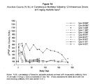
- the specification discloses a method predicting prognosis of an inflammatory bowel disease for a patient comprising comparing a ratio between amount of gut-homing lymphocytes and amount of peripheral-homing lymphocytes in a blood sample of said patient with a ratio between amount of gut-homing lymphocytes and amount of peripheral homing lymphocytes in a blood sample of a healthy individual, wherein a decreased ratio of said patient as compared to that of the healthy individual is indicative of the prognosis of the disease.
- the specification discloses a method of identifying a population of lymphocytes comprising lymphocytes expressing alphaEbeta7 integrin and lymphocytes expressing alpha4beta7 integrin, comprising binding said lymphocytes with an isolated antibody that bind to the same epitope of an antibody comprising a light chain variable region sequence of SEQ ID NO:10, and a heavy chain variable region sequence of SEQ ID NO:11.
- said lymphocytes are in peripheral blood of a patient diagnosed with an inflammatory bowel disease.
- said lymphocytes are in lymph node and tissues of the intestine of a patient diagnosed with an inflammatory bowel disease.
- the term "prediction” or “predicting” is used herein to refer to the likelihood that a patient will respond either favorably or unfavorably to a drug or set of drugs. In one aspect, the prediction relates to the extent of those responses. In one aspect, the prediction relates to whether and/or the probability that a patient will survive or improve following treatment, for example treatment with a particular therapeutic agent, for instance, anti-beta7 integrin antibody, and for a certain period of time without disease recurrence. For example, the predictive methods disclosed can be used clinically to make treatment decisions by choosing the most appropriate treatment modalities for any particular patient.
- the predictive methods disclosed are valuable tools in predicting whether a patient is likely to respond favorably to a treatment regimen, including for example, administration of a given therapeutic agent or combination, surgical intervention, steroid treatment, etc., or whether long-term survival of the patient, following a therapeutic regimen is likely.
- Treatment refers to clinical intervention in an attempt to alter the natural course of the individual or cell being treated, and can be performed either for prophylaxis or during the course of clinical pathology. Desirable effects of treatment include preventing occurrence or recurrence of disease, alleviation of symptoms, diminishment of any direct or indirect pathological consequences of the disease, decreasing the rate of disease progression, amelioration or palliation of the disease state, and remission or improved prognosis.
- Treatment regimen refers to a combination of dosage, frequency of administration, or duration of treatment, with or without addition of a second medication.
- Effective treatment regimen refers to a treatment regimen that will offer beneficial response to a patient receiving the treatment.
- Modifying a treatment refers to changing the treatment regimen including, changing dosage, frequency of administration, or duration of treatment, and/or addition of a second medication.
- Patient response or “patient responsiveness” can be assessed using any endpoint indicating a benefit to the patient, including, without limitation, (1) inhibition, to some extent, of disease progression, including slowing down and complete arrest; (2) reduction in the number of disease episodes and/or symptoms; (3) reduction in lesional size; (4) inhibition (i.e., reduction, slowing down or complete stopping) of disease cell infiltration into adjacent peripheral organs and/or tissues; (5) inhibition ( i.e., reduction, slowing down or complete stopping) of disease spread; (6) decrease of auto-immune response, which may, but does not have to, result in the regression or ablation of the disease lesion; (7) relief, to some extent, of one or more symptoms associated with the disorder; (8) increase in the length of disease-free presentation following treatment; and/or (9) decreased mortality at a given point of time following treatment.
- responsiveness refers to a measurable response, including complete response (CR) and partial response (PR).
- Partial response refers to a decrease of at least 50% in the severity of inflammation, in response to treatment.
- An "beneficial response" of a patient to treatment with an integrin beta7 antagonist and similar wording refers to the clinical or therapeutic benefit imparted to a patient at risk for or suffering from a gastrointestinal inflammatory disorder from or as a result of the treatment with the antagonist, such as an anti-beta7 integrin antibody.
- Such benefit includes cellular or biological responses, a complete response, a partial response, a stable disease (without progression or relapse), or a response with a later relapse of the patient from or as a result of the treatment with the antagonist.
- a patient maintains responsiveness to a treatment" when the patient' responsiveness does not decrease with time during the course of a treatment.
- diagnosis is used herein to refer to the identification or classification of a molecular or pathological state, disease or condition.
- diagnosis may refer to identification of a particular type of gastrointestinal inflammatory disorder, and more particularly, the classification of a particular sub-type of gastrointestinal inflammatory disorder, by tissue/organ involvement (e.g., inflammatory bowel disease), or by other features (e.g., a patient subpopulation characterized by responsiveness to a treatment, such as to a treatment with an integrin beta7 antagonist).
- prognosis is used herein to refer to the prediction of the likelihood of disease symptoms, including, for example, recurrence, flaring, and drug resistance, of a gastrointestinal inflammatory disorder.
- sample refers to a composition that is obtained or derived from a subject of interest that contains a cellular and/or other molecular entity that is to be characterized and/or identified, for example based on physical, biochemical, chemical and/or physiological characteristics.
- disease sample and variations thereof refers to any sample obtained from a subject of interest that would be expected or is known to contain the cellular and/or molecular entity that is to be characterized.
- the sample can be obtained from a tissue for the subject of interest or from peripheral blood of the subject.
- a beta7 integrin antagonist or “beta7 antagonist” refers to any molecule that inhibits one or more biological activities or blocking binding of beta7 integrin with one or more of its associated molecules.
- Antagonists of the invention can be used to modulate one or more aspects of beta7 associated effects, including but not limited to association with alpha4 integrin subunit, association with alphaE integrin subunit, binding of alpha4beta7 integrin to MAdCAM, VCAM-1 or fibronectin and binding of alphaEbeta7 integrin to E-cadherin.
- the beta7 antagonist is an anti-beta7 integrin antibody (or anti-beta7 antibody).
- the anti-beta7 integrin antibody is a humanized anti-beta7 integrin antibody and more particularly a recombinant humanized monoclonal anti-beta7 antibody (or rhuMAb beta7).
- the anti-beta7 antibodies of the present invention are anti-integrin beta7 antagonistic antibodies that inhibit or block the binding of beta7 subunit with alpha4 integrin subunit, association with alphaE integrin subunit, binding of alpha4beta7 integrin to MAdCAM, VCAM-1 or fibronectin and binding of alphaEbeta7 integrin to E-cadherin.
- beta7 subunit or ".beta. 7 subunit” is meant the human .beta.7 integrin subunit ( Erle et al., (1991) J. Biol. Chem. 266:11009-11016 ).
- the beta7 subunit associates with alpha4 integrin subunit, such as the human .alpha.4 subunit ( Kilger and Holzmann (1995) J. Mol. Biol. 73:347-354 ).
- the alpha4beta7 integrin is reportedly expressed on a majority of mature lymphocytes, as well as a small population of thymocytes, bone marrow cells and mast cells. ( Kilshaw and Murant (1991) Eur. J. Immunol.
- the beta7 subunit also associates with the alphaE subunit, such as the human alphaE integrin subunit ( Cepek, K. L, et al. (1993) J. Immunol. 150:3459 ).
- the alphaEbeta7 integrin is expressed on intra-intestinal epithelial lymphocytes (iIELs) (Cepek, K. L. (1993) supra ).
- alphaE subunit or "alphaE integrin subunit” or “.alpha.E subunit” or “.alpha.E integrin subunit” or “CD103” is meant an integrin subunit found to be associated with beta7 integrin on intra-epithelial lymphocytes, which alphaEbeta7 integrin mediates binding of the iELs to intestinal epithelium expressing E-cadherin ( Cepek, K. L. et al. (1993) J. Immunol. 150:3459 ; Shaw, S. K. and Brenner, M. B. (1995) Semin. Immunol. 7:335 ).
- MAdCAM or “MAdCAM-1” are used interchangeably in the context of the present invention and refer to the protein mucosal addressin cell adhesion molecule-1, which is a single chain polypeptide comprising a short cytoplasmic tail, a transmembrane region and an extracellular sequence composed of three immunoglobulin-like domains.
- the cDNAs for murine, human and macaque MAdCAM-1 have been cloned ( Briskin, et al., (1993) Nature, 363:461-464 ; Shyjan et al., (1996) J. Immunol. 156:2851-2857 ).
- VCAM-1 or "vascular cell adhesion molecule-1”
- CD106 refers to a ligand of alpha4beta7 and alpha4beta1, expressed on activated endothelium and important in endothelial-leukocyte interactions such as binding and transmigration of leukocytes during inflammation.
- CD45 refers to a protein of the protein tyrosine phosphatase (PTP) family.
- PTPs are known to be signaling molecules that regulate a variety of cellular processes including cell growth, differentiation, mitotic cycle, and oncogenic transformation.
- This PTP contains an extracellular domain, a single transmembrane segment and two tandem intracytoplasmic catalytic domains, and thus belongs to receptor type PTP.
- This gene is specifically expressed in hematopoietic cells.
- This PTP has been shown to be an essential regulator of T-and B-cell antigen receptor signaling. It functions through either direct interaction with components of the antigen receptor complexes, or by activating various Src family kinases required for the antigen receptor signaling.
- CD45RA, CD45RB, CD45RC, CD45RAB, CD45RAC, CD45RBC, CD45RO, CD45R (ABC).
- CD45 is also highly glycosylated.
- CD45R is the longest protein and migrates at 200 kDa when isolated from T cells.
- B cells also express CD45R with heavier glycosylation, bringing the molecular weight to 220 kDa, hence the name B220;
- B220 expression is not restricted to B cells and can also be expressed on activated T cells, on a subset of dendritic cells and other antigen presenting cells.
- CD45 variant alleles possibly increased frequency of a novel exon 4 CD45 polymorphism in HIV seropositive Kenyans.”
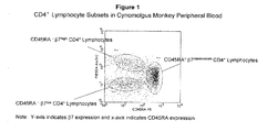
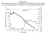
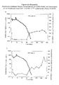
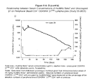
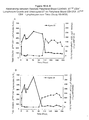
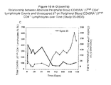
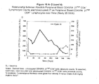
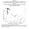
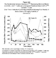
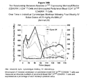
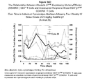
- “Gut-homing lymphocytes” refer to a subgroup of lymphocytes having the characteristic of selectively homing to intestinal lymph nodes and tissues but not homing to peripheral lymph nodes and tissues. This subgroup of lymphocytes are characterized by an unique expression pattern of a combination of multiples cell surface molecules, including, but not limited to, the combination of CD4, CD45RA and Beta7. Typically, at least two subsets of peripheral blood CD4 + lymphocytes can be subdivided based on the markers of CD45RA and Beta7, CD45RA - ⁇ 7 ⁇ high , and CD45RA - ⁇ 7 ⁇ low CD4 + cells.
- CD45RA - ⁇ 7 ⁇ high CD4 + cells home preferentially to intestinal lymph nodes and tissues
- CD45RA- ⁇ 7 ⁇ low CD4 + cells home preferentially to peripheral lymph nodes and tissues
- Gut-homing lymphocytes are therefore a distinctive subgroup of lymphocytes identified as CD45RA - ⁇ 7 high CD4 + in a flow cytometry assay. The methods of identifying this group of lymphocytes are well-known in the art and also disclosed in detail in Examples of the present application.
- CD4 + lymphocytes are a group of lymphocytes having CD4 expressed on their cell surfaces.
- CD45RA - lymphocytes are a group of lymphocytes having no CD45RA expressed on their cell surfaces.
- the symbol “low” indicates a relatively low level of expression of a cell surface marker on lymphocytes
- “high” indicates a relatively high level of expression of a cell surface marker on lymphocytes.
- the intensity of ⁇ 7 high is at least about 10 or 100 fold higher than that of ⁇ 7 ⁇ low .
- the CD45RA - ⁇ 7 ⁇ low CD4 + and CD45RA - ⁇ 7 ⁇ high CD4 + lymphocytes locate in distinct portions of a dot plot or histogram of a flow cytometry analysis where X-axis is the intensity of expression of CD45AR and Y-axis is the intensity of the expression of Beta7.
- Peripheral-homing lymphocytes refer to a subgroup of lymphocytes having the characteristic of homing to peripheral lymph nodes and tissues and not homing to intestinal lymph nodes and tissues.
- Peripheral-homing lymphocytes are a distinctive group of lymphocytes identified as CD45RA - ⁇ 7 ⁇ low CD4 + cells in a flow cytometry assay. The methods of identifying this group of lymphocytes are known in the art and disclosed in detail in the present application.
- an “amount” or “level” of biomarker can be determined using methods known in the art.
- the "amount” or “level” of gut-homing lymphocytes which is associated with the responsiveness of a patient to a treatment with integrin beta7 antagonist is a detectable level in a biological sample, preferably in a peripheral blood sample.
- the amount of the gut-homing lymphocytes can be quantified by methods known to the expert skilled in the art and disclosed by this invention, such as flow cytometry analysis.
- an "elevated” or “increased” amount or level of a biomarker is as compared to a reference/comparator amount of the biomarker.
- the increase is preferably greater than about 10%, preferably greater than about 30%, preferably greater than about 50%, preferably greater than about 100%, preferably greater than about 300% as a function of the value for the reference or comparator amount.
- a reference or comparator amount can be the amount of a biomarker before treatment and more particularly, can be the baseline or pre-dose amount.
- an "amount" or "level” of gut-homing lymphocytes denotes a sufficient increase in the number of lymphocytes such that one of skill in the art would consider the increase to be of statistical significance within the context of the biological characteristic measured by said values.
- the increase is preferably greater than about 10%, preferably greater than about 30%, preferably greater than about 50%, preferably greater than about 100%, preferably greater than about 300% as a function of the value for the reference/comparator amount of lymphocytes.
- a "decreased" amount or level of a biomarker is as compared to a reference/comparator amount of the biomarker.
- the decrease is preferably less than about 10%, preferably less than about 30%, preferably less than about 50%, preferably less than about 100%, preferably less than about 300% as a function of the value for the reference or comparator amount.
- a reference or comparator amount can be the amount of a biomarker before treatment and more particularly, can be the baseline or pre-dose amount.
- a "treatment that effectively increases the amount of the gut-homing lymphocytes” refers to a treatment that sufficiently increases in the number of lymphocytes such that one of skill in the art would consider the increase to be of statistical significance within the context of the biological characteristic measured by said values.
- the increase is preferably greater than about 10%, preferably greater than about 30%, preferably greater than about 50%, preferably greater than about 100%, preferably greater than about 300% as a function of the value for the reference/comparator amount of lymphocytes prior to the treatment.
- the change is preferably less than about 10%, preferably less than about 5%, preferably less than about 1%.
- Gastrointestinal inflammatory disorders are a group of chronic disorders that cause inflammation and/or ulceration in the mucous membrane. These disorders include, for example, inflammatory bowel disease (e.g., Crohn's disease, ulcerative colitis, indeterminate colitis and infectious colitis), mucositis (e.g., oral mucositis, gastrointestinal mucositis, nasal mucositis and proctitis), necrotizing enterocolitis and esophagitis.
- the gastrointestinal inflammatory disorder is a inflammatory bowel disease.
- IBD Inflammatory Bowel Disease
- IBD ulcerative colitis
- Crohn's disease (CD) or “ulcerative colitis (UC)” are chronic inflammatory bowel diseases of unknown etiology. Crohn's disease, unlike ulcerative colitis, can affect any part of the bowel. The most prominent feature Crohn's disease is the granular, reddish-purple edmatous thickening of the bowel wall. With the development of inflammation, these granulomas often lose their circumscribed borders and integrate with the surrounding tissue. Diarrhea and obstruction of the bowel are the predominant clinical features. As with ulcerative colitis, the course of Crohn's disease may be continuous or relapsing, mild or severe, but unlike ulcerative colitis, Crohn's disease is not curable by resection of the involved segment of bowel. Most patients with Crohn's disease require surgery at some point, but subsequent relapse is common and continuous medical treatment is usual.
- Crohn's disease may involve any part of the alimentary tract from the mouth to the anus, although typically it appears in the ileocolic, small-intestinal or colonic-anorectal regions. Histopathologically, the disease manifests by discontinuous granulomatomas, crypt abscesses, fissures and aphthous ulcers.
- the inflammatory infiltrate is mixed, consisting of lymphocytes (both T and B cells), plasma cells, macrophages, and neutrophils. There is a disproportionate increase in IgM- and IgG-secreting plasma cells, macrophages and neutrophils.
- Anti-inflammatory drugs sulfasalazine and 5-aminosalisylic acid (5-ASA) are useful for treating mildly active colonic Crohn's disease and are commonly prescribed to maintain remission of the disease.
- Metroidazole and ciprofloxacin are similar in efficacy to sulfasalazine and appear to be particularly useful for treating perianal disease.
- corticosteroids are effective in treating active exacerbations and can even maintain remission.
- Azathioprine and 6-mercaptopurine have also shown success in patients who require chronic administration of cortico steroids. It is also possible that these drugs may play a role in the long-term prophylaxis.
- Antidiarrheal drugs can also provide symptomatic relief in some patients.
- Nutritional therapy or elemental diet can improve the nutritional status of patients and induce symptomatic improvement of acute disease, but it does not induce sustained clinical remissions.
- Antibiotics are used in treating secondary small bowel bacterial overgrowth and in treatment of pyogenic complications.
- Ulcerative colitis afflicts the large intestine.
- the course of the disease may be continuous or relapsing, mild or severe.
- the earliest lesion is an inflammatory infiltration with abscess formation at the base of the crypts of Lieberkuhn. Coalescence of these distended and ruptured crypts tends to separate the overlying mucosa from its blood supply, leading to ulceration.
- Symptoms of the disease include cramping, lower abdominal pain, rectal bleeding, and frequent, loose discharges consisting mainly of blood, pus and mucus with scanty fecal particles.
- a total colectomy may be required for acute, severe or chronic, unremitting ulcerative colitis.
- UC ulcerative colitis
- Treatment for UC includes sulfasalazine and related salicylate-containing drugs for mild cases and corticosteroid drugs in severe cases.
- Topical administration of either salicylates or corticosteroids is sometimes effective, particularly when the disease is limited to the distal bowel, and is associated with decreased side effects compared with systemic use.
- Supportive measures such as administration of iron and antidiarrheal agents are sometimes indicated.
- Azathioprine, 6-mercaptopurine and methotrexate are sometimes also prescribed for use in refractory corticosteroid-dependent cases.
- an “effective dosage” refers to an amount effective, at dosages and for periods of time necessary, to achieve the desired therapeutic or prophylactic result.
- the term "patient” refers to any single animal, more preferably a mammal (including such non-human animals as, for example, dogs, cats, horses, rabbits, zoo animals, cows, pigs, sheep, and non-human primates) for which treatment is desired. Most preferably, the patient herein is a human.
- non-human subject refers to any single non-human animal, more preferably a mammal (including such non-human animals as, for example, dogs, cats, horses, rabbits, zoo animals, cows, pigs, sheep, and non-human primates).
- antibody and “immunoglobulin” are used interchangeably in the broadest sense and include monoclonal antibodies (for example, full length or intact monoclonal antibodies), polyclonal antibodies, multivalent antibodies, multispecific antibodies (e.g., bispecific antibodies so long as they exhibit the desired biological activity) and may also include certain antibody fragments (as described in greater detail herein).
- An antibody can be human, humanized and/or affinity matured.
- Antibody fragments comprise only a portion of an intact antibody, wherein the portion preferably retains at least one, preferably most or all, of the functions normally associated with that portion when present in an intact antibody.
- an antibody fragment comprises an antigen binding site of the intact antibody and thus retains the ability to bind antigen.
- an antibody fragment for example one that comprises the Fc region, retains at least one of the biological functions normally associated with the Fc region when present in an intact antibody, such as FcRn binding, antibody half life modulation, ADCC function and complement binding.
- an antibody fragment is a monovalent antibody that has an in vivo half life substantially similar to an intact antibody.
- such an antibody fragment may comprise on antigen binding arm linked to an Fc sequence capable of conferring in vivo stability to the fragment.
- monoclonal antibody refers to an antibody obtained from a population of substantially homogeneous antibodies, i.e., the individual antibodies comprising the population are identical except for possible naturally occurring mutations that may be present in minor amounts. Monoclonal antibodies are highly specific, being directed against a single antigen. Furthermore, in contrast to polyclonal antibody preparations that typically include different antibodies directed against different determinants (epitopes), each monoclonal antibody is directed against a single determinant on the antigen.
- the monoclonal antibodies herein specifically include "chimeric" antibodies in which a portion of the heavy and/or light chain is identical with or homologous to corresponding sequences in antibodies derived from a particular species or belonging to a particular antibody class or subclass, while the remainder of the chain(s) is identical with or homologous to corresponding sequences in antibodies derived from another species or belonging to another antibody class or subclass, as well as fragments of such antibodies, so long as they exhibit the desired biological activity ( U.S. Patent No. 4,816,567 ; and Morrison et al., Proc. Natl. Acad. Sci. USA 81:6851-6855 (1984 )).
- Humanized forms of non-human (e.g., murine) antibodies are chimeric antibodies that contain minimal sequence derived from non-human immunoglobulin.
- humanized antibodies are human immunoglobulins (recipient antibody) in which residues from a hypervariable region of the recipient are replaced by residues from a hypervariable region of a non-human species (donor antibody) such as mouse, rat, rabbit or nonhuman primate having the desired specificity, affinity, and capacity.
- donor antibody such as mouse, rat, rabbit or nonhuman primate having the desired specificity, affinity, and capacity.
- framework region (FR) residues of the human immunoglobulin are replaced by corresponding non-human residues.
- humanized antibodies may comprise residues that are not found in the recipient antibody or in the donor antibody. These modifications are made to further refine antibody performance.
- the humanized antibody will comprise substantially all of at least one, and typically two, variable domains, in which all or substantially all of the hypervariable loops correspond to those of a non-human immunoglobulin and all or substantially all of the FRs are those of a human immunoglobulin lo sequence.
- the humanized antibody optionally will also comprise at least a portion of an immunoglobulin constant region (Fc), typically that of a human immunoglobulin.
- Fc immunoglobulin constant region
- a "human antibody” is one which comprises an amino acid sequence corresponding to that of an antibody produced by a human and/or has been made using any of the techniques for making human antibodies as disclosed herein. Such techniques include screening human-derived combinatorial libraries, such as phage display libraries (see, e.g., Marks et al., J. Mol. Biol., 222: 581-597 (1991 ) and Hoogenboom et al., Nucl. Acids Res., 19: 4133-4137 (1991 )); using human myeloma and mouse-human heteromyeloma cell lines for the production of human monoclonal antibodies (see, e.g., Kozbor J.
- human-derived combinatorial libraries such as phage display libraries (see, e.g., Marks et al., J. Mol. Biol., 222: 581-597 (1991 ) and Hoogenboom et al., Nucl. Acids Res., 19: 4133-4137
- This definition of a human antibody specifically excludes a humanized antibody comprising antigen-binding residues from a non-human animal.
- an “isolated” antibody is one which has been identified and separated and/or recovered from a component of its natural environment. Contaminant components of its natural environment are materials which would interfere with diagnostic or therapeutic uses for the antibody, and may include enzymes, hormones, and other proteinaceous or nonproteinaceous solutes.
- the antibody will be purified (1) to greater than 95% by weight of antibody as determined by the Lowry method, and most preferably more than 99% by weight, (2) to a degree sufficient to obtain at least 15 residues of N-terminal or internal amino acid sequence by use of a spinning cup sequenator, or (3) to homogeneity by SDS-PAGE under reducing or nonreducing conditions using Coomassie blue or, preferably, silver stain.
- Isolated antibody includes the antibody in situ within recombinant cells since at least one component of the antibody's natural environment will not be present. Ordinarily, however, isolated antibody will be prepared by at least one purification step.
- hypervariable region when used herein refers to the regions of an antibody variable domain which are hypervariable in sequence and/or form structurally defined loops.
- antibodies comprise six hypervariable regions; three in the VH (H1, H2, H3), and three in the VL (L1, L2, L3).
- a number of hypervariable region delineations are in use and are encompassed herein.
- the Kabat Complementarity Determining Regions are based on sequence variability and are the most commonly used ( Kabat et al., Sequences of Proteins of Immunological Interest, 5th Ed. Public Health Service, National Institutes of Health, Bethesda, Md. (1991 )).
- Chothia refers instead to the location of the structural loops ( Chothia and Lesk J. Mol. Biol. 196:901-917 (1987 )).
- the AbM hypervariable regions represent a compromise between the Kabat CDRs and Chothia structural loops, and are used by Oxford Molecular's AbM antibody modeling software.
- the "contact" hypervariable regions are based on an analysis of the available complex crystal structures. The residues from each of these HVRs are noted below.
- Hypervariable regions may comprise "extended hypervariable regions” as follows: 24-36 or 24-34 (L1), 46-56 or 49-56 or 50-56 or 52-56 (L2) and 89-97 (L3) in the VL and 26-35 (H1), 50-65 or 49-65 (H2) and 93-102, 94-102 or 95-102 (H3) in the VH.
- the variable domain residues are numbered according to Kabat et al., supra for each of these definitions.
- Framework or "FR” residues are those variable domain residues other than the hypervariable region residues as herein defined.
- a "human consensus framework” is a framework which represents the most commonly occurring amino acid residue in a selection of human immunoglobulin VL or VH framework sequences.
- the selection of human immunoglobulin VL or VH sequences is from a subgroup of variable domain sequences.
- the subgroup of sequences is a subgroup as in Kabat et al.
- the subgroup is subgroup kappa I as in Kabat et al.
- the subgroup III as in Kabat et al.
- affinity matured antibody is one with one or more alterations in one or more CDRs thereof which result in an improvement in the affinity of the antibody for antigen, compared to a parent antibody which does not possess those alteration(s).
- Preferred affinity matured antibodies will have nanomolar or even picomolar affinities for the target antigen.
- Affinity matured antibodies are produced by procedures known in the art. Marks et al. Bio/Technology 10:779-783 (1992 ) describes affinity maturation by VH and VL domain shuffling. Random mutagenesis of CDR and/or framework residues is described by: Barbas et al. Proc Nat. Acad. Sci, USA 91:3809-3813 (1994 ); Schier et al.
- substantially similar denotes a sufficiently high degree of similarity between two numeric values (generally one associated with an antibody of the invention and the other associated with a reference/comparator antibody) such that one of skill in the art would consider the difference between the two values to be of little or no biological and/or statistical significance within the context of the biological characteristic measured by said values (e.g., Kd values).
- the difference between said two values is preferably less than about 50%, preferably less than about 40%, preferably less than about 30%, preferably less than about 20%, preferably less than about 10% as a function of the value for the reference/comparator antibody.
- Binding affinity generally refers to the strength of the sum total of noncovalent interactions between a single binding site of a molecule (e.g., an antibody) and its binding partner (e.g., an antigen). Unless indicated otherwise, as used herein, "binding affinity” refers to intrinsic binding affinity which reflects a 1:1 interaction between members of a binding pair ( e.g., antibody and antigen).
- the affinity of a molecule X for its partner Y can generally be represented by the dissociation constant (Kd). Affinity can be measured by common methods known in the art, including those described herein. Low-affinity antibodies generally bind antigen slowly and tend to dissociate readily, whereas high-affinity antibodies generally bind antigen faster and tend to remain bound longer. A variety of methods of measuring binding affinity are known in the art, any of which can be used for purposes of the present invention.
- variable refers to the fact that certain portions of the variable domains differ extensively in sequence among antibodies and are used in the binding and specificity of each particular antibody for its particular antigen. However, the variability is not evenly distributed throughout the variable domains of antibodies. It is concentrated in three segments called hypervariable regions both in the light chain and the heavy chain variable domains. The more highly conserved portions of variable domains are called the framework regions (FRs).
- the variable domains of native heavy and light chains each comprise four FRs, largely adopting a ⁇ -sheep configuration, connected by three hypervariable regions, which form loops connecting, and in some cases forming part of, the ⁇ -sheep structure.
- the hypervariable regions in each chain are held together in close proximity by the FRs and, with the hypervariable regions from the other chain, contribute to the formation of the antigen-binding site of antibodies (see Kabat et al., Sequences of Proteins of Immunological Interest, 5th Ed. Public Health Service, National Institutes of Health, Bethesda, MD. (1991 )).
- the constant domains are not involved directly in binding an antibody to an antigen, but exhibit various effector functions, such as participation of the antibody in antibody dependent cellular cytotoxicity (ADCC).
- Papain digestion of antibodies produces two identical antigen-binding fragments, called “Fab” fragments, each with a single antigen-binding site, and a residual "Fc” fragment, whose name reflects its ability to crystallize readily. Pepsin treatment yields an F(ab') 2 fragment that has two antigen-binding sites and is still capable of cross-linking antigen.
- Fv is the minimum antibody fragment which contains a complete antigen-recognition and antigen-binding site. This region consists of a dimer of one heavy chain and one light chain variable domain in tight, non-covalent association. It is in this configuration that the three hypervariable regions of each variable domain interact to define an antigen-binding site on the surface of the V H -V L dimer. Collectively, the six hypervariable regions confer antigen-binding specificity to the antibody. However, even a single variable domain (or half of an Fv comprising only three hypervariable regions specific for an antigen) has the ability to recognize and bind antigen, although at a lower affinity than the entire binding site.
- the Fab fragment also contains the constant domain of the light chain and the first constant domain (CH1) of the heavy chain.
- Fab fragments differ from Fab fragments by the addition of a few residues at the carboxy terminus of the heavy chain CH1 domain including one or more cysteines from the antibody hinge region.
- Fab'-SH is the designation herein for Fab' in which the cysteine residue(s) of the constant domains bear at least one free thiol group.
- F(ab') 2 antibody fragments originally were produced as pairs of Fab' fragments which have hinge cysteines between them. Other chemical couplings of antibody fragments are also known.
- the "light chains" of antibodies from any vertebrate species can be assigned to one of two clearly distinct types, called kappa ( ⁇ ) and lambda ( ⁇ ), based on the amino acid sequences of their constant domains.
- antibodies can be assigned to different classes.
- immunoglobulins There are five major classes of immunoglobulins: IgA, IgD, IgE, IgG, and IgM, and several of these may be further divided into subclasses (isotypes), e.g., IgG 1 , IgG 2 , IgG 3 , IgG 4 , IgA 1 , and IgA 2 .
- the heavy-chain constant domains that correspond to the different classes of immunoglobulins are called ⁇ , ⁇ , ⁇ , ⁇ , and ⁇ , respectively.
- An antibody may be part of a larger fusion molecule, formed by covalent or non-covalent association of the antibody with one or more other proteins or peptides.
- full-length antibody “intact antibody,” and “whole antibody” are used herein interchangeably to refer to an antibody in its substantially intact form, not antibody fragments as defined below.
- naked antibody for the purposes herein is an antibody that is not conjugated to a cytotoxic moiety or radiolabel.
- Fc region herein is used to define a C-terminal region of an immunoglobulin heavy chain, including native sequence Fc regions and variant Fc regions.
- the human IgG heavy chain Fc region is usually defined to stretch from an amino acid residue at position Cys226, or from Pro230, to the carboxyl-terminus thereof.
- the C-terminal lysine (residue 447 according to the EU numbering system) of the Fc region may be removed, for example, during production or purification of the antibody, or by recombinantly engineering the nucleic acid encoding a heavy chain of the antibody. Accordingly, a composition of intact antibodies may comprise antibody populations with all K447 residues removed, antibody populations with no K447 residues removed, and antibody populations having a mixture of antibodies with and without the K447 residue.
- the numbering of the residues in an immunoglobulin heavy chain is that of the EU index as in Kabat et al., Sequences of Proteins of Immunological Interest, 5th Ed. Public Health Service, National Institutes of Health, Bethesda, MD (1991 ), expressly incorporated herein by reference.
- the "EU index as in Kabat” refers to the residue numbering of the human IgG1 EU antibody.
- a “functional Fc region” possesses an “effector function” of a native sequence Fc region.
- effector functions include C1q binding; complement dependent cytotoxicity; Fc receptor binding; antibody-dependent cell-mediated cytotoxicity (ADCC); phagocytosis; down regulation of cell surface receptors (e.g., B cell receptor; BCR), etc.
- ADCC antibody-dependent cell-mediated cytotoxicity
- phagocytosis down regulation of cell surface receptors (e.g., B cell receptor; BCR), etc.
- Such effector functions generally require the Fc region to be combined with a binding domain (e.g., an antibody variable domain) and can be assessed using various assays as herein disclosed, for example.
- a “native sequence Fc region” comprises an amino acid sequence identical to the amino acid sequence of an Fc region found in nature.
- Native sequence human Fc regions include a native sequence human IgG1 Fc region (non-A and A allotypes); native sequence human IgG2 Fc region; native sequence human IgG3 Fc region; and native sequence human IgG4 Fc region as well as naturally occurring variants thereof.
- a “variant Fc region” comprises an amino acid sequence which differs from that of a native sequence Fc region by virtue of at least one amino acid modification, preferably one or more amino acid substitution(s).
- the variant Fc region has at least one amino acid substitution compared to a native sequence Fc region or to the Fc region of a parent polypeptide, e.g., from about one to about ten amino acid substitutions, and preferably from about one to about five amino acid substitutions in a native sequence Fc region or in the Fc region of the parent polypeptide.
- the variant Fc region herein will preferably possess at least about 80% homology with a native sequence Fc region and/or with an Fc region of a parent polypeptide, and most preferably at least about 90% homology therewith, more preferably at least about 95% homology therewith.
- intact antibodies can be assigned to different "classes.” There are five major classes of intact antibodies: IgA, IgD, IgE, IgG, and IgM, and several of these may be further divided into “subclasses” (isotypes), e.g., IgG1, IgG2, IgG3, IgG4, IgA, and IgA2.
- the heavy-chain constant domains that correspond to the different classes of antibodies are called ⁇ , ⁇ , ⁇ , ⁇ , and ⁇ , respectively.
- the subunit structures and three-dimensional configurations of different classes of immunoglobulins are well known.
- Antibody-dependent cell-mediated cytotoxicity and “ADCC” refer to a cell-mediated reaction in which nonspecific cytotoxic cells that express Fc receptors (FcRs) (e.g. Natural Killer (NK) cells, neutrophils, and macrophages) recognize bound antibody on a target cell and subsequently cause lysis of the target cell.
- FcRs Fc receptors
- FcR expression on hematopoietic cells in summarized is Table 3 on page 464 of Ravetch and Kinet, Annu. Rev. Immunol 9:457-92 (1991 ).
- ADCC activity of a molecule of interest may be assessed in vitro, such as that described in U.S. Patent No. 5,500,362 or 5,821,337.
- Useful effector cells for such assays include peripheral blood mononuclear cells (PBMC) and Natural Killer (NK) cells.
- PBMC peripheral blood mononuclear cells
- NK Natural Killer
- ADCC activity of the molecule of interest may be assessed in vivo, e.g., in a animal model such as that disclosed in Clynes et al. PNAS (USA) 95:652-656 (1998 ).
- Human effector cells are leukocytes which express one or more FcRs and perform effector functions. Preferably, the cells express at least Fc ⁇ RIII and perform ADCC effector function. Examples of human leukocytes which mediate ADCC include peripheral blood mononuclear cells (PBMC), natural killer (NK) cells, monocytes, cytotoxic T cells and neutrophils; with PBMCs and NK cells being preferred.
- PBMC peripheral blood mononuclear cells
- NK natural killer cells
- monocytes cytotoxic T cells and neutrophils
- the effector cells may be isolated from a native source thereof, e.g., from blood or PBMCs as described herein.
- Fc receptor or “FcR” are used to describe a receptor that binds to the Fc region of an antibody.
- the preferred FcR is a native sequence human FcR.
- a preferred FcR is one which binds an IgG antibody (a gamma receptor) and includes receptors of the Fc ⁇ RI, Fc ⁇ RII, and Fc ⁇ RIII subclasses, including allelic variants and alternatively spliced forms of these receptors.
- Fc ⁇ RII receptors include Fc ⁇ RIIA (an “activating receptor") and Fc ⁇ RIIB (an “inhibiting receptor”), which have similar amino acid sequences that differ primarily in the cytoplasmic domains thereof.
- Activating receptor Fc ⁇ RIIA contains an immunoreceptor tyrosine-based activation motif (ITAM) in its cytoplasmic domain.
- Inhibiting receptor Fc ⁇ RIIB contains an immunoreceptor tyrosine-based inhibition motif (ITIM) in its cytoplasmic domain (see review M. in Da ⁇ ron, Annu. Rev. Immunol. 15:203-234 (1997 )).
- FcRs are reviewed in Ravetch and Kinet, Annu. Rev. Immunol 9:457-92 (1991 ); Capel et al., Immunomethods 4:25-34 (1994 ); and de Haas et al., J. Lab. Clin. Med. 126:330-41 (1995 ).
- FcR neonatal receptor
- FcRn the neonatal receptor
- Antibodies with improved binding to the neonatal Fc receptor (FcRn), and increased half-lives, are described in WO00/42072 (Presta, L. ) and US2005/0014934A1 (Hinton et al .) .
- These antibodies comprise an Fc region with one or more substitutions therein which improve binding of the Fc region to FcRn.
- the Fc region may have substitutions at one or more of positions 238, 250, 256, 265, 272, 286, 303, 305, 307, 311, 312, 314, 317, 340, 356, 360, 362, 376, 378, 380, 382, 413, 424, 428 or 434 (Eu numbering of residues).
- the preferred Fc region-comprising antibody variant with improved FcRn binding comprises amino acid substitutions at one, two or three of positions 307, 380 and 434 of the Fc region thereof (Eu numbering of residues).
- Single-chain Fv or “scFv” antibody fragments comprise the V H and V L domains of antibody, wherein these domains are present in a single polypeptide chain.
- the Fv polypeptide further comprises a polypeptide linker between the V H and V L domains which enables the scFv to form the desired structure for antigen binding.
- HER2 antibody scFv fragments are described in WO93/16185 ; U.S. Patent No. 5,571,894 ; and U.S. Patent No. 5,587,458 .
- diabodies refers to small antibody fragments with two antigen-binding sites, which fragments comprise a variable heavy domain (V H ) connected to a variable light domain (V L ) in the same polypeptide chain (V H - V L ).
- V H variable heavy domain
- V L variable light domain
- the domains are forced to pair with the complementary domains of another chain and create two antigen-binding sites.
- Diabodies are described more fully in, for example, EP 404,097 ; WO 93/11161 ; and Hollinger et al., Proc. Natl. Acad. Sci. USA, 90:6444-6448 (1993 ).
- affinity matured antibody is one with one or more alterations in one or more hypervariable regions thereof which result an improvement in the affinity of the antibody for antigen, compared to a parent antibody which does not possess those alteration(s).
- Preferred affinity matured antibodies will have nanomolar or even picomolar affinities for the target antigen.
- Affinity matured antibodies are produced by procedures known in the art. Marks et al. Bio/Technology 10:779-783 (1992 ) describes affinity maturation by VH and VL domain shuffling. Random mutagenesis of CDR and/or framework residues is described by: Barbas et al. Proc Nat. Acad. Sci, USA 91:3809-3813 (1994 ); Schier et al.
- amino acid sequence variant antibody herein is an antibody with an amino acid sequence which differs from a main species antibody.
- amino acid sequence variants will possess at least about 70% homology with the main species antibody, and preferably, they will be at least about 80%, more preferably at least about 90% homologous with the main species antibody.
- the amino acid sequence variants possess substitutions, deletions, and/or additions at certain positions within or adjacent to the amino acid sequence of the main species antibody.
- amino acid sequence variants herein include an acidic variant (e.g., deamidated antibody variant), a basic variant, an antibody with an amino-terminal leader extension (e.g.
- VHS- on one or two light chains thereof, an antibody with a C-terminal lysine residue on one or two heavy chains thereof, etc, and includes combinations of variations to the amino acid sequences of heavy and/or light chains.
- the antibody variant of particular interest herein is the antibody comprising an amino-terminal leader extension on one or two light chains thereof, optionally further comprising other amino acid sequence and/or glycosylation differences relative to the main species antibody.
- a “glycosylation variant” antibody herein is an antibody with one or more carbohydrate moieties attached thereto which differ from one or more carbohydrate moieties attached to a main species antibody.
- glycosylation variants herein include antibody with a G1 or G2 oligosaccharide structure, instead a G0 oligosaccharide structure, attached to an Fc region thereof, antibody with one or two carbohydrate moieties attached to one or two light chains thereof, antibody with no carbohydrate attached to one or two heavy chains of the antibody, etc, and combinations of glycosylation alterations. Where the antibody has an Fc region, an oligosaccharide structure may be attached to one or two heavy chains of the antibody, e.g. at residue 299 (298, Eu numbering of residues).
- cytotoxic agent refers to a substance that inhibits or prevents the function of cells and/or causes destruction of cells.
- the term is intended to include radioactive isotopes (e.g . At 211 , I 131 , I 125 , Y 90 , Re 186 , Re 188 , Sm 153 , Bi 212 , P 32 and radioactive isotopes of Lu), chemotherapeutic agents, and toxins such as small molecule toxins or enzymatically active toxins of bacterial, fungal, plant or animal origin, including fragments and/or variants thereof.
- radioactive isotopes e.g . At 211 , I 131 , I 125 , Y 90 , Re 186 , Re 188 , Sm 153 , Bi 212 , P 32 and radioactive isotopes of Lu
- chemotherapeutic agents e.g . At 211 , I 131 , I 125 , Y 90 , Re
- cytokine is a generic term for proteins released by one cell population which act on another cell as intercellular mediators.
- cytokines are lymphokines, monokines, and traditional polypeptide hormones. Included among the cytokines are growth hormone such as human growth hormone, N-methionyl human growth hormone, and bovine growth hormone; parathyroid hormone; thyroxine; insulin; proinsulin; relaxin; prorelaxin; glycoprotein hormones such as follicle stimulating hormone (FSH), thyroid stimulating hormone (TSH), and luteinizing hormone (LH); hepatic growth factor; fibroblast growth factor; prolactin; placental lactogen; tumor necrosis factor- ⁇ and - ⁇ ; mullerian-inhibiting substance; mouse gonadotropin-associated peptide; inhibin; activin; vascular endothelial growth factor; integrin; thrombopoietin (TPO); nerve growth factors such as NGF- ⁇ ; platelet-growth factor;
- immunosuppressive agent refers to substances that act to suppress or mask the immune system of the subject being treated herein. This would include substances that suppress cytokine production, down-regulate or suppress self-antigen expression, or mask the MHC antigens. Examples of such agents include 2-amino-6-aryl-5-substituted pyrimidines (see U.S. Patent No.
- non-steroidal anti-inflammatory drugs NSAIDs
- ganciclovir tacrolimus
- glucocorticoids such as cortisol or aldosterone
- anti-inflammatory agents such as a cyclooxygenase inhibitor; a 5-lipoxygenase inhibitor; or a leukotriene receptor antagonist
- purine antagonists such as azathioprine or mycophenolate mofetil (MMF)
- alkylating agents such as cyclophosphamide; bromocryptine; danazol; dapsone; glutaraldehyde (which masks the MHC antigens, as described in U.S. Patent No.
- anti-idiotypic antibodies for MHC antigens and MHC fragments include cyclosporine; 6 mercaptopurine; steroids such as corticosteroids or glucocorticosteroids or glucocorticoid analogs, e.g., prednisone, methylprednisolone, including SOLU-MEDROL.RTM.
- methylprednisolone sodium succinate, and dexamethasone dihydrofolate reductase inhibitors such as methotrexate (oral or subcutaneous); anti-malarial agents such as chloroquine and hydroxychloroquine; sulfasalazine; leflunomide; cytokine or cytokine receptor antibodies or antagonists including anti-interferon-alpha, -beta, or -gamma antibodies, anti-tumor necrosis factor(TNF)-alpha antibodies (infliximab (REMICADE.RTM.) or adalimumab), anti-TNF-alpha immunoadhesin (etanercept), anti-TNF-beta antibodies, anti-interleukin-2 (IL-2) antibodies and anti-IL-2 receptor antibodies, and anti-interleukin-6 (IL-6) receptor antibodies and antagonists; anti-LFA-1 antibodies, including anti-CD11a and anti-CD18 antibodies; anti-L3
- streptokinase transforming growth factor-beta (TGF-beta); streptodomase; RNA or DNA from the host; FK506; RS-61443; chlorambucil; deoxyspergualin; rapamycin; T-cell receptor ( Cohen et al., U.S. Patent No.
- T-cell receptor fragments Offner et al., Science, 251: 430-432 (1991 ); WO 90/11294 ; Ianeway, Nature, 341: 482 (1989 ); and WO 91/01133 ); BAFF antagonists such as BAFF or BR3 antibodies or immunoadhesins and zTNF4 antagonists (for review, see Mackay and Mackay, Trends Immunol., 23:113-5 (2002 ) and see also definition below); biologic agents that interfere with T cell helper signals, such as anti-CD40 receptor or anti-CD40 ligand (CD154), including blocking antibodies to CD40-CD40 ligand.( e.g ., Durie etal., Science, 261: 1328-30 (1993 ); Mohan et al., J.
- the present specification discloses methods of predicting the responsiveness of a patient to the treatment of beta7 integrin antagonists.
- potential antagonists include an oligonucleotide that binds to the fusions of immunoglobulin with beta7 integrin, and, in particular, antibodies including, without limitation, poly- and monoclonal antibodies and antibody fragments, single-chain antibodies, anti-idiotypic antibodies, and chimeric or humanized versions of such antibodies or fragments, as well as human antibodies and antibody fragments.
- a potential antagonist may be a closely related protein, for example, a mutated form of the beta7 integrin that recognizes the ligand but imparts no effect, thereby competitively inhibiting the action of the beta7 integrin.
- beta7 integrin antagonist is an antisense RNA or DNA construct prepared using antisense technology, where, e.g., an antisense RNA or DNA molecule acts to block directly the translation of mRNA by hybridizing to targeted mRNA and preventing protein translation.
- Antisense technology can be used to control gene expression through triple-helix formation or antisense DNA or RNA, both of which methods are based on binding of a polynucleotide to DNA or RNA.
- the 5' coding portion of the polynucleotide sequence, which encodes the beta7 integrin herein is used to design an antisense RNA oligonucleotide of from about 10 to 40 base pairs in length.
- a DNA oligonucleotide is designed to be complementary to a region of the gene involved in transcription (triple helix--see Lee et al., Nucl. Acids Res., 6:3073 (1979 ); Cooney et al., Science, 241: 456 (1988 ); Dervan et al., Science, 251:1360 (1991 )), thereby preventing transcription and the production of the beta7 integrin.
- the antisense RNA oligonucleotide hybridizes to the mRNA in vivo and blocks translation of the mRNA molecule into beta7 integrin protein (antisense-- Okano, Neurochem., 56:560 (1991 ); Oligodeoxynucleotides as Antisense Inhibitors of Gene Expression (CRC Press: Boca Raton, Fla., 1988 ).
- the oligonucleotides described above can also be delivered to cells such that the antisense RNA or DNA may be expressed in vivo to inhibit production of the PRO polypeptide.
- antisense DNA is used, oligodeoxyribonucleotides derived from the translation-initiation site, e.g., between about -10 and +10 positions of the target gene nucleotide sequence, are preferred.
- small molecules that bind to the active site, the ligand or binding molecule binding site, thereby blocking the normal biological activity of the beta7 integrin.
- small molecules include, but are not limited to, small peptides or peptide-like molecules, preferably soluble peptides, and synthetic non-peptidyl organic or inorganic compounds.
- Ribozymes are enzymatic RNA molecules capable of catalyzing the specific cleavage of RNA. Ribozymes act by sequence-specific hybridization to the complementary target RNA, followed by endonucleolytic cleavage. Specific ribozyme cleavage sites within a potential RNA target can-be identified by known techniques. For further details see, e.g., Rossi, Current Biology, 4:469-471 (1994 ), and PCT Publication No. WO 97/33551 (published Sep. 18, 1997 ).
- Nucleic acid molecules in triple-helix formation used to inhibit transcription should be single-stranded and composed of deoxynucleotides.
- the base composition of these oligonucleotides is designed such that it promotes triple-helix formation via Hoogsteen base-pairing rules, which generally require sizeable stretches of purines or pyrimidines on one strand of a duplex.
- PCT Publication No. WO 97/33551 See, e.g., PCT Publication No. WO 97/33551 .
- These small molecules can be identified by any one or more of the screening assays discussed hereinabove and/or by any other screening techniques well known for those skilled in the art.
- Screening assays for antagonists are designed to identify compounds that bind or complex with the beta7 integrin encoded by the genes identified herein, or otherwise interfere with the interaction of the encoded polypeptides with other cellular proteins.
- Such screening assays will include assays amenable to high-throughput screening of chemical libraries, making them particularly suitable for identifying small molecule drug candidates.
- the assays can be performed in a variety of formats, including protein-protein binding assays, biochemical screening assays, immunoassays, and cell-based assays, which are well characterized in the art.
- the beta7 integrin antagonists are anti-beta7 antibodies.
- Exemplary antibodies include polyclonal, monoclonal, humanized, human, bispecific, and heteroconjugate antibodies, etc., as described below.
- a protein that is immunogenic in the species to be immunized e.g., keyhole limpet hemocyanin, serum albumin, bovine thy
- Animals are immunized against the antigen, immunogenic conjugates, or derivatives by combining, e.g., 100 ⁇ g or 5 ⁇ g of the protein or conjugate (for rabbits or mice, respectively) with 3 volumes of Freund's complete adjuvant and injecting the solution intradermally at multiple sites.
- the animals are boosted with 1/5 to 1/10 the original amount of peptide or conjugate in Freund's complete adjuvant by subcutaneous injection at multiple sites.
- Seven to 14 days later the animals are bled and the serum is assayed for antibody titer. Animals are boosted until the titer plateaus.
- the animal is boosted with the conjugate of the same antigen, but conjugated to a different protein and/or through a different cross-linking reagent.
- Conjugates also can be made in recombinant cell culture as protein fusions.
- aggregating agents such as alum are suitably used to enhance the immune response.
- Monoclonal antibodies may be made using the hybridoma method first described by Kohler et al., Nature, 256:495 (1975 ), or may be made by recombinant DNA methods ( U.S. Patent No. 4,816,567 ).
- lymphocytes In the hybridoma method, a mouse or other appropriate host animal, such as a hamster, is immunized as described above to elicit lymphocytes that produce or are capable of producing antibodies that will specifically bind to the protein used for immunization.
- lymphocytes may be immunized in vitro. After immunization, lymphocytes are isolated and then fused with a myeloma cell line using a suitable fusing agent, such as polyethylene glycol, to form a hybridoma cell ( Goding, Monoclonal Antibodies: Principles and Practice, pp. 59-103 (Academic Press, 1986 )).
- the hybridoma cells thus prepared are seeded and grown in a suitable culture medium which medium preferably contains one or more substances that inhibit the growth or survival of the unfused, parental myeloma cells (also referred to as fusion partner).
- a suitable culture medium which medium preferably contains one or more substances that inhibit the growth or survival of the unfused, parental myeloma cells (also referred to as fusion partner).
- the parental myeloma cells lack the enzyme hypoxanthine guanine phosphoribosyl transferase (HGPRT or HPRT)
- HGPRT or HPRT the selective culture medium for the hybridomas typically will include hypoxanthine, aminopterin, and thymidine (HAT medium), which substances prevent the growth of HGPRT-deficient cells.
- Preferred fusion partner myeloma cells are those that fuse efficiently, support stable high-level production of antibody by the selected antibody-producing cells, and are sensitive to a selective medium that selects against the unfused parental cells.
- Preferred myeloma cell lines are murine myeloma lines, such as those derived from MOPC-21 and MPC-11 mouse tumors available from the Salk Institute Cell Distribution Center, San Diego, Calif. USA, and SP-2 and derivatives e.g., X63-Ag8-653 cells available from the American Type Culture Collection, Manassas, Va., USA.
- Human myeloma and mouse-human heteromyeloma cell lines also have been described for the production of human monoclonal antibodies ( Kozbor, J. Immunol., 133:3001 (1984 ); and Brön et al., Monoclonal Antibody Production Techniques and Applications, pp. 51-63 (Marcel Dekker, Inc., New York, 1987 )).
- Culture medium in which hybridoma cells are growing is assayed for production of monoclonal antibodies directed against the antigen.
- the binding specificity of monoclonal antibodies produced by hybridoma cells is determined by immunoprecipitation or by an in vitro binding assay, such as radioimmunoassay (RIA) or enzyme-linked immunosorbent assay (ELISA).
- RIA radioimmunoassay
- ELISA enzyme-linked immunosorbent assay
- the binding affinity of the monoclonal antibody can, for example, be determined by the Scatchard analysis described in Munson et al., Anal. Biochem., 107:220 (1980 ).
- the clones may be subcloned by limiting dilution procedures and grown by standard methods ( Goding, Monoclonal Antibodies: Principles and Practice, pp. 59-103 (Academic Press, 1986 )).
- Suitable culture media for this purpose include, for example, D-MEM or RPMI-1640 medium.
- the hybridoma cells may be grown in vivo as ascites tumors in an animal e.g., by i.p.
- the monoclonal antibodies secreted by the subclones are suitably separated from the culture medium, ascites fluid, or serum by conventional antibody purification procedures such as, for example, affinity chromatography (e.g., using protein A or protein G-Sepharose) or ion-exchange chromatography, hydroxylapatite chromatography, gel electrophoresis, dialysis, etc.
- DNA encoding the monoclonal antibodies is readily isolated and sequenced using conventional procedures (e.g., by using oligonucleotide probes that are capable of binding specifically to genes encoding the heavy and light chains of murine antibodies).
- the hybridoma cells serve as a preferred source of such DNA.
- the DNA may be placed into expression vectors, which are then transfected into host cells such as E. coli cells, simian COS cells, Chinese Hamster Ovary (CHO) cells, or myeloma cells that do not otherwise produce antibody protein, to obtain the synthesis of monoclonal antibodies in the recombinant host cells.
- host cells such as E. coli cells, simian COS cells, Chinese Hamster Ovary (CHO) cells, or myeloma cells that do not otherwise produce antibody protein.
- Review articles on recombinant expression in bacteria of DNA encoding the antibody include Skerra et al., Curr. Opinion in Immunol., 5:256-262 (19
- monoclonal antibodies or antibody fragments can be isolated from antibody phage libraries generated using the techniques described in McCafferty et al., Nature, 348:552-554 (1990 ). Clackson et al., Nature, 352:624-628 (1991 ) and Marks et al., J. Mol. Biol., 222:581-597 (1991 ) describe the isolation of murine and human antibodies, respectively, using phage libraries.
- the DNA that encodes the antibody may be modified to produce chimeric or fusion antibody polypeptides, for example, by substituting human heavy chain and light chain constant domain (CH and CL) sequences for the homologous murine sequences ( U.S. Patent No. 4,816,567 ; and Morrison, et al., Proc. Natl. Acad. Sci. USA, 81:6851 (1984 )), or by fusing the immunoglobulin coding sequence with all or part of the coding sequence for a non-immunoglobulin polypeptide (heterologous polypeptide).
- CH and CL constant domain
- the non-immunoglobulin polypeptide sequences can substitute for the constant domains of an antibody, or they are substituted for the variable domains of one antigen-combining site of an antibody to create a chimeric bivalent antibody comprising one antigen-combining site having specificity for an antigen and another antigen-combining site having specificity for a different antigen.
- Exemplary anti-beta7 antibodies are Fib504, Fib 21, 22, 27, 30 ( Tidswell, M. J Immunol. 1997 Aug 1;159(3):1497-505 ) or humanized derivatives thereof.
- Humanized antibodies of Fib504 was disclosed in detail in U.S. Patent Publication No. 20060093601 (issued as U.S. Patent No. 7,528,236 ), the content of which is incorporated by reference in its entirety (also see discussion below).
- the anti-beta7 integrin antibodies of the invention may further comprise humanized antibodies or human antibodies.
- Humanized forms of non-human (e.g., murine) antibodies are chimeric immunoglobulins, immunoglobulin chains or fragments thereof (such as Fv, Fab, Fab', F(ab') 2 or other antigen-binding subsequences of antibodies) which contain minimal sequence derived from non-human immunoglobulin.
- Humanized antibodies include human immunoglobulins (recipient antibody) in which residues from a complementary determining region (CDR) of the recipient are replaced by residues from a CDR of a non-human species (donor antibody) such as mouse, rat or rabbit having the desired specificity, affinity and capacity.
- CDR complementary determining region
- Fv framework residues of the human immunoglobulin are replaced by corresponding non-human residues.
- Humanized antibodies may also comprise residues which are found neither in the recipient antibody nor in the imported CDR or framework sequences.
- the humanized antibody will comprise substantially all of at least one, and typically two, variable domains, in which all or substantially all of the CDR regions correspond to those of a non-human immunoglobulin and all or substantially all of the FR regions are those of a human immunoglobulin consensus sequence.
- the humanized antibody optimally also will comprise at least a portion of an immunoglobulin constant region (Fc), typically that of a human immunoglobulin [ Jones et al., Nature, 321:522-525 (1986 ); Riechmann et al., Nature 332:323-329 (1988 ); and Presta, Curr. Op. Struct. Biol., 2:593-596 (1992 )].
- Fc immunoglobulin constant region
- a humanized antibody has one or more amino acid residues introduced into it from a source which is non-human. These non-human amino acid residues are often referred to as "import" residues, which are typically taken from an "import” variable domain.
- Humanization can be essentially performed following the method of Winter and co-workers [ Jones et al., Nature, 321:522-525 (1986 ); Riechmann et al., Nature, 332:323-327 (1988 ); Verhoeyen et al., Science, 239:1534-1536 (1988 )], by substituting rodent CDRs or CDR sequences for the corresponding sequences of a human antibody.
- humanized antibodies are chimeric antibodies ( U.S. Patent No. 4,816,567 ), wherein substantially less than an intact human variable domain has been substituted by the corresponding sequence from a non-human species.
- humanized antibodies are typically human antibodies in which some CDR residues and possibly some FR residues are substituted by residues from analogous sites in rodent antibodies.
- the choice of human variable domains, both light and heavy, to be used in making the humanized antibodies is very important to reduce antigenicity and HAMA response (human anti-mouse antibody) when the antibody is intended for human therapeutic use.
- HAMA response human anti-mouse antibody
- the sequence of the variable domain of a rodent antibody is screened against the entire library of known human variable domain sequences.
- the human V domain sequence which is closest to that of the rodent is identified and the human framework region (FR) within it accepted for the humanized antibody ( Sims et al., J. Immunol. 151:2296 (1993 ); Chothia et al., J. Mol. Biol., 196:901 (1987 )).
- Another method uses a particular framework region derived from the consensus sequence of all human antibodies of a particular subgroup of light or heavy chains. The same framework may be used for several different humanized antibodies ( Carter et al., Proc. Natl. Acad. Sci. USA, 89:4285 (1992 ); Presta et al., J. Immunol. 151:2623 (1993 )).
- humanized antibodies are prepared by a process of analysis of the parental sequences and various conceptual humanized products using three-dimensional models of the parental and humanized sequences.
- Three-dimensional immunoglobulin models are commonly available and are familiar to those skilled in the art.
- Computer programs are available which illustrate and display probable three-dimensional conformational structures of selected candidate immunoglobulin sequences. Inspection of these displays permits analysis of the likely role of the residues in the functioning of the candidate immunoglobulin sequence, i.e., the analysis of residues that influence the ability of the candidate immunoglobulin to bind its antigen.
- FR residues can be selected and combined from the recipient and import sequences so that the desired antibody characteristic, such as increased affinity for the target antigen(s), is achieved.
- the hypervariable region residues are directly and most substantially involved in influencing antigen binding.
- the humanized antibody may be an antibody fragment, such as a Fab, which is optionally conjugated with one or more cytotoxic agent(s) in order to generate an immunoconjugate.
- the humanized antibody may be an intact antibody, such as an intact IgG1 antibody.
- Exemplary humanized anti-beta7 antibodies include, but are not limited to rhuMAb Beta7, which is a humanized monoclonal antibody against the integrin subunit ⁇ 7 and was derived from the rat anti-mouse/human monoclonal antibody FIB504 ( Andrew et al., 1994 J Immunol 1994;153:3847-61 .). It has been engineered to include human immunoglobulin (Ig)G1 heavy chain and ⁇ 1 light chain frameworks and is produced by Chinese hamster ovary cells. This antibody binds to two integrins, ⁇ 4 ⁇ 7 ( Holzmann et al.
- IBD inflammatory bowel diseases
- UC ulcerative colitis
- CD Crohn's disease
- rhuMAb Beta7 is a potent in vitro blocker of the cellular interaction between ⁇ 4 ⁇ 7 and its ligands (mucosal addressin cell adhesion molecule-1 [MAdCAM]-1, vascular cell adhesion molecule [VCAM]-1, and fibronectin) as well as the interaction between ⁇ 4 ⁇ 7 and its ligand (E-cadherin).
- rhuMAb Beta7 binds reversibly, with similar high affinity, to ⁇ 7 on lymphocytes from rabbits, cynomolgus monkeys, and humans. It also binds to mouse ⁇ 7 with high affinity.
- FIGS. 12A and 12B depict alignment of sequences of the variable light and heavy chains for the following: light chain human subgroup kappa I consensus sequence ( FIG. 12A , SEQ ID NO:12), heavy chain human subgroup III consensus sequence ( FIG. 12B , SEQ ID NO:13), rat anti-mouse beta7 antibody (Fib504) variable light chain ( FIG. 12A , SEQ ID NO:10), rat anti-mouse beta7 antibody (Fib504) variable heavy chain ( FIG. 12B , SEQ ID NO:11), and humanized antibody variants: Humanized hu504Kgraft variable light chain ( FIG. 12A , SEQ ID NO:14), humanized hu504K graft variable heavy chain ( FIG.
- FIG. 12B SEQ ID NO:15
- variants hu504-5, hu504-16, and hu504-32 amino acid variations from humanized hu504K graft are indicated in FIG. 12A ) (light chain) (SEQ ID NOS:22-24, respectively, in order of appearance) and FIG. 12B (heavy chain) for variants hu504-5, hu504-16, and hu504-32 (SEQ ID NO:25).
- human antibodies can be generated.
- transgenic animals e.g., mice
- transgenic animals e.g., mice
- J.sub.H antibody heavy-chain joining region
- transfer of the human germ-line immunoglobulin gene array into such germ-line mutant mice will result in the production of human antibodies upon antigen challenge. See, e.g., Jakobovits et al., Proc. Natl. Acad. Sci.
- phage display technology McCafferty et al., Nature 348:552-553 [1990 ]
- V domain gene repertoires from unimmunized donors.
- antibody V domain genes are cloned in-frame into either a major or minor coat protein gene of a filamentous bacteriophage, such as M13 or fd, and displayed as functional antibody fragments on the surface of the phage particle. Because the filamentous particle contains a single-stranded DNA copy of the phage genome, selections based on the functional properties of the antibody also result in selection of the gene encoding the antibody exhibiting those properties.
- the phage mimics some of the properties of the B-cell.
- Phage display can be performed in a variety of formats, reviewed in, e.g., Johnson, Kevin S. and Chiswell, David J., Current Opinion in Structural Biology 3:564-571 (1993 ).
- V-gene segments can be used for phage display. Clackson et al., Nature, 352:624-628(1991 ) isolated a diverse array of anti-oxazolone antibodies from a small random combinatorial library of V genes derived from the spleens of immunized mice.
- a repertoire of V genes from unimmunized human donors can be constructed and antibodies to a diverse array of antigens (including self-antigens) can be isolated essentially following the techniques described by Marks et al, J. Mol. Biol. 222:581-597 (1991 ), or Griffith et al., EMBO J. 12:725-734 (1993 ). See, also, U.S. Patent Nos. 5,565,332 and 5,573,905 .
- human antibodies may also be generated by in vitro activated B cells (see U.S. Patent Nos. 5,567,610 and 5,229,275 ).
- antibody fragments Traditionally, these fragments were derived via proteolytic digestion of intact antibodies (see, e.g., Morimoto et al., Journal of Biochemical and Biophysical Methods 24:107-117 (1992 ) and Brennan et al., Science, 229:81 (1985 )). However, these fragments can now be produced directly by recombinant host cells. For example, the antibody fragments can be isolated from the antibody phage libraries discussed above. Alternatively, Fab'-SH fragments can be directly recovered from E. coli and chemically coupled to form F(ab') 2 fragments ( Carter et al., Bio/Technology 10:163-167 (1992 )).
- F(ab') 2 fragments can be isolated directly from recombinant host cell culture.
- the antibody of choice is a single chain Fv fragment (scFv). See WO 93/16185 ; US Patent No. 5,571,894 ; and US Patent No. 5,587,458 .
- the antibody fragment may also be a "linear antibody," e.g., as described in US Patent No. 5,641,870 for example. Such linear antibody fragments may be monospecific or bispecific.
- Bispecific antibodies are antibodies that have binding specificities for at least two different epitopes. Exemplary bispecific antibodies may bind to two different epitopes of beta7 integrin as described herein. Other such antibodies may combine a TAT binding site with a binding site for another protein. Alternatively, an anti-Beta7 integrin arm may be combined with an arm which binds to a triggering molecule on a leukocyte such as a T-cell receptor molecule (e.g., CD3), or Fc receptors for IgG (Fc. ⁇ .R), such as Fc. ⁇ RI (CD64), Fc. ⁇ RII (CD32) and Fc. ⁇ .RIII (CD16), so as to focus and localize cellular defense mechanisms to the TAT-expressing cell.
- a triggering molecule such as a T-cell receptor molecule (e.g., CD3), or Fc receptors for IgG (Fc. ⁇ .R), such as Fc. ⁇ RI (CD64), Fc. ⁇ RII (CD
- Bispecific antibodies may also be used to localize cytotoxic agents to cells which express TAT. These antibodies possess a TAT-binding arm and an arm which binds the cytotoxic agent (e.g., saporin, anti-interferon-.alpha., vinca alkaloid, ricin A chain, methotrexate or radioactive isotope hapten). Bispecific antibodies can be prepared as full length antibodies or antibody fragments ( e.g., F(ab') 2 bispecific antibodies).
- bispecific antibodies are known in the art. Traditional production of full length bispecific antibodies is based on the co-expression of two immunoglobulin heavy chain-light chain pairs, where the two chains have different specificities ( Millstein et al., Nature 305:537-539 (1983 )). Because of the random assortment of immunoglobulin heavy and light chains, these hybridomas (quadromas) produce a potential mixture of 10 different antibody molecules, of which only one has the correct bispecific structure. Purification of the correct molecule, which is usually done by affinity chromatography steps, is rather cumbersome, and the product yields are low. Similar procedures are disclosed in WO 93/08829 , and in Traunecker et al., EMBO J. 10:3655-3659 (1991 ).
- antibody variable domains with the desired binding specificities are fused to immunoglobulin constant domain sequences.
- the fusion is with an Ig heavy chain constant domain, comprising at least part of the hinge, C H2 , and C H3 regions. It is preferred to have the first heavy-chain constant region (C H1 ) containing the site necessary for light chain bonding, present in at least one of the fusions.
- DNAs encoding the immunoglobulin heavy chain fusions and, if desired, the immunoglobulin light chain are inserted into separate expression vectors, and are co-transfected into a suitable host cell.
- the bispecific antibodies are composed of a hybrid immunoglobulin heavy chain with a first binding specificity in one arm, and a hybrid immunoglobulin heavy chain-light chain pair (providing a second binding specificity) in the other arm. It was found that this asymmetric structure facilitates the separation of the desired bispecific compound from unwanted immunoglobulin chain combinations, as the presence of an immunoglobulin light chain in only one half of the bispecific molecule provides for a facile way of separation. This approach is disclosed in WO 94/04690 . For further details of generating bispecific antibodies see, for example, Suresh et al., Methods in Enzymology 121:210 (1986 ).
- the interface between a pair of antibody molecules can be engineered to maximize the percentage of heterodimers which are recovered from recombinant cell culture.
- the preferred interface comprises at least a part of the C.sub.H3 domain.
- one or more small amino acid side chains from the interface of the first antibody molecule are replaced with larger side chains (e.g., tyrosine or tryptophan).
- Compensatory "cavities" of identical or similar size to the large side chain(s) are created on the interface of the second antibody molecule by replacing large amino acid side chains with smaller ones (e.g., alanine or threonine). This provides a mechanism for increasing the yield of the heterodimer over other unwanted end-products such as homodimers.
- Bispecific antibodies include cross-linked or "heteroconjugate" antibodies.
- one of the antibodies in the heteroconjugate can be coupled to avidin, the other to biotin.
- Such antibodies have, for example, been proposed to target immune system cells to unwanted cells ( U.S. Patent No. 4,676,980 ), and for treatment of HIV infection ( WO 91/00360 , WO 92/200373 , and EP 03089 ).
- Heteroconjugate antibodies may be made using any convenient cross-linking methods. Suitable cross-linking agents are well known in the art, and are disclosed in U.S. Patent No. 4,676,980 , along with a number of cross-linking techniques.
- bispecific antibodies can be prepared using chemical linkage.
- Brennan et al., Science 229:81 (1985 ) describe a procedure wherein intact antibodies are proteolytically cleaved to generate F(ab').sub.2 fragments. These fragments are reduced in the presence of the dithiol complexing agent, sodium arsenite, to stabilize vicinal dithiols and prevent intermolecular disulfide formation.
- the Fab' fragments generated are then converted to thionitrobenzoate (TNB) derivatives:
- TAB thionitrobenzoate
- One of the Fab'-TNB derivatives is then reconverted to the Fab'-thiol by reduction with mercaptoethylamine and is mixed with an equimolar amount of the other Fab'-TNB derivative to form the bispecific antibody.
- the bispecific antibodies produced can be used as agents for the selective immobilization of enzymes.
- bispecific antibodies have been produced using leucine zippers.
- the leucine zipper peptides from the Fos and Jun proteins were linked to the Fab' portions of two different antibodies by gene fusion.
- the antibody homodimers were reduced at the hinge region to form monomers and then re-oxidized to form the antibody heterodimers. This method can also be utilized for the production of antibody homodimers.
- the fragments comprise a V.sub.H connected to a V.sub.L by a linker which is too short to allow pairing between the two domains on the same chain. Accordingly, the V.sub.H and V.sub.L domains of one fragment are forced to pair with the complementary V.sub.L and V.sub.H domains of another fragment, thereby forming two antigen-binding sites.
- Another strategy for making bispecific antibody fragments by the use of single-chain Fv (sFv) dimers has also been reported. See Gruber et al., J. Immunol., 152:5368 (1994 ).
- Antibodies with more than two valencies are contemplated.
- trispecific antibodies can be prepared. Tutt et al., J. Immunol. 147:60 (1991 ).
- Heteroconjugate antibodies are also within the scope of the present invention.
- Heteroconjugate antibodies are composed of two covalently joined antibodies. Such antibodies have, for example, been proposed to target immune system cells to unwanted cells [ U.S. Patent No. 4,676,980 ], and for treatment of HIV infection [ WO 91/00360 ; WO 92/200373 ; EP 03089 ].
- the antibodies may be prepared in vitro using known methods in synthetic protein chemistry, including those involving crosslinking agents.
- immunotoxins may be constructed using a disulfide exchange reaction or by forming a thioether bond. Examples of suitable reagents for this purpose include iminothiolate and methyl-4-mercaptobutyrimidate and those disclosed, for example, in U.S. Patent No. 4,676,980 .
- a multivalent antibody may be internalized (and/or catabolized) faster than a bivalent antibody by a cell expressing an antigen to which the antibodies bind.
- the antibodies of the present invention can be multivalent antibodies (which are other than of the IgM class) with three or more antigen binding sites (e.g., tetravalent antibodies), which can be readily produced by recombinant expression of nucleic acid encoding the polypeptide chains of the antibody.
- the multivalent antibody can comprise a dimerization domain and three or more antigen binding sites.
- the preferred dimerization domain comprises (or consists of) an Fc region or a hinge region. In this scenario, the antibody will comprise an Fc region and three or more antigen binding sites amino-terminal to the Fc region.
- the preferred multivalent antibody herein comprises (or consists of) three to about eight, but preferably four, antigen binding sites.
- the multivalent antibody comprises at least one polypeptide chain (and preferably two polypeptide chains), wherein the polypeptide chain(s) comprise two or more variable domains.
- the polypeptide chain(s) may comprise VD1-(X1).sub.n-VD2-(X2).sub.n-Fc, wherein VD1 is a first variable domain, VD2 is a second variable domain, Fc is one polypeptide chain of an Fc region, X1 and X2 represent an amino acid or polypeptide, and n is 0 or 1.
- the polypeptide chain(s) may comprise: VH-CH1-flexible linker-VH-CH1-Fc region chain; or VH-CH1-VH-CH1-Fc region chain.
- the multivalent antibody herein preferably further comprises at least two (and preferably four) light chain variable domain polypeptides.
- the multivalent antibody herein may, for instance, comprise from about two to about eight light chain variable domain polypeptides.
- the light chain variable domain polypeptides contemplated here comprise a light chain variable domain and, optionally, further comprise a CL domain.
- ADCC antigen-dependent cell-mediated cytotoxicity
- CDC complement dependent cytotoxicity
- This may be achieved by introducing one or more amino acid substitutions in an Fc region of the antibody.
- cysteine residue(s) may be introduced in the Fc region, thereby allowing interchain disulfide bond formation in this region.
- the homodimeric antibody thus generated may have improved internalization capability and/or increased complement-mediated cell killing and antibody-dependent cellular cytotoxicity (ADCC). See Caron et al., J. Exp Med. 176:1191-1195 (1992 ) and Shopes, B. J. Immunol.
- Homodimeric antibodies with enhanced anti-tumor activity may also be prepared using heterobifunctional cross-linkers as described in Wolff et al., Cancer Research 53:2560-2565 (1993 ).
- an antibody can be engineered which has dual Fc regions and may thereby have enhanced complement lysis and ADCC capabilities. See Stevenson et al., AntiCancer Drug Design 3:219-230 (1989 ).
- a salvage receptor binding epitope into the antibody (especially an antibody fragment) as described in U.S. Patent No. 5,739,277 , for example.
- the term "salvage receptor binding epitope” refers to an epitope of the Fc region of an IgG molecule (e.g., IgG 1 , IgG 2 , IgG 3 , or IgG 4 ) that is responsible for increasing the in vivo serum half-life of the IgG molecule.
- the antagonist or antibody used in the methods herein is optionally conjugated to another agent, such as a cytotoxic agent, or cytokine.
- Conjugation will ordinarily be achieved through a covalent linkage, the precise nature of which will be determined by the targeting molecule and the linking site on the integrin beta7 antagonist or antibody polypeptide.
- a non-peptidic agent is modified by the addition of a linker that allows conjugation to anti-beta7 integrin antibody through its amino acid side chains, carbohydrate chains, or reactive groups introduced on antibody by chemical modification.
- a drug may be attached through the .epsilon.-amino group of a lysine residue, through a free .alpha.-amino group, by disulfide exchange to a cysteine residue, or by oxidation of the 1,2- diols in a carbohydrate chain with periodic acid to allow attachment of drugs containing various nucleophiles through a Schiff-base linkage. See, for example, U.S. Patent No. 4,256,833 .
- Protein modifying agents include amine-reactive reagents (e.g., reactive esters, isothiocyanates, aldehydes, and sulfonyl halides), thiol-reactive reagents (e.g., haloacetyl derivatives and maleimides), and carboxylic acid- and aldehyde-reactive reagents.
- Integrin beta7 antagonist or antibody polypeptides can be covalently joined to peptidic agents through the use of bifunctional cross-linking reagents.
- Heterobifunctional reagents are more commonly used and permit the controlled coupling of two different proteins through the use of two different reactive moieties (e.g., amine-reactive plus thiol, iodoacetamide, or maleimide).
- reactive moieties e.g., amine-reactive plus thiol, iodoacetamide, or maleimide.
- Peptidic linkers can also be employed.
- an anti-beta7 integrin antibody polypeptide can be linked to a peptidic moiety through preparation of a fusion polypeptide.
- bifunctional protein coupling agents include N-succinimidyl-3-(2-pyridyldithiol) propionate (SPDP), succinimidyl-4-(N-maleimidomethyl) cyclohexane-1-carboxylate, iminothiolane (IT), bifunctional derivatives of imidoesters (such as dimethyl adipimidate HCL), active esters (such as disuccinimidyl suberate), aldehydes (such as glutareldehyde), bis-azido compounds (such as bis (p-azidobenzoyl) hexanediamine), bis-diazonium derivatives (such as bis-(p-diazoniumbenzoyl)-ethylenediamine), diisocyanates (such as tolyene 2,6-diisocyanate), and bis-active fluorine compounds (such as 1,5-difluoro-2,4-dinitrobenzene).
- SPDP N-s
- the anti-beta7 integrin antibodies disclosed herein may also be formulated as immunoliposomes.
- a "liposome” is a small vesicle composed of various types of lipids, phospholipids and/or surfactant which is useful for delivery of a drug to a mammal. The components of the liposome are commonly arranged in a bilayer formation, similar to the lipid arrangement of biological membranes.
- Liposomes containing the antibody are prepared by methods known in the art, such as described in Epstein et al., Proc. Natl. Acad. Sci. USA 82:3688 (1985 ); Hwang et al., Proc. Natl Acad. Sci. USA 77:4030 (1980 ); U.S. Patent Nos. 4,485,045 and 4,544,545 ; and WO97/38731 published Oct. 23, 1997 . Liposomes with enhanced circulation time are disclosed in U.S. Patent No. 5,013,556 .
- Particularly useful liposomes can be generated by the reverse phase evaporation method with a lipid composition comprising phosphatidylcholine, cholesterol and PEG-derivatized phosphatidylethanolamine (PEG-PE). Liposomes are extruded through filters of defined pore size to yield liposomes with the desired diameter.
- PEG-PE PEG-derivatized phosphatidylethanolamine
- Fab' fragments of the antibody of the present invention can be conjugated to the liposomes as described in Martin et al., J. Biol. Chem. 257:286-288 (1982 ) via a disulfide interchange reaction.
- a chemotherapeutic agent is optionally contained within the liposome. See Gabizon et al., J. National Cancer Inst. 81(19):1484 (1989 ).
- the invention also provides isolated nucleic acids encoding the anti-beta7 antibodies or polypeptide agents described herein, vectors and host cells comprising the nucleic acids and recombinant techniques for the production of the antibodies.
- the nucleic acid encoding it may be isolated and inserted into a replicable vector for further cloning (amplification of the DNA) or for expression.
- the antibody may be produced by homologous recombination, e.g., as described in U.S. Patent No. 5,204,244 , specifically incorporated herein by reference.
- DNA encoding the monoclonal antibody is readily isolated and sequenced using conventional procedures (e.g., by using oligonucleotide probes that are capable of binding specifically to genes encoding the heavy and light chains of the antibody). Many vectors are available.
- the vector components generally include, but are not limited to, one or more of the following: a signal sequence, an origin of replication, one or more marker genes, an enhancer element, a promoter, and a transcription termination sequence, e.g., as described in U.S. Patent No. 5,534,615 issued Jul. 9, 1996 and specifically incorporated herein by reference.
- Suitable host cells for cloning or expressing the DNA in the vectors herein are the prokaryote, yeast, or higher eukaryote cells described above.
- Suitable prokaryotes for this purpose include eubacteria, such as Gram-negative or Gram-positive organisms, for example, Enterobacteriaceae such as Escherichia, e.g., E. coli, Enterobacter, Erwinia, Klebsiella, Proteus, Salmonella, e.g., Salmonella typhimurium, Serratia, e.g., Serratia marcescans, and Shigella, as well as Bacilli such as B. subtilis and B. licheniformis ( e.g., B.
- E. coli cloning host is E. coli 294 (ATCC 31,446), although other strains such as E. coli B, E. coli X1776 (ATCC 31,537), and E. coli W3110 (ATCC 27,325) are suitable. These examples are illustrative rather than limiting.
- eukaryotic microbes such as filamentous fungi or yeast are suitable cloning or expression hosts for anti-beta7 integrin antibody-encoding vectors.
- Saccharomyces cerevisiae or common baker's yeast, is the most commonly used among lower eukaryotic host microorganisms.
- a number of other genera, species, and strains are commonly available and useful herein, such as Schizosaccharomyces pombe; Kluyveromyces hosts such as, e.g., K. lactis, K. fragilis (ATCC 12,424), K. bulgaricus (ATCC 16,045), K. wickeramii (ATCC 24,178), K.
- waltii ATCC 56,500
- K. drosophilarum ATCC 36,906
- K. thermotolerans K. marxianus
- yarrowia EP 402,226
- Pichia pastoris EP 183,070
- Candida Trichoderma reesia
- Neurospora crassa Schwanniomyces such as Schwanniomyces occidentalis
- filamentous fungi such as, e.g., Neurospora, Penicillium, Tolypocladium, and Aspergillus hosts such as A. nidulans and A. niger.
- Suitable host cells for the expression of glycosylated anti-Beta7 antibody are derived from multicellular organisms.
- invertebrate cells include plant and insect cells.
- Numerous baculoviral strains and variants and corresponding permissive insect host cells from hosts such as Spodoptera frugiperda (caterpillar), Aedes aegypti (mosquito), Aedes albopictus (mosquito), Drosophila melanogaster (fruitfly), and Bombyx mori have been identified.
- a variety of viral strains for transfection are publicly available, e.g., the L-1 variant of Autographa californica NPV and the Bm-5 strain of Bombyx mori NPV, and such viruses may be used as the virus herein according to the present invention, particularly for transfection of Spodoptera frugiperda cells.
- Plant cell cultures of cotton, corn, potato, soybean, petunia, tomato, and tobacco can also be utilized as hosts.
- vertebrate cells have been greatest in vertebrate cells, and propagation of vertebrate cells in culture (tissue culture) has become a routine procedure.
- useful mammalian host cell lines are monkey kidney CV1 line transformed by SV40 (COS-7, ATCC CRL 1651); human embryonic kidney line (293 or 293 cells subcloned for growth in suspension culture, Graham et al., J. Gen Virol. 36:59 (1977 )); baby hamster kidney cells (BHK, ATCC CCL 10); Chinese hamster ovary cells/-DHFR(CHO, Urlaub et al., Proc. Natl. Acad. Sci. USA 77:4216 (1980 )); mouse sertoli cells (TM4, Mather, Biol. Reprod.
- monkey kidney cells (CV1 ATCC CCL 70); African green monkey kidney cells (VERO-76, ATCC CRL-1587); human cervical carcinoma cells (HELA, ATCC CCL 2); canine kidney cells (MDCK, ATCC CCL 34); buffalo rat liver cells (BRL 3A, ATCC CRL 1442); human lung cells (W138, ATCC CCL 75); human liver cells (Hep G2, HB 8065); mouse mammary tumor (MMT 060562, ATCC CCL51); TRI cells ( Mather et al., Annals N.Y. Acad. Sci. 383:44-68 (1982 )); MRC 5 cells; FS4 cells; and a human hepatoma line (Hep G2).
- Host cells are transformed with the above-described expression or cloning vectors for anti-beta7 integrin antibody production and cultured in conventional nutrient media modified as appropriate for inducing promoters, selecting transformants, or amplifying the genes encoding the desired sequences.
- the host cells used to produce the anti-beta7 integrin antibody of this invention may be cultured in a variety of media.
- Commercially available media such as Ham's F10 (Sigma), Minimal Essential Medium ((MEM), (Sigma), RPMI-1640 (Sigma), and Dulbecco's Modified Eagle's Medium ((DMEM), Sigma) are suitable for culturing the host cells.
- any of these media may be supplemented as necessary with hormones and/or other growth factors (such as insulin, transferrin, or epidermal growth factor), salts (such as sodium chloride, calcium, magnesium, and phosphate), buffers (such as HEPES), nucleotides (such as adenosine and thymidine), antibiotics (such as GENTAMYCIN.TM.drug), trace elements (defined as inorganic compounds usually present at final concentrations in the micromolar range), and glucose or an equivalent energy source. Any other necessary supplements may also be included at appropriate concentrations that would be known to those skilled in the art.
- the culture conditions such as temperature, pH, and the like, are those previously used with the host cell selected for expression, and will be apparent to the ordinarily skilled artisan.
- the antibody can be produced intracellularly, in the periplasmic space, or directly secreted into the medium. If the antibody is produced intracellularly, as a first step, the particulate debris, either host cells or lysed fragments, is removed, for example, by centrifugation or ultrafiltration. Carter et al., Bio/Technology 10:163-167 (1992 ) describe a procedure for isolating antibodies which are secreted to the periplasmic space of E. coli. Briefly, cell paste is thawed in the presence of sodium acetate (pH 3.5), EDTA, and phenylmethylsulfonylfluoride (PMSF) over about 30 min.
- sodium acetate pH 3.5
- EDTA EDTA
- PMSF phenylmethylsulfonylfluoride
- Cell debris can be removed by centrifugation.
- supernatants from such expression systems are generally first concentrated using a commercially available protein concentration filter, for example, an Amicon or Millipore Pellicon ultrafiltration unit.
- a protease inhibitor such as PMSF may be included in any of the foregoing steps to inhibit proteolysis and antibiotics may be included to prevent the growth of adventitious contaminants.
- the antibody composition prepared from the cells can be purified using, for example, hydroxylapatite chromatography, gel electrophoresis, dialysis, and affinity chromatography, with affinity chromatography being the preferred purification technique.
- affinity chromatography is the preferred purification technique.
- the suitability of protein A as an affinity ligand depends on the species and isotype of any immunoglobulin Fc domain that is present in the antibody. Protein A can be used to purify antibodies that are based on human .gamma. 1, .gamma.2, or .gamma.4 heavy chains ( Lindmark et al., J. Immunol. Meth. 62:1-13 (1983 )).
- Protein G is recommended for all mouse isotypes and for human .gamma.3 ( Guss et al., EMBO J. 5:15671575 (1986 )).
- the matrix to which the affinity ligand is attached is most often agarose, but other matrices are available. Mechanically stable matrices such as controlled pore glass or poly(styrenedivinyl)benzene allow for faster flow rates and shorter processing times than can be achieved with agarose.
- the antibody comprises a C.sub.H3 domain
- the Bakerbond ABX.TM.resin J. T. Baker, Phillipsburg, N.J.
- the mixture comprising the antibody of interest and contaminants may be subjected to low pH hydrophobic interaction chromatography using an elution buffer at a pH between about 2.5-4.5, preferably performed at low salt concentrations (e.g., from about 0-0.25 M salt).
- Therapeutic formulations comprising the therapeutic agents, antagonists or antibodies of the invention are prepared for storage by mixing the antibody having the desired degree of purity with optional physiologically acceptable carriers, excipients or stabilizers ( Remington's Pharmaceutical Sciences 16th edition, Osol, A. Ed. (1980 )), in the form of aqueous solutions, lyophilized or other dried formulations.
- Acceptable carriers, excipients, or stabilizers are nontoxic to recipients at the dosages and concentrations employed, and include buffers such as phosphate, citrate, histidine and other organic acids; antioxidants including ascorbic acid and methionine; preservatives (such as octadecyldimethylbenzyl ammonium chloride; hexamethonium chloride; benzalkonium chloride, benzethonium chloride; phenol, butyl or benzyl alcohol; alkyl parabens such as methyl or propyl paraben; catechol; resorcinol; cyclohexanol; 3-pentanol; and m-cresol); low molecular weight (less than about 10 residues) polypeptides; proteins, such as serum albumin, gelatin, or immunoglobulins; hydrophilic polymers such as polyvinylpyrrolidone; amino acids such as glycine, glutamine, asparagine, hist
- the formulation herein may also contain more than one active compound as necessary for the particular indication being treated, preferably those with complementary activities that do not adversely affect each other.
- Such molecules are suitably present in combination in amounts that are effective for the purpose intended.
- the active ingredients may also be entrapped in microcapsule prepared, for example, by coacervation techniques or by interfacial polymerization, for example, hydroxymethylcellulose or gelatin-microcapsule and poly-(methylmethacylate) microcapsule, respectively, in colloidal drug delivery systems (for example, liposomes, albumin microspheres, microemulsions, nano-particles and nanocapsules) or in macroemulsions.
- colloidal drug delivery systems for example, liposomes, albumin microspheres, microemulsions, nano-particles and nanocapsules
- the formulations to be used for in vivo administration must be sterile. This is readily accomplished by filtration through sterile filtration membranes.
- Sustained-release preparations may be prepared. Suitable examples of sustained-release preparations include semipermeable matrices of solid hydrophobic polymers containing the immunoglobulin of the invention, which matrices are in the form of shaped articles, e.g., films, or microcapsule. Examples of sustained-release matrices include polyesters, hydrogels (for example, poly(2-hydroxyethyl-methacrylate), or poly(vinylalcohol)), polylactides ( U.S. Patent No. 3,773,919 ), copolymers of L-glutamic acid and .gamma.
- sustained-release preparations include polyesters, hydrogels (for example, poly(2-hydroxyethyl-methacrylate), or poly(vinylalcohol)), polylactides ( U.S. Patent No. 3,773,919 ), copolymers of L-glutamic acid and .gamma.
- ethyl-L-glutamate non-degradable ethylene-vinyl acetate
- degradable lactic acid-glycolic acid copolymers such as the LUPRON DEPOT.TM. (injectable microspheres composed of lactic acid-glycolic acid copolymer and leuprolide acetate), and poly-D-(-)-3-hydroxybutyric acid. While polymers such as ethylene-vinyl acetate and lactic acid-glycolic acid enable release of molecules for over 100 days, certain hydrogels release proteins for shorter time periods.
- encapsulated immunoglobulins When encapsulated immunoglobulins remain in the body for a long time, they may denature or aggregate as a result of exposure to moisture at 37°C., resulting in a loss of biological activity and possible changes in immunogenicity. Rational strategies can be devised for stabilization depending on the mechanism involved. For example, if the aggregation mechanism is discovered to be intermolecular S--S bond formation through thio-disulfide interchange, stabilization may be achieved by modifying sulfhydryl residues, lyophilizing from acidic solutions, controlling moisture content, using appropriate additives, and developing specific polymer matrix compositions.
- integrin beta7 antagonists such as anti-beta7 antibodies of the invention (and adjunct therapeutic agents) are administered by any suitable means, including parenteral, subcutaneous, intraperitoneal, intrapulmonary, and intranasal, and, if desired for local treatment, intralesional administration.
- Parenteral infusions include intramuscular, intravenous, intraarterial, intraperitoneal, or subcutaneous administration.
- the antibody is suitably administered by pulse infusion, particularly with declining doses of the antibody. Dosing can be by any suitable route, for example by injections, such as intravenous or subcutaneous injections, depending in part on whether the administration is brief or chronic.
- the therapeutic agents of the invention will be formulated, dosed, and administered in a fashion consistent with good medical practice. Factors for consideration in this context include the particular disorder being treated, the particular mammal being treated, the clinical condition of the individual patient, the cause of the disorder, the site of delivery of the agent, the method of administration, the scheduling of administration, and other factors known to medical practitioners.
- the therapeutic agent need not be, but is optionally formulated with one or more agents currently used to prevent or treat the disorder in question.
- the effective amount of such other agents depends on the amount of antibodies of the invention present in the formulation, the type of disorder or treatment, and other factors discussed above. These are generally used in the same dosages and with administration routes as used hereinbefore or about from 1 to 99% of the heretofore employed dosages.
- the appropriate dosage of a beta7-antagonist of the invention (when used alone or in combination with other agents such as chemotherapeutic agents) will depend on the type of disease to be treated, the type of antibody, the severity and course of the disease, whether the agent is administered for preventive or therapeutic purposes, previous therapy, the patient's clinical history and response to the antibody, and the discretion of the attending physician.
- the beta7-antagonist, such as a beta7 antibody is suitably administered to the patient at one time or over a series of treatments.
- about 1 mg/kg to 15 mg/kg of antibody is an initial candidate dosage for administration to the patient, whether, for example, by one or more separate administrations, or by continuous infusion.
- One typical daily dosage might range from about 1 mg/kg to 100 mg/kg or more, depending on the factors mentioned above. For repeated administrations over several days or longer, depending on the condition, the treatment is sustained until a desired suppression of disease symptoms occurs.
- One exemplary dosage of the antibody would be in the range from about 5 mg/kg to about 50 mg/kg. Thus, one or more doses of about 15 mg/kg, 20 mg/kg, 30 mg/kg or 50 mg/kg (or any combination thereof) may be administered to the patient.
- Such doses may be administered intermittently, e.g., at least once every week or at least once every two, three, four, five, six, seven, eight, ten, twelve weeks (e.g., such that the patient receives from about two to about twenty, e.g., about six doses of the antibody).
- An initial higher loading dose, followed by one or more lower doses may be administered.
- An exemplary dosing regimen comprises administering an initial loading dose of about 4 mg/kg, followed by a weekly maintenance dose of about 2 mg/kg of the antibody.
- other dosage regimens may be useful. The progress of this therapy is easily monitored by conventional techniques and assays.
- the patient may be treated for at least about 1, 2, 6, 12, 24, 36, or 52 weeks.
- the standard of care for subjects with active moderate-severe active UC involves therapy with standard doses of: an aminosalicylate, an oral corticosteroid, 6-mercaptopurine (6-MP) and/or azathioprine.
- an integrin beta7 antagonist such as an anti-beta7 integrin antibody as disclosed herein will result in an improvement in disease remission (rapid control of disease and/or prolonged remission), and/or clinical response, superior to that achieved with the standard of care for such subjects.
- the treatment for inflammatory bowel disease (IBD) in a human subject with IBD comprises administering to the subject an effective amount of an therapeutic agent, such as an anti-beta7 integrin antibody, and further comprising administering to the subject an effective amount of a second medicament, that is an immunosuppressant, a pain-control agent, an antidiarrheal agent, an antibiotic, or a combination thereof.
- an therapeutic agent such as an anti-beta7 integrin antibody
- a second medicament that is an immunosuppressant, a pain-control agent, an antidiarrheal agent, an antibiotic, or a combination thereof.
- said secondary medicine is selected from the group consisting of an aminosalicylate, an oral corticosteroid, 6-mercaptopurine (6-MP) and azathioprine.
- said secondary medicine is another integrin beta7 antagonist, such as another anti-beta7 integrin antibody or an antibody against a cytokine.
- second medicaments may be used in combination with each other or by themselves with the first medicament, so that the expression "second medicament” as used herein does not mean it is the only medicament besides the first medicament, respectively.
- the second medicament need not be one medicament, but may constitute or comprise more than one such drug.
- second medicaments as set forth herein are generally used in the same dosages and with administration routes as used hereinbefore or about from 1 to 99% of the heretofore-employed dosages. If such second medicaments are used at all, optionally, they are used in lower amounts than if the first medicament were not present, especially in subsequent dosings beyond the initial dosing with the first medicament, so as to eliminate or reduce side effects caused thereby. For instance, therapy with a anti-beta7 integrin antibody herein permits tapering or discontinued administration of steroid.
- Combined administration herein includes co-administration, using separate formulations or a single pharmaceutical formulation, and consecutive administration in either order, wherein preferably there is a time period while both (or all) active agents simultaneously exert their biological activities.
- the combined administration of a second medicament includes co-administration (concurrent administration), using separate formulations or a single pharmaceutical formulation, and consecutive administration in either order, wherein preferably there is a time period while both (or all) active agents (medicaments) simultaneously exert their biological activities.
- the present specification discloses methods of assessing the effect, efficacy, safety, prognosis, and/or dosing of therapeutic agents (or drug), such as integrin beta7 antagonists, for the treatment of a patient having a gastrointestinal inflammatory disorder.
- therapeutic agents or drug
- the present specification discloses methods of using the level of gut-homing lymphocytes in the patient's peripheral blood, the level of occupancy of a drug on gut-homing lymphocytes, and/or the level of beta7 integrin receptors on gut-homing lymphocytes as indicators (or "biomarkers") of the effect, efficacy, safety, prognosis, and/or dosing of therapeutic agents such as beta7 integrin antagonists for the treatment of gastrointestinal inflammatory disorders.
- Such methods include, for example, assessing the responsiveness of a patient to treatment of a gastrointestinal disorder with an integrin beta7 antagonist, using one or more of these biomarkers. Additionally, the present specification discloses methods of using such biomarkers to design a drug treatment and/or dosing regimen, or for prognosis.
- the level of a biomarker before treatment of the patient with a therapeutic agent is compared to the level of the same biomarker during and/or after treatment of the patient, and a change (e.g., increase or decrease) in the level of this biomarker in the patient is indicative of the effect, efficacy, safety, and/or prognosis of the therapeutic agent, e.g., a beta7 integrin antagonist, for the treatment of a gastrointestinal inflammatory disorder in a patient.
- the therapeutic agent e.g., a beta7 integrin antagonist
- such methods of the present invention can be used to design a drug treatment or dosing regimen for treatment of a gastrointestinal inflammatory disorder in a patient.
- the specification discloses a method of determining the efficacy of an integrin beta7 antagonist for treatment of a gastrointestinal inflammatory disorder in a patient, the method comprising comparing the amount of a biomarker in a sample obtained from the patient after or during treatment with the integrin beta7 antagonist, to an amount of the biomarker in a sample obtained from the patient before the treatment, wherein a change in the amount of the biomarker after or during the treatment, as compared to before the treatment, is indicative of the efficacy of the antagonist for treatment of the gastrointestinal disorder in the patient.
- the specification discloses a method of predicting the responsiveness of a patient having a gastrointestinal inflammatory disorder to treatment with an integrin beta7 antagonist, the method comprising comparing the amount of a biomarker in a sample obtained from the patient after or during treatment with the integrin beta7 antagonist, to the amount of the biomarker in a sample obtained from the patient before the treatment, wherein a change in the amount of the biomarker after or during the treatment, as compared to before the treatment is indicative of the responsiveness of said patient to treatment with said antagonist.
- the present specification discloses a method for predicting whether a patient diagnosed with a gastrointestinal inflammatory disorder will be responsive to a treatment comprising administration of a beta7.
- the method comprises comparing the amount of gut-homing lymphocytes in a blood sample of the patient following treatment with the amount of gut-homing lymphocytes prior to treatment, wherein an elevated amount following treatment with the integrin beta7 antagonist indicates that the patient responds to the treatment.
- Measurement of the amount of biomarkers including gut-homing lymphocytes in the patient's peripheral blood, integrin beta7 antagonist occupancy on gut-homing lymphocytes, and beta7 integrin receptors on gut-homing lymphocytes, offers the advantage of predicting the responsiveness of a patient to a treatment with beta7 antagonist at early stage during the course of treatment, such as right after receiving the first dose of the beta7 antagonist. Thus, a patient and the patient's physician do not need to wait until the treatment is complete to find out whether the treatment offered any benefit to the patient.
- the method of predicting the responsiveness to the treatment with a beta7 antagonist comprises a) obtaining a blood sample from a patient prior to the treatment; b) measuring the amount of biomarker in the blood sample; c) administering an integrin beta7 antagonist to the patient; d) obtaining a blood sample from the patient after receiving the integrin beta7 antagonist, e) measuring the amount of biomarkers in the blood sample; f) comparing the amount of biomarkers in b) and the amount of biomarkers in e). If an elevated amount between e) and b) is observed, the patient will be responsive to the treatment.
- measurement of amount of the biomarkers at multiple time points prior to the treatment is preferred, for example, at least 1, 2, 5, 10, 20, 30, 40 days prior to the treatment.
- the amount of the gut-homing lymphocytes in the blood sample of the patient is measured at 5, 10, 20, 30, 60 days prior to the treatment.
- a average values of the measured amount at each points is calculated and will be used as the pre-treatment baseline level of gut-homing lymphocytes.
- the amount of biomarkers in patients' blood can be measured at any given point during the course of treatment, and preferably at the early stage during the course of treatment, for example, but not limited to, within about 8 hours, 12 hours, 1, 2, 4, 10, 20, 50, 100, 300 or 500 days after the patient receives the first dose, preferably from 1-50 days after the patient receives the first dose.
- the amount of biomarkers is measured at a single point of interest during the course of treatment. The value obtained at the measured point is compared to the pre-treatment baseline level to generate the differential value for this point.
- the amount of biomarkers is measured at multiple time points (for example, at least 2, 4, 6, 8, 10, 16, 20, 40 points) during the course of treatment.
- the value obtained at each point can be compared to the pre-treatment baseline level to generate the differential value for each measuring point.
- the responsive to the treatment will be evaluated based on the analysis of these differential values.
- the gut-homing lymphocytes are identified by the expression levels of a combination of biomarkers, for example, but not limited to, CD4, CD45RA and Beta7, using the techniques available in the art, for example, flow cytometry analysis (FACS), etc.
- FACS flow cytometry analysis
- the subset population of lymphocytes with different expression patterns of these markers are identified with the standard techniques used in the art, such as flow cytometry (FACS).
- Fluorescence-activated cell sorting provides a method for sorting a heterogeneous mixture of biological cells into two or more containers, one cell at a time, based upon the specific light scattering and florescent characteristics of each cell. It is a useful scientific instrument as it provides fast, objective and quantitative recording of fluorescent signals from individual cells as well as physical separation of cells of particular interest.
- the cell suspension is entrained in the center of a narrow, rapidly flowing stream of liquid. The flow is arranged so that there is a large separation between cells relative to their diameter.
- a vibrating mechanism causes the stream of cells to break into individual droplets. The system is adjusted so that there is a low probability of more than one cell being in a droplet.
- the flow passes through a fluorescence measuring station where the fluorescent character of interest of each cell is measured.
- An electrical charging ring is placed just at the point where the stream breaks into droplets.
- a charge is placed on the ring based on the immediately prior fluorescence intensity measurement and the opposite charge is trapped on the droplet as it breaks from the stream.
- the charged droplets then fall through an electrostatic deflection system that diverts droplets into containers based upon their charge. In some systems the charge is applied directly to the stream and the droplet breaking off retains charge of the same sign as the stream. The stream is then returned to neutral after the droplet breaks off.
- the data generated by flow-cytometers can be plotted in a single dimension to produce a histogram, or in two dimensional dot plots or even in three dimensions.
- the regions on these plots can be sequentially separated, based on fluorescence intensity, by creating a series of subset extractions, termed "gates."
- Specific gating protocols exist for diagnostic and clinical purposes.
- the plots are often made on logarithmic scales. Because different fluorescent dyes' emission spectra overlap signals at the detectors have to be compensated electronically as well as computationally. Often, data accumulated using the flow cytometer can be re-analyzed elsewhere freeing up the machine for other people to use ( Loken MR. "Immunofluorescence Techniques in Flow Cytometry and Sorting": 341-53. Wiley. (1990 ).
- peripheral blood CD4 + lymphocytes can be distinguished by flow cytometry with the biomarkers of the present invention: CD45RA- ⁇ 7 ⁇ high cells, which home preferentially to intestinal lymph nodes and tissues, CD45RA- ⁇ 7 ⁇ low cells, which home preferentially to peripheral lymph nodes and tissues, and CD45RA + ⁇ 7 ⁇ intermediate cells, which localize non-preferentially to intestinal and peripheral lymph nodes and tissues.
- gut-homing lymphocytes can be identified at least by CD4 + CD45RA - ⁇ 7 ⁇ high , or other cell surface markers or combination thereof, such as CD8 + CD45RA - ⁇ 7 ⁇ high in a flow cytometry.
- These three subsets of lymphocytes locate in distinct portions of a dot plot or histogram of a FACS analysis and therefore is distinguishable by a skilled artisan.
- the amount of gut-homing lymphocytes can be quantified in various ways in the art. Absolute counts as percentage of baseline levels at predose can be calculated for the gut-homing CDR4 + lymphocyte subsets at each time point in the samples collected from a patient. Also can be calculated are the absolute counts for each respective subset of lymphocytes, which equal to the absolute lymphocyte counts (a value obtained from hematology measurements expressed as lymphocytes per microliter of peripheral blood) times the percentage of gated lymphocytes for each subset (obtained from flow cytometry analysis). Gut-homing lymphocytes can also be quantified by antibody counts, which are absolute counts as a percentage of baseline antibody counts obtained before dosing; calculated as antibody counts divided by average of predose absolute counts. As a control, the amount of other subset of lymphocytes, such as the amount of peripheral-homing lymphocytes can also be quantifies for the same sample at each time point.
- the amount of integrin beta 7 antagonist occupancy on gut-homing lymphocytes and the amount of beta 7 integrin receptors on gut-homing lymphocytes can also be measured using the techniques known in the art and disclosed in Examples of the present application.
- a treatment that will likely provide beneficial response to the patient should significantly increase the amount of the gut-homing lymphocytes in patient blood without substantially increasing the amount of peripheral-homing lymphocytes in the blood sample of the patient.
- the treatment increase the amount of biomarkers by at least about 30%, 50%, or 1, 2, 3, 4, or 5 folds, 1, 2, 4, 6, 8, 20, 30, 50, 100 days after the treatment as compared to the pre-treatment base level.
- an increase of at least 3 or 5 folds is indicative of an effective response to the treatment.
- the present specification discloses a prognostic method for predicting the likelihood of beneficial response of a patient diagnosed with a gastrointestinal inflammatory disorder to a treatment comprising administering an integrin beta7 antagonist into said patient, comprising comparing the amount of biomarkers in a blood sample of said patient after receiving the integrin beta7 antagonist with that prior to the treatment, wherein an elevated amount of biomarkers in blood sample after receiving the integrin beta7 antagonist is indicative of a likelihood of beneficial response.
- the amount of biomarkers in peripheral blood can be used to predicting the likelihood of long-term beneficial responses.
- the beneficial responses include, but not limited to, reducing or eliminating one or more of the symptoms typical for gastrointestinal inflammatory disorder, such as, abdominal pain, nausea, vomiting, diarrhea, frequent bowl movement, fever, fatigue and weight loss, or achieving partial or full remission of the diseases.
- the amount of the gut-homing lymphocytes in the patient's blood sample can be quantified with the same methods described herein.
- a treatment that will likely to provide long term beneficial responses to the patient should significantly increase the amount of biomarkers in patient's blood without substantially increasing the amount of peripheral-homing lymphocytes.
- the treatment increases the amount of biomarkers by at least about 1, 3, 5, 8, 10 folds, 1, 2, 4, 6, 8, 20, 30, 50, 100 days after the first does of integrin beta7 antagonist compared is administered to the patient as compared to the pre-treatment baseline level.
- the present specification discloses a method of monitoring responsiveness of a patient diagnosed with a gastrointestinal inflammatory disorder to a treatment with an integrin beta7 antagonist comprising monitoring that amount of biomarkers in peripheral blood of said patient during said treatment, wherein a maintained elevated amount as compared to that prior to the treatment is indicative that the responsiveness to said treatment is maintained.
- pre-treatment baseline level of the amount of biomarkers in a tested patient is calculated as described herein.
- the amount of biomarkers in the patient' blood sample can be measured at any given points during the course of the treatment and preferably at multiple points throughout the course of the treatment.
- the amount of biomarker is measured at least every 1, 2, 3, 4, 5, 6, 7, 8, 9, 10, 11, 12, 13, 14, 15, 20 days depending on the duration of the treatment or the desired efficacy of the treatment.
- the responsiveness to the treatment in reaction to a intervening event can also be monitored using this approach.
- the amount of the biomarkers in the patient's blood sample can be measured right after administrating a second medication or starting a changed dosage, or any changes in patients' physical or mental states, etc.
- a maintained elevated level of the gut-homing lymphocytes should be observed for a maintained responsiveness to the treatment. If the level of biomarkers decreases at some point during the treatment or in reaction to certain intervening events, it is indicative that the patient has a reduced responsiveness to the treatment.
- the treatment regimen should be modified accordingly to maintain an elevated amount of the biomarkers.
- the present specification also discloses a method of modifying a treatment with a beta7 antagonist for a patient diagnosed with a gastrointestinal inflammatory disorder comprising: a) monitoring responsiveness of said patient to said treatment with the method described above; and b) changing said treatment so that an elevated amount of said lymphocytes is maintained.
- the present invention includes a method of determining the dosing or dosing regimen of an integrin beta7 antagonist for treatment of a gastrointestinal inflammatory disorder in a patient, the method comprising adjusting the dose or a dosing regimen of the integrin beta7 antagonist based on a comparison of the amount of a biomarker in a sample obtained from the patient after or during treatment with a dose or dosing regimen of the integrin beta7 antagonist, to an amount of the biomarker in a sample obtained from the patient before the treatment, wherein a change in the amount of the biomarker after or during the treatment, as compared to before the treatment, is indicative of the efficacy of or responsiveness to the dose or dosing regimen of the integrin beta7 antagonist for treatment of the gastrointestinal disorder in the patient.
- PK Pharmacokinetics
- PD Pharmacodynamics
- PK/PD modeling establishes a mathematical and theoretical link between these two processes and helps better predict drug action.
- Integrated PK/PD modeling and computer-assisted trial design via simulation are being incorporated into many drug development programs and are having a growing impact ( Lakshmi Kamath, Drug Discovery and Development; Modeling Success in PK/PD Testing Drug Discovery & Development (2006 )).
- PK/PD testing is typically performed at every stage of the drug development process. Because development is becoming increasingly complex, time consuming, and cost intensive, companies are looking to make better use of PK/PD data to eliminate flawed candidates at the beginning and identify those with the best chance of clinical success. (Lakshmi Kamath, supra ).
- PK/PD modeling approaches are proving useful in determining relationships between biomarker response, drug levels, and dosing regimens.
- the PK/PD profile of a drug candidate and the ability to predict a patient's response to it are critical to the success of clinical trials.
- Recent advances in molecular biology techniques and a better understanding of targets for various diseases have validated biomarkers as a good clinical indicator of a drug's therapeutic efficacy.
- Biomarker assays help identify a biological response to a drug candidate. Once a biomarker is clinically validated, trial simulations can be effectively modeled. Biomarkers have the potential to achieve surrogate status that may someday substitute for clinical outcomes in drug development. (Lakshmi Kamath, supra ).
- the amount of biomarkers in the peripheral blood can be used in identifying the biological response to a treatment with integrin beta7 antagonists and can therefore function as a good clinical indicator for the therapeutic efficacy of a candidate treatment.
- the present invention includes a method of designing a treatment with a candidate agent for a human patient diagnosed with a gastrointestinal inflammatory disorder, comprising determining an effective dosage for the human patient based on a dosage that effectively increases the amount of a biomarker in peripheral blood of a non-human subject in response to a treatment with said candidate agent, wherein the biomarker is selected from a group consisting of gut-homing lymphocytes in the patient's peripheral blood, occupancy of said therapeutic agent on gut-homing lymphocytes, and beta7 integrin receptors on gut-homing lymphocytes.
- a method of designing a treatment with an integrin beta7 antagonist for a human patient diagnosed with a gastrointestinal inflammatory disorder comprising: a) measuring the amount of biomarkers in blood sample of a non-human subject in response to a candidate treatment comprising administering a candidate dosage of an integrin beta7 antagonist into said non-human subject; b) determining whether the candidate dosage can effectively increase the amount of biomarkers in peripheral blood of said non-human subject; c) identifying a dosage that effectively increases the amount of the biomarkers in peripheral blood of said non-human subject; and d) determining an effective dosage for a human patient based on the identified dosage.
- the method of the present invention is used to identify candidate dosages that potentially provide desirable beneficial response in a Phase II and/or Phase III clinical trial study.
- the blood sample of the tested non-human subject is collected prior to receiving a candidate treatment in Phase I clinical trial and a baseline level of the biomarkers in peripheral blood of the subject is established as described under Section E1 of the application.
- the tested non-human subject then receives a candidate treatment comprising administering a candidate dosage of an integrin beta7 antagonist into the tested subject.
- the blood sample is collected again from the subject at any desired time points during the course of the treatment as described in Section E1, or after the treatment ends.
- the amount of the biomarkers in response to the candidate treatment will first compared to the pre-treatment baseline to determine the differential value as compared to the pre-treatment baseline for the candidate treatment.
- the dosage will be selected as a candidate dosage for treatment design in Phase II or Phase III clinical trials.
- the gut-homing lymphocytes can be identified using standard techniques in the art and described under E1 of the present application, particularly by cell surface markers, such as CD45AR, CD4 (CD8) and beta7.
- the amount of biomarkers can be quantified via the approaches available in the art and described in Section E1.
- the above-described process can be used for multiple times for testing a purity of candidate treatments, especially a purity of candidate dosages.
- the candidate dosage gives rise to the optimal increase of the amount of lymphocytes among the tested candidate dosages will be selected for treatment design in Phase II or III clinical trials.
- the same non-human subject can be tested for each of a purity of candidate dosages.
- the residual effect of the treatment will be examined using the methods of the present application, for instance, by examining whether the amount of the biomarkers returns to the pre-treatment baseline level. After the amount of the biomarkers returns to the pre-treatment baseline level, another candidate treatment can be delivered to the same subject to see its effect on the gut-homing lymphocytes.
- a purity of candidate treatment may be tested in multiple subjects at the same time, with each candidate treatment given to each subject. Multiple subjects may need to be tested for the same candidate regimen to eliminate the individual difference among the subjects in response to same treatment.
- PK/PD modeling Traditional PK/PD modeling in drug development defines parameters such as drug dose concentration, drug exposure effects, drug half-life, drug concentrations against time, and drug effects against time. When used more broadly, quantitative techniques such as drug modeling, disease modeling, trial modeling, and market modeling can support the entire development process, which results in better decisions through explicit consideration of risk and better utilization of knowledge.
- PES Knowledgebase Server
- the amount of the biomarkers can also be used to predict the responsiveness to a treatment with a candidate therapeutic agent other than integrin beta7 antagonist.
- the methods predicting the outcome of a treatment with an integrin beta7 antagonist described herein can be used in the same manner to: 1) predicting whether a patient will be responsive to a treatment comprising administering a candidate therapeutic agent to the patient; 2) predicting the likelihood of beneficial of the treatment; 3) monitoring responsiveness of the patient to the treatment; and 4) modifying the treatment with the candidate therapeutic agent.
- the amount of the biomarkers can also be used to design a treatment with a candidate therapeutic agent other than integrin beta7 antagonist using the methods describer above.
- the candidate agent offers beneficial effect to the patients with inflammatory bowels disease, such as such as an immunosuppressive agent described herein. More preferably, the agent inhibits infiltration of gut-homing lymphocytes into the intestine of the patient, either directly or indirectly.
- the amount of biomarkers is also useful in predicting the prognosis of an inflammatory bowel disease.
- the present description therefore includes a method predicting prognosis of an inflammatory bowel disease for a patient comprising: a) measuring a ratio between that amount of gut-homing lymphocytes and that amount of peripheral-homing lymphocytes in a blood sample of said patient; and b) measuring a ratio between amount of gut-homing lymphocytes and amount of peripheral homing lymphocytes in a blood sample of a healthy individual, wherein a changed ratio of said patient as compared to that of the healthy individual is inductive of prognosis of the disease.
- the blood samples of a number of healthy individuals are obtained.
- the amount of gut-homing lymphocytes specifically CD45RA - ⁇ 7 ⁇ high CD4+ cells, are measured with the methods of the present invention.
- the amount of peripheral homing lymphocytes (CD45RA - ⁇ 7 ⁇ low CD4 + ) are also measured in the same blood samples.
- the ratio of amount of gut-homing lymphocytes versus the amount of peripheral homing lymphocytes is then calculated for each blood sample.
- An average baseline ratio for healthy individuals is established based on the ratio for each healthy individual. This baseline ratio will be used as a control for deterring whether a decreased ratio in an examined patient is observed.
- the blood sample is then obtained from the tested patient and the ratio of the amount of gut-homing lymphocytes versus the amount of peripheral homing lymphocytes is calculated. If a significant change (increase or decrease) in the ratio is observed for the patient as compared to the baseline control, such as at least about 30%, 50%, 100%, 200% change , the patient will likely have poor prognosis, for example, a low probability of remission, high probability of recurrence, flaring, or drug resistance.
- the humanized anti-beta7 integrin antibody rhuMAb Beta7, binds to the beta7 integrin subunit regardless of its binding partner.
- rhuMAbBeta7 binds to beta7 when it is alone, or associated with alpha4 or alphaE.
- this antibody and its variants are good markers to identify a population of lymphocytes comprising lymphocytes expressing alphaEbeta7 integrin and/or alpha4beta7 integrin.
- the identified population of lymphocytes comprises lymphocytes expressing alphaEbeta7 integrin, lymphocytes expressing alpha4beta7 integrin and lymphocytes expressing both alpha4beta7 integrin and alphaEbeta7.
- This is distinct from the existing anti-beta7 antibodies in the art, such as, act1 (Meenan et al., supra ), which can only bind to the beta7 subunit when it is connected to alpha4.
- the present specification discloses a method of identifying a population of lymphocytes comprising lymphocytes expressing alphaEbeta7 integrin and/or alpha4 beta7 , comprising binding said lymphocytes with an isolated antibody that binds to the same epitope of an antibody comprising a light chain variable region sequence of SEQ ID NO:10, and a heavy chain variable region sequence of SEQ ID NO:11.
- the method is used to identify lymphocytes expressing beta7 in peripheral blood of a patient.
- patient's blood sample is collected and the lymphocyte population comprising cells expressing alphaEbeta7 integrin, cells expressing alpha4beta7 integrin and cells expressing both can be identified and isolated by using flow cytometry with beta7, CD4 and CD45AR as cell surface markers.
- the method is used to visualize lymphocyte population comprising cells expressing alphaEbeta7 integrin and cells expressing alpha4beta7 integrin in lymph node and tissues of the intestine of a patient via standard procedure used in the art, such as immunohisto-staining with rhuMAb Beta7.
- the method described herein is particularly useful for detecting the lymphocyte population closely related to the development and progress of inflammatory bowel disease.
- the identified population especially the population obtained from a patient's peripheral blood, can be used as a biomarker in predicting patient's responsiveness to a treatment, as well as designing treatments regimens as described in Sections E and F.
- rhuMAb Beta7 is a humanized anti-human integrin ⁇ 7 monoclonal antibody that binds the integrins ⁇ E ⁇ 7 ⁇ and ⁇ ⁇ 4 ⁇ 7. Cells that express these integrins have been associated with inflammatory diseases of the gut such as ulcerative colitis and Crohn's disease (Feagan et al. 2005; Sandborn et al. 2005). rhuMAb Beta7 is being investigated as a potential therapeutic for immune system-mediated diseases such as inflammatory bowel disease.
- the purpose of this study was to evaluate the safety, toxicokinetics, and pharmacodynamics of rhuMAb Beta7 when administered weekly via intravenous (IV) injection or subcutaneous (SC) injection to cynomolgus monkeys (Macaca fascicularis) for at least 12 weeks and to assess the reversibility, persistence, and delayed occurrence of any drug-related effects after a 15-week recovery.
- IV intravenous
- SC subcutaneous
- subsets of animals from the control, mid-, and high-dose groups were observed for an additional period before necropsy (during an extension phase) to assess the reversal of pharmacodynamic effects.
- Naive cynomolgus monkeys were assigned to six groups (7 animals/sex/group in Groups 1, 3, 4, 5, and 6 and 5 animals/sex in Group 2).
- Group 1 animals were given a total of 12 weekly doses of vehicle via IV and SC injection.
- Animals in Groups 2-6 were given a total of 12 weekly doses of rhuMAb Beta7 at 5 mg/kg IV (Group 2), 15 mg/kg IV (Group 3), 50 mg/kg IV (Group 4), 15 mg/kg SC (Group 5), or 50 mg/kg SC (Group 6), respectively.
- 3 male and 3 female animals from each dose group were necropsied on Study Day 80, which was 2 days after the final dose. The remaining animals were observed for 15 additional weeks during a recovery phase.
- CD45RA- ⁇ 7 ⁇ high , CD45RA - ⁇ 7 ⁇ low , and CD45RA + ⁇ 7 ⁇ intermediate CD4 + cells were monitored by flow cytometry as exploratory PD markers: CD45RA- ⁇ 7 ⁇ high , CD45RA - ⁇ 7 ⁇ low , and CD45RA + ⁇ 7 ⁇ intermediate CD4 + cells (see Figure 1 ).
- CD45RA- ⁇ 7 ⁇ high CD 4 + cells are phenotypically similar to human and mouse memory/effector lymphocytes that home preferentially to intestinal lymph nodes and tissues
- CD45RA - ⁇ 7 ⁇ low CD4 + cells are phenotypically similar to human and mouse memory/effector lymphocytes that home preferentially to peripheral lymph nodes and tissues (Rott et al.
- CD45RA + ⁇ 7 ⁇ intermediate CD4 + cells phenotypically similar to human and mouse naive lymphocytes, traffic without apparent preference to both intestinal and peripheral lymph nodes (Rott et al. 1996; Rott et al. 1997; Williams et al. 1998; Rosé et al. 1998; Williams and Butcher 1997; Butcher et al. 1999).
- Administration of 15 mg/kg or 50 mg/kg rhuMAb Beta7 (IV or SC) resulted in slightly greater increases (approximately 5- to 6-fold) in group mean numbers of CD45RA-7 high CD4 + lymphocytes during the dosing phase, compared with baseline levels.
- the majority of the animals given 15 or 50 mg/kg (IV or SC) had sustained PD effects during the dosing phase, unlike the majority of animals in the 5 mg/kg dose group.
- the numbers of CD45RA - ⁇ 7 ⁇ high CD4 + lymphocytes returned to baseline levels in all of the animals given 5 mg/kg IV doses of rhuMAb Beta7 (10 of 10).
- the numbers of CD45RA - ⁇ 7 ⁇ high CD4 + lymphocytes returned to baseline levels in 7 of 13 and 2 of 14 animals, respectively.
- the numbers of CD45RA - ⁇ 7 ⁇ high CD4 + lymphocytes returned to baseline levels in 6 of 14 and 1 of 14 animals, respectively.
- PD effects appeared to be related to pharmacokinetics, because the observed reversibility and return to baseline levels of CD45RA - ⁇ 7 high CD4 + lymphocytes correlated with clearance of rhuMAb Beta7 and the reversibility appeared to be dose dependent.
- rhuMAb Beta7 The safety, toxicokinetics, and pharmacodynamics of rhuMAb Beta7 was evaluated when administered weekly via intravenous (IV) injection or subcutaneous (SC) injection to cynomolgus monkeys (Macaca fascicularis) for at least 12 weeks and to assess the reversibility, persistence, and delayed occurrence of any drug-related effects after a 15-week recovery.
- IV intravenous
- SC subcutaneous
- subsets of animals from the control, mid-, and high-dose groups were observed for an additional period before necropsy (during an extension phase) to assess the reversal of pharmacodynamic effects.
- rhuMAb Beta7, Lot M3-TOX165 was provided by Genentech as a clear liquid at a concentration of 150 mg/mL (1.3 mL per vial). Before its use in the study, the test agent was stored in a refrigerator set to maintain a temperature range of 2°C-8°C.
- This study comprised three phases: a 12-week dosing phase, a 15-week recovery phase, and an extension phase.
- the 12-week dosing phase ended at necropsy on Study Day 80, 2 days after the final dose of rhuMAb Beta7 was administered.
- the 15-week recovery phase followed the 12-week dosing phase and ended at necropsy on Study Day 184. Following the 15-week recovery phase, there was an additional period (an extension phase through Study Day 288) to assess the reversal of PD effects.
- the complete study design is summarized in Table 1.
- Table 1 Study Summary Group Route of Administration No./Sex a Dose Level b (mg/kg/dose) Dose Conc.
- Blood samples (approximately 1 mL) for PD analysis were collected from each animal by venipuncture from the femoral vein once during the predose phase (approximately 6 days before dose initiation), before dosing on Study Day 1, on Study Day 2 (24 hours after the first dose), before dosing on Study Days 15, 29, 43, 57, 71, and 78, and on Study Day 80 before necropsy.
- samples were collected on Study Days 92 (R13), 106 (R27), 120 (R41), 134 (R55), 148 (R69), 162 (R83), and 184 (R105).
- blood samples were collected from all surviving animals every other week, starting on Study Day 190 (R111) until and including the day of the final necropsy (Study Day 288), to assess the reversal of PD effects.
- the samples for PD analysis were packed on refrigerated gel packs to maintain a temperature range of approximately 2°C-15°C for at least 72 hours and shipped to Genentech on the day of collection.
- CD45RA - ⁇ 7 ⁇ high cells phenotypically similar to human memory/effector CD4 + lymphocytes that home preferentially to intestinal lymph nodes and tissues
- CD45RA - ⁇ 7 ⁇ low cells phenotypically similar to human memory/effector CD4 + lymphocytes that home preferentially to peripheral lymph nodes and tissues
- CD45RA + ⁇ 7 ⁇ intermediate cells phenotypically similar to human naive CD4 + lymphocytes that localize non-preferentially to intestinal and peripheral lymph nodes and tissues
- FIG. 1 A representative flow cytometry plot of the CD4 + lymphocyte subsets in cynomolgus monkeys is presented in Figure 1 .
- Subsets of peripheral blood CD8 + lymphocytes (CD45RA - ⁇ 7 ⁇ high , CD45RA- ⁇ 7 ⁇ low , and CD45RA + ⁇ 7 ⁇ intermediate CD8 + lymphocytes) were examined in a similar manner, but were not used in PD evaluations.
- CD45RA - ⁇ 7 ⁇ high ⁇ ⁇ CD45RA - ⁇ 7 ⁇ low , and CD45RA + ⁇ 7 ⁇ intennediate ⁇ CD4 + or CD8 + lymphocytes were evaluated by flow cytometry using a non-blocking anti- ⁇ 7 antibody (9D8) that binds ⁇ 7 in the presence of rhuMAb Beta7.
- the panel configuration for the analysis of whole blood samples is presented in Table 2. All antibodies were purchased from BD Biosciences (BD; San Jose, CA) with the exception of streptavidin-allophycocyanin (SA-APC), which was purchased from Invitrogen Corp. (Carlsbad, CA) and 9D8-biotin, which was provided by Genentech.
- samples (Tubes 2 and 3) were incubated with saturating concentrations of fluorescence-conjugated monoclonal antibodies either to CD4 (Clone L200) (Tube 2) or CD8 (Clone SK1) (Tube 3), along with CD45RA (Clone 5H9) and CD49d (Clone 9F10), or isotype controls antibodies (Tube 1).
- CD4 Clone L200
- CD8 CD8
- CD45RA Clone 5H9
- CD49d CD49d
- isotype controls antibodies Tube 1.
- SA-APC was also added to Tubes 1, 2, and 3 to detect biotinylated antibodies. Incubations were performed in the dark on wet ice for 30 + 5 minutes. A series of washing steps was performed and then cells were resuspended in fixative buffer. Samples were held at 2°C ⁇ 4°C for a minimum of 30 minutes and acquired on the same day.
- Flow cytometric acquisition was performed on a FACSCalibur ® (BD, serial number E2020). Twenty-five thousand lymphocyte events were acquired using a forward-scatter (FSC)/side-scatter (SSC) plot. Data analysis was based on FSC/SSC-gated lymphocytes and either CD4 + /SSC low -gated lymphocytes or CD8 + /SSC low -gated lymphocytes using CellQuest Pro software (BD CellQuest Pro, version 5.2.1). Flow cytograms were generated to establish the fraction of cells positive for each cell surface marker.
- FSC forward-scatter
- SSC side-scatter
- the ⁇ 7 ⁇ sub-populations were analyzed as percentage of gated CD4 + cells or gated CD8 + cells, and separately, as geometric mean fluorescence intensity (GMFI).
- GMFI geometric mean fluorescence intensity
- MOEF equivalent soluble fluorescence
- Occupancy of ⁇ 7 on CD45RA- ⁇ 7 ⁇ high CD4 + lymphocytes and CD45RA- ⁇ 7 ⁇ high CD8 + lymphocytes was measured using a blocking anti- ⁇ 7 antibody (fluorescently-labeled test article, rhuMAb Beta7) that does not bind in the presence of saturating concentrations of rhuMAb Beta7.
- Fluorescence-conjugated Herceptin ® a humanized IgG1 antibody, was used as an isotype control for rhuMAb Beta7.
- Lymphocytes were identified from a SSC/FSC scattergram.
- CD4 ⁇ and CD8 ⁇ cellos were then identified using a SSC/CD4 FITC or a SSC/CD8 PerCP plot, respectively.
- Tube 1 was used as negative control for gating.
- the CD45RA ⁇ ⁇ 7 high , CD45RA ⁇ ⁇ low , and CD45RA ⁇ ⁇ 7 intermediate CD4 ⁇ cells or CD8 ⁇ cells were identified for the tubes with and without the saturating concentration of rhuMAb Beta7.
- Figure 1 shows CD4 ⁇ lymphocyte subsets in cynomolgus monkey peripheral blood.
- Absolute counts as percentage of baseline levels at predose were calculated for each of the CD4 + lymphocyte subsets (CD45RA- ⁇ 7 high , CD45RA- ⁇ 7 low , and CD45RA + ⁇ 7 intermediate ) and CD8 + lymphocyte subsets (CD45RA- ⁇ 7 high , CD45RA - ⁇ 7 low , and CD45RA + ⁇ 7 intermediate ) at each timepoint in the samples collected from animals given vehicle or rhuMAb Beta7.
- Administration of 15 mg/kg or 50 mg/kg rhuMAb Beta7 (IV or SC) resulted in slightly greater increases (approximately 5- to 6-fold) in group mean numbers of CD45RA - ⁇ 7 high CD4 + lymphocytes, compared with baseline levels (see Figures 2 and 3 ).
- the majority of the animals given15 or 50 mg/kg (IV or SC) had sustained PD effects during the dosing phase, unlike the majority of animals in the 5 mg/kg dose group ( , Figures 14-18 ).
- the numbers of peripheral blood CD45RA - ⁇ 7 high CD4 + lymphocytes returned to baseline levels in 10 of 10 animals in the group given IV doses of 5 mg/kg rhuMAb Beta7 (see Figure 14 ).
- the numbers of peripheral blood CD45RA- ⁇ 7 high CD4 + lymphocytes returned to baseline in 7 of 13 and 2 of 14 animals, respectively (see Figures 15-16 ).
- the numbers of CD45RA - ⁇ 7 high CD4 + lymphocytes returned to predose levels in 6 of 14 and 1 of 14 animals, respectively (see , Figures 17-18 ).
- the rate of return to baseline levels of CD45RA - ⁇ 7 high CD4 + lymphocytes in peripheral blood appeared to be dose dependent in cynomolgus monkeys given rhuMAb Beta7.
- the majority of the animals given 15 or 50 mg/kg (IV or SC) had sustained PD effects during the dosing phase, unlike the majority of animals in the 5 mg/kg dose group.
- the PD effects appeared to be related to pharmacokinetics, because the observed reversibility and return to baseline levels of CD45RA - ⁇ 7 hig CD4 + lymphocytes correlated with clearance of rhuMAb Beta7 and appeared to be dose dependent.
- Group mean numbers of CD45RA - ⁇ 7 high CD4 + lymphocytes (phenotypically similar to gut-homing cells) increased approximately 4- to 6-fold, compared with predose levels, following IV or SC doses of 5, 15, or 50 mg/kg rhuMAb Beta7 in cynomolgus monkeys.
- animals that were dosed with vehicle showed no substantial changes in group mean numbers of peripheral blood CD45PA - ⁇ 7 high CD4 + lymphocytes through Study Day 184.
- CD45RA + ⁇ 7 intermediate CD4 + lymphocytes phenotypically similar to na ⁇ ve CD4 + cells
- CD45RA - ⁇ 7 low CD4 + lymphocytes phenotypically similar to peripheral-homing CD4 + cells
- rhuMAb ⁇ 7 (also referred to as PRO145223) is a humanized immunoglobulin G1 (IgG1) anti ⁇ human ⁇ 7 antibody that binds to ⁇ E ⁇ 7 and ⁇ 4 ⁇ 7 integrins on lymphocytes in humans and cynomolgus monkeys (Macaca fascicularis). Cells that express ⁇ E ⁇ 7and ⁇ 4 ⁇ 7 integrins are thought to have a significant role in inflammatory diseases of the gut, such as ulcerative colitis (UC) and Crohn's disease (CD). rhuMAb ⁇ 7 is being investigated as a potential therapeutic for patients with inflammatory bowel diseases such as CD and UC.
- IgG1 immunoglobulin G1
- the objectives of this study were to evaluate the safety, pharmacokinetic (PK), and pharmacodynamic (PD) effects of rhuMAb ⁇ 7 following intravenous (IV) administration once weekly for 4 weeks to cynomolgus monkeys and to characterize the reversibility of any adverse drug-related effects during a 23-week recovery period.
- Groups of cynomolgus monkeys were given four weekly IV doses of vehicle or rhuMAb ⁇ 7 at 5 or 25 mg/kg.
- Three male and 3 female animals from each dose group were necropsied 7 days after the final dose; the remaining animals (2/sex/group) from the vehicle and 25 mg/kg dose groups were observed for 23 weeks after the final dose before necropsy.
- the serum concentration-time profile of rhuMAb ⁇ 7 following four weekly IV doses was biphasic, with a fast initial distribution phase followed by a slower elimination phase.
- the trough serum concentrations increased after the second dose except when anti-therapeutic antibodies (ATAs) were detected.
- the dose-normalized area under the serum concentration-time profile from Day 0 to Day 7 (AUC 0 ⁇ 7 /dose) following the first dose was similar for the 5 mg/kg and 25 mg/kg dose groups, suggesting that rhuMAb ⁇ 7exhibits dose-proportional pharmacokinetics in cynomolgus monkeys at the dose range studied.
- the AUC 0 ⁇ 7 and maximum serum concentration (C max ) increased approximately 2-fold after the last dose, compared with the first dose.
- Integrin ⁇ 7-receptor occupancy assays suggested that ⁇ 7 receptors on peripheral-blood T-cell subsets were fully saturated following the first of four weekly doses of 5 or 25 mg/kg rhuMAb ⁇ 7 to cynomolgus monkeys.
- the occupancy of ⁇ 7 receptors on peripheral-blood T cells was maintained when serum concentrations were above approximately 1 ⁇ 10 ⁇ g/mL and the detection of available ⁇ 7 correlated with a decrease in serum concentrations of rhuMAb ⁇ 7.
- CD45RA ⁇ ⁇ 7 high T cells (phenotypically similar to gut-homing T cells in humans and mice) increased approximately 5-fold over baseline levels following four weekly doses of 25 mg/kg to cynomolgus monkeys.
- CD45RA + ⁇ 7 intermediate T cells (phenotypically similar to human naive T cells) also increased following four weekly doses of 25 mg/kg (approximately 2.5-fold over baseline levels).
- the CD45RA ⁇ ⁇ 7 low T cells remained stable throughout the study.
- rhuMAb ⁇ 7 (also referred to as PRO145223) is a humanized immunoglobulin G1 (IgG1) anti-human ⁇ 7 antibody that binds to ⁇ E ⁇ 7 and ⁇ 4 ⁇ 7 integrins on lymphocytes in humans and non-human primates.
- IgG1 immunoglobulin G1
- rhuMAb ⁇ 7 blocks interactions of ⁇ 4 ⁇ 7 with MadCAM-1, VCAM-1, and fibronectin with affinities (50% maximal inhibitory concentration [IC 50 ]) of 0.07-0.1 nM, and also blocks E-cadherin binding to ⁇ E ⁇ 7 with an IC 50 of 5 nM.
- ⁇ E ⁇ 7 and ⁇ 4 ⁇ 7 integrins are thought to have a significant role in inflammatory diseases of the gut, such as Crohn's disease (CD) and ulcerative colitis (UC).
- CD Crohn's disease
- UC ulcerative colitis
- the objectives of this study were to evaluate the safety, pharmacokinetic (PK), and pharmacodynamic (PD) effects of rhuMAb ⁇ 7 following intravenous (IV) administration once weekly for 4 weeks to cynomolgus monkeys (Macaca fascicularis) and to characterize the reversibility of any adverse test article-related effects during a 23-week recovery period.
- the objective of this study is to report the results of the PK and flow cytometry analyses.
- rhuMAb ⁇ 7 (PRO145223), Lot M3-TOX132, was provided by Genentech as a clear to slightly opalescent, colorless to slightly yellow liquid at a concentration of 20 mg/mL (4.5 mL/vial).
- rhuMAb ⁇ 7 Vehicle, Lot M3-TOX133 was also provided as a clear to slightly opalescent, colorless to slightly yellow liquid (10.5 mL/vial). Before being used in the study, test articles were stored in a refrigerator set to maintain a temperature range of 2°C-8°C.
- Twenty-six test article-naive cynomolgus monkeys were obtained from the SNBL USA stock colony. The animals weighed 2-6 kg and were 2-5 years old at the initiation of treatment. The animals were acclimated for at least 1 week before the initiation of treatment. Only animals that appeared to be healthy and that were free of obvious abnormalities were used for the study.
- Animals in Group 1 (5 males and 5 females) received rhuMAb ⁇ 7 Vehicle.
- Animals in Group 2 (3 males and 3 females) and Group 3 (5 males and 5 females) received rhuMAb ⁇ 7 at 5 and 25 mg/kg, respectively.
- Three males and 3 females from each group had terminal necropsies 7 days after the last dose.
- Two males and 2 females from the groups administered vehicle or 25 mg/kg rhuMAb ⁇ 7 were followed for a recovery period of approximately 23 weeks before a recovery necropsy.
- Table 2_1 The study design is summarized in Table 2_1.
- test article for each group was provided at the appropriate concentration by Genentech.
- All blood samples were collected using a needle and syringe via a saphenous, femoral, or cephalic vein.
- timepoints were converted to start at Day 0 and are referred to in this report as Days 0, 0.0104, 0.25, 1, 3, 7, 14, 21, 21.0104, 21.25, 22, 24, 28, 35, 42, 49, 56, 63, 70, 77, 84, 91, 98, 105, 112, 119, 126, 133, 140, 147, 154, 161, 168, 175, 182, and 190.
- ATA response was analyzed at the following timepoints:
- timepoints were converted to start at Day 0 and are referred to in this report as Days 0, 7, 14, 21, 28, 49, 70, 91, 112, 133, 154, 175, and 190.
- Blood (approximately 1.5 mL) was collected from each animal at the following timepoints for the total lymphocyte (B cells, T cells, and Natural Killer [NK] cells) flow cytometry assay:
- samples were transferred into serum separator tubes and allowed to clot at room temperature for 30 to 80 minutes. The samples were centrifuged at 2000 ⁇ g for 15 minutes at room temperature. The serum was harvested and transferred to labeled 1.5-mL Eppendorf tubes.
- Serum samples were analyzed by quantitative ELISA to measure total rhuMAb ⁇ 7 levels. This assay was run according to the Notebook 47886-27 draft assay development summary and the Notebook 47886-22 draft standard operating procedure (SOP) for all samples.
- SOP standard operating procedure
- 96-well microtiter plates were coated with sheep anti ⁇ human IgG heavy and light chain (H&L), pre-absorbed with monkey serum. This antibody was obtained from The Binding Site (Birmingham, UK; Catalog AU003.CUS01; Lot 086001) and diluted to 1 ⁇ g/mL in 0.05 M sodium carbonate, pH 9.6.
- Recombinant human rhuMAb ⁇ 7 (Lot 46994-8, produced at Genentech) was used as the standard.
- the standard curve ranged from 0.781 to 50.0 ng/mL.
- Antibodies used to determine the standard curve, matrix controls, and samples were diluted in assay diluent (phosphate-buffered saline [PBS], 0.5%, bovine serum albumin [BSA], 0.05% polysorbate 20, 0.05% Proclin 300, 0.25% CHAPS buffer, 5 mM ethylenediaminetetraacetic acid [EDTA], 0.35M NaCl). Samples were diluted by a minimum of 1/20.
- assay diluent phosphate-buffered saline [PBS], 0.5%, bovine serum albumin [BSA], 0.05% polysorbate 20, 0.05% Proclin 300, 0.25% CHAPS buffer, 5 mM ethylenediaminetetraacetic acid [EDTA], 0.35M NaCl.
- Horseradish peroxidase (HRP) ⁇ tagged sheep anti ⁇ human IgG was obtained from The Binding Site (Catalog CUS1684.H; Lot 11245) and was used as the conjugate.
- the signal was generated using tetramethyl benzidine peroxidase as substrate for HRP.
- Data for controls and samples were corrected for dilution; values for each dilution were averaged.
- the minimum quantifiable concentration for neat cynomolgus monkey serum was determined to be 20 ng/mL, based on the assay limit of 1 ng/mL and the minimum dilution of 1/20.
- Serum samples from cynomolgus monkeys were analyzed by a bridging electrochemiluminescence assay to determine the level of antibody response against rhuMAb ⁇ 7. This assay was run according to the Notebook 47886-48 draft assay development summary for antibodies against rhuMAb ⁇ 7. Polyclonal antibodies directed against human IgG (H&L, pre-absorbed with monkey serum) were obtained from The Binding Site and were used as surrogate positive control.
- sample diluent HEPES buffer, 2% fish gelatin, 0.05% polysorbate 20, 0.05 ProClin300, 0.15M NaCl, 10% fetal bovine serum
- sample diluent HEPES buffer, 2% fish gelatin, 0.05% polysorbate 20, 0.05 ProClin300, 0.15M NaCl, 10% fetal bovine serum
- Biotinylated rhuMAb ⁇ 7 and BV-tagged rhuMAb ⁇ 7 were used as conjugates.
- streptavidin-coated Dynabeads M280 (Invitrogen Corp.; Carlsbad, CA) were diluted in assay diluent (HEPES buffer, 2% fish gelatin, 0.5% polysorbate 20, 0.05% ProClin300, 0.15 M NaCl) and added to the plates. Assay response was measured in electrochemiluminescence units. The cutpoint was set based on the value of the negative control, and was used to calculate the titer values for the samples and positive controls. The minimum reporting titer for the rhuMAb ⁇ 7 antibody assay is 1.0.
- the total ⁇ 7-expressing CD4 + and CD8 + T cells were measured using 9D8, an anti- ⁇ 7 antibody targeting a different epitope than rhuMAb ⁇ 7.
- the panel configuration for the analysis of whole blood samples is presented in Table 2-2.
- Antigen Markers Fluorochrome Cell Types Identified 1 IgG-FITC, IgG-PE, CD4-PerCP, SA-APC Isotype used for gating population on SSC and FSC. Setting quadrant markers.
- CD45RA-FITC CD49d-PE, CD4-PerCP or CD4-PerCP-Cy5.5, 9D8-Biotin/SA-APC a , ⁇ 4 and ⁇ 7 expressing cell subsets of CD4 T cells
- CD45RA-FITC CD49d-PE, CD8-PerCP or CD8-PerCP-Cy5.5, 9D8-Biotin/SA-APC a , ⁇ 4 and ⁇ 7 expressing cell subsets of CD8 T cells
- FSC forward scatter
- SA streptavidin
- SSC side scatter. a 9D8-biotin was incubated first before SA-APC was added.
- the flow cytometry assay was carried out according to Procedures 48038-29 and 48038-84. Tubes 2 and 3 for each sample were first incubated with saturating concentrations of 9D8-biotin, an anti- ⁇ 7 antibody that binds ⁇ 7 in the presence of rhuMAb ⁇ 7. Instrument set-up control tubes (isotype control and single stains) and assay tubes (Nos. 1, 2, and 3) were prepared manually. After the first incubation, 2 mL of lyse solution was added to lyse red cells followed by one cycle of cell wash.
- samples (Tubes 2 and 3) were incubated with saturating concentrations of fluorescently conjugated monoclonal antibodies to CD4 (Clone M-T477), CD8 (Clone SK1), CD45RA (Clone 5H9), and CD49d (Clone 9F10), which are known to cross-react with cynomolgus monkey leukocytes, or isotype controls antibodies (Tube 1).
- Streptavidin-APC (SA-APC) was also added to Tubes 1, 2, and 3 to detect biotinylated antibodies. Both incubations were performed in the dark on wet ice for 60 ⁇ 5 minutes. A series of washing steps was performed with a final step of resuspending the cells in fixative buffer. Samples were held at 2°C to 4°C for a minimum of 30 minutes and acquired on the same day.
- Flow cytometric acquisition was performed on a FACSCaliburTM (BD Biosciences; San Jose, CA) with serial number E3982. Twenty-five thousand lymphocyte events were acquired using a forward-scatter (FSC)/side-scatter (SSC) plot. Data analysis was based on both FSC/SSC gated lymphocytes and, depending on the cocktail combination, CD4 + /SSC low - or CD8 + /SSC low -gated T cells using CellQuest Pro software (BD CellQuest Pro, version 5.2). Flow cytograms were generated to establish the fraction of cells positive for each cell surface marker.
- FSC forward-scatter
- SSC side-scatter
- ⁇ 7 sub-populations were analyzed as percent of gated T cells, and separately, as geometric mean fluorescence intensity (GMFI).
- GMFI geometric mean fluorescence intensity
- MOEF equivalent soluble fluorescence
- GMFI and percentage of gated cells for CD4 + or CD8 + T-lymphocytes population-expressing CD49d + 9D8 inf , CD45RA - 9D8 high , CD45RA - 9D8 low , CD45RA + , and CD45RA- were monitored throughout the study to examine the effect of drug on each subset.
- the absolute cell count was calculated based on the lymphocyte count per ⁇ L of peripheral blood provided by SNBL.
- GMFI geometric mean fluorescence intensity
- MOEF equivalent soluble fluorescence
- GMFI and percentage of gated cells for CD4 + or CD8 + T-lymphocytes population expressing CD49d + 9D8 inf , CD45RA ⁇ 9D8 high , CD45RA ⁇ 9D8 low , CD45RA + , and CD45RA ⁇ were monitored throughout the study to examine the effect of drug on each subset.
- the absolute cell count was calculated based on the lymphocyte count per ⁇ L of peripheral blood provided by SNBL.
- the panel configuration for the analysis of whole blood samples is presented in Table 2-3.
- Table 2-3 Whole Blood Panel Configuration Tube No.
- Lymphocytes were identified from a SSC/FSC scattergram. CD4 + and CD8 + cells were then identified using a SSC/CD4FITC plot and a SSC/CD8 PerCP plot, respectively. Tube 1 was used as negative control for gating. The lymphocytes showing ⁇ 7/CD45RA ⁇ , ⁇ 7 low /CD45RA ⁇ , and ⁇ 7 intermediate /CD45RA + cells were identified for the tubes with and without the saturating concentration of rhuMAb ⁇ 7. The GMFI and the percentage of gated cells were noted and the GMFI was then expressed as MOEF using the slope of the APC channel where MOEF GMFI ® slope of the FL4 channel.
- Lymphocytes were identified from a SSC/FSC scattergram. CD4 + and CD8 + cells were then identified using a SSC/CD4FITC plot and a SSC/CD8 PerCP plot, respectively. Tube 1 was used as negative control for gating. The lymphocytes showing ⁇ 7 high /CD45RA - , ⁇ 7 low /CD45RA ⁇ , and ⁇ 7 intermediate /CD45RA + cells were identified for the tubes with and without the saturating concentration of rhuMAb ⁇ 7. The GMFI and the percentage of gated cells were noted and the GMFI was then expressed as MOEF using the slope of the APC channel where MOEF ® GMFI slope of the FL4 channel.
- the panel configuration for the analysis of whole blood samples is presented in Table 2-4.
- Table 2-4 Whole Blood Panel Configuration Tube No.
- the serum concentrations-time data from each animal in the 5 mg/kg dose group until 7 days after the first dose were analyzed using the IV bolus input model (Model 201, WinNonlin-Pro, version 5.0.1; Pharsight Corporation; Mountain View, CA).
- the data following the last dose in the 5 mg/kg dose group were not analyzed because the serum concentrations were affected by ATAs after the second or third dose.
- the serum concentrations-time data from each animal in the 25 mg/kg dose group for 7 days after the first and last dose were also analyzed using the IV bolus input model (Model 201). The following methods were used to estimate specific PK parameters:
- the Nelder-Mead minimization algorithm was used.
- Model selection was based on goodness of fit by visual inspection and by the between-model comparison of Akaike's Information Criterion (AIC).
- AIC Akaike's Information Criterion
- the zero-time intercepts associated with the alpha and beta phases (A and B, respectively), macro-rate constants ( ⁇ and ⁇ ), and intercompartmental micro-rate constants ( k 10 , k 12 , and k 21 ) are provided in Table 7.
- Absolute counts as percentage of baseline were calculated for each of the CD4 and CD8 T-cell subsets (CD45RA - ⁇ 7 high , CD45RA - ⁇ 7 low , and CD45RA + ⁇ 7 intermediate ) and the CD4 + CD3 + T cells, CD4 - CD3 + (CD8) T cells, CD3 - CD20 - (NK) cells, and CD20 + B cells at each timepoint in the study for vehicle- and rhuMAb ⁇ 7-treated animals.
- Individual absolute counts as a percentage of baseline were averaged for each treatment group (vehicle, 5 mg/kg rhuMAb ⁇ 7, and 25 mg/kg rhuMAb ⁇ 7) to calculate group means. Definitions of values determined by flow cytometry analysis are as follows:
- the average serum concentration 15 minutes following the first dose was 173 ⁇ g/mL (range: 125-207 ⁇ g/mL) compared with 15 minutes following the last dose, which was 196 ⁇ g/mL (range: 70.9-326 ⁇ g/mL).
- the average trough serum concentrations before the second, third, and fourth doses were 56.5 ⁇ g/mL (range: 28.7-77.1 ⁇ g/mL), 35.1 ⁇ g/mL (range: 0.0401-97.1 ⁇ g/mL), and 60.2 ⁇ g/mL (range: 4.29-137 ⁇ g/mL), respectively.
- the variability in the trough serum concentrations after the second dose may be explained by the presence of ATAs, because all 6 monkeys in this 5 mg/kg dose group developed an ATA response that was detected after the second dose; 1 monkey had positive anti-human antibody titers before the first dose of rhuMAb ⁇ 7. The 4 monkeys with the lowest trough concentrations had the highest ATA titers.
- the average serum concentration 15 minutes following the first dose was 917 ⁇ g/mL (range: 740-1070 ⁇ g/mL) compared with 15 minutes following the last dose, which was 1690 ⁇ g/mL (range: 1130-2250 ⁇ g/mL).
- the average trough serum concentrations before the second, third, and fourth doses were 325 ⁇ g/mL (range: 243-677 ⁇ g/mL), 535 ⁇ g/mL (range: 240-709 ⁇ g/mL), and 594 ⁇ g/mL (range: 194-776 ⁇ g/mL), respectively.
- the serum concentrations over the 28 days during the in-life portion of the study following four weekly IV doses of 5 or 25 mg/kg are presented in Figure 23 .
- the serum concentrations over the in-life portion (up to 7 days after the last dose) were compared by non-compartmental analysis because there was no recovery cohort in the 5 mg/kg dose group for comparison.
- V 1 The volume of distribution to the central compartment ( V 1 ) was also similar following the first dose of 5 or 25 mg/kg (29.0 ⁇ 4.30 and 27.7 ⁇ 3.54 mL/kg, respectively; see Table 5). Following administration of 25 mg/kg, AUC 21-28 after the last dose was more than 2-fold greater compared with AUC 0-7 after the first dose (7024 ⁇ 1958 and 2987 ⁇ 394 day • ⁇ g/mL, respectively; see Table 2-5).
- V ss was estimated to be 56.9 mL/kg, which suggests that rhuMAb ⁇ 7 remained mostly in the vascular compartment (see Tables 2-6).
- the total exposure (AUC all ) following four weekly IV doses of 25 mg/kg was estimated as 33,400 day • ⁇ g/mL by non-compartmental analysis of the data from the recovery animals (see Tables 2-6).
- Table 2-7 Two-Compartment Model Parameters (%CV) of Mean ( ⁇ SD) Data from 4 Recovery Animals following Four Weekly IV Bolus Doses of 25 mg/kg rhuMAb ⁇ 7 to Cynomolgus Monkeys A ( ⁇ g/mL) B ( ⁇ g/mL) ⁇ (day -1 ) ⁇ (day -1 ) k 12 (day -1 ) k 21 (day -1 ) k 10 (day -1 ) 509 (26.4) 378 (8.64) 0.795 (57.5) 0.0479 (1.90) 0.372 (72.5) 0.366 (51.4) 0.104 (16.0)
- Figures 39-40 show the relationship between serum concentrations of rhuMAb ⁇ 7 and available ⁇ 7-expressing peripheral-blood CD4 + ⁇ 7 high CD45RA - T cells and CD8 + ⁇ 7 high CD45RA- T cells in individual cynomolgus monkeys. Individual and group mean ( ⁇ SD) absolute counts, ⁇ 7 expression, and available ⁇ 7 expression on subsets of CD4 and CD8 T cells are presented in Appendix E.
- CD45RA + and CD45RA - T cells phenotypically similar to naive and memory/effector T cells in humans, express intermediate vs. high levels of ⁇ 7 (see Figure 25 for representative FACS dot plot of T-cell subsets).
- CD45RA - ⁇ 7 high CD4 T cells home preferentially to intestinal sites.
- CD45RA - ⁇ 7 high CD4 + T cells (phenotypically similar to gut-homing CD4 + cells in humans and mice) increased approximately 5-fold over baseline levels following four weekly doses of 25 mg/kg (see Figure 26 ).
- CD45RA + ⁇ 7 intermediate CD4 + T cells (phenotypically similar to human naive T cells) also increased following four weekly doses of 25 mg/kg (approximately 2.5-fold over baseline levels) (see Figure 27 ).
- There were no substantial changes in CD45RA- ⁇ 7 low T cells or in T-cell subsets of vehicle-control animals (see Figures 26 and 27 ). Similar results were observed with CD45RA - ⁇ 7 high and CD45RA + ⁇ 7 intermediate CD8 + T cells.
- Integrin ⁇ 7-preceptor saturation and the levels of circulating ⁇ 7 + T cells were used as PD markers.
- Integrin ⁇ 7-receptor occupancy assays suggested that ⁇ 7 receptors on peripheral blood T-cell subsets were fully saturated following the first of four weekly doses of 5 or 25 mg/kg rhuMAb ⁇ 7 to cynomolgus monkeys.
- the detection of available ⁇ 7 correlated with a decrease in serum concentrations of rhuMAb ⁇ 7 and suggested that a serum concentration above approximately 10 ⁇ g/mL will maintain occupancy of ⁇ 7 receptors on peripheral-blood T cells.
- CD45RA - ⁇ 7 high T cells (phenotypically similar to gut-homing T cells in humans and mice) increased approximately 5-fold over baseline levels following four weekly doses of 25 mg/kg to cynomolgus monkeys.
- CD45RA + ⁇ 7 intermediate T cells (phenotypically similar to human naive T cells) also increased following four weekly doses of 25 mg/kg (approximately 2.5-fold over baseline levels).
- Test Article rhuMAb ⁇ 7 Type of Study: Non-GLP, in vivo, multiple dose, safety and pharmacokinetics.
- Method The animals were randomized into one of three groups and received four weekly intravenous (IV) doses of the test material. Animals in Group 1 (5 males and 5 females) received rhuMAb ⁇ 7 Vehicle at 1.25 mL/kg. Animals in Group 2 (3 males and 3 females) and Group 3 (5 males and 5 females) received rhuMAb ⁇ 7 at 5 and 25 mg/kg, respectively. Three males and 3 females from each group had terminal necropsies 1 week after the last dose.
- rhuMAb ⁇ 7 Two males and 2 females from the groups administered vehicle or 25 mg/kg rhuMAb ⁇ 7 were followed for a recovery period of approximately 23 weeks before a recovery necropsy. Serum samples were analyzed by quantitative ELISA to measure total rhuMAb ⁇ 7 levels and by a bridging electrochemiluminescence assay to determine the level of antibody response against rhuMAb ⁇ 7. Total occupied and unoccupied ⁇ 7-expressing CD4 + and CD8 + T-cell subsets, occupancy of ⁇ 7 on CD4 + and CD8 + T-cell subsets, and lymphocyte subsets (total B cells, T cells, and natural killer cells) were analyzed by flow cytometry.
- Integrin ⁇ 7-preceptor occupancy assays suggested that ⁇ 7 receptors on peripheral-blood T-cell subsets were fully saturated following the first of four weekly doses of 5 or 25 mg/kg rhuMAb ⁇ 7 to cynomolgus monkeys.
- the detection of available ⁇ 7 correlated with a decrease in serum concentrations of rhuMAb ⁇ 7 and suggested that a serum concentration above approximately 1-10 ⁇ g/mL will maintain occupancy of ⁇ 7 receptors on peripheral-blood T cells.
- CD45RA - ⁇ 7 high T cells (phenotypically similar to gut-homing T cells in humans and mice) increased approximately 5-fold over baseline levels following four weekly doses of 25 mg/kg to cynomolgus monkeys.
- CD45RA + ⁇ 7 intermediate T cells (phenotypically similar to human na ⁇ ve T cells) also increased following four weekly doses of 25 mg/kg (approximately 2.5-fold over baseline levels).
Priority Applications (2)
| Application Number | Priority Date | Filing Date | Title |
|---|---|---|---|
| PL09747765T PL2279004T3 (pl) | 2008-05-16 | 2009-05-18 | Zastosowanie biomarkerów do oceny leczenia zaburzeń zapalnych przewodu pokarmowego antagonistami integryny beta7 |
| SI200931167T SI2279004T1 (sl) | 2008-05-16 | 2009-05-18 | Uporaba biomarkerjev za ocenjevanje zdravljenja gastrointestinalnih vnetnih motenj z antagonisti beta7 integrina |
Applications Claiming Priority (2)
| Application Number | Priority Date | Filing Date | Title |
|---|---|---|---|
| US5411508P | 2008-05-16 | 2008-05-16 | |
| PCT/US2009/044375 WO2009140684A2 (en) | 2008-05-16 | 2009-05-18 | Use of biomarkers for assessing treatment of gastrointestinal inflammatory disorders with beta7integrin antagonists |
Publications (2)
| Publication Number | Publication Date |
|---|---|
| EP2279004A2 EP2279004A2 (en) | 2011-02-02 |
| EP2279004B1 true EP2279004B1 (en) | 2015-01-14 |
Family
ID=41175703
Family Applications (1)
| Application Number | Title | Priority Date | Filing Date |
|---|---|---|---|
| EP09747765.7A Active EP2279004B1 (en) | 2008-05-16 | 2009-05-18 | Use of biomarkers for assessing treatment of gastrointestinal inflammatory disorders with beta7integrin antagonists |
Country Status (15)
| Country | Link |
|---|---|
| US (5) | US20100255508A1 (sl) |
| EP (1) | EP2279004B1 (sl) |
| JP (4) | JP6219556B2 (sl) |
| KR (2) | KR101511453B1 (sl) |
| CN (1) | CN102124344B (sl) |
| AU (1) | AU2009246071B2 (sl) |
| BR (1) | BRPI0908665A2 (sl) |
| CA (1) | CA2723614C (sl) |
| DK (1) | DK2279004T3 (sl) |
| ES (1) | ES2533480T3 (sl) |
| IL (1) | IL209079A (sl) |
| MX (1) | MX2010012368A (sl) |
| PL (1) | PL2279004T3 (sl) |
| SI (1) | SI2279004T1 (sl) |
| WO (1) | WO2009140684A2 (sl) |
Families Citing this family (18)
| Publication number | Priority date | Publication date | Assignee | Title |
|---|---|---|---|---|
| US20130337470A1 (en) * | 2010-10-25 | 2013-12-19 | Biogen Idec Ma Inc. | METHODS FOR DETERMINING DIFFERENCES IN ALPHA-4 INTEGRIN ACTIVITY BY CORRELATING DIFFERENCES IN sVCAM AND/OR sMadCAM LEVELS |
| SG10201606950RA (en) | 2011-03-31 | 2016-10-28 | Genentech Inc | Methods of administering beta7 integrin antagonists |
| US9914779B2 (en) | 2011-11-23 | 2018-03-13 | Amgen Inc. | Administration of alpha4beta7 hetero-dimer-specific antibody |
| EP2642276A1 (en) * | 2012-03-22 | 2013-09-25 | Inoviem Scientific | Method of dynamic spectroscopy under physiological conditions |
| KR101850591B1 (ko) | 2012-10-05 | 2018-04-19 | 제넨테크, 인크. | 염증성 장 질환의 진단 및 치료 방법 |
| AU2014216959A1 (en) * | 2013-02-15 | 2015-10-01 | Perseus Proteomics Inc. | Anti-CDH3 humanized antibody, drug conjugate thereof, and use thereof |
| RU2015145610A (ru) * | 2013-03-27 | 2017-05-04 | Дженентек, Инк. | Применение биомаркеров для оценки лечения желудочно-кишечных воспалительных расстройств антагонистами бета7 интегрина |
| CN104623637A (zh) | 2013-11-07 | 2015-05-20 | 健能隆医药技术(上海)有限公司 | Il-22二聚体在制备静脉注射药物中的应用 |
| CN113604543A (zh) * | 2014-03-27 | 2021-11-05 | 豪夫迈·罗氏有限公司 | 用于诊断和治疗炎症性肠病的方法 |
| WO2016088068A1 (en) * | 2014-12-02 | 2016-06-09 | Nestec S.A. | Methods for establishing a vedolizumab dosing regimen to treat patients with irritable bowel disease |
| AR103782A1 (es) | 2015-02-26 | 2017-05-31 | Genentech Inc | ANTAGONISTAS DE INTEGRINA b7 Y MÉTODOS DE TRATAMIENTO DE LA ENFERMEDAD DE CROHN |
| US10295527B2 (en) | 2016-03-14 | 2019-05-21 | Bruce Yacyshyn | Process and system for predicting responders and non-responders to mesalamine treatment of ulcerative colitis |
| US11946927B2 (en) | 2016-03-14 | 2024-04-02 | Musidora Biotechnology Llc | Process and system for identifying individuals having a high risk of inflammatory bowel disease and a method of treatment |
| US11510966B2 (en) | 2016-04-15 | 2022-11-29 | Evive Biotechnology (Shanghai) Ltd | Use of IL-22 in treating necrotizing enterocolitis |
| WO2018112232A1 (en) * | 2016-12-14 | 2018-06-21 | Progenity Inc. | Treatment of a disease of the gastrointestinal tract with an il-12/il-23 inhibitor released using an ingestible device |
| US20210147547A1 (en) * | 2018-04-13 | 2021-05-20 | Novartis Ag | Dosage Regimens For Anti-Pd-L1 Antibodies And Uses Thereof |
| TW202222832A (zh) | 2020-07-31 | 2022-06-16 | 美商建南德克公司 | 抗整聯蛋白β7抗體調配物及裝置 |
| WO2023086910A1 (en) | 2021-11-12 | 2023-05-19 | Genentech, Inc. | Methods of treating crohn's disease using integrin beta7 antagonists |
Family Cites Families (80)
| Publication number | Priority date | Publication date | Assignee | Title |
|---|---|---|---|---|
| US4A (en) * | 1836-08-10 | Stock | ||
| US4151042A (en) * | 1977-03-31 | 1979-04-24 | Takeda Chemical Industries, Ltd. | Method for producing maytansinol and its derivatives |
| US4137230A (en) * | 1977-11-14 | 1979-01-30 | Takeda Chemical Industries, Ltd. | Method for the production of maytansinoids |
| FR2413974A1 (fr) | 1978-01-06 | 1979-08-03 | David Bernard | Sechoir pour feuilles imprimees par serigraphie |
| US4265814A (en) * | 1978-03-24 | 1981-05-05 | Takeda Chemical Industries | Matansinol 3-n-hexadecanoate |
| US4307016A (en) * | 1978-03-24 | 1981-12-22 | Takeda Chemical Industries, Ltd. | Demethyl maytansinoids |
| JPS5562090A (en) * | 1978-10-27 | 1980-05-10 | Takeda Chem Ind Ltd | Novel maytansinoid compound and its preparation |
| JPS5566585A (en) * | 1978-11-14 | 1980-05-20 | Takeda Chem Ind Ltd | Novel maytansinoid compound and its preparation |
| JPS55164687A (en) * | 1979-06-11 | 1980-12-22 | Takeda Chem Ind Ltd | Novel maytansinoid compound and its preparation |
| US4256746A (en) * | 1978-11-14 | 1981-03-17 | Takeda Chemical Industries | Dechloromaytansinoids, their pharmaceutical compositions and method of use |
| US4256833A (en) | 1979-01-08 | 1981-03-17 | Majid Ali | Enzyme immunoassay for allergic disorders |
| JPS55102583A (en) * | 1979-01-31 | 1980-08-05 | Takeda Chem Ind Ltd | 20-acyloxy-20-demethylmaytansinoid compound |
| JPS55164685A (en) * | 1979-06-08 | 1980-12-22 | Takeda Chem Ind Ltd | Novel maytansinoid compound and its preparation |
| JPS55164686A (en) * | 1979-06-11 | 1980-12-22 | Takeda Chem Ind Ltd | Novel maytansinoid compound and its preparation |
| US4309428A (en) * | 1979-07-30 | 1982-01-05 | Takeda Chemical Industries, Ltd. | Maytansinoids |
| JPS5645483A (en) * | 1979-09-19 | 1981-04-25 | Takeda Chem Ind Ltd | C-15003phm and its preparation |
| JPS5645485A (en) * | 1979-09-21 | 1981-04-25 | Takeda Chem Ind Ltd | Production of c-15003pnd |
| EP0028683A1 (en) * | 1979-09-21 | 1981-05-20 | Takeda Chemical Industries, Ltd. | Antibiotic C-15003 PHO and production thereof |
| WO1982001188A1 (en) * | 1980-10-08 | 1982-04-15 | Takeda Chemical Industries Ltd | 4,5-deoxymaytansinoide compounds and process for preparing same |
| US4450254A (en) * | 1980-11-03 | 1984-05-22 | Standard Oil Company | Impact improvement of high nitrile resins |
| US4315929A (en) * | 1981-01-27 | 1982-02-16 | The United States Of America As Represented By The Secretary Of Agriculture | Method of controlling the European corn borer with trewiasine |
| US4313946A (en) * | 1981-01-27 | 1982-02-02 | The United States Of America As Represented By The Secretary Of Agriculture | Chemotherapeutically active maytansinoids from Trewia nudiflora |
| US4563304A (en) * | 1981-02-27 | 1986-01-07 | Pharmacia Fine Chemicals Ab | Pyridine compounds modifying proteins, polypeptides or polysaccharides |
| JPS57192389A (en) * | 1981-05-20 | 1982-11-26 | Takeda Chem Ind Ltd | Novel maytansinoid |
| US4485045A (en) | 1981-07-06 | 1984-11-27 | Research Corporation | Synthetic phosphatidyl cholines useful in forming liposomes |
| US4671958A (en) | 1982-03-09 | 1987-06-09 | Cytogen Corporation | Antibody conjugates for the delivery of compounds to target sites |
| US4816567A (en) * | 1983-04-08 | 1989-03-28 | Genentech, Inc. | Recombinant immunoglobin preparations |
| DD266710A3 (de) | 1983-06-06 | 1989-04-12 | Ve Forschungszentrum Biotechnologie | Verfahren zur biotechnischen Herstellung van alkalischer Phosphatase |
| US4544545A (en) | 1983-06-20 | 1985-10-01 | Trustees University Of Massachusetts | Liposomes containing modified cholesterol for organ targeting |
| US4970198A (en) * | 1985-10-17 | 1990-11-13 | American Cyanamid Company | Antitumor antibiotics (LL-E33288 complex) |
| US4676980A (en) | 1985-09-23 | 1987-06-30 | The United States Of America As Represented By The Secretary Of The Department Of Health And Human Services | Target specific cross-linked heteroantibodies |
| US5079233A (en) * | 1987-01-30 | 1992-01-07 | American Cyanamid Company | N-acyl derivatives of the LL-E33288 antitumor antibiotics, composition and methods for using the same |
| WO1988007089A1 (en) * | 1987-03-18 | 1988-09-22 | Medical Research Council | Altered antibodies |
| US4975278A (en) * | 1988-02-26 | 1990-12-04 | Bristol-Myers Company | Antibody-enzyme conjugates in combination with prodrugs for the delivery of cytotoxic agents to tumor cells |
| US5204244A (en) | 1987-10-27 | 1993-04-20 | Oncogen | Production of chimeric antibodies by homologous recombination |
| US5606040A (en) * | 1987-10-30 | 1997-02-25 | American Cyanamid Company | Antitumor and antibacterial substituted disulfide derivatives prepared from compounds possessing a methyl-trithio group |
| US5053394A (en) * | 1988-09-21 | 1991-10-01 | American Cyanamid Company | Targeted forms of methyltrithio antitumor agents |
| IL85746A (en) | 1988-03-15 | 1994-05-30 | Yeda Res & Dev | Preparations comprising t-lymphocyte cells treated with 8-methoxypsoralen or cell membranes separated therefrom for preventing or treating autoimmune diseases |
| FI891226A (fi) | 1988-04-28 | 1989-10-29 | Univ Leland Stanford Junior | Reseptordeterminanter i anti-t-celler foer behandling av autoimmunsjukdom. |
| GB8823869D0 (en) | 1988-10-12 | 1988-11-16 | Medical Res Council | Production of antibodies |
| US5175384A (en) | 1988-12-05 | 1992-12-29 | Genpharm International | Transgenic mice depleted in mature t-cells and methods for making transgenic mice |
| US5530101A (en) * | 1988-12-28 | 1996-06-25 | Protein Design Labs, Inc. | Humanized immunoglobulins |
| WO1990008187A1 (en) | 1989-01-19 | 1990-07-26 | Dana Farber Cancer Institute | Soluble two domain cd2 protein |
| RU2138512C1 (ru) | 1989-03-21 | 1999-09-27 | Дзе Иммюн Риспонз Корпорейшн | Вакцина для профилактики или лечения опосредованной т-клетками патологии или нерегулируемой репликации клонами т-клеток, способ выделения вакцины, способ диагностирования или прогнозирования восприимчивости к ревматоидному артриту или рассеянному склерозу, способ профилактики или лечения ревматоидного артрита или рассеянного склероза и содержащий последовательность sgdqggne пептид, являющийся агентом для обнаружения, профилактики или лечения рассеянного склероза |
| WO1991000360A1 (en) | 1989-06-29 | 1991-01-10 | Medarex, Inc. | Bispecific reagents for aids therapy |
| ES2112838T5 (es) | 1989-07-19 | 2004-09-01 | Connetics Corporation | Peptidos receptores de celulas t como agentes terapeuticos para enfermedades autoinmunitarias y malignas. |
| US5013556A (en) | 1989-10-20 | 1991-05-07 | Liposome Technology, Inc. | Liposomes with enhanced circulation time |
| US5208020A (en) * | 1989-10-25 | 1993-05-04 | Immunogen Inc. | Cytotoxic agents comprising maytansinoids and their therapeutic use |
| US5427908A (en) * | 1990-05-01 | 1995-06-27 | Affymax Technologies N.V. | Recombinant library screening methods |
| GB9015198D0 (en) * | 1990-07-10 | 1990-08-29 | Brien Caroline J O | Binding substance |
| US6172197B1 (en) * | 1991-07-10 | 2001-01-09 | Medical Research Council | Methods for producing members of specific binding pairs |
| ES2108048T3 (es) | 1990-08-29 | 1997-12-16 | Genpharm Int | Produccion y utilizacion de animales inferiores transgenicos capaces de producir anticuerpos heterologos. |
| US5545806A (en) | 1990-08-29 | 1996-08-13 | Genpharm International, Inc. | Ransgenic non-human animals for producing heterologous antibodies |
| EP0564531B1 (en) * | 1990-12-03 | 1998-03-25 | Genentech, Inc. | Enrichment method for variant proteins with altered binding properties |
| EP0580737B1 (en) * | 1991-04-10 | 2004-06-16 | The Scripps Research Institute | Heterodimeric receptor libraries using phagemids |
| JPH06507398A (ja) | 1991-05-14 | 1994-08-25 | リプリジェン コーポレーション | Hiv感染治療のための異種複合抗体 |
| US5733743A (en) * | 1992-03-24 | 1998-03-31 | Cambridge Antibody Technology Limited | Methods for producing members of specific binding pairs |
| CA2150262C (en) * | 1992-12-04 | 2008-07-08 | Kaspar-Philipp Holliger | Multivalent and multispecific binding proteins, their manufacture and use |
| US5534615A (en) | 1994-04-25 | 1996-07-09 | Genentech, Inc. | Cardiac hypertrophy factor and uses therefor |
| US5610281A (en) * | 1994-05-03 | 1997-03-11 | Brigham & Women's Hospital, Inc. | Antibodies for modulating heterotypic E-cadherin interactions with human T lymphocytes |
| US5773001A (en) * | 1994-06-03 | 1998-06-30 | American Cyanamid Company | Conjugates of methyltrithio antitumor agents and intermediates for their synthesis |
| US6214388B1 (en) | 1994-11-09 | 2001-04-10 | The Regents Of The University Of California | Immunoliposomes that optimize internalization into target cells |
| DK0808367T3 (da) * | 1995-02-10 | 2007-11-05 | Millennium Pharm Inc | Vaskulære slimhindeaddressiner samt anvendelse deraf |
| US6551593B1 (en) * | 1995-02-10 | 2003-04-22 | Millennium Pharmaceuticals, Inc. | Treatment of Inflammatory bowel disease by inhibiting binding and/or signalling through α 4 β 7 and its ligands and madcam |
| US5731168A (en) * | 1995-03-01 | 1998-03-24 | Genentech, Inc. | Method for making heteromultimeric polypeptides |
| US5712374A (en) * | 1995-06-07 | 1998-01-27 | American Cyanamid Company | Method for the preparation of substantiallly monomeric calicheamicin derivative/carrier conjugates |
| US5714586A (en) * | 1995-06-07 | 1998-02-03 | American Cyanamid Company | Methods for the preparation of monomeric calicheamicin derivative/carrier conjugates |
| DE19544393A1 (de) | 1995-11-15 | 1997-05-22 | Hoechst Schering Agrevo Gmbh | Synergistische herbizide Mischungen |
| US5922570A (en) * | 1996-01-05 | 1999-07-13 | Icos Corporation | Cytoplasmic Modulators of Integrin Binding/Signalling |
| WO1997033551A2 (en) | 1996-03-15 | 1997-09-18 | Millennium Pharmaceuticals | Compositions and methods for the diagnosis, prevention, and treatment of neoplastic cell growth and proliferation |
| US20010046496A1 (en) * | 2000-04-14 | 2001-11-29 | Brettman Lee R. | Method of administering an antibody |
| DK2177537T3 (da) * | 2004-01-09 | 2011-12-12 | Pfizer | Antistoffer til MAdCAM |
| SI3530673T1 (sl) * | 2004-09-03 | 2022-08-31 | Genentech, Inc. | Humanizirani antagonisti proti beta7 in njihove uporabe |
| EP1805210A4 (en) * | 2004-09-09 | 2007-10-24 | Auckland Uniservices Ltd | NOVEL PEPTIDES AND METHODS FOR THE TREATMENT OF INFLAMMATORY DISEASE |
| US20100028450A1 (en) * | 2006-01-25 | 2010-02-04 | The Board Of Trustees Of The University Of Illinoi S | Tolerogenic biodegradable artificial antigen presenting system |
| DE602007008209D1 (de) * | 2006-03-20 | 2010-09-16 | St Vincents Hosp Sydney | Verfahren zum nachweis antigenspezifischer oder mitogenaktivierter t-zellen |
| US10982250B2 (en) * | 2006-09-18 | 2021-04-20 | Genentech, Inc. | Methods of protein production |
| JP2007302676A (ja) * | 2007-05-29 | 2007-11-22 | Genentech Inc | ヒト化抗β7アンタゴニストおよびその使用 |
| WO2008157282A1 (en) * | 2007-06-18 | 2008-12-24 | Genentech, Inc. | Biological markers predictive of rheumatoid arthritis response to b-cell antagonists |
| CN113604543A (zh) * | 2014-03-27 | 2021-11-05 | 豪夫迈·罗氏有限公司 | 用于诊断和治疗炎症性肠病的方法 |
-
2009
- 2009-05-18 AU AU2009246071A patent/AU2009246071B2/en active Active
- 2009-05-18 US US12/467,402 patent/US20100255508A1/en not_active Abandoned
- 2009-05-18 PL PL09747765T patent/PL2279004T3/pl unknown
- 2009-05-18 KR KR1020137005417A patent/KR101511453B1/ko active IP Right Grant
- 2009-05-18 BR BRPI0908665-0A patent/BRPI0908665A2/pt not_active IP Right Cessation
- 2009-05-18 EP EP09747765.7A patent/EP2279004B1/en active Active
- 2009-05-18 MX MX2010012368A patent/MX2010012368A/es active IP Right Grant
- 2009-05-18 SI SI200931167T patent/SI2279004T1/sl unknown
- 2009-05-18 CN CN200980127645.9A patent/CN102124344B/zh active Active
- 2009-05-18 DK DK09747765.7T patent/DK2279004T3/en active
- 2009-05-18 ES ES09747765.7T patent/ES2533480T3/es active Active
- 2009-05-18 CA CA2723614A patent/CA2723614C/en active Active
- 2009-05-18 JP JP2011509793A patent/JP6219556B2/ja active Active
- 2009-05-18 WO PCT/US2009/044375 patent/WO2009140684A2/en active Application Filing
- 2009-05-18 KR KR1020107028233A patent/KR101361905B1/ko active IP Right Grant
-
2010
- 2010-11-02 IL IL209079A patent/IL209079A/en active IP Right Grant
-
2011
- 2011-09-16 US US13/234,976 patent/US20130109032A1/en not_active Abandoned
-
2013
- 2013-08-19 JP JP2013169404A patent/JP2013253987A/ja active Pending
-
2016
- 2016-02-29 JP JP2016037442A patent/JP2016136963A/ja not_active Withdrawn
- 2016-05-06 US US15/148,573 patent/US20170102393A1/en not_active Abandoned
-
2017
- 2017-07-18 JP JP2017139448A patent/JP2017223685A/ja active Pending
-
2018
- 2018-11-20 US US16/196,687 patent/US20190310266A1/en not_active Abandoned
-
2021
- 2021-12-16 US US17/552,486 patent/US20220349903A1/en not_active Abandoned
Non-Patent Citations (2)
| Title |
|---|
| LUSIJAH ROTT ET AL: "Expression of Mucosal Homing Receptor a4 b7 by Circulating CD4 1 Cells with Memory for Intestinal Rotavirus", 5 September 1997 (1997-09-05), pages 1204 - 1208, XP055034062, Retrieved from the Internet <URL:http://www.ncbi.nlm.nih.gov/pmc/articles/PMC508297/pdf/1001204.pdf> [retrieved on 20120727] * |
| MARNA WILLIAMS ET AL: "The Memory B Cell Subset Responsible for the Secretory IgA Response and Protective Humoral Immunity to Rotavirus Expresses the Intestinal Homing Receptor, a4b7", 1 January 1998 (1998-01-01), pages 4227 - 4235;, XP055034070, Retrieved from the Internet <URL:http://www.jimmunol.org/content/161/8/4227.full.pdf> [retrieved on 20120727] * |
Also Published As
| Publication number | Publication date |
|---|---|
| PL2279004T3 (pl) | 2015-06-30 |
| CN102124344B (zh) | 2015-04-01 |
| CN102124344A (zh) | 2011-07-13 |
| US20190310266A1 (en) | 2019-10-10 |
| JP2011521236A (ja) | 2011-07-21 |
| JP2017223685A (ja) | 2017-12-21 |
| JP2013253987A (ja) | 2013-12-19 |
| KR20110018365A (ko) | 2011-02-23 |
| EP2279004A2 (en) | 2011-02-02 |
| KR101511453B1 (ko) | 2015-04-10 |
| SI2279004T1 (sl) | 2015-05-29 |
| IL209079A (en) | 2016-05-31 |
| JP2016136963A (ja) | 2016-08-04 |
| IL209079A0 (en) | 2011-01-31 |
| WO2009140684A2 (en) | 2009-11-19 |
| US20130109032A1 (en) | 2013-05-02 |
| BRPI0908665A2 (pt) | 2020-08-18 |
| US20170102393A1 (en) | 2017-04-13 |
| ES2533480T3 (es) | 2015-04-10 |
| KR20130038946A (ko) | 2013-04-18 |
| JP6219556B2 (ja) | 2017-10-25 |
| CA2723614C (en) | 2015-07-14 |
| KR101361905B1 (ko) | 2014-02-21 |
| US20100255508A1 (en) | 2010-10-07 |
| US20220349903A1 (en) | 2022-11-03 |
| CA2723614A1 (en) | 2009-11-19 |
| AU2009246071B2 (en) | 2013-10-03 |
| AU2009246071A1 (en) | 2009-11-19 |
| DK2279004T3 (en) | 2015-02-02 |
| WO2009140684A3 (en) | 2011-01-13 |
| MX2010012368A (es) | 2010-12-06 |
Similar Documents
| Publication | Publication Date | Title |
|---|---|---|
| US20220349903A1 (en) | Use of biomarkers for assessing treatment of gastrointestinal inflammatory disorders with beta7 integrin antagonists | |
| US20220357343A1 (en) | Use of biomarkers for assessing treatment of gastrointestinal inflammatory disorders with beta7 integrin antagonists | |
| AU2012236304B2 (en) | Methods of administering beta7 integrin antagonists | |
| US20220403033A1 (en) | Methods of administering beta7 integrin antagonists | |
| US20210238288A1 (en) | Methods of administering beta7 integrin antagonists |
Legal Events
| Date | Code | Title | Description |
|---|---|---|---|
| PUAI | Public reference made under article 153(3) epc to a published international application that has entered the european phase |
Free format text: ORIGINAL CODE: 0009012 |
|
| AK | Designated contracting states |
Kind code of ref document: A2 Designated state(s): AT BE BG CH CY CZ DE DK EE ES FI FR GB GR HR HU IE IS IT LI LT LU LV MC MK MT NL NO PL PT RO SE SI SK TR |
|
| AX | Request for extension of the european patent |
Extension state: AL BA RS |
|
| REG | Reference to a national code |
Free format text: PREVIOUS MAIN CLASS: A61K0039395000 Ref country code: DE Ref legal event code: R079 Ref document number: 602009028976 Country of ref document: DE Free format text: PREVIOUS MAIN CLASS: A61K0039395000 Ipc: G01N0033680000 |
|
| R17D | Deferred search report published (corrected) |
Effective date: 20110113 |
|
| RIC1 | Information provided on ipc code assigned before grant |
Ipc: G01N 33/68 20060101AFI20110211BHEP Ipc: C07K 16/28 20060101ALI20110211BHEP |
|
| 17P | Request for examination filed |
Effective date: 20110713 |
|
| RAX | Requested extension states of the european patent have changed |
Extension state: RS Payment date: 20110713 Extension state: BA Payment date: 20110713 Extension state: AL Payment date: 20110713 |
|
| RBV | Designated contracting states (corrected) |
Designated state(s): AT BE BG CH CY CZ DE DK EE ES FI FR GB GR HR HU IE IS IT LI LT LU LV MC MK MT NL NO PL PT RO SE SI SK TR |
|
| 17Q | First examination report despatched |
Effective date: 20110919 |
|
| REG | Reference to a national code |
Ref country code: DE Ref legal event code: R079 Ref document number: 602009028976 Country of ref document: DE Free format text: PREVIOUS MAIN CLASS: G01N0033680000 Ipc: A61K0039000000 |
|
| RIC1 | Information provided on ipc code assigned before grant |
Ipc: G01N 33/569 20060101ALI20140623BHEP Ipc: C07K 16/28 20060101ALI20140623BHEP Ipc: A61K 39/00 20060101AFI20140623BHEP Ipc: G01N 33/68 20060101ALI20140623BHEP |
|
| GRAP | Despatch of communication of intention to grant a patent |
Free format text: ORIGINAL CODE: EPIDOSNIGR1 |
|
| INTG | Intention to grant announced |
Effective date: 20140804 |
|
| GRAS | Grant fee paid |
Free format text: ORIGINAL CODE: EPIDOSNIGR3 |
|
| GRAA | (expected) grant |
Free format text: ORIGINAL CODE: 0009210 |
|
| AK | Designated contracting states |
Kind code of ref document: B1 Designated state(s): AT BE BG CH CY CZ DE DK EE ES FI FR GB GR HR HU IE IS IT LI LT LU LV MC MK MT NL NO PL PT RO SE SI SK TR |
|
| AX | Request for extension of the european patent |
Extension state: AL BA RS |
|
| REG | Reference to a national code |
Ref country code: GB Ref legal event code: FG4D |
|
| REG | Reference to a national code |
Ref country code: CH Ref legal event code: EP |
|
| REG | Reference to a national code |
Ref country code: DK Ref legal event code: T3 Effective date: 20150130 |
|
| REG | Reference to a national code |
Ref country code: SE Ref legal event code: TRGR |
|
| REG | Reference to a national code |
Ref country code: IE Ref legal event code: FG4D |
|
| REG | Reference to a national code |
Ref country code: AT Ref legal event code: REF Ref document number: 706650 Country of ref document: AT Kind code of ref document: T Effective date: 20150215 |
|
| REG | Reference to a national code |
Ref country code: DE Ref legal event code: R096 Ref document number: 602009028976 Country of ref document: DE Effective date: 20150226 |
|
| REG | Reference to a national code |
Ref country code: DE Ref legal event code: R082 Ref document number: 602009028976 Country of ref document: DE Representative=s name: VOSSIUS & PARTNER PATENTANWAELTE RECHTSANWAELT, DE |
|
| REG | Reference to a national code |
Ref country code: ES Ref legal event code: FG2A Ref document number: 2533480 Country of ref document: ES Kind code of ref document: T3 Effective date: 20150410 |
|
| REG | Reference to a national code |
Ref country code: LT Ref legal event code: MG4D |
|
| REG | Reference to a national code |
Ref country code: HU Ref legal event code: AG4A Ref document number: E022754 Country of ref document: HU |
|
| REG | Reference to a national code |
Ref country code: PL Ref legal event code: T3 |
|
| PG25 | Lapsed in a contracting state [announced via postgrant information from national office to epo] |
Ref country code: LT Free format text: LAPSE BECAUSE OF FAILURE TO SUBMIT A TRANSLATION OF THE DESCRIPTION OR TO PAY THE FEE WITHIN THE PRESCRIBED TIME-LIMIT Effective date: 20150114 Ref country code: NO Free format text: LAPSE BECAUSE OF FAILURE TO SUBMIT A TRANSLATION OF THE DESCRIPTION OR TO PAY THE FEE WITHIN THE PRESCRIBED TIME-LIMIT Effective date: 20150414 Ref country code: BG Free format text: LAPSE BECAUSE OF FAILURE TO SUBMIT A TRANSLATION OF THE DESCRIPTION OR TO PAY THE FEE WITHIN THE PRESCRIBED TIME-LIMIT Effective date: 20150414 Ref country code: FI Free format text: LAPSE BECAUSE OF FAILURE TO SUBMIT A TRANSLATION OF THE DESCRIPTION OR TO PAY THE FEE WITHIN THE PRESCRIBED TIME-LIMIT Effective date: 20150114 Ref country code: HR Free format text: LAPSE BECAUSE OF FAILURE TO SUBMIT A TRANSLATION OF THE DESCRIPTION OR TO PAY THE FEE WITHIN THE PRESCRIBED TIME-LIMIT Effective date: 20150114 |
|
| PG25 | Lapsed in a contracting state [announced via postgrant information from national office to epo] |
Ref country code: IS Free format text: LAPSE BECAUSE OF FAILURE TO SUBMIT A TRANSLATION OF THE DESCRIPTION OR TO PAY THE FEE WITHIN THE PRESCRIBED TIME-LIMIT Effective date: 20150514 Ref country code: GR Free format text: LAPSE BECAUSE OF FAILURE TO SUBMIT A TRANSLATION OF THE DESCRIPTION OR TO PAY THE FEE WITHIN THE PRESCRIBED TIME-LIMIT Effective date: 20150415 Ref country code: LV Free format text: LAPSE BECAUSE OF FAILURE TO SUBMIT A TRANSLATION OF THE DESCRIPTION OR TO PAY THE FEE WITHIN THE PRESCRIBED TIME-LIMIT Effective date: 20150114 |
|
| REG | Reference to a national code |
Ref country code: DE Ref legal event code: R097 Ref document number: 602009028976 Country of ref document: DE |
|
| PG25 | Lapsed in a contracting state [announced via postgrant information from national office to epo] |
Ref country code: RO Free format text: LAPSE BECAUSE OF FAILURE TO SUBMIT A TRANSLATION OF THE DESCRIPTION OR TO PAY THE FEE WITHIN THE PRESCRIBED TIME-LIMIT Effective date: 20150114 Ref country code: EE Free format text: LAPSE BECAUSE OF FAILURE TO SUBMIT A TRANSLATION OF THE DESCRIPTION OR TO PAY THE FEE WITHIN THE PRESCRIBED TIME-LIMIT Effective date: 20150114 Ref country code: SK Free format text: LAPSE BECAUSE OF FAILURE TO SUBMIT A TRANSLATION OF THE DESCRIPTION OR TO PAY THE FEE WITHIN THE PRESCRIBED TIME-LIMIT Effective date: 20150114 |
|
| PLBE | No opposition filed within time limit |
Free format text: ORIGINAL CODE: 0009261 |
|
| STAA | Information on the status of an ep patent application or granted ep patent |
Free format text: STATUS: NO OPPOSITION FILED WITHIN TIME LIMIT |
|
| 26N | No opposition filed |
Effective date: 20151015 |
|
| PG25 | Lapsed in a contracting state [announced via postgrant information from national office to epo] |
Ref country code: LU Free format text: LAPSE BECAUSE OF FAILURE TO SUBMIT A TRANSLATION OF THE DESCRIPTION OR TO PAY THE FEE WITHIN THE PRESCRIBED TIME-LIMIT Effective date: 20150518 Ref country code: MC Free format text: LAPSE BECAUSE OF FAILURE TO SUBMIT A TRANSLATION OF THE DESCRIPTION OR TO PAY THE FEE WITHIN THE PRESCRIBED TIME-LIMIT Effective date: 20150114 |
|
| REG | Reference to a national code |
Ref country code: FR Ref legal event code: PLFP Year of fee payment: 8 |
|
| PG25 | Lapsed in a contracting state [announced via postgrant information from national office to epo] |
Ref country code: MT Free format text: LAPSE BECAUSE OF FAILURE TO SUBMIT A TRANSLATION OF THE DESCRIPTION OR TO PAY THE FEE WITHIN THE PRESCRIBED TIME-LIMIT Effective date: 20150114 |
|
| REG | Reference to a national code |
Ref country code: FR Ref legal event code: PLFP Year of fee payment: 9 |
|
| PG25 | Lapsed in a contracting state [announced via postgrant information from national office to epo] |
Ref country code: CY Free format text: LAPSE BECAUSE OF FAILURE TO SUBMIT A TRANSLATION OF THE DESCRIPTION OR TO PAY THE FEE WITHIN THE PRESCRIBED TIME-LIMIT Effective date: 20150114 |
|
| REG | Reference to a national code |
Ref country code: FR Ref legal event code: PLFP Year of fee payment: 10 |
|
| PG25 | Lapsed in a contracting state [announced via postgrant information from national office to epo] |
Ref country code: MK Free format text: LAPSE BECAUSE OF FAILURE TO SUBMIT A TRANSLATION OF THE DESCRIPTION OR TO PAY THE FEE WITHIN THE PRESCRIBED TIME-LIMIT Effective date: 20150114 Ref country code: PT Free format text: LAPSE BECAUSE OF FAILURE TO SUBMIT A TRANSLATION OF THE DESCRIPTION OR TO PAY THE FEE WITHIN THE PRESCRIBED TIME-LIMIT Effective date: 20150114 |
|
| PGFP | Annual fee paid to national office [announced via postgrant information from national office to epo] |
Ref country code: IE Payment date: 20180425 Year of fee payment: 10 Ref country code: CZ Payment date: 20180426 Year of fee payment: 10 Ref country code: DK Payment date: 20180424 Year of fee payment: 10 |
|
| PGFP | Annual fee paid to national office [announced via postgrant information from national office to epo] |
Ref country code: GR Payment date: 20180426 Year of fee payment: 19 |
|
| REG | Reference to a national code |
Ref country code: DK Ref legal event code: EBP Effective date: 20190531 |
|
| PG25 | Lapsed in a contracting state [announced via postgrant information from national office to epo] |
Ref country code: CZ Free format text: LAPSE BECAUSE OF NON-PAYMENT OF DUE FEES Effective date: 20190518 Ref country code: HU Free format text: LAPSE BECAUSE OF NON-PAYMENT OF DUE FEES Effective date: 20190519 |
|
| REG | Reference to a national code |
Ref country code: SI Ref legal event code: KO00 Effective date: 20191224 |
|
| PG25 | Lapsed in a contracting state [announced via postgrant information from national office to epo] |
Ref country code: SI Free format text: LAPSE BECAUSE OF NON-PAYMENT OF DUE FEES Effective date: 20190519 |
|
| PG25 | Lapsed in a contracting state [announced via postgrant information from national office to epo] |
Ref country code: DK Free format text: LAPSE BECAUSE OF NON-PAYMENT OF DUE FEES Effective date: 20190531 Ref country code: IE Free format text: LAPSE BECAUSE OF NON-PAYMENT OF DUE FEES Effective date: 20190518 |
|
| REG | Reference to a national code |
Ref country code: FR Ref legal event code: PLFP Year of fee payment: 15 |
|
| PGFP | Annual fee paid to national office [announced via postgrant information from national office to epo] |
Ref country code: SE Payment date: 20230315 Year of fee payment: 15 Ref country code: PL Payment date: 20230322 Year of fee payment: 15 |
|
| PGFP | Annual fee paid to national office [announced via postgrant information from national office to epo] |
Ref country code: NL Payment date: 20230417 Year of fee payment: 15 |
|
| PGFP | Annual fee paid to national office [announced via postgrant information from national office to epo] |
Ref country code: IT Payment date: 20230510 Year of fee payment: 15 Ref country code: FR Payment date: 20230412 Year of fee payment: 15 Ref country code: ES Payment date: 20230608 Year of fee payment: 15 Ref country code: DE Payment date: 20230412 Year of fee payment: 15 Ref country code: CH Payment date: 20230602 Year of fee payment: 15 |
|
| PGFP | Annual fee paid to national office [announced via postgrant information from national office to epo] |
Ref country code: TR Payment date: 20230516 Year of fee payment: 15 Ref country code: AT Payment date: 20230425 Year of fee payment: 15 |
|
| PGFP | Annual fee paid to national office [announced via postgrant information from national office to epo] |
Ref country code: BE Payment date: 20230413 Year of fee payment: 15 |
|
| PGFP | Annual fee paid to national office [announced via postgrant information from national office to epo] |
Ref country code: GB Payment date: 20230412 Year of fee payment: 15 |