EP2229592B1 - Rekombinanter bakteriophage zum nachweis einer nosokomialen infektion - Google Patents
Rekombinanter bakteriophage zum nachweis einer nosokomialen infektion Download PDFInfo
- Publication number
- EP2229592B1 EP2229592B1 EP08807592.4A EP08807592A EP2229592B1 EP 2229592 B1 EP2229592 B1 EP 2229592B1 EP 08807592 A EP08807592 A EP 08807592A EP 2229592 B1 EP2229592 B1 EP 2229592B1
- Authority
- EP
- European Patent Office
- Prior art keywords
- signal
- protein
- pathogenic bacterium
- instance
- site
- Prior art date
- Legal status (The legal status is an assumption and is not a legal conclusion. Google has not performed a legal analysis and makes no representation as to the accuracy of the status listed.)
- Active
Links
Images


Classifications
-
- G—PHYSICS
- G01—MEASURING; TESTING
- G01N—INVESTIGATING OR ANALYSING MATERIALS BY DETERMINING THEIR CHEMICAL OR PHYSICAL PROPERTIES
- G01N21/00—Investigating or analysing materials by the use of optical means, i.e. using sub-millimetre waves, infrared, visible or ultraviolet light
- G01N21/75—Systems in which material is subjected to a chemical reaction, the progress or the result of the reaction being investigated
- G01N21/77—Systems in which material is subjected to a chemical reaction, the progress or the result of the reaction being investigated by observing the effect on a chemical indicator
- G01N21/7703—Systems in which material is subjected to a chemical reaction, the progress or the result of the reaction being investigated by observing the effect on a chemical indicator using reagent-clad optical fibres or optical waveguides
-
- A—HUMAN NECESSITIES
- A61—MEDICAL OR VETERINARY SCIENCE; HYGIENE
- A61K—PREPARATIONS FOR MEDICAL, DENTAL OR TOILETRY PURPOSES
- A61K49/00—Preparations for testing in vivo
- A61K49/001—Preparation for luminescence or biological staining
- A61K49/0013—Luminescence
-
- A—HUMAN NECESSITIES
- A61—MEDICAL OR VETERINARY SCIENCE; HYGIENE
- A61K—PREPARATIONS FOR MEDICAL, DENTAL OR TOILETRY PURPOSES
- A61K49/00—Preparations for testing in vivo
- A61K49/001—Preparation for luminescence or biological staining
- A61K49/0013—Luminescence
- A61K49/0017—Fluorescence in vivo
- A61K49/0019—Fluorescence in vivo characterised by the fluorescent group, e.g. oligomeric, polymeric or dendritic molecules
- A61K49/0045—Fluorescence in vivo characterised by the fluorescent group, e.g. oligomeric, polymeric or dendritic molecules the fluorescent agent being a peptide or protein used for imaging or diagnosis in vivo
- A61K49/0047—Green fluorescent protein [GFP]
-
- A—HUMAN NECESSITIES
- A61—MEDICAL OR VETERINARY SCIENCE; HYGIENE
- A61K—PREPARATIONS FOR MEDICAL, DENTAL OR TOILETRY PURPOSES
- A61K49/00—Preparations for testing in vivo
- A61K49/001—Preparation for luminescence or biological staining
- A61K49/0063—Preparation for luminescence or biological staining characterised by a special physical or galenical form, e.g. emulsions, microspheres
- A61K49/0069—Preparation for luminescence or biological staining characterised by a special physical or galenical form, e.g. emulsions, microspheres the agent being in a particular physical galenical form
- A61K49/0097—Cells, viruses, ghosts, red blood cells, viral vectors, used for imaging or diagnosis in vivo
-
- C—CHEMISTRY; METALLURGY
- C12—BIOCHEMISTRY; BEER; SPIRITS; WINE; VINEGAR; MICROBIOLOGY; ENZYMOLOGY; MUTATION OR GENETIC ENGINEERING
- C12Q—MEASURING OR TESTING PROCESSES INVOLVING ENZYMES, NUCLEIC ACIDS OR MICROORGANISMS; COMPOSITIONS OR TEST PAPERS THEREFOR; PROCESSES OF PREPARING SUCH COMPOSITIONS; CONDITION-RESPONSIVE CONTROL IN MICROBIOLOGICAL OR ENZYMOLOGICAL PROCESSES
- C12Q1/00—Measuring or testing processes involving enzymes, nucleic acids or microorganisms; Compositions therefor; Processes of preparing such compositions
- C12Q1/02—Measuring or testing processes involving enzymes, nucleic acids or microorganisms; Compositions therefor; Processes of preparing such compositions involving viable microorganisms
- C12Q1/04—Determining presence or kind of microorganism; Use of selective media for testing antibiotics or bacteriocides; Compositions containing a chemical indicator therefor
-
- A—HUMAN NECESSITIES
- A61—MEDICAL OR VETERINARY SCIENCE; HYGIENE
- A61B—DIAGNOSIS; SURGERY; IDENTIFICATION
- A61B5/00—Measuring for diagnostic purposes; Identification of persons
- A61B5/0059—Measuring for diagnostic purposes; Identification of persons using light, e.g. diagnosis by transillumination, diascopy, fluorescence
- A61B5/0071—Measuring for diagnostic purposes; Identification of persons using light, e.g. diagnosis by transillumination, diascopy, fluorescence by measuring fluorescence emission
-
- G—PHYSICS
- G01—MEASURING; TESTING
- G01N—INVESTIGATING OR ANALYSING MATERIALS BY DETERMINING THEIR CHEMICAL OR PHYSICAL PROPERTIES
- G01N21/00—Investigating or analysing materials by the use of optical means, i.e. using sub-millimetre waves, infrared, visible or ultraviolet light
- G01N21/75—Systems in which material is subjected to a chemical reaction, the progress or the result of the reaction being investigated
- G01N21/77—Systems in which material is subjected to a chemical reaction, the progress or the result of the reaction being investigated by observing the effect on a chemical indicator
- G01N2021/7769—Measurement method of reaction-produced change in sensor
- G01N2021/7786—Fluorescence
-
- G—PHYSICS
- G01—MEASURING; TESTING
- G01N—INVESTIGATING OR ANALYSING MATERIALS BY DETERMINING THEIR CHEMICAL OR PHYSICAL PROPERTIES
- G01N21/00—Investigating or analysing materials by the use of optical means, i.e. using sub-millimetre waves, infrared, visible or ultraviolet light
- G01N21/75—Systems in which material is subjected to a chemical reaction, the progress or the result of the reaction being investigated
- G01N21/77—Systems in which material is subjected to a chemical reaction, the progress or the result of the reaction being investigated by observing the effect on a chemical indicator
- G01N2021/7796—Special mountings, packaging of indicators
Definitions
- HAI hospital acquired infection
- WHO World Health Organization
- nosocomial infections result from treatment in any healthcare service unit, and are generally defined as infections that are secondary to the patient's original condition.
- HAIs are estimated to occur in 5 percent of all acute care hospitalizations, resulting in more than $4.5 billion in excess health care costs. According to a survey of U.S. hospitals by the Centers for Disease Control and Prevention (CDC), HAIs accounted for about 1.7 million infections and about 99,000 associated deaths in 2002.
- CDC Centers for Disease Control and Prevention
- HAIs including surgical site infections (SSIs), catheter related blood stream infections (CRBSIs), urinary tract infections (UTIs), ventilator associated pneumonia (VAP), and others, may be caused by bacteria, viruses, fungi, or parasites. Infections acquired in a hospital setting are commonly caused by bacterial organisms, such as Escherichia coli, Staphylococcus aureus, and Pseudomonas aeruginosa. According to the CDC's Guideline for Prevention of Surgical Site Infections (1996), these species are ranked among the top five pathogens isolated from surgical site infections between 1986 and 1996.
- a ranking of the percentage distributions of infections that can be directly attributable to individual pathogen species may vary slightly between SSI, CRBSI, UTI, and VAP, but it is generally understood that less than about a dozen species are responsible for the vast majority of cases (see, e.g., National Nosocomial Infections Surveillance (NNIS) Report, Data Summary from October 1986 - April 1996, May, 1996 ).
- NIS National Nosocomial Infections Surveillance
- US 2003/0171666 discloses a prior art method of identifying bacteria using a fluorescently labeled virus as a molecular recognition element.
- a device and method for detecting the presence or amount of a pathogenic bacterium that is a source of a hospital acquired infection is disclosed, but is outside the scope of the present invention.
- Such a method includes locating a recombinant bacteriophage in an in vivo environment.
- the recombinant bacteriophage carries exogenous genetic material encoding a protein that is capable of directly or indirectly producing an optically detectable signal.
- the genetic material from the bacteriophage is transferred to the pathogenic bacterium and the bacterium begins to express the protein.
- the protein can directly produce the optically detectable signal.
- the protein can emit an optically detectable signal upon excitation.
- the protein can interact with a cofactor to form a detectable marker that directly produces the optically detectable signal.
- the optically detectable signal may be transmitted through a fiber optic cable to a detector and the presence or amount of the pathogenic bacterium in the environment may be determined.
- a portable device for detecting the presence or amount of a pathogenic bacterium as recited in claim 1.
- the present disclosure is generally directed to a portable device for detecting the presence or amount of a pathogenic bacterium.
- Methods for detection of bacterial HAI are also disclosed but are outside the scope of the present invention.
- Disclosed methods may be utilized for continuous in vivo monitoring of a potential bacterial infection site and may be utilized to alert patients and/or health care providers to the presence of pathogenic bacteria at an early stage of infection, thereby providing for earlier intervention and improved recovery rates from bacterial infection.
- Disclosed methods may also be utilized for in vitro testing protocols to determine the presence of pathogenic bacteria in fluid or tissue samples obtained from a patient.
- Presently disclosed methods include utilization of a recombinant bacteriophage to deliver to a pathogenic bacterium a translatable genetic sequence encoding a protein that may directly or indirectly produce an optically detectable signal. More specifically, the genetic sequence may encode an optically detectable marker or an enzyme capable of producing an optically detectable marker.
- any bacterial source of HAI may be detected according to disclosed methods. For instance, while Escherichia coli, Staphylococcus aureus, and Pseudomonas aeruginosa may be of particular interest in certain embodiments, disclosed methods are not limited to these bacteria.
- HAI HAI-negative staphylococci
- Enterococcus spp. Enterobacter spp.
- Klebsiella pneumoniae Proteus mirablis
- Streptococcus spp. and so forth.
- An optically detectable marker that may be used to determine the presence of a bacterial pathogen may include a protein that is directly produces an optically detectable signal upon formation and excitation of the protein.
- a fluorescent protein such as green fluorescent protein (GFP) or a related variant may be utilized as a detectable marker.
- the green fluorescent protein is a protein comprised of 238 amino acids (26.9 kDa), originally isolated from the jellyfish Aequorea victoria / Aequorea aequorea / Aequorea forskalea, which fluoresces green when exposed to blue light. Green fluorescent proteins and their uses are known in the art. For instance, U.S. Patent No. 5,491,084 to Chalfie, et al.
- U.S. Patent Nos. 5,625,048 and 5,777,079 both to Tsien. et al. disclose modified GFPs having emission and excitation spectra different to those of wild-type GFPs.
- U.S. Patent No. 5,804,387 to Cormack, et al . discloses GFP mutants having modified excitation and emission spectra.
- a detectable marker that may be encoded by a recombinant phage for utilization as described herein is aequorin.
- Aequorin is composed of two subunits, the apoprotein apoaequorin and the prosthetic group coelenterazine. In the presence of molecular oxygen, the subunits form the functional protein that upon binding of calcium ion undergoes a conformational change and through oxidation converts to excited coelenteramide and carbon dioxide. Upon relaxation of the coelenteramide to its ground state, blue light is emitted.
- a protein may be encoded by a recombinant bacteriophage that may indirectly produce the optically detectable signal.
- an optically detectable marker may be a product of an enzyme-catalyzed reaction and may formed and emit an optically detectable signal upon interaction of a chemical cofactor with the enzyme.
- a recombinant phage may encode an enzyme that may interact with a cofactor to produce the detectable marker.
- a recombinant phage may encode an enzyme such as bacterial luciferase.
- Bacterial luciferase is a mixed function oxidase formed by the association of two protein subunits, ⁇ and ⁇ .
- the subunits associate to form a 2-chain complex that catalyzes the flavin-mediated hydroxylation of a long-chain aldehyde (e.g., luciferin) to yield carboxylic acid and an excited flavin. Upon the decay of the flavin to ground state, an optically detectable signal is emitted.
- a long-chain aldehyde e.g., luciferin
- a recombinant bacteriophage can be utilized to deliver genetic material to a targeted bacterial pathogen.
- a bacteriophage is a mobile genetic element specifically attracted to bacterial cells. Most phages are a few hundred nanometers in length and include a hexagonal base plate, tail pins, and tail fibers, all of which are involved in the specific binding of a phage to surface receptors of a bacterium. Following the irreversible binding of a phage to a bacterium, the sheath of the phage contracts and a hollow core is injected through the bacterial envelope, allowing the contents of the head (primarily genetic nucleic acids) to be injected into the bacterium. The foreign nucleic acids become incorporated into the host DNA and commandeer the cellular bio-machinery to replicate the genetic code of the phage.
- a specific bacteriophage for a targeted bacterium may be utilized as a delivery vehicle for genetic material encoding an optically detectable marker or an enzyme capable of producing an optically detectable marker.
- a wide variety of bacteriophages are available for any given bacterial cell from, for example, the American Type Culture Collection (ATCC, P.O. Box 1549 Manassas, Va., USA) or by isolation from natural sources that harbor the host cells.
- a list of phage types has been published as the Catalogue of Bacteria & Bacteriophages (ATCC, Rockville, Md. 1989). Specific phages for over 100 bacterial genera have been isolated ( Ackermann, 1996 Arch Virol., 141:2, 209-18 .). Other microorganism depositories are also known in the art.
- Specific phages may include, without limitation, Escherichia phages (lambda, M13, mp18, MS2, Mu, P1, PhiX174, QB, R17, T1, T2, T3, T4, T5, T6, T7, U3), Psudomonas phages (Psp1, Psp2, Psp3, Psp4, Psp5, 73, 119X, B3, D3, EL, F8, F10, F116, gh-1, LKA1, LKD16, M6, PA11, PaP2, PaP3, Pf1, Pf3, phi6, phi8, phi12, phi13, phiCTX, phiKMV, phiKZ, PP7, PRR1), Staphylococcus phages (94, S1, S2, S3, S4, S5, S6, S7, phiNM1, phiNM2, phiNM3, phiNM4, 3A, 11, 29, 37, 42E, 47, 52A, 53, 55,
- bacteriophages with a high level of specificity for a particular bacterial pathogen may be developed. For instance, methods for screening samples for specific bacteriophage as have been described in U.S. Patent No. 6,322,783 to Takahashi , may be utilized to develop specific bacteriophage for utilization as described herein.
- a bacteriophage utilized as described herein may be a recombinant phage that has been engineered to include exogenous translatable genetic code.
- Recombinant bacteriophages may be prepared or obtained according to any means as is generally known in the art.
- a recombinant bacteriophage may include an inserted DNA cassette that allows for translation and transcription of a DNA sequence, e.g., a cDNA sequence, which may in one embodiment encode an optically detectable protein.
- a DNA cassette may include any DNA that encodes a protein in a sense orientation.
- constructs suitable for use in the disclosed process may encode any GFP, luciferase, aequorin, or other materials as discussed above.
- DNA cassettes encoding GFPs are known in the art that may be incorporated into a recombinant phage.
- U.S. Patent Nos. 6,146,826 and 5,491,084, to Chalfie, et al. describe DNA sequences that may encode a GFP for use in forming a recombinant bacteriophage.
- a single DNA cassette may encode all of the subunits of the protein or optionally multiple cassettes may be inserted into the bacteriophage DNA, as is generally known in the art.
- a single construct encoding a fusion luciferase luxAB gene as is described by U.S. Patent No. 5,196,524 to Gustafson, et al. , may be utilized in forming a recombinant bacteriophage.
- a recombinant phage may encode the cofactor as well as the enzyme.
- genetic material of a recombinant phage may encode a luciferin substrate, in addition to one or both subunits of a luciferase enzyme.
- Genetic material encoding a cofactor may be provided to a bacteriophage in the same or in a different DNA cassette as is utilized to provide genetic material encoding the primary protein.
- a recombinant bacteriophage may include a single DNA cassette that encodes both subunits of a luciferase protein as well as a luciferin substrate for the enzyme.
- a recombinant phage including a lux DNA cassette derived from the marine bacterium Vibrio fischeri as described by Engebrecht, et al. (Cell, 32:3, 1983, 773-781 ) and U.S. Published Patent Application No. 2003/0027241 to Saylor, et al. may be utilized.
- the complete cassette may encode five genes, luxA, luxB, luxC, luxD, and luxE.
- LuxA and luxB encode the ⁇ and ⁇ subunits of the protein, as discussed above, while luxC and luxD encode a luciferin aldehyde substrate.
- a DNA cassette may also include suitable operably linked regulatory sequences as are generally known to those of skill in the art.
- a DNA cassette may include DNA encoding one or more of a suitable translation leader sequence, a promoter, and polyadenylation and transcription termination sequences.
- recombinant bacteriophage and suitable vectors and plasmids as may be utilized in formation processes are generally known to those of skill in the art. For instance, delivery of DNA to bacteria using recombinant bacteriophage have been described, for example by Clark, et al. (Trends in Biotechnology, 24(5):2122-218 ) and by Westwater, et al. (Microbiology 148 (pt4):943-950 ). Similarly, recombinant luciferase reporter phages and methods for forming such are generally known to those in the art (see, e.g., Riska, et al., J. Clin. Microb., Dec. 1997, 3225-3231 ).
- a recombinant bacteriophage may include other added genetic material as well.
- a bacterium that has been infected with the DNA of a bacteriophage will lyse following a period of time, leading to spread of the replicated phage.
- it may be preferred in some embodiments to prevent the lysing of the infected bacteria for instance so as to provide for an increased concentration of detectable markers as well as to limit release of endotoxins and/or virulence factor from bacteria upon lysing.
- recombinant phages for use as disclosed herein may be subjected to additional genetic manipulation so as to prevent lysing of bacteria following insertion of the phage genetic material into a bacterium.
- U.S. Patent No. 7,087,226 to Ramachandran, et al. describes lysin-deficient bacteriophages that are incapable of facilitating efficient lysis of the bacterial host.
- recombinant bacteriophages may be located in an environment in which the targeted bacteria may exist to detect the presence of the pathogens in the environment.
- one or more recombinant bacteriophages as described herein are located in vivo at a potential bacterial infection site such as a wound, a catheter site, a surgical drain site, an endotracheal (ET) tube site, or the like.
- bacteriophages may be located in an in vitro environment in conjunction with a tissue or fluid sample from a patient. The in vitro environment may be controlled so as to encourage existence of any living bacteria in the sample such that the bacteria may be genetically transformed by the phages and subsequently emit an optically detectable signal.
- Fig. 1 schematically illustrates an exemplary detection regime.
- a recombinant bacteriophage 20 including genetic material 24 that encodes for a detectable marker may specifically bind a pathogenic bacterium 22.
- the genetic material 24 of the bacteriophage 20 may be inserted into and taken up by the bacterium 22 upon which the biomachinery of the bacterium may transcribe the genetic code and produce the detectable marker.
- the optical signal produced by the detectable marker ( ⁇ ) may be detected, as with a device 100 described at more length below, and appropriate notification may be obtained from the device 100. Following notification, suitable medical intervention against the pathogen may be instituted.
- Disclosed methods may be utilized to simultaneously detect a plurality of different pathogens.
- a plurality of recombinant bacteriophages specific for different bacterial pathogens may be located at a potential infection site or in an in vitro environment in which the pathogenic bacteria may exist.
- the recombinant bacteriophages may be engineered to encode for the same detectable markers or for different detectable markers, as desired.
- a plurality of different phages may all encode the same detectable marker.
- a medical professional may be alerted to the presence of a pathogen at the site of interest, signaling the need for further investigation to determine the specific bacteria involved.
- different phages may encode different markers.
- determination of the characteristics of a detected signal may provide information regarding the specific bacteria involved in the infection.
- a class of phages for example all phages specific to members of a first genus, e.g., Escherichia, may encode a first detectable marker, and all phages specific to members of a second genus, e.g., Staphylococcus, may encode a second detectable marker.
- the subsequent detection of an emitted optical signal may inform medical personnel as to the general type of bacterial infection, though the determination of the specific pathogen involved may require additional investigation.
- recombinant bacteriophages are delivered to the targeted site via a suitable delivery vehicle.
- the delivery vehicle constitutes a degradable polymeric matrix, such as a hydrogel.
- a delivery vehicle may include a biocompatible hydrogel that may be located at an in vivo site of interest.
- Hydrogels generally include polymeric matrices that may be highly hydrated, e.g., from about 20% to more than 99% water by weight, while maintaining structural stability of the matrix.
- Suitable hydrogel matrices may include un-crosslinked and crosslinked hydrogels.
- crosslinked hydrogel delivery vehicles may include hydrolyzable portions, such that the matrix may be degradable when utilized in an aqueous environment.
- a delivery vehicle may include a cross-linked hydrogel including a hydrolyzable cross-linking agent, such as polylactic acid, and may be degradable in vivo.
- a degradable delivery vehicle is formed so as to have predetermined rate of degradation following location of the vehicle at an in vivo site of interest, i.e., a sustained release delivery vehicle having a predetermined rate of degradation.
- Biodegradable polymeric matrices may include natural biopolymers such as glycosaminoglycans, polysaccharides, proteins, and so forth, as well as synthetic polymers, as are generally known in the art.
- a non-limiting list of polymeric materials that may be utilized in forming a hydrogel delivery vehicle may include, without limitation, dextran, hyaluronic acid, chitin, heparin, collagen, elastin, keratin, albumin, polymers and copolymers of lactic acid, glycolic acid, carboxymethyl cellulose, polyacrylates, polymethacrylates, epoxides, silicones, polyols such as polypropylene glycol, polyvinyl alcohol and polyethylene glycol and their derivatives, alginates such as sodium alginate or crosslinked alginate gum, polycaprolactone, polyanhydride, pectin, gelatin, crosslinked proteins and peptides, and so forth.
- Biodegradable polymeric matrices including hydrogels, may be formed according to any method as is generally known in the art.
- a hydrogel may self-assemble upon mere contact of the various components or upon contact in conjunction with the presence of particular environmental conditions (such as temperature or pH).
- assembly may be induced according to any known method following mixing of the components.
- step-wise or chain polymerization of multifunctional monomers, oligomers, or macromers may be induced via photopolymerization, temperature dependent polymerization, and/or chemically activated polymerization.
- a hydrogel may be polymerized in the presence of an initiator.
- a hydrogel may be photopolymerized in the presence of a suitable initiator such as Irgacure® or Darocur® photoinitiators available from Ciba Specialty Chemicals.
- a cationic initiator may be present.
- a polyvalent elemental cation such as Ca 2+ , Mg 2+ , Al 3+ , La 3+ , or Mn 2+ may be used.
- a polycationic polypeptide such as polylysine or polyarginine may be utilized as an initiator.
- the components of the delivery vehicle may also be designed so as to provide a self-assembling matrix.
- a hydrogel precursor may be administered to a patient in conjunction with one or more recombinant bacteriophages, and the hydrogel matrix may self-assemble at physiological conditions following administration of a precursor.
- a hydrogel precursor may include self-assembling biopolymers such as collagens, laminins, pro-elastin peptides, and so forth.
- a self-assembling hydrogel precursor may include synthetic polymers that may array themselves according to domains, as is generally known in the art. For example, hydrophilic, relatively charge-neutral synthetic polypeptides such as polyglycine or polylysine may be modified to function in this capacity.
- Polypeptides may be crosslinked by using carboxy-activating crosslinking agents such as water-soluble carbodiimides. Such cross-linking agents may be used to attach self-assembling proteins or other self-assembling macromolecules to the polypeptides.
- carboxy-activating crosslinking agents such as water-soluble carbodiimides.
- Such cross-linking agents may be used to attach self-assembling proteins or other self-assembling macromolecules to the polypeptides.
- This approach includes formation of a carbodiimide linkage of collagen or laminin with polylysine.
- Other hydroxylated entities may be linked in a similar manner.
- polyvinyl alcohol may be linked with polypeptides using an epoxy-activation approach or crosslinked via polymerizable methacrylate groups along its side chains, as is known in the art.
- a self-assembling biodegradable polymeric matrix may be generated by use of precursors that have been derivatized to contain favorably reactive groups.
- a hydrogel of this type may assemble using a first precursor derivatized with a particular reactive moiety and a second precursor derivatized with or comprising a second moiety that may preferentially react with the first moiety on the first precursor.
- other such hydrogels could be generated using such reactive pairs wherein the two moieties that react to form the bond are each conjugated to the same or a different type of polymer.
- the pairs may be antibody-antigen pairs or avidin-biotin (e.g. streptavidin-biotin).
- a delivery vehicle need not include a self-assembling matrix.
- a degradable matrix delivery vehicle incorporating one or more recombinant bacteriophages may be administered to a patient according to a suitable administration method following assembly of the matrix.
- a hydrogel delivery vehicle may be loaded with a recombinant phage, either via formation of the hydrogel in the presence of the phage, via absorption of the phage into the hydrogel along a concentration gradient, or according to any other suitable method, and the loaded hydrogel may then be located at a potential bacterial infection site.
- delivery vehicles may be formed to degrade over time. Accordingly, bacteriophages may be released to a site of interest as the delivery vehicle, e.g., the hydrogel, degrades over time, providing a sustained release effect at the site and lengthening the term for examination of the site as compared to a delivery system in which all of the bacteriophages are available at a single time to interact with pathogenic bacteria.
- the delivery vehicle e.g., the hydrogel
- a delivery vehicle does not require a hydrogel component.
- a delivery vehicle may include a biodegradable polymeric coating that may encapsulate recombinant bacteriophages for release into the site of interest following location of the vehicle at a site of interest.
- a delivery vehicle includes a biodegradable microparticle that encapsulates the recombinant phages. Recombinant phages may be held within a microparticle in conjunction with a hydrogel or other secondary carrier, but this is not a requirement.
- Biodegradable coatings as may be utilized may include, for instance, coatings including polylactide, polyglycolide, poly(lactide-co-glycolide), polyacrylate, polyanhydride, latex, starch, cellulose, dextran, hydroxypropylmethyl cellulose, polyorthoester, polycaprolactone, polyphosphazene, polysaccharide, proteinaceous polymers such as gelatin and fibrin, soluble derivatives of polysaccharides, soluble derivatives of proteinaceous polymers, polypeptides, polyester, polyorthoesters, and so forth, or mixtures or blends of any of these.
- Exemplary biodegradable microspheres are disclosed, for example, in U.S. Patent Nos. 4,897,268 ; 5,075,109 ; 5,811,128 ; and 5,407,609 to Tice, et al. .
- Polysaccharides may include, for example poly-1,4-glucans, e.g., starch glycogen, amylose, amylopectin, and mixtures thereof.
- a biodegradable hydrophilic or hydrophobic polymer may be a water-soluble derivative of a poly-1,4-glucan, including hydrolyzed amylopectin, hydroxyalkyl derivatives of hydrolyzed amylopectin such as hydroxyethyl starch (HES), hydroxyethyl amylose, dialdehyde starch, and so forth.
- Preferred delivery vehicles may depend upon the specific application of the detection scheme. For instance, as polylactic acid may exhibit a relatively long degradation period, e.g., about one year in vivo, this particular homopolymer may be utilized in circumstances where such a degradation rate is desirable and/or acceptable.
- illustrative delayed-release carriers may include particles that include recombinant phages encapsulated in a non-liquid hydrophilic core (e.g., a cross-linked polysaccharide or oligosaccharide) and, optionally, an external layer comprising an amphiphilic compound, such as a phospholipid (as described by e.g., PCT Published Patent Application WO 94/20078 to Daniel, et al. , US Patent No. 5,894,175 to Perrin, et al. , and PCT Published Patent Application WO 96/06638 to Didier, et al. ).
- a non-liquid hydrophilic core e.g., a cross-linked polysaccharide or oligosaccharide
- an external layer comprising an amphiphilic compound such as a phospholipid
- Microcapsules may be prepared, for example, by dissolving or dispersing recombinant bacteriophages in an organic solvent and dissolving the polymer carrier material in the solvent. Following, the solvent containing the recombinant phages and the polymer may be dispersed in a continuous-phase processing medium. Following evaporation of a portion of the solvent, microcapsules may form containing the recombinant phages in suspension. Remaining solvent may then be extracted from the microcapsules. Exemplary formation procedures are described in more detail in U.S. Patent Nos. 4,389,330 and 4,530,840 .
- a delivery vehicle may carry materials in addition to recombinant bacteriophages.
- a delivery vehicle may carry the cofactor, such that upon formation of the primary protein, the requisite cofactor may be in the vicinity, and the detectable marker and optically detectable signal may be generated.
- a sensor Upon generation of a detectable signal, a sensor is utilized to detect and transmit the signal to appropriate personnel.
- a fiber optic-based sensor is utilized, wherein one or more fiber optic cables are located at a potential infection site.
- the fiber optic cable of the sensor carries the delivery vehicle that in turn carries recombinant bacteriophages as described herein.
- optical fibers may be formed of biocompatible materials that may remain at a site of interest for a relatively long period of time, for instance to monitor the site for infection throughout the healing process and until high potential for bacterial infection has past. For instance, when considering detection of SSI, it may be beneficial to monitor the site for infection for a period of up to about 30 days following surgery. When detecting other types of infection, a longer or shorter time period may be preferred. For example, when considering detection of CRBSI, monitoring may continue for the entire time a venous catheter is held in place, anywhere from a few hours up to several weeks. For instance, between about 72 hours and about 96 hours. In addition, at the time of removal, optical fibers may be easily removed from the site without the necessity of causing excessive tissue damage at the site, due to the small cross-section of the fibers.
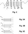
- FIG. 2 schematically illustrates several embodiments of optical fibers as may be utilized in a sensor according to certain disclosed detection methods.
- An optical fiber may include a core 30, through which light may travel, and an external cladding layer 32.
- the difference in the index of refraction between the core material and the clad material defines the critical angle ⁇ at which total internal reflection takes place at the core/clad interface.
- Optical fibers may generally include multi-mode fibers having a core diameter greater than about 10 micrometers ( ⁇ m).
- the preferred core diameter in any particular embodiment may depend upon the characteristics of excitation light (when required) and/or emission light, among other system parameters.
- a core diameter may be between about 50 ⁇ m and about 100 ⁇ m, or about 80 ⁇ m in one embodiment.
- an excitation light source produces less coherent radiation, such as a multi-wavelength light emitting diode (LED), for example, it may be preferable to utilize an optical fiber having a larger core diameter, for instance between about 90 ⁇ m and about 400 ⁇ m.
- LED multi-wavelength light emitting diode
- the core/clad boundary of the fibers may be abrupt, as in a step-index fiber, or may be gradual, as in a graded-index fiber.
- a graded-index fiber may be preferred in some embodiments, as graded index fibers may reduce dispersion of multiple modes traveling through the fiber. This is not a requirement of disclosed sensors, however, and step-index fibers may alternatively be utilized, particularly in those embodiments in which the optical fiber is of a length such that dispersion will not be of great concern.
- Optical fibers may be formed of sterilizable, biocompatible materials that may be safely placed and held at a potential infection site, and in one particular embodiment, at a surgical site.
- optical fibers formed of any suitable type of glass may be used, including, without limitation, silica glass, fluorozirconate glass, fluoroaluminate glass, any chalcogenide glass, or the like may form the core and/or the clad.
- Polymer optical fibers are also encompassed by the present disclosure.
- optical fibers formed of suitable acrylate core/clad combinations e.g., polymethyl methacrylates, may be utilized. It may be preferred in some embodiments to utilize a multi-core POF so as to lower losses common to POF due to bending of the fiber. For instance, this may be preferred in those embodiments in which the optical fiber(s) of the sensor are in a non-linear conformation during use.
- the end of a fiber may be shaped as desired. For instance, and as illustrated in Figs. 2A-2E , polishing or otherwise forming a specific angle at the end face of a fiber may maintain the acceptance angle ⁇ and collection efficiency of the fiber, while rotating the field of view of the fiber, as depicted by the arrows on Figs. 2A-2E .
- light may enter the fiber from angles up to about 90° of the fiber axis (e.g., as shown at Fig. 2E ) (see, e.g., Utzinger, et al., Journal of Biomedical Optics, 8(1):121-147, 2003 ).
- Optical fibers of a sensor may be formed so as to detect light at locations along the length of the fiber, in addition to at the terminal end of the fiber. For instance, at locations along the length of the fiber may be bent or notched so as to allow light through the clad, optionally at a predetermined angle, such that excitation light (when needed) and detectable signals emitted due to the presence of transformed bacteria may enter the optical fiber at these locations.
- the clad of a fiber may be bent or otherwise notched at a predetermined angle to form a 'window' in the fiber.
- a single optical fiber may detect signals from transformed bacterial over a larger area.
- a fiber optic sensor for use as described herein may include a fiber optic cable comprised of a single optical fiber or a plurality of optical fibers, depending upon the specific design of the sensor. For instance, a plurality of optical fibers may be joined to form a single fiber cable of a size to be located at an in vivo site of interest (e.g., less than about 1.5 mm in cross-sectional diameter).
- Figs. 3A-3C illustrate several different embodiments of a fiber optic cable 40 comprising multiple optical fibers 6 in a bundle.
- Fig. 3A through location of a plurality of fiber ends at a single cross-sectional area, improved light collection may be attained, as the total field area covered by the combined fibers will be larger than that for a single fiber.
- the geometry of the end face of different fibers contained in the cable 40 may be different from one another, so as to allow light collection from a variety of different directions.
- fiber ends are staggered over a length, so as to increase the axial length of the light collection area and increase the area of inquiry in an axial direction.
- combinations of such designs, as well as other fiber design for improving the collection of a signal area including methods as discussed above as well as methods as are generally known to those in the art, may be utilized as well.
- a fiber optic bundle or cable of optical fibers 40 may generally be held as a cohesive unit with any biocompatible sheath that can hold the unit together while maintaining flexibility of the fibers.
- a fiber optic cable may include an outer sheath of a flexible polyurethane.
- one or more optical fibers may be utilized as a portion of a sensor that can be contained by use of a portable device, one embodiment of which is schematically illustrated in Fig. 4 .
- device 100 can include several components that may be housed within an enclosure 20.
- Enclosure 20 may be, for example, a molded plastic enclosure of a size so as to be easily carried by or attached to a patient.
- enclosure 20 may include clips, loops, or the like so as to be attachable to a patient's clothing or body.
- enclosure 20 may include a biocompatible adhesive at a surface, and may be adhered directly to a patient's skin.
- enclosure 20 may be relatively small, for instance less than about 10 cm by about 8 cm by about 5 cm, so as to be inconspicuously carried by a patient and so as to avoid impedance of a patient's motion.
- Enclosure 20 may completely enclose the components contained therein, or may partially enclose the components contained therein.
- enclosure 20 may include an access port (not shown) that may provide access to the interior of enclosure 20.
- an access port may be covered with a removable cover, as is known in the art.
- a first component as may be held within enclosure 20 is power supply 2 that may be configured in one embodiment to supply power to an excitation source 4 as well as other of the operational components as will be later described.
- power supply 2 may correspond to a battery, however those of ordinary skill in the art will appreciate that other power supplies may be used including those that may be coupled to an external alternating current (AC) supply so that the enclosed power supply may include those components necessary to convert such external supply to a suitable source for the remaining components requiring a power source.
- AC alternating current
- power supply 2 may be configured in one embodiment to supply power to excitation source 4.
- an excitation source 4 may be included within enclosure 20 in those embodiments in which a detectable marker expressed by a transformed bacterium requires excitation from an external source in order to emit a detectable signal.
- an excitation signal may be provided to the marker in order for the marker to emit an optically detectable signal.
- a sensor may include an excitation source 4.
- an excitation source 4 need not be included in enclosure 20.
- excitation source 4 may correspond to a light emitting diode (LED), however, again, such source may vary and may include, but is not limited to, laser diodes and incandescent light sources.
- Excitation source 4 may correspond to a white light source, a non-white multi-wavelength source, or a single wavelength source, as desired or required. In a preferred exemplary configuration, an LED may be selected due to the low power consumption of such sources.
- the wavelength of the excitation energy supplied by excitation source 4 may be of any suitable wavelength, from infrared (IR) to ultraviolet (UV). In general, the preferred excitation energy wavelength may depend upon the specific design of the detectable marker(s).
- an excitation source 4 may provide a single excitation wavelength.
- an excitation source may provide multiple wavelengths, either through combination of signals from a plurality of single wavelength sources or through a single, incoherent source, as desired.
- Excitation energy source 4 is optically coupled to an optical fiber 6 as illustrated.
- Optical fiber 6 is configured to extend externally from enclosure 20 to the field of inquiry, e.g., within a surgical site or other wound. It should be appreciated that although a single optical fiber 6 is illustrated in Figure 4 , such is not a specific limitation of the present disclosure as a sensor may include multiple fibers in a single cable in alternate embodiments, and as discussed above. Those of ordinary skill in the art will appreciate that a single excitation energy source may be optically coupled to a plurality of optical fibers through utilization of suitable beam splitters, mirrors, and so forth.
- each excitation source may be optically coupled to one or more optical fibers such that multiple excitation wavelengths may be delivered to the field of enquiry.
- optical detector 8 Housed within enclosure 20 is an optical detector 8 coupled to optical fiber 6.
- Optical detector 8 may correspond to a photodiode, a photoresistor, or the like.
- Optical detector 8 may include optical filters, beam splitters, and so forth that may remove background light and reduce the total input optical signal at the detector 8 to one or more diagnostically relevant emission peaks.
- Optical detector 8 produces a signal proportional to targeted emission peaks and couple such signal to line 10 for transmission to signal processor 12.
- Signal processor 12 includes a microprocessor configured to evaluate the strength or other characteristics of the output signal received over line 10 to, e.g., correlate the optical signal to the concentration of bacteria at the detection site and to produce a detection signal that is coupled to line 14 for passage to a signaling device 16. Accordingly, if the detection signal reaches a predetermined threshold value, corresponding to a known concentration of the target pathogen, a detectable signal is initiated at signaling device 16. In an exemplary configuration, a detectable signal may initiate a visible or audible signal within or at the surface of the enclosure 20 by way of signaling device 16 that may be detected by the wearer.
- a visible signal may optionally include utilization of a liquid crystal diode (LCD) device, or an equivalent thereof, that may provide the signal as a readable output.
- a visual signal may be provided at a surface of the device 100 as an instruction such as, for instance, "CALL YOUR DOCTOR", “VISIT HOSPITAL,” or the like.
- signaling device 16 may include a transmitter portion that, upon initiation of the detectable signal, may transmit an electromagnetic signal to receiver 18.
- Receiver 18 may be remote from the signaling device 16.
- receiver 18 may be on the wearer's body at a distance from the signaling device 16, at a location apart from the wearer's body that may be conveniently chosen by the wearer, e.g., within the wearer's home, office, or the like, or may be at a monitoring facility, for instance at a medical facility, such that appropriate medical personal may be quickly informed of the change in status of the patient's site of inquiry.
- the detectable signal may be transmitted to multiple receivers, so as to inform both the wearer and others (e.g., medical personnel) of the change in status of a site.
- Transmission of a signal to a remote site may be carried out with a radio frequency transmission system or with any other wireless-type transmission system, as is generally known in the art.
- a wireless telephone or internet communications system may be utilized to transmit a signal to a remote location according to known methods.
- Wireless transmission systems as may be utilized in conjunction with disclosed devices and methods may include, for example, components and systems as disclosed in U.S. Patent Nos. 6,289,238 to Besson, et al. , 6,441,747 to Khair, et al. , 6,802,811 to Slepian , 6,659,947 to Carter, et al. , and 7,294,105 to Islam .
Landscapes
- Health & Medical Sciences (AREA)
- Life Sciences & Earth Sciences (AREA)
- Engineering & Computer Science (AREA)
- Chemical & Material Sciences (AREA)
- General Health & Medical Sciences (AREA)
- Biomedical Technology (AREA)
- Physics & Mathematics (AREA)
- Organic Chemistry (AREA)
- Epidemiology (AREA)
- Animal Behavior & Ethology (AREA)
- Pathology (AREA)
- Public Health (AREA)
- Veterinary Medicine (AREA)
- Molecular Biology (AREA)
- Zoology (AREA)
- Immunology (AREA)
- Nuclear Medicine, Radiotherapy & Molecular Imaging (AREA)
- Wood Science & Technology (AREA)
- Radiology & Medical Imaging (AREA)
- Analytical Chemistry (AREA)
- Biochemistry (AREA)
- Proteomics, Peptides & Aminoacids (AREA)
- Plasma & Fusion (AREA)
- Virology (AREA)
- Biophysics (AREA)
- General Physics & Mathematics (AREA)
- Biotechnology (AREA)
- Microbiology (AREA)
- Chemical Kinetics & Catalysis (AREA)
- Toxicology (AREA)
- Hematology (AREA)
- Bioinformatics & Cheminformatics (AREA)
- General Engineering & Computer Science (AREA)
- Genetics & Genomics (AREA)
- Measuring Or Testing Involving Enzymes Or Micro-Organisms (AREA)
- Micro-Organisms Or Cultivation Processes Thereof (AREA)
- Apparatus Associated With Microorganisms And Enzymes (AREA)
- Investigating Or Analysing Materials By The Use Of Chemical Reactions (AREA)
Claims (4)
- Tragbare Vorrichtung (100) zum Detektieren des Vorhandenseins oder einer Menge eines pathogenen Bakteriums (22), das eine Quelle einer im Krankenhaus erworbenen Infektion ist, umfassend:ein tragbares Gehäuse (20), das eine Energiequelle (2), einen optischen Detektor (8), einen Signalprozessor (12) und eine Signalisierungsvorrichtung (16) zum Ausstrahlen eines detektierbaren Signals bei Detektion des pathogenen Bakteriums (22) in einer In-vivo-Umgebung enthält;eine Verbindungsvorrichtung zum Befestigen des Gehäuses an der Kleidung oder am Körper eines Trägers;ein Glasfaserkabel (40) zum Einsetzen in die In-vivo-Umgebung, wobei das Glasfaserkabel (40) in optischer Kommunikation mit dem optischen Detektor (8) ist, wobei sich das Glasfaserkabel (40) über eine Länge außerhalb des Gehäuses erstreckt; undein Abgabevehikel an zumindest einem Teil der Außenfläche des Glasfaserkabels (40), dadurch gekennzeichnet, dass das Abgabevehikel angepasst ist, um einen rekombinanten Bakteriophagen (20) an eine gezielte Stelle abzugeben, wobei der Bakteriophage (20) exogenes genetisches Material (24) trägt, das für ein Protein codiert, wobei der Bakteriophage (20) konfiguriert ist, um sich an das pathogene Bakterium (22) zu binden und das genetische Material (24) auf das pathogene Bakterium (22) zu übertragen, sodass das pathogene Bakterium (22) das Protein exprimiert und das exprimierte Protein bei Erregung durch eine Erregungsquelle (4) ein optisch detektierbares Signal ausstrahlt oder mit einem Co-Faktor interagiert, um einen detektierbaren Marker zu bilden, der direkt ein optisch detektierbares Signal erzeugt, wobei das Abgabevehikel ein biologisch abbaubares Mikropartikel mit einer biologisch abbaubaren polymeren Beschichtung, die den rekombinanten Bakteriophagen (20) einkapselt, zur Freisetzung an der Stelle nach Positionierung des Abgabevehikels an der Stelle umfasst; undwobei das Glasfaserkabel (40) konfiguriert ist, um das optisch detektierbare Signal zum optischen Detektor (8) zu senden, der Mittel zum Entfernen von Hintergrundlicht und zum Reduzieren des optisch detektierbaren Signals auf eine oder mehrere diagnostisch relevante Emissionsspitzen umfasst, wobei der optische Detektor (8) konfiguriert ist, um ein Ausgangssignal proportional zu der einen oder den mehreren Emissionsspitzen zu erzeugen und das Ausgangssignal an den Signalprozessor (12) zu senden, der konfiguriert ist, um das Ausgangssignal vom optischen Detektor (8) mit der Konzentration des pathogenen Bakteriums (22) an der Stelle zu korrelieren, ein Detektionssignal zu erzeugen und das Detektionssignal an die Signalisierungsvorrichtung (16) zu senden, die konfiguriert ist, um das detektierbare Signal zu initiieren, falls das Detektionssignal einen vorbestimmten Schwellenwert erreicht, der einer bekannten Konzentration des pathogenen Bakteriums (22) entspricht.
- Vorrichtung nach Anspruch 1, das Gehäuse weiter enthaltend einen Sender zum Senden eines Signals an einen Empfänger (18), das Informationen bezüglich des Vorhandenseins oder einer Menge des Bakteriums (22) in der Umgebung enthält, wobei der Sender vorzugsweise mit der Signalisierungsvorrichtung (16) in elektrischer Kommunikation ist und konfiguriert ist, um das detektierbare Signal, das von der Signalisierungsvorrichtung (16) ausgestrahlt wird, zu senden; und wobei der Sender weiter vorzugsweise mit dem Empfänger (18) in drahtloser Kommunikation ist.
- Vorrichtung nach einem der Ansprüche 1-2, das tragbare Gehäuse weiter umfassend eine Erregungsquelle (4) in optischer Kommunikation mit dem Glasfaserkabel (40) und/oder wobei die Signalisierungsvorrichtung (16) eine Vielzahl von Signalen bei Detektion des pathogenen Bakteriums (22) in der Umgebung ausstrahlt.
- Vorrichtung nach einem der Ansprüche 1-3, wobei das Protein ein grün fluoreszierendes Protein oder eine Farbvariante davon ist; oder wobei das Protein Luciferase ist.
Applications Claiming Priority (2)
| Application Number | Priority Date | Filing Date | Title |
|---|---|---|---|
| US11/955,779 US8619257B2 (en) | 2007-12-13 | 2007-12-13 | Recombinant bacteriophage for detection of nosocomial infection |
| PCT/IB2008/053655 WO2009074893A2 (en) | 2007-12-13 | 2008-09-10 | Recombinant bacteriophage for detection of nosocomial infection |
Publications (3)
| Publication Number | Publication Date |
|---|---|
| EP2229592A2 EP2229592A2 (de) | 2010-09-22 |
| EP2229592A4 EP2229592A4 (de) | 2013-10-16 |
| EP2229592B1 true EP2229592B1 (de) | 2019-05-01 |
Family
ID=40755943
Family Applications (1)
| Application Number | Title | Priority Date | Filing Date |
|---|---|---|---|
| EP08807592.4A Active EP2229592B1 (de) | 2007-12-13 | 2008-09-10 | Rekombinanter bakteriophage zum nachweis einer nosokomialen infektion |
Country Status (7)
| Country | Link |
|---|---|
| US (1) | US8619257B2 (de) |
| EP (1) | EP2229592B1 (de) |
| JP (2) | JP5956110B2 (de) |
| AU (1) | AU2008334326B8 (de) |
| CA (1) | CA2705837C (de) |
| MX (1) | MX2010005972A (de) |
| WO (1) | WO2009074893A2 (de) |
Families Citing this family (14)
| Publication number | Priority date | Publication date | Assignee | Title |
|---|---|---|---|---|
| CA3162577C (en) | 2008-05-20 | 2023-09-26 | University Health Network | Device and method for fluorescence-based imaging and monitoring |
| US9481903B2 (en) | 2013-03-13 | 2016-11-01 | Roche Molecular Systems, Inc. | Systems and methods for detection of cells using engineered transduction particles |
| DK2968424T3 (da) | 2013-03-13 | 2020-03-30 | Geneweave Biosciences Inc | Ikke-replikative transduktionspartikler og transduktionspartikelbaserede reportersystemer |
| US9540675B2 (en) | 2013-10-29 | 2017-01-10 | GeneWeave Biosciences, Inc. | Reagent cartridge and methods for detection of cells |
| WO2016011534A1 (en) | 2014-07-24 | 2016-01-28 | University Health Network | Collection and analysis of data for diagnostic purposes |
| ES2778727T3 (es) * | 2014-08-25 | 2020-08-11 | Geneweave Biosciences Inc | Partículas de transducción no replicativas y sistemas indicadores basados en partículas de transducción |
| US10351893B2 (en) | 2015-10-05 | 2019-07-16 | GeneWeave Biosciences, Inc. | Reagent cartridge for detection of cells |
| CN105754958B (zh) * | 2016-03-07 | 2019-05-07 | 中国科学院广州生物医药与健康研究院 | 一种可投送自主发光元件的分枝杆菌噬菌体及其应用 |
| US10174295B1 (en) * | 2017-08-01 | 2019-01-08 | The Charles Stark Draper Laboratory, Inc. | Composition of matter: engineering of Escherichia coli phage K1E |
| US11008602B2 (en) | 2017-12-20 | 2021-05-18 | Roche Molecular Systems, Inc. | Non-replicative transduction particles and transduction particle-based reporter systems |
| US11873524B2 (en) | 2018-01-12 | 2024-01-16 | Laboratory Corporation Of America Holdings | Methods and systems for the rapid detection of Salmonella using infectious agents |
| CN121204098A (zh) * | 2018-04-24 | 2025-12-26 | 美国控股实验室公司 | 选择和监测治疗功效的指示物噬菌体及其使用方法 |
| CA3187281A1 (en) * | 2020-06-15 | 2021-12-23 | Laboratory Corporation Of America Holdings | Methods for the diagnosis and treatment of biofilm-related infections |
| CN116676274B (zh) * | 2022-12-21 | 2024-04-16 | 暨南大学 | 可自失活噬菌体及其制备方法和应用 |
Family Cites Families (56)
| Publication number | Priority date | Publication date | Assignee | Title |
|---|---|---|---|---|
| US4389330A (en) | 1980-10-06 | 1983-06-21 | Stolle Research And Development Corporation | Microencapsulation process |
| US4530840A (en) | 1982-07-29 | 1985-07-23 | The Stolle Research And Development Corporation | Injectable, long-acting microparticle formulation for the delivery of anti-inflammatory agents |
| US6024983A (en) | 1986-10-24 | 2000-02-15 | Southern Research Institute | Composition for delivering bioactive agents for immune response and its preparation |
| US5075109A (en) | 1986-10-24 | 1991-12-24 | Southern Research Institute | Method of potentiating an immune response |
| US4897268A (en) | 1987-08-03 | 1990-01-30 | Southern Research Institute | Drug delivery system and method of making the same |
| US5196524A (en) | 1989-01-06 | 1993-03-23 | Eli Lilly And Company | Fusion reporter gene for bacterial luciferase |
| DK0471036T4 (da) | 1989-05-04 | 2004-07-19 | Southern Res Inst | Indkapslingsfremgangsmåde |
| GB2249029A (en) | 1990-10-26 | 1992-04-29 | Margaret Pamela Richardson | Surgical apparatus for laser suture insertion |
| GB2278235B (en) | 1991-10-21 | 1996-05-08 | Holm Kennedy James W | Method and device for biochemical sensing |
| FR2702160B1 (fr) | 1993-03-02 | 1995-06-02 | Biovecteurs As | Vecteurs particulaires synthétiques et procédé de préparation. |
| EP0702524A4 (de) * | 1993-03-05 | 1998-12-30 | Armen N Sahagen | Sensor zur überwachung einer flüssigkeit und/oder eines gases |
| DE4329898A1 (de) | 1993-09-04 | 1995-04-06 | Marcus Dr Besson | Kabelloses medizinisches Diagnose- und Überwachungsgerät |
| US5491084A (en) | 1993-09-10 | 1996-02-13 | The Trustees Of Columbia University In The City Of New York | Uses of green-fluorescent protein |
| EP0759170B1 (de) | 1993-09-10 | 2008-07-09 | The Trustees Of Columbia University In The City Of New York | Verwendung von grünem fluoreszenzprotein |
| US5458140A (en) * | 1993-11-15 | 1995-10-17 | Non-Invasive Monitoring Company (Nimco) | Enhancement of transdermal monitoring applications with ultrasound and chemical enhancers |
| FR2723849B1 (fr) | 1994-08-31 | 1997-04-11 | Biovector Therapeutics Sa | Procede pour augmenter l'immunogenicite, produit obtenu et composition pharmaceutique |
| US5599717A (en) * | 1994-09-02 | 1997-02-04 | Martin Marietta Energy Systems, Inc. | Advanced synchronous luminescence system |
| US5625048A (en) | 1994-11-10 | 1997-04-29 | The Regents Of The University Of California | Modified green fluorescent proteins |
| US5777079A (en) | 1994-11-10 | 1998-07-07 | The Regents Of The University Of California | Modified green fluorescent proteins |
| AU705688B2 (en) | 1995-11-10 | 1999-05-27 | Cytogen Corporation | Peptides which enhance transport across tissues and methods of identifying and using the same |
| EP1820517A3 (de) | 1995-11-17 | 2009-10-21 | The Board Of Trustees Of The Leland Stanford Junior University | Nichtinvasive Lokalisierung eines lichtemittierenden Konjugats bei einem Säugetier |
| US5804387A (en) | 1996-02-01 | 1998-09-08 | The Board Of Trustees Of The Leland Stanford Junior University | FACS-optimized mutants of the green fluorescent protein (GFP) |
| JPH09276275A (ja) * | 1996-04-18 | 1997-10-28 | Kazumasa Sasaki | 光ファイバ形生体肝機能センサーと検査装置 |
| JP2002335957A (ja) * | 1996-08-26 | 2002-11-26 | Bio Venture Bank Kk | 新規なバクテリオファージならびにそのスクリーニング法 およびそれを使用した新規バイオ殺菌材料ならびに検出用 試薬 |
| US6322783B1 (en) | 1996-08-26 | 2001-11-27 | Seishi Takahashi | Bacteriophages, method for screening same and bactericidal compositions using same, and detection kits using same |
| TW371617B (en) | 1996-10-09 | 1999-10-11 | Of Animal And Plant Health Inspection And Quarantine Council Of Agriculture Executive Yuan Bureau | Method to transplant GFP into autographa californica multiple nuclear polyhedrosis virus for inflicting pest in an attempt to detect and flow up it existence and to improve life span against UV |
| US5894175A (en) | 1997-08-27 | 1999-04-13 | Intermatic Incorporated | Photo-control apparatus and circuit |
| US5968766A (en) | 1998-03-31 | 1999-10-19 | B.E. Safe | Method and apparatus for sensing the presence of microbes |
| EP0951068A1 (de) | 1998-04-17 | 1999-10-20 | Interuniversitair Micro-Elektronica Centrum Vzw | Herstellungsverfahren für eine Mikrostruktur mit Innenraum |
| EP1114319B1 (de) * | 1998-09-17 | 2008-05-14 | Regents Of The University Of Minnesota | Kompositivvorrichtungen, die biologisches material enthalten und verfahren |
| JP2000308486A (ja) * | 1999-02-22 | 2000-11-07 | Matsushita Electric Ind Co Ltd | 細菌検出法 |
| GB9908195D0 (en) | 1999-04-09 | 1999-06-02 | Microbiological Res Authority | Treatment of intracellular infection |
| US6802811B1 (en) | 1999-09-17 | 2004-10-12 | Endoluminal Therapeutics, Inc. | Sensing, interrogating, storing, telemetering and responding medical implants |
| US6441747B1 (en) | 2000-04-18 | 2002-08-27 | Motorola, Inc. | Wireless system protocol for telemetry monitoring |
| NZ523385A (en) * | 2000-06-28 | 2005-09-30 | Atul J | Biodegradable vehicles and BAS-loaded delivery systems for use as biodegradable fillers and/or spacers, e.g. artificial skin |
| US6659947B1 (en) | 2000-07-13 | 2003-12-09 | Ge Medical Systems Information Technologies, Inc. | Wireless LAN architecture for integrated time-critical and non-time-critical services within medical facilities |
| JP2002045183A (ja) * | 2000-07-31 | 2002-02-12 | Univ Waseda | 細菌の検出方法 |
| US6544729B2 (en) | 2001-07-20 | 2003-04-08 | University Of Tennessee | Bioluminescent biosensor device |
| MXPA04002899A (es) | 2001-09-27 | 2005-07-01 | Gangen Inc | Bacteriofagos deficientes en lisina, que tienen inmunogenicidad reducida. |
| EP1519678A2 (de) * | 2001-10-02 | 2005-04-06 | Alfred E. Mann Institute for Biomedical Engineering at the University of Southern California | Interne biochemische messeinrichtung |
| US20050101841A9 (en) | 2001-12-04 | 2005-05-12 | Kimberly-Clark Worldwide, Inc. | Healthcare networks with biosensors |
| US20050003346A1 (en) | 2002-04-12 | 2005-01-06 | Colorado School Of Mines | Apparatus and method for detecting microscopic living organisms using bacteriophage |
| US7374874B2 (en) * | 2002-04-24 | 2008-05-20 | University Of Northern Iowa Research Foundation | Bacteriophages that infect Bacillus bacteria (Anthrax) |
| US7294105B1 (en) | 2002-09-03 | 2007-11-13 | Cheetah Omni, Llc | System and method for a wireless medical communication system |
| US20070010726A1 (en) | 2002-10-02 | 2007-01-11 | Alfred E. Mann Inst. For Biomedical Engineering At The University Of Southern California | Internal biochemical sensing device |
| JP2004283003A (ja) | 2003-01-31 | 2004-10-14 | Rikogaku Shinkokai | バクテリオファージによる大腸菌の検出 |
| US8206946B2 (en) * | 2003-03-24 | 2012-06-26 | Mary Beth Tabacco | Fluorescent virus probes for identification of bacteria |
| US8688384B2 (en) | 2003-05-12 | 2014-04-01 | River Diagnostics B.V. | Automated characterization and classification of microorganisms |
| US20040253293A1 (en) * | 2003-06-16 | 2004-12-16 | Afshin Shafiee | Rate controlled release of a pharmaceutical agent in a biodegradable device |
| EP1682568A4 (de) | 2003-10-15 | 2009-10-28 | Univ Texas | Multifunktionelle biologische materialien als gerüste für elektronische, optische, magnetische, halbleitdende und biotechnologische anwendungen |
| US20060159589A1 (en) | 2005-01-14 | 2006-07-20 | Indu Saxena | Bacteria sensor and method |
| WO2006092629A1 (en) * | 2005-03-04 | 2006-09-08 | Blaze Venture Technologies Limited | Method and device for bacterial sampling |
| WO2006105504A1 (en) | 2005-03-31 | 2006-10-05 | Microphage Incorporated | Apparatus and method for detecting microorganisms using flagged bacteriophage |
| US20070178450A1 (en) | 2006-01-27 | 2007-08-02 | Microphage (Tm) Incorporation | Method and apparatus for determining level of microorganisms using bacteriophage |
| US20090304551A1 (en) * | 2006-01-31 | 2009-12-10 | Drexel University | Ultra Sensitive Tapered Fiber Optic Biosensor For Pathogens, Proteins, and DNA |
| JP2007284362A (ja) * | 2006-04-13 | 2007-11-01 | Tokyo Univ Of Science | 薬物送達粒子及びその製造方法 |
-
2007
- 2007-12-13 US US11/955,779 patent/US8619257B2/en not_active Expired - Fee Related
-
2008
- 2008-09-10 AU AU2008334326A patent/AU2008334326B8/en not_active Ceased
- 2008-09-10 JP JP2010537546A patent/JP5956110B2/ja not_active Expired - Fee Related
- 2008-09-10 EP EP08807592.4A patent/EP2229592B1/de active Active
- 2008-09-10 MX MX2010005972A patent/MX2010005972A/es active IP Right Grant
- 2008-09-10 WO PCT/IB2008/053655 patent/WO2009074893A2/en not_active Ceased
- 2008-09-10 CA CA2705837A patent/CA2705837C/en not_active Expired - Fee Related
-
2015
- 2015-02-26 JP JP2015036378A patent/JP5993048B2/ja active Active
Non-Patent Citations (1)
| Title |
|---|
| None * |
Also Published As
| Publication number | Publication date |
|---|---|
| CA2705837A1 (en) | 2009-06-18 |
| AU2008334326B8 (en) | 2014-03-06 |
| US8619257B2 (en) | 2013-12-31 |
| JP2011507494A (ja) | 2011-03-10 |
| CA2705837C (en) | 2017-04-18 |
| JP2015107130A (ja) | 2015-06-11 |
| MX2010005972A (es) | 2010-06-23 |
| EP2229592A4 (de) | 2013-10-16 |
| JP5993048B2 (ja) | 2016-09-14 |
| EP2229592A2 (de) | 2010-09-22 |
| AU2008334326A8 (en) | 2014-03-06 |
| JP5956110B2 (ja) | 2016-07-20 |
| WO2009074893A3 (en) | 2009-10-15 |
| US20120143024A1 (en) | 2012-06-07 |
| WO2009074893A2 (en) | 2009-06-18 |
| AU2008334326B2 (en) | 2013-10-24 |
| AU2008334326A1 (en) | 2009-06-18 |
Similar Documents
| Publication | Publication Date | Title |
|---|---|---|
| EP2229592B1 (de) | Rekombinanter bakteriophage zum nachweis einer nosokomialen infektion | |
| Jiang et al. | Searching for the secret of stickiness: how biofilms adhere to surfaces | |
| Sun et al. | Hydrogel‐based biosensors for bacterial infections | |
| EP2217907B1 (de) | Nachweis einer nosokomialen infektion auf resonanzenergietransfer-basis | |
| US8280471B2 (en) | Fiber optic based detection of autofluorescent bacterial pathogens | |
| WO1999045354A2 (en) | Luminescent microbe assay | |
| McShane et al. | Monte Carlo modeling for implantable fluorescent analyte sensors | |
| Mills et al. | Molecular detection of Gram-positive bacteria in the human lung through an optical fiber–based endoscope | |
| Waldeck et al. | ROS-mediated killing efficiency with visible light of bacteria carrying different red fluorochrome proteins | |
| Shehatou et al. | Characterizing the antimicrobial properties of 405 nm light and the Corning® light‐diffusing fiber delivery system | |
| Nooshabadi et al. | Intravital fluorescence excitation in whole-animal optical imaging | |
| CN111279124B (zh) | 用于引导和散射紫外光的光扩散光纤 | |
| Davenport et al. | Fiber-optic fluorescence-quenching oxygen partial pressure sensor using platinum octaethylporphyrin | |
| Huynh et al. | Stability and performance study of fluorescent organosilica pH nanosensors | |
| Kong et al. | Small molecular fluorescent probes: application progress of specific bacteria detection and antibacterial phototherapy | |
| CA2906871A1 (en) | Luminescent resonance energy transfer sensors for non-invasively and continuously monitoring glucose for diabetes | |
| US20200029887A1 (en) | Method of assessing wound contamination and infection | |
| WO2012156432A1 (en) | Device and probe for detection of infection | |
| Rengifo | Real-time fluorescence microscopy to study bacterial response to antimicrobial strategies | |
| Chen et al. | Detection of dual-gene expression in arteries using an optical imaging method | |
| Dragavon et al. | Validation of method for enhanced production of red-shifted bioluminescent photons in vivo | |
| KR20220076951A (ko) | 그람 음성 아시네토박토 바우마니의 검출용 시약 조성물, 키트, 및 검출 방법 | |
| Long et al. | Dynamic testing and in-vivo evaluation of dermally implantable luminescent microparticle glucose sensors | |
| Kurmoo | Novel materials and optical fibre sensors to reduce the risk of infection associated with endotracheal tubes | |
| Mufti | Fiber Optic Micro-endoscopy for Detection of Bacteria in Early Stages of Infection |
Legal Events
| Date | Code | Title | Description |
|---|---|---|---|
| PUAI | Public reference made under article 153(3) epc to a published international application that has entered the european phase |
Free format text: ORIGINAL CODE: 0009012 |
|
| 17P | Request for examination filed |
Effective date: 20100607 |
|
| AK | Designated contracting states |
Kind code of ref document: A2 Designated state(s): AT BE BG CH CY CZ DE DK EE ES FI FR GB GR HR HU IE IS IT LI LT LU LV MC MT NL NO PL PT RO SE SI SK TR |
|
| AX | Request for extension of the european patent |
Extension state: AL BA MK RS |
|
| RIN1 | Information on inventor provided before grant (corrected) |
Inventor name: PHILLIPS, ERICA, M. Inventor name: PRESLEY, TALBOT Inventor name: HANTKE, RICHARD Inventor name: BAIRD, DANIEL Inventor name: RAINONE, MIKE Inventor name: PLOWMAN, THOMAS EDWARD |
|
| RIN1 | Information on inventor provided before grant (corrected) |
Inventor name: PRESLEY, TALBOT Inventor name: PLOWMAN, THOMAS EDWARD Inventor name: RAINONE, MIKE Inventor name: BAIRD, DANIEL Inventor name: HANTKE, RICHARD Inventor name: PHILLIPS, ERICA, M. |
|
| DAX | Request for extension of the european patent (deleted) | ||
| A4 | Supplementary search report drawn up and despatched |
Effective date: 20130912 |
|
| RIC1 | Information provided on ipc code assigned before grant |
Ipc: G01N 21/64 20060101ALI20130906BHEP Ipc: G01N 21/00 20060101ALI20130906BHEP Ipc: A61K 49/00 20060101ALI20130906BHEP Ipc: G01N 33/569 20060101AFI20130906BHEP Ipc: G01N 33/535 20060101ALI20130906BHEP Ipc: C12Q 1/04 20060101ALI20130906BHEP Ipc: A61B 5/00 20060101ALI20130906BHEP Ipc: G01N 21/77 20060101ALI20130906BHEP |
|
| 17Q | First examination report despatched |
Effective date: 20140715 |
|
| RAP1 | Party data changed (applicant data changed or rights of an application transferred) |
Owner name: AVENT, INC. |
|
| STAA | Information on the status of an ep patent application or granted ep patent |
Free format text: STATUS: EXAMINATION IS IN PROGRESS |
|
| REG | Reference to a national code |
Ref country code: DE Ref legal event code: R079 Ref document number: 602008059930 Country of ref document: DE Free format text: PREVIOUS MAIN CLASS: G01N0033569000 Ipc: G01N0033535000 |
|
| RIC1 | Information provided on ipc code assigned before grant |
Ipc: A61B 5/00 20060101ALI20180326BHEP Ipc: A61K 49/00 20060101ALI20180326BHEP Ipc: G01N 33/535 20060101AFI20180326BHEP Ipc: C12N 15/86 20060101ALI20180326BHEP Ipc: G01N 21/77 20060101ALI20180326BHEP Ipc: C12Q 1/04 20060101ALI20180326BHEP Ipc: G01N 33/569 20060101ALI20180326BHEP |
|
| GRAP | Despatch of communication of intention to grant a patent |
Free format text: ORIGINAL CODE: EPIDOSNIGR1 |
|
| STAA | Information on the status of an ep patent application or granted ep patent |
Free format text: STATUS: GRANT OF PATENT IS INTENDED |
|
| INTG | Intention to grant announced |
Effective date: 20180606 |
|
| RIN1 | Information on inventor provided before grant (corrected) |
Inventor name: RAINONE, MIKE Inventor name: PHILLIPS, ERICA M. Inventor name: BAIRD, DANIEL Inventor name: PRESLEY, TALBOT Inventor name: HANTKE, RICHARD Inventor name: PLOWMAN, THOMAS EDWARD |
|
| GRAS | Grant fee paid |
Free format text: ORIGINAL CODE: EPIDOSNIGR3 |
|
| GRAA | (expected) grant |
Free format text: ORIGINAL CODE: 0009210 |
|
| STAA | Information on the status of an ep patent application or granted ep patent |
Free format text: STATUS: THE PATENT HAS BEEN GRANTED |
|
| AK | Designated contracting states |
Kind code of ref document: B1 Designated state(s): AT BE BG CH CY CZ DE DK EE ES FI FR GB GR HR HU IE IS IT LI LT LU LV MC MT NL NO PL PT RO SE SI SK TR |
|
| REG | Reference to a national code |
Ref country code: GB Ref legal event code: FG4D |
|
| REG | Reference to a national code |
Ref country code: CH Ref legal event code: EP Ref country code: AT Ref legal event code: REF Ref document number: 1127631 Country of ref document: AT Kind code of ref document: T Effective date: 20190515 |
|
| REG | Reference to a national code |
Ref country code: DE Ref legal event code: R096 Ref document number: 602008059930 Country of ref document: DE |
|
| REG | Reference to a national code |
Ref country code: IE Ref legal event code: FG4D |
|
| REG | Reference to a national code |
Ref country code: NL Ref legal event code: MP Effective date: 20190501 |
|
| REG | Reference to a national code |
Ref country code: LT Ref legal event code: MG4D |
|
| PG25 | Lapsed in a contracting state [announced via postgrant information from national office to epo] |
Ref country code: FI Free format text: LAPSE BECAUSE OF FAILURE TO SUBMIT A TRANSLATION OF THE DESCRIPTION OR TO PAY THE FEE WITHIN THE PRESCRIBED TIME-LIMIT Effective date: 20190501 Ref country code: NO Free format text: LAPSE BECAUSE OF FAILURE TO SUBMIT A TRANSLATION OF THE DESCRIPTION OR TO PAY THE FEE WITHIN THE PRESCRIBED TIME-LIMIT Effective date: 20190801 Ref country code: LT Free format text: LAPSE BECAUSE OF FAILURE TO SUBMIT A TRANSLATION OF THE DESCRIPTION OR TO PAY THE FEE WITHIN THE PRESCRIBED TIME-LIMIT Effective date: 20190501 Ref country code: SE Free format text: LAPSE BECAUSE OF FAILURE TO SUBMIT A TRANSLATION OF THE DESCRIPTION OR TO PAY THE FEE WITHIN THE PRESCRIBED TIME-LIMIT Effective date: 20190501 Ref country code: HR Free format text: LAPSE BECAUSE OF FAILURE TO SUBMIT A TRANSLATION OF THE DESCRIPTION OR TO PAY THE FEE WITHIN THE PRESCRIBED TIME-LIMIT Effective date: 20190501 Ref country code: PT Free format text: LAPSE BECAUSE OF FAILURE TO SUBMIT A TRANSLATION OF THE DESCRIPTION OR TO PAY THE FEE WITHIN THE PRESCRIBED TIME-LIMIT Effective date: 20190901 Ref country code: ES Free format text: LAPSE BECAUSE OF FAILURE TO SUBMIT A TRANSLATION OF THE DESCRIPTION OR TO PAY THE FEE WITHIN THE PRESCRIBED TIME-LIMIT Effective date: 20190501 Ref country code: NL Free format text: LAPSE BECAUSE OF FAILURE TO SUBMIT A TRANSLATION OF THE DESCRIPTION OR TO PAY THE FEE WITHIN THE PRESCRIBED TIME-LIMIT Effective date: 20190501 |
|
| PG25 | Lapsed in a contracting state [announced via postgrant information from national office to epo] |
Ref country code: GR Free format text: LAPSE BECAUSE OF FAILURE TO SUBMIT A TRANSLATION OF THE DESCRIPTION OR TO PAY THE FEE WITHIN THE PRESCRIBED TIME-LIMIT Effective date: 20190802 Ref country code: BG Free format text: LAPSE BECAUSE OF FAILURE TO SUBMIT A TRANSLATION OF THE DESCRIPTION OR TO PAY THE FEE WITHIN THE PRESCRIBED TIME-LIMIT Effective date: 20190801 Ref country code: LV Free format text: LAPSE BECAUSE OF FAILURE TO SUBMIT A TRANSLATION OF THE DESCRIPTION OR TO PAY THE FEE WITHIN THE PRESCRIBED TIME-LIMIT Effective date: 20190501 |
|
| REG | Reference to a national code |
Ref country code: AT Ref legal event code: MK05 Ref document number: 1127631 Country of ref document: AT Kind code of ref document: T Effective date: 20190501 |
|
| PG25 | Lapsed in a contracting state [announced via postgrant information from national office to epo] |
Ref country code: IS Free format text: LAPSE BECAUSE OF FAILURE TO SUBMIT A TRANSLATION OF THE DESCRIPTION OR TO PAY THE FEE WITHIN THE PRESCRIBED TIME-LIMIT Effective date: 20190901 |
|
| PG25 | Lapsed in a contracting state [announced via postgrant information from national office to epo] |
Ref country code: SK Free format text: LAPSE BECAUSE OF FAILURE TO SUBMIT A TRANSLATION OF THE DESCRIPTION OR TO PAY THE FEE WITHIN THE PRESCRIBED TIME-LIMIT Effective date: 20190501 Ref country code: AT Free format text: LAPSE BECAUSE OF FAILURE TO SUBMIT A TRANSLATION OF THE DESCRIPTION OR TO PAY THE FEE WITHIN THE PRESCRIBED TIME-LIMIT Effective date: 20190501 Ref country code: DK Free format text: LAPSE BECAUSE OF FAILURE TO SUBMIT A TRANSLATION OF THE DESCRIPTION OR TO PAY THE FEE WITHIN THE PRESCRIBED TIME-LIMIT Effective date: 20190501 Ref country code: CZ Free format text: LAPSE BECAUSE OF FAILURE TO SUBMIT A TRANSLATION OF THE DESCRIPTION OR TO PAY THE FEE WITHIN THE PRESCRIBED TIME-LIMIT Effective date: 20190501 Ref country code: EE Free format text: LAPSE BECAUSE OF FAILURE TO SUBMIT A TRANSLATION OF THE DESCRIPTION OR TO PAY THE FEE WITHIN THE PRESCRIBED TIME-LIMIT Effective date: 20190501 Ref country code: RO Free format text: LAPSE BECAUSE OF FAILURE TO SUBMIT A TRANSLATION OF THE DESCRIPTION OR TO PAY THE FEE WITHIN THE PRESCRIBED TIME-LIMIT Effective date: 20190501 |
|
| REG | Reference to a national code |
Ref country code: DE Ref legal event code: R097 Ref document number: 602008059930 Country of ref document: DE |
|
| PG25 | Lapsed in a contracting state [announced via postgrant information from national office to epo] |
Ref country code: IT Free format text: LAPSE BECAUSE OF FAILURE TO SUBMIT A TRANSLATION OF THE DESCRIPTION OR TO PAY THE FEE WITHIN THE PRESCRIBED TIME-LIMIT Effective date: 20190501 |
|
| PLBE | No opposition filed within time limit |
Free format text: ORIGINAL CODE: 0009261 |
|
| STAA | Information on the status of an ep patent application or granted ep patent |
Free format text: STATUS: NO OPPOSITION FILED WITHIN TIME LIMIT |
|
| PG25 | Lapsed in a contracting state [announced via postgrant information from national office to epo] |
Ref country code: TR Free format text: LAPSE BECAUSE OF FAILURE TO SUBMIT A TRANSLATION OF THE DESCRIPTION OR TO PAY THE FEE WITHIN THE PRESCRIBED TIME-LIMIT Effective date: 20190501 |
|
| REG | Reference to a national code |
Ref country code: DE Ref legal event code: R119 Ref document number: 602008059930 Country of ref document: DE |
|
| 26N | No opposition filed |
Effective date: 20200204 |
|
| PG25 | Lapsed in a contracting state [announced via postgrant information from national office to epo] |
Ref country code: PL Free format text: LAPSE BECAUSE OF FAILURE TO SUBMIT A TRANSLATION OF THE DESCRIPTION OR TO PAY THE FEE WITHIN THE PRESCRIBED TIME-LIMIT Effective date: 20190501 |
|
| PG25 | Lapsed in a contracting state [announced via postgrant information from national office to epo] |
Ref country code: MC Free format text: LAPSE BECAUSE OF FAILURE TO SUBMIT A TRANSLATION OF THE DESCRIPTION OR TO PAY THE FEE WITHIN THE PRESCRIBED TIME-LIMIT Effective date: 20190501 Ref country code: SI Free format text: LAPSE BECAUSE OF FAILURE TO SUBMIT A TRANSLATION OF THE DESCRIPTION OR TO PAY THE FEE WITHIN THE PRESCRIBED TIME-LIMIT Effective date: 20190501 |
|
| REG | Reference to a national code |
Ref country code: CH Ref legal event code: PL |
|
| PG25 | Lapsed in a contracting state [announced via postgrant information from national office to epo] |
Ref country code: DE Free format text: LAPSE BECAUSE OF NON-PAYMENT OF DUE FEES Effective date: 20200401 Ref country code: IE Free format text: LAPSE BECAUSE OF NON-PAYMENT OF DUE FEES Effective date: 20190910 Ref country code: LU Free format text: LAPSE BECAUSE OF NON-PAYMENT OF DUE FEES Effective date: 20190910 Ref country code: LI Free format text: LAPSE BECAUSE OF NON-PAYMENT OF DUE FEES Effective date: 20190930 Ref country code: CH Free format text: LAPSE BECAUSE OF NON-PAYMENT OF DUE FEES Effective date: 20190930 |
|
| REG | Reference to a national code |
Ref country code: BE Ref legal event code: MM Effective date: 20190930 |
|
| PG25 | Lapsed in a contracting state [announced via postgrant information from national office to epo] |
Ref country code: BE Free format text: LAPSE BECAUSE OF NON-PAYMENT OF DUE FEES Effective date: 20190930 |
|
| PG25 | Lapsed in a contracting state [announced via postgrant information from national office to epo] |
Ref country code: CY Free format text: LAPSE BECAUSE OF FAILURE TO SUBMIT A TRANSLATION OF THE DESCRIPTION OR TO PAY THE FEE WITHIN THE PRESCRIBED TIME-LIMIT Effective date: 20190501 |
|
| PG25 | Lapsed in a contracting state [announced via postgrant information from national office to epo] |
Ref country code: MT Free format text: LAPSE BECAUSE OF FAILURE TO SUBMIT A TRANSLATION OF THE DESCRIPTION OR TO PAY THE FEE WITHIN THE PRESCRIBED TIME-LIMIT Effective date: 20190501 Ref country code: HU Free format text: LAPSE BECAUSE OF FAILURE TO SUBMIT A TRANSLATION OF THE DESCRIPTION OR TO PAY THE FEE WITHIN THE PRESCRIBED TIME-LIMIT; INVALID AB INITIO Effective date: 20080910 |
|
| P01 | Opt-out of the competence of the unified patent court (upc) registered |
Effective date: 20230518 |
|
| PGFP | Annual fee paid to national office [announced via postgrant information from national office to epo] |
Ref country code: GB Payment date: 20240701 Year of fee payment: 17 |
|
| PGFP | Annual fee paid to national office [announced via postgrant information from national office to epo] |
Ref country code: FR Payment date: 20240702 Year of fee payment: 17 |