EP2212716B1 - Interventional navigation using 3d contrast-enhanced ultrasound - Google Patents
Interventional navigation using 3d contrast-enhanced ultrasound Download PDFInfo
- Publication number
- EP2212716B1 EP2212716B1 EP08850546.6A EP08850546A EP2212716B1 EP 2212716 B1 EP2212716 B1 EP 2212716B1 EP 08850546 A EP08850546 A EP 08850546A EP 2212716 B1 EP2212716 B1 EP 2212716B1
- Authority
- EP
- European Patent Office
- Prior art keywords
- image
- ceus
- tissue
- ultrasound
- contrast
- Prior art date
- Legal status (The legal status is an assumption and is not a legal conclusion. Google has not performed a legal analysis and makes no representation as to the accuracy of the status listed.)
- Active
Links
- 238000002607 contrast-enhanced ultrasound Methods 0.000 title claims description 149
- 238000002604 ultrasonography Methods 0.000 claims description 78
- 238000013152 interventional procedure Methods 0.000 claims description 30
- 239000000523 sample Substances 0.000 claims description 30
- 238000003384 imaging method Methods 0.000 claims description 24
- 210000003484 anatomy Anatomy 0.000 claims description 14
- 238000012285 ultrasound imaging Methods 0.000 claims description 13
- 239000002872 contrast media Substances 0.000 claims description 6
- 239000003795 chemical substances by application Substances 0.000 claims description 4
- 238000012545 processing Methods 0.000 claims description 3
- 238000000034 method Methods 0.000 description 29
- 230000009466 transformation Effects 0.000 description 19
- 206010028980 Neoplasm Diseases 0.000 description 12
- 238000012800 visualization Methods 0.000 description 10
- 238000002347 injection Methods 0.000 description 8
- 239000007924 injection Substances 0.000 description 8
- 238000002679 ablation Methods 0.000 description 6
- 238000001574 biopsy Methods 0.000 description 5
- 238000012937 correction Methods 0.000 description 5
- 238000000844 transformation Methods 0.000 description 5
- 230000008901 benefit Effects 0.000 description 4
- 238000010586 diagram Methods 0.000 description 4
- 230000006870 function Effects 0.000 description 4
- 230000003287 optical effect Effects 0.000 description 3
- 230000001052 transient effect Effects 0.000 description 3
- 230000000694 effects Effects 0.000 description 2
- 238000012986 modification Methods 0.000 description 2
- 230000004048 modification Effects 0.000 description 2
- 210000000056 organ Anatomy 0.000 description 2
- 230000008569 process Effects 0.000 description 2
- 238000011524 similarity measure Methods 0.000 description 2
- 230000001225 therapeutic effect Effects 0.000 description 2
- 239000005441 aurora Substances 0.000 description 1
- 230000008859 change Effects 0.000 description 1
- 238000004891 communication Methods 0.000 description 1
- 230000001419 dependent effect Effects 0.000 description 1
- 238000003745 diagnosis Methods 0.000 description 1
- 238000002059 diagnostic imaging Methods 0.000 description 1
- 239000002961 echo contrast media Substances 0.000 description 1
- 230000010354 integration Effects 0.000 description 1
- 238000001990 intravenous administration Methods 0.000 description 1
- 238000010253 intravenous injection Methods 0.000 description 1
- 230000005865 ionizing radiation Effects 0.000 description 1
- 238000002324 minimally invasive surgery Methods 0.000 description 1
- 238000013188 needle biopsy Methods 0.000 description 1
- 230000005180 public health Effects 0.000 description 1
- 238000012827 research and development Methods 0.000 description 1
- 230000029058 respiratory gaseous exchange Effects 0.000 description 1
- 230000004044 response Effects 0.000 description 1
- 238000010408 sweeping Methods 0.000 description 1
- 230000008685 targeting Effects 0.000 description 1
- 238000002560 therapeutic procedure Methods 0.000 description 1
- 238000012546 transfer Methods 0.000 description 1
Images


Classifications
-
- G—PHYSICS
- G01—MEASURING; TESTING
- G01S—RADIO DIRECTION-FINDING; RADIO NAVIGATION; DETERMINING DISTANCE OR VELOCITY BY USE OF RADIO WAVES; LOCATING OR PRESENCE-DETECTING BY USE OF THE REFLECTION OR RERADIATION OF RADIO WAVES; ANALOGOUS ARRANGEMENTS USING OTHER WAVES
- G01S15/00—Systems using the reflection or reradiation of acoustic waves, e.g. sonar systems
- G01S15/88—Sonar systems specially adapted for specific applications
- G01S15/89—Sonar systems specially adapted for specific applications for mapping or imaging
- G01S15/8906—Short-range imaging systems; Acoustic microscope systems using pulse-echo techniques
- G01S15/899—Combination of imaging systems with ancillary equipment
-
- A—HUMAN NECESSITIES
- A61—MEDICAL OR VETERINARY SCIENCE; HYGIENE
- A61B—DIAGNOSIS; SURGERY; IDENTIFICATION
- A61B8/00—Diagnosis using ultrasonic, sonic or infrasonic waves
- A61B8/08—Clinical applications
- A61B8/0833—Clinical applications involving detecting or locating foreign bodies or organic structures
-
- A—HUMAN NECESSITIES
- A61—MEDICAL OR VETERINARY SCIENCE; HYGIENE
- A61B—DIAGNOSIS; SURGERY; IDENTIFICATION
- A61B8/00—Diagnosis using ultrasonic, sonic or infrasonic waves
- A61B8/42—Details of probe positioning or probe attachment to the patient
- A61B8/4245—Details of probe positioning or probe attachment to the patient involving determining the position of the probe, e.g. with respect to an external reference frame or to the patient
- A61B8/4263—Details of probe positioning or probe attachment to the patient involving determining the position of the probe, e.g. with respect to an external reference frame or to the patient using sensors not mounted on the probe, e.g. mounted on an external reference frame
-
- G—PHYSICS
- G01—MEASURING; TESTING
- G01S—RADIO DIRECTION-FINDING; RADIO NAVIGATION; DETERMINING DISTANCE OR VELOCITY BY USE OF RADIO WAVES; LOCATING OR PRESENCE-DETECTING BY USE OF THE REFLECTION OR RERADIATION OF RADIO WAVES; ANALOGOUS ARRANGEMENTS USING OTHER WAVES
- G01S7/00—Details of systems according to groups G01S13/00, G01S15/00, G01S17/00
- G01S7/52—Details of systems according to groups G01S13/00, G01S15/00, G01S17/00 of systems according to group G01S15/00
- G01S7/52017—Details of systems according to groups G01S13/00, G01S15/00, G01S17/00 of systems according to group G01S15/00 particularly adapted to short-range imaging
- G01S7/52053—Display arrangements
- G01S7/52057—Cathode ray tube displays
- G01S7/52074—Composite displays, e.g. split-screen displays; Combination of multiple images or of images and alphanumeric tabular information
-
- G—PHYSICS
- G01—MEASURING; TESTING
- G01S—RADIO DIRECTION-FINDING; RADIO NAVIGATION; DETERMINING DISTANCE OR VELOCITY BY USE OF RADIO WAVES; LOCATING OR PRESENCE-DETECTING BY USE OF THE REFLECTION OR RERADIATION OF RADIO WAVES; ANALOGOUS ARRANGEMENTS USING OTHER WAVES
- G01S7/00—Details of systems according to groups G01S13/00, G01S15/00, G01S17/00
- G01S7/52—Details of systems according to groups G01S13/00, G01S15/00, G01S17/00 of systems according to group G01S15/00
- G01S7/52017—Details of systems according to groups G01S13/00, G01S15/00, G01S17/00 of systems according to group G01S15/00 particularly adapted to short-range imaging
- G01S7/52098—Details of systems according to groups G01S13/00, G01S15/00, G01S17/00 of systems according to group G01S15/00 particularly adapted to short-range imaging related to workflow protocols
-
- G—PHYSICS
- G01—MEASURING; TESTING
- G01S—RADIO DIRECTION-FINDING; RADIO NAVIGATION; DETERMINING DISTANCE OR VELOCITY BY USE OF RADIO WAVES; LOCATING OR PRESENCE-DETECTING BY USE OF THE REFLECTION OR RERADIATION OF RADIO WAVES; ANALOGOUS ARRANGEMENTS USING OTHER WAVES
- G01S15/00—Systems using the reflection or reradiation of acoustic waves, e.g. sonar systems
- G01S15/88—Sonar systems specially adapted for specific applications
- G01S15/89—Sonar systems specially adapted for specific applications for mapping or imaging
- G01S15/8906—Short-range imaging systems; Acoustic microscope systems using pulse-echo techniques
- G01S15/8993—Three dimensional imaging systems
Definitions
- US 2006/0020204 A1 discloses a method for managing a 3D space in which substantially real-time images are acquired.
- the substantial real-time images are acquired of an object or a body, wherein prior image data are co-registered to the 3D space from which the substantially real-time images were acquired.
- a scan probe and a handheld tool are tracked in 3D, and the tracking information from the scan probe is used to fuse images from prior scans of the object or the body to one or more substantially real-time images of the object or the body.
- the tracking information from the handheld tool is used to control display parameters and manipulational operations on the one or more substantially real-time images.
- the present embodiments relate generally to ultrasound diagnostic imaging systems and more particularly, to an apparatus for interventional navigation using 3D contrast enhanced ultrasound (CEUS).
- CEUS 3D contrast enhanced ultrasound
- Ultrasound imaging is one of the primary image guidance methods for many minimally invasive and interventional procedures.
- most needle biopsies and needle-based ablation procedures are guided by ultrasound.
- the advantages of ultrasound include the real time imaging capability, low cost, flexibility in its application, and the fact that no ionizing radiation is used.
- non-enhanced ultrasound including the commonly used gray-scale ultrasound image, may not be able to visualize a particular target (e.g., a tumor) with the desired contrast, and in some cases, may not visualize the target at all.
- needle placement becomes very difficult and prone to inaccuracies, since it involves imaging the target using a different modality, and "mentally" transferring the tumor location into the real time ultrasound image based on anatomical landmarks near the tumor identified in both imaging modalities.
- the result may be false-negative biopsies, failed tumor therapy, and in general, poor therapeutic outcomes.
- Contrast enhanced ultrasound (CEUS) imaging is another form of ultrasound imaging that refers to ultrasound imaging after intra-venous injection of an ultrasound contrast agent (such as Definite®, Bristol-Myers Squibb).
- an ultrasound contrast agent such as Definite®, Bristol-Myers Squibb.
- specific imaging modes have been implemented to take advantage of the non-linear acoustic response of contrast agents, thus only highlighting tissue with contrast uptake.
- the resulting image is called “contrast image” and has a very different appearance compared to non-contrast images. It is also possible to image tissue after contrast injection in regular grayscale mode. In the later instance, the resulting image is called the "tissue image” and looks similar to grayscale images obtained without contrast injection, showing only a small enhancement in areas of contrast uptake.
- CEUS can provide superior visualization of tumors, vascularity, and other tissues of interest compared to non-contrast enhanced ultrasound imaging.
- the contrast enhancement after bolus injection is a transient phenomenon, and typically disappears after a few minutes.
- Such a time limitation of a few minutes is often insufficient time to perform a desired procedure (e.g., such a placing a needle for biopsy or ablation).
- the time window of the contrast enhancement is insufficient in comparison to the time required to perform the interventional procedure.
- a second injection of the contrast agent is possible to prolong the enhancement effect, but this may still be insufficient to complete the desired procedure.
- Additional limitations of prior techniques associated with use of a pre-acquired CEUS volume alone include, for example, the position of a subsequent acquired real time ultrasound tissue image during the interventional procedure relative to the pre-acquired CEUS volume is unknown and needs to be estimated, thus being prone to inaccuracy. Furthermore, tissue motion complicates an estimation of target location based upon a pre-acquired CEUS volume.
- the embodiments of the present disclosure provide a system to utilize contrast-enhanced ultrasound imaging (CEUS) for image guidance during interventional procedures according to claim 1.
- CEUS contrast-enhanced ultrasound imaging
- the embodiments of the present disclosure advantageously enable a system for using CEUS to improve targeting accuracy in interventional procedures without having to modify the workflow or switch to a different imaging modality entirely.
- the system includes a spatial tracking system configured to track the position of an ultrasound probe.
- a spatial tracking system configured to track the position of an ultrasound probe.
- Use of the tracking system enables determination of the location of a 3D CEUS volume at the beginning of an interventional procedure.
- a 3D tissue volume is also acquired simultaneously with the 3D CEUS volume, as discussed further herein below.
- spatial tracking of the ultrasound probe enables joint display of a current real time ultrasound tissue image with a corresponding CEUS multi-planar reconstruction (MPR), wherein the corresponding MPR is derived from the initial 3D CEUS volume.
- MPR CEUS multi-planar reconstruction
- Tissue motion between the initial 3D CEUS acquisition and the real time ultrasound tissue imaging is corrected by image-based registration between the real time ultrasound tissue image and the 3D ultrasound tissue volume co-acquired with the initial 3D CEUS volume.
- the joint display of (i) a corresponding MPR derived from the initial CEUS volume and (ii) real time non-contrast ultrasound tissue image advantageously enables joint visualization of a needle position and target location, and thus enables guidance of the needle into the target for the duration of the interventional procedure, subsequent to expiration of the enhancement effect of the contrast agent.
- FIG. 1 is a block diagram view of a system 10 for interventional navigation using 3D contrast-enhanced ultrasound according to one embodiment of the present disclosure.
- System 10 comprises an ultrasound scanner (US) 12 equipped and/or coupled with an ultrasound imaging probe 14.
- the ultrasound scanner 12 comprises, for example, an iU22 ultrasound scanner commercially available from Philips Medical Systems.
- Imaging probe 14 comprises any suitable 3D ultrasound imaging probe.
- ultrasound scanner 12 includes a scanner display 16.
- ultrasound scanner 12 is configured for simultaneous acquisition of contrast and tissue images.
- Ultrasound scanner 12 is further configured for transferring images in real-time, for example, via data streaming, to workstation 18. For example, transferring images in real-time can be accomplished using an iU22 ultrasound scanner with Digital Navigation Link software. While illustrated as separate from the ultrasound scanner 12, workstation 18 may also be integrated in and part of the ultrasound scanner 12.
- workstation 18 includes a workstation display 20.
- the ultrasound scanner 12, probe 14, and workstation 18 are used in conjunction with a patient 22 having an anatomy that is subject to a given ultrasound diagnosis and/or a corresponding treatment or medical procedure, wherein the patient 22 is positioned upon a patient table 24.
- Ultrasound scanner 12 is configured to acquire contrast and tissue ultrasound images, for example, in a "side by side mode" corresponding to an interleaved acquisition of contrast and tissue frames, and sends both acquired images to the workstation 18.
- Software is executed by workstation 18 to accommodate the workflow as discussed further herein.
- the system 10 for interventional navigation using 3D contrast-enhanced ultrasound also includes position tracking according to the embodiments of the present disclosure.
- System 10 is enhanced by integration with an external position tracking system (TS) 26.
- the external position tracking system 26 includes a tracking field generator 28 which is configured for producing a tracking field, generally designated by reference numeral 30.
- a sensor 32 is coupled to the ultrasound probe 14, wherein responsive to the sensor being located within a range of sensing field 30, the sensor's position and orientation can be tracked by the tracking system 26.
- workstation 18 is coupled to tracking system 26 and configured to communicate tracking information and/or provide tracking instructions between workstation 18 and tracking system 26 according to the requirements of a given interventional navigation or implementation.
- Tracking system 26 can comprise any suitable tracking system, for example, such as the electro-magnetic "Aurora” system by Northern Digital Inc., of Waterloo, Canada.
- tracking system 26 includes an optical tracking system, in which tracking field generator 28 comprises, for example, a camera, for optically tracking the ultrasound probe 14 within the tracking field 30 corresponding to an optical field of view.
- tracking field generator 28 comprises, for example, a camera
- Such an optical tracking system may comprise, for example, a "Polaris" system by Northern Digital Inc., of Waterloo, Canada.
- the external position tracking system (TS) 26 is set up next to or proximate the patient 22.
- a 6 degree-of-freedom (6DoF) position sensor (S) 32 is coupled to the ultrasound probe 14, and the tracking field generator 28 is positioned such that the probe position can be tracked within tracking field 30.
- Workstation 18 also includes software contained on computer readable medium and loaded in a memory thereof, wherein the software includes instructions executable by a processor of the workstation to (i) acquire and store at least one 3D CEUS volume, together with the volume's tracking system coordinates provided by the sensor 32, and (ii) acquire and process tracking sensor coordinates in real time, and using the real-time tracking sensor coordinates for computing and displaying multi-planar reconstructions (MPRs) of the at least one acquired and stored 3D CEUS volume, such that the corresponding MPR shows the same tissues as the latest acquired real time tissue image.
- MPRs multi-planar reconstructions
- the ultrasound scanner 12 is configured to acquire and transmit a CEUS image/volume and a tissue image/volume simultaneously.
- the software of workstation 18 further includes instructions executable by the processor of the workstation 18 to (iii) acquire and store simultaneously acquired 3D CEUS volumes and 3D tissue volumes, together with the corresponding volumes' tracking system coordinates provided by the sensor 32; and (iv) acquire and process tracking sensor coordinates in real time, and use the real time tracking sensor coordinates for image-based registration of the non-contrast real time tissue image with the 3D tissue volume acquired simultaneously with the 3D CEUS volume, wherein a resulting registration transformation is used for computing and displaying MPRs of the initial acquired and stored 3D CEUS volume, such that the CEUS MPR shows the same tissues as the latest acquired real time tissue image.
- the system comprises using an ultrasound scanner 12 capable of acquiring real time 3D images in contrast and tissue mode, and capable of streaming (i.e., transferring in real time) the image data to the workstation 18 (or other system, for example, via a suitable communication network).
- the system assumes that a patient is able to follow breathing commands and is further able to produce reproducible breath holds.
- software running on workstation 18 is configured to communicate with various hardware (e.g., tracking system, ultrasound scanner, etc.) using real time electronic data transfer.
- a patient 22 being presented for an ultrasound-guided needle ablation is positioned on an examination table 24.
- Position sensor 32 is attached to the ultrasound probe 14.
- the transmitter/receiver 28 of tracking system 26 is be positioned close to the patient 22 such that the ultrasound probe 14 with attached position sensor 32 is in the field of view 30 of the transmitter/receiver 28 during the necessary ultrasound imaging for the procedure.
- the ultrasound scanner 12 is initially configured or set to contrast imaging mode, and an intra-venous bolus injection of contrast agent is administered to the patient 22.
- a three-dimensional (3D) CEUS scan covering the tumor or area of interest is then acquired.
- the 3D CEUS volume and corresponding probe position data is transferred to the navigation workstation 18.
- the real time 2D or 3D ultrasound tissue images and corresponding probe position data are continuously transferred (i.e., streamed) to the workstation 18.
- the location of the current ultrasound tissue image relative to the initially acquired 3D CEUS image (also refered to as a pre-acquired 3D CEUS image) is calculated.
- the current real-time ultrasound tissue image is displayed jointly with the corresponding multi-planar reconstruction (MPR) (or other visualization) of the CEUS image.
- MPR multi-planar reconstruction
- Figure 2 is a partial block diagram view illustrating transformations between various coordinate systems relevant to the system for interventional navigation using 3D contrast-enhanced ultrasound of Figure 1 , according to embodiments of the present disclosure.
- Figure 2 is an illustration of transformations between the coordinate systems of a 2D ultrasound image, a 3D ultrasound image, the tracking sensor attached to the ultrasound probe, and the tracking system.
- Figure 2 illustrates the relationship of transformations between the 6 DoF position sensor, the tracking system, and the corresponding ultrasound frame.
- the transformation T tracking describes a current position and orientation (otherwise referred to as a "pose") of the tracking sensor 14 relative to the tracking system (26,28).
- the transformation T tracking describes the relationship between the coordinate system of tracking system C tracking and the coordinate system of the tracking sensor C sensor .
- the transformation T tracking is provided by the tracking system and is acquired by the workstation 18, for example, continuously in real time or as required for a given implementation of the interventional procedure according to the embodiments of the present disclosure.
- the transformation T calibration describes the relationship between the coordinate system C 3DUS of the 3D ultrasound image (i.e., voxel coordinates) and the coordinate system C sensor of the tracking sensor 38 attached to the probe 14.
- the transformation T calibration is determined in a one-time calibration procedure and remains fixed for a given tracking sensor 32 rigidly attached to the ultrasound probe 14, subject to being re-calibrated upon replacement of and/or change in the sensor and/or probe.
- the transformation T 2DUS ⁇ 3DUS describes the relationship between the coordinate system C 2DUS of a 2D ultrasound image and the coordinate system C 3DUS of the 3D ultrasound image (i.e., voxel coordinates). That is, the transformation between coordinate systems for tracked 2D and 3D ultrasound image acquisition is given by T 2DUS ⁇ 3DUS .
- T 2DUS ⁇ 3DUS transforms 2D image pixel coordinates to 3D image voxel coordinates
- T calibration transforms 3D image voxel coordinates into sensor coordinates
- T tracking transforms sensor coordinates into tracking system coordinates.
- T tracking is the real time pose information of the sensor 32 provided by the tracking system (26,28).
- CEUS image acquisition is a follows. After contrast injection, the navigation software on the workstation requests the sonographer to find a probe position that visualizes the tumor target in 3D CEUS mode. All 3D images from the scanner and all position data from the sensor are continuously streamed to the workstation. When an appropriate image has been acquired and confirmed on the workstation, the image will be stored on the workstation along with the corresponding probe position T tracking , 3DCEUS given by the tracking sensor.
- the reference coordinate system assigned to the 3D CEUS volume is that of the tracking system, which is immobile throughout the procedure.
- T 3 ⁇ DCEUS T calibration ⁇ T tracking , 3 ⁇ DCEUS
- the embodiments of the present disclosure provide image guidance as follows. After 3D CEUS acquisition, the ultrasound scanner is switched to 2D imaging mode for image guidance.
- the transformation T 2DUS ⁇ 3DUS between the 2D image coordinates and 3D image coordinates is known based on the imaging algorithms on the ultrasound scanner.
- the workstation will use this relationship to extract- in real time - the MPR from the 3D CEUS image that corresponds to the current pose of the 2D tissue image.
- both images are displayed side-by-side, or in another embodiment, displayed using semi-transparent overlay with user-determined transparency, on the workstation display 20.
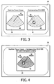
- Figure 3 in an illustrative view of the side-by-side display 36 of real time tissue image 38, showing an inserted needle 40, and display 42 of the corresponding MPR 44 from the pre-acquired CEUS volume, showing the target 46 highlighted by the contrast enhancement.
- Figure 4 is an illustrative view of a semi-transparent overlay 48 of the same tissue and contrast image, as indicated in Figure 4 by reference numeral 50.
- the system is configured for acquisition of multiple 3D CEUS volumes. That is, during the 3D CEUS volume acquisition, rather than storing on the workstation 18 only a single volume showing the contrast-enhance tissue of interest, a time series of volumes and their corresponding position information are also stored. The system can then ascertain the dynamics of contrast uptake (e.g., in-flow and out-flow), which are also diagnostically valuable indicators.
- the system is further configured for (a) co-displaying not just a single CEUS MPR with the real time tissue image, but co-displaying multiple MPRs of the entire time-series as a "movie".
- system is also configured for (b) processing the time-series data to create a volumetric map of diagnostically relevant parameters, such as the in-flow time constant, and then co-displaying an MPR of this parameter MAP with the real time tissue image during the interventional procedure.
- the system is configured for using alternate target visualization. That is, rather than co-displaying or over-laying an MPR of the 3D CEUS volume with the tissue image, a target volume-of-interest (VOI) is segmented from the 3D CEUS volume after CEUS acquisition.
- a target VOI includes, for example, a sphere covering the area of strongest tissue enhancement in the 3D CEUS volume.
- the system is configured for using real time 3D imaging during guidance. That is, instead of using 2D imaging for guidance, the system uses real time 3D imaging. In this case, rather than generating MPRs, other means of visualization are used, such as maximum intensity projections (MIPs).
- MIPs maximum intensity projections
- the MIP projections can be computed using the same set of transformations as described in the embodiments herein above.
- the system is configured for non-real time 3D CEUS acquisition.
- the reference CEUS volume can also be generated by tracking a 2D ultrasound probe, streaming to the workstation all 2D image frames and corresponding tracking data, obtaining a "sweep" over the target area after contrast injection, and reconstructing the 3D CEUS volume based on the acquired 2D images and tracking data.
- the system is configured for use of image-based registration for motion correction.
- the above described joint display of real time tissue image and corresponding 3D CEUS MPR is only accurate if the patient, or the organ being imaged, does not move between acquisition of the 3D CEUS volume and of the real time tissue image.
- the system according to the embodiments of the present invention is further configured for example, one of the following motion correction methods.
- a first motion correction method includes simultaneous acquisition of the 3D CEUS image with a 3D tissue image.
- a second motion correction method makes use of a simultaneous dual-mode acquisition and visualization, for example, as is available in real time 2D on modern scanners such as Philips iU22, and which enables non-real time simultaneous 3D acquisition as described above.
- a second motion correction method includes an image-based registration of a current 2D tissue image (or the last N 2D tissue images, with N is small, e.g. ⁇ 10) with the 3D tissue image acquired simultaneously with the 3D CEUS image.
- the tracking coordinates associated with the 2D images serve as a starting position for the registration (i.e. assuming no organ motion).
- the registration itself can utilize various optimizers and similarity measures, such as the Gauss-Newton optimizer and sum-of-squared-differences similarity measure.
- the result of the registration is the transformation T 2DUS ⁇ 3Dtissue .Since the 3D CEUS and tissue images were acquired simultaneously, their coordinate systems are identical, and the same transformation can be used to transform current 2D coordinates into 3D
- the system uses a spatial tracking system to track the position of an ultrasound probe and for image guidance in interventional procedures during contrast-enhanced ultrasound imaging (CEUS).
- a three-dimensional (3D) CEUS volume is acquired simultaneously with a regular 3D ultrasound tissue image.
- spatial tracking of the ultrasound probe enables joint display of the current real time ultrasound tissue image with the corresponding multi-planar reconstruction (MPR) from the 3D CEUS volume acquired at the beginning of the procedure (also referred to herein as the pre-acquired 3D CEUS volume).
- MPR multi-planar reconstruction
- Tissue motion between 3D CEUS acquisition obtained at the beginning of the procedure and real time imaging obtained subsequent or after the beginning of the procedure is corrected by image-based registration between the real time 3D tissue image and a 3D tissue image co-acquired with the 3D CEUS at the beginning of the procedure.
- MPR multi-planar reconstruction
- CEUS can provide superior visualization of tumors, vascularity, and other tissues of interest compared to non-contrast enhanced ultrasound imaging.
- contrast enhancement after bolus injection is a transient phenomenon and typically disappears after a few minutes.
- the time window of the transient phenomenon of contrast enhancement alone is insufficient.
- the embodiments of the present disclosure advantageously provide a system and method that overcomes this limitation.
- a method for interventional navigation using 3D contrast-enhanced ultrasound (CEUS) imaging comprises: acquiring a reference 3D CEUS volume and tracking information for a desired portion of an anatomy that is subject to an interventional procedure with an instrument, wherein the acquiring takes place during a useful lifetime of a contrast enhancement agent administered to the desired portion of the anatomy; acquiring real-time tracked tissue images during the interventional procedure; generating at least one corresponding contrast-enhanced ultrasound image multiplanar reconstruction (CEUS MPR) for at least one of the acquired real-time tracked tissue images during the interventional procedure as a function of the reference 3D CEUS volume and tracking information; displaying at least one of the acquired real-time tracked tissue images, wherein the displayed real-time tracked tissue image includes at least an image of the instrument within the desired portion of the anatomy; and displaying the at least one corresponding contrast-enhanced ultrasound image multiplanar reconstruction (CEUS MPR) that corresponds to the displayed real-time tracked tissue image, wherein the
- acquisition of the reference 3D CEUS volume includes concurrent acquisition of at least one tracked 3D ultrasound contrast image and tissue image pair.
- the at least one tracked 3D ultrasound contrast and tissue image pair comprise an initial ultrasound contrast image and a corresponding initial tissue image captured substantially concurrently and automatically registered to one another.
- the interventional procedure includes a first portion thereof that occurs prior to expiration of the contrast enhancement useful lifetime and a second portion thereof that occurs subsequent expiration of the contrast enhancement useful lifetime.
- the at least one corresponding contrast-enhanced ultrasound image multiplanar reconstruction is spatially registered with a corresponding one of the acquired real-time tracked tissue images.
- the real-time tracked tissue image and the corresponding CEUS MPR are displayed adjacent to one another.
- the real-time tracked tissue image and the corresponding CEUS MPR reference image are displayed together on a single display.
- the real-time tracked tissue image and the corresponding CEUS MPR can further be displayed in an overlaying arrangement such that one image overlays the other image.
- the overlaying arrangement can comprise one image being semi-transparent with respect to the other image.
- the system is configured for: generating a maximum intensity projection (MIP) as a function of the at least one pair of acquired tracked 3D real-time ultrasound contrast and tissue images; displaying a maximum intensity projection (MIP) of at least one of the acquired real-time tracked 3D tissue images, wherein the displayed real-time tracked tissue image MIP includes at least an image of the instrument within the desired portion of the anatomy; and displaying the at least one corresponding contrast-enhanced ultrasound image maximum intensity projection (CEUS MIP) that corresponds to the displayed MIP of the at least one acquired real-time tracked 3D tissue image, wherein the CEUS MIP image includes a contrast enhanced MIP image from a target volume of interest, thereby providing a concurrent display useful for the interventional navigation at least subsequent to the expiration of
- the reference 3D CEUS volume is acquired by tracking a 2D ultrasound probe, acquiring a series of 2D contrast and tissue image frames and corresponding tracking data while sweeping the tracked 2D ultrasound probe over the desired portion of the anatomy after contrast enhancement agent administration, streaming the acquired series of 2D contrast and tissue images and corresponding tracking data to a workstation, and reconstructing the reference 3D CEUS volume at the workstation based upon the acquired 2D contrast and tissue images and corresponding tracking data.
- acquiring the reference 3D CEUS volume includes using a spatial tracking system configured to track a position of an ultrasound probe used in acquiring the reference 3D CEUS volume, wherein the tracking system enables determination of a location and orientation of the reference 3D CEUS volume; and wherein the tracking system is further configured to track the acquired tissue images during the interventional procedure.
- the system is further configured to: correcting for tissue motion within the desired portion of the anatomy which may occur between (i) a time of the acquisition of the reference 3D CEUS volume, and (ii) a time of the acquisition of the at least one real-time tracked tissue image.
- correcting tissue motion includes using an image-based registration between (a) the real-time tracked 3D tissue image and (b) the ultrasound tissue image of the tracked 3D ultrasound contrast and tissue image pair.
- acquiring the reference 3D CEUS volume and tracking information includes acquiring a time series of 3D CEUS volumes and corresponding tracking information, and wherein generating the at least one corresponding CEUS MPR image includes generating a time series of CEUS MPR images, and wherein displaying the acquired real-time tracked tissue images and displaying the at least one corresponding CEUS MPR images comprises co-displaying the CEUS MPR images with the real-time tissue images as time-series data.
- the system is further configured to processing the time-series data to create a volumetric map of diagnostically relevant parameters, and co-displaying the volumetric map with (i) the CEUS MPR images and (ii) the real-time tissue images.
- the diagnostically relevant parameters can include at least an in-flow time constant of the contrast agent.
- system is further configured to segmenting the target volume of interest from the reference 3D CEUS volume, wherein the target volume of interest comprises an areas of strongest tissue enhancement; and displaying a corresponding cross-section segment of the target volume of interest, instead of displaying the at least one corresponding contrast-enhanced ultrasound image multiplanar reconstruction (CEUS MPR), that corresponds to the displayed real-time tracked tissue image, and superimposing the corresponding cross-section segment of the target volume of interest on the displayed real-time tracked tissue image.
- CEUS MPR contrast-enhanced ultrasound image multiplanar reconstruction
- a method for interventional navigation using 3D contrast-enhanced ultrasound (CEUS) imaging comprises: acquiring a reference 3D CEUS volume and tracking information for a desired portion of an anatomy that is subject to an interventional procedure with an instrument, wherein the acquiring takes place during a useful lifetime of a contrast enhancement agent administered to the desired portion of the anatomy, wherein acquisition of the reference 3D CEUS volume includes concurrent acquisition of at least one tracked 3D ultrasound contrast image and tissue image pair; acquiring real-time tracked tissue images during the interventional procedure; generating at least one corresponding contrast-enhanced ultrasound image multiplanar reconstruction (CEUS MPR) for at least one of the acquired real-time tracked tissue images during the interventional procedure as a function of the reference 3D CEUS volume and tracking information, wherein the at least one corresponding contrast-enhanced ultrasound image multiplanar reconstruction is spatially registered with a corresponding one of the acquired real-time tracked tissue images; displaying the acquired real-time tracked tissue images, wherein the displayed real-time tracked tissue image includes at least
- the embodiments of the present disclosure can be applied in ultrasound-based image guidance of diagnostic and therapeutic medical procedures.
- the embodiments of the present disclosure can improve needle guidance for biopsy and ablation procedures.
- the embodiments of the present disclosure advantageously overcome limitations and disadvantages in current ultrasound-based procedure guidance, such as, limited visualization or no visualization of some tumors/targets; poor accuracy in execution of the procedure if target location is estimated based on a pre-acquired modality (other than pre-acquired CEUS as disclosed with respect to the embodiments of the present disclosure); and brief duration of contrast enhancement in CEUS imaging which is insufficient for execution of a given interventional procedure.
- any reference signs placed in parentheses in one or more claims shall not be construed as limiting the claims.
- the word “comprising” and “comprises,” and the like, does not exclude the presence of elements or steps other than those listed in any claim or the specification as a whole.
- the singular reference of an element does not exclude the plural references of such elements and vice-versa.
- One or more of the embodiments may be implemented by means of hardware comprising several distinct elements, and/or by means of a suitably programmed computer. In a device claim enumerating several means, several of these means may be embodied by one and the same item of hardware.
- the mere fact that certain measures are recited in mutually different dependent claims does not indicate that a combination of these measures cannot be used to an advantage.
Landscapes
- Engineering & Computer Science (AREA)
- Health & Medical Sciences (AREA)
- Life Sciences & Earth Sciences (AREA)
- Physics & Mathematics (AREA)
- Radar, Positioning & Navigation (AREA)
- Remote Sensing (AREA)
- Nuclear Medicine, Radiotherapy & Molecular Imaging (AREA)
- Medical Informatics (AREA)
- Computer Networks & Wireless Communication (AREA)
- Biophysics (AREA)
- Veterinary Medicine (AREA)
- Pathology (AREA)
- Radiology & Medical Imaging (AREA)
- Biomedical Technology (AREA)
- Heart & Thoracic Surgery (AREA)
- General Physics & Mathematics (AREA)
- Molecular Biology (AREA)
- Surgery (AREA)
- Animal Behavior & Ethology (AREA)
- General Health & Medical Sciences (AREA)
- Public Health (AREA)
- Acoustics & Sound (AREA)
- Ultra Sonic Daignosis Equipment (AREA)
Applications Claiming Priority (3)
| Application Number | Priority Date | Filing Date | Title |
|---|---|---|---|
| US98847207P | 2007-11-16 | 2007-11-16 | |
| US5228808P | 2008-05-12 | 2008-05-12 | |
| PCT/IB2008/054769 WO2009063423A1 (en) | 2007-11-16 | 2008-11-13 | Interventional navigation using 3d contrast-enhanced ultrasound |
Publications (2)
| Publication Number | Publication Date |
|---|---|
| EP2212716A1 EP2212716A1 (en) | 2010-08-04 |
| EP2212716B1 true EP2212716B1 (en) | 2014-02-26 |
Family
ID=40384555
Family Applications (1)
| Application Number | Title | Priority Date | Filing Date |
|---|---|---|---|
| EP08850546.6A Active EP2212716B1 (en) | 2007-11-16 | 2008-11-13 | Interventional navigation using 3d contrast-enhanced ultrasound |
Country Status (7)
| Country | Link |
|---|---|
| US (1) | US9651662B2 (enExample) |
| EP (1) | EP2212716B1 (enExample) |
| JP (1) | JP2011502687A (enExample) |
| CN (1) | CN101868737B (enExample) |
| BR (1) | BRPI0819439A8 (enExample) |
| RU (1) | RU2494676C2 (enExample) |
| WO (1) | WO2009063423A1 (enExample) |
Families Citing this family (53)
| Publication number | Priority date | Publication date | Assignee | Title |
|---|---|---|---|---|
| MX2007008248A (es) * | 2005-10-26 | 2007-08-22 | Thomson Licensing | Un metodo y sistema para compensar una falla de pasarela de satelite. |
| WO2008017051A2 (en) | 2006-08-02 | 2008-02-07 | Inneroptic Technology Inc. | System and method of providing real-time dynamic imagery of a medical procedure site using multiple modalities |
| JP5148094B2 (ja) * | 2006-09-27 | 2013-02-20 | 株式会社東芝 | 超音波診断装置、医用画像処理装置及びプログラム |
| WO2009094646A2 (en) | 2008-01-24 | 2009-07-30 | The University Of North Carolina At Chapel Hill | Methods, systems, and computer readable media for image guided ablation |
| US8340379B2 (en) | 2008-03-07 | 2012-12-25 | Inneroptic Technology, Inc. | Systems and methods for displaying guidance data based on updated deformable imaging data |
| US8554307B2 (en) | 2010-04-12 | 2013-10-08 | Inneroptic Technology, Inc. | Image annotation in image-guided medical procedures |
| US8641621B2 (en) | 2009-02-17 | 2014-02-04 | Inneroptic Technology, Inc. | Systems, methods, apparatuses, and computer-readable media for image management in image-guided medical procedures |
| US8690776B2 (en) | 2009-02-17 | 2014-04-08 | Inneroptic Technology, Inc. | Systems, methods, apparatuses, and computer-readable media for image guided surgery |
| US11464578B2 (en) | 2009-02-17 | 2022-10-11 | Inneroptic Technology, Inc. | Systems, methods, apparatuses, and computer-readable media for image management in image-guided medical procedures |
| KR101121286B1 (ko) | 2009-07-31 | 2012-03-23 | 한국과학기술원 | 센서의 교정을 수행하는 초음파 시스템 및 방법 |
| US9545242B2 (en) | 2009-07-31 | 2017-01-17 | Samsung Medison Co., Ltd. | Sensor coordinate calibration in an ultrasound system |
| JP5508801B2 (ja) * | 2009-09-30 | 2014-06-04 | 株式会社東芝 | 超音波診断装置及び超音波診断装置制御プログラム |
| CN102081697B (zh) * | 2009-11-27 | 2013-12-11 | 深圳迈瑞生物医疗电子股份有限公司 | 一种在超声成像空间中定义感兴趣容积的方法及其装置 |
| JP5645160B2 (ja) * | 2010-10-14 | 2014-12-24 | 国立大学法人東京農工大学 | 超音波治療システム |
| EP2640275A1 (en) * | 2010-11-19 | 2013-09-25 | Koninklijke Philips N.V. | Three dimensional ultrasonic guidance of surgical instruments |
| RU2591595C2 (ru) * | 2011-01-17 | 2016-07-20 | Конинклейке Филипс Электроникс Н.В. | Система и способ детектирования размещения иглы при биопсии под контролем изображения |
| EP2509013A1 (en) * | 2011-04-04 | 2012-10-10 | Agfa Healthcare | 3D image navigation method |
| EP2699166B1 (en) | 2011-04-21 | 2019-09-04 | Koninklijke Philips N.V. | Mpr slice selection for visualization of catheter in three-dimensional ultrasound |
| JP6176818B2 (ja) * | 2011-12-06 | 2017-08-09 | 東芝メディカルシステムズ株式会社 | 超音波診断装置及び座標変換プログラム |
| US8670816B2 (en) | 2012-01-30 | 2014-03-11 | Inneroptic Technology, Inc. | Multiple medical device guidance |
| US11006923B2 (en) * | 2012-06-28 | 2021-05-18 | Koninklijke Philips N.V. | Ultrasonically guided biopsies in three dimensions |
| BR112015009947A2 (pt) * | 2012-11-06 | 2017-07-11 | Koninklijke Philips Nv | dispositivo de processamento de imagens para melhorar imagens de ultrassom; sistema de imageamento médico; método para melhorar imagens de ultrassom; elemento de programa de computador para controlar um dispositivo de processamento de imagens; e meio legível por computador |
| US20140142419A1 (en) * | 2012-11-19 | 2014-05-22 | Biosense Webster (Israel), Ltd. | Patient movement compensation in intra-body probe |
| US9232934B2 (en) * | 2012-12-14 | 2016-01-12 | General Electric Company | Systems and methods for communicating ultrasound probe location and image information |
| US10314559B2 (en) | 2013-03-14 | 2019-06-11 | Inneroptic Technology, Inc. | Medical device guidance |
| RU2677191C2 (ru) * | 2013-06-28 | 2019-01-15 | Конинклейке Филипс Н.В. | Установление границ блокирования ребром в анатомически интеллектуальной эхокардиографии |
| WO2015044901A1 (en) * | 2013-09-30 | 2015-04-02 | Koninklijke Philips N.V. | Image guidance system with user definable regions of interest |
| US20170169609A1 (en) * | 2014-02-19 | 2017-06-15 | Koninklijke Philips N.V. | Motion adaptive visualization in medical 4d imaging |
| KR102250086B1 (ko) * | 2014-05-16 | 2021-05-10 | 삼성전자주식회사 | 의료 영상 정합 방법, 이를 포함하는 장치 및 컴퓨터 기록 매체 |
| WO2016004302A1 (en) * | 2014-07-02 | 2016-01-07 | Covidien Lp | Alignment ct |
| US11188285B2 (en) * | 2014-07-02 | 2021-11-30 | Covidien Lp | Intelligent display |
| US9901406B2 (en) | 2014-10-02 | 2018-02-27 | Inneroptic Technology, Inc. | Affected region display associated with a medical device |
| US10188467B2 (en) | 2014-12-12 | 2019-01-29 | Inneroptic Technology, Inc. | Surgical guidance intersection display |
| JP6405058B2 (ja) * | 2015-03-31 | 2018-10-17 | コーニンクレッカ フィリップス エヌ ヴェKoninklijke Philips N.V. | 医療イメージング装置 |
| JP6714019B2 (ja) * | 2015-05-07 | 2020-06-24 | コーニンクレッカ フィリップス エヌ ヴェKoninklijke Philips N.V. | 医療手順における動き補償のためのシステム及び方法 |
| DE102015209143B4 (de) * | 2015-05-19 | 2020-02-27 | Esaote S.P.A. | Verfahren zur Bestimmung einer Abbildungsvorschrift und bildgestützten Navigation sowie Vorrichtung zur bildgestützten Navigation |
| WO2017003480A1 (en) * | 2015-07-02 | 2017-01-05 | Siemens Medical Solutions Usa, Inc. | Intervolume lesion detection and image preparation |
| US9949700B2 (en) | 2015-07-22 | 2018-04-24 | Inneroptic Technology, Inc. | Medical device approaches |
| CN107847291B (zh) * | 2015-07-28 | 2022-03-01 | 皇家飞利浦有限公司 | 用于活检记载的针尖端识别的工作流 |
| CN108369273B (zh) * | 2015-12-16 | 2022-09-06 | 皇家飞利浦有限公司 | 介入设备识别 |
| US9675319B1 (en) | 2016-02-17 | 2017-06-13 | Inneroptic Technology, Inc. | Loupe display |
| CN109310399B (zh) * | 2016-06-06 | 2022-12-06 | 皇家飞利浦有限公司 | 医学超声图像处理设备 |
| US10278778B2 (en) | 2016-10-27 | 2019-05-07 | Inneroptic Technology, Inc. | Medical device navigation using a virtual 3D space |
| EP3369381A1 (en) * | 2017-03-01 | 2018-09-05 | Koninklijke Philips N.V. | Ultrasound probe arrangement |
| JP7181226B2 (ja) * | 2017-05-11 | 2022-11-30 | コーニンクレッカ フィリップス エヌ ヴェ | 超音波処置における動き補償のためのワークフロー、システム及び方法 |
| US11259879B2 (en) | 2017-08-01 | 2022-03-01 | Inneroptic Technology, Inc. | Selective transparency to assist medical device navigation |
| US20200275915A1 (en) * | 2017-09-08 | 2020-09-03 | Koninklijke Philips N.V. | Ultrasound probe localization with drift correction |
| EP3482690A1 (en) * | 2017-11-14 | 2019-05-15 | Koninklijke Philips N.V. | Ultrasound tracking and visualization |
| US11484365B2 (en) | 2018-01-23 | 2022-11-01 | Inneroptic Technology, Inc. | Medical image guidance |
| CN112040875A (zh) * | 2018-04-06 | 2020-12-04 | 美敦力公司 | 基于图像的导航系统和使用相同的导航系统的方法 |
| CN109934888B (zh) * | 2019-04-24 | 2020-11-17 | 清华大学 | 非对比剂增强的磁共振动态血管成像方法及系统 |
| US12236680B2 (en) * | 2019-09-20 | 2025-02-25 | Gn Hearing A/S | Application for assisting a hearing device wearer |
| CN115243621B (zh) | 2020-03-05 | 2025-11-21 | 皇家飞利浦有限公司 | 三维超声成像数据的背景多平面重建以及相关联的设备、系统和方法 |
Family Cites Families (17)
| Publication number | Priority date | Publication date | Assignee | Title |
|---|---|---|---|---|
| WO1996025882A1 (en) | 1995-02-22 | 1996-08-29 | Groenningsaeter Aage | Method for ultrasound guidance during clinical procedures |
| US7328059B2 (en) * | 1996-08-23 | 2008-02-05 | The Texas A & M University System | Imaging of light scattering tissues with fluorescent contrast agents |
| IL119262A0 (en) | 1996-02-15 | 1996-12-05 | Biosense Israel Ltd | Locatable biopsy needle |
| EP0883860B1 (en) * | 1996-02-29 | 2006-08-23 | Acuson Corporation | Multiple ultrasound image registration system, method and transducer |
| JP2003093389A (ja) * | 2001-09-27 | 2003-04-02 | Hitachi Medical Corp | 超音波診断装置 |
| US7477763B2 (en) * | 2002-06-18 | 2009-01-13 | Boston Scientific Scimed, Inc. | Computer generated representation of the imaging pattern of an imaging device |
| JP4058368B2 (ja) * | 2003-03-27 | 2008-03-05 | ジーイー・メディカル・システムズ・グローバル・テクノロジー・カンパニー・エルエルシー | 超音波診断装置 |
| EP1498746B1 (en) * | 2003-07-09 | 2013-12-11 | Panasonic Corporation | Ultrasonic diagnostic apparatus and tomographic image processing apparatus |
| US20060020204A1 (en) | 2004-07-01 | 2006-01-26 | Bracco Imaging, S.P.A. | System and method for three-dimensional space management and visualization of ultrasound data ("SonoDEX") |
| JP5150267B2 (ja) * | 2005-02-23 | 2013-02-20 | コーニンクレッカ フィリップス エレクトロニクス エヌ ヴィ | 肝臓の障害を検出する超音波診断イメージングシステム |
| US20060239585A1 (en) | 2005-04-04 | 2006-10-26 | Valadez Gerardo H | System and method for reducing artifacts in motion corrected dynamic image sequences |
| JP4801968B2 (ja) | 2005-11-02 | 2011-10-26 | 株式会社東芝 | 画像診断・治療支援装置及び画像データ表示方法 |
| US8303505B2 (en) * | 2005-12-02 | 2012-11-06 | Abbott Cardiovascular Systems Inc. | Methods and apparatuses for image guided medical procedures |
| KR20070058785A (ko) | 2005-12-05 | 2007-06-11 | 주식회사 메디슨 | 중재적 시술을 위한 초음파 시스템 |
| KR20070110965A (ko) | 2006-05-16 | 2007-11-21 | 주식회사 메디슨 | 초음파 영상과 외부 의료영상의 합성 영상을디스플레이하기 위한 초음파 시스템 |
| CN107126182B (zh) * | 2007-01-19 | 2020-06-16 | 桑尼布鲁克健康科学中心 | 用于成像探头的扫描机构 |
| US8355550B2 (en) * | 2007-05-01 | 2013-01-15 | Siemens Aktiengesellschaft | Methods and apparatus for virtual coronary mapping |
-
2008
- 2008-11-13 JP JP2010533703A patent/JP2011502687A/ja active Pending
- 2008-11-13 WO PCT/IB2008/054769 patent/WO2009063423A1/en not_active Ceased
- 2008-11-13 US US12/742,255 patent/US9651662B2/en active Active
- 2008-11-13 RU RU2010124373/14A patent/RU2494676C2/ru not_active IP Right Cessation
- 2008-11-13 BR BRPI0819439A patent/BRPI0819439A8/pt not_active Application Discontinuation
- 2008-11-13 CN CN2008801161164A patent/CN101868737B/zh active Active
- 2008-11-13 EP EP08850546.6A patent/EP2212716B1/en active Active
Also Published As
| Publication number | Publication date |
|---|---|
| RU2010124373A (ru) | 2011-12-27 |
| JP2011502687A (ja) | 2011-01-27 |
| CN101868737B (zh) | 2013-04-24 |
| US9651662B2 (en) | 2017-05-16 |
| BRPI0819439A8 (pt) | 2015-11-10 |
| US20100268085A1 (en) | 2010-10-21 |
| BRPI0819439A2 (enExample) | 2009-05-22 |
| CN101868737A (zh) | 2010-10-20 |
| WO2009063423A1 (en) | 2009-05-22 |
| EP2212716A1 (en) | 2010-08-04 |
| RU2494676C2 (ru) | 2013-10-10 |
Similar Documents
| Publication | Publication Date | Title |
|---|---|---|
| EP2212716B1 (en) | Interventional navigation using 3d contrast-enhanced ultrasound | |
| RU2654608C2 (ru) | Ультразвуковая система визуализации и способ для процедуры наведения по изображению | |
| US8126239B2 (en) | Registering 2D and 3D data using 3D ultrasound data | |
| US20090275831A1 (en) | Image registration and methods for compensating intraoperative motion in image-guided interventional procedures | |
| US20250151976A1 (en) | Systems and methods for registering imaging data from different imaging modalities based on subsurface image scanning | |
| US11026747B2 (en) | Endoscopic view of invasive procedures in narrow passages | |
| EP1685535B1 (en) | Device and method for combining two images | |
| JP6395995B2 (ja) | 医療映像処理方法及び装置 | |
| EP2208182B1 (en) | System and method for quantitative 3d ceus analysis | |
| US11672505B2 (en) | Correcting probe induced deformation in an ultrasound fusing imaging system | |
| US20170084036A1 (en) | Registration of video camera with medical imaging | |
| US10755453B2 (en) | Image processing apparatus, image processing method, and ultrasound imaging apparatus having image processing unit | |
| US20160030008A1 (en) | System and method for registering ultrasound information to an x-ray image | |
| JP2006305360A (ja) | 2次元扇形超音波イメージの表示 | |
| WO2010064348A1 (ja) | 医用画像の位置あわせのための情報処理装置、情報処理方法、プログラム | |
| US20140192054A1 (en) | Method and apparatus for providing medical images | |
| US9460548B2 (en) | Method and apparatus for displaying medical image | |
| KR20080034447A (ko) | 2차원 x-선 영상과 3차원 초음파 영상의 선택적인 혼합을위한 시스템 및 방법 | |
| JP2006305361A (ja) | 超音波システム用のビーム方向を用いたカテーテル先端部の表示 | |
| CN116528752A (zh) | 自动分割与配准系统及方法 | |
| CN108430376B (zh) | 提供投影数据集 | |
| US12465428B2 (en) | Image-based device tracking | |
| von Berg et al. | A hybrid method for registration of interventional CT and ultrasound images | |
| US7943892B2 (en) | Projection image generation apparatus, method for generating projection image of moving target, and program | |
| Lang | Improvement of Speckle-Tracked Freehand 3-D Ultrasound Through the Use of Sensor Fusion |
Legal Events
| Date | Code | Title | Description |
|---|---|---|---|
| PUAI | Public reference made under article 153(3) epc to a published international application that has entered the european phase |
Free format text: ORIGINAL CODE: 0009012 |
|
| 17P | Request for examination filed |
Effective date: 20100616 |
|
| AK | Designated contracting states |
Kind code of ref document: A1 Designated state(s): AT BE BG CH CY CZ DE DK EE ES FI FR GB GR HR HU IE IS IT LI LT LU LV MC MT NL NO PL PT RO SE SI SK TR |
|
| AX | Request for extension of the european patent |
Extension state: AL BA MK RS |
|
| 17Q | First examination report despatched |
Effective date: 20101012 |
|
| DAX | Request for extension of the european patent (deleted) | ||
| GRAP | Despatch of communication of intention to grant a patent |
Free format text: ORIGINAL CODE: EPIDOSNIGR1 |
|
| RAP1 | Party data changed (applicant data changed or rights of an application transferred) |
Owner name: KONINKLIJKE PHILIPS N.V. |
|
| INTG | Intention to grant announced |
Effective date: 20130906 |
|
| GRAS | Grant fee paid |
Free format text: ORIGINAL CODE: EPIDOSNIGR3 |
|
| GRAA | (expected) grant |
Free format text: ORIGINAL CODE: 0009210 |
|
| AK | Designated contracting states |
Kind code of ref document: B1 Designated state(s): AT BE BG CH CY CZ DE DK EE ES FI FR GB GR HR HU IE IS IT LI LT LU LV MC MT NL NO PL PT RO SE SI SK TR |
|
| REG | Reference to a national code |
Ref country code: GB Ref legal event code: FG4D |
|
| REG | Reference to a national code |
Ref country code: CH Ref legal event code: EP |
|
| REG | Reference to a national code |
Ref country code: AT Ref legal event code: REF Ref document number: 653908 Country of ref document: AT Kind code of ref document: T Effective date: 20140315 |
|
| REG | Reference to a national code |
Ref country code: IE Ref legal event code: FG4D |
|
| REG | Reference to a national code |
Ref country code: DE Ref legal event code: R096 Ref document number: 602008030497 Country of ref document: DE Effective date: 20140410 |
|
| REG | Reference to a national code |
Ref country code: NL Ref legal event code: VDEP Effective date: 20140226 |
|
| REG | Reference to a national code |
Ref country code: AT Ref legal event code: MK05 Ref document number: 653908 Country of ref document: AT Kind code of ref document: T Effective date: 20140226 |
|
| REG | Reference to a national code |
Ref country code: LT Ref legal event code: MG4D |
|
| PG25 | Lapsed in a contracting state [announced via postgrant information from national office to epo] |
Ref country code: IS Free format text: LAPSE BECAUSE OF FAILURE TO SUBMIT A TRANSLATION OF THE DESCRIPTION OR TO PAY THE FEE WITHIN THE PRESCRIBED TIME-LIMIT Effective date: 20140626 Ref country code: NO Free format text: LAPSE BECAUSE OF FAILURE TO SUBMIT A TRANSLATION OF THE DESCRIPTION OR TO PAY THE FEE WITHIN THE PRESCRIBED TIME-LIMIT Effective date: 20140526 Ref country code: LT Free format text: LAPSE BECAUSE OF FAILURE TO SUBMIT A TRANSLATION OF THE DESCRIPTION OR TO PAY THE FEE WITHIN THE PRESCRIBED TIME-LIMIT Effective date: 20140226 |
|
| PG25 | Lapsed in a contracting state [announced via postgrant information from national office to epo] |
Ref country code: FI Free format text: LAPSE BECAUSE OF FAILURE TO SUBMIT A TRANSLATION OF THE DESCRIPTION OR TO PAY THE FEE WITHIN THE PRESCRIBED TIME-LIMIT Effective date: 20140226 Ref country code: NL Free format text: LAPSE BECAUSE OF FAILURE TO SUBMIT A TRANSLATION OF THE DESCRIPTION OR TO PAY THE FEE WITHIN THE PRESCRIBED TIME-LIMIT Effective date: 20140226 Ref country code: CY Free format text: LAPSE BECAUSE OF FAILURE TO SUBMIT A TRANSLATION OF THE DESCRIPTION OR TO PAY THE FEE WITHIN THE PRESCRIBED TIME-LIMIT Effective date: 20140226 Ref country code: PT Free format text: LAPSE BECAUSE OF FAILURE TO SUBMIT A TRANSLATION OF THE DESCRIPTION OR TO PAY THE FEE WITHIN THE PRESCRIBED TIME-LIMIT Effective date: 20140626 Ref country code: SE Free format text: LAPSE BECAUSE OF FAILURE TO SUBMIT A TRANSLATION OF THE DESCRIPTION OR TO PAY THE FEE WITHIN THE PRESCRIBED TIME-LIMIT Effective date: 20140226 Ref country code: AT Free format text: LAPSE BECAUSE OF FAILURE TO SUBMIT A TRANSLATION OF THE DESCRIPTION OR TO PAY THE FEE WITHIN THE PRESCRIBED TIME-LIMIT Effective date: 20140226 |
|
| PG25 | Lapsed in a contracting state [announced via postgrant information from national office to epo] |
Ref country code: BE Free format text: LAPSE BECAUSE OF FAILURE TO SUBMIT A TRANSLATION OF THE DESCRIPTION OR TO PAY THE FEE WITHIN THE PRESCRIBED TIME-LIMIT Effective date: 20140226 Ref country code: LV Free format text: LAPSE BECAUSE OF FAILURE TO SUBMIT A TRANSLATION OF THE DESCRIPTION OR TO PAY THE FEE WITHIN THE PRESCRIBED TIME-LIMIT Effective date: 20140226 Ref country code: HR Free format text: LAPSE BECAUSE OF FAILURE TO SUBMIT A TRANSLATION OF THE DESCRIPTION OR TO PAY THE FEE WITHIN THE PRESCRIBED TIME-LIMIT Effective date: 20140226 |
|
| PG25 | Lapsed in a contracting state [announced via postgrant information from national office to epo] |
Ref country code: CZ Free format text: LAPSE BECAUSE OF FAILURE TO SUBMIT A TRANSLATION OF THE DESCRIPTION OR TO PAY THE FEE WITHIN THE PRESCRIBED TIME-LIMIT Effective date: 20140226 Ref country code: EE Free format text: LAPSE BECAUSE OF FAILURE TO SUBMIT A TRANSLATION OF THE DESCRIPTION OR TO PAY THE FEE WITHIN THE PRESCRIBED TIME-LIMIT Effective date: 20140226 Ref country code: RO Free format text: LAPSE BECAUSE OF FAILURE TO SUBMIT A TRANSLATION OF THE DESCRIPTION OR TO PAY THE FEE WITHIN THE PRESCRIBED TIME-LIMIT Effective date: 20140226 Ref country code: DK Free format text: LAPSE BECAUSE OF FAILURE TO SUBMIT A TRANSLATION OF THE DESCRIPTION OR TO PAY THE FEE WITHIN THE PRESCRIBED TIME-LIMIT Effective date: 20140226 |
|
| REG | Reference to a national code |
Ref country code: DE Ref legal event code: R097 Ref document number: 602008030497 Country of ref document: DE |
|
| PG25 | Lapsed in a contracting state [announced via postgrant information from national office to epo] |
Ref country code: SK Free format text: LAPSE BECAUSE OF FAILURE TO SUBMIT A TRANSLATION OF THE DESCRIPTION OR TO PAY THE FEE WITHIN THE PRESCRIBED TIME-LIMIT Effective date: 20140226 Ref country code: ES Free format text: LAPSE BECAUSE OF FAILURE TO SUBMIT A TRANSLATION OF THE DESCRIPTION OR TO PAY THE FEE WITHIN THE PRESCRIBED TIME-LIMIT Effective date: 20140226 Ref country code: PL Free format text: LAPSE BECAUSE OF FAILURE TO SUBMIT A TRANSLATION OF THE DESCRIPTION OR TO PAY THE FEE WITHIN THE PRESCRIBED TIME-LIMIT Effective date: 20140226 |
|
| PLBE | No opposition filed within time limit |
Free format text: ORIGINAL CODE: 0009261 |
|
| STAA | Information on the status of an ep patent application or granted ep patent |
Free format text: STATUS: NO OPPOSITION FILED WITHIN TIME LIMIT |
|
| 26N | No opposition filed |
Effective date: 20141127 |
|
| REG | Reference to a national code |
Ref country code: DE Ref legal event code: R097 Ref document number: 602008030497 Country of ref document: DE Effective date: 20141127 |
|
| PG25 | Lapsed in a contracting state [announced via postgrant information from national office to epo] |
Ref country code: IT Free format text: LAPSE BECAUSE OF FAILURE TO SUBMIT A TRANSLATION OF THE DESCRIPTION OR TO PAY THE FEE WITHIN THE PRESCRIBED TIME-LIMIT Effective date: 20140226 |
|
| PG25 | Lapsed in a contracting state [announced via postgrant information from national office to epo] |
Ref country code: SI Free format text: LAPSE BECAUSE OF FAILURE TO SUBMIT A TRANSLATION OF THE DESCRIPTION OR TO PAY THE FEE WITHIN THE PRESCRIBED TIME-LIMIT Effective date: 20140226 |
|
| PG25 | Lapsed in a contracting state [announced via postgrant information from national office to epo] |
Ref country code: LU Free format text: LAPSE BECAUSE OF FAILURE TO SUBMIT A TRANSLATION OF THE DESCRIPTION OR TO PAY THE FEE WITHIN THE PRESCRIBED TIME-LIMIT Effective date: 20141113 Ref country code: MC Free format text: LAPSE BECAUSE OF FAILURE TO SUBMIT A TRANSLATION OF THE DESCRIPTION OR TO PAY THE FEE WITHIN THE PRESCRIBED TIME-LIMIT Effective date: 20140226 |
|
| REG | Reference to a national code |
Ref country code: CH Ref legal event code: PL |
|
| PG25 | Lapsed in a contracting state [announced via postgrant information from national office to epo] |
Ref country code: LI Free format text: LAPSE BECAUSE OF NON-PAYMENT OF DUE FEES Effective date: 20141130 Ref country code: CH Free format text: LAPSE BECAUSE OF NON-PAYMENT OF DUE FEES Effective date: 20141130 |
|
| REG | Reference to a national code |
Ref country code: IE Ref legal event code: MM4A |
|
| PG25 | Lapsed in a contracting state [announced via postgrant information from national office to epo] |
Ref country code: IE Free format text: LAPSE BECAUSE OF NON-PAYMENT OF DUE FEES Effective date: 20141113 |
|
| REG | Reference to a national code |
Ref country code: FR Ref legal event code: PLFP Year of fee payment: 8 |
|
| PGFP | Annual fee paid to national office [announced via postgrant information from national office to epo] |
Ref country code: TR Payment date: 20151105 Year of fee payment: 8 |
|
| PG25 | Lapsed in a contracting state [announced via postgrant information from national office to epo] |
Ref country code: BG Free format text: LAPSE BECAUSE OF FAILURE TO SUBMIT A TRANSLATION OF THE DESCRIPTION OR TO PAY THE FEE WITHIN THE PRESCRIBED TIME-LIMIT Effective date: 20140226 |
|
| PG25 | Lapsed in a contracting state [announced via postgrant information from national office to epo] |
Ref country code: GR Free format text: LAPSE BECAUSE OF FAILURE TO SUBMIT A TRANSLATION OF THE DESCRIPTION OR TO PAY THE FEE WITHIN THE PRESCRIBED TIME-LIMIT Effective date: 20140527 |
|
| PG25 | Lapsed in a contracting state [announced via postgrant information from national office to epo] |
Ref country code: MT Free format text: LAPSE BECAUSE OF FAILURE TO SUBMIT A TRANSLATION OF THE DESCRIPTION OR TO PAY THE FEE WITHIN THE PRESCRIBED TIME-LIMIT Effective date: 20140226 Ref country code: HU Free format text: LAPSE BECAUSE OF FAILURE TO SUBMIT A TRANSLATION OF THE DESCRIPTION OR TO PAY THE FEE WITHIN THE PRESCRIBED TIME-LIMIT; INVALID AB INITIO Effective date: 20081113 |
|
| REG | Reference to a national code |
Ref country code: FR Ref legal event code: PLFP Year of fee payment: 9 |
|
| REG | Reference to a national code |
Ref country code: FR Ref legal event code: PLFP Year of fee payment: 10 |
|
| PGFP | Annual fee paid to national office [announced via postgrant information from national office to epo] |
Ref country code: FR Payment date: 20191126 Year of fee payment: 12 |
|
| PG25 | Lapsed in a contracting state [announced via postgrant information from national office to epo] |
Ref country code: FR Free format text: LAPSE BECAUSE OF NON-PAYMENT OF DUE FEES Effective date: 20201130 |
|
| PG25 | Lapsed in a contracting state [announced via postgrant information from national office to epo] |
Ref country code: TR Free format text: LAPSE BECAUSE OF FAILURE TO SUBMIT A TRANSLATION OF THE DESCRIPTION OR TO PAY THE FEE WITHIN THE PRESCRIBED TIME-LIMIT Effective date: 20140226 |
|
| REG | Reference to a national code |
Ref country code: DE Ref legal event code: R084 Ref document number: 602008030497 Country of ref document: DE |
|
| REG | Reference to a national code |
Ref country code: GB Ref legal event code: 746 Effective date: 20240620 |
|
| PGFP | Annual fee paid to national office [announced via postgrant information from national office to epo] |
Ref country code: DE Payment date: 20241128 Year of fee payment: 17 |
|
| PGFP | Annual fee paid to national office [announced via postgrant information from national office to epo] |
Ref country code: GB Payment date: 20241126 Year of fee payment: 17 |