EP2199796A1 - Méthode de prognose du cancer avec la quantification du niveau de récepteurs folate - Google Patents
Méthode de prognose du cancer avec la quantification du niveau de récepteurs folate Download PDFInfo
- Publication number
- EP2199796A1 EP2199796A1 EP10155913A EP10155913A EP2199796A1 EP 2199796 A1 EP2199796 A1 EP 2199796A1 EP 10155913 A EP10155913 A EP 10155913A EP 10155913 A EP10155913 A EP 10155913A EP 2199796 A1 EP2199796 A1 EP 2199796A1
- Authority
- EP
- European Patent Office
- Prior art keywords
- cancer cells
- cancer
- antibody
- staining
- cells
- Prior art date
- Legal status (The legal status is an assumption and is not a legal conclusion. Google has not performed a legal analysis and makes no representation as to the accuracy of the status listed.)
- Withdrawn
Links
Images


Classifications
-
- G—PHYSICS
- G01—MEASURING; TESTING
- G01N—INVESTIGATING OR ANALYSING MATERIALS BY DETERMINING THEIR CHEMICAL OR PHYSICAL PROPERTIES
- G01N33/00—Investigating or analysing materials by specific methods not covered by groups G01N1/00 - G01N31/00
- G01N33/48—Biological material, e.g. blood, urine; Haemocytometers
- G01N33/50—Chemical analysis of biological material, e.g. blood, urine; Testing involving biospecific ligand binding methods; Immunological testing
- G01N33/53—Immunoassay; Biospecific binding assay; Materials therefor
- G01N33/574—Immunoassay; Biospecific binding assay; Materials therefor for cancer
- G01N33/57484—Immunoassay; Biospecific binding assay; Materials therefor for cancer involving compounds serving as markers for tumor, cancer, neoplasia, e.g. cellular determinants, receptors, heat shock/stress proteins, A-protein, oligosaccharides, metabolites
- G01N33/57492—Immunoassay; Biospecific binding assay; Materials therefor for cancer involving compounds serving as markers for tumor, cancer, neoplasia, e.g. cellular determinants, receptors, heat shock/stress proteins, A-protein, oligosaccharides, metabolites involving compounds localized on the membrane of tumor or cancer cells
Definitions
- This invention relates to methods and kits for obtaining a prognosis for a cancer by quantification of vitamin receptor expression levels in the cancer cells and to guide the management or develop an effective treatment for the cancer.
- the invention also relates to methods and kits for determining the presence of vitamin receptors on cancer cells to select patients that should be treated with a therapy that utilizes vitamin receptor targeting.
- Effective treatment regimens for cancers often depend on the ability to obtain a reliable prognosis for the cancer so that the most effective treatment regimen for the patient can be developed.
- An important clinical priority is to improve prognostic capabilities for cancers, and to develop molecular-based therapeutic approaches to improve patient management and treatment. Determination of which prognostic group a patient diagnosed with cancer falls into is critical in determining an optimal treatment regimen and, thus, is critical to patient survival.
- breast cancer is the most commonly diagnosed life threatening malignancy in North America. Breast cancer is the leading cause of death for women between 30-50 years of age in the United States and more than 200,000 new cases occur in the United States each year. Tumor size and nodal status are still the most reliable methods for predicting outcome although tumor grade, nuclear grade, histologic type, DNA ploidy, and hormone receptor status are also used. Only a few tumor markers have been identified for breast cancer, and most of those markers are not reliable enough to be used in prognostic assays. Accordingly, there is a critical need for better prognostic assays for breast cancer, and for other cancers.
- selecting patients that should be treated with a particular therapy can depend on the ability to detect the presence of a tumor marker on the surface of a tumor so it can be determined whether a therapy that targets that tumor marker is warranted.
- human epidermal growth factor receptor 2 HER2
- Herceptin® (Genentech, Inc., San Francisco, CA), a monoclonal antibody directed to the HER2 protein, has been developed as a breast cancer therapy. Herceptzn® is not administered to breast cancer patients, however, until the HER2 status of the breast cancer patient is determined. If the breast cancer patient is HER2 positive, Herceptin® treatment is warranted.
- Vitamin receptors are overexpressed on cancer cells.
- the high affinity folate receptor is a membrane-associated glycoprotein identified as a monoclonal antibody-defined antigen in placenta and trophoblastic cells.
- the high affinity folate receptor is rarely expressed or is nondetectable in most normal cells. However, it is overexpressed or preferentially expressed in cancers of epithelial origin, perhaps providing a growth advantage to these malignant cells.
- a high level of expression of the folate receptor is detectable in > 90% of ovarian cancers, and lesser degrees of positivity have been detected in endometrial, breast, renal, lung, brain, uterine, pancreatic, bladder, testicular, and colorectal cancers, and lymphomas, and other head and neck cancers.
- Applicants have investigated the use of vitamin receptor overexpression in cancer cells in an assay for obtaining a prognosis for cancers and to guide the management or develop an effective treatment regimen for the patient. This method employs the quantification of vitamin receptor overexpression in cancer cells. Applicants have also investigated the use of methods to determine the presence of vitamin receptors in cancer cells for selecting patients that should be treated with a therapy that utilizes vitamin receptor targeting.
- a method for determining a prognosis for a cancer by quantifying vitamin receptor expression in the cancer cells.
- the method comprises the steps of quantifying vitamin receptor expression in the cancer cells, and determining a prognosis for the cancer.
- the vitamin receptor can be a folate receptor.
- the cancer cells can be breast cancer cells or the cancer cells can be selected from the group consisting of ovarian cancer cells, uterine cancer cells, endometrial cancer cells, colorectal cancer cells, brain cancer cells, renal cancer cells, lymphoma cells, and lung cancer cells.
- the breast cancer can comprise node-negative disease, but the invention is not limited to node-negative disease.
- the method can further comprise the step of determining a treatment regimen for the cancer.
- the quantifying step can comprise immunohistochemical staining using an antibody.
- the antibody can be a polyclonal antibody, or a mixture thereof, or a monoclonal antibody, or a mixture thereof, or polyclonal and monoclonal antibodies in combination.
- the quantifying step can comprise in situ hybridization or receptor quantification using a radioreceptor assay that employs a radiolabeled ligand.
- an immunohistochemical method for determining a prognosis for a cancer comprises the steps of contacting the cancer cells with an antibody directed to a vitamin receptor, quantifying vitamin receptor expression on the cancer cells, and determining a prognosis for the cancer.
- the vitamin receptor can be a folate receptor.
- the cancer cells can be breast cancer cells. In this embodiment, the breast cancer can be node-negative disease, but the invention is not limited to node-negative disease.
- the cancer cells can be selected from the group consisting of ovarian cancer cells, uterine cancer cells, endometrial cancer cells, colorectal cancer cells, brain cancer cells, renal cancer cells, melanoma cells, multiple myeloma cells, lymphoma cells, and lung cancer cells.
- the antibody can be a polyclonal antibody, or a mixture thereof, or a monoclonal antibody, or a mixture thereof, or polyclonal and monoclonal antibodies in combination.
- a method for determining a treatment regimen for a cancer patient selected for treatment with a therapy that utilizes vitamin receptor targeting. The method comprises the steps of contacting cancer cells from the patient with an antibody directed to the vitamin receptor, quantifying vitamin receptor expression on the cancer cells, and determining a treatment regimen for the cancer patient.
- an in situ hybridization method for determining a prognosis for a cancer comprises the steps of contacting the cancer cells with a nucleic acid probe wherein the nucleic acid probe hybridizes to a nucleic acid that encodes the vitamin receptor or hybridizes to a nucleic acid that is complementary to the nucleic acid that encodes the vitamin receptor, quantifying vitamin receptor expression in the cancer cells, and determining a prognosis for the cancer.
- the vitamin receptor can be a folate receptor.
- the cancer cells can be breast cancer cells.
- the breast cancer can comprise node-negative disease, but the invention is not limited to node-negative disease.
- the cancer cells can be selected from the group consisting of ovarian cancer cells, uterine cancer cells, endometrial cancer cells, colorectal cancer cells, brain cancer cells, renal cancer cells, melanoma cells, multiple myeloma cells, lymphoma cells, and lung cancer cells.
- a method for determining a prognosis for a cancer.
- the method comprises the steps of contacting the cancer cells with a radiolabeled vitamin receptor-binding ligand, or an analog thereof, quantifying the number of vitamin receptors on the cancer cells, and determining a prognosis for the cancer.
- the vitamin receptor can be a folate receptor.
- the cancer cells can be breast cancer cells.
- the breast cancer can comprise node-negative disease, but the invention is not limited to node-negative disease.
- the cancer cells can be selected from the group consisting of ovarian cancer cells, uterine cancer cells, endometrial cancer cells, colorectal cancer cells, brain cancer cells, renal cancer cells, melanoma cells, multiple myeloma cells, lymphoma cells, and lung cancer cells.
- the radiolabeled ligand can be radiolabeled folate, or an analog thereof.
- a kit for use in performing an immunohistochemical staining assay.
- the kit comprises calibration micrographs wherein the calibration micrographs are derived from cancer tissues stained with an antibody to a vitamin receptor.
- the kit can further comprise an antibody to a vitamin receptor.
- the kit can further comprise reagents for immunohistochemical staining.
- the kit can comprise instructions for use of the calibration micrographs and for performing the immunohistochemical staining assay.
- a kit for performing a fluorescence in situ hybridization assay.
- the kit comprises calibration micrographs where the calibration micrographs are derived from cancer tissues where nucleic acids from the cancer tissues have been hybridized in situ with a fluorescently-labeled nucleic acid probe.
- the nucleic acid probe hybridizes to a nucleic acid that encodes a vitamin receptor or the probe hybridizes to a nucleic acid that is complementary to the nucleic acid that encodes the vitamin receptor.
- the kit can further comprise the fluorescently-labeled nucleic acid probe.
- the kit can comprise reagents for in situ hybridization.
- the kit can comprise instructions for use of the calibration micrographs and for performing the fluorescence in situ hybridization assay.
- a kit for performing a vitamin receptor-binding assay.
- the kit comprises a calibration table where the calibration table specifies ranges of numbers of vitamin receptors on the cancer cells wherein the ranges are correlated with a good versus a poor outcome for the cancer.
- the kit can further comprise a radiolabeled vitamin receptor-binding ligand, or an analog thereof.
- the kit can further comprise reagents for performing the vitamin receptor-binding assay.
- the kilt can further comprise instructions for performing the vitamin receptor-binding assay.
- Methods and kits are provided for obtaining a prognosis for a cancer by quantification of vitamin receptor expression in the cancer cells and to guide the management or develop an effective treatment for the cancer. Methods and kits are also provided for determining the presence of vitamin receptors (i.e., detecting vitamin receptors) in cancer cells to select patients that should be treated with a therapy that utilizes vitamin receptor targeting.
- the vitamin receptor can be a folate receptor
- the cancer cells can be selected from the group consisting of ovarian cancer cells, uterine cancer cells, endometrial cancer cells, colorectal cancer cells, brain cancer cells, renal cancer cells, melanoma cells, multiple myeloma cells, lymphoma cells, and lung cancer cells.
- the cancer tissues for use in the methods can be surgically removed from the patient.
- the cancer cells are breast cancer cells
- the breast cancer can comprise node-negative disease, but the invention is not limited to node-negative disease.
- the method can be performed on the primary malignant mass and can have prognostic value for metastatic disease.
- vitamin receptor expression can be quantified or detected by using such techniques as an immunohistochemical staining method, a fluorescent in situ hybridization method, Southern blotting, dot blot hybridizations, radioreceptor assays using a radiolabeled ligand, and the like.
- an immunohistochemical staining method the antibody used can be a polyclonal antibody, or a mixture thereof, or a monoclonal antibody, or a mixture thereof, or polyclonal and monoclonal antibodies in combination.
- an immunohistochemical staining method or an in situ hybridization method or a radioreceptor assay using a radiolabeled ligand is provided that uses any of the above-described features.
- a kit for performing an immunohistochemical staining assay.
- the kit comprises calibration micrographs wherein the calibration micrographs are derived from cancer tissues stained with an antibody to a vitamin receptor.
- the kit can further comprise an antibody to a vitamin receptor.
- the kit can further comprise reagents for immunohistochemical staining.
- the kit can comprise instructions for use of the calibration micrographs and/or for performing the immunohistochemical staining assay.
- a kit for performing fluorescence in situ hybridization.
- the kit comprises calibration micrographs where the calibration micrographs are derived from cancer tissues where nucleic acids from the cancer tissues have been hybridized in situ with a fluorescently-labeled nucleic acid probe.
- the nucleic acid probe hybridizes to a nucleic acid that encodes a vitamin receptor or the probe hybridizes to a nucleic acid that is complementary to the nucleic acid that encodes the vitamin receptor.
- the kit can further comprise the fluorescently-labeled nucleic acid probe.
- the kit can comprise reagents for in situ hybridization.
- the kit can comprise instructions for use of the calibration micrographs and/or for performing the fluorescence in situ hybridization assay.
- a kit for performing a vitamin receptor-binding assay.
- the kit comprises a calibration table where the calibration table specifies ranges of numbers of vitamin receptors on the cancer cells wherein the ranges are correlated with a good versus a poor outcome for the cancer.
- the kit can further comprise a radiolabeled vitamin receptor-binding ligand, or an analog thereof.
- the kit can further comprise reagents for performing the vitamin receptor-binding assay.
- the kit can further comprise instructions for use of the calibration table and/or for performing the vitamin receptor-binding assay.
- “quantifying” or “quantify” as used herein means determining the number of vitamin receptors on the cancer cells or means determining the level of vitamin receptor expression in the cancer cells, directly or indirectly. Examples of the meaning of “quantifying” or “quantify” as used herein can be found in Examples 5, 8, 9, and 10 where immunohistochemical staining (Examples 5, 8, and 9) is assigned a staining intensity of 1+, 2+, or 3+.
- amplification of the vitamin receptor gene is quantified by counting signals in nuclei representing the presence of a vitamin receptor gene and comparing the number of vitamin receptor gene signals to the number of signals for a gene that is not amplified to obtain a ratio of amplified to nonamplified gene signals (Example 10).
- FISH fluorescence in situ hybridization
- Such methods are known in the art for the quantification of amplification of the HER-2/neu gene.
- “Quantifying” or “quantify” can also mean determining a more absolute number of vitamin receptors on the cancer cells by, for example, using a vitamin receptor-binding assay that employs a radiolabeled ligand (Example 11).
- the method and kits of the present invention can be used for both human clinical medicine and veterinary medicine applications.
- the methods and kits described herein may be used alone, or in combination with other prognostic methods or prognostic indicators (such as those described herein) or prognostic kits or kits for developing an effective therapy for a cancer or with methods or kits for determining the presence of vitamin receptors on cancer cells to select patients that should be treated with a therapy that utilizes vitamin receptor targeting.
- vitamin receptor expression alone is used to determine a prognosis, without another prognostic indicator, vitamin receptor expression is considered an independent prognostic factor.
- the cancer cells can be a cancer cell population that is tumorigenic, including benign tumors and malignant tumors (e.g., metastatic), or the cancer cells can be non-tumorigenic.
- the cancer cell population may arise spontaneously or by such processes as mutations present in the germline of the host animal or somatic mutations, or the cancer cell population may be chemically-, virally-, or radiation-induced.
- the invention can be used to obtain a prognosis for or to detect vitamin receptors on such cancers as carcinomas, sarcomas, lymphomas, Hodgekin' s disease, melanomas, mesotheliomas, Burkitt's lymphoma, nasopharyngeal carcinomas, leukemias, and myelomas (e.g., multiple myeloma).
- cancers as carcinomas, sarcomas, lymphomas, Hodgekin' s disease, melanomas, mesotheliomas, Burkitt's lymphoma, nasopharyngeal carcinomas, leukemias, and myelomas (e.g., multiple myeloma).
- the cancer cell population can include, but is not limited to, brain, oral, thyroid, endocrine, skin, gastric, esophageal, endometrial, laryngeal, other head and neck, pancreatic, colon, colorectal, bladder, bone, ovarian (e.g., serous, endometrioid, and mucinous), cervical, uterine, breast, testicular, prostate, rectal, kidney, liver, and lung cancers (e.g., adenocarcinoma and mesothelioma), or any other cancer that overexpresses vitamin receptors.
- brain oral, thyroid, endocrine, skin, gastric, esophageal, endometrial, laryngeal, other head and neck, pancreatic, colon, colorectal, bladder, bone, ovarian (e.g., serous, endometrioid, and mucinous), cervical, uterine, breast, testicular, prostate, rectal, kidney,
- the therapeutic regimen that is developed as a result of obtaining a cancer prognosis can include, for example, the vitamin receptor targeting therapies described in U.S. Patent Application Publications Nos. US-2001-0031252-A1 ; US-2003-0086900-A1 ; US-2003-0198643-A1 ; US-2005-0002942-A1 ; or PCT International Publication No. WO 03/097647 , each of these publications incorporated herein by reference, or a combination of these therapies.
- the therapeutic regimen that is developed can also include radiation therapy, chemotherapy, immunotherapy, or aggressive monitoring, or a combination of these therapies, if the patient falls into the poor outcome group.
- a less aggressive approach can be used for patients that fall into the good outcome group.
- More aggressive therapies are required when, for example, strong staining or fluorescence (e.g., 2+ or 3+) is observed using the methods described herein or when a threshold number of receptors or amplified genes is quantified in or on the cancer cells using a FISH assay or a vitamin receptor-binding assay, respectively.
- the cancer cells can be selected from the group consisting of ovarian cancer cells, uterine cancer cells, endometria1 cancer cells, colorectal cancer cells, brain cancer cells, renal cancer cells, melanoma cells, multiple myeloma cells, lymphoma, and lung cancer cells, or any other cancer that overexpresses vitamin receptors.
- the cancer cells are breast cancer cells
- the breast cancer can comprise node-negative disease, but the invention is not limited to node-negative disease.
- the antibodies for use in the methods and kits for the immunohistochemical staining assay described herein can be polyclonal or monoclonal antibodies (e.g., PU17 or mAb 343).
- a mixture of polyclonal antibodies or a mixture of monoclonal antibodies or a mixture of polyclonal and monoclonal antibodies can be used.
- the antibody can be an Fab fragment or an scFv fragment of an antibody (i.e., an Fab fragment or a single chain variable region of an antibody that is directly labeled), or a mixture thereof, capable of preferential binding to cancer cells due to overexpression of the receptors to which these antibodies are directed.
- any of the types of antibodies described herein can be used in combination. Any anti-vitamin receptor antibodies known in the art can be used, such as those described in Ross et al., Cancer 73: 2432-2443 (1994 ), Garin-Chesa et al., Am. J. Pathol. 142(2): 557-67 (1993 ), Franklin, et al., Int. J. Cancer Suppl. 8: 89-95 (1994 ) (i.e., monoclonal antibodies 146, 343, 458, and 741), Li et al., J: Nuc. Med. 37:665-672 (1996 ), Toffoli, Int. J.
- any of the antibodies for use in the immunohistochemical methods and kits described herein can be capable of being directly labeled with reagents known to the skilled artisan for direct detection of the antibody (e.g., horse radish peroxidase, alkaline phosphatase, chemiluminescent compounds, and the like) in an immunohistochemical staining method.
- reagents known to the skilled artisan for direct detection of the antibody (e.g., horse radish peroxidase, alkaline phosphatase, chemiluminescent compounds, and the like) in an immunohistochemical staining method.
- a second labeled antibody that binds to the anti-vitamin receptor antibody can be used in the immunohistochemical staining method.
- the antibody can bind with high affinity to receptors on cancer cells or other cell types.
- the high affinity binding can be inherent to the antibody or the binding affinity can be enhanced by the use of a chemically modified antibody.
- any of these antibody populations for use in the immunohistochemical methods and kits described herein can be purified by standard methods used for purification of proteins.
- a variety of methods for antibody purification are well-known to those skilled in the art.
- a polyclonal antibody is collected from the serum of the animal injected with the antigen
- a monoclonal antibody is collected from the culture medium of the hybridoma cells that secrete the monoclonal antibody or from ascites fluid in animals injected with the hybridoma cells.
- the antibody can be subjected to a purification or fractionation technique known to those skilled in the art.
- purification or fractionation techniques include gel filtration, ion exchange chromatography, DEAE-Sepharose column chromatography, affinity chromatography, solvent-solvent extraction, ultrafiltration, FPLC, and HPLC.
- Purified antibodies can be concentrated by such techniques as, for example, ultrafiltration and tangential flow filtration. It should be understood that the purification methods described above for purification of antibodies from serum, culture medium, or ascites fluid are nonlimiting and any purification techniques known to those skilled in the art can be used to purify the antibodies if such techniques are required to obtain a purified antibody.
- compositions comprising effective amounts of the antibody.
- compositions that can be used include aqueous solutions of the antibody, for example, in a solution of phosphate-buffered saline, or other buffer solutions known in the art, a saline solution with 5% glucose or other well-known compositions such as alcohols, glycols, esters and amides.
- the antibodies can be stabilized through the addition of other proteins (e.g., bovine serum albumin, gelatin, and the like) or chemical agents (e.g., glycerol, polyethylene glycol, EDTA, potassium sorbate, sodium benzoate, protease inhibitors, reducing agents, aldehydes, and the like).
- the antibody compositions in the kits can also be in the form of a reconstitutable lyophilizate comprising the antibody.
- the binding site for the antibody can include any receptors preferentially expressed/presented on the surface of or within the cancer cells.
- the binding site for the antibody may also be present on the surface of activated macrophages or other stimulated immune cells.
- a surface-presented protein preferentially expressed by these cells is a receptor that is either not present or is present at insignificant concentrations on normal cells providing a means for preferential binding of antibodies to these cells. Accordingly, any receptor that is upregulated on these cells compared to normal cells, or which is not expressed/presented on the surface of normal cells, or any receptor that is not expressed/presented on the surface of normal cells in significant amounts could be used for quantification.
- the site that binds the antibody is a vitamin receptor, for example, the folate receptor.
- vitamin receptors constitute the entity that is quantified, either directly (e.g., immunohistochemistry) or indirectly (e.g., using a fluorescence in situ hybridization assay), or detected in accordance with the methods and kits described herein.
- Acceptable vitamin receptors that can be quantified or detected in accordance with the methods and kits described herein include the receptors for niacin, pantothenic acid, folic acid, riboflavin, thiamine, biotin, vitamin B 12 , and the lipid soluble vitamins A, D, E and K.
- Km 10 -5 M
- Kd 10 -10 M
- the reduced folate carrier is ubiquitously expressed and constitutes the sole folate uptake pathway for most normal cells. With the exception of kidney and placenta, normal tissues express low or nondetectable levels of the high affinity folate receptor.
- tumor tissues including malignant tissues, such as ovarian, breast, uterine, renal, endometrial, bronchial, colon, lung, and brain cancers, and melanomas, lymphomas, and myelomas express significantly elevated levels of the high affinity folate receptor. In fact, it is estimated that 90% of all ovarian carcinomas overexpress this receptor. Also, it has recently been reported that the folate receptor ⁇ , the nonepithelial isoform of the folate receptor, is expressed on activated, but not resting synovial macrophages. Thus, Applicants have found that a prognosis can be obtained for a variety of cancers in which vitamin receptor overexpression can be quantified.
- the immunohistochemical staining assay described herein can be performed by any immunohistochemical staining assay protocol known in this art such as those described in U.S. Patents Nos. 5,846,739 and 5,989,838 , incorporated herein by reference.
- paraffin-embedded tissue sections can be deparaffinized, rehydrated, and blocked.
- the tissue sections can be fixed prior to the immunohistochemical assay with any fixing agent known in the art, such as formalin, and/or dried in a hot oven before staining.
- An antigen retrieval step can also be performed.
- the sections can then be incubated with a primary antibody, washed to remove unbound antibody, and incubated with a secondary antibody, such as an enzyme-linked antibody.
- the antibody complexes can be incubated with an insoluble chromogen resulting in an insoluble colored precipitate.
- the sections can then be counterstained for examination using light microscopy.
- frozen tissue sections can be used and the frozen sections can be fixed in ethanol.
- the tissue sections can be blocked, for example, with peroxidase in methanol and with PowerblockTM reagent (Biogenics, San Ramon, CA).
- Antibody staining can then be performed with the DAKO LSABTM-HRP system (DAKO, Carpinteria, CA).
- any reagents known in the art for immunohistochemical staining assays can be used in the method described herein.
- the tissues can be fixed before staining using any fixing agent known in the art, such as ethanol, acetone, or formalin.
- the reagents for performing an immunohistochemical staining assay can include xylene for deparaffinization, graded alcohol wash solutions (e.g., 100 %, 95%, and 70% ethanol solutions) and water for hydration, phosphate-buffered saline or other buffer solutions, peroxidase, CAS BlockTM (DAKO, Carpinteria, CA), or PowerblockTM reagent (Biogenics, San Ramon, CA) for blocking, Borg Decloaker BufferTM (Biocare Medical,Walnut Creek, CA) for antigen retrieval, hematoxylin (Sigma, St.
- a polyclonal antibody directed against a vitamin receptor for counterstaining, a polyclonal antibody directed against a vitamin receptor, a monoclonal antibody directed against a vitamin receptor, and an EnVision + TM ERP/DAB + detection kit (DAKO Cytomation, Carpinteria, CA) or a DAKO LSABTM-HRP detection kit (DAKO, Carpinteria, CA), using a biotin-avidin-horseradish peroxidase method with diaminobenzidine as the substrate for antibody staining. Any other reagents known in the art for immunohistochemical staining assays can be used in the method described herein, including any other antibody staining kit known in the art.
- kits for use in the method contain calibration micrographs, described in more detail below, and can also contain any one or more of the reagents described above for use in performing an immunohistochemical staining assay. Any other reagents known in the art for use in performing an immunohistochemical staining assay can also be included in the kits.
- the kits can also include instructions for using the kit reagents and/or the calibration micrographs to determine a prognosis for a cancer.
- the calibration micrographs are prepared from control tissue sections stained using the same immunohistochemical staining assay protocol used to stain the test samples, and from a cancer tissue. Any cancer tissue can be used, and, for example, a slide with a weak, finely granular staining (1+ staining), a coarse, granular staining (2+ staining), and a strong, intense, coarsely granular staining (3+ staining) can be included.
- Another method that can be used 1) to quantify vitamin receptor expression i.e., for purposes of the fluorescence in situ hybridization assay described below "quantifying vitamin receptor expression” means indirect quantification by quantifying vitamin receptor gene amplification) in cancer cells to obtain a prognosis for cancers and to develop an effective treatment regimen for the patient, or 2) to determine the presence of vitamin receptors in cancer cells for selecting patients that should be treated with a therapy that utilizes vitamin receptor targeting, is fluorescence in situ hybridization (FISH).
- FISH fluorescence in situ hybridization
- FISH technology can be used to detect gene amplification in cancer cells that overexpress vitamin receptors, such as the receptors for niacin, pantothenic acid, folic acid, riboflavin, thiamine, biotin, vitamin B 12 , and the lipid soluble vitamins A, D, E and K, where the gene encoding the receptor is amplified.
- vitamin receptors such as the receptors for niacin, pantothenic acid, folic acid, riboflavin, thiamine, biotin, vitamin B 12 , and the lipid soluble vitamins A, D, E and K, where the gene encoding the receptor is amplified.
- FISH is advantageous because localized amplification can be detected where only a few cells in the specimen are cancerous.
- FISH assays are described in detail in U.S. Patents Nos. 6,358,682 and 6,218,529 , each incorporated herein by reference.
- a FISH assay utilizes formalin fixed, paraffin-embedded cancer tissues, such as those selected from the group consisting of ovarian cancer cells, uterine cancer cells, endometrial cancer cells, breast cancer cells, colorectal cancer cells, brain cancer cells, renal cancer cells, melanoma cells, multiple myeloma cells, lymphoma cells, and lung cancer cells.
- Formalin-fixed, paraffin-embedded cancer tissues can be treated chemically and/or enzymatically to digest proteins, and can be treated to convert the DNA from double-stranded DNA to single-strand DNA, such as by heating and/or with high salt concentrations.
- the DNA can then be fixed in the single-stranded form with a fixing agent such as formamide.
- the single-stranded, fixed DNA can then be contacted with a hybridization solution, containing a fluorescently-labeled DNA probe.
- the probe can be complementary to a nucleic acid that encodes the vitamin receptor or the probe can be complementary to a nucleic acid that is complementary to the nucleic acid that encodes the vitamin receptor.
- the sections can then be incubated under any conditions known in the art that are favorable for hybridization, and washed in a hybridization wash solution. These hybridization solutions and wash solutions for hybridizations are described in Molecular Cloning, 3rd edition, Edited by Sambrook and Russell, 2001 , incorporated herein by reference.
- the probe can be fluorescently labeled using a fluorescently-tagged ligand (e.g., fluorescein-labeled avidin) which binds to biotin linked to the DNA probe, or the probe can be directly fluorescently labeled, such as with fluorescein or rhodamine.
- the nuclear DNA can be counterstained, such as with an intercalating fluorescent dye (e.g., 4',6-diamidino-2-phenylidole (DAPI) in Antifade).
- An epifluorescence microscope can be used for detection of fluorescence. For example, green light will be emitted by a probe labeled with fluorescein and blue light will be emitted by nuclear DNA labeled with DAPI.
- nuclei in the tissue section can be scored for the number of green signals on a blue background. This protocol is described in more detail in U.S. Patent No. 6,358,682 , incorporated herein by reference.
- a kit for performing a FISH assay can include a fluorescently-labeled nucleic acid probe, reagents for in situ hybridization, calibration micrographs (i.e., control slides), and instructions for use of the calibration micrographs or the kit, or any combination thereof.
- the target population for analysis can, for example, be ovarian cancer cells, uterine cancer cells, endometrial cancer cells, breast cancer cells, colorectal cancer cells, brain cancer cells, renal cancer cells, melanoma cells, multiple myeloma cells, lymphoma cells, or lung cancer cells, or any other cancer cells that overexpress vitamin receptors.
- the fluorescently-labeled probe in the kit can be labeled directly with any fluorescent compound known in the art to be useful in FISH such as fluorescein, rhodamine, and the like.
- the probe in the kit can be labeled indirectly, such as where biotin is conjugated to the probe and the fluorescent probe is indirectly fluorescently labeled by incubation with an avidin-fluorescent label conjugate.
- One of the kit reagents for in situ hybridization can be a counterstain to stain background nuclear DNA (e.g., DAPI).
- Calibration micrographs can be made from tissue sections from the tumor or from cell lines.
- the cells lines can be evenly distributed on the slide. Cell lines may be advantageous because of the uniformity of the cells.
- a slide with a normal copy number of the vitamin receptor gene, a slide with a high copy number of the vitamin receptor gene, and a slide with a low copy number of the vitamin receptor gene can be used.
- a cell line derived from the same tissue of the cancer is used.
- the cell line is not a tumor cell line.
- the control slides can be prepared, for example, by immobilizing the cells in an immobilization material such as agarose, gelatin, pectin, alginate, carrageenan, monomers, polymers, and the like.
- the immobilization material can be formed by cooling, adding ions, adding a polymerizing agent, adding a cross linking agent, and the like.
- the cells can be clotted in plasma, fixed with formalin, and embedded in paraffin. The paraffin can then be sectioned and the section of the paraffin block can be mounted on a slide.
- Other fixing agents known in the art can be used. Such techniques are described in Diagnostic Molecular Pathology, Vol. 1, IRL Press, N.Y ., incorporated herein by reference.
- a kit similar to the commercially available Oncor INFORM HER- 2 / neu Gene Detection System (Ventana Medical Systems, Gaithersburg, Md., USA; Cat. No: 58000-KIT) or the Abbott Path VysionTM HER-2/ neu kit, but for the detection of vitamin receptor gene amplification can be used.
- the instruction manuals in the Oncor INFORM kit and the Abbott Path VysionTM kit are expressly incorporated by reference herein.
- a vitamin receptor binding assay for determining a prognosis for a cancer.
- the method comprises the steps of contacting the cancer cells with a radiolabeled vitamin receptor-binding ligand, or an analog thereof, quantifying the number of vitamin receptors on the cancer cells, and determining a prognosis for the cancer.
- the vitamin receptor can be a folate receptor.
- the cancer cells can be breast cancer cells.
- the breast cancer can comprise node-negative disease, but the invention is not limited to node-negative disease.
- the cancer cells can be selected from the group consisting of ovarian cancer cells, uterine cancer cells, endometrial cancer cells, colorectal cancer cells, brain cancer cells, renal cancer cells, melanoma cells, multiple myeloma cells, lymphoma cells, and lung cancer cells, or any other cancer that overexpresses vitamin receptors.
- the radiolabeled ligand can be radiolabeled folate, or an analog thereof.
- Analogs of folate include folinic acid, pteropolyglutamic acid, and folate receptor-binding pteridines such as tetrahydropterins, dihydrofolates, tetrahydrofolates, and their deaza and dideaza analogs.
- the terms "deaza” and “dideaza” analogs refers to the art recognized analogs having a carbon atom substituted for one or two nitrogen atoms in the naturally occurring folic acid structure.
- the deaza analogs include the 1-deaza, 3-deaza, 5-deaza, 8-deaza, and 10-deaza analogs.
- the dideaza analogs include, for example, 1,5 dideaza, 5,10-dideaza, 8,10-dideaza, and 5,8-dideaza analogs.
- folates reflecting their capacity to bind to folate receptors.
- Other folate receptor-binding analogs include aminopterin, amethopterin (methotrexate), N 10 -methylfolate, 2-deamino-hydroxyfolate, deaza analogs such as 1-deazamethopterin or 3-deazamethopterin, and 3',5'-dichloro-4-amino-4-deoxy-N 10 -methylpteroylglutamic acid (dichloromethotrexate).
- Any vitamin receptor binding assay i.e., a radioreceptor assay
- a radioreceptor assay known in the art can be used such as the assay for quantifying soluble vitamin receptors (see Example 11) described in Parker et al., Anal. Biochem. (2005 ), incorporated herein by reference.
- Any reagents known in the art to be useful for performing a vitamin receptor binding assay can be used, such as a radiolabeled vitamin receptor-binding ligand, or an analog thereof.
- kits for use in determining a prognosis for a cancer can be incorporated into a kit for use in determining a prognosis for a cancer.
- the kit can also include a calibration table where the calibration table specifies ranges of numbers of vitamin receptors on the cancer cells and the ranges are correlated with a good versus a poor outcome for the cancer.
- the kit can also include instructions for use of the kit reagents and for use of the calibration table to determine a prognosis for a cancer.
- the vitamin receptor can be a folate receptor
- the cancer cells can be selected from the group consisting of ovarian cancer cells, uterine cancer cells, endometrial cancer cells, colorectal cancer cells, brain cancer cells, renal cancer cells, melanoma cells, multiple myeloma cells, lymphoma cells, and lung cancer cells, or any other cancer that overexpresses vitamin receptors.
- the cancer tissues for use in the methods can be surgically removed from the patient.
- the breast cancer can comprise node-negative disease, but the invention is not limited to node-negative disease.
- Vitamin receptors can be detected by any of the methods described herein including immunohistochemical staining assays, FISH assays, and vitamin receptor-binding assays employing a radiolabeled ligand.
- the therapy that utilizes vitamin receptor targeting can be, for example, a therapy such as that described in U.S. Patent Application Publications Nos. US-2001-0031252-A1 ; US-2003-0086900-A1 ; US-2003-0198643-A1 ; and US-2005-0002942-A1 ; or PCT International Publication No. WO 03/097647 , each of these applications incorporated herein by reference, or a combination of these therapies.
- a tissue microarray of invasive breast cancers selected from women with divergent clinical outcomes was constructed. Specifically, of the 67 samples included, 34 were obtained from women who were free of recurrence for a minimum of seven years from diagnosis. The other thirty-three specimens came from women whose disease recurred less than 3.5 years after diagnosis. To find markers relevant to node-negative disease, the set was enriched with node-negative samples. All cancers were diagnosed in 1984-1985, assuring sufficient follow-up for the good outcome group.
- the patient and tumor characteristics are shown in Table 1.
- Polyclonal antiserum (PU-17) to the folate receptor was from Endocyte, Inc. Briefly, bovine milk folate binding protein was purchased from Sigma Chemical Co., and was affinity-purified on an immobilized folic acid column, and emulsified with Freund's adjuvant before being used to vaccinate New Zealand white rabbits according to established procedures. Two weeks after a second boost of the antigen along with incomplete Freund's adjuvant, blood was drawn and the antiserum was collected.
- An FBP-coupled HiTrap ® Affinity Column was warmed to room temperature, and the column was washed with 20 mL of Wash Buffer, pH 6.8, at a flow rate of about 5 mL/min to equilibrate.
- An FPLC system was used for the washing and elution steps of the purification process.
- a sample of 15 mL of PU-17 was mixed with Wash Buffer, pH 6.8 in a 1:1 ratio.
- the diluted antiserum was filtered using a 25 mm MCE Syringe Driven Filter Unit, 0.45 ⁇ m and a 10 mL tuberculin syringe.
- the antiserum was injected with a 5 mL tuberculin syringe.
- the antiserum was allowed to bind to column for about 10 minutes at room temperature. After this incubation period, another 5 mL of antiserum was loaded onto the column. After loading the antiserum, 10 mL of Wash Buffer, pH 6.8, was added to the column using a 10 mL tuberculin syringe.
- the eluted antibody was neutralized by the addition of about 400 ⁇ L of Collection Buffer, pH 8.
- the Collection Buffer was added to the sample slowly and the pH was monitored with pHydrion 6-8 pH paper.
- the antibody sample was adequately neutralized when the pH reached pH 6.4-7.0.
- the purified antibody was dispensed into Millipore Ultrafree ® -4 Biomax 10K NMWL Centrifugal Concentrators. The concentrators were centrifuged at 3200 x g and at 4°C until a final total volume of about 2 mL was reached. Buffer Exchange and Second Antibody Concentration Step
- the buffer was exchanged using an equilibrated PD-10 column (Bio-Rad Econo-Pac ® 10 DG Disposable Chromatography Column) equilibrated with PBS, pH 7.4. About 2 mL of sample was placed in the column reservoir. All of the sample was allowed to enter the column matrix. At this point, 1 mL fractions were collected from the column. PBS was poured to the top of the column reservoir to elute the antibody. The fractions were checked for protein content using a Quartz UV cuvette and an absorbance of 280 on a spectrophotometer (PBS was used to zero the spectrophotometer). All fractions with protein were pooled.
- PD-10 column Bio-Rad Econo-Pac ® 10 DG Disposable Chromatography Column
- the pooled antibody solution was dispensed into Millipore Ultrafree ® -4 Biomax 10K NMWL Centrifugal Concentrators and the antibody was concentrated as described above until a final volume of around 1-2 mL of purified antibody was achieved.
- Tissue microarrays were constructed using a custom fabricated device to produce 0.6 mm tissue cores arrayed in a 216 core-capacity recipient block. Multiple cores from each patient tumor block were incorporated into the recipient block in addition to cores of liver as fiducial markers and controls for immunohistochemistry reactions. Immunohistochemistry was performed on tissue microarray sections mounted on charged slides.
- Immunohistochemistry was performed by the following assay. IHC was done on formalin-fixed paraffin-embedded sections, cut at 5 microns onto SuperFrost Charged Slides from Fisher. Slides were baked in a 65°C oven for 30-40 minutes prior to staining. Formalin-fixed, paraffin-embedded samples were deparaffinized with 3 changes of xylene and rehydrated in a series of ethanol washes (100%, 95%, then 70% ethanol) to running distilled water. After dewaxing, slides were placed in a BORG Decloaker Buffer in the Biocare Decloaker Unit (Biocare Medical, Walnut Creek, CA). After the sections were cooled, the sections were rinsed well in running distilled water.
- DAB+ diaminobenzidine
- the grading system utilized evaluates the architectural pattern, degree of nuclear atypia, and mitotic rate ( Broders, A.C., JAMA 74: 656-664 (1920 ) and Broders, A.C., Surg. Clin. Nof-th America 21: 947-62 (1941 )).
- Grade 1 tumors have predominantly a glandular or papillary growth pattern and slight nuclear pleomorphism.
- Grade 4 tumors have a predominantly solid growth pattern and marked nuclear pleomorphism.
- Grade 2 tumors have predominantly glandular or papillary growth with moderate nuclear pleomorphism.
- Grade 3 tumors have mixed glandular and solid growth with moderate nuclear pleomorphism.
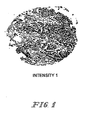
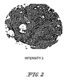
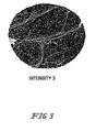
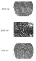
- the staining for folate receptor expression was scored 0 to 3. Absence of discernible staining was scored 0. A score of 1 represented weak finely granular staining. A score of 2 reflected a coarser granular staining and 3, a strong intense coarsely granular staining. Positive staining, when present, was typically diffuse throughout the tumor. Representative samples of 1+, 2+, and 3+ staining are shown in Figs. 1-3 .
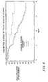
- the average folate receptor staining intensity was 1+ in 11 samples (17%), 2+ in 17 samples (25%), and 3+ in 39 (58%) samples. No sample had all cores negative. None of the 11 women with 1+ staining has experienced a recurrence. Of the 17 women with 2+ staining samples, five (29.4%) recurred. In the group with 3+ staining, 28 of the 39 (71.8%) recurred at a median of 2.5 years. The median time to recurrence for the 1+ and 2+ staining groups is greater than four years, as shown in Fig. 4 . Figs. 1-3 show examples of 1+, 2+, and 3+ staining. Folate receptor staining in normal tissue controls was negative.
- folate receptor staining remained significantly associated with poor outcome, p ⁇ 0.001.
- Strong folate receptor positivity was the most significant prognostic factor in this cohort of patients with breast cancer. Accordingly, there is a strong correlation between overexpression of the folate receptor and early recurrence of breast cancer.
- the assay described herein may be used to determine a prognosis for a patient with cancer and to determine a treatment regimen based on that prognosis. Table 2.
- the immunohistochemical staining assay can be performed by any immunohistochemical staining procedure known in the art. Another illustrative procedure is described below. Deparaffinization and Rehydration of Tissue Sections
- the slides were placed in 3 successive baths of xylene for 3 minutes in each bath. Excess liquid was tapped off and the slides were placed in 3 successive baths of 100% ethanol for 15 dips in each bath. Excess liquid was tapped off and the slides were placed in 3 successive baths of 95% ethanol for 15 dips in each bath. The excess liquid was tapped off and the slides were placed in deionized water for 3 changes for 30 seconds each.
- the slides were placed in a staining dish filled with 250 mL of BORG Decloaker reagent.
- the staining dish was placed in a Decloaking chamber (BioCare Medical, Walnut Creek, CA) for 3 minutes at a pressure of about 17-25 psi and a temperature of about 120°C.
- the slides were cooled for 10 minutes.
- the slides were rinsed briefly in DI water. The excess water was tapped off with an absorbent wipe. The slides were rinsed again in DI water for 1 minute and excess water was tapped off onto an absorbent wipe. Enough ready-to-use Peroxidase Block from the DAKO EnvisonPlus Kit to cover specimen was applied. the slides were incubated for 5 +/- 1 minutes at room temperature. The solutions were drained from the slides. The slides were washed three times in a PBS bath for 3 minutes +/- 30 seconds each.
- the specimens were incubated for 30 +/-1 minutes at room temperature in a humid chamber.
- the slides were rinsed gently with PBS, flowing in a direction from isotype control to test article.
- the slides were washed three times in a PBS bath for 3 minutes +/- 30 seconds each wash.
- the positive control tissue should be examined first to ascertain that all reagents are functioning properly. Presence of a brown-colored end-product at the site of the target antigen is indicative of positive reactivity. If the positive control tissue fails to demonstrate positive staining, results with the test specimens should be considered invalid.
- the negative control tissue should be examined after the positive control tissue to verify the specific labeling of the target antigen by the primary antibody.
- the absence of specific staining in the negative control tissue confirms the lack of antibody cross-reactivity to cells/cellular components. If specific staining occurs in the negative control tissue, results with the test specimen should be considered invalid.
- Nonspecific staining if present, will be of a diffuse appearance. Sporadic light staining of connective tissue may be observed in sections from excessively formalin-fixed tissues. Necrotic or degenerated cells often stain nonspecifically.
- Test specimens stained with the primary antibody should be examined last. Positive staining intensity should be assessed within the context of any nonspecific background staining of the negative control reagent. The absence of a specific positive staining reaction can be interpreted as no antigen detected.
- the staining intensity of the test article is judged relative to the intensity of a control slide containing an adjacent section stained with a negative control antibody. Staining of the section labeled with the negative reagent control was considered “background.” "0” indicates no staining relative to isotype background staining. "1+” indicates weak reactivity, seen as faint or light brown staining. 1+ staining is usually not visible at low magnifications by microscopic examination. "2+” indicates moderate reactivity, seen as shades of brown staining of intermediate darkness (intensity). 2+ staining may be visible, but not prominent, at low magnifications by microscopic examination. "3+” indicates strong reactivity, seen as dark brown to black staining.
- Formalin-fixed, paraffin-embedded sections of cancer tissue will be prepared and will be treated chemically and enzymatically to digest proteins.
- the sections will then be heated at 75°C in the presence of 20 X SSC and formamide to convert DNA from double-stranded DNA to single-strand DNA.
- the section will then be contacted with a hybridization solution, containing a fluorescently-labeled DNA probe which is complementary to a nucleic acid that encodes the vitamin receptor or to a nucleic acid that is complementary to the nucleic acid that encodes the vitamin receptor.
- the sections will then be incubated under conditions favorable for hybridization.
- the sections will be washed in a mixture of 20 X SSC and formamide.
- the hybridized probe will be detected using a fluorescently-tagged ligand (e.g., fluorescein-labeled avidin) which binds to biotin linked to the DNA probe.
- a fluorescently-tagged ligand e.g., fluorescein-labeled avidin
- the nuclear DNA will then be counterstained with an intercalating fluorescent dye (e.g., DAPI in Antifade).
- An epifluorescence microscope will be used for detection of fluorescence and emission of green (fluorescein) and blue light (DAPI) will result. Nuclei in the tissue section will be scored for the number of green signals on a blue background. This protocol is described in more detail in U.S. Patent No. 6,358,682 , incorporated herein by reference.
- Tissue specimens from cancer patients e.g., ovarian cancer patients or breast cancer patients
- All sample preparation procedures will be performed at 4°C and will be performed by a published procedure ( parer et al, Anal. Biochem. (2005 ), incorporated herein by reference).
- Tissue samples were homogenized in homogenization buffer (10 mM Tris, pH 8.0, 0.02 mg/ml each of leupeptin and aprotinin; 1 ml buffer/50 mg tissue) using a PowerGen 125 homogenizer (Fisher Scientific). Large debris was removed by mild centrifugation (3000 x g for 15 minutes).
- homogenization buffer 10 mM Tris, pH 8.0, 0.02 mg/ml each of leupeptin and aprotinin; 1 ml buffer/50 mg tissue
- Membrane pellets were collected by centrifugation at 40,000 x g for 60 minutes and resuspended in solubilization buffer (50 mM Tris, pH 7.4, 150 mM NaCl, 2 mM n -octyl- ⁇ -D-glucopyranoside, 5 mM EDTA, and 0.02% sodium azide). Insoluble material was removed by a second 40,000 x g 60 minute centrifugation, and the total protein concentration of the supernatants was determined by the bicinchoninic acid (BCA) method (Pierce Chemical).
- solubilization buffer 50 mM Tris, pH 7.4, 150 mM NaCl, 2 mM n -octyl- ⁇ -D-glucopyranoside, 5 mM EDTA, and 0.02% sodium azide.
- Insoluble material was removed by a second 40,000 x g 60 minute centrifugation, and the total protein concentration of the supernatants was determined by the
- Acetate buffer (55 ⁇ l of 30 mM acetic acid, pH 3.0, 150 mM NaCl) was added to each microconcentrator, followed by a centrifugation step. Next, 55 ⁇ l of phosphate-buffered saline (PBS) was dispensed into each microconentrator, followed by another centrifugation.
- PBS phosphate-buffered saline
- [ 3 H]-folic acid binding reagent 120 nM [ 3 H]-folic acid (Amersham) in 10 mM Na 2 PO 4 , 1.8 mM KH 2 PO 4 , pH 7.4, containing 500 mM NaCl, 2.7 mM KCl and 25 mM n -octyl- ⁇ -D-glucopyranoside) or 50 ⁇ L of a competing reagent (binding reagent plus 120 ⁇ M unlabeled folic acid) were added to the appropriate concentrators.
- the concentrators were washed/centrifuged 3 times with 75 ⁇ L of 50 mM n -octyl- ⁇ -D-glucopyranoside, 0.7 M NaCl in PBS, pH 7.4.
- the retentates containing the solubilized folate receptors were recovered from the membrane surface of the microconcentrators by 2 rinses with 100 ⁇ L of PBS containing 4% Triton-X 100 ® .
- the samples were then counted in a liquid scintillation counter (Packard Bioscience Co.). CPM values were converted to picomole of folate receptor based upon the CPM of a known standard, and the final results were normalized with respect to the sample protein content.
- the folate receptor assay procedure outlined above was followed with a few modifications.
- concentration dependence assay a human B-cell lymphoma tissue known to express elevated folate receptor levels was chosen for the analysis, and 50 ⁇ g of total membrane-derived protein were assayed per microconcentrator.
- Five [ 3 H]folic acid binding reagent concentrations (5, 15, 30, 60, and 120 nM) were tested in the presence and absence of a 1000-fold excess of unlabeled folic acid. Using this assay saturation was achieved.
- the assay was also shown to exhibit linearity.
- linearity assay 14 24, 34, and 45 ⁇ g of total membrane-derived protein from a human ovarian papillary serous cystadenocarcinoma tissue specimen were analyzed using the 120 nM [ 3 H]folic acid binding reagent solution. Protein concentrations were determined by the BCA Protein Assay (Pierce).
- Sections were rinsed with TBST Wash Buffer.
- CSAD Biotin-free Tyramide Signal Amplification System (DAKO Cytomation, Carpinteria, CA) was applied and was allowed to incubate for 15 minutes.
- the slides were rinsed with TBST Wash Buffer.
- Sections were then incubated in diaminobenzidine (DAB+) solution (DAKO Cytomation, Carpinteria,CA) for 5 minutes, counterstained with Modified Schmidt's Hematoxylin for 5 minutes, blued in running tap water for 3 minutes, and were mounted and coverslipped.
- DAB+ diaminobenzidine
- Modified Schmidt's Hematoxylin for 5 minutes, blued in running tap water for 3 minutes, and were mounted and coverslipped.
- the majority of invasive colon, lung, ovary, and endometrial tumors were moderately (2+) to strongly positive (3+) when immunolabeled with the 343 monoclonal antibody against the folate receptor alpha.
- the sections were then incubated for 5 minutes with the Peroxidase Blocking reagent followed by a 5 minute incubation with a Protein Block reagent, both reagents were included in the CSAll kit (DAKO Cytomation, Cat # K1497).
- the sections were incubated for 30 minutes with mouse 343 monoclonal antibody (10 ⁇ g/ml) in Background Reducing Diluent (DAKO Cytomation, Cat # S3022).
- the negative control sections were incubated with either non-immune mouse IgG 1 (DAKO Cytomation, Cat # X0931) at 10 ⁇ g/ml or Background Reducing Diluent ('no primary' control).
- the sections were then rinsed (3 x 5 minutes) in TBST, incubated with anti-mouse immunoglobulins-HRP for 15 minutes, rinsed in TBST (3 x 5 minutes) and then incubated in the dark with amplification reagent (fluorescyl-tyramide/HRP) for 15 minutes at room temperature.
- amplification reagent fluorescyl-tyramide/HRP
- the sections were rinsed in TBST (3 x 5 minutes) and then incubated with anti-fluorescein-HRP for 15 minutes at room temperature.
- diaminobenzidine for 3 minutes. All reagents used during the antibody amplification and visualization steps were supplied as part of the CSA II kit.
- the invention also provides the following preferred embodiments.
Landscapes
- Health & Medical Sciences (AREA)
- Life Sciences & Earth Sciences (AREA)
- Immunology (AREA)
- Cell Biology (AREA)
- Engineering & Computer Science (AREA)
- Urology & Nephrology (AREA)
- Molecular Biology (AREA)
- Biomedical Technology (AREA)
- Chemical & Material Sciences (AREA)
- Hematology (AREA)
- Oncology (AREA)
- Physics & Mathematics (AREA)
- Biochemistry (AREA)
- Biotechnology (AREA)
- Food Science & Technology (AREA)
- Medicinal Chemistry (AREA)
- Hospice & Palliative Care (AREA)
- Analytical Chemistry (AREA)
- Microbiology (AREA)
- General Health & Medical Sciences (AREA)
- General Physics & Mathematics (AREA)
- Pathology (AREA)
- Investigating Or Analysing Biological Materials (AREA)
- Measuring Or Testing Involving Enzymes Or Micro-Organisms (AREA)
- Pharmaceuticals Containing Other Organic And Inorganic Compounds (AREA)
- Organic Low-Molecular-Weight Compounds And Preparation Thereof (AREA)
- Peptides Or Proteins (AREA)
Applications Claiming Priority (2)
| Application Number | Priority Date | Filing Date | Title |
|---|---|---|---|
| US66643005P | 2005-03-30 | 2005-03-30 | |
| EP06739883A EP1864133B1 (fr) | 2005-03-30 | 2006-03-30 | Methode destinee a pronostiquer un cancer du sein par quantification des recepteurs des folates cellulaires |
Related Parent Applications (1)
| Application Number | Title | Priority Date | Filing Date |
|---|---|---|---|
| EP06739883.4 Division | 2006-03-30 |
Publications (1)
| Publication Number | Publication Date |
|---|---|
| EP2199796A1 true EP2199796A1 (fr) | 2010-06-23 |
Family
ID=36754569
Family Applications (2)
| Application Number | Title | Priority Date | Filing Date |
|---|---|---|---|
| EP10155913A Withdrawn EP2199796A1 (fr) | 2005-03-30 | 2006-03-30 | Méthode de prognose du cancer avec la quantification du niveau de récepteurs folate |
| EP06739883A Not-in-force EP1864133B1 (fr) | 2005-03-30 | 2006-03-30 | Methode destinee a pronostiquer un cancer du sein par quantification des recepteurs des folates cellulaires |
Family Applications After (1)
| Application Number | Title | Priority Date | Filing Date |
|---|---|---|---|
| EP06739883A Not-in-force EP1864133B1 (fr) | 2005-03-30 | 2006-03-30 | Methode destinee a pronostiquer un cancer du sein par quantification des recepteurs des folates cellulaires |
Country Status (10)
| Country | Link |
|---|---|
| US (1) | US20090081710A1 (fr) |
| EP (2) | EP2199796A1 (fr) |
| JP (1) | JP2008537778A (fr) |
| CN (1) | CN101203759A (fr) |
| AT (1) | ATE460668T1 (fr) |
| AU (1) | AU2006230219A1 (fr) |
| CA (1) | CA2602585A1 (fr) |
| DE (1) | DE602006012816D1 (fr) |
| RU (1) | RU2007139912A (fr) |
| WO (1) | WO2006105141A1 (fr) |
Cited By (2)
| Publication number | Priority date | Publication date | Assignee | Title |
|---|---|---|---|---|
| WO2013012722A1 (fr) | 2011-07-15 | 2013-01-24 | Eisai R&D Management Co., Ltd. | Anticorps anti-récepteurs alpha du folate et leurs utilisations |
| WO2018098277A1 (fr) | 2016-11-23 | 2018-05-31 | Eisai R&D Management Co., Ltd | Anticorps anti-récepteurs alpha du folate et leurs utilisations |
Families Citing this family (31)
| Publication number | Priority date | Publication date | Assignee | Title |
|---|---|---|---|---|
| ATE427948T1 (de) | 2001-04-24 | 2009-04-15 | Purdue Research Foundation | Folat-mimetika und deren folatrezeptorbindende konjugate |
| EP2060272A3 (fr) | 2002-05-15 | 2009-05-27 | Endocyte, Inc. | Conjugués vitamine-mitomycine |
| DK1592457T3 (da) | 2003-01-27 | 2012-10-22 | Endocyte Inc | Folat-vinblastin-konjugat som lægemiddel |
| CA2556027C (fr) | 2004-02-12 | 2015-11-24 | Morphotek, Inc. | Anticorps monoclonaux qui bloquent specifiquement l'activite biologique d'un antigene tumoral |
| CN101098854B (zh) | 2004-07-23 | 2012-12-05 | 恩多塞特公司 | 二价连接体及其轭合物 |
| JP5289935B2 (ja) | 2005-03-16 | 2013-09-11 | エンドサイト,インコーポレイテッド | プテロイン酸およびその結合体の合成と精製 |
| US20060239910A1 (en) | 2005-04-22 | 2006-10-26 | Morphotek Inc. | Antibodies with immune effector activity and that internalize in folate receptor alpha-positive cells |
| RU2470668C2 (ru) | 2005-08-19 | 2012-12-27 | Эндосайт, Инк. | Конъюгаты лиганда с несколькими лекарственными средствами |
| US20100104626A1 (en) | 2007-02-16 | 2010-04-29 | Endocyte, Inc. | Methods and compositions for treating and diagnosing kidney disease |
| CN101678124A (zh) | 2007-03-14 | 2010-03-24 | 恩多塞特公司 | 结合配体连接的微管溶素递药缀合物 |
| JP5690589B2 (ja) | 2007-06-25 | 2015-03-25 | エンドサイト・インコーポレイテッドEndocyte, Inc. | 親水性スペーサーリンカーを含有する結合体 |
| US9877965B2 (en) | 2007-06-25 | 2018-01-30 | Endocyte, Inc. | Vitamin receptor drug delivery conjugates for treating inflammation |
| EP2209374B1 (fr) | 2007-10-25 | 2014-12-03 | Endocyte, Inc. | Tubulysines et leurs procédés de préparation |
| JP2013501224A (ja) * | 2009-07-31 | 2013-01-10 | エンドサイト,インク. | 葉酸を標的とした診断及び処置 |
| TWI504408B (zh) | 2010-02-24 | 2015-10-21 | Immunogen Inc | 葉酸受體1抗體類和免疫共軛物類及彼等之用途 |
| JP6224456B2 (ja) * | 2010-11-05 | 2017-11-01 | モルフォテック、インク. | 葉酸受容体アルファ発現癌の診断および予後マーカーとしての葉酸受容体アルファ |
| BR112013011590A2 (pt) * | 2010-11-12 | 2019-09-24 | Endocyte Inc | método de tratamento de câncer |
| KR102101160B1 (ko) * | 2011-04-01 | 2020-04-16 | 이뮤노젠 아이엔씨 | Folr1 암 치료의 효능을 증가시키기 위한 방법 |
| WO2013126797A1 (fr) | 2012-02-24 | 2013-08-29 | Purdue Research Foundation | Ciblage du récepteur de la cholécystokinine de type b pour imagerie et thérapie |
| US20140080175A1 (en) | 2012-03-29 | 2014-03-20 | Endocyte, Inc. | Processes for preparing tubulysin derivatives and conjugates thereof |
| SI2890717T1 (sl) | 2012-08-31 | 2020-07-31 | Immunogen, Inc. | Diagnostične preiskave in kompleti za odkrivanje folatnega receptorja 1 |
| CN102861020A (zh) * | 2012-09-25 | 2013-01-09 | 上海交通大学医学院附属仁济医院 | 叶酸在制备预防大肠腺瘤初发药物中的应用 |
| EP2908818A4 (fr) | 2012-10-16 | 2016-07-13 | Endocyte Inc | Conjugués d'administration de médicament contenant des acides aminés artificiels et procédés d'utilisation |
| US20140154702A1 (en) * | 2012-11-30 | 2014-06-05 | Endocyte, Inc. | Methods For Treating Cancer Using Combination Therapies |
| SI3038650T1 (sl) | 2013-08-30 | 2021-11-30 | Immunogen, Inc. | Protitelesa in preiskave za odkrivanje folatnega receptorja 1 |
| SG11201602361UA (en) * | 2013-10-08 | 2016-04-28 | Immunogen Inc | Anti-folr1 immunoconjugate dosing regimens |
| WO2015077303A1 (fr) | 2013-11-19 | 2015-05-28 | Purdue Research Foundation | Méthode de sélection de patient pour le traitement de l'inflammation |
| JP6246030B2 (ja) * | 2014-03-12 | 2017-12-13 | 東芝メディカルシステムズ株式会社 | 病理染色装置及び病理染色方法 |
| CN108601828B (zh) | 2015-09-17 | 2023-04-28 | 伊缪诺金公司 | 包含抗folr1免疫缀合物的治疗组合 |
| WO2017218632A1 (fr) * | 2016-06-14 | 2017-12-21 | Board Of Regents, The University Of Texas System | Procédés de détection d'anticorps anti-acide folique et leurs utilisations |
| CN106483282B (zh) * | 2016-09-29 | 2018-08-31 | 北京世纪沃德生物科技有限公司 | 一种抗原稳定剂及其制备方法与应用 |
Citations (9)
| Publication number | Priority date | Publication date | Assignee | Title |
|---|---|---|---|---|
| US5846739A (en) | 1995-12-05 | 1998-12-08 | Wisconsin Alumni Research Foundation | Immunohistochemical detection assay for carcinoma proliferative status |
| US5989838A (en) | 1992-03-11 | 1999-11-23 | Institute Of Virology, Slovak Academy Of Sciences | Immunological methods of detecting MN proteins and MN polypeptides |
| US6218529B1 (en) | 1995-07-31 | 2001-04-17 | Urocor, Inc. | Biomarkers and targets for diagnosis, prognosis and management of prostate, breast and bladder cancer |
| US20010031252A1 (en) | 2000-03-31 | 2001-10-18 | Low Philip Stewart | Method of treatment using ligand-immunogen conjugates |
| US6358682B1 (en) | 1998-01-26 | 2002-03-19 | Ventana Medical Systems, Inc. | Method and kit for the prognostication of breast cancer |
| US20030086900A1 (en) | 2001-09-28 | 2003-05-08 | Low Philip S. | Method of treatment using ligand-immunogen conjugates |
| US20030198643A1 (en) | 2002-04-19 | 2003-10-23 | Yingjuan Lu | Adjuvant enhanced immunotherapy |
| WO2003097647A1 (fr) | 2002-05-15 | 2003-11-27 | Endocyte, Inc. | Conjugues vitamine-mitomycine |
| US20050002942A1 (en) | 2003-01-27 | 2005-01-06 | Vlahov Iontcho R. | Vitamin receptor binding drug delivery conjugates |
-
2006
- 2006-03-30 JP JP2008504275A patent/JP2008537778A/ja not_active Withdrawn
- 2006-03-30 DE DE602006012816T patent/DE602006012816D1/de active Active
- 2006-03-30 AT AT06739883T patent/ATE460668T1/de not_active IP Right Cessation
- 2006-03-30 EP EP10155913A patent/EP2199796A1/fr not_active Withdrawn
- 2006-03-30 US US11/887,438 patent/US20090081710A1/en not_active Abandoned
- 2006-03-30 CA CA002602585A patent/CA2602585A1/fr not_active Abandoned
- 2006-03-30 RU RU2007139912/15A patent/RU2007139912A/ru unknown
- 2006-03-30 AU AU2006230219A patent/AU2006230219A1/en not_active Abandoned
- 2006-03-30 CN CNA2006800192306A patent/CN101203759A/zh active Pending
- 2006-03-30 WO PCT/US2006/011376 patent/WO2006105141A1/fr active Application Filing
- 2006-03-30 EP EP06739883A patent/EP1864133B1/fr not_active Not-in-force
Patent Citations (9)
| Publication number | Priority date | Publication date | Assignee | Title |
|---|---|---|---|---|
| US5989838A (en) | 1992-03-11 | 1999-11-23 | Institute Of Virology, Slovak Academy Of Sciences | Immunological methods of detecting MN proteins and MN polypeptides |
| US6218529B1 (en) | 1995-07-31 | 2001-04-17 | Urocor, Inc. | Biomarkers and targets for diagnosis, prognosis and management of prostate, breast and bladder cancer |
| US5846739A (en) | 1995-12-05 | 1998-12-08 | Wisconsin Alumni Research Foundation | Immunohistochemical detection assay for carcinoma proliferative status |
| US6358682B1 (en) | 1998-01-26 | 2002-03-19 | Ventana Medical Systems, Inc. | Method and kit for the prognostication of breast cancer |
| US20010031252A1 (en) | 2000-03-31 | 2001-10-18 | Low Philip Stewart | Method of treatment using ligand-immunogen conjugates |
| US20030086900A1 (en) | 2001-09-28 | 2003-05-08 | Low Philip S. | Method of treatment using ligand-immunogen conjugates |
| US20030198643A1 (en) | 2002-04-19 | 2003-10-23 | Yingjuan Lu | Adjuvant enhanced immunotherapy |
| WO2003097647A1 (fr) | 2002-05-15 | 2003-11-27 | Endocyte, Inc. | Conjugues vitamine-mitomycine |
| US20050002942A1 (en) | 2003-01-27 | 2005-01-06 | Vlahov Iontcho R. | Vitamin receptor binding drug delivery conjugates |
Non-Patent Citations (24)
| Title |
|---|
| "Diagnostic Molecular Pathology", vol. 1, IRL PRESS |
| "Molecular Cloning", 2001 |
| BRODERS, A. C., JAMA, vol. 74, 1920, pages 656 - 664 |
| BRODERS, A.C., SURG. CLIN. NORTH AMERICA, vol. 21, 1941, pages 947 - 62 |
| BUENO ET AL., J. THOR. CARD. SUR. FEB., vol. 121, 2001, pages 225 - 233 |
| BUENO ET AL., THOR. CARD. SUR. FEB., vol. 121, 2001, pages 225 - 233 |
| CERNIGOI C ET AL: "Folate binding protein as a marker for survival in ovarian cancer patients and a predictor of response to chemotherapy", CELL PROLIFERATION 1997 UNITED KINGDOM, vol. 30, no. 10-12, 1997, pages 468, XP009070977, ISSN: 0960-7722 * |
| CONEY ET AL., CANCER RESEARCH, vol. 51, 1991, pages 6125 - 6132 |
| FRANKLIN ET AL., INT. J. CANCER SUPPL., vol. 8, 1994, pages 89 - 95 |
| GARIN-CHESA ET AL., AM. 1. PATHOL., vol. 142, no. 2, 1993, pages 557 - 67 |
| GARIN-CHESA ET AL., AM. J. PATHOL., vol. 142, no. 2, 1993, pages 557 - 67 |
| HOLM JAN ET AL: "Folate receptor of human mammary adenocarcinoma", APMIS, vol. 102, no. 6, 1994, pages 413 - 419, XP009070950, ISSN: 0903-4641 * |
| LI ET AL., J. NUC. MED., vol. 37, 1996, pages 665 - 672 |
| LI ET AL., J: NUC. MED., vol. 37, 1996, pages 665 - 672 |
| PARKER ET AL., ANAL BIOCHEM., 2005 |
| PARKER ET AL., ANAL. BIOCHEM., 2005 |
| PARKER N ET AL: "Folate receptor expression in carcinomas and normal tissues determined by a quantitative radioligand binding assay", ANALYTICAL BIOCHEMISTRY, ACADEMIC PRESS, NEW YORK, NY, US, vol. 338, no. 2, 15 March 2005 (2005-03-15), pages 284 - 293, XP004770829, ISSN: 0003-2697 * |
| ROSS ET AL., CANCER, vol. 73, 1994, pages 2432 - 2443 |
| TOFFOLI GIUSEPPE ET AL: "Expression of folate binding protein as a prognostic factor for response to platinum-containing chemotherapy and survival in human ovarian cancer", INTERNATIONAL JOURNAL OF CANCER, vol. 79, no. 2, 17 April 1998 (1998-04-17), pages 121 - 126, XP002394383, ISSN: 0020-7136 * |
| TOFFOLI GIUSEPPE ET AL: "Prognostic significance of folate binding protein in ovarian neoplasms", TUMORI, vol. 85, no. 3 SUPPL. 1, May 1999 (1999-05-01), pages S39 - S41, XP009070976, ISSN: 0300-8916 * |
| TOFFOLI, INT. J. CANCER, vol. 74, 1997, pages 193 - 198 |
| VEGGIAN, TUMOR, vol. 75, 1989, pages 510 - 51 3 |
| VEGGIAN, TUMOR, vol. 75, 1989, pages 510 - 513 |
| WEITMAN ET AL., CANCER RESEARCH, vol. 52, pages 3396 - 3401 |
Cited By (5)
| Publication number | Priority date | Publication date | Assignee | Title |
|---|---|---|---|---|
| WO2013012722A1 (fr) | 2011-07-15 | 2013-01-24 | Eisai R&D Management Co., Ltd. | Anticorps anti-récepteurs alpha du folate et leurs utilisations |
| EP3330291A1 (fr) | 2011-07-15 | 2018-06-06 | Eisai R&D Management Co., Ltd. | Anticorps anti-récepteurs alpha du folate et leurs utilisations |
| US10101343B2 (en) | 2011-07-15 | 2018-10-16 | Eisai R&D Management Co., Ltd. | Anti-folate receptor alpha antibodies and uses thereof |
| WO2018098277A1 (fr) | 2016-11-23 | 2018-05-31 | Eisai R&D Management Co., Ltd | Anticorps anti-récepteurs alpha du folate et leurs utilisations |
| US10822410B2 (en) | 2016-11-23 | 2020-11-03 | Eisai R&D Management Co., Ltd. | Anti-folate receptor alpha antibodies and uses thereof |
Also Published As
| Publication number | Publication date |
|---|---|
| CA2602585A1 (fr) | 2006-10-05 |
| EP1864133A1 (fr) | 2007-12-12 |
| RU2007139912A (ru) | 2009-05-10 |
| DE602006012816D1 (de) | 2010-04-22 |
| JP2008537778A (ja) | 2008-09-25 |
| WO2006105141A1 (fr) | 2006-10-05 |
| AU2006230219A1 (en) | 2006-10-05 |
| EP1864133B1 (fr) | 2010-03-10 |
| US20090081710A1 (en) | 2009-03-26 |
| ATE460668T1 (de) | 2010-03-15 |
| CN101203759A (zh) | 2008-06-18 |
Similar Documents
| Publication | Publication Date | Title |
|---|---|---|
| EP1864133B1 (fr) | Methode destinee a pronostiquer un cancer du sein par quantification des recepteurs des folates cellulaires | |
| US10273308B2 (en) | Methods of producing antibodies specific for p95 | |
| JP2012177706A (ja) | 個別化抗癌化学療法(pac)のための包括的な診断試験 | |
| EP3283886B1 (fr) | Procédés de traitement du cancer du poumon | |
| US9297812B2 (en) | Means and methods for diagnosing cancer using an antibody which specifically binds to BRAF V600E | |
| Pothos et al. | Comparison of chromogenic in situ hybridisation with fluorescence in situ hybridisation and immunohistochemistry for the assessment of her-2/neu oncogene in archival material of breast carcinoma | |
| US20160311921A1 (en) | Methods for treatment of ovarian cancer | |
| EP1470239B1 (fr) | Expression de la psoriasine par les cellules epitheliales mammaires | |
| EP2707720A2 (fr) | Diagnostic et pronostic de cancer du sein triple-négatif et de l'ovaire | |
| US20080227098A1 (en) | Method for diagnosing cancer of the prostate | |
| US20140148353A1 (en) | Protein expression-based classifier for prediction of recurrence in adenocarcinoma | |
| US20140030254A1 (en) | Signal pathway alterations and drug target elevations in primary metachronous metastatic colorectal cancer compared to non-metastatic disease | |
| WO2016146654A1 (fr) | Matériels et méthodes permettant de détecter des variants d'épissage du récepteur des androgènes et leurs utilisations |
Legal Events
| Date | Code | Title | Description |
|---|---|---|---|
| PUAI | Public reference made under article 153(3) epc to a published international application that has entered the european phase |
Free format text: ORIGINAL CODE: 0009012 |
|
| AC | Divisional application: reference to earlier application |
Ref document number: 1864133 Country of ref document: EP Kind code of ref document: P |
|
| AK | Designated contracting states |
Kind code of ref document: A1 Designated state(s): AT BE BG CH CY CZ DE DK EE ES FI FR GB GR HU IE IS IT LI LT LU LV MC NL PL PT RO SE SI SK TR |
|
| STAA | Information on the status of an ep patent application or granted ep patent |
Free format text: STATUS: THE APPLICATION IS DEEMED TO BE WITHDRAWN |
|
| 18D | Application deemed to be withdrawn |
Effective date: 20101227 |