EP2149332B1 - Image information display processing device and display processing method - Google Patents
Image information display processing device and display processing method Download PDFInfo
- Publication number
- EP2149332B1 EP2149332B1 EP08751662.1A EP08751662A EP2149332B1 EP 2149332 B1 EP2149332 B1 EP 2149332B1 EP 08751662 A EP08751662 A EP 08751662A EP 2149332 B1 EP2149332 B1 EP 2149332B1
- Authority
- EP
- European Patent Office
- Prior art keywords
- image
- mark
- display
- displayed
- bar
- Prior art date
- Legal status (The legal status is an assumption and is not a legal conclusion. Google has not performed a legal analysis and makes no representation as to the accuracy of the status listed.)
- Not-in-force
Links
- 238000003672 processing method Methods 0.000 title description 3
- 230000003902 lesion Effects 0.000 claims description 9
- 230000004044 response Effects 0.000 claims description 3
- 239000002775 capsule Substances 0.000 description 46
- 238000007689 inspection Methods 0.000 description 10
- 230000006870 function Effects 0.000 description 9
- 210000000056 organ Anatomy 0.000 description 7
- 238000003745 diagnosis Methods 0.000 description 6
- 230000000740 bleeding effect Effects 0.000 description 4
- 241000167880 Hirundinidae Species 0.000 description 3
- 238000005516 engineering process Methods 0.000 description 3
- 238000001727 in vivo Methods 0.000 description 3
- 238000000034 method Methods 0.000 description 3
- 230000008859 change Effects 0.000 description 2
- 210000001035 gastrointestinal tract Anatomy 0.000 description 2
- 210000002429 large intestine Anatomy 0.000 description 2
- 230000008569 process Effects 0.000 description 2
- 210000000813 small intestine Anatomy 0.000 description 2
- 230000007480 spreading Effects 0.000 description 2
- 210000002784 stomach Anatomy 0.000 description 2
- UFHFLCQGNIYNRP-UHFFFAOYSA-N Hydrogen Chemical compound [H][H] UFHFLCQGNIYNRP-UHFFFAOYSA-N 0.000 description 1
- 230000002457 bidirectional effect Effects 0.000 description 1
- 238000004891 communication Methods 0.000 description 1
- 238000001514 detection method Methods 0.000 description 1
- 210000003238 esophagus Anatomy 0.000 description 1
- 229910052739 hydrogen Inorganic materials 0.000 description 1
- 239000001257 hydrogen Substances 0.000 description 1
- 238000003384 imaging method Methods 0.000 description 1
- 229910001416 lithium ion Inorganic materials 0.000 description 1
- 238000013341 scale-up Methods 0.000 description 1
Images


Classifications
-
- A—HUMAN NECESSITIES
- A61—MEDICAL OR VETERINARY SCIENCE; HYGIENE
- A61B—DIAGNOSIS; SURGERY; IDENTIFICATION
- A61B1/00—Instruments for performing medical examinations of the interior of cavities or tubes of the body by visual or photographical inspection, e.g. endoscopes; Illuminating arrangements therefor
- A61B1/00002—Operational features of endoscopes
- A61B1/00043—Operational features of endoscopes provided with output arrangements
- A61B1/00045—Display arrangement
- A61B1/0005—Display arrangement combining images e.g. side-by-side, superimposed or tiled
-
- A—HUMAN NECESSITIES
- A61—MEDICAL OR VETERINARY SCIENCE; HYGIENE
- A61B—DIAGNOSIS; SURGERY; IDENTIFICATION
- A61B1/00—Instruments for performing medical examinations of the interior of cavities or tubes of the body by visual or photographical inspection, e.g. endoscopes; Illuminating arrangements therefor
- A61B1/00002—Operational features of endoscopes
- A61B1/00043—Operational features of endoscopes provided with output arrangements
- A61B1/00045—Display arrangement
-
- A—HUMAN NECESSITIES
- A61—MEDICAL OR VETERINARY SCIENCE; HYGIENE
- A61B—DIAGNOSIS; SURGERY; IDENTIFICATION
- A61B1/00—Instruments for performing medical examinations of the interior of cavities or tubes of the body by visual or photographical inspection, e.g. endoscopes; Illuminating arrangements therefor
- A61B1/04—Instruments for performing medical examinations of the interior of cavities or tubes of the body by visual or photographical inspection, e.g. endoscopes; Illuminating arrangements therefor combined with photographic or television appliances
- A61B1/041—Capsule endoscopes for imaging
-
- G—PHYSICS
- G06—COMPUTING; CALCULATING OR COUNTING
- G06T—IMAGE DATA PROCESSING OR GENERATION, IN GENERAL
- G06T11/00—2D [Two Dimensional] image generation
- G06T11/20—Drawing from basic elements, e.g. lines or circles
- G06T11/206—Drawing of charts or graphs
-
- G—PHYSICS
- G11—INFORMATION STORAGE
- G11B—INFORMATION STORAGE BASED ON RELATIVE MOVEMENT BETWEEN RECORD CARRIER AND TRANSDUCER
- G11B27/00—Editing; Indexing; Addressing; Timing or synchronising; Monitoring; Measuring tape travel
- G11B27/10—Indexing; Addressing; Timing or synchronising; Measuring tape travel
- G11B27/102—Programmed access in sequence to addressed parts of tracks of operating record carriers
- G11B27/105—Programmed access in sequence to addressed parts of tracks of operating record carriers of operating discs
-
- G—PHYSICS
- G11—INFORMATION STORAGE
- G11B—INFORMATION STORAGE BASED ON RELATIVE MOVEMENT BETWEEN RECORD CARRIER AND TRANSDUCER
- G11B27/00—Editing; Indexing; Addressing; Timing or synchronising; Monitoring; Measuring tape travel
- G11B27/10—Indexing; Addressing; Timing or synchronising; Measuring tape travel
- G11B27/34—Indicating arrangements
Definitions
- the present invention relates to a display processing apparatus and display processing method of image information for displaying plural pieces of image information obtained by capturing images with time by moving, for example, a capsule endoscope with autonomy or heteronomy.
- an endoscope to be swallowed that is, a capsule endoscope
- the specification of the patent document 1 discloses the technology of having an image capturing function and a wireless communication function, sequentially capturing an organ such as a stomach, a small intestine, etc. for an observation period after a patient swallows an endoscope for an observation or an inspection from the mouth until the endoscope is naturally discharged from the human body, and sequentially transmitting image information (electronic data representing an image) by capturing images.
- patent document 2 discloses the technology of receiving the image information transmitted by wireless as described above by a receiver provided outside the patient and accumulating in predetermined memory, and afterwards reading the information as necessary and displaying the information on a display unit, thereby utilizing the information for a diagnosis etc. by a doctor.
- the capsule endoscope unlike a normal endoscope, the period after a patient swallows from his or her mouth until it is naturally discharged is an observation period or an inspection period. Therefore, the observation period or the inspection period can require a long time, for example, ten or more hours, and there are a large number of pieces of image information obtained during the period by capturing image.
- EP 1 618 828 A1 discloses an image display apparatus with the aim of improving the ability to retrieve a taken image of an internal body and of recognizing the organ depicted in each image.
- the apparatus comprises the display of an average colour bar indicating the overall imaging period of images taken in time sequence by a capsule endoscope, an image displayed in an image display field, a movable slider superposed on the average colour bar, and a flag as a bleeding part displayed above and parallel to the average colour bar.
- US 2007/060798 A1 and WO 2005/031650 A1 disclose an in-vivo sensing system with the aim of creating a summarized graphical presentation of a data stream captured in-vivo.
- the system comprises a colour bar serving as the graphical presentation and being a fixed display alongside a streaming display of the in-vivo data stream, and a cursor, icon or other indicator movable along the fixed colour bar and/or a time bar when the data stream is displayed and/or streamed.
- JP 2006-334297 and JP 2006-345929 disclose a similar system, wherein thumbnail images are displayed next to the image bar. The area for the display of observation images remains fixed, although the number of images displayed therein can vary.
- An object of the present invention is to provide a display processing apparatus and a display processing method capable of easily grasping the image information about captured images of a desired position to be checked and affected parts etc. from a large number of pieces of image information, and capable of quickly displaying a large number of pieces of image information on a small display screen.
- the display processing apparatus displays, on a display screen, image information about the video of plural pieces obtained by capturing with time the video in a plurality of positions in the body of a test subject using an image pickup apparatus provided in the body of the test subject.
- the apparatus includes: an image bar generation device adapted to arrange a line expressed in an average color calculated for each piece of video or for plural pieces of video on a display screen in a time series, and adapted to generate an image bar that can be totally displayed as a bar; a mark generation device adapted to generate a mark for displaying a feature determined from video as a feature extracted mark extracted from the video, and a mark display device adapted to display a mark generated by the mark generation device superposed on the image bar or separated in parallel above the image bar, characterized in that the mark display device is further adapted to switch the display of the mark from the display of the mark superposed and displayed on the image bar when an observation image is displayed, into the display of the mark separated in parallel above the image bar in response to a user input and in cooperation with
- a display area of a display screen can be space-saved, thereby reserving a larger observation screen area.
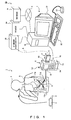
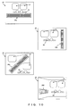
- FIG. 1 shows the capsule endoscope system according to the present invention and the capsule endoscope image filing system included in the capsule endoscope system.
- a capsule endoscope system 1 includes a capsule endoscope 3 contained in a package 2; a patient who swallows the capsule endoscope 3 taken out of the package 2, that is, a test subject 4; a jacket 5 for the test subject 4; and a receiver 6 freely attached to and detached from the jacket 5.
- a capsule endoscope image filing system 20 is configured by a work station 7 for storing and editing image data received by the receiver 6, and a database 9 connected to the work station 7 through a network 8.
- the database 9 can also be built in the work station 7.
- the capsule endoscope 3 contains an image pickup unit, a radio unit and a power supply unit. In the period after the capsule endoscope 3 is swallowed by the test subject 4 for an observation or an inspection from his or her mouth until it is discharged from the body of the test subject, it transmits by wireless the image data obtained by sequentially capturing images of the organs such as the esophagus, the stomach, the small intestine, the large intestine, etc. of the test subject as radio waves from the radio unit to the outside the body of the test subject.
- the jacket 5 clothed in the test subject 4 is provided with a plurality of (four in the example shown in FIG. 1 ) antennas 11 (11a, 11b, 11c, and 11d) for capturing emitted radio waves of image data emitted from the radio unit of the capsule endoscope 3.
- These antennas 11 can communicate with the receiver 6 by wireless or by cable.
- the number of the antennas 11 is not limited to four, but can be appropriately determined. That is, any number appropriate for receiving radio waves emitted depending on the position with respect to the movement of the capsule endoscope 3 is acceptable.
- the receiver 6 is externally provided with an antenna 12 for use when image data is received from the jacket 5 through the antenna 11, a display unit 13 for displaying necessary information for an observation or an inspection, and an input unit 14 for inputting necessary information for an observation or an inspection.
- the power supply unit 15 for providing power also for a portable unit is provided below the receiver 6.
- the power supply unit 15 is configured by, for example, a dry battery, a Li ion secondary battery, a Ni hydrogen battery, etc. (a battery in other formats can be acceptable).
- a signal processing and controlling unit 16 for performing necessary processes for an observation or an inspection is provided, and an attachment unit 18 for attaching CF (CompactFlash (registered trademark)) memory 17 for storing received image data in such a way that the memory can be attached/detached as indicated by the bidirectional arrow a shown in FIG. 1 .
- CF CompactFlash (registered trademark)
- the work station 7 is provided with a body device 19, a monitor device 21 connected to the body device 19, a keyboard 22, a mouse 23, etc.
- the body device 19 is provided with various interfaces in addition to an interface for connection to the network 8 although not shown in FIG. 1 .
- a printer 24 and a CF memory reader/writer 25 are connected to the work station 7 through the interfaces.
- the work station 7 has the image processing function for a doctor or a nurse performing a diagnosis etc. by displaying an image in the gut of the test subject 4 captured by the capsule endoscope 3.
- the doctor or the nurse While the doctor or the nurse are performing an inputting operation on a man-machine interface displayed on the monitor device 21 of the work station 7 using the keyboard 22 or the mouse 23, the doctor or the nurse can issue an instruction to fetch the image data about the inside of the gut of the test subject 4 transmitted from the package 2 and received by the receiver 6.
- data can be directly fetched from the receiver 6, and input data can also be fetched from the CF memory 17 by attaching the CF memory 17 at the CF memory reader/writer 25 as indicated by the arrow b shown in FIG. 1 .
- the doctor or the nurse can issue an instruction to store the captured image screen data fetched from the receiver 6 as described above in the database 9, an instruction to display an image relating to the image data described later on the display screen of the monitor device 21 by calling the image data stored in the database 9, an instruction to record a diagnosis result etc. based on an observation of an image in the database 9, an instruction to print a card etc. on the printer 24, etc.
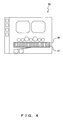
- FIG. 2 shows an example of an image displayed on the display screen of a monitor device of a work station in the capsule endoscope image filing system as the first embodiment not according to the present invention.
- a display screen 30 displays four observation images 31. At the upper left of the display screen 30, a plurality of (four in the example shown in FIG. 2 ) instruction buttons 34 are displayed.
- a reproduction button group 35 including a stop button at the center, a replay button, a fast forward button, and a frame advance button on the right, and the respective inverse buttons on the left.
- a image bar 36 Displayed further below the buttons is a image bar 36, and a mark 37 are superposed on the image bar 36.
- the image bar 36 shows as a vertical line an average color calculated for each captured image or a plurality of captured images at each position on a time axis when the capsule endoscope 3 moves in an organ.
- a vertical line is arranged in a time series horizontally on the display screen, and the entire vertical lines are represented as a horizontal bar.
- the representation of an average color by the image bar 36 is the image bar 36 as a horizontal arrangement on the display screen 30 by arranging a vertical line represented by an average color calculated for each captured image (video) or for a plurality of captured images along a movement time axis of the capsule endoscope 3, that is, in a time series, to roughly know the position of a captured image based on the characteristic of a different color for each organ.
- the mark 37 is displayed by a red line superposed on the image bar 36 at a position corresponding to the time axis of the image bar 36 as a detection position obtained by detecting the characteristic determined from the video on a time axis, for example, red color indicating bleeding. That is, the mark 37 is a type of lesion color mark.
- Such a lesion is not limited to bleeding, but can be another lesion confirmed by a captured image by the capsule endoscope 3.
- These lesions can also be displayed as other color marks than the red color mark such as blue, white, or other color marks.
- thumbnail images are display at the bottom row, or although there are no thumbnail images to be displayed, a lesion color bar and a time axis bar are arranged parallel to the image bar 36. Therefore, at most two observation images 31 can be displayed.
- the mark 37 indicating a lesion color is superposed and displayed on the display of the image bar 36. That is, in the present embodiment, since the information about a lesion etc. is display as a mark in the display area of the image bar 36, the display area of the observation image 31 of the display screen 30 can be expanded, thereby simultaneously displaying four currently selected observation images 31.
- the mark 37 superposed and displayed on the display of the image bar 36 is displayed as red vertical lines (black vertical lines in FIG. 2 ) that are discontinuous vertical stripe markings. However, when a bleeding lesion portion is continuously arranged on the image pickup path of the capsule endoscope 3, the vertical stripe markings appear as continuous markings.
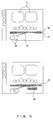
- FIG. 3 shows the state of the marking of a vertical stripe of the mark 37 as another example of the first embodiment continuously spreading as a plane along the superposing display area of an image bar 36.
- FIG. 3 the display portion having the same function as in FIG. 2 is assigned the same reference numeral as in FIG. 2 .
- thumbnail images 39 are display below the mark 37 superposed and displayed on the image bar 36.
- the lower display area can be larger by the mark 37 superposed and displayed on the image bar 36, thereby displaying the thumbnail images 39 larger.
- the bar display area is large, the mark 37 is superposed and displayed on the image bar 36 to allow a user to recognize at a glance the position of an important marking of the mark 37 occurring in a time series of the image bar 36.
- a user of the monitor device 21 has to move the view point up and down on the display screen of the display screen 30 to compare the position of the image bar 36 with the position of the mark 37 and confirm the time position of the mark 37.
- the mark 37 can be displayed as a semitransparent mark superposed and displayed on the image bar 36. Furthermore, the transparence of the semitransparent mark can be appropriately specified.
- the color of the image bar 36 of the portion where the marking of the mark 37 appears can be known, thereby performing an appropriate diagnosis.
- FIG. 4 shows an example of an image displayed on the display screen of a monitor device of a work station in the capsule endoscope image filing system as the second embodiment not according to the present invention.
- FIG. 4 the display portion having the same function as in FIGS. 2 and 3 is assigned the same reference numeral as in FIGS. 2 and 3 .
- the widths of the image bar 36 and the mark 37 superposed and displayed on the image bar 36 are expanded vertically on the screen.
- the vertical line of the mark 37 is elongated with the vertical expansion of the width on the screen of the mark 37.
- FIG. 5 shows an example of an image displayed on the display screen of a monitor device of a work station in the capsule endoscope image filing system as the third embodiment of the present invention
- FIG. 5 the display portion having the same function as in FIGS. 2 through 4 is assigned the same reference numeral as in FIGS. 2 through 4 .
- the mark 37 is arranged above the image bar 36 in parallel.
- the display of the observation image 31 is displaying four images or enlarging and displaying an image
- the display area is automatically narrowed below the 35, and the mark 37 display in parallel above the image bar 36 is superposed and displayed on the image bar 36.
- the mark 37 can be changed to be superposed and displayed on the image bar 36, thereby reserving a broad display area for the observation image 31 without impairing the conditions of a comparing observation between the image bar 36 and the mark 37.
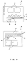
- FIG. 6 shows an example of an image displayed on the display screen of a monitor device of a work station in the capsule endoscope image filing system as the fourth embodiment not according to the present invention.
- FIG. 6 the display portion having the same function as in FIGS. 2 through 5 is assigned the same reference numeral as in FIGS. 2 through 5 .
- the mark 37 is superposed and displayed on the mark 37 as shown in FIG. 6 , and the two thumbnail images 39 and the two observation images 31 selected on the check point 42 by the user in the superposing display are displayed.
- a target area 43 in the superposed and displayed bar specified by the user is enlarged and can be separately displayed as a partial enlargement display 44 of the mark 37 superposed and displayed on the image bar 36 as shown at the lower portion in FIG. 6 .
- FIG. 7 shows an example of an image displayed on the display screen of a monitor device of a work station in the capsule endoscope image filing system as the fifth embodiment not according to the present invention.
- FIG. 7 the display portion having the same function as in FIGS. 2 through 6 is assigned the same reference numeral as in FIGS. 2 through 6 .
- a switch button 45 is displayed close to the leftmost portion of the mark 37 superposed and displayed on the image bar 36 as shown at the upper portion in FIG. 7 .
- the display control unit determines that a no display instruction is input for the mark 37, and suppresses the display of the mark 37 superposed and displayed on the image bar 36 as shown at the lower part in FIG. 7 .
- the display control unit determines that a display instruction is input for the mark 37, and superposes and displays the mark 37 on the image bar 36 as shown at the upper part in FIG. 7 .
- a "display/no-display" switch instruction can be specified for the image bar 36. In this case, when the display of the image bar 36 is cleared, the entire information about the mark 37 can be confirmed.
- the "display/no-display" switch instruction is not limited to the clicking operation on the switch button 45 as described above, but the instruction can be specified by clicking the pointing device on a menu bar or a tool chip.
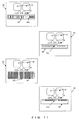
- FIG. 8 shows an example of an image displayed on the display screen of a monitor device of a work station in the capsule endoscope image filing system as the sixth embodiment of the present invention.
- FIG. 8 the display portion having the same function as in FIGS. 2 through 7 is assigned the same reference numeral as in FIGS. 2 through 7 .
- a report entry area 46 is displayed in the area where the observation image 31 is displayed, and furthermore the image bar 36 and the mark 37 superposed and displayed on the image bar 36 are separately displayed as the image bar 36, and the marking of the mark 37 along the image bar 36 above the image bar 36. Below them, two thumbnail images 39 and the corresponding report contents 47 are displayed.
- the user can read the observation image 31 and the information about a check point 42 from the report contents, and can newly attaches the information confirmed by the user from observation image 31 and the check point 42 to the contents of the report.
- the user can display the mark 37 at the part image bar 36, and can display the time mark at the part of the marking of the mark 37.
- the time mark at the part of the marking of the mark 37.
- FIGS. 9A , 9B , 9C , and 9D show examples of images displayed on the display screen of a monitor device of a work station in the capsule endoscope image filing system as the seventh embodiment not according to the present invention.
- FIG. 9A shows a scale mark 48-1 displayed along the display of the image bar 36 or mark 37. From the leftmost to the right most along the mark display, the number of significant captured images (number of frames) is displayed by the scale up to, for example, 59860 in 5000 image units.
- 59860 images indicates the total number of images of the captured images in all image pickup paths from the mouth of the test subject 4 by the capsule endoscope 3.
- a scale mark 48-1 indicating the entire captured images by the number of images is provided.
- the position of a selected thumbnail image can be show to the user.
- the user can select a thumbnail image from the scale mark 48-1 indicating the number of images, and add to the thumbnail image a label and comment including the information about the time, marking , etc.
- a gap 49 as totally black display can be displayed for the space of the number of acquired images of estimated captured images.
- the obtained significant images are displayed with the number of images in the order of acquisition in serial numbers, the practical amount of data can be clarified.
- images can be easily identified how many images are omitted between the significant images to identify the distance between the images.
- the display unit of the number of images by the scale mark 48-1 is 5000 images in the example shown in FIG. 9A .
- the present invention is not limited to this application, and the user can optionally specify and change the number.
- a user can set a specific region (landmark).
- the number of images in the set region can be display at the position of the scale mark 48-1 corresponding to the set region.
- FIG. 9B shows a scale mark 48-2 displayed with the display of the image bar 36 or the mark 37.
- the scale mark 48-2 shows the scale of the capture time of images from the leftmost to the rightmost along the mark display in, for example, one hour (1:00:00) unit from 0 hour 00 minute 00 second to 8 hours 15 minutes 00 second.
- the space corresponding to the capture time of the images estimated for the gap is displayed by total black display as the gap 49.
- the display can be used in estimating the time and the position at which the capsule endoscope 3 passes a target organ.
- FIG. 9C shows a scale mark 48-3 displayed along the display of the image bar 36 or the mark 37.
- the scale mark 48-3 indicates a scale of 0% to 100% from the leftmost to the rightmost along the capture time axis or along the mark display in a 10% unit as a rate of the number of captured images.
- the relative position of the target point can be indicated with respect to the entire inspection or the feature region selected by a user.
- the gap 49 shown in FIGS. 9A , 9B , and 9C can be, in any case, displayed as a simple gap mark 50 indicating the gap starting position as shown in the example of the scale mark 48-1 shown in, for example, FIG. 9D .
- An image bar 38 is represented as a long horizontal image bar 36a by horizontally arranging a provided line in a time series, but it is obvious that the direction is not limited to the application. That is, the vertical direction (36b), the diagonal direction (36c), or the screen depth direction can be expressed in a three-dimensional array (36d).
- the mark 37 is expressed by a vertical line (37a) with respect to a horizontal bar, but the arrangement is not limited to this application.
- a dot, a circle (37b), a square, or an application in which the length of a vertical line is different from the width of a bar (longer line (37c), or shorter line (37d)) can be used.
- FIG. 11 shows these examples.
- a partial enlargement display 44 shown in FIG. 6 can display not only an existing available area of thumbnail images, but display a generated available area by shifting thumbnail images in a target range specified by a user.
- the present invention is not limited to the above-mentioned embodiments.
Landscapes
- Health & Medical Sciences (AREA)
- Life Sciences & Earth Sciences (AREA)
- Surgery (AREA)
- Physics & Mathematics (AREA)
- Engineering & Computer Science (AREA)
- Biomedical Technology (AREA)
- Animal Behavior & Ethology (AREA)
- Radiology & Medical Imaging (AREA)
- Optics & Photonics (AREA)
- Nuclear Medicine, Radiotherapy & Molecular Imaging (AREA)
- Biophysics (AREA)
- Heart & Thoracic Surgery (AREA)
- Medical Informatics (AREA)
- Molecular Biology (AREA)
- Pathology (AREA)
- General Health & Medical Sciences (AREA)
- Public Health (AREA)
- Veterinary Medicine (AREA)
- General Physics & Mathematics (AREA)
- Theoretical Computer Science (AREA)
- Endoscopes (AREA)
- Measurement Of The Respiration, Hearing Ability, Form, And Blood Characteristics Of Living Organisms (AREA)
- Image Processing (AREA)
Applications Claiming Priority (2)
| Application Number | Priority Date | Filing Date | Title |
|---|---|---|---|
| JP2007131764 | 2007-05-17 | ||
| PCT/JP2008/001137 WO2008142831A1 (ja) | 2007-05-17 | 2008-05-01 | 画像情報の表示処理装置及び表示処理方法 |
Publications (3)
| Publication Number | Publication Date |
|---|---|
| EP2149332A1 EP2149332A1 (en) | 2010-02-03 |
| EP2149332A4 EP2149332A4 (en) | 2010-11-03 |
| EP2149332B1 true EP2149332B1 (en) | 2014-12-17 |
Family
ID=40027520
Family Applications (1)
| Application Number | Title | Priority Date | Filing Date |
|---|---|---|---|
| EP08751662.1A Not-in-force EP2149332B1 (en) | 2007-05-17 | 2008-05-01 | Image information display processing device and display processing method |
Country Status (5)
| Country | Link |
|---|---|
| US (1) | US20080285826A1 (ja) |
| EP (1) | EP2149332B1 (ja) |
| JP (1) | JP5327641B2 (ja) |
| CN (1) | CN101677756B (ja) |
| WO (1) | WO2008142831A1 (ja) |
Families Citing this family (13)
| Publication number | Priority date | Publication date | Assignee | Title |
|---|---|---|---|---|
| JP5247785B2 (ja) * | 2010-10-12 | 2013-07-24 | 富士フイルム株式会社 | 内視鏡装置 |
| CN104640496A (zh) * | 2013-05-31 | 2015-05-20 | 奥林巴斯医疗株式会社 | 医疗装置 |
| EP3235418A4 (en) * | 2014-12-15 | 2018-10-24 | Olympus Corporation | Display control device and endoscope system |
| CN107613839B (zh) * | 2015-06-11 | 2019-10-01 | 奥林巴斯株式会社 | 内窥镜装置和内窥镜装置的工作方法 |
| JP6664070B2 (ja) * | 2015-10-28 | 2020-03-13 | Hoya株式会社 | 内視鏡プロセッサ、並びに、内視鏡プロセッサの信号処理方法及び制御プログラム |
| DE102016121668A1 (de) * | 2016-11-11 | 2018-05-17 | Karl Storz Se & Co. Kg | Automatische Identifizierung medizinisch relevanter Videoelemente |
| WO2018207264A1 (ja) * | 2017-05-09 | 2018-11-15 | オリンパス株式会社 | 画像再生装置及び観察システム |
| WO2019039252A1 (ja) * | 2017-08-24 | 2019-02-28 | 富士フイルム株式会社 | 医療画像処理装置及び医療画像処理方法 |
| WO2020136858A1 (ja) * | 2018-12-28 | 2020-07-02 | オリンパス株式会社 | 内視鏡画像処理装置、内視鏡画像処理方法及びプログラム |
| CN112083855A (zh) * | 2019-06-13 | 2020-12-15 | 昆山研达电脑科技有限公司 | 多媒体档案管理方法及计算机可读取储存媒体 |
| CN110969603B (zh) * | 2019-11-26 | 2021-01-05 | 联博智能科技有限公司 | 病变位置的相对定位方法、装置及终端设备 |
| JP7396329B2 (ja) | 2021-05-20 | 2023-12-12 | 株式会社村田製作所 | 表示方法、プログラム、表示システム、及び、評価システム |
| CN113492410B (zh) * | 2021-09-09 | 2022-03-18 | 成都博恩思医学机器人有限公司 | 显示机器人操作过程的方法、系统、机械设备及存储介质 |
Citations (2)
| Publication number | Priority date | Publication date | Assignee | Title |
|---|---|---|---|---|
| JP2006334297A (ja) * | 2005-06-06 | 2006-12-14 | Olympus Medical Systems Corp | 画像表示装置 |
| JP2006345929A (ja) * | 2005-06-13 | 2006-12-28 | Olympus Medical Systems Corp | 画像表示装置 |
Family Cites Families (9)
| Publication number | Priority date | Publication date | Assignee | Title |
|---|---|---|---|---|
| IL157892A0 (en) | 2001-03-14 | 2004-03-28 | Given Imaging Ltd | Method and system for detecting colorimetric abnormalities |
| JP4493386B2 (ja) * | 2003-04-25 | 2010-06-30 | オリンパス株式会社 | 画像表示装置、画像表示方法および画像表示プログラム |
| CN101264001B (zh) * | 2003-04-25 | 2010-11-10 | 奥林巴斯株式会社 | 图像显示装置 |
| DE602004031443D1 (de) * | 2003-10-02 | 2011-03-31 | Given Imaging Ltd | System und methode zur darstellung von datenströmen |
| EP1690491A4 (en) * | 2003-12-05 | 2011-04-13 | Olympus Corp | DISPLAY PROCESSING DEVICE |
| JP3984230B2 (ja) | 2004-02-04 | 2007-10-03 | オリンパス株式会社 | 画像情報の表示処理装置、その表示処理方法及び表示処理プログラム |
| JP4804739B2 (ja) * | 2004-11-10 | 2011-11-02 | オリンパス株式会社 | 画像表示装置、画像表示方法および画像表示プログラム |
| JP4526447B2 (ja) * | 2005-06-20 | 2010-08-18 | オリンパスメディカルシステムズ株式会社 | 画像表示装置、画像表示方法および画像表示プログラム |
| US20070060798A1 (en) * | 2005-09-15 | 2007-03-15 | Hagai Krupnik | System and method for presentation of data streams |
-
2008
- 2008-05-01 EP EP08751662.1A patent/EP2149332B1/en not_active Not-in-force
- 2008-05-01 WO PCT/JP2008/001137 patent/WO2008142831A1/ja active Application Filing
- 2008-05-01 JP JP2009515075A patent/JP5327641B2/ja active Active
- 2008-05-01 CN CN200880016079XA patent/CN101677756B/zh not_active Expired - Fee Related
- 2008-05-16 US US12/122,140 patent/US20080285826A1/en not_active Abandoned
Patent Citations (2)
| Publication number | Priority date | Publication date | Assignee | Title |
|---|---|---|---|---|
| JP2006334297A (ja) * | 2005-06-06 | 2006-12-14 | Olympus Medical Systems Corp | 画像表示装置 |
| JP2006345929A (ja) * | 2005-06-13 | 2006-12-28 | Olympus Medical Systems Corp | 画像表示装置 |
Also Published As
| Publication number | Publication date |
|---|---|
| US20080285826A1 (en) | 2008-11-20 |
| CN101677756A (zh) | 2010-03-24 |
| WO2008142831A1 (ja) | 2008-11-27 |
| EP2149332A1 (en) | 2010-02-03 |
| JP5327641B2 (ja) | 2013-10-30 |
| CN101677756B (zh) | 2012-10-17 |
| JPWO2008142831A1 (ja) | 2010-08-05 |
| EP2149332A4 (en) | 2010-11-03 |
Similar Documents
| Publication | Publication Date | Title |
|---|---|---|
| EP2149332B1 (en) | Image information display processing device and display processing method | |
| JP5385138B2 (ja) | 画像処理装置、該動作方法及び該プログラム | |
| US8368746B2 (en) | Apparatus and method for processing image information captured over time at plurality of positions in subject body | |
| US8413079B2 (en) | Display processing apparatus for image information | |
| US8900124B2 (en) | Image display device | |
| EP1618828B1 (en) | Device, method and program for image processing | |
| US8225209B2 (en) | Capsule endoscope image display device | |
| EP1952751B1 (en) | Device for displaying in vivo image, receiving device, and image display system and method using them | |
| EP2316327B1 (en) | Image display device, image display method, and image display program | |
| EP2662016A1 (en) | Capsule endoscope system | |
| US8406489B2 (en) | Image display apparatus | |
| JP4574983B2 (ja) | 画像表示装置、画像表示方法、及び画像表示プログラム | |
| JP2007307395A (ja) | 画像表示装置、画像表示方法および画像表示プログラム | |
| JP4547401B2 (ja) | 画像表示装置、画像表示方法および画像表示プログラム | |
| JP2005218584A (ja) | 画像情報の表示処理装置、その表示処理方法及び表示処理プログラム | |
| JP5231160B2 (ja) | 画像表示装置、画像表示方法、および画像表示プログラム | |
| JP5684300B2 (ja) | 画像表示装置、画像表示方法、および画像表示プログラム | |
| JP4923096B2 (ja) | 画像表示装置 | |
| JP2007075154A (ja) | 画像表示装置 |
Legal Events
| Date | Code | Title | Description |
|---|---|---|---|
| PUAI | Public reference made under article 153(3) epc to a published international application that has entered the european phase |
Free format text: ORIGINAL CODE: 0009012 |
|
| 17P | Request for examination filed |
Effective date: 20091118 |
|
| AK | Designated contracting states |
Kind code of ref document: A1 Designated state(s): AT BE BG CH CY CZ DE DK EE ES FI FR GB GR HR HU IE IS IT LI LT LU LV MC MT NL NO PL PT RO SE SI SK TR |
|
| AX | Request for extension of the european patent |
Extension state: AL BA MK RS |
|
| DAX | Request for extension of the european patent (deleted) | ||
| A4 | Supplementary search report drawn up and despatched |
Effective date: 20100930 |
|
| 17Q | First examination report despatched |
Effective date: 20110622 |
|
| GRAP | Despatch of communication of intention to grant a patent |
Free format text: ORIGINAL CODE: EPIDOSNIGR1 |
|
| RIC1 | Information provided on ipc code assigned before grant |
Ipc: A61B 1/04 20060101AFI20140707BHEP Ipc: G06T 1/00 20060101ALI20140707BHEP |
|
| INTG | Intention to grant announced |
Effective date: 20140729 |
|
| GRAS | Grant fee paid |
Free format text: ORIGINAL CODE: EPIDOSNIGR3 |
|
| GRAA | (expected) grant |
Free format text: ORIGINAL CODE: 0009210 |
|
| AK | Designated contracting states |
Kind code of ref document: B1 Designated state(s): AT BE BG CH CY CZ DE DK EE ES FI FR GB GR HR HU IE IS IT LI LT LU LV MC MT NL NO PL PT RO SE SI SK TR |
|
| REG | Reference to a national code |
Ref country code: GB Ref legal event code: FG4D |
|
| REG | Reference to a national code |
Ref country code: CH Ref legal event code: EP |
|
| REG | Reference to a national code |
Ref country code: IE Ref legal event code: FG4D |
|
| REG | Reference to a national code |
Ref country code: AT Ref legal event code: REF Ref document number: 701263 Country of ref document: AT Kind code of ref document: T Effective date: 20150115 |
|
| REG | Reference to a national code |
Ref country code: DE Ref legal event code: R096 Ref document number: 602008035901 Country of ref document: DE Effective date: 20150129 |
|
| PG25 | Lapsed in a contracting state [announced via postgrant information from national office to epo] |
Ref country code: FI Free format text: LAPSE BECAUSE OF FAILURE TO SUBMIT A TRANSLATION OF THE DESCRIPTION OR TO PAY THE FEE WITHIN THE PRESCRIBED TIME-LIMIT Effective date: 20141217 Ref country code: NO Free format text: LAPSE BECAUSE OF FAILURE TO SUBMIT A TRANSLATION OF THE DESCRIPTION OR TO PAY THE FEE WITHIN THE PRESCRIBED TIME-LIMIT Effective date: 20150317 Ref country code: LT Free format text: LAPSE BECAUSE OF FAILURE TO SUBMIT A TRANSLATION OF THE DESCRIPTION OR TO PAY THE FEE WITHIN THE PRESCRIBED TIME-LIMIT Effective date: 20141217 |
|
| REG | Reference to a national code |
Ref country code: LT Ref legal event code: MG4D |
|
| PG25 | Lapsed in a contracting state [announced via postgrant information from national office to epo] |
Ref country code: HR Free format text: LAPSE BECAUSE OF FAILURE TO SUBMIT A TRANSLATION OF THE DESCRIPTION OR TO PAY THE FEE WITHIN THE PRESCRIBED TIME-LIMIT Effective date: 20141217 Ref country code: SE Free format text: LAPSE BECAUSE OF FAILURE TO SUBMIT A TRANSLATION OF THE DESCRIPTION OR TO PAY THE FEE WITHIN THE PRESCRIBED TIME-LIMIT Effective date: 20141217 Ref country code: LV Free format text: LAPSE BECAUSE OF FAILURE TO SUBMIT A TRANSLATION OF THE DESCRIPTION OR TO PAY THE FEE WITHIN THE PRESCRIBED TIME-LIMIT Effective date: 20141217 Ref country code: GR Free format text: LAPSE BECAUSE OF FAILURE TO SUBMIT A TRANSLATION OF THE DESCRIPTION OR TO PAY THE FEE WITHIN THE PRESCRIBED TIME-LIMIT Effective date: 20150318 |
|
| REG | Reference to a national code |
Ref country code: AT Ref legal event code: MK05 Ref document number: 701263 Country of ref document: AT Kind code of ref document: T Effective date: 20141217 |
|
| PG25 | Lapsed in a contracting state [announced via postgrant information from national office to epo] |
Ref country code: NL Free format text: LAPSE BECAUSE OF FAILURE TO SUBMIT A TRANSLATION OF THE DESCRIPTION OR TO PAY THE FEE WITHIN THE PRESCRIBED TIME-LIMIT Effective date: 20141217 |
|
| PG25 | Lapsed in a contracting state [announced via postgrant information from national office to epo] |
Ref country code: EE Free format text: LAPSE BECAUSE OF FAILURE TO SUBMIT A TRANSLATION OF THE DESCRIPTION OR TO PAY THE FEE WITHIN THE PRESCRIBED TIME-LIMIT Effective date: 20141217 Ref country code: RO Free format text: LAPSE BECAUSE OF FAILURE TO SUBMIT A TRANSLATION OF THE DESCRIPTION OR TO PAY THE FEE WITHIN THE PRESCRIBED TIME-LIMIT Effective date: 20141217 Ref country code: SK Free format text: LAPSE BECAUSE OF FAILURE TO SUBMIT A TRANSLATION OF THE DESCRIPTION OR TO PAY THE FEE WITHIN THE PRESCRIBED TIME-LIMIT Effective date: 20141217 Ref country code: ES Free format text: LAPSE BECAUSE OF FAILURE TO SUBMIT A TRANSLATION OF THE DESCRIPTION OR TO PAY THE FEE WITHIN THE PRESCRIBED TIME-LIMIT Effective date: 20141217 Ref country code: CZ Free format text: LAPSE BECAUSE OF FAILURE TO SUBMIT A TRANSLATION OF THE DESCRIPTION OR TO PAY THE FEE WITHIN THE PRESCRIBED TIME-LIMIT Effective date: 20141217 |
|
| PG25 | Lapsed in a contracting state [announced via postgrant information from national office to epo] |
Ref country code: IS Free format text: LAPSE BECAUSE OF FAILURE TO SUBMIT A TRANSLATION OF THE DESCRIPTION OR TO PAY THE FEE WITHIN THE PRESCRIBED TIME-LIMIT Effective date: 20150417 Ref country code: AT Free format text: LAPSE BECAUSE OF FAILURE TO SUBMIT A TRANSLATION OF THE DESCRIPTION OR TO PAY THE FEE WITHIN THE PRESCRIBED TIME-LIMIT Effective date: 20141217 Ref country code: PL Free format text: LAPSE BECAUSE OF FAILURE TO SUBMIT A TRANSLATION OF THE DESCRIPTION OR TO PAY THE FEE WITHIN THE PRESCRIBED TIME-LIMIT Effective date: 20141217 |
|
| REG | Reference to a national code |
Ref country code: DE Ref legal event code: R097 Ref document number: 602008035901 Country of ref document: DE |
|
| PLBE | No opposition filed within time limit |
Free format text: ORIGINAL CODE: 0009261 |
|
| STAA | Information on the status of an ep patent application or granted ep patent |
Free format text: STATUS: NO OPPOSITION FILED WITHIN TIME LIMIT |
|
| PG25 | Lapsed in a contracting state [announced via postgrant information from national office to epo] |
Ref country code: DK Free format text: LAPSE BECAUSE OF FAILURE TO SUBMIT A TRANSLATION OF THE DESCRIPTION OR TO PAY THE FEE WITHIN THE PRESCRIBED TIME-LIMIT Effective date: 20141217 |
|
| REG | Reference to a national code |
Ref country code: DE Ref legal event code: R082 Ref document number: 602008035901 Country of ref document: DE Representative=s name: WUESTHOFF & WUESTHOFF, PATENTANWAELTE PARTG MB, DE Ref country code: DE Ref legal event code: R081 Ref document number: 602008035901 Country of ref document: DE Owner name: OLYMPUS CORPORATION, JP Free format text: FORMER OWNER: OLYMPUS MEDICAL SYSTEMS CORP., TOKYO, JP |
|
| 26N | No opposition filed |
Effective date: 20150918 |
|
| PG25 | Lapsed in a contracting state [announced via postgrant information from national office to epo] |
Ref country code: IT Free format text: LAPSE BECAUSE OF FAILURE TO SUBMIT A TRANSLATION OF THE DESCRIPTION OR TO PAY THE FEE WITHIN THE PRESCRIBED TIME-LIMIT Effective date: 20141217 |
|
| REG | Reference to a national code |
Ref country code: CH Ref legal event code: PL |
|
| GBPC | Gb: european patent ceased through non-payment of renewal fee |
Effective date: 20150501 |
|
| PG25 | Lapsed in a contracting state [announced via postgrant information from national office to epo] |
Ref country code: CH Free format text: LAPSE BECAUSE OF NON-PAYMENT OF DUE FEES Effective date: 20150531 Ref country code: LU Free format text: LAPSE BECAUSE OF FAILURE TO SUBMIT A TRANSLATION OF THE DESCRIPTION OR TO PAY THE FEE WITHIN THE PRESCRIBED TIME-LIMIT Effective date: 20150501 Ref country code: MC Free format text: LAPSE BECAUSE OF FAILURE TO SUBMIT A TRANSLATION OF THE DESCRIPTION OR TO PAY THE FEE WITHIN THE PRESCRIBED TIME-LIMIT Effective date: 20141217 Ref country code: LI Free format text: LAPSE BECAUSE OF NON-PAYMENT OF DUE FEES Effective date: 20150531 |
|
| REG | Reference to a national code |
Ref country code: IE Ref legal event code: MM4A |
|
| REG | Reference to a national code |
Ref country code: FR Ref legal event code: ST Effective date: 20160129 |
|
| PG25 | Lapsed in a contracting state [announced via postgrant information from national office to epo] |
Ref country code: SI Free format text: LAPSE BECAUSE OF FAILURE TO SUBMIT A TRANSLATION OF THE DESCRIPTION OR TO PAY THE FEE WITHIN THE PRESCRIBED TIME-LIMIT Effective date: 20141217 |
|
| PG25 | Lapsed in a contracting state [announced via postgrant information from national office to epo] |
Ref country code: GB Free format text: LAPSE BECAUSE OF NON-PAYMENT OF DUE FEES Effective date: 20150501 Ref country code: IE Free format text: LAPSE BECAUSE OF NON-PAYMENT OF DUE FEES Effective date: 20150501 |
|
| PG25 | Lapsed in a contracting state [announced via postgrant information from national office to epo] |
Ref country code: FR Free format text: LAPSE BECAUSE OF NON-PAYMENT OF DUE FEES Effective date: 20150601 Ref country code: BE Free format text: LAPSE BECAUSE OF FAILURE TO SUBMIT A TRANSLATION OF THE DESCRIPTION OR TO PAY THE FEE WITHIN THE PRESCRIBED TIME-LIMIT Effective date: 20141217 |
|
| PG25 | Lapsed in a contracting state [announced via postgrant information from national office to epo] |
Ref country code: MT Free format text: LAPSE BECAUSE OF FAILURE TO SUBMIT A TRANSLATION OF THE DESCRIPTION OR TO PAY THE FEE WITHIN THE PRESCRIBED TIME-LIMIT Effective date: 20141217 |
|
| PG25 | Lapsed in a contracting state [announced via postgrant information from national office to epo] |
Ref country code: HU Free format text: LAPSE BECAUSE OF FAILURE TO SUBMIT A TRANSLATION OF THE DESCRIPTION OR TO PAY THE FEE WITHIN THE PRESCRIBED TIME-LIMIT; INVALID AB INITIO Effective date: 20080501 Ref country code: BG Free format text: LAPSE BECAUSE OF FAILURE TO SUBMIT A TRANSLATION OF THE DESCRIPTION OR TO PAY THE FEE WITHIN THE PRESCRIBED TIME-LIMIT Effective date: 20141217 |
|
| PG25 | Lapsed in a contracting state [announced via postgrant information from national office to epo] |
Ref country code: CY Free format text: LAPSE BECAUSE OF FAILURE TO SUBMIT A TRANSLATION OF THE DESCRIPTION OR TO PAY THE FEE WITHIN THE PRESCRIBED TIME-LIMIT Effective date: 20141217 |
|
| PG25 | Lapsed in a contracting state [announced via postgrant information from national office to epo] |
Ref country code: PT Free format text: LAPSE BECAUSE OF FAILURE TO SUBMIT A TRANSLATION OF THE DESCRIPTION OR TO PAY THE FEE WITHIN THE PRESCRIBED TIME-LIMIT Effective date: 20150417 |
|
| PGFP | Annual fee paid to national office [announced via postgrant information from national office to epo] |
Ref country code: DE Payment date: 20170426 Year of fee payment: 10 |
|
| PG25 | Lapsed in a contracting state [announced via postgrant information from national office to epo] |
Ref country code: TR Free format text: LAPSE BECAUSE OF FAILURE TO SUBMIT A TRANSLATION OF THE DESCRIPTION OR TO PAY THE FEE WITHIN THE PRESCRIBED TIME-LIMIT Effective date: 20141217 |
|
| REG | Reference to a national code |
Ref country code: DE Ref legal event code: R119 Ref document number: 602008035901 Country of ref document: DE |
|
| PG25 | Lapsed in a contracting state [announced via postgrant information from national office to epo] |
Ref country code: DE Free format text: LAPSE BECAUSE OF NON-PAYMENT OF DUE FEES Effective date: 20181201 |