EP2097754B2 - Activated her3 as a marker for predicting therapeutic efficacy - Google Patents
Activated her3 as a marker for predicting therapeutic efficacy Download PDFInfo
- Publication number
- EP2097754B2 EP2097754B2 EP07846863.4A EP07846863A EP2097754B2 EP 2097754 B2 EP2097754 B2 EP 2097754B2 EP 07846863 A EP07846863 A EP 07846863A EP 2097754 B2 EP2097754 B2 EP 2097754B2
- Authority
- EP
- European Patent Office
- Prior art keywords
- her3
- receptor
- antibody
- modulator
- disease
- Prior art date
- Legal status (The legal status is an assumption and is not a legal conclusion. Google has not performed a legal analysis and makes no representation as to the accuracy of the status listed.)
- Active
Links
Images


Classifications
-
- G—PHYSICS
- G01—MEASURING; TESTING
- G01N—INVESTIGATING OR ANALYSING MATERIALS BY DETERMINING THEIR CHEMICAL OR PHYSICAL PROPERTIES
- G01N33/00—Investigating or analysing materials by specific methods not covered by groups G01N1/00 - G01N31/00
- G01N33/48—Biological material, e.g. blood, urine; Haemocytometers
- G01N33/50—Chemical analysis of biological material, e.g. blood, urine; Testing involving biospecific ligand binding methods; Immunological testing
- G01N33/53—Immunoassay; Biospecific binding assay; Materials therefor
- G01N33/574—Immunoassay; Biospecific binding assay; Materials therefor for cancer
- G01N33/5748—Immunoassay; Biospecific binding assay; Materials therefor for cancer involving oncogenic proteins
-
- A—HUMAN NECESSITIES
- A61—MEDICAL OR VETERINARY SCIENCE; HYGIENE
- A61P—SPECIFIC THERAPEUTIC ACTIVITY OF CHEMICAL COMPOUNDS OR MEDICINAL PREPARATIONS
- A61P17/00—Drugs for dermatological disorders
- A61P17/06—Antipsoriatics
-
- A—HUMAN NECESSITIES
- A61—MEDICAL OR VETERINARY SCIENCE; HYGIENE
- A61P—SPECIFIC THERAPEUTIC ACTIVITY OF CHEMICAL COMPOUNDS OR MEDICINAL PREPARATIONS
- A61P35/00—Antineoplastic agents
-
- A—HUMAN NECESSITIES
- A61—MEDICAL OR VETERINARY SCIENCE; HYGIENE
- A61P—SPECIFIC THERAPEUTIC ACTIVITY OF CHEMICAL COMPOUNDS OR MEDICINAL PREPARATIONS
- A61P43/00—Drugs for specific purposes, not provided for in groups A61P1/00-A61P41/00
-
- C—CHEMISTRY; METALLURGY
- C07—ORGANIC CHEMISTRY
- C07K—PEPTIDES
- C07K16/00—Immunoglobulins [IGs], e.g. monoclonal or polyclonal antibodies
- C07K16/18—Immunoglobulins [IGs], e.g. monoclonal or polyclonal antibodies against material from animals or humans
- C07K16/28—Immunoglobulins [IGs], e.g. monoclonal or polyclonal antibodies against material from animals or humans against receptors, cell surface antigens or cell surface determinants
- C07K16/2863—Immunoglobulins [IGs], e.g. monoclonal or polyclonal antibodies against material from animals or humans against receptors, cell surface antigens or cell surface determinants against receptors for growth factors, growth regulators
-
- C—CHEMISTRY; METALLURGY
- C07—ORGANIC CHEMISTRY
- C07K—PEPTIDES
- C07K16/00—Immunoglobulins [IGs], e.g. monoclonal or polyclonal antibodies
- C07K16/18—Immunoglobulins [IGs], e.g. monoclonal or polyclonal antibodies against material from animals or humans
- C07K16/32—Immunoglobulins [IGs], e.g. monoclonal or polyclonal antibodies against material from animals or humans against translation products of oncogenes
-
- G—PHYSICS
- G01—MEASURING; TESTING
- G01N—INVESTIGATING OR ANALYSING MATERIALS BY DETERMINING THEIR CHEMICAL OR PHYSICAL PROPERTIES
- G01N33/00—Investigating or analysing materials by specific methods not covered by groups G01N1/00 - G01N31/00
- G01N33/48—Biological material, e.g. blood, urine; Haemocytometers
- G01N33/50—Chemical analysis of biological material, e.g. blood, urine; Testing involving biospecific ligand binding methods; Immunological testing
- G01N33/53—Immunoassay; Biospecific binding assay; Materials therefor
- G01N33/574—Immunoassay; Biospecific binding assay; Materials therefor for cancer
- G01N33/57407—Specifically defined cancers
-
- G—PHYSICS
- G01—MEASURING; TESTING
- G01N—INVESTIGATING OR ANALYSING MATERIALS BY DETERMINING THEIR CHEMICAL OR PHYSICAL PROPERTIES
- G01N33/00—Investigating or analysing materials by specific methods not covered by groups G01N1/00 - G01N31/00
- G01N33/48—Biological material, e.g. blood, urine; Haemocytometers
- G01N33/50—Chemical analysis of biological material, e.g. blood, urine; Testing involving biospecific ligand binding methods; Immunological testing
- G01N33/53—Immunoassay; Biospecific binding assay; Materials therefor
- G01N33/574—Immunoassay; Biospecific binding assay; Materials therefor for cancer
- G01N33/57484—Immunoassay; Biospecific binding assay; Materials therefor for cancer involving compounds serving as markers for tumor, cancer, neoplasia, e.g. cellular determinants, receptors, heat shock/stress proteins, A-protein, oligosaccharides, metabolites
- G01N33/57492—Immunoassay; Biospecific binding assay; Materials therefor for cancer involving compounds serving as markers for tumor, cancer, neoplasia, e.g. cellular determinants, receptors, heat shock/stress proteins, A-protein, oligosaccharides, metabolites involving compounds localized on the membrane of tumor or cancer cells
-
- A—HUMAN NECESSITIES
- A61—MEDICAL OR VETERINARY SCIENCE; HYGIENE
- A61K—PREPARATIONS FOR MEDICAL, DENTAL OR TOILETRY PURPOSES
- A61K39/00—Medicinal preparations containing antigens or antibodies
- A61K2039/505—Medicinal preparations containing antigens or antibodies comprising antibodies
-
- A—HUMAN NECESSITIES
- A61—MEDICAL OR VETERINARY SCIENCE; HYGIENE
- A61K—PREPARATIONS FOR MEDICAL, DENTAL OR TOILETRY PURPOSES
- A61K39/00—Medicinal preparations containing antigens or antibodies
- A61K39/395—Antibodies; Immunoglobulins; Immune serum, e.g. antilymphocytic serum
-
- C—CHEMISTRY; METALLURGY
- C07—ORGANIC CHEMISTRY
- C07K—PEPTIDES
- C07K2317/00—Immunoglobulins specific features
- C07K2317/70—Immunoglobulins specific features characterized by effect upon binding to a cell or to an antigen
- C07K2317/76—Antagonist effect on antigen, e.g. neutralization or inhibition of binding
-
- G—PHYSICS
- G01—MEASURING; TESTING
- G01N—INVESTIGATING OR ANALYSING MATERIALS BY DETERMINING THEIR CHEMICAL OR PHYSICAL PROPERTIES
- G01N2333/00—Assays involving biological materials from specific organisms or of a specific nature
- G01N2333/435—Assays involving biological materials from specific organisms or of a specific nature from animals; from humans
- G01N2333/705—Assays involving receptors, cell surface antigens or cell surface determinants
- G01N2333/71—Assays involving receptors, cell surface antigens or cell surface determinants for growth factors; for growth regulators
-
- G—PHYSICS
- G01—MEASURING; TESTING
- G01N—INVESTIGATING OR ANALYSING MATERIALS BY DETERMINING THEIR CHEMICAL OR PHYSICAL PROPERTIES
- G01N2333/00—Assays involving biological materials from specific organisms or of a specific nature
- G01N2333/90—Enzymes; Proenzymes
- G01N2333/91—Transferases (2.)
- G01N2333/912—Transferases (2.) transferring phosphorus containing groups, e.g. kinases (2.7)
- G01N2333/91205—Phosphotransferases in general
-
- G—PHYSICS
- G01—MEASURING; TESTING
- G01N—INVESTIGATING OR ANALYSING MATERIALS BY DETERMINING THEIR CHEMICAL OR PHYSICAL PROPERTIES
- G01N2440/00—Post-translational modifications [PTMs] in chemical analysis of biological material
- G01N2440/14—Post-translational modifications [PTMs] in chemical analysis of biological material phosphorylation
-
- G—PHYSICS
- G01—MEASURING; TESTING
- G01N—INVESTIGATING OR ANALYSING MATERIALS BY DETERMINING THEIR CHEMICAL OR PHYSICAL PROPERTIES
- G01N2800/00—Detection or diagnosis of diseases
- G01N2800/20—Dermatological disorders
- G01N2800/205—Scaling palpular diseases, e.g. psoriasis, pytiriasis
-
- G—PHYSICS
- G01—MEASURING; TESTING
- G01N—INVESTIGATING OR ANALYSING MATERIALS BY DETERMINING THEIR CHEMICAL OR PHYSICAL PROPERTIES
- G01N2800/00—Detection or diagnosis of diseases
- G01N2800/52—Predicting or monitoring the response to treatment, e.g. for selection of therapy based on assay results in personalised medicine; Prognosis
Definitions
- the present invention provides methods for the determination of the activation level of Receptor Tyrosine kinases, e.g. phosporylated HER3, for the selection of patients for disease treatment. Methods are also provided for the evaluation of the biological and pharmacodynamic effects of an active substance and/or its efficacy in disease treatment, utilizing a tissue sample from a test subject, for example tumor material or normal tissue such as skin or hair follicle. Further, methods for the treatment of HER receptor-associated diseases are disclosed.
- the human epidermal growth factor receptor 3 (HER3, also known as ErbB3) is a receptor protein tyrosine kinase and belongs to the epidermal growth factor receptor (EGFR) subfamily of receptor protein tyrosine kinases, which also includes HER1 (also known as EGFR), HER2, and HER4 ( Plowman et al., Proc. Natl. Acad. Sci. U.S.A. 87 (1990), 4905-4909 ; Kraus et al., Proc. Natl. Acad. Sci. U.S.A, 86 (1 989), 9193-9197 ; and Kraus et al., Proc. Natl. Acad. Sci. U.S.A. 90 (1993), 2900-2904 ).
- EGFR epidermal growth factor receptor
- HER3 has been found to be overexpressed in several types of cancer such as breast, gastrointestinal and pancreatic cancers. Interestingly a correlation between the expression of HER2/HER3 and the progression from a non-invasive to an invasive stage has been shown ( Alimandi et al., Oncogene 10,181 3-1 821 ; deFazio et al., Cancer 87, 487-498 ; Naidu et al., Br. J. Cancer 78, 1385-1390 ).
- HER3 antibodies described in WO 03/013602 are reported to induce accelerated receptor internalization and to reduce tumor cell proliferation and migration.
- U.S. patent 5,968,511 corresponding to WO 97/135885
- HER3 antibodies were found to reduce ligand-induced formation of HER2/HER3 heterodimers.
- WO 00/078347 discloses methods for arresting or inhibiting cell growth, comprising preventing or reducing HER2/HER3 heterodimer formation, for example, by administering a combination of an anti-HER2 antibody, e.g. Herceptin, and an anti-HER3 antibody, e.g, antibody 105.5 purchased from Neomarkers.
- a principle aim of medical/pharmaceutical drug development is the development of individual or targeted therapies for the treatment of diseases.
- Such specific therapies may e.g. comprise therapeutic antibodies, small molecule inhibitors, nucleic acid interference, and the administration of an individually selected or dosed pharmaceutical composition.
- target specific therapies predominantly affect a single target. Thus it is critical in modern drug development to identify those patients responsive to the target specific therapy.
- Herceptin that is directed against the receptor tyrosine kinase HER2.
- This particular antibody has been approved for the treatment of breast cancer, a tumor indication which is associated with an amplification of the HER2 gene in about 20% of cases causing overexpression of the corresponding protein.
- a diagnostic assay, HercepTest has been developed.
- HercepTest only detect the amount of the targeted protein, whereas often it is the activity of the protein that is actually causing the cellular signal deregulation and subsequent malignancy.
- the HercepTest only predicts a successful patient response in approximately 30% of the cases when Herceptin is used as a single agent ( Leyland-Jones, Lancet Oncol (2002) Mar;3(3):137-44 ). This low predictive rate is observed even though all of the patients treated are judged to be overexpressing HER2, demonstrating the significant limitations of this type of diagnostic assay and the need for identifying better biomarkers of responsiveness to therapy.
- Another critical step during drug development is the selection of the dose for therapeutic agents.
- the assumption of the maximally tolerated dose is used.
- This same principle does not apply for targeted therapies, where an optimal biologic dose would be preferred instead.
- the definition of the optimal dose to be administered may be defined by pharmacodynamic or -kinetic parameters and the determination of the efficacy on the target molecule ( Albanell et al., 2002, J. Clin. Oncol. 20, 110-124 ).
- pharmacodynamic parameters such as for example sufficient solubility and stability of a compound that allows delivery to the site of action in sufficient concentration, metabolic stability so that the compound is not cleared from the body so rapidly that it does not have a chance to be an effective pharmacological agent, or pharmacokinetics that allow the compound to reach a desired plasma/serum concentration.
- pharmacodynamic effects can be measured through extensive imaging and radioactive labeling of substance or substrate (e.g. PET, CT) and the readout is to be compared with the observed side effects and clinical efficacy.
- substance or substrate e.g. PET, CT
- surrogate markers for biological efficacy PD markers
- these markers don't indicate the direct biological effect of the therapeutic on normal and/or cancerous cells and therefore may be subject to off-target effects and activation/deactivation through external (not target specific) pathways. Examples for these markers are CA-125, KI67, PTEN, and ⁇ HCG.
- a desirable marker would be specific to the pathway and the therapeutic (targeted) intervention, easily accessible and analyzable without intra- and/or intersubject variability.
- EGF epidermal growth factor
- EGFR its cognate receptor
- WO 2006/063042 discloses a method of identifying a patient for therapy with a HER3 dimerization inhibitor. The procedure involves the determination of the expression of two or more HER3 receptors and one or more HER3 ligands in a sample from the patient.
- a first essential difference to the approach of present application is that at least two HER3 receptors and one or more HER3 ligands have to be determined in order to provide information whether a patient is likely to respond to therapy with a HER3 dimerization inhibitor.
- the technical problem underlying the present invention was to provide a rapid, quantitative, reproducible, and inexpensive assay that is compatible with current clinical laboratory instrumentation and which is suitable for determination of the activation and/or expression level of HER receptors.
- a method for the determination of the sensitivity or responsiveness of a disease to a HER modulator or to a combination of at least one HER modulator with a further agent is provided.
- a HER3 modulator correlates with HER3 receptor activation, e.g. phosphorylation
- methods and procedures have been devised for predicting the responsiveness of a subject to treatment with a HER modulator.
- tumor cells such as BxPC3 (pancreas cancer), A431 (epithelial carcinoma) or A549 (lung carcinoma) grown in vitro express HER3 and show basal HER3 phosphorylation.
- examination of tumor xenograft models treated with HER3 inhibitors showed that those tumors arising from tumor cell lines with HER3 expression and elevated basal HER3 phosphorylation, e.g. T47D (breast cancer), BxPC3 (pancreas cancer), HT-29 (colon cancer) and CaLu-3 (NSCLC) are particularly responsive to treatment protocols targeting a HER3 receptor.
- HER receptor activation may be a general biological switch that predefines the level of responsiveness of a disease to HER modulators.
- activation of a HER receptor such as HER3 is indicative of a disorder that is particularly sensitive to treatment with a HER modulator.
- the application relates to a method for determining whether a disease is responsive to treatment with a HER modulator, by obtaining at least one sample from a subject at risk of or having said disease, examining the activity of at least one HER receptor in a cellular assay, and identifying a disease as responsive if activity of at least one HER receptor is detected.
- HER receptor is intended to mean a HER1 protein, e.g. human HER1/EGFR (Acc-Nr. Swiss Prot P00533), a HER2 protein, e.g. human HER2 (Ace-Nr. Swiss Prot P04626), a HER3 protein, e.g. human HER3 (Acc-Nr. Swiss Prot P21860) or a HER4 protein (Acc-Nr. Swiss Prot Q155503).
- the HER receptor is a HER3 protein, more preferably the human HER3 protein.
- the present application relates to the use of a modulator that affects a HER3 receptor.
- HER modulator is intended to mean a compound or drug that acts either on the nucleic acid level or on the protein level to directly or indirectly modulate HER receptor activity.
- Direct or indirect modulation includes activation or inhibition of HER receptor activity or HER receptor signal transduction pathway.
- the modulation includes an inhibition.
- the modulator of HER receptor activity may act on the nucleic acid level, either on the transcription or on the gene itself. On the gene level said modulator may cause a partial or complete gene inactivation, for example by gene disruption. Reducing or inhibiting transcription may comprise application of effector nucleic acids, such as antisense molecules, for example DNA or RNA molecules or RNA analogues, ribozymes, small double-stranded RNA molecules capable of RNA interference (siRNA) or microRNAs. Further, precursor RNA molecules of siRNA or DNA molecules encoding the latter may be suitable.
- Effector molecules may be directly introduced into a cell or generated within a cell by transcription from suitable nucleic acid templates. Production and uses of effector nucleic acids are extensively discussed in the literature and are widely known and available to one skilled in the art.
- the HER modulator may act on the protein level by at least partially inhibiting HER receptor mediated signal transduction.
- the modulator may block the ligand induced activation of a HER receptor.
- a ligand is meant a polypeptide that binds to and/or activates a HER receptor.
- ligands are selected from the group of: AMPR (amphiregulin) NM_001657 BTC (betacellutin) NM_001729 DTR (diphtheria toxin receptor (heparin-binding epidermal growth factor-like growth factor)) NM_001945 EGF (epidermal GF, beta-urogastrone) NM_001963 EREG (epiregulin) NM_001432.1 NRG1 (neuregulin 1) NM_013957 NRG2 (neuregulin 2) NM_013982 NRG3 (neuregulin 3) AL096706 NRG4 (neuregulin 4) NM_138573 TGFA (transforming growth factor, alpha) NM_003236
- neuregulin 1 isoforms encoded by the neuregulin 1 gene are particularly preferred.
- such a modulator may act by occupying the ligand binding site or a portion thereof of the HER receptor, thereby making the receptor inaccessible to its natural ligand so that its normal biological activity is prevented or reduced.
- ligand muteins capable of binding to the receptor, but unable to induce signal transduction, or antibodies directed against ligands are examples of HER modulators. Suitable types of antibodies are discussed in detail below.
- the modulator interferes with ligand dependent or independent formation of HER receptor oligomers, e.g. hetero-oligomers or homo-oligomers.
- An HER receptor hetero-oligomer herein is a non-covalently associated oligomer comprising at least two different HER receptors.
- a HER receptor homo-oligomer is a non-covalently associated oligomer that comprises at least two HER receptors of the same. Examples of such HER oligomers include, but are not limited to HER1/HER1, HER1/HER2, HER1/HER3, HER1/HER4, HER2/HER2, HER2/HER3, HER2/HER4, HER3/HER4, HER4/HER4.
- preferred hetero-oligomers may comprise one, two or more HER2 receptors combined with a different HER receptor, such as HER1, HER3, or HER4.
- HER2 receptors such as HER1, HER3, or HER4.
- Other proteins such as a cytokine receptor subunit (e.g., gp130) or other receptor tyrosine kinases such as the IGF-1R may also be included in the hetero-oligomer.
- a reduction of HER receptor mediated signal transduction may be further caused by a downregulation from the membrane and/or degradation of HER receptor resulting in an at least partial disappearance of HER molecules from the cell surface or by a stabilization of HER molecules on the cell surface in a substantially inactive form, i.e, a form which exhibits a lower signal transduction compared to the non-stabilized form.
- a reduction of HER mediated signal transduction may also be caused by influencing, e.g. decreasing or inhibiting, the binding of a signal transduction molecule, e.g PI3K, Shc or Grb7 to HER-3, of GRB2 to HER-2, of GRB2 to SHC, or by inhibiting AKT phosphorylation, PYK2 tyrosine phosphorylation or ERK2 phosphorylation.
- Negative regulators such as PTPs or proteases, could also be influenced.
- the HER modulator may be an antibody or a fragment thereof, directed against a HER receptor.
- the antibody may be a monoclonal or polyclonal antibody, as well as a recombinant antibody, e.g. single chain antibody or a fragment thereof, which contains at least one antigen-binding site, an antibody fragment such as a Fab, Fab' or F(ab') 2 fragment or a recombinant fragment such as a scFv fragment and a humanized antibody or a human antibody.
- a Fab, Fab' or F(ab') 2 fragment e.g. single chain antibody or a fragment thereof, which contains at least one antigen-binding site
- an antibody fragment such as a Fab, Fab' or F(ab') 2 fragment or a recombinant fragment such as a scFv fragment and a humanized antibody or a human antibody.
- the application of chimeric antibodies, humanized antibodies or human antibodies is especially preferred.
- An anti-HER3 antibody is selected from the group consisting of antibody 105.5 ( Chen et al, JBC 1996, 271 (3) 7620-9 ), SGP-1 ( Rajkumar et al, The Breast 1995, 4 84-91 ), H3 90.6 ( Chen et al, JBC 1996, 271 (3) 7620-9 ), 1B4C3 and 2D1D12 ( PCT/EP02/08938 ) or one of the human anti-HER3 antibodies disclosed in US 60/755,022 .
- An anti-HER2 antibody is selected from the group consisting of Trastuzumab, Pertuzumab, Herceptin-geldanamycin, 213-bi-Herceptin-alpha conjugate, Herceptin-DM1 and an anti-HER1 antibody is selected from the group consisting of Panitumumab, Cetuximab, Matuzumab, Erbitux-paclitaxel conjugate, Erbitux-MMC (mitomycinC) and LA22-MMC.
- scaffold protein having an antibody like binding activity that binds to a HER family member.
- the term "scaffold protein”, as used herein, means a polypeptide or protein with exposed surface areas in which amino acid insertions, substitutions or deletions are highly tolerable.
- scaffold proteins that can be used in accordance with the present invention are protein A from Staphylococcus aureus, the bilin binding protein from Pieris brassicae or other lipocalins, ankyrin repeat proteins, and human fibronectin (reviewed in Binz and Plückthun, (2005) Curr Opin Biotechnol, 16, 459-69 ).
- Engineering of a scaffold protein can be regarded as grafting or integrating an affinity function onto or into the structural framework of a stably folded protein.
- Affinity function means a protein binding affinity according to the present invention.
- a scaffold can be structurally separable from the amino acid sequences conferring binding specificity.
- proteins appearing suitable for the development of such artificial affinity reagents may be obtained by rational, or most commonly, combinatorial protein engineering techniques such as panning against a HER family member, either purified protein or protein displayed on the cell surface, for binding agents in an artificial scaffold library displayed in vitro, skills which are known in the art (Binz and Plückthun, 2005, supra).
- a scaffold protein having an antibody like binding activity can be derived from an acceptor polypeptide containing the scaffold domain, which can be grafted with binding domains of a donor polypeptide to confer the binding specificity of the donor polypeptide onto the scaffold domain containing the acceptor polypeptide. Insertion can be accomplished by various methods known to those skilled in the art including, for example, polypeptide synthesis, nucleic acid synthesis of an encoding amino acid as well by various forms of recombinant methods well known to those skilled in the art.
- Low molecular weight inhibitors may include organic compounds, organometallic compounds, salts of organic and organometallic compounds, saccharides, amino acids, and nucleotides. Low molecular weight inhibitors further include molecules that would otherwise be considered biological molecules, except their molecular weight is preferably not greater than 600, more preferably not greater than 450. Thus, low molecular weight inhibitors may also be lipids, oligosaccharides, oligopeptides, and oligonucleotides and their derivatives.
- Low molecular weight inhibitors are merely called low molecular weight inhibitors because they typically have molecular weights not greater than 600 and the term shall not be construed as restricted to a specific molecular weight.
- Low molecular weight inhibitors include compounds that are found in nature as well as synthetic compounds.
- the HER modulator is a low molecular weight inhibitor that inhibits cell growth. In another embodiment, the HER modulator is a low molecular weight inhibitor that inhibits at least partially HER mediated signal transduction.
- the low molecular weight inhibitor is one of the group comprising Gefitinib, Erlotinib, Lapatinib, BIBW2992, AV412.
- the low molecular weight inhibitor belongs to the group of indirect HER modulators such as kahahalide F (Janmaat et al, 2005) or estrogen receptor inhibitors such as tamoxifen.
- the application also encompasses combinations of HER modulators, e.g. HER modulators directed against the same receptor, e.g. HER3, or HER modulators directed against different HER receptors, e.g. HER3 and HER1, HER3 and HER2, and HER3 and HER4.
- HER modulators e.g. HER modulators directed against the same receptor, e.g. HER3, or HER modulators directed against different HER receptors, e.g. HER3 and HER1, HER3 and HER2, and HER3 and HER4.
- combinations of antibodies may be used.
- the present application further relates to a method for determining responsiveness of disorder to the administration of at least one modulator of a HER3 receptor and/or a further agent as described in detail below.
- the active ingredient e.g. the HER modulator is usually administered as a pharmaceutical composition.
- the composition may be in solid, liquid or gaseous form and may be, inter alia, in a form of (a) powder(s), (a) tablet(s), (a) solution(s) or (an) aerosol(s).
- Said composition may comprise at least one, e.g. two, three, four, or five active compounds.
- the pharmaceutical composition is useful for the treatment of a disease as referred to below.
- Said disease is a hyperproliferative disease, an inflammatory disease or a neurodegenerative disease.
- the hyperproliferative disease may comprise, but is not limited to psoriasis or breast, lung, colon, kidney, lymphoma, skin, ovary, prostate, pancreas, esophagus, barret, stomach, bladder, cervix, liver, thyroid cancer, soft tissue sarcoma, melanoma or other hyperplastic or neoplastic diseases associated with HER receptor expression, overexpression and/or activation.
- the pharmaceutical composition may comprise at least one further active agent.
- additional active agents which may be used in accordance with the present invention, are antibodies or low molecular weight inhibitors of other receptor protein kinases, such as IGF-1R, or c-met, receptor ligands such as vascular endothelial factor (VECF), cytotoxic or anti-neoplastic agents, such as doxorubicin, platinum compounds such as cis-platin or carboplatin, cytokines, antisense molecules, aptamers, or siRNA molecules.
- VECF vascular endothelial factor
- cytotoxic or anti-neoplastic agents such as doxorubicin
- platinum compounds such as cis-platin or carboplatin
- cytokines antisense molecules
- aptamers aptamers
- siRNA molecules siRNA molecules.
- the cytotoxic or antineoplastic agent may be selected from the group of therapeutic proteins including, but not limited to, antibodies or immunomodulatory proteins, or from the group of small molecule inhibitors or chemotherapeutic agents consisting of mitotic inhibitors, kinase inhibitors, alkylating agents, antimetabolites, intercalating antibiotics, growth factor inhibitors, cell cycle inhibitors, enzymes, topoisomerase inhibitors, histone deacetylase inhibitors, anti-survival agents, biological response modifiers, anti-hormones, e.g. antiandrogens, and anti-angiogenesis agents.
- the anti-neoplastic agent is radiation
- treatment can be achieved either with an internal (brachytherapy BT) or external (external beam radiation therapy: EBRT) source.
- disease when used in the present invention shall mean any condition that would benefit from a medical treatment or that is associated with an abnormal HER receptor expression, activation and/or signal transduction. This includes chronic and acute diseases or diseases including those pathological conditions which predispose the candidate to the disease in question.
- a preferred disease to be treated in accordance with the present invention is a hyperproliferative disease.
- a hyperproliferative disease as mentioned above includes any neoplasia, i.e. any abnormal and/or uncontrolled new growth of tissue.
- uncontrolled new growth of tissue as used herein may depend upon a dysfunction and/or loss of growth regulation.
- a hyperproliferative disease further includes tumor diseases and/or cancer, such as metastatic or invasive cancers.
- said hyperproliferative disease is in brain, central nervous system, soft-tissue sarcoma, hematological malignancies, oral cavity, head and neck, breast, lung, colon, gastric, kidney, lymphoma, skin, ovary, prostate, pancreas, esophagus, bladder, cervix, liver, thyroid cancer, melanoma, cancer of unknown origin, or other hyperplastic or neoplastic diseases associated with HER receptor expression, overexpression and/or activation, e.g. hyperphosphorylation.
- a disease which is associated with the expression or overexpression of a HER receptor is a disease with cells comprising on their cell surface a HER receptor protein and/or a ligand binding to a HER receptor.
- a disease which "expresses" a HER family member is one which has significantly higher levels of an HER receptor, such as HER3, at the cell surface thereof, compared to a healthy cell of the same tissue type.
- Such expression may be caused by gene amplification or by increased transcription or translation.
- HER receptor expression may be determined in a diagnostic or prognostic assay by evaluating levels of the HER protein present on the surface of a cell (e.g., via immunohistochemistry; IHC).
- FISH fluorescent in situ hybridization
- PCR polymerase chain reaction
- Expression of the HER ligand may be determined diagnostically by evaluating levels of the ligand (or nucleic acid encoding it) in the patient by various diagnostic assays such as DNA arrays, Northern blotting, FISH, Southern blotting, PCR or protein based assays described above.
- the presence of various N-terminal HER3 isoforms or serum concentrations of shed receptor domains may be evaluated when practicing the present invention.
- various other assays are available to the skilled practitioner.
- a detectable label e.g., a radioactive isotope
- the disease may be associated with HER activation.
- Activation of a HER family member may generally involve formation of HER oligomers, followed by activation of the intrinsic receptor kinase activity, the binding of intracellular second messenger molecules to the receptor and/or modification, e.g. tyrosine phosphorylation, of the HER receptor and/or the second messenger molecules, which leads to specific biologic responses, as for example cel proliferation, cell migration or anti-apoptosis
- Another aspect of the present application is concerned with a method for determining and/or predicting the sensitivity of a disease or condition associated with HER3 receptor mediated signal transduction to a HER3 modulator, optionally in combination with a further agent, comprising analyzing a sample by detecting the activity of a HER3 receptor in that sample, wherein the method comprises detecting the activity, e.g. the degree of the phosphorylation of a HER3 receptor.
- the method may be used for the detection of a HER receptor in a cell, for the determination of HER receptor concentration in subjects suffering from a disease as mentioned above or for the staging of said disease in a subject.
- the amount of the HER receptor present in the sample and/or its activation level is determined in a tissue sample, taken from the subject. The amount so identified correlates with a stage of progression or a stage of therapy identified in the various population of diagnosed subjects, thereby providing a determination of the disease stage in the subject under study.
- the amount and/or activity of the HER receptor present in the disease tissue may be assessed by immunohistochemistry, ELISA or antibody arrays including phospho-specific antibodies using HER receptor and/or other signal transduction antibodies.
- Other suitable methods may include bead-based technologies such as Luminex bead assays and proteomics approaches (2-D gels, MS analysis etc).
- Cellular preparations with methodical prerequisites such as phosphatase inhibitors (ortho-Vanadate, Suramine, H 2 O 2 or specific inhibitors) as would be the case with phosphatase inhibitor tablets, could be envisioned as part of the quantification of phospho-specific antigen/epitopes.
- oligomerization state As well as the oligomerization partners of a HER receptor.
- Protein analytical methods to determine those parameters are well known in the art and are among others western blot and immunoprecipitation techniques, FACS analysis, chemical crosslinking, bioluminescence resonance energy transfer (BRET), fluorescence resonance energy transfer (FRET) and the like (e.g. Price et al, Methods in Molecular Biology, 218: 255-268 (2002 ) or the eTag technology ( WO 05/03707 , WO 04/091384 , WO 04/011900 ).
- the kinase activity can be measured by capturing the kinase in the cell lysate by an antibody with immunoprecipitated and is then subjected to kinase activity reactions in the presence of 32 P- ⁇ -TP.
- the activity of the kinase in the reaction is analyzed by sodium dodecyl sulfate (SDS)-polyacrylamide gel electrophoresis (PAGE) and autoradiography.
- SDS sodium dodecyl sulfate
- PAGE polyacrylamide gel electrophoresis
- in vitro kinase assays can be performed with non-radioactive detection methods (e.g. CST kinase assays) or synthetic peptides that can serve as substrates for a HER receptor, such as HER3, can be spotted on arrays for estimating HER kinase activity.
- non-radioactive detection methods e.g. CST kinase assays
- the activation level of a HER receptor correlates with the activation status of a second messenger molecule involved in HER receptor mediated signal transduction.
- one embodiment of the present invention refers to a method for identifying the responsiveness of a disease to treatment with a HER3 modulator, by obtaining at least one sample from a subject at risk of or having said disease, examining the activity of at least one molecule involved in HER receptor mediated signal transduction in a cellular assay, and identifying a disease as responsive if activity of at least one a molecule involved in HER3 receptor mediated signal transduction is detected. The expression and/or activity of HER3, optionally in combination with other HER receptors, is examined.
- Signal transduction refers to a series of molecular events usually beginning with the interaction of a cell surface receptor with an extracellular ligand or with the binding of an intracellular molecule to a phosphorylated site of a cell surface receptor, e.g. a HER receptor, that triggers a series of molecular interactions, wherein the series of molecular interactions results in a regulation of gene expression in the nucleus of a cell.
- intracellular molecule molecule involved in HER receptor mediated signaling
- substrate of HER receptor refers to molecules involved in HER-mediated signaling pathways as for example reviewed in Citri and Yarden, Nat Reviews Mol Cell Biol, 2006 (7), 505-516 ; Shawver et al, Cancer Cell, 2002 (1), 117-123 ; Yarden and Sliwkowski, Nat Reviews Mol Cell Biol, 2001 (2), 127-137 .
- Exemplary molecules that may be part of a HER receptor mediated signaling pathway include, but are not limited to, PI3K proteins, AKT proteins, Grb2 proteins, Grb7 proteins, Shc proteins, Gab-1 proteins, Sos proteins, Src proteins, Cbl proteins, PLCy proteins, Shp2 proteins, GAP proteins, Vav proteins, Nck proteins and Crk proteins.
- the phosphorylation state of one of the HER3 receptors or their substrates can be assessed as a measure of activation of the receptor.
- phosphorylation of a HER receptor indicates that the receptor has been activated and is the mechanism for transducing the downstream signal.
- Phosphorylation of one or multiple tyrosine residues in a HER receptor or in one or more of its substrates can be analysed using various tyrosine phosphorylation assays.
- HER receptors or their substrates may be immunoprecipitated with specific antibodies from lysates of cells expressing HER receptors and their substrates and then assayed for tyrosine phosphorylation activity using a phosphotyrosine monoclonal antibody (which is optionally conjugated with a detectable label).
- Tyrosine phosphorylation of HER receptors and their substrates is detected by using phospho-specific antibodies.
- said phospho-specific antibody is selected from the group comprising phospho-specific HER3 antibodies 21 D3 (Y1289, Cell Signalling Technology, USA) and 50C2 (Y1222, Cell Signalling Technology, USA), as well as pEGFR, pHER2, pHER4, pIGF-1R, pAkt, pErk, pBad, pp70-S6K, pGSK, p-src, pPyk2, with all relevant phosphotyrosines in a given protein being covered here.
- the term "phospho-specific antibody” is meant to represent either a polyclonal or a monoclonal antibody that binds to a phosphorylated epitope in a HER receptor and/or a second messenger molecule associated with HER mediated signal transduction.
- the phosphorylated epitope may include a eastone phosphorylated serin- residue.
- the phosphorylated epitope includes at least one phosphorylated tyrosine residue.
- the phospho-tyrosine residue is selected from the group consisting of Y1054, Y1197, Y1199, Y1222, Y1224, Y1260, Y1262, Y1276, Y1289 and Y1328 in the HER3 protein (numbering according to Kraus et al, PNAS 1989 (86 9193-919 ).
- the term also encompasses a phospho-specific recombinant antibody, e.g.
- single chain antibody or a fragment thereof which contains at least one antigen-binding site, an antibody fragment such as a Fab, Fab' or F(ab') 2 fragment or a recombinant fragment such as a scFv fragment and a humanized antibody or a human antibody directed against a phosphorylated epitope in a HER receptor and/or a molecule associated with HER mediated signal transduction.
- an antibody fragment such as a Fab, Fab' or F(ab') 2 fragment or a recombinant fragment such as a scFv fragment and a humanized antibody or a human antibody directed against a phosphorylated epitope in a HER receptor and/or a molecule associated with HER mediated signal transduction.
- Phospho-specific polyclonal antibodies can be obtained by methods well known in the art. For example any animal, which is known to produce antibodies can be immunized with a phospho-HER receptor polypeptide. Antibody containing sera is isolated from the immunized animal and is screened for the presence of antibodies with the desired specificity using methods as for example, ELISA or FACS.
- Monoclonal antibodies produced by the hybridoma method are first described by Köhler et al., Nature, 256:495 (1975 ).
- Monoclonal antibodies can also be produced by recombinant DNA methods (see, for example, US. Pat. No. 4,816,567 ) or may be isolated from phage antibody libraries using the techniques described in Clackson et al., Nature, 352:624-628 (1991 ) and Marks et al., J. Mol. Biol., 222:581-597 (1%l), for example.
- Humanized forms of the antibodies may be generated according to the methods known in the art such as chimerization or CDR grafting.
- the present invention also relates to a hybridoma or recombinant cell line, which produces the above described monoclonal antibodies or binding fragments thereof.
- a disease which is responsive to treatment shows statistically significant improvement in response to a HER modulator treatment when compared to no treatment or treatment with placebo in a recognized animal model or a human clinical trial.
- the terms "treat” or treatment” refer to both therapeutic treatment and prophylactic or preventative measures, wherein the object is to prevent or slow down (lessen) an undesired physiological change or disorder, such as the development of a hyperproliferative disease, e.g. cancer.
- beneficial or desired clinical results include, but are not limited to, alleviation of symptoms, diminishment of extent of disease, stabilized (i.e., not worsening) state of disease, delay or slowing of disease progression, amelioration or palliation of the disease state, and remission (whether partial or total), whether detectable or undetectable.
- Treatment can also mean prolonging survival as compared to expected survival if not receiving treatment.
- Those in need of treatment include those already with the condition or disorder as well as those prone to have the condition or disorder or those in which the condition or disorder is to be revented.
- a method of treating a subject in need thereof comprising determining expression and/or activation of a HER receptor in said subject, and administering to a subject in which HER receptor expression and/or activation has been determined, a therapeutically effective amount of a HER modulator and optionally at least one further agent.
- the HER receptor is HER3.
- HER modulator Depending on the type of the HER modulator, type and severity of the condition to be treated, about 0.01-10000 mg of the HER modulator may be administered to a patient in need thereof, e.g. by one or more separate administrations or by continuous infusion.
- a typical daily dosage might range from about 0.001 mg/kg to about 1000 mg/kg or more, depending on the factors mentioned above.
- the treatment is sustained until a desired suppression of disease symptoms occurs.
- the dose of the at least one antineoplastic agent administered depends on a variety of factors. These are, for example, the nature of the agent, the tumor type or the route of administration. It should be emphasized that the present application is not limited to any dose.
- the present invention provides additional methods and procedures to evaluate the therapeutic efficacy of a HER3 modulator or a pharmaceutical composition comprising a HER modulator and/or at least one further agent.
- Determination of the pharmacodynamics of a modulator targeting a HER receptor and/or a HER receptor mediated signaling pathway may involve immunohistochemical staining with phospho-specific antibodies of samples of diseased tissue, e.g. tumor tissue, in order to quantitate the activation level of HER receptors and/or related second messenger molecules.
- relevant pharmacodynamic parameters e.g. the activation level of a HER3 receptor
- primary i.e. non-diseased normal tissue samples.
- HER3 particularly in its activated form in primary human tissues may be determined, e.g. by immunohistochemistry.
- the present application provides a method for determining the therapeutic efficacy of the treatment of a HER3 receptor-associated disease with a HER3 modulator and/or a further active agent comprising exposing a subject to the HER modulator and/or the further active agent, obtaining at least one sample from the subject, detecting the activation level of the HER3 receptor in said sample wherein a difference in the activation level of HER3 is observed as a result of the exposure to the HER3 modulator and/or the further active agent as compared to the absence of the exposure to the HER3 modulator and/or the further active agent.
- sample as embraced by the present invention preferably means the use of a tissue sample for the detection of an activated form of a HER family member.
- the HER receptor is HER3.
- the activation level is the degree of phosphorylation.
- tissue sample is meant to include a collection of cells obtained from a tissue of a subject or patient, preferably containing nucleated cells with protein material.
- the four main human tissues are (1) epithelium; (2) the connective tissues, including blood vessels, bone and cartilage; (3) muscle tissue; and (4) nerve tissue.
- the source of the tissue sample may be selected from the group comprising of solid tissues as from a fresh, frozen and/or preserved organ or tissue sample or biopsy or aspirate.
- the present invention also includes the use of samples derived from blood or any blood constituents, bodily fluids such as cerebral spinal fluid, amniotic fluid, peritoneal fluid, or interstitial fluid and cells from any time in gestation or development of the subject.
- the tissue sample may also be primary or cultured cells or cell lines.
- the tissue sample may contain compounds which are not naturally intermixed with the tissue in nature such as preservatives, anticoagulants, buffers, fixatives, nutrients, antibiotics, or the like.
- tissue sample is may be a single part or piece of a tissue sample, e.g., a thin slice of tissue or cells cut or micro-dissected from a tissue sample.
- tissue arrays can be formalin-fixed tissue samples cut into thin sections and mounted on silanised glass slides that can be used for expression analysis and cellular localization on a protein, RNA or DNA level.
- at least 10 samples are mounted on one silanised glass slide.
- at least 20 samples are mounted on one silanised glass slide.
- 40 or more samples are mounted on one silanised glass slide.
- the tissue may be fixed (i.e.preserved) by conventional methods known to one skilled in the art.
- tissue In order to preserve cellular morphology tissue can be fixed in 4% neutral buffered formalin for 16-20 hours and embedded in paraffin.
- the tissue sample is a hair follicle sample which can be obtained by using a punch biopsy procedure. Suitable areas to be biopsied are the forearm, upper extremity and torso. The selected sites should have visible hair growing.
- the size of the biopsy can vary between 2 and 8 mm, whenever possible a specimen with at least 3.5 mm diameter should be harvested.
- the skin is cleansed and anesthetized. A small needle is used to administer the anesthetic to limit discomfort.
- the lines of least skin tension should be identified for the area to be biopsied. For example, on the arm, these lines run perpendicular to the long axis of the extremity.
- the incision line created by the suturing after the biopsy is performed will be oriented parallel to the lines of least skin tension. Physicians who cannot recall the line orientation for a specific body area should consult the widely published drawings of these lines.
- the skin is stretched perpendicular to the lines of least skin tension.
- an elliptical-shaped wound remains that is oriented in the same direction as the lines of least skin tension.
- the punch biopsy instrument is held vertically over the skin and rotated downward using a twirling motion. Once the instrument has penetrated the dermis into the subcutaneous fat, or once the instrument reaches the hub, it is removed.
- the cylindrical skin specimen is elevated with the anesthesia needle. The use of forceps is discouraged because these instruments may cause crush artifacts.
- the specimen is then cut free from the subcutaneous tissues. The cut is made below the level of the dermis.
- the wound is closed, if necessary, with one or two interrupted nylon sutures: 5-0 nylon is used for most non-facial areas, and 6-0 nylon for most facial areas.
- the skin specimen is immediately transferred into buffer medium and processed further for (protein) analysis.
- Suitable areas for the hair collection are the scalp (posterior neck region), the eyebrows and the eyelashes.
- the number of individual hairs collected can vary between 2 and 6, whenever possible at least 4 individual hair (follicles) should be harvested. Without further anesthesia, the hairs are pulled from the regions previously described. The hairs are inspected for intactness of the shaft and follicle and the suitable specimen will be individually mounted on slides for further processing and protein analysis.
- tissue samples In order to preserve phospho-epitopes in fixed and paraffin-embedded material, tissue samples have to be processed as quickly as possible; i.e. as soon as the surgeon has removed the biopsy material, it needs to be fixed/frozen and subsequently processed.
- the fixation solutions to be used may depend on the specific phospho-epitopes that are to be analyzed.
- therapeutic efficacy refers to the amount of a HER modulator and/or further agent effective to at least partially block HER receptor activation.
- the therapeutically effective amount shows beneficial or clinical results as mentioned before.
- the therapeutically effective amount may reduce the number of cancer cells, reduce the tumor size, inhibit at least partially cancer cell infiltration into peripheral organs and tumor metastasis, inhibit at least partially tumor growth and/or relieve at least partially one or more of the symptoms associated with the cancer.
- the present application also provides a method for determining the therapeutic efficacy of a HER3 modulator and/or a further agent in a subject by using the HER3 receptor activation level as a surrogate marker.
- the term "subject” is meant to be an individual or a patient, either treated or untreated with a HER modulator or pharmaceutical composition comprising a HER modulator and at least one further agent, for any purpose.
- the term “subject” may also include animals, preferably mammals such as mouse, rat, rabbit, dog, pig and nonhuman primates, e.g. cynomolgous monkey, chimpanzee that are treated with a HER modulator.
- patient refers to a human in need of a treatment with a HER modulator and/or at least one further agent.
- the human is in need of such a treatment to treat a hyperproliferative disease, e.g. any neoplastic disease or cancer.
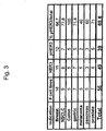
- the detection of basal phosphorylation of HER3 was conceived to underlie autocrine receptor activation and represent a selection marker for potentially suitable models in the use of HER3-directed therapeutic intervention.
- several cell lines were chosen and analysed for their phospho-HER3 content in the presence or absence of serum.
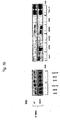
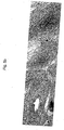
- An initial experiment showed that the pancreatic tumor cell line Bx-PC3 contains high levels of basally phosphorylated, i. e. activated HER3 in serum-starved and unstarved cells, indicating that Bx-PC3 may be a suitable model for an anti-HER3 therapeutic approach ( Fig.1 a) .
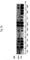
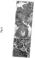
- HER3 phosphorylated i. e. activated HER3 was almost exclusively associated with cell surface membranes. This finding supported the idea that the presence of phosphorylated HER3 in such tissues could be used for selecting tumor patients responsive to anti-HER3 therapy. Furthermore, as well as monitoring HER3-directed therapy hair follicle biopsies could serve as a pharmacodynamic marker for monitoring HER3-directed treatment. Activated HER3 was also detected in a number of additional normal human tissues, including the GI tract, testis and bladder ( Fig. 6 ).
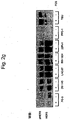
- HER3 The role of HER3 in normal skin has not been characterized previously. RNA expression was previously detected in postnatal skin (Kraus et al, 1989) Thus, our present analysis represents the first description in this respect. Surprisingly, we found that HER3 and its activated form are expressed in the hair follicles and in cells of the eccrine and sebaceous glands. This was not expected since the preferred partner of HER3, HER2, has not been reported to be expressed in these tissues This opens up the use of activated HER3 for patients selection etc. In contrast to activated EGFR, activated HER3 is not located intracellularly, but predominantly membranous.
- HER3 was also not observed in normal keratinocytes, where expression of EGFR is widespread (Expression of HER3 is rather low in keratinocytes (Laux et al, 2006). Thus, use of HER3 for diagnosis/selection and therapy may not only provide a regimen with less severe side effects compared to EGFR therapy which causes prominent skin rash, but may prove to be very useful for the monitoring of combination therapy.
- Immunoprecipitates were incubated for 4 hours at 4°C, washed three times with 1x HNTG (50 mM Hepes pH 7,5, 150 mM NaCl, 10% Glycerine, 1 mM EDTA pH 8,0, 0,1% Triton X-100) and denatured with 3x Laemmli buffer containing b-mercaptoethanol for 5 min at 100°C.
- the protein samples were separated by 7,5% SDS-PAGE, transferred to nitrocellulose membrane and incubated with anti-phosphotyrosine (4G10) or anti-pHER3 (21D3).
- Phosphoproteins were detected with anti-mouse-POD (for 4G10) or anti-rabbit-POD (for 21D3) secondary antibodies.
- the membranes were stripped and reprobed with anti-HER3 antibody (sc-285).
- HER modulator The anti-tumor efficacy of a HER modulator were evaluated in human xenograft tumor studies. In these studies, human tumors were grown as xenografts in immunocompromised mice and therapeutic efficacy was measured by the degree of tumor growth inhibition in response to administrations of the HER modulator. In order to determine, whether a HER modulator, as defined in forgoing paragraphs, at least partially interferes with tumor growth of human cancer cells in vivo, cells were implanted in nude/nude or SCID mice, using protocols known to the skilled artisan ( Sausville and Burger, (2006), Cancer Res. 66, 3351-3354 ). For example tumor cells were injected under the skin of nude mice, resulting in subcutaneous tumor growth on the back of the animals.
- Treatment was either started at the time of tumor cell implantation or when tumors had reached a defined size, e.g. a mean volume of 20-50 mm 3 .
- mice Prior to first treatment, mice were randomized to assure uniform tumor volumes (mean, median and standard deviation) across treatment groups. Typical dosing regimen included weekly administrations of 25 mg/kg of the HER modulator into the interpeneum. The first treatment included a loading dose of 50 mg/kg.
- Mice in control arms received agents, e.g. doxorubicin (pharmaceutical grade) with known cytostatic or cytotoxic activity against the human tumor cells.
- the HER3 receptor activation will be measured via IHC in cellular samples (tumor material at time of diagnosis, fresh tumor material prior to treatment, normal tissue) derived from a patient deemed to be a candidate for an anti-HER3 mAb treatment.
- the cellular sample will be achieved through various methods of biopsies (e.g. punch, brush, incisional, core) or other methods (e.g. plucking of hair and air follicles, buccal swab).
- the harvested tissue material will be processed, fixed and analyzed for presence of pHER3 (qualitative assay) and the relative amount of pHER3 (quantitative assay) via immunohistochemistry or other applicable methods (e.g. rtPCR, WB).
- An activation score for pHER3 will be calculated and the subject will be enrolled in the clinical study/treatment routine accordingly.
- the efficacy of an anti HER3 antibody in reducing HER3 receptor activation and/or HER3 mediated signal transduction can be assessed in cellular samples derived from a subject that has been treated with said anti HER3 antibody.
- the cellular samples can be retrieved in the previously described way, the timing of the samples is dependent on the treatment duration, schedule and follow up of therapy, but at least 2 samples will be taken (one at treatment start and one at maximum response).
- the quantitative and qualitative measurements for the 2 time points will be compared and the pharmacodynamic effect will be calculated from the delta/shift of values for the HER3 receptor activation.
- Normal tissue e.g. skin, hair follicles
- the outcome of the treatment will be correlated with the level of HER3 phosphorylation and the modulation of the phosphorylation/activation over time.
- the resulting prognostic index will be compared with standard indices (e.g. tumor grade, stage, patient demographics, treatment) and it will be determined whether pHER3 can serve as a superior marker for efficacy of the treatment, prognostic index for outcome, variabilities in response to the treatment or recurrence of the disease.
- HER3 phosphorylation may become a new surrogate marker for the assessment of a rsik-benefit score or a positive/negative prognosis with respect to anti-HER3 mAb therapy and other targeted or classical antineoplastic therapies.
- a cellular sample comprising normal and/or cancer cells is obtained from a subject deemed eligible for the treatment.
- the following methods are used in routine clinical practice to retrieve a tissue sample: swab (buccal, nasal swab), cuts (finger nails, toe nails), fine needle aspiration, punch biopsy, brush biopsy, scratch biopsy, biopsy using pincers or other surgical instruments, aspiration (e.g. blood, bone marrow), puncture (e.g.
- tissue samples can be used as well.
- micro-derm abrasive cytology, incision, surgical removal of organ parts or whole anatomical structures (bloc resection, tumor excision, lumpectomy), radiation assisted surgical procedure (gamma-knife surgery, laser assisted surgery), lavage (e.g. broncho-alveolar lavage, abdominal lavage), external drainage of organs (e.g. hydrocephalus, nephrostomy, T-drain bile duct). Any other method known in clinical practice for harvesting of tissue samples can be used as well.
- the biological sample is analyzed for HER3 phosphorylation, e.g., by immunoprecipitation or Western blot analysis, and/or for the presence of HER2/HER3 and/or HER3/HER4 heterodimers by any of the techniques described above.
- Patients with solid tumors will undergo at least 2 biopsies for the assessment of the pharmacodynamic effects of an anti-HER3 mAb treatment evaluated through changes/modulations in the HER3 phosphorylation.
- patients will be stratified for the pHER3 level and at the time of maximum clinical response, a second tissue sample will be taken from the patient.
- the samples will be analyzed for pHER3 expression (quantitative and qualitative) and the results are correlated with other parameters and clinical outcome.
- a rise in pHER3 activation may be considered as progression or non-response, whereas a decrease of pHER3 may be considered response to therapy.
Landscapes
- Health & Medical Sciences (AREA)
- Life Sciences & Earth Sciences (AREA)
- Chemical & Material Sciences (AREA)
- Immunology (AREA)
- Engineering & Computer Science (AREA)
- Molecular Biology (AREA)
- Urology & Nephrology (AREA)
- Hematology (AREA)
- Biomedical Technology (AREA)
- Medicinal Chemistry (AREA)
- General Health & Medical Sciences (AREA)
- Oncology (AREA)
- Cell Biology (AREA)
- Biochemistry (AREA)
- Organic Chemistry (AREA)
- Physics & Mathematics (AREA)
- Hospice & Palliative Care (AREA)
- Pathology (AREA)
- General Physics & Mathematics (AREA)
- Analytical Chemistry (AREA)
- Food Science & Technology (AREA)
- Microbiology (AREA)
- Biotechnology (AREA)
- General Chemical & Material Sciences (AREA)
- Animal Behavior & Ethology (AREA)
- Nuclear Medicine, Radiotherapy & Molecular Imaging (AREA)
- Public Health (AREA)
- Pharmacology & Pharmacy (AREA)
- Chemical Kinetics & Catalysis (AREA)
- Veterinary Medicine (AREA)
- Biophysics (AREA)
- Proteomics, Peptides & Aminoacids (AREA)
- Genetics & Genomics (AREA)
- Bioinformatics & Cheminformatics (AREA)
- Dermatology (AREA)
- Medicines That Contain Protein Lipid Enzymes And Other Medicines (AREA)
- Medicines Containing Antibodies Or Antigens For Use As Internal Diagnostic Agents (AREA)
- Peptides Or Proteins (AREA)
- Enzymes And Modification Thereof (AREA)
- Measuring Or Testing Involving Enzymes Or Micro-Organisms (AREA)
Priority Applications (2)
| Application Number | Priority Date | Filing Date | Title |
|---|---|---|---|
| EP12176507A EP2518508A1 (en) | 2006-11-28 | 2007-11-28 | Activated HER3 as a marker for predicting therapeutic efficacy |
| EP07846863.4A EP2097754B2 (en) | 2006-11-28 | 2007-11-28 | Activated her3 as a marker for predicting therapeutic efficacy |
Applications Claiming Priority (4)
| Application Number | Priority Date | Filing Date | Title |
|---|---|---|---|
| US86124306P | 2006-11-28 | 2006-11-28 | |
| EP06024658 | 2006-11-28 | ||
| PCT/EP2007/010335 WO2008064884A1 (en) | 2006-11-28 | 2007-11-28 | Activated her3 as a marker for predicting therapeutic efficacy |
| EP07846863.4A EP2097754B2 (en) | 2006-11-28 | 2007-11-28 | Activated her3 as a marker for predicting therapeutic efficacy |
Related Child Applications (1)
| Application Number | Title | Priority Date | Filing Date |
|---|---|---|---|
| EP12176507A Division-Into EP2518508A1 (en) | 2006-11-28 | 2007-11-28 | Activated HER3 as a marker for predicting therapeutic efficacy |
Publications (3)
| Publication Number | Publication Date |
|---|---|
| EP2097754A1 EP2097754A1 (en) | 2009-09-09 |
| EP2097754B1 EP2097754B1 (en) | 2015-01-07 |
| EP2097754B2 true EP2097754B2 (en) | 2018-01-24 |
Family
ID=38961070
Family Applications (2)
| Application Number | Title | Priority Date | Filing Date |
|---|---|---|---|
| EP12176507A Withdrawn EP2518508A1 (en) | 2006-11-28 | 2007-11-28 | Activated HER3 as a marker for predicting therapeutic efficacy |
| EP07846863.4A Active EP2097754B2 (en) | 2006-11-28 | 2007-11-28 | Activated her3 as a marker for predicting therapeutic efficacy |
Family Applications Before (1)
| Application Number | Title | Priority Date | Filing Date |
|---|---|---|---|
| EP12176507A Withdrawn EP2518508A1 (en) | 2006-11-28 | 2007-11-28 | Activated HER3 as a marker for predicting therapeutic efficacy |
Country Status (6)
| Country | Link |
|---|---|
| US (2) | US20100047829A1 (enExample) |
| EP (2) | EP2518508A1 (enExample) |
| JP (1) | JP5656406B2 (enExample) |
| AU (1) | AU2007324868B2 (enExample) |
| CA (1) | CA2670522C (enExample) |
| WO (1) | WO2008064884A1 (enExample) |
Families Citing this family (25)
| Publication number | Priority date | Publication date | Assignee | Title |
|---|---|---|---|---|
| BRPI0717416A2 (pt) | 2006-09-21 | 2013-11-12 | Prometheus Lab Inc | Método para realizar um imunoensaio complexo de alta produtividade, e, arranjo |
| BRPI0808055A2 (pt) | 2007-02-16 | 2013-07-30 | Merrimack Pharmaceuticals Inc | anticorpo monoclonal isolado ou porÇço do mesmo que se liga ao antÍgeno, composiÇço, Ácido nucleico isolado, vetor de expressço, cÉlula hospedeira, hibridoma, kit, uso de um anticorpo monoclonal isolado ou porÇço do mesmo que se liga ao antÍgeno, mÉtodo para diagnosticar um cÂncer associado com erbb3 em um paciente, e, anticorpo |
| KR20150039212A (ko) | 2007-03-02 | 2015-04-09 | 제넨테크, 인크. | 낮은 her3 발현을 기초로 한 her 이량체화 억제제에 대한 반응 예측 |
| KR101553723B1 (ko) | 2007-07-13 | 2015-09-16 | 네스텍 소시에테아노님 | 항체기반 어레이를 사용한 폐 암 치료를 위한 약물 선별법 |
| CA2716826C (en) | 2008-02-25 | 2017-05-09 | Prometheus Laboratories Inc. | Drug selection for breast cancer therapy using antibody-based arrays |
| EP2138511A1 (en) * | 2008-06-27 | 2009-12-30 | Max-Planck-Gesellschaft zur Förderung der Wissenschaften e.V. | HER3 as a determinant for the prognosis of melanoma |
| AU2009281721A1 (en) | 2008-08-15 | 2010-02-18 | Merrimack Pharmaceuticals, Inc. | Methods and systems for predicting response of cells to a therapeutic agent |
| US8652787B2 (en) | 2008-11-12 | 2014-02-18 | The United States Of America, As Represented By The Secretary, Department Of Health And Human Services | Use of ERBB4 as a prognostic and therapeutic marker for melanoma |
| US20120270745A1 (en) * | 2009-07-15 | 2012-10-25 | Prometheus Laboratories Inc. | Drug selection for cancer therapy by profiling signal transduction proteins in ascites or pleural efflux samples |
| JP5795311B2 (ja) | 2009-07-15 | 2015-10-14 | ネステク ソシエテ アノニム | 抗体ベースのアレイを使用する胃癌療法のための薬物選択 |
| MX2012007340A (es) | 2009-12-22 | 2012-08-01 | Roche Glycart Ag | Anticuerpos anti/her3 y usos de los mismos. |
| PL2544680T3 (pl) | 2010-03-11 | 2015-08-31 | Merrimack Pharmaceuticals Inc | Zastosowanie inhibitorów ERBB1 w leczeniu potrójnie ujemnego raka gruczołu sutkowego |
| US20110318336A1 (en) * | 2010-03-29 | 2011-12-29 | George Mason Intellectual Properties, Inc. | Identification and Treatment of Aggressive Lung Cancer Tumors |
| AR084469A1 (es) | 2010-07-09 | 2013-05-22 | Exelixis Inc | Combinaciones de inhibidores de quinasas para el tratamiento del cancer |
| TW201302793A (zh) | 2010-09-03 | 2013-01-16 | Glaxo Group Ltd | 新穎之抗原結合蛋白 |
| US9719995B2 (en) | 2011-02-03 | 2017-08-01 | Pierian Holdings, Inc. | Drug selection for colorectal cancer therapy using receptor tyrosine kinase profiling |
| JP6186575B2 (ja) | 2011-09-02 | 2017-08-30 | ダイアテック ホールディングス, インコーポレイテッドDiaTech Holdings, Inc. | 治療有効性を判定するためのシグナル経路タンパク質のプロファイリング |
| JP6073337B2 (ja) * | 2011-10-04 | 2017-02-01 | エクスプレッション、パソロジー、インコーポレイテッドExpression Pathology, Inc. | レセプター型チロシンキナーゼerbB−4タンパク質(HER4)に対するSRM/MRMアッセイ |
| EP3608340A1 (en) | 2011-11-23 | 2020-02-12 | Medlmmune, LLC | Binding molecules specific for her3 and uses thereof |
| AR094403A1 (es) | 2013-01-11 | 2015-07-29 | Hoffmann La Roche | Terapia de combinación de anticuerpos anti-her3 |
| KR101524915B1 (ko) * | 2013-05-10 | 2015-06-02 | 포항공과대학교 산학협력단 | 타이로신 인산화효소를 감지하는 형광 프로브 및 이의 용도 |
| WO2015048008A2 (en) | 2013-09-24 | 2015-04-02 | Medimmune, Llc | Binding molecules specific for her3 and uses thereof |
| WO2015100459A2 (en) | 2013-12-27 | 2015-07-02 | Merrimack Pharmaceuticals, Inc. | Biomarker profiles for predicting outcomes of cancer therapy with erbb3 inhibitors and/or chemotherapies |
| WO2015157634A1 (en) | 2014-04-11 | 2015-10-15 | Kolltan Pharmaceuticals, Inc. | Anti-erbb antibodies and methods of use thereof |
| US10184006B2 (en) | 2015-06-04 | 2019-01-22 | Merrimack Pharmaceuticals, Inc. | Biomarkers for predicting outcomes of cancer therapy with ErbB3 inhibitors |
Family Cites Families (22)
| Publication number | Priority date | Publication date | Assignee | Title |
|---|---|---|---|---|
| US4816567A (en) | 1983-04-08 | 1989-03-28 | Genentech, Inc. | Recombinant immunoglobin preparations |
| US5183884A (en) * | 1989-12-01 | 1993-02-02 | United States Of America | Dna segment encoding a gene for a receptor related to the epidermal growth factor receptor |
| US5804396A (en) * | 1994-10-12 | 1998-09-08 | Sugen, Inc. | Assay for agents active in proliferative disorders |
| US5968511A (en) | 1996-03-27 | 1999-10-19 | Genentech, Inc. | ErbB3 antibodies |
| EP1728802A3 (en) | 1996-03-27 | 2006-12-13 | Genentech, Inc. | ErbB3 antibodies |
| US5994071A (en) | 1997-04-04 | 1999-11-30 | Albany Medical College | Assessment of prostate cancer |
| AUPQ105799A0 (en) | 1999-06-18 | 1999-07-08 | Victor Chang Cardiac Research Institute, The | Cell growth inhibition |
| US6277640B1 (en) * | 2000-07-31 | 2001-08-21 | Isis Pharmaceuticals, Inc. | Antisense modulation of Her-3 expression |
| EP1958642A1 (en) * | 2000-12-22 | 2008-08-20 | Dana-Farber Cancer Institute | Regulation of cell growth by MUC1 |
| WO2003011897A1 (en) * | 2001-07-27 | 2003-02-13 | The Regents Of The University Of California | Modulation of heregulin and her3 interaction |
| EP1283053A1 (en) * | 2001-08-09 | 2003-02-12 | Max-Planck-Gesellschaft zur Förderung der Wissenschaften e.V. | Inhibitors of HER3 activity |
| US20040018528A1 (en) * | 2002-05-17 | 2004-01-29 | Sugen, Inc. | Novel biomarkers of tyrosine kinase inhibitor exposure and activity in mammals |
| DK1509230T3 (da) * | 2002-06-05 | 2007-05-14 | Cedars Sinai Medical Center | Gefitinib ( IRESSA) til behandlingen af cancer |
| DE60329194D1 (de) | 2002-07-25 | 2009-10-22 | Aclara Biosciences Inc | Nachweis der rezeptoroligomerisierung |
| WO2004087887A2 (en) | 2003-04-01 | 2004-10-14 | Monogram Biosciences, Inc. | Intracellular complexes as biomarkers |
| EP2348110B1 (en) * | 2003-05-30 | 2013-03-27 | OncoTherapy Science, Inc. | Process for screening a drug response in cancer patients |
| JP2005024385A (ja) | 2003-07-02 | 2005-01-27 | Matsushita Electric Ind Co Ltd | 感圧センサ |
| WO2005011607A2 (en) * | 2003-08-01 | 2005-02-10 | Smithkline Beecham Corporation | Treatment of cancers expressing p95 erbb2 |
| WO2005117553A2 (en) * | 2004-05-27 | 2005-12-15 | The Regents Of The University Of Colorado | Methods for prediction of clinical outcome to epidermal growth factor receptor inhibitors by cancer patients |
| CN101115849A (zh) * | 2004-12-07 | 2008-01-30 | 健泰科生物技术公司 | 选择her抑制剂疗法的患者 |
| WO2006096861A2 (en) * | 2005-03-08 | 2006-09-14 | Genentech, Inc. | METHODS FOR IDENTIFYING TUMORS RESPONSIVE TO TREATMENT WITH HER DIMERIZATION INHIBITORS (HDIs) |
| AR056857A1 (es) * | 2005-12-30 | 2007-10-24 | U3 Pharma Ag | Anticuerpos dirigidos hacia her-3 (receptor del factor de crecimiento epidérmico humano-3) y sus usos |
-
2007
- 2007-11-28 AU AU2007324868A patent/AU2007324868B2/en active Active
- 2007-11-28 CA CA2670522A patent/CA2670522C/en active Active
- 2007-11-28 US US12/516,682 patent/US20100047829A1/en not_active Abandoned
- 2007-11-28 WO PCT/EP2007/010335 patent/WO2008064884A1/en not_active Ceased
- 2007-11-28 JP JP2009538639A patent/JP5656406B2/ja active Active
- 2007-11-28 EP EP12176507A patent/EP2518508A1/en not_active Withdrawn
- 2007-11-28 EP EP07846863.4A patent/EP2097754B2/en active Active
-
2016
- 2016-02-18 US US15/047,041 patent/US10365283B2/en active Active
Also Published As
| Publication number | Publication date |
|---|---|
| JP2010510784A (ja) | 2010-04-08 |
| CA2670522A1 (en) | 2008-06-05 |
| EP2097754B1 (en) | 2015-01-07 |
| WO2008064884A1 (en) | 2008-06-05 |
| AU2007324868A1 (en) | 2008-06-05 |
| US20160161491A1 (en) | 2016-06-09 |
| US20100047829A1 (en) | 2010-02-25 |
| EP2518508A1 (en) | 2012-10-31 |
| AU2007324868B2 (en) | 2014-03-20 |
| US10365283B2 (en) | 2019-07-30 |
| EP2097754A1 (en) | 2009-09-09 |
| CA2670522C (en) | 2018-08-07 |
| JP5656406B2 (ja) | 2015-01-21 |
Similar Documents
| Publication | Publication Date | Title |
|---|---|---|
| US10365283B2 (en) | Activated HER3 as a marker for predicting therapeutic efficacy | |
| EP2438442B1 (en) | Methods and assays for measuring p95 and/or p95 complexes in a sample and antibodies specific for p95 | |
| US20070059785A1 (en) | Biomarkers in cancer | |
| KR101976219B1 (ko) | 유방암의 바이오마커 | |
| US20040248151A1 (en) | Method for predicting the response to HER2-directed therapy | |
| US20060094068A1 (en) | Predictive markers in cancer therapy | |
| JP4609930B2 (ja) | Her−2指向性治療に対する応答を予測する方法 | |
| JP2002530629A (ja) | 癌診断インジケータとしてのn末端切断型her−2/neu蛋白質 | |
| JP4628365B2 (ja) | 免疫組織化学的方法 | |
| KR20180069904A (ko) | 마커 분자를 기반으로 화학요법으로 치료되어야 하는 개체를 식별하는 방법 및 관련 용도 | |
| Personeni et al. | Correlation between the response to cetuximab alone or in combination with irinotecan and the activated/phosphorylated epidermal growth factor receptor in metastatic colorectal cancer | |
| KR20200016242A (ko) | T-dm1에 의한 암 치료 결과의 예측 | |
| AU2013203603A1 (en) | Activated HER3 as a marker for predicting therapeutic efficacy | |
| JP5295268B2 (ja) | がん疾患に罹患している患者の生物学的療法に対する感受性を測定するための方法 | |
| ES2527926T3 (es) | HER3 activado como marcador para predecir la eficacia terapéutica | |
| US20180209981A1 (en) | Wbp2 as a co-prognostic factor with her2 for stratification of patients for treatment | |
| TW202417641A (zh) | 預測對egfr激酶活性之抑制劑的反應之方法 | |
| CN107828886A (zh) | Gbp2基因和/或gbp2蛋白作为乳腺癌新的诊治靶点的应用 |
Legal Events
| Date | Code | Title | Description |
|---|---|---|---|
| PUAI | Public reference made under article 153(3) epc to a published international application that has entered the european phase |
Free format text: ORIGINAL CODE: 0009012 |
|
| 17P | Request for examination filed |
Effective date: 20090526 |
|
| AK | Designated contracting states |
Kind code of ref document: A1 Designated state(s): AT BE BG CH CY CZ DE DK EE ES FI FR GB GR HU IE IS IT LI LT LU LV MC MT NL PL PT RO SE SI SK TR |
|
| RIN1 | Information on inventor provided before grant (corrected) |
Inventor name: TREDER, MARTIN Inventor name: ROTHE, MIKE |
|
| DAX | Request for extension of the european patent (deleted) | ||
| 17Q | First examination report despatched |
Effective date: 20100719 |
|
| RAP1 | Party data changed (applicant data changed or rights of an application transferred) |
Owner name: U3 PHARMA GMBH |
|
| GRAP | Despatch of communication of intention to grant a patent |
Free format text: ORIGINAL CODE: EPIDOSNIGR1 |
|
| INTG | Intention to grant announced |
Effective date: 20140813 |
|
| GRAS | Grant fee paid |
Free format text: ORIGINAL CODE: EPIDOSNIGR3 |
|
| GRAA | (expected) grant |
Free format text: ORIGINAL CODE: 0009210 |
|
| AK | Designated contracting states |
Kind code of ref document: B1 Designated state(s): AT BE BG CH CY CZ DE DK EE ES FI FR GB GR HU IE IS IT LI LT LU LV MC MT NL PL PT RO SE SI SK TR |
|
| REG | Reference to a national code |
Ref country code: GB Ref legal event code: FG4D |
|
| REG | Reference to a national code |
Ref country code: CH Ref legal event code: EP |
|
| REG | Reference to a national code |
Ref country code: IE Ref legal event code: FG4D |
|
| REG | Reference to a national code |
Ref country code: ES Ref legal event code: FG2A Ref document number: 2527926 Country of ref document: ES Kind code of ref document: T3 Effective date: 20150202 |
|
| REG | Reference to a national code |
Ref country code: AT Ref legal event code: REF Ref document number: 706079 Country of ref document: AT Kind code of ref document: T Effective date: 20150215 |
|
| REG | Reference to a national code |
Ref country code: DE Ref legal event code: R096 Ref document number: 602007040001 Country of ref document: DE Effective date: 20150219 |
|
| REG | Reference to a national code |
Ref country code: NL Ref legal event code: VDEP Effective date: 20150107 |
|
| REG | Reference to a national code |
Ref country code: AT Ref legal event code: MK05 Ref document number: 706079 Country of ref document: AT Kind code of ref document: T Effective date: 20150107 |
|
| REG | Reference to a national code |
Ref country code: LT Ref legal event code: MG4D |
|
| PG25 | Lapsed in a contracting state [announced via postgrant information from national office to epo] |
Ref country code: FI Free format text: LAPSE BECAUSE OF FAILURE TO SUBMIT A TRANSLATION OF THE DESCRIPTION OR TO PAY THE FEE WITHIN THE PRESCRIBED TIME-LIMIT Effective date: 20150107 Ref country code: SE Free format text: LAPSE BECAUSE OF FAILURE TO SUBMIT A TRANSLATION OF THE DESCRIPTION OR TO PAY THE FEE WITHIN THE PRESCRIBED TIME-LIMIT Effective date: 20150107 Ref country code: BG Free format text: LAPSE BECAUSE OF FAILURE TO SUBMIT A TRANSLATION OF THE DESCRIPTION OR TO PAY THE FEE WITHIN THE PRESCRIBED TIME-LIMIT Effective date: 20150407 Ref country code: LT Free format text: LAPSE BECAUSE OF FAILURE TO SUBMIT A TRANSLATION OF THE DESCRIPTION OR TO PAY THE FEE WITHIN THE PRESCRIBED TIME-LIMIT Effective date: 20150107 |
|
| PG25 | Lapsed in a contracting state [announced via postgrant information from national office to epo] |
Ref country code: LV Free format text: LAPSE BECAUSE OF FAILURE TO SUBMIT A TRANSLATION OF THE DESCRIPTION OR TO PAY THE FEE WITHIN THE PRESCRIBED TIME-LIMIT Effective date: 20150107 Ref country code: AT Free format text: LAPSE BECAUSE OF FAILURE TO SUBMIT A TRANSLATION OF THE DESCRIPTION OR TO PAY THE FEE WITHIN THE PRESCRIBED TIME-LIMIT Effective date: 20150107 Ref country code: PL Free format text: LAPSE BECAUSE OF FAILURE TO SUBMIT A TRANSLATION OF THE DESCRIPTION OR TO PAY THE FEE WITHIN THE PRESCRIBED TIME-LIMIT Effective date: 20150107 Ref country code: GR Free format text: LAPSE BECAUSE OF FAILURE TO SUBMIT A TRANSLATION OF THE DESCRIPTION OR TO PAY THE FEE WITHIN THE PRESCRIBED TIME-LIMIT Effective date: 20150408 Ref country code: IS Free format text: LAPSE BECAUSE OF FAILURE TO SUBMIT A TRANSLATION OF THE DESCRIPTION OR TO PAY THE FEE WITHIN THE PRESCRIBED TIME-LIMIT Effective date: 20150507 Ref country code: NL Free format text: LAPSE BECAUSE OF FAILURE TO SUBMIT A TRANSLATION OF THE DESCRIPTION OR TO PAY THE FEE WITHIN THE PRESCRIBED TIME-LIMIT Effective date: 20150107 |
|
| REG | Reference to a national code |
Ref country code: DE Ref legal event code: R026 Ref document number: 602007040001 Country of ref document: DE |
|
| PLBI | Opposition filed |
Free format text: ORIGINAL CODE: 0009260 |
|
| PG25 | Lapsed in a contracting state [announced via postgrant information from national office to epo] |
Ref country code: SK Free format text: LAPSE BECAUSE OF FAILURE TO SUBMIT A TRANSLATION OF THE DESCRIPTION OR TO PAY THE FEE WITHIN THE PRESCRIBED TIME-LIMIT Effective date: 20150107 Ref country code: RO Free format text: LAPSE BECAUSE OF FAILURE TO SUBMIT A TRANSLATION OF THE DESCRIPTION OR TO PAY THE FEE WITHIN THE PRESCRIBED TIME-LIMIT Effective date: 20150107 Ref country code: CZ Free format text: LAPSE BECAUSE OF FAILURE TO SUBMIT A TRANSLATION OF THE DESCRIPTION OR TO PAY THE FEE WITHIN THE PRESCRIBED TIME-LIMIT Effective date: 20150107 Ref country code: DK Free format text: LAPSE BECAUSE OF FAILURE TO SUBMIT A TRANSLATION OF THE DESCRIPTION OR TO PAY THE FEE WITHIN THE PRESCRIBED TIME-LIMIT Effective date: 20150107 Ref country code: EE Free format text: LAPSE BECAUSE OF FAILURE TO SUBMIT A TRANSLATION OF THE DESCRIPTION OR TO PAY THE FEE WITHIN THE PRESCRIBED TIME-LIMIT Effective date: 20150107 |
|
| 26 | Opposition filed |
Opponent name: F.HOFFMANN-LA ROCHE AG Effective date: 20151001 |
|
| PLAX | Notice of opposition and request to file observation + time limit sent |
Free format text: ORIGINAL CODE: EPIDOSNOBS2 |
|
| REG | Reference to a national code |
Ref country code: FR Ref legal event code: PLFP Year of fee payment: 9 |
|
| PG25 | Lapsed in a contracting state [announced via postgrant information from national office to epo] |
Ref country code: SI Free format text: LAPSE BECAUSE OF FAILURE TO SUBMIT A TRANSLATION OF THE DESCRIPTION OR TO PAY THE FEE WITHIN THE PRESCRIBED TIME-LIMIT Effective date: 20150107 |
|
| PLAF | Information modified related to communication of a notice of opposition and request to file observations + time limit |
Free format text: ORIGINAL CODE: EPIDOSCOBS2 |
|
| PLBB | Reply of patent proprietor to notice(s) of opposition received |
Free format text: ORIGINAL CODE: EPIDOSNOBS3 |
|
| PG25 | Lapsed in a contracting state [announced via postgrant information from national office to epo] |
Ref country code: BE Free format text: LAPSE BECAUSE OF FAILURE TO SUBMIT A TRANSLATION OF THE DESCRIPTION OR TO PAY THE FEE WITHIN THE PRESCRIBED TIME-LIMIT Effective date: 20150107 |
|
| PG25 | Lapsed in a contracting state [announced via postgrant information from national office to epo] |
Ref country code: MC Free format text: LAPSE BECAUSE OF FAILURE TO SUBMIT A TRANSLATION OF THE DESCRIPTION OR TO PAY THE FEE WITHIN THE PRESCRIBED TIME-LIMIT Effective date: 20150107 Ref country code: LU Free format text: LAPSE BECAUSE OF FAILURE TO SUBMIT A TRANSLATION OF THE DESCRIPTION OR TO PAY THE FEE WITHIN THE PRESCRIBED TIME-LIMIT Effective date: 20151128 |
|
| REG | Reference to a national code |
Ref country code: CH Ref legal event code: PL |
|
| PG25 | Lapsed in a contracting state [announced via postgrant information from national office to epo] |
Ref country code: LI Free format text: LAPSE BECAUSE OF NON-PAYMENT OF DUE FEES Effective date: 20151130 Ref country code: CH Free format text: LAPSE BECAUSE OF NON-PAYMENT OF DUE FEES Effective date: 20151130 |
|
| REG | Reference to a national code |
Ref country code: IE Ref legal event code: MM4A |
|
| REG | Reference to a national code |
Ref country code: FR Ref legal event code: PLFP Year of fee payment: 10 |
|
| PG25 | Lapsed in a contracting state [announced via postgrant information from national office to epo] |
Ref country code: IE Free format text: LAPSE BECAUSE OF NON-PAYMENT OF DUE FEES Effective date: 20151128 |
|
| RAP2 | Party data changed (patent owner data changed or rights of a patent transferred) |
Owner name: DAIICHI SANKYO EUROPE GMBH |
|
| REG | Reference to a national code |
Ref country code: DE Ref legal event code: R082 Ref document number: 602007040001 Country of ref document: DE Representative=s name: WEICKMANN & WEICKMANN PATENTANWAELTE - RECHTSA, DE Ref country code: DE Ref legal event code: R082 Ref document number: 602007040001 Country of ref document: DE Representative=s name: WEICKMANN & WEICKMANN PATENT- UND RECHTSANWAEL, DE Ref country code: DE Ref legal event code: R081 Ref document number: 602007040001 Country of ref document: DE Owner name: DAIICHI SANKYO EUROPE GMBH, DE Free format text: FORMER OWNER: U3 PHARMA GMBH, 82152 PLANEGG, DE |
|
| PG25 | Lapsed in a contracting state [announced via postgrant information from national office to epo] |
Ref country code: HU Free format text: LAPSE BECAUSE OF FAILURE TO SUBMIT A TRANSLATION OF THE DESCRIPTION OR TO PAY THE FEE WITHIN THE PRESCRIBED TIME-LIMIT; INVALID AB INITIO Effective date: 20071128 |
|
| PG25 | Lapsed in a contracting state [announced via postgrant information from national office to epo] |
Ref country code: CY Free format text: LAPSE BECAUSE OF FAILURE TO SUBMIT A TRANSLATION OF THE DESCRIPTION OR TO PAY THE FEE WITHIN THE PRESCRIBED TIME-LIMIT Effective date: 20150107 |
|
| PG25 | Lapsed in a contracting state [announced via postgrant information from national office to epo] |
Ref country code: MT Free format text: LAPSE BECAUSE OF FAILURE TO SUBMIT A TRANSLATION OF THE DESCRIPTION OR TO PAY THE FEE WITHIN THE PRESCRIBED TIME-LIMIT Effective date: 20150107 Ref country code: TR Free format text: LAPSE BECAUSE OF FAILURE TO SUBMIT A TRANSLATION OF THE DESCRIPTION OR TO PAY THE FEE WITHIN THE PRESCRIBED TIME-LIMIT Effective date: 20150107 |
|
| REG | Reference to a national code |
Ref country code: FR Ref legal event code: PLFP Year of fee payment: 11 |
|
| PUAH | Patent maintained in amended form |
Free format text: ORIGINAL CODE: 0009272 |
|
| STAA | Information on the status of an ep patent application or granted ep patent |
Free format text: STATUS: PATENT MAINTAINED AS AMENDED |
|
| 27A | Patent maintained in amended form |
Effective date: 20180124 |
|
| AK | Designated contracting states |
Kind code of ref document: B2 Designated state(s): AT BE BG CH CY CZ DE DK EE ES FI FR GB GR HU IE IS IT LI LT LU LV MC MT NL PL PT RO SE SI SK TR |
|
| REG | Reference to a national code |
Ref country code: DE Ref legal event code: R102 Ref document number: 602007040001 Country of ref document: DE |
|
| REG | Reference to a national code |
Ref country code: ES Ref legal event code: DC2A Ref document number: 2527926 Country of ref document: ES Kind code of ref document: T5 Effective date: 20180312 |
|
| PG25 | Lapsed in a contracting state [announced via postgrant information from national office to epo] |
Ref country code: PT Free format text: LAPSE BECAUSE OF FAILURE TO SUBMIT A TRANSLATION OF THE DESCRIPTION OR TO PAY THE FEE WITHIN THE PRESCRIBED TIME-LIMIT Effective date: 20150107 |
|
| REG | Reference to a national code |
Ref country code: FR Ref legal event code: PLFP Year of fee payment: 12 |
|
| P01 | Opt-out of the competence of the unified patent court (upc) registered |
Effective date: 20230504 |
|
| PGFP | Annual fee paid to national office [announced via postgrant information from national office to epo] |
Ref country code: DE Payment date: 20241001 Year of fee payment: 18 |
|
| PGFP | Annual fee paid to national office [announced via postgrant information from national office to epo] |
Ref country code: GB Payment date: 20241002 Year of fee payment: 18 |
|
| PGFP | Annual fee paid to national office [announced via postgrant information from national office to epo] |
Ref country code: IT Payment date: 20241010 Year of fee payment: 18 Ref country code: ES Payment date: 20241204 Year of fee payment: 18 |
|
| PGFP | Annual fee paid to national office [announced via postgrant information from national office to epo] |
Ref country code: FR Payment date: 20250930 Year of fee payment: 19 |
|
| REG | Reference to a national code |
Ref country code: DE Ref legal event code: R079 Ref document number: 602007040001 Country of ref document: DE Free format text: PREVIOUS MAIN CLASS: G01N0033574000 Ipc: G01N0033575000 |