EP2075019A1 - Tubule rénal bioartificiel - Google Patents
Tubule rénal bioartificiel Download PDFInfo
- Publication number
- EP2075019A1 EP2075019A1 EP07829816A EP07829816A EP2075019A1 EP 2075019 A1 EP2075019 A1 EP 2075019A1 EP 07829816 A EP07829816 A EP 07829816A EP 07829816 A EP07829816 A EP 07829816A EP 2075019 A1 EP2075019 A1 EP 2075019A1
- Authority
- EP
- European Patent Office
- Prior art keywords
- cells
- bioartificial
- renal
- tubular epithelial
- epithelial cells
- Prior art date
- Legal status (The legal status is an assumption and is not a legal conclusion. Google has not performed a legal analysis and makes no representation as to the accuracy of the status listed.)
- Granted
Links
- 210000005239 tubule Anatomy 0.000 title claims abstract description 90
- 210000004027 cell Anatomy 0.000 claims abstract description 142
- 210000004926 tubular epithelial cell Anatomy 0.000 claims abstract description 81
- DVEXZJFMOKTQEZ-JYFOCSDGSA-N U0126 Chemical group C=1C=CC=C(N)C=1SC(\N)=C(/C#N)\C(\C#N)=C(/N)SC1=CC=CC=C1N DVEXZJFMOKTQEZ-JYFOCSDGSA-N 0.000 claims abstract description 54
- 229940124647 MEK inhibitor Drugs 0.000 claims abstract description 44
- 239000000823 artificial membrane Substances 0.000 claims abstract description 42
- 239000002829 mitogen activated protein kinase inhibitor Substances 0.000 claims abstract description 42
- 239000002356 single layer Substances 0.000 claims abstract description 41
- 230000030944 contact inhibition Effects 0.000 claims abstract description 22
- 239000012510 hollow fiber Substances 0.000 claims description 65
- 239000012528 membrane Substances 0.000 claims description 32
- 238000000034 method Methods 0.000 claims description 21
- 238000012423 maintenance Methods 0.000 claims description 16
- 238000013517 stratification Methods 0.000 claims description 16
- 229920002492 poly(sulfone) Polymers 0.000 claims description 6
- 229920002301 cellulose acetate Polymers 0.000 claims description 4
- 239000004642 Polyimide Substances 0.000 claims description 3
- 238000012258 culturing Methods 0.000 claims description 3
- 229920001721 polyimide Polymers 0.000 claims description 3
- 210000003734 kidney Anatomy 0.000 abstract description 63
- 239000000126 substance Substances 0.000 abstract description 21
- 238000000502 dialysis Methods 0.000 abstract description 9
- 238000009563 continuous hemofiltration Methods 0.000 abstract description 2
- IAZDPXIOMUYVGZ-UHFFFAOYSA-N Dimethylsulphoxide Chemical compound CS(C)=O IAZDPXIOMUYVGZ-UHFFFAOYSA-N 0.000 description 56
- 239000002609 medium Substances 0.000 description 55
- 238000011282 treatment Methods 0.000 description 28
- 230000006870 function Effects 0.000 description 24
- 239000008280 blood Substances 0.000 description 17
- 102000019149 MAP kinase activity proteins Human genes 0.000 description 16
- 108040008097 MAP kinase activity proteins Proteins 0.000 description 16
- 210000004369 blood Anatomy 0.000 description 16
- 108090000623 proteins and genes Proteins 0.000 description 16
- 235000010633 broth Nutrition 0.000 description 14
- 238000001914 filtration Methods 0.000 description 14
- DDRJAANPRJIHGJ-UHFFFAOYSA-N creatinine Chemical compound CN1CC(=O)NC1=N DDRJAANPRJIHGJ-UHFFFAOYSA-N 0.000 description 12
- 102000004169 proteins and genes Human genes 0.000 description 12
- 230000000694 effects Effects 0.000 description 11
- 239000008103 glucose Substances 0.000 description 11
- 238000001631 haemodialysis Methods 0.000 description 11
- 229920000219 Ethylene vinyl alcohol Polymers 0.000 description 10
- 102000043136 MAP kinase family Human genes 0.000 description 10
- 108091054455 MAP kinase family Proteins 0.000 description 10
- 230000000322 hemodialysis Effects 0.000 description 10
- WQZGKKKJIJFFOK-GASJEMHNSA-N Glucose Natural products OC[C@H]1OC(O)[C@H](O)[C@@H](O)[C@@H]1O WQZGKKKJIJFFOK-GASJEMHNSA-N 0.000 description 9
- 108090000744 Mitogen-Activated Protein Kinase Kinases Proteins 0.000 description 9
- 102000004232 Mitogen-Activated Protein Kinase Kinases Human genes 0.000 description 9
- 210000002919 epithelial cell Anatomy 0.000 description 9
- 239000004715 ethylene vinyl alcohol Substances 0.000 description 9
- RZXDTJIXPSCHCI-UHFFFAOYSA-N hexa-1,5-diene-2,5-diol Chemical compound OC(=C)CCC(O)=C RZXDTJIXPSCHCI-UHFFFAOYSA-N 0.000 description 9
- XLYOFNOQVPJJNP-UHFFFAOYSA-N water Substances O XLYOFNOQVPJJNP-UHFFFAOYSA-N 0.000 description 9
- 230000009056 active transport Effects 0.000 description 8
- 210000000056 organ Anatomy 0.000 description 8
- 210000002700 urine Anatomy 0.000 description 8
- 239000006144 Dulbecco’s modified Eagle's medium Substances 0.000 description 7
- 102000007665 Extracellular Signal-Regulated MAP Kinases Human genes 0.000 description 7
- 108010007457 Extracellular Signal-Regulated MAP Kinases Proteins 0.000 description 7
- 230000001461 cytolytic effect Effects 0.000 description 7
- 239000000706 filtrate Substances 0.000 description 7
- 239000000463 material Substances 0.000 description 7
- 230000035755 proliferation Effects 0.000 description 7
- 230000009103 reabsorption Effects 0.000 description 7
- 230000015572 biosynthetic process Effects 0.000 description 6
- 230000001684 chronic effect Effects 0.000 description 6
- 229940109239 creatinine Drugs 0.000 description 6
- 239000003792 electrolyte Substances 0.000 description 6
- 238000010899 nucleation Methods 0.000 description 6
- 238000001262 western blot Methods 0.000 description 6
- 208000009304 Acute Kidney Injury Diseases 0.000 description 5
- 241000282465 Canis Species 0.000 description 5
- 208000033626 Renal failure acute Diseases 0.000 description 5
- 102100023132 Transcription factor Jun Human genes 0.000 description 5
- 230000009471 action Effects 0.000 description 5
- 201000011040 acute kidney failure Diseases 0.000 description 5
- 208000012998 acute renal failure Diseases 0.000 description 5
- 238000002474 experimental method Methods 0.000 description 5
- 230000007774 longterm Effects 0.000 description 5
- 239000002207 metabolite Substances 0.000 description 5
- 210000001519 tissue Anatomy 0.000 description 5
- 230000032258 transport Effects 0.000 description 5
- 239000003146 anticoagulant agent Substances 0.000 description 4
- WQZGKKKJIJFFOK-VFUOTHLCSA-N beta-D-glucose Chemical compound OC[C@H]1O[C@@H](O)[C@H](O)[C@@H](O)[C@@H]1O WQZGKKKJIJFFOK-VFUOTHLCSA-N 0.000 description 4
- 230000008859 change Effects 0.000 description 4
- 238000011161 development Methods 0.000 description 4
- 238000005516 engineering process Methods 0.000 description 4
- 239000003112 inhibitor Substances 0.000 description 4
- 239000010410 layer Substances 0.000 description 4
- 239000007788 liquid Substances 0.000 description 4
- 230000007102 metabolic function Effects 0.000 description 4
- 230000037361 pathway Effects 0.000 description 4
- 210000005084 renal tissue Anatomy 0.000 description 4
- 108010001336 Horseradish Peroxidase Proteins 0.000 description 3
- 208000034486 Multi-organ failure Diseases 0.000 description 3
- 208000010718 Multiple Organ Failure Diseases 0.000 description 3
- GLNADSQYFUSGOU-GPTZEZBUSA-J Trypan blue Chemical compound [Na+].[Na+].[Na+].[Na+].C1=C(S([O-])(=O)=O)C=C2C=C(S([O-])(=O)=O)C(/N=N/C3=CC=C(C=C3C)C=3C=C(C(=CC=3)\N=N\C=3C(=CC4=CC(=CC(N)=C4C=3O)S([O-])(=O)=O)S([O-])(=O)=O)C)=C(O)C2=C1N GLNADSQYFUSGOU-GPTZEZBUSA-J 0.000 description 3
- XSQUKJJJFZCRTK-UHFFFAOYSA-N Urea Chemical compound NC(N)=O XSQUKJJJFZCRTK-UHFFFAOYSA-N 0.000 description 3
- PNNCWTXUWKENPE-UHFFFAOYSA-N [N].NC(N)=O Chemical compound [N].NC(N)=O PNNCWTXUWKENPE-UHFFFAOYSA-N 0.000 description 3
- 230000009102 absorption Effects 0.000 description 3
- 238000010521 absorption reaction Methods 0.000 description 3
- 230000002965 anti-thrombogenic effect Effects 0.000 description 3
- 239000012237 artificial material Substances 0.000 description 3
- 229920001577 copolymer Polymers 0.000 description 3
- 229940079593 drug Drugs 0.000 description 3
- 239000003814 drug Substances 0.000 description 3
- 230000001434 glomerular Effects 0.000 description 3
- 150000002500 ions Chemical class 0.000 description 3
- 208000029744 multiple organ dysfunction syndrome Diseases 0.000 description 3
- 230000026731 phosphorylation Effects 0.000 description 3
- 238000006366 phosphorylation reaction Methods 0.000 description 3
- 239000002243 precursor Substances 0.000 description 3
- 238000001878 scanning electron micrograph Methods 0.000 description 3
- 230000001629 suppression Effects 0.000 description 3
- 238000012546 transfer Methods 0.000 description 3
- 239000002699 waste material Substances 0.000 description 3
- QGZKDVFQNNGYKY-UHFFFAOYSA-N Ammonia Chemical compound N QGZKDVFQNNGYKY-UHFFFAOYSA-N 0.000 description 2
- 102100031480 Dual specificity mitogen-activated protein kinase kinase 1 Human genes 0.000 description 2
- 101710146526 Dual specificity mitogen-activated protein kinase kinase 1 Proteins 0.000 description 2
- 102100023266 Dual specificity mitogen-activated protein kinase kinase 2 Human genes 0.000 description 2
- 101710146529 Dual specificity mitogen-activated protein kinase kinase 2 Proteins 0.000 description 2
- 102100021616 Ephrin type-A receptor 4 Human genes 0.000 description 2
- 102000010834 Extracellular Matrix Proteins Human genes 0.000 description 2
- 108010037362 Extracellular Matrix Proteins Proteins 0.000 description 2
- 206010016803 Fluid overload Diseases 0.000 description 2
- DHCLVCXQIBBOPH-UHFFFAOYSA-N Glycerol 2-phosphate Chemical compound OCC(CO)OP(O)(O)=O DHCLVCXQIBBOPH-UHFFFAOYSA-N 0.000 description 2
- 206010019280 Heart failures Diseases 0.000 description 2
- HTTJABKRGRZYRN-UHFFFAOYSA-N Heparin Chemical compound OC1C(NC(=O)C)C(O)OC(COS(O)(=O)=O)C1OC1C(OS(O)(=O)=O)C(O)C(OC2C(C(OS(O)(=O)=O)C(OC3C(C(O)C(O)C(O3)C(O)=O)OS(O)(=O)=O)C(CO)O2)NS(O)(=O)=O)C(C(O)=O)O1 HTTJABKRGRZYRN-UHFFFAOYSA-N 0.000 description 2
- 206010058467 Lung neoplasm malignant Diseases 0.000 description 2
- 102000001291 MAP Kinase Kinase Kinase Human genes 0.000 description 2
- 108060006687 MAP kinase kinase kinase Proteins 0.000 description 2
- -1 MKK-3 Proteins 0.000 description 2
- 108091000080 Phosphotransferase Proteins 0.000 description 2
- 102000003923 Protein Kinase C Human genes 0.000 description 2
- 108090000315 Protein Kinase C Proteins 0.000 description 2
- 208000001647 Renal Insufficiency Diseases 0.000 description 2
- PXIPVTKHYLBLMZ-UHFFFAOYSA-N Sodium azide Chemical compound [Na+].[N-]=[N+]=[N-] PXIPVTKHYLBLMZ-UHFFFAOYSA-N 0.000 description 2
- FAPWRFPIFSIZLT-UHFFFAOYSA-M Sodium chloride Chemical compound [Na+].[Cl-] FAPWRFPIFSIZLT-UHFFFAOYSA-M 0.000 description 2
- 238000009825 accumulation Methods 0.000 description 2
- 230000004913 activation Effects 0.000 description 2
- 230000001154 acute effect Effects 0.000 description 2
- 150000001413 amino acids Chemical class 0.000 description 2
- 229940127090 anticoagulant agent Drugs 0.000 description 2
- 229940127219 anticoagulant drug Drugs 0.000 description 2
- 230000008901 benefit Effects 0.000 description 2
- 239000004202 carbamide Substances 0.000 description 2
- 230000003915 cell function Effects 0.000 description 2
- 239000013553 cell monolayer Substances 0.000 description 2
- 230000004663 cell proliferation Effects 0.000 description 2
- 230000005754 cellular signaling Effects 0.000 description 2
- 208000020832 chronic kidney disease Diseases 0.000 description 2
- 208000022831 chronic renal failure syndrome Diseases 0.000 description 2
- 239000000039 congener Substances 0.000 description 2
- 230000001276 controlling effect Effects 0.000 description 2
- 210000004748 cultured cell Anatomy 0.000 description 2
- 230000006866 deterioration Effects 0.000 description 2
- 230000007368 endocrine function Effects 0.000 description 2
- 238000011156 evaluation Methods 0.000 description 2
- 210000002744 extracellular matrix Anatomy 0.000 description 2
- 230000012010 growth Effects 0.000 description 2
- 229960002897 heparin Drugs 0.000 description 2
- 229920000669 heparin Polymers 0.000 description 2
- 201000006370 kidney failure Diseases 0.000 description 2
- 201000005202 lung cancer Diseases 0.000 description 2
- 208000020816 lung neoplasm Diseases 0.000 description 2
- 102000020233 phosphotransferase Human genes 0.000 description 2
- 229920002981 polyvinylidene fluoride Polymers 0.000 description 2
- 239000011148 porous material Substances 0.000 description 2
- 239000000047 product Substances 0.000 description 2
- 238000002731 protein assay Methods 0.000 description 2
- 102000009929 raf Kinases Human genes 0.000 description 2
- 108010077182 raf Kinases Proteins 0.000 description 2
- 238000011160 research Methods 0.000 description 2
- 239000000523 sample Substances 0.000 description 2
- 238000004626 scanning electron microscopy Methods 0.000 description 2
- 238000000926 separation method Methods 0.000 description 2
- 239000000758 substrate Substances 0.000 description 2
- 230000008961 swelling Effects 0.000 description 2
- 208000024891 symptom Diseases 0.000 description 2
- 230000009885 systemic effect Effects 0.000 description 2
- 238000002560 therapeutic procedure Methods 0.000 description 2
- IHIXIJGXTJIKRB-UHFFFAOYSA-N trisodium vanadate Chemical compound [Na+].[Na+].[Na+].[O-][V]([O-])([O-])=O IHIXIJGXTJIKRB-UHFFFAOYSA-N 0.000 description 2
- 238000000108 ultra-filtration Methods 0.000 description 2
- QKNYBSVHEMOAJP-UHFFFAOYSA-N 2-amino-2-(hydroxymethyl)propane-1,3-diol;hydron;chloride Chemical compound Cl.OCC(N)(CO)CO QKNYBSVHEMOAJP-UHFFFAOYSA-N 0.000 description 1
- 101100513486 Caenorhabditis elegans mkk-4 gene Proteins 0.000 description 1
- OYPRJOBELJOOCE-UHFFFAOYSA-N Calcium Chemical compound [Ca] OYPRJOBELJOOCE-UHFFFAOYSA-N 0.000 description 1
- 241000282472 Canis lupus familiaris Species 0.000 description 1
- 102000008186 Collagen Human genes 0.000 description 1
- 108010035532 Collagen Proteins 0.000 description 1
- 102000012422 Collagen Type I Human genes 0.000 description 1
- 108010022452 Collagen Type I Proteins 0.000 description 1
- 102100023033 Cyclic AMP-dependent transcription factor ATF-2 Human genes 0.000 description 1
- 102000013701 Cyclin-Dependent Kinase 4 Human genes 0.000 description 1
- 108010025464 Cyclin-Dependent Kinase 4 Proteins 0.000 description 1
- 206010064553 Dialysis amyloidosis Diseases 0.000 description 1
- 102100023275 Dual specificity mitogen-activated protein kinase kinase 3 Human genes 0.000 description 1
- 102100023274 Dual specificity mitogen-activated protein kinase kinase 4 Human genes 0.000 description 1
- 102100023272 Dual specificity mitogen-activated protein kinase kinase 5 Human genes 0.000 description 1
- 102100023401 Dual specificity mitogen-activated protein kinase kinase 6 Human genes 0.000 description 1
- 102100023332 Dual specificity mitogen-activated protein kinase kinase 7 Human genes 0.000 description 1
- ZGTMUACCHSMWAC-UHFFFAOYSA-L EDTA disodium salt (anhydrous) Chemical compound [Na+].[Na+].OC(=O)CN(CC([O-])=O)CCN(CC(O)=O)CC([O-])=O ZGTMUACCHSMWAC-UHFFFAOYSA-L 0.000 description 1
- 239000012824 ERK inhibitor Substances 0.000 description 1
- 102000016359 Fibronectins Human genes 0.000 description 1
- 108010067306 Fibronectins Proteins 0.000 description 1
- SXRSQZLOMIGNAQ-UHFFFAOYSA-N Glutaraldehyde Chemical compound O=CCCCC=O SXRSQZLOMIGNAQ-UHFFFAOYSA-N 0.000 description 1
- 101000974934 Homo sapiens Cyclic AMP-dependent transcription factor ATF-2 Proteins 0.000 description 1
- 101001115394 Homo sapiens Dual specificity mitogen-activated protein kinase kinase 3 Proteins 0.000 description 1
- 101001115395 Homo sapiens Dual specificity mitogen-activated protein kinase kinase 4 Proteins 0.000 description 1
- 101001115390 Homo sapiens Dual specificity mitogen-activated protein kinase kinase 5 Proteins 0.000 description 1
- 101000624426 Homo sapiens Dual specificity mitogen-activated protein kinase kinase 6 Proteins 0.000 description 1
- 101000624594 Homo sapiens Dual specificity mitogen-activated protein kinase kinase 7 Proteins 0.000 description 1
- 101100065486 Homo sapiens EPHA4 gene Proteins 0.000 description 1
- 101000997829 Homo sapiens Glial cell line-derived neurotrophic factor Proteins 0.000 description 1
- 101000950687 Homo sapiens Mitogen-activated protein kinase 7 Proteins 0.000 description 1
- 101001050288 Homo sapiens Transcription factor Jun Proteins 0.000 description 1
- DGAQECJNVWCQMB-PUAWFVPOSA-M Ilexoside XXIX Chemical compound C[C@@H]1CC[C@@]2(CC[C@@]3(C(=CC[C@H]4[C@]3(CC[C@@H]5[C@@]4(CC[C@@H](C5(C)C)OS(=O)(=O)[O-])C)C)[C@@H]2[C@]1(C)O)C)C(=O)O[C@H]6[C@@H]([C@H]([C@@H]([C@H](O6)CO)O)O)O.[Na+] DGAQECJNVWCQMB-PUAWFVPOSA-M 0.000 description 1
- 229920001202 Inulin Polymers 0.000 description 1
- 108010055717 JNK Mitogen-Activated Protein Kinases Proteins 0.000 description 1
- 102000019145 JUN kinase activity proteins Human genes 0.000 description 1
- AYFVYJQAPQTCCC-GBXIJSLDSA-N L-threonine Chemical compound C[C@@H](O)[C@H](N)C(O)=O AYFVYJQAPQTCCC-GBXIJSLDSA-N 0.000 description 1
- OUYCCCASQSFEME-QMMMGPOBSA-N L-tyrosine Chemical compound OC(=O)[C@@H](N)CC1=CC=C(O)C=C1 OUYCCCASQSFEME-QMMMGPOBSA-N 0.000 description 1
- GDBQQVLCIARPGH-UHFFFAOYSA-N Leupeptin Natural products CC(C)CC(NC(C)=O)C(=O)NC(CC(C)C)C(=O)NC(C=O)CCCN=C(N)N GDBQQVLCIARPGH-UHFFFAOYSA-N 0.000 description 1
- FYYHWMGAXLPEAU-UHFFFAOYSA-N Magnesium Chemical compound [Mg] FYYHWMGAXLPEAU-UHFFFAOYSA-N 0.000 description 1
- 241001465754 Metazoa Species 0.000 description 1
- 102100037805 Mitogen-activated protein kinase 7 Human genes 0.000 description 1
- 101710135898 Myc proto-oncogene protein Proteins 0.000 description 1
- 102100038895 Myc proto-oncogene protein Human genes 0.000 description 1
- 229910020700 Na3VO4 Inorganic materials 0.000 description 1
- 206010028980 Neoplasm Diseases 0.000 description 1
- 239000004695 Polyether sulfone Substances 0.000 description 1
- 229920001213 Polysorbate 20 Polymers 0.000 description 1
- 239000004372 Polyvinyl alcohol Substances 0.000 description 1
- 102100027584 Protein c-Fos Human genes 0.000 description 1
- 108010071563 Proto-Oncogene Proteins c-fos Proteins 0.000 description 1
- 108091027981 Response element Proteins 0.000 description 1
- 206010040047 Sepsis Diseases 0.000 description 1
- AYFVYJQAPQTCCC-UHFFFAOYSA-N Threonine Natural products CC(O)C(N)C(O)=O AYFVYJQAPQTCCC-UHFFFAOYSA-N 0.000 description 1
- 239000004473 Threonine Substances 0.000 description 1
- 101710150448 Transcriptional regulator Myc Proteins 0.000 description 1
- 102000004142 Trypsin Human genes 0.000 description 1
- 108090000631 Trypsin Proteins 0.000 description 1
- GBOGMAARMMDZGR-UHFFFAOYSA-N UNPD149280 Natural products N1C(=O)C23OC(=O)C=CC(O)CCCC(C)CC=CC3C(O)C(=C)C(C)C2C1CC1=CC=CC=C1 GBOGMAARMMDZGR-UHFFFAOYSA-N 0.000 description 1
- LEHOTFFKMJEONL-UHFFFAOYSA-N Uric Acid Chemical compound N1C(=O)NC(=O)C2=C1NC(=O)N2 LEHOTFFKMJEONL-UHFFFAOYSA-N 0.000 description 1
- TVWHNULVHGKJHS-UHFFFAOYSA-N Uric acid Natural products N1C(=O)NC(=O)C2NC(=O)NC21 TVWHNULVHGKJHS-UHFFFAOYSA-N 0.000 description 1
- 230000004308 accommodation Effects 0.000 description 1
- 239000002998 adhesive polymer Substances 0.000 description 1
- 229910021529 ammonia Inorganic materials 0.000 description 1
- 229960000510 ammonia Drugs 0.000 description 1
- 210000001367 artery Anatomy 0.000 description 1
- 238000003556 assay Methods 0.000 description 1
- 230000004888 barrier function Effects 0.000 description 1
- 102000015736 beta 2-Microglobulin Human genes 0.000 description 1
- 108010081355 beta 2-Microglobulin Proteins 0.000 description 1
- 239000000560 biocompatible material Substances 0.000 description 1
- 239000012620 biological material Substances 0.000 description 1
- 210000000601 blood cell Anatomy 0.000 description 1
- 230000017531 blood circulation Effects 0.000 description 1
- 230000036772 blood pressure Effects 0.000 description 1
- 210000000481 breast Anatomy 0.000 description 1
- 239000011575 calcium Substances 0.000 description 1
- 229910052791 calcium Inorganic materials 0.000 description 1
- 201000011510 cancer Diseases 0.000 description 1
- 101150073031 cdk2 gene Proteins 0.000 description 1
- 230000004956 cell adhesive effect Effects 0.000 description 1
- 230000022131 cell cycle Effects 0.000 description 1
- 210000000170 cell membrane Anatomy 0.000 description 1
- 230000001413 cellular effect Effects 0.000 description 1
- 239000003153 chemical reaction reagent Substances 0.000 description 1
- 230000004087 circulation Effects 0.000 description 1
- 229920001436 collagen Polymers 0.000 description 1
- GBOGMAARMMDZGR-JREHFAHYSA-N cytochalasin B Natural products C[C@H]1CCC[C@@H](O)C=CC(=O)O[C@@]23[C@H](C=CC1)[C@H](O)C(=C)[C@@H](C)[C@@H]2[C@H](Cc4ccccc4)NC3=O GBOGMAARMMDZGR-JREHFAHYSA-N 0.000 description 1
- GBOGMAARMMDZGR-TYHYBEHESA-N cytochalasin B Chemical compound C([C@H]1[C@@H]2[C@@H](C([C@@H](O)[C@@H]3/C=C/C[C@H](C)CCC[C@@H](O)/C=C/C(=O)O[C@@]23C(=O)N1)=C)C)C1=CC=CC=C1 GBOGMAARMMDZGR-TYHYBEHESA-N 0.000 description 1
- 238000001514 detection method Methods 0.000 description 1
- 230000004069 differentiation Effects 0.000 description 1
- 238000001378 electrochemiluminescence detection Methods 0.000 description 1
- 239000000839 emulsion Substances 0.000 description 1
- 210000002889 endothelial cell Anatomy 0.000 description 1
- 239000002158 endotoxin Substances 0.000 description 1
- DNJIEGIFACGWOD-UHFFFAOYSA-N ethyl mercaptane Natural products CCS DNJIEGIFACGWOD-UHFFFAOYSA-N 0.000 description 1
- DEFVIWRASFVYLL-UHFFFAOYSA-N ethylene glycol bis(2-aminoethyl)tetraacetic acid Chemical compound OC(=O)CN(CC(O)=O)CCOCCOCCN(CC(O)=O)CC(O)=O DEFVIWRASFVYLL-UHFFFAOYSA-N 0.000 description 1
- 235000013861 fat-free Nutrition 0.000 description 1
- 239000012530 fluid Substances 0.000 description 1
- 230000037406 food intake Effects 0.000 description 1
- 235000012631 food intake Nutrition 0.000 description 1
- 210000001707 glomerular endothelial cell Anatomy 0.000 description 1
- 239000003102 growth factor Substances 0.000 description 1
- 238000002615 hemofiltration Methods 0.000 description 1
- 229940088597 hormone Drugs 0.000 description 1
- 239000005556 hormone Substances 0.000 description 1
- 210000005260 human cell Anatomy 0.000 description 1
- 238000000338 in vitro Methods 0.000 description 1
- 230000006698 induction Effects 0.000 description 1
- 230000002401 inhibitory effect Effects 0.000 description 1
- 230000005764 inhibitory process Effects 0.000 description 1
- 230000003993 interaction Effects 0.000 description 1
- JYJIGFIDKWBXDU-MNNPPOADSA-N inulin Chemical compound O[C@H]1[C@H](O)[C@@H](CO)O[C@@]1(CO)OC[C@]1(OC[C@]2(OC[C@]3(OC[C@]4(OC[C@]5(OC[C@]6(OC[C@]7(OC[C@]8(OC[C@]9(OC[C@]%10(OC[C@]%11(OC[C@]%12(OC[C@]%13(OC[C@]%14(OC[C@]%15(OC[C@]%16(OC[C@]%17(OC[C@]%18(OC[C@]%19(OC[C@]%20(OC[C@]%21(OC[C@]%22(OC[C@]%23(OC[C@]%24(OC[C@]%25(OC[C@]%26(OC[C@]%27(OC[C@]%28(OC[C@]%29(OC[C@]%30(OC[C@]%31(OC[C@]%32(OC[C@]%33(OC[C@]%34(OC[C@]%35(OC[C@]%36(O[C@@H]%37[C@@H]([C@@H](O)[C@H](O)[C@@H](CO)O%37)O)[C@H]([C@H](O)[C@@H](CO)O%36)O)[C@H]([C@H](O)[C@@H](CO)O%35)O)[C@H]([C@H](O)[C@@H](CO)O%34)O)[C@H]([C@H](O)[C@@H](CO)O%33)O)[C@H]([C@H](O)[C@@H](CO)O%32)O)[C@H]([C@H](O)[C@@H](CO)O%31)O)[C@H]([C@H](O)[C@@H](CO)O%30)O)[C@H]([C@H](O)[C@@H](CO)O%29)O)[C@H]([C@H](O)[C@@H](CO)O%28)O)[C@H]([C@H](O)[C@@H](CO)O%27)O)[C@H]([C@H](O)[C@@H](CO)O%26)O)[C@H]([C@H](O)[C@@H](CO)O%25)O)[C@H]([C@H](O)[C@@H](CO)O%24)O)[C@H]([C@H](O)[C@@H](CO)O%23)O)[C@H]([C@H](O)[C@@H](CO)O%22)O)[C@H]([C@H](O)[C@@H](CO)O%21)O)[C@H]([C@H](O)[C@@H](CO)O%20)O)[C@H]([C@H](O)[C@@H](CO)O%19)O)[C@H]([C@H](O)[C@@H](CO)O%18)O)[C@H]([C@H](O)[C@@H](CO)O%17)O)[C@H]([C@H](O)[C@@H](CO)O%16)O)[C@H]([C@H](O)[C@@H](CO)O%15)O)[C@H]([C@H](O)[C@@H](CO)O%14)O)[C@H]([C@H](O)[C@@H](CO)O%13)O)[C@H]([C@H](O)[C@@H](CO)O%12)O)[C@H]([C@H](O)[C@@H](CO)O%11)O)[C@H]([C@H](O)[C@@H](CO)O%10)O)[C@H]([C@H](O)[C@@H](CO)O9)O)[C@H]([C@H](O)[C@@H](CO)O8)O)[C@H]([C@H](O)[C@@H](CO)O7)O)[C@H]([C@H](O)[C@@H](CO)O6)O)[C@H]([C@H](O)[C@@H](CO)O5)O)[C@H]([C@H](O)[C@@H](CO)O4)O)[C@H]([C@H](O)[C@@H](CO)O3)O)[C@H]([C@H](O)[C@@H](CO)O2)O)[C@@H](O)[C@H](O)[C@@H](CO)O1 JYJIGFIDKWBXDU-MNNPPOADSA-N 0.000 description 1
- 229940029339 inulin Drugs 0.000 description 1
- 210000003292 kidney cell Anatomy 0.000 description 1
- 210000001985 kidney epithelial cell Anatomy 0.000 description 1
- 230000003907 kidney function Effects 0.000 description 1
- GDBQQVLCIARPGH-ULQDDVLXSA-N leupeptin Chemical compound CC(C)C[C@H](NC(C)=O)C(=O)N[C@@H](CC(C)C)C(=O)N[C@H](C=O)CCCN=C(N)N GDBQQVLCIARPGH-ULQDDVLXSA-N 0.000 description 1
- 108010052968 leupeptin Proteins 0.000 description 1
- 238000011866 long-term treatment Methods 0.000 description 1
- 229920002521 macromolecule Polymers 0.000 description 1
- 239000011777 magnesium Substances 0.000 description 1
- 229910052749 magnesium Inorganic materials 0.000 description 1
- 238000005259 measurement Methods 0.000 description 1
- 230000007246 mechanism Effects 0.000 description 1
- 108020004999 messenger RNA Proteins 0.000 description 1
- 230000002503 metabolic effect Effects 0.000 description 1
- 230000004060 metabolic process Effects 0.000 description 1
- 210000003632 microfilament Anatomy 0.000 description 1
- 239000003226 mitogen Substances 0.000 description 1
- 230000004660 morphological change Effects 0.000 description 1
- 210000000885 nephron Anatomy 0.000 description 1
- 230000035699 permeability Effects 0.000 description 1
- 210000001778 pluripotent stem cell Anatomy 0.000 description 1
- 229920002401 polyacrylamide Polymers 0.000 description 1
- 238000002264 polyacrylamide gel electrophoresis Methods 0.000 description 1
- 229920002239 polyacrylonitrile Polymers 0.000 description 1
- 229920006393 polyether sulfone Polymers 0.000 description 1
- 239000000256 polyoxyethylene sorbitan monolaurate Substances 0.000 description 1
- 235000010486 polyoxyethylene sorbitan monolaurate Nutrition 0.000 description 1
- 229920002451 polyvinyl alcohol Polymers 0.000 description 1
- 230000023603 positive regulation of transcription initiation, DNA-dependent Effects 0.000 description 1
- 238000002360 preparation method Methods 0.000 description 1
- 238000012545 processing Methods 0.000 description 1
- 238000004393 prognosis Methods 0.000 description 1
- 230000002062 proliferating effect Effects 0.000 description 1
- 210000005234 proximal tubule cell Anatomy 0.000 description 1
- 230000009467 reduction Effects 0.000 description 1
- 230000001172 regenerating effect Effects 0.000 description 1
- 230000004044 response Effects 0.000 description 1
- 230000003938 response to stress Effects 0.000 description 1
- 230000002441 reversible effect Effects 0.000 description 1
- 210000002966 serum Anatomy 0.000 description 1
- 238000007873 sieving Methods 0.000 description 1
- 235000020183 skimmed milk Nutrition 0.000 description 1
- 239000011734 sodium Substances 0.000 description 1
- 229910052708 sodium Inorganic materials 0.000 description 1
- 239000011780 sodium chloride Substances 0.000 description 1
- FQENQNTWSFEDLI-UHFFFAOYSA-J sodium diphosphate Chemical compound [Na+].[Na+].[Na+].[Na+].[O-]P([O-])(=O)OP([O-])([O-])=O FQENQNTWSFEDLI-UHFFFAOYSA-J 0.000 description 1
- 238000002415 sodium dodecyl sulfate polyacrylamide gel electrophoresis Methods 0.000 description 1
- 229940048086 sodium pyrophosphate Drugs 0.000 description 1
- 210000000130 stem cell Anatomy 0.000 description 1
- 235000019818 tetrasodium diphosphate Nutrition 0.000 description 1
- 239000001577 tetrasodium phosphonato phosphate Substances 0.000 description 1
- 238000002054 transplantation Methods 0.000 description 1
- GPRLSGONYQIRFK-MNYXATJNSA-N triton Chemical compound [3H+] GPRLSGONYQIRFK-MNYXATJNSA-N 0.000 description 1
- 239000012588 trypsin Substances 0.000 description 1
- 230000010245 tubular reabsorption Effects 0.000 description 1
- 210000005233 tubule cell Anatomy 0.000 description 1
- OUYCCCASQSFEME-UHFFFAOYSA-N tyrosine Natural products OC(=O)C(N)CC1=CC=C(O)C=C1 OUYCCCASQSFEME-UHFFFAOYSA-N 0.000 description 1
- 229940045136 urea Drugs 0.000 description 1
- 229940116269 uric acid Drugs 0.000 description 1
- 210000003556 vascular endothelial cell Anatomy 0.000 description 1
- 210000003462 vein Anatomy 0.000 description 1
- DGVVWUTYPXICAM-UHFFFAOYSA-N β‐Mercaptoethanol Chemical compound OCCS DGVVWUTYPXICAM-UHFFFAOYSA-N 0.000 description 1
Images


Classifications
-
- A—HUMAN NECESSITIES
- A61—MEDICAL OR VETERINARY SCIENCE; HYGIENE
- A61M—DEVICES FOR INTRODUCING MEDIA INTO, OR ONTO, THE BODY; DEVICES FOR TRANSDUCING BODY MEDIA OR FOR TAKING MEDIA FROM THE BODY; DEVICES FOR PRODUCING OR ENDING SLEEP OR STUPOR
- A61M1/00—Suction or pumping devices for medical purposes; Devices for carrying-off, for treatment of, or for carrying-over, body-liquids; Drainage systems
- A61M1/34—Filtering material out of the blood by passing it through a membrane, i.e. hemofiltration or diafiltration
- A61M1/3472—Filtering material out of the blood by passing it through a membrane, i.e. hemofiltration or diafiltration with treatment of the filtrate
- A61M1/3486—Biological, chemical treatment, e.g. chemical precipitation; treatment by absorbents
- A61M1/3489—Biological, chemical treatment, e.g. chemical precipitation; treatment by absorbents by biological cells, e.g. bioreactor
-
- C—CHEMISTRY; METALLURGY
- C12—BIOCHEMISTRY; BEER; SPIRITS; WINE; VINEGAR; MICROBIOLOGY; ENZYMOLOGY; MUTATION OR GENETIC ENGINEERING
- C12M—APPARATUS FOR ENZYMOLOGY OR MICROBIOLOGY; APPARATUS FOR CULTURING MICROORGANISMS FOR PRODUCING BIOMASS, FOR GROWING CELLS OR FOR OBTAINING FERMENTATION OR METABOLIC PRODUCTS, i.e. BIOREACTORS OR FERMENTERS
- C12M25/00—Means for supporting, enclosing or fixing the microorganisms, e.g. immunocoatings
- C12M25/10—Hollow fibers or tubes
-
- C—CHEMISTRY; METALLURGY
- C12—BIOCHEMISTRY; BEER; SPIRITS; WINE; VINEGAR; MICROBIOLOGY; ENZYMOLOGY; MUTATION OR GENETIC ENGINEERING
- C12M—APPARATUS FOR ENZYMOLOGY OR MICROBIOLOGY; APPARATUS FOR CULTURING MICROORGANISMS FOR PRODUCING BIOMASS, FOR GROWING CELLS OR FOR OBTAINING FERMENTATION OR METABOLIC PRODUCTS, i.e. BIOREACTORS OR FERMENTERS
- C12M29/00—Means for introduction, extraction or recirculation of materials, e.g. pumps
- C12M29/16—Hollow fibers
-
- C—CHEMISTRY; METALLURGY
- C12—BIOCHEMISTRY; BEER; SPIRITS; WINE; VINEGAR; MICROBIOLOGY; ENZYMOLOGY; MUTATION OR GENETIC ENGINEERING
- C12N—MICROORGANISMS OR ENZYMES; COMPOSITIONS THEREOF; PROPAGATING, PRESERVING, OR MAINTAINING MICROORGANISMS; MUTATION OR GENETIC ENGINEERING; CULTURE MEDIA
- C12N5/00—Undifferentiated human, animal or plant cells, e.g. cell lines; Tissues; Cultivation or maintenance thereof; Culture media therefor
- C12N5/06—Animal cells or tissues; Human cells or tissues
- C12N5/0602—Vertebrate cells
- C12N5/0684—Cells of the urinary tract or kidneys
- C12N5/0686—Kidney cells
-
- C—CHEMISTRY; METALLURGY
- C12—BIOCHEMISTRY; BEER; SPIRITS; WINE; VINEGAR; MICROBIOLOGY; ENZYMOLOGY; MUTATION OR GENETIC ENGINEERING
- C12N—MICROORGANISMS OR ENZYMES; COMPOSITIONS THEREOF; PROPAGATING, PRESERVING, OR MAINTAINING MICROORGANISMS; MUTATION OR GENETIC ENGINEERING; CULTURE MEDIA
- C12N2501/00—Active agents used in cell culture processes, e.g. differentation
- C12N2501/70—Enzymes
- C12N2501/72—Transferases (EC 2.)
- C12N2501/727—Kinases (EC 2.7.)
-
- C—CHEMISTRY; METALLURGY
- C12—BIOCHEMISTRY; BEER; SPIRITS; WINE; VINEGAR; MICROBIOLOGY; ENZYMOLOGY; MUTATION OR GENETIC ENGINEERING
- C12N—MICROORGANISMS OR ENZYMES; COMPOSITIONS THEREOF; PROPAGATING, PRESERVING, OR MAINTAINING MICROORGANISMS; MUTATION OR GENETIC ENGINEERING; CULTURE MEDIA
- C12N2533/00—Supports or coatings for cell culture, characterised by material
- C12N2533/30—Synthetic polymers
Definitions
- the present invention relates to a bioartificial renal tubule, that is, an artificial renal tubule containing a confluent monolayer of renal tubular epithelial cells and also relates to a method of maintaining of a monolayer of the cells.
- Non-patent Documents 1 to 3 were the first to develop a bioartificial renal tubule using an artificial membrane and renal tubular epithelial cells and released a basic study on the bioartificial renal tubule in 1987. However, the study was suspended in 1989. In 1998, Humes et al. at University of Michigan reported that a bioartificial renal tubule prepared by grafting proximal renal tubular epithelial cells onto the inner surface of a polysulfone hollow fiber was capable of inhibiting the leakage of inulin and had various metabolic functions due to almost complete adhesion of the cells (Non-patent Document 4). Humes et al.
- Non-patent Document 5 failed in extracorporeal circulation experiments on renal failure dogs using bioartificial renal tubule devices (Non-patent Document 5) and performed the first clinical application of a bioartificial renal tubule device to an acute renal failure patient with endotoxin-induced sepsis in 2001 (Non-patent Document 6).
- Humes et al. treated ten acute renal failure patients with multiple organ failure in 2004 and then reported that six of the patients were improved in life prognosis (Non-patent Document 7).
- the bioartificial renal tubule devices were used only for 24 hours and were not used for more than 24 hours for long-term treatments.
- the inventors have cultured various renal tubular epithelial cells on flat membranes and in hollow fiber membrane modules over a long term to develop a bioartificial renal tubule device which protects a chronic dialysis patient from complications and which is capable of maintaining its performance for a long period of time. From long-term culture, the inventors have confirmed that renal tubular epithelial cells isolated from kidney tissues cannot maintain contact inhibition which enables the cells to form a monolayer cylindrical renal tubule in a kidney tissue because the renal tubular epithelial cells lack interactions with other cells in the kidney tissue or are varied in properties during culture.
- Non-patent Documents 8 and 9 The inventors have found that in long-term observation, renal tubular epithelial cell lines, which have been supposed to stop multiplying because of contact inhibition after these cells form confluent monolayers, are stratified after these cells form the confluent monolayers (Non-patent Documents 8 and 9). The inventors have also found that a renal tubule device seriously deteriorates in performance because cells used are stratified one to two weeks later after the cells form a confluent monolayer depending on the type of the cells. In the case where a large number of artificial renal tubule devices are prepared and supplied to renal failure patients, each artificial renal tubule device needs to wait for a while until the artificial renal tubule device is required for treatment.
- the stratification of cells used causes the deterioration of the artificial renal tubule device.
- the artificial renal tubule devices need to be used to repeatedly treat a chronic patient, the cells are stratified during cultivation on treatment intervals and therefore the artificial renal tubule devices are deteriorated in performance.
- a bioartificial renal tubule according to the present invention includes an artificial membrane having renal tubular epithelial cells attached to the inner surface thereof and a vessel containing the artificial membrane.
- This bioartificial renal tubule is characterized by that the cells are prevented by the use of a MEK inhibitor from being stratified and therefore form a confluent monolayer on the artificial membrane.
- the renal tubular epithelial cells may exhibit contact inhibition because of the use of the MEK inhibitor.
- the renal tubular epithelial cells may be RPTEC or other primary cultured renal tubular epithelial cells, MDCK cells, LLC-PK 1 cells, JTC-12 cells, or HK-2 cells.
- the MEK inhibitor may be at least one selected from the group consisting of U0126, PD98059, and CI-1040.
- the MEK inhibitor is preferably U0126.
- the artificial membrane may be formed from a hollow fiber membrane made of polysulfone, cellulose acetate, or polyimide.
- a method of maintaining a confluent monolayer of renal tubular epithelial cells in a bioartificial renal tubule according to the present invention is characterized in including applying a MEK inhibitor to renal tubular epithelial cells such that the cells exhibit contact inhibition and preventing the stratification of the cells after the cells form the confluent monolayer on the inner surface of the artificial membrane.
- the MEK inhibitor may be used in such a manner that the MEK inhibitor is added to a maintenance broth for culturing the renal tubular epithelial cells such that the concentration of the MEK inhibitor in the maintenance broth is 30 to 50 ⁇ M.
- the bioartificial renal tubule according to the present invention aims to faithfully reproduce the reabsorption function of a kidney and is an artificial renal tubule device which is prepared by attaching a confluent monolayer of renal tubular epithelial cells to the inner surface of an artificial membrane so as to efficiently and selectively reabsorb a useful substance, an electrolyte, and water.
- the following problem can be prevented over a long period in such a manner that a MEK inhibitor is used to maintain the contact inhibition of the renal tubular epithelial cells: a problem that other renal tubular epithelial cells overlap a renal tubular epithelial cells and therefore active transport is prevented or a problem that the renal tubular epithelial cells are stratified and therefore lumens of the artificial membrane are narrowed so that the flow of primitive urine is interrupted and therefore the reabsorption of useful substances. This allows the function of the renal tubular epithelial cells, which are attached to the inner surface of the artificial membrane in the form of a confluent monolayer, to be maintained.
- the bioartificial renal tubule according to the present invention is a device which forms an artificial kidney together with a bioartificial glomerulus.
- the bioartificial renal tubule is useful in providing a safe, simple sustainable treatment system that prevents a disadvantage due to the continuous use of an anticoagulant agent such as heparin when blood needs to be continuously filtered to improve symptoms, such as chronic or acute cardiac failure, chronic or acute renal failure, and multiple organ failure, causing overhydration or accumulation of metabolites.
- Blood is filtered through glomeruli of kidneys, so that substances other than blood cells and protein move into renal tubules in the form of solutes in "primitive urine", which is a filtrate. Necessary substances contained in primitive urine are reabsorbed into the blood through the renal tubules but waste products are discarded. Then urine is formed and discharged from a body.
- Vital kidneys have a metabolic function, a function of adjusting blood pressure and electrolyte concentration, a function of adjusting acid-base equilibrium, and an endocrine function and various types of complicated cells are involved in these functions; hence, kidney is probably one of the least regenerative organs.
- artificial renal tubule means a renal tubule device artificially carrying out renal tubular reabsorption and metabolism. This renal tuble device has a function similar to the reabsorption function of renal tubules recovering glucose, amino acids, ions, and water from primitive urine, acting as a kidney.
- biological renal tubule means a combination of such an artificial renal tubule with cells, tissues, and a biomaterial.
- bioartificial kidney means a system including a bioartificial glomerulus, bioartificial renal tubules, and an antithrombogenic circuit (which may include a liquid transfer pump) connecting the bioartificial glomerulus and the bioartificial renal tubules.
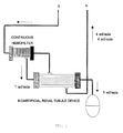
- Fig. 1 is a schematic view of a target bioartificial kidney proposed by the inventors. Artificial kidneys currently used for dialysis treatments perform rapid dialysis within a limited time and have limited effects.
- the bioartificial renal tubule according to the present invention inclueds an artificial membrane having renal tubular epithelial cells attached to the inner or outer surface thereof and a vessel containing the artificial membrane.
- This bioartificial renal tubule is characterized by that the cells are prevented by the use of a MEK inhibitor from being stratified and therefore form a confluent monolayer on the artificial membrane.
- the renal tubular epithelial cells are attached to the inner surface of a hollow fiber placed in the artificial membrane. This allows the bioartificial renal tubule to have a selective substance-reabsorbing function similar to that owned by a vital kidney.
- the following selectivity can be achieved by the use of the renal tubular epithelial cells: the selectivity that glucose, ions, water, and the like are absorbed from a plasma filtrate (or primitive urine flowing through a nephron renal tubule) flowing in an inner portion of the artificial membrane (or a lumen of the hollow fiber) but ammonia, creatinine, drugs, waste products, and the like are not absorbed.
- low-molecular-weight useful substances are filtered into a dialysate from a blood stream by the sieving action of a glomerulus and are then reabsorbed into the blood stream by the active transport of a substance by a renal tubule.
- the following techniques are important: a technique for improving the affinity of an artificial material, used as a scaffold, to cells and a technique for allowing cells which are isolated from a vital organ or tissue and which are placed on a scaffold of an artificial material to exhibit the same function as that of the vital organ.
- a technique for improving the affinity of an artificial material, used as a scaffold, to cells and a technique for allowing cells which are isolated from a vital organ or tissue and which are placed on a scaffold of an artificial material to exhibit the same function as that of the vital organ.
- the formation and maintenance of a monolayer, as well as the function of selected and/or taken cells is important to allow the renal tubular epithelial cells, which have polarity on mass transfer, to function as a renal tubule.
- the renal tubular epithelial cells exhibit "contact inhibition” (a feature that the growth of a cell is arrested when the cell comes into contact with other grown cells all around), which is a feature of normal cells to form and maintain a monolayer when being present in a kidney tissue.
- Non-patent Documents 8 and 9 Studies performed by the inventors have shown that the renal tubular epithelial cells isolated from a tissue form a confluent monolayer and are then stratified and this gradually reduces the metabolic function and transport function of a renal tubule.
- MDCK cells and LLC-PK 1 cells which are cell lines, chronically cultured lack contact inhibition; hence, cells overlap cells two weeks later after the formation of confluent monolayers to cause stratification.
- Non-patent Documents 8 and 9 the inventors have reported that the above stratification reduces the ability to transport water, glucose, sodium, and the like.
- RPTECs which are primary cultured proximal renal tubular epithelial cells, lack contact inhibition during long-term culture and therefore are stratified ( Fig.
- the bioartificial renal tubule according to the present invention includes the artificial membrane, which has the renal tubular epithelial cells attached thereto, and the vessel, which contains the artificial membrane.
- the artificial membrane needs to be porous. Filtration membranes such as hollow fiber membranes are used for hemodialysis because of its various advantages, an artificial hollow fiber membrane is also used as the artificial membrane included in the bioartificial renal tubule according to the present invention.
- the bioartificial renal tubule according to the present invention preferably includes a hollow fiber membrane which has micropores uniformly distributed and also has an extremely large support capacity.
- the hollow fiber membrane is widely used for filtration and material separation. A large number of the micropores are present in a side surface of the hollow fiber membrane that is located between the inner and outer surfaces of the hollow fiber membrane.
- the micropores preferably have a size of 0.001 to several micrometers and more preferably 0.03 to 1 ⁇ m.
- the membrane includes a hollow fiber and therefore is extremely greater in support capacity or accommodation capacity per unit surface area as compared to other porous materials. Therefore, the membrane can be used as a support, having an extremely great ability, suitable for use in a fine medical device.
- Examples of a material used to form the hollow fiber include polysulfone, polyethersulfone, polyacrylonitrile, polyvinyl alcohol, and cellulose acetate.
- a preferable base material may be any one of hollow fiber membranes for hemodialysis, and synthesized macromolecule hollow fiber membranes for artificial kidneys are preferably used.
- the hollow fiber is desirably formed from a film made of cellulose acetate, polysulfone, polyimide, or an ethylene vinylalcohol copolymer.
- Japanese Patent No. 2916446 discloses a method of producing an asymmetric microporous hollow filament suitably used as an ultrafiltration hollow fiber for hemodialysis.
- the hollow filament has such a continuous porous sponge structure that micropores increase in size outward and also has an inner surface having a microporous barrier layer thereon. Therefore, the hollow filament is available for artificial kidneys, filters for artificial hemodialysis, and the like.
- the hollow filament has excellent fluid permeability, mechanical strength, and workability and therefore is a preferred material for forming the hollow fiber membrane of the present invention.
- the renal tubular epithelial cells which are attached to the inner surface of the hollow fiber in the form of a confluent monolayer, are not particularly limited and various types of renal tubular epithelial cells may be used herein.
- epithelial cells of a human renal tubule taken from a kidney anatomically unsuitable for transplantation may be used.
- the renal tubular epithelial cells may be derived from cultured epithelial cell lines, gene-introduced substitute epithelial cells, pluripotent stem cells, or similar cells.
- renal tubular epithelial cells examples include human renal tubular epithelial cells, Madin-Darby canine kidney (MDCK) cells, human proximal tubular cell lines HK-2, JTC-12 cells, LLC-PK 1 cells, and primary cultured human proximal renal tubular epithelial cells (RPTECs).
- a suitable renal tubular epithelial cells is selected desirably in view of cell grafitability, active transport ability, metabolizability, duration of forming monolayer, and the like.
- To attach the renal tubular epithelial cell to the inner surface of the artificial membrane in the form of a confluent monolayer means to graft the cells, preferably to adhere the cells in a form of a confluent monolayer, onto the inner surface of the artificial membrane.
- the inner surface of the artificial membrane may be coated with the cells.
- a technique for attaching the renal tubular epithelial cells to the inner surface of the artificial membrane is not particularly limited.
- the renal tubular epithelial cells may be adsorbed on, bonded to, or supported on the inner surface thereof so as not to be liberated into a liquid but so as to be fixed.
- the inner surface of the artificial membrane is where a plasma filtrate flows, which corresponds to a lumen of a hollow fiber placed in a hollow fiber membrane which is a preferred example of the artificial membrane.
- the renal tubular epithelial cells may be attached to the outer surface of the artificial membrane as required. If the renal tubular epithelial cells are attached to the inner surface (lumen) of the hollow fiber membrane, the renal tubular epithelial cells are attached to the inner wall of the lumen of the hollow fiber.
- the inner wall of the lumen has a large number of micropores communicatively connected to the outside of the hollow fiber. Water, electrolytes, and useful substances selectively pass through the micropores to move into blood flowing outside the hollow fiber.
- the micropores are covered with the confluent monolayer of the renal tubular epithelial cells and therefore are blocked, the active transport ability of the renal tubular epithelial cells allows the control of selective movement.
- a particular technique is as described below. For the use of, for example, a polysulfone hollow fiber membrane device (including 1600 hollow fibers with an inner diameter of 300 nm) with a membrane area of 0.4 m 2 , LLC-PK 1 renal tubular epithelial cells are seeded in the hollow fibers at a density of 10 6 cells/mL four times every hour, whereby a confluent monolayer is formed within 24 hours. This has been confirmed by the inventors.
- An extracellular matrix may be attached to the hollow fiber in addition to the renal tubular epithelial cells.
- the extracellular matrix include Collagen I, Collagen IV, laminin, fibronectin, and Pronectin.
- the application of these matrices to the hollow fiber will promote the renal tubular epithelial cells to be fixed on the inner surface of the hollow fiber.
- These cell-adhesive proteins and adhesive polymers are used in such a manner that they are applied to the hollow fiber in advance of seeding cells.
- the MEK inhibitor is used to maintain the contact inhibition of the renal tubular epithelial cells.
- the MEK inhibitor may be at least one selected from the group consisting of U0126, PD98059, and Cl-1040. These drugs may be used alone or in combination.
- the MEK inhibitor is preferably U0126, which inhibits the activation of a MAP kinase kinase (MEK) and suppresses the activity of ERK 1/ERK 2.
- MEK MAP kinase kinase
- Mitogen-activated protein kinase (MAPK) pathways are involved in cell functions such as growth, differentiation, and stress response. These pathways are linear kinase cascades.
- a MAP kinase kinase kinase phosphorylates and activates a MAP kinase kinase that phosphorylates and activates a MAP kinase.
- the following congeners and members have been identified at present: seven MAP kinase congeners, that is, MEK1, MEK2, MKK3, MKK4/SEK, MEK5, MKK6, and MKK7 and four MAPK family members, that is, ERK1/2, JNK, p38, and ERK5.
- the activation of these pathways controls the activity of various substrates through phosphorylation.
- the substrates include p62 TCF (Elk-1), c-Myc, and ATF2 and also include c-Fos and c-Jun, which are AP-1 factors.
- U0126 which has the chemical name 1,4-diamino-2,3-dicyano-1,4-bis[2-aminophenylthio]butadiene (C 18 H 16 N 6 S 2 ), has been developed through studies on the suppression of cell proliferation in the field of cancer and its action, that is, the suppression of the expression of MEK has been studied.
- U0126 which is an ERK/MEK inhibitor, has been identified as a suppressor of AP-1 transactivation in a cell-based reporter assay. It has been reported that U0126 suppresses an endogenous promoter containing an AP-1 response molecule and has no influence on a gene containing a promoter that lacks an AP-1 response element.
- U0126 results from the direct inhibition of the MAPK family members: MEK-1 and MEK-2.
- U0126 shows little effect on the kinase activity of protein kinase C, Abl kinase, Raf, MEKK, ERK, JNK, MKK-3, MKK-4/SEK, MKK-6, Cdk2, or Cdk4 ( Favata MF, Horiuchi KY, Manos EJ, Daulerio AJ, Stradley DA, Feeser WS, Van Dyk DE, Pitts WJ, Earl RA, Hobbs F, Copeland RA, Magolda RL, Scherle PA, and Trzaskos JM, Identification of a novel inhibitor of mitogen-activated protein kinase kinase, J. Biol. Chem. 1998, 273, 18623-18632 ).
- Epithelial cell systems are supposed to have such a feature (contact inhibition) that the proliferation thereof is arrested when the epithelial cell systems come into contact with proliferated cells all around.
- a MEK inhibitor allows Madin-Darby canine kidney (MDCK) cells to exhibit contact inhibition when the cells lack such a feature because of the action of a hepato-growth factor (HGF) ( Li S, Gerrard ER Jr., and Balkovetz DF, Evidence for ERK1/2 phosphorylation controlling contact inhibition of proliferation in Madin-Darby canine kidney epithelial cells, Am. J. Physiol Cell Physiol, 2004, 287, C432-C439 ). It has been shown that U0126 and PD-98059 have a function of restoring the contact inhibition of the MDCK cells; however, this is involved in a mechanism of controlling cell density depending on proliferation. There is no description about the maintenance of cell monolayers.
- the inventors have confirmed that MDCK cells, porcine proximal renal tubular epithelial LLC-PK 1 cells, human proximal tubular cell lines HK-2, and RPTECs, which are primary cultured proximal renal tubular epithelial cells grow on cell layers two weeks later after the formation of the confluent monolayers to cause stratification.
- the device In consideration of providing an artificial renal tubule device to a patient when the device is medically required, it is too late that the device is manufactured at the point of time when the device is required. Hence, the device needs to be manufactured in advance and also needs to be preserved until being delivered. However, since stratification starts at the point of time when a confluent monolayer is formed, the device can have been apparently deteriorated in function at the point of time when the device is used to treat the patient. Therefore, the device is desirably received by the patient in such a state that the confluent monolayer is maintained until just before the treatment of the patient. For the use of the device for intermittent bioartificial kidney treatment, the MEK inhibitor needs to be added to the device during intervals between treatments such that the confluent monolayer is maintained.
- the inventors have investigated conditions that overcome the above stratification and that allow renal tubule devices to be timely delivered in such a state that the devices are protected from stratification.
- This issue applies to the functional maintenance of continuous hemofilters (which may be referred to as bioartificial glomeruli) prepared by attaching endothelial cells, having no polarity on mass transfer, to surfaces of artificial membranes such as hollow fibers in view of that a formed monolayer needs to be maintained for a long time for the functional maintenance thereof although no substance is actively transported.
- renal tubular epithelial cells are cultured in bulk. An obtained emulsion containing the cultured cells is seeded in a hollow fiber module. After the renal tubular epithelial cells form a confluent monolayer, the active transport of metabolites is reduced because the cells over-confluent (Non-patent Documents 8 and 9). This is because the proliferation and stratification of the cells prevents directional active transport and the swelling of the cells narrows lumens of a hollow fiber. The hollow fiber lumens are blocked with clusters of the cells in some cases.
- an inhibitor of an extracellular-signal regulated kinase kinase (MAPK kinase) (MEK), for example, U0126 needs to be added to a broth for culturing the cells.
- the MEK inhibitor U0126 suppresses the MAPK kinase-induced phosphorylation of threonine and tyrosine to inhibit MAPK (mitogen-activated protein kinase), which is one of ERKs, from acting on the cell cycle of MAP (mitogen-activated protein).
- U0126 has no influence on cellular activities (protein kinase C, Ab1 kinase, Raf, JNK, MKK, and Cdk) other than the MAPK pathway and therefore is supposed to be safe ( Favata MF, Horiuchi KY, Manos EJ, Daulerio AJ, Stradley DA, Feeser WS, Van Dyk DE, Pitts WJ, Earl RA, Hobbs F, Copeland RA, Magolda RL, Scherle PA, and Trzaskos JM, Identification of a novel inhibitor of mitogen-activated protein kinase kinase, J. Biol. Chem.
- LLC-PK 1 cells which are porcine proximal renal tubular epithelial cells, and adding U0126 to their broths and also have evaluated influences on the proliferation of the cells free from U0126 as described below.
- the method for maintaining a confluent monolayer of renal tubular epithelial cells is characterized in that a MEK inhibitor is used to allow the renal tubular epithelial cells to exhibit contact inhibition and the cells are thereby prevented from being stratified after the cells form a confluent monolayer on the inner surface of an artificial membrane.
- the formed confluent monolayer is preferably maintained continuously for at least two weeks or more, more preferably three weeks, and further more preferably four weeks by the method according to the present invention.
- the MEK inhibitor is used in such a manner that the MEK inhibitor is added to a maintenance broth for maintaining the bioartificial renal tubule and bioartificial glomerular device, which are manufactured in advance and are in a standby condition, such that the concentration of the MEK inhibitor in the maintenance broth is 20 to 100 ⁇ M, preferably 30 to 50 ⁇ M, whereby the cells are prevented from being stratified.
- the maintenance broth is replaced with a standard broth in advance of use, whereby an original function can be restored.
- the stratification of the cells can be prevented by using the MEK inhibitor to culture the cells during intervals between treatments.
- a renal tubule vessel for containing the hollow fiber membrane may be made of the same material as that used to form conventional cartridges.
- the bioartificial renal tubule is of a wearable or embedded type for the purpose of continuous hemofiltration, it is preferred that the bioartificial renal tubule has a configuration suitable for the purpose, be more compact, and be made of a biocompatible material.
- the vessel not only contains the artificial membrane but also includes necessary components such as a structural component for fixing and supporting the artificial membrane and structural components such as inlets and outlets for liquids such as blood and a dialysate. That is, the vessel occupies most of the bioartificial renal tubule according to the present invention excluding the artificial hollow fiber membrane.
- a bioartificial renal tubule according to the present invention as well as a bioartificial glomerulus, made by the inventors ( Saito A, Research in the development of a wearable bioartificial kidney with a continuous hemofilter and a bioartificial tubule device using tubular epithelial cells, Artif. Organs, 2004, 28, 58-6 ), will probably play a pioneering role in developing a complete artificial kidney ( Fig. 1 ). That is, the bioartificial renal tubule according to the present invention is used in combination with the bioartificial glomerulus with forming an antithrombogenic circuit. This enables continuous hemodialysis and the selective reabsorption of useful substances in the field of next-generation artificial kidney treatments such as ambulatory artificial kidney treatments and dialysate regeneration-type peritoneal dialyses.
- bioartificial glomeruli having not only high blood compatibility and high antithrombogenicity but high filtration performance, large fenestrae need to be formed in such a manner that confluent cell monolayers are formed on the inner surfaces of filtration membranes used in hemofilters using vascular endothelial cells of patients.
- the bioartificial glomerulus made by the inventors enables the increase of the number and size of pores (windows), called fenestrae, present in the cell membranes of glomerular endothelial cells and also enables the prolongation of duration depending on the use of an actin filament inhibitor such as Cytochalasin B. This is important in maintaining the dialysis efficiency of bioartificial kidneys.
- Fig. 1 Two types of liquids discharged through separate outlets arranged in the bioartificial glomerulus are fed through the next device, that is, the bioartificial renal tubule as shown in Fig. 1 .
- Filtered blood is introduced into the bioartificial renal tubule according to the present invention through a side surface thereof.
- the blood flows outside a hollow fiber placed in the bioartificial renal tubule and gets substances reabsorbed through lumens of the hollow fiber.
- the blood drains from another portion of the side surface and returns to patient's venous blood stream.
- a plasma filtrate (dialysate) discharged from a side surface of the bioartificial glomerulus is an ultrafiltrate and contains various low-molecular-weight substances and electrolytes passing through a hollow fiber filtration membrane.
- the filtrate is fed through an inner portion (lumen) of the hollow fiber of the bioartificial renal tubule.
- a serum filtrate drains from another bottom surface thereof and is eliminated in the form of "urine".
- Useful substances, water, electrolytes, and the like are reabsorbed when passing inside the bioartificial renal tubule and are then taken into blood flowing outside the hollow fiber.
- An exemplary system is a continuous wearable bioartificial kidney in which the bioartificial glomerulus and the bioartificial renal tubule are interlocked with a rotary pump through an antithrombogenic circuit and which is continuously operable for a long period.
- the inventors have prepared this type of artificial kidney, and have proved that the artificial kidney is capable of maintaining the contents of low-molecular-weight substances such as urea, creatinine, and uric acid as well as the content of ⁇ 2 -microglobulin, which is a precursor protein causing dialysis amyloidosis, at extremely low values as compared to current hemodialyses when continuously filtering 10 litters (L) of blood per day with the artificial kidney ( Saito A, Takagi T, Sugiura S, Ono M, Minakuchi K, Teraoka S, and Ota K, Maintaining low concentration of plasma ⁇ 2-microglobulin through continuous slow haemodialysis, Nephrol. Dial. Transplant. 10 (Suppl.
- the artificial kidney need not be a common-use hollow fiber module having a membrane area of about 1.8 m 2 but may be a filtration module which has a membrane area of 0.2 to 0.3 m 2 and which can be contained in a breast pocket. This minimizes the burden of carrying the artificial kidney ( Saito A, Research in the development of a wearable bioartificial kidney with a continuous hemofilter and a bioartificial tubule device using tubular epithelial cells, Artif. Organs, 2004, 28, 58-6 ).
- the artificial kidney as well as a vital kidney, is capable of immediately filtering off ingested water and produced metabolites, which are therefore not stored in a body overnight. Hence, the artificial kidney is low in physical stress and hardly causes complications.
- a single hollow fiber filtration device biologicalartificial glomerulus
- the bioartificial renal tubule needs to work for about one month even though systemic anticoagulant therapy is minimized.
- a single current continuous hemofilter using an artificial material needs to work only for up to 24 hours after systemic anticoagulant therapy is performed.
- An ideal system is supposed to be an implantable bioartificial kidney that continuously works with self-blood pressure for a long period in a pumpless mode.
- LLC-PK 1 Lewis-lung cancer porcine kidney cells, which were cryopreserved porcine proximal renal tubular epithelial cell lines, were seeded.
- the LLC-PK 1 cells were cultured in (1) a high-glucose -DMEM (DMEM-HG) medium, (2) a DMEM-HG medium containing 1% dimethyl sulfoxide (DMSO), or (3) a DMEM-HG medium, containing 50 ⁇ M U0126 and 1% DMSO, placed in outside of inserts (lumens were filled with the DMEM-HG medium).
- DMEM-HG high-glucose -DMEM
- DMSO dimethyl sulfoxide
- the LLC-PK 1 cells on 0 day, the first day, the second day, the third day, and the sixth day after seeding were colored with trypan blue and then measured for number for each group.
- the LLC-PK 1 cells were gently washed with a sterilized PBS solution containing no magnesium or calcium and were then trypsin-treated with 1 ml of a trypsin-EDTA solution at 37°C for 15 minutes.
- the treated cells (1 ml) were added to 4 ml of DMEM, were colored with trypan blue, and were then measured for number with a hemocytometer. Measurement was performed three times for each group.
- Fig. 2 shows the change in number of the LLC-PK 1 cells cultured in (1) the DMEM-HG medium, (2) the DMEM-HG medium containing 1% DMSO, or (3) the DMEM-HG medium containing 50 ⁇ M U0126 and 1% DMSO from 0 day to the sixth day.
- the cells cultured in these media increase in number with day; however, the cells cultured in the U0126-containing medium are prevented from significantly increasing in number.
- DMEM-HG high-glucose DMEM
- DMEM-HG high-glucose DMEM
- DMEM-HG medium containing 10 ⁇ M U0126, which is a MEK inhibitor
- DMEM-HG medium containing 30 ⁇ M U0126 and 1% DMSO 1%
- DMEM-HG medium containing 50 ⁇ M U0126 and 1% DMSO.
- two groups of the cells cultured in (4) a DMEM-HG medium and (5) a DMEM-HG medium containing 1% DMSO were checked.
- the cells on 0 day, the first day, the second day, and the third day after seeding were colored with trypan blue and then measured for number.
- Fig. 3 shows the change in number of the LLC-PK 1 cells grown in the media (1) to (5) from 0 day to the third day.
- the increase of the cells is suppressed to some extent as compared to in the DMEM-HG medium group, but the cells clearly increase in number with day.
- the cells of the 10 ⁇ M U126-containing medium group are not sufficiently prevented from increasing in number with day; however, the cells of the 30 ⁇ M and 50 ⁇ M U126-containing medium groups are clearly prevented from proliferating.
- each of 6-well plates 5 ⁇ 10 5 LLC-PK 1 cells were seeded.
- the cells were divided into three groups on the following day (0 day). That is, the cells of each group were cultured in a corresponding one of the following three media for three days: (1) a DMEM-HG medium (C) used as a control, (2) a DMEM-HG medium containing 1% DMSO (D), and (3) a DMEM-HG medium containing 50 ⁇ M U0126 and 1% DMSO (U). Then the media (D) and (U) were replaced with a DMEM-HG medium.
- the cells of each group were further cultured for three days. The cells were subjected to Western blot analysis for ERK1/2 on 0 day, the first day, the second day, the third day, and the sixth day.
- the cultured LLC-PK 1 cells were used in the form of a cytolytic solution for Western blot analysis.
- the sampled cells of each group were rinsed with ice-chilled PBS and were then centrifuged at 4°C for five minutes at 1500 rpm. The centrifugate was collected and then maintained at -80°C.
- Proteins were taken from 50 ⁇ L of each of ice-chilled cytolytic solutions containing 20mM Tris-HCl (pH 7.5), 150 mM NaCl, 1 mM Na 2 EDTA, 1 mM EGTA, 1% Triton, 2.5 mM sodium pyrophosphate, 1 mM ⁇ -glycerophosphate, 1mM Na 3 VO 4 (sodium orthovanadate), and 1 ⁇ g/mL leupeptin. The proteins were centrifuged at 4°C for 15 minutes at 15000 ⁇ g, whereby soluble proteins were extracted. The protein concentration of each cytolytic solution was measured with a protein assay kit (Protein Assay Rapid Kit; Wako, Osaka, Japan).
- a Laemmli buffer containing mercaptoethanol was added to the cytolytic solutions such that the diluted cytolytic solutions had the same concentration.
- the cytolytic solutions were heated at 100°C for five minutes.
- Proteins (20 ⁇ g) in each LLC-PK 1 cytolytic solution were analyzed with SDS-PAGE (polyacrylamide gel electrophoresis) using a 12.5% polyacrylamide gel (e-PAGEL; ATTO, Tokyo, Japan).
- the proteins were transferred on polyvinylidene difluoride (PVDF) films (BIO-RAD, Hercules, CA) and were then treated with PBS containing fat-free skim milk, 0.1% sodium azide, and 0.1% Tween-20 for 60 minutes for the purpose of reducing the amount of nonspecific antibodies.
- PVDF polyvinylidene difluoride
- Fig. 4 shows culture conditions of ERK1/2 subjected to Western blot analysis after the LLC-PK 1 cells formed confluent monolayers (0 day) and also shows daily changes during culture.
- the upper row of this figure shows the expression of total ERK1/2 and the lower row of this figure shows the expression of activated ERK1/2.
- Three Western blots are arranged at each of positions corresponding to the first day, the second day, the third day, and the sixth day in a left-to-right manner.
- the blots of each of the first day, the second day, the third day, and the sixth day show the expression of an ERK1/2 protein obtained by culture with the DMEM-HG medium (C), that with the DMEM-HG medium containing 1% DMSO (D), and that with the DMEM-HG medium containing 50 ⁇ L U0126 and 1% DMSO (U) arranged from the left.
- Samples taken on 0 day and the sixth day and control (C) samples taken on the first day, the second day, and the third day are results obtained by culture with the DMEM-HG medium.
- the modules were divided into two groups: one in which the DMEM-HG medium was replaced with a medium containing 50 ⁇ M U0126 and 1% DMSO and the cells were cultured using this medium, and another in which the cells were cultured using the former medium, which contained no U0126 or DMSO.
- the medium was circulated outside the hollow fibers of the module in a closed system.
- the media of both groups were replaced with a medium containing no U0126 or DMSO.
- three of the modules of each group were fixed with glutaraldehyde and were then observed in vertical cross section by scanning electron microscopy (SEM) .
- Typical examples of both groups are shown in Fig. 5 .
- confluent monolayers are substantially maintained as shown in a lower region of this figure.
- lumens of the hollow fibers are narrowed because of the progress of stratification and swelling as shown in an upper region of this figure.
- EVAL ethylene-vinyl alcohol
- LLC-PK 1 Lewis-lung cancer porcine kidney cells, which were porcine proximal renal tubular epithelial cell lines, were seeded.
- the cells were cultured in such a manner that a DMEM-HG medium was circulated inside and outside the hollow fibers at a rate of 0.25 ml/min. In this operation, the transmembrane pressure was maintained at zero.
- the DMEM-HG medium circulated outside the hollow fibers in an experiment group (four) was replaced with a DMEM-HG medium containing 50 ⁇ M U0126.
- the former DMEM-HG medium containing no U0126 or DMSO was circulated in a control group (four).
- a DMEM-HG broth containing 50 mg/dl urea nitrogen (UN) and 5 mg/dl creatinine (Cr) was fed inside the hollow fibers at a rate of 0.25 ml/min and a DMEM-HG broth containing no urea or creatinine was fed outside the hollow fibers at that rate.
- the leakage of urea nitrogen, the leakage of creatinine, the amount of reabsorbed glucose, and the amount of reabsorbed Na + were measured for 24 hours.
- Absorption rate leaking rate C 0 ⁇ V 0 - C I ⁇ V I - V R / C 2 ⁇ V 2
- C 0 represents the concentration of a substance in a discharged external solution
- V 0 represents the volume of the discharged external solution
- C 1 represents the concentration of the substance in a fed external solution
- V I represents the volume of the fed external solution
- V R represents the volume of the external solution remaining outside the hollow fibers at the end of an experiment
- C 2 represents the concentration of the substance in a fed internal solution
- V 2 represents the volume of the fed internal solution.
- results shown in Table 1 were obtained.
- the leakage of urea nitrogen (UN) in a U0126 group is significantly less than that in a control group and the amount of reabsorbed Na + in the U0126 group is significantly greater than that in the control group.
- the leakage of creatinine (Cr) in the U0126 group is less than that in the control group, though there is no significant difference therebetween.
- the amount of reabsorbed glucose in the U0126 group is greater than that in the control group, though there is no significant difference therebetween.
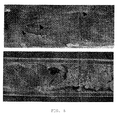
- Fig. 6 shows SEM images of layers of primary cultured human proximal renal tubular epithelial cells RPTEC placed in an EVAL hollow fiber.
- the primary cultured human proximal renal tubular epithelial cells RPTEC were seeded in an EVAL hollow fiber module at a density of 1 ⁇ 10 7 /ml and were then cultured in a medium.
- One of the cross-sectional SEM images of hollow fibers was taken after the cells were cultured for six days (upper image) and the other one was taken after the cells were cultured for 14 days (lower image).
- the cells cultured for six days are stacked on a monolayer (a portion indicated by an arrow in the upper image).
- the bioartificial renal tubule according to the present invention is a device which forms an artificial kidney together with a bioartificial glomerulus.
- the bioartificial renal tubule is useful in providing a safe, simple sustainable treatment system that prevents a disadvantage due to the continuous use of an anticoagulant agent such as heparin when blood needs to be continuously filtered to improve symptoms, such as chronic or acute cardiac failure, chronic or acute renal failure, and multiple organ failure, causing overhydration or accumulation of metabolites.
Applications Claiming Priority (2)
| Application Number | Priority Date | Filing Date | Title |
|---|---|---|---|
| JP2006282826 | 2006-10-17 | ||
| PCT/JP2007/070082 WO2008047760A1 (fr) | 2006-10-17 | 2007-10-15 | Tubule rénal bioartificiel |
Publications (3)
| Publication Number | Publication Date |
|---|---|
| EP2075019A1 true EP2075019A1 (fr) | 2009-07-01 |
| EP2075019A4 EP2075019A4 (fr) | 2011-02-23 |
| EP2075019B1 EP2075019B1 (fr) | 2012-03-07 |
Family
ID=39313987
Family Applications (1)
| Application Number | Title | Priority Date | Filing Date |
|---|---|---|---|
| EP07829816A Active EP2075019B1 (fr) | 2006-10-17 | 2007-10-15 | Tubule rénal bioartificiel |
Country Status (5)
| Country | Link |
|---|---|
| US (1) | US20090209019A1 (fr) |
| EP (1) | EP2075019B1 (fr) |
| JP (1) | JP5166275B2 (fr) |
| AT (1) | ATE548059T1 (fr) |
| WO (1) | WO2008047760A1 (fr) |
Families Citing this family (11)
| Publication number | Priority date | Publication date | Assignee | Title |
|---|---|---|---|---|
| JP5671791B2 (ja) * | 2009-09-04 | 2015-02-18 | ニプロ株式会社 | 近位尿細管上皮細胞(rptec)の培養方法 |
| WO2011040889A1 (fr) * | 2009-10-02 | 2011-04-07 | Agency For Science, Technology And Research | Reins bioartificiels améliorés |
| SG190002A1 (en) * | 2010-10-25 | 2013-06-28 | Agency Science Tech & Res | Tubular fiber membrane with nanoporous skin |
| US9447407B2 (en) | 2011-02-02 | 2016-09-20 | Agency For Science, Technology And Research | Double coating procedure for the membranes of bioartificial kidneys |
| JP2012157653A (ja) * | 2011-02-02 | 2012-08-23 | Agency For Science Technology & Research | バイオ人工腎臓の膜のための二重コーティング手法 |
| CN110511901A (zh) | 2011-05-27 | 2019-11-29 | 德普伊新特斯产品有限责任公司 | 生物人工近端小管系统及使用方法 |
| EP3375860A4 (fr) * | 2015-11-10 | 2019-07-03 | Nikkiso Co., Ltd. | Composite support de cellules et procédé de production de composite support de cellules |
| JP6755085B2 (ja) * | 2015-11-10 | 2020-09-16 | 日機装株式会社 | 細胞支持複合体および細胞支持複合体の製造方法 |
| EP3608398A4 (fr) * | 2017-04-06 | 2020-12-09 | Nikkiso Co., Ltd. | Procédé de culture de cellules, procédé de fabrication d'un composite de support de cellules, cellules cultivées et composite de support de cellules |
| JP6876327B2 (ja) * | 2017-04-28 | 2021-05-26 | 国立大学法人静岡大学 | 血液浄化装置 |
| US20190247560A1 (en) * | 2018-02-13 | 2019-08-15 | Gambro Lundia Ab | Extracorporeal devices and methods of treating complement factor related diseases |
Citations (2)
| Publication number | Priority date | Publication date | Assignee | Title |
|---|---|---|---|---|
| WO1989001967A1 (fr) * | 1987-09-03 | 1989-03-09 | Brown University Research Foundation | Purification du sang a l'aide de cellules renales cultivees |
| US20050238687A1 (en) * | 1996-09-30 | 2005-10-27 | The Regents Of The University Of Michigan | Methods and compositions of bioartificial kidney suitable for use in vivo or ex vivo |
Family Cites Families (7)
| Publication number | Priority date | Publication date | Assignee | Title |
|---|---|---|---|---|
| DE3426331A1 (de) | 1984-07-17 | 1986-01-30 | 6380 Bad Homburg Fresenius AG | Asymmetrische mikroporoese hohlfaser fuer die haemodialyse sowie verfahren zu ihrer herstellung |
| US5549674A (en) * | 1992-03-02 | 1996-08-27 | The Regents Of The University Of Michigan | Methods and compositions of a bioartificial kidney suitable for use in vivo or ex vivo |
| EP0929271B1 (fr) * | 1996-09-05 | 2004-02-25 | Children's Medical Center Corporation | Rein prothetique |
| JP2000271213A (ja) * | 1999-03-26 | 2000-10-03 | Daikin Ind Ltd | 定濃度酸素供給装置 |
| CN100335015C (zh) * | 2001-05-17 | 2007-09-05 | 钟渊化学工业株式会社 | 用于构建具有蛋白代谢功能的体内型人工肾的组合 |
| JP2003055266A (ja) * | 2001-06-04 | 2003-02-26 | Univ Texas Syst | Mek5ならびに心臓肥大および拡張型心筋症に関連する方法および組成物 |
| ATE486540T1 (de) * | 2003-12-23 | 2010-11-15 | Fmc Biopolymer As | Verwendung von alginat-matrizen zur kontrolle des zellwachstums |
-
2007
- 2007-10-15 EP EP07829816A patent/EP2075019B1/fr active Active
- 2007-10-15 AT AT07829816T patent/ATE548059T1/de active
- 2007-10-15 JP JP2008539804A patent/JP5166275B2/ja active Active
- 2007-10-15 US US12/441,106 patent/US20090209019A1/en not_active Abandoned
- 2007-10-15 WO PCT/JP2007/070082 patent/WO2008047760A1/fr active Application Filing
Patent Citations (2)
| Publication number | Priority date | Publication date | Assignee | Title |
|---|---|---|---|---|
| WO1989001967A1 (fr) * | 1987-09-03 | 1989-03-09 | Brown University Research Foundation | Purification du sang a l'aide de cellules renales cultivees |
| US20050238687A1 (en) * | 1996-09-30 | 2005-10-27 | The Regents Of The University Of Michigan | Methods and compositions of bioartificial kidney suitable for use in vivo or ex vivo |
Non-Patent Citations (3)
| Title |
|---|
| INAGAKI ET AL: "Prevention of LLC-PK1 cell overgrowth in a bioartificial renal tubule device using a MEK inhibitor, U0126", JOURNAL OF BIOTECHNOLOGY, ELSEVIER SCIENCE PUBLISHERS, AMSTERDAM, NL, vol. 132, no. 1, 5 October 2007 (2007-10-05), pages 57-64, XP022285748, ISSN: 0168-1656, DOI: DOI:10.1016/J.JBIOTEC.2007.08.025 * |
| LI S ET AL: "Evidence for ERK1/2 phosphorylation controlling contact inhibition of proliferation in Madin-Darby canine kidney epithelial cells", AMERICAN JOURNAL OF PHYSIOLOGY. CELL PHYSIOLOGY, AMERICAN PHYSIOLOGICAL SOCIETY, US, vol. 287, no. 2, 1 August 2004 (2004-08-01), pages C432-C439, XP003022207, ISSN: 0363-6143, DOI: DOI:10.1152/AJPCELL.00020.2004 * |
| See also references of WO2008047760A1 * |
Also Published As
| Publication number | Publication date |
|---|---|
| JP5166275B2 (ja) | 2013-03-21 |
| US20090209019A1 (en) | 2009-08-20 |
| JPWO2008047760A1 (ja) | 2010-02-25 |
| EP2075019A4 (fr) | 2011-02-23 |
| EP2075019B1 (fr) | 2012-03-07 |
| ATE548059T1 (de) | 2012-03-15 |
| WO2008047760A1 (fr) | 2008-04-24 |
Similar Documents
| Publication | Publication Date | Title |
|---|---|---|
| EP2075019B1 (fr) | Tubule rénal bioartificiel | |
| JP4023828B2 (ja) | 人工腎臓および腎臓疾患治療へのその利用 | |
| Humes et al. | The bioartificial kidney: current status and future promise | |
| US5605835A (en) | Bioreactor device with application as a bioartificial liver | |
| US6673339B1 (en) | Prosthetic kidney and its use for treating kidney disease | |
| US6472200B1 (en) | Device and method for performing a biological modification of a fluid | |
| Tzanakakis et al. | Extracorporeal tissue engineered liver-assist devices | |
| WO1989001967A1 (fr) | Purification du sang a l'aide de cellules renales cultivees | |
| Amiel et al. | Renal therapy using tissue-engineered constructs and gene delivery | |
| Tasnim et al. | Achievements and challenges in bioartificial kidney development | |
| US20040167634A1 (en) | Prosthetic kidney and its use for treating kidney disease | |
| US20120184940A1 (en) | Bioartificial kidneys | |
| US20030129736A1 (en) | Device and method for performing a biological modification of a fluid | |
| WO1992007615A1 (fr) | Foie bio-artificiel | |
| Saito et al. | Present status and future perspectives on the development of bioartificial kidneys for the treatment of acute and chronic renal failure patients | |
| Fissell et al. | Innovation in the Treatment of Uremia: Proceedings from the Cleveland Clinic Workshop: The Implantable Artificial Kidney | |
| Oo et al. | A novel design of bioartificial kidneys with improved cell performance and haemocompatibility | |
| WO2017082024A1 (fr) | Composite de support de cellules, et procédé de production de composite de support de cellules | |
| Saito | Research into the development of a wearable bioartificial kidney with a continuous hemofilter and a bioartificial tubule device using tubular epithelial cells | |
| AU2002326365B2 (en) | Method of treating systemic inflammatory response syndrome | |
| WO2013158047A1 (fr) | Unité de bioréacteur à utiliser dans un dispositif de rein bioartificiel | |
| Humes et al. | Renal replacement devices | |
| Saito et al. | Present status and perspectives of bioartificial kidneys | |
| AU2002326365A1 (en) | Method of treating systemic inflammatory response syndrome | |
| WO2017082025A1 (fr) | Composite support de cellules et procédé de production de composite support de cellules |
Legal Events
| Date | Code | Title | Description |
|---|---|---|---|
| PUAI | Public reference made under article 153(3) epc to a published international application that has entered the european phase |
Free format text: ORIGINAL CODE: 0009012 |
|
| 17P | Request for examination filed |
Effective date: 20090313 |
|
| AK | Designated contracting states |
Kind code of ref document: A1 Designated state(s): DE |
|
| AX | Request for extension of the european patent |
Extension state: AL BA HR MK RS |
|
| RBV | Designated contracting states (corrected) |
Designated state(s): DE |
|
| RBV | Designated contracting states (corrected) |
Designated state(s): AT BE BG CH CY CZ DE DK EE ES FI FR GB GR HU IE IS IT LI LT LU LV MC MT NL PL PT RO SE SI SK TR |
|
| A4 | Supplementary search report drawn up and despatched |
Effective date: 20110120 |
|
| RIC1 | Information provided on ipc code assigned before grant |
Ipc: A61M 1/34 20060101AFI20110114BHEP |
|
| GRAP | Despatch of communication of intention to grant a patent |
Free format text: ORIGINAL CODE: EPIDOSNIGR1 |
|
| DAX | Request for extension of the european patent (deleted) | ||
| GRAS | Grant fee paid |
Free format text: ORIGINAL CODE: EPIDOSNIGR3 |
|
| GRAA | (expected) grant |
Free format text: ORIGINAL CODE: 0009210 |
|
| AK | Designated contracting states |
Kind code of ref document: B1 Designated state(s): AT BE BG CH CY CZ DE DK EE ES FI FR GB GR HU IE IS IT LI LT LU LV MC MT NL PL PT RO SE SI SK TR |
|
| REG | Reference to a national code |
Ref country code: GB Ref legal event code: FG4D |
|
| REG | Reference to a national code |
Ref country code: CH Ref legal event code: EP Ref country code: AT Ref legal event code: REF Ref document number: 548059 Country of ref document: AT Kind code of ref document: T Effective date: 20120315 |
|
| REG | Reference to a national code |
Ref country code: IE Ref legal event code: FG4D |
|
| REG | Reference to a national code |
Ref country code: DE Ref legal event code: R096 Ref document number: 602007021217 Country of ref document: DE Effective date: 20120510 |
|
| REG | Reference to a national code |
Ref country code: NL Ref legal event code: VDEP Effective date: 20120307 |
|
| PG25 | Lapsed in a contracting state [announced via postgrant information from national office to epo] |
Ref country code: LT Free format text: LAPSE BECAUSE OF FAILURE TO SUBMIT A TRANSLATION OF THE DESCRIPTION OR TO PAY THE FEE WITHIN THE PRESCRIBED TIME-LIMIT Effective date: 20120307 Ref country code: NL Free format text: LAPSE BECAUSE OF FAILURE TO SUBMIT A TRANSLATION OF THE DESCRIPTION OR TO PAY THE FEE WITHIN THE PRESCRIBED TIME-LIMIT Effective date: 20120307 |
|
| LTIE | Lt: invalidation of european patent or patent extension |
Effective date: 20120307 |
|
| PG25 | Lapsed in a contracting state [announced via postgrant information from national office to epo] |
Ref country code: FI Free format text: LAPSE BECAUSE OF FAILURE TO SUBMIT A TRANSLATION OF THE DESCRIPTION OR TO PAY THE FEE WITHIN THE PRESCRIBED TIME-LIMIT Effective date: 20120307 Ref country code: GR Free format text: LAPSE BECAUSE OF FAILURE TO SUBMIT A TRANSLATION OF THE DESCRIPTION OR TO PAY THE FEE WITHIN THE PRESCRIBED TIME-LIMIT Effective date: 20120608 Ref country code: LV Free format text: LAPSE BECAUSE OF FAILURE TO SUBMIT A TRANSLATION OF THE DESCRIPTION OR TO PAY THE FEE WITHIN THE PRESCRIBED TIME-LIMIT Effective date: 20120307 |
|
| REG | Reference to a national code |
Ref country code: AT Ref legal event code: MK05 Ref document number: 548059 Country of ref document: AT Kind code of ref document: T Effective date: 20120307 |
|
| PG25 | Lapsed in a contracting state [announced via postgrant information from national office to epo] |
Ref country code: CY Free format text: LAPSE BECAUSE OF FAILURE TO SUBMIT A TRANSLATION OF THE DESCRIPTION OR TO PAY THE FEE WITHIN THE PRESCRIBED TIME-LIMIT Effective date: 20120307 |
|
| PG25 | Lapsed in a contracting state [announced via postgrant information from national office to epo] |
Ref country code: BE Free format text: LAPSE BECAUSE OF FAILURE TO SUBMIT A TRANSLATION OF THE DESCRIPTION OR TO PAY THE FEE WITHIN THE PRESCRIBED TIME-LIMIT Effective date: 20120307 Ref country code: PL Free format text: LAPSE BECAUSE OF FAILURE TO SUBMIT A TRANSLATION OF THE DESCRIPTION OR TO PAY THE FEE WITHIN THE PRESCRIBED TIME-LIMIT Effective date: 20120307 Ref country code: IS Free format text: LAPSE BECAUSE OF FAILURE TO SUBMIT A TRANSLATION OF THE DESCRIPTION OR TO PAY THE FEE WITHIN THE PRESCRIBED TIME-LIMIT Effective date: 20120707 Ref country code: CZ Free format text: LAPSE BECAUSE OF FAILURE TO SUBMIT A TRANSLATION OF THE DESCRIPTION OR TO PAY THE FEE WITHIN THE PRESCRIBED TIME-LIMIT Effective date: 20120307 Ref country code: RO Free format text: LAPSE BECAUSE OF FAILURE TO SUBMIT A TRANSLATION OF THE DESCRIPTION OR TO PAY THE FEE WITHIN THE PRESCRIBED TIME-LIMIT Effective date: 20120307 Ref country code: EE Free format text: LAPSE BECAUSE OF FAILURE TO SUBMIT A TRANSLATION OF THE DESCRIPTION OR TO PAY THE FEE WITHIN THE PRESCRIBED TIME-LIMIT Effective date: 20120307 Ref country code: SI Free format text: LAPSE BECAUSE OF FAILURE TO SUBMIT A TRANSLATION OF THE DESCRIPTION OR TO PAY THE FEE WITHIN THE PRESCRIBED TIME-LIMIT Effective date: 20120307 Ref country code: SE Free format text: LAPSE BECAUSE OF FAILURE TO SUBMIT A TRANSLATION OF THE DESCRIPTION OR TO PAY THE FEE WITHIN THE PRESCRIBED TIME-LIMIT Effective date: 20120307 |
|
| PG25 | Lapsed in a contracting state [announced via postgrant information from national office to epo] |
Ref country code: SK Free format text: LAPSE BECAUSE OF FAILURE TO SUBMIT A TRANSLATION OF THE DESCRIPTION OR TO PAY THE FEE WITHIN THE PRESCRIBED TIME-LIMIT Effective date: 20120307 |
|
| PLBE | No opposition filed within time limit |
Free format text: ORIGINAL CODE: 0009261 |
|
| STAA | Information on the status of an ep patent application or granted ep patent |
Free format text: STATUS: NO OPPOSITION FILED WITHIN TIME LIMIT |
|
| PG25 | Lapsed in a contracting state [announced via postgrant information from national office to epo] |
Ref country code: DK Free format text: LAPSE BECAUSE OF FAILURE TO SUBMIT A TRANSLATION OF THE DESCRIPTION OR TO PAY THE FEE WITHIN THE PRESCRIBED TIME-LIMIT Effective date: 20120307 Ref country code: AT Free format text: LAPSE BECAUSE OF FAILURE TO SUBMIT A TRANSLATION OF THE DESCRIPTION OR TO PAY THE FEE WITHIN THE PRESCRIBED TIME-LIMIT Effective date: 20120307 |
|
| 26N | No opposition filed |
Effective date: 20121210 |
|
| PG25 | Lapsed in a contracting state [announced via postgrant information from national office to epo] |
Ref country code: IT Free format text: LAPSE BECAUSE OF FAILURE TO SUBMIT A TRANSLATION OF THE DESCRIPTION OR TO PAY THE FEE WITHIN THE PRESCRIBED TIME-LIMIT Effective date: 20120307 |
|
| REG | Reference to a national code |
Ref country code: DE Ref legal event code: R097 Ref document number: 602007021217 Country of ref document: DE Effective date: 20121210 |
|
| REG | Reference to a national code |
Ref country code: DE Ref legal event code: R082 Ref document number: 602007021217 Country of ref document: DE Representative=s name: MICHALSKI HUETTERMANN & PARTNER PATENTANWAELTE, DE |
|
| REG | Reference to a national code |
Ref country code: DE Ref legal event code: R081 Ref document number: 602007021217 Country of ref document: DE Owner name: NIPRO CORPORATION, JP Free format text: FORMER OWNER: TOKAI UNIVERSITY EDUCATIONAL SYSTEM, TOKIO/TOKYO, JP Effective date: 20130408 Ref country code: DE Ref legal event code: R082 Ref document number: 602007021217 Country of ref document: DE Representative=s name: MICHALSKI HUETTERMANN & PARTNER PATENTANWAELTE, DE Effective date: 20130408 |
|
| PG25 | Lapsed in a contracting state [announced via postgrant information from national office to epo] |
Ref country code: MC Free format text: LAPSE BECAUSE OF NON-PAYMENT OF DUE FEES Effective date: 20121031 |
|
| REG | Reference to a national code |
Ref country code: CH Ref legal event code: PL |
|
| GBPC | Gb: european patent ceased through non-payment of renewal fee |
Effective date: 20121015 |
|
| REG | Reference to a national code |
Ref country code: IE Ref legal event code: MM4A |
|
| REG | Reference to a national code |
Ref country code: FR Ref legal event code: ST Effective date: 20130628 |
|
| PG25 | Lapsed in a contracting state [announced via postgrant information from national office to epo] |
Ref country code: CH Free format text: LAPSE BECAUSE OF NON-PAYMENT OF DUE FEES Effective date: 20121031 Ref country code: BG Free format text: LAPSE BECAUSE OF FAILURE TO SUBMIT A TRANSLATION OF THE DESCRIPTION OR TO PAY THE FEE WITHIN THE PRESCRIBED TIME-LIMIT Effective date: 20120607 Ref country code: LI Free format text: LAPSE BECAUSE OF NON-PAYMENT OF DUE FEES Effective date: 20121031 Ref country code: IE Free format text: LAPSE BECAUSE OF NON-PAYMENT OF DUE FEES Effective date: 20121015 Ref country code: GB Free format text: LAPSE BECAUSE OF NON-PAYMENT OF DUE FEES Effective date: 20121015 |
|
| PG25 | Lapsed in a contracting state [announced via postgrant information from national office to epo] |
Ref country code: FR Free format text: LAPSE BECAUSE OF NON-PAYMENT OF DUE FEES Effective date: 20121031 |
|
| PG25 | Lapsed in a contracting state [announced via postgrant information from national office to epo] |
Ref country code: MT Free format text: LAPSE BECAUSE OF FAILURE TO SUBMIT A TRANSLATION OF THE DESCRIPTION OR TO PAY THE FEE WITHIN THE PRESCRIBED TIME-LIMIT Effective date: 20120307 |
|
| PG25 | Lapsed in a contracting state [announced via postgrant information from national office to epo] |
Ref country code: TR Free format text: LAPSE BECAUSE OF FAILURE TO SUBMIT A TRANSLATION OF THE DESCRIPTION OR TO PAY THE FEE WITHIN THE PRESCRIBED TIME-LIMIT Effective date: 20120307 |
|
| PG25 | Lapsed in a contracting state [announced via postgrant information from national office to epo] |
Ref country code: ES Free format text: LAPSE BECAUSE OF FAILURE TO SUBMIT A TRANSLATION OF THE DESCRIPTION OR TO PAY THE FEE WITHIN THE PRESCRIBED TIME-LIMIT Effective date: 20120307 Ref country code: LU Free format text: LAPSE BECAUSE OF NON-PAYMENT OF DUE FEES Effective date: 20121015 |
|
| PG25 | Lapsed in a contracting state [announced via postgrant information from national office to epo] |
Ref country code: HU Free format text: LAPSE BECAUSE OF FAILURE TO SUBMIT A TRANSLATION OF THE DESCRIPTION OR TO PAY THE FEE WITHIN THE PRESCRIBED TIME-LIMIT Effective date: 20071015 Ref country code: PT Free format text: LAPSE BECAUSE OF FAILURE TO SUBMIT A TRANSLATION OF THE DESCRIPTION OR TO PAY THE FEE WITHIN THE PRESCRIBED TIME-LIMIT Effective date: 20120307 |
|
| PGFP | Annual fee paid to national office [announced via postgrant information from national office to epo] |
Ref country code: DE Payment date: 20231020 Year of fee payment: 17 |