EP2003196A2 - Compositions and methods for treating and diagnosing cancer - Google Patents
Compositions and methods for treating and diagnosing cancer Download PDFInfo
- Publication number
- EP2003196A2 EP2003196A2 EP08014571A EP08014571A EP2003196A2 EP 2003196 A2 EP2003196 A2 EP 2003196A2 EP 08014571 A EP08014571 A EP 08014571A EP 08014571 A EP08014571 A EP 08014571A EP 2003196 A2 EP2003196 A2 EP 2003196A2
- Authority
- EP
- European Patent Office
- Prior art keywords
- cells
- cancer
- tumor
- cell
- tumors
- Prior art date
- Legal status (The legal status is an assumption and is not a legal conclusion. Google has not performed a legal analysis and makes no representation as to the accuracy of the status listed.)
- Ceased
Links
Images


Classifications
-
- A—HUMAN NECESSITIES
- A61—MEDICAL OR VETERINARY SCIENCE; HYGIENE
- A61P—SPECIFIC THERAPEUTIC ACTIVITY OF CHEMICAL COMPOUNDS OR MEDICINAL PREPARATIONS
- A61P31/00—Antiinfectives, i.e. antibiotics, antiseptics, chemotherapeutics
-
- G—PHYSICS
- G01—MEASURING; TESTING
- G01N—INVESTIGATING OR ANALYSING MATERIALS BY DETERMINING THEIR CHEMICAL OR PHYSICAL PROPERTIES
- G01N33/00—Investigating or analysing materials by specific methods not covered by groups G01N1/00 - G01N31/00
- G01N33/48—Biological material, e.g. blood, urine; Haemocytometers
- G01N33/50—Chemical analysis of biological material, e.g. blood, urine; Testing involving biospecific ligand binding methods; Immunological testing
- G01N33/68—Chemical analysis of biological material, e.g. blood, urine; Testing involving biospecific ligand binding methods; Immunological testing involving proteins, peptides or amino acids
- G01N33/6893—Chemical analysis of biological material, e.g. blood, urine; Testing involving biospecific ligand binding methods; Immunological testing involving proteins, peptides or amino acids related to diseases not provided for elsewhere
-
- A—HUMAN NECESSITIES
- A61—MEDICAL OR VETERINARY SCIENCE; HYGIENE
- A61P—SPECIFIC THERAPEUTIC ACTIVITY OF CHEMICAL COMPOUNDS OR MEDICINAL PREPARATIONS
- A61P35/00—Antineoplastic agents
-
- C—CHEMISTRY; METALLURGY
- C12—BIOCHEMISTRY; BEER; SPIRITS; WINE; VINEGAR; MICROBIOLOGY; ENZYMOLOGY; MUTATION OR GENETIC ENGINEERING
- C12N—MICROORGANISMS OR ENZYMES; COMPOSITIONS THEREOF; PROPAGATING, PRESERVING, OR MAINTAINING MICROORGANISMS; MUTATION OR GENETIC ENGINEERING; CULTURE MEDIA
- C12N5/00—Undifferentiated human, animal or plant cells, e.g. cell lines; Tissues; Cultivation or maintenance thereof; Culture media therefor
- C12N5/06—Animal cells or tissues; Human cells or tissues
- C12N5/0602—Vertebrate cells
- C12N5/0693—Tumour cells; Cancer cells
- C12N5/0695—Stem cells; Progenitor cells; Precursor cells
-
- C—CHEMISTRY; METALLURGY
- C12—BIOCHEMISTRY; BEER; SPIRITS; WINE; VINEGAR; MICROBIOLOGY; ENZYMOLOGY; MUTATION OR GENETIC ENGINEERING
- C12Q—MEASURING OR TESTING PROCESSES INVOLVING ENZYMES, NUCLEIC ACIDS OR MICROORGANISMS; COMPOSITIONS OR TEST PAPERS THEREFOR; PROCESSES OF PREPARING SUCH COMPOSITIONS; CONDITION-RESPONSIVE CONTROL IN MICROBIOLOGICAL OR ENZYMOLOGICAL PROCESSES
- C12Q1/00—Measuring or testing processes involving enzymes, nucleic acids or microorganisms; Compositions therefor; Processes of preparing such compositions
- C12Q1/68—Measuring or testing processes involving enzymes, nucleic acids or microorganisms; Compositions therefor; Processes of preparing such compositions involving nucleic acids
- C12Q1/6876—Nucleic acid products used in the analysis of nucleic acids, e.g. primers or probes
- C12Q1/6883—Nucleic acid products used in the analysis of nucleic acids, e.g. primers or probes for diseases caused by alterations of genetic material
- C12Q1/6886—Nucleic acid products used in the analysis of nucleic acids, e.g. primers or probes for diseases caused by alterations of genetic material for cancer
-
- G—PHYSICS
- G01—MEASURING; TESTING
- G01N—INVESTIGATING OR ANALYSING MATERIALS BY DETERMINING THEIR CHEMICAL OR PHYSICAL PROPERTIES
- G01N33/00—Investigating or analysing materials by specific methods not covered by groups G01N1/00 - G01N31/00
- G01N33/48—Biological material, e.g. blood, urine; Haemocytometers
- G01N33/50—Chemical analysis of biological material, e.g. blood, urine; Testing involving biospecific ligand binding methods; Immunological testing
- G01N33/53—Immunoassay; Biospecific binding assay; Materials therefor
- G01N33/575—Immunoassay; Biospecific binding assay; Materials therefor for cancer
- G01N33/5758—Immunoassay; Biospecific binding assay; Materials therefor for cancer involving compounds serving as markers for tumours, cancers or neoplasias, e.g. cellular determinants, receptors, heat shock/stress proteins, A-protein, oligosaccharides or metabolites
- G01N33/57595—Immunoassay; Biospecific binding assay; Materials therefor for cancer involving compounds serving as markers for tumours, cancers or neoplasias, e.g. cellular determinants, receptors, heat shock/stress proteins, A-protein, oligosaccharides or metabolites involving intracellular compounds
-
- C—CHEMISTRY; METALLURGY
- C12—BIOCHEMISTRY; BEER; SPIRITS; WINE; VINEGAR; MICROBIOLOGY; ENZYMOLOGY; MUTATION OR GENETIC ENGINEERING
- C12Q—MEASURING OR TESTING PROCESSES INVOLVING ENZYMES, NUCLEIC ACIDS OR MICROORGANISMS; COMPOSITIONS OR TEST PAPERS THEREFOR; PROCESSES OF PREPARING SUCH COMPOSITIONS; CONDITION-RESPONSIVE CONTROL IN MICROBIOLOGICAL OR ENZYMOLOGICAL PROCESSES
- C12Q2600/00—Oligonucleotides characterized by their use
- C12Q2600/106—Pharmacogenomics, i.e. genetic variability in individual responses to drugs and drug metabolism
-
- C—CHEMISTRY; METALLURGY
- C12—BIOCHEMISTRY; BEER; SPIRITS; WINE; VINEGAR; MICROBIOLOGY; ENZYMOLOGY; MUTATION OR GENETIC ENGINEERING
- C12Q—MEASURING OR TESTING PROCESSES INVOLVING ENZYMES, NUCLEIC ACIDS OR MICROORGANISMS; COMPOSITIONS OR TEST PAPERS THEREFOR; PROCESSES OF PREPARING SUCH COMPOSITIONS; CONDITION-RESPONSIVE CONTROL IN MICROBIOLOGICAL OR ENZYMOLOGICAL PROCESSES
- C12Q2600/00—Oligonucleotides characterized by their use
- C12Q2600/136—Screening for pharmacological compounds
-
- C—CHEMISTRY; METALLURGY
- C12—BIOCHEMISTRY; BEER; SPIRITS; WINE; VINEGAR; MICROBIOLOGY; ENZYMOLOGY; MUTATION OR GENETIC ENGINEERING
- C12Q—MEASURING OR TESTING PROCESSES INVOLVING ENZYMES, NUCLEIC ACIDS OR MICROORGANISMS; COMPOSITIONS OR TEST PAPERS THEREFOR; PROCESSES OF PREPARING SUCH COMPOSITIONS; CONDITION-RESPONSIVE CONTROL IN MICROBIOLOGICAL OR ENZYMOLOGICAL PROCESSES
- C12Q2600/00—Oligonucleotides characterized by their use
- C12Q2600/158—Expression markers
Definitions
- the present invention relates to compositions and methods for treating, characterizing, and diagnosing cancer.
- the present invention provides gene expression profiles associated with solid tumor stem cells, as well as novel stem cell cancer markers useful for the diagnosis, characterization, and treatment of solid tumor stem cells.
- Breast cancer is the most common female malignancy in most industrialized countries, as it is estimated to affect about 10% of the female population during their lifespan. Although its mortality has not increased along with its incidence, due to earlier diagnosis and improved treatment, it is still onè125.7e of the predominant causes of death in middle-aged women. Despite earlier diagnosis of breast cancer, about 1-5% of women with newly diagnosed breast cancer have a distant metastasis at the time of the diagnosis. In addition, approximately 50% of the patients with local disease who are primarily diagnosed eventually relapse with the metastasis. Eighty-five percent of these recurrences take place within the first five years after the primary manifestation of the disease.
- metastatic breast cancer On presentation, most patients with metastatic breast cancer have only one or two organ systems involved. As the disease progresses over time, multiple sites usually become involved. Indeed, metastases may be found in nearly every organ of the body at autopsy. The most common sites of metastatic involvement observed are locoregional recurrences in the skin and soft tissues of the chest wall, as well as in axilla, and supraclavicular area. The most common site for distant metastasis is the bone (30 - 40% of distant metastasis), followed by lung and liver. Metastatic breast cancer is generally considered to be an incurable disease. However, the currently available treatment options often prolong the disease-free state and overall survival rate, as well as increase the quality of the life. The median survival from the manifestation of distant metastases is about three years.
- the present invention relates to compositions and methods for treating, characterizing, and diagnosing cancer.
- the present invention provides gene expression profiles associated with solid tumor stem cells, as well as novel stem cell cancer markers useful for the diagnosis, characterization, and treatment of solid tumor stem cells.
- the present invention provides methods of detecting solid tumor stem cells, comprising; a) providing a tissue sample from a subject, and b) detecting at least one stem cell cancer marker (e.g., 1, 2, 3, 5, 10, ... etc.) from Tables 4-8 in the tissue sample under conditions such that the presence or absence of solid tumor stem cells in the tissue sample is determined.
- the detecting comprises determining the presence of (or absence of), or an expression level for the at least one stem cell cancer marker.
- the detecting comprises detecting mRNA expression of the at least one stem cell cancer marker.
- the detecting comprises exposing the stem cell cancer marker mRNA to a nucleic acid probe complementary to the stem cell cancer marker mRNA.
- the detecting comprises detecting polypeptide expression of the at least one stem cell cancer marker. In other embodiments, the detecting comprises exposing the stem cell cancer marker polypeptide to an antibody specific to the stem cell cancer marker polypeptide and detecting the binding of the antibody to the stem cell cancer polypeptide.
- the subject comprises a human subject.
- the tissue sample comprises tumor tissue. In some embodiments, the tumor tissue sample is a post-surgical tumor tissue sample (e.g. tumor biopsy).
- the methods further comprise c) providing a prognosis to the subject.
- the at least one stem cell cancer marker is from Table 8.
- the at least one stem cell cancer marker comprises: Bmi-1, eed, easyh1, easyh2, mf2, yyl, smarcA3, smarcA5, smarcD3, smarcE1, mllt3, FZD1, FZD2, FZD3, FZD4, FZD6, FZD7, FZD8, FZD9, FZD10, WNT2, WNT2B, WNT3, WNT5A, WNT10B, WNT16, AXIN1, BCL9, MYC, and (TCF4).
- the present invention provides methods for reducing the size of a solid tumor (e.g. in research drug screening, or therapeutic applications) comprising contacting cells of a solid tumor with a biologically (e.g. therapeutically) effective amount of a composition comprising at least one agent directed against at least one stem cell cancer marker shown in Tables 4-8.
- the biologically effective amount is an amount sufficient to cause cell death of or inhibit proliferation of solid tumor stem cells in the solid tumor.
- the biologically effective amount is an amount interferences with the survival pathyways (e.g. notch related genes) or self-renewal pathaways (e.g. WNT pathways) of the solid tumor stem cell.
- solid tumors from which solid tumor stem cells can be isolated or enriched for according to the invention include, but are not limited to, sarcomas and carcinomas such as, but not limited to: fibrosarcoma, myxosarcoma, liposarcoma, chondrosarcoma, osteogenic sarcoma, chordoma, angiosarcoma, endotheliosarcoma, lymphangiosarcoma, lymphangioendotheliosarcoma, synovioma, mesothelioma, Ewing's tumor, leiomyosarcoma, rhabdomyosarcoma, colon carcinoma, pancreatic cancer, breast cancer, ovarian cancer, prostate cancer, squamous cell carcinoma, basal cell carcinoma, adenocarcinoma, sweat gland carcinoma, sebaceous gland carcinoma, papillary carcinoma, papillary adenocarcinomas, cystadenocarcinoma, medullary carcinoma, bronchogenic
- the at least one agent is an antibody, peptide or small molecule.
- the antibody, peptide, anti-sense, siRNA, or small molecule is directed against an extracellular domain of the at least one stem cell cancer marker.
- the at least one stem cell cancer marker is selected from the group consisting of: Bmi-1, eed, easyh1, easyh2, mf2, yyl, smarcA3, smarcA5, smarcD3, smarcE1, mllt3, FZD1, FZD2, FZD3, FZD4, FZD6, FZD7, FZD8, FZD9, FZD10, WNT2, WNT2B, WNT3, WNT5A, WNT10B, WNT16, AXINI, BCL9, MYC, and (TCF4).
- the present invention provides methods for reducing the size of a solid tumor, comprising contacting cells of a solid tumor with a biologically (e.g. therapeutically) effective amount of a composition comprising at least one agent that modulates the activity of at least one stem cell cancer marker shown in Tables 4-8.
- the present invention provides methods for killing or inhibiting the proliferation of solid tumor stem cells comprising contacting the solid tumor stem cells with a biologically effective amount of a composition comprising at least one agent targeted to at least one stem cell cancer marker shown in Tables 4-8.
- the methods further comprise identifying the death of or the prevention of the growth of the solid tumor stem cells following the contacting.
- the cell death is caused by apoptosis.
- the biologically effective amount is an amount interferences with the survival pathyways (e.g. notch related genes) or self-renewal pathaways (e.g. WNT pathways) of the solid tumor stem cell.
- the at least one stem cell cancer marker is selected from the group consisting of: Bmi-1, eed, easyh1, easyh2, mf2, yy1, smarcA3, smarcA5, smarcD3, smarcE1, mllt3, FZD1, FZD2, FZD3, FZD4, FZD6, FZD7, FZD8, FZD9, FZD10, WNT2, WNT2B, WNT3, WNT5A, WNT10B, WNT16, AXIN1, BCL9, MYC, and (TCF4).
- the solid tumor stem cells express cell surface marker CD44, ESA, or B38.1.
- the solid tumor stem cells fail to express at least one LINEAGE marker selected from the group consisting of CD2, CD3, CD IO, CD 14, CD16, CD31, CD45, CD64, and CD140b (see,e.g., US Pat. Pub. US20040037815A1 , and US20020119565 , both of which are herein incorporated by reference).
- the present invention provides methods for selectively targeting a solid tumor stem cell comprising, (a) identifying at least one stem cell cancer marker from Tables 4-8 present on a solid tumor stem cell; and (b) obtaining an agent or set of agents that selectively binds to or regulates the at least one stem cell cancer marker.
- the agent genetically modifies the solid tumor stem cell.
- the agent comprises a bi-specific conjugate.
- the agent comprises an adenoviral vector.
- the present invention provides methods for forming a tumor in an animal, comprising: introducing purified solid tumor stem cells (e.g. a cell dose of) into an animal, wherein: (a) the solid tumor stem cells are derived from a solid tumor; and (b) the solid tumor stem cells are enriched at least 2-fold relative to unfractionated tumor cells based on the presence of at least one stem cell cancer marker in Tables 4-8.
- the animal is an immunocompromised animal.
- the animal is an immunocompromised mammal, such as a mouse (e.g., a nude mouse, SCID mouse, NOD/SCID mouse, Beige/SCID mouse; and microglobin deficient NOD/SCID mouse).
- the number of cells in the cell dose is between about 100 cells and about 5x10 5 cells.
- kits for detecting solid tumor stem cells in a subject comprising: a) a reagent capable of specifically detecting at least one stem cell cancer marker from Tables 4-8 in a tissue or cell sample from a subject, and, optionally, b) instructions for using the reagent for detecting the presence or absence of solid tumor stem cells in the tissue sample.
- the reagent comprises a nucleic acid probe complementary to mRNA from the at least one stem cell cancer marker.
- the reagent comprises an antibody or antibody fragment.
- the present invention provides methods of screening compounds, comprising: a) providing; i) a solid tumor stem cell; and ii) one or more test compounds; and b) contacting the solid tumor stem cell with the test compound; and c) detecting a change in expression of at least one stem cell cancer marker shown in Tables 4-8 in the presence of the test compound relative to the absence of the test compound.
- the detecting comprises determining an expression level for the at least one stem cell cancer marker.
- the detecting comprises detecting mRNA expression of the at least one stem cell cancer marker.
- the detecting comprises detecting polypeptide expression of the at least one stem cell cancer marker.
- the solid tumor stem cell is in vitro. In other embodiments, the solid tumor stem cell is in vivo.
- the test compound comprises a drug (e.g. small molecule, antibody, antibody-toxin conjugate, siRNA, etc.).
- the present invention provides compositions comprising at least two agents (e.g. small molecule, antibody, antibody-toxin conjugate, siRNA, etc.), wherein each of the agents modulates the activity of at least one stem cell cancer marker shown in Tables 4-8.
- the composition comprises at least three agents.
- the present invention provides methods of distinguishing tumorigenic from non-tumorigenic cancer cells, comprising: detecting the presence of ⁇ -catenin in a cancer cell such that the localization of ⁇ -catenin in the cancer cell is determined to be primarily nuclear or primarily cytoplasmic.
- the method further comprises identifying the cancer cell as tumorigenic if the ⁇ -catenin localization is primarily nuclear, or identifying the cancer cell as non-tumorigenic if the ⁇ -catenin localization is primarily cytoplasmic.
- the present invention provides methods of distinguishing a tumorigenic from a non-tumorigenic cancer cell, comprising; a) providing; i) a cancer cell, and ii) a composition comprising an agent configured to bind ⁇ -catenin; and b) contacting the cancer cell with the composition under conditions such that the localization of ⁇ -catenin in the cancer cell is determined to be primarily nuclear or primarily cytoplasmic, and c) identifying the cancer cell as tumorigenic if the ⁇ -catenin localization is primarily nuclear, or identifying the cancer cell as non-tumorigenic if the ⁇ -catenin localization is primarily cytoplasmic.
- the present invention relates to compositions and methods for treating, characterizing and diagnosing cancer.
- the present invention provides gene expression profiles associated with solid tumor stem cells, as well as novel markers useful for the diagnosis, characterization, and treatment of solid tumor stem cells.
- Suitable markers that may be targeted are the genes and peptides encoded by the genes that are differentially expressed in solid tumor stem cells as shown in Tables 4-8.
- the differentially expressed genes, and the peptides encoded thereby may be detected (e.g. quantitatively) in order to identify the presence of solid tumor stem cells, and to determine and screen molecules suitable for reducing the proliferation (or killing), interfering with self-renewal pathways, or interfering with survival pathways of any solid tumor stem cells that are present.
- the differentially expressed genes, and peptides encoded thereby, shown in these tables are also useful for generating therapeutic agents targeted to one or more of these markers (e.g. to inhibit or promote the activity of the marker).
- HSC5 normal hematopoietic stem cells
- normal colon epithelial cells normal breast epithelial cells
- the present invention also provides solid tumor stem cells that differentially express from other cells one or more of the markers provided in Tables 4-8.
- the solid tumor stem cells can be human or other animal. The expression can be either to a greater extent or to a lesser extent.
- the other cells can be selected from normal cells, hematopoietic stem cells, acute myelogenous leukemia (AML) stem cells, or any other class of cells.
- the invention provides a method of selecting cells of a population, which results in a purified population of solid tumor stem cells (e.g. from a patient to select or test therapetuic agents are preferred for the patient).
- the present invention also provides a method of selecting a purified population of tumor cells other than solid tumor stem cells, such as a population of non-tumorigenic (NTG) tumor cells.
- NTG non-tumorigenic
- the present invention provides methods of raising antibodies to the selected cells.
- the invention provides diagnostic methods using the selected cells.
- the invention also provides therapeutic methods, where the therapeutic is directed to a solid tumor stem cell (e.g. directed to one of the stem cells cancer markers identified herein directly or indirectly).
- the invention provides methods of selecting cells, diagnosing disease, conducting research studies, and treating solid tumors using selection methods, diagnostic methods and therapeutics directed to specific genes on a given pathway. Included are one or more of the following genes and gene products: Bmi-1, eed, easyhi, easyh2, mf2, yyl, smarcA3, smarcA5, smarcD3, smarcE 1 and mllt3, as well as those shown in Tables 4-8. Many of these genes are differentially expressed in solid tumor stem cells as compared with normal cells and non-tumorigenic cancer cells, as shown herein.
- the invention provides in vivo and in vitro assays of solid tumor stem cell function and cell function by the various populations of cells isolated from a solid tumor.
- the invention provides methods for using the various populations of cells isolated from a solid tumor (such as a population of cells enriched for solid tumor stem cells) to identify factors influencing solid tumor stem cell proliferation.
- a solid tumor such as a population of cells enriched for solid tumor stem cells
- By the methods of the present invention one can characterize the phenotypically heterogeneous populations of cells within a solid tumor. In particular, one can identify, isolate, and characterize a phenotypically distinct cell population within a tumor having the stem cell properties of extensive proliferation and the ability to give rise to all other tumor cell types.
- Solid tumor stem cells are the tumorigenic cells that are capable of re-establishing a tumor following treatment.
- the invention thus provides a method for selectively targeting diagnostic or therapeutic agents to solid tumor stem cells.
- the invention also provides an agent, such as a biomolecule, that is selectively targeted to solid tumor stem cells (e.g. directed to one of the solid tumor stem cell cancer markers disclosed herein).
- the stem cell cancer marker this targeted is part of a self-renewal or cell survival pathway.
- Bmi-1 was shown to be required for maintenance of adult self-renewing heamatopoietic stem cells (see, e.g., Park et al., Nature, 2003 May 15;423(6937):302-5 , herein incorporated by reference).
- the present invention provides methods for screening for anti-cancer agents; for the testing of anti-cancer therapies; for the development of drugs targeting novel pathways; for the identification of new anti-cancer therapeutic targets; the identification and diagnosis of malignant cells in pathology specimens; for the testing and assaying of solid tumor stem cell drug sensitivity; for the measurement of specific factors that predict drug sensitivity; and for the screening of patients (e.g., as an adjunct for mammography).
- antibody is used in the broadest sense and specifically covers monoclonal antibodies (including full length monoclonal antibodies), polyclonal antibodies, multispecific antibodies (e.g., bispecific antibodies), and antibody fragments so long as they exhibit the desired biological activity (e.g. able to bind a stem cell cancer marker as described herein). Antibodies may be conjugated to other molecules (e.g., toxins).
- antibody fragments refers to a portion of an intact antibody.
- antibody fragments include, but are not limited to, linear antibodies; single-chain antibody molecules; Fc or Fc' peptides, Fab and Fab fragments, and multispecific antibodies formed from antibody fragments.
- humanized forms of non-human (e.g., murine) antibodies are chimeric antibodies that contain minimal sequence, or no sequence, derived from non-human immunoglobulin.
- humanized antibodies are human immunoglobulins (recipient antibody) in which residues from a hypervariable region of the recipient are replaced by residues from a hypervariable region of a non-human species (donor antibody) such as mouse, rat, rabbit or nonhuman primate having the desired specificity, affinity, and capacity.
- donor antibody such as mouse, rat, rabbit or nonhuman primate having the desired specificity, affinity, and capacity.
- Fv framework region (FR) residues of the human immunoglobulin are replaced by corresponding non-human residues.
- humanized antibodies may comprise residues that are not found in the recipient antibody or in the donor antibody.
- the humanized antibody will comprise substantially all of at least one, and typically two, variable domains, in which all or substantially all of the hypervariable loops correspond to those of a nonhuman immunoglobulin and all or substantially all of the FR residues are those of a human immunoglobulin sequence.
- the humanized antibody may also comprise at least a portion of an immunoglobulin constant region (Fc), typically that of a human immunoglobulin. Examples of methods used to generate humanized antibodies are described in U.S. Pat. 5,225,539 to Winter et al . (herein incorporated by reference).
- Enriched as in an enriched population of cells, can be defined phenotypically based upon the increased number of cells having a particular marker (e.g. as shown in Tables 4-8) in a fractionated set of cells as compared with the number of cells having the marker in the unfractionated set of cells.
- the term "enriched can be preferably defined fucntionally by tumorigenic function as the minimum number of cells that form tumors at limit dilution frequency in test mice. For example, if 500 tumor stem cells form tumors in 63% of test animals, but 5000 unfractionated tumor cells are required to form tumors in 63% of test animals, then the solid tumor stem cell population is 10-fold enriched for tumorigenic activity.
- the stem cell cancer markers of the present invention can be used to generate enriched populations of cancer stem cells.
- the stem cell population is enriched at least 1.4 fold relative to unfractioned tumor cells (e.g. 1.4 fold, 1.5 fold, 2 fold, 5 fold ....20 fold).
- isolated in regard to cells, refers to a cell that is removed from its natural environment (such as in a solid tumor) and that is isolated or separated, and is at least about 30%, 50%, 75% free, and most preferably about 90% free, from other cells with which it is naturally present, but which lack the marker based on which the cells were isolated.
- the stem cell cancer markers of the present invention can be used to generate isolated populations of cancer stem cells.
- receptor binding domain refers to any native ligand for a receptor, including cell adhesion molecules, or any region or derivative of such native ligand retaining at least a qualitative receptor binding ability of a corresponding native ligand.
- antibody-immunoadhesin chimera comprises a molecule that combines at least one binding domain of an antibody with at least one immunoadhesin.
- examples include, but are not limited to, the bispecific CD4-IgG chimeras described in Berg et al., PNAS (USA) 88:4723-4727 (1991 ) and Charnow et al., J. Immunol., 153:4268 (1994 ), both of which are hereby incorporated by reference.
- cancer and “cancerous” refer to or describe the physiological condition in mammals that is typically characterized by unregulated cell growth.
- examples of cancer include, but are not limited to, carcinoma, lymphoma, blastoma, sarcoma, and leukemia.
- cancers include squamous cell cancer, small-cell lung cancer, non-small cell lung cancer, adenocarcinoma of the lung, squamous carcinoma of the lung, cancer of the peritoneum, hepatocellular cancer, gastrointestinal cancer, pancreatic cancer, glioblastoma, cervical cancer, ovarian cancer, liver cancer, bladder cancer, hepatoma, breast cancer, colon cancer, colorectal cancer, endometrial or uterine carcinoma, salivary gland carcinoma, kidney cancer, liver cancer, prostate cancer, vulval cancer, thyroid cancer, hepatic carcinoma and various types of head and neck cancer.
- epitope refers to that portion of an antigen that makes contact with a particular antibody.
- an antigenic determinant may compete with the intact antigen (i.e., the "immunogen" used to elicit the immune response) for binding to an antibody.
- telomere binding when used in reference to the interaction of an antibody and a protein or peptide means that the interaction is dependent upon the presence of a particular structure (i.e., the antigenic determinant or epitope) on the protein; in other words the antibody is recognizing and binding to a specific protein structure rather than to proteins in general. For example, if an antibody is specific for epitope "A,” the presence of a protein containing epitope A (or free, unlabelled A) in a reaction containing labeled "A" and the antibody will reduce the amount of labeled A bound to the antibody.
- non-specific binding and “background binding” when used in reference to the interaction of an antibody and a protein or peptide refer to an interaction that is not dependent on the presence of a particular structure (i.e., the antibody is binding to proteins in general rather that a particular structure such as an epitope).
- the term “subject” refers to any animal (e.g ., a mammal), including, but not limited to, humans, non-human primates, rodents, and the like, which is to be the recipient of a particular treatment.
- the terms “subject” and “patient” are used interchangeably herein in reference to a human subject.
- the term "subject suspected of having cancer” refers to a subject that presents one or more symptoms indicative of a cancer (e.g., a noticeable lump or mass) or is being screened for a cancer (e.g., during a routine physical).
- a subject suspected of having cancer may also have one or more risk factors.
- a subject suspected of having cancer has generally not been tested for cancer.
- a "subject suspected of having cancer” encompasses an individual who has received an initial diagnosis but for whom the stage of cancer is not known. The term further includes people who once had cancer ( e.g ., an individual in remission).
- the term "subject at risk for cancer” refers to a subject with one or more risk factors for developing a specific cancer.
- Risk factors include, but are not limited to, gender, age, genetic predisposition, environmental expose, previous incidents of cancer, preexisting non-cancer diseases, and lifestyle.
- the term "characterizing cancer in subject” refers to the identification of one or more properties of a cancer sample in a subject, including but not limited to, the presence of benign, pre-cancerous or cancerous tissue, the stage of the cancer, and the subject's prognosis. Cancers may be characterized by the identification of the expression of one or more cancer marker genes, including but not limited to, the cancer markers disclosed herein.
- stem cell cancer markers refers to a gene or peptide expressed by the gene whose expression level, alone or in combination with other genes, is correlated with the presence of tumorigenic cancer cells.
- the correlation may relate to either an increased or decreased expression of the gene (e.g. increased or decreased levels of mRNA or the peptide encoded by the gene).
- a reagent that specifically detects expression levels refers to reagents used to detect the expression of one or more genes (e.g., including but not limited to, the cancer markers of the present invention).
- suitable reagents include but are not limited to, nucleic acid probes capable of specifically hybridizing to the gene of interest, aptamers, PCR primers capable of specifically amplifying the gene of interest, and antibodies capable of specifically binding to proteins expressed by the gene of interest.
- suitable reagents include but are not limited to, nucleic acid probes capable of specifically hybridizing to the gene of interest, aptamers, PCR primers capable of specifically amplifying the gene of interest, and antibodies capable of specifically binding to proteins expressed by the gene of interest.
- Other non-limiting examples can be found in the description and examples below.
- detecting a decreased or increased expression relative to non-cancerous control refers to measuring the level of expression of a gene (e.g ., the level of mRNA or protein) relative to the level in a non-cancerous control sample.
- Gene expression can be measured using any suitable method, including but not limited to, those described herein.
- detecting a change in gene expression in a a cell sample in the presence of said test compound relative to the absence of said test compound refers to measuring an altered level of expression (e.g ., increased or decreased) in the presence of a test compound relative to the absence of the test compound.
- Gene expression can be measured using any suitable method.
- the term "instructions for using said kit for detecting cancer in said subject” includes instructions for using the reagents contained in the kit for the detection and characterization of cancer in a sample from a subject.
- the term "providing a prognosis” refers to providing information regarding the impact of the presence of cancer (e.g ., as determined by the diagnostic methods of the present invention) on a subject's future health (e.g ., expected morbidity or mortality, the likelihood of getting cancer, and the risk of metastasis).
- post surgical tumor tissue refers to cancerous tissue (e.g ., biopsy tissue) that has been removed from a subject ( e.g ., during surgery).
- the term "subject diagnosed with a cancer” refers to a subject who has been tested and found to have cancerous cells.
- the cancer may be diagnosed using any suitable method, including but not limited to, biopsy, x-ray, blood test, and the diagnostic methods of the present invention.
- biopsy tissue refers to a sample of tissue that is removed from a subject for the purpose of determining if the sample contains cancerous tissue. In some embodiment, biopsy tissue is obtained because a subject is suspected of having cancer. The biopsy tissue is then examined for the presence or absence of cancer.
- gene transfer system refers to any means of delivering a composition comprising a nucleic acid sequence to a cell or tissue.
- gene transfer systems include, but are not limited to, vectors (e.g ., retroviral, adenoviral, adeno-associated viral, and other nucleic acid-based delivery systems), microinjection of naked nucleic acid, polymer-based delivery systems (e.g ., liposome-based and metallic particle-based systems), biolistic injection, and the like.
- viral gene transfer system refers to gene transfer systems comprising viral elements (e.g ., intact viruses, modified viruses and viral components such as nucleic acids or proteins) to facilitate delivery of the sample to a desired cell or tissue.
- viral elements e.g ., intact viruses, modified viruses and viral components such as nucleic acids or proteins
- adenovirus gene transfer system refers to gene transfer systems comprising intact or altered viruses belonging to the family Adenoviridae.
- site-specific recombination target sequences refers to nucleic acid sequences that provide recognition sequences for recombination factors and the location where recombination takes place.
- nucleic acid molecule refers to any nucleic acid containing molecule, including but not limited to, DNA or RNA.
- the term encompasses sequences that include any of the known base analogs of DNA and RNA including, but not limited to, 4-acetylcytosine, 8-hydroxy-N6-methyladenosine, aziridinylcytosine, pseudoisocytosine, 5-(carboxyhydroxylmethyl) uracil, 5-fluorouracil, 5-bromouracil, 5-carboxymethylaminomethyl-2-thiouracil, 5-carboxymethylaminomethyluracil, dihydrouracil, inosine, N6-isopentenyladenine, 1-methyladenine, 1-methylpseudouracil, 1-methylguanine, 1-methylinosine, 2,2-dimethylguanine, 2-methyladenine, 2-methylguanine, 3-methylcytosine, 5-methylcytosine, N6-methyladenine,
- gene refers to a nucleic acid (e.g., DNA) sequence that comprises coding sequences necessary for the production of a polypeptide, precursor, or RNA (e.g., rRNA, tRNA).
- the polypeptide can be encoded by a full length coding sequence or by any portion of the coding sequence so long as the desired activity or functional properties (e.g ., enzymatic activity, ligand binding, signal transduction, immunogenicity, etc.) of the full-length or fragment are retained.
- the term also encompasses the coding region of a structural gene and the sequences located adjacent to the coding region on both the 5' and 3' ends for a distance of about 1 kb or more on either end such that the gene corresponds to the length of the full-length mRNA. Sequences located 5' of the coding region and present on the mRNA are referred to as 5' non-translated sequences. Sequences located 3' or downstream of the coding region and present on the mRNA are referred to as 3' non-translated sequences.
- the term "gene” encompasses both cDNA and genomic forms of a gene.
- a genomic form or clone of a gene contains the coding region interrupted with non-coding sequences termed "introns” or “intervening regions” or “intervening sequences.”
- Introns are segments of a gene that are transcribed into nuclear RNA (hnRNA); introns may contain regulatory elements such as enhancers. Introns are removed or “spliced out” from the nuclear or primary transcript; introns therefore are absent in the messenger RNA (mRNA) transcript.
- mRNA messenger RNA
- heterologous gene refers to a gene that is not in its natural environment.
- a heterologous gene includes a gene from one species introduced into another species.
- a heterologous gene also includes a gene native to an organism that has been altered in some way (e.g ., mutated, added in multiple copies, linked to non-native regulatory sequences, etc).
- Heterologous genes are distinguished from endogenous genes in that the heterologous gene sequences are typically joined to DNA sequences that are not found naturally associated with the gene sequences in the chromosome or are associated with portions of the chromosome not found in nature ( e.g ., genes expressed in loci where the gene is not normally expressed).
- RNA expression refers to the process of converting genetic information encoded in a gene into RNA (e.g ., mRNA, rRNA, tRNA, or snRNA) through "transcription" of the gene ( e.g ., via the enzymatic action of an RNA polymerase), and for protein encoding genes, into protein through “translation” of mRNA.
- Gene expression can be regulated at many stages in the process.
- Up-regulation” or “activation” refers to regulation that increases the production of gene expression products (e.g ., RNA or protein), while “down-regulation” or “repression” refers to regulation that decrease production.
- Molecules e.g ., transcription factors
- activators e.g ., transcription factors
- genomic forms of a gene may also include sequences located on both the 5' and 3' end of the sequences that are present on the RNA transcript. These sequences are referred to as "flanking" sequences or regions (these flanking sequences are located 5' or 3' to the non-translated sequences present on the mRNA transcript).
- the 5' flanking region may contain regulatory sequences such as promoters and enhancers that control or influence the transcription of the gene.
- the 3' flanking region may contain sequences that direct the termination of transcription, post-transcriptional cleavage and polyadenylation.
- siRNAs refers to short interfering RNAs.
- siRNAs comprise a duplex, or double-stranded region, of about 18-25 nucleotides long; often siRNAs contain from about two to four unpaired nucleotides at the 3' end of each strand.
- At least one strand of the duplex or double-stranded region of a siRNA is substantially homologous to or substantially complementary to a target RNA molecule.
- the strand complementary to a target RNA molecule is the "antisense strand;" the strand homologous to the target RNA molecule is the "sense strand,” and is also complementary to the siRNA antisense strand.
- siRNAs may also contain additional sequences; non-limiting examples of such sequences include linking sequences, or loops, as well as stem and other folded structures. siRNAs appear to function as key intermediaries in triggering RNA interference in invertebrates and in vertebrates, and in triggering sequence-specific RNA degradation during posttranscriptional gene silencing in plants.
- RNA interference refers to the silencing or decreasing of gene expression by siRNAs. It is the process of sequence-specific, post-transcriptional gene silencing in animals and plants, initiated by siRNA that is homologous in its duplex region to the sequence of the silenced gene.
- the gene may be endogenous or exogenous to the organism, present integrated into a chromosome or present in a transfection vector that is not integrated into the genome. The expression of the gene is either completely or partially inhibited.
- RNAi may also be considered to inhibit the function of a target RNA; the function of the target RNA may be complete or partial.
- nucleic acid molecule encoding As used herein, the terms “nucleic acid molecule encoding,” “DNA sequence encoding,” and “DNA encoding” refer to the order or sequence of deoxyribonucleotides along a strand of deoxyribonucleic acid. The order of these deoxyribonucleotides determines the order of amino acids along the polypeptide (protein) chain. The DNA sequence thus codes for the amino acid sequence.
- an oligonucleotide having a nucleotide sequence encoding a gene and “polynucleotide having a nucleotide sequence encoding a gene,” means a nucleic acid sequence comprising the coding region of a gene or in other words the nucleic acid sequence that encodes a gene product.
- the coding region may be present in a cDNA, genomic DNA or RNA form.
- the oligonucleotide or polynucleotide may be single-stranded (i.e., the sense strand) or double-stranded.
- Suitable control elements such as enhancers/promoters, splice junctions, polyadenylation signals, etc. may be placed in close proximity to the coding region of the gene if needed to permit proper initiation of transcription and/or correct processing of the primary RNA transcript.
- the coding region utilized in the expression vectors of the present invention may contain endogenous enhancers/promoters, splice junctions, intervening sequences, polyadenylation signals, etc. or a combination of both endogenous and exogenous control elements.
- portion when in reference to a nucleotide sequence (as in “a portion of a given nucleotide sequence”) refers to fragments of that sequence.
- the fragments may range in size from four nucleotides to the entire nucleotide sequence minus one nucleotide (10 nucleotides, 20, 30, 40, 50, 100, 200, etc.).
- operable combination refers to the linkage of nucleic acid sequences in such a manner that a nucleic acid molecule capable of directing the transcription of a given gene and/or the synthesis of a desired protein molecule is produced.
- the term also refers to the linkage of amino acid sequences in such a manner so that a functional protein is produced.
- isolated when used in relation to a nucleic acid, as in “an isolated oligonucleotide” or “isolated polynucleotide” refers to a nucleic acid sequence that is identified and separated from at least one component or contaminant with which it is ordinarily associated in its natural source. Isolated nucleic acid is such present in a form or setting that is different from that in which it is found in nature. In contrast, non-isolated nucleic acids as nucleic acids such as DNA and RNA found in the state they exist in nature.
- a given DNA sequence e.g ., a gene
- RNA sequences such as a specific mRNA sequence encoding a specific protein
- isolated nucleic acid encoding a given protein includes, by way of example, such nucleic acid in cells ordinarily expressing the given protein where the nucleic acid is in a chromosomal location different from that of natural cells, or is otherwise flanked by a different nucleic acid sequence than that found in nature.
- the isolated nucleic acid, oligonucleotide, or polynucleotide may be present in single-stranded or double-stranded form.
- the oligonucleotide or polynucleotide will contain at a minimum the sense or coding strand (i.e ., the oligonucleotide or polynucleotide may be single-stranded), but may contain both the sense and anti-sense strands ( i.e ., the oligonucleotide or polynucleotide may be double-stranded).
- amino acid sequence and terms such as “polypeptide” or “protein” are not meant to limit the amino acid sequence to the complete, native amino acid sequence associated with the recited protein molecule.
- native protein as used herein to indicate that a protein does not contain amino acid residues encoded by vector sequences; that is, the native protein contains only those amino acids found in the protein as it occurs in nature.
- a native protein may be produced by recombinant means or may be isolated from a naturally occurring source.
- portion when in reference to a protein (as in “a portion of a given protein”) refers to fragments of that protein.
- the fragments may range in size from four amino acid residues to the entire amino acid sequence minus one amino acid.
- Southern blot refers to the analysis of DNA on agarose or acrylamide gels to fractionate the DNA according to size followed by transfer of the DNA from the gel to a solid support, such as nitrocellulose or a nylon membrane.
- the immobilized DNA is then probed with a labeled probe to detect DNA species complementary to the probe used.
- the DNA may be cleaved with restriction enzymes prior to electrophoresis. Following electrophoresis, the DNA may be partially depurinated and denatured prior to or during transfer to the solid support.
- Southern blots are a standard tool of molecular biologists ( J. Sambrook et al., Molecular Cloning: A Laboratory Manual, Cold Spring Harbor Press, NY, pp 9.31-9.58 [1989 ]).
- Northern blot refers to the analysis of RNA by electrophoresis of RNA on agarose gels to fractionate the RNA according to size followed by transfer of the RNA from the gel to a solid support, such as nitrocellulose or a nylon membrane. The immobilized RNA is then probed with a labeled probe to detect RNA species complementary to the probe used.
- Northern blots are a standard tool of molecular biologists (J. Sambrook, et al., supra, pp 7.39-7.52 [1989]).
- the term "Western blot” refers to the analysis of protein(s) (or polypeptides) immobilized onto a support such as nitrocellulose or a membrane.
- the proteins are run on acrylamide gels to separate the proteins, followed by transfer of the protein from the gel to a solid support, such as nitrocellulose or a nylon membrane.
- the immobilized proteins are then exposed to antibodies with reactivity against an antigen of interest.
- the binding of the antibodies may be detected by various methods, including the use of radiolabeled antibodies.
- transgene refers to a foreign gene that is placed into an organism by, for example, introducing the foreign gene into newly fertilized eggs or early embryos.
- foreign gene refers to any nucleic acid (e.g ., gene sequence) that is introduced into the genome of an animal by experimental manipulations and may include gene sequences found in that animal so long as the introduced gene does not reside in the same location as does the naturally occurring gene.
- vector is used in reference to nucleic acid molecules that transfer DNA segment(s) from one cell to another.
- vehicle is sometimes used interchangeably with “vector.”
- Vectors are often derived from plasmids, bacteriophages, or plant or animal viruses.
- expression vector refers to a recombinant DNA molecule containing a desired coding sequence and appropriate nucleic acid sequences necessary for the expression of the operably linked coding sequence in a particular host organism.
- Nucleic acid sequences necessary for expression in prokaryotes usually include a promoter, an operator (optional), and a ribosome binding site, often along with other sequences.
- Eukaryotic cells are known to utilize promoters, enhancers, and termination and polyadenylation signals.
- overexpression and “overexpressing” and grammatical equivalents are used in reference to levels of mRNA to indicate a level of expression approximately 1.5-fold higher (or greater) than that observed in a given tissue in a control or non-transgenic animal.
- Levels of mRNA are measured using any of a number of techniques known to those skilled in the art including, but not limited to Northern blot analysis.
- RNA loaded from each tissue analyzed e.g ., the amount of 28S rRNA, an abundant RNA transcript present at essentially the same amount in all tissues, present in each sample can be used as a means of normalizing or standardizing the mRNA-specific signal observed on Northern blots).
- the amount of mRNA present in the band corresponding in size to the correctly spliced transgene RNA is quantified; other minor species of RNA which hybridize to the transgene probe are not considered in the quantification of the expression of the transgenic mRNA.
- in vitro refers to an artificial environment and to processes or reactions that occur within an artificial environment.
- in vitro environments can consist of, but are not limited to, test tubes and cell culture.
- in vivo refers to the natural environment (e.g. , an animal or a cell) and to processes or reaction that occur within a natural environment.
- test compound and “candidate compound” refer to any chemical entity, pharmaceutical, drug, and the like that is a candidate for use to treat or prevent a disease, illness, sickness, or disorder of bodily function (e.g ., cancer).
- Test compounds comprise both known and potential therapeutic compounds.
- a test compound can be determined to be therapeutic by screening using the screening methods of the present invention.
- test compounds include antisense compounds.
- sample is used in its broadest sense. In one sense, it is meant to include a specimen or culture obtained from any source, as well as biological and environmental samples. Biological samples may be obtained from animals (including humans) and encompass fluids, solids, tissues, and gases. Biological samples include blood products, such as plasma, serum and the like. Environmental samples include environmental material such as surface matter, soil, water, crystals and industrial samples. Such examples are not however to be construed as limiting the sample types applicable to the present invention.
- the present invention provides compositions and methods for treating, characterizing, and diagnosing cancer.
- the present invention provides gene expression profiles associated with solid tumor stem cells, as well as novel markers useful for the diagnosis, characterization, and treatment of solid tumor stem cells.
- HSCs hematopoietic stem cells
- tissue containing HSCs has been demonstrated in cancer therapy with their extensive use for bone marrow transplantation to regenerate the hematolymphoid system following myeloablative protocols 12 .
- the prospective isolation of HSCs from patients can result in a population that is cancer free for autologous transplantation 13-17 .
- HSCs are the most studied and best understood somatic stem cell population'. Hematopoiesis is a tightly regulated process in which a pool of hematopoietic stem cells eventually gives rise to the lymphohematopoietic system consisting of the formed blood elements, e.g., red blood cells, platelets, granulocytes, macrophages, and B-and T-lymphocytes. These cells are important for oxygenation, prevention of bleeding, immunity, and infections, respectively.
- HSCs have two fundamental properties. First, HSCs need to self-renew in order to maintain the stem cell pool; the total number of HSCs is under strict genetic regulation 27 . Second, they must undergo differentiation to maintain a constant pool of mature cells in normal conditions, and to produce increased numbers of a particular lineage in response to stresses such as bleeding or infection.
- multipotent cells constitute 0.05% of mouse bone marrow cells and are heterogeneous with respect to their ability to self-renew.
- multipotent progenitors without detectable self-renewal potential 7,28 .
- These populations form a hierarchy in which the long-term HSCs give rise to short-term HSCs, which in turn give rise to multipotent progenitors [ Fig. 1 in 7 ].
- HSCs mature from the long-term self-renewing pool to multipotent progenitors they become more mitotically active but lose the ability to self-renew. Only long-term HSCs can give rise to mature hematopoietic cells for the lifetime of the animal, while short-term HSCs and multipotent progenitors reconstitute lethally irradiated mice for less than eight weeks 7 .
- HSCs differentiate when exposed to combinations of growth factors that can induce extensive proliferation in long-term cultures 31 .
- recent progress has been made in identifying culture conditions that maintain HSC activity in culture for a limited period of time [for example see Miller and Graves 32 ], it has proven to be exceedingly difficult to identify tissue culture conditions that promote a significant and prolonged expansion of progenitors with transplantable HSC activity.
- the prevention of apoptosis by enforced expression of the oncogene Bcl-2 promotes the development of lymphoma and also results in increased numbers of HSCs in vivo, suggesting that cell death plays a role in regulating the homeostasis of HSCs 36,37 .
- the progression to experimental acute myelogenous leukemia in mice requires at least 3, and likely 4 independent events to block the several intrinsically triggered and extrinsically induce programmed cell death pathways of myeloid cells 38 .
- Proto-oncogenes such as c-myb and c-myc that drive proliferation of tumor cells are also essential for HSCs development 39-42 .
- mice deficient for tal-1 / SCL which is involved in some cases of human acute leukemia, lack embryonic hematopoiesis 45 , suggesting that it is required for intrinsic or extrinsic events necessary to initiate hematopoiesis, for maintenance of the earliest definitive blood cells, or for the decision to form blood cells downstream of embryonic HSCs 45,46 .
- Members of the Hox family have also been implicated in human leukemia. Enforced expression of HoxB4 can affect stem cell functions 47,48 .
- One of the major targets of the p53 tumor suppressor gene is p21 cip1 . Bone marrow from p21 cip1 deficient mice has a reduced ability to serially reconstitute lethally irradiated recipients.
- telomeres failure at serial transfer could result from exhaustion of the stem cell pool, loss of telomeres, or loss of transplantability 49 .
- bmi-1 a gene that cooperates with c-myc to induce lymphoma 50,51 , is required for the maintenance of adult HSCs and leukemia cells.
- stem cell fate decisions are also involved in malignant transformation.
- Notch Two other signaling pathways implicated in oncogenesis in both mice and humans, the Wnt/ ⁇ -catenin and Notch pathways, may play central roles in the self-renewal of both normal and cancer stem cells.
- the Notch family of receptors was first identified in Drosophila and has been implicated in development and differentiation 52 .
- C elegans Notch plays a role in germ cell self renewal 53 .
- neural development transient Notch activation initiates an irreversible switch from neurogenesis to gliogenesis by embryonic neural crest stem cells 10 .
- the Notch pathway plays a central role in development and the mouse int-3 oncogene is a truncated Notch4 56
- the role for Notch in de novo human cancer is complex and less well understood.
- Various members of the Notch signaling pathway are expressed in cancers of epithelial origin and activation by Notch by chromosomal translocation is involved in some cases of leukemia 57-61 .
- Wnt/ ⁇ -catenin signaling also plays a pivotal role in the self-renewal of normal stem cells and malignant transformation 65-67 .
- the Wnt pathway was first implicated in MMTV-induced breast cancer where in deregulated expression of Wnt-1 due to proviral insertion resulted in mammary tumors 68,69 . Subsequently, it has been shown that Wnt proteins play a central role in pattern formation.
- Wnt-1 belongs to large family of highly hydrophobic secreted proteins that function by binding to their cognate receptors, members of the Frizzled and low-density lipoprotein receptor-related protein families, resulting in activation of ⁇ -catenin 43,58,65,70,71 .
- ⁇ -catenin is marked for degradation by a complex consisting of the Adenomatous Polyposis Coli (APC), Axin and glycogen synthase kinase-3 ⁇ proteins 58,67,72-74 .) 66,75 .
- Wnt proteins are expressed in the bone marrow, and activation of Wnt/ ⁇ -catenin signaling by Wnt proteins in vitro or by expression of a constitutively active ⁇ -catenin expands the pool of early progenitor cells and enriched normal transplantable hematopoietic stem cells in tissue culture and in vivo 25,67,72 .
- mice that fail to express TCF-4 one of the transcription factors that is activated when bound to ⁇ -catenin, soon exhaust their undifferentiated crypt epithelial progenitor cells, further suggesting that Wnt signaling is involved in the self renewal of epithelial stem cells 43,76 .
- Expression of certain Wnt genes is elevated in some other epithelial cancers suggesting that activation of ⁇ -catenin is secondary to ligand activation in such cancers 65,78-83 .
- constitutive activation of the Wnt/ ⁇ -catenin pathway may confer a stem/progenitor cell phenotype to cancer cells.
- Inhibition of ⁇ -catenin/TCF-4 in a colon cancer cell line induced the expression of the cell cycle inhibitor p21 cip-1 and induced the cells to stop proliferating and to acquire a more differentiated phenotype 83 .
- Enforced expression of the proto-oncogene c-myc which is transcriptionally activated by ⁇ -catenin/TCF-4, inhibited the expression of p21 cip-1 and allowed the colon cancer cells to proliferate when ⁇ -catenin/TCF-4 signaling was blocked, linking Wnt signaling to c-myc in the regulation of cell proliferation and differentiation.
- the Wnt pathway is involved in the self-renewal of normal stem cells and activating mutations of Wnt induce breast cancer in mice.
- This pathway plays a role in tumor formation by human breast cancer stem cells isolated from some patients.
- the ability of different populations of breast cancer cells to form tumors differs.
- the expression of members of the Wnt/Frizzled/ ⁇ -catenin pathway are heterogeneously expressed by different populations of cancer cells and expression of particular members of the pathway may correlate with the capacity to form tumors.
- Activated ⁇ -catenin is seen in the cancer cells in a significant number of patients.
- the marker expression of both normal hematopoietic and leukemic tissue culture cells can change rapidly in tissue culture and often does not reflect that of the original stem cells from which they were derived 92,94,95,98 .
- the conditions often promote self-renewal or differentiation in a way that prevents the stem cells in culture from recapitulating the hierarchy of cell populations that exist in vivo.
- the lack of an effective method to consistently grow primary human breast cancer cells in vitro or in vivo for long periods of time has severely limited our ability to understand the biology of this disease.
- the most efficient xenograft models report the engraftment of pieces of breast cancer tumors in the ovarian, but not mammary, fat pad of SCID mice approximately 60-75% of the time 99 . Engraftment of dissociated cells is not possible in this model, and cancer cells isolated from pleural effusions only form tumors in immunodeficient mice approximately 10% of the time 90 .
- the present invention provides a xenograft model in which one is able to establish tumors from primary breast tumors via injection of tumors in the mammary gland of severely immunodeficient mice.
- Xenograft of the present invention allows one to do biological and molecular tests to characterize the clonogenic breast cancer cell as well as other cell types.
- the xenograft tumors developed in accordance with the present invention contain the phenotypically diverse cancer cell types found in the human tumors from which they were derived and the different populations of cancer cells differ markedly in their ability to form tumors 100 .
- an efficient xenograft model in accordance with the present invention has for the first time reliably allows dissociated solid tumor cells obtained from a patient to form tumors. Importantly, this enables one to routinely analyze biochemical pathways in an individual patient's cancer cells and to do molecular manipulations that allow one to understand the cellular consequences of specific genetic pathways on tumor formation by de novo human solid tumor cancer cells.
- the present invention provides markers whose expression is specifically altered in solid tumor stem cells (e.g. up regulated or down regulated). Such markers find use in the diagnosis and characterization and alteration (e.g., therapeutic targeting) of various cancers (e.g. breast cancer).
- Example 4 describes methods used to identify solid tumor cancer markers.
- Preferred cancer markers are provided below in Tables 4-8, as well as Notch 4. While these tables provide gene names, it is noted that the present invention contemplates the use of both the nucleic acid sequences as well as the peptides encoded thereby, as well as fragments of the nucleic acid and peptides, in the therapeutic and diagnostic methods and compositions of the present invention.
- Table 8 Preferred Solid Tumor Stem Cell Cancer Markers Bmi-1, eed, easyhl, easyh2, rnf2, yyl, smarcA3, smarckA5, smarcD3, smarcE1, mllt3, frizzled 2, frizzled 6, frizzled 7, mf2, Frizzled 1, Frizzled2, Frizzled4, Frizzled10, Frizzled6, FZD1, FZD2, FZD3, FZD4, FZD6, FZD7, FZD8, FZD9, FZD10, WNT2, WNT2B, WNT3, WNT5A, WNT10B, WNT16, AXIN1, BCL9, MYC, (TCF4),, SLC7A8, IL1RAP, TEM8, TMPRSS4, MUC16, GPRC5B, SLC6A14, SLC4A11, PPAP2C, CAV1, CAV2, PTPN3, EPHA1, SLC1A1, CX3
- the present invention provides methods for detection of expression of stem cell cancer markers (e.g ., breast cancer stem cell cancer markers).
- expression is measured directly (e.g ., at the RNA or protein level).
- expression is detected in tissue samples (e.g ., biopsy tissue).
- expression is detected in bodily fluids (e.g. , including but not limited to, plasma, serum, whole blood, mucus, and urine).
- the present invention further provides panels and kits for the detection of markers.
- the presence of a stem cell cancer marker is used to provide a prognosis to a subject. The information provided is also used to direct the course of treatment.
- additional therapies e.g ., hormonal or radiation therapies
- additional therapies can be started at a earlier point when they are more likely to be effective (e.g ., before metastasis).
- additional therapies e.g ., hormonal or radiation therapies
- the expense and inconvenience of such therapies can be avoided.
- the present invention is not limited to the markers described above. Any suitable marker that correlates with cancer or the progression of cancer may be utilized. Additional markers are also contemplated to be within the scope of the present invention. Any suitable method may be utilized to identify and characterize cancer markers suitable for use in the methods of the present invention, including but not limited to, those described in illustrative Example 4 below. For example, in some embodiments, markers identified as being up or down-regulated in solid tumor stem cells using the gene expression microarray methods of the present invention are further characterized using tissue microarray, immunohistochemistry, Northern blot analysis, siRNA or antisense RNA inhibition, mutation analysis, investigation of expression with clinical outcome, as well as other methods disclosed herein.
- the present invention provides a panel for the analysis of a plurality of markers.
- the panel allows for the simultaneous analysis of multiple markers correlating with carcinogenesis and/or metastasis.
- panels may be analyzed alone or in combination in order to provide the best possible diagnosis and prognosis.
- Markers for inclusion on a panel are selected by screening for their predictive value using any suitable method, including but not limited to, those described in the illustrative examples below.
- detection of solid tumor stem cell cancer markers are detected by measuring the expression of corresponding mRNA in a tissue sample (e.g ., breast cancer tissue).
- mRNA expression may be measured by any suitable method, including but not limited to, those disclosed below.
- RNA is detection by Northern blot analysis.
- Northern blot analysis involves the separation of RNA and hybridization of a complementary labeled probe.
- RNA is detected by hybridization to a oligonucleotide probe.
- a variety of hybridization assays using a variety of technologies for hybridization and detection are available.
- TaqMan assay PE Biosystems, Foster City, CA; See e.g., U.S. Patent Nos. 5,962,233 and 5,538,848 , each of which is herein incorporated by reference
- the assay is performed during a PCR reaction.
- the TaqMan assay exploits the 5'-3' exonuclease activity of the AMPLITAQ GOLD DNA polymerase.
- a probe consisting of an oligonucleotide with a 5'-reporter dye (e.g ., a fluorescent dye) and a 3'-quencher dye is included in the PCR reaction.
- a 5'-reporter dye e.g ., a fluorescent dye
- a 3'-quencher dye is included in the PCR reaction.
- the 5'-3' nucleolytic activity of the AMPLITAQ GOLD polymerase cleaves the probe between the reporter and the quencher dye.
- the separation of the reporter dye from the quencher dye results in an increase of fluorescence.
- the signal accumulates with each cycle of PCR and can be monitored with a fluorimeter.
- RNA reverse-transcriptase PCR
- RNA is enzymatically converted to complementary DNA or "cDNA" using a reverse transcriptase enzyme.
- the cDNA is then used as a template for a PCR reaction.
- PCR products can be detected by any suitable method, including but not limited to, gel electrophoresis and staining with a DNA specific stain or hybridization to a labeled probe.
- the quantitative reverse transcriptase PCR with standardized mixtures of competitive templates method described in U.S. Patents 5,639,606 , 5,643,765 , and 5,876,978 (each of which is herein incorporated by reference) is utilized.
- gene expression of stem cell cancer markers is detected by measuring the expression of the corresponding protein or polypeptide.
- Protein expression may be detected by any suitable method.
- proteins are detected by immunohistochemistry.
- proteins are detected by their binding to an antibody raised against the protein. The generation of antibodies is described below.
- Antibody binding is detected by techniques known in the art (e.g ., radioimmunoassay, ELISA (enzyme-linked immunosorbant assay), "sandwich” immunoassays, immunoradiometric assays, gel diffusion precipitation reactions, immunodiffusion assays, in situ immunoassays ( e.g ., using colloidal gold, enzyme or radioisotope labels, for example), Western blots, precipitation reactions, agglutination assays (e.g ., gel agglutination assays, hemagglutination assays, etc.), complement fixation assays, immunofluorescence assays, protein A assays, and immunoelectrophoresis assays, etc.
- radioimmunoassay e.g ., radioimmunoassay, ELISA (enzyme-linked immunosorbant assay), "sandwich” immunoassays, immunoradiometric assays,
- antibody binding is detected by detecting a label on the primary antibody.
- the primary antibody is detected by detecting binding of a secondary antibody or reagent to the primary antibody.
- the secondary antibody is labeled. Many methods are known in the art for detecting binding in an immunoassay and are within the scope of the present invention.
- an automated detection assay is utilized.
- Methods for the automation of immunoassays include those described in U.S. Patents 5,885,530 , 4,981,785 , 6,159,750 , and 5,358,691 , each of which is herein incorporated by reference.
- the analysis and presentation of results is also automated.
- software that generates a prognosis based on the presence or absence of a series of proteins corresponding to cancer markers is utilized.
- a computer-based analysis program is used to translate the raw data generated by the detection assay (e.g ., the presence, absence, or amount of a given marker or markers) into data of predictive value for a clinician.
- the clinician can access the predictive data using any suitable means.
- the present invention provides the further benefit that the clinician, who is not likely to be trained in genetics or molecular biology, need not understand the raw data.
- the data is presented directly to the clinician in its most useful form. The clinician is then able to immediately utilize the information in order to optimize the care of the subject.
- the present invention contemplates any method capable of receiving, processing, and transmitting the information to and from laboratories conducting the assays, information provides, medical personal, and subjects.
- a sample e.g ., a biopsy or a serum or urine sample
- a profiling service e.g ., clinical lab at a medical facility, genomic profiling business, etc.
- any part of the world e.g ., in a country different than the country where the subject resides or where the information is ultimately used
- the subject may visit a medical center to have the sample obtained and sent to the profiling center, or subjects may collect the sample themselves and directly send it to a profiling center.
- the sample comprises previously determined biological information
- the information may be directly sent to the profiling service by the subject (e.g ., an information card containing the information may be scanned by a computer and the data transmitted to a computer of the profiling center using an electronic communication systems).
- the profiling service Once received by the profiling service, the sample is processed and a profile is produced (e.g ., expression data), specific for the diagnostic or prognostic information desired for the subj ect.
- the profile data is then prepared in a format suitable for interpretation by a treating clinician.
- the prepared format may represent a diagnosis or risk assessment for the subject, along with recommendations for particular treatment options.
- the data may be displayed to the clinician by any suitable method.
- the profiling service generates a report that can be printed for the clinician ( e.g ., at the point of care) or displayed to the clinician on a computer monitor.
- the information is first analyzed at the point of care or at a regional facility.
- the raw data is then sent to a central processing facility for further analysis and/or to convert the raw data to information useful for a clinician or patient.
- the central processing facility provides the advantage of privacy (all data is stored in a central facility with uniform security protocols), speed, and uniformity of data analysis.
- the central processing facility can then control the fate of the data following treatment of the subject. For example, using an electronic communication system, the central facility can provide data to the clinician, the subject, or researchers.
- the subject is able to directly access the data using the electronic communication system.
- the subject may chose further intervention or counseling based on the results.
- the data is used for research use.
- the data may be used to further optimize the inclusion or elimination of markers as useful indicators of a particular condition or stage of disease.
- kits for the detection and characterization of cancer e.g. for detecting one or more of the markers shown in Tables 4-8, or for modulating the activity of a peptide expressed by one or more of markes shown in Tables 4-8).
- the kits contain antibodies specific for a cancer marker, in addition to detection reagents and buffers.
- the kits contain reagents specific for the detection of mRNA or cDNA (e.g ., oligonucleotide probes or primers).
- the kits contain all of the components necessary to perform a detection assay, including all controls, directions for performing assays, and any necessary software for analysis and presentation of results.
- in vivo imaging techniques are used to visualize the expression of cancer markers in an animal (e.g. , a human or non-human mammal).
- cancer marker mRNA or protein is labeled using an labeled antibody specific for the cancer marker.
- a specifically bound and labeled antibody can be detected in an individual using an in vivo imaging method, including, but not limited to, radionuclide imaging, positron emission tomography, computerized axial tomography, X-ray or magnetic resonance imaging method, fluorescence detection, and chemiluminescent detection. Methods for generating antibodies to the cancer markers of the present invention are described below.
- the in vivo imaging methods of the present invention are useful in the diagnosis of cancers that express the solid tumor stem cell cancer markers of the present invention (e.g ., in breast cancer). In vivo imaging is used to visualize the presence of a marker indicative of the cancer. Such techniques allow for diagnosis without the use of an unpleasant biopsy.
- the in vivo imaging methods of the present invention are also useful for providing prognoses to cancer patients. For example, the presence of a marker indicative of cancer stem cells can be detected.
- the in vivo imaging methods of the present invention can further be used to detect metastatic cancers in other parts of the body.
- reagents e.g ., antibodies
- specific for the cancer markers of the present invention are fluorescently labeled.
- the labeled antibodies are introduced into a subject ( e.g ., orally or parenterally). Fluorescently labeled antibodies are detected using any suitable method ( e.g ., using the apparatus described in U.S. Patent 6,198,107 , herein incorporated by reference).
- antibodies are radioactively labeled.
- the use of antibodies for in vivo diagnosis is well known in the art. Sumerdon et al., (Nucl. Med. Biol 17:247-254 [1990 ] have described an optimized antibody-chelator for the radioimmunoscintographic imaging of tumors using Indium-111 as the label. Griffin et al., (J Clin One 9:631-640 [1991 ]) have described the use of this agent in detecting tumors in patients suspected of having recurrent colorectal cancer. The use of similar agents with paramagnetic ions as labels for magnetic resonance imaging is known in the art ( Lauffer, Magnetic Resonance in Medicine 22:339-342 [1991 ]).
- Radioactive labels such as Indium-111, Technetium-99m, or Iodine-131 can be used for planar scans or single photon emission computed tomography (SPECT).
- Positron emitting labels such as Fluorine-19 can also be used for positron emission tomography (PET).
- PET positron emission tomography
- paramagnetic ions such as Gadolinium (III) or Manganese (II) can be used.
- Radioactive metals with half-lives ranging from 1 hour to 3.5 days are available for conjugation to antibodies, such as scandium-47 (3.5 days) gallium-67 (2.8 days), gallium-68 (68 minutes), technetiium-99m (6 hours), and indium-111 (3.2 days), of which gallium-67, technetium-99m, and indium-111 are preferable for gamma camera imaging, gallium-68 is preferable for positron emission tomography.
- a useful method of labeling antibodies with such radiometals is by means of a bifunctional chelating agent, such as diethylenetriaminepentaacetic acid (DTPA), as described, for example, by Khaw et al. (Science 209:295 [1980 ]) for In-111 and Tc-99m, and by Scheinberg et al. (Science 215:1511 [1982 ]).
- DTPA diethylenetriaminepentaacetic acid
- Other chelating agents may also be used, but the 1-(p-carboxymethoxybenzyl)EDTA and the carboxycarbonic anhydride of DTPA are advantageous because their use permits conjugation without affecting the antibody's immunoreactivity substantially.
- Another method for coupling DPTA to proteins is by use of the cyclic anhydride of DTPA, as described by Hnatowich et al. (Int. J. Appl. Radiat. Isot. 33:327 [1982 ]) for labeling of albumin with In-111, but which can be adapted for labeling of antibodies.
- a suitable method of labeling antibodies with Tc-99m which does not use chelation with DPTA is the pretinning method of Crockford et al., ( U.S. Pat. No. 4,323,546 , herein incorporated by reference).
- a preferred method of labeling immunoglobulins with Tc-99m is that described by Wong et al. (Int. J. Appl. Radiat. Isot., 29:251 [1978 ]) for plasma protein, and recently applied successfully by Wong et al. (J. Nucl. Med., 23:229 [1981 ]) for labeling antibodies.
- radiometals conjugated to the specific antibody it is likewise desirable to introduce as high a proportion of the radiolabel as possible into the antibody molecule without destroying its immunospecificity.
- a further improvement may be achieved by effecting radiolabeling in the presence of the specific stem cell cancer marker of the present invention, to insure that the antigen binding site on the antibody will be protected.
- in vivo biophotonic imaging (Xenogen, Almeda, CA) is utilized for in vivo imaging.
- This real-time in vivo imaging utilizes luciferase.
- the luciferase gene is incorporated into cells, microorganisms, and animals ( e.g ., as a fusion protein with a cancer marker of the present invention). When active, it leads to a reaction that emits light.
- a CCD camera and software is used to capture the image and analyze it.
- the present invention provides isolated antibodies and antibody fragments (e.g, Fabs).
- the present invention provides monoclonal antibodies or antibody fragments that specifically bind to an isolated polypeptide comprised of at least five, or at least 15 amino acid residues of the stem cell cancer markers described herein (e.g ., as shown in Tables 4-8). These antibodies or antibody fragments find use in the diagnostic, drug screening, and therapetuic methods described herein (e.g. to detect or modulate the activity of a stem cell cancer marker peptide).
- An antibody, or antibody fragment, against a protein of the present invention may be any monoclonal or polyclonal antibody, as long as it can recognize the protein.
- Antibodies can be produced by using a protein of the present invention as the antigen according to a conventional antibody or antiserum preparation process.
- the present invention contemplates the use of both monoclonal and polyclonal antibodies. Any suitable method may be used to generate the antibodies used in the methods and compositions of the present invention, including but not limited to, those disclosed herein.
- a monoclonal antibody protein, as such, or together with a suitable carrier or diluent is administered to an animal (e.g ., a mammal) under conditions that permit the production of antibodies.
- complete or incomplete Freund's adjuvant may be administered.
- the protein is administered once every 2 weeks to 6 weeks, in total, about 2 times to about 10 times.
- Animals suitable for use in such methods include, but are not limited to, primates, rabbits, dogs, guinea pigs, mice, rats, sheep, goats, etc.
- an individual animal whose antibody titer has been confirmed e.g ., a mouse
- 2 days to 5 days after the final immunization, its spleen or lymph node is harvested and antibody-producing cells contained therein are fused with myeloma cells to prepare the desired monoclonal antibody producer hybridoma.
- Measurement of the antibody titer in antiserum can be carried out, for example, by reacting the labeled protein, as described hereinafter and antiserum and then measuring the activity of the labeling agent bound to the antibody.
- the cell fusion can be carried out according to known methods, for example, the method described by Koehler and Milstein (Nature 256:495 [1975 ]).
- a fusion promoter for example, polyethylene glycol (PEG) or Sendai virus (HVJ), preferably PEG is used.
- myeloma cells examples include NS-1, P3U1, SP2/0, AP-1 and the like.
- the proportion of the number of antibody producer cells (spleen cells) and the number of myeloma cells to be used is preferably about 1:1 to about 20:1.
- PEG preferably PEG 1000-PEG 6000
- Cell fusion can be carried out efficiently by incubating a mixture of both cells at about 20°C to about 40°C, preferably about 30°C to about 37°C for about 1 minute to 10 minutes.
- a hybridoma producing the antibody e.g ., against a tumor antigen or autoantibody of the present invention
- a supernatant of the hybridoma is added to a solid phase (e.g ., microplate) to which antibody is adsorbed directly or together with a carrier and then an anti-immunoglobulin antibody (if mouse cells are used in cell fusion, anti-mouse immunoglobulin antibody is used) or Protein A labeled with a radioactive substance or an enzyme is added to detect the monoclonal antibody against the protein bound to the solid phase.
- a supernatant of the hybridoma is added to a solid phase to which an anti-immunoglobulin antibody or Protein A is adsorbed and then the protein labeled with a radioactive substance or an enzyme is added to detect the monoclonal antibody against the protein bound to the solid phase.
- Selection of the monoclonal antibody can be carried out according to any known method or its modification. Normally, a medium for animal cells to which HAT (hypoxanthine, aminopterin, thymidine) are added is employed. Any selection and growth medium can be employed as long as the hybridoma can grow. For example, RPMI 1640 medium containing 1% to 20%, preferably 10% to 20% fetal bovine serum, GIT medium containing 1% to 10% fetal bovine serum, a serum free medium for cultivation of a hybridoma (SFM-101, Nissui Seiyaku) and the like can be used.
- HAT hyperxanthine, aminopterin, thymidine
- the cultivation is carried out at 20°C to 40°C, preferably 37°C for about 5 days to 3 weeks, preferably 1 week to 2 weeks under about 5% CO 2 gas.
- the antibody titer of the supernatant of a hybridoma culture can be measured according to the same manner as described above with respect to the antibody titer of the anti-protein in the antiserum.
- Separation and purification of a monoclonal antibody can be carried out according to the same manner as those of conventional polyclonal antibodies such as separation and purification of immunoglobulins, for example, salting-out, alcoholic precipitation, isoelectric point precipitation, electrophoresis, adsorption and desorption with ion exchangers ( e.g ., DEAE), ultracentrifugation, gel filtration, or a specific purification method wherein only an antibody is collected with an active adsorbent such as an antigen-binding solid phase, Protein A or Protein G and dissociating the binding to obtain the antibody.
- an active adsorbent such as an antigen-binding solid phase, Protein A or Protein G and dissociating the binding to obtain the antibody.
- Polyclonal antibodies may be prepared by any known method or modifications of these methods including obtaining antibodies from patients. For example, a complex of an immunogen (an antigen against the protein) and a carrier protein is prepared and an animal is immunized by the complex according to the same manner as that described with respect to the above monoclonal antibody preparation. A material containing the antibody against is recovered from the immunized animal and the antibody is separated and purified.
- an immunogen an antigen against the protein
- a carrier protein is prepared and an animal is immunized by the complex according to the same manner as that described with respect to the above monoclonal antibody preparation.
- a material containing the antibody against is recovered from the immunized animal and the antibody is separated and purified.
- any carrier protein and any mixing proportion of the carrier and a hapten can be employed as long as an antibody against the hapten, which is crosslinked on the carrier and used for immunization, is produced efficiently.
- bovine serum albumin, bovine cycloglobulin, keyhole limpet hemocyanin, etc. may be coupled to an hapten in a weight ratio of about 0.1 part to about 20 parts, preferably, about 1 part to about 5 parts per 1 part of the hapten.
- various condensing agents can be used for coupling of a hapten and a carrier.
- glutaraldehyde, carbodiimide, maleimide activated ester, activated ester reagents containing thiol group or dithiopyridyl group, and the like find use with the present invention.
- the condensation product as such or together with a suitable carrier or diluent is administered to a site of an animal that permits the antibody production.
- complete or incomplete Freund's adjuvant may be administered. Normally, the protein is administered once every 2 weeks to 6 weeks, in total, about 3 times to about 10 times.
- the polyclonal antibody is recovered from blood, ascites and the like, of an animal immunized by the above method.
- the antibody titer in the antiserum can be measured according to the same manner as that described above with respect to the supernatant of the hybridoma culture. Separation and purification of the antibody can be carried out according to the same separation and purification method of immunoglobulin as that described with respect to the above monoclonal antibody.
- the protein used herein as the immunogen is not limited to any particular type of immunogen.
- a stem cell cancer marker of the present invention (further including a gene having a nucleotide sequence partly altered) can be used as the immunogen.
- fragments of the protein may be used. Fragments may be obtained by any methods including, but not limited to expressing a fragment of the gene, enzymatic processing of the protein, chemical synthesis, and the like.
- the antibodies and antibody fragments may also be conjugated to therapeutic (e.g. cancer cell killing compounds).
- the antibody directed toward one of the stem cell cancer markers is used to specifically deliver a therapeutic agent to a solid tumor cancer cell (e.g. to inhibit the proliferation of such sell or kill such a cell).
- the present invention provides drug screening assays (e.g ., to screen for anticancer drugs).
- the screening methods of the present invention utilize stem cell cancer markers identified using the methods of the present invention (e.g ., including but not limited to, the stem cell cancer markers shown in Tables 4-8).
- the present invention provides methods of screening for compound that alter ( e.g ., increase or decrease) the expression of stem cell cancer marker genes.
- candidate compounds are antisense agents or siRNA agents (e.g ., oligonucleotides) directed against cancer markers.
- candidate compounds are antibodies that specifically bind to a stem cell cancer marker of the present invention.
- libraries of compounds of small molecules are screened using the methods described herein.
- candidate compounds are evaluated for their ability to alter stem cell cancer marker expression by contacting a compound with a cell expressing a stem cell cancer marker and then assaying for the effect of the candidate compounds on expression.
- the effect of candidate compounds on expression of a cancer marker gene is assayed by detecting the level of cancer marker mRNA expressed by the cell. mRNA expression can be detected by any suitable method.
- the effect of candidate compounds on expression of cancer marker genes is assayed by measuring the level of polypeptide encoded by the cancer markers. The level of polypeptide expressed can be measured using any suitable method, including but not limited to, those disclosed herein.
- other changes in cel biology e.g., apoptosis
- the present invention provides screening methods for identifying modulators, i.e ., candidate or test compounds or agents (e.g ., proteins, peptides, peptidomimetics, peptoids, small molecules or other drugs) which bind to, or alter the signalling or function associated with the cancer markers of the present invention, have an inhibitory (or stimulatory) effect on, for example, stem cell cancer marker expression or cancer markers activity, or have a stimulatory or inhibitory effect on, for example, the expression or activity of a cancer marker substrate.
- modulators i.e ., candidate or test compounds or agents (e.g ., proteins, peptides, peptidomimetics, peptoids, small molecules or other drugs) which bind to, or alter the signalling or function associated with the cancer markers of the present invention, have an inhibitory (or stimulatory) effect on, for example, stem cell cancer marker expression or cancer markers activity, or have a stimulatory or inhibitory effect on, for example, the expression or activity of a cancer marker substrate.
- Target gene products e.g ., stem cell cancer marker genes
- target gene products e.g ., stem cell cancer marker genes
- Compounds which inhibit the activity or expression of cancer markers are useful in the treatment of proliferative disorders, e.g ., cancer, particularly metastatic cancer or eliminating or controlling tumor stem cells to prevent or reduce the risk of cancer.
- the invention provides assays for screening candidate or test compounds that are substrates of a cancer markers protein or polypeptide or a biologically active portion thereof. In another embodiment, the invention provides assays for screening candidate or test compounds that bind to or modulate the activity of a cancer marker protein or polypeptide or a biologically active portion thereof.
- test compounds of the present invention can be obtained using any of the numerous approaches in combinatorial library methods known in the art, including biological libraries; peptoid libraries (libraries of molecules having the functionalities of peptides, but with a novel, non-peptide backbone, which are resistant to enzymatic degradation but which nevertheless remain bioactive; see, e.g ., Zuckennann et al., J. Med. Chem. 37: 2678-85 [1994 ]); spatially addressable parallel solid phase or solution phase libraries; synthetic library methods requiring deconvolution; the 'one-bead one-compound' library method; and synthetic library methods using affinity chromatography selection.
- the biological library and peptoid library approaches are preferred for use with peptide libraries, while the other four approaches are applicable to peptide, non-peptide oligomer or small molecule libraries of compounds ( Lam (1997) Anticancer Drug Des. 12:145 ).
- an assay is a cell-based assay in which a cell that expresses a stem cell cancer marker protein or biologically active portion thereof is contacted with a test compound, and the ability of the test compound to the modulate cancer marker's activity is determined. Determining the ability of the test compound to modulate stem cell cancer marker activity can be accomplished by monitoring, for example, changes in enzymatic activity.
- the cell for example, can be of mammalian origin.
- test compound to modulate cancer marker binding to a compound, e.g ., a stem cell cancer marker substrate.
- a compound e.g ., a stem cell cancer marker substrate
- This can be accomplished, for example, by coupling the compound, e.g ., the substrate, with a radioisotope or enzymatic label such that binding of the compound, e.g ., the substrate, to a cancer marker can be determined by detecting the labeled compound, e.g ., substrate, in a complex.
- the stem cell cancer marker is coupled with a radioisotope or enzymatic label to monitor the ability of a test compound to modulate cancer marker binding to a cancer markers substrate in a complex.
- compounds e.g ., substrates
- compounds can be labeled with 125 I, 35 S 14 C or 3 H, either directly or indirectly, and the radioisotope detected by direct counting of radioemmission or by scintillation counting.
- compounds can be enzymatically labeled with, for example, horseradish peroxidase, alkaline phosphatase, or luciferase, and the enzymatic label detected by determination of conversion of an appropriate substrate to product.
- a compound e.g ., a stem cell cancer marker substrate
- a microphysiorneter can be used to detect the interaction of a compound with a cancer marker without the labeling of either the compound or the cancer marker ( McConnell et al. Science 257:1906-1912 [1992 ]).
- a "microphysiometer” e.g ., Cytosensor
- LAPS light-addressable potentiometric sensor
- a cell-free assay in which a cancer marker protein or biologically active portion thereof is contacted with a test compound and the ability of the test compound to bind to the stem cell cancer marker protein or biologically active portion thereof is evaluated.
- Preferred biologically active portions of the cancer markers proteins to be used in assays of the present invention include fragments that participate in interactions with substrates or other proteins, e.g. , fragments with high surface probability scores.
- Cell-free assays involve preparing a reaction mixture of the target gene protein and the test compound under conditions and for a time sufficient to allow the two components to interact and bind, thus forming a complex that can be removed and/or detected.
- FRET fluorescence energy transfer
- the 'donor' protein molecule may simply utilize the natural fluorescent energy of tryptophan residues. Labels are chosen that emit different wavelengths of light, such that the 'acceptor' molecule label may be differentiated from that of the 'donor'. Since the efficiency of energy transfer between the labels is related to the distance separating the molecules, the spatial relationship between the molecules can be assessed. In a situation in which binding occurs between the molecules, the fluorescent emission of the 'acceptor' molecule label in 1 5 the assay should be maximal.
- An FRET binding event can be conveniently measured through standard fluorometric detection means well known in the art ( e.g ., using a fluorimeter).
- determining the ability of the stem cell cancer markers protein to bind to a target molecule can be accomplished using real-time Biomolecular Interaction Analysis (BIA) (see, e.g., Sjolander and Urbaniczky, Anal. Chem. 63:2338-2345 [1991 ] and Szabo et al. Curr. Opin. Struct. Biol. 5:699-705 [1995 ]).
- BiA Biomolecular Interaction Analysis
- the target gene product or the test substance is anchored onto a solid phase.
- the target gene product/test compound complexes anchored on the solid phase can be detected at the end of the reaction.
- the target gene product can be anchored onto a solid surface, and the test compound, (which is not anchored), can be labeled, either directly or indirectly, with detectable labels discussed herein.
- binding of a test compound to a stem cell cancer marker protein, or interaction of a cancer marker protein with a target molecule in the presence and absence of a candidate compound can be accomplished in any vessel suitable for containing the reactants. Examples of such vessels include microtiter plates, test tubes, and micro-centrifuge tubes.
- a fusion protein can be provided which adds a domain that allows one or both of the proteins to be bound to a matrix.
- glutathione-S-transferase-cancer marker fusion proteins or glutathione-S-transferase/target fusion proteins can be adsorbed onto glutathione Sepharose beads (Sigma Chemical, St. Louis, MO) or glutathione-derivatized microtiter plates, which are then combined with the test compound or the test compound and either the non-adsorbed target protein or cancer marker protein, and the mixture incubated under conditions conducive for complex formation (e.g ., at physiological conditions for salt and pH). Following incubation, the beads or microtiter plate wells are washed to remove any unbound components, the matrix immobilized in the case of beads, complex determined either directly or indirectly, for example, as described above.
- glutathione Sepharose beads Sigma Chemical, St. Louis, MO
- glutathione-derivatized microtiter plates which are then combined with the test compound or the test compound and either the non-adsorbed target protein or cancer marker protein, and the mixture incuba
- the complexes can be dissociated from the matrix, and the level of cancer markers binding or activity determined using standard techniques.
- Other techniques for immobilizing either cancer markers protein or a target molecule on matrices include using conjugation of biotin and streptavidin.
- Biotinylated cancer marker protein or target molecules can be prepared from biotin-NHS (N-hydroxysuccinimide) using techniques known in the art (e.g ., biotinylation kit, Pierce Chemicals, Rockford, EL), and immobilized in the wells of streptavidin-coated 96 well plates (Pierce Chemical).
- the non-immobilized component is added to the coated surface containing the anchored component. After the reaction is complete, unreacted components are removed ( e.g ., by washing) under conditions such that any complexes formed will remain immobilized on the solid surface.
- the detection of complexes anchored on the solid surface can be accomplished in a number of ways. Where the previously non-immobilized component is pre-labeled, the detection of label immobilized on the surface indicates that complexes were formed.
- an indirect label can be used to detect complexes anchored on the surface; e.g ., using a labeled antibody specific for the immobilized component (the antibody, in turn, can be directly labeled or indirectly labeled with, e.g ., a labeled anti-IgG antibody).
- This assay is performed utilizing antibodies reactive with stem cell cancer marker protein or target molecules but which do not interfere with binding of the stem cell cancer markers protein to its target molecule.
- Such antibodies can be derivatized to the wells of the plate, and unbound target or cancer markers protein trapped in the wells by antibody conjugation.
- Methods for detecting such complexes include immunodetection of complexes using antibodies reactive with the cancer marker protein or target molecule, as well as enzyme-linked assays which rely on detecting an enzymatic activity associated with the cancer marker protein or target molecule.
- cell free assays can be conducted in a liquid phase.
- the reaction products are separated from unreacted components, by any of a number of standard techniques, including, but not limited to: differential centrifugation (see, for example, Rivas and Minton, Trends Biochem Sci 18:284-7 [1993 ]); chromatography (gel filtration chromatography, ion-exchange chromatography); electrophoresis ( see, e.g., Ausubel et al., eds. Current Protocols in Molecular Biology 1999, J. Wiley: New York .); and immunoprecipitation ( see , for example, Ausubel et al., eds. Current Protocols in Molecular Biology 1999, J.
- the assay can include contacting the stem cell cancer markers protein or biologically active portion thereof with a known compound that binds the cancer marker to form an assay mixture, contacting the assay mixture with a test compound, and determining the ability of the test compound to interact with a cancer marker protein, wherein determining the ability of the test compound to interact with a cancer marker protein includes determining the ability of the test compound to preferentially bind to cancer markers or biologically active portion thereof, or to modulate the activity of a target molecule, as compared to the known compound.
- stem cell cancer markers can, in vivo, interact with one or more cellular or extracellular macromolecules, such as proteins, inhibitors of such an interaction are useful.
- a homogeneous assay can be used can be used to identify inhibitors
- a preformed complex of the target gene product and the interactive cellular or extracellular binding partner product is prepared such that either the target gene products or their binding partners are labeled, but the signal generated by the label is quenched due to complex formation (see , e.g., U.S. Patent No. 4,109,496 , herein incorporated by reference, that utilizes this approach for immunoassays).
- the addition of a test substance that competes with and displaces one of the species from the preformed complex will result in the generation of a signal above background. In this way, test substances that disrupt target gene product-binding partner interaction can be identified.
- cancer markers protein can be used as a "bait protein" in a two-hybrid assay or three-hybrid assay (see, e.g., U.S. Patent No. 5,283,317 ; Zervos et al., Cell 72:223-232 [1993 ]; Madura et al., J. Biol. Chem.
- cancer marker-binding proteins or "cancer marker-bp"
- Such cancer marker-bps can be activators or inhibitors of signals by the cancer marker proteins or targets as, for example, downstream elements of a cancer markers-mediated signaling pathway.
- Modulators of cancer markers expression can also be identified. For example, a cell or cell free mixture is contacted with a candidate compound and the expression of cancer marker mRNA or protein evaluated relative to the level of expression of stem cell cancer marker mRNA or protein in the absence of the candidate compound. When expression of cancer marker mRNA or protein is greater in the presence of the candidate compound than in its absence, the candidate compound is identified as a stimulator of cancer marker mRNA or protein expression. Alternatively, when expression of cancer marker mRNA or protein is less (i.e., statistically significantly less) in the presence of the candidate compound than in its absence, the candidate compound is identified as an inhibitor of cancer marker mRNA or protein expression.
- the level of cancer markers mRNA or protein expression can be determined by methods described herein for detecting cancer markers mRNA or protein.
- a modulating agent can be identified using a cell-based or a cell free assay, and the ability of the agent to modulate the activity of a cancer markers protein can be confirmed in vivo, e.g., in an animal such as an animal model for a disease (e.g ., an animal with prostate cancer or metastatic prostate cancer; or an animal harboring a xenograft of a prostate cancer from an animal ( e.g ., human) or cells from a cancer resulting from metastasis of a prostate cancer ( e.g ., to a lymph node, bone, or liver), or cells from a prostate cancer cell line.
- an animal model for a disease e.g ., an animal with prostate cancer or metastatic prostate cancer
- an animal harboring a xenograft of a prostate cancer from an animal ( e.g ., human) or cells from a cancer resulting from metastasis of a prostate cancer e.g ., to a lymph node, bone, or
- This invention further pertains to novel agents identified by the above-described screening assays ( See e.g., below description of cancer therapies). Accordingly, it is within the scope of this invention to further use an agent identified as described herein (e.g ., a cancer marker modulating agent, an antisense cancer marker nucleic acid molecule, a siRNA molecule, a cancer marker specific antibody, or a cancer marker-binding partner) in an appropriate animal model (such as those described herein) to determine the efficacy, toxicity, side effects, or mechanism of action, of treatment with such an agent.
- novel agents identified by the above-described screening assays can be, e.g ., used for treatments as described herein (e.g. to treat a human patient who has cancer).
- the present invention provides therapies for cancer (e.g ., breast cancer).
- therapies target cancer markers (e.g ., including but not limited to, those shown in Tables 4-8).
- the present invention targets the expression of stem cell cancer markers.
- the present invention employs compositions comprising oligomeric antisense compounds, particularly oligonucleotides (e.g ., those identified in the drug screening methods described above), for use in modulating the function of nucleic acid molecules encoding stem cell cancer markers of the present invention, ultimately modulating the amount of cancer marker expressed. This is accomplished by providing antisense compounds that specifically hybridize with one or more nucleic acids encoding cancer markers of the present invention. The specific hybridization of an oligomeric compound with its target nucleic acid interferes with the normal function of the nucleic acid.
- RNA to be interfered with include all vital functions such as, for example, translocation of the RNA to the site of protein translation, translation of protein from the RNA, splicing of the RNA to yield one or more mRNA species, and catalytic activity that may be engaged in or facilitated by the RNA.
- the overall effect of such interference with target nucleic acid function is modulation of the expression of cancer markers of the present invention.
- modulation means either an increase (stimulation) or a decrease (inhibition) in the expression of a gene. For example, expression may be inhibited to potentially prevent tumor proliferation.
- Targeting an antisense compound to a particular nucleic acid is a multistep process. The process usually begins with the identification of a nucleic acid sequence whose function is to be modulated. This may be, for example, a cellular gene (or mRNA transcribed from the gene) whose expression is associated with a particular disorder or disease state, or a nucleic acid molecule from an infectious agent.
- the target is a nucleic acid molecule encoding a stem cell cancer marker of the present invention.
- the targeting process also includes determination of a site or sites within this gene for the antisense interaction to occur such that the desired effect, e.g ., detection or modulation of expression of the protein, will result.
- a preferred intragenic site is the region encompassing the translation initiation or termination codon of the open reading frame (ORF) of the gene. Since the translation initiation codon is typically 5'-AUG (in transcribed mRNA molecules; 5'-ATG in the corresponding DNA molecule), the translation initiation codon is also referred to as the "AUG codon,” the “start codon” or the "AUG start codon”.
- translation initiation codon having the RNA sequence 5'-GUG, 5'-UUG or 5'-CUG, and 5'-AUA, 5'-ACG and 5'-CUG have been shown to function in vivo.
- the terms "translation initiation codon” and "start codon” can encompass many codon sequences, even though the initiator amino acid in each instance is typically methionine (in eukaryotes) or formylmethionine (in prokaryotes).
- Eukaryotic and prokaryotic genes may have two or more alternative start codons, any one of which may be preferentially utilized for translation initiation in a particular cell type or tissue, or under a particular set of conditions.
- start codon and “translation initiation codon” refer to the codon or codons that are used in vivo to initiate translation of an mRNA molecule transcribed from a gene encoding a tumor antigen of the present invention, regardless of the sequence(s) of such codons.
- Translation termination codon (or "stop codon") of a gene may have one of three sequences (i.e. , 5'-UAA, 5'-UAG and 5'-UGA; the corresponding DNA sequences are 5'-TAA, 5'-TAG and 5'-TGA, respectively).
- start codon region and “translation initiation codon region” refer to a portion of such an mRNA or gene that encompasses from about 25 to about 50 contiguous nucleotides in either direction ( i.e ., 5' or 3') from a translation initiation codon.
- stop codon region and “translation termination codon region” refer to a portion of such an mRNA or gene that encompasses from about 25 to about 50 contiguous nucleotides in either direction ( i.e ., 5' or 3') from a translation termination codon.
- Other target regions include the 5' untranslated region (5' UTR), referring to the portion of an mRNA in the 5' direction from the translation initiation codon, and thus including nucleotides between the 5' cap site and the translation initiation codon of an mRNA or corresponding nucleotides on the gene, and the 3' untranslated region (3' UTR), referring to the portion of an mRNA in the 3' direction from the translation termination codon, and thus including nucleotides between the translation termination codon and 3' end of an mRNA or corresponding nucleotides on the gene.
- 5' UTR 5' untranslated region
- 3' UTR 3' untranslated region
- the 5' cap of an mRNA comprises an N7-methylated guanosine residue joined to the 5'-most residue of the mRNA via a 5'-5' triphosphate linkage.
- the 5' cap region of an mRNA is considered to include the 5' cap structure itself as well as the first 50 nucleotides adjacent to the cap.
- the cap region may also be a preferred target region.
- introns regions that are excised from a transcript before it is translated.
- exons regions that are excised from a transcript before it is translated.
- mRNA splice sites i.e ., intron-exon junctions
- intron-exon junctions may also be preferred target regions, and are particularly useful in situations where aberrant splicing is implicated in disease, or where an overproduction of a particular mRNA splice product is implicated in disease. Aberrant fusion junctions due to rearrangements or deletions are also preferred targets.
- introns can also be effective, and therefore preferred, target regions for antisense compounds targeted, for example, to DNA or pre-mRNA.
- target sites for antisense inhibition are identified using commercially available software programs (e.g., Biognostik, Gottingen, Germany; SysArris Software, Bangalore, India; Antisense Research Group, University of Liverpool, Liverpool, England; GeneTrove, Carlsbad, CA). In other embodiments, target sites for antisense inhibition are identified using the accessible site method described in U.S. Patent W00198537A2 , herein incorporated by reference.
- oligonucleotides are chosen that are sufficiently complementary to the target (i.e. , hybridize sufficiently well and with sufficient specificity) to give the desired effect.
- antisense oligonucleotides are targeted to or near the start codon.
- hybridization with respect to antisense compositions and methods, means hydrogen bonding, which may be Watson-Crick, Hoogsteen or reversed Hoogsteen hydrogen bonding, between complementary nucleoside or nucleotide bases.
- adenine and thymine are complementary nucleobases that pair through the formation of hydrogen bonds. It is understood that the sequence of an antisense compound need not be 100% complementary to that of its target nucleic acid to be specifically hybridizable.
- An antisense compound is specifically hybridizable when binding of the compound to the target DNA or RNA molecule interferes with the normal function of the target DNA or RNA to cause a loss of utility, and there is a sufficient degree of complementarity to avoid non-specific binding of the antisense compound to non-target sequences under conditions in which specific binding is desired (i.e ., under physiological conditions in the case of in vivo assays or therapeutic treatment, and in the case of in vitro assays, under conditions in which the assays are performed).
- Antisense compounds are commonly used as research reagents and diagnostics. For example, antisense oligonucleotides, which are able to inhibit gene expression with specificity, can be used to elucidate the function of particular genes. Antisense compounds are also used, for example, to distinguish between functions of various members of a biological pathway.
- antisense oligonucleotides have been employed as therapeutic moieties in the treatment of disease states in animals and man. Antisense oligonucleotides have been safely and effectively administered to humans and numerous clinical trials are presently underway. It is thus established that oligonucleotides are useful therapeutic modalities that can be configured to be useful in treatment regimes for treatment of cells, tissues, and animals, especially humans.
- antisense oligonucleotides are a preferred form of antisense compound
- the present invention comprehends other oligomeric antisense compounds, including but not limited to oligonucleotide mimetics such as are described below.
- the antisense compounds in accordance with this invention preferably comprise from about 8 to about 30 nucleobases ( i.e. , from about 8 to about 30 linked bases), although both longer and shorter sequences may find use with the present invention.
- Particularly preferred antisense compounds are antisense oligonucleotides, even more preferably those comprising from about 12 to about 25 nucleobases.
- oligonucleotides containing modified backbones or non-natural internucleoside linkages include those that retain a phosphorus atom in the backbone and those that do not have a phosphorus atom in the backbone.
- modified oligonucleotides that do not have a phosphorus atom in their internucleoside backbone can also be considered to be oligonucleosides.
- Preferred modified oligonucleotide backbones include, for example, phosphorothioates, chiral phosphorothioates, phosphorodithioates, phosphotriesters, aminoalkylphosphotriesters, methyl and other alkyl phosphonates including 3'-alkylene phosphonates and chiral phosphonates, phosphinates, phosphoramidates including 3'-amino phosphoramidate and aminoalkylphosphoramidates, thionophosphoramidates, thionoalkylphosphonates, thionoalkylphosphotriesters, and boranophosphates having normal 3'-5' linkages, 2'-5' linked analogs of these, and those having inverted polarity wherein the adjacent pairs of nucleoside units are linked 3'-5' to 5'-3' or 2'-5' to 5'-2'.
- Various salts, mixed salts and free acid forms are also included.
- Preferred modified oligonucleotide backbones that do not include a phosphorus atom therein have backbones that are formed by short chain alkyl or cycloalkyl internucleoside linkages, mixed heteroatom and alkyl or cycloalkyl internucleoside linkages, or one or more short chain heteroatomic or heterocyclic internucleoside linkages.
- morpholino linkages formed in part from the sugar portion of a nucleoside
- siloxane backbones sulfide, sulfoxide and sulfone backbones
- formacetyl and thioformacetyl backbones methylene formacetyl and thioformacetyl backbones
- alkene containing backbones sulfamate backbones
- sulfonate and sulfonamide backbones amide backbones; and others having mixed N, O, S and CH 2 component parts.
- both the sugar and the internucleoside linkage (i.e. , the backbone) of the nucleotide units are replaced with novel groups.
- the base units are maintained for hybridization with an appropriate nucleic acid target compound.
- an oligomeric compound an oligonucleotide mimetic that has been shown to have excellent hybridization properties, is referred to as a peptide nucleic acid (PNA).
- PNA peptide nucleic acid
- the sugar-backbone of an oligonucleotide is replaced with an amide containing backbone, in particular an aminoethylglycine backbone.
- nucleobases are retained and are bound directly or indirectly to aza nitrogen atoms of the amide portion of the backbone.
- Representative United States patents that teach the preparation of PNA compounds include, but are not limited to, U.S. Pat. Nos.: 5,539,082 ; 5,714,331 ; and 5,719,262 , each of which is herein incorporated by reference. Further teaching of PNA compounds can be found in Nielsen et al., Science 254:1497 (1991 ).
- Most preferred embodiments of the invention are oligonucleotides with phosphorothioate backbones and oligonucleosides with heteroatom backbones, and in particular --CH 2 , --NH--O--CH 2 --, --CH 2 --N(CH 3 )--O--CH 2 -- [known as a methylene (methylimino) or MMI backbone], --CH 2 --O--N(CH 3 )--CH 2 --, --CH 2 --N(CH 3 )--N(CH 3 )--CH 2 --, and --O--N(CH 3 )--CH 2 --CH 2 -- [wherein the native phosphodiester backbone is represented as --O--P--O--CH 2 --] of the above referenced U.S.
- Modified oligonucleotides may also contain one or more substituted sugar moieties.
- Preferred oligonucleotides comprise one of the following at the 2' position: OH; F; O-, S-, or N-alkyl; O-, S-, or N-alkenyl; O-, S- or N-alkynyl; or O-alkyl-O-alkyl, wherein the alkyl, alkenyl and alkynyl may be substituted or unsubstituted C 1 to C 10 alkyl or C 2 to C 10 alkenyl and alkynyl.
- oligonucleotides comprise one of the following at the 2' position: C 1 to C 10 lower alkyl, substituted lower alkyl, alkaryl, aralkyl, O-alkaryl or O-aralkyl, SH, SCH 3 , OCN, Cl, Br, CN, CF 3 , OCF 3 , SOCH 3 , SO 2 CH 3 , ONO 2 , NO 2 , N 3 , NH 2 , heterocycloalkyl, heterocycloalkaryl, aminoalkylamino, polyalkylamino, substituted silyl, an RNA cleaving group, a reporter group, an intercalator, a group for improving the pharmacokinetic properties of an oligonucleotide, or a group for improving the pharmacodynamic properties of an oligonucleotide, and other substituents having similar properties.
- a preferred modification includes 2'-methoxyethoxy (2'-O--CH 2 CH 2 OCH 3 , also known as 2'-O-(2-methoxyethyl) or 2'-MOE) ( Martin et al., Helv. Chim. Acta 78:486 [1995 ]) i.e., an alkoxyalkoxy group.
- a further preferred modification includes 2'-dimethylaminooxyethoxy (i.e., a O(CH 2 ) 2 ON(CH 3 ) 2 group), also known as 2'-DMAOE, and 2'-dimethylaminoethoxyethoxy (also known in the art as 2'-O-dimethylaminoethoxyethyl or 2'-DMAEOE), i.e ., 2'-O--CH 2 --O--CH 2 --N(CH 2 ) 2 .
- Oligonucleotides may also have sugar mimetics such as cyclobutyl moieties in place of the pentofuranosyl sugar.
- Oligonucleotides may also include nucleobase (often referred to in the art simply as “base”) modifications or substitutions.
- nucleobases include the purine bases adenine (A) and guanine (G), and the pyrimidine bases thymine (T), cytosine (C) and uracil (U).
- Modified nucleobases include other synthetic and natural nucleobases such as 5-methylcytosine (5-me-C), 5-hydroxymethyl cytosine, xanthine, hypoxanthine, 2-aminoadenine, 6-methyl and other alkyl derivatives of adenine and guanine, 2-propyl and other alkyl derivatives of adenine and guanine, 2-thiouracil, 2-thiothymine and 2-thiocytosine, 5-halouracil and cytosine, 5-propynyl uracil and cytosine, 6-azo uracil, cytosine and thymine, 5-uracil (pseudouracil), 4-thiouracil, 8-halo, 8-amino, 8-thiol, 8-thioalkyl, 8-hydroxyl and other 8-substituted adenines and guanines, 5-halo particularly 5-bromo, 5-trifluoromethyl and other 5-substitute
- nucleobases include those disclosed in U.S. Pat. No. 3,687,808 . Certain of these nucleobases are particularly useful for increasing the binding affinity of the oligomeric compounds of the invention. These include 5-substituted pyrimidines, 6-azapyrimidines and N-2, N-6 and O-6 substituted purines, including 2-aminopropyladenine, 5-propynyluracil and 5-propynylcytosine. 5-methylcytosine substitutions have been shown to increase nucleic acid duplex stability by 0.6-1.2. degree °C and are presently preferred base substitutions, even more particularly when combined with 2'-O-methoxyethyl sugar modifications.
- oligonucleotides of the present invention involves chemically linking to the oligonucleotide one or more moieties or conjugates that enhance the activity, cellular distribution or cellular uptake of the oligonucleotide.
- moieties include but are not limited to lipid moieties such as a cholesterol moiety, cholic acid, a thioether, ( e.g ., hexyl-S-tritylthiol), a thiocholesterol, an aliphatic chain, ( e.g.
- dodecandiol or undecyl residues e.g ., dodecandiol or undecyl residues
- a phospholipid e.g ., di-hexadecyl-rac-glycerol or triethylammonium 1,2-di-O-hexadecyl-rac-glycero-3-H-phosphonate
- a polyamine or a polyethylene glycol chain or adamantane acetic acid a palmityl moiety
- an octadecylamine or hexylamino-carbonyl-oxycholesterol moiety e.g ., dodecandiol or undecyl residues
- a phospholipid e.g ., di-hexadecyl-rac-glycerol or triethylammonium 1,2-di-O-hexadecyl-rac-glycero-3-H-phosphonate
- oligonucleotides containing the above-described modifications are not limited to the antisensce oligonucleotides described above. Any suitable modification or substitution may be utilized.
- the present invention also includes antisense compounds that are chimeric compounds.
- "Chimeric” antisense compounds or “chimeras,” in the context of the present invention, are antisense compounds, particularly oligonucleotides, which contain two or more chemically distinct regions, each made up of at least one monomer unit, i.e. , a nucleotide in the case of an oligonucleotide compound.
- oligonucleotides typically contain at least one region wherein the oligonucleotide is modified so as to confer upon the oligonucleotide increased resistance to nuclease degradation, increased cellular uptake, and/or increased binding affinity for the target nucleic acid.
- An additional region of the oligonucleotide may serve as a substrate for enzymes capable of cleaving RNA:DNA or RNA:RNA hybrids.
- RNaseH is a cellular endonuclease that cleaves the RNA strand of an RNA:DNA duplex.
- RNA target Activation of RNase H, therefore, results in cleavage of the RNA target, thereby greatly enhancing the efficiency of oligonucleotide inhibition of gene expression. Consequently, comparable results can often be obtained with shorter oligonucleotides when chimeric oligonucleotides are used, compared to phosphorothioate deoxyoligonucleotides hybridizing to the same target region.
- Cleavage of the RNA target can be routinely detected by gel electrophoresis and, if necessary, associated nucleic acid hybridization techniques known in the art.
- Chimeric antisense compounds of the present invention may be formed as composite structures of two or more oligonucleotides, modified oligonucleotides, oligonucleosides and/or oligonucleotide mimetics as described above.
- the present invention also includes pharmaceutical compositions and formulations that include the antisense compounds of the present invention as described below.
- the present invention contemplates the use of any genetic manipulation for use in modulating the expression of stem cell cancer markers of the present invention.
- genetic manipulation include, but are not limited to, gene knockout (e.g ., removing the cancer marker gene from the chromosome using, for example, recombination), expression of antisense constructs with or without inducible promoters, addition of a heterologous gene (e.g. controlled by an inducible promoter), and the like.
- Delivery of nucleic acid construct to cells in vitro or in vivo may be conducted using any suitable method.
- a suitable method is one that introduces the nucleic acid construct into the cell such that the desired event occurs (e.g ., expression of an antisense construct).
- Plasmids carrying genetic information into cells are achieved by any of various methods including, but not limited to, directed injection of naked DNA constructs, bombardment with gold particles loaded with said constructs, and macromolecule mediated gene transfer using, for example, liposomes, biopolymers, and the like.
- Preferred methods use gene delivery vehicles derived from viruses, including, but not limited to, adenoviruses, retroviruses, vaccinia viruses, and adeno-associated viruses. Because of the higher efficiency as compared to retroviruses, vectors derived from adenoviruses are the preferred gene delivery vehicles for transferring nucleic acid molecules into host cells in vivo.
- Adenoviral vectors have been shown to provide very efficient in vivo gene transfer into a variety of solid tumors in animal models and into human solid tumor xenografts in immune-deficient mice. Examples of adenoviral vectors and methods for gene transfer are described in PCT publications WO 00/12738 and WO 00/09675 and U.S. Pat. Appl. Nos. 6,033,908 , 6,019,978 , 6,001,557 , 5,994,132 , 5,994,128 , 5,994,106 , 5,981,225 , 5,885,808 , 5,872,154 , 5,830,730 , and 5,824,544 , each of which is herein incorporated by reference in its entirety.
- Vectors may be administered to a subject in a variety of ways.
- vectors are administered into tumors or tissue associated with tumors using direct injection.
- administration is via the blood or lymphatic circulation ( See e.g., PCT publication 99/02685 herein incorporated by reference in its entirety).
- Exemplary dose levels of adenoviral vector are preferably 10 8 to 10 11 vector particles added to the perfusate.
- the present invention provides antibodies that target tumors that express a stem cell cancer marker of the present invention (e.g ., those shown in Tables 4-8). Any suitable antibody (e.g ., monoclonal, polyclonal, or synthetic) may be utilized in the therapeutic methods disclosed herein.
- the antibodies used for cancer therapy are humanized antibodies. Methods for humanizing antibodies are well known in the art ( See e.g., U.S. Patents 6,180,370 , 5,585,089 , 6,054,297 , and 5,565,332 ; each of which is herein incorporated by reference).
- the therapeutic antibodies comprise an antibody generated against a stem cell cancer marker of the present invention, wherein the antibody is conjugated to a cytotoxic agent.
- a tumor specific therapeutic agent is generated that does not target normal cells, thus reducing many of the detrimental side effects of traditional chemotherapy.
- the therapeutic agents will be pharmacologic agents that will serve as useful agents for attachment to antibodies, particularly cytotoxic or otherwise anticellular agents having the ability to kill or suppress the growth or cell division of endothelial cells.
- the present invention contemplates the use of any pharmacologic agent that can be conjugated to an antibody, and delivered in active form.
- Exemplary anticellular agents include chemotherapeutic agents, radioisotopes, and cytotoxins.
- the therapeutic antibodies of the present invention may include a variety of cytotoxic moieties, including but not limited to, radioactive isotopes (e.g ., iodine-131, iodine-123, technicium-99m, indium-111, rhenium-188, rhenium-186, gallium-67, copper-67, yttrium-90, iodine-125 or astatine-211), hormones such as a steroid, antimetabolites such as cytosines (e.g ., arabinoside, fluorouracil, methotrexate or aminopterin; an anthracycline; mitomycin C), vinca alkaloids ( e.g ., demecolcine; etoposide; mithramycin), and antitumor alkylating agent such as chlorambucil or melphalan.
- radioactive isotopes e.g ., iodine-131, iod
- agents such as a coagulant, a cytokine, growth factor, bacterial endotoxin or the lipid A moiety of bacterial endotoxin.
- therapeutic agents will include plant-, fungus- or bacteria-derived toxin, such as an A chain toxins, a ribosome inactivating protein, ⁇ - sarcin, aspergillin, restrictocin, a ribonuclease, diphtheria toxin or pseudomonas exotoxin, to mention just a few examples.
- deglycosylated ricin A chain is utilized.
- agents such as these may, if desired, be successfully conjugated to an antibody, in a manner that will allow their targeting, internalization, release or presentation to blood components at the site of the targeted tumor cells as required using known conjugation technology ( See, e.g., Ghose et al., Methods Enzymol., 93:280 [1983 ]).
- the present invention provides immunotoxins targeted a stem cell cancer marker of the present invention.
- Immunotoxins are conjugates of a specific targeting agent typically a tumor-directed antibody or fragment, with a cytotoxic agent, such as a toxin moiety.
- the targeting agent directs the toxin to, and thereby selectively kills, cells carrying the targeted antigen.
- therapeutic antibodies employ crosslinkers that provide high in vivo stability ( Thorpe et al., Cancer Res., 48:6396 [1988 ]).
- antibodies are designed to have a cytotoxic or otherwise anticellular effect against the tumor vasculature, by suppressing the growth or cell division of the vascular endothelial cells. This attack is intended to lead to a tumor-localized vascular collapse, depriving the tumor cells, particularly those tumor cells distal of the vasculature, of oxygen and nutrients, ultimately leading to cell death and tumor necrosis.
- antibody based therapeutics are formulated as pharmaceutical compositions as described below.
- administration of an antibody composition of the present invention results in a measurable decrease in cancer (e.g ., decrease or elimination of tumor).
- RNAi is used to regulate expression of the stem cell cancer markers of the present invention (e.g. those shown in Tables 4-8).
- RNAi represents an evolutionary conserved cellular defense for controlling the expression of foreign genes in most eukaryotes, including humans.
- RNAi is triggered by double-stranded RNA (dsRNA) and causes sequence-specific mRNA degradation of single-stranded target RNAs homologous in response to dsRNA.
- the mediators of mRNA degradation are small interfering RNA duplexes (siRNAs), which are normally produced from long dsRNA by enzymatic cleavage in the cell.
- siRNAs are generally approximately twenty-one nucleotides in length (e.g.
- RNA-induced silencing complex RNA-induced silencing complex
- siRNAs Chemically synthesized siRNAs have become powerful reagents for genome-wide analysis of mammalian gene function in cultured somatic cells. Beyond their value for validation of gene function, siRNAs also hold great potential as genespecific therapeutic agents ( Tuschl and Borkhardt, Molecular Intervent. 2002; 2(3):158-67 , herein incorporated by reference).
- siRNAs are extraordinarily effective at lowering the amounts of targeted RNA, and by extension proteins, frequently to undetectable levels.
- the silencing effect can last several months, and is extraordinarily specific, because one nucleotide mismatch between the target RNA and the central region of the siRNA is frequently sufficient to prevent silencing Brummelkamp et al, Science 2002; 296:550-3 ; and Holen et al, Nucleic Acids Res. 2002; 30:1757-66 , both of which are herein incorporated by reference.
- the present invention further provides pharmaceutical compositions (e.g ., comprising a small molecule, antisent, antibody, or siRNA that targets the stem cell cancer markers of the present invention).
- the pharmaceutical compositions of the present invention may be administered in a number of ways depending upon whether local or systemic treatment is desired and upon the area to be treated. Administration may be topical (including ophthalmic and to mucous membranes including vaginal and rectal delivery), pulmonary ( e.g ., by inhalation or insufflation of powders or aerosols, including by nebulizer; intratracheal, intranasal, epidermal and transdermal), oral or parenteral. Parenteral administration includes intravenous, intraarterial, subcutaneous, intraperitoneal or intramuscular injection or infusion; or intracranial, e.g ., intrathecal or intraventricular, administration.
- compositions and formulations for topical administration may include transdermal patches, ointments, lotions, creams, gels, drops, suppositories, sprays, liquids and powders.
- Conventional pharmaceutical carriers, aqueous, powder or oily bases, thickeners and the like may be necessary or desirable.
- compositions and formulations for oral administration include powders or granules, suspensions or solutions in water or non-aqueous media, capsules, sachets or tablets. Thickeners, flavoring agents, diluents, emulsifiers, dispersing aids or binders may be desirable.
- compositions and formulations for parenteral, intrathecal or intraventricular administration may include sterile aqueous solutions that may also contain buffers, diluents and other suitable additives such as, but not limited to, penetration enhancers, carrier compounds and other pharmaceutically acceptable carriers or excipients.
- compositions of the present invention include, but are not limited to, solutions, emulsions, and liposome-containing formulations. These compositions may be generated from a variety of components that include, but are not limited to, preformed liquids, self-emulsifying solids and self-emulsifying semisolids.
- the pharmaceutical formulations of the present invention may be prepared according to conventional techniques well known in the pharmaceutical industry. Such techniques include the step of bringing into association the active ingredients with the pharmaceutical carrier(s) or excipient(s). In general the formulations are prepared by uniformly and intimately bringing into association the active ingredients with liquid carriers or finely divided solid carriers or both, and then, if necessary, shaping the product.
- compositions of the present invention may be formulated into any of many possible dosage forms such as, but not limited to, tablets, capsules, liquid syrups, soft gels, suppositories, and enemas.
- the compositions of the present invention may also be formulated as suspensions in aqueous, non-aqueous or mixed media.
- Aqueous suspensions may further contain substances that increase the viscosity of the suspension including, for example, sodium carboxymethylcellulose, sorbitol and/or dextran.
- the suspension may also contain stabilizers.
- the pharmaceutical compositions may be formulated and used as foams.
- Pharmaceutical foams include formulations such as, but not limited to, emulsions, microemulsions, creams, jellies and liposomes. While basically similar in nature these formulations vary in the components and the consistency of the final product.
- Agents that enhance uptake of oligonucleotides at the cellular level may also be added to the pharmaceutical and other compositions of the present invention.
- cationic lipids such as lipofectin ( U.S. Pat. No. 5,705,188 ), cationic glycerol derivatives, and polycationic molecules, such as polylysine ( WO 97/30731 ), also enhance the cellular uptake of oligonucleotides.
- compositions of the present invention may additionally contain other adjunct components conventionally found in pharmaceutical compositions.
- the compositions may contain additional, compatible, pharmaceutically-active materials such as, for example, antipruritics, astringents, local anesthetics or anti-inflammatory agents, or may contain additional materials useful in physically formulating various dosage forms of the compositions of the present invention, such as dyes, flavoring agents, preservatives, antioxidants, opacifiers, thickening agents and stabilizers.
- additional materials useful in physically formulating various dosage forms of the compositions of the present invention such as dyes, flavoring agents, preservatives, antioxidants, opacifiers, thickening agents and stabilizers.
- such materials when added, should not unduly interfere with the biological activities of the components of the compositions of the present invention.
- the formulations can be sterilized and, if desired, mixed with auxiliary agents, e.g ., lubricants, preservatives, stabilizers, wetting agents, emulsifiers, salts for influencing osmotic pressure, buffers, colorings, flavorings and/or aromatic substances and the like which do not deleteriously interact with the nucleic acid(s) of the formulation.
- auxiliary agents e.g ., lubricants, preservatives, stabilizers, wetting agents, emulsifiers, salts for influencing osmotic pressure, buffers, colorings, flavorings and/or aromatic substances and the like which do not deleteriously interact with the nucleic acid(s) of the formulation.
- compositions containing (a) one or more compounds that modulate the activity of a stem cell caner marker (e.g. antibody, small molecule, siRNA, anti-sense, etc.) and (b) one or more other chemotherapeutic agents.
- a stem cell caner marker e.g. antibody, small molecule, siRNA, anti-sense, etc.
- chemotherapeutic agents include, but are not limited to, anticancer drugs such as daunorubicin, dactinomycin, doxorubicin, bleomycin, mitomycin, nitrogen mustard, chlorambucil, melphalan, cyclophosphamide, 6-mercaptopurine, 6-thioguanine, cytarabine (CA), 5-fluorouracil (5-FU), floxuridine (5-FUdR), methotrexate (MTX), colchicine, vincristine, vinblastine, etoposide, teniposide, cisplatin and diethylstilbestrol (DES).
- anticancer drugs such as daunorubicin, dactinomycin, doxorubicin, bleomycin, mitomycin, nitrogen mustard, chlorambucil, melphalan, cyclophosphamide, 6-mercaptopurine, 6-thioguanine, cytarabine (CA), 5-fluorour
- Anti-inflammatory drugs including but not limited to nonsteroidal anti-inflammatory drugs and corticosteroids, and antiviral drugs, including but not limited to ribivirin, vidarabine, acyclovir and ganciclovir, may also be combined in compositions of the invention.
- Other chemotherapeutic agents are also within the scope of this invention. Two or more combined compounds may be used together or sequentially.
- Dosing is dependent on severity and responsiveness of the disease state to be treated, with the course of treatment lasting from several days to several months, or until a cure is effected or a diminution of the disease state is achieved (e.g. reduction in tumor size).
- Optimal dosing schedules can be calculated from measurements of drug accumulation in the body of the patient. The administering physician can easily determine optimum dosages, dosing methodologies and repetition rates. Optimum dosages may vary depending on the relative potency of individual oligonucleotides, and can generally be estimated based on EC 50 s found to be effective in in vitro and in vivo animal models or based on the examples described herein.
- dosage is from 0.01 ⁇ g to 100 g per kg of body weight, and may be given once or more daily, weekly, monthly or yearly.
- the treating physician can estimate repetition rates for dosing based on measured residence times and concentrations of the drug in bodily fluids or tissues.
- the present invention contemplates the generation of transgenic animals comprising an exogenous cancer marker gene of the present invention or mutants and variants thereof (e.g ., truncations or single nucleotide polymorphisms) or knock-outs thereof.
- the transgenic animal displays an altered phenotype (e.g ., increased or decreased presence of markers) as compared to wild-type animals. Methods for analyzing the presence or absence of such phenotypes include but are not limited to, those disclosed herein.
- the transgenic animals further display an increased or decreased growth of tumors or evidence of cancer.
- the transgenic animals of the present invention find use in drug (e.g ., cancer therapy) screens.
- test compounds e.g ., a drug that is suspected of being useful to treat cancer
- control compounds e.g ., a placebo
- the transgenic animals can be generated via a variety of methods.
- embryonal cells at various developmental stages are used to introduce transgenes for the production of transgenic animals. Different methods are used depending on the stage of development of the embryonal cell.
- the zygote is the best target for micro-injection. In the mouse, the male pronucleus reaches the size of approximately 20 micrometers in diameter that allows reproducible injection of 1-2 picoliters (pl) of DNA solution.
- pl picoliters
- the use of zygotes as a target for gene transfer has a major advantage in that in most cases the injected DNA will be incorporated into the host genome before the first cleavage ( Brinster et al., Proc. Natl. Acad. Sci.
- retroviral infection is used to introduce transgenes into a non-human animal.
- the retroviral vector is utilized to transfect oocytes by injecting the retroviral vector into the perivitelline space of the oocyte ( U.S. Pat. No. 6,080,912 , incorporated herein by reference).
- the developing non-human embryo can be cultured in vitro to the blastocyst stage. During this time, the blastomeres can be targets for retroviral infection ( Janenich, Proc. Natl. Acad. Sci. USA 73:1260 [1976 ]).
- Efficient infection of the blastomeres is obtained by enzymatic treatment to remove the zona pellucida ( Hogan et al., in Manipulating the Mouse Embryo, Cold Spring Harbor Laboratory Press, Cold Spring Harbor, N.Y. [1986 ]).
- the viral vector system used to introduce the transgene is typically a replication-defective retrovirus carrying the transgene ( Jahner et al., Proc. Natl. Acad Sci. USA 82:6927 [1985 ]).
- Transfection is easily and efficiently obtained by culturing the blastomeres on a monolayer of virus-producing cells ( Stewart, et al., EMBO J., 6:383 [1987 ]).
- infection can be performed at a later stage.
- Virus or virus-producing cells can be injected into the blastocoele ( Jahner et al., Nature 298:623 [1982 ]).
- Most of the founders will be mosaic for the transgene since incorporation occurs only in a subset of cells that form the transgenic animal. Further, the founder may contain various retroviral insertions of the transgene at different positions in the genome that generally will segregate in the offspring.
- retroviruses or retroviral vectors to create transgenic animals known to the art involve the micro-injection of retroviral particles or mitomycin C-treated cells producing retrovirus into the perivitelline space of fertilized eggs or early embryos ( PCT International Application WO 90/08832 [1990 ], and Haskell and Bowen, Mol. Reprod. Dev., 40:386 [1995 ]).
- the transgene is introduced into embryonic stem cells and the transfected stem cells are utilized to form an embryo.
- ES cells are obtained by culturing pre-implantation embryos in vitro under appropriate conditions ( Evans et al., Nature 292:154 [1981 ]; Bradley et al., Nature 309:255 [1984 ]; Gossler et al., Proc. Acad. Sci. USA 83:9065 [1986 ]; and Robertson et al., Nature 322:445 [1986 ]).
- Transgenes can be efficiently introduced into the ES cells by DNA transfection by a variety of methods known to the art including calcium phosphate co-precipitation, protoplast or spheroplast fusion, lipofection and DEAE-dextran-mediated transfection. Transgenes may also be introduced into ES cells by retrovirus-mediated transduction or by micro-injection. Such transfected ES cells can thereafter colonize an embryo following their introduction into the blastocoel of a blastocyst-stage embryo and contribute to the germ line of the resulting chimeric animal (for review, See , Jaenisch, Science 240:1468 [1988 ]).
- the transfected ES cells Prior to the introduction of transfected ES cells into the blastocoel, the transfected ES cells may be subjected to various selection protocols to enrich for ES cells which have integrated the transgene assuming that the transgene provides a means for such selection.
- the polymerase chain reaction may be used to screen for ES cells that have integrated the transgene. This technique obviates the need for growth of the transfected ES cells under appropriate selective conditions prior to transfer into the blastocoel.
- homologous recombination is utilized to knock-out gene function or create deletion mutants (e.g ., truncation mutants).
- Methods for homologous recombination are described in U.S. Pat. No. 5,614,396 , incorporated herein by reference.
- N normal
- M molar
- mM millimolar
- ⁇ M micromolar
- mol molecular weight
- mmol millimoles
- ⁇ mol micromol
- nmol nanomoles
- pmol picomoles
- g grams
- mg milligrams
- ⁇ g micrograms
- ng nanograms
- 1 or L L
- ml milliliters
- ⁇ l microliters
- cm centimeters
- mm millimeters
- ⁇ m micrometers
- nm nanometers
- °C degrees Centigrade
- This examples describes the generation of tumors in mice using human solid tumor cells from humans and the analysis of these tumors.
- mice 8-week old female NOD-SCID mice were anesthetized by an intra-peritoneal injection of 0.2ml Ketamine/Xylazine (300mg Ketamine combined with 20mg Xylazine in a 4ml volume. 0.02ml of the solution was used per 20g mouse). Dilution to 200 ⁇ l was done using HBSS. Mice were then treated with VP-16 (etoposide) via an intra-peritoneal injection (30mg etoposide dose per 1 kg mouse, diluted in serum-free HBSS for a final injection volume of 200 ⁇ l). At the same time, estrogen pellets were placed subcutaneously on the back of the mouse's neck using a trocar. All tumor injections/implants were done 5 days after this procedure. In the following procedures, mice were anesthetized as described above.
- Pleural effusions injections For the injection of the pleural effusions, cells were received shortly after thorocentesis and washed with serum-free HBSS. Cells were then suspended in serum free-RPMI/Matrigel mixture (1:1 volume) and then injected into the upper right and left mammary pads using an 18G needed. 0.2ml containing 1-2 million cells were typically injected. The site of the needle injection was sealed with Nexaban to prevent any cell leakage.
- Antibodies (using appropriate dilution per antibody) were then added and incubated for 20 minutes on ice, and then washed twice with HBSS 2% HICS. When needed, a secondary antibody addition was conducted by re-suspending in 100ul (per 10 6 cells) of HBSS 2% HICS, and then adding 1-4 ml of secondary antibody (depending on the secondary antibody and its concentration), followed by a 20 minute incubation. When a streptavidin step was used, cells were re-suspended in 100ul (per 10 6 cells) of HBSS 2% HICS and then 1ul of strepavidin conjugated with the indicated fluorescent dye was added, followed by a 20 minute incubation.
- the cells were washed twice with HBSS 2% heat-inactivated fetal calf serum (HICS) and re-suspended in 0.5ml (per million cells) of HBSS 2% HICS that contained 7AAD (1 mg/ml final concentration).
- HICS heat-inactivated fetal calf serum
- the antibodies used were anti-CD44 (APC, PE or Biotin), anti-CD24 (PE or FITC), anti-B38.1 (APC), anti-ESA-FITC (Biomeda, CA), anti-H2K d , (Santa Cruz Products, Santa Cruz, CA).
- Lineage marker antibodies were anti-CD2, -CD3 - CD10, -CD 16, -CD18, -CD31, -CD64 and -CD140b. Unless noted, antibodies were purchased from Pharmingen (San Diego, CA). Antibodies were directly conjugated to various fluorochromes depending on the tests.
- mouse cells and/or Lineage + cells were eliminated by discarding H2K d+ (class I MHC) cells or Lineage + cells during flow-cytometry. Dead cells were eliminated using the viability dye 7-AAD.
- Flow-cytometry was performed on a FACSVantage (Becton Dickinson, San Jose, CA). Side scatter and forward scatter profiles were used to eliminate cell doublets. Cells were routinely sorted twice and the cells were re-analyzed for purity, which typically was greater than 95%.
- AML acute myelogenous leukemia
- solid tumors contain a distinct population of cells with the exclusive ability to form tumors in mice. These cells are referred to as tumorigenic cells or cancer initiating cells since they consistently formed tumors while other cancer cell populations were depleted of cells capable of tumor formation. Cell surface markers were identified which can distinguish between these cell populations.
- Tumor specimens and engraftment rate Human breast cancer specimens obtained from primary or metastatic sites in 9 different patients (designated tumors 1-9; T1-T9) all engrafted in the NOD/SCID mice. (Table 1). In one case, the cancer cells were obtained from a primary breast tumor (T2) while in other cases the cells were obtained from metastatic pleural effusions (T1, T3-T9). Some tests were conducted on cells after they had been passaged once or twice in mice (designated Passage 1 & 2) while other tests were conducted on unpassaged fresh or frozen tumor samples obtained directly from patients. When using human cancer cells from tumors passaged in mice, contaminating mouse cells were removed by eliminating H2K + cells [mouse histocompatability class I (MHC)].
- MHC human histocompatability class I
- Table 1 presented the results of engraftment of human breast cancers into NOD/SCID mice. Mice were injected with unsorted T1 and T3 cells, and a 2 mm piece of T2. Cells from T4-T9 were isolated by flow cytometry as described in Figure 1 . All 9 tumors tested engrafted in the NOD/SCID mouse model. Except for T2 which was a primary breast tumor, all other tumors were metastases. All of the tumors were passaged serially in mice except for T4.
- Breast cancer cells were heterogeneous with respect to expression of a variety of cell surface-markers including CD44, CD24, and B38.1.
- CD24 and CD44 are adhesion molecules, while B38.1 has been described as a breast/ovarian cancer-specific marker 9,107,108 .
- flow-cytometry was used to isolate cells that were positive or negative for each marker from first passage T1 or T2 cells.
- Lineage markers CD2, CD3, CD10, CD16, CD18, CD31, CD64, and CD140b
- Lineage markers CD2, CD3, CD10, CD16, CD18, CD31, CD64, and CD140b
- Table 2 Tumors / Injections Cells/Injection 8x10 5 5x10 5 2x10 5 Passsaged T1 CD44- 0/2 0/2 - CD44+ 2/2 2/2 - B38.1- 0/2 0/2 - B38.1+ 2/2 2/2 - CD24+ - - 1/6 CD24- - - 6/6 Passaged T2 CD44- 0/2 0/2 - CD44+ 2/2 2/2 - B38.1- 0/2 0/2 - B38.1+ 2/2 2/2 - CD24+ - - 1/6 CD24- - - 6/6
- Table 2 shows the results of cells isolated by flow cytometry as described in figure 1 based upon expression of the indicated marker and assayed for the ability to form tumors after injection into the mammary fat pads of NOD/SCID mice. For 12 weeks, mice were examined weekly for tumors by observation and palpation, then all mice were necropsied to look for growths at injection sites that were too small to palpate. The number of tumors that formed/ the number of injections that were performed is indicated for each population. All tumors were readily apparent by visual inspection and palpation except for tumors from the CD24+ population that were only detected upon necropsy.
- CD44 + CD24 -/low Lineage - cells or other populations of Lineage cancer cells that had been isolated from nine patients were injected into the mammary fat pads of mice (Table 3).
- T1 or T2 cells 5x10 4 cells consistently gave rise to tumors, but 10 4 cells gave rise to tumors in only a minority of cases.
- 10 3 T1 or T2 CD44 + CD24 -/low Lineage cells gave rise to tumors in all cases (Table 3).
- CD24 + Lineage cancer cells in both unpassaged and passaged tumors were unable to form new tumors (Table 3). Therefore, the xenograft and unpassaged patient tumors were composed of similar populations of phenotypically diverse cancer cell types, and in both cases only the CD44 + CD24 -/low Lineage cells had the capacity to proliferate to form new tumors (p ⁇ 0.001).
- tumorigenic breast cancer cells were highly enriched in the ESA+CD44+CD24-/low population.
- Cells were isolated from first passage (designated Mouse Passage 1) Tumor 1, Tumour 2 and Tumor 3, second passage Tumor 3 (designated mouse Passage 2), unpassaged cells obtained from 6 different patients, T1, T4, T5, T6 , T8 and T9, (designated Patients' tumor cells).
- CD44+CD24+Lineage- populations and CD44+CD24-/lowLineage- cells were isolated by flow-cytometry as described in figure 1 . The indicated number of cells of each phenotype was injected into the breast of NOD/SCID mice.
- the frequency of tumorigenic cells calculated by the modified maximum likelihood analysis method is ⁇ 5/10 5 if single tumorigenic cells were capable of forming tumors, and every transplanted tumorigenic cell gave rise to a tumor 109 . Therefore, this calculation may underestimate the frequency of the tumorigenic cells since it does not take into account cell-cell interactions and local environment factors that may influence engraftment.
- all sorted cells in all tests were Lineage-, and the tumorigenic cells from T1, T2, and T3 were further selected as B38.1+, The mice were observed weekly for 4-61 ⁇ 2 months, or until the mice became sick from the tumors. #Tumor formation by T5 ESA-CD44+CD24-/lowLINEAGE-cells was delayed by 2-4 weeks. *2,000 cells were injected in these tests.
- FIG. 1 shows isolation of tumorigenic cells. Flow cytometry was used to isolate subpopulations of Tumor 1 (a, b), Tumor 3 (c), Tumor 5 (d), Tumor 6 (e) and Tumor 7 cells (f) that were tested for tumorigenicity in NOD/SCID mice.
- T1 (b) and T3 (c) had been passaged (P) once in NOD/SCID mice while the rest of the cells were frozen or unfrozen samples obtained directly after removal from a patient (UP). Cells were stained with antibodies against CD44, CD24, Lineage markers, and mouse-H2K (for passaged tumors obtained from mice), and 7AAD. Dead cells (7AAD+), mouse cells (H2K+) and Lineage+ normal cells were eliminated from all analyses.
- Each plot in Figure 1 depicts the CD24 and CD44 staining patterns of live human Lineage-cancer cells, and the frequency of the boxed tumorigenic cancer population as a percentage of cancer cells/all cells in each specimen is shown.
- ESA Epidermal Specific Antigen
- Ep-CAM Epidermal growth factor receptor
- ESA + CD44 + CD24 -/low Lineage - cells were isolated from passaged T1, as few as 200 cells consistently formed tumors of approximately 1 cm between 5-6 months after injection whereas 2000 ESA - CD44 + CD24 -/low Lineage - cells or 20,000 CD44 + CD24 + cells always failed to form tumors (Table 3).
- Ten thousand unsorted cells formed tumors in only 3 of 12 mice.
- ESA + CD44 + CD24 -/low Lineage - population was more than 50 fold enriched for the ability to form tumors relative to unfractionated tumor cells (Table 3).
- the ESA + CD44 + CD24 -/low Lineage - population accounted for 2-4% of first passage T1 cells (2.5-5% of cancer cells).
- the ESA + CD44 + CD24 -/low Lineage - population (0.6% of cancer cells) from unpassaged T5 cells was also enriched for tumorigenic activity compared to ESA - CD44 + CD24 -/low Lineage - cells, but both the ESA + and ESA - fractions had some tumorigenic activity (Table 3).
- unpassaged T5 cells as few as 1000 ESA + CD44 + CD24 -/low Lineage - cells consistently formed tumors.
- Figure 2 shows the DNA content of tumorigenic and non-tumorigenic breast cancer cells.
- the cell cycle status of the ESA+CD44+CD24-/lowLineage-tumorigenic cells (a) and the remaining Lineage- non-tumorigenic cancer cells (b) isolated from T1 were determined by hoechst 33342 staining of DNA content (20).
- the tumorigenic and non-tumorigenic cell populations exhibited similar cell cycle distributions
- the injection sites of 20,000 tumorigenic CD44 + CD24 -/low Lineage - cells and 20,000 CD44 + CD24 + Lineage - cells were examined by histology.
- the CD44 + CD24 -/low Lineage - injection sites contained tumors approximately 1 cm in diameter while the CD44 + CD24 + Lineage - injection sites contained no detectable tumors ( Figure 6c ).
- Only normal mouse mammary tissue was seen by histology at the sites of the CD44 + CD24 + Lineage - injections ( Figure 3a ), whereas the tumors formed the CD44 + CD24 -/low Lineage - cells contained malignant cells as judged by hematoxylin and eosin stained sections ( Figure 3b ).
- Figure 3 shows histology from the CD24 + injection site (a), (20x objective magnification) revealed only normal mouse tissue while the CD24 -/low injection site (b), (40x objective magnification) contained malignant cells.
- (c) A representative tumor in a mouse at the CD44 + CD24 -/low Lineage - injection site, but not at the CD44 + CD24 + Lineage - injection site. T3 cells were stained with Papanicolaou stain and examined microscopically (100x objective). Both the non-tumorigenic (c) and tumorigenic (d) populations contained cells with a neoplastic appearance, with large nuclei and prominent nucleoli.
- the tumorigenic population is capable of generating the phenotypic heterogeneity found in the initial tumor.
- the ability of small numbers of CD44 + CD24 -/low Lineage - tumorigenic cells to give rise to new tumors was pronounced of the organogenic capacity of normal stem cells. Normal stem cells self-renew and give rise to phenotypically diverse cells with reduced proliferative potential. To test whether tumorigenic breast cancer cells also exhibit these properties; tumors arising from 200 ESA + CD44 + CD24 -/low Lineage - T1 or 1,000 CD44 + CD24 -/low Lineage - T2 cells were dissociated and analyzed by flow-cytometry.
- the heterogeneous expression patterns of ESA, CD44 or CD24 in the secondary tumors resembled the phenotypic complexity of the tumors from which they were derived ( Figure 7a,7b vs 7e,7f ).
- the CD44 + CD24 -/low Lineage - cells remained tumorigenic, while other populations of Lineage cancer cells remained non-tumorigenic (Table 3).
- tumorigenic cells gave rise to both additional CD44 + CD24 -/low Lineage - tumorigenic cells as well as to phenotypically diverse non-tumorigenic cells that recapitulated the complexity of the primary tumors from which the tumorigenic cells had been derived.
- CD44 + CD24 -/low Lineage - tumorigenic cells from T1, T2 and T3 have now been serially passaged through four rounds of tumor formation in mice, yielding similar results in each passage with no evidence of decreased tumorigeneity. These observations suggest that CD44 + CD24 -/low Lineage - tumorigenic cancer cells undergo processes analogous to the self-renewal and differentiation of normal stem cells.
- Figure 4 shows the phenotypic diversity in tumors arising from CD44+CD24-/lowLineage- cells.
- the plots depict the CD24 and CD44 or ESA staining patterns of live human Lineage- cancer cells from Tumor 1 (a, c and e) or Tumor 2 (b, d and f).
- T1 CD44+Lineage- cells (a) or T2 Lineage- cells (b) were obtained from tumors that had been passaged once in NOD/SCID mice.
- ESA+CD44+CD24-/lowLineage-tumorigenic cells from T1 (c) or CD44+CD24-/lowLineage- tumorigenic cells from T2 (d) were isolated and injected into the breasts of NOD/SCID mice.
- Panels (e) and (f) depict analyses of the tumors that arose from these cells. In both cases, the tumorigenic cells formed tumors that contained phenotypically diverse cells similar to those observed in the original tumor.
- Frizzled proteins are receptors for the growth/survival factors of the Wnt family.
- Wnt is known to play a role in proliferation, survival and differentiation.
- stimulation of Wnt can promote stem cell self-renewal.
- Wnt induces the stabilization of ⁇ -catenin.
- Flow cytometry using an antibody against ⁇ -catenin demonstrates that Tumor 1 cells express this protein ( Figure 5 ). Immunohistochemistry shows that the ⁇ -catenin is located in the cytoplasm and the nucleus, indicating that the protein is active (data not shown).
- Different Wnt proteins specifically activate different frizzled receptors (44).
- the cDNA was cloned, and sequencing revealed that these cells expressed Wnt 3A, 4, 7A, 7B, 10B, and 11. Wnt signals have been implicated in the growth of both breast cancer cells and normal endothelial cells. While not necessary to understand to practice the present invention, this suggests that the non-tumorigenic cells promote tumor formation both by stimulation of breast cancer stem cells and vessel formation via the Wnt pathway. This model fits very well with known observations that it is much easier to grow breast cancers using pieces of tissue as opposed to individual cells (22).
- Figure 5 shows the expression of Wnt (left panel) and Frizzled (right panel).
- RT-PCR was done using degenerate Wnt primers with RNA isolated from 10,000 cells of the indicated type. + or - indicates whether RT was used.
- Right panel RNA was isolated from one hundred breast cancer cells or breast cancer stem cells isolated by flow cytometry as described in figure 1 .
- RT-PCR was done using nested primers to detect the indicated mRNA. Control RT-PCR reactions omitting RT were negative.
- Figure 6 shows the isolation of normal tumor fibroblasts and endothelial cells.
- Tumors were dissociated as described in the methods section and tumor cells were stained with cytochrome labeled with antibodies against -CD2, -CD3, -CD16, -CD18 ,-CD45, -CD64, and anti-B38.1-APC (to eliminate hematopoietic cells and tumor cells respectively), anti-CD140b-PE and anti-CD31-FITC.
- B shows the sorting gate for endothelial cells, which are CD31 + Lineage - cells.
- Figures 7 shows infection of breast cancer stem cells with an adenovirus vector.
- Flow cytometry was used to isolate CD44 + CD24 -/low Lineage - cells.
- the Tumor 1 stem cells or control MCF-7 cells were infected with 0, or 500, or 5,000 LacZ adenovirus particle/cell. Two days later, the cells were stained with X-gal. Note that the Tumor 1 stem cells were easily infected by the adenovirus vector.
- the following data is a description of work that has been done studying hematopoietic stem cells. It illustrates fundamental stem cell properties, and it also demonstrates how the isolation of stem cells enables one to first characterize these cells and then to do molecular and biochemical studies to functionally characterize them.
- HSC hematopoietic stem cell
- the AKR/J chromosome 17 locus was not sufficient to increase HSC frequencies when bred onto a C57BL background. This suggests that to affect HSC frequencies, the product(s) of this locus likely depend on interactions with unlinked modifying loci.
- the present invention demonstrates that stem cell expansion is under tight genetic regulation in an animal.
- HSCs Hematopoietic stem cells
- HSCs have self-renewal capacity and multilineage developmental potentials.
- the molecular mechanisms that control the self-renewal of HSCs are still largely unknown.
- a systematic approach using bioinformatics and array hybridization techniques to analyze gene expression profiles in HSCs was done.
- cDNA clones generated from a highly enriched population of HSCs and a mixed population of stem and early multipotent progenitor (MPP) cells were arrayed on nylon membranes (macroarray or high-density array), and subtracted with cDNA probes derived from mature lineage cells including spleen, thymus, and bone marrow.
- MPP multipotent progenitor
- HSCs have the ability to self-renew, while MPP cells have lost the capacity for self-renewal.
- Bmi-1 is required for HSC self-renewal.
- the gene expression analysis of HSCs allowed us to identify genes potentially important for self-renewal.
- mechanistic studies to identify important stem cell regulatory genes.
- a central issue in stem cell biology is to understand the mechanisms that regulate self-renewal of HSCs, which is required for hematopoiesis to persist for the lifetime of the animal.
- adult and E14.5 fetal mouse and adult human hematopoietic stem cells express the proto-oncogene bmi-1.
- the number of fetal liver HSCs was normal in loss of function bmi-1 mice, and the bmi-1 -/- HSCs were able to migrate normally towards a chemokine gradient. In post-natal bmi-1 - / - mice, the number of HSCs, but not early progenitor cells was markedly reduced. Both fetal liver and bone marrow cells obtained from bmi-1 - / - mice were able to contribute only transiently to hematopoiesis when transplanted into lethally irradiated recipients.
- the xenograft model developed by this laboratory has made possible the analysis of human breast cancer cells at the cellular level.
- cancer cell lines have proven useful for many studies, the cell lines are adapted to the unique conditions imposed by tissue culture and many of their properties clearly differ from the cancer cells in patients' tumors 91,109 .
- the size of primary breast cancer tumors prior to resection has markedly decreased. This has made biological and biochemical studies using patient samples difficult. It is contemplated that the xenograft model described in the preliminary results ameliorates this problem. Preliminary results suggest that the xenograft tumors appear to recapitulate the phenotypic and biological diversity seen in the original patients' tumors.
- the NOD/SCID model described here is the best available model of human breast cancer. Results demonstrate that breast cancer cells reliably engraft in this xenograft model and in the early passages reflect the cellular and biological diversity found in the original human tumor. These tests also show that different populations of cancer cells may differ in their ability to form tumors.
- This examples describes how one could characterize the Wnt/ ⁇ -catenin pathway in human breast cancer tumors using the xenograft model described above.
- the Wnt/ ⁇ -catenin pathway plays a role in the proliferation and self-renewal of normal stem cells. Although a significant percentage of human breast cancers appear to have constitutive activation of this critical pathway, unlike colon cancer, it has not been definitively established what role this pathway plays in the pathology of this disease in humans 84-89 .
- the xenograft model described above may be used to characterize the biological consequences of this pathway in human breast cancer tumors. These tests are done using cancer cells directly after removal from patients and early passage xenograft tumors.
- ⁇ -catenin is then degraded via the ubiquitin degradation pathway.
- ⁇ -catenin is stabilized.
- the protein then translocates to the nucleus where it forms a complex with the LGLS/BCL9, PYGO and TCF proteins to activate transcription 113,114 .
- our xenograft model and cellular assays are unique and powerful tools for understanding this critical pathway.
- ⁇ -catenin signaling accelerates cancer cell growth, but is not necessary for tumorigenicity.
- constitutive ⁇ -catenin signaling accelerates cancer cell growth, but is not necessary for tumorigenicity.
- the role of ⁇ -catenin signaling in tumor formation might differ in tumors with and without constitutive activation of ⁇ -catenin.
- the former tumors might require ⁇ -catenin signaling whereas the latter tumors might require Wnt signals from other tumor cells or they might be independent of ⁇ -catenin because they have constitutive activation of downstream targets such as c-myc and/or cyclin D1.
- the tests described here are designed to distinguish between these possibilities using a novel xenograft model of human cancer.
- the data shows that the xenograft model virtually recapitulates a human breast tumor.
- this model allows us to study the Wnt pathway in de novo human tumors in as physiological conditions as possible.
- the tests here determine whether the ⁇ -catenin pathway is obligate for breast cancer cell growth or whether activation is not required for tumor formation but does increase the rate of proliferation of the cancer cells.
- the xenograft tumors appear to closely resemble human tumors, over time selection pressure result in tumors that are adapted to the mouse environment.
- the cancer cells in such tumors differ in some ways with the cancer cells that made up the original human tumors.
- ⁇ -catenin When not activated, ⁇ -catenin is associated with the plasma membrane.
- the cancer cells then are stained with an anti- ⁇ -catenin-FITC antibody for immunohistochemistry and flow cytometry analysis using the antibody manufacturer's protocol (Transduction Laboratories). Cells with activated ⁇ -catenin have cytoplasmic/ nuclear localization and increased levels of the protein.
- Lineage - cancer cells isolated from each of the 20 tumors are infected with either an adenovirus vector or a lentivirus vector that contains a dominant-negative (dn) TCF4-IRES-GFP minigene or a control GFP virus (for details of virus construction and use, see 117 ).
- the adenovirus vector express the dnTCF4 transiently for 1-3 weeks, while the lentivirus vector express the dnTCF4 permanently.
- the dnTCF4 adenovirus has already been made using a dnTCF4 minigene (a gift from Eric Fearon).
- the dnTCF4 forms a complex with ⁇ -catenin thereby inhibiting transcriptional transactivation by the activated ⁇ -catenin. Note that the dnTCF4 blocks signaling from all members of the TCF family that mediate ⁇ -catenin signaling (Eric Fearon, personal communication). Limiting dilution tests are done to determine the ability of the transduced cells to form colonies in vitro and tumors in vivo. The tests here are done using cancer cells isolated from either patient or human tumors by flow-cytometry. By eliminating the lineage cocktail to eliminate the normal cells, colony formation in tissue culture and tumor formation in mice by cancer cells can be measured (The possible contributions of normal stromal cells to the growth of tumorigenic cells are analyzed as described below in aim 2B).
- in vivo limiting dilution tests are done to determine whether the dnTCF4 viruses affect tumor formation by the cancer cells isolated from the different patients. After infection, ten sets of 5, 000, 20,000, 50,000 and 100,000 Lineage - cancer cells are isolated by flow-cytometry and then infected with the dnTCF4 adenovirus or control adenovirus. The infected cells are injected into the breast of NOD/SCID mice. We then determine the number of cancer cells needed to form tumors in each group, the time needed to form tumors in each group, the rate of growth of each group, and the size of the tumors that form in each group. This allow us to determine whether ⁇ -catenin is necessary for tumor formation by cancer cells that do or do not have constitutively activated ⁇ -catenin.
- mice we treat mice with Adriamycin (8mg/kg) or Taxol (60mg/kg) five days after the dnTCF4-transduced or control cancer stem cells were injected into mice to determine whether inhibition of ⁇ -catenin enhance the efficacy of chemotherapy. The effect on tumor formation and tumor growth rate is determined as described above.
- cancer cells in a significant number of breast tumors have a constitutively active ⁇ -catenin signaling pathway, it is not known whether this pathway is essential for malignant transformation. If the Wnt/ ⁇ -catenin pathway is necessary for the cancer cells to form tumors, then dominant-negative inhibitors block the ability of cancer cells to form tumors. If constitutive ⁇ -catenin signaling enhances tumor cell growth after malignant transformation but is not necessary for tumor formation, then the dominant-negative inhibitor slow growth of the tumor cells but not block tumor formation. If oncogenic mutations subsequent to tumor initiation make the cells independent of Wnt signaling, then the dominant-negative inhibitor do not affect tumor formation or growth. Finally, it is possible that constitutive activation of the Wnt pathway contributes to resistance to apoptosis and therefore makes the cells resistant to chemotherapy.
- a brief inhibition of c-ras or c-myc activity in cancer cells transformed by these genes resulted in a permanent loss of tumorigenicity 123,124 . If this is also true for ⁇ -catenin signaling, then transient inhibition of signaling by the adenovirus inhibit tumor formation. If inhibition of ⁇ -catenin signaling inhibits tumorigenicity, but the cells remain viable and restoration of ⁇ -catenin signaling enables them to form tumors, then the adenovirus vector slow tumor formation wherease the lentivirus vector inhibit tumor formation.
- ⁇ -catenin signaling increases the rate of proliferation but is not obligate for tumorigenicity, then both viral vectors delay tumor formation and slow the growth of the tumors. If some tumors rely on ⁇ -catenin signaling and others rely on other pathways or have constitutive activation of downstream effectors of ⁇ -catenin signaling, then some tumors are affected by the viral vectors while others do not.
- the tests described in this aim allow us to answer these critical questions using a unique model recapitulates human tumors. These tests for the first time delineate the biological function(s) of ⁇ -catenin signaling in de novo human breast cancers.
- the lentivirus can be made using other envelopes until one is found that infects the cells efficiently 125,126 .
- infection efficiency may only be in the range of 30-70%. This would mean that a significant number of tumor cells would remain that could form tumors. However if inhibition of ⁇ -catenin signaling inhibits tumor formation, then the resultant tumors would not express gfp.
- Flow cytometry is used to measure gfp-expressing cells in the tumors infected with the dnTCF4 and control viruses. The tumors arising from the dnTCF4 group have a marked decrease in such cells if ⁇ -catenin signaling does play a role in tumor formation.
- CD44 is one of the target genes that is transcriptionally upregulated by ⁇ -catenin and epithelial stem cells, but not their differentiated progeny, are felt to express this marker 83,127 .
- ⁇ -catenin signaling result in the differentiation of the tumorigenic breast cancer cells and cause them to lose expression of CD44.
- CD44 - non-tumorigenic cancer cells do not have active ⁇ -catenin.
- ESA + CD44 + CD24 -/low Lineage-cancer cells from Tumor 1, Tumor 2 and Tumor 3 and infect them with the dnTCF4 adenovirus or a control adenovirus.
- the cells are cultured in tissue culture medium containing soluble Delta. We have found that this medium allows the tumorigenic cells to grow in tissue culture for 1-3 weeks. The cells are monitored for growth in vitro over a 3-week period. In addition, 1, 3 and 7 days after infection, the dnTCF4 adenovirus or a control adenovirus infected cells are analyzed by flow-cytometry for the expression of ESA, CD44 and CD24.
- CD44 is one of the best markers that allows one to distinguish tumorigenic cancer cells from non-tumorigenic cancer cells. Since CD44 is transcriptionally activated by ⁇ -catenin, then inhibition of ⁇ -catenin signaling result in downregulation of CD44.
- Tumor 1 tumorigenic cells express frizzled 6 and non-tumorigenic cancer cells express frizzled 7. It is possible that frizzled 6 enhances and frizzled 7 inhibits the proliferation or self-renewal of the cancer cells.
- in vitro and in vivo clonogenic assays are done.
- Tumor 1 tumorigenic and non-tumorigenic cancer cells are infected with a lentivirus vector that expresses either frizzled 6-IRES-GFP or frizzled 7-IRES-GFP.
- a lentivirus is used rather than an adenovirus since the former virus can infect and stably transduce a high proportion of primary cells, whereas adenovirus transduction is often transient.
- frizzled 6 confers the ability to self renew to the cancer cells. If so, infection of stem cells and/or non-tumorigenic cells with a lentivirus vector containing a frizzled 6-IRES-GFP minigene may enhance tumorigenicity of the stem cell or allow the previously non-tumorigenic cells to form tumors. Conversely, enforced expression of frizzled 7 may inhibit tumorigenicity. After infection with either the frizzled or control virus, limiting dilution tests are done to determine whether enforced expression of each gene alters the ability of each population of cancer cells to form tumors.
- in vitro assays are designed to determine the affects of each gene on colony formation by tumorigenic and non-tumorigenic cancer cells in tissue culture.
- each population of cells are infected with an identical MOI of either the frizzled 6 /GFP, frizzled 7/GRP or a control GFP virus.
- Triplicate cultures of 100, 500, 1,000 and 5,000 cells are placed in tissue culture medium.
- the total number of GFP + colonies as well as the total number of colonies and the number of GFP + colonies are counted on days 3, 7, 14, 21 and 28. At the end of 21 days, we attempt to pass the cells to determine whether expression of the particular frizzled gene affects self-renewal.
- each frizzled gene The influence of enforced expression of each frizzled gene on the ability of the neoplastic cells to form tumors in the NOD/SCID mice are determined. Normally, 200 Tumor 1 cells are required to form a tumor. Therefore, the frizzled 6, frizzled 7 or a control GFP lentivirus are used to infect 50, 100, 500, 1,000, 5,000, and 10,000 tumorigenic cancer cells or non-tumorigenic cancer cells. The cells are injected into the immunodeficient mice. The number of cells needed to form tumors and the rate of tumor growth are monitored. After the tumors have reached one centimeter in size, they are excised and analyzed by flow cytometry for expression of GFP. By comparing the percentage of cells infected by the GFP virus and frizzled /GFP virus, we are able to estimate the efficiency of infection and the affect of the latter virus on proliferation. These tests are replicated three times.
- a Feline Leukemia Virus lentivirus based vector system is used. This latter vector efficiently transduces non-replicating cells, and results in prolonged expression of transgenes.
- the ⁇ -catenin signaling pathway differs in the cancer cells isolated from cancers with and without constitutively activated ⁇ -catenin . Rationale: Unlike colon cancer, mutations in the ⁇ -catenin signaling pathway have been detected in only a minority of breast cancer cells. However, these studies have concentrated only on APC and ⁇ -catenin. In this aim, we closely examine the ⁇ -catenin pathway in each of the tumors that were analyzed at the biological level in specific aim 1A.
- the mutated gene-IRES-GFP are cloned into the pCDNA3 eukaryotic expression vector. For example, if we find a mutant frizzled 2, then HEK 293 cells (which do not have activated B-catenin 130 ) are transfected with the mutant frizzled 2-IRES-GFP expression vector or a control IRES-GFP vector.
- Cells are stained with an anti- ⁇ -catenin-PE antibody and fluorescent microscopy is done to determine whether the mutant frizzled 6 causes cytoplasmic/nuclear localization of ⁇ -catenin, indicating activation of signaling.
- This assay allow us to determine whether mutation of components of the ⁇ -catenin pathway result in aberrant signaling in human breast cancer stem cells.
- a breast cancer tumor contains a heterogeneous population of normal cells including mesenchymal (stromal) cells, inflammatory cells, and endothelial cells that interact with malignant cells to modulate tumor growth and invasion.
- normal cells including mesenchymal (stromal) cells, inflammatory cells, and endothelial cells that interact with malignant cells to modulate tumor growth and invasion.
- stromal mesenchymal
- endothelial cells that interact with malignant cells to modulate tumor growth and invasion.
- the purpose is to begin to understand the role of the Wnt pathway in such interactions.
- normal stromal elements including mesenchymal and endothelial cells produce different Wnts that influence tumor cell proliferation and invasion.
- Just unpassaged tumors are analyzed since the xenograft tumors would be expected to have infiltrating normal mouse stromal cells and analysis of the mouse cells would be too complicated. Purification of these cells by flow-cytometry allow both molecular and biological analysis of these cells without first placing the cells
- the normal stromal cells are thought to play a role in the proliferation of breast cancer cells. It is also likely that the cell-cell interactions between cancer cells contribute to tumor growth. Wnt signaling is one of the major pathways that normal tissue cells use to talk to each other. Therefore, it is important to understand how this pathway is regulated in tumors. Specific Wnt proteins can activate specific frizzled receptors. Some frizzled receptors signal through ⁇ -catenin, while others signal through different pathways. To understand how the various populations of tumor cells within a tumor might talk to the tumorigenic breast cancer cells through this pathway, we must first determine which frizzled and Wnt genes are expressed by the normal cells and the cancer cells from multiple patients' tumors.
- Results are confirmed by quantitative RT-PCR of the different populations of cancer cells isolated from the primary tumors with and without activated ⁇ -catenin in the cancer cells.
- Real time RT-PCR is done to determine the level of expression of each of the frizzled and Wnt genes by different populations of normal and neoplastic tumor cells. To do this, we make PCR primers for detection each of these genes. Each set of primers span at least one exon so RT-PCR can be used to detect expression of the mRNA in different populations of tumor cells.
- Flow-cytometry is used to isolate the tumorigenic population of cells identified in each of the tumors.
- Real-time PCR then is used to measure the expression of each of the Wnt pathway-related RNAs by each respective cell population identified in the microarray analysis (reviewed in 136 ).
- mRNA is purified from 3x10 4 cells (isolated by flow-cytometry). Part of the RNA is used to directly measure RNA amount by the Ribogreen RNA quantitation method (Molecular Probes, Eugene, OR), and part used to measure rRNA and GAPDH expression (a control housekeeping gene) via the Taqman real-time RT-PCR assay. Taken together, these control measurements allow us to normalize expression of the genes of interest between the different populations of cells 136 . Although fewer cells may be used in this assay, analysis of RNA isolated from 3x10 4 cells should result in a more accurate measurement of gene expression.
- Each frizzled receptor expressed by the different populations of cancer cells from each tumor is analyzed for the ability to activate ⁇ -catenin and transform cells when stimulated by each of the different Wnt genes that are expressed by different populations of cells within a tumor.
- Two biological systems are used for these studies. First, we use HEK 293 cells transfected with each individual frizzled identified in this screen to test the ability of the identified Wnts to activate ⁇ -catenin through the frizzled proteins expressed by the tumorigenic cells. Next, we use a mammary epithelial cell line to determine whether a particular Wnt or frizzled gene is able to transform the cell line.
- HEK 293T cells are transiently transfected with a frizzled minigene or a control minigene and aTCF-luciferase or control reporter minigene.
- a second group of HEK 293T cells are transfected with each of the Wnt genes expressed by the various populations of tumor cells.
- the frizzled-transfected cells are mixed with the Wnt-transfected cells to measure paracrine activation of a particular frizzled receptor expressed by the breast cancer stem cells activates ⁇ -catenin when stimulated by a particular Wnt protein expressed by one of the various populations of tumor cells.
- the C57MG cell line is used to determine whether activation of particular frizzled receptors by particular Wnts causes morphological transformation 137 . These cells undergo morphologic transformation when exposed to Wnt-1, Wnt-2, Wnt-3A, Wnt-6 and Wnt-7A, but not Wnt4, Wnt-5A, Wnt-5B and Wnt-7B. These data suggest that the non-transforming Wnts signal differently than the transforming Wnts, or that they signal through different receptors not expressed by the C57MG cells. Therefore, to fully characterize the functions of the different frizzled and Wnt proteins expressed by the cancer cells in the patients' tumors, we must first determine which frizzled genes are expressed by the C57MG cells.
- the cells are transfected with minigenes that express any frizzled genes expressed by tumorigenic breast cancer cells but not expressed by the C57MG cells.
- cells are cultured in the presence of lethally irradiated fibroblasts or HEK 293T cells transfected with individual Wnt genes that were expressed by the different populations of tumor cells.
- the cells are analyzed for morphological transformation as described by Shimizu 138 .
- Tumor 1 stem cells are mixed with 500,000 lethally irradiated control 293 cells or 293 cells transfected with one or more relevant Wnt minigenes and then injected into immunodeficient mice. Each injection is done in five mice. The mice then be monitored weekly for tumor formation. If a particular Wnt stimulates self renewing cell division, then either fewer cells are needed to initiate a tumor and/or tumors form more quickly. Conversely, if the ligand induces commitment to differentiation, then more cells are required to form a tumor and/or tumors take longer to form.
- the interaction of cancer cells with the normal stromal cells in tumors is thought to be critical for tumor formation and metastasis 33 .
- the Wnt pathway is one of the central pathways by which cells in normal tissues communicate 65 . It is therefore likely that such communications are maintained to some extent in tumors.
- the models described in this proposal for the first time enable such studies to be conducted using patients' tumor cells. If the stromal cells indeed promote tumor growth through Wnt signaling, then the various populations of stromal cells make specific Wnts that provide a proliferative signal for the tumorigenic cancer cells.
- RNA of a known quantity is used to construct a standard curve to analyze the data(reviewed in 136 ). If necessary, new PCR primers are made, or RT is done with gene specific primers recognizing a different part of the mRNA (oligo dT primers are used for the RT reaction initially).
- ⁇ -catenin In normal hematopoietic cells, nuclear ⁇ -catenin is found only in the stem cell compartment. Reya et al . further demonstrate that ⁇ -catenin signaling is necessary for normal stem cells to self-renew. A recently completed analysis of the subcellular localization of ⁇ -catenin in tumorigenic and non-tumorigenic tumor 1 breast cancer cells further supports this notion. Normally, the subcellular distribution of ⁇ -catenin is heterogeneous in cancer cells. In some cells, the protein is located primarily in the outer membrane, while in others primarily in the nucleus. The subcellular distribution of the protein differs in the tumorigenic and non-tumorigenic cancer cells.
- the ⁇ -catenin is primarily located in the cytoplasm of the non-tumorigenic cancer cells, while it is primarily in the nucleus of the tumorigenic cells ( Fig. 8 ). Since upon activation by a Wnt signal, ⁇ -catenin translocates from the cell membrane to the nucleus to activate downstream target genes, this data supports the hypothesis that Wnt signaling plays a role in the self-renewal of breast cancer stem cells.
- Figure 8 shows subcellular localization of ⁇ -catenin.
- a FITC labeled anti- ⁇ -catenin antibody was used to stain ( A ) colon cancer cells, which have a constitutively activated ⁇ -catenin, ( B ) non-tumorigenic T1 breast cancer cells, and ( C ) tumorigenic breast cancer cells.
- the tumorigenic and non-tumorigenic cancer cells were isolated by flow cytometry as described in the PNAS manuscript by Al-Hajj et al . Note that the ⁇ -catenin is located primarily in the nucleus of the colon cancer cells and the breast cancer stem cells, but it is primarily located on the surface of the non-tumorigenic cells.
- TCF-4 dominant negative TCF-4
- This adenovirus acts to inhibit ⁇ -catenin signaling.
- RKO gastrointestinal tract cancer cell line
- the number of viable cells in each group was determined.
- Figure 9 the breast cancer cells infected with the dTCF4 adenovirus, but not the control adenovirus, died.
- Figure 9 shows inhibition of ⁇ -catenin signaling in cancer cells.
- Triplicate cultures of SKBR3 cells (A), MCF7 cells (B) and RKO cells (C) were infected with either an control adenovirus (empty vector) or an adenvovirus vector that expresses a dominant-negative TCF4 minigent (dTCF4).
- dTCF4 dominant-negative TCF4 minigent
- ⁇ -catenin is located primarily in the nucleus in the tumorigenic but not the non-tumorigenic cancer cells taken together with the observation that inhibition of ⁇ -catenin signaling affects the viability of some breast cancer cell lines shows that like normal stem cells, Wnt signals may play a role in the self-renewal of cancer stem cells.
- This Example describes how various stem cell cancer markers were identified using microarray screens. The results of these screens were processed and the names of the differentially expressed genes are reported in Tables 4-8 (see above).
- human breast tumorigenic cells which were initially isolated. A series of samples were accumulated from human breast tumors or normal tissues. These were generated as follows. Three passaged breast tumors---breast tumor cells from patient 1, 2, 3 were engrafted on miceach tumor was engrafted on three mice to make the triplicate tumors. The breast tumorigenic cells were then isolated from these tumors. Two or three unpassaged breast tumors from three patients SUM, PE13, PE15 were labeled and sorted into tumorigenic cells (TG) or non-tumorigenic cells (NTG). Both PE15-TG and PE15-NTG were triplicate. Two or three normal breast samples were from breast reduction patients.
- Breast epithelial cells were isolated with flow cytometry and used for microarray. Two or three normal colon samples were collected freshly from colon patients. Colon epithelial cells (Colon) were isolated with flow cytometry and used for microarray. Two or three normal stem cell samples (normal bone marrow) were collected from bone marrow donors. Hematopoietic stem cells (HSC) were isolated with flow cytometry. Probes were made from the following were made from the various cells types for use in the mircoarray analysis.
- HG-U133 gene chips were used in order to perform the various microarray screens.
- the normalized gene expression intensity was used to generate the data that was collected in a number of large tables.
- the results in these tables was processed and used to generate Tables 4, 5, 6, 7a, 7b, 7c, 7d and 8, which present the names of the genes found to be differentially expressed.
- candidate cancer markers were sorted by identifying genes whose expression was greater or less than 1.5 fold in unpassaged breast tumorigenic cells comparing to non-tumorigenic cells or the normal stem cells (HSC).
- Table 6 shows only those genes found to be down regulated in UPTG vs. UPNTG.
- Table 5 shows only those genes found to be up regulated in UPTG vs. HSC.
- Table 4 shows only those genes found to be up regulated in UPTG vs UPNTG.
- Table 7a, 7b, 7c and 7d cancer markers were were generated from the larger tables by standard T-test. These tables were sorted based on T-score is ⁇ 0.01 and ratio is more than 2 fold.
- Table 7a shows only those genes found to be up regulated in UPTG vs. HSC.
- Tables 7b shows only those genes found to be down regulated in UPTG vs. HSC.
- Table 7c shows only those genes found to be up regulated in PTG vs HSC.
- Table 7d shows only those genes found to be down regulated in PTG vs HSC.
Landscapes
- Health & Medical Sciences (AREA)
- Life Sciences & Earth Sciences (AREA)
- Engineering & Computer Science (AREA)
- Chemical & Material Sciences (AREA)
- Biomedical Technology (AREA)
- Immunology (AREA)
- Molecular Biology (AREA)
- Biotechnology (AREA)
- Organic Chemistry (AREA)
- General Health & Medical Sciences (AREA)
- Hematology (AREA)
- Urology & Nephrology (AREA)
- Microbiology (AREA)
- Biochemistry (AREA)
- Pathology (AREA)
- Analytical Chemistry (AREA)
- Zoology (AREA)
- Genetics & Genomics (AREA)
- Wood Science & Technology (AREA)
- Cell Biology (AREA)
- Proteomics, Peptides & Aminoacids (AREA)
- Physics & Mathematics (AREA)
- Bioinformatics & Cheminformatics (AREA)
- Medicinal Chemistry (AREA)
- General Physics & Mathematics (AREA)
- Food Science & Technology (AREA)
- Oncology (AREA)
- General Engineering & Computer Science (AREA)
- Hospice & Palliative Care (AREA)
- Developmental Biology & Embryology (AREA)
- Biophysics (AREA)
- Public Health (AREA)
- General Chemical & Material Sciences (AREA)
- Chemical Kinetics & Catalysis (AREA)
- Veterinary Medicine (AREA)
- Animal Behavior & Ethology (AREA)
- Nuclear Medicine, Radiotherapy & Molecular Imaging (AREA)
- Pharmacology & Pharmacy (AREA)
- Communicable Diseases (AREA)
- Measuring Or Testing Involving Enzymes Or Micro-Organisms (AREA)
Abstract
Description
- This application claims priority to
U.S. Provisional Application Serial No. 60/477,228 filed 06/09/03 andU.S. Provisional Application Serial no. 60/477,235 - This invention was made with government support under Grant No. 5P01CA07513606 awarded by the National Institutes of Health. The Government has certain rights in the invention.
- The present invention relates to compositions and methods for treating, characterizing, and diagnosing cancer. In particular, the present invention provides gene expression profiles associated with solid tumor stem cells, as well as novel stem cell cancer markers useful for the diagnosis, characterization, and treatment of solid tumor stem cells.
- Breast cancer is the most common female malignancy in most industrialized countries, as it is estimated to affect about 10% of the female population during their lifespan. Although its mortality has not increased along with its incidence, due to earlier diagnosis and improved treatment, it is still onè125.7e of the predominant causes of death in middle-aged women. Despite earlier diagnosis of breast cancer, about 1-5% of women with newly diagnosed breast cancer have a distant metastasis at the time of the diagnosis. In addition, approximately 50% of the patients with local disease who are primarily diagnosed eventually relapse with the metastasis. Eighty-five percent of these recurrences take place within the first five years after the primary manifestation of the disease.
- On presentation, most patients with metastatic breast cancer have only one or two organ systems involved. As the disease progresses over time, multiple sites usually become involved. Indeed, metastases may be found in nearly every organ of the body at autopsy. The most common sites of metastatic involvement observed are locoregional recurrences in the skin and soft tissues of the chest wall, as well as in axilla, and supraclavicular area. The most common site for distant metastasis is the bone (30 - 40% of distant metastasis), followed by lung and liver. Metastatic breast cancer is generally considered to be an incurable disease. However, the currently available treatment options often prolong the disease-free state and overall survival rate, as well as increase the quality of the life. The median survival from the manifestation of distant metastases is about three years.
- Although great strides have been made understanding the genetic changes that lead to cancer (e.g. breast cancer), the lack of reliable tumor assay for de novo human cancer cells has hindered the ability to undertand the effects of these mutations at the cellular level. Also, the lack of identified cancer markers for solid tumor stem cells has hindered the development of diagnostics and thereapeutics for cancer patients (e.g. breast cancer patients). As such, what is needed is a reliable tumor assay as well as the identification of cancer markers for solid tumor stem cells.
- The present invention relates to compositions and methods for treating, characterizing, and diagnosing cancer. In particular, the present invention provides gene expression profiles associated with solid tumor stem cells, as well as novel stem cell cancer markers useful for the diagnosis, characterization, and treatment of solid tumor stem cells.
- In some embodiments, the present invention provides methods of detecting solid tumor stem cells, comprising; a) providing a tissue sample from a subject, and b) detecting at least one stem cell cancer marker (e.g., 1, 2, 3, 5, 10, ... etc.) from Tables 4-8 in the tissue sample under conditions such that the presence or absence of solid tumor stem cells in the tissue sample is determined. In particular embodimnets, the detecting comprises determining the presence of (or absence of), or an expression level for the at least one stem cell cancer marker. In other embodiments, the detecting comprises detecting mRNA expression of the at least one stem cell cancer marker. In particular embodiments, the detecting comprises exposing the stem cell cancer marker mRNA to a nucleic acid probe complementary to the stem cell cancer marker mRNA.
- In certain embodiments, the detecting comprises detecting polypeptide expression of the at least one stem cell cancer marker. In other embodiments, the detecting comprises exposing the stem cell cancer marker polypeptide to an antibody specific to the stem cell cancer marker polypeptide and detecting the binding of the antibody to the stem cell cancer polypeptide. In further embodiments, the subject comprises a human subject. In additional embodiments, the tissue sample comprises tumor tissue. In some embodiments, the tumor tissue sample is a post-surgical tumor tissue sample (e.g. tumor biopsy).
- In other embodiments, the methods further comprise c) providing a prognosis to the subject. In some embodiments, the at least one stem cell cancer marker is from Table 8. In preferred embodiments, the at least one stem cell cancer marker comprises: Bmi-1, eed, easyh1, easyh2, mf2, yyl, smarcA3, smarcA5, smarcD3, smarcE1, mllt3, FZD1, FZD2, FZD3, FZD4, FZD6, FZD7, FZD8, FZD9, FZD10, WNT2, WNT2B, WNT3, WNT5A, WNT10B, WNT16, AXIN1, BCL9, MYC, and (TCF4).
- In particular embodiments, the present invention provides methods for reducing the size of a solid tumor (e.g. in research drug screening, or therapeutic applications) comprising contacting cells of a solid tumor with a biologically (e.g. therapeutically) effective amount of a composition comprising at least one agent directed against at least one stem cell cancer marker shown in Tables 4-8. In some embodiments, the biologically effective amount is an amount sufficient to cause cell death of or inhibit proliferation of solid tumor stem cells in the solid tumor. In other embodiments, the biologically effective amount is an amount interferences with the survival pathyways (e.g. notch related genes) or self-renewal pathaways (e.g. WNT pathways) of the solid tumor stem cell.
- Examples of solid tumors from which solid tumor stem cells can be isolated or enriched for according to the invention include, but are not limited to, sarcomas and carcinomas such as, but not limited to: fibrosarcoma, myxosarcoma, liposarcoma, chondrosarcoma, osteogenic sarcoma, chordoma, angiosarcoma, endotheliosarcoma, lymphangiosarcoma, lymphangioendotheliosarcoma, synovioma, mesothelioma, Ewing's tumor, leiomyosarcoma, rhabdomyosarcoma, colon carcinoma, pancreatic cancer, breast cancer, ovarian cancer, prostate cancer, squamous cell carcinoma, basal cell carcinoma, adenocarcinoma, sweat gland carcinoma, sebaceous gland carcinoma, papillary carcinoma, papillary adenocarcinomas, cystadenocarcinoma, medullary carcinoma, bronchogenic carcinoma, renal cell carcinoma, hepatoma, bile duct carcinoma, choriocarcinoma, seminoma, embryonal carcinoma, Wilms' tumor, cervical cancer, testicular tumor, lung carcinoma, small cell lung carcinoma, bladder carcinoma, epithelial carcinoma, glioma, astrocytoma, medulloblastoma, craniopharyngioma, ependymoma, pinealoma, hemangioblastoma, acoustic neuroma, oligodendroglioma, meningioma, melanoma, neuroblastoma, and retinoblastoma. The invention is applicable to sarcomas and epithelial cancers, such as ovarian cancers and breast cancers.
- In additional embodiments, the at least one agent is an antibody, peptide or small molecule. In further embodiments, the antibody, peptide, anti-sense, siRNA, or small molecule is directed against an extracellular domain of the at least one stem cell cancer marker. In some embodiments, the at least one stem cell cancer marker is selected from the group consisting of: Bmi-1, eed, easyh1, easyh2, mf2, yyl, smarcA3, smarcA5, smarcD3, smarcE1, mllt3, FZD1, FZD2, FZD3, FZD4, FZD6, FZD7, FZD8, FZD9, FZD10, WNT2, WNT2B, WNT3, WNT5A, WNT10B, WNT16, AXINI, BCL9, MYC, and (TCF4).
- In other embodiments, the present invention provides methods for reducing the size of a solid tumor, comprising contacting cells of a solid tumor with a biologically (e.g. therapeutically) effective amount of a composition comprising at least one agent that modulates the activity of at least one stem cell cancer marker shown in Tables 4-8. In some embodiments, the present invention provides methods for killing or inhibiting the proliferation of solid tumor stem cells comprising contacting the solid tumor stem cells with a biologically effective amount of a composition comprising at least one agent targeted to at least one stem cell cancer marker shown in Tables 4-8. In certain embodiments, the methods further comprise identifying the death of or the prevention of the growth of the solid tumor stem cells following the contacting. In additional embodiments, the cell death is caused by apoptosis. In other embodiments, the biologically effective amount is an amount interferences with the survival pathyways (e.g. notch related genes) or self-renewal pathaways (e.g. WNT pathways) of the solid tumor stem cell. In other embodiments, the at least one stem cell cancer marker is selected from the group consisting of: Bmi-1, eed, easyh1, easyh2, mf2, yy1, smarcA3, smarcA5, smarcD3, smarcE1, mllt3, FZD1, FZD2, FZD3, FZD4, FZD6, FZD7, FZD8, FZD9, FZD10, WNT2, WNT2B, WNT3, WNT5A, WNT10B, WNT16, AXIN1, BCL9, MYC, and (TCF4).
- In particular embodiments, the solid tumor stem cells express cell surface marker CD44, ESA, or B38.1. In other embodiments, the solid tumor stem cells fail to express at least one LINEAGE marker selected from the group consisting of CD2, CD3, CD IO, CD 14, CD16, CD31, CD45, CD64, and CD140b (see,e.g., US Pat. Pub.
US20040037815A1 , andUS20020119565 , both of which are herein incorporated by reference). - In other embodiments, the present invention provides methods for selectively targeting a solid tumor stem cell comprising, (a) identifying at least one stem cell cancer marker from Tables 4-8 present on a solid tumor stem cell; and (b) obtaining an agent or set of agents that selectively binds to or regulates the at least one stem cell cancer marker. In some embodiments, the agent genetically modifies the solid tumor stem cell. In particular embodiments, the agent comprises a bi-specific conjugate. In further embodiments, the agent comprises an adenoviral vector.
- In some embodiments, the present invention provides methods for forming a tumor in an animal, comprising: introducing purified solid tumor stem cells (e.g. a cell dose of) into an animal, wherein: (a) the solid tumor stem cells are derived from a solid tumor; and (b) the solid tumor stem cells are enriched at least 2-fold relative to unfractionated tumor cells based on the presence of at least one stem cell cancer marker in Tables 4-8. In other embodiments, the animal is an immunocompromised animal. In certain embodiments, the animal is an immunocompromised mammal, such as a mouse (e.g., a nude mouse, SCID mouse, NOD/SCID mouse, Beige/SCID mouse; and microglobin deficient NOD/SCID mouse). In particular embodiments, the number of cells in the cell dose is between about 100 cells and about 5x10⁵ cells.
- In certain embodiments, the present invention provides kits for detecting solid tumor stem cells in a subject, comprising: a) a reagent capable of specifically detecting at least one stem cell cancer marker from Tables 4-8 in a tissue or cell sample from a subject, and, optionally, b) instructions for using the reagent for detecting the presence or absence of solid tumor stem cells in the tissue sample. In further embodiments, the reagent comprises a nucleic acid probe complementary to mRNA from the at least one stem cell cancer marker. In other embodiments, the reagent comprises an antibody or antibody fragment.
- In some embodiments, the present invention provides methods of screening compounds, comprising: a) providing; i) a solid tumor stem cell; and ii) one or more test compounds; and b) contacting the solid tumor stem cell with the test compound; and c) detecting a change in expression of at least one stem cell cancer marker shown in Tables 4-8 in the presence of the test compound relative to the absence of the test compound. In particular embodiments, the detecting comprises determining an expression level for the at least one stem cell cancer marker. In particular embodiments, the detecting comprises detecting mRNA expression of the at least one stem cell cancer marker. In some embodiments, the detecting comprises detecting polypeptide expression of the at least one stem cell cancer marker. In additional embodiments, the solid tumor stem cell is in vitro. In other embodiments, the solid tumor stem cell is in vivo. In further embodiments, the test compound comprises a drug (e.g. small molecule, antibody, antibody-toxin conjugate, siRNA, etc.).
- In some embodiments, the present invention provides compositions comprising at least two agents (e.g. small molecule, antibody, antibody-toxin conjugate, siRNA, etc.), wherein each of the agents modulates the activity of at least one stem cell cancer marker shown in Tables 4-8. In additional embodiments, the composition comprises at least three agents.
- In particular embodiments, the present invention provides methods of distinguishing tumorigenic from non-tumorigenic cancer cells, comprising: detecting the presence of β-catenin in a cancer cell such that the localization of β -catenin in the cancer cell is determined to be primarily nuclear or primarily cytoplasmic. In some embodiments, the method further comprises identifying the cancer cell as tumorigenic if the β -catenin localization is primarily nuclear, or identifying the cancer cell as non-tumorigenic if the β -catenin localization is primarily cytoplasmic.
- In certain embodiments, the present invention provides methods of distinguishing a tumorigenic from a non-tumorigenic cancer cell, comprising; a) providing; i) a cancer cell, and ii) a composition comprising an agent configured to bind β-catenin; and b) contacting the cancer cell with the composition under conditions such that the localization of β -catenin in the cancer cell is determined to be primarily nuclear or primarily cytoplasmic, and c) identifying the cancer cell as tumorigenic if the β -catenin localization is primarily nuclear, or identifying the cancer cell as non-tumorigenic if the β -catenin localization is primarily cytoplasmic.
-
-
Figure 1 shows isolation of tumorigenic cells. -
Figure 2 shows the DNA content of tumorigenic and non-tumorigenic breast cancer cells. -
Figure 3 shows histology from the CD24+ injection site (a), (20x objective magnification) revealed only normal mouse tissue while the CD24- /low injection site (b), (40x objective magnification) contained malignant cells. (c) A representative tumor in a mouse at the CD44+CD24-/lowLineage- injection site, but not at the CD44+CD24+Lineage- injection site. T3 cells were stained with Papanicolaou stain and examined microscopically (100x objective). Both the non-tumorigenic (c) and tumorigenic (d) populations contained cells with a neoplastic appearance, with large nuclei and prominent nucleoli. -
Figure 4 shows the phenotypic diversity in tumors arising from CD44+CD24-/lowLineage- cells. -
Figure 5 shows the expression of Wnt (left panel) and Frizzled (right panel). -
Figure 6 shows the isolation of normal tumor fibroblasts and endothelial cells. -
Figures 7 shows infection of breast cancer stem cells with an adenovirus vector. -
Figure 8 shows subcellular localization of β-catenin. -
Figure 9 shows inhibition of β-catenin signaling in cancer cells. - The present invention relates to compositions and methods for treating, characterizing and diagnosing cancer. In particular, the present invention provides gene expression profiles associated with solid tumor stem cells, as well as novel markers useful for the diagnosis, characterization, and treatment of solid tumor stem cells. Suitable markers that may be targeted (e.g. for diagnostic or therapeutic purposes) are the genes and peptides encoded by the genes that are differentially expressed in solid tumor stem cells as shown in Tables 4-8. The differentially expressed genes, and the peptides encoded thereby, may be detected (e.g. quantitatively) in order to identify the presence of solid tumor stem cells, and to determine and screen molecules suitable for reducing the proliferation (or killing), interfering with self-renewal pathways, or interfering with survival pathways of any solid tumor stem cells that are present. The differentially expressed genes, and peptides encoded thereby, shown in these tables are also useful for generating therapeutic agents targeted to one or more of these markers (e.g. to inhibit or promote the activity of the marker).
- In order to identify solid tumor stem cell markers, cells from 5 patients, AML stem cells and non-tumorigenic cancer cells from 6 patients, normal hematopoietic stem cells (HSC5), normal hematopoietic cells, normal colon epithelial cells, and normal breast epithelial cells were analyszed for differential expression.
- The present invention also provides solid tumor stem cells that differentially express from other cells one or more of the markers provided in Tables 4-8. The solid tumor stem cells can be human or other animal. The expression can be either to a greater extent or to a lesser extent. The other cells can be selected from normal cells, hematopoietic stem cells, acute myelogenous leukemia (AML) stem cells, or any other class of cells.
- The invention provides a method of selecting cells of a population, which results in a purified population of solid tumor stem cells (e.g. from a patient to select or test therapetuic agents are preferred for the patient). The present invention also provides a method of selecting a purified population of tumor cells other than solid tumor stem cells, such as a population of non-tumorigenic (NTG) tumor cells. The present invention provides methods of raising antibodies to the selected cells. The invention provides diagnostic methods using the selected cells. The invention also provides therapeutic methods, where the therapeutic is directed to a solid tumor stem cell (e.g. directed to one of the stem cells cancer markers identified herein directly or indirectly).
- Accordingly, the invention provides methods of selecting cells, diagnosing disease, conducting research studies, and treating solid tumors using selection methods, diagnostic methods and therapeutics directed to specific genes on a given pathway. Included are one or more of the following genes and gene products: Bmi-1, eed, easyhi, easyh2, mf2, yyl, smarcA3, smarcA5, smarcD3,
smarcE 1 and mllt3, as well as those shown in Tables 4-8. Many of these genes are differentially expressed in solid tumor stem cells as compared with normal cells and non-tumorigenic cancer cells, as shown herein. - The invention provides in vivo and in vitro assays of solid tumor stem cell function and cell function by the various populations of cells isolated from a solid tumor. The invention provides methods for using the various populations of cells isolated from a solid tumor (such as a population of cells enriched for solid tumor stem cells) to identify factors influencing solid tumor stem cell proliferation. By the methods of the present invention, one can characterize the phenotypically heterogeneous populations of cells within a solid tumor. In particular, one can identify, isolate, and characterize a phenotypically distinct cell population within a tumor having the stem cell properties of extensive proliferation and the ability to give rise to all other tumor cell types. Solid tumor stem cells are the tumorigenic cells that are capable of re-establishing a tumor following treatment.
- The invention thus provides a method for selectively targeting diagnostic or therapeutic agents to solid tumor stem cells. The invention also provides an agent, such as a biomolecule, that is selectively targeted to solid tumor stem cells (e.g. directed to one of the solid tumor stem cell cancer markers disclosed herein). In preferred embodiments, the stem cell cancer marker this targeted is part of a self-renewal or cell survival pathway. One example of such a marker is Bmi-1, which was shown to be required for maintenance of adult self-renewing heamatopoietic stem cells (see, e.g., Park et al., Nature, 2003 May 15;423(6937):302-5, herein incorporated by reference).
- In certain embodiments, the present invention provides methods for screening for anti-cancer agents; for the testing of anti-cancer therapies; for the development of drugs targeting novel pathways; for the identification of new anti-cancer therapeutic targets; the identification and diagnosis of malignant cells in pathology specimens; for the testing and assaying of solid tumor stem cell drug sensitivity; for the measurement of specific factors that predict drug sensitivity; and for the screening of patients (e.g., as an adjunct for mammography).
- Other features, objects, and advantages of the invention will be apparent from the detailed description below. Additional guidance is provided in Published
PCT patent application WO 02/12447 PCT/USO2/39191 by the Regents of the University of Michigan, both of which are incorporated herein by reference. - To facilitate an understanding of the present invention, a number of terms and phrases are defined below:
- As used here, the term "antibody" is used in the broadest sense and specifically covers monoclonal antibodies (including full length monoclonal antibodies), polyclonal antibodies, multispecific antibodies (e.g., bispecific antibodies), and antibody fragments so long as they exhibit the desired biological activity (e.g. able to bind a stem cell cancer marker as described herein). Antibodies may be conjugated to other molecules (e.g., toxins).
- As used herein, the term "antibody fragments" refers to a portion of an intact antibody. Examples of antibody fragments include, but are not limited to, linear antibodies; single-chain antibody molecules; Fc or Fc' peptides, Fab and Fab fragments, and multispecific antibodies formed from antibody fragments.
- As used herein, "humanized" forms of non-human (e.g., murine) antibodies are chimeric antibodies that contain minimal sequence, or no sequence, derived from non-human immunoglobulin. For the most part, humanized antibodies are human immunoglobulins (recipient antibody) in which residues from a hypervariable region of the recipient are replaced by residues from a hypervariable region of a non-human species (donor antibody) such as mouse, rat, rabbit or nonhuman primate having the desired specificity, affinity, and capacity. In some instances, Fv framework region (FR) residues of the human immunoglobulin are replaced by corresponding non-human residues. Furthermore, humanized antibodies may comprise residues that are not found in the recipient antibody or in the donor antibody. These modifications are generally made to further refine antibody performance. In general, the humanized antibody will comprise substantially all of at least one, and typically two, variable domains, in which all or substantially all of the hypervariable loops correspond to those of a nonhuman immunoglobulin and all or substantially all of the FR residues are those of a human immunoglobulin sequence. The humanized antibody may also comprise at least a portion of an immunoglobulin constant region (Fc), typically that of a human immunoglobulin. Examples of methods used to generate humanized antibodies are described in
U.S. Pat. 5,225,539 to Winter et al . (herein incorporated by reference). - "Enriched", as in an enriched population of cells, can be defined phenotypically based upon the increased number of cells having a particular marker (e.g. as shown in Tables 4-8) in a fractionated set of cells as compared with the number of cells having the marker in the unfractionated set of cells. However, the term "enriched can be preferably defined fucntionally by tumorigenic function as the minimum number of cells that form tumors at limit dilution frequency in test mice. For example, if 500 tumor stem cells form tumors in 63% of test animals, but 5000 unfractionated tumor cells are required to form tumors in 63% of test animals, then the solid tumor stem cell population is 10-fold enriched for tumorigenic activity. The stem cell cancer markers of the present invention can be used to generate enriched populations of cancer stem cells. In preferred embodiments, the stem cell population is enriched at least 1.4 fold relative to unfractioned tumor cells (e.g. 1.4 fold, 1.5 fold, 2 fold, 5 fold ....20 fold).
- "Isolated" in regard to cells, refers to a cell that is removed from its natural environment (such as in a solid tumor) and that is isolated or separated, and is at least about 30%, 50%, 75% free, and most preferably about 90% free, from other cells with which it is naturally present, but which lack the marker based on which the cells were isolated. The stem cell cancer markers of the present invention can be used to generate isolated populations of cancer stem cells.
- As used herein, the term "receptor binding domain" refers to any native ligand for a receptor, including cell adhesion molecules, or any region or derivative of such native ligand retaining at least a qualitative receptor binding ability of a corresponding native ligand.
- As used herein, the term "antibody-immunoadhesin chimera" comprises a molecule that combines at least one binding domain of an antibody with at least one immunoadhesin. Examples include, but are not limited to, the bispecific CD4-IgG chimeras described in Berg et al., PNAS (USA) 88:4723-4727 (1991) and Charnow et al., J. Immunol., 153:4268 (1994), both of which are hereby incorporated by reference.
- As used herein, the terms "cancer" and "cancerous" refer to or describe the physiological condition in mammals that is typically characterized by unregulated cell growth. Examples of cancer include, but are not limited to, carcinoma, lymphoma, blastoma, sarcoma, and leukemia. More particular examples of such cancers include squamous cell cancer, small-cell lung cancer, non-small cell lung cancer, adenocarcinoma of the lung, squamous carcinoma of the lung, cancer of the peritoneum, hepatocellular cancer, gastrointestinal cancer, pancreatic cancer, glioblastoma, cervical cancer, ovarian cancer, liver cancer, bladder cancer, hepatoma, breast cancer, colon cancer, colorectal cancer, endometrial or uterine carcinoma, salivary gland carcinoma, kidney cancer, liver cancer, prostate cancer, vulval cancer, thyroid cancer, hepatic carcinoma and various types of head and neck cancer.
- The term "epitope" as used herein refers to that portion of an antigen that makes contact with a particular antibody.
- When a protein or fragment of a protein is used to immunize a host animal, numerous regions of the protein may induce the production of antibodies which bind specifically to a given region or three-dimensional structure on the protein; these regions or structures are referred to as "antigenic determinants". An antigenic determinant may compete with the intact antigen (i.e., the "immunogen" used to elicit the immune response) for binding to an antibody.
- The terms "specific binding" or "specifically binding" when used in reference to the interaction of an antibody and a protein or peptide means that the interaction is dependent upon the presence of a particular structure (i.e., the antigenic determinant or epitope) on the protein; in other words the antibody is recognizing and binding to a specific protein structure rather than to proteins in general. For example, if an antibody is specific for epitope "A," the presence of a protein containing epitope A (or free, unlabelled A) in a reaction containing labeled "A" and the antibody will reduce the amount of labeled A bound to the antibody.
- As used herein, the terms "non-specific binding" and "background binding" when used in reference to the interaction of an antibody and a protein or peptide refer to an interaction that is not dependent on the presence of a particular structure (i.e., the antibody is binding to proteins in general rather that a particular structure such as an epitope).
- As used herein, the term "subject" refers to any animal (e.g., a mammal), including, but not limited to, humans, non-human primates, rodents, and the like, which is to be the recipient of a particular treatment. Typically, the terms "subject" and "patient" are used interchangeably herein in reference to a human subject.
- As used herein, the term "subject suspected of having cancer" refers to a subject that presents one or more symptoms indicative of a cancer (e.g., a noticeable lump or mass) or is being screened for a cancer (e.g., during a routine physical). A subject suspected of having cancer may also have one or more risk factors. A subject suspected of having cancer has generally not been tested for cancer. However, a "subject suspected of having cancer" encompasses an individual who has received an initial diagnosis but for whom the stage of cancer is not known. The term further includes people who once had cancer (e.g., an individual in remission).
- As used herein, the term "subject at risk for cancer" refers to a subject with one or more risk factors for developing a specific cancer. Risk factors include, but are not limited to, gender, age, genetic predisposition, environmental expose, previous incidents of cancer, preexisting non-cancer diseases, and lifestyle.
- As used herein, the term "characterizing cancer in subject" refers to the identification of one or more properties of a cancer sample in a subject, including but not limited to, the presence of benign, pre-cancerous or cancerous tissue, the stage of the cancer, and the subject's prognosis. Cancers may be characterized by the identification of the expression of one or more cancer marker genes, including but not limited to, the cancer markers disclosed herein.
- As used herein, the term "stem cell cancer markers" refers to a gene or peptide expressed by the gene whose expression level, alone or in combination with other genes, is correlated with the presence of tumorigenic cancer cells. The correlation may relate to either an increased or decreased expression of the gene (e.g. increased or decreased levels of mRNA or the peptide encoded by the gene).
- As used herein, the term "a reagent that specifically detects expression levels" refers to reagents used to detect the expression of one or more genes (e.g., including but not limited to, the cancer markers of the present invention). Examples of suitable reagents include but are not limited to, nucleic acid probes capable of specifically hybridizing to the gene of interest, aptamers, PCR primers capable of specifically amplifying the gene of interest, and antibodies capable of specifically binding to proteins expressed by the gene of interest. Other non-limiting examples can be found in the description and examples below.
- As used herein, the term "detecting a decreased or increased expression relative to non-cancerous control" refers to measuring the level of expression of a gene (e.g., the level of mRNA or protein) relative to the level in a non-cancerous control sample. Gene expression can be measured using any suitable method, including but not limited to, those described herein.
- As used herein, the term "detecting a change in gene expression in a a cell sample in the presence of said test compound relative to the absence of said test compound" refers to measuring an altered level of expression (e.g., increased or decreased) in the presence of a test compound relative to the absence of the test compound. Gene expression can be measured using any suitable method.
- As used herein, the term "instructions for using said kit for detecting cancer in said subject" includes instructions for using the reagents contained in the kit for the detection and characterization of cancer in a sample from a subject.
- As used herein, the term "providing a prognosis" refers to providing information regarding the impact of the presence of cancer (e.g., as determined by the diagnostic methods of the present invention) on a subject's future health (e.g., expected morbidity or mortality, the likelihood of getting cancer, and the risk of metastasis).
- As used herein, the term "post surgical tumor tissue" refers to cancerous tissue (e.g., biopsy tissue) that has been removed from a subject (e.g., during surgery).
- As used herein, the term "subject diagnosed with a cancer" refers to a subject who has been tested and found to have cancerous cells. The cancer may be diagnosed using any suitable method, including but not limited to, biopsy, x-ray, blood test, and the diagnostic methods of the present invention.
- As used herein, the term "biopsy tissue" refers to a sample of tissue that is removed from a subject for the purpose of determining if the sample contains cancerous tissue. In some embodiment, biopsy tissue is obtained because a subject is suspected of having cancer. The biopsy tissue is then examined for the presence or absence of cancer.
- As used herein, the term "gene transfer system" refers to any means of delivering a composition comprising a nucleic acid sequence to a cell or tissue. For example, gene transfer systems include, but are not limited to, vectors (e.g., retroviral, adenoviral, adeno-associated viral, and other nucleic acid-based delivery systems), microinjection of naked nucleic acid, polymer-based delivery systems (e.g., liposome-based and metallic particle-based systems), biolistic injection, and the like. As used herein, the term "viral gene transfer system" refers to gene transfer systems comprising viral elements (e.g., intact viruses, modified viruses and viral components such as nucleic acids or proteins) to facilitate delivery of the sample to a desired cell or tissue. As used herein, the term "adenovirus gene transfer system" refers to gene transfer systems comprising intact or altered viruses belonging to the family Adenoviridae.
- As used herein, the term "site-specific recombination target sequences" refers to nucleic acid sequences that provide recognition sequences for recombination factors and the location where recombination takes place.
- As used herein, the term "nucleic acid molecule" refers to any nucleic acid containing molecule, including but not limited to, DNA or RNA. The term encompasses sequences that include any of the known base analogs of DNA and RNA including, but not limited to, 4-acetylcytosine, 8-hydroxy-N6-methyladenosine, aziridinylcytosine, pseudoisocytosine, 5-(carboxyhydroxylmethyl) uracil, 5-fluorouracil, 5-bromouracil, 5-carboxymethylaminomethyl-2-thiouracil, 5-carboxymethylaminomethyluracil, dihydrouracil, inosine, N6-isopentenyladenine, 1-methyladenine, 1-methylpseudouracil, 1-methylguanine, 1-methylinosine, 2,2-dimethylguanine, 2-methyladenine, 2-methylguanine, 3-methylcytosine, 5-methylcytosine, N6-methyladenine, 7-methylguanine, 5-methylaminomethyluracil, 5-methoxyaminomethyl-2-thiouracil, beta-D-mannosylqueosine, 5'-methoxycarbonylmethyluracil, 5-methoxyuracil, 2-methylthio-N6-isopentenyladenine, uracil-5-oxyacetic acid methylester, uracil-5-oxyacetic acid, oxybutoxosine, pseudouracil, queosine, 2-thiocytosine, 5-methyl-2-thiouracil, 2-thiouracil, 4-thiouracil, 5-methyluracil, N-uracil-5-oxyacetic acid methylester, uracil-5-oxyacetic acid, pseudouracil, queosine, 2-thiocytosine, and 2,6-diaminopurine.
- The term "gene" refers to a nucleic acid (e.g., DNA) sequence that comprises coding sequences necessary for the production of a polypeptide, precursor, or RNA (e.g., rRNA, tRNA). The polypeptide can be encoded by a full length coding sequence or by any portion of the coding sequence so long as the desired activity or functional properties (e.g., enzymatic activity, ligand binding, signal transduction, immunogenicity, etc.) of the full-length or fragment are retained. The term also encompasses the coding region of a structural gene and the sequences located adjacent to the coding region on both the 5' and 3' ends for a distance of about 1 kb or more on either end such that the gene corresponds to the length of the full-length mRNA. Sequences located 5' of the coding region and present on the mRNA are referred to as 5' non-translated sequences. Sequences located 3' or downstream of the coding region and present on the mRNA are referred to as 3' non-translated sequences. The term "gene" encompasses both cDNA and genomic forms of a gene. A genomic form or clone of a gene contains the coding region interrupted with non-coding sequences termed "introns" or "intervening regions" or "intervening sequences." Introns are segments of a gene that are transcribed into nuclear RNA (hnRNA); introns may contain regulatory elements such as enhancers. Introns are removed or "spliced out" from the nuclear or primary transcript; introns therefore are absent in the messenger RNA (mRNA) transcript. The mRNA functions during translation to specify the sequence or order of amino acids in a nascent polypeptide.
- As used herein, the term "heterologous gene" refers to a gene that is not in its natural environment. For example, a heterologous gene includes a gene from one species introduced into another species. A heterologous gene also includes a gene native to an organism that has been altered in some way (e.g., mutated, added in multiple copies, linked to non-native regulatory sequences, etc). Heterologous genes are distinguished from endogenous genes in that the heterologous gene sequences are typically joined to DNA sequences that are not found naturally associated with the gene sequences in the chromosome or are associated with portions of the chromosome not found in nature (e.g., genes expressed in loci where the gene is not normally expressed).
- As used herein, the term "gene expression" refers to the process of converting genetic information encoded in a gene into RNA (e.g., mRNA, rRNA, tRNA, or snRNA) through "transcription" of the gene (e.g., via the enzymatic action of an RNA polymerase), and for protein encoding genes, into protein through "translation" of mRNA. Gene expression can be regulated at many stages in the process. "Up-regulation" or "activation" refers to regulation that increases the production of gene expression products (e.g., RNA or protein), while "down-regulation" or "repression" refers to regulation that decrease production. Molecules (e.g., transcription factors) that are involved in up-regulation or down-regulation are often called "activators" and "repressors," respectively.
- In addition to containing introns, genomic forms of a gene may also include sequences located on both the 5' and 3' end of the sequences that are present on the RNA transcript. These sequences are referred to as "flanking" sequences or regions (these flanking sequences are located 5' or 3' to the non-translated sequences present on the mRNA transcript). The 5' flanking region may contain regulatory sequences such as promoters and enhancers that control or influence the transcription of the gene. The 3' flanking region may contain sequences that direct the termination of transcription, post-transcriptional cleavage and polyadenylation.
- The term "siRNAs" refers to short interfering RNAs. In some embodiments, siRNAs comprise a duplex, or double-stranded region, of about 18-25 nucleotides long; often siRNAs contain from about two to four unpaired nucleotides at the 3' end of each strand. At least one strand of the duplex or double-stranded region of a siRNA is substantially homologous to or substantially complementary to a target RNA molecule. The strand complementary to a target RNA molecule is the "antisense strand;" the strand homologous to the target RNA molecule is the "sense strand," and is also complementary to the siRNA antisense strand. siRNAs may also contain additional sequences; non-limiting examples of such sequences include linking sequences, or loops, as well as stem and other folded structures. siRNAs appear to function as key intermediaries in triggering RNA interference in invertebrates and in vertebrates, and in triggering sequence-specific RNA degradation during posttranscriptional gene silencing in plants.
- The term "RNA interference" or "RNAi" refers to the silencing or decreasing of gene expression by siRNAs. It is the process of sequence-specific, post-transcriptional gene silencing in animals and plants, initiated by siRNA that is homologous in its duplex region to the sequence of the silenced gene. The gene may be endogenous or exogenous to the organism, present integrated into a chromosome or present in a transfection vector that is not integrated into the genome. The expression of the gene is either completely or partially inhibited. RNAi may also be considered to inhibit the function of a target RNA; the function of the target RNA may be complete or partial.
- As used herein, the terms "nucleic acid molecule encoding," "DNA sequence encoding," and "DNA encoding" refer to the order or sequence of deoxyribonucleotides along a strand of deoxyribonucleic acid. The order of these deoxyribonucleotides determines the order of amino acids along the polypeptide (protein) chain. The DNA sequence thus codes for the amino acid sequence.
- As used herein, the terms "an oligonucleotide having a nucleotide sequence encoding a gene" and "polynucleotide having a nucleotide sequence encoding a gene," means a nucleic acid sequence comprising the coding region of a gene or in other words the nucleic acid sequence that encodes a gene product. The coding region may be present in a cDNA, genomic DNA or RNA form. When present in a DNA form, the oligonucleotide or polynucleotide may be single-stranded (i.e., the sense strand) or double-stranded. Suitable control elements such as enhancers/promoters, splice junctions, polyadenylation signals, etc. may be placed in close proximity to the coding region of the gene if needed to permit proper initiation of transcription and/or correct processing of the primary RNA transcript. Alternatively, the coding region utilized in the expression vectors of the present invention may contain endogenous enhancers/promoters, splice junctions, intervening sequences, polyadenylation signals, etc. or a combination of both endogenous and exogenous control elements.
- As used herein the term "portion" when in reference to a nucleotide sequence (as in "a portion of a given nucleotide sequence") refers to fragments of that sequence. The fragments may range in size from four nucleotides to the entire nucleotide sequence minus one nucleotide (10 nucleotides, 20, 30, 40, 50, 100, 200, etc.).
- The terms "in operable combination," "in operable order," and "operably linked" as used herein refer to the linkage of nucleic acid sequences in such a manner that a nucleic acid molecule capable of directing the transcription of a given gene and/or the synthesis of a desired protein molecule is produced. The term also refers to the linkage of amino acid sequences in such a manner so that a functional protein is produced.
- The term "isolated" when used in relation to a nucleic acid, as in "an isolated oligonucleotide" or "isolated polynucleotide" refers to a nucleic acid sequence that is identified and separated from at least one component or contaminant with which it is ordinarily associated in its natural source. Isolated nucleic acid is such present in a form or setting that is different from that in which it is found in nature. In contrast, non-isolated nucleic acids as nucleic acids such as DNA and RNA found in the state they exist in nature. For example, a given DNA sequence (e.g., a gene) is found on the host cell chromosome in proximity to neighboring genes; RNA sequences, such as a specific mRNA sequence encoding a specific protein, are found in the cell as a mixture with numerous other mRNAs that encode a multitude of proteins. However, isolated nucleic acid encoding a given protein includes, by way of example, such nucleic acid in cells ordinarily expressing the given protein where the nucleic acid is in a chromosomal location different from that of natural cells, or is otherwise flanked by a different nucleic acid sequence than that found in nature. The isolated nucleic acid, oligonucleotide, or polynucleotide may be present in single-stranded or double-stranded form. When an isolated nucleic acid, oligonucleotide or polynucleotide is to be utilized to express a protein, the oligonucleotide or polynucleotide will contain at a minimum the sense or coding strand (i.e., the oligonucleotide or polynucleotide may be single-stranded), but may contain both the sense and anti-sense strands (i.e., the oligonucleotide or polynucleotide may be double-stranded).
- "Amino acid sequence" and terms such as "polypeptide" or "protein" are not meant to limit the amino acid sequence to the complete, native amino acid sequence associated with the recited protein molecule.
- The term "native protein" as used herein to indicate that a protein does not contain amino acid residues encoded by vector sequences; that is, the native protein contains only those amino acids found in the protein as it occurs in nature. A native protein may be produced by recombinant means or may be isolated from a naturally occurring source.
- As used herein the term "portion" when in reference to a protein (as in "a portion of a given protein") refers to fragments of that protein. The fragments may range in size from four amino acid residues to the entire amino acid sequence minus one amino acid.
- The term "Southern blot," refers to the analysis of DNA on agarose or acrylamide gels to fractionate the DNA according to size followed by transfer of the DNA from the gel to a solid support, such as nitrocellulose or a nylon membrane. The immobilized DNA is then probed with a labeled probe to detect DNA species complementary to the probe used. The DNA may be cleaved with restriction enzymes prior to electrophoresis. Following electrophoresis, the DNA may be partially depurinated and denatured prior to or during transfer to the solid support. Southern blots are a standard tool of molecular biologists (J. Sambrook et al., Molecular Cloning: A Laboratory Manual, Cold Spring Harbor Press, NY, pp 9.31-9.58 [1989]).
- The term "Northern blot," as used herein refers to the analysis of RNA by electrophoresis of RNA on agarose gels to fractionate the RNA according to size followed by transfer of the RNA from the gel to a solid support, such as nitrocellulose or a nylon membrane. The immobilized RNA is then probed with a labeled probe to detect RNA species complementary to the probe used. Northern blots are a standard tool of molecular biologists (J. Sambrook, et al., supra, pp 7.39-7.52 [1989]).
- The term "Western blot" refers to the analysis of protein(s) (or polypeptides) immobilized onto a support such as nitrocellulose or a membrane. The proteins are run on acrylamide gels to separate the proteins, followed by transfer of the protein from the gel to a solid support, such as nitrocellulose or a nylon membrane. The immobilized proteins are then exposed to antibodies with reactivity against an antigen of interest. The binding of the antibodies may be detected by various methods, including the use of radiolabeled antibodies.
- The term "transgene" as used herein refers to a foreign gene that is placed into an organism by, for example, introducing the foreign gene into newly fertilized eggs or early embryos. The term "foreign gene" refers to any nucleic acid (e.g., gene sequence) that is introduced into the genome of an animal by experimental manipulations and may include gene sequences found in that animal so long as the introduced gene does not reside in the same location as does the naturally occurring gene.
- As used herein, the term "vector" is used in reference to nucleic acid molecules that transfer DNA segment(s) from one cell to another. The term "vehicle" is sometimes used interchangeably with "vector." Vectors are often derived from plasmids, bacteriophages, or plant or animal viruses.
- The term "expression vector" as used herein refers to a recombinant DNA molecule containing a desired coding sequence and appropriate nucleic acid sequences necessary for the expression of the operably linked coding sequence in a particular host organism. Nucleic acid sequences necessary for expression in prokaryotes usually include a promoter, an operator (optional), and a ribosome binding site, often along with other sequences. Eukaryotic cells are known to utilize promoters, enhancers, and termination and polyadenylation signals.
- The terms "overexpression" and "overexpressing" and grammatical equivalents, are used in reference to levels of mRNA to indicate a level of expression approximately 1.5-fold higher (or greater) than that observed in a given tissue in a control or non-transgenic animal. Levels of mRNA are measured using any of a number of techniques known to those skilled in the art including, but not limited to Northern blot analysis. Appropriate controls are included on the Northern blot to control for differences in the amount of RNA loaded from each tissue analyzed (e.g., the amount of 28S rRNA, an abundant RNA transcript present at essentially the same amount in all tissues, present in each sample can be used as a means of normalizing or standardizing the mRNA-specific signal observed on Northern blots). The amount of mRNA present in the band corresponding in size to the correctly spliced transgene RNA is quantified; other minor species of RNA which hybridize to the transgene probe are not considered in the quantification of the expression of the transgenic mRNA.
- As used herein, the term "in vitro" refers to an artificial environment and to processes or reactions that occur within an artificial environment. In vitro environments can consist of, but are not limited to, test tubes and cell culture. The term "in vivo" refers to the natural environment (e.g., an animal or a cell) and to processes or reaction that occur within a natural environment.
- The terms "test compound" and "candidate compound" refer to any chemical entity, pharmaceutical, drug, and the like that is a candidate for use to treat or prevent a disease, illness, sickness, or disorder of bodily function (e.g., cancer). Test compounds comprise both known and potential therapeutic compounds. A test compound can be determined to be therapeutic by screening using the screening methods of the present invention. In some embodiments of the present invention, test compounds include antisense compounds.
- As used herein, the term "sample" is used in its broadest sense. In one sense, it is meant to include a specimen or culture obtained from any source, as well as biological and environmental samples. Biological samples may be obtained from animals (including humans) and encompass fluids, solids, tissues, and gases. Biological samples include blood products, such as plasma, serum and the like. Environmental samples include environmental material such as surface matter, soil, water, crystals and industrial samples. Such examples are not however to be construed as limiting the sample types applicable to the present invention.
- The present invention provides compositions and methods for treating, characterizing, and diagnosing cancer. In particular, the present invention provides gene expression profiles associated with solid tumor stem cells, as well as novel markers useful for the diagnosis, characterization, and treatment of solid tumor stem cells.
- Common cancers arise in tissues that contain a large sub-population of proliferating cells that are responsible for replenishing the short-lived mature cells. In such organs, cell maturation is arranged in a hierarchy in which a rare population of stem cells give rise to the mature cells and perpetuate themselves through a process called self renewal1-11. Due to their rarity, stem cells should be isolated in order to study their biological, molecular, and biochemical properties. Although it is likely that they give rise to most tissues, stem cells have been rigorously identified and purified in only a few tissues. The stem cells that give rise to the lymphohematopoietic system, called hematopoietic stem cells (HSCs), have been isolated from mice and humans and are the best characterized stem cells. The utility of tissue containing HSCs has been demonstrated in cancer therapy with their extensive use for bone marrow transplantation to regenerate the hematolymphoid system following myeloablative protocols12. The prospective isolation of HSCs from patients can result in a population that is cancer free for autologous transplantation13-17.
- Understanding the cellular biology of the tissues in which cancers arise, and specifically of the stem cells residing in those tissues, provides new insights into cancer biology. Several aspects of stem cell biology are relevant to cancer. First, both normal stem cells and cancer stem cells undergo self-renewal, and emerging evidence suggests that similar molecular mechanisms regulate self-renewal in normal stem cells and their malignant counterparts. Next, it is quite likely that mutations that lead to cancer accumulate in normal stem cells. Finally, it is likely that tumors contain a "cancer stem cell" population with indefinite proliferative potential that drives the growth and metastasis of tumors18-26.
- HSCs are the most studied and best understood somatic stem cell population'. Hematopoiesis is a tightly regulated process in which a pool of hematopoietic stem cells eventually gives rise to the lymphohematopoietic system consisting of the formed blood elements, e.g., red blood cells, platelets, granulocytes, macrophages, and B-and T-lymphocytes. These cells are important for oxygenation, prevention of bleeding, immunity, and infections, respectively. In the adult, HSCs have two fundamental properties. First, HSCs need to self-renew in order to maintain the stem cell pool; the total number of HSCs is under strict genetic regulation27. Second, they must undergo differentiation to maintain a constant pool of mature cells in normal conditions, and to produce increased numbers of a particular lineage in response to stresses such as bleeding or infection.
- In the hematopoietic system, multipotent cells constitute 0.05% of mouse bone marrow cells and are heterogeneous with respect to their ability to self-renew. There are three different populations of multipotent cells: long-term self-renewing HSCs, short-term self-renewing HSCs, and multipotent progenitors without detectable self-renewal potential7,28. These populations form a hierarchy in which the long-term HSCs give rise to short-term HSCs, which in turn give rise to multipotent progenitors [
Fig. 1 in7]. As HSCs mature from the long-term self-renewing pool to multipotent progenitors they become more mitotically active but lose the ability to self-renew. Only long-term HSCs can give rise to mature hematopoietic cells for the lifetime of the animal, while short-term HSCs and multipotent progenitors reconstitute lethally irradiated mice for less than eight weeks7. - Despite the fact that the phenotypic and functional properties of mouse and human HSCs have been extensively characterized2, our understanding of the fundamental stem cell property, self renewal, is minimal25,29,30. In most cases, HSCs differentiate when exposed to combinations of growth factors that can induce extensive proliferation in long-term cultures31. Although recent progress has been made in identifying culture conditions that maintain HSC activity in culture for a limited period of time [for example see Miller and Graves 32], it has proven to be exceedingly difficult to identify tissue culture conditions that promote a significant and prolonged expansion of progenitors with transplantable HSC activity.
- Maintenance of a tissue or a tumor is determined by a balance of proliferation and cell death33. In a normal tissue, stem cell numbers are under tight genetic regulation resulting in maintenance a constant number of stem cells in the organ27,34,35. By contrast, cancer cells have escaped this homeostatic regulation and the number of cells within a tumor that have the ability to self renew is constantly expanding, resulting in the inevitable growth of the tumor. As would be expected, many of the mutations that drive tumor expansion regulate either cell proliferation or survival. For example, the prevention of apoptosis by enforced expression of the oncogene Bcl-2 promotes the development of lymphoma and also results in increased numbers of HSCs in vivo, suggesting that cell death plays a role in regulating the homeostasis of HSCs36,37. In fact, the progression to experimental acute myelogenous leukemia in mice requires at least 3, and likely 4 independent events to block the several intrinsically triggered and extrinsically induce programmed cell death pathways of myeloid cells38. Proto-oncogenes such as c-myb and c-myc that drive proliferation of tumor cells are also essential for HSCs development39-42.
- Since cancer cells and normal stem cells share the ability to self-renew, it is not surprising that a number of genes classically associated with cancer may also regulate normal stem cell development [reviewed in 25,43]. In combination with other growth factors, Shh signaling has also been implicated in the regulation of self-renewal by the finding that cells highly enriched for human hematopoietic stem cells (CD34+Lin-CD38-) exhibited increased self-renewal in response to Shh stimulation in vitro44. Several other genes related to oncogenesis have been shown to be important for stem cell function. For example, mice deficient for tal-1/SCL, which is involved in some cases of human acute leukemia, lack embryonic hematopoiesis45, suggesting that it is required for intrinsic or extrinsic events necessary to initiate hematopoiesis, for maintenance of the earliest definitive blood cells, or for the decision to form blood cells downstream of embryonic HSCs45,46. Members of the Hox family have also been implicated in human leukemia. Enforced expression of HoxB4 can affect stem cell functions47,48. One of the major targets of the p53 tumor suppressor gene is p21cip1. Bone marrow from p21cip1 deficient mice has a reduced ability to serially reconstitute lethally irradiated recipients. Failure at serial transfer could result from exhaustion of the stem cell pool, loss of telomeres, or loss of transplantability 49. In mice, bmi-1, a gene that cooperates with c-myc to induce lymphoma50,51, is required for the maintenance of adult HSCs and leukemia cells. Thus, many genes involved in stem cell fate decisions are also involved in malignant transformation.
- Two other signaling pathways implicated in oncogenesis in both mice and humans, the Wnt/β-catenin and Notch pathways, may play central roles in the self-renewal of both normal and cancer stem cells. The Notch family of receptors was first identified in Drosophila and has been implicated in development and differentiation52. In C elegans, Notch plays a role in germ cell self renewal53. In neural development transient Notch activation initiates an irreversible switch from neurogenesis to gliogenesis by embryonic neural crest stem cells10. Notch activation of HSCs in culture using either of the Notch ligands Jagged-1 or Delta transiently increased primitive progenitor activity that could be observed in vitro and in vivo, suggesting that Notch activation promotes either the maintenance of progenitor cell multipotenitality or HSC self-renewal54,55. While the Notch pathway plays a central role in development and the mouse int-3 oncogene is a truncated Notch456, the role for Notch in de novo human cancer is complex and less well understood. Various members of the Notch signaling pathway are expressed in cancers of epithelial origin and activation by Notch by chromosomal translocation is involved in some cases of leukemia 57-61. Microarray analysis has shown that members of the Notch pathway are often over-expressed by tumor cells58,59. A truncated Notch4 mRNA is expressed by some breast cancer cell lines62. Overexpression of Notch1 leads to growth arrest of a small cell lung cancer cell line, while inhibition of Notch1 signals can induce leukemia cell lines to undergo apoptosis52,54,63. Work by Miele and colleagues showed that activation of Notch-1 signaling maintains the neoplastic phenotype in Ras-transformed human cells64. They also found that in de novo cancers, cells with an activating Ras mutation also demonstrated increased expression of Notch-1 and Notch-4.
- Wnt/β-catenin signaling also plays a pivotal role in the self-renewal of normal stem cells and malignant transformation65-67. The Wnt pathway was first implicated in MMTV-induced breast cancer where in deregulated expression of Wnt-1 due to proviral insertion resulted in mammary tumors68,69. Subsequently, it has been shown that Wnt proteins play a central role in pattern formation. Wnt-1 belongs to large family of highly hydrophobic secreted proteins that function by binding to their cognate receptors, members of the Frizzled and low-density lipoprotein receptor-related protein families, resulting in activation of β-catenin 43,58,65,70,71. In the absence of receptor activation, β-catenin is marked for degradation by a complex consisting of the Adenomatous Polyposis Coli (APC), Axin and glycogen synthase kinase-3β proteins58,67,72-74.) 66,75. Wnt proteins are expressed in the bone marrow, and activation of Wnt/β-catenin signaling by Wnt proteins in vitro or by expression of a constitutively active β-catenin expands the pool of early progenitor cells and enriched normal transplantable hematopoietic stem cells in tissue culture and in vivo 25,67,72. Inhibition of Wnt/β-catenin by ectopic expression of Axin, an inhibitor of β-catenin signaling, leads to inhibition of stem cell proliferation both in vitro and in vivo. Other studies suggest that the Wnt/β-catenin pathway mediates stem or progenitor cell self-renewal in other tissues73,74,76,77. Higher levels of β-catenin are seen in keratinocytes with higher proliferative potential than those seen in keratinocytes with lower proliferative capacity73,74,77. Like their normal hematopoietic stem cell counterparts, enforced expression of an activated β-catenin increased the ability of epidermal stem cells to self renew and decreased their ability to differentiate. Mice that fail to express TCF-4, one of the transcription factors that is activated when bound to β-catenin, soon exhaust their undifferentiated crypt epithelial progenitor cells, further suggesting that Wnt signaling is involved in the self renewal of epithelial stem cells43,76.
- Activation of β-catenin in colon cancer by inactivation of the protein degradation pathway, most frequently by mutation of APC, is common43,58,66,75. Expression of certain Wnt genes is elevated in some other epithelial cancers suggesting that activation of β-catenin is secondary to ligand activation in such cancers65,78-83. There is evidence that constitutive activation of the Wnt/β-catenin pathway may confer a stem/progenitor cell phenotype to cancer cells. Inhibition of β-catenin/TCF-4 in a colon cancer cell line induced the expression of the cell cycle inhibitor p21cip-1 and induced the cells to stop proliferating and to acquire a more differentiated phenotype83. Enforced expression of the proto-oncogene c-myc, which is transcriptionally activated by β-catenin/TCF-4, inhibited the expression of p21cip-1 and allowed the colon cancer cells to proliferate when β-catenin/TCF-4 signaling was blocked, linking Wnt signaling to c-myc in the regulation of cell proliferation and differentiation. Although many studies have implicated the Wnt/β-catenin pathway in breast cancer, activating mutations of β-catenin are rare in this disease and no studies have definitively linked this pathway to human breast cancer84-89.
- The implication of roles for genes like Notch, Wnt, c-myc and Shh in the regulation of self-renewal of HSCs and perhaps of stem cells from multiple tissues suggests that there may be common self-renewal pathways in many types of normal somatic stem cells and cancer stem cells. It is important to identify the molecular mechanisms by which these pathways work and to determine whether the pathways interact to regulate the self-renewal of normal stem cells and cancer cells.
- The Wnt pathway is involved in the self-renewal of normal stem cells and activating mutations of Wnt induce breast cancer in mice. This pathway plays a role in tumor formation by human breast cancer stem cells isolated from some patients. Furthermore, evidence suggests that the ability of different populations of breast cancer cells to form tumors differs. Interestingly, the expression of members of the Wnt/Frizzled/β-catenin pathway are heterogeneously expressed by different populations of cancer cells and expression of particular members of the pathway may correlate with the capacity to form tumors.
- The different populations of cancer cells and tumor cells drive the proliferation of breast cancer cells. Activated β-catenin is seen in the cancer cells in a significant number of patients. The tumors that contain cancer cells with this pathway constitutively active behave differently than those without constitutively activated β-catenin.
- Although cell lines have led to remarkable advances in our understanding of the molecular and biochemical changes in cancer cells, their use in the identification of effective cancer therapies is somewhat limited90,91. Cell lines are imperfect predictors of drug efficacy in de novo tumors90,91. Several factors likely account for this deficiency. Cancer cell lines are selected from a sub-population of cancer cells that are specifically adapted to growth in tissue culture and the biological and functional properties of these cell lines can change dramatically92-95 Furthermore, cancer cells from only a minority of breast cancer tumors establish cell lines or xenograft tumors96,97. The phenotypic and functional characteristics of these cell lines can change drastically relative to their properties in vivo94. For example, the marker expression of both normal hematopoietic and leukemic tissue culture cells can change rapidly in tissue culture and often does not reflect that of the original stem cells from which they were derived92,94,95,98. Even when conditions are devised to permit the proliferation of normal stem cells in culture, the conditions often promote self-renewal or differentiation in a way that prevents the stem cells in culture from recapitulating the hierarchy of cell populations that exist in vivo. Taken together, these observations suggest that the biological properties of cancer cell lines can differ markedly from the cancer cells from which they were derived. This likely explains at least in part why the cell lines often are poor predictors of a drug's efficacy in the clinic.
- Thus, the lack of an effective method to consistently grow primary human breast cancer cells in vitro or in vivo for long periods of time has severely limited our ability to understand the biology of this disease. The most efficient xenograft models report the engraftment of pieces of breast cancer tumors in the ovarian, but not mammary, fat pad of SCID mice approximately 60-75% of the time99. Engraftment of dissociated cells is not possible in this model, and cancer cells isolated from pleural effusions only form tumors in immunodeficient mice approximately 10% of the time90. The present invention (see Example 1 below) provides a xenograft model in which one is able to establish tumors from primary breast tumors via injection of tumors in the mammary gland of severely immunodeficient mice. These Xenograft of the present invention allows one to do biological and molecular tests to characterize the clonogenic breast cancer cell as well as other cell types. Importantly, the xenograft tumors developed in accordance with the present invention contain the phenotypically diverse cancer cell types found in the human tumors from which they were derived and the different populations of cancer cells differ markedly in their ability to form tumors100.
- The development of an efficient xenograft model in accordance with the present invention (see e.g., Example 1), has for the first time reliably allows dissociated solid tumor cells obtained from a patient to form tumors. Importantly, this enables one to routinely analyze biochemical pathways in an individual patient's cancer cells and to do molecular manipulations that allow one to understand the cellular consequences of specific genetic pathways on tumor formation by de novo human solid tumor cancer cells.
- The present invention provides markers whose expression is specifically altered in solid tumor stem cells (e.g. up regulated or down regulated). Such markers find use in the diagnosis and characterization and alteration (e.g., therapeutic targeting) of various cancers (e.g. breast cancer).
- Example 4, provided below, describes methods used to identify solid tumor cancer markers. Preferred cancer markers are provided below in Tables 4-8, as well as
Notch 4. While these tables provide gene names, it is noted that the present invention contemplates the use of both the nucleic acid sequences as well as the peptides encoded thereby, as well as fragments of the nucleic acid and peptides, in the therapeutic and diagnostic methods and compositions of the present invention.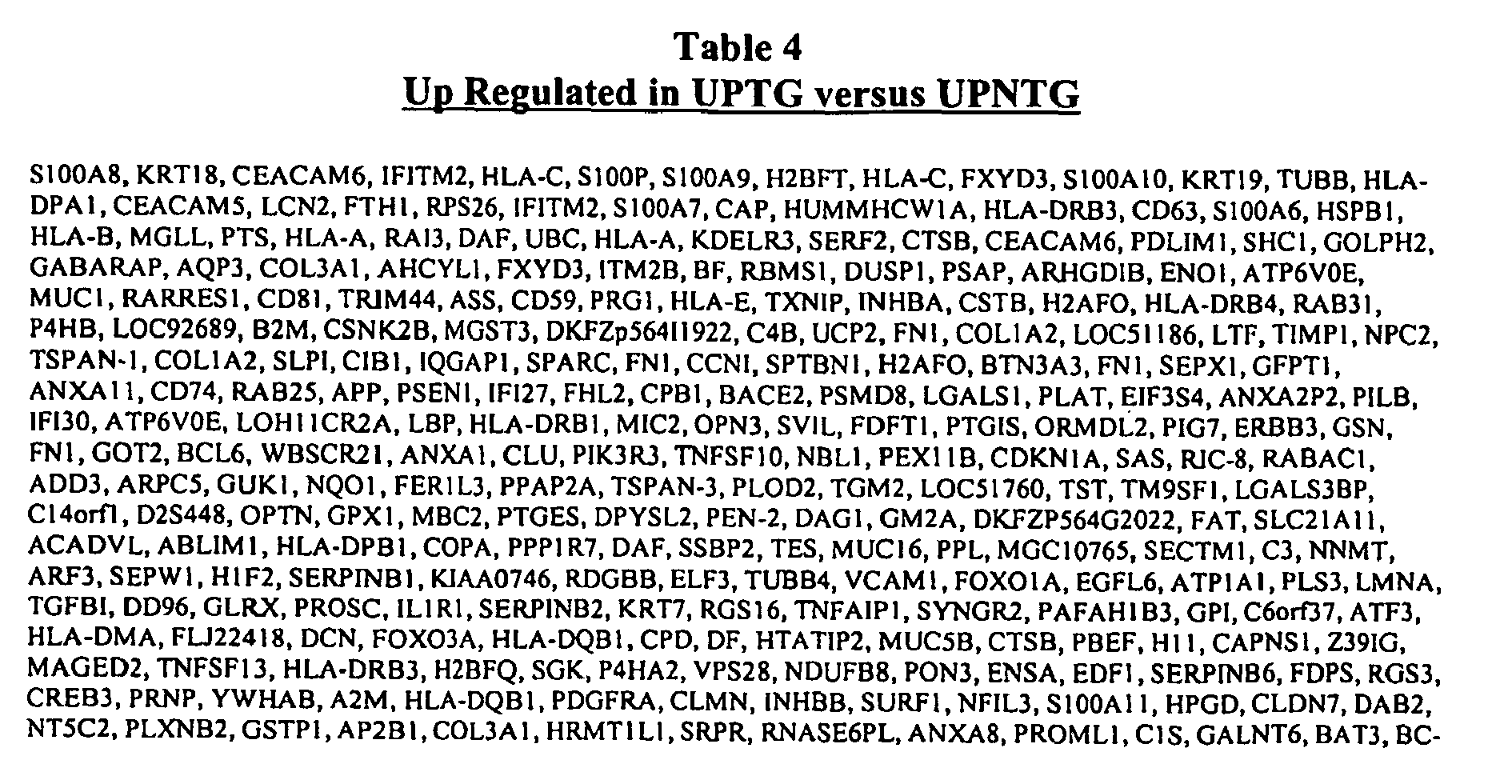


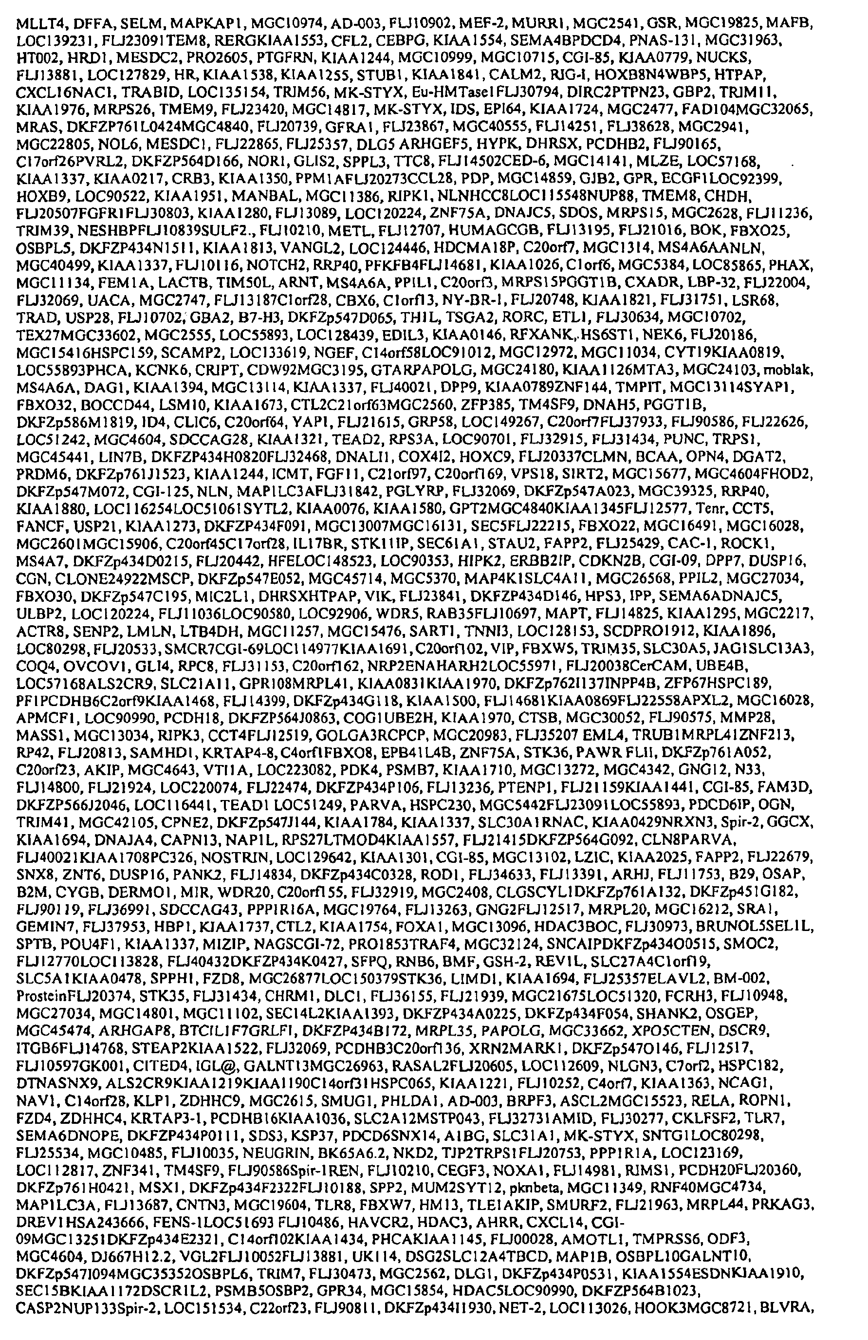


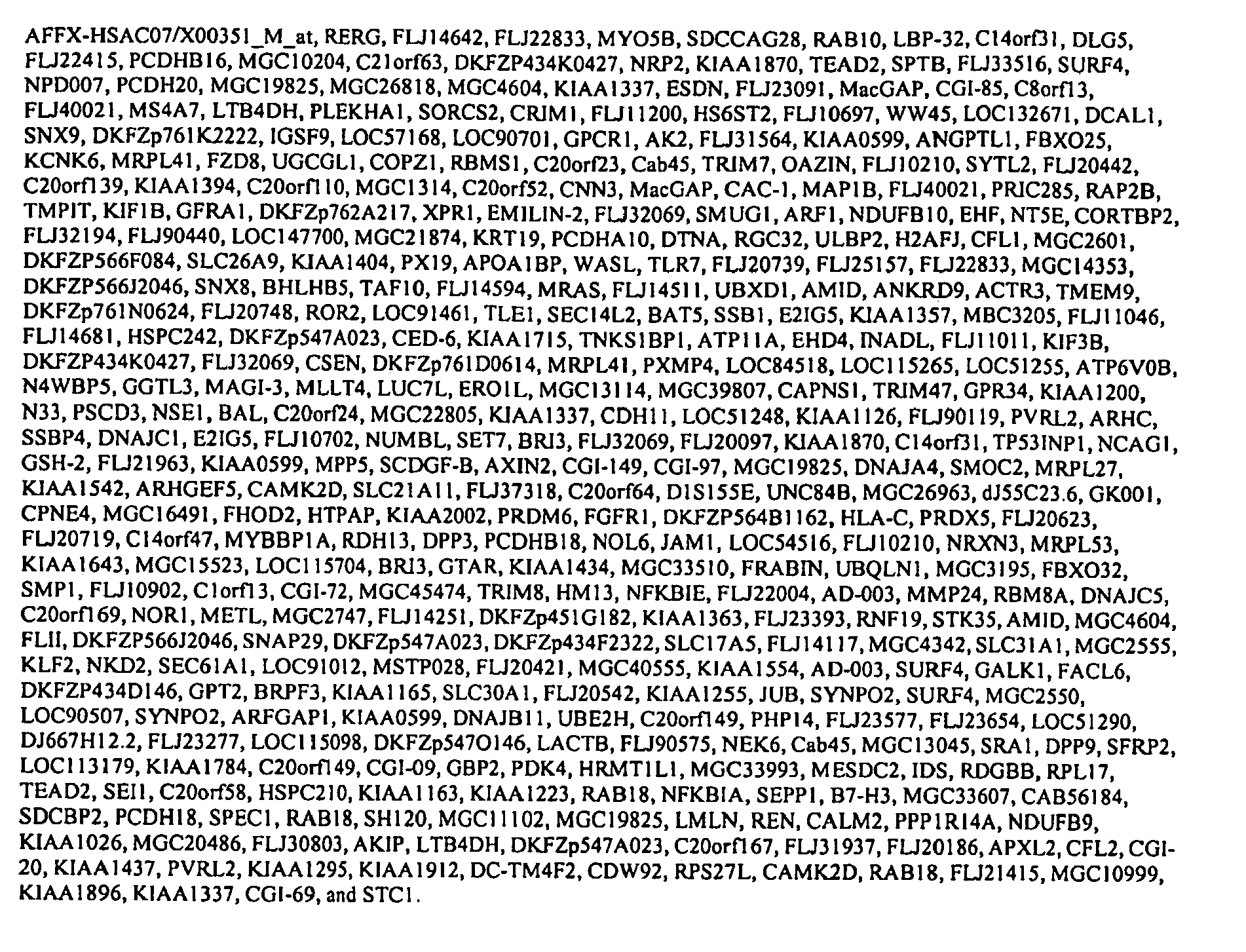
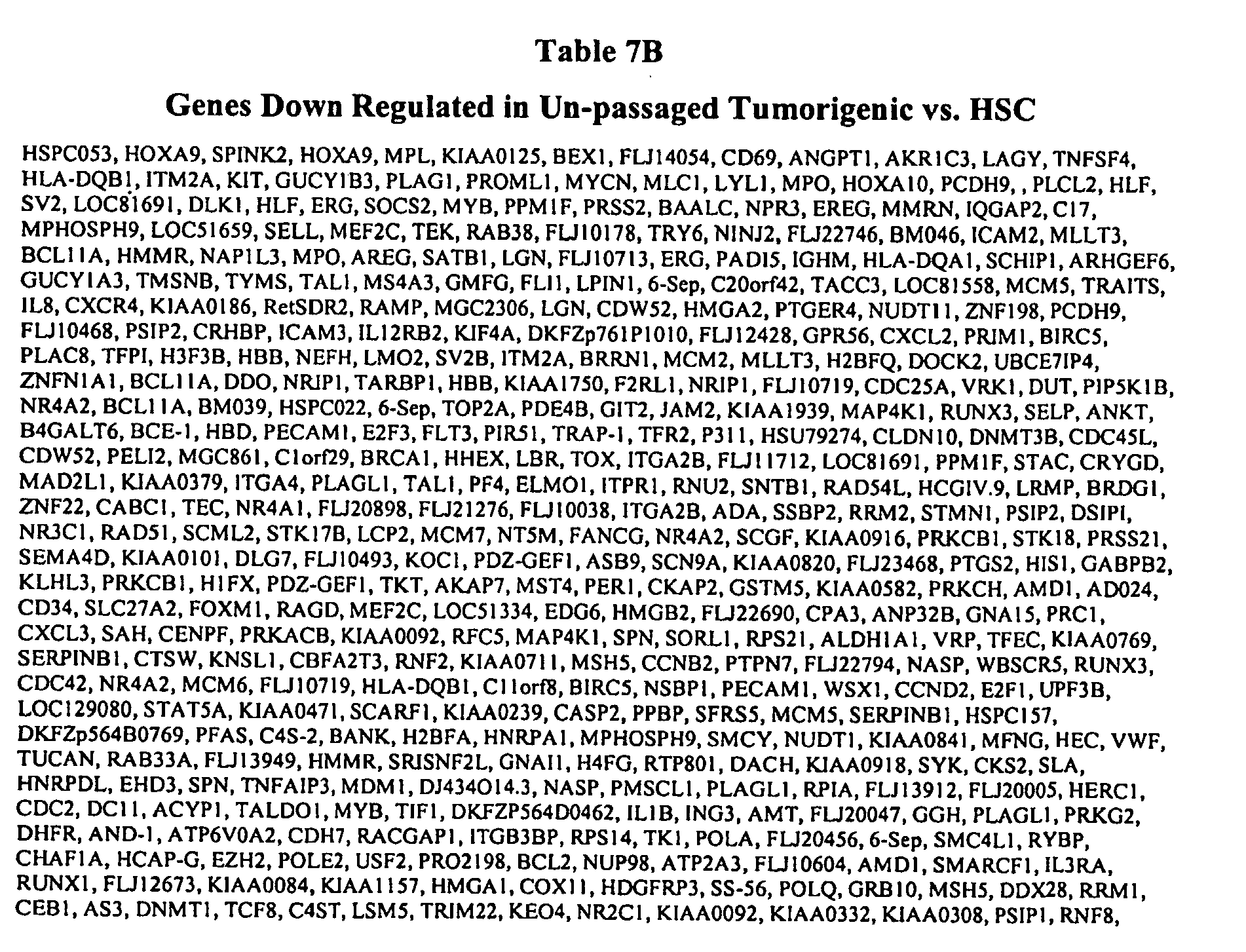


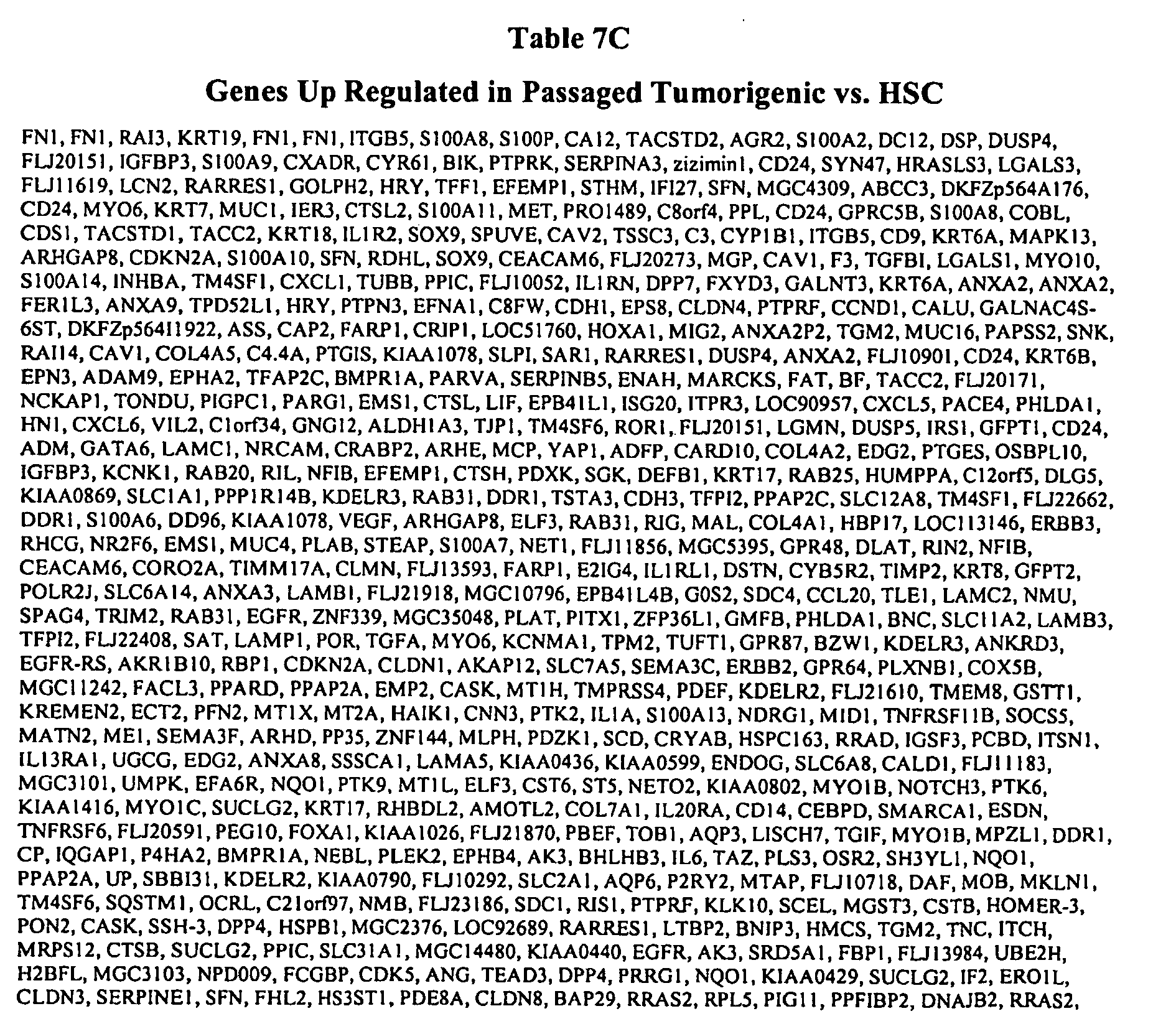


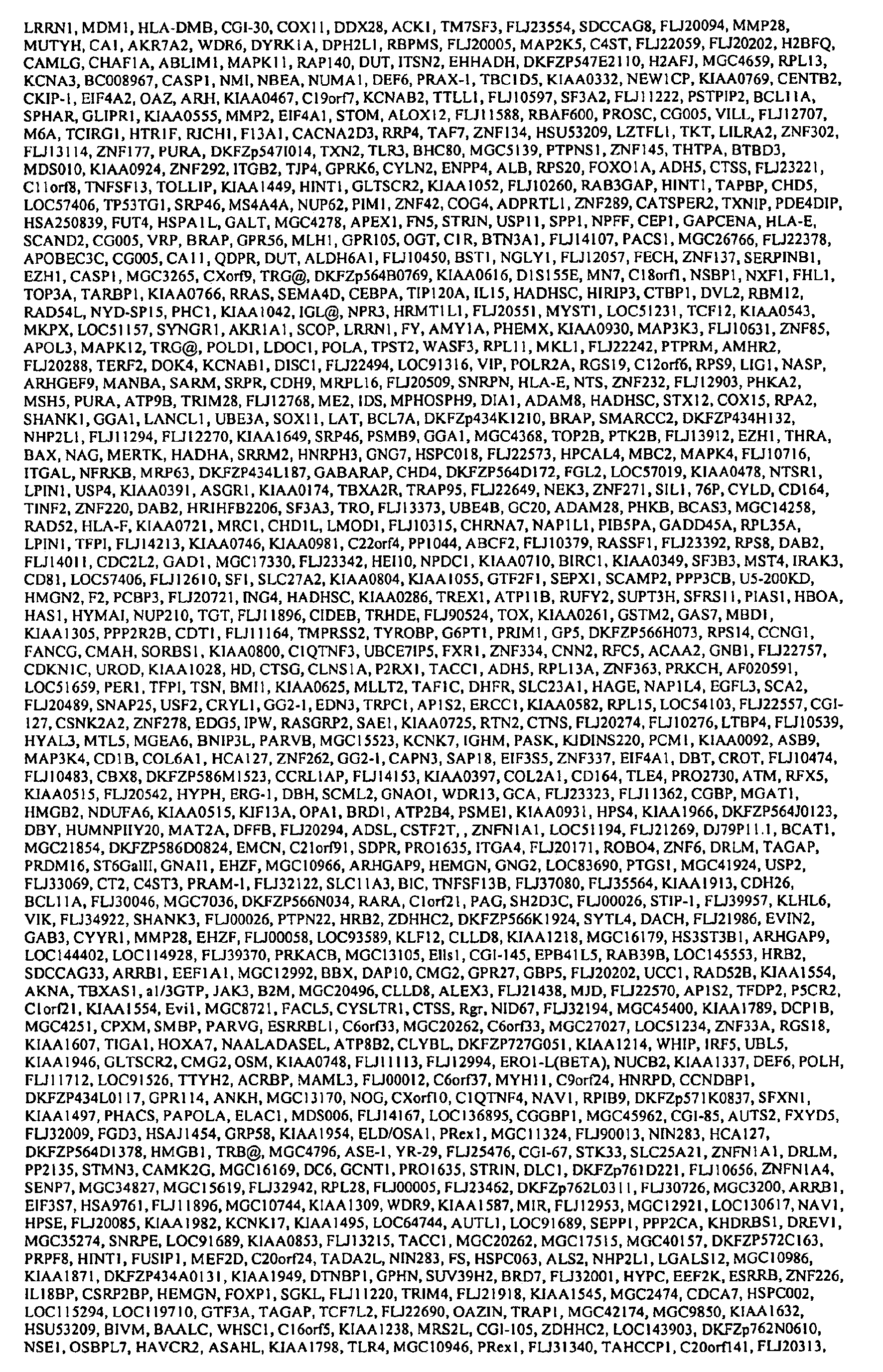

Table 8 Preferred Solid Tumor Stem Cell Cancer Markers Bmi-1, eed, easyhl, easyh2, rnf2, yyl, smarcA3, smarckA5, smarcD3, smarcE1, mllt3, frizzled 2, frizzled 6, frizzled 7, mf2, Frizzled 1, Frizzled2, Frizzled4, Frizzled10, Frizzled6, FZD1, FZD2, FZD3, FZD4, FZD6, FZD7, FZD8, FZD9, FZD10, WNT2, WNT2B, WNT3, WNT5A, WNT10B, WNT16, AXIN1, BCL9, MYC, (TCF4),, SLC7A8, IL1RAP, TEM8, TMPRSS4, MUC16, GPRC5B, SLC6A14, SLC4A11, PPAP2C, CAV1, CAV2, PTPN3, EPHA1, SLC1A1, CX3CL1, ADORA2A, MPZL1, FLJ10052, C4.4A, EDG3, RARRES1, TMEPAI, PTS, CEACAM6, , NID2, STEAP, ABCA3, CRIM1, IL1R1, OPN3, DAF, MUC1, MCP, CPD, NMA, ADAM9, GJA1, CD14, SLC19A2, ABCA1, PCDH7, ADCY9, SLC39A1, NPC1, ENPP1, N33, GPNMB, LY6E, CELSR1, LRP3, C20orf52, TMEPAI, FLVCR, PCDHA10, GPR54, TGFBR3, SEMA4B, and PCDHB2. - In some embodiments, the present invention provides methods for detection of expression of stem cell cancer markers (e.g., breast cancer stem cell cancer markers). In preferred embodiments, expression is measured directly (e.g., at the RNA or protein level). In some embodiments, expression is detected in tissue samples (e.g., biopsy tissue). In other embodiments, expression is detected in bodily fluids (e.g., including but not limited to, plasma, serum, whole blood, mucus, and urine). The present invention further provides panels and kits for the detection of markers. In preferred embodiments, the presence of a stem cell cancer marker is used to provide a prognosis to a subject. The information provided is also used to direct the course of treatment. For example, if a subject is found to have a marker indicative of a solid tumor stem cell (see, e.g. Tables 4-8), additional therapies (e.g., hormonal or radiation therapies) can be started at a earlier point when they are more likely to be effective (e.g., before metastasis). In addition, if a subject is found to have a tumor that is not responsive to hormonal therapy, the expense and inconvenience of such therapies can be avoided.
- The present invention is not limited to the markers described above. Any suitable marker that correlates with cancer or the progression of cancer may be utilized. Additional markers are also contemplated to be within the scope of the present invention. Any suitable method may be utilized to identify and characterize cancer markers suitable for use in the methods of the present invention, including but not limited to, those described in illustrative Example 4 below. For example, in some embodiments, markers identified as being up or down-regulated in solid tumor stem cells using the gene expression microarray methods of the present invention are further characterized using tissue microarray, immunohistochemistry, Northern blot analysis, siRNA or antisense RNA inhibition, mutation analysis, investigation of expression with clinical outcome, as well as other methods disclosed herein.
- In some embodiments, the present invention provides a panel for the analysis of a plurality of markers. The panel allows for the simultaneous analysis of multiple markers correlating with carcinogenesis and/or metastasis. Depending on the subject, panels may be analyzed alone or in combination in order to provide the best possible diagnosis and prognosis. Markers for inclusion on a panel are selected by screening for their predictive value using any suitable method, including but not limited to, those described in the illustrative examples below.
- In some preferred embodiments, detection of solid tumor stem cell cancer markers (e.g., including but not limited to, those disclosed in Tables 4-8) are detected by measuring the expression of corresponding mRNA in a tissue sample (e.g., breast cancer tissue). mRNA expression may be measured by any suitable method, including but not limited to, those disclosed below.
- In some embodiments, RNA is detection by Northern blot analysis. Northern blot analysis involves the separation of RNA and hybridization of a complementary labeled probe.
- In still further embodiments, RNA (or corresponding cDNA) is detected by hybridization to a oligonucleotide probe). A variety of hybridization assays using a variety of technologies for hybridization and detection are available. For example, in some embodiments, TaqMan assay (PE Biosystems, Foster City, CA; See e.g.,
U.S. Patent Nos. 5,962,233 and5,538,848 , each of which is herein incorporated by reference) is utilized. The assay is performed during a PCR reaction. The TaqMan assay exploits the 5'-3' exonuclease activity of the AMPLITAQ GOLD DNA polymerase. A probe consisting of an oligonucleotide with a 5'-reporter dye (e.g., a fluorescent dye) and a 3'-quencher dye is included in the PCR reaction. During PCR, if the probe is bound to its target, the 5'-3' nucleolytic activity of the AMPLITAQ GOLD polymerase cleaves the probe between the reporter and the quencher dye. The separation of the reporter dye from the quencher dye results in an increase of fluorescence. The signal accumulates with each cycle of PCR and can be monitored with a fluorimeter. - In yet other embodiments, reverse-transcriptase PCR (RT-PCR) is used to detect the expression of RNA. In RT-PCR, RNA is enzymatically converted to complementary DNA or "cDNA" using a reverse transcriptase enzyme. The cDNA is then used as a template for a PCR reaction. PCR products can be detected by any suitable method, including but not limited to, gel electrophoresis and staining with a DNA specific stain or hybridization to a labeled probe. In some embodiments, the quantitative reverse transcriptase PCR with standardized mixtures of competitive templates method described in
U.S. Patents 5,639,606 ,5,643,765 , and5,876,978 (each of which is herein incorporated by reference) is utilized. - In other embodiments, gene expression of stem cell cancer markers is detected by measuring the expression of the corresponding protein or polypeptide. Protein expression may be detected by any suitable method. In some embodiments, proteins are detected by immunohistochemistry. In other embodiments, proteins are detected by their binding to an antibody raised against the protein. The generation of antibodies is described below.
- Antibody binding is detected by techniques known in the art (e.g., radioimmunoassay, ELISA (enzyme-linked immunosorbant assay), "sandwich" immunoassays, immunoradiometric assays, gel diffusion precipitation reactions, immunodiffusion assays, in situ immunoassays (e.g., using colloidal gold, enzyme or radioisotope labels, for example), Western blots, precipitation reactions, agglutination assays (e.g., gel agglutination assays, hemagglutination assays, etc.), complement fixation assays, immunofluorescence assays, protein A assays, and immunoelectrophoresis assays, etc.
- In one embodiment, antibody binding is detected by detecting a label on the primary antibody. In another embodiment, the primary antibody is detected by detecting binding of a secondary antibody or reagent to the primary antibody. In a further embodiment, the secondary antibody is labeled. Many methods are known in the art for detecting binding in an immunoassay and are within the scope of the present invention.
- In some embodiments, an automated detection assay is utilized. Methods for the automation of immunoassays include those described in
U.S. Patents 5,885,530 ,4,981,785 ,6,159,750 , and5,358,691 , each of which is herein incorporated by reference. In some embodiments, the analysis and presentation of results is also automated. For example, in some embodiments, software that generates a prognosis based on the presence or absence of a series of proteins corresponding to cancer markers is utilized. - In other embodiments, the immunoassay described in
U.S. Patents 5,599,677 and5,672,480 ; each of which is herein incorporated by reference. - In some embodiments, a computer-based analysis program is used to translate the raw data generated by the detection assay (e.g., the presence, absence, or amount of a given marker or markers) into data of predictive value for a clinician. The clinician can access the predictive data using any suitable means. Thus, in some preferred embodiments, the present invention provides the further benefit that the clinician, who is not likely to be trained in genetics or molecular biology, need not understand the raw data. The data is presented directly to the clinician in its most useful form. The clinician is then able to immediately utilize the information in order to optimize the care of the subject.
- The present invention contemplates any method capable of receiving, processing, and transmitting the information to and from laboratories conducting the assays, information provides, medical personal, and subjects. For example, in some embodiments of the present invention, a sample (e.g., a biopsy or a serum or urine sample) is obtained from a subject and submitted to a profiling service (e.g., clinical lab at a medical facility, genomic profiling business, etc.), located in any part of the world (e.g., in a country different than the country where the subject resides or where the information is ultimately used) to generate raw data. Where the sample comprises a tissue or other biological sample, the subject may visit a medical center to have the sample obtained and sent to the profiling center, or subjects may collect the sample themselves and directly send it to a profiling center. Where the sample comprises previously determined biological information, the information may be directly sent to the profiling service by the subject (e.g., an information card containing the information may be scanned by a computer and the data transmitted to a computer of the profiling center using an electronic communication systems). Once received by the profiling service, the sample is processed and a profile is produced (e.g., expression data), specific for the diagnostic or prognostic information desired for the subj ect.
- The profile data is then prepared in a format suitable for interpretation by a treating clinician. For example, rather than providing raw expression data (e.g. examining a number of the markers described in Tables 4-8), the prepared format may represent a diagnosis or risk assessment for the subject, along with recommendations for particular treatment options. The data may be displayed to the clinician by any suitable method. For example, in some embodiments, the profiling service generates a report that can be printed for the clinician (e.g., at the point of care) or displayed to the clinician on a computer monitor.
- In some embodiments, the information is first analyzed at the point of care or at a regional facility. The raw data is then sent to a central processing facility for further analysis and/or to convert the raw data to information useful for a clinician or patient. The central processing facility provides the advantage of privacy (all data is stored in a central facility with uniform security protocols), speed, and uniformity of data analysis. The central processing facility can then control the fate of the data following treatment of the subject. For example, using an electronic communication system, the central facility can provide data to the clinician, the subject, or researchers.
- In some embodiments, the subject is able to directly access the data using the electronic communication system. The subject may chose further intervention or counseling based on the results. In some embodiments, the data is used for research use. For example, the data may be used to further optimize the inclusion or elimination of markers as useful indicators of a particular condition or stage of disease.
- In yet other embodiments, the present invention provides kits for the detection and characterization of cancer (e.g. for detecting one or more of the markers shown in Tables 4-8, or for modulating the activity of a peptide expressed by one or more of markes shown in Tables 4-8). In some embodiments, the kits contain antibodies specific for a cancer marker, in addition to detection reagents and buffers. In other embodiments, the kits contain reagents specific for the detection of mRNA or cDNA (e.g., oligonucleotide probes or primers). In preferred embodiments, the kits contain all of the components necessary to perform a detection assay, including all controls, directions for performing assays, and any necessary software for analysis and presentation of results.
- In some embodiments, in vivo imaging techniques are used to visualize the expression of cancer markers in an animal (e.g., a human or non-human mammal). For example, in some embodiments, cancer marker mRNA or protein is labeled using an labeled antibody specific for the cancer marker. A specifically bound and labeled antibody can be detected in an individual using an in vivo imaging method, including, but not limited to, radionuclide imaging, positron emission tomography, computerized axial tomography, X-ray or magnetic resonance imaging method, fluorescence detection, and chemiluminescent detection. Methods for generating antibodies to the cancer markers of the present invention are described below.
- The in vivo imaging methods of the present invention are useful in the diagnosis of cancers that express the solid tumor stem cell cancer markers of the present invention (e.g., in breast cancer). In vivo imaging is used to visualize the presence of a marker indicative of the cancer. Such techniques allow for diagnosis without the use of an unpleasant biopsy. The in vivo imaging methods of the present invention are also useful for providing prognoses to cancer patients. For example, the presence of a marker indicative of cancer stem cells can be detected. The in vivo imaging methods of the present invention can further be used to detect metastatic cancers in other parts of the body.
- In some embodiments, reagents (e.g., antibodies) specific for the cancer markers of the present invention are fluorescently labeled. The labeled antibodies are introduced into a subject (e.g., orally or parenterally). Fluorescently labeled antibodies are detected using any suitable method (e.g., using the apparatus described in
U.S. Patent 6,198,107 , herein incorporated by reference). - In other embodiments, antibodies are radioactively labeled. The use of antibodies for in vivo diagnosis is well known in the art. Sumerdon et al., (Nucl. Med. Biol 17:247-254 [1990] have described an optimized antibody-chelator for the radioimmunoscintographic imaging of tumors using Indium-111 as the label. Griffin et al., (J Clin One 9:631-640 [1991]) have described the use of this agent in detecting tumors in patients suspected of having recurrent colorectal cancer. The use of similar agents with paramagnetic ions as labels for magnetic resonance imaging is known in the art (Lauffer, Magnetic Resonance in Medicine 22:339-342 [1991]). The label used will depend on the imaging modality chosen. Radioactive labels such as Indium-111, Technetium-99m, or Iodine-131 can be used for planar scans or single photon emission computed tomography (SPECT). Positron emitting labels such as Fluorine-19 can also be used for positron emission tomography (PET). For MRI, paramagnetic ions such as Gadolinium (III) or Manganese (II) can be used.
- Radioactive metals with half-lives ranging from 1 hour to 3.5 days are available for conjugation to antibodies, such as scandium-47 (3.5 days) gallium-67 (2.8 days), gallium-68 (68 minutes), technetiium-99m (6 hours), and indium-111 (3.2 days), of which gallium-67, technetium-99m, and indium-111 are preferable for gamma camera imaging, gallium-68 is preferable for positron emission tomography.
- A useful method of labeling antibodies with such radiometals is by means of a bifunctional chelating agent, such as diethylenetriaminepentaacetic acid (DTPA), as described, for example, by Khaw et al. (Science 209:295 [1980]) for In-111 and Tc-99m, and by Scheinberg et al. (Science 215:1511 [1982]). Other chelating agents may also be used, but the 1-(p-carboxymethoxybenzyl)EDTA and the carboxycarbonic anhydride of DTPA are advantageous because their use permits conjugation without affecting the antibody's immunoreactivity substantially.
- Another method for coupling DPTA to proteins is by use of the cyclic anhydride of DTPA, as described by Hnatowich et al. (Int. J. Appl. Radiat. Isot. 33:327 [1982]) for labeling of albumin with In-111, but which can be adapted for labeling of antibodies. A suitable method of labeling antibodies with Tc-99m which does not use chelation with DPTA is the pretinning method of Crockford et al., (
U.S. Pat. No. 4,323,546 , herein incorporated by reference). - A preferred method of labeling immunoglobulins with Tc-99m is that described by Wong et al. (Int. J. Appl. Radiat. Isot., 29:251 [1978]) for plasma protein, and recently applied successfully by Wong et al. (J. Nucl. Med., 23:229 [1981]) for labeling antibodies.
- In the case of the radiometals conjugated to the specific antibody, it is likewise desirable to introduce as high a proportion of the radiolabel as possible into the antibody molecule without destroying its immunospecificity. A further improvement may be achieved by effecting radiolabeling in the presence of the specific stem cell cancer marker of the present invention, to insure that the antigen binding site on the antibody will be protected.
- In still further embodiments, in vivo biophotonic imaging (Xenogen, Almeda, CA) is utilized for in vivo imaging. This real-time in vivo imaging utilizes luciferase. The luciferase gene is incorporated into cells, microorganisms, and animals (e.g., as a fusion protein with a cancer marker of the present invention). When active, it leads to a reaction that emits light. A CCD camera and software is used to capture the image and analyze it.
- The present invention provides isolated antibodies and antibody fragments (e.g, Fabs). In preferred embodiments, the present invention provides monoclonal antibodies or antibody fragments that specifically bind to an isolated polypeptide comprised of at least five, or at least 15 amino acid residues of the stem cell cancer markers described herein (e.g., as shown in Tables 4-8). These antibodies or antibody fragments find use in the diagnostic, drug screening, and therapetuic methods described herein (e.g. to detect or modulate the activity of a stem cell cancer marker peptide).
- An antibody, or antibody fragment, against a protein of the present invention may be any monoclonal or polyclonal antibody, as long as it can recognize the protein. Antibodies can be produced by using a protein of the present invention as the antigen according to a conventional antibody or antiserum preparation process.
- The present invention contemplates the use of both monoclonal and polyclonal antibodies. Any suitable method may be used to generate the antibodies used in the methods and compositions of the present invention, including but not limited to, those disclosed herein. For example, for preparation of a monoclonal antibody, protein, as such, or together with a suitable carrier or diluent is administered to an animal (e.g., a mammal) under conditions that permit the production of antibodies. For enhancing the antibody production capability, complete or incomplete Freund's adjuvant may be administered. Normally, the protein is administered once every 2 weeks to 6 weeks, in total, about 2 times to about 10 times. Animals suitable for use in such methods include, but are not limited to, primates, rabbits, dogs, guinea pigs, mice, rats, sheep, goats, etc.
- For preparing monoclonal antibody-producing cells, an individual animal whose antibody titer has been confirmed (e.g., a mouse) is selected, and 2 days to 5 days after the final immunization, its spleen or lymph node is harvested and antibody-producing cells contained therein are fused with myeloma cells to prepare the desired monoclonal antibody producer hybridoma. Measurement of the antibody titer in antiserum can be carried out, for example, by reacting the labeled protein, as described hereinafter and antiserum and then measuring the activity of the labeling agent bound to the antibody. The cell fusion can be carried out according to known methods, for example, the method described by Koehler and Milstein (Nature 256:495 [1975]). As a fusion promoter, for example, polyethylene glycol (PEG) or Sendai virus (HVJ), preferably PEG is used.
- Examples of myeloma cells include NS-1, P3U1, SP2/0, AP-1 and the like. The proportion of the number of antibody producer cells (spleen cells) and the number of myeloma cells to be used is preferably about 1:1 to about 20:1. PEG (preferably PEG 1000-PEG 6000) is preferably added in concentration of about 10% to about 80%. Cell fusion can be carried out efficiently by incubating a mixture of both cells at about 20°C to about 40°C, preferably about 30°C to about 37°C for about 1 minute to 10 minutes.
- Various methods may be used for screening for a hybridoma producing the antibody (e.g., against a tumor antigen or autoantibody of the present invention). For example, where a supernatant of the hybridoma is added to a solid phase (e.g., microplate) to which antibody is adsorbed directly or together with a carrier and then an anti-immunoglobulin antibody (if mouse cells are used in cell fusion, anti-mouse immunoglobulin antibody is used) or Protein A labeled with a radioactive substance or an enzyme is added to detect the monoclonal antibody against the protein bound to the solid phase. Alternately, a supernatant of the hybridoma is added to a solid phase to which an anti-immunoglobulin antibody or Protein A is adsorbed and then the protein labeled with a radioactive substance or an enzyme is added to detect the monoclonal antibody against the protein bound to the solid phase.
- Selection of the monoclonal antibody can be carried out according to any known method or its modification. Normally, a medium for animal cells to which HAT (hypoxanthine, aminopterin, thymidine) are added is employed. Any selection and growth medium can be employed as long as the hybridoma can grow. For example, RPMI 1640 medium containing 1% to 20%, preferably 10% to 20% fetal bovine serum, GIT medium containing 1% to 10% fetal bovine serum, a serum free medium for cultivation of a hybridoma (SFM-101, Nissui Seiyaku) and the like can be used. Normally, the cultivation is carried out at 20°C to 40°C, preferably 37°C for about 5 days to 3 weeks, preferably 1 week to 2 weeks under about 5% CO2 gas. The antibody titer of the supernatant of a hybridoma culture can be measured according to the same manner as described above with respect to the antibody titer of the anti-protein in the antiserum.
- Separation and purification of a monoclonal antibody (e.g., against a cancer marker of the present invention) can be carried out according to the same manner as those of conventional polyclonal antibodies such as separation and purification of immunoglobulins, for example, salting-out, alcoholic precipitation, isoelectric point precipitation, electrophoresis, adsorption and desorption with ion exchangers (e.g., DEAE), ultracentrifugation, gel filtration, or a specific purification method wherein only an antibody is collected with an active adsorbent such as an antigen-binding solid phase, Protein A or Protein G and dissociating the binding to obtain the antibody.
- Polyclonal antibodies may be prepared by any known method or modifications of these methods including obtaining antibodies from patients. For example, a complex of an immunogen (an antigen against the protein) and a carrier protein is prepared and an animal is immunized by the complex according to the same manner as that described with respect to the above monoclonal antibody preparation. A material containing the antibody against is recovered from the immunized animal and the antibody is separated and purified.
- As to the complex of the immunogen and the carrier protein to be used for immunization of an animal, any carrier protein and any mixing proportion of the carrier and a hapten can be employed as long as an antibody against the hapten, which is crosslinked on the carrier and used for immunization, is produced efficiently. For example, bovine serum albumin, bovine cycloglobulin, keyhole limpet hemocyanin, etc. may be coupled to an hapten in a weight ratio of about 0.1 part to about 20 parts, preferably, about 1 part to about 5 parts per 1 part of the hapten.
- In addition, various condensing agents can be used for coupling of a hapten and a carrier. For example, glutaraldehyde, carbodiimide, maleimide activated ester, activated ester reagents containing thiol group or dithiopyridyl group, and the like find use with the present invention. The condensation product as such or together with a suitable carrier or diluent is administered to a site of an animal that permits the antibody production. For enhancing the antibody production capability, complete or incomplete Freund's adjuvant may be administered. Normally, the protein is administered once every 2 weeks to 6 weeks, in total, about 3 times to about 10 times.
- The polyclonal antibody is recovered from blood, ascites and the like, of an animal immunized by the above method. The antibody titer in the antiserum can be measured according to the same manner as that described above with respect to the supernatant of the hybridoma culture. Separation and purification of the antibody can be carried out according to the same separation and purification method of immunoglobulin as that described with respect to the above monoclonal antibody.
- The protein used herein as the immunogen is not limited to any particular type of immunogen. For example, a stem cell cancer marker of the present invention (further including a gene having a nucleotide sequence partly altered) can be used as the immunogen. Further, fragments of the protein may be used. Fragments may be obtained by any methods including, but not limited to expressing a fragment of the gene, enzymatic processing of the protein, chemical synthesis, and the like. The antibodies and antibody fragments may also be conjugated to therapeutic (e.g. cancer cell killing compounds). In this regard, the antibody directed toward one of the stem cell cancer markers is used to specifically deliver a therapeutic agent to a solid tumor cancer cell (e.g. to inhibit the proliferation of such sell or kill such a cell).
- In some embodiments, the present invention provides drug screening assays (e.g., to screen for anticancer drugs). The screening methods of the present invention utilize stem cell cancer markers identified using the methods of the present invention (e.g., including but not limited to, the stem cell cancer markers shown in Tables 4-8). For example, in some embodiments, the present invention provides methods of screening for compound that alter (e.g., increase or decrease) the expression of stem cell cancer marker genes. In some embodiments, candidate compounds are antisense agents or siRNA agents (e.g., oligonucleotides) directed against cancer markers. In other embodiments, candidate compounds are antibodies that specifically bind to a stem cell cancer marker of the present invention. In certain embodiments, libraries of compounds of small molecules are screened using the methods described herein.
- In one screening method, candidate compounds are evaluated for their ability to alter stem cell cancer marker expression by contacting a compound with a cell expressing a stem cell cancer marker and then assaying for the effect of the candidate compounds on expression. In some embodiments, the effect of candidate compounds on expression of a cancer marker gene is assayed by detecting the level of cancer marker mRNA expressed by the cell. mRNA expression can be detected by any suitable method. In other embodiments, the effect of candidate compounds on expression of cancer marker genes is assayed by measuring the level of polypeptide encoded by the cancer markers. The level of polypeptide expressed can be measured using any suitable method, including but not limited to, those disclosed herein. In some embodiments, other changes in cel biology (e.g., apoptosis) are detected.
- Specifically, the present invention provides screening methods for identifying modulators, i.e., candidate or test compounds or agents (e.g., proteins, peptides, peptidomimetics, peptoids, small molecules or other drugs) which bind to, or alter the signalling or function associated with the cancer markers of the present invention, have an inhibitory (or stimulatory) effect on, for example, stem cell cancer marker expression or cancer markers activity, or have a stimulatory or inhibitory effect on, for example, the expression or activity of a cancer marker substrate. Compounds thus identified can be used to modulate the activity of target gene products (e.g., stem cell cancer marker genes) either directly or indirectly in a therapeutic protocol, to elaborate the biological function of the target gene product, or to identify compounds that disrupt normal target gene interactions. Compounds which inhibit the activity or expression of cancer markers are useful in the treatment of proliferative disorders, e.g., cancer, particularly metastatic cancer or eliminating or controlling tumor stem cells to prevent or reduce the risk of cancer.
- In one embodiment, the invention provides assays for screening candidate or test compounds that are substrates of a cancer markers protein or polypeptide or a biologically active portion thereof. In another embodiment, the invention provides assays for screening candidate or test compounds that bind to or modulate the activity of a cancer marker protein or polypeptide or a biologically active portion thereof.
- The test compounds of the present invention can be obtained using any of the numerous approaches in combinatorial library methods known in the art, including biological libraries; peptoid libraries (libraries of molecules having the functionalities of peptides, but with a novel, non-peptide backbone, which are resistant to enzymatic degradation but which nevertheless remain bioactive; see, e.g., Zuckennann et al., J. Med. Chem. 37: 2678-85 [1994]); spatially addressable parallel solid phase or solution phase libraries; synthetic library methods requiring deconvolution; the 'one-bead one-compound' library method; and synthetic library methods using affinity chromatography selection. The biological library and peptoid library approaches are preferred for use with peptide libraries, while the other four approaches are applicable to peptide, non-peptide oligomer or small molecule libraries of compounds (Lam (1997) Anticancer Drug Des. 12:145).
- Examples of methods for the synthesis of molecular libraries can be found in the art, for example in: DeWitt et al., Proc. Natl. Acad. Sci. U.S.A. 90:6909 [1993]; Erb et al., Proc. Nad. Acad. Sci. USA 91:11422 [1994]; Zuckermann et al., J. Med. Chem. 37:2678 [1994]; Cho et al., Science 261:1303 [1993]; Carrell et al., Angew. Chem. Int. Ed. Engl. 33.2059 [1994]; Carell et al., Angew. Chem. Int. Ed. Engl. 33:2061 [1994]; and Gallop et al., J. Med. Chem. 37:1233 [1994].
- Libraries of compounds may be presented in solution (e.g., Houghten, Biotechniques 13:412-421 [1992]), or on beads (Lam, Nature 354:82-84 [1991]), chips (Fodor, Nature 364:555-556 [1993]), bacteria or spores (
U.S. Patent No. 5,223,409 ; herein incorporated by reference), plasmids (Cull et al., Proc. Nad. Acad. Sci. USA 89:18651869 [1992]) or on phage (Scott and Smith, Science 249:386-390 [1990]; Devlin Science 249:404-406 [1990]; Cwirla et al., Proc. Natl. Acad. Sci. 87:6378-6382 [1990]; Felici, J. Mol. Biol. 222:301 [1991]). - In one embodiment, an assay is a cell-based assay in which a cell that expresses a stem cell cancer marker protein or biologically active portion thereof is contacted with a test compound, and the ability of the test compound to the modulate cancer marker's activity is determined. Determining the ability of the test compound to modulate stem cell cancer marker activity can be accomplished by monitoring, for example, changes in enzymatic activity. The cell, for example, can be of mammalian origin.
- The ability of the test compound to modulate cancer marker binding to a compound, e.g., a stem cell cancer marker substrate, can also be evaluated. This can be accomplished, for example, by coupling the compound, e.g., the substrate, with a radioisotope or enzymatic label such that binding of the compound, e.g., the substrate, to a cancer marker can be determined by detecting the labeled compound, e.g., substrate, in a complex.
- Alternatively, the stem cell cancer marker is coupled with a radioisotope or enzymatic label to monitor the ability of a test compound to modulate cancer marker binding to a cancer markers substrate in a complex. For example, compounds (e.g., substrates) can be labeled with 125I, 35S 14C or 3H, either directly or indirectly, and the radioisotope detected by direct counting of radioemmission or by scintillation counting. Alternatively, compounds can be enzymatically labeled with, for example, horseradish peroxidase, alkaline phosphatase, or luciferase, and the enzymatic label detected by determination of conversion of an appropriate substrate to product.
- The ability of a compound (e.g., a stem cell cancer marker substrate) to interact with a stem cell cancer marker with or without the labeling of any of the interactants can be evaluated. For example, a microphysiorneter can be used to detect the interaction of a compound with a cancer marker without the labeling of either the compound or the cancer marker (McConnell et al. Science 257:1906-1912 [1992]). As used herein, a "microphysiometer" (e.g., Cytosensor) is an analytical instrument that measures the rate at which a cell acidifies its environment using a light-addressable potentiometric sensor (LAPS). Changes in this acidification rate can be used as an indicator of the interaction between a compound and cancer markers.
- In yet another embodiment, a cell-free assay is provided in which a cancer marker protein or biologically active portion thereof is contacted with a test compound and the ability of the test compound to bind to the stem cell cancer marker protein or biologically active portion thereof is evaluated. Preferred biologically active portions of the cancer markers proteins to be used in assays of the present invention include fragments that participate in interactions with substrates or other proteins, e.g., fragments with high surface probability scores.
- Cell-free assays involve preparing a reaction mixture of the target gene protein and the test compound under conditions and for a time sufficient to allow the two components to interact and bind, thus forming a complex that can be removed and/or detected.
- The interaction between two molecules can also be detected, e.g., using fluorescence energy transfer (FRET) (see, for example,
Lakowicz et al., U.S. Patent No. 5,631,169 ;Stavrianopoulos et al., U.S. Patent No. 4,968,103 ; each of which is herein incorporated by reference). A fluorophore label is selected such that a first donor molecule's emitted fluorescent energy will be absorbed by a fluorescent label on a second, 'acceptor' molecule, which in turn is able to fluoresce due to the absorbed energy. - Alternately, the 'donor' protein molecule may simply utilize the natural fluorescent energy of tryptophan residues. Labels are chosen that emit different wavelengths of light, such that the 'acceptor' molecule label may be differentiated from that of the 'donor'. Since the efficiency of energy transfer between the labels is related to the distance separating the molecules, the spatial relationship between the molecules can be assessed. In a situation in which binding occurs between the molecules, the fluorescent emission of the 'acceptor' molecule label in 1 5 the assay should be maximal. An FRET binding event can be conveniently measured through standard fluorometric detection means well known in the art (e.g., using a fluorimeter).
- In another embodiment, determining the ability of the stem cell cancer markers protein to bind to a target molecule can be accomplished using real-time Biomolecular Interaction Analysis (BIA) (see, e.g., Sjolander and Urbaniczky, Anal. Chem. 63:2338-2345 [1991] and Szabo et al. Curr. Opin. Struct. Biol. 5:699-705 [1995]). "Surface plasmon resonance" or "BIA" detects biospecific interactions in real time, without labeling any of the interactants (e.g., BlAcore). Changes in the mass at the binding surface (indicative of a binding event) result in alterations of the refractive index of light near the surface (the optical phenomenon of surface plasmon resonance (SPR)), resulting in a detectable signal that can be used as an indication of real-time reactions between biological molecules.
- In one embodiment, the target gene product or the test substance is anchored onto a solid phase. The target gene product/test compound complexes anchored on the solid phase can be detected at the end of the reaction. Preferably, the target gene product can be anchored onto a solid surface, and the test compound, (which is not anchored), can be labeled, either directly or indirectly, with detectable labels discussed herein.
- It may be desirable to immobilize stem cell cancer markers, an anti-cancer marker antibody or its target molecule to facilitate separation of complexed from non-complexed forms of one or both of the proteins, as well as to accommodate automation of the assay. Binding of a test compound to a stem cell cancer marker protein, or interaction of a cancer marker protein with a target molecule in the presence and absence of a candidate compound, can be accomplished in any vessel suitable for containing the reactants. Examples of such vessels include microtiter plates, test tubes, and micro-centrifuge tubes. In one embodiment, a fusion protein can be provided which adds a domain that allows one or both of the proteins to be bound to a matrix. For example, glutathione-S-transferase-cancer marker fusion proteins or glutathione-S-transferase/target fusion proteins can be adsorbed onto glutathione Sepharose beads (Sigma Chemical, St. Louis, MO) or glutathione-derivatized microtiter plates, which are then combined with the test compound or the test compound and either the non-adsorbed target protein or cancer marker protein, and the mixture incubated under conditions conducive for complex formation (e.g., at physiological conditions for salt and pH). Following incubation, the beads or microtiter plate wells are washed to remove any unbound components, the matrix immobilized in the case of beads, complex determined either directly or indirectly, for example, as described above.
- Alternatively, the complexes can be dissociated from the matrix, and the level of cancer markers binding or activity determined using standard techniques. Other techniques for immobilizing either cancer markers protein or a target molecule on matrices include using conjugation of biotin and streptavidin. Biotinylated cancer marker protein or target molecules can be prepared from biotin-NHS (N-hydroxysuccinimide) using techniques known in the art (e.g., biotinylation kit, Pierce Chemicals, Rockford, EL), and immobilized in the wells of streptavidin-coated 96 well plates (Pierce Chemical).
- In order to conduct the assay, the non-immobilized component is added to the coated surface containing the anchored component. After the reaction is complete, unreacted components are removed (e.g., by washing) under conditions such that any complexes formed will remain immobilized on the solid surface. The detection of complexes anchored on the solid surface can be accomplished in a number of ways. Where the previously non-immobilized component is pre-labeled, the detection of label immobilized on the surface indicates that complexes were formed. Where the previously non-immobilized component is not pre-labeled, an indirect label can be used to detect complexes anchored on the surface; e.g., using a labeled antibody specific for the immobilized component (the antibody, in turn, can be directly labeled or indirectly labeled with, e.g., a labeled anti-IgG antibody).
- This assay is performed utilizing antibodies reactive with stem cell cancer marker protein or target molecules but which do not interfere with binding of the stem cell cancer markers protein to its target molecule. Such antibodies can be derivatized to the wells of the plate, and unbound target or cancer markers protein trapped in the wells by antibody conjugation. Methods for detecting such complexes, in addition to those described above for the GST-immobilized complexes, include immunodetection of complexes using antibodies reactive with the cancer marker protein or target molecule, as well as enzyme-linked assays which rely on detecting an enzymatic activity associated with the cancer marker protein or target molecule.
- Alternatively, cell free assays can be conducted in a liquid phase. In such an assay, the reaction products are separated from unreacted components, by any of a number of standard techniques, including, but not limited to: differential centrifugation (see, for example, Rivas and Minton, Trends Biochem Sci 18:284-7 [1993]); chromatography (gel filtration chromatography, ion-exchange chromatography); electrophoresis (see, e.g., Ausubel et al., eds. Current Protocols in Molecular Biology 1999, J. Wiley: New York.); and immunoprecipitation (see, for example, Ausubel et al., eds. Current Protocols in Molecular Biology 1999, J. Wiley: New York). Such resins and chromatographic techniques are known to one skilled in the art (See e.g., Heegaard J. Mol. Recognit 11:141-8 [1998]; Hageand Tweed J. Chromatogr. Biomed. Sci. Appl 699:499-525 [1997]). Further, fluorescence energy transfer may also be conveniently utilized, as described herein, to detect binding without further purification of the complex from solution.
- The assay can include contacting the stem cell cancer markers protein or biologically active portion thereof with a known compound that binds the cancer marker to form an assay mixture, contacting the assay mixture with a test compound, and determining the ability of the test compound to interact with a cancer marker protein, wherein determining the ability of the test compound to interact with a cancer marker protein includes determining the ability of the test compound to preferentially bind to cancer markers or biologically active portion thereof, or to modulate the activity of a target molecule, as compared to the known compound.
- To the extent that stem cell cancer markers can, in vivo, interact with one or more cellular or extracellular macromolecules, such as proteins, inhibitors of such an interaction are useful. A homogeneous assay can be used can be used to identify inhibitors
- For example, a preformed complex of the target gene product and the interactive cellular or extracellular binding partner product is prepared such that either the target gene products or their binding partners are labeled, but the signal generated by the label is quenched due to complex formation (see, e.g.,
U.S. Patent No. 4,109,496 , herein incorporated by reference, that utilizes this approach for immunoassays). The addition of a test substance that competes with and displaces one of the species from the preformed complex will result in the generation of a signal above background. In this way, test substances that disrupt target gene product-binding partner interaction can be identified. Alternatively, cancer markers protein can be used as a "bait protein" in a two-hybrid assay or three-hybrid assay (see, e.g.,U.S. Patent No. 5,283,317 ; Zervos et al., Cell 72:223-232 [1993]; Madura et al., J. Biol. Chem. 268.12046-12054 [1993]; Bartel et al., Biotechniques 14:920-924 [1993]; Iwabuchi et al., Oncogene 8:1693-1696 [1993]; andBrent W0 94/10300 - Modulators of cancer markers expression can also be identified. For example, a cell or cell free mixture is contacted with a candidate compound and the expression of cancer marker mRNA or protein evaluated relative to the level of expression of stem cell cancer marker mRNA or protein in the absence of the candidate compound. When expression of cancer marker mRNA or protein is greater in the presence of the candidate compound than in its absence, the candidate compound is identified as a stimulator of cancer marker mRNA or protein expression. Alternatively, when expression of cancer marker mRNA or protein is less (i.e., statistically significantly less) in the presence of the candidate compound than in its absence, the candidate compound is identified as an inhibitor of cancer marker mRNA or protein expression. The level of cancer markers mRNA or protein expression can be determined by methods described herein for detecting cancer markers mRNA or protein.
- A modulating agent can be identified using a cell-based or a cell free assay, and the ability of the agent to modulate the activity of a cancer markers protein can be confirmed in vivo, e.g., in an animal such as an animal model for a disease (e.g., an animal with prostate cancer or metastatic prostate cancer; or an animal harboring a xenograft of a prostate cancer from an animal (e.g., human) or cells from a cancer resulting from metastasis of a prostate cancer (e.g., to a lymph node, bone, or liver), or cells from a prostate cancer cell line.
- This invention further pertains to novel agents identified by the above-described screening assays (See e.g., below description of cancer therapies). Accordingly, it is within the scope of this invention to further use an agent identified as described herein (e.g., a cancer marker modulating agent, an antisense cancer marker nucleic acid molecule, a siRNA molecule, a cancer marker specific antibody, or a cancer marker-binding partner) in an appropriate animal model (such as those described herein) to determine the efficacy, toxicity, side effects, or mechanism of action, of treatment with such an agent. Furthermore, novel agents identified by the above-described screening assays can be, e.g., used for treatments as described herein (e.g. to treat a human patient who has cancer).
- In some embodiments, the present invention provides therapies for cancer (e.g., breast cancer). In some embodiments, therapies target cancer markers (e.g., including but not limited to, those shown in Tables 4-8).
- Candidate therapeutic agents also find use in drug screening and research applications. In some embodiments, the present invention targets the expression of stem cell cancer markers. For example, in some embodiments, the present invention employs compositions comprising oligomeric antisense compounds, particularly oligonucleotides (e.g., those identified in the drug screening methods described above), for use in modulating the function of nucleic acid molecules encoding stem cell cancer markers of the present invention, ultimately modulating the amount of cancer marker expressed. This is accomplished by providing antisense compounds that specifically hybridize with one or more nucleic acids encoding cancer markers of the present invention. The specific hybridization of an oligomeric compound with its target nucleic acid interferes with the normal function of the nucleic acid. This modulation of function of a target nucleic acid by compounds that specifically hybridize to it is generally referred to as "antisense." The functions of DNA to be interfered with include replication and transcription. The functions of RNA to be interfered with include all vital functions such as, for example, translocation of the RNA to the site of protein translation, translation of protein from the RNA, splicing of the RNA to yield one or more mRNA species, and catalytic activity that may be engaged in or facilitated by the RNA. The overall effect of such interference with target nucleic acid function is modulation of the expression of cancer markers of the present invention. In the context of the present invention, "modulation" means either an increase (stimulation) or a decrease (inhibition) in the expression of a gene. For example, expression may be inhibited to potentially prevent tumor proliferation.
- It is preferred to target specific nucleic acids for antisense. "Targeting" an antisense compound to a particular nucleic acid, in the context of the present invention, is a multistep process. The process usually begins with the identification of a nucleic acid sequence whose function is to be modulated. This may be, for example, a cellular gene (or mRNA transcribed from the gene) whose expression is associated with a particular disorder or disease state, or a nucleic acid molecule from an infectious agent. In the present invention, the target is a nucleic acid molecule encoding a stem cell cancer marker of the present invention. The targeting process also includes determination of a site or sites within this gene for the antisense interaction to occur such that the desired effect, e.g., detection or modulation of expression of the protein, will result. Within the context of the present invention, a preferred intragenic site is the region encompassing the translation initiation or termination codon of the open reading frame (ORF) of the gene. Since the translation initiation codon is typically 5'-AUG (in transcribed mRNA molecules; 5'-ATG in the corresponding DNA molecule), the translation initiation codon is also referred to as the "AUG codon," the "start codon" or the "AUG start codon". A minority of genes have a translation initiation codon having the RNA sequence 5'-GUG, 5'-UUG or 5'-CUG, and 5'-AUA, 5'-ACG and 5'-CUG have been shown to function in vivo. Thus, the terms "translation initiation codon" and "start codon" can encompass many codon sequences, even though the initiator amino acid in each instance is typically methionine (in eukaryotes) or formylmethionine (in prokaryotes). Eukaryotic and prokaryotic genes may have two or more alternative start codons, any one of which may be preferentially utilized for translation initiation in a particular cell type or tissue, or under a particular set of conditions. In the context of the present invention, "start codon" and "translation initiation codon" refer to the codon or codons that are used in vivo to initiate translation of an mRNA molecule transcribed from a gene encoding a tumor antigen of the present invention, regardless of the sequence(s) of such codons.
- Translation termination codon (or "stop codon") of a gene may have one of three sequences (i.e., 5'-UAA, 5'-UAG and 5'-UGA; the corresponding DNA sequences are 5'-TAA, 5'-TAG and 5'-TGA, respectively). The terms "start codon region" and "translation initiation codon region" refer to a portion of such an mRNA or gene that encompasses from about 25 to about 50 contiguous nucleotides in either direction (i.e., 5' or 3') from a translation initiation codon. Similarly, the terms "stop codon region" and "translation termination codon region" refer to a portion of such an mRNA or gene that encompasses from about 25 to about 50 contiguous nucleotides in either direction (i.e., 5' or 3') from a translation termination codon.
- The open reading frame (ORF) or "coding region," which refers to the region between the translation initiation codon and the translation termination codon, is also a region that may be targeted effectively. Other target regions include the 5' untranslated region (5' UTR), referring to the portion of an mRNA in the 5' direction from the translation initiation codon, and thus including nucleotides between the 5' cap site and the translation initiation codon of an mRNA or corresponding nucleotides on the gene, and the 3' untranslated region (3' UTR), referring to the portion of an mRNA in the 3' direction from the translation termination codon, and thus including nucleotides between the translation termination codon and 3' end of an mRNA or corresponding nucleotides on the gene. The 5' cap of an mRNA comprises an N7-methylated guanosine residue joined to the 5'-most residue of the mRNA via a 5'-5' triphosphate linkage. The 5' cap region of an mRNA is considered to include the 5' cap structure itself as well as the first 50 nucleotides adjacent to the cap. The cap region may also be a preferred target region.
- Although some eukaryotic mRNA transcripts are directly translated, many contain one or more regions, known as "introns," that are excised from a transcript before it is translated. The remaining (and therefore translated) regions are known as "exons" and are spliced together to form a continuous mRNA sequence. mRNA splice sites (i.e., intron-exon junctions) may also be preferred target regions, and are particularly useful in situations where aberrant splicing is implicated in disease, or where an overproduction of a particular mRNA splice product is implicated in disease. Aberrant fusion junctions due to rearrangements or deletions are also preferred targets. It has also been found that introns can also be effective, and therefore preferred, target regions for antisense compounds targeted, for example, to DNA or pre-mRNA.
- In some embodiments, target sites for antisense inhibition are identified using commercially available software programs (e.g., Biognostik, Gottingen, Germany; SysArris Software, Bangalore, India; Antisense Research Group, University of Liverpool, Liverpool, England; GeneTrove, Carlsbad, CA). In other embodiments, target sites for antisense inhibition are identified using the accessible site method described in U.S. Patent
W00198537A2 - Once one or more target sites have been identified, oligonucleotides are chosen that are sufficiently complementary to the target (i.e., hybridize sufficiently well and with sufficient specificity) to give the desired effect. For example, in preferred embodiments of the present invention, antisense oligonucleotides are targeted to or near the start codon.
- In the context of this invention, "hybridization," with respect to antisense compositions and methods, means hydrogen bonding, which may be Watson-Crick, Hoogsteen or reversed Hoogsteen hydrogen bonding, between complementary nucleoside or nucleotide bases. For example, adenine and thymine are complementary nucleobases that pair through the formation of hydrogen bonds. It is understood that the sequence of an antisense compound need not be 100% complementary to that of its target nucleic acid to be specifically hybridizable. An antisense compound is specifically hybridizable when binding of the compound to the target DNA or RNA molecule interferes with the normal function of the target DNA or RNA to cause a loss of utility, and there is a sufficient degree of complementarity to avoid non-specific binding of the antisense compound to non-target sequences under conditions in which specific binding is desired (i.e., under physiological conditions in the case of in vivo assays or therapeutic treatment, and in the case of in vitro assays, under conditions in which the assays are performed).
- Antisense compounds are commonly used as research reagents and diagnostics. For example, antisense oligonucleotides, which are able to inhibit gene expression with specificity, can be used to elucidate the function of particular genes. Antisense compounds are also used, for example, to distinguish between functions of various members of a biological pathway.
- The specificity and sensitivity of antisense is also applied for therapeutic uses. For example, antisense oligonucleotides have been employed as therapeutic moieties in the treatment of disease states in animals and man. Antisense oligonucleotides have been safely and effectively administered to humans and numerous clinical trials are presently underway. It is thus established that oligonucleotides are useful therapeutic modalities that can be configured to be useful in treatment regimes for treatment of cells, tissues, and animals, especially humans.
- While antisense oligonucleotides are a preferred form of antisense compound, the present invention comprehends other oligomeric antisense compounds, including but not limited to oligonucleotide mimetics such as are described below. The antisense compounds in accordance with this invention preferably comprise from about 8 to about 30 nucleobases (i.e., from about 8 to about 30 linked bases), although both longer and shorter sequences may find use with the present invention. Particularly preferred antisense compounds are antisense oligonucleotides, even more preferably those comprising from about 12 to about 25 nucleobases.
- Specific examples of preferred antisense compounds useful with the present invention include oligonucleotides containing modified backbones or non-natural internucleoside linkages. As defined in this specification, oligonucleotides having modified backbones include those that retain a phosphorus atom in the backbone and those that do not have a phosphorus atom in the backbone. For the purposes of this specification, modified oligonucleotides that do not have a phosphorus atom in their internucleoside backbone can also be considered to be oligonucleosides.
- Preferred modified oligonucleotide backbones include, for example, phosphorothioates, chiral phosphorothioates, phosphorodithioates, phosphotriesters, aminoalkylphosphotriesters, methyl and other alkyl phosphonates including 3'-alkylene phosphonates and chiral phosphonates, phosphinates, phosphoramidates including 3'-amino phosphoramidate and aminoalkylphosphoramidates, thionophosphoramidates, thionoalkylphosphonates, thionoalkylphosphotriesters, and boranophosphates having normal 3'-5' linkages, 2'-5' linked analogs of these, and those having inverted polarity wherein the adjacent pairs of nucleoside units are linked 3'-5' to 5'-3' or 2'-5' to 5'-2'. Various salts, mixed salts and free acid forms are also included.
- Preferred modified oligonucleotide backbones that do not include a phosphorus atom therein have backbones that are formed by short chain alkyl or cycloalkyl internucleoside linkages, mixed heteroatom and alkyl or cycloalkyl internucleoside linkages, or one or more short chain heteroatomic or heterocyclic internucleoside linkages. These include those having morpholino linkages (formed in part from the sugar portion of a nucleoside); siloxane backbones; sulfide, sulfoxide and sulfone backbones; formacetyl and thioformacetyl backbones; methylene formacetyl and thioformacetyl backbones; alkene containing backbones; sulfamate backbones; methyleneimino and methylenehydrazino backbones; sulfonate and sulfonamide backbones; amide backbones; and others having mixed N, O, S and CH2 component parts.
- In other preferred oligonucleotide mimetics, both the sugar and the internucleoside linkage (i.e., the backbone) of the nucleotide units are replaced with novel groups. The base units are maintained for hybridization with an appropriate nucleic acid target compound. One such oligomeric compound, an oligonucleotide mimetic that has been shown to have excellent hybridization properties, is referred to as a peptide nucleic acid (PNA). In PNA compounds, the sugar-backbone of an oligonucleotide is replaced with an amide containing backbone, in particular an aminoethylglycine backbone. The nucleobases are retained and are bound directly or indirectly to aza nitrogen atoms of the amide portion of the backbone. Representative United States patents that teach the preparation of PNA compounds include, but are not limited to,
U.S. Pat. Nos.: 5,539,082 ;5,714,331 ; and5,719,262 , each of which is herein incorporated by reference. Further teaching of PNA compounds can be found in Nielsen et al., Science 254:1497 (1991). - Most preferred embodiments of the invention are oligonucleotides with phosphorothioate backbones and oligonucleosides with heteroatom backbones, and in particular --CH2, --NH--O--CH2--, --CH2--N(CH3)--O--CH2-- [known as a methylene (methylimino) or MMI backbone], --CH2--O--N(CH3)--CH2--, --CH2--N(CH3)--N(CH3)--CH2--, and --O--N(CH3)--CH2--CH2-- [wherein the native phosphodiester backbone is represented as --O--P--O--CH2--] of the above referenced
U.S. Pat. No. 5,489,677 , and the amide backbones of the above referencedU.S. Pat. No. 5,602,240 . Also preferred are oligonucleotides having morpholino backbone structures of the above-referencedU.S. Pat. No. 5,034,506 . - Modified oligonucleotides may also contain one or more substituted sugar moieties. Preferred oligonucleotides comprise one of the following at the 2' position: OH; F; O-, S-, or N-alkyl; O-, S-, or N-alkenyl; O-, S- or N-alkynyl; or O-alkyl-O-alkyl, wherein the alkyl, alkenyl and alkynyl may be substituted or unsubstituted C1 to C10 alkyl or C2 to C10 alkenyl and alkynyl. Particularly preferred are O[(CH2)nO]mCH3, O(CH2)nOCH3, O(CH2)nNH2, O(CH2)nCH3, O(CH2)nONH2, and O(CH2)nON[(CH2)nCH3)]2, where n and m are from 1 to about 10. Other preferred oligonucleotides comprise one of the following at the 2' position: C1 to C10 lower alkyl, substituted lower alkyl, alkaryl, aralkyl, O-alkaryl or O-aralkyl, SH, SCH3, OCN, Cl, Br, CN, CF3, OCF3, SOCH3, SO2CH3, ONO2, NO2, N3, NH2, heterocycloalkyl, heterocycloalkaryl, aminoalkylamino, polyalkylamino, substituted silyl, an RNA cleaving group, a reporter group, an intercalator, a group for improving the pharmacokinetic properties of an oligonucleotide, or a group for improving the pharmacodynamic properties of an oligonucleotide, and other substituents having similar properties. A preferred modification includes 2'-methoxyethoxy (2'-O--CH2CH2OCH3, also known as 2'-O-(2-methoxyethyl) or 2'-MOE) (Martin et al., Helv. Chim. Acta 78:486 [1995]) i.e., an alkoxyalkoxy group. A further preferred modification includes 2'-dimethylaminooxyethoxy (i.e., a O(CH2)2ON(CH3)2 group), also known as 2'-DMAOE, and 2'-dimethylaminoethoxyethoxy (also known in the art as 2'-O-dimethylaminoethoxyethyl or 2'-DMAEOE), i.e., 2'-O--CH2--O--CH2--N(CH2)2.
- Other preferred modifications include 2'-methoxy(2'-O--CH3), 2'-aminopropoxy(2'-OCH2CH2CH2NH2) and 2'-fluoro (2'-F). Similar modifications may also be made at other positions on the oligonucleotide, particularly the 3' position of the sugar on the 3' terminal nucleotide or in 2'-5' linked oligonucleotides and the 5' position of 5' terminal nucleotide. Oligonucleotides may also have sugar mimetics such as cyclobutyl moieties in place of the pentofuranosyl sugar.
- Oligonucleotides may also include nucleobase (often referred to in the art simply as "base") modifications or substitutions. As used herein, "unmodified" or "natural" nucleobases include the purine bases adenine (A) and guanine (G), and the pyrimidine bases thymine (T), cytosine (C) and uracil (U). Modified nucleobases include other synthetic and natural nucleobases such as 5-methylcytosine (5-me-C), 5-hydroxymethyl cytosine, xanthine, hypoxanthine, 2-aminoadenine, 6-methyl and other alkyl derivatives of adenine and guanine, 2-propyl and other alkyl derivatives of adenine and guanine, 2-thiouracil, 2-thiothymine and 2-thiocytosine, 5-halouracil and cytosine, 5-propynyl uracil and cytosine, 6-azo uracil, cytosine and thymine, 5-uracil (pseudouracil), 4-thiouracil, 8-halo, 8-amino, 8-thiol, 8-thioalkyl, 8-hydroxyl and other 8-substituted adenines and guanines, 5-halo particularly 5-bromo, 5-trifluoromethyl and other 5-substituted uracils and cytosines, 7-methylguanine and 7-methyladenine, 8-azaguanine and 8-azaadenine, 7-deazaguanine and 7-deazaadenine and 3-deazaguanine and 3-deazaadenine. Further nucleobases include those disclosed in
U.S. Pat. No. 3,687,808 . Certain of these nucleobases are particularly useful for increasing the binding affinity of the oligomeric compounds of the invention. These include 5-substituted pyrimidines, 6-azapyrimidines and N-2, N-6 and O-6 substituted purines, including 2-aminopropyladenine, 5-propynyluracil and 5-propynylcytosine. 5-methylcytosine substitutions have been shown to increase nucleic acid duplex stability by 0.6-1.2. degree °C and are presently preferred base substitutions, even more particularly when combined with 2'-O-methoxyethyl sugar modifications. - Another modification of the oligonucleotides of the present invention involves chemically linking to the oligonucleotide one or more moieties or conjugates that enhance the activity, cellular distribution or cellular uptake of the oligonucleotide. Such moieties include but are not limited to lipid moieties such as a cholesterol moiety, cholic acid, a thioether, (e.g., hexyl-S-tritylthiol), a thiocholesterol, an aliphatic chain, (e.g., dodecandiol or undecyl residues), a phospholipid, (e.g., di-hexadecyl-rac-glycerol or
triethylammonium 1,2-di-O-hexadecyl-rac-glycero-3-H-phosphonate), a polyamine or a polyethylene glycol chain or adamantane acetic acid, a palmityl moiety, or an octadecylamine or hexylamino-carbonyl-oxycholesterol moiety. - One skilled in the relevant art knows well how to generate oligonucleotides containing the above-described modifications. The present invention is not limited to the antisensce oligonucleotides described above. Any suitable modification or substitution may be utilized.
- It is not necessary for all positions in a given compound to be uniformly modified, and in fact more than one of the aforementioned modifications may be incorporated in a single compound or even at a single nucleoside within an oligonucleotide. The present invention also includes antisense compounds that are chimeric compounds. "Chimeric" antisense compounds or "chimeras," in the context of the present invention, are antisense compounds, particularly oligonucleotides, which contain two or more chemically distinct regions, each made up of at least one monomer unit, i.e., a nucleotide in the case of an oligonucleotide compound. These oligonucleotides typically contain at least one region wherein the oligonucleotide is modified so as to confer upon the oligonucleotide increased resistance to nuclease degradation, increased cellular uptake, and/or increased binding affinity for the target nucleic acid. An additional region of the oligonucleotide may serve as a substrate for enzymes capable of cleaving RNA:DNA or RNA:RNA hybrids. By way of example, RNaseH is a cellular endonuclease that cleaves the RNA strand of an RNA:DNA duplex. Activation of RNase H, therefore, results in cleavage of the RNA target, thereby greatly enhancing the efficiency of oligonucleotide inhibition of gene expression. Consequently, comparable results can often be obtained with shorter oligonucleotides when chimeric oligonucleotides are used, compared to phosphorothioate deoxyoligonucleotides hybridizing to the same target region. Cleavage of the RNA target can be routinely detected by gel electrophoresis and, if necessary, associated nucleic acid hybridization techniques known in the art.
- Chimeric antisense compounds of the present invention may be formed as composite structures of two or more oligonucleotides, modified oligonucleotides, oligonucleosides and/or oligonucleotide mimetics as described above.
- The present invention also includes pharmaceutical compositions and formulations that include the antisense compounds of the present invention as described below.
- The present invention contemplates the use of any genetic manipulation for use in modulating the expression of stem cell cancer markers of the present invention. Examples of genetic manipulation include, but are not limited to, gene knockout (e.g., removing the cancer marker gene from the chromosome using, for example, recombination), expression of antisense constructs with or without inducible promoters, addition of a heterologous gene (e.g. controlled by an inducible promoter), and the like. Delivery of nucleic acid construct to cells in vitro or in vivo may be conducted using any suitable method. A suitable method is one that introduces the nucleic acid construct into the cell such that the desired event occurs (e.g., expression of an antisense construct).
- Introduction of molecules carrying genetic information into cells is achieved by any of various methods including, but not limited to, directed injection of naked DNA constructs, bombardment with gold particles loaded with said constructs, and macromolecule mediated gene transfer using, for example, liposomes, biopolymers, and the like. Preferred methods use gene delivery vehicles derived from viruses, including, but not limited to, adenoviruses, retroviruses, vaccinia viruses, and adeno-associated viruses. Because of the higher efficiency as compared to retroviruses, vectors derived from adenoviruses are the preferred gene delivery vehicles for transferring nucleic acid molecules into host cells in vivo. Adenoviral vectors have been shown to provide very efficient in vivo gene transfer into a variety of solid tumors in animal models and into human solid tumor xenografts in immune-deficient mice. Examples of adenoviral vectors and methods for gene transfer are described in
PCT publications WO 00/12738 WO 00/09675 U.S. Pat. Appl. Nos. 6,033,908 ,6,019,978 ,6,001,557 ,5,994,132 ,5,994,128 ,5,994,106 ,5,981,225 ,5,885,808 ,5,872,154 ,5,830,730 , and5,824,544 , each of which is herein incorporated by reference in its entirety. - Vectors may be administered to a subject in a variety of ways. For example, in some embodiments of the present invention, vectors are administered into tumors or tissue associated with tumors using direct injection. In other embodiments, administration is via the blood or lymphatic circulation (See e.g.,
PCT publication 99/02685 - In some embodiments, the present invention provides antibodies that target tumors that express a stem cell cancer marker of the present invention (e.g., those shown in Tables 4-8). Any suitable antibody (e.g., monoclonal, polyclonal, or synthetic) may be utilized in the therapeutic methods disclosed herein. In preferred embodiments, the antibodies used for cancer therapy are humanized antibodies. Methods for humanizing antibodies are well known in the art (See e.g.,
U.S. Patents 6,180,370 ,5,585,089 ,6,054,297 , and5,565,332 ; each of which is herein incorporated by reference). - In some embodiments, the therapeutic antibodies comprise an antibody generated against a stem cell cancer marker of the present invention, wherein the antibody is conjugated to a cytotoxic agent. In such embodiments, a tumor specific therapeutic agent is generated that does not target normal cells, thus reducing many of the detrimental side effects of traditional chemotherapy. For certain applications, it is envisioned that the therapeutic agents will be pharmacologic agents that will serve as useful agents for attachment to antibodies, particularly cytotoxic or otherwise anticellular agents having the ability to kill or suppress the growth or cell division of endothelial cells. The present invention contemplates the use of any pharmacologic agent that can be conjugated to an antibody, and delivered in active form. Exemplary anticellular agents include chemotherapeutic agents, radioisotopes, and cytotoxins. The therapeutic antibodies of the present invention may include a variety of cytotoxic moieties, including but not limited to, radioactive isotopes (e.g., iodine-131, iodine-123, technicium-99m, indium-111, rhenium-188, rhenium-186, gallium-67, copper-67, yttrium-90, iodine-125 or astatine-211), hormones such as a steroid, antimetabolites such as cytosines (e.g., arabinoside, fluorouracil, methotrexate or aminopterin; an anthracycline; mitomycin C), vinca alkaloids (e.g., demecolcine; etoposide; mithramycin), and antitumor alkylating agent such as chlorambucil or melphalan. Other embodiments may include agents such as a coagulant, a cytokine, growth factor, bacterial endotoxin or the lipid A moiety of bacterial endotoxin. For example, in some embodiments, therapeutic agents will include plant-, fungus- or bacteria-derived toxin, such as an A chain toxins, a ribosome inactivating protein, α - sarcin, aspergillin, restrictocin, a ribonuclease, diphtheria toxin or pseudomonas exotoxin, to mention just a few examples. In some preferred embodiments, deglycosylated ricin A chain is utilized.
- In any event, it is proposed that agents such as these may, if desired, be successfully conjugated to an antibody, in a manner that will allow their targeting, internalization, release or presentation to blood components at the site of the targeted tumor cells as required using known conjugation technology (See, e.g., Ghose et al., Methods Enzymol., 93:280 [1983]).
- For example, in some embodiments the present invention provides immunotoxins targeted a stem cell cancer marker of the present invention. Immunotoxins are conjugates of a specific targeting agent typically a tumor-directed antibody or fragment, with a cytotoxic agent, such as a toxin moiety. The targeting agent directs the toxin to, and thereby selectively kills, cells carrying the targeted antigen. In some embodiments, therapeutic antibodies employ crosslinkers that provide high in vivo stability (Thorpe et al., Cancer Res., 48:6396 [1988]).
- In other embodiments, particularly those involving treatment of solid tumors, antibodies are designed to have a cytotoxic or otherwise anticellular effect against the tumor vasculature, by suppressing the growth or cell division of the vascular endothelial cells. This attack is intended to lead to a tumor-localized vascular collapse, depriving the tumor cells, particularly those tumor cells distal of the vasculature, of oxygen and nutrients, ultimately leading to cell death and tumor necrosis.
- In preferred embodiments, antibody based therapeutics are formulated as pharmaceutical compositions as described below. In preferred embodiments, administration of an antibody composition of the present invention results in a measurable decrease in cancer (e.g., decrease or elimination of tumor).
- In other embodiments, RNAi is used to regulate expression of the stem cell cancer markers of the present invention (e.g. those shown in Tables 4-8). RNAi represents an evolutionary conserved cellular defense for controlling the expression of foreign genes in most eukaryotes, including humans. RNAi is triggered by double-stranded RNA (dsRNA) and causes sequence-specific mRNA degradation of single-stranded target RNAs homologous in response to dsRNA. The mediators of mRNA degradation are small interfering RNA duplexes (siRNAs), which are normally produced from long dsRNA by enzymatic cleavage in the cell. siRNAs are generally approximately twenty-one nucleotides in length (e.g. 21-23 nucleotides in length), and have a base-paired structure characterized by two nucleotide 3'-overhangs. Following the introduction of a small RNA, or RNAi, into the cell, it is believed the sequence is delivered to an enzyme complex called RISC (RNA-induced silencing complex). RISC recognizes the target and cleaves it with an endonuclease. It is noted that if larger RNA sequences are delivered to a cell, RNase III enzyme (Dicer) converts longer dsRNA into 21-23 nt ds siRNA fragments.
- Chemically synthesized siRNAs have become powerful reagents for genome-wide analysis of mammalian gene function in cultured somatic cells. Beyond their value for validation of gene function, siRNAs also hold great potential as genespecific therapeutic agents (Tuschl and Borkhardt, Molecular Intervent. 2002; 2(3):158-67, herein incorporated by reference).
- The transfection of siRNAs into animal cells results in the potent, long-lasting post-transcriptional silencing of specific genes (Caplen et al, Proc Natl Acad Sci U.S.A. 2001; 98: 9742-7; Elbashir et al., Nature. 2001; 411:494-8; Elbashir et al., Genes Dev. 2001;15: 188-200; and Elbashir et al., EMBO J. 2001; 20: 6877-88, all of which are herein incorporated by reference). Methods and compositions for performing RNAi with siRNAs are described, for example, in
U.S. Patent 6,506,559 , herein incorporated by reference. - siRNAs are extraordinarily effective at lowering the amounts of targeted RNA, and by extension proteins, frequently to undetectable levels. The silencing effect can last several months, and is extraordinarily specific, because one nucleotide mismatch between the target RNA and the central region of the siRNA is frequently sufficient to prevent silencing Brummelkamp et al, Science 2002; 296:550-3; and Holen et al, Nucleic Acids Res. 2002; 30:1757-66, both of which are herein incorporated by reference.
- The present invention further provides pharmaceutical compositions (e.g., comprising a small molecule, antisent, antibody, or siRNA that targets the stem cell cancer markers of the present invention). The pharmaceutical compositions of the present invention may be administered in a number of ways depending upon whether local or systemic treatment is desired and upon the area to be treated. Administration may be topical (including ophthalmic and to mucous membranes including vaginal and rectal delivery), pulmonary (e.g., by inhalation or insufflation of powders or aerosols, including by nebulizer; intratracheal, intranasal, epidermal and transdermal), oral or parenteral. Parenteral administration includes intravenous, intraarterial, subcutaneous, intraperitoneal or intramuscular injection or infusion; or intracranial, e.g., intrathecal or intraventricular, administration.
- Pharmaceutical compositions and formulations for topical administration may include transdermal patches, ointments, lotions, creams, gels, drops, suppositories, sprays, liquids and powders. Conventional pharmaceutical carriers, aqueous, powder or oily bases, thickeners and the like may be necessary or desirable.
- Compositions and formulations for oral administration include powders or granules, suspensions or solutions in water or non-aqueous media, capsules, sachets or tablets. Thickeners, flavoring agents, diluents, emulsifiers, dispersing aids or binders may be desirable.
- Compositions and formulations for parenteral, intrathecal or intraventricular administration may include sterile aqueous solutions that may also contain buffers, diluents and other suitable additives such as, but not limited to, penetration enhancers, carrier compounds and other pharmaceutically acceptable carriers or excipients.
- Pharmaceutical compositions of the present invention include, but are not limited to, solutions, emulsions, and liposome-containing formulations. These compositions may be generated from a variety of components that include, but are not limited to, preformed liquids, self-emulsifying solids and self-emulsifying semisolids.
- The pharmaceutical formulations of the present invention, which may conveniently be presented in unit dosage form, may be prepared according to conventional techniques well known in the pharmaceutical industry. Such techniques include the step of bringing into association the active ingredients with the pharmaceutical carrier(s) or excipient(s). In general the formulations are prepared by uniformly and intimately bringing into association the active ingredients with liquid carriers or finely divided solid carriers or both, and then, if necessary, shaping the product.
- The compositions of the present invention may be formulated into any of many possible dosage forms such as, but not limited to, tablets, capsules, liquid syrups, soft gels, suppositories, and enemas. The compositions of the present invention may also be formulated as suspensions in aqueous, non-aqueous or mixed media. Aqueous suspensions may further contain substances that increase the viscosity of the suspension including, for example, sodium carboxymethylcellulose, sorbitol and/or dextran. The suspension may also contain stabilizers.
- In one embodiment of the present invention the pharmaceutical compositions may be formulated and used as foams. Pharmaceutical foams include formulations such as, but not limited to, emulsions, microemulsions, creams, jellies and liposomes. While basically similar in nature these formulations vary in the components and the consistency of the final product.
- Agents that enhance uptake of oligonucleotides at the cellular level may also be added to the pharmaceutical and other compositions of the present invention. For example, cationic lipids, such as lipofectin (
U.S. Pat. No. 5,705,188 ), cationic glycerol derivatives, and polycationic molecules, such as polylysine (WO 97/30731 - The compositions of the present invention may additionally contain other adjunct components conventionally found in pharmaceutical compositions. Thus, for example, the compositions may contain additional, compatible, pharmaceutically-active materials such as, for example, antipruritics, astringents, local anesthetics or anti-inflammatory agents, or may contain additional materials useful in physically formulating various dosage forms of the compositions of the present invention, such as dyes, flavoring agents, preservatives, antioxidants, opacifiers, thickening agents and stabilizers. However, such materials, when added, should not unduly interfere with the biological activities of the components of the compositions of the present invention. The formulations can be sterilized and, if desired, mixed with auxiliary agents, e.g., lubricants, preservatives, stabilizers, wetting agents, emulsifiers, salts for influencing osmotic pressure, buffers, colorings, flavorings and/or aromatic substances and the like which do not deleteriously interact with the nucleic acid(s) of the formulation.
- Certain embodiments of the invention provide pharmaceutical compositions containing (a) one or more compounds that modulate the activity of a stem cell caner marker (e.g. antibody, small molecule, siRNA, anti-sense, etc.) and (b) one or more other chemotherapeutic agents. Examples of such chemotherapeutic agents include, but are not limited to, anticancer drugs such as daunorubicin, dactinomycin, doxorubicin, bleomycin, mitomycin, nitrogen mustard, chlorambucil, melphalan, cyclophosphamide, 6-mercaptopurine, 6-thioguanine, cytarabine (CA), 5-fluorouracil (5-FU), floxuridine (5-FUdR), methotrexate (MTX), colchicine, vincristine, vinblastine, etoposide, teniposide, cisplatin and diethylstilbestrol (DES). Anti-inflammatory drugs, including but not limited to nonsteroidal anti-inflammatory drugs and corticosteroids, and antiviral drugs, including but not limited to ribivirin, vidarabine, acyclovir and ganciclovir, may also be combined in compositions of the invention. Other chemotherapeutic agents are also within the scope of this invention. Two or more combined compounds may be used together or sequentially.
- Dosing is dependent on severity and responsiveness of the disease state to be treated, with the course of treatment lasting from several days to several months, or until a cure is effected or a diminution of the disease state is achieved (e.g. reduction in tumor size). Optimal dosing schedules can be calculated from measurements of drug accumulation in the body of the patient. The administering physician can easily determine optimum dosages, dosing methodologies and repetition rates. Optimum dosages may vary depending on the relative potency of individual oligonucleotides, and can generally be estimated based on EC50s found to be effective in in vitro and in vivo animal models or based on the examples described herein. In general, dosage is from 0.01 µg to 100 g per kg of body weight, and may be given once or more daily, weekly, monthly or yearly. The treating physician can estimate repetition rates for dosing based on measured residence times and concentrations of the drug in bodily fluids or tissues. Following successful treatment, it may be desirable to have the subject undergo maintenance therapy to prevent the recurrence of the disease state, wherein the oligonucleotide is administered in maintenance doses, ranging from 0.01 µg to 100 g per kg of body weight, once or more daily, to once every 20 years.
- The present invention contemplates the generation of transgenic animals comprising an exogenous cancer marker gene of the present invention or mutants and variants thereof (e.g., truncations or single nucleotide polymorphisms) or knock-outs thereof. In preferred embodiments, the transgenic animal displays an altered phenotype (e.g., increased or decreased presence of markers) as compared to wild-type animals. Methods for analyzing the presence or absence of such phenotypes include but are not limited to, those disclosed herein. In some preferred embodiments, the transgenic animals further display an increased or decreased growth of tumors or evidence of cancer.
- The transgenic animals of the present invention find use in drug (e.g., cancer therapy) screens. In some embodiments, test compounds (e.g., a drug that is suspected of being useful to treat cancer) and control compounds (e.g., a placebo) are administered to the transgenic animals and the control animals and the effects evaluated.
- The transgenic animals can be generated via a variety of methods. In some embodiments, embryonal cells at various developmental stages are used to introduce transgenes for the production of transgenic animals. Different methods are used depending on the stage of development of the embryonal cell. The zygote is the best target for micro-injection. In the mouse, the male pronucleus reaches the size of approximately 20 micrometers in diameter that allows reproducible injection of 1-2 picoliters (pl) of DNA solution. The use of zygotes as a target for gene transfer has a major advantage in that in most cases the injected DNA will be incorporated into the host genome before the first cleavage (Brinster et al., Proc. Natl. Acad. Sci. USA 82:4438-4442 [1985]). As a consequence, all cells of the transgenic non-human animal will carry the incorporated transgene. This will in general also be reflected in the efficient transmission of the transgene to offspring of the founder since 50% of the germ cells will harbor the transgene.
U.S. Patent No. 4,873,191 describes a method for the micro-injection of zygotes; the disclosure of this patent is incorporated herein in its entirety. - In other embodiments, retroviral infection is used to introduce transgenes into a non-human animal. In some embodiments, the retroviral vector is utilized to transfect oocytes by injecting the retroviral vector into the perivitelline space of the oocyte (
U.S. Pat. No. 6,080,912 , incorporated herein by reference). In other embodiments, the developing non-human embryo can be cultured in vitro to the blastocyst stage. During this time, the blastomeres can be targets for retroviral infection (Janenich, Proc. Natl. Acad. Sci. USA 73:1260 [1976]). Efficient infection of the blastomeres is obtained by enzymatic treatment to remove the zona pellucida (Hogan et al., in Manipulating the Mouse Embryo, Cold Spring Harbor Laboratory Press, Cold Spring Harbor, N.Y. [1986]). The viral vector system used to introduce the transgene is typically a replication-defective retrovirus carrying the transgene (Jahner et al., Proc. Natl. Acad Sci. USA 82:6927 [1985]). Transfection is easily and efficiently obtained by culturing the blastomeres on a monolayer of virus-producing cells (Stewart, et al., EMBO J., 6:383 [1987]). - Alternatively, infection can be performed at a later stage. Virus or virus-producing cells can be injected into the blastocoele (Jahner et al., Nature 298:623 [1982]). Most of the founders will be mosaic for the transgene since incorporation occurs only in a subset of cells that form the transgenic animal. Further, the founder may contain various retroviral insertions of the transgene at different positions in the genome that generally will segregate in the offspring. In addition, it is also possible to introduce transgenes into the germline, albeit with low efficiency, by intrauterine retroviral infection of the midgestation embryo (Jahner et al., supra [1982]). Additional means of using retroviruses or retroviral vectors to create transgenic animals known to the art involve the micro-injection of retroviral particles or mitomycin C-treated cells producing retrovirus into the perivitelline space of fertilized eggs or early embryos (
PCT International Application WO 90/08832 [1990 - In other embodiments, the transgene is introduced into embryonic stem cells and the transfected stem cells are utilized to form an embryo. ES cells are obtained by culturing pre-implantation embryos in vitro under appropriate conditions (Evans et al., Nature 292:154 [1981]; Bradley et al., Nature 309:255 [1984]; Gossler et al., Proc. Acad. Sci. USA 83:9065 [1986]; and Robertson et al., Nature 322:445 [1986]). Transgenes can be efficiently introduced into the ES cells by DNA transfection by a variety of methods known to the art including calcium phosphate co-precipitation, protoplast or spheroplast fusion, lipofection and DEAE-dextran-mediated transfection. Transgenes may also be introduced into ES cells by retrovirus-mediated transduction or by micro-injection. Such transfected ES cells can thereafter colonize an embryo following their introduction into the blastocoel of a blastocyst-stage embryo and contribute to the germ line of the resulting chimeric animal (for review, See, Jaenisch, Science 240:1468 [1988]). Prior to the introduction of transfected ES cells into the blastocoel, the transfected ES cells may be subjected to various selection protocols to enrich for ES cells which have integrated the transgene assuming that the transgene provides a means for such selection. Alternatively, the polymerase chain reaction may be used to screen for ES cells that have integrated the transgene. This technique obviates the need for growth of the transfected ES cells under appropriate selective conditions prior to transfer into the blastocoel.
- In still other embodiments, homologous recombination is utilized to knock-out gene function or create deletion mutants (e.g., truncation mutants). Methods for homologous recombination are described in
U.S. Pat. No. 5,614,396 , incorporated herein by reference. - The following examples are provided in order to demonstrate and further illustrate certain preferred embodiments and aspects of the present invention and are not to be construed as limiting the scope thereof.
- In the experimental disclosure which follows, the following abbreviations apply: N (normal); M (molar); mM (millimolar); µM (micromolar); mol (moles); mmol (millimoles); µmol (micromoles); nmol (nanomoles); pmol (picomoles); g (grams); mg (milligrams); µg (micrograms); ng (nanograms); 1 or L (liters); ml (milliliters); µl (microliters); cm (centimeters); mm (millimeters); µm (micrometers); nm (nanometers); and °C (degrees Centigrade).
- This examples describes the generation of tumors in mice using human solid tumor cells from humans and the analysis of these tumors.
- Mouse preparation. 8-week old female NOD-SCID mice were anesthetized by an intra-peritoneal injection of 0.2ml Ketamine/Xylazine (300mg Ketamine combined with 20mg Xylazine in a 4ml volume. 0.02ml of the solution was used per 20g mouse). Dilution to 200µl was done using HBSS. Mice were then treated with VP-16 (etoposide) via an intra-peritoneal injection (30mg etoposide dose per 1 kg mouse, diluted in serum-free HBSS for a final injection volume of 200µl). At the same time, estrogen pellets were placed subcutaneously on the back of the mouse's neck using a trocar. All tumor injections/implants were done 5 days after this procedure. In the following procedures, mice were anesthetized as described above.
- Primary tumor specimen implantations. For the implantation of fresh specimens, samples of human breast tumors were received within an hour after surgery. The tumors were cut up with scissors into small pieces, and the pieces were then minced with a blade to yield 2x2 mm-size pieces. Mincing was done in sterile RPMI 1640 medium supplemented with 20% Fetal Bovine Serum (FBS) under sterile conditions on ice. The tumor pieces were washed with serum-free HBSS before implantation. A 2-mm incision was then made in the mid abdomen area, and using a trocar, one to two small tumor pieces were implanted in the region of the upper right and upper left mammary fat pats (right below the second nipple on both sides). A 6-0 suture was wrapped twice around the MFP-Nipple allowing it to hold the implanted pieces in place. Sutures were removed after 5 days. Nexaban was used to seal the incision and mice were monitored weekly for tumor growth.
- Pleural effusions injections. For the injection of the pleural effusions, cells were received shortly after thorocentesis and washed with serum-free HBSS. Cells were then suspended in serum free-RPMI/Matrigel mixture (1:1 volume) and then injected into the upper right and left mammary pads using an 18G needed. 0.2ml containing 1-2 million cells were typically injected. The site of the needle injection was sealed with Nexaban to prevent any cell leakage.
- Preparation of single cell suspensions of tumor cells. Prior to digestion with collagenase, Xenograft tumors or primary human tumors were cut up into small pieces and then minced completely using sterile blades. To obtain single cell suspensions, either pleural effusion cells or the resulting tumor pieces were then mixed with ultra-pure Collagenase III in HBSS solution (200-250U Collagenase per ml) and allowed to incubate at 37°C for 3-4 hours. Pipetting with a 10ml pipette was done every 15-20 minutes. At the end of the incubation, cells were filtered through a 45µl nylon mesh and washed with RPMI-20% FBS, then washed twice with HBSS. Cells to be injected were then suspended in HBSS/Matrigel mix (1:1 volume) and injected into the area of the mammary fat pad as described above. Nexaban was used to seal the injection site.
- Cell staining for flow-cytometry. Cells were counted and then transferred to a 5 ml tube, washed twice with HBSS with 2% Heat-inactivated calf serum (HICS) (5 min @ 1000 rpm), then re-suspended in 100µl (per 106 cells) of HBSS with 2% HICS. 5ml of Sandoglobin solution (1mg/ml) was then added and incubated on ice for 10 minutes, after which the sample was washed twice with
HBSS 2% HICS and re-suspended in 100ml (per 106 cells) ofHBSS 2% HICS. Antibodies (using appropriate dilution per antibody) were then added and incubated for 20 minutes on ice, and then washed twice withHBSS 2% HICS. When needed, a secondary antibody addition was conducted by re-suspending in 100ul (per 106 cells) ofHBSS 2% HICS, and then adding 1-4 ml of secondary antibody (depending on the secondary antibody and its concentration), followed by a 20 minute incubation. When a streptavidin step was used, cells were re-suspended in 100ul (per 106 cells) ofHBSS 2% HICS and then 1ul of strepavidin conjugated with the indicated fluorescent dye was added, followed by a 20 minute incubation. The cells were washed twice withHBSS 2% heat-inactivated fetal calf serum (HICS) and re-suspended in 0.5ml (per million cells) ofHBSS 2% HICS that contained 7AAD (1 mg/ml final concentration). - Flow-cytometry. The antibodies used were anti-CD44 (APC, PE or Biotin), anti-CD24 (PE or FITC), anti-B38.1 (APC), anti-ESA-FITC (Biomeda, CA), anti-H2Kd, (Santa Cruz Products, Santa Cruz, CA). Lineage marker antibodies were anti-CD2, -CD3 - CD10, -
CD 16, -CD18, -CD31, -CD64 and -CD140b. Unless noted, antibodies were purchased from Pharmingen (San Diego, CA). Antibodies were directly conjugated to various fluorochromes depending on the tests. In all tests, mouse cells and/or Lineage+ cells were eliminated by discarding H2Kd+ (class I MHC) cells or Lineage+ cells during flow-cytometry. Dead cells were eliminated using the viability dye 7-AAD. Flow-cytometry was performed on a FACSVantage (Becton Dickinson, San Jose, CA). Side scatter and forward scatter profiles were used to eliminate cell doublets. Cells were routinely sorted twice and the cells were re-analyzed for purity, which typically was greater than 95%. - In solid tumors, it has been demonstrated that only a small proportion of the tumor cells are able to form colonies in an in vitro clonogenic assay21-24,101-103. Furthermore, large numbers of cells must typically be transplanted to form tumors in xenograft models. One possible explanation for these observations is that every cell within a tumor has the ability to proliferate and form new tumors but that the probability of an individual cell completing the necessary steps in these assays is small. An alternative explanation is that only a rare, phenotypically distinct subset of cells has the capacity to significantly proliferate and form new tumors, but that cells within this subset do so very efficiently 25. To distinguish between these possibilities it is necessary to identify the clonogenic cells in these tumors with markers that distinguish these cells from other non-tumorigenic cells. This has been accomplished in acute myelogenous leukemia (AML), where it was demonstrated that a specific subpopulation of leukemia cells (that expressed markers similar to normal hematopoietic stem cells) was consistently enriched for clonogenic activity in NOD/SCID immunocompromised mice while other cancer cells were depleted of clonogenic activity104-106. Such tests have not been reported in solid cancers.
- To investigate the mechanisms of solid tumor heterogeneity, a mouse model was developed that was a modification of the NOD/SCID immunodeficient mouse model in which human breast cancers were efficiently propagated in the mouse mammary fat pad99. In the present application, it was shown that solid tumors contain a distinct population of cells with the exclusive ability to form tumors in mice. These cells are referred to as tumorigenic cells or cancer initiating cells since they consistently formed tumors while other cancer cell populations were depleted of cells capable of tumor formation. Cell surface markers were identified which can distinguish between these cell populations. These findings provide a new model of breast tumor biology in which a defined subset of cells drives tumorigenesis, as well as generating tumor cell heterogeneity. The prospective identification of this tumorigenic population of cancer cells allows for the identification of molecules expressed in these cells that can then serve as targets to eliminate this critical population of cancer cells.
- Tumor specimens and engraftment rate. Human breast cancer specimens obtained from primary or metastatic sites in 9 different patients (designated tumors 1-9; T1-T9) all engrafted in the NOD/SCID mice. (Table 1). In one case, the cancer cells were obtained from a primary breast tumor (T2) while in other cases the cells were obtained from metastatic pleural effusions (T1, T3-T9). Some tests were conducted on cells after they had been passaged once or twice in mice (designated
Passage 1 & 2) while other tests were conducted on unpassaged fresh or frozen tumor samples obtained directly from patients. When using human cancer cells from tumors passaged in mice, contaminating mouse cells were removed by eliminating H2K+ cells [mouse histocompatability class I (MHC)].Table 1 Tumor Origin Formation In mice Passage In mice Diagnosis 1 Metastasis Yes Yes Infiltrating ductal carcinoma T2 Breast Primary Yes Yes Adenocarcinoma T3 Metastasis Yes Yes Invasive lobular carcinoma T4 Metastasis Yes No Invasive lobular carcinoma T5 Metastasis Yes Yes Invasive lobular carcinoma T6 Metastasis Yes Yes Inflammatory breast carcinoma T7 Metastasis Yes Yes Invasive lobular carcinoma T8 Metastasis Yes Yes Inflammatory breast carcinoma T9 Metastasis Yes Yes Adenocarcinoma - Table 1 presented the results of engraftment of human breast cancers into NOD/SCID mice. Mice were injected with unsorted T1 and T3 cells, and a 2 mm piece of T2. Cells from T4-T9 were isolated by flow cytometry as described in
Figure 1 . All 9 tumors tested engrafted in the NOD/SCID mouse model. Except for T2 which was a primary breast tumor, all other tumors were metastases. All of the tumors were passaged serially in mice except for T4. - Identification of tumorigenicity markers. Breast cancer cells were heterogeneous with respect to expression of a variety of cell surface-markers including CD44, CD24, and B38.1. CD24 and CD44 are adhesion molecules, while B38.1 has been described as a breast/ovarian cancer-specific marker 9,107,108. To determine whether these markers could distinguish tumorigenic from non-tumorigenic cells, flow-cytometry was used to isolate cells that were positive or negative for each marker from first passage T1 or T2 cells. When 2x10⁵-8x10⁵ cells of each population were injected, all injections of CD44+ cells (8/8), B38.1+ cells (8/8), or CD24-/low cells (12/12) gave rise to visible tumors within 12 weeks of injection, but none of the CD44- cell (0/8), or B38.1- cell (0/8) injections formed detectable tumors (Table 2). Although no tumors could be detected by palpation in the locations injected with CD24+ cells, 2 of 12 mice injected with CD24+ cells did contain small growths at the injection site that were detected upon necropsy. These growths most likely arose from the 1-3% of CD24- cells that invariably contaminate the sorted CD24+ cells, or alternatively from CD24+ cells with reduced proliferative capacity (Table 2). Because the CD44+ cells were exclusively B38.1+, we focused on the CD44 and CD24 markers in subsequent tests.
- Several antigens associated with normal cell types (Lineage markers; CD2, CD3, CD10, CD16, CD18, CD31, CD64, and CD140b) were found not to be expressed by the cancer cells based on analyses of tumors that had been passaged multiple times in mice. By eliminating Lineage+ cells from unpassaged or early passage tumor cells, normal human leukocytes, endothelial cells, mesothelial cells and fibroblasts were eliminated. By microscopic examination, the Lineage tumor cells had the appearance of neoplastic cells (
Figure 6 ).Table 2 Tumors/Injections Cells/Injection 8x10⁵ 5x10⁵ 2x10⁵ Passsaged T1 CD44- 0/2 0/2 - CD44+ 2/2 2/2 - B38.1- 0/2 0/2 - B38.1+ 2/2 2/2 - CD24+ - - 1/6 CD24- - - 6/6 Passaged T2 CD44- 0/2 0/2 - CD44+ 2/2 2/2 - B38.1- 0/2 0/2 - B38.1+ 2/2 2/2 - CD24+ - - 1/6 CD24- - - 6/6 - Table 2 shows the results of cells isolated by flow cytometry as described in
figure 1 based upon expression of the indicated marker and assayed for the ability to form tumors after injection into the mammary fat pads of NOD/SCID mice. For 12 weeks, mice were examined weekly for tumors by observation and palpation, then all mice were necropsied to look for growths at injection sites that were too small to palpate. The number of tumors that formed/ the number of injections that were performed is indicated for each population. All tumors were readily apparent by visual inspection and palpation except for tumors from the CD24+ population that were only detected upon necropsy. - Depending on the tumor, 11% to 35% of the Lineage- cancer cells in tumors or pleural effusions were CD44+CD24-/low (
Figure 4a -1f ). CD44+CD24-/low Lineage- cells or other populations of Lineage cancer cells that had been isolated from nine patients were injected into the mammary fat pads of mice (Table 3). When injecting unsorted, passaged T1 or T2 cells, 5x10⁴ cells consistently gave rise to tumors, but 104 cells gave rise to tumors in only a minority of cases. In contrast, as few as 103 T1 or T2 CD44+CD24-/low Lineage cells gave rise to tumors in all cases (Table 3). In T1 and T2, up to 2x10⁴ cells that were CD44+Lineage but CD24+ failed to form tumors. These data suggest that the CD44+CD24-/low Lineage- population is 10-50 fold enriched for the ability to form tumors in NOD/SCID mice relative to unfractionated tumor cells. Whether the CD44+CD24-/low Lineage- cells were isolated from passaged tumors (T1, T2, T3) or from unpassaged cancer cells obtained directly from patients (T1, T4-T6, T8, T9), they were enriched for tumorigenic activity. Note that T7 was the only one of 9 cancers studied that did not fit this pattern (Figure 4f ). Other than T7, CD24+Lineage cancer cells in both unpassaged and passaged tumors were unable to form new tumors (Table 3). Therefore, the xenograft and unpassaged patient tumors were composed of similar populations of phenotypically diverse cancer cell types, and in both cases only the CD44+CD24-/low Lineage cells had the capacity to proliferate to form new tumors (p<0.001).Table 3 # of cells per injection 5x10 5 105 5 x10 42 x10 4104 5 x10 3103 500 200 100 Mouse passage 1Unsorted 8/8 8/8 10/10 3/12 0/12 CD44+CD24+ 0/10 0/10 0/14 0/10 CD44+CD24-/low 10/10 10/10 14/14 10/1 0 CD44+CD24- 10/1 0* 4/4 4/4 1/6 CD44+CD24- /lowESA- 0/10* 0/4 0/4 0/6 Mouse passage 2CD44+CD24+ 0/9 CD44+CD24-/low 9/9 Patients' tumor cells CD44+CD24+ 0/3 0/4 0/8 1/13 0/2 CD44+CD24-/low 3/3 4/4 11/13 1/1 CD44+CD24- /lowESA+ 2/2 2/2 CD44+CD24- /lowESA- 2/2# 0/2 - As shown in Table 3, tumorigenic breast cancer cells were highly enriched in the ESA+CD44+CD24-/low population. Cells were isolated from first passage (designated Mouse Passage 1)
Tumor 1,Tumour 2 andTumor 3, second passage Tumor 3 (designated mouse Passage 2), unpassaged cells obtained from 6 different patients, T1, T4, T5, T6 , T8 and T9, (designated Patients' tumor cells). CD44+CD24+Lineage- populations and CD44+CD24-/lowLineage- cells were isolated by flow-cytometry as described infigure 1 . The indicated number of cells of each phenotype was injected into the breast of NOD/SCID mice. The frequency of tumorigenic cells calculated by the modified maximum likelihood analysis method is ~5/105 if single tumorigenic cells were capable of forming tumors, and every transplanted tumorigenic cell gave rise to a tumor109. Therefore, this calculation may underestimate the frequency of the tumorigenic cells since it does not take into account cell-cell interactions and local environment factors that may influence engraftment. In addition to the markers that are shown, all sorted cells in all tests were Lineage-, and the tumorigenic cells from T1, T2, and T3 were further selected as B38.1+, The mice were observed weekly for 4-6½ months, or until the mice became sick from the tumors. #Tumor formation by T5 ESA-CD44+CD24-/lowLINEAGE-cells was delayed by 2-4 weeks. *2,000 cells were injected in these tests. -
Figure 1 shows isolation of tumorigenic cells. Flow cytometry was used to isolate subpopulations of Tumor 1 (a, b), Tumor 3 (c), Tumor 5 (d), Tumor 6 (e) andTumor 7 cells (f) that were tested for tumorigenicity in NOD/SCID mice. T1 (b) and T3 (c) had been passaged (P) once in NOD/SCID mice while the rest of the cells were frozen or unfrozen samples obtained directly after removal from a patient (UP). Cells were stained with antibodies against CD44, CD24, Lineage markers, and mouse-H2K (for passaged tumors obtained from mice), and 7AAD. Dead cells (7AAD+), mouse cells (H2K+) and Lineage+ normal cells were eliminated from all analyses. Each plot inFigure 1 depicts the CD24 and CD44 staining patterns of live human Lineage-cancer cells, and the frequency of the boxed tumorigenic cancer population as a percentage of cancer cells/all cells in each specimen is shown. - In three of the tumors, further enrichment of tumorigenic activity was possible by isolating the ESA+ subset of the CD44+CD24-/low population. ESA (Epithelial Specific Antigen, Ep-CAM) has been used in the past to distinguish epithelial cancer cells from benign reactive mesothelial cells110. When ESA+CD44+CD24-/lowLineage- cells were isolated from passaged T1, as few as 200 cells consistently formed tumors of approximately 1 cm between 5-6 months after injection whereas 2000 ESA- CD44+CD24-/lowLineage- cells or 20,000 CD44+CD24+ cells always failed to form tumors (Table 3). Ten thousand unsorted cells formed tumors in only 3 of 12 mice. This suggests that the ESA+CD44+CD24-/lowLineage- population was more than 50 fold enriched for the ability to form tumors relative to unfractionated tumor cells (Table 3). The ESA+CD44+CD24-/lowLineage- population accounted for 2-4% of first passage T1 cells (2.5-5% of cancer cells). The ESA+CD44+CD24-/lowLineage- population (0.6% of cancer cells) from unpassaged T5 cells was also enriched for tumorigenic activity compared to ESA-CD44+CD24-/lowLineage- cells, but both the ESA+ and ESA- fractions had some tumorigenic activity (Table 3). Among unpassaged T5 cells, as few as 1000 ESA+CD44+CD24-/lowLineage- cells consistently formed tumors.
- In order to determine whether the difference in tumorigenicity of the cell populations was due to differences in cell cycle, populations were analyzed by flow-cytometry. Comparison of the cell cycle status of tumorigenic and non-tumorigenic cancer cells from T1 revealed that both exhibited a similar cell cycle distribution (
Fig. 2a, 2b ). Therefore, neither population was enriched for cells at a particular stage of the cell-cycle, and the non-tumorigenic cells were able to undergo at least a limited number of divisions in the xenograft model. -
Figure 2 shows the DNA content of tumorigenic and non-tumorigenic breast cancer cells. The cell cycle status of the ESA+CD44+CD24-/lowLineage-tumorigenic cells (a) and the remaining Lineage- non-tumorigenic cancer cells (b) isolated from T1 were determined by hoechst 33342 staining of DNA content (20). The tumorigenic and non-tumorigenic cell populations exhibited similar cell cycle distributions - Six months after inoculation, the injection sites of 20,000 tumorigenic CD44+CD24-/lowLineage- cells and 20,000 CD44+CD24+Lineage- cells were examined by histology. The CD44+CD24-/lowLineage- injection sites contained tumors approximately 1 cm in diameter while the CD44+CD24+Lineage- injection sites contained no detectable tumors (
Figure 6c ). Only normal mouse mammary tissue was seen by histology at the sites of the CD44+CD24+Lineage- injections (Figure 3a ), whereas the tumors formed the CD44+CD24-/lowLineage- cells contained malignant cells as judged by hematoxylin and eosin stained sections (Figure 3b ). Even when CD44+CD24+Lineage- injection sites from 58 mice, each administered 1,000-50,000 cells, were examined after 16-29 weeks, no tumors were detected. Furthermore, the tumorigenic and non-tumorigenic populations were indistinguishable morphologically. Both the tumorigenic and non-tumorigenic subsets of Lineage- cells from passaged and unpassaged tumors contained >95% cancer cells as judged by Wright staining or Papanicolaou staining and microscopic analysis. By histology, the CD44+CD24-/lowLineage- cells and the rest of the Lineage- cells had the appearances of epithelial cancer cells (Figure 3d, 3e ). -
Figure 3 shows histology from the CD24+ injection site (a), (20x objective magnification) revealed only normal mouse tissue while the CD24-/low injection site (b), (40x objective magnification) contained malignant cells. (c) A representative tumor in a mouse at the CD44+CD24-/lowLineage- injection site, but not at the CD44+CD24+Lineage- injection site. T3 cells were stained with Papanicolaou stain and examined microscopically (100x objective). Both the non-tumorigenic (c) and tumorigenic (d) populations contained cells with a neoplastic appearance, with large nuclei and prominent nucleoli. - The tumorigenic population is capable of generating the phenotypic heterogeneity found in the initial tumor. The ability of small numbers of CD44+CD24-/lowLineage- tumorigenic cells to give rise to new tumors was reminiscent of the organogenic capacity of normal stem cells. Normal stem cells self-renew and give rise to phenotypically diverse cells with reduced proliferative potential. To test whether tumorigenic breast cancer cells also exhibit these properties; tumors arising from 200 ESA+CD44+CD24-/lowLineage- T1 or 1,000 CD44+CD24-/lowLineage- T2 cells were dissociated and analyzed by flow-cytometry. The heterogeneous expression patterns of ESA, CD44 or CD24 in the secondary tumors resembled the phenotypic complexity of the tumors from which they were derived (
Figure 7a,7b vs 7e,7f ). Within these secondary tumors, the CD44+CD24-/lowLineage- cells remained tumorigenic, while other populations of Lineage cancer cells remained non-tumorigenic (Table 3). Thus tumorigenic cells gave rise to both additional CD44+CD24-/lowLineage- tumorigenic cells as well as to phenotypically diverse non-tumorigenic cells that recapitulated the complexity of the primary tumors from which the tumorigenic cells had been derived. These CD44+CD24-/lowLineage- tumorigenic cells from T1, T2 and T3 have now been serially passaged through four rounds of tumor formation in mice, yielding similar results in each passage with no evidence of decreased tumorigeneity. These observations suggest that CD44+CD24-/lowLineage- tumorigenic cancer cells undergo processes analogous to the self-renewal and differentiation of normal stem cells. -
Figure 4 shows the phenotypic diversity in tumors arising from CD44+CD24-/lowLineage- cells. The plots depict the CD24 and CD44 or ESA staining patterns of live human Lineage- cancer cells from Tumor 1 (a, c and e) or Tumor 2 (b, d and f). T1 CD44+Lineage- cells (a) or T2 Lineage- cells (b) were obtained from tumors that had been passaged once in NOD/SCID mice. ESA+CD44+CD24-/lowLineage-tumorigenic cells from T1 (c) or CD44+CD24-/lowLineage- tumorigenic cells from T2 (d) were isolated and injected into the breasts of NOD/SCID mice. Panels (e) and (f) depict analyses of the tumors that arose from these cells. In both cases, the tumorigenic cells formed tumors that contained phenotypically diverse cells similar to those observed in the original tumor. - Expression of Wnt pathway genes in subpopulations of breast cancer tumor cells. The Frizzled proteins are receptors for the growth/survival factors of the Wnt family. In some normal stem cells, Wnt is known to play a role in proliferation, survival and differentiation. In certain situations, stimulation of Wnt can promote stem cell self-renewal. Upon activation, Wnt induces the stabilization of β-catenin. Flow cytometry using an antibody against β-catenin demonstrates that
Tumor 1 cells express this protein (Figure 5 ). Immunohistochemistry shows that the β-catenin is located in the cytoplasm and the nucleus, indicating that the protein is active (data not shown). Different Wnt proteins specifically activate different frizzled receptors (44). Since the Wnt signaling pathway appears to play a critical role in proliferation of both normal and breast cancer cell proliferation (14,27), the expression of Wnt pathway genes inTumor 1 tumorigenic cells and non-tumorigenic cells was examined (Figure 5 ). To do this, one hundred ESA+B38.1+CD24-/loLINEAGE- (tumorigenic) or non-tumorigenic tumor cells were isolated. RT-PCR using nested primers for each of the frizzled proteins was done. These results demonstrate that the tumorigenic cells expressedfrizzled frizzled 2 and 7 (Figure 5 ). These tests have been repeated twice with identical results. Next, members of the Wnt family expressed by the breast cancer cells were identified. RNA was isolated from 10,000 stem and non-tumorigenic cells. There are more than 20 known members of the Wnt family, making it difficult to analyze expression of particular Wnts in breast cancer tumors. Therefore RT-PCR was performed using degenerate primers that recognize all known Wnt genes and cloned and sequenced the resultant cDNA. Surprisingly, we were able to detect expression of cDNA only by the non-tumorigenic cells (Figure 5 ). This was confirmed doing RT-PCR at the tencell level. Frizzled 6 expression was detected in nine often tumorigenic samples, and only one of ten non-tumorigenic cell samples. The cDNA was cloned, and sequencing revealed that these cells expressedWnt -
Figure 5 shows the expression of Wnt (left panel) and Frizzled (right panel). In regard to the left panel, RT-PCR was done using degenerate Wnt primers with RNA isolated from 10,000 cells of the indicated type. + or - indicates whether RT was used. Right panel. RNA was isolated from one hundred breast cancer cells or breast cancer stem cells isolated by flow cytometry as described infigure 1 . RT-PCR was done using nested primers to detect the indicated mRNA. Control RT-PCR reactions omitting RT were negative. - To confirm the RT-PCR results for the expression of frizzled proteins, an Affymetrix microarray was probed with cDNA made from
Tumor 1,Tumor 2 andTumor 3 cancer stem cells. All three tumors expressed Frizzled 2 & 6. In addition,Tumors 2 & 3 appeared to expressfrizzled 4. - Isolation of normal cells from a tumor. Efforts were then made to determine whether sufficient normal cells could be isolated from a tumor to do molecular studies with these cells. Normal fibroblast and endothelial cells from a patient's tumor (approximately 3cm in size) were isolated by flow cytometry. 2% of the tumor cells were CD31+ endothelial cells and 8% were CD140b+ fibroblasts (
Figure 6 ). Nine thousand fibroblasts and two thousand endothelial cells were collected when 1/45 of the tumor was used for flow cytometry. By extrapolation, it would have been possible to isolate approximately 90,000 endothelial cells and 405,000 fibroblasts from the entire tumor. -
Figure 6 shows the isolation of normal tumor fibroblasts and endothelial cells. Tumors were dissociated as described in the methods section and tumor cells were stained with cytochrome labeled with antibodies against -CD2, -CD3, -CD16, -CD18 ,-CD45, -CD64, and anti-B38.1-APC (to eliminate hematopoietic cells and tumor cells respectively), anti-CD140b-PE and anti-CD31-FITC. A: the box shows the sorting gate for fibroblasts, which are Lineage CD31- CD140b+ cells. B: the box shows the sorting gate for endothelial cells, which are CD31+ Lineage- cells. - Infection of breast cancer stem cells with an adenovirus vector. Since the xenograft tumors can only be grown briefly in tissue culture, conventional transfection methods are generally not useful for gene expression studies and only viral vectors have the potential to efficiently transduce the breast cancer stem cells. Therefore, the ability of adenovirus vectors to infect T1 breast cancer stem cells was tested. To do this, groups of 10,000 breast cancer stem cells or control MCF-7 cells were infected with 0, 50, 500, or 5,000 LacZ adenovirus particles.
Figure 7 shows that we could easily transduce greater than 90% of the stem cells and they were more easily infected with the adenovirus vector than were the control MCF-7 cells. This demonstrates that we can use adenovirus vectors to transduce the stem cells with recombinant genes. -
Figures 7 shows infection of breast cancer stem cells with an adenovirus vector. Flow cytometry was used to isolate CD44+CD24-/lowLineage- cells. TheTumor 1 stem cells or control MCF-7 cells were infected with 0, or 500, or 5,000 LacZ adenovirus particle/cell. Two days later, the cells were stained with X-gal. Note that theTumor 1 stem cells were easily infected by the adenovirus vector. - The following data is a description of work that has been done studying hematopoietic stem cells. It illustrates fundamental stem cell properties, and it also demonstrates how the isolation of stem cells enables one to first characterize these cells and then to do molecular and biochemical studies to functionally characterize them.
- Adult stem cell numbers are strictly regulated. The regulation of hematopoietic stem cell (HSC) homeostasis is not well understood. We screened for genetic polymorphisms that were linked to differences between mouse strains in the numbers of long-term reconstituting HSCs or restricted progenitors in the bone marrow. AKR/J mice had significantly higher frequencies and numbers of both HSCs and restricted progenitors in their bone marrow than C57BL/Ka-Thy-1.1 mice. The C57BL/Ka-Thy-1.1 alleles were partially dominant. A locus on
chromosome 17, including the H-2 complex, was significantly linked to the frequency of long-term self-renewing HSCs but showed no evidence of linkage to the frequency of restricted progenitors. Conversely, achromosome 1 locus exhibited suggestive linkage to restricted progenitor frequencies but was not linked to HSC frequency. This demonstrates that there are distinct genetic determinants of the frequencies of HSCs and restricted progenitors in vivo. The AKR/J chromosome 17 locus was not sufficient to increase HSC frequencies when bred onto a C57BL background. This suggests that to affect HSC frequencies, the product(s) of this locus likely depend on interactions with unlinked modifying loci. The present invention demonstrates that stem cell expansion is under tight genetic regulation in an animal. - Genomic analysis of hematopoietic stem cells. Hematopoietic stem cells (HSCs) have self-renewal capacity and multilineage developmental potentials. The molecular mechanisms that control the self-renewal of HSCs are still largely unknown. A systematic approach using bioinformatics and array hybridization techniques to analyze gene expression profiles in HSCs was done. To enrich mRNAs predominantly expressed in uncommitted cell lineages, 54 000 cDNA clones generated from a highly enriched population of HSCs and a mixed population of stem and early multipotent progenitor (MPP) cells were arrayed on nylon membranes (macroarray or high-density array), and subtracted with cDNA probes derived from mature lineage cells including spleen, thymus, and bone marrow. Five thousand cDNA clones with very low hybridization signals were selected for sequencing and further analysis using microarrays on glass slides. Two populations of cells, HSCs and MPP cells, were compared for differential gene expression using microarray analysis. HSCs have the ability to self-renew, while MPP cells have lost the capacity for self-renewal. A large number of genes that were differentially expressed by enriched populations of HSCs and MPP cells were identified. These included transcription factors, signaling molecules, and previously unknown genes.
- Bmi-1 is required for HSC self-renewal. The gene expression analysis of HSCs allowed us to identify genes potentially important for self-renewal. After analysis of the gene expression data, we began mechanistic studies to identify important stem cell regulatory genes. A central issue in stem cell biology is to understand the mechanisms that regulate self-renewal of HSCs, which is required for hematopoiesis to persist for the lifetime of the animal. We found that adult and E14.5 fetal mouse and adult human hematopoietic stem cells express the proto-oncogene bmi-1. The number of fetal liver HSCs, as measured by flow cytometry, was normal in loss of function bmi-1 mice, and the bmi-1 -/-HSCs were able to migrate normally towards a chemokine gradient. In post-natal bmi-1 -/- mice, the number of HSCs, but not early progenitor cells was markedly reduced. Both fetal liver and bone marrow cells obtained from bmi-1 -/- mice were able to contribute only transiently to hematopoiesis when transplanted into lethally irradiated recipients. There was no detectable self-renewal of adult hematopoietic stem cells, indicating a cell autonomous defect in bmi-1 -/- mice. This study indicates that expression of bmi-1 is essential for the generation of self-renewing adult hematopoietic stem cells. See the manuscript by Park et al., "Bmi-1 is required for maintenance of adult self-renewing hematopoietic stem cells" Nature (2003).
- Summary: The xenograft model developed by this laboratory has made possible the analysis of human breast cancer cells at the cellular level. Although cancer cell lines have proven useful for many studies, the cell lines are adapted to the unique conditions imposed by tissue culture and many of their properties clearly differ from the cancer cells in patients' tumors91,109. Recently, the size of primary breast cancer tumors prior to resection has markedly decreased. This has made biological and biochemical studies using patient samples difficult. It is contemplated that the xenograft model described in the preliminary results ameliorates this problem. Preliminary results suggest that the xenograft tumors appear to recapitulate the phenotypic and biological diversity seen in the original patients' tumors. Although there may be some differences in the mouse and human tumors due to environmental factors, the NOD/SCID model described here is the best available model of human breast cancer. Results demonstrate that breast cancer cells reliably engraft in this xenograft model and in the early passages reflect the cellular and biological diversity found in the original human tumor. These tests also show that different populations of cancer cells may differ in their ability to form tumors.
- This examples describes how one could characterize the Wnt/β-catenin pathway in human breast cancer tumors using the xenograft model described above. The Wnt/β-catenin pathway plays a role in the proliferation and self-renewal of normal stem cells. Although a significant percentage of human breast cancers appear to have constitutive activation of this critical pathway, unlike colon cancer, it has not been definitively established what role this pathway plays in the pathology of this disease in humans84-89. The xenograft model described above may be used to characterize the biological consequences of this pathway in human breast cancer tumors. These tests are done using cancer cells directly after removal from patients and early passage xenograft tumors.
- The function of the Wnt/frizzled/β-catenin signaling pathway in multiple patients' tumors. Rationale: Almost 90% of colon cancers contain mutations that result in activation of β-catenin. The most common mutations are in the APC gene, which is involved in targeting β-catenin for degradationn, or mutations in the β-catenin protein itself43,111. These latter mutations prevent degradation. Although the cancer cells in many breast tumors appear to have constitutively acitve of β-catenin84, in contrast to colon cancer, mutations in the APC gene or β-catenin itself account for only 6-10% of these cases84-89. Examination of the Wnt/β-catenin signaling pathway in breast cancer cells should lead to new insights into the pathogenesis of this disease. There are a large number of Wnt proteins that are thought to differentially bind to different Frizzled receptors43,65,69,78,112. Only a subset of Wnts, and by inference Frizzled receptors, can activate β-catenin. Normally, β-catenin is bound to E-cadherin at the cell membrane. Cytoplasmic β-catenin forms a complex with the APC and Axin proteins and facilitates β-catenin phosphorylation by GSK3β87,88,111. The phosphorylated β-catenin is then degraded via the ubiquitin degradation pathway. However, upon activation of frizzled receptors by a Wnt, β-catenin is stabilized. The protein then translocates to the nucleus where it forms a complex with the LGLS/BCL9, PYGO and TCF proteins to activate transcription113,114. We believe that our xenograft model and cellular assays are unique and powerful tools for understanding this critical pathway. We analyze 10 tumors that have constitutive β-catenin signaling and 10 that do not. These studies give new insights into the mechanisms by which the Wnt pathway is activated and the consequences of this activation in human breast cancer.
- In mice, ectopic expression of various Wnt proteins results in breast tumor formation, while in humans activated β-catenin in breast cancer cells is associated with expression of cyclin D1 and poor prognosis78,84,115,116. However, it is not known whether continuous β-catenin signaling is necessary for tumorigenic breast cancer cells to form tumors. There are several possible roles that constitutive β-catenin signaling can play in human breast cancer. First, it can be necessary for continued proliferation and/or viability of the tumorigenic cancer cells. Next, it can be necessary for the initiation of the tumor, but subsequent mutations bypass the need for β-catenin signaling. Third, it may make the cancer cells more resistant to chemotherapy due to the activation of downstream targets such as cyclin D1. Fourth, constitutive β-catenin signaling accelerates cancer cell growth, but is not necessary for tumorigenicity. Finally, the role of β-catenin signaling in tumor formation might differ in tumors with and without constitutive activation of β-catenin. For example, the former tumors might require β-catenin signaling whereas the latter tumors might require Wnt signals from other tumor cells or they might be independent of β-catenin because they have constitutive activation of downstream targets such as c-myc and/or cyclin D1. The tests described here are designed to distinguish between these possibilities using a novel xenograft model of human cancer. The data shows that the xenograft model virtually recapitulates a human breast tumor. Thus, this model allows us to study the Wnt pathway in de novo human tumors in as physiological conditions as possible.
- Is β-catenin signaling required for tumor formation by cancer cells isolated from multiple pateints? The tests here determine whether the β-catenin pathway is obligate for breast cancer cell growth or whether activation is not required for tumor formation but does increase the rate of proliferation of the cancer cells. Although the xenograft tumors appear to closely resemble human tumors, over time selection pressure result in tumors that are adapted to the mouse environment. The cancer cells in such tumors differ in some ways with the cancer cells that made up the original human tumors. We identify cancer cells from five different xenograft tumors and five unpassaged tumors that have activated β-catenin (cytoplasmic and/or nuclear expression by immunohistochemistry) and cancer cells from five xenograft tumors and five unpassaged tumors that do not (membrane-associated expression by immunohistochemistry). We select tumors that are heterogeneous for important prognostic features that include estrogen receptor/progesterone receptor (ER/PgR), primary tumor vs. metastatic tumor, wild type vs. mutant p53, and amplification of Her2/neu.
- To identify cells that have constitutive activation of β-catenin, we take advantage of the observation that this results in stabilization of β-catenin and accumulation of the protein in the cytoplasm and nucleus. When not activated, β-catenin is associated with the plasma membrane. We therefore analyze the breast cancer cell population from each of the tumors using immunohistochemistry to determine the sub-cellular localization of β-catenin and using flow cytometry to determine the amount of β-catenin expressed by each population of cells. To do this, we use flow cytometry to isolate the Lineage cancer cells from multiple tumors. Viably frozen xenograft or patient tumor cells are used for this analysis. The cancer cells then are stained with an anti-β-catenin-FITC antibody for immunohistochemistry and flow cytometry analysis using the antibody manufacturer's protocol (Transduction Laboratories). Cells with activated β-catenin have cytoplasmic/ nuclear localization and increased levels of the protein.
- To determine the role of β-catenin signaling in tumorigenesis, Lineage- cancer cells isolated from each of the 20 tumors are infected with either an adenovirus vector or a lentivirus vector that contains a dominant-negative (dn) TCF4-IRES-GFP minigene or a control GFP virus (for details of virus construction and use, see117). The adenovirus vector express the dnTCF4 transiently for 1-3 weeks, while the lentivirus vector express the dnTCF4 permanently. The dnTCF4 adenovirus has already been made using a dnTCF4 minigene (a gift from Eric Fearon). The dnTCF4 forms a complex with β-catenin thereby inhibiting transcriptional transactivation by the activated β-catenin. Note that the dnTCF4 blocks signaling from all members of the TCF family that mediate β-catenin signaling (Eric Fearon, personal communication). Limiting dilution tests are done to determine the ability of the transduced cells to form colonies in vitro and tumors in vivo. The tests here are done using cancer cells isolated from either patient or human tumors by flow-cytometry. By eliminating the lineage cocktail to eliminate the normal cells, colony formation in tissue culture and tumor formation in mice by cancer cells can be measured (The possible contributions of normal stromal cells to the growth of tumorigenic cells are analyzed as described below in aim 2B). To determine the role of β-catenin signaling on cancer cell growth and viability, five sets of 1,000, 5, 000, 20,000, 50,000 and 100,000 Lineage- cancer cells from each of the tumors infected with the dnTCF4 viruses (either the adenovirus or lentivirus vectors) and control viruses are cultured in vitro in medium containing the Notch ligand Delta and the number of colonies that form are determined. The colonies in a control tissue culture plate are stained with cytokeratin to confirm that they arose from neoplastic cells96. Two days after infection the cells are examined with a fluorescent microscope to confirm that greater than 90% of the cells were transduced by the virus. Similarly, in vivo limiting dilution tests are done to determine whether the dnTCF4 viruses affect tumor formation by the cancer cells isolated from the different patients. After infection, ten sets of 5, 000, 20,000, 50,000 and 100,000 Lineage- cancer cells are isolated by flow-cytometry and then infected with the dnTCF4 adenovirus or control adenovirus. The infected cells are injected into the breast of NOD/SCID mice. We then determine the number of cancer cells needed to form tumors in each group, the time needed to form tumors in each group, the rate of growth of each group, and the size of the tumors that form in each group. This allow us to determine whether β-catenin is necessary for tumor formation by cancer cells that do or do not have constitutively activated β-catenin.
- Subsequent tests depend on the results of the in vivo and in vitro limiting dilution tests. If inhibition of β-catenin transcriptional transactivation blocks tumor formation or slows tumor growth, then we begin to test whether downstream β-catenin targets such as cyclin D1 or c-myc are required for tumorigenicity84,85,118. To do this, we infect the Lineage- cancer cells isolated by flow-cytometry and infect the cells with either the control or dnTCF4 adenovirus as well as a control gfp vector, a c-myc-IRES-gfp retrovirus vector, a cyclin D1-IRES-rfp retrovirus vector, or both the myc-IRES-gfp and the cyclin D1-IRES-rfp retrovirus vectors. Infected cells are isolated by flow cytometry, and then ten sets of 5,000, 10,000, 20,000, 50,000 or 100,000 Lineage- cancer cells of each test group are injected into mice. The mice are analyzed weekly for the formation of tumors, and the rate of growth of each test group. This allows us to determine whether enforced expression of either c-myc and/or cyclin rescues the cells from inhibition of β-catenin signaling.
- If inhibition of β-catenin does not have any discernable effects on tumor formation, we first confirm that both of the dominant-negative viruses are inhibiting expression of the dnTCF4 minigene. If not, we use another method to inhibit the β-catenin pathway. In addition to RNA-i and antisense approaches119-122, overexpression of Axin (which targets β-catenin for degradation) can be used to inhibit β-catenin66,75. If β-catenin signaling was inhibited and there was minimal or no effect on tumor formation, then we determine whether there are more subtle changes on the cancer stem cells. Expression of cyclin D1, whose expression is induced by β-catenin, has been associated with resistance to chemotherapy. Therefore, we treat mice with Adriamycin (8mg/kg) or Taxol (60mg/kg) five days after the dnTCF4-transduced or control cancer stem cells were injected into mice to determine whether inhibition of β-catenin enhance the efficacy of chemotherapy. The effect on tumor formation and tumor growth rate is determined as described above.
- Expected results. Although cancer cells in a significant number of breast tumors have a constitutively active β-catenin signaling pathway, it is not known whether this pathway is essential for malignant transformation. If the Wnt/ β-catenin pathway is necessary for the cancer cells to form tumors, then dominant-negative inhibitors block the ability of cancer cells to form tumors. If constitutive β-catenin signaling enhances tumor cell growth after malignant transformation but is not necessary for tumor formation, then the dominant-negative inhibitor slow growth of the tumor cells but not block tumor formation. If oncogenic mutations subsequent to tumor initiation make the cells independent of Wnt signaling, then the dominant-negative inhibitor do not affect tumor formation or growth. Finally, it is possible that constitutive activation of the Wnt pathway contributes to resistance to apoptosis and therefore makes the cells resistant to chemotherapy.
- In a model of mouse cancer, a brief inhibition of c-ras or c-myc activity in cancer cells transformed by these genes resulted in a permanent loss of tumorigenicity123,124. If this is also true for β-catenin signaling, then transient inhibition of signaling by the adenovirus inhibit tumor formation. If inhibition of β-catenin signaling inhibits tumorigenicity, but the cells remain viable and restoration of β-catenin signaling enables them to form tumors, then the adenovirus vector slow tumor formation wherease the lentivirus vector inhibit tumor formation. If β-catenin signaling increases the rate of proliferation but is not obligate for tumorigenicity, then both viral vectors delay tumor formation and slow the growth of the tumors. If some tumors rely on β-catenin signaling and others rely on other pathways or have constitutive activation of downstream effectors of β-catenin signaling, then some tumors are affected by the viral vectors while others do not. The tests described in this aim allow us to answer these critical questions using a unique model recapitulates human tumors. These tests for the first time delineate the biological function(s) of β-catenin signaling in de novo human breast cancers.
- The lentivirus can be made using other envelopes until one is found that infects the cells efficiently125,126.
- Note that with the lentivirus vector, infection efficiency may only be in the range of 30-70%. This would mean that a significant number of tumor cells would remain that could form tumors. However if inhibition of β-catenin signaling inhibits tumor formation, then the resultant tumors would not express gfp. Flow cytometry is used to measure gfp-expressing cells in the tumors infected with the dnTCF4 and control viruses. The tumors arising from the dnTCF4 group have a marked decrease in such cells if β-catenin signaling does play a role in tumor formation.
- Does inhibition of β-catenin signaling alter the phenotype of tumorigenic breast cancer cells? One of the informative markers useful for the separation of tumrorigenic and non-tumorigenic breast cancer cells is CD44. Interestingly, CD44 is one of the target genes that is transcriptionally upregulated by β-catenin and epithelial stem cells, but not their differentiated progeny, are felt to express this marker83,127. We contemplate that inhibition of β-catenin signaling result in the differentiation of the tumorigenic breast cancer cells and cause them to lose expression of CD44. We further contemplate that the CD44- non-tumorigenic cancer cells do not have active β-catenin. To test this, we use flow-cytometry to isolate ESA+CD44+CD24-/lowLineage-cancer cells from
Tumor 1,Tumor 2 andTumor 3 and infect them with the dnTCF4 adenovirus or a control adenovirus. The cells are cultured in tissue culture medium containing soluble Delta. We have found that this medium allows the tumorigenic cells to grow in tissue culture for 1-3 weeks. The cells are monitored for growth in vitro over a 3-week period. In addition, 1, 3 and 7 days after infection, the dnTCF4 adenovirus or a control adenovirus infected cells are analyzed by flow-cytometry for the expression of ESA, CD44 and CD24. - Next, we determine if there is a difference in β-catenin signaling in the tumorigenic cancer cells, the CD44+ cancer cells, or the CD44- cancer cells. To do this, we use flow-cytometry to isolate ESA+CD44+CD24-/lowLineage- tumorigenic cancer cells, CD44+ cancer cells, and CD44- non-tumorigenic cancer cells from
tumor 1,tumor 2 andtumor 3. Each population of cells are stained with an anti-β-catenin antibody that has been conjugated with APC. Each population of cells are analyzed by fluorescent microscopy to determine whether the β-catenin is membrane bound (not constitutively active), and by flow-cytometry to determine the amount of the protein in the cells. The level of β-catenin is associated with activity. In addition, we use commercially available antibodies that recognize phosphorylated and unphosphorylated β-catenin. The phosphorylated form is marked for degradation while the unphosphorylated form is active128,129. These tests allow us to determine whether CD44 expression and β-catenin signaling are linked in patients' cancer cells. - CD44 is one of the best markers that allows one to distinguish tumorigenic cancer cells from non-tumorigenic cancer cells. Since CD44 is transcriptionally activated by β-catenin, then inhibition of β-catenin signaling result in downregulation of CD44.
- Does the differential expression of the frizzled proteins affect breast cancer stem cell fate in
Tumor 1? Data suggest that inTumor 1, the tumorigenic stem cells express frizzled 2 and 6, whereas the non-tumorigenic neoplastic cells expressWnt Tumor 1, the tumorigenic stem cells express frizzled 2 and 6, whereas the non-tumorigenic neoplastic cells express frizzled 2 and 7. It is possible that differential expression of frizzled genes plays a role in cancer cell fate decisions. The other possibility is that differential expression of these genes is a function of differentiation or immortality but does not directly regulate cell fate decisions in this tumor. This sub-aim distinguish between these possibilities. -
Tumor 1 tumorigenic cells express frizzled 6 and non-tumorigenic cancer cells express frizzled 7. It is possible that frizzled 6 enhances and frizzled 7 inhibits the proliferation or self-renewal of the cancer cells. To test this possibility, in vitro and in vivo clonogenic assays are done.Tumor 1 tumorigenic and non-tumorigenic cancer cells are infected with a lentivirus vector that expresses either frizzled 6-IRES-GFP or frizzled 7-IRES-GFP. A lentivirus is used rather than an adenovirus since the former virus can infect and stably transduce a high proportion of primary cells, whereas adenovirus transduction is often transient. It is conceivable that expression offrizzled 6 confers the ability to self renew to the cancer cells. If so, infection of stem cells and/or non-tumorigenic cells with a lentivirus vector containing a frizzled 6-IRES-GFP minigene may enhance tumorigenicity of the stem cell or allow the previously non-tumorigenic cells to form tumors. Conversely, enforced expression offrizzled 7 may inhibit tumorigenicity. After infection with either the frizzled or control virus, limiting dilution tests are done to determine whether enforced expression of each gene alters the ability of each population of cancer cells to form tumors. - These tests allow us to determine whether enforced expression of frizzled 6 increases stem cell proliferation and/or self-renewal or expression of
frizzled 7 inhibits tumorigenic cancer cell proliferation and/or self-renewal. To test this possibility, we isolate the tumorigenic ESA+CD44+CD24-/lowLineage- cancer cells and the other Lineage-, non-tumorigenic cancer cells are isolated by flow-cytometry from each of the tumors. First, immunohistochemistry are done using the anti-β-catenin antibody to determine whether there is a difference in the amount of active β-catenin in the tumorigenic and non-tumorigenic cells. Next, we determine the amount of phosphorylated (inactivated) and non-phosphorylated (active) β-catenin the tumorigenic and non-tumorigenic cells128. - Next, in vitro assays are designed to determine the affects of each gene on colony formation by tumorigenic and non-tumorigenic cancer cells in tissue culture. After isolation by flow cytometry, each population of cells are infected with an identical MOI of either the frizzled 6 /GFP, frizzled 7/GRP or a control GFP virus. Triplicate cultures of 100, 500, 1,000 and 5,000 cells are placed in tissue culture medium. The total number of GFP+ colonies as well as the total number of colonies and the number of GFP+ colonies are counted on
days - The influence of enforced expression of each frizzled gene on the ability of the neoplastic cells to form tumors in the NOD/SCID mice are determined. Normally, 200
Tumor 1 cells are required to form a tumor. Therefore, thefrizzled 6, frizzled 7 or a control GFP lentivirus are used to infect 50, 100, 500, 1,000, 5,000, and 10,000 tumorigenic cancer cells or non-tumorigenic cancer cells. The cells are injected into the immunodeficient mice. The number of cells needed to form tumors and the rate of tumor growth are monitored. After the tumors have reached one centimeter in size, they are excised and analyzed by flow cytometry for expression of GFP. By comparing the percentage of cells infected by the GFP virus and frizzled /GFP virus, we are able to estimate the efficiency of infection and the affect of the latter virus on proliferation. These tests are replicated three times. - Predicted Results: In
Tumor 1, different populations of cells express different frizzled proteins, and the non-tumorigenic cells appear to preferentially express Wnt proteins. This suggests that certain populations of non-tumorigenic cells promote tumor formation through Wnts. If β-catenin signaling is downregulated in the non-tumorigenic cells and active in the tumorigenic subset, this is detected by the immunohistochemistry analysis of the expression patterns of phosphorylated & unphosphorylated β-catenin in the non-tumorigenic and tumorigenic cancer cells respectively. If there is no affect of the particular frizzled/GFP virus, then a similar percentage of cells would express GFP in each group and there is no difference in the number of cells needed to form a tumor. If the particular frizzled virus decreases or increases tumorigenicity or proliferation, then tumors infected with frizzled/GFP virus would have fewer or more GFP+ cells and/or would require more or fewer cells to form tumors, respectively. - If necessary, a Feline Leukemia Virus lentivirus based vector system is used. This latter vector efficiently transduces non-replicating cells, and results in prolonged expression of transgenes. We can infect cells using a tet-inducible dnTCF-IRES-GFP lentivirus flanked by gene insulators or a control GFP lentivirus. 1-2 days prior to harvesting tumors, the transgene is activated. GFP+ breast cancer stem cells are harvested and transplanted into the breasts of NOD/SCID mice. We continue to induce the expression of the transgene in the mice, and we are able to monitor them for the ability of the cells to form tumors.
- The β-catenin signaling pathway differs in the cancer cells isolated from cancers with and without constitutively activated β-catenin . Rationale: Unlike colon cancer, mutations in the β-catenin signaling pathway have been detected in only a minority of breast cancer cells. However, these studies have concentrated only on APC and β-catenin. In this aim, we closely examine the β-catenin pathway in each of the tumors that were analyzed at the biological level in specific aim 1A.
- Does the Wnt pathway differ in cancer cells isolated from different tumors? In these experiments, we characterize the Wnt/β-catenin pathway in each of the tumors. To do this, we use RT-PCR to amplify the coding sequence of β-catenin, each of the frizzled proteins, the low-density lipoprotein-related Wnt receptors, APC, TCF family members, Axin, and Bcl-9 expressed by the cancer cells from each of the 10 tumors with constitutive activation of β-catenin. RT-PCR products of the expressed genes are sequenced to determine whether there are mutations in any of the genes. Any possible mutant genes are confirmed by repeated sequencing of an independent RT-PCR sample. If mutations are found, we determine whether the mutations result in the constitutive activation of the Wnt/β-catenin pathway. To do this, the mutated gene-IRES-GFP are cloned into the pCDNA3 eukaryotic expression vector. For example, if we find a
mutant frizzled 2, then HEK 293 cells (which do not have activated B-catenin130) are transfected with the mutant frizzled 2-IRES-GFP expression vector or a control IRES-GFP vector. Cells are stained with an anti-β-catenin-PE antibody and fluorescent microscopy is done to determine whether the mutant frizzled 6 causes cytoplasmic/nuclear localization of β-catenin, indicating activation of signaling. This assay allow us to determine whether mutation of components of the β-catenin pathway result in aberrant signaling in human breast cancer stem cells. - Expected results. Although constitutively active β-catenin is seen in the cancer cells in a significant number of breast cancer tumors, the mechanism is not known. There are differences in the signaling pathway in different tumor cells that are detected by these studies. If a mutation in a Wnt receptor or β-catenin modifier is present, then the sequencing studies detect this difference. If autocrine stimulation is present, then we see expression of one of the Wnt ligands by the cancer cells.
- Does Wnt expression by different populations of tumor cells in some tumors drive breast cancer cell growth? Perhaps more so than any other type of cancer, a breast cancer tumor contains a heterogeneous population of normal cells including mesenchymal (stromal) cells, inflammatory cells, and endothelial cells that interact with malignant cells to modulate tumor growth and invasion. The purpose is to begin to understand the role of the Wnt pathway in such interactions. We contemplate that normal stromal elements including mesenchymal and endothelial cells produce different Wnts that influence tumor cell proliferation and invasion. Just unpassaged tumors are analyzed since the xenograft tumors would be expected to have infiltrating normal mouse stromal cells and analysis of the mouse cells would be too complicated. Purification of these cells by flow-cytometry allow both molecular and biological analysis of these cells without first placing the cells in tissue culture. This is particularly important since the normal cells are known to change expression of genes when cultured in vitro.
- The normal stromal cells are thought to play a role in the proliferation of breast cancer cells. It is also likely that the cell-cell interactions between cancer cells contribute to tumor growth. Wnt signaling is one of the major pathways that normal tissue cells use to talk to each other. Therefore, it is important to understand how this pathway is regulated in tumors. Specific Wnt proteins can activate specific frizzled receptors. Some frizzled receptors signal through β-catenin, while others signal through different pathways. To understand how the various populations of tumor cells within a tumor might talk to the tumorigenic breast cancer cells through this pathway, we must first determine which frizzled and Wnt genes are expressed by the normal cells and the cancer cells from multiple patients' tumors. Therefore, we identify the Wnt pathway genes that are expressed by each population of normal cells and the cancer cells isolated from the 5 patients' tumor samples that have constitutive β-catenin signaling and the cancer cells from 5 patients' tumors that do not have constitutive activation of this protein.
- Since our evidence suggests that there are differences in the expression of Wnt and frizzled genes in the different populations of cancer cells, it is important to isolate the different phenotype subsets of cells in the cancer to do these tests. This is because the apparently tumorigenic population of cells is a minority population, and the genes that these cells express might otherwise be missed in the analyses. Therefore, flow-cytometry is used to isolate tumorigenic and non-tumorigenic breast cancer cells, as well as normal endothelial cells and fibroblasts from the patients' original tumor. This is done as described in preliminary results and
aim 1. RNA is isolated from pools of 35,000 of each population of cells and then linear amplification is done to make sufficient probe for the microarray analysis131-135. To determine which frizzled and Wnt genes are expressed by the each population of cells found in each tumor, we probe an affymetrix microarray chip (3 chips for each cell type) that includes the Wnt and frizzled genes (the newly released U133 chip has the majority of these genes). - Results are confirmed by quantitative RT-PCR of the different populations of cancer cells isolated from the primary tumors with and without activated β-catenin in the cancer cells. Real time RT-PCR is done to determine the level of expression of each of the frizzled and Wnt genes by different populations of normal and neoplastic tumor cells. To do this, we make PCR primers for detection each of these genes. Each set of primers span at least one exon so RT-PCR can be used to detect expression of the mRNA in different populations of tumor cells. Flow-cytometry is used to isolate the tumorigenic population of cells identified in each of the tumors. Real-time PCR then is used to measure the expression of each of the Wnt pathway-related RNAs by each respective cell population identified in the microarray analysis (reviewed in 136). To do the real-time PCR gene expression analysis, mRNA is purified from 3x10⁴ cells (isolated by flow-cytometry). Part of the RNA is used to directly measure RNA amount by the Ribogreen RNA quantitation method (Molecular Probes, Eugene, OR), and part used to measure rRNA and GAPDH expression (a control housekeeping gene) via the Taqman real-time RT-PCR assay. Taken together, these control measurements allow us to normalize expression of the genes of interest between the different populations of cells136. Although fewer cells may be used in this assay, analysis of RNA isolated from 3x10⁴ cells should result in a more accurate measurement of gene expression.
- Each frizzled receptor expressed by the different populations of cancer cells from each tumor is analyzed for the ability to activate β-catenin and transform cells when stimulated by each of the different Wnt genes that are expressed by different populations of cells within a tumor. Two biological systems are used for these studies. First, we use HEK 293 cells transfected with each individual frizzled identified in this screen to test the ability of the identified Wnts to activate β-catenin through the frizzled proteins expressed by the tumorigenic cells. Next, we use a mammary epithelial cell line to determine whether a particular Wnt or frizzled gene is able to transform the cell line.
- To measure the biochemical functions of the different Wnt and Frizzled proteins expressed by the breast cancer cells, we use a transient transfection assay as described by Gazit et al.130. In this assay, HEK 293T cells are transiently transfected with a frizzled minigene or a control minigene and aTCF-luciferase or control reporter minigene. To test the ability of a particular Wnt protein to stimulate β-catenin signaling, a second group of HEK 293T cells are transfected with each of the Wnt genes expressed by the various populations of tumor cells. The frizzled-transfected cells are mixed with the Wnt-transfected cells to measure paracrine activation of a particular frizzled receptor expressed by the breast cancer stem cells activates β-catenin when stimulated by a particular Wnt protein expressed by one of the various populations of tumor cells.
- The C57MG cell line is used to determine whether activation of particular frizzled receptors by particular Wnts causes morphological transformation137. These cells undergo morphologic transformation when exposed to Wnt-1, Wnt-2, Wnt-3A, Wnt-6 and Wnt-7A, but not Wnt4, Wnt-5A, Wnt-5B and Wnt-7B. These data suggest that the non-transforming Wnts signal differently than the transforming Wnts, or that they signal through different receptors not expressed by the C57MG cells. Therefore, to fully characterize the functions of the different frizzled and Wnt proteins expressed by the cancer cells in the patients' tumors, we must first determine which frizzled genes are expressed by the C57MG cells. The cells are transfected with minigenes that express any frizzled genes expressed by tumorigenic breast cancer cells but not expressed by the C57MG cells. Next, cells are cultured in the presence of lethally irradiated fibroblasts or HEK 293T cells transfected with individual Wnt genes that were expressed by the different populations of tumor cells. The cells are analyzed for morphological transformation as described by Shimizu138.
- Next, we characterize the in vivo response of cancer cells from the different patients' tumors to different Wnts made by the tumor cells. The Wnt proteins are often found in the extracellular matrix and difficult to prepare in soluble forms. Therefore, we make control HEK 293 cell lines that express each of the Wnts made by the various types of tumor cells present in 2 patients' tumors. To do this, we first analyze HEK 293 cells to determine whether they constitutively make any of the Wnt proteins. Next, we stably transfect the HEK 293 cells with each of the Wnts made by the patients' tumor cells. To determine the affect of Wnt stimulation in the breast cancer cells by each of its ligands in vivo, 0,10, 50, 100, 200, 500 and 1,000
Tumor 1 stem cells are mixed with 500,000 lethally irradiated control 293 cells or 293 cells transfected with one or more relevant Wnt minigenes and then injected into immunodeficient mice. Each injection is done in five mice. The mice then be monitored weekly for tumor formation. If a particular Wnt stimulates self renewing cell division, then either fewer cells are needed to initiate a tumor and/or tumors form more quickly. Conversely, if the ligand induces commitment to differentiation, then more cells are required to form a tumor and/or tumors take longer to form. - Expected results. The interaction of cancer cells with the normal stromal cells in tumors is thought to be critical for tumor formation and metastasis33. The Wnt pathway is one of the central pathways by which cells in normal tissues communicate65. It is therefore likely that such communications are maintained to some extent in tumors. The models described in this proposal for the first time enable such studies to be conducted using patients' tumor cells. If the stromal cells indeed promote tumor growth through Wnt signaling, then the various populations of stromal cells make specific Wnts that provide a proliferative signal for the tumorigenic cancer cells.
- To minimize these problems, all tests are done in triplicate with different numbers of cells. Expression of a control RNA of a known quantity is used to construct a standard curve to analyze the data(reviewed in 136). If necessary, new PCR primers are made, or RT is done with gene specific primers recognizing a different part of the mRNA (oligo dT primers are used for the RT reaction initially).
- Summary: These tests for the first time describe in comprehensive detail the molecular mechanisms by which the β-catenin pathway is activated in vivo in tumorigenic populations of breast cancer cells obtained directly from multiple patients' tumors, and the biological consequences of this activation in de novo breast cancer cells.
- In normal hematopoietic cells, nuclear β-catenin is found only in the stem cell compartment. Reya et al. further demonstrate that β-catenin signaling is necessary for normal stem cells to self-renew. A recently completed analysis of the subcellular localization of β-catenin in tumorigenic and
non-tumorigenic tumor 1 breast cancer cells further supports this notion. Normally, the subcellular distribution of β-catenin is heterogeneous in cancer cells. In some cells, the protein is located primarily in the outer membrane, while in others primarily in the nucleus. The subcellular distribution of the protein differs in the tumorigenic and non-tumorigenic cancer cells. The β-catenin is primarily located in the cytoplasm of the non-tumorigenic cancer cells, while it is primarily in the nucleus of the tumorigenic cells (Fig. 8 ). Since upon activation by a Wnt signal, β-catenin translocates from the cell membrane to the nucleus to activate downstream target genes, this data supports the hypothesis that Wnt signaling plays a role in the self-renewal of breast cancer stem cells. -
Figure 8 shows subcellular localization of β-catenin. A FITC labeled anti-β-catenin antibody was used to stain (A) colon cancer cells, which have a constitutively activated β-catenin, (B) non-tumorigenic T1 breast cancer cells, and (C) tumorigenic breast cancer cells. The tumorigenic and non-tumorigenic cancer cells were isolated by flow cytometry as described in the PNAS manuscript by Al-Hajj et al. Note that the β-catenin is located primarily in the nucleus of the colon cancer cells and the breast cancer stem cells, but it is primarily located on the surface of the non-tumorigenic cells. - To begin to understand the biological consequences of β-catenin signaling in breast cancer, we have tested our dominant negative TCF-4 (dTCF4) adenovirus vector in several cell lines. This adenovirus acts to inhibit β-catenin signaling. Two different breast cancer cell lines, SKBR3 and MCF7, and a gastrointestinal tract cancer cell line, RKO, were infected with the dTCF4 adenovirus or a control adenovirus (empty vector). Four days after infection, the number of viable cells in each group was determined. As shown in
Figure 9 , the breast cancer cells infected with the dTCF4 adenovirus, but not the control adenovirus, died. These data show that the Wnt pathway does play a role in human breast cancer. -
Figure 9 shows inhibition of β-catenin signaling in cancer cells. Triplicate cultures of SKBR3 cells (A), MCF7 cells (B) and RKO cells (C) were infected with either an control adenovirus (empty vector) or an adenvovirus vector that expresses a dominant-negative TCF4 minigent (dTCF4). With increasing virus concentrations, SKBR3 cells and MCF7 cells, but not RKO cells, lost viablility. Note that the virus titers resulting in cell death were those needed to efficiently infect most of the target cells with a control GFP virus (data not shown). This experiment has been repeated with similar results.
The observation that β-catenin is located primarily in the nucleus in the tumorigenic but not the non-tumorigenic cancer cells taken together with the observation that inhibition of β-catenin signaling affects the viability of some breast cancer cell lines shows that like normal stem cells, Wnt signals may play a role in the self-renewal of cancer stem cells. - This Example describes how various stem cell cancer markers were identified using microarray screens. The results of these screens were processed and the names of the differentially expressed genes are reported in Tables 4-8 (see above).
- In order to generate gene expression profiles, human breast tumorigenic cells which were initially isolated. A series of samples were accumulated from human breast tumors or normal tissues. These were generated as follows. Three passaged breast tumors---breast tumor cells from
patient - In order to perform the various microarray screens Affymetrix HG-U133 gene chips were used. The normalized gene expression intensity was used to generate the data that was collected in a number of large tables. The results in these tables was processed and used to generate Tables 4, 5, 6, 7a, 7b, 7c, 7d and 8, which present the names of the genes found to be differentially expressed. For tables 4-6, candidate cancer markers were sorted by identifying genes whose expression was greater or less than 1.5 fold in unpassaged breast tumorigenic cells comparing to non-tumorigenic cells or the normal stem cells (HSC). Table 6 shows only those genes found to be down regulated in UPTG vs. UPNTG. Table 5 shows only those genes found to be up regulated in UPTG vs. HSC. Table 4 shows only those genes found to be up regulated in UPTG vs UPNTG. For tables 7a, 7b, 7c and 7d, cancer markers were were generated from the larger tables by standard T-test. These tables were sorted based on T-score is <0.01 and ratio is more than 2 fold. Table 7a shows only those genes found to be up regulated in UPTG vs. HSC. Tables 7b shows only those genes found to be down regulated in UPTG vs. HSC. Table 7c shows only those genes found to be up regulated in PTG vs HSC. Table 7d shows only those genes found to be down regulated in PTG vs HSC.
-
- 1. Akashi, K. & Weissman, I. L. Developmental Biology of Hematopoiesis (ed. Zon, L. I.) (Oxford Univ. Press, New York, 2001).
- 2. Baum, C. M., Weissman, I. L., Tsukamoto, A. S., Buckle, A. M. & Peault, B. Isolation of a candidate human hematopoietic stem-cell population. Proc Natl Acad Sci USA 89, 2804-8 (1992).
- 3. Morrison, S., Hemmati, H., Wandycz, A. & Weissman, I. The Purification and Characterization of Fetal Liver Hematopoietic Stem Cells. Proc. Natl. Acad. Sci. USA 92, 10302-10306 (1995).
- 4. Morrison, S., Prowse, K., Ho, P. & Weissman, I. Telomerase activity in hematopoietic cells is associated with self-renewal potential. Immunity 5, 207-16(1996).
- 5. Morrison, S. J., Uchida, N. & Weissman, I. L. The biology of hematopoietic stem cells. Annu. Rev. Cell Dev. Biol. 11, 35-71 (1995).
- 6. Morrison, S. J., Wandycz, A. M., Hemmati, H. D., Wright, D. E. & Weissman, I. L. Identification of a lineage of multipotent hematopoietic progenitors. Development 124, 1929-39 (1997).
- 7. Morrison, S. J. & Weissman, I. L. The long-term repopulating subset of hematopoietic stem cells is deterministic and isolatable by phenotype. ).
- 8. Spangrude, G. J., Heimfeld, S. & Weissman, I. L. Purification and characterization of mouse hematopoietic stem cells. Science 241, 58-62 (1988).
- 9. Uchida, N. et al. Direct isolation of human central nervous system stem cells. Proceedings of the National Academy of Sciences of the United States of America 97, 14720-5 (2000).
- 10. Morrison, S. et al. Transient Notch activation initiates an irreversible switch from neurogenesis to gliogenesis by neural crest stem cells. ).
- 11. Morrison, S. J., Shah, N. M. & Anderson, D. J. Regulatory mechanisms in stem cell biology. Cell 88, 287-98 (1997).
- 12. Baum, C., Uchida, N., Peault, B. & Weissman, I. L. Bone Marrow Transplantation (eds. Forman, S. J., Blume, K. G. & Thomas, E. D.) (Blackwell Scientific Publications, Boston, 1994).
- 13. Negrin, R. S. et al. Transplantation of highly purified CD34+Thy-1+ hematopoietic stem cells in patients with metastatic breast cancer. Biol ).
- 14. Voena, C. et al. Qualitative and quantitative polymerase chain reaction detection of the residual myeloma cell contamination after positive selection of CD34+ cells with small- and large-scale Miltenyi cell sorting system. Br J Haematol 117, 642-5 (2002).
- 15. Michallet, M. et al. Transplantation with selected autologous peripheral blood CD34+Thyl+ hematopoietic stem cells (HSCs) in multiple myeloma: impact of HSC dose on engraftment, safety, and immune reconstitution. Exp Hematol 28, 858-70 (2000).
- 16. Tricot, G. et al. Collection, tumor contamination, and engraftment kinetics of highly purified hematopoietic progenitor cells to support high dose therapy in multiple myeloma. Blood 91, 4489-95 (1998).
- 17. Barbui, A. et al. Negative selection of peripheral blood stem cells to support a tandem autologous transplantation programme in multiple myeloma. British Journal of Haematology. 116, 202-210 (2002).
- 18. Al-Hajj, M., Wicha, M., Morrison, S. J. & Clarke, M. F. Prospecitve identification and isolation of breast cancer tumorigenic cells. Proc Natl Acad Sci USA (2002).
- 19. Park, C. H., Bergsagel, D. E. & McCulloch, E. A. Mouse myeloma tumor stem cells: a primary cell culture assay. J Natl Cancer Inst 46, 411-22 (1971).
- 20. Bruce, W. R. & Gaag, H. A quantitative assay for the number of murine lymphoma cells capable of proliferation in vivo. Nature 199, 79-80 (1963).
- 21. Wodinsky, I., Swiniarski, J. & Kensler, C. J. Spleen colony studies of leukemia L1210.I. Growth kinetics of lymphocytic L1210 cells in vivo as determined by spleen colony assay. Cancer Chemother. Rep. 51, 415-421 (1967).
- 22. Bergsagel, D. E. & Valeriote, F. A. Growth characteristics of a mouse plasma cell tumor. Cancer Res 28, 2187-96 (1968).
- 23. Southam, C. & Brunschwig, A. Quantitative studies of autotransplanation of human cancer. Cancer 14, 971-978 (1961).
- 24. Hamburger, A. W. & Salmon, S. E. Primary bioassay of human tumor stem cells. Science 197, 461-3 (1977).
- 25. Reya, T., Morrison, S. J., Clarke, M. F. & Weissman, I. L. Stem cells, cancer, and cancer stem cells. Nature 414, 105-11 (2001).
- 26. Lagasse, E. & Weissman, I. L. bcl-2 inhibits apoptosis of neutrophils but not their engulfment by macrophages. J Exp Med 179, 1047-52 (1994).
- 27. Morrison, S. J. et al. A Genetic Determinant That Specifically Regulates the Frequency of Hematopoietic Stem Cells. J. Immunol. 168, 635-642 (2002).
- 28. Christensen, J. L. & Weissman, I. L. Flk-2 is a marker in hematopoietic stem cell differentiation: a simple method to isolate long-term stem cells. Proc Natl Acad Sci USA 98, 14541-6 (2001).
- 29. Weissman, I. L. Translating stem and progenitor cell biology to the clinic: barriers and opportunities. Science 287, 1442-6 (2000).
- 30. Osawa, M., Hanada, K., Hamada, H. & Nakauchi, H. Long-term lymphohematopoietic reconstitution by a single CD34-low/negative hematopoietic stem cell. Science 273, 242-5 (1996).
- 31. Domen, J. & Weissman, I. L. Hematopoietic stem cells need two signals to prevent apoptosis; BCL-2 can provide one of these, Kitl/c-Kit signaling the other. J Exp Med 192, 1707-18 (2000).
- 32. Miller, C. L. & Eaves, C. J. Expansion in vitro of adult murine hematopoietic stem cells with transplantable lympho-myeloid reconstituting ability. Proc Natl Acad Sci USA 94, 13648-53 (1997).
- 33. Hanahan, D. & Weinberg, R. A. The hallmarks of cancer. ).
- 34. Phillips, R. L., Reinhart, A. J. & Van Zant, G. Genetic control of murine hematopoietic stem cell pool sizes and cycling kinetics. Proc Natl Acad Sci U SA 89, 11607-11 (1992).
- 35. Muller-Sieburg, C. E., Cho, R. H., Sieburg, H. B., Kupriyanov, S. & Riblet, R. Genetic control of hematopoietic stem cell frequency in mice is mostly cell autonomous. Blood 95, 2446-8 (2000).
- 36. Domen, J., Cheshier, S. H. & Weissman, I. L. The Role of Apoptosis in the Regulation of Hematopoietic Stem Cells: Overexpression of BCL-2 Increases Both Their Number and Repopulation Potential. J. Exp. Med. 191, 253-264 (2000).
- 37. Domen, J., Gandy, K. L. & Weissman, I. L. Systemic overexpression of BCL-2 in the hematopoietic system protects transgenic mice from the consequences of lethal irradiation. Blood 91, 2272-82 (1998).
- 38. Traver, D., Akashi, K., Weissman, I. L. & Lagasse, E. Mice defective in two apoptosis pathways in the myeloid lineage develop acute myeloblastic leukemia. Immunity 9, 47-57 (1998).
- 39. Mucenski, M. L. et al. A functional c-myb gene is required for normal murine fetal hepatic hematopoiesis. Cell 65, 677-89 (1991).
- 40. Clarke, M. F., Kukowska-Latallo, J. F., Westin, E., Smith, M. & Prochownik, E. V. Constitutive expression of a c-myb cDNA blocks Friend murine erythroleukemia cell differentiation. Molecular & Cellular Biology 8, 884-92 (1988).
- 41. Danish, R. et al. c-myb effects on kinetic events during MEL cell differentiation. ).
- 42. Prochowinik, E. & Kukowska, J. Deregulated expression of c-myc by murine erythroleukaemia cells prevents differentiation. Nature 322, 848-50 (1986).
- 43. Taipale, J. & Beachy, P. A. The Hedgehog and Wnt signalling pathways in cancer. Nature 411, 349-54 (2001).
- 44. Bhardwaj, G. et al. Sonic hedgehog induces the proliferation of primitive human hematopoietic cells via BMP regulation. ).
- 45. Shivdasani, R., Mayer, E. & Orkin, S. Absence of blood formation in mice lacking the T-cell leukaemia oncoprotein tal-1/SCL. Nature 373, 432-4 (1995).
- 46. Porcher, C. et al. The T cell leukemia oncoprotein SCL/tal-1 is essential for development of all hematopoietic lineages. Cell 86, 47-57 (1996).
- 47. Buske, C. et al. Deregulated expression of HOXB4 enhances the primitive growth activity of human hematopoietic cells. ).
- 48. Antonchuk J, S. G., Humphries RK. Related Articles. HOXB4-induced expansion of adult hematopoietic stem cells ex vivo. Cell 109, 39-45 (2002).
- 49. Cheng, T. et al. Hematopoietic Stem Cell Quiescence Maintained by p21cip1/waf1. Science 287, 1804-1808. (2000).
- 50. van Lohuizen, M., Frasch, M., Wientjens, E. & Berns, A. Sequence similarity between the mammalian bmi-1 proto-oncogene and the Drosophila regulatory genes Psc and Su(z)2. Nature 353, 353-355 (1991).
- 51. van der Lugt, N. M. et al. Posterior transformation, neurological abnormalities, and severe hematopoietic defects in mice with a targeted deletion of the bmi-1 proto-oncogene. Genes & Development 8, 757-769 (1994).
- 52. Artavanis-Tsakonas, S., Rand, M. D. & Lake, R. J. Notch signaling: cell fate control and signal integration in development. Science 284, 770-776 (1999).
- 53. Berry, L., Westlund, B. & Schedl, T. Germ-line tumor formation caused by activation of glp-1, a Caenorhabditis elegans member of the Notch family of receptors. Development 124, 925-936 (1997).
- 54. Shelly, L. L., Fuchs, C. & Miele, L. Notch-1 inhibits apoptosis in murine erythroleukemia cells and is necessary for differentiation induced by hybrid polar compounds. J Cell Biochem. 73, 164-175 (1999).
- 55. Varnum-Finney, B. et al. Pluripotent, cytokine-dependent, hematopoietic stem cells are immortalized by constitutive Notch1 signaling. ).
- 56. Gallahan, D. & Callahan, R. The mouse mammary tumor associated gene INT3 is a unique member of the NOTCH gene family (NOTCH4). Oncogene 14, 1883-1890 (1997).
- 57. Zagouras, P., Stifani, S., Blaumueller, C., Carcangiu, M. & Artavanis-Tsakonas, S. Alterations in Notch signaling in neoplastic lesions of the human cervix. Proceedings of the National Academy of Sciences of the United States of America 92, 6414 (1995).
- 58. Leethanakul, C. et al. Distinct pattern of expression of differentiation and growth-related genes in squamous cell carcinomas of the head and neck revealed by the use of laser capture microdissection and cDNA arrays. Oncogene 19, 3220-4 (2000).
- 59. Liu, Y. et al. Epitheial expression and chromosomal location of human TLE genes: implications for notch signaling and neoplasia. Genomics 31, 58-64 (1996).
- 60. Capobianco, A. J., Zagouras, P., Blaumueller, C. M., Artavanis-Tsakonas, S. & Bishop, J. M. Neoplastic transformation by truncated alleles of human NOTCH1/TAN1 and NOTCH2. Mol. Cell Biol. 17, 6265-6273 (1997).
- 61. Ellisen, L. W. et al. TAN-1, the human homolog of the Drosophila notch gene, is broken by chromosomal translocations in T lymphoblastic neoplasms. Cell 66, 649-661 (1991).
- 62. Imatani, A. & Callahan, R. Identification of a novel NOTCH-4/INT-3 RNA species encoding an activated gene product in certain human tumor cell lines. Oncogene 19, 223-231 (2000).
- 63. Jehn, B. M., Bielke, W., Pear, W. S. & Osborne, B. A. Cutting edge: protective effects of notch-1 on TCR-induced apoptosis. J Immunol. 162, 635-638 (1999).
- 64. Weizen, S. et al. Activation of Notch-1 signaling maintains the neoplasitc phenotype in human Ras-transformed cells. Nature Medicine 8, 979-986 (2002).
- 65. Cadigan, K. M. & Nusse, R. Wnt signaling: a common theme in animal development. Genes & ).
- 66. Spink, K. E., Polakis, P. & Weis, W. I. Structural basis of the Axin-adenomatous polyposis coli interaction. Embo J 19, 2270-9 (2000).
- 67. Austin, T. W., Solar, G. P., Ziegler, F. C., Liem, L. & Matthews, W. A role for the Wnt gene family in hematopoiesis: expansion of multilineage progenitor cells. Blood 89, 3624-35 (1997).
- 68. Tsukamoto, A. S., Grosschedl, R., Guzman, R. C., Parslow, T. & Varmus, H.E. Expression of the int-1 gene in transgenic mice is associated with mammary gland hyperplasia and adenocarcinomas in male and female mice. Cell 55, 619-25 (1988).
- 69. Nusse, R. et al. A new nomenclature for int-1 and related genes: the Wnt gene family. Cell 64, 231 (1991).
- 70. Reya, T. et al. Wnt signaling regulates B lymphocyte proliferation through a LEF-1 dependent mechanism. ).
- 71. Wu, C., Zeng, Q., Blumer, K. J. & Muslin, A. J. RGS proteins inhibit Xwnt-8 signaling in Xenopus embryonic development. Development 127, 2773-2784 (2000).
- 72. VanDenBerg, D. J., Sharma, A. K., Bruno, E. & Hoffman, R. Role of members of the Wnt gene family in human hematopoiesis. Blood 92, 3189-3202 (1998).
- 73. Gat, U., DasGupta, R., Degenstein, L. & Fuchs, E. De Novo hair follicle morphogenesis and hair tumors in mice expressing a truncated beta-catenin in skin. Cell 95, 605-14(1998).
- 74. Chan, E. F., Gat, U., McNiff, J. M. & Fuchs, E. A common human skin tumour is caused by activating mutations in beta-catenin. Nat Genet 21, 410-3 (1999).
- 75. Hedgepeth, C. M., Deardorff, M. A., Rankin, K. & Klein, P. S. Regulation of Glycogen Synthase Kinase 3beta and Downstream Wnt Signaling by Axin. Mol. Cell. Biol. 19, 7147-7157 (1999).
- 76. Korinek, V. et al. Depletion of epithelial stem-cell compartments in the small intestine of mice lacking Tcf-4. Nat Genet 19, 379-83 (1998).
- 77. Zhu, A. J. & Watt, F. M. beta-catenin signalling modulates proliferative potential of human epidermal keratinocytes independently of intercellular adhesion. Development 126, 2285-98 (1999).
- 78. Nusse, r. The Wnt gene family in tumorigenesis and in normal development. Journal of Steroid Biochemistry & Molecular Biology 43, 9-12 (1992).
- 79. Weeraratna, A. T. et al. Wnt5 signaling directly affects cell motility and invasion of metastatic melanoma. ).
- 80. Saitoh, T., Mine, T. & Katoh, M. Up-regulation of WNT8B mRNA in human gastric cancer. International Journal of ).
- 81. Saitoh, T., Mine, T. & Katoh, M. Frequent Up-regulation of WNT5A mRNA in primary gastric cancer. International Journal of Molecular Medicine. 9, 515-9 (2002).
- 82. Kirikoshi, H., Inoue, S., Sekihara, H. & Katoh, M. Expression of WNT10A in human cancer. International Journal of Oncology 19, 997-1001 (2001).
- 83. van de Wetering, M. et al. The beta-catenin/TCF-4 complex imposes a crypt progenitor phenotype on colorectal cancer cells. Cell 111, 241-50 (2002).
- 84. Lin, S. Y. et al. Beta-catenin, a novel prognostic marker for breast cancer: its roles in cyclin D1 expression and cancer progression. Proc Natl Acad Sci U S A 97, 4262-6 (2000).
- 85. Wong, S. C. et al. Expression of frizzled-related protein and Wnt-signalling molecules in invasive human breast tumours. J Pathol 196, 145-53 (2002).
- 86. Candidus, S., Bischoff, P., Becker, K. F. & Hofler, H. No evidence for mutations in the alpha- and beta-catenin genes in human gastric and breast carcinomas. Cancer Res 56, 49-52 (1996).
- 87. Sorlie, T., Bukholm, I. & Borresen-Dale, A. L. Truncating somatic mutation in exon 15 of the APC gene is a rare event in human breast carcinomas. Mutations in brief no. 179. Online. ).
- 88. Jonsson, M., Borg, A., Nilbert, M. & Andersson, T. Involvement of adenomatous polyposis coli (APC)/beta-catenin signalling in human breast cancer. Eur J Cancer 36, 242-8 (2000).
- 89. Schlosshauer, P. W. et al. APC truncation and increased beta-catenin levels in a human breast cancer cell line. Carcinogenesis 21, 1453-6 (2000).
- 90. Hoffman, R. M. Orthotopic metastatic mouse models for anticancer drug discovery and evaluation: a bridge to the clinic. Invest ).
- 91. Brown, J. M. NCI's anticancer drug screening program may not be selecting for clinically active compounds. Oncol Res. 9, 213-215 (1997).
- 92. Ikeda, H. et al. Changes in phenotype and proliferative potential of human acute myeloblastic leukemia cells in culture with stem cell factor. Exp. Hematol. 21, 1686-1694 (1993).
- 93. Weidmann, E. et al. Establishment and characterization of a new, factor-independent acute myeloid leukemia line designated Ei501. ).
- 94. Leglise, M. C., Dent, G. A., Ayscue, L. H. & Ross, D. W. Leukemic cell maturation: phenotypic variability and oncogene expression in HL60 cells: a review. ).
- 95. Dorrell, C., Gan, O.I., Pereira, D.S., Hawley, R. G. & Dick, J. E. Expansion of human cord blood CD34(+)CD38(-) cells in ex vivo culture during retroviral transduction without a corresponding increase in SCID repopulating cell (SRC) frequency: dissociation of SRC phenotype and function. Blood 95, 102-110 (2000).
- 96. Ethier, S. P., Mahacek, M. L., Gullick, W. J., Frank, T. S. & Weber, B. L. Differential isolation of normal luminal mammary epithelial cells and breast cancer cells from primary and metastatic sites using selective media. Cancer Res 53, 627-35 (1993).
- 97. Krasna, L. et al. Large expansion of morphologically heterogeneous mammary epithelial cells, including the luminal phenotype, from human breast tumours. Breast Cancer Res Treat 71, 219-35 (2002).
- 98. Furley, A. J. et al. Divergent molecular phenotypes of KG1 and KGl a myeloid cell lines. Blood 68, 1101-1107 (1986).
- 99. Sakakibara, T. et al. Growth and metastasis of surgical specimens of human breast carcinomas in SCID mice. Cancer J. Si. ).
- 100. Al-Hajj, M., Wicha, M., Morrison, S. J. & Clarke, M. F. Prospecitve identification and isolation of breast cancer tumorigenic cells. Proc Natl Acad Sci USA (2003).
- 101. Heppner, G. H. Tumor heterogeneity. Cancer Res 44, 2259-65 (1984).
- 102. Fialkow, P. J. Human tumors studied with genetic markers. Birth Defects ).
- 103. Weisenthal, L. & Lippman, M. E. Clonoggenic and nonclonogenic in vitro chemosensitivity assays. Cancer Treatment Reports 69, 615-632 (1985).
- 104. Lapidot, T. et al. A cell initiating human acute myeloid leukaemia after transplantation into SCID mice. ).
- 105. Bonnet, D. & Dick, J. Human acute myeloid leukemia is organized as a hierarchy that originates from a primitive hematopoietic cell. ).
- 106. Larochelle, A. et al. Identification of primitive human hematopoietic cells capable of repopulating NOD/SCID mouse bone marrow: implications for gene therapy. ).
- 107. Ahrens, T. et al. Soluble CD44 inhibits melanoma tumor growth by blocking cell surface CD44 binding to hyaluronic acid. Oncogene 20 (2001).
- 108. Kufe, D. et al. Biological behavior of human breast carcinoma-associated antigens expressed during cellular proliferation. Cancer Research 43, 851-857 (1983).
- 109. Porter, E. H. & Berry, R. J. The efficient design of transplantable tumor assays. Br J Cancer 17 (1964).
- 110. Packeisen, J., Kaup-Franzen, C. & Knieriem, H. Detection of surface antigen 17-1A in breast and colorectal cancer. ).
- 111. Webster, M. T. et al. Sequence variants of the axin gene in breast, colon, and other cancers: an analysis of mutations that interfere with GSK3 binding. Genes Chromosomes Cancer 28, 443-53 (2000).
- 112. Nusse, R. WNT targets. Repression and activation. Trends in Genetics 15, 1-3 (1999).
- 113. Korinek, V. et al. Two members of the Tcf family implicated in Wnt/beta-catenin signaling during embryogenesis in the mouse. ).
- 114. Kramps, T. et al. Wnt/wingless signaling requires BCL9/legless-mediated recruitment of pygopus to the nuclear beta-catenin-TCF complex. Cell 109, 47-60 (2002).
- 115. Nusse, R. & Varmus, H. E. Many tumors induced by the mouse mammary tumor virus contain a provirus integrated in the same region of the host genome. Cell 31, 99-109 (1982).
- 116. Nusse, R. Insertional mutagenesis in mouse mammary tumorigenesis. Current Topics in Microbiology & Immunology 171, 43-65 (1991).
- 117. Clarke, M. F. et al. A recombinant bcl-x s adenovirus selectively induces apoptosis in cancer cells but not in normal bone marrow cells. Proc.Natl.Acad.Sci. U.S.A 92, 11024-11028 (1995).
- 118. Yu, Q., Geng, Y. & Sicinski, P. Specific protection against breast cancers by cyclin D1 ablation. Nature 411, 1017-21 (2001).
- 119. Paul, C., Good, P., Winer, I. & Engelke, D. Effective expression of small interfering RNA in human cells. Nature Biotechnology 29, 505-508 (2002).
- 120. Caplen, N. J., Parrish, S., Imani, F., Fire, A. & Morgan, R. A. Specific inhibition of gene expression by small double-stranded RNAs in invertebrate and vertebrate systems. Proc.Natl.Acad.Sci.U.S.A 98, 9742-9747'(2001).
- 121. Martinez, J., Patkaniowska, A., Urlaub, H., Luhrmann, R. & Tuschi, T. Single-Stranded Antisense siRNAs Guide Target RNA Cleavage in RNAi. Cell 110, 563-574 (2002).
- 122. Sazani, P. et al. Nuclear antisense effects of neutral, anionic and cationic oligonucleatide analogs. Nucleic Acids Research 29, 3965-74 (2001).
- 123. Jain, M. et al. Sustained loss of a neoplastic phenotype by brief inactivation of MYC. Science 297, 102-4 (2002).
- 124. Chin, L. et al. Essential role for oncogenic Ras in tumour maintenance. ).
- 125. Hughes, S. M., Moussavi-Harami, F., Sauter, S. L. & Davidson, B. L. Viral-mediated gene transfer to mouse primary neural progenitor cells. Mol Ther 5, 16-24 (2002).
- 126. Wang, G. et al. Apical barriers to airway epithelial cell gene transfer with amphotropic retroviral vectors. Gene Ther 9, 922-31 (2002).
- 127. Liu, A. et al. Cell-cell interaction in prostate generegulation and cytodifferentiation. Proc.Natl.Acad.Sci. U.S.A 94, 10705-10 (1997).
- 128. van Noort, M., Meeldijk, J., van der Zee, R., Destree, O. & Clevers, H. Wnt signaling controls the phosphorylation status of beta-catenin. J Biol Chem 277, 17901-5 (2002).
- 129. van Noort, M., van de Wetering, M. & Clevers, H. Identification of two novel regulated serines in the N terminus of beta-catenin. Exp Cell Res 276, 264-72 (2002).
- 130. Gazit, A. et al. Human frizzled 1 interacts with transforming Wnts to transduce a TCF dependent transcriptional response. ).
- 131. Ramalho-Santos, M., Yoon, S., Matsuzaki, Y., Mulligan, R. C. & Melton, D. A. "Stemness": Transcriptional Profiling of Embryonic and Adult Stem Cells. Science 298, 597-600 (2002).
- 132. Ivanova, N. B. et al. A Stem Cell Molecular Signature. Science, 1073823 (2002).
- 133. Akashi, K. et al. Transcriptional accessibility for genes of multiple tissues and hematopoietic lineages is hierarchically controlled during early hematopoiesis. ).
- 134. Terskikh, A. V. et al. From hematopoiesis to neuropoiesis: evidence of overlapping genetic programs. Proc Natl Acad Sci U S A 98, 7934-9 (2001).
- 135. Park, I. K. et al. Differential gene expression profiling of adult murine hematopoietic stem cells. Blood 99, 488-498 (2002).
- 136. Bustin, S. A. Absolute quantification of mRNA using real-time reverse transcription polymerase chain reaction assays. J Mol. Endocrinol. 25, 169-193 (2000).
- 137. Wong, G. T., Gavin, B. J. & McMahon, A. P. Differential transformation of mammary epithelial cells by Wnt genes. Mol Cell Biol 14, 6278-86 (1994).
- 138. Shimizu, H. et al. Transformation by Wnt family proteins correlates with regulation of beta-catenin. Cell Growth Differ 8, 1349-58 (1997).
- All publications and patents mentioned in the above specification are herein incorporated by reference. Various modifications and variations of the described method and system of the invention will be apparent to those skilled in the art without departing from the scope and spirit of the invention. Although the invention has been described in connection with specific preferred embodiments, it should be understood that the invention as claimed should not be unduly limited to such specific embodiments. Indeed, various modifications of the described modes for carrying out the invention which are obvious to those skilled in the relevant fields are intended to be within the scope of the following claims.
- Additional aspects of the invention are set out in the statements below.
- 1. A method of detecting solid tumor stem cells, comprising;
- a) providing a tissue sample from a subject, and
- b) detecting at least one stem cell cancer marker from Tables 4-8 in said tissue sample under conditions such that the presence or absence of solid tumor stem cells in said tissue sample is determined.
- 2. The method of
statement 1, wherein said detecting comprises determining an expression level for said at least one stem cell cancer marker. - 3. The method of
statement 1, wherein said detecting comprises detecting mRNA expression of said at least one stem cell cancer marker. - 4. The method of
statement 1, wherein said detecting comprises detecting polypeptide expression of said at least one stem cell cancer marker. - 5. The method of
statement 1, wherein said subject comprises a human subject. - 6. The method of
statement 1, wherein said tissue sample comprises tumor tissue. - 7. The method of
statement 1, further comprising the step of c) providing a prognosis to said subject. - 8. The method of
statement 1, wherein said at least one stem cell cancer marker is from Table 8. - 9. The method of
statement 1, wherein said at least one stem cell cancer marker is selected from the group consisting of: Bmi-1, eed, easyh1, easyh2, mf2, yy1, smarcA3, smarcA5, smarcD3, smarcE1, mllt3, FZD1, FZD2, FZD3, FZD4, FZD6, FZD7, FZD8, FZD9, FZD10, WNT2, WNT2B, WNT3, WNT5A, WNT10B, WNT16, AXIN1, BCL9, MYC, and (TCF4). - 10. A method for reducing the size of a solid tumor comprising contacting cells of a solid tumor with a biologically effective amount of a composition comprising at least one agent directed against at least one stem cell cancer marker shown in Tables 4-8.
- 11. The method of
statement 10, wherein said biologically effective amount is an amount sufficient to cause cell death of or inhibit proliferation of solid tumor stem cells in said solid tumor. - 12. The method of
statement 10, wherein said at least one agent is an antibody, peptide or small molecule. - 13. The method of
statement 12, wherein the antibody, peptide or small molecule is directed against the extracellular domain of said at least one stem cell cancer marker. - 14. The method of
statement 10, wherein said at least one stem cell cancer marker is selected from the group consisting of: Bmi-1, eed, easyh1, easyh2, mf2, yy1, smarcA3, smarcA5, smarcD3, smarcE1, mllt3, FZD1, FZD2, FZD3, FZD4, FZD6, FZD7, FZD8, FZD9, FZD10, WNT2, WNT2B, WNT3, WNT5A, WNT10B, WNT16, AXIN1, BCL9, MYC, and (TCF4). - 15. A method for killing or inhibiting the proliferation of solid tumor stem cells comprising contacting said solid tumor stem cells with a biologically effective amount of a composition comprising at least one agent targeted to at least one stem cell cancer marker shown in Tables 4-8.
- 16. The method of statement 15, further comprising identifying the death of or the prevention of the growth of said solid tumor stem cells following said contacting.
- 17. The method of
statement 16, wherein said cell death is apoptosis. - 18. The method of statement 15, wherein said at least one stem cell cancer marker is selected from the group consisting of: Bmi-1, eed, easyh1, easyh2, rnf2, yy1, smarcA3, smarcA5, smarcD3, smarcE1, mllt3, FZD1, FZD2, FZD3, FZD4, FZD6, FZD7, FZD8, FZD9, FZD10, WNT2, WNT2B, WNT3, WNT5A, WNT10B, WNT16, AXIN1, BCL9, MYC, and (TCF4).
- 19. The method of statement 15, wherein said solid tumor stem cells express cell surface marker CD44, ESA, or B3 8. 1.
- 20. A method of distinguishing tumorigenic from non-tumorigenic cancer cells, comprising: detecting the presence of β-catenin in a cancer cell such that the localization of β-catenin in the cancer cell is determined to be primarily nuclear or primarily cytoplasmic.
- 21. The method of
statement 20, further comprising identifying the cancer cell as tumorigenic if the β-catenin localization is primarily nuclear, or identifying the cancer cell as non-tumorigenic if the β-catenin localization is primarily cytoplasmic.
Claims (15)
- A composition comprising at least two antibodies, wherein each of the antibodies binds and modulates the activity of at least one of FZD1, FZD2, FZD3, FZD4, FZD6, FZD7, FZD8, FZD9 or FZD10, for use in a method of treating cancer by reducing the size of a solid tumor.
- A composition according to claim 1, wherein the two antibodies bind to and modulate the activity of more than one of FZD1, FZD2, FZD3, FZD4, FZD6, FZD7, FZD8, FZD9 or FZD10.
- A composition according to claim 1 or claim 2, wherein the antibodies are for sequential use in the method.
- A composition according to any of the preceding claims, wherein at least one antibody is an antibody fragment.
- A composition according to any of the preceding claims, wherein at least one antibody is bispecific.
- A composition according to any of the preceding claims, wherein at least one antibody is humanised.
- A composition according to any of the preceding claims, wherein at least one antibody is monoclonal.
- A composition according to any of the preceding claims, wherein at least one antibody is conjugated to a cytotoxic agent.
- A composition according to any of the preceding claims, further comprising a chemotherapeutic agent.
- A composition according to any of the preceding claims, wherein at least one antibody binds and modulates the activity of FZD2.
- A composition according to any of the preceding claims, wherein at least one antibody binds and modulates the activity of FZD6.
- A composition according to any of the preceding claims, wherein at least one antibody binds and modulates the activity of FZD7.
- A composition according to any of the preceding claims, wherein at least one antibody binds and modulates the activity of FZD8.
- A composition according to any of the preceding claims, wherein at least one antibody binds and modulates the activity of FZD4.
- A composition according to any of the preceding claims, wherein the cancer is small cell lung cancer, non small cell lung cancer, adenocarcinoma of the lung, squamous carcinoma of the lung, pancreatic cancer, ovarian cancer, breast cancer, colorectal cancer, prostate cancer or head and neck cancer.
Priority Applications (1)
| Application Number | Priority Date | Filing Date | Title |
|---|---|---|---|
| EP11177482A EP2481814A3 (en) | 2003-06-09 | 2004-06-09 | Compositions and methods for treating and diagnosing cancer |
Applications Claiming Priority (4)
| Application Number | Priority Date | Filing Date | Title |
|---|---|---|---|
| US47723503P | 2003-06-09 | 2003-06-09 | |
| US47722803P | 2003-06-09 | 2003-06-09 | |
| US10/864,207 US20060019256A1 (en) | 2003-06-09 | 2004-06-09 | Compositions and methods for treating and diagnosing cancer |
| EP04776395A EP1639090A4 (en) | 2003-06-09 | 2004-06-09 | COMPOSITIONS AND METHODS FOR TREATING AND DIAGNOSING CANCER |
Related Parent Applications (1)
| Application Number | Title | Priority Date | Filing Date |
|---|---|---|---|
| EP04776395A Division EP1639090A4 (en) | 2003-06-09 | 2004-06-09 | COMPOSITIONS AND METHODS FOR TREATING AND DIAGNOSING CANCER |
Publications (2)
| Publication Number | Publication Date |
|---|---|
| EP2003196A2 true EP2003196A2 (en) | 2008-12-17 |
| EP2003196A3 EP2003196A3 (en) | 2009-01-07 |
Family
ID=34396986
Family Applications (3)
| Application Number | Title | Priority Date | Filing Date |
|---|---|---|---|
| EP08014571A Ceased EP2003196A3 (en) | 2003-06-09 | 2004-06-09 | Compositions and methods for treating and diagnosing cancer |
| EP04776395A Withdrawn EP1639090A4 (en) | 2003-06-09 | 2004-06-09 | COMPOSITIONS AND METHODS FOR TREATING AND DIAGNOSING CANCER |
| EP11177482A Withdrawn EP2481814A3 (en) | 2003-06-09 | 2004-06-09 | Compositions and methods for treating and diagnosing cancer |
Family Applications After (2)
| Application Number | Title | Priority Date | Filing Date |
|---|---|---|---|
| EP04776395A Withdrawn EP1639090A4 (en) | 2003-06-09 | 2004-06-09 | COMPOSITIONS AND METHODS FOR TREATING AND DIAGNOSING CANCER |
| EP11177482A Withdrawn EP2481814A3 (en) | 2003-06-09 | 2004-06-09 | Compositions and methods for treating and diagnosing cancer |
Country Status (7)
| Country | Link |
|---|---|
| US (4) | US20060019256A1 (en) |
| EP (3) | EP2003196A3 (en) |
| JP (1) | JP2007516693A (en) |
| KR (1) | KR20060031809A (en) |
| AU (2) | AU2004256425A1 (en) |
| CA (1) | CA2528669A1 (en) |
| WO (1) | WO2005005601A2 (en) |
Cited By (1)
| Publication number | Priority date | Publication date | Assignee | Title |
|---|---|---|---|---|
| US20120149027A1 (en) * | 2009-08-21 | 2012-06-14 | Siemens Healthcare Diagnostics Inc. | Method for Determining the Risk of Metastasis as an Indicator for Diagnostic Imaging |
Families Citing this family (532)
| Publication number | Priority date | Publication date | Assignee | Title |
|---|---|---|---|---|
| US6657048B2 (en) * | 1999-10-08 | 2003-12-02 | Arius Research, Inc. | Individualized anti-cancer antibodies |
| EP1229922B1 (en) * | 1999-11-15 | 2007-06-06 | Pharma Mar, S.A. | Aplidine treatment of cancers |
| US8044259B2 (en) | 2000-08-03 | 2011-10-25 | The Regents Of The University Of Michigan | Determining the capability of a test compound to affect solid tumor stem cells |
| US6984522B2 (en) | 2000-08-03 | 2006-01-10 | Regents Of The University Of Michigan | Isolation and use of solid tumor stem cells |
| US20030114410A1 (en) * | 2000-08-08 | 2003-06-19 | Technion Research And Development Foundation Ltd. | Pharmaceutical compositions and methods useful for modulating angiogenesis and inhibiting metastasis and tumor fibrosis |
| ES2185472B1 (en) * | 2001-01-23 | 2004-10-16 | Universidad De Salamanca (O.T.R.I.) | USE OF THE GEN SLUG, OR ITS TRANSCRIPTION OR EXPRESSION PRODUCTS, IN THE DETECTION AND / OR TREATMENT OF CANCER CELLS. |
| US8981061B2 (en) | 2001-03-20 | 2015-03-17 | Novo Nordisk A/S | Receptor TREM (triggering receptor expressed on myeloid cells) and uses thereof |
| JP2002348254A (en) * | 2001-05-24 | 2002-12-04 | Japan Science & Technology Corp | Nervous system tumor treatment |
| FI20011664A7 (en) * | 2001-08-17 | 2003-02-18 | Carbion Oy | Cancer-specific oligosaccharide sequences and their use |
| US8008012B2 (en) * | 2002-01-24 | 2011-08-30 | Health Discovery Corporation | Biomarkers downregulated in prostate cancer |
| US20060088899A1 (en) * | 2002-05-31 | 2006-04-27 | Alvarez Vernon L | Combination chemotherapy with chlorotoxin |
| EP1382969A1 (en) * | 2002-07-17 | 2004-01-21 | Max-Planck-Gesellschaft zur Förderung der Wissenschaften e.V. | Diagnosis and prevention of cancer cell invasion |
| US20080214437A1 (en) * | 2002-09-06 | 2008-09-04 | Mohapatra Shyam S | Methods and compositions for reducing activity of the atrial natriuretic peptide receptor and for treatment of diseases |
| US20050272650A1 (en) * | 2004-02-17 | 2005-12-08 | Mohapatra Shyam S | Materials and methods for treatment of inflammatory and cell proliferation disorders |
| NO326130B1 (en) * | 2002-10-08 | 2008-10-06 | Enok Tjotta | Method for selecting and testing compounds that inhibit or stimulate clonal cell growth |
| DE10248751A1 (en) * | 2002-10-18 | 2004-05-06 | Berdel, Wolfgang E., Prof. Dr.med. | Dyslocation molecules and their use |
| DE10254601A1 (en) | 2002-11-22 | 2004-06-03 | Ganymed Pharmaceuticals Ag | Gene products differentially expressed in tumors and their use |
| GB0227908D0 (en) * | 2002-11-29 | 2003-01-08 | Univ London | Cancer related methods and means |
| US7468254B2 (en) * | 2003-01-21 | 2008-12-23 | Arius Research Inc. | Cytotoxicity mediation of cells evidencing surface expression of MCSP |
| WO2004071436A2 (en) * | 2003-02-10 | 2004-08-26 | Thomas Jefferson University | The use of gcc ligands |
| EP1595546A4 (en) * | 2003-02-17 | 2010-08-04 | Mebiopharm Co Ltd | REMEDY AGAINST CANCER |
| US7488713B2 (en) | 2004-03-18 | 2009-02-10 | University Of South Florida | Cancer treatment using C-type natriuretic peptides |
| EP1617218B1 (en) * | 2003-04-01 | 2011-02-23 | Kyushu TLO Company, Limited | METHOD OF SCREENING DRUG WITH THE USE OF 67 kDa LAMININ RECEPTOR |
| US20050147593A1 (en) * | 2003-05-22 | 2005-07-07 | Medimmune, Inc. | EphA2, EphA4 and LMW-PTP and methods of treatment of hyperproliferative cell disorders |
| EP1636354A4 (en) * | 2003-06-17 | 2008-03-19 | Univ Virginia | ERK7 AND ERK8, NEW DIAGNOSTIC MARKERS FOR CANCER |
| WO2005002413A2 (en) * | 2003-07-01 | 2005-01-13 | Bar Ilan University | Methods and reagents for diagnosing gliomas and treating gliomas |
| US10179935B2 (en) * | 2003-07-17 | 2019-01-15 | Pacific Edge Limited | Markers for detection of gastric cancer |
| KR20060120652A (en) * | 2003-10-07 | 2006-11-27 | 밀레니엄 파머슈티컬스 인코퍼레이티드 | Nucleic Acid Molecules and Proteins for Identification, Evaluation, Prevention and Treatment of Ovarian Cancer |
| US20120077703A1 (en) * | 2003-11-03 | 2012-03-29 | Lars Dyrskjot Andersen | Expression of MBNL2 and Other Genes Associated with Bladder Cancer Progression |
| US9499864B2 (en) * | 2003-11-03 | 2016-11-22 | Aab Patent Holding Aps | Expression of FABP4 and other genes associated with bladder cancer progression |
| US20050106642A1 (en) * | 2003-11-19 | 2005-05-19 | Lipps Binie V. | Saliva test for early diagnosis of cancers |
| US20060003956A1 (en) * | 2004-03-03 | 2006-01-05 | Casadome David O | Materials and methods for the derepression of the E-cadherin promoter |
| CA2559544A1 (en) * | 2004-03-12 | 2005-09-29 | Exelixis, Inc. | C20orf23 as modifier of the igfr pathway and methods of use |
| US8759317B2 (en) | 2004-03-18 | 2014-06-24 | University Of South Florida | Method of treatment of cancer using guanosine 3′, 5′ cyclic monophosphate (cyclic GMP) |
| US20050272824A1 (en) * | 2004-03-26 | 2005-12-08 | Pinney Kevin G | Methods for monitoring combretastatin resistance |
| US20070298444A1 (en) * | 2004-04-02 | 2007-12-27 | Garber Mitchell E | Methods and Compositions for Use in Evaluating and Treating Neoplastic Disease Conditions |
| AU2005232616A1 (en) * | 2004-04-06 | 2005-10-27 | Transmolecular, Inc. | Diagnosis and treatment of myeloid and lymphoid cell cancers |
| US20050260659A1 (en) * | 2004-04-23 | 2005-11-24 | Exagen Diagnostics, Inc. | Compositions and methods for breast cancer prognosis |
| US8076304B2 (en) * | 2004-04-26 | 2011-12-13 | The Regents Of The University Of Colorado | Methods and compositions for the diagnosis and treatment of cyclin A-1 associated conditions |
| US8252591B2 (en) * | 2004-05-07 | 2012-08-28 | Whitehead Institute For Biomedical Research | Hormone responsive tissue culture system and uses thereof |
| DE102004024617A1 (en) | 2004-05-18 | 2005-12-29 | Ganymed Pharmaceuticals Ag | Differentially expressed in tumors gene products and their use |
| US7759318B1 (en) | 2004-05-28 | 2010-07-20 | Isis Pharmaceuticals, Inc. | Identification of novel pathways, genes and promoter motifs regulating adipogenesis |
| CN101010342A (en) * | 2004-07-02 | 2007-08-01 | 健泰科生物技术公司 | Compositions and methods for treating non-Hodgkin's lymphoma |
| US20080008719A1 (en) * | 2004-07-10 | 2008-01-10 | Bowdish Katherine S | Methods and compositions for the treatment of prostate cancer |
| WO2006017173A1 (en) * | 2004-07-10 | 2006-02-16 | Alexion Pharmaceuticals, Inc. | Methods for discovering antibodies specific to cancer cells and antibodies discovered thereby |
| EP2436779B1 (en) * | 2004-07-23 | 2015-03-25 | Pacific Edge Limited | Urine markers for detection of bladder cancer |
| US20090239214A1 (en) * | 2004-07-30 | 2009-09-24 | Hongyue Dai | Prognosis of breast cancer patients |
| ITRM20040438A1 (en) * | 2004-09-15 | 2004-12-15 | Univ Palermo | METHOD FOR PURIFICATION AND AMPLIFICATION OF CANCER STEM CELLS. |
| US8747867B2 (en) * | 2004-09-30 | 2014-06-10 | Ifom Fondazione Instituto Firc Di Oncologia Molecolare | Cancer markers |
| JP2008518207A (en) * | 2004-10-21 | 2008-05-29 | ザ ペン ステート リサーチ ファウンデーション | EPH receptor tumor biomarker |
| CA2629330C (en) | 2004-11-12 | 2018-05-22 | Cambridge University Technical Services Ltd. | Methods and means related to cancer stem cells |
| NZ556158A (en) * | 2004-12-22 | 2010-04-30 | Auckland Uniservices Ltd | Trefoil factors and methods of treating proliferation disorders using same |
| CN101120016A (en) * | 2005-02-17 | 2008-02-06 | 儿童医疗中心有限公司 | ADAMTS-7 as biomarker for epithelial-derived cancers |
| GB0504302D0 (en) * | 2005-03-02 | 2005-04-06 | Univ Dublin | Markers for melanoma |
| NZ560976A (en) * | 2005-03-11 | 2011-06-30 | Univ Johns Hopkins | Biomarkers for ovarian cancer and endometrial cancer: hepcidin |
| EP2604703B1 (en) | 2005-03-14 | 2017-02-01 | The Board of Trustees of the Leland Stanford Junior University | Methods for evaluating graft survival in a solid organ transplant recipient |
| USRE46843E1 (en) | 2005-03-14 | 2018-05-15 | The Board Of Trustees Of The Leland Stanford Junior University | Methods and compositions for evaluating graft survival in a solid organ transplant recipient |
| US7608413B1 (en) * | 2005-03-25 | 2009-10-27 | Celera Corporation | Kidney disease targets and uses thereof |
| EP1868616A2 (en) * | 2005-03-29 | 2007-12-26 | The University of Maryland, Baltimore | Inhibitors for extracellular signal-regulated kinase docking domains and uses therefor |
| WO2006106823A1 (en) * | 2005-03-31 | 2006-10-12 | Japan Science And Technology Agency | Method for distinguishing mesenchymal stem cell using molecular marker and use thereof |
| JP2008534002A (en) * | 2005-04-01 | 2008-08-28 | エヌシィーシィー テクノロジー ベンチャーズ ピィーティーイー.リミテッド | Materials and methods for classification of breast cancer |
| WO2006110585A2 (en) * | 2005-04-07 | 2006-10-19 | Novartis Vaccines And Diagnostics Inc. | Cancer-related genes (prlr) |
| US20080070242A1 (en) * | 2005-04-08 | 2008-03-20 | Linda Pilarski | Method for cancer detection and monitoring |
| WO2006113181A2 (en) * | 2005-04-08 | 2006-10-26 | University Of Florida Research Foundation, Inc. | Stem-like cells in bone sarcomas |
| US20070026424A1 (en) * | 2005-04-15 | 2007-02-01 | Powell Charles A | Gene profiles correlating with histology and prognosis |
| US20090203639A1 (en) * | 2005-04-15 | 2009-08-13 | Oncomethylome Sciences, Inc. | Methylation Markers for Diagnoses and Treatment of Cancers |
| US20090075305A1 (en) * | 2005-05-02 | 2009-03-19 | The Brigham And Womern's Hospital, Inc. | Diagnostic serum antibody profiling |
| US20070041944A1 (en) * | 2005-05-05 | 2007-02-22 | The Trustees Of Columbia University In The City Of New York | Treating tumors by ENH dislocation of ID proteins |
| CA2608359A1 (en) * | 2005-05-13 | 2006-11-23 | Duke University | Gene expression signatures for oncogenic pathway deregulation |
| US20070037186A1 (en) | 2005-05-20 | 2007-02-15 | Yuqiu Jiang | Thyroid fine needle aspiration molecular assay |
| EP1731620A1 (en) * | 2005-06-07 | 2006-12-13 | Institut National De La Sante Et De La Recherche Medicale (Inserm) | Method for the diagnosis of immune graft tolerance |
| US8247419B2 (en) * | 2005-06-09 | 2012-08-21 | Bristol-Myers Squibb Company | Methods of identifying and treating individuals exhibiting mutant kit protein |
| WO2006138275A2 (en) * | 2005-06-13 | 2006-12-28 | The Regents Of The University Of Michigan | Compositions and methods for treating and diagnosing cancer |
| US20070225242A1 (en) * | 2005-06-21 | 2007-09-27 | The Board Of Trustees Of The Leland Stanford Junior University | Method and composition for treating and preventing tumor metastasis in vivo |
| US20070021365A1 (en) * | 2005-06-21 | 2007-01-25 | The Board Of Trustees Of The Leland Stanford Junior University | Inhibition of Lysyl oxidase for treating tumor growth and diagnostics relating thereto |
| EP1907533A4 (en) * | 2005-06-30 | 2008-09-03 | Whitehead Biomedical Inst | PRE-CELL CELLS AND USES THEREOF |
| AU2006273892A1 (en) * | 2005-07-26 | 2007-02-01 | Procure Therapeutics Limited | Prostate stem cell markers |
| EP2295570A1 (en) * | 2005-07-27 | 2011-03-16 | Oncotherapy Science, Inc. | Method of diagnosing small cell lung cancer |
| CN101273145A (en) * | 2005-07-28 | 2008-09-24 | 肿瘤疗法科学股份有限公司 | Cancer-associated gene RASGEF1A |
| RU2409817C2 (en) * | 2005-08-16 | 2011-01-20 | Дженентек, Инк. | Analyses and methods of biomarker application |
| WO2007072220A2 (en) * | 2005-09-12 | 2007-06-28 | Aurelium Biopharma Inc. | Focused microarray and methods of diagnosing cancer |
| US9957569B2 (en) * | 2005-09-12 | 2018-05-01 | The Regents Of The University Of Michigan | Recurrent gene fusions in prostate cancer |
| US7718369B2 (en) | 2005-09-12 | 2010-05-18 | The Regents Of The University Of Michigan | Recurrent gene fusions in prostate cancer |
| US20080050726A1 (en) | 2005-09-19 | 2008-02-28 | Yixin Wang | Methods for diagnosing pancreatic cancer |
| EP1926996B1 (en) * | 2005-09-20 | 2011-11-09 | OSI Pharmaceuticals, Inc. | Biological markers predictive of anti-cancer response to insulin-like growth factor-1 receptor kinase inhibitors |
| US20070071766A1 (en) * | 2005-09-23 | 2007-03-29 | Ko Jiunn L | Compositions comprising fungal immunomodulatory protein and use thereof |
| US20100009915A1 (en) * | 2005-09-23 | 2010-01-14 | Yeastern Biotech Co., Ltd. | Compositions comprising fungal immunomodulatory protein and use thereof |
| CA2624086A1 (en) * | 2005-09-28 | 2007-04-05 | H. Lee Moffitt Cancer Center | Individualized cancer treatments |
| EP1772521A1 (en) * | 2005-10-04 | 2007-04-11 | OncoScore AG | Methods for the prognosis of cancer patients |
| US20100029504A1 (en) | 2007-01-16 | 2010-02-04 | Phigenix, Inc. | Detecting pax2 for the diagnosis of breast cancer |
| US7723112B2 (en) | 2005-10-31 | 2010-05-25 | The Regents Of The University Of Michigan | Compositions and methods for treating and diagnosing cancer |
| PT2500360E (en) * | 2005-10-31 | 2015-10-15 | Oncomed Pharm Inc | Compositions and methods for diagnosing and treating cancer |
| US7723477B2 (en) | 2005-10-31 | 2010-05-25 | Oncomed Pharmaceuticals, Inc. | Compositions and methods for inhibiting Wnt-dependent solid tumor cell growth |
| US20070220621A1 (en) * | 2005-10-31 | 2007-09-20 | Clarke Michael F | Genetic characterization and prognostic significance of cancer stem cells in cancer |
| US8007995B2 (en) * | 2005-11-10 | 2011-08-30 | Bristol-Myers Squibb Company | Moesin, caveolin 1 and yes associated protein 1 as predictive markers of response to dasatinib in breast cancers |
| EP1790664A1 (en) | 2005-11-24 | 2007-05-30 | Ganymed Pharmaceuticals AG | Monoclonal antibodies against claudin-18 for treatment of cancer |
| EP1830289A1 (en) * | 2005-11-30 | 2007-09-05 | Institut National De La Sante Et De La Recherche Medicale (Inserm) | Methods for hepatocellular carninoma classification and prognosis |
| EP1793003A1 (en) * | 2005-11-30 | 2007-06-06 | Deutsches Krebsforschungszentrum, Stiftung des öffentlichen Rechts | CITED4 as prognostic marker in oligodendroglial tumors |
| DK1957539T3 (en) | 2005-12-08 | 2013-05-06 | Medarex Inc | HUMAN MONOCLONAL ANTIBODIES FOR PROTEINTYROSINKINASE 7 (PTK7) AND THEIR USE |
| WO2007069882A1 (en) * | 2005-12-13 | 2007-06-21 | Erasmus University Medical Center Rotterdam | Genetic brain tumor markers |
| EP3153860A3 (en) * | 2005-12-22 | 2017-06-14 | Abbott Molecular Inc. | Methods and marker combinations for screening for predisposition to lung cancer |
| NZ544432A (en) * | 2005-12-23 | 2009-07-31 | Pacific Edge Biotechnology Ltd | Prognosis prediction for colorectal cancer using a prognositc signature comprising markers ME2 and FAS |
| JP2009522281A (en) * | 2005-12-28 | 2009-06-11 | トランスレーショナル セラピューティクス,インク. | Treatment based on translational dysfunction |
| US8497292B2 (en) * | 2005-12-28 | 2013-07-30 | Translational Therapeutics, Inc. | Translational dysfunction based therapeutics |
| NZ545243A (en) | 2006-02-10 | 2009-07-31 | Pacific Edge Biotechnology Ltd | Urine gene expression ratios for detection of cancer |
| WO2007098086A2 (en) * | 2006-02-17 | 2007-08-30 | Avalon Pharmaceuticals | Hydroxypiperidine derivatives and uses thereof |
| WO2007100640A2 (en) * | 2006-02-21 | 2007-09-07 | The Regents Of The University Of Michigan | Growth hormone receptor antagonist cancer treatment |
| WO2008121102A2 (en) * | 2006-02-21 | 2008-10-09 | The Regents Of The University Of Michigan | Hedgehog signaling pathway antagonist cancer treatment |
| WO2007099529A1 (en) | 2006-02-28 | 2007-09-07 | Sandisk Il Ltd | Bookmarked synchronization of files |
| MX2008010999A (en) * | 2006-02-28 | 2008-09-08 | Pharma Mar Sa | Improved antitumoral treatments. |
| EP1840227A1 (en) * | 2006-03-02 | 2007-10-03 | University College Dublin | Markers for melanoma |
| WO2007103876A2 (en) * | 2006-03-03 | 2007-09-13 | Board Of Regents, The University Of Texas System | Cancer therapeutic |
| FI20060246A0 (en) * | 2006-03-16 | 2006-03-16 | Jukka Westermarck | A new growth-stimulating protein and its use |
| WO2007112330A2 (en) * | 2006-03-24 | 2007-10-04 | Diadexus, Inc. | Compositions and methods for detection, prognosis and treatment of colon cancer |
| WO2007127002A2 (en) * | 2006-03-31 | 2007-11-08 | Cell Signaling Technology, Inc. | Protein markers of responsiveness to type iii receptor tyrosine kinase inhibitors |
| GB0606954D0 (en) * | 2006-04-06 | 2006-05-17 | Randox Lab Ltd | Method |
| US8288112B2 (en) | 2006-04-25 | 2012-10-16 | P&M Venge Ab | Protein, an antibody and measurement of the protein |
| WO2007130672A2 (en) * | 2006-05-05 | 2007-11-15 | University Of South Florida | Urodilatin cancer treatment |
| WO2007133957A1 (en) * | 2006-05-09 | 2007-11-22 | Vermillion, Inc. | Biomarkers for mesothelioma: apoc1 and apoa2 |
| US8309528B2 (en) * | 2006-05-10 | 2012-11-13 | The Trustees Of Columbia University In The City Of New York | Two pore channels as regulators of proliferation in cancer |
| US12366585B2 (en) | 2006-05-18 | 2025-07-22 | Caris Mpi, Inc. | Molecular profiling of tumors |
| AU2016201396B2 (en) * | 2006-05-18 | 2017-11-30 | Caris Mpi, Inc. | System and method for determining individualized medical intervention for a disease state |
| CN103424541B (en) * | 2006-05-18 | 2018-01-30 | 分子压型学会股份有限公司 | It is determined that the system and method intervened for the personalized medicine of symptom |
| US8768629B2 (en) * | 2009-02-11 | 2014-07-01 | Caris Mpi, Inc. | Molecular profiling of tumors |
| EP2028492A4 (en) | 2006-06-05 | 2009-06-10 | Shimadzu Corp | TUMOR MARKER AND METHOD FOR DETERMINING THE APPEARANCE OF CANCER DISEASE |
| CA2658003A1 (en) * | 2006-06-06 | 2007-12-21 | University Of Tennessee Research Foundation | Compositions enriched in neoplastic stem cells and methods comprising same |
| WO2007147067A2 (en) * | 2006-06-14 | 2007-12-21 | Rosetta Inpharmatics Llc | Methods and compositions for regulating cell cycle progression |
| EP2044434B1 (en) * | 2006-06-20 | 2015-11-18 | Genentech, Inc. | Methods and materials for observing apoptosis by detecting fragments of clathrin-pathway components |
| WO2008005281A2 (en) * | 2006-06-30 | 2008-01-10 | Rosetta Inpharmatics Llc | Genes associated with chemotherapy response and uses thereof |
| WO2009045201A1 (en) * | 2006-08-02 | 2009-04-09 | Biogen Idec Ma Inc. | Cancer stem cells |
| US7825092B2 (en) * | 2006-08-08 | 2010-11-02 | University Of South Florida | Dendroaspis natriuretic peptide for treatment of cancer |
| JP2010502210A (en) * | 2006-09-07 | 2010-01-28 | ウニベルシダド・デ・サラマンカ | Identification of cancer stem cells using genetic markers |
| US20080187938A1 (en) | 2006-09-22 | 2008-08-07 | The Regents Of The University Of Michigan | ALDH1 As A Cancer Stem Cell Marker |
| US20080085243A1 (en) * | 2006-10-05 | 2008-04-10 | Sigma-Aldrich Company | Molecular markers for determining taxane responsiveness |
| US8338109B2 (en) | 2006-11-02 | 2012-12-25 | Mayo Foundation For Medical Education And Research | Predicting cancer outcome |
| JP2010508855A (en) * | 2006-11-08 | 2010-03-25 | ザ リージェンツ オブ ザ ユニバーシティ オブ ミシガン | SPINK1 and its use as a prostate cancer marker |
| AU2007321664A1 (en) * | 2006-11-13 | 2008-05-22 | F. Hoffmann-La Roche Ag | Cancerous disease modifying antibodies |
| US20100092476A1 (en) * | 2006-11-17 | 2010-04-15 | Hanash Samir M | Pancreatic cancer biomarkers |
| WO2008065020A1 (en) * | 2006-11-29 | 2008-06-05 | Friedrich-Alexander-Universitaet Erlangen-Nuernberg | Microarray for the detection of an angiostatic tumor stage of colorectal carcinoma |
| WO2008073899A2 (en) | 2006-12-08 | 2008-06-19 | The Children's Hospital Of Philadelphia | Prrg4-associated compositions and methods of use thereof in methods of tumor diagnosis |
| WO2008080153A2 (en) * | 2006-12-22 | 2008-07-03 | The Children's Hospital Of Philadelphia | A novel protein kinase c therapy for the treatment of acute lung injury |
| US8003761B2 (en) * | 2007-01-23 | 2011-08-23 | Hoffmann-La Roche Inc. | Cancerous disease modifying antibodies |
| WO2008092002A2 (en) | 2007-01-24 | 2008-07-31 | The Regents Of The University Of Michigan | Compositions and methods for treating and diagnosing pancreatic cancer |
| JP5055543B2 (en) * | 2007-01-25 | 2012-10-24 | 国立大学法人広島大学 | Novel cancer detection method, cancer detection instrument, and cancer detection kit |
| AU2008210421B2 (en) * | 2007-01-30 | 2014-03-13 | Pharmacyclics Llc | Methods for determining cancer resistance to histone deacetylase inhibitors |
| US7972870B2 (en) * | 2007-02-02 | 2011-07-05 | Dana-Farber Cancer Institute, Inc. | Methods and compositions relating to the regulation of MUC1 by HSF1 and STAT3 |
| US7871784B2 (en) * | 2007-02-02 | 2011-01-18 | Dana-Farber Cancer Institute, Inc. | Methods and compositions relating to the regulation of apoptosis by MUC1 and BH3-containing proapoptotic proteins |
| US20110178154A1 (en) * | 2007-02-06 | 2011-07-21 | Birrer Michael J | gene expression profile that predicts ovarian cancer subject response to chemotherapy |
| WO2008103265A2 (en) * | 2007-02-16 | 2008-08-28 | The Brigham And Women's Hospital, Inc. | Methods of using ledgf/p75 |
| EP2474627B1 (en) * | 2007-02-21 | 2015-05-20 | Oslo Universitetssykehus HF | New markers for cancer |
| AU2008229601B2 (en) * | 2007-03-19 | 2013-05-02 | Chu Sainte-Justine | Methods of stratifying adolescent idiopathic scoliosis, isolated nucleic acid molecules for use in same and kits using same |
| JP5103621B2 (en) * | 2007-03-20 | 2012-12-19 | 国立大学法人愛媛大学 | SiRNA specific for the ADAT1 gene |
| WO2008115601A1 (en) * | 2007-03-22 | 2008-09-25 | The Johns Hopkins University | 67lr antibodies and their uses in the treatment of cancer |
| US7960139B2 (en) | 2007-03-23 | 2011-06-14 | Academia Sinica | Alkynyl sugar analogs for the labeling and visualization of glycoconjugates in cells |
| US8221984B2 (en) | 2007-03-27 | 2012-07-17 | Vermillion, Inc. | Biomarkers for ovarian cancer |
| DK2644205T3 (en) | 2007-04-12 | 2018-09-24 | Brigham & Womens Hospital Inc | Targeting ABCB5 for Cancer Therapy |
| EP2134875A2 (en) * | 2007-04-13 | 2009-12-23 | Brystol-Myers Squibb Company | Biomarkers and methods for determining sensitivity to vascular endothelial growth factor receptor-2 modulators |
| JP2010527915A (en) * | 2007-04-26 | 2010-08-19 | アバロン ファーマシューティカルズ,インコーポレイテッド | Multiple ring compounds and uses thereof |
| WO2008132752A2 (en) * | 2007-05-01 | 2008-11-06 | Tel Hashomer Medical Research Infrastructure And Services Ltd. | Methods and kits for predicting cancer metastasis |
| GB0709092D0 (en) * | 2007-05-11 | 2007-06-20 | Borrebaeck Carl | Diagnosis and method of disease |
| WO2008141275A1 (en) * | 2007-05-11 | 2008-11-20 | The Johns Hopkins University | Biomarkers for melanoma |
| JP4824815B2 (en) * | 2007-05-23 | 2011-11-30 | ファルマコーム合同会社 | G protein-coupled receptor with modified ligand affinity and use thereof |
| GB0712524D0 (en) * | 2007-06-28 | 2007-08-08 | Mitsubishi Pharma Corp | Novel schizophrenia associated genes |
| EP3009148B1 (en) | 2007-07-02 | 2018-08-22 | Oncomed Pharmaceuticals, Inc. | Compositions and methods for treating and diagnosing cancer |
| KR100881058B1 (en) * | 2007-07-06 | 2009-01-30 | 부산대학교 산학협력단 | Glioblastoma treatment and prevention composition comprising inhibitor |
| WO2009009431A2 (en) | 2007-07-06 | 2009-01-15 | The Regents Of The University Of Michigan | Mipol1-etv1 gene rearrangements |
| US20090047295A1 (en) * | 2007-07-12 | 2009-02-19 | Berry David A | Methods and Compositions for Reducing Stemness in Oncogenesis |
| ES2402334T3 (en) | 2007-08-02 | 2013-04-30 | Gilead Biologics, Inc | Procedures and compositions for the treatment and diagnosis of fibrosis |
| WO2009023194A2 (en) | 2007-08-10 | 2009-02-19 | Whitehead Institute For Biomedical Research | Hormone responsive tissue culture system and uses thereof |
| TW200920405A (en) * | 2007-08-24 | 2009-05-16 | Oncotherapy Science Inc | PKIB and NAALADL2 for target genes of prostate cancer therapy and diagnosis |
| KR101039235B1 (en) * | 2007-08-29 | 2011-06-07 | 메디포스트(주) | A composition for diagnosing, preventing or treating a disease involving interleukin-8 or gialpha-alpha expressing cells including cord blood-derived mesenchymal stem cells |
| EP3067054B1 (en) | 2007-09-10 | 2020-12-30 | Boston Biomedical, Inc. | Novel compositions and methods for cancer treatment |
| US7951785B2 (en) * | 2007-09-21 | 2011-05-31 | California Institute Of Technology | NFIA in glial fate determination, glioma therapy and astrocytoma treatment |
| US8426140B2 (en) * | 2007-09-28 | 2013-04-23 | Royal College Of Surgeons In Ireland | Method of assessing colorectal cancer status in an individual |
| US20100285001A1 (en) * | 2007-10-02 | 2010-11-11 | University Of Rochester | Method and Compositions Related to Synergistic Responses to Oncogenic Mutations |
| US20120082659A1 (en) * | 2007-10-02 | 2012-04-05 | Hartmut Land | Methods And Compositions Related To Synergistic Responses To Oncogenic Mutations |
| US20090123439A1 (en) * | 2007-11-09 | 2009-05-14 | The Jackson Laboratory | Diagnostic and prognosis methods for cancer stem cells |
| US20130331294A1 (en) | 2007-11-09 | 2013-12-12 | Fox Chase Cancer Center | Egfr/nedd9/tgf-beta interactome and methods of use thereof for the identification of agents having efficacy in the treatment of hyperproliferative disorders |
| WO2009062199A1 (en) * | 2007-11-09 | 2009-05-14 | Fox Chase Cancer Center | EGFR/NEDD9/TGF-β LNTERACTOME AND METHODS OF USE THEREOF FOR THE IDENTIFICATION OF AGENTS HAVING EFFICACY IN THE TREATMENT OF HYPERPROLIFERATIVE DISORDERS |
| MX2010005281A (en) * | 2007-11-13 | 2010-08-09 | Veridex Llc | Diagnostic biomarkers of diabetes. |
| WO2009063970A1 (en) | 2007-11-14 | 2009-05-22 | Forerunner Pharma Research Co., Ltd. | Diagnosis and treatment of cancer using anti-gpr49 antibody |
| JP5580205B2 (en) * | 2007-11-19 | 2014-08-27 | セレラ コーポレーション | Lung cancer markers and their use |
| US8486905B2 (en) | 2007-12-06 | 2013-07-16 | Korea Research Institute Of Bioscience And Biotechnology | Use of FLJ25416 gene |
| WO2009099561A2 (en) * | 2008-01-31 | 2009-08-13 | The Brigham And Womens' Hospital, Inc. | Urinary ca125 peptides as biomarkers of ovarian cancer |
| CA2716321A1 (en) * | 2008-02-21 | 2009-08-27 | The Regents Of The University Of Colorado | Methods for treating cancer using combination therapy |
| CA2716623A1 (en) * | 2008-02-27 | 2009-09-03 | Vib Vzw | Use of sip1 as determinant of breast cancer stemness |
| JP2011515088A (en) | 2008-03-22 | 2011-05-19 | メルク・シャープ・エンド・ドーム・コーポレイション | Methods and gene expression signatures for assessing growth factor signaling pathway regulation status |
| US20100152129A1 (en) * | 2008-03-31 | 2010-06-17 | Periyasamy Giridharan | Novel synergistic combination of gemcitabine with P276-00 or P1446A in treatment of cancer |
| WO2009145815A2 (en) * | 2008-04-01 | 2009-12-03 | The Brigham And Women's Hospital, Inc. | Biomarkers of ovarian cancer |
| EP2868754A1 (en) * | 2008-04-10 | 2015-05-06 | Genenews Corporation | Method for determining a probability of colorectal cancer in a subject |
| BRPI0913578A2 (en) | 2008-05-14 | 2017-06-06 | Dermtech Int | diagnosis of melanoma and solar lentigo by nucleic acid analysis |
| US8227439B2 (en) | 2008-05-15 | 2012-07-24 | Morphotek, Inc. | Treatment of metastatic tumors |
| US11480574B2 (en) * | 2008-05-21 | 2022-10-25 | Biaoyang Lin | Reagent kits for diagnosis of hepatocarcinoma |
| CN101587125B (en) * | 2008-05-21 | 2013-07-24 | 林标扬 | High expression cancer marker and low expression tissue organ marker kit |
| EP2123258A1 (en) | 2008-05-23 | 2009-11-25 | Liplasome Pharma A/S | Liposomes for drug delivery |
| EP2806054A1 (en) | 2008-05-28 | 2014-11-26 | Genomedx Biosciences Inc. | Systems and methods for expression-based discrimination of distinct clinical disease states in prostate cancer |
| US10407731B2 (en) | 2008-05-30 | 2019-09-10 | Mayo Foundation For Medical Education And Research | Biomarker panels for predicting prostate cancer outcomes |
| EP2307570B1 (en) * | 2008-06-27 | 2018-07-18 | Institut Pasteur | Molecular signature of liver tumor grade and use to evaluate prognosis and therapeutic regimen |
| EP2138589A1 (en) * | 2008-06-27 | 2009-12-30 | Institut Pasteur | Molecular signature of liver tumor grade and use to evaluate prognosis and therapeutic regimen |
| US20090325874A1 (en) * | 2008-06-30 | 2009-12-31 | Boehning Darren F | Inhibition of inositol 1,4,5-triphosphate receptor calcium channel/cytochrome c interactions and uses thereof |
| EP2318832B1 (en) | 2008-07-15 | 2013-10-09 | Academia Sinica | Glycan arrays on ptfe-like aluminum coated glass slides and related methods |
| WO2010123608A2 (en) | 2009-01-29 | 2010-10-28 | The Regents Of The University Of California | A spatial biomarker of disease and detection of spatial organization of cellular recptors |
| BRPI0911925B8 (en) * | 2008-08-05 | 2024-02-06 | Toray Industries | Method to detect cancer |
| PT2322221E (en) | 2008-08-05 | 2014-09-19 | Toray Industries | Pharmaceutical composition for treatment and prevention of cancer |
| WO2010021696A1 (en) * | 2008-08-18 | 2010-02-25 | The Board Of Trustees Of The Leland Stanford Junior University | Methods and compositions for determining a graft tolerant phenotype in a subject |
| GB0815846D0 (en) * | 2008-09-01 | 2008-10-08 | Immunovia Ab | diagnosis, prognosis and imaging of disease |
| JP5515071B2 (en) | 2008-09-01 | 2014-06-11 | 国立大学法人名古屋大学 | Anti-cancer agent potentiator and use thereof, and biomarker for cancer patient prognosis estimation and use thereof |
| US10359425B2 (en) * | 2008-09-09 | 2019-07-23 | Somalogic, Inc. | Lung cancer biomarkers and uses thereof |
| WO2010030365A2 (en) | 2008-09-12 | 2010-03-18 | Cornell Research Foundation, Inc. Wmc | Thyroid tumors identified |
| CN102159729B (en) * | 2008-09-22 | 2015-07-15 | 怡发科技股份有限公司 | Molecular markers of lung and colorectal cancer |
| ES2663536T3 (en) | 2008-09-26 | 2018-04-13 | Oncomed Pharmaceuticals, Inc. | Agents that bind to frizzled receptors and uses thereof |
| US20100221752A2 (en) * | 2008-10-06 | 2010-09-02 | Somalogic, Inc. | Ovarian Cancer Biomarkers and Uses Thereof |
| WO2010042228A2 (en) * | 2008-10-10 | 2010-04-15 | Cornell University | Methods for predicting disease outcome in patients with colon cancer |
| WO2010045318A2 (en) * | 2008-10-14 | 2010-04-22 | Caris Mpi, Inc. | Gene and gene expressed protein targets depicting biomarker patterns and signature sets by tumor type |
| WO2010045470A2 (en) * | 2008-10-15 | 2010-04-22 | Dana-Farber Cancer Institute, Inc. | Compositions, kits, and methods for identification, assessment, prevention, and therapy of hepatic disorders |
| ES2579210T3 (en) * | 2008-10-24 | 2016-08-08 | INSERM (Institut National de la Santé et de la Recherche Médicale) | NGAL as a biomarker for the activation of the mineralocorticoid receptor |
| EP2687609B1 (en) | 2008-11-10 | 2017-01-04 | The United States of America, as represented by The Secretary, Department of Health and Human Services | Method for treating solid tumor |
| KR101588547B1 (en) | 2008-11-11 | 2016-01-28 | 더 리젠츠 오브 더 유니버시티 오브 미시간 | Anti-cxcr1 compositions and methods |
| US11542328B2 (en) | 2008-11-14 | 2023-01-03 | The Brigham And Women's Hospital, Inc. | Therapeutic and diagnostic methods relating to cancer stem cells |
| ES2796111T3 (en) * | 2008-11-14 | 2020-11-25 | Brigham & Womens Hospital Inc | Therapeutic methods related to cancer stem cells |
| US9495515B1 (en) | 2009-12-09 | 2016-11-15 | Veracyte, Inc. | Algorithms for disease diagnostics |
| US10236078B2 (en) | 2008-11-17 | 2019-03-19 | Veracyte, Inc. | Methods for processing or analyzing a sample of thyroid tissue |
| US8541170B2 (en) | 2008-11-17 | 2013-09-24 | Veracyte, Inc. | Methods and compositions of molecular profiling for disease diagnostics |
| US8436145B2 (en) | 2008-12-04 | 2013-05-07 | Yeda Research And Development Co. Ltd. | Compositions and methods for diagnosing and treating cancer and neurodegenerative diseases related to Beclin-1 |
| WO2010064250A1 (en) * | 2008-12-04 | 2010-06-10 | Yeda Research And Development Co. Ltd. | Compositions and methods for diagnosing and treating cancer and neurodegenerative diseases related to beclin-1 |
| US9107935B2 (en) * | 2009-01-06 | 2015-08-18 | Gilead Biologics, Inc. | Chemotherapeutic methods and compositions |
| ES2492498T3 (en) | 2009-01-15 | 2014-09-09 | The Board Of Trustees Of The Leland Stanford Junior University | Biomarker panel for the diagnosis and prediction of graft rejection |
| EP2214019A1 (en) * | 2009-01-28 | 2010-08-04 | Externautics S.p.A. | Tumor markers and methods of use thereof |
| EP3301446B1 (en) * | 2009-02-11 | 2020-04-15 | Caris MPI, Inc. | Molecular profiling of tumors |
| EP2524928A1 (en) * | 2011-05-18 | 2012-11-21 | Atlas Antibodies AB | RBM3 in bladder cancer |
| WO2010094499A1 (en) | 2009-02-20 | 2010-08-26 | Ganymed Pharmaceuticals Ag | Methods and compositions for diagnosis and treatment of cancer |
| JP2012519170A (en) * | 2009-02-26 | 2012-08-23 | オーエスアイ・ファーマシューティカルズ,エルエルシー | INSITU method for monitoring EMT status of tumor cells in vivo |
| WO2010102157A1 (en) * | 2009-03-04 | 2010-09-10 | The Regents Of The University Of California | Molecular predictors of biological response to a cenpe inhibitor in cancer |
| US9074258B2 (en) | 2009-03-04 | 2015-07-07 | Genomedx Biosciences Inc. | Compositions and methods for classifying thyroid nodule disease |
| US20110020221A1 (en) * | 2009-04-09 | 2011-01-27 | The Johns Hopkins University | Cancer stem cell expression patterns and compounds to target cancer stem cells |
| EP2418490A4 (en) * | 2009-04-10 | 2012-10-10 | Lsip Llc | TUMOR MARKER AND USE THEREOF |
| WO2010121123A1 (en) * | 2009-04-18 | 2010-10-21 | Merck Sharp & Dohme Corp. | Methods and gene expression signature for assessing ras pathway activity |
| US20130058992A1 (en) * | 2009-04-21 | 2013-03-07 | Andrew K. Godwin | Gene expression signatures associated with response to imatinib mesylate in gastrointestinal stromal tumors and use thereof for predicting patient response to therapy and identification of agents which have efficacy for the treatment of cancer |
| EP3360978A3 (en) | 2009-05-07 | 2018-09-26 | Veracyte, Inc. | Methods for diagnosis of thyroid conditions |
| CN102422294B (en) * | 2009-05-11 | 2015-11-25 | 皇家飞利浦电子股份有限公司 | For comparing equipment and the method for molecular label |
| WO2011004379A1 (en) | 2009-07-10 | 2011-01-13 | Tel Hashomer Medical Research Infrastructure And Services Ltd. | Compositions and methods for treating cancer |
| US8980269B2 (en) * | 2009-07-14 | 2015-03-17 | Temple University Of The Commonwealth System Of Higher Education | G-protein coupled receptor-associated sorting protein 1 as a cancer biomarker |
| WO2011017126A1 (en) * | 2009-07-27 | 2011-02-10 | The Regents Of The University Of California | Biomarker of lung cancer |
| US9241949B2 (en) * | 2009-08-14 | 2016-01-26 | Susann Brady-Kalnay | Compositions and methods for treating cancer |
| US20120252028A1 (en) * | 2009-08-14 | 2012-10-04 | Michael Shtulman | Target genes for cancer therapy |
| SG178845A1 (en) | 2009-08-21 | 2012-04-27 | Gilead Biologics Inc | Catalytic domains from lysyl oxidase and loxl2 |
| US20110207144A1 (en) * | 2009-08-21 | 2011-08-25 | Derek Marshall | In vitro screening assays |
| GB2472856B (en) | 2009-08-21 | 2012-07-11 | Cantargia Ab | IL1-RAP modulators and uses thereof |
| WO2011022670A1 (en) * | 2009-08-21 | 2011-02-24 | Arresto Biosciences, Inc | In vivo screening assays |
| CN102711820A (en) * | 2009-08-21 | 2012-10-03 | 吉联亚生物科技有限公司 | Methods and compositions for treatment of pulmonary fibrotic disorders |
| CN102711821A (en) * | 2009-08-21 | 2012-10-03 | 吉联亚生物科技有限公司 | Therapeutic methods and compositions |
| EP2309273B1 (en) * | 2009-09-16 | 2016-05-18 | ZEILLINGER, Robert | Novel tumor marker determination |
| JP5800817B2 (en) | 2009-09-17 | 2015-10-28 | ザ リージェンツ オブ ザ ユニバーシティ オブ ミシガン | Recurrent gene fusion in prostate cancer |
| EP2308998A1 (en) * | 2009-09-18 | 2011-04-13 | Universitätsklinikum Jena Körperschaft des öffentlichen Rechts und Teilkörperschaft der Friedrich-Schiller-Universität Jena | Method for early diagnosis of carcinomas of the anogenital tract |
| CN102834116B (en) * | 2009-09-21 | 2015-09-16 | 杜克大学 | The treatment of WNT/ FZ relevant disease |
| US20120244170A1 (en) * | 2009-09-22 | 2012-09-27 | Rafal Ciosk | Treating cancer by modulating mex-3 |
| US10428388B2 (en) | 2009-11-05 | 2019-10-01 | Genomictree, Inc. | Method for detecting the methylation of colorectal-cancer-specific methylation marker genes for colorectal cancer diagnosis |
| KR101142131B1 (en) * | 2009-11-05 | 2012-05-11 | (주)지노믹트리 | Method for Detecting Methylation of Colorectal Cancer Specific Methylation Marker Gene for Colorectal Cancer Diagnosis |
| US9493833B1 (en) * | 2009-11-06 | 2016-11-15 | The Regents Of The University Of California | Methods for preventing induced synthesis of proteasomes in human cells |
| EP3305813B1 (en) | 2009-11-11 | 2020-01-15 | Ganymed Pharmaceuticals GmbH | Antibodies specific for claudin 6 (cldn6) |
| US10087236B2 (en) | 2009-12-02 | 2018-10-02 | Academia Sinica | Methods for modifying human antibodies by glycan engineering |
| US11377485B2 (en) | 2009-12-02 | 2022-07-05 | Academia Sinica | Methods for modifying human antibodies by glycan engineering |
| US9290813B2 (en) | 2009-12-02 | 2016-03-22 | The Board Of Trustees Of The Leland Stanford Junior University | Biomarkers for determining an allograft tolerant phenotype |
| WO2011069014A1 (en) | 2009-12-04 | 2011-06-09 | Cedars-Sinai Medical Center | Tox3 as a biomarker for breast cancer |
| US10446272B2 (en) | 2009-12-09 | 2019-10-15 | Veracyte, Inc. | Methods and compositions for classification of samples |
| TWI535445B (en) | 2010-01-12 | 2016-06-01 | 安可美德藥物股份有限公司 | Wnt antagonists and methods of treatment and screening |
| AU2011205409B2 (en) * | 2010-01-12 | 2015-08-20 | Oncomed Pharmaceuticals, Inc. | Wnt-binding agents and uses thereof |
| US20110206689A1 (en) * | 2010-01-21 | 2011-08-25 | Dana-Farber Cancer Institute, Inc. | Molecular Determinants Associated With Prostate Cancer And Methods Of Use Thereof |
| PL2532364T3 (en) | 2010-02-04 | 2016-11-30 | Pharmaceutical composition comprising anti caprin-1 antibodies for treating and/or preventing cancer | |
| US8709418B2 (en) | 2010-02-04 | 2014-04-29 | Toray Industries, Inc. | Pharmaceutical composition for treating CAPRIN-1 expressing cancer |
| EP2531206B1 (en) | 2010-02-04 | 2017-05-31 | Morphotek, Inc. | Chlorotoxin polypeptides and conjugates and uses thereof |
| CA2788716C (en) | 2010-02-04 | 2019-06-18 | Toray Industries, Inc. | Pharmaceutical composition for treating and/or preventing cancer |
| BR112012019693A2 (en) | 2010-02-04 | 2017-06-20 | Gilead Biologics Inc | antibodies that bind to lysyl oxidase-like 2 (loxl2) and methods of use for them. |
| KR101805520B1 (en) | 2010-02-04 | 2017-12-07 | 도레이 카부시키가이샤 | Pharmaceutical composition for treatment and/or prevention of cancer |
| ES2583627T3 (en) | 2010-02-04 | 2016-09-21 | Toray Industries, Inc. | Pharmaceutical composition for the treatment and / or prevention of cancer |
| BR112012018943A8 (en) | 2010-02-04 | 2017-12-19 | Toray Industries | medicine and method for treating and / or preventing cancer |
| EP2415877A3 (en) | 2010-02-17 | 2012-02-15 | Deutsches Krebsforschungszentrum | Means and methods for diagnosing pancreatic cancer |
| WO2011102999A2 (en) * | 2010-02-19 | 2011-08-25 | The Regents Of The University Of Michigan | Spink1 targeted therapy |
| CN103002914A (en) * | 2010-02-19 | 2013-03-27 | 密执安大学评议会 | Compositions and methods for inhibiting MMSET |
| WO2011109663A1 (en) * | 2010-03-03 | 2011-09-09 | Nox Technologies, Inc. | Neoplasia-specific autoantibodies and methods |
| US8673823B2 (en) * | 2010-03-18 | 2014-03-18 | Sanofi | Methods and uses relating to the identification of compound involved in pain as well as methods of diagnosing algesia |
| AU2011227023B2 (en) | 2010-03-19 | 2015-05-28 | Boston Biomedical, Inc. | Novel methods for targeting cancer stem cells |
| GB201005048D0 (en) | 2010-03-24 | 2010-05-12 | Medical Res Council | Prognosis of oesophageal and gastro-oesophageal junctional cancer |
| CA2794255C (en) | 2010-03-25 | 2020-01-14 | The Board Of Trustees Of The Leland Stanford Junior University | Protein and gene biomarkers for rejection of organ transplants |
| CN102971337B (en) | 2010-04-01 | 2016-09-21 | 昂考梅德药品有限公司 | FZ combines medicament and application thereof |
| US10338069B2 (en) | 2010-04-12 | 2019-07-02 | Academia Sinica | Glycan arrays for high throughput screening of viruses |
| US20110269171A1 (en) * | 2010-04-19 | 2011-11-03 | Shapiro Virginia M | Methods and materials for using nkap polypeptide expression levels or patterns to detect or assess cancer |
| EP2380991A1 (en) | 2010-04-20 | 2011-10-26 | Universitätsklinikum Hamburg-Eppendorf | Method of determining the metastatic potential of a tumor |
| US9598734B2 (en) | 2010-04-29 | 2017-03-21 | Medical Prognosis Institute A/S | Methods and devices for predicting treatment efficacy |
| CN106957356B (en) | 2010-05-11 | 2021-05-25 | 弗雷德哈钦森癌症研究中心 | Chlorotoxin variants, conjugates and methods of using the same |
| WO2011150400A1 (en) * | 2010-05-28 | 2011-12-01 | New York Blood Center, Inc. | Nuclear scaffold protein stip/tfip11 target for cancer therapy |
| GB201009798D0 (en) | 2010-06-11 | 2010-07-21 | Immunovia Ab | Method,array and use thereof |
| US8431532B2 (en) | 2010-06-28 | 2013-04-30 | Five Prime Therepeutics, Inc. | FZD8 extracellular domains and FZD8 extracellular domain fusion molecules and treatments using same |
| EP2404936A1 (en) | 2010-07-06 | 2012-01-11 | Ganymed Pharmaceuticals AG | Cancer therapy using CLDN6 target-directed antibodies in vivo |
| CA2801110C (en) | 2010-07-09 | 2021-10-05 | Somalogic, Inc. | Lung cancer biomarkers and uses thereof |
| US20130260376A1 (en) * | 2010-08-02 | 2013-10-03 | Whitehead Institute For Biomedical Research | Prediction of and Monitoring Cancer Therapy Response Based on Gene Expression Profiling |
| CN106198980B (en) | 2010-08-13 | 2018-09-07 | 私募蛋白质体公司 | Cancer of pancreas biomarker and application thereof |
| US20140322705A1 (en) * | 2010-08-20 | 2014-10-30 | Thomas Jefferson University | Cancer diagnostic and cancer therapeutic |
| US9778264B2 (en) | 2010-09-03 | 2017-10-03 | Abbvie Stemcentrx Llc | Identification and enrichment of cell subpopulations |
| CN106434561A (en) | 2010-09-03 | 2017-02-22 | 艾伯维施特姆森特克斯有限责任公司 | Identification and enrichment of cell subpopulations |
| US20130061340A1 (en) * | 2011-09-02 | 2013-03-07 | Stem Centrx, Inc. | Identification and Enrichment of Cell Subpopulations |
| WO2012046191A2 (en) | 2010-10-08 | 2012-04-12 | Koninklijke Philips Electronics N.V. | Identification of multi-modal associations between biomedical markers |
| US9587033B2 (en) | 2010-11-15 | 2017-03-07 | University Of Florida Research Foundation, Inc. | Therapeutic and diagnostic applications targeting TNK-1 |
| US8945556B2 (en) | 2010-11-19 | 2015-02-03 | The Regents Of The University Of Michigan | RAF gene fusions |
| US20130267443A1 (en) | 2010-11-19 | 2013-10-10 | The Regents Of The University Of Michigan | ncRNA AND USES THEREOF |
| US20130004481A1 (en) * | 2011-01-12 | 2013-01-03 | Boehringer Ingelheim International Gmbh | Anticancer therapy |
| US9164105B2 (en) | 2011-01-13 | 2015-10-20 | Industry-Academic Cooperation Foundation, Yonsei University | Pancreatic cancer biomarker using the characteristics of pancreatic cancer stem cells, and use thereof |
| KR101250193B1 (en) * | 2011-01-17 | 2013-04-05 | 서울대학교산학협력단 | Screening Methods of Anti-apoptotic or Pro-apoptotic Agents Using Bcl3 |
| RU2597831C2 (en) | 2011-01-19 | 2016-09-20 | Кантаргия Аб | Anti-il1rap antibodies and their use for treating human |
| CN102251013A (en) * | 2011-02-22 | 2011-11-23 | 北京市肿瘤防治研究所 | Antibody and antigen for recognizing tumor initiator cell and application thereof |
| WO2012115437A2 (en) * | 2011-02-25 | 2012-08-30 | 한국생명공학연구원 | Pharmaceutical composition for cancer treatment or metastasis inhibition containing expression or activation inhibitors of map7d2 protein, novel cancer therapeutic target |
| KR101376599B1 (en) | 2011-02-25 | 2014-03-20 | 한국생명공학연구원 | Pharmaceutical composition for the treatment of cancers or inhibition of metastasis containing the expression or activity inhibitors of MAP7D2, a novel cancer therapeutic target |
| RU2468088C1 (en) * | 2011-03-16 | 2012-11-27 | Учреждение Российской академии наук Институт биоорганической химии им. академиков М.М. Шемякина и Ю.А. Овчинникова РАН | Method of estimating efficiency of therapy urinary bladder cancer in humans by method of immunofermentative analysis |
| RU2469098C2 (en) * | 2011-03-16 | 2012-12-10 | Учреждение Российской академии наук Институт биоорганической химии им. академиков М.М. Шемякина и Ю.А. Овчинникова РАН | Method for estimating clinical effectiveness of human bladder cancer by real-time pcr and kit for implementation thereof |
| US10184942B2 (en) | 2011-03-17 | 2019-01-22 | University Of South Florida | Natriuretic peptide receptor as a biomarker for diagnosis and prognosis of cancer |
| CN103748113B (en) * | 2011-03-28 | 2017-05-17 | 哈佛大学校长及研究员协会 | Modulators of plexin b2 activity |
| WO2012139051A2 (en) | 2011-04-06 | 2012-10-11 | The Board Of Trustees Of The Leland Stanford Junior University | Autoantibody biomarkers for iga nephropathy |
| CN108276490B (en) | 2011-04-15 | 2022-07-05 | 达纳-法伯癌症研究所有限公司 | Targeting dysregulated WNT signaling in cancer using the stabilized alpha helix of BCL-9 |
| KR101285434B1 (en) | 2011-04-26 | 2013-07-12 | 사회복지법인 삼성생명공익재단 | Compositions for anticancer and cancer sensitization comprising RRP12 inhibitor |
| HUE028603T2 (en) | 2011-05-13 | 2016-12-28 | Ganymed Pharmaceuticals Ag | Antibody for treating cancers expressing claudin-6 |
| US20130065228A1 (en) * | 2011-06-01 | 2013-03-14 | University Of Southern California | Genome-scale analysis of aberrant dna methylation in colorectal cancer |
| AU2012271413B2 (en) | 2011-06-17 | 2017-07-13 | President And Fellows Of Harvard College | Frizzled 2 as a target for therapeutic antibodies in the treatment of cancer |
| CA2840472A1 (en) * | 2011-06-22 | 2012-12-27 | Oncocyte Corporation | Methods and compositions for the treatment and diagnosis of bladder cancer |
| EP3401329A1 (en) | 2011-07-15 | 2018-11-14 | OncoMed Pharmaceuticals, Inc. | Rspo binding agents and uses thereof |
| PT2741085T (en) | 2011-08-04 | 2017-06-30 | Toray Industries | Method for detecting pancreatic cancer |
| RU2595400C2 (en) | 2011-08-04 | 2016-08-27 | Торэй Индастриз, Инк. | Pharmaceutical composition for treating and/or preventing malignant growth |
| HUE030137T2 (en) | 2011-08-04 | 2017-04-28 | Toray Industries | Cancer treatment and/or prevention drug composition |
| RU2624049C2 (en) | 2011-08-04 | 2017-06-30 | Торэй Индастриз, Инк. | Pharmaceutical composition for cancer treatment and prevention |
| ES2634249T3 (en) | 2011-08-04 | 2017-09-27 | Toray Industries, Inc. | Pharmaceutical composition for the treatment and / or prophylaxis of cancer |
| MX348578B (en) | 2011-08-04 | 2017-06-20 | Toray Industries | Pharmaceutical composition for treatment and/or prophylaxis of cancer. |
| WO2013018886A1 (en) | 2011-08-04 | 2013-02-07 | 東レ株式会社 | Pharmaceutical composition for treatment and/or prevention of pancreatic cancer |
| CN103890193A (en) * | 2011-08-29 | 2014-06-25 | 心脏Dx公司 | Methods and compositions for determining smoking status |
| JP6652781B2 (en) * | 2011-09-30 | 2020-02-26 | ソマロジック・インコーポレーテッド | Prediction of cardiovascular risk events and their use |
| WO2013052108A2 (en) * | 2011-10-03 | 2013-04-11 | Oncocyte Corporation | Methods and compositions for the treatment and diagnosis of ovarian cancer |
| US20130084582A1 (en) * | 2011-10-04 | 2013-04-04 | Err-Cheng Chan | Serological markers for detecting colorectal cancer and their application for inhibiting colorectal cancer cells |
| KR101398079B1 (en) * | 2011-10-10 | 2014-05-27 | 재단법인 의약바이오컨버젼스연구단 | method for screening an agent preventing or treating cancer using glycyl-tRNA synthetase and cadherin |
| WO2013059152A2 (en) * | 2011-10-17 | 2013-04-25 | Applied Informatic Solutions, Inc. | Methods and kits for selection of a treatment for breast cancer |
| US9327023B2 (en) | 2011-10-25 | 2016-05-03 | The Regents Of The University Of Michigan | HER2 targeting agent treatment in non-HER2-amplified cancers having HER2 expressing cancer stem cells |
| EP2780471A2 (en) * | 2011-11-14 | 2014-09-24 | Protagen AG | Novel method for identifying specific marker sequences for prostate cancer |
| HK1202313A1 (en) * | 2011-11-15 | 2015-09-25 | Oncocyte Corporation | Methods and compositions for the treatment and diagnosis of bladder cancer |
| CA2857073A1 (en) * | 2011-11-28 | 2013-06-06 | Institucio Catalana De Recerca I Estudis Avancats | Methods and kits for the prognosis of colorectal cancer |
| WO2013090620A1 (en) | 2011-12-13 | 2013-06-20 | Genomedx Biosciences, Inc. | Cancer diagnostics using non-coding transcripts |
| US20140357518A1 (en) * | 2012-01-12 | 2014-12-04 | Oncocyte Corporation | Methods and Compositions for the Treatment and Diagnosis of Thyroid Cancer |
| GB2523211B (en) | 2012-01-27 | 2020-03-18 | Univ Jefferson | MCT protein inhibitor-related prognostic and therapeutic methods |
| WO2013113942A2 (en) * | 2012-02-03 | 2013-08-08 | MAX-PLANCK-Gesellschaft zur Förderung der Wissenschaften e.V. | Novel fusion genes in lung cancer |
| US9550830B2 (en) | 2012-02-15 | 2017-01-24 | Novo Nordisk A/S | Antibodies that bind and block triggering receptor expressed on myeloid cells-1 (TREM-1) |
| LT2814842T (en) | 2012-02-15 | 2018-11-12 | Novo Nordisk A/S | Antibodies that bind peptidoglycan recognition protein 1 |
| LT2814844T (en) | 2012-02-15 | 2017-10-25 | Novo Nordisk A/S | Antibodies that bind and block triggering receptor expressed on myeloid cells-1 (trem-1) |
| EP2818483B1 (en) | 2012-02-21 | 2017-08-02 | Toray Industries, Inc. | Medicinal composition for treating and/or preventing cancer |
| CA2864999C (en) | 2012-02-21 | 2020-06-30 | Toray Industries, Inc. | Pharmaceutical composition for treatment and/or prevention of cancer |
| US9273130B2 (en) | 2012-02-21 | 2016-03-01 | Toray Industries, Inc. | Pharmaceutical composition for treatment and/or prevention of cancer |
| MX360211B (en) | 2012-02-21 | 2018-10-24 | Toray Industries | Pharmaceutical composition for treatment and/or prevention of cancer. |
| CN102895672B (en) * | 2012-02-24 | 2013-11-20 | 中国科学院北京基因组研究所 | Composition for treating cancers and use thereof |
| US20150025017A1 (en) * | 2012-02-28 | 2015-01-22 | Fred Hutchinson Cancer Research Center | Compositions and methods for treating cancer |
| WO2013134649A1 (en) * | 2012-03-09 | 2013-09-12 | Verastem, Inc. | Biomarkers for cancer stem cells and related methods of use |
| CN104321439A (en) * | 2012-03-15 | 2015-01-28 | 凯杰科技有限公司 | Thyroid cancer biomarker |
| GB201205361D0 (en) * | 2012-03-27 | 2012-05-09 | Univ Nottingham Trent | Breast cancer assay |
| CN104220095B (en) | 2012-03-30 | 2016-09-07 | 东丽株式会社 | The treatment of liver cancer and/or prophylactic compositions |
| CA2869123C (en) | 2012-03-30 | 2021-03-16 | Toray Industries, Inc. | Pharmaceutical composition comprising anti-caprin-1 antibodies for the treatment of gallbladder cancer |
| WO2013152301A1 (en) * | 2012-04-05 | 2013-10-10 | H. Lee Moffitt Cancer Center And Research Institute, Inc. | O-glycan pathway ovarian cancer signature |
| GB201206323D0 (en) * | 2012-04-10 | 2012-05-23 | Immunovia Ab | Methods and arrays for use in the same |
| US10130714B2 (en) | 2012-04-14 | 2018-11-20 | Academia Sinica | Enhanced anti-influenza agents conjugated with anti-inflammatory activity |
| KR101387663B1 (en) * | 2012-04-27 | 2014-04-22 | (주)지노믹트리 | Method for Detecting Bladder Cancer Using Bladder Cancer Specific Methylation Marker Gene |
| WO2013167153A1 (en) | 2012-05-09 | 2013-11-14 | Ganymed Pharmaceuticals Ag | Antibodies useful in cancer diagnosis |
| EA201492284A1 (en) | 2012-06-27 | 2015-11-30 | Берг Ллк | APPLICATION OF MARKERS IN DIAGNOSTICS AND TREATMENT OF PROSTATE CANCER |
| CN103520703B (en) * | 2012-07-06 | 2015-10-28 | 中国人民解放军第三军医大学第一附属医院 | EIF6 is for detecting, preventing or treat the purposes of sports fatigue or hypomnesis |
| DK2872646T3 (en) * | 2012-07-12 | 2017-12-04 | Université Paris Descartes | PROCEDURES FOR PREDICTING SURVIVAL TIME AND TREATMENT RESPONSE OF A PATIENT, SUFFERING A FAST CANCER WITH A SIGNATURE OF AT LEAST 7 GENES |
| JP6335896B2 (en) | 2012-07-13 | 2018-05-30 | オンコメッド ファーマシューティカルズ インコーポレイテッド | RSPO3 binding agent and method of use thereof |
| EP2876446B1 (en) | 2012-07-19 | 2018-12-26 | Toray Industries, Inc. | Method for detecting cancer |
| PL2876447T3 (en) | 2012-07-19 | 2020-05-18 | Toray Industries, Inc. | Method for detecting cancer |
| GB201213229D0 (en) * | 2012-07-25 | 2012-09-05 | Isis Innovation | Assay |
| KR101557183B1 (en) * | 2012-08-14 | 2015-10-02 | 충북대학교 산학협력단 | Prognostic Marker for Diagnosis of Bladder Cancer |
| ES2945036T3 (en) | 2012-08-16 | 2023-06-28 | Veracyte Sd Inc | Prognosis of prostate cancer using biomarkers |
| JP6302909B2 (en) | 2012-08-18 | 2018-03-28 | アカデミア シニカAcademia Sinica | Cell-permeable probes for sialidase identification and imaging |
| SG11201501394SA (en) * | 2012-09-04 | 2015-03-30 | Univ Niigata | Cancer diagnostic and therapeutic method targeting molecules expressed in cancer stem cells |
| TWI468519B (en) * | 2012-09-05 | 2015-01-11 | Univ China Medical | Methods of detecting squamous cell carcinoma in vitro |
| WO2014059178A1 (en) * | 2012-10-10 | 2014-04-17 | Rhode Island Hospital | Differential expression of novel protein markers for the diagnosis and treatment of eosinophilic esophagitis |
| HK1212216A1 (en) | 2012-10-23 | 2016-06-10 | Oncomed Pharmaceuticals, Inc. | Methods of treating neuroendocrine tumors using wnt pathway-binding agents |
| JP2016505826A (en) | 2012-12-10 | 2016-02-25 | フレッド ハッチンソン キャンサー リサーチ センター | Screening method |
| KR101467289B1 (en) * | 2013-01-22 | 2014-12-02 | 한국원자력의학원 | Composition for diagnosis of radio-resistance or radio-sensitive WDFY3 marker and use thereof |
| KR101469917B1 (en) * | 2013-01-22 | 2014-12-09 | 한국원자력의학원 | Composition for diagnosis of radio-resistance or radio-sensitive PSAP marker and use thereof |
| JP2016510411A (en) | 2013-02-04 | 2016-04-07 | オンコメッド ファーマシューティカルズ インコーポレイテッド | Methods and monitoring of treatment with WNT pathway inhibitors |
| US9168300B2 (en) | 2013-03-14 | 2015-10-27 | Oncomed Pharmaceuticals, Inc. | MET-binding agents and uses thereof |
| US11976329B2 (en) | 2013-03-15 | 2024-05-07 | Veracyte, Inc. | Methods and systems for detecting usual interstitial pneumonia |
| US20140349856A1 (en) * | 2013-03-15 | 2014-11-27 | Biotheranostics, Inc. | Neuroendocrine Tumors |
| MX2015014181A (en) * | 2013-04-09 | 2016-05-24 | Boston Biomedical Inc | 2-acetylnaphtho[2,3-b]furan -4,9-dione for use on treating cancer. |
| EP2986326B1 (en) * | 2013-04-15 | 2018-09-05 | Cedars-Sinai Medical Center | Methods for detecting cancer metastasis |
| WO2014172627A1 (en) | 2013-04-19 | 2014-10-23 | Thomas Jefferson University | Caveolin-1 related methods for treating glioblastoma with temozolomide |
| US9753042B2 (en) * | 2013-04-23 | 2017-09-05 | Rosalind Franklin University Of Medicine And Science | Kits for determining male fertility by measuring levels of a2V-ATPase, G-CSF, MIP 1 alpha, MCP-1, and methods and kits for improving reproductive outcomes in artificial insemination procedures |
| US20160090638A1 (en) * | 2013-05-17 | 2016-03-31 | National Health Research Institutes | Methods of prognostically classifying and treating glandular cancers |
| CN104178562B (en) * | 2013-05-21 | 2018-11-09 | 生物梅里埃股份公司 | A kind of colorectal cancer prognosis kit |
| CA2914193A1 (en) | 2013-06-20 | 2014-12-24 | Immunexpress Pty Ltd | Biomarker identification |
| US10086054B2 (en) | 2013-06-26 | 2018-10-02 | Academia Sinica | RM2 antigens and use thereof |
| WO2014210564A1 (en) | 2013-06-27 | 2014-12-31 | Academia Sinica | Glycan conjugates and use thereof |
| WO2015014376A1 (en) * | 2013-07-31 | 2015-02-05 | Biontech Ag | Diagnosis and therapy of cancer involving cancer stem cells |
| EP3031826B1 (en) | 2013-08-09 | 2018-10-10 | Toray Industries, Inc. | Pharmaceutical composition for treatment and/or prevention of cancer |
| US11136625B2 (en) * | 2013-08-28 | 2021-10-05 | Crown Bioscience, Inc. (Taicang) | Gene expression signatures predictive of subject response to a multi-kinase inhibitor and methods of using the same |
| CA2923579C (en) | 2013-09-06 | 2023-09-05 | Academia Sinica | Human inkt cell activation using glycolipids with altered glycosyl groups |
| US11559580B1 (en) | 2013-09-17 | 2023-01-24 | Blaze Bioscience, Inc. | Tissue-homing peptide conjugates and methods of use thereof |
| GB201316783D0 (en) * | 2013-09-20 | 2013-11-06 | Lordan Jeffrey | Cancer biomarkers and diagnostics |
| WO2015052147A1 (en) * | 2013-10-09 | 2015-04-16 | Roche Diagnostics Gmbh | Methods and compositions for detecting mutation in the human ezh2 gene |
| KR20150043937A (en) | 2013-10-15 | 2015-04-23 | 삼성전자주식회사 | A compositon for diagnosing a liver cancer in a subject, method for diagnosing a liver cancer in a subject and method for obtaining information for diagnosing a liver cancer in a subject |
| WO2015070045A1 (en) * | 2013-11-08 | 2015-05-14 | Baylor College Of Medicine | A novel diagnostic/prognostic markers and therapeutic target for cancer |
| US9982041B2 (en) | 2014-01-16 | 2018-05-29 | Academia Sinica | Compositions and methods for treatment and detection of cancers |
| US10150818B2 (en) | 2014-01-16 | 2018-12-11 | Academia Sinica | Compositions and methods for treatment and detection of cancers |
| US11578373B2 (en) | 2019-03-26 | 2023-02-14 | Dermtech, Inc. | Gene classifiers and uses thereof in skin cancers |
| WO2016179043A1 (en) | 2015-05-01 | 2016-11-10 | Dermtech, Inc. | Non-invasive skin collection system |
| EP3103046B1 (en) | 2014-02-06 | 2020-03-25 | Immunexpress Pty Ltd | Biomarker signature method, and apparatus and kits therefor |
| US20170058354A1 (en) * | 2014-02-19 | 2017-03-02 | The Trustees Of Columbia University In The City Of New York | Method and composition for diagnosis of aggressive prostate cancer |
| GB201403875D0 (en) | 2014-03-05 | 2014-04-16 | Cantargia Ab | Novel antibodies and uses thereof |
| CN106415244B (en) | 2014-03-27 | 2020-04-24 | 中央研究院 | Reactive marker compounds and uses thereof |
| GB201406259D0 (en) * | 2014-04-07 | 2014-05-21 | Univ Edinburgh | Molecular predictors of neonatal sepsis |
| US10613090B2 (en) | 2014-05-09 | 2020-04-07 | Ascendant Diagnostics, LLC | Methods of detecting cancer |
| US12320811B2 (en) | 2014-05-09 | 2025-06-03 | Ascendant Diagnostics, LLC | Methods of detecting cancer |
| US20150344585A1 (en) | 2014-05-27 | 2015-12-03 | Academia Sinica | Anti-cd20 glycoantibodies and uses thereof |
| US10118969B2 (en) | 2014-05-27 | 2018-11-06 | Academia Sinica | Compositions and methods relating to universal glycoforms for enhanced antibody efficacy |
| EP3904388A1 (en) | 2014-05-27 | 2021-11-03 | Academia Sinica | Fucosidase from bacteroides and methods using the same |
| CN106661099A (en) | 2014-05-27 | 2017-05-10 | 中央研究院 | anti-HER 2 glycoantibodies and uses thereof |
| EP3154582A4 (en) | 2014-05-28 | 2018-01-10 | Academia Sinica | Anti-tnf-alpha glycoantibodies and uses thereof |
| SG10201403640XA (en) | 2014-06-26 | 2016-01-28 | Agency Science Tech & Res | Stem cells |
| US12297505B2 (en) | 2014-07-14 | 2025-05-13 | Veracyte, Inc. | Algorithms for disease diagnostics |
| CN106536559B (en) | 2014-07-17 | 2021-04-27 | 诺和诺德股份有限公司 | Site-directed mutagenesis of TREM-1 antibody to reduce viscosity |
| US20160033516A1 (en) * | 2014-07-31 | 2016-02-04 | Salk Institute For Biological Studies | Use of polyclonal and monoclonal antibodies specific for 3-phosphohistidine |
| GB201413913D0 (en) | 2014-08-06 | 2014-09-17 | Cantargia Ab | Novel antibodies and uses thereof |
| EP3186365A4 (en) * | 2014-08-29 | 2018-04-25 | Immunomedics, Inc. | Identification of cancer genes by in-vivo fusion of human cancer cells and animal cells |
| WO2016040167A1 (en) * | 2014-09-08 | 2016-03-17 | Brandon Higgs | Compositions and methods for detecting and treating small cell lung cancer |
| EP3191500A4 (en) | 2014-09-08 | 2018-04-11 | Academia Sinica | HUMAN iNKT CELL ACTIVATION USING GLYCOLIPIDS |
| CA2961374A1 (en) | 2014-09-16 | 2016-03-24 | Oncomed Pharmaceuticals, Inc. | Treatment of fibrotic diseases |
| US10570457B2 (en) | 2014-09-26 | 2020-02-25 | Medical Prognosis Institute A/S | Methods for predicting drug responsiveness |
| US20170335396A1 (en) | 2014-11-05 | 2017-11-23 | Veracyte, Inc. | Systems and methods of diagnosing idiopathic pulmonary fibrosis on transbronchial biopsies using machine learning and high dimensional transcriptional data |
| WO2016086403A1 (en) * | 2014-12-05 | 2016-06-09 | Meridigen Biotech Co., Ltd. | Method of distinguishing mesenchymal stem cells |
| EP4242329A3 (en) | 2014-12-08 | 2023-10-25 | Berg LLC | Use of markers including filamin a in the diagnosis and treatment of prostate cancer |
| EA032416B1 (en) | 2014-12-23 | 2019-05-31 | Новартис Аг | Triazolopyrimidine compounds and uses thereof |
| US9975965B2 (en) | 2015-01-16 | 2018-05-22 | Academia Sinica | Compositions and methods for treatment and detection of cancers |
| US10495645B2 (en) | 2015-01-16 | 2019-12-03 | Academia Sinica | Cancer markers and methods of use thereof |
| CA2972072A1 (en) | 2015-01-24 | 2016-07-28 | Academia Sinica | Novel glycan conjugates and methods of use thereof |
| US20170022576A1 (en) * | 2015-03-18 | 2017-01-26 | The Regents Of The University Of California | Compositions and methods for identifying anti-cancer, anti-metastatic and anti-stress agents |
| GB201505585D0 (en) | 2015-03-31 | 2015-05-13 | Immatics Biotechnologies Gmbh | Novel peptides and combination of peptides and scaffolds for use in immunotherapy against renal cell carinoma (RCC) and other cancers |
| JP2018521962A (en) * | 2015-04-30 | 2018-08-09 | ナント ホールディングス アイピー エルエルシーNant Holdings IP, LLC | Patient treatment through teratogenic pharmaceutical compounds |
| US11028443B2 (en) | 2015-08-31 | 2021-06-08 | Showa Denko Materials Co., Ltd. | Molecular methods for assessing urothelial disease |
| US10758567B2 (en) * | 2015-09-16 | 2020-09-01 | Immune Ventures LLC | In vivo priming of natural killer cells |
| CN116218988A (en) * | 2015-10-14 | 2023-06-06 | 斯坦福大学托管董事会 | Method for diagnosing tuberculosis |
| KR102601499B1 (en) * | 2015-11-06 | 2023-11-13 | 연세대학교 산학협력단 | Method for diagnosing uqcrb related desease by measuring micro rna expression level |
| WO2017083640A1 (en) * | 2015-11-13 | 2017-05-18 | Dana-Farber Cancer Institute, Inc. | Compositions and methods for diagnosing prostate cancer using a gene expression signature |
| GB201520568D0 (en) | 2015-11-23 | 2016-01-06 | Immunocore Ltd | Peptides |
| GB201520550D0 (en) * | 2015-11-23 | 2016-01-06 | Immunocore Ltd & Adaptimmune Ltd | Peptides |
| US9856291B2 (en) * | 2015-12-08 | 2018-01-02 | Agricultural Technology Research Institute | Cancer stem cell targeting peptide and use thereof |
| WO2017100796A1 (en) * | 2015-12-11 | 2017-06-15 | SINACA, Academia | Modulation of globoseries glycosphingolipid synthesis and cancer biomarkers |
| KR20180114210A (en) | 2016-03-08 | 2018-10-17 | 아카데미아 시니카 | Method for synthesizing N-glycan and its array |
| KR101863433B1 (en) * | 2016-04-05 | 2018-06-01 | 건국대학교 산학협력단 | Composition for preventing or treating cancer comprising inducer or activator of KRT19 |
| US12048732B2 (en) | 2016-04-15 | 2024-07-30 | Blaze Bioscience, Inc. | Methods of treating breast cancer |
| CN105755154A (en) * | 2016-05-09 | 2016-07-13 | 北京泱深生物信息技术有限公司 | Molecular marker differentiating metastatic squamous cell lung carcinoma from non-metastatic squamous cell lung carcinoma |
| CN107400704A (en) * | 2016-05-20 | 2017-11-28 | 博尔诚(北京)科技有限公司 | A kind of composition and its application for breast cancer examination |
| WO2017221092A1 (en) | 2016-06-20 | 2017-12-28 | Novartis Ag | Triazolopyridine compounds and uses thereof |
| EP3472168B1 (en) | 2016-06-20 | 2024-01-10 | Novartis AG | Crystalline forms of triazolopyrimidine compound |
| US10676479B2 (en) | 2016-06-20 | 2020-06-09 | Novartis Ag | Imidazolepyridine compounds and uses thereof |
| US10538592B2 (en) | 2016-08-22 | 2020-01-21 | Cho Pharma, Inc. | Antibodies, binding fragments, and methods of use |
| AU2017315425B2 (en) | 2016-08-24 | 2023-11-09 | The Regents Of The University Of Michigan | Use of genomic signatures to predict responsiveness of patients with prostate cancer to post-operative radiation therapy |
| US9725769B1 (en) | 2016-10-07 | 2017-08-08 | Oncology Venture ApS | Methods for predicting drug responsiveness in cancer patients |
| WO2018089541A1 (en) | 2016-11-08 | 2018-05-17 | Cedars-Sinai Medical Center | Methods for diagnosing cancer |
| GB201619808D0 (en) * | 2016-11-23 | 2017-01-04 | Univ London Queen Mary | Biomarkers for the prognosis and diagnosis of cancer |
| JP7106563B2 (en) | 2016-11-29 | 2022-07-26 | スミトモ ファーマ オンコロジー, インコーポレイテッド | Naphthofuran derivatives, their preparation and methods of use |
| JP7579057B2 (en) * | 2016-12-23 | 2024-11-07 | ザ ユニバーシティー オブ クイーンズランド | Inhibitors of sox18 protein activity for the treatment of angiogenesis and/or lymphangiogenesis-related diseases - Patents.com |
| AU2017258901A1 (en) | 2016-12-30 | 2018-07-19 | Allarity Therapeutics Europe ApS | Methods for predicting drug responsiveness in cancer patients |
| EP3571322B9 (en) | 2017-01-20 | 2023-10-04 | VERACYTE SD, Inc. | Molecular subtyping, prognosis, and treatment of bladder cancer |
| US11236398B2 (en) | 2017-03-01 | 2022-02-01 | Bioventures, Llc | Compositions and methods for detecting sessile serrated adenomas/polyps |
| CA3055925A1 (en) | 2017-03-09 | 2018-09-13 | Decipher Biosciences, Inc. | Subtyping prostate cancer to predict response to hormone therapy |
| US11339447B2 (en) * | 2017-03-29 | 2022-05-24 | Crown Bioscience, Inc. (Taicang) | System and method for determining Kareniticin sensitivity on cancer |
| TWI807411B (en) | 2017-04-27 | 2023-07-01 | 西班牙商瑪製藥股份有限公司 | Antitumoral compounds |
| US11078542B2 (en) | 2017-05-12 | 2021-08-03 | Decipher Biosciences, Inc. | Genetic signatures to predict prostate cancer metastasis and identify tumor aggressiveness |
| WO2018213141A1 (en) * | 2017-05-16 | 2018-11-22 | Hitachi Chemical Co. America, Ltd. | Methods for detecting ovarian cancer using extracellular vesicles for molecular analysis |
| US10646464B2 (en) | 2017-05-17 | 2020-05-12 | Boston Biomedical, Inc. | Methods for treating cancer |
| CA3063450A1 (en) * | 2017-05-19 | 2018-11-22 | Lunella Biotech, Inc. | Companion diagnostics for mitochondrial inhibitors |
| US12564623B2 (en) | 2017-06-15 | 2026-03-03 | The University Of Chicago | Methods and compositions for treating cancer |
| US11217329B1 (en) | 2017-06-23 | 2022-01-04 | Veracyte, Inc. | Methods and systems for determining biological sample integrity |
| CN107312854A (en) * | 2017-07-20 | 2017-11-03 | 北京泱深生物信息技术有限公司 | A kind of diagnosis marker and its therapeutic targets of larynx squamous carcinoma |
| US12497660B2 (en) | 2017-08-04 | 2025-12-16 | Veracyte SD, Inc. | Use of immune cell-specific gene expression for prognosis of prostate cancer and prediction of responsiveness to radiation therapy |
| KR20200041330A (en) | 2017-08-10 | 2020-04-21 | 워싱턴 유니버시티 | Composition and treatment method using nicotinamide mononucleotide |
| CN111182894A (en) | 2017-08-22 | 2020-05-19 | 西达赛奈医疗中心 | Compositions and methods for treating cancer |
| CA3078791A1 (en) * | 2017-10-11 | 2019-04-18 | Lunella Biotech, Inc. | Anti-mitochondrial inhibitors for oncogenic ras and myc |
| US11976332B2 (en) | 2018-02-14 | 2024-05-07 | Dermtech, Inc. | Gene classifiers and uses thereof in non-melanoma skin cancers |
| CN108203734B (en) * | 2018-03-12 | 2020-04-21 | 南通大学附属医院 | Application of RHCG gene in the preparation of cancer drugs and diagnostic kits |
| TWI790370B (en) | 2018-04-02 | 2023-01-21 | 美商必治妥美雅史谷比公司 | Anti-trem-1 antibodies and uses thereof |
| CN108588212B (en) * | 2018-05-02 | 2020-05-19 | 青岛泱深生物医药有限公司 | Gene related to preeclampsia occurrence and development and application thereof |
| CN108676878B (en) * | 2018-05-23 | 2019-12-17 | 杭州诺辉健康科技有限公司 | Application of product for detecting NDRG4 gene methylation site in preparation of product for early detection of colorectal cancer |
| WO2020021556A1 (en) * | 2018-07-26 | 2020-01-30 | Tel Hashomer Medical Research Infrastructure And Services Ltd. | Compositions targeting gabra2 and methods for diagnosis and treatment of cancer |
| JP2019032334A (en) * | 2018-10-03 | 2019-02-28 | イムノヴィア・アクチエボラーグ | Method for determining breast cancer-related disease state, and array to be used in the method |
| US12331320B2 (en) | 2018-10-10 | 2025-06-17 | The Research Foundation For The State University Of New York | Genome edited cancer cell vaccines |
| KR102258977B1 (en) * | 2018-11-26 | 2021-06-01 | 국립암센터 | A method for screening a therapeutic agent for cancer using binding inhibitor of Cyclin-dependent kinase 1(CDK1) - Cyclin B1 |
| CN109355388B (en) * | 2018-11-28 | 2021-08-24 | 浙江大学 | A detection kit for identifying genetic susceptibility to cervical cancer |
| CN109680005A (en) * | 2019-01-14 | 2019-04-26 | 广州市妇女儿童医疗中心 | Application of the WDR12 gene in the inhibition and apoptosis of brain glioblastoma cell |
| KR102176419B1 (en) * | 2019-01-28 | 2020-11-09 | 고려대학교 산학협력단 | Method for prediction of treatment response in acute myelogenous leukemia using CD markers derived from exosomes |
| WO2020163591A1 (en) * | 2019-02-06 | 2020-08-13 | Beth Israel Deaconess Medical Center, Inc. | Compositions and methods for characterizing pancreatic ductal adenocarcinoma |
| SG11202108140SA (en) | 2019-02-12 | 2021-08-30 | Medpacto Inc | Anti-bag2 antibody and methods of treating cancer |
| TW202102207A (en) * | 2019-03-27 | 2021-01-16 | 新加坡商新加坡保健服務集團有限公司 | Biomarker with therapeutic implications for carcinomatosis |
| KR102328654B1 (en) * | 2019-04-17 | 2021-11-19 | 한국원자력의학원 | A composition for preventing or treating cancer |
| KR102169901B1 (en) * | 2019-05-17 | 2020-10-26 | 연세대학교 산학협력단 | Methods for providing information about responses to cancer immunotherapy using dna methylation and kits using the same |
| CN110108878A (en) * | 2019-05-30 | 2019-08-09 | 四川大学华西医院 | EIF2S1 autoantibody detection reagent is preparing the purposes in screening lung cancer kit |
| EP3989949A4 (en) * | 2019-06-27 | 2023-07-12 | Board of Regents, The University of Texas System | PRC1 INHIBITORS FOR THE TREATMENT OF CANCER |
| US20220291220A1 (en) * | 2019-07-15 | 2022-09-15 | Oncocyte Corporation | Methods and compositions for detection and treatment of lung cancer |
| WO2021030604A1 (en) * | 2019-08-14 | 2021-02-18 | University Of Massachusetts | Urinary rna signatures in renal cell carcinoma (rcc) |
| CN110596386B (en) * | 2019-09-20 | 2020-06-02 | 四川大学华西医院 | Application of EGFLAM autoantibody detection reagent in preparation of lung cancer screening kit |
| CN110819714B (en) * | 2019-11-22 | 2023-01-10 | 南方医科大学深圳医院 | A tumor suppressor gene and its application |
| CN110872630A (en) * | 2019-12-12 | 2020-03-10 | 湖南中医药大学第一附属医院((中医临床研究所)) | Long-chain non-coding RNA for glioma diagnosis and application thereof |
| CN111308074B (en) * | 2019-12-12 | 2022-11-01 | 中山大学附属第三医院 | Application of diagnosis marker for detecting hepatocellular carcinoma and screening or auxiliary diagnosis product |
| CN110938630B (en) * | 2019-12-20 | 2023-07-28 | 中国人民解放军第四军医大学 | Application of human B3GNT5 gene and related products |
| CN111073979B (en) * | 2019-12-31 | 2021-08-17 | 上海交通大学 | Gastric cancer therapy by blocking the CCL28 chemotactic pathway |
| JP2023520118A (en) * | 2020-04-03 | 2023-05-16 | クオリシュア・ダイアグノスティックス・インコーポレイテッド | Methods of prognosis and treatment for thyroid cancer |
| IL300080A (en) | 2020-08-18 | 2023-03-01 | Onchilles Pharma Inc | Modified porcine pancreatic elastase proteins |
| CN112415198B (en) * | 2020-11-20 | 2022-11-11 | 四川大学华西医院 | Application of GP1BB detection reagent in preparation of lung cancer screening kit |
| GB202019682D0 (en) * | 2020-12-14 | 2021-01-27 | Univ Cape Town | Biomarkers for detecting cancer |
| CN112522414A (en) * | 2020-12-30 | 2021-03-19 | 北京泱深生物信息技术有限公司 | Biomarker for gastric cancer diagnosis and derivative product thereof |
| CN113249382B (en) * | 2021-04-12 | 2023-05-12 | 右江民族医学院 | SiRNA for down regulating TRIM56 gene expression and application thereof |
| CN113201540B (en) * | 2021-04-16 | 2023-10-10 | 重庆医科大学 | Non-coding RNA, RNA sequence containing non-coding RNA and application thereof |
| EP4089415A1 (en) * | 2021-05-14 | 2022-11-16 | Servizo Galego de Saúde (SERGAS) | In vitro method for the diagnosis or prognosis of breast cancer |
| WO2022272291A1 (en) * | 2021-06-23 | 2022-12-29 | Northwestern University | Methods of determining responsiveness to chemotherapeutic compounds for cancer therapy |
| CN113671180B (en) * | 2021-09-16 | 2023-07-07 | 郑州大学 | Application of PAIP1 autoantibody in auxiliary diagnosis of esophageal squamous cell carcinoma |
| WO2023081190A1 (en) * | 2021-11-02 | 2023-05-11 | The Regents Of The University Of Michigan | Epithelial-mesenchymal transition-based gene expression signature for kidney cancer |
| CN114164274A (en) * | 2021-12-16 | 2022-03-11 | 上海市胸科医院 | Exosomal circular RNA101093 as a diagnostic marker for lung adenocarcinoma |
| CN114350787B (en) * | 2022-01-12 | 2023-06-30 | 中国人民解放军军事科学院军事医学研究院 | Application of RNA m6A modification of ATE1 as gamma-ray radiation marker |
| CN118891271A (en) * | 2022-02-14 | 2024-11-01 | 香港科技大学 | Disrupting the Sonic Hedgehog-SURF4 interaction to treat cancer |
| CN114457157B (en) * | 2022-02-22 | 2025-09-12 | 浙大城市学院 | PDE1A, a tumor treatment target and diagnostic biomarker, and a kit and application thereof |
| CN115343474B (en) * | 2022-06-27 | 2024-12-27 | 陕西佰美基因股份有限公司 | Applications, products, expression interference reagents and prognostic model construction methods of CKAP4 protein |
| CN117385041A (en) * | 2022-09-03 | 2024-01-12 | 昂凯生命科技(苏州)有限公司 | Gene marker combinations, kits and systems for predicting tumor progression and prognosis |
| CN116103395B (en) * | 2022-09-05 | 2025-09-09 | 南京鼓楼医院 | CircRNA marker for gastric cancer diagnosis and diagnostic kit |
| CN115678995A (en) * | 2022-09-27 | 2023-02-03 | 上海爱谱蒂康生物科技有限公司 | Combination of biomarkers and its application in predicting the effect of drug therapy on gastric cancer |
| CN116254340B (en) * | 2022-12-30 | 2025-03-04 | 浙江百越生物技术有限公司 | Thoracic aortic aneurysm biomarkers and their applications |
| CN115927642B (en) * | 2023-01-21 | 2024-08-13 | 四川大学华西医院 | Application of RGS18 in the treatment of tumor metastasis |
| CN116381237B (en) * | 2023-02-28 | 2024-06-11 | 杭州凯保罗生物科技有限公司 | An early prediction system for thyroid cancer and its application |
| CN116953261B (en) * | 2023-08-10 | 2024-09-17 | 浙江格物致知生物科技有限公司 | Combined detection kit for detecting sepsis and application thereof |
| CN117343932B (en) * | 2023-12-06 | 2024-03-22 | 中国农业科学院哈尔滨兽医研究所(中国动物卫生与流行病学中心哈尔滨分中心) | siRNA targeting the expression of MS4A7 gene and its application |
| CN120559235B (en) * | 2025-02-13 | 2026-02-10 | 浙江大学 | A biomarker for early colorectal cancer detection and its application |
Citations (51)
| Publication number | Priority date | Publication date | Assignee | Title |
|---|---|---|---|---|
| US3687808A (en) | 1969-08-14 | 1972-08-29 | Univ Leland Stanford Junior | Synthetic polynucleotides |
| US4109496A (en) | 1977-12-20 | 1978-08-29 | Norris Industries | Trapped key mechanism |
| US4323546A (en) | 1978-05-22 | 1982-04-06 | Nuc Med Inc. | Method and composition for cancer detection in humans |
| US4873191A (en) | 1981-06-12 | 1989-10-10 | Ohio University | Genetic transformation of zygotes |
| WO1990008832A1 (en) | 1989-01-27 | 1990-08-09 | National Research Development Corporation | Retroviral vector and its use in the production of transgenic animals |
| US4968103A (en) | 1988-07-22 | 1990-11-06 | Photofinish Cosmetics Inc. | Method of making a brush |
| US4981785A (en) | 1988-06-06 | 1991-01-01 | Ventrex Laboratories, Inc. | Apparatus and method for performing immunoassays |
| US5034506A (en) | 1985-03-15 | 1991-07-23 | Anti-Gene Development Group | Uncharged morpholino-based polymers having achiral intersubunit linkages |
| US5223409A (en) | 1988-09-02 | 1993-06-29 | Protein Engineering Corp. | Directed evolution of novel binding proteins |
| US5225539A (en) | 1986-03-27 | 1993-07-06 | Medical Research Council | Recombinant altered antibodies and methods of making altered antibodies |
| US5283317A (en) | 1987-08-03 | 1994-02-01 | Ddi Pharmaceuticals, Inc. | Intermediates for conjugation of polypeptides with high molecular weight polyalkylene glycols |
| WO1994010300A1 (en) | 1992-10-30 | 1994-05-11 | The General Hospital Corporation | Interaction trap system for isolating novel proteins |
| US5358691A (en) | 1992-03-27 | 1994-10-25 | Abbott Laboratories | Automated continuous and random access analytical system |
| US5489677A (en) | 1990-07-27 | 1996-02-06 | Isis Pharmaceuticals, Inc. | Oligonucleoside linkages containing adjacent oxygen and nitrogen atoms |
| US5539082A (en) | 1993-04-26 | 1996-07-23 | Nielsen; Peter E. | Peptide nucleic acids |
| US5538848A (en) | 1994-11-16 | 1996-07-23 | Applied Biosystems Division, Perkin-Elmer Corp. | Method for detecting nucleic acid amplification using self-quenching fluorescence probe |
| US5565332A (en) | 1991-09-23 | 1996-10-15 | Medical Research Council | Production of chimeric antibodies - a combinatorial approach |
| US5585089A (en) | 1988-12-28 | 1996-12-17 | Protein Design Labs, Inc. | Humanized immunoglobulins |
| US5599677A (en) | 1993-12-29 | 1997-02-04 | Abbott Laboratories | Immunoassays for prostate specific antigen |
| US5602240A (en) | 1990-07-27 | 1997-02-11 | Ciba Geigy Ag. | Backbone modified oligonucleotide analogs |
| US5614396A (en) | 1990-06-14 | 1997-03-25 | Baylor College Of Medicine | Methods for the genetic modification of endogenous genes in animal cells by homologous recombination |
| US5631169A (en) | 1992-01-17 | 1997-05-20 | Joseph R. Lakowicz | Fluorescent energy transfer immunoassay |
| US5639606A (en) | 1993-04-06 | 1997-06-17 | The University Of Rochester | Method for quantitative measurement of gene expression using multiplex competitive reverse transcriptase-polymerase chain reaction |
| US5643765A (en) | 1993-04-06 | 1997-07-01 | University Of Rochester | Method for quantitative measurement of gene expression using multiplex competitive reverse transcriptase-polymerase chain reaction |
| WO1997030731A2 (en) | 1996-02-21 | 1997-08-28 | The Immune Response Corporation | Method of preparing polynucleotide-carrier complexes for delivery to cells |
| US5705188A (en) | 1993-02-19 | 1998-01-06 | Nippon Shinyaku Company, Ltd. | Drug composition containing nucleic acid copolymer |
| US5714331A (en) | 1991-05-24 | 1998-02-03 | Buchardt, Deceased; Ole | Peptide nucleic acids having enhanced binding affinity, sequence specificity and solubility |
| US5719262A (en) | 1993-11-22 | 1998-02-17 | Buchardt, Deceased; Ole | Peptide nucleic acids having amino acid side chains |
| US5824544A (en) | 1995-03-24 | 1998-10-20 | Genzyme Corporation | Adenovirus vectors for gene therapy |
| US5830730A (en) | 1997-05-08 | 1998-11-03 | The Regents Of The University Of California | Enhanced adenovirus-assisted transfection composition and method |
| WO1999002685A1 (en) | 1997-07-11 | 1999-01-21 | Introgene B.V. | Interleukin-3 gene therapy for cancer |
| US5872154A (en) | 1995-02-24 | 1999-02-16 | The Trustees Of The University Of Pennsylvania | Method of reducing an immune response to a recombinant adenovirus |
| US5876978A (en) | 1993-04-06 | 1999-03-02 | Medical College Of Ohio | Method for quantitative measurement of gene expression using multiplex competitive reverse transcriptase-polymerase chain reaction |
| US5885808A (en) | 1992-11-04 | 1999-03-23 | Imperial Cancer Research Technology Limited | Adenovirus with modified binding moiety specific for the target cells |
| US5885530A (en) | 1996-06-28 | 1999-03-23 | Dpc Cirrus, Inc. | Automated immunoassay analyzer |
| US5981225A (en) | 1998-04-16 | 1999-11-09 | Baylor College Of Medicine | Gene transfer vector, recombinant adenovirus particles containing the same, method for producing the same and method of use of the same |
| US5994128A (en) | 1995-06-15 | 1999-11-30 | Introgene B.V. | Packaging systems for human recombinant adenovirus to be used in gene therapy |
| US5994132A (en) | 1996-10-23 | 1999-11-30 | University Of Michigan | Adenovirus vectors |
| US5994106A (en) | 1994-06-10 | 1999-11-30 | Genvec, Inc. | Stocks of recombinant, replication-deficient adenovirus free of replication-competent adenovirus |
| US6001557A (en) | 1994-10-28 | 1999-12-14 | The Trustees Of The University Of Pennsylvania | Adenovirus and methods of use thereof |
| US6019978A (en) | 1995-06-05 | 2000-02-01 | The Wistar Institute Of Anatomy And Biology | Replication-defective adenovirus human type 5 recombinant as a vaccine carrier |
| WO2000009675A1 (en) | 1998-08-14 | 2000-02-24 | Aventis Pharmaceuticals Products Inc. | Adenovirus formulations for gene therapy |
| WO2000012738A1 (en) | 1998-08-27 | 2000-03-09 | Aventis Pharma S.A. | Targeted adenovirus vectors for delivery of heterologous genes |
| US6054297A (en) | 1991-06-14 | 2000-04-25 | Genentech, Inc. | Humanized antibodies and methods for making them |
| US6080912A (en) | 1997-03-20 | 2000-06-27 | Wisconsin Alumni Research Foundation | Methods for creating transgenic animals |
| US6159750A (en) | 1995-12-22 | 2000-12-12 | Abbott Laboratories | Fluorescence polarization immunoassay diagnostic method |
| US6198107B1 (en) | 1997-03-07 | 2001-03-06 | Clare Chemical Research, Inc. | Fluorometric detection using visible light |
| WO2001098537A2 (en) | 2000-06-17 | 2001-12-27 | Third Wave Technologies, Inc. | Nucleic acid accessible hybridization sites |
| WO2002012447A2 (en) | 2000-08-03 | 2002-02-14 | Regents Of The University Of Michigan | Isolation and use of solid tumor stem cells |
| US6506559B1 (en) | 1997-12-23 | 2003-01-14 | Carnegie Institute Of Washington | Genetic inhibition by double-stranded RNA |
| WO2003050502A2 (en) | 2001-12-07 | 2003-06-19 | Regents Of The University Of Michigan | Prospective identification and characterization of breast cancer stem cells |
Family Cites Families (53)
| Publication number | Priority date | Publication date | Assignee | Title |
|---|---|---|---|---|
| US4411990A (en) * | 1979-06-13 | 1983-10-25 | University Patents, Inc. | Primary bioassay of human tumor stem cells |
| US4612282A (en) * | 1981-12-15 | 1986-09-16 | The United States Of America As Represented By The Secretary Of The Department Of Health And Human Services | Monoclonal antibodies reactive with human breast cancer |
| US4670393A (en) * | 1982-03-22 | 1987-06-02 | Genentech, Inc. | DNA vectors encoding a novel human growth hormone-variant protein |
| US5019497A (en) * | 1984-11-09 | 1991-05-28 | Lennart Olsson | Human squamous lung carcinoma cell specific antigens and antibodies |
| US5087570A (en) * | 1988-05-10 | 1992-02-11 | Weissman Irving L | Homogeneous mammalian hematopoietic stem cell composition |
| US5994617A (en) * | 1988-09-19 | 1999-11-30 | Hsc Research Development Corporation | Engraftment of immune-deficient mice with human cells |
| US5688666A (en) * | 1988-10-28 | 1997-11-18 | Genentech, Inc. | Growth hormone variants with altered binding properties |
| US5534617A (en) * | 1988-10-28 | 1996-07-09 | Genentech, Inc. | Human growth hormone variants having greater affinity for human growth hormone receptor at site 1 |
| US5061620A (en) * | 1990-03-30 | 1991-10-29 | Systemix, Inc. | Human hematopoietic stem cell |
| DE69129154T2 (en) * | 1990-12-03 | 1998-08-20 | Genentech, Inc., South San Francisco, Calif. | METHOD FOR ENRICHING PROTEIN VARIANTS WITH CHANGED BINDING PROPERTIES |
| US5856441A (en) * | 1991-05-03 | 1999-01-05 | Yale University | Serrate fragments and derivatives |
| IE20030749A1 (en) * | 1991-05-03 | 2003-11-12 | Indiana University Foundation | Human notch and delta binding domains in torporythmic proteins, and methods based thereon |
| IL101728A (en) * | 1991-05-03 | 2007-08-19 | Univ Yale | Human notch and delta, binding domains in toporythmic proteins, and methods based thereon |
| US5750376A (en) * | 1991-07-08 | 1998-05-12 | Neurospheres Holdings Ltd. | In vitro growth and proliferation of genetically modified multipotent neural stem cells and their progeny |
| US5851832A (en) * | 1991-07-08 | 1998-12-22 | Neurospheres, Ltd. | In vitro growth and proliferation of multipotent neural stem cells and their progeny |
| US5753229A (en) * | 1991-09-25 | 1998-05-19 | Mordoh; Jose | Monoclonal antibodies reactive with tumor proliferating cells |
| US6353150B1 (en) * | 1991-11-22 | 2002-03-05 | Hsc Research And Development Limited Partnership | Chimeric mammals with human hematopoietic cells |
| US6004924A (en) * | 1991-12-11 | 1999-12-21 | Imperial Cancer Research Technology, Ltd. | Protein sequences of serrate gene products |
| US5869282A (en) * | 1991-12-11 | 1999-02-09 | Imperial Cancer Research Technology, Ltd. | Nucleotide and protein sequences of the serrate gene and methods based thereon |
| US5786158A (en) * | 1992-04-30 | 1998-07-28 | Yale University | Therapeutic and diagnostic methods and compositions based on notch proteins and nucleic acids |
| WO1994002593A1 (en) * | 1992-07-27 | 1994-02-03 | California Institute Of Technology | Mammalian multipotent neural stem cells |
| US5654183A (en) * | 1992-07-27 | 1997-08-05 | California Institute Of Technology | Genetically engineered mammalian neural crest stem cells |
| US5589376A (en) * | 1992-07-27 | 1996-12-31 | California Institute Of Technology | Mammalian neural crest stem cells |
| US5693482A (en) * | 1992-07-27 | 1997-12-02 | California Institute Of Technology | Neural chest stem cell assay |
| US5849553A (en) * | 1992-07-27 | 1998-12-15 | California Institute Of Technology | Mammalian multipotent neural stem cells |
| US5650317A (en) * | 1994-09-16 | 1997-07-22 | Michigan State University | Human breast epithelial cell type with stem cell and luminal epithelial cell characteristics |
| US5736396A (en) * | 1995-01-24 | 1998-04-07 | Case Western Reserve University | Lineage-directed induction of human mesenchymal stem cell differentiation |
| US5633161A (en) * | 1995-03-29 | 1997-05-27 | Millennium Pharmaceuticals, Inc. | Murine gene fomy030 coding for tumor progression inhibitor |
| US5821108A (en) * | 1995-04-07 | 1998-10-13 | The Board Of Trustees Of The Leland Stanford Junior University | Enrichment for a thymocyte subset having progenitor cell activity using c-kit as a selection marker |
| US6117985A (en) * | 1995-06-16 | 2000-09-12 | Stemcell Technologies Inc. | Antibody compositions for preparing enriched cell preparations |
| ATE442373T1 (en) * | 1995-06-28 | 2009-09-15 | Imp Cancer Res Tech | NUCLEOTIDE AND PROTEIN SEQUENCES OF VERTEBRATE DELTA GENES AND METHODS BASED THEREOF |
| JP3628333B2 (en) * | 1995-09-21 | 2005-03-09 | ジェネンテック インコーポレーテッド | Human growth hormone mutant |
| US5780300A (en) * | 1995-09-29 | 1998-07-14 | Yale University | Manipulation of non-terminally differentiated cells using the notch pathway |
| US5986170A (en) * | 1995-11-13 | 1999-11-16 | Corixa Corporation | Murine model for human carcinoma |
| US5753506A (en) * | 1996-05-23 | 1998-05-19 | Cns Stem Cell Technology, Inc. | Isolation propagation and directed differentiation of stem cells from embryonic and adult central nervous system of mammals |
| CA2263883C (en) * | 1996-08-29 | 2003-07-08 | The Regents Of The University Of California | Kuz, a novel family of metalloproteases |
| US5859535A (en) * | 1997-02-12 | 1999-01-12 | The United States Of America As Represented By The Secretary Of The Navy | System for determining size and location of defects in material by use of microwave radiation |
| US6121045A (en) * | 1997-04-04 | 2000-09-19 | Millennium Biotherapeutics, Inc. | Human Delta3 nucleic acid molecules |
| EP1004669B1 (en) * | 1997-05-14 | 2007-04-18 | Asahi Kasei Kabushiki Kaisha | Novel differentiation inhibitor |
| US6379925B1 (en) * | 1997-06-18 | 2002-04-30 | The Trustees Of Columbia University In The City Of New York | Angiogenic modulation by notch signal transduction |
| US6136952A (en) * | 1997-06-25 | 2000-10-24 | University Of Washington | Human jagged polypeptide, encoding nucleic acids and methods of use |
| US6004528A (en) * | 1997-09-18 | 1999-12-21 | Bergstein; Ivan | Methods of cancer diagnosis and therapy targeted against the cancer stemline |
| US6197523B1 (en) * | 1997-11-24 | 2001-03-06 | Robert A. Levine | Method for the detection, identification, enumeration and confirmation of circulating cancer and/or hematologic progenitor cells in whole blood |
| US6581038B1 (en) * | 1999-03-15 | 2003-06-17 | Nexcura, Inc. | Automated profiler system for providing medical information to patients |
| US6380362B1 (en) * | 1999-12-23 | 2002-04-30 | Genesis Research & Development Corporation Ltd. | Polynucleotides, polypeptides expressed by the polynucleotides and methods for their use |
| AU2001288628A1 (en) * | 2000-08-31 | 2002-03-13 | Loyola University Chicago | Method and reagents for treatment of skin disorders by modulating the notch pathway |
| US20030064384A1 (en) * | 2001-04-02 | 2003-04-03 | Mien-Chie Hung | Beta-catenin is a strong and independent prognostic factor for cancer |
| AU2002308557A1 (en) * | 2001-05-01 | 2002-11-11 | The Regents Of The University Of California | Wnt and frizzled receptors as targets for immunotherapy in head and neck squamous cell carcinomas |
| US20030044409A1 (en) * | 2001-05-01 | 2003-03-06 | Carson Dennis A. | Immunologic compositions and methods for studying and treating cancers expressing frizzled antigens |
| US7713526B2 (en) * | 2001-05-01 | 2010-05-11 | The Regents Of The University Of California | Wnt and frizzled receptors as targets for immunotherapy in head and neck squamous cell carcinomas |
| US7171311B2 (en) * | 2001-06-18 | 2007-01-30 | Rosetta Inpharmatics Llc | Methods of assigning treatment to breast cancer patients |
| ES2292772T3 (en) * | 2001-06-22 | 2008-03-16 | Stemcells, Inc. | LIVER GRAFT CELLS, ANALYSIS, AND ITS USES. |
| EP1530418A4 (en) * | 2002-05-31 | 2005-10-12 | Univ Leland Stanford Junior | METHODS OF IDENTIFYING AND ISOLATING STEM CELLS AND CANCER STEM CELLS |
-
2004
- 2004-06-09 EP EP08014571A patent/EP2003196A3/en not_active Ceased
- 2004-06-09 EP EP04776395A patent/EP1639090A4/en not_active Withdrawn
- 2004-06-09 KR KR1020057023773A patent/KR20060031809A/en not_active Ceased
- 2004-06-09 US US10/864,207 patent/US20060019256A1/en not_active Abandoned
- 2004-06-09 CA CA002528669A patent/CA2528669A1/en not_active Abandoned
- 2004-06-09 WO PCT/US2004/018266 patent/WO2005005601A2/en not_active Ceased
- 2004-06-09 JP JP2006533633A patent/JP2007516693A/en active Pending
- 2004-06-09 EP EP11177482A patent/EP2481814A3/en not_active Withdrawn
- 2004-06-09 AU AU2004256425A patent/AU2004256425A1/en not_active Abandoned
-
2008
- 2008-05-27 US US12/127,636 patent/US20080292546A1/en not_active Abandoned
- 2008-06-04 AU AU2008202471A patent/AU2008202471B2/en not_active Ceased
-
2012
- 2012-07-24 US US13/556,875 patent/US20120315216A1/en not_active Abandoned
-
2013
- 2013-02-08 US US13/762,645 patent/US20130244256A1/en not_active Abandoned
Patent Citations (55)
| Publication number | Priority date | Publication date | Assignee | Title |
|---|---|---|---|---|
| US3687808A (en) | 1969-08-14 | 1972-08-29 | Univ Leland Stanford Junior | Synthetic polynucleotides |
| US4109496A (en) | 1977-12-20 | 1978-08-29 | Norris Industries | Trapped key mechanism |
| US4323546A (en) | 1978-05-22 | 1982-04-06 | Nuc Med Inc. | Method and composition for cancer detection in humans |
| US4873191A (en) | 1981-06-12 | 1989-10-10 | Ohio University | Genetic transformation of zygotes |
| US5034506A (en) | 1985-03-15 | 1991-07-23 | Anti-Gene Development Group | Uncharged morpholino-based polymers having achiral intersubunit linkages |
| US5225539A (en) | 1986-03-27 | 1993-07-06 | Medical Research Council | Recombinant altered antibodies and methods of making altered antibodies |
| US5283317A (en) | 1987-08-03 | 1994-02-01 | Ddi Pharmaceuticals, Inc. | Intermediates for conjugation of polypeptides with high molecular weight polyalkylene glycols |
| US4981785A (en) | 1988-06-06 | 1991-01-01 | Ventrex Laboratories, Inc. | Apparatus and method for performing immunoassays |
| US4968103A (en) | 1988-07-22 | 1990-11-06 | Photofinish Cosmetics Inc. | Method of making a brush |
| US5223409A (en) | 1988-09-02 | 1993-06-29 | Protein Engineering Corp. | Directed evolution of novel binding proteins |
| US6180370B1 (en) | 1988-12-28 | 2001-01-30 | Protein Design Labs, Inc. | Humanized immunoglobulins and methods of making the same |
| US5585089A (en) | 1988-12-28 | 1996-12-17 | Protein Design Labs, Inc. | Humanized immunoglobulins |
| WO1990008832A1 (en) | 1989-01-27 | 1990-08-09 | National Research Development Corporation | Retroviral vector and its use in the production of transgenic animals |
| US5614396A (en) | 1990-06-14 | 1997-03-25 | Baylor College Of Medicine | Methods for the genetic modification of endogenous genes in animal cells by homologous recombination |
| US5489677A (en) | 1990-07-27 | 1996-02-06 | Isis Pharmaceuticals, Inc. | Oligonucleoside linkages containing adjacent oxygen and nitrogen atoms |
| US5602240A (en) | 1990-07-27 | 1997-02-11 | Ciba Geigy Ag. | Backbone modified oligonucleotide analogs |
| US5714331A (en) | 1991-05-24 | 1998-02-03 | Buchardt, Deceased; Ole | Peptide nucleic acids having enhanced binding affinity, sequence specificity and solubility |
| US6054297A (en) | 1991-06-14 | 2000-04-25 | Genentech, Inc. | Humanized antibodies and methods for making them |
| US5565332A (en) | 1991-09-23 | 1996-10-15 | Medical Research Council | Production of chimeric antibodies - a combinatorial approach |
| US5631169A (en) | 1992-01-17 | 1997-05-20 | Joseph R. Lakowicz | Fluorescent energy transfer immunoassay |
| US5358691A (en) | 1992-03-27 | 1994-10-25 | Abbott Laboratories | Automated continuous and random access analytical system |
| WO1994010300A1 (en) | 1992-10-30 | 1994-05-11 | The General Hospital Corporation | Interaction trap system for isolating novel proteins |
| US5885808A (en) | 1992-11-04 | 1999-03-23 | Imperial Cancer Research Technology Limited | Adenovirus with modified binding moiety specific for the target cells |
| US5705188A (en) | 1993-02-19 | 1998-01-06 | Nippon Shinyaku Company, Ltd. | Drug composition containing nucleic acid copolymer |
| US5643765A (en) | 1993-04-06 | 1997-07-01 | University Of Rochester | Method for quantitative measurement of gene expression using multiplex competitive reverse transcriptase-polymerase chain reaction |
| US5639606A (en) | 1993-04-06 | 1997-06-17 | The University Of Rochester | Method for quantitative measurement of gene expression using multiplex competitive reverse transcriptase-polymerase chain reaction |
| US5876978A (en) | 1993-04-06 | 1999-03-02 | Medical College Of Ohio | Method for quantitative measurement of gene expression using multiplex competitive reverse transcriptase-polymerase chain reaction |
| US5539082A (en) | 1993-04-26 | 1996-07-23 | Nielsen; Peter E. | Peptide nucleic acids |
| US5719262A (en) | 1993-11-22 | 1998-02-17 | Buchardt, Deceased; Ole | Peptide nucleic acids having amino acid side chains |
| US5599677A (en) | 1993-12-29 | 1997-02-04 | Abbott Laboratories | Immunoassays for prostate specific antigen |
| US5994106A (en) | 1994-06-10 | 1999-11-30 | Genvec, Inc. | Stocks of recombinant, replication-deficient adenovirus free of replication-competent adenovirus |
| US6001557A (en) | 1994-10-28 | 1999-12-14 | The Trustees Of The University Of Pennsylvania | Adenovirus and methods of use thereof |
| US5538848A (en) | 1994-11-16 | 1996-07-23 | Applied Biosystems Division, Perkin-Elmer Corp. | Method for detecting nucleic acid amplification using self-quenching fluorescence probe |
| US5872154A (en) | 1995-02-24 | 1999-02-16 | The Trustees Of The University Of Pennsylvania | Method of reducing an immune response to a recombinant adenovirus |
| US5824544A (en) | 1995-03-24 | 1998-10-20 | Genzyme Corporation | Adenovirus vectors for gene therapy |
| US6019978A (en) | 1995-06-05 | 2000-02-01 | The Wistar Institute Of Anatomy And Biology | Replication-defective adenovirus human type 5 recombinant as a vaccine carrier |
| US5994128A (en) | 1995-06-15 | 1999-11-30 | Introgene B.V. | Packaging systems for human recombinant adenovirus to be used in gene therapy |
| US6033908A (en) | 1995-06-15 | 2000-03-07 | Introgene, B.V. | Packaging systems for human recombinant adenovirus to be used in gene therapy |
| US6159750A (en) | 1995-12-22 | 2000-12-12 | Abbott Laboratories | Fluorescence polarization immunoassay diagnostic method |
| WO1997030731A2 (en) | 1996-02-21 | 1997-08-28 | The Immune Response Corporation | Method of preparing polynucleotide-carrier complexes for delivery to cells |
| US5885530A (en) | 1996-06-28 | 1999-03-23 | Dpc Cirrus, Inc. | Automated immunoassay analyzer |
| US5994132A (en) | 1996-10-23 | 1999-11-30 | University Of Michigan | Adenovirus vectors |
| US6198107B1 (en) | 1997-03-07 | 2001-03-06 | Clare Chemical Research, Inc. | Fluorometric detection using visible light |
| US6080912A (en) | 1997-03-20 | 2000-06-27 | Wisconsin Alumni Research Foundation | Methods for creating transgenic animals |
| US5830730A (en) | 1997-05-08 | 1998-11-03 | The Regents Of The University Of California | Enhanced adenovirus-assisted transfection composition and method |
| WO1999002685A1 (en) | 1997-07-11 | 1999-01-21 | Introgene B.V. | Interleukin-3 gene therapy for cancer |
| US6506559B1 (en) | 1997-12-23 | 2003-01-14 | Carnegie Institute Of Washington | Genetic inhibition by double-stranded RNA |
| US5981225A (en) | 1998-04-16 | 1999-11-09 | Baylor College Of Medicine | Gene transfer vector, recombinant adenovirus particles containing the same, method for producing the same and method of use of the same |
| WO2000009675A1 (en) | 1998-08-14 | 2000-02-24 | Aventis Pharmaceuticals Products Inc. | Adenovirus formulations for gene therapy |
| WO2000012738A1 (en) | 1998-08-27 | 2000-03-09 | Aventis Pharma S.A. | Targeted adenovirus vectors for delivery of heterologous genes |
| WO2001098537A2 (en) | 2000-06-17 | 2001-12-27 | Third Wave Technologies, Inc. | Nucleic acid accessible hybridization sites |
| WO2002012447A2 (en) | 2000-08-03 | 2002-02-14 | Regents Of The University Of Michigan | Isolation and use of solid tumor stem cells |
| US20020119565A1 (en) | 2000-08-03 | 2002-08-29 | Regents Of The University Of Michigan | Isolation and use of solid tumor stem cells |
| US20040037815A1 (en) | 2000-08-03 | 2004-02-26 | Clarke Michael F. | Isolation and use of solid tumor stem cells |
| WO2003050502A2 (en) | 2001-12-07 | 2003-06-19 | Regents Of The University Of Michigan | Prospective identification and characterization of breast cancer stem cells |
Non-Patent Citations (204)
| Title |
|---|
| "Current Protocols in Molecular Biology", 1999, J. WILEY |
| AHRENS, T. ET AL.: "Soluble CD44 inhibits melanoma tumor growth by blocking cell surface CD44 binding to hyaluronic acid", ONCOGENE, 2001, pages 20 |
| AKASHI, K. ET AL.: "Transcriptional accessibility for genes of multiple tissues and hematopoietic lineages is hierarchically controlled during early hematopoiesis", BLOOD, vol. 101, 2003, pages 383 - 9 |
| AKASHI, K.; WEISSMAN, I. L.: "Developmental Biology of Hematopoiesis", 2001, OXFORD UNIV. PRESS |
| AL-HAJJ, M. ET AL.: "Prospecitve identification and isolation of breast cancer tumorigenic cells", PROC NATL ACAD SCI USA, 2002 |
| AL-HAJJ, M. ET AL.: "Prospecitve identification and isolation of breast cancer tumorigenic cells", PROC NATL ACAD SCI USA, 2003 |
| ANTONCHUK J, S. G.: "Humphries RK. Related Articles. HOXB4-induced expansion of adult hematopoietic stem cells ex vivo", CELL, vol. 109, 2002, pages 39 - 45 |
| ARTAVANIS-TSAKONAS, S.; RAND, M. D.; LAKE, R. J.: "Notch signaling: cell fate control and signal integration in development", SCIENCE, vol. 284, 1999, pages 770 - 776 |
| AUSTIN, T. W. ET AL.: "A role for the Wnt gene family in hematopoiesis: expansion ofmultilineage progenitor cells", BLOOD, vol. 89, 1997, pages 3624 - 35 |
| BARBUI, A. ET AL.: "Negative selection of peripheral blood stem cells to support a tandem autologous transplantation programme in multiple myeloma", BRITISH JOURNAL OF HAEMATOLOGY., vol. 116, 2002, pages 202 - 210 |
| BARTEL ET AL., BIOTECHNIQUES, vol. 14, 1993, pages 920 - 924 |
| BAUM, C. ET AL.: "Bone Marrow Transplantation", 1994, BLACKWELL SCIENTIFIC PUBLICATIONS |
| BAUM, C. M. ET AL.: "Isolation of a candidate human hematopoietic stem-cell population", PROC NATL ACAD SCI USA, vol. 89, 1992, pages 2804 - 8, XP002144544, DOI: doi:10.1073/pnas.89.7.2804 |
| BERG ET AL., PNAS (USA, vol. 88, 1991, pages 4723 - 4727 |
| BERGSAGEL, D. E.; VALERIOTE, F. A.: "Growth characteristics of a mouse plasma cell tumor", CANCER RES, vol. 28, 1968, pages 2187 - 96 |
| BERRY, L.; WESTLUND, B.; SCHEDL, T.: "Germ-line tumor formation caused by activation of glp-1, a Caenorhabditis elegans member of the Notch family of receptors", DEVELOPMENT, vol. 124, 1997, pages 925 - 936 |
| BHARDWAJ, G. ET AL.: "Sonic hedgehog induces the proliferation of primitive human hematopoietic cells via BMP regulation", NAT IMMUNOL, vol. 2, 2001, pages 172 - 80 |
| BONNET, D.; DICK, J.: "Human acute myeloid leukemia is organized as a hierarchy that originates from a primitive hematopoietic cell", NATURE MEDICINE, vol. 3, 1997, pages 730 - 737, XP009065505, DOI: doi:10.1038/nm0797-730 |
| BRADLEY ET AL., NATURE, vol. 309, 1984, pages 255 |
| BRINSTER ET AL., PROC. NATL. ACAD. SCI. USA, vol. 82, 1985, pages 4438 - 4442 |
| BROWN, J. M.: "NCI's anticancer drug screening program may not be selecting for clinically active compounds", ONCOL RES., vol. 9, 1997, pages 213 - 215 |
| BRUCE, W. R.; GAAG, H.: "A quantitative assay for the number of murine lymphoma cells capable of proliferation in vivo", NATURE, vol. 199, 1963, pages 79 - 80 |
| BRUMMELKAMP ET AL., SCIENCE, vol. 296, 2002, pages 550 - 3 |
| BUSKE, C. ET AL.: "Deregulated expression of HOXB4 enhances the primitive growth activity of human hematopoietic cells", BLOOD, vol. 100, 2002, pages 862 - 868, XP002275832, DOI: doi:10.1182/blood-2002-01-0220 |
| BUSTIN, S. A.: "Absolute quantification ofmRNA using real-time reverse transcription polymerase chain reaction assays", J MOL. ENDOCRINOL., vol. 25, 2000, pages 169 - 193, XP002251381, DOI: doi:10.1677/jme.0.0250169 |
| CADIGAN, K. M.; NUSSE, R.: "Wnt signaling: a common theme in animal development", GENES & DEVELOPMENT, vol. 11, 1997, pages 3286 - 305, XP002094019 |
| CANDIDUS, S. ET AL.: "No evidence for mutations in the alpha- and beta-catenin genes in human gastric and breast carcinomas", CANCER RES, vol. 56, 1996, pages 49 - 52 |
| CAPLEN ET AL., PROC NATL ACAD SCI U.S.A., vol. 98, 2001, pages 9742 - 7 |
| CAPLEN, N. J. ET AL.: "Specific inhibition of gene expression by small double-stranded RNAs in invertebrate and vertebrate systems", PROC.NATL.ACAD.SCI.U.S.A, vol. 98, 2001, pages 9742 - 9747, XP002232936, DOI: doi:10.1073/pnas.171251798 |
| CAPOBIANCO, A. J.; ZAGOURAS, P.; BLAUMUELLER, C. M.: "Artavanis-Tsakonas, S. & Bishop, J. M. Neoplastic transformation by truncated alleles of human NOTCH1/TAN1 and NOTCH2", MOL. CELL BIOL., vol. 17, 1997, pages 6265 - 6273 |
| CARELL ET AL., ANGEW. CHEM. INT. ED. ENGL., vol. 33, 1994, pages 2061 |
| CARRELL ET AL., ANGEW. CHEM. INT. ED. ENGL., vol. 33, 1994, pages 2059 |
| CHAMOW ET AL., J. IMMUNOL., vol. 153, 1994, pages 4268 |
| CHAN, E. F. ET AL.: "A common human skin tumour is caused by activating mutations in beta-catenin", NAT GENET, vol. 21, 1999, pages 410 - 3, XP001080468, DOI: doi:10.1038/7747 |
| CHENG, T. ET AL.: "Hematopoietic Stem Cell Quiescence Maintained by p21cip1/wafl", SCIENCE, vol. 287, 2000, pages 1804 - 1808, XP002181179, DOI: doi:10.1126/science.287.5459.1804 |
| CHIN, L. ET AL.: "Essential role for oncogenic Ras in tumour maintenance", NATURE, vol. 400, 1999, pages 468 - 72, XP002284952, DOI: doi:10.1038/22788 |
| CHO ET AL., SCIENCE, vol. 261, 1993, pages 1303 |
| CHRISTENSEN, J. L.; WEISSMAN, I. L.: "Flk-2 is a marker in hematopoietic stem cell differentiation: a simple method to isolate long-term stem cells", PROC NATL ACAD SCI USA, vol. 98, 2001, pages 14541 - 6, XP002431675, DOI: doi:10.1073/pnas.261562798 |
| CLARKE, M. F. ET AL.: "A recombinant bcl-x s adenovirus selectively induces apoptosis in cancer cells but not in normal bone marrow cells", PROC.NATL.ACAD.SCI. U.S.A, vol. 92, 1995, pages 11024 - 11028, XP002911030, DOI: doi:10.1073/pnas.92.24.11024 |
| CLARKE, M. F. ET AL.: "Constitutive expression of a c-myb cDNA blocks Friend murine erythroleukemia cell differentiation", MOLECULAR & CELLULAR BIOLOGY, vol. 8, 1988, pages 884 - 92 |
| CULL ET AL., PROC. NAD. ACAD. SCI. USA, vol. 89, 1992, pages 18651869 |
| CWIRLA ET AL., PROC. NATI. ACAD. SCI., vol. 87, 1990, pages 6378 - 6382 |
| DANISH, R. ET AL.: "c-myb effects on kinetic events during MEL cell differentiation", ONCOGENE, vol. 7, 1992, pages 901 - 7 |
| DEVLIN, SCIENCE, vol. 249, 1990, pages 404 - 406 |
| DEWITT ET AL., PROC. NATL. ACAD. SCI. U.S.A., vol. 90, 1993, pages 6909 |
| DOMEN, J.; CHESHIER, S. H.; WEISSMAN, I. L.: "The Role of Apoptosis in the Regulation of Hematopoietic Stem Cells: Overexpression of BCL-2 Increases Both Their Number and Repopulation Potential", 1. EXP. MED., vol. 191, 2000, pages 253 - 264 |
| DOMEN, J.; GANDY, K. L.; WEISSMAN, I. L.: "Systemic overexpression of BCL-2 in the hematopoietic system protects transgenic mice from the consequences of lethal irradiation", BLOOD, vol. 91, 1998, pages 2272 - 82 |
| DOMEN, J.; WEISSMAN, I. L.: "Hematopoietic stem cells need two signals to prevent apoptosis; BCL-2 can provide one of these, Kitl/c-Kit signaling the other", J EXP MED, vol. 192, 2000, pages 1707 - 18 |
| DORRELL, C. ET AL.: "Expansion of human cord blood CD34(+)CD38(-) cells in ex vivo culture during retroviral transduction without a corresponding increase in SCID repopulating cell (SRC) frequency: dissociation of SRC phenotype and function", BLOOD, vol. 95, 2000, pages 102 - 110, XP002909282 |
| ELBASHIR ET AL., EMBO J., vol. 20, 2001, pages 6877 - 88 |
| ELBASHIR ET AL., GENES DEV., vol. 15, 2001, pages 188 - 200 |
| ELBASHIR ET AL., NATURE, vol. 411, 2001, pages 494 - 8 |
| ELLISEN, L. W. ET AL.: "TAN-1, the human homolog of the Drosophila notch gene, is broken by chromosomal translocations in T lymphoblastic neoplasms.", CELL, vol. 66, 1991, pages 649 - 661, XP027461699, DOI: doi:10.1016/0092-8674(91)90111-B |
| ERB ET AL., PROC. NAD. ACAD. SCI. USA, vol. 91, 1994, pages 11422 |
| ETHIER, S. P. ET AL.: "Differential isolation of normal luminal mammary epithelial cells and breast cancer cells from primary and metastatic sites using selective media", CANCER RES, vol. 53, 1993, pages 627 - 35 |
| EVANS ET AL., NATURE, vol. 292, 1981, pages 154 |
| FELICI, J. MOL. BIOL., vol. 222, 1991, pages 301 |
| FIALKOW, P. J.: "Human tumors studied with genetic markers", BIRTH DEFECTS ORIG ARTIC SER, vol. 12, 1976, pages 123 - 32 |
| FODOR, NATURE, vol. 364, 1993, pages 555 - 556 |
| FURLEY, A. J. ET AL.: "Divergent molecular phenotypes of KG 1 and KGla myeloid cell lines", BLOOD, vol. 68, 1986, pages 1101 - 1107 |
| GALLAHAN, D.; CALLAHAN, R.: "The mouse mammary tumor associated gene INT3 is a unique member of the NOTCH gene family (NOTCH4", ONCOGENE, vol. 14, 1997, pages 1883 - 1890 |
| GALLOP ET AL., J. MED. CHEM., vol. 37, 1994, pages 1233 |
| GAT, U. ET AL.: "De Novo hair follicle morphogenesis and hair tumors in mice expressing a truncated beta-catenin in skin", CELL, vol. 95, 1998, pages 605 - 14, XP000891225, DOI: doi:10.1016/S0092-8674(00)81631-1 |
| GAZIT, A. ET AL.: "Human frizzled 1 interacts with transforming Wnts to transduce a TCF dependent transcriptional response", ONCOGENE, vol. 18, 1999, pages 5959 - 66, XP009012391, DOI: doi:10.1038/sj.onc.1202985 |
| GHOSE ET AL., METHODS ENZYMOL., vol. 93, 1983, pages 280 |
| GOSSLER ET AL., PROC. ACAD. SCI. USA, vol. 83, 1986, pages 9065 |
| GRIFFIN ET AL., J CLIN ONE, vol. 9, 1991, pages 631 - 640 |
| HAGEAND TWEED J., CHROMATOGR. BIOMED. SCI. APPL, vol. 699, 1997, pages 499 - 525 |
| HAMBURGER, A. W.; SALMON, S. E.: "Primary bioassay of human tumor stem cells", SCIENCE, vol. 197, 1977, pages 461 - 3, XP001094575, DOI: doi:10.1126/science.560061 |
| HANAHAN, D.; WEINBERG, R. A.: "The hallmarks of cancer", CELL, vol. 100, 2000, pages 57 - 70 |
| HASKELL; BOWEN, MOL. REPROD. DEV., vol. 40, 1995, pages 386 |
| HEDGEPETH, C. M. ET AL.: "Regulation of Glycogen Synthase Kinase 3beta and Downstream Wnt Signaling by Axin", MOL. CELL. BIOL., vol. 19, 1999, pages 7147 - 7157, XP002227204 |
| HEEGAARD J., MOL. RECOGNIT, vol. 11, 1998, pages 141 - 8 |
| HEPPNER, G. H.: "Tumor heterogeneity", CANCER RES, vol. 44, 1984, pages 2259 - 65 |
| HNATOWICH ET AL., INT. J. APPL. RADIAT. ISOT., vol. 33, 1982, pages 327 |
| HOFFMAN, R. M.: "Orthotopic metastatic mouse models for anticancer drug discovery and evaluation: a bridge to the clinic", INVEST NEW DRUGS, vol. 17, 1999, pages 343 - 359 |
| HOGAN ET AL.: "Manipulating the Mouse Embryo", 1986, COLD SPRING HARBOR LABORATORY PRESS |
| HOLEN ET AL., NUCLEIC ACIDS RES., vol. 30, 2002, pages 1757 - 66 |
| HOUGHTEN, BIOTECHNIQUES, vol. 13, 1992, pages 412 - 421 |
| HUGHES, S. M. ET AL.: "Viral- mediated gene transfer to mouse primary neural progenitor cells", MOL THER, vol. 5, 2002, pages 16 - 24 |
| IKEDA, H. ET AL.: "Changes in phenotype and proliferative potential of human acute myeloblastic leukemia cells in culture with stem cell factor", EXP.HEMATOL., vol. 21, 1993, pages 1686 - 1694 |
| IMATANI, A.; CALLAHAN, R.: "Identification of a novel NOTCH-4/INT-3 RNA species encoding an activated gene product in certain human tumor cell lines", ONCOGENE, vol. 19, 2000, pages 223 - 231 |
| IVANOVA, N. B. ET AL.: "A Stem Cell Molecular Signature", SCIENCE, 2002, pages 1073823 |
| IWABUCHI ET AL., ONCOGENE, vol. 8, 1993, pages 1693 - 1696 |
| J. SAMBROOK ET AL.: "Molecular Cloning: A Laboratory Manual", 1989, COLD SPRING HARBOR PRESS, pages: 9.31 - 9.58 |
| JAENISCH, SCIENCE, vol. 240, 1988, pages 1468 |
| JAHNER ET AL., NATURE, vol. 298, 1982, pages 623 |
| JAHNER ET AL., PROC. NATL. ACAD SCI. USA, vol. 82, 1985, pages 6927 |
| JAIN, M. ET AL.: "Sustained loss of a neoplastic phenotype by brief inactivation of MYC", SCIENCE, vol. 297, 2002, pages 102 - 4, XP055194601, DOI: doi:10.1126/science.1071489 |
| JANENICH, PROC. NATL. ACAD. SCI. USA, vol. 73, 1976, pages 1260 |
| JEHN, B. M. ET AL.: "Cutting edge: protective effects of notch- on TCR-induced apoptosis", JLMMUNOL., vol. 162, 1999, pages 635 - 638 |
| JONSSON, M. ET AL.: "Involvement of adenomatous polyposis coli (APC)/beta-catenin signalling in human breast cancer", EUR J CANCER, vol. 36, 2000, pages 242 - 8 |
| KHAW ET AL., SCIENCE, vol. 209, 1980, pages 295 |
| KIRIKOSHI, H. ET AL.: "Expression of WNT10A in human cancer", INTERNATIONAL JOURNAL OF ONCOLOGY, vol. 19, 2001, pages 997 - 1001 |
| KOEHLER; MILSTEIN, NATURE, vol. 256, 1975, pages 495 |
| KORINEK, V. ET AL.: "Depletion of epithelial stem-cell compartments in the small intestine of mice lacking Tcf-4", NAT GENET, vol. 19, 1998, pages 379 - 83, XP002233453, DOI: doi:10.1038/1270 |
| KORINEK, V. ET AL.: "Two members of the Tcf family implicated in Wnt/beta- catenin signaling during embryogenesis in the mouse", MOL CELL BIOL, vol. 18, 1998, pages 1248 - 56, XP002075641 |
| KRAMPS, T. ET AL.: "Wnt/wingless signaling requires BCL9/legless-mediated recruitment of pygopus to the nuclear beta-catenin-TCF complex", CELL, vol. 109, 2002, pages 47 - 60, XP002294390, DOI: doi:10.1016/S0092-8674(02)00679-7 |
| KRASNA, L. ET AL.: "Large expansion of morphologically heterogeneous mammary epithelial cells, including the luminal phenotype, from human breast tumours", BREAST CANCER RES TREAT, vol. 71, 2002, pages 219 - 35 |
| KUFE, D. ET AL.: "Biological behavior of human breast carcinoma-associated antigens expressed during cellular proliferation", CANCER RESEARCH, vol. 43, 1983, pages 851 - 857 |
| LAGASSE, E; WEISSMAN, I. L.: "bcl-2 inhibits apoptosis ofneutrophils but not their engulfment by macrophages", J EXP MED, vol. 179, 1994, pages 1047 - 52 |
| LAM, ANTICANCER DRUG DES., vol. 12, 1997, pages 145 |
| LAM, NATURE, vol. 354, 1991, pages 82 - 84 |
| LAPIDOT, T. ET AL.: "A cell initiating human acute myeloid leukaemia after transplantation into SCID mice", NATURE, vol. 17, 1994, pages 645 - 648, XP003025767, DOI: doi:10.1038/367645a0 |
| LAROCHELLE, A. ET AL.: "Identification of primitive human hematopoietic cells capable of repopulating NOD/SCID mouse bone marrow: implications for gene therapy", NAT MED, vol. 2, 1996, pages 1329 - 37, XP002127668, DOI: doi:10.1038/nm1296-1329 |
| LAUFFER, MAGNETIC RESONANCE IN MEDICINE, vol. 22, 1991, pages 339 - 342 |
| LEETHANAKUL, C. ET AL.: "Distinct pattern of expression of differentiation and growth-related genes in squamous cell carcinomas of the head and neck revealed by the use of laser capture microdissection and cDNA arrays", ONCOGENE, vol. 19, 2000, pages 3220 - 4, XP002986518, DOI: doi:10.1038/sj.onc.1203703 |
| LEGLISE, M. C. ET AL.: "Leukemic cell maturation: phenotypic variability and oncogene expression in HL60 cells: a review", BLOOD CELLS, vol. 13, 1988, pages 319 - 337 |
| LIN, S. Y. ET AL.: "Beta-catenin, a novel prognostic marker for breast cancer: its roles in cyclin D expression and cancer progression", PROC NATL ACAD SCI U S A, vol. 97, 2000, pages 4262 - 6, XP055027409, DOI: doi:10.1073/pnas.060025397 |
| LIU, A. ET AL.: "Cell-cell interaction in prostate generegulation and cytodifferentiation", PROC.NATL.ACAD.SCI. U.S.A, vol. 94, 1997, pages 10705 - 10 |
| LIU, Y. ET AL.: "Epitheial expression and chromosomal location of human TLE genes: implications for notch signaling and neoplasia", GENOMICS, vol. 31, 1996, pages 58 - 64, XP002081319, DOI: doi:10.1006/geno.1996.0009 |
| M. AL-HAJJ: "From the Cover: Prospective identification of tumorigenic breast cancer cells", PROCEEDINGS OF THE NATIONAL ACADEMY OF SCIENCES, vol. 100, no. 7, 1 April 2003 (2003-04-01), pages 3983 - 3988, XP055027397, ISSN: 0027-8424, DOI: 10.1073/pnas.0530291100 * |
| MADURA ET AL., J. BIOL. CHEM., vol. 268, 1993, pages 12046 - 12054 |
| MARTIN ET AL., HELV. CHIM. ACTA, vol. 78, 1995, pages 486 |
| MARTINEZ, J. ET AL.: "Single-Stranded Antisense siRNAs Guide Target RNA Cleavage in RNAi", CELL, vol. 110, 2002, pages 563 - 574, XP002281436, DOI: doi:10.1016/S0092-8674(02)00908-X |
| MCCONNELL ET AL., SCIENCE, vol. 257, 1992, pages 1906 - 1912 |
| MICHALLET, M. ET AL.: "Transplantation with selected autologous peripheral blood CD34+Thyl+ hematopoietic stem cells (HSCs) in multiple myeloma: impact of HSC dose on engraftment, safety, and immune reconstitution", EXP HEMATOL, vol. 28, 2000, pages 858 - 70 |
| MILLER, C. L.; EAVES, C. J.: "Expansion in vitro of adult murine hematopoietic stem cells with transplantable lympho-myeloid reconstituting ability", PROC NATL ACAD SCI USA, vol. 94, 1997, pages 13648 - 53 |
| MORRISON, S. ET AL.: "Telomerase activity in hematopoietic cells is associated with self-renewal potential", IMMUNITY, vol. 5, 1996, pages 207 - 16 |
| MORRISON, S. ET AL.: "The Purification and Characterization of Fetal Liver Hematopoietic Stem Cells", PROC. NATL. ACAD. SCI. USA, vol. 92, 1995, pages 10302 - 10306 |
| MORRISON, S. ET AL.: "Transient Notch activation initiates an irreversible switch from neurogenesis to gliogenesis by neural crest stem cells", CELL, vol. 101, 2000, pages 499 - 510 |
| MORRISON, S. J. ET AL.: "A Genetic Determinant That Specifically Regulates the Frequency of Hematopoietic Stem Cells", J. IMMUNOL., vol. 168, 2002, pages 635 - 642 |
| MORRISON, S. J. ET AL.: "Identification of a lineage of multipotent hematopoietic progenitors", DEVELOPMENT, vol. 124, 1997, pages 1929 - 39 |
| MORRISON, S. J.; SHAH, N. M.; ANDERSON, D. J.: "Regulatory mechanisms in stem cell biology", CELL, vol. 88, 1997, pages 287 - 98 |
| MORRISON, S. J.; UCHIDA, N.; WEISSMAN, I. L.: "The biology of hematopoietic stem cells", ANNU. REV. CELL DEV. BIOL., vol. 11, 1995, pages 35 - 71 |
| MORRISON, S. J.; WEISSMAN, I. L.: "The long-term repopulating subset of hematopoietic stem cells is deterministic and isolatable by phenotype", IMMUNITY, vol. 1, 1994, pages 661 - 73, XP027463733, DOI: doi:10.1016/1074-7613(94)90037-X |
| MUCENSKI, M. L. ET AL.: "A functional c-myb gene is required for normal murine fetal hepatic hematopoiesis", CELL, vol. 65, 1991, pages 677 - 89, XP023884312, DOI: doi:10.1016/0092-8674(91)90099-K |
| MULLER-SIEBURG, C. E. ET AL.: "Genetic control of hematopoietic stem cell frequency in mice is mostly cell autonomous", BLOOD, vol. 95, 2000, pages 2446 - 8 |
| NEGRIN, R. S. ET AL.: "Transplantation of highly purified CD34+Thy-l+ hematopoietic stem cells in patients with metastatic breast cancer", BIOL BLOOD MARROW TRANSPLANT, vol. 6, 2000, pages 262 - 71, XP005152435, DOI: doi:10.1016/S1083-8791(00)70008-5 |
| NIELSEN ET AL., SCIENCE, vol. 254, 1991, pages 1497 |
| NUSSE, R. ET AL.: "A new nomenclature for int-1 and related genes: the Wnt gene family", CELL, vol. 64, 1991, pages 231, XP024246361, DOI: doi:10.1016/0092-8674(91)90633-A |
| NUSSE, R.: "Insertional mutagenesis in mouse mammary tumorigenesis", CURRENT TOPICS IN MICROBIOLOGY & IMMUNOLOGY, vol. 171, 1991, pages 43 - 65 |
| NUSSE, R.: "The Wnt gene family in tumorigenesis and in normal development", JOURNAL OF STEROID BIOCHEMISTRY & MOLECULAR BIOLOGY, vol. 43, 1992, pages 9 - 12, XP025825056, DOI: doi:10.1016/0960-0760(92)90181-H |
| NUSSE, R.: "WNT targets. Repression and activation", TRENDS IN GENETICS, vol. 15, 1999, pages 1 - 3, XP004155533, DOI: doi:10.1016/S0168-9525(98)01634-5 |
| NUSSE, R.; VARMUS, H. E.: "Many tumors induced by the mouse mammary tumor virus contain a provirus integrated in the same region of the host genome", CELL, vol. 31, 1982, pages 99 - 109, XP023912005, DOI: doi:10.1016/0092-8674(82)90409-3 |
| OSAWA, M. ET AL.: "Long-term lymphohematopoietic reconstitution by a single CD34-low/negative hematopoietic stem cell", SCIENCE, vol. 273, 1996, pages 242 - 5, XP002925582, DOI: doi:10.1126/science.273.5272.242 |
| PACKEISEN, J.; KAUP-FRANZEN, C.; KNIERIEM, H.: "Detection of surface antigen 17-1A in breast and colorectal cancer", HYBRIDOMA, vol. 18, 1999, pages 37 - 40, XP009016649 |
| PARK ET AL., NATURE, vol. 423, no. 6937, 15 May 2003 (2003-05-15), pages 302 - 5 |
| PARK ET AL.: "Bmi-1 is required for maintenance of adult self-renewing hematopoietic stem cells", NATURE, 2003 |
| PARK, C. H.; BERGSAGEL, D. E.; MCCULLOCH, E. A.: "Mouse myeloma tumor stem cells: a primary cell culture assay", J NATL CANCER INST, vol. 46, 1971, pages 411 - 22 |
| PARK, I. K. ET AL.: "Differential gene expression profiling of adult murine hematopoietic stem cells", BLOOD, vol. 99, 2002, pages 488 - 498, XP003012838, DOI: doi:10.1182/blood.V99.2.488 |
| PAUL, C. ET AL.: "Effective expression of small interfering RNA in human cells", NATURE BIOTECHNOLOGY, vol. 29, 2002, pages 505 - 508, XP001121066, DOI: doi:10.1038/nbt0502-505 |
| PHILLIPS, R. L.; REINHART, A. J.; VAN ZANT, G.: "Genetic control of murine hematopoietic stem cell pool sizes and cycling kinetics", PROC NATL ACAD SCI U SA, vol. 89, 1992, pages 11607 - 11 |
| PORCHER, C. ET AL.: "The T cell leukemia oncoprotein SCL/tal- is essential for development of all hematopoietic lineages", CELL, vol. 86, 1996, pages 47 - 57 |
| PORTER, E. H.; BERRY, R. J.: "The efficient design of transplantable tumor assays", BR J CANCER, 1964, pages 17 |
| PROCHOWINIK, E.; KUKOWSKA, J.: "Deregulated expression of c-myc by murine erythroleukaemia cells prevents differentiation", NATURE, vol. 322, 1986, pages 848 - 50 |
| QILU QIAO ET AL: "Reduced membranous and ectopic cytoplasmic expression of ? -catenin correlate with cyclin D1 overexpression and poor prognosis in pancreatic cancer", INTERNATIONAL JOURNAL OF CANCER, vol. 95, no. 3, 20 May 2001 (2001-05-20), pages 194 - 197, XP055027411, ISSN: 0020-7136, DOI: 10.1002/1097-0215(20010520)95:3<194::AID-IJC1033>3.0.CO;2-M * |
| RAMALHO-SANTOS, M. ET AL.: ""Sternness": Transcriptional Profiling of Embryonic and Adult Stem Cells", SCIENCE, vol. 298, 2002, pages 597 - 600 |
| REYA, T. ET AL.: "Stem cells, cancer, and cancer stem cells", NATURE, vol. 414, 2001, pages 105 - 11 |
| REYA, T. ET AL.: "Wnt signaling regulates B lymphocyte proliferation through a LEF-1 dependent mechanism", IMMUNITY, vol. 13, 2000, pages 15 - 24, XP002267322, DOI: doi:10.1016/S1074-7613(00)00004-2 |
| RIVAS; MINTON, TRENDS BIOCHEM SCI, vol. 18, 1993, pages 284 - 7 |
| ROBERTSON ET AL., NATURE, vol. 322, 1986, pages 445 |
| S.-Y. LIN: "beta -Catenin, a novel prognostic marker for breast cancer: Its roles in cyclin D1 expression and cancer progression", PROCEEDINGS OF THE NATIONAL ACADEMY OF SCIENCES, vol. 97, no. 8, 11 April 2000 (2000-04-11), pages 4262 - 4266, XP055027409, ISSN: 0027-8424, DOI: 10.1073/pnas.060025397 * |
| SAITOH, T.; MINE, T.; KATOH, M.: "Frequent Up-regulation of WNT5A mRNA in primary gastric cancer", INTERNATIONAL JOURNAL OF MOLECULAR MEDICINE., vol. 9, 2002, pages 515 - 9, XP008069199 |
| SAITOH, T.; MINE, T.; KATOH, M.: "Up-regulation of WNT8B mRNA in human gastric cancer", INTERNATIONAL JOURNAL OF ONCOLOGY, vol. 20, 2002, pages 343 - 8 |
| SAKAKIBARA, T. ET AL.: "Growth and metastasis of surgical specimens of human breast carcinomas in SCID mice", CANCER J SI. AM, vol. 2, 1996, pages 291 - 300 |
| SAZANI, P. ET AL.: "Nuclear antisense effects of neutral, anionic and cationic oligonucleatide analogs", NUCLEIC ACIDS RESEARCH, vol. 29, 2001, pages 3965 - 74, XP002978135 |
| SCHEINBERG ET AL., SCIENCE, vol. 215, 1982, pages 1511 |
| SCHLOSSHAUER, P. W. ET AL.: "APC truncation and increased beta-catenin levels in a human breast cancer cell line", CARCINOGENESIS, vol. 21, 2000, pages 1453 - 6 |
| SCOTT; SMITH, SCIENCE, vol. 249, 1990, pages 386 - 390 |
| SHELLY, L. L.; FUCHS, C.; MIELE, L.: "Notch- inhibits apoptosis in murine erythroleukemia cells and is necessary for differentiation induced by hybrid polar compounds", J CELL BIOCHEM., vol. 73, 1999, pages 164 - 175, XP002140810, DOI: doi:10.1002/(SICI)1097-4644(19990501)73:2<164::AID-JCB3>3.0.CO;2-0 |
| SHIMIZU, H. ET AL.: "Transformation by Wnt family proteins correlates with regulation of beta-catenin", CELL GROWTH DIFFER, vol. 8, 1997, pages 1349 - 58 |
| SHIVDASANI, R.; MAYER, E.; ORKIN, S.: "Absence of blood formation in mice lacking the T-cell leukaemia oncoprotein tal-l/SCL", NATURE, vol. 373, 1995, pages 432 - 4 |
| SJOLANDER; URBANICZKY, ANAL. CHEM., vol. 63, 1991, pages 2338 - 2345 |
| SORLIE, T.; BUKHOLM, I.; BORRESEN-DALE, A. L.: "Truncating somatic mutation in exon 15 of the APC gene is a rare event in human breast carcinomas. Mutations in brief no. 179", ONLINE. HUM MUTAT, vol. 12, 1998, pages 215 |
| SOUTHAM, C.; BRUNSCHWIG, A.: "Quantitative studies of autotransplanation of human cancer", CANCER, vol. 14, 1961, pages 971 - 978 |
| SPANGRUDE, G. J.; HEIMFELD, S.; WEISSMAN, I. L.: "Purification and characterization of mouse hematopoietic stem cells", SCIENCE, vol. 241, 1988, pages 58 - 62, XP001181455, DOI: doi:10.1126/science.2898810 |
| SPINK, K. E.; POLAKIS, P.; WEIS, W. I.: "Structural basis of the Axin- adenomatous polyposis coli interaction", EMBO J, vol. 19, 2000, pages 2270 - 9 |
| STEWART ET AL., EMBO J., vol. 6, 1987, pages 383 |
| SUMERDON ET AL., NUCL. MED. BIOL, vol. 17, 1990, pages 247 - 254 |
| SZABO ET AL., CURR. OPIN. STRUCT. BIOL., vol. 5, 1995, pages 699 - 705 |
| TAIPALE, J.; BEACHY, P. A.: "The Hedgehog and Wnt signalling pathways in cancer", NATURE, vol. 411, 2001, pages 349 - 54, XP002209558, DOI: doi:10.1038/35077219 |
| TERSKIKH, A. V. ET AL.: "From hematopoiesis to neuropoiesis: evidence of overlapping genetic programs", PROC NATL ACAD SCI USA, vol. 98, 2001, pages 7934 - 9, XP001094069, DOI: doi:10.1073/pnas.131200898 |
| THORPE ET AL., CANCER RES., vol. 48, 1988, pages 6396 |
| TRAVER, D. ET AL.: "Mice defective in two apoptosis pathways in the myeloid lineage develop acute myeloblastic leukemia", IMMUNITY, vol. 9, 1998, pages 47 - 57 |
| TRICOT, G. ET AL.: "Collection, tumor contamination, and engraftment kinetics of highly purified hematopoietic progenitor cells to support high dose therapy in multiple myeloma", BLOOD, vol. 91, 1998, pages 4489 - 95 |
| TSUKAMOTO, A. S. ET AL.: "Expression of the int-1 gene in transgenic mice is associated with mammary gland hyperplasia and adenocarcinomas in male and female mice", CELL, vol. 55, 1988, pages 619 - 25, XP023884121, DOI: doi:10.1016/0092-8674(88)90220-6 |
| TUSCHL; BORKHARDT, MOLECULAR INTERVENT., vol. 2, no. 3, 2002, pages 158 - 67 |
| UCHIDA, N. ET AL.: "Direct isolation of human central nervous system stem cells", PROCEEDINGS OF THE NATIONAL ACADEMY OF SCIENCES, vol. 97, 2000, pages 14720 - 5, XP002336911, DOI: doi:10.1073/pnas.97.26.14720 |
| VAN DE WETERING, M. ET AL.: "The beta-catenin/TCF-4 complex imposes a crypt progenitor phenotype on colorectal cancer cells", CELL, vol. 111, 2002, pages 241 - 50, XP002324236, DOI: doi:10.1016/S0092-8674(02)01014-0 |
| VAN DER LUGT, N. M. ET AL.: "Posterior transformation, neurological abnormalities, and severe hematopoietic defects in mice with a targeted deletion of the bmi-1 proto-oncogene", GENES & DEVELOPMENT, vol. 8, 1994, pages 757 - 769, XP009046162 |
| VAN LOHUIZEN, M. ET AL.: "Sequence similarity between the mammalian bmi-1 proto-oncogene and the Drosophila regulatory genes Psc and Su(z)2", NATURE, vol. 353, 1991, pages 353 - 355 |
| VAN NOORT, M. ET AL.: "Wnt signaling controls the phosphorylation status of beta-catenin", JBIOL CHEM, vol. 277, 2002, pages 17901 - 5 |
| VAN NOORT, M.; VAN DE WETERING, M.; CLEVERS, H.: "Identification of two novel regulated serines in the N terminus of beta-catenin", EXP CELL RES, vol. 276, 2002, pages 264 - 72 |
| VANDENBERG, D. J. ET AL.: "Role of members of the Wnt gene family in human hematopoiesis", BLOOD, vol. 92, 1998, pages 3189 - 3202 |
| VARNUM-FINNEY, B. ET AL.: "Pluripotent, cytokine-dependent, hematopoietic stem cells are immortalized by constitutive Notchl signaling", NAT MED, vol. 6, 2000, pages 1278 - 81 |
| VOENA, C. ET AL.: "Qualitative and quantitative polymerase chain reaction detection of the residual myeloma cell contamination after positive selection of CD34+ cells with small- and large-scale Miltenyi cell sorting system", BR J HAEMATOL, vol. 117, 2002, pages 642 - 5 |
| WANG, G. ET AL.: "Apical barriers to airway epithelial cell gene transfer with amphotropic retroviral vectors", GENE THER, vol. 9, 2002, pages 922 - 31 |
| WEBSTER, M. T. ET AL.: "Sequence variants of the axin gene in breast, colon, and other cancers: an analysis of mutations that interfere with GSK3 binding", GENES CHROMOSOMES CANCER, vol. 28, 2000, pages 443 - 53, XP008061873, DOI: doi:10.1002/1098-2264(200008)28:4<443::AID-GCC10>3.0.CO;2-D |
| WEERARATNA, A. T. ET AL.: "Wnt5 signaling directly affects cell motility and invasion of metastatic melanoma", CANCER CELL, vol. 1, 2002, pages 279 - 88, XP002244429, DOI: doi:10.1016/S1535-6108(02)00045-4 |
| WEIDMANN, E. ET AL.: "Establishment and characterization of a new, factor- independent acute myeloid leukemia line designated Ei501", LEUKEMIA, vol. 11, 1997, pages 709 - 713 |
| WEISENTHAL, L.; LIPPMAN, M. E.: "Clonoggenic and nonclonogenic in vitro chemosensitivity assays", CANCER TREATMENT REPORTS, vol. 69, 1985, pages 615 - 632, XP009143098 |
| WEISSMAN, I. L.: "Translating stem and progenitor cell biology to the clinic: barriers and opportunities", SCIENCE, vol. 287, 2000, pages 1442 - 6, XP000919228, DOI: doi:10.1126/science.287.5457.1442 |
| WEIZEN, S. ET AL.: "Activation of Notch-1 signaling maintains the neoplasitc phenotype in human Ras-transformed cells", NATURE MEDICINE, vol. 8, 2002, pages 979 - 986, XP002402557 |
| WODINSKY, I.; SWINIARSKI, J.; KENSLER, C. J.: "Spleen colony studies of leukemia L1210.I. Growth kinetics of lymphocytic L1210 cells in vivo as determined by spleen colony assay", CANCER CHEMOTHER. REP., vol. 51, 1967, pages 415 - 421 |
| WONG, G. T.; GAVIN, B. J.; MCMAHON, A. P.: "Differential transformation of mammary epithelial cells by Wnt genes", MOL CELL BIOL, vol. 14, 1994, pages 6278 - 86 |
| WONG, S. C. ET AL.: "Expression of frizzled-related protein and Wnt-signalling molecules in invasive human breast tumours", J PATHOL, vol. 196, 2002, pages 145 - 53 |
| WU, C. ET AL.: "RGS proteins inhibit Xwnt-8 signaling in Xenopus embryonic development", DEVELOPMENT, vol. 127, 2000, pages 2773 - 2784 |
| YU, Q.; GENG, Y.; SICINSKI, P.: "Specific protection against breast cancers by cyclin Dl ablation", NATURE, vol. 411, 2001, pages 1017 - 21 |
| ZAGOURAS, P. ET AL.: "Alterations in Notch signaling in neoplastic lesions of the human cervix", PROCEEDINGS OF THE NATIONAL ACADEMY OF SCIENCES OF THE UNITED STATES OFAMERICA, vol. 92, 1995, pages 6414 |
| ZERVOS ET AL., CELL, vol. 72, 1993, pages 223 - 232 |
| ZHU, A. J.; WATT, F. M.: "beta-catenin signalling modulates proliferative potential of human epidermal keratinocytes independently of intercellular adhesion", DEVELOPMENT, vol. 126, 1999, pages 2285 - 98, XP002233451 |
| ZUCKENNANN ET AL., J. MED. CHEM., vol. 37, 1994, pages 2678 - 85 |
| ZUCKERMANN ET AL., J. MED. CHEM., vol. 37, 1994, pages 2678 |
Cited By (1)
| Publication number | Priority date | Publication date | Assignee | Title |
|---|---|---|---|---|
| US20120149027A1 (en) * | 2009-08-21 | 2012-06-14 | Siemens Healthcare Diagnostics Inc. | Method for Determining the Risk of Metastasis as an Indicator for Diagnostic Imaging |
Also Published As
| Publication number | Publication date |
|---|---|
| EP1639090A4 (en) | 2008-04-16 |
| US20060019256A1 (en) | 2006-01-26 |
| EP2481814A2 (en) | 2012-08-01 |
| EP1639090A2 (en) | 2006-03-29 |
| US20120315216A1 (en) | 2012-12-13 |
| WO2005005601A8 (en) | 2005-04-07 |
| EP2003196A3 (en) | 2009-01-07 |
| WO2005005601A3 (en) | 2006-04-27 |
| CA2528669A1 (en) | 2005-01-20 |
| AU2008202471B2 (en) | 2012-07-05 |
| WO2005005601A2 (en) | 2005-01-20 |
| JP2007516693A (en) | 2007-06-28 |
| KR20060031809A (en) | 2006-04-13 |
| US20130244256A1 (en) | 2013-09-19 |
| AU2008202471A1 (en) | 2008-06-26 |
| US20080292546A1 (en) | 2008-11-27 |
| EP2481814A3 (en) | 2012-10-10 |
| AU2004256425A1 (en) | 2005-01-20 |
Similar Documents
| Publication | Publication Date | Title |
|---|---|---|
| EP2003196A2 (en) | Compositions and methods for treating and diagnosing cancer | |
| US11773165B2 (en) | Markers of acute myeloid leukemia stem cells | |
| ES2379297T3 (en) | Prostate cancer expression profile | |
| CN102272599B (en) | Anti-CXCR1 compositions and methods | |
| JP2008546387A (en) | Compositions and methods for treating and diagnosing cancer | |
| US10202605B2 (en) | Methods of identifying and treating poor-prognosis cancers | |
| AU2012203564A1 (en) | Compositions and methods for treating and diagnosing cancer | |
| US8501472B2 (en) | Compositions and methods for treating and diagnosing pancreatic cancer | |
| US20060134708A1 (en) | Detection and treatment of renal cancer | |
| US10745701B2 (en) | Methods of identifying and treating poor-prognosis cancers | |
| García-García et al. | High-mobility group box (TOX) antibody a useful tool for the identification of B and T cell subpopulations. | |
| EP1527190A2 (en) | Hip1 cancer markers | |
| HK1206256B (en) | Anti-cxcr1 compositions and methods | |
| HK1161350B (en) | Anti-cxcr1 compositions and methods | |
| HK1206421B (en) | Anti-cxcr1 compositions and methods |
Legal Events
| Date | Code | Title | Description |
|---|---|---|---|
| PUAI | Public reference made under article 153(3) epc to a published international application that has entered the european phase |
Free format text: ORIGINAL CODE: 0009012 |
|
| PUAL | Search report despatched |
Free format text: ORIGINAL CODE: 0009013 |
|
| 17P | Request for examination filed |
Effective date: 20080902 |
|
| AC | Divisional application: reference to earlier application |
Ref document number: 1639090 Country of ref document: EP Kind code of ref document: P |
|
| AK | Designated contracting states |
Kind code of ref document: A2 Designated state(s): AT BE BG CH CY CZ DE DK EE ES FI FR GB GR HU IE IT LI LU MC NL PL PT RO SE SI SK TR |
|
| AX | Request for extension of the european patent |
Extension state: AL HR LT LV MK |
|
| RIN1 | Information on inventor provided before grant (corrected) |
Inventor name: CLARKE, MICHAEL F. Inventor name: LIU, RUI |
|
| AK | Designated contracting states |
Kind code of ref document: A3 Designated state(s): AT BE BG CH CY CZ DE DK EE ES FI FR GB GR HU IE IT LI LU MC NL PL PT RO SE SI SK TR |
|
| AX | Request for extension of the european patent |
Extension state: AL HR LT LV MK |
|
| AKX | Designation fees paid |
Designated state(s): AT BE BG CH CY CZ DE DK EE ES FI FR GB GR HU IE IT LI LU MC NL PL PT RO SE SI SK TR |
|
| AXX | Extension fees paid |
Extension state: MK Payment date: 20080902 Extension state: LV Payment date: 20080902 Extension state: LT Payment date: 20080902 Extension state: HR Payment date: 20080902 Extension state: AL Payment date: 20080902 |
|
| 17Q | First examination report despatched |
Effective date: 20090812 |
|
| 111L | Licence recorded |
Designated state(s): AT BE BG CH CY CZ DE DK EE ES FI FR GB GR HU IE IT LU MC NL PL PT RO SE SI SK TR Free format text: EXCLUSIVE LICENSE Name of requester: ONCOMED PHARMACEUTICALS, INC., US Effective date: 20111202 |
|
| RAP1 | Party data changed (applicant data changed or rights of an application transferred) |
Owner name: THE REGENTS OF THE UNIVERSITY OF MICHIGAN |
|
| STAA | Information on the status of an ep patent application or granted ep patent |
Free format text: STATUS: THE APPLICATION HAS BEEN REFUSED |
|
| 18R | Application refused |
Effective date: 20130923 |
|
| REG | Reference to a national code |
Ref country code: DE Ref legal event code: R079 Free format text: PREVIOUS MAIN CLASS: C12N0005080000 Ipc: C12N0005070000 |
|
| REG | Reference to a national code |
Ref country code: DE Ref legal event code: R079 Free format text: PREVIOUS MAIN CLASS: C12N0005080000 Ipc: C12N0005070000 Effective date: 20140602 |