EP2000148A1 - Compositions and methods for the diagnosis and treatment of prostate cancer - Google Patents
Compositions and methods for the diagnosis and treatment of prostate cancer Download PDFInfo
- Publication number
- EP2000148A1 EP2000148A1 EP08012998A EP08012998A EP2000148A1 EP 2000148 A1 EP2000148 A1 EP 2000148A1 EP 08012998 A EP08012998 A EP 08012998A EP 08012998 A EP08012998 A EP 08012998A EP 2000148 A1 EP2000148 A1 EP 2000148A1
- Authority
- EP
- European Patent Office
- Prior art keywords
- antibody
- tat
- polypeptide
- tumor
- seq
- Prior art date
- Legal status (The legal status is an assumption and is not a legal conclusion. Google has not performed a legal analysis and makes no representation as to the accuracy of the status listed.)
- Withdrawn
Links
- 238000000034 method Methods 0.000 title claims abstract description 247
- 238000011282 treatment Methods 0.000 title claims abstract description 59
- 206010060862 Prostate cancer Diseases 0.000 title claims description 6
- 208000000236 Prostatic Neoplasms Diseases 0.000 title claims description 6
- 239000000203 mixture Substances 0.000 title abstract description 26
- 238000003745 diagnosis Methods 0.000 title abstract description 9
- 206010028980 Neoplasm Diseases 0.000 claims abstract description 176
- 241000124008 Mammalia Species 0.000 claims abstract description 46
- 108090000765 processed proteins & peptides Proteins 0.000 claims description 471
- 102000004196 processed proteins & peptides Human genes 0.000 claims description 466
- 229920001184 polypeptide Polymers 0.000 claims description 459
- 210000004027 cell Anatomy 0.000 claims description 289
- 125000003275 alpha amino acid group Chemical group 0.000 claims description 155
- 108090000623 proteins and genes Proteins 0.000 claims description 107
- 201000011510 cancer Diseases 0.000 claims description 96
- 102000004169 proteins and genes Human genes 0.000 claims description 91
- 239000012634 fragment Substances 0.000 claims description 62
- 230000012010 growth Effects 0.000 claims description 56
- 229940127089 cytotoxic agent Drugs 0.000 claims description 52
- 239000003053 toxin Substances 0.000 claims description 50
- 231100000765 toxin Toxicity 0.000 claims description 50
- 239000002254 cytotoxic agent Substances 0.000 claims description 45
- 231100000599 cytotoxic agent Toxicity 0.000 claims description 45
- 230000002401 inhibitory effect Effects 0.000 claims description 43
- 239000000523 sample Substances 0.000 claims description 38
- 239000003795 chemical substances by application Substances 0.000 claims description 33
- 208000037265 diseases, disorders, signs and symptoms Diseases 0.000 claims description 33
- 239000005557 antagonist Substances 0.000 claims description 32
- 210000001519 tissue Anatomy 0.000 claims description 30
- 230000014509 gene expression Effects 0.000 claims description 27
- 210000004881 tumor cell Anatomy 0.000 claims description 24
- 230000002062 proliferating effect Effects 0.000 claims description 22
- 238000012360 testing method Methods 0.000 claims description 20
- 230000015572 biosynthetic process Effects 0.000 claims description 18
- 230000000890 antigenic effect Effects 0.000 claims description 16
- 230000002265 prevention Effects 0.000 claims description 15
- 230000034994 death Effects 0.000 claims description 14
- 208000023958 prostate neoplasm Diseases 0.000 claims description 10
- 108091034117 Oligonucleotide Proteins 0.000 claims description 7
- 230000005764 inhibitory process Effects 0.000 claims description 7
- 210000002307 prostate Anatomy 0.000 claims description 7
- 239000000074 antisense oligonucleotide Substances 0.000 claims description 6
- 238000012230 antisense oligonucleotides Methods 0.000 claims description 6
- 239000013068 control sample Substances 0.000 claims description 6
- 239000003937 drug carrier Substances 0.000 claims description 4
- 230000004083 survival effect Effects 0.000 claims description 3
- 108020000948 Antisense Oligonucleotides Proteins 0.000 claims description 2
- 238000010240 RT-PCR analysis Methods 0.000 claims description 2
- 238000013388 immunohistochemistry analysis Methods 0.000 claims description 2
- 238000007901 in situ hybridization Methods 0.000 claims description 2
- 238000001262 western blot Methods 0.000 claims description 2
- 230000009286 beneficial effect Effects 0.000 claims 3
- 239000008194 pharmaceutical composition Substances 0.000 claims 3
- 101710085461 Alpha-tubulin N-acetyltransferase 1 Proteins 0.000 description 326
- 101710175714 Tyrosine aminotransferase Proteins 0.000 description 326
- 101710085003 Alpha-tubulin N-acetyltransferase Proteins 0.000 description 325
- 230000027455 binding Effects 0.000 description 194
- 102000015636 Oligopeptides Human genes 0.000 description 147
- 108010038807 Oligopeptides Proteins 0.000 description 147
- 108010076504 Protein Sorting Signals Proteins 0.000 description 106
- 239000002773 nucleotide Substances 0.000 description 98
- 125000003729 nucleotide group Chemical group 0.000 description 98
- 150000007523 nucleic acids Chemical class 0.000 description 95
- 235000018102 proteins Nutrition 0.000 description 85
- 239000000427 antigen Substances 0.000 description 64
- 108091007433 antigens Proteins 0.000 description 62
- 102000036639 antigens Human genes 0.000 description 62
- 102000039446 nucleic acids Human genes 0.000 description 55
- 108020004707 nucleic acids Proteins 0.000 description 55
- 108700012359 toxins Proteins 0.000 description 49
- 108091026890 Coding region Proteins 0.000 description 43
- 108091028043 Nucleic acid sequence Proteins 0.000 description 41
- 239000003814 drug Substances 0.000 description 41
- 102000008394 Immunoglobulin Fragments Human genes 0.000 description 39
- 108010021625 Immunoglobulin Fragments Proteins 0.000 description 39
- 108020004414 DNA Proteins 0.000 description 35
- 108060003951 Immunoglobulin Proteins 0.000 description 35
- 102000018358 immunoglobulin Human genes 0.000 description 35
- 238000002360 preparation method Methods 0.000 description 32
- 230000001225 therapeutic effect Effects 0.000 description 29
- 235000001014 amino acid Nutrition 0.000 description 26
- 208000035475 disorder Diseases 0.000 description 25
- 102000004190 Enzymes Human genes 0.000 description 24
- 108090000790 Enzymes Proteins 0.000 description 24
- 229940088598 enzyme Drugs 0.000 description 24
- 230000002285 radioactive effect Effects 0.000 description 24
- 238000004519 manufacturing process Methods 0.000 description 23
- 229940024606 amino acid Drugs 0.000 description 22
- HXCHCVDVKSCDHU-LULTVBGHSA-N calicheamicin Chemical compound C1[C@H](OC)[C@@H](NCC)CO[C@H]1O[C@H]1[C@H](O[C@@H]2C\3=C(NC(=O)OC)C(=O)C[C@](C/3=C/CSSSC)(O)C#C\C=C/C#C2)O[C@H](C)[C@@H](NO[C@@H]2O[C@H](C)[C@@H](SC(=O)C=3C(=C(OC)C(O[C@H]4[C@@H]([C@H](OC)[C@@H](O)[C@H](C)O4)O)=C(I)C=3C)OC)[C@@H](O)C2)[C@@H]1O HXCHCVDVKSCDHU-LULTVBGHSA-N 0.000 description 22
- 150000001413 amino acids Chemical group 0.000 description 21
- 229930195731 calicheamicin Natural products 0.000 description 21
- 125000000539 amino acid group Chemical group 0.000 description 20
- 230000000295 complement effect Effects 0.000 description 19
- 230000010056 antibody-dependent cellular cytotoxicity Effects 0.000 description 18
- 210000004978 chinese hamster ovary cell Anatomy 0.000 description 18
- 239000003242 anti bacterial agent Substances 0.000 description 17
- 230000001293 nucleolytic effect Effects 0.000 description 17
- 108010087819 Fc receptors Proteins 0.000 description 16
- 102000009109 Fc receptors Human genes 0.000 description 16
- 238000002823 phage display Methods 0.000 description 16
- 239000012636 effector Substances 0.000 description 15
- 238000001727 in vivo Methods 0.000 description 15
- 241001465754 Metazoa Species 0.000 description 14
- 239000002246 antineoplastic agent Substances 0.000 description 14
- 238000004113 cell culture Methods 0.000 description 14
- 238000001514 detection method Methods 0.000 description 14
- 125000005647 linker group Chemical group 0.000 description 14
- 102000005962 receptors Human genes 0.000 description 14
- 108020003175 receptors Proteins 0.000 description 14
- -1 agarose) Chemical class 0.000 description 13
- 230000000694 effects Effects 0.000 description 13
- 230000006870 function Effects 0.000 description 13
- 210000004408 hybridoma Anatomy 0.000 description 13
- 238000000338 in vitro Methods 0.000 description 13
- 230000008569 process Effects 0.000 description 12
- 239000013598 vector Substances 0.000 description 12
- 102000053602 DNA Human genes 0.000 description 11
- 238000003556 assay Methods 0.000 description 11
- 239000013604 expression vector Substances 0.000 description 11
- 229940127121 immunoconjugate Drugs 0.000 description 11
- 230000001965 increasing effect Effects 0.000 description 11
- 239000002502 liposome Substances 0.000 description 11
- 239000012528 membrane Substances 0.000 description 11
- 210000004379 membrane Anatomy 0.000 description 11
- 241000894007 species Species 0.000 description 11
- 241000588724 Escherichia coli Species 0.000 description 10
- FAPWRFPIFSIZLT-UHFFFAOYSA-M Sodium chloride Chemical compound [Na+].[Cl-] FAPWRFPIFSIZLT-UHFFFAOYSA-M 0.000 description 10
- 229940088710 antibiotic agent Drugs 0.000 description 10
- 231100000433 cytotoxic Toxicity 0.000 description 10
- 230000001472 cytotoxic effect Effects 0.000 description 10
- 230000003389 potentiating effect Effects 0.000 description 10
- QWPXBEHQFHACTK-KZVYIGENSA-N (10e,12e)-86-chloro-12,14,4-trihydroxy-85,14-dimethoxy-33,2,7,10-tetramethyl-15,16-dihydro-14h-7-aza-1(6,4)-oxazina-3(2,3)-oxirana-8(1,3)-benzenacyclotetradecaphane-10,12-dien-6-one Chemical compound CN1C(=O)CC(O)C2(C)OC2C(C)C(OC(=O)N2)CC2(O)C(OC)\C=C\C=C(C)\CC2=CC(OC)=C(Cl)C1=C2 QWPXBEHQFHACTK-KZVYIGENSA-N 0.000 description 9
- 206010035226 Plasma cell myeloma Diseases 0.000 description 9
- 230000001580 bacterial effect Effects 0.000 description 9
- 230000001419 dependent effect Effects 0.000 description 9
- 230000004927 fusion Effects 0.000 description 9
- 238000009396 hybridization Methods 0.000 description 9
- 241000894006 Bacteria Species 0.000 description 8
- AOJJSUZBOXZQNB-TZSSRYMLSA-N Doxorubicin Chemical compound O([C@H]1C[C@@](O)(CC=2C(O)=C3C(=O)C=4C=CC=C(C=4C(=O)C3=C(O)C=21)OC)C(=O)CO)[C@H]1C[C@H](N)[C@H](O)[C@H](C)O1 AOJJSUZBOXZQNB-TZSSRYMLSA-N 0.000 description 8
- ZHNUHDYFZUAESO-UHFFFAOYSA-N Formamide Chemical compound NC=O ZHNUHDYFZUAESO-UHFFFAOYSA-N 0.000 description 8
- 241001529936 Murinae Species 0.000 description 8
- 230000004071 biological effect Effects 0.000 description 8
- 238000006243 chemical reaction Methods 0.000 description 8
- 230000004540 complement-dependent cytotoxicity Effects 0.000 description 8
- 230000009036 growth inhibition Effects 0.000 description 8
- 201000000050 myeloid neoplasm Diseases 0.000 description 8
- 230000009467 reduction Effects 0.000 description 8
- 238000012216 screening Methods 0.000 description 8
- 239000000243 solution Substances 0.000 description 8
- 230000009870 specific binding Effects 0.000 description 8
- 206010027476 Metastases Diseases 0.000 description 7
- 241000699670 Mus sp. Species 0.000 description 7
- 239000000556 agonist Substances 0.000 description 7
- 230000001588 bifunctional effect Effects 0.000 description 7
- 230000003115 biocidal effect Effects 0.000 description 7
- 230000030833 cell death Effects 0.000 description 7
- 238000004590 computer program Methods 0.000 description 7
- 238000012258 culturing Methods 0.000 description 7
- 229940079593 drug Drugs 0.000 description 7
- 238000005516 engineering process Methods 0.000 description 7
- XJMOSONTPMZWPB-UHFFFAOYSA-M propidium iodide Chemical compound [I-].[I-].C12=CC(N)=CC=C2C2=CC=C(N)C=C2[N+](CCC[N+](C)(CC)CC)=C1C1=CC=CC=C1 XJMOSONTPMZWPB-UHFFFAOYSA-M 0.000 description 7
- 239000000126 substance Substances 0.000 description 7
- BWGNESOTFCXPMA-UHFFFAOYSA-N Dihydrogen disulfide Chemical compound SS BWGNESOTFCXPMA-UHFFFAOYSA-N 0.000 description 6
- 102100030086 Receptor tyrosine-protein kinase erbB-2 Human genes 0.000 description 6
- 210000004899 c-terminal region Anatomy 0.000 description 6
- 125000000151 cysteine group Chemical class N[C@@H](CS)C(=O)* 0.000 description 6
- 230000003013 cytotoxicity Effects 0.000 description 6
- 231100000135 cytotoxicity Toxicity 0.000 description 6
- 229940072221 immunoglobulins Drugs 0.000 description 6
- 239000003446 ligand Substances 0.000 description 6
- 102000040430 polynucleotide Human genes 0.000 description 6
- 108091033319 polynucleotide Proteins 0.000 description 6
- 239000002157 polynucleotide Substances 0.000 description 6
- 238000011160 research Methods 0.000 description 6
- 210000002966 serum Anatomy 0.000 description 6
- 239000007787 solid Substances 0.000 description 6
- 206010006187 Breast cancer Diseases 0.000 description 5
- 206010009944 Colon cancer Diseases 0.000 description 5
- 102000004127 Cytokines Human genes 0.000 description 5
- 108090000695 Cytokines Proteins 0.000 description 5
- 101001012157 Homo sapiens Receptor tyrosine-protein kinase erbB-2 Proteins 0.000 description 5
- 102100029185 Low affinity immunoglobulin gamma Fc region receptor III-B Human genes 0.000 description 5
- QWPXBEHQFHACTK-UHFFFAOYSA-N Maytansinol Natural products CN1C(=O)CC(O)C2(C)OC2C(C)C(OC(=O)N2)CC2(O)C(OC)C=CC=C(C)CC2=CC(OC)=C(Cl)C1=C2 QWPXBEHQFHACTK-UHFFFAOYSA-N 0.000 description 5
- 241000699666 Mus <mouse, genus> Species 0.000 description 5
- 108091007491 NSP3 Papain-like protease domains Proteins 0.000 description 5
- 108010039491 Ricin Proteins 0.000 description 5
- 241000283984 Rodentia Species 0.000 description 5
- 239000002253 acid Substances 0.000 description 5
- 238000013459 approach Methods 0.000 description 5
- 230000004663 cell proliferation Effects 0.000 description 5
- 230000001413 cellular effect Effects 0.000 description 5
- 150000001875 compounds Chemical class 0.000 description 5
- 230000001086 cytosolic effect Effects 0.000 description 5
- 230000006378 damage Effects 0.000 description 5
- 201000010099 disease Diseases 0.000 description 5
- 150000002148 esters Chemical class 0.000 description 5
- 239000001963 growth medium Substances 0.000 description 5
- 230000001900 immune effect Effects 0.000 description 5
- 239000003112 inhibitor Substances 0.000 description 5
- 150000002632 lipids Chemical class 0.000 description 5
- 230000009401 metastasis Effects 0.000 description 5
- 210000003819 peripheral blood mononuclear cell Anatomy 0.000 description 5
- 238000000746 purification Methods 0.000 description 5
- 239000011780 sodium chloride Substances 0.000 description 5
- 239000001509 sodium citrate Substances 0.000 description 5
- 229940124597 therapeutic agent Drugs 0.000 description 5
- 210000005253 yeast cell Anatomy 0.000 description 5
- VPFUWHKTPYPNGT-UHFFFAOYSA-N 3-(3,4-dihydroxyphenyl)-1-(5-hydroxy-2,2-dimethylchromen-6-yl)propan-1-one Chemical compound OC1=C2C=CC(C)(C)OC2=CC=C1C(=O)CCC1=CC=C(O)C(O)=C1 VPFUWHKTPYPNGT-UHFFFAOYSA-N 0.000 description 4
- 208000026310 Breast neoplasm Diseases 0.000 description 4
- 108010091358 Hypoxanthine Phosphoribosyltransferase Proteins 0.000 description 4
- 108010073807 IgG Receptors Proteins 0.000 description 4
- 108010054477 Immunoglobulin Fab Fragments Proteins 0.000 description 4
- 102000001706 Immunoglobulin Fab Fragments Human genes 0.000 description 4
- ZDZOTLJHXYCWBA-VCVYQWHSSA-N N-debenzoyl-N-(tert-butoxycarbonyl)-10-deacetyltaxol Chemical compound O([C@H]1[C@H]2[C@@](C([C@H](O)C3=C(C)[C@@H](OC(=O)[C@H](O)[C@@H](NC(=O)OC(C)(C)C)C=4C=CC=CC=4)C[C@]1(O)C3(C)C)=O)(C)[C@@H](O)C[C@H]1OC[C@]12OC(=O)C)C(=O)C1=CC=CC=C1 ZDZOTLJHXYCWBA-VCVYQWHSSA-N 0.000 description 4
- 241000283973 Oryctolagus cuniculus Species 0.000 description 4
- 108020004511 Recombinant DNA Proteins 0.000 description 4
- 150000001299 aldehydes Chemical class 0.000 description 4
- 238000004458 analytical method Methods 0.000 description 4
- 229940041181 antineoplastic drug Drugs 0.000 description 4
- 238000002591 computed tomography Methods 0.000 description 4
- 239000007822 coupling agent Substances 0.000 description 4
- 229960004679 doxorubicin Drugs 0.000 description 4
- 238000013467 fragmentation Methods 0.000 description 4
- 238000006062 fragmentation reaction Methods 0.000 description 4
- 125000002887 hydroxy group Chemical group [H]O* 0.000 description 4
- 230000006872 improvement Effects 0.000 description 4
- 210000004072 lung Anatomy 0.000 description 4
- 210000004698 lymphocyte Anatomy 0.000 description 4
- 239000002609 medium Substances 0.000 description 4
- 230000009826 neoplastic cell growth Effects 0.000 description 4
- 230000002018 overexpression Effects 0.000 description 4
- 239000002245 particle Substances 0.000 description 4
- 239000000047 product Substances 0.000 description 4
- 230000002829 reductive effect Effects 0.000 description 4
- NLJMYIDDQXHKNR-UHFFFAOYSA-K sodium citrate Chemical compound O.O.[Na+].[Na+].[Na+].[O-]C(=O)CC(O)(CC([O-])=O)C([O-])=O NLJMYIDDQXHKNR-UHFFFAOYSA-K 0.000 description 4
- 239000007790 solid phase Substances 0.000 description 4
- 238000006467 substitution reaction Methods 0.000 description 4
- 208000024891 symptom Diseases 0.000 description 4
- 238000003786 synthesis reaction Methods 0.000 description 4
- 230000004614 tumor growth Effects 0.000 description 4
- 238000005406 washing Methods 0.000 description 4
- JWDFQMWEFLOOED-UHFFFAOYSA-N (2,5-dioxopyrrolidin-1-yl) 3-(pyridin-2-yldisulfanyl)propanoate Chemical compound O=C1CCC(=O)N1OC(=O)CCSSC1=CC=CC=N1 JWDFQMWEFLOOED-UHFFFAOYSA-N 0.000 description 3
- IEUUDEWWMRQUDS-UHFFFAOYSA-N (6-azaniumylidene-1,6-dimethoxyhexylidene)azanium;dichloride Chemical compound Cl.Cl.COC(=N)CCCCC(=N)OC IEUUDEWWMRQUDS-UHFFFAOYSA-N 0.000 description 3
- YBBNVCVOACOHIG-UHFFFAOYSA-N 2,2-diamino-1,4-bis(4-azidophenyl)-3-butylbutane-1,4-dione Chemical compound C=1C=C(N=[N+]=[N-])C=CC=1C(=O)C(N)(N)C(CCCC)C(=O)C1=CC=C(N=[N+]=[N-])C=C1 YBBNVCVOACOHIG-UHFFFAOYSA-N 0.000 description 3
- 102000000412 Annexin Human genes 0.000 description 3
- 108050008874 Annexin Proteins 0.000 description 3
- 108020004705 Codon Proteins 0.000 description 3
- 108020004635 Complementary DNA Proteins 0.000 description 3
- 241000196324 Embryophyta Species 0.000 description 3
- 102100029098 Hypoxanthine-guanine phosphoribosyltransferase Human genes 0.000 description 3
- FBOZXECLQNJBKD-ZDUSSCGKSA-N L-methotrexate Chemical compound C=1N=C2N=C(N)N=C(N)C2=NC=1CN(C)C1=CC=C(C(=O)N[C@@H](CCC(O)=O)C(O)=O)C=C1 FBOZXECLQNJBKD-ZDUSSCGKSA-N 0.000 description 3
- OUYCCCASQSFEME-QMMMGPOBSA-N L-tyrosine Chemical compound OC(=O)[C@@H](N)CC1=CC=C(O)C=C1 OUYCCCASQSFEME-QMMMGPOBSA-N 0.000 description 3
- 229930126263 Maytansine Natural products 0.000 description 3
- 239000002202 Polyethylene glycol Substances 0.000 description 3
- 230000018199 S phase Effects 0.000 description 3
- DBMJMQXJHONAFJ-UHFFFAOYSA-M Sodium laurylsulphate Chemical compound [Na+].CCCCCCCCCCCCOS([O-])(=O)=O DBMJMQXJHONAFJ-UHFFFAOYSA-M 0.000 description 3
- IQFYYKKMVGJFEH-XLPZGREQSA-N Thymidine Chemical compound O=C1NC(=O)C(C)=CN1[C@@H]1O[C@H](CO)[C@@H](O)C1 IQFYYKKMVGJFEH-XLPZGREQSA-N 0.000 description 3
- 108060008682 Tumor Necrosis Factor Proteins 0.000 description 3
- 102000000852 Tumor Necrosis Factor-alpha Human genes 0.000 description 3
- 239000002671 adjuvant Substances 0.000 description 3
- 238000001042 affinity chromatography Methods 0.000 description 3
- 230000003042 antagnostic effect Effects 0.000 description 3
- 230000000259 anti-tumor effect Effects 0.000 description 3
- 230000006907 apoptotic process Effects 0.000 description 3
- 210000003719 b-lymphocyte Anatomy 0.000 description 3
- 210000004369 blood Anatomy 0.000 description 3
- 239000008280 blood Substances 0.000 description 3
- 238000004364 calculation method Methods 0.000 description 3
- 210000000170 cell membrane Anatomy 0.000 description 3
- 239000002738 chelating agent Substances 0.000 description 3
- 208000029742 colonic neoplasm Diseases 0.000 description 3
- 239000002299 complementary DNA Substances 0.000 description 3
- 230000021615 conjugation Effects 0.000 description 3
- 239000000356 contaminant Substances 0.000 description 3
- 238000004132 cross linking Methods 0.000 description 3
- 239000000032 diagnostic agent Substances 0.000 description 3
- 229940039227 diagnostic agent Drugs 0.000 description 3
- 125000005442 diisocyanate group Chemical group 0.000 description 3
- 239000000539 dimer Substances 0.000 description 3
- ZWIBGKZDAWNIFC-UHFFFAOYSA-N disuccinimidyl suberate Chemical compound O=C1CCC(=O)N1OC(=O)CCCCCCC(=O)ON1C(=O)CCC1=O ZWIBGKZDAWNIFC-UHFFFAOYSA-N 0.000 description 3
- 229960003668 docetaxel Drugs 0.000 description 3
- 230000002357 endometrial effect Effects 0.000 description 3
- 239000003623 enhancer Substances 0.000 description 3
- 150000002222 fluorine compounds Chemical class 0.000 description 3
- 108020001507 fusion proteins Proteins 0.000 description 3
- 102000037865 fusion proteins Human genes 0.000 description 3
- 210000004602 germ cell Anatomy 0.000 description 3
- 239000003102 growth factor Substances 0.000 description 3
- 229940088597 hormone Drugs 0.000 description 3
- 239000005556 hormone Substances 0.000 description 3
- 150000002463 imidates Chemical class 0.000 description 3
- 239000012642 immune effector Substances 0.000 description 3
- 230000003053 immunization Effects 0.000 description 3
- 238000002649 immunization Methods 0.000 description 3
- 230000016784 immunoglobulin production Effects 0.000 description 3
- 229940121354 immunomodulator Drugs 0.000 description 3
- 239000002596 immunotoxin Substances 0.000 description 3
- 230000002637 immunotoxin Effects 0.000 description 3
- 229940051026 immunotoxin Drugs 0.000 description 3
- 231100000608 immunotoxin Toxicity 0.000 description 3
- 230000008676 import Effects 0.000 description 3
- 210000003734 kidney Anatomy 0.000 description 3
- 210000000265 leukocyte Anatomy 0.000 description 3
- 230000003211 malignant effect Effects 0.000 description 3
- WKPWGQKGSOKKOO-RSFHAFMBSA-N maytansine Chemical compound CO[C@@H]([C@@]1(O)C[C@](OC(=O)N1)([C@H]([C@@H]1O[C@@]1(C)[C@@H](OC(=O)[C@H](C)N(C)C(C)=O)CC(=O)N1C)C)[H])\C=C\C=C(C)\CC2=CC(OC)=C(Cl)C1=C2 WKPWGQKGSOKKOO-RSFHAFMBSA-N 0.000 description 3
- 230000007246 mechanism Effects 0.000 description 3
- 230000001404 mediated effect Effects 0.000 description 3
- 229960000485 methotrexate Drugs 0.000 description 3
- 125000002496 methyl group Chemical group [H]C([H])([H])* 0.000 description 3
- 238000011275 oncology therapy Methods 0.000 description 3
- 229920001223 polyethylene glycol Polymers 0.000 description 3
- 230000005855 radiation Effects 0.000 description 3
- 238000003156 radioimmunoprecipitation Methods 0.000 description 3
- 238000012552 review Methods 0.000 description 3
- 206010041823 squamous cell carcinoma Diseases 0.000 description 3
- RCINICONZNJXQF-MZXODVADSA-N taxol Chemical compound O([C@@H]1[C@@]2(C[C@@H](C(C)=C(C2(C)C)[C@H](C([C@]2(C)[C@@H](O)C[C@H]3OC[C@]3([C@H]21)OC(C)=O)=O)OC(=O)C)OC(=O)[C@H](O)[C@@H](NC(=O)C=1C=CC=CC=1)C=1C=CC=CC=1)O)C(=O)C1=CC=CC=C1 RCINICONZNJXQF-MZXODVADSA-N 0.000 description 3
- CNHYKKNIIGEXAY-UHFFFAOYSA-N thiolan-2-imine Chemical compound N=C1CCCS1 CNHYKKNIIGEXAY-UHFFFAOYSA-N 0.000 description 3
- OUYCCCASQSFEME-UHFFFAOYSA-N tyrosine Natural products OC(=O)C(N)CC1=CC=C(O)C=C1 OUYCCCASQSFEME-UHFFFAOYSA-N 0.000 description 3
- OGWKCGZFUXNPDA-XQKSVPLYSA-N vincristine Chemical compound C([N@]1C[C@@H](C[C@]2(C(=O)OC)C=3C(=CC4=C([C@]56[C@H]([C@@]([C@H](OC(C)=O)[C@]7(CC)C=CCN([C@H]67)CC5)(O)C(=O)OC)N4C=O)C=3)OC)C[C@@](C1)(O)CC)CC1=C2NC2=CC=CC=C12 OGWKCGZFUXNPDA-XQKSVPLYSA-N 0.000 description 3
- 229960004528 vincristine Drugs 0.000 description 3
- OGWKCGZFUXNPDA-UHFFFAOYSA-N vincristine Natural products C1C(CC)(O)CC(CC2(C(=O)OC)C=3C(=CC4=C(C56C(C(C(OC(C)=O)C7(CC)C=CCN(C67)CC5)(O)C(=O)OC)N4C=O)C=3)OC)CN1CCC1=C2NC2=CC=CC=C12 OGWKCGZFUXNPDA-UHFFFAOYSA-N 0.000 description 3
- YBJHBAHKTGYVGT-ZKWXMUAHSA-N (+)-Biotin Chemical compound N1C(=O)N[C@@H]2[C@H](CCCCC(=O)O)SC[C@@H]21 YBJHBAHKTGYVGT-ZKWXMUAHSA-N 0.000 description 2
- TZCPCKNHXULUIY-RGULYWFUSA-N 1,2-distearoyl-sn-glycero-3-phosphoserine Chemical compound CCCCCCCCCCCCCCCCCC(=O)OC[C@H](COP(O)(=O)OC[C@H](N)C(O)=O)OC(=O)CCCCCCCCCCCCCCCCC TZCPCKNHXULUIY-RGULYWFUSA-N 0.000 description 2
- VILFTWLXLYIEMV-UHFFFAOYSA-N 1,5-difluoro-2,4-dinitrobenzene Chemical compound [O-][N+](=O)C1=CC([N+]([O-])=O)=C(F)C=C1F VILFTWLXLYIEMV-UHFFFAOYSA-N 0.000 description 2
- FZDFGHZZPBUTGP-UHFFFAOYSA-N 2-[[2-[bis(carboxymethyl)amino]-3-(4-isothiocyanatophenyl)propyl]-[2-[bis(carboxymethyl)amino]propyl]amino]acetic acid Chemical compound OC(=O)CN(CC(O)=O)C(C)CN(CC(O)=O)CC(N(CC(O)=O)CC(O)=O)CC1=CC=C(N=C=S)C=C1 FZDFGHZZPBUTGP-UHFFFAOYSA-N 0.000 description 2
- FBUTXZSKZCQABC-UHFFFAOYSA-N 2-amino-1-methyl-7h-purine-6-thione Chemical compound S=C1N(C)C(N)=NC2=C1NC=N2 FBUTXZSKZCQABC-UHFFFAOYSA-N 0.000 description 2
- QFVHZQCOUORWEI-UHFFFAOYSA-N 4-[(4-anilino-5-sulfonaphthalen-1-yl)diazenyl]-5-hydroxynaphthalene-2,7-disulfonic acid Chemical compound C=12C(O)=CC(S(O)(=O)=O)=CC2=CC(S(O)(=O)=O)=CC=1N=NC(C1=CC=CC(=C11)S(O)(=O)=O)=CC=C1NC1=CC=CC=C1 QFVHZQCOUORWEI-UHFFFAOYSA-N 0.000 description 2
- GANZODCWZFAEGN-UHFFFAOYSA-N 5-mercapto-2-nitro-benzoic acid Chemical compound OC(=O)C1=CC(S)=CC=C1[N+]([O-])=O GANZODCWZFAEGN-UHFFFAOYSA-N 0.000 description 2
- STQGQHZAVUOBTE-UHFFFAOYSA-N 7-Cyan-hept-2t-en-4,6-diinsaeure Natural products C1=2C(O)=C3C(=O)C=4C(OC)=CC=CC=4C(=O)C3=C(O)C=2CC(O)(C(C)=O)CC1OC1CC(N)C(O)C(C)O1 STQGQHZAVUOBTE-UHFFFAOYSA-N 0.000 description 2
- 108010066676 Abrin Proteins 0.000 description 2
- 206010003445 Ascites Diseases 0.000 description 2
- CIWBSHSKHKDKBQ-JLAZNSOCSA-N Ascorbic acid Chemical compound OC[C@H](O)[C@H]1OC(=O)C(O)=C1O CIWBSHSKHKDKBQ-JLAZNSOCSA-N 0.000 description 2
- 101000669426 Aspergillus restrictus Ribonuclease mitogillin Proteins 0.000 description 2
- 241000972773 Aulopiformes Species 0.000 description 2
- 108090001008 Avidin Proteins 0.000 description 2
- 206010005003 Bladder cancer Diseases 0.000 description 2
- 108091003079 Bovine Serum Albumin Proteins 0.000 description 2
- 101710158575 Cap-specific mRNA (nucleoside-2'-O-)-methyltransferase Proteins 0.000 description 2
- OKTJSMMVPCPJKN-NJFSPNSNSA-N Carbon-14 Chemical compound [14C] OKTJSMMVPCPJKN-NJFSPNSNSA-N 0.000 description 2
- 201000009030 Carcinoma Diseases 0.000 description 2
- 102000007644 Colony-Stimulating Factors Human genes 0.000 description 2
- 108010071942 Colony-Stimulating Factors Proteins 0.000 description 2
- 208000001333 Colorectal Neoplasms Diseases 0.000 description 2
- 108091035707 Consensus sequence Proteins 0.000 description 2
- 108700032819 Croton tiglium crotin II Proteins 0.000 description 2
- XZMCDFZZKTWFGF-UHFFFAOYSA-N Cyanamide Chemical compound NC#N XZMCDFZZKTWFGF-UHFFFAOYSA-N 0.000 description 2
- 108010053770 Deoxyribonucleases Proteins 0.000 description 2
- 102000016911 Deoxyribonucleases Human genes 0.000 description 2
- 102000016607 Diphtheria Toxin Human genes 0.000 description 2
- 108010053187 Diphtheria Toxin Proteins 0.000 description 2
- KCXVZYZYPLLWCC-UHFFFAOYSA-N EDTA Chemical compound OC(=O)CN(CC(O)=O)CCN(CC(O)=O)CC(O)=O KCXVZYZYPLLWCC-UHFFFAOYSA-N 0.000 description 2
- 238000002965 ELISA Methods 0.000 description 2
- 102000003951 Erythropoietin Human genes 0.000 description 2
- 108090000394 Erythropoietin Proteins 0.000 description 2
- 101710082714 Exotoxin A Proteins 0.000 description 2
- 108010021468 Fc gamma receptor IIA Proteins 0.000 description 2
- 108010021472 Fc gamma receptor IIB Proteins 0.000 description 2
- 241000724791 Filamentous phage Species 0.000 description 2
- PXGOKWXKJXAPGV-UHFFFAOYSA-N Fluorine Chemical compound FF PXGOKWXKJXAPGV-UHFFFAOYSA-N 0.000 description 2
- GHASVSINZRGABV-UHFFFAOYSA-N Fluorouracil Chemical compound FC1=CNC(=O)NC1=O GHASVSINZRGABV-UHFFFAOYSA-N 0.000 description 2
- 108700004714 Gelonium multiflorum GEL Proteins 0.000 description 2
- ZWZWYGMENQVNFU-UHFFFAOYSA-N Glycerophosphorylserin Natural products OC(=O)C(N)COP(O)(=O)OCC(O)CO ZWZWYGMENQVNFU-UHFFFAOYSA-N 0.000 description 2
- DHMQDGOQFOQNFH-UHFFFAOYSA-N Glycine Chemical compound NCC(O)=O DHMQDGOQFOQNFH-UHFFFAOYSA-N 0.000 description 2
- 102000003886 Glycoproteins Human genes 0.000 description 2
- 108090000288 Glycoproteins Proteins 0.000 description 2
- 208000031886 HIV Infections Diseases 0.000 description 2
- 208000037357 HIV infectious disease Diseases 0.000 description 2
- 241000282412 Homo Species 0.000 description 2
- 101000840258 Homo sapiens Immunoglobulin J chain Proteins 0.000 description 2
- 101000878605 Homo sapiens Low affinity immunoglobulin epsilon Fc receptor Proteins 0.000 description 2
- 108010000521 Human Growth Hormone Proteins 0.000 description 2
- 102000002265 Human Growth Hormone Human genes 0.000 description 2
- 239000000854 Human Growth Hormone Substances 0.000 description 2
- 108090000144 Human Proteins Proteins 0.000 description 2
- 102000003839 Human Proteins Human genes 0.000 description 2
- 102000009786 Immunoglobulin Constant Regions Human genes 0.000 description 2
- 108010009817 Immunoglobulin Constant Regions Proteins 0.000 description 2
- 102000006496 Immunoglobulin Heavy Chains Human genes 0.000 description 2
- 108010019476 Immunoglobulin Heavy Chains Proteins 0.000 description 2
- 102100029571 Immunoglobulin J chain Human genes 0.000 description 2
- 102000013463 Immunoglobulin Light Chains Human genes 0.000 description 2
- 108010065825 Immunoglobulin Light Chains Proteins 0.000 description 2
- 108010067060 Immunoglobulin Variable Region Proteins 0.000 description 2
- 102000017727 Immunoglobulin Variable Region Human genes 0.000 description 2
- 102000015696 Interleukins Human genes 0.000 description 2
- 108010063738 Interleukins Proteins 0.000 description 2
- ZCYVEMRRCGMTRW-AHCXROLUSA-N Iodine-123 Chemical compound [123I] ZCYVEMRRCGMTRW-AHCXROLUSA-N 0.000 description 2
- XEEYBQQBJWHFJM-UHFFFAOYSA-N Iron Chemical compound [Fe] XEEYBQQBJWHFJM-UHFFFAOYSA-N 0.000 description 2
- 208000008839 Kidney Neoplasms Diseases 0.000 description 2
- 102100020880 Kit ligand Human genes 0.000 description 2
- ROHFNLRQFUQHCH-YFKPBYRVSA-N L-leucine Chemical compound CC(C)C[C@H](N)C(O)=O ROHFNLRQFUQHCH-YFKPBYRVSA-N 0.000 description 2
- ROHFNLRQFUQHCH-UHFFFAOYSA-N Leucine Natural products CC(C)CC(N)C(O)=O ROHFNLRQFUQHCH-UHFFFAOYSA-N 0.000 description 2
- 102100038007 Low affinity immunoglobulin epsilon Fc receptor Human genes 0.000 description 2
- 102100029204 Low affinity immunoglobulin gamma Fc region receptor II-a Human genes 0.000 description 2
- 102000009151 Luteinizing Hormone Human genes 0.000 description 2
- 108010073521 Luteinizing Hormone Proteins 0.000 description 2
- 108090000542 Lymphotoxin-alpha Proteins 0.000 description 2
- 102000004083 Lymphotoxin-alpha Human genes 0.000 description 2
- 230000027311 M phase Effects 0.000 description 2
- 101710125418 Major capsid protein Proteins 0.000 description 2
- 102000029749 Microtubule Human genes 0.000 description 2
- 108091022875 Microtubule Proteins 0.000 description 2
- 244000302512 Momordica charantia Species 0.000 description 2
- 235000009811 Momordica charantia Nutrition 0.000 description 2
- NWIBSHFKIJFRCO-WUDYKRTCSA-N Mytomycin Chemical compound C1N2C(C(C(C)=C(N)C3=O)=O)=C3[C@@H](COC(N)=O)[C@@]2(OC)[C@@H]2[C@H]1N2 NWIBSHFKIJFRCO-WUDYKRTCSA-N 0.000 description 2
- WTBIAPVQQBCLFP-UHFFFAOYSA-N N.N.N.CC(O)=O.CC(O)=O.CC(O)=O.CC(O)=O.CC(O)=O Chemical compound N.N.N.CC(O)=O.CC(O)=O.CC(O)=O.CC(O)=O.CC(O)=O WTBIAPVQQBCLFP-UHFFFAOYSA-N 0.000 description 2
- 238000005481 NMR spectroscopy Methods 0.000 description 2
- 108700020796 Oncogene Proteins 0.000 description 2
- 229930012538 Paclitaxel Natural products 0.000 description 2
- 102000035195 Peptidases Human genes 0.000 description 2
- 108091005804 Peptidases Proteins 0.000 description 2
- 108010067902 Peptide Library Proteins 0.000 description 2
- 101100413173 Phytolacca americana PAP2 gene Proteins 0.000 description 2
- 101710149951 Protein Tat Proteins 0.000 description 2
- 241000589517 Pseudomonas aeruginosa Species 0.000 description 2
- 238000011529 RT qPCR Methods 0.000 description 2
- 241000700159 Rattus Species 0.000 description 2
- 206010038389 Renal cancer Diseases 0.000 description 2
- BQCADISMDOOEFD-UHFFFAOYSA-N Silver Chemical compound [Ag] BQCADISMDOOEFD-UHFFFAOYSA-N 0.000 description 2
- 108010039445 Stem Cell Factor Proteins 0.000 description 2
- 208000005718 Stomach Neoplasms Diseases 0.000 description 2
- NKANXQFJJICGDU-QPLCGJKRSA-N Tamoxifen Chemical compound C=1C=CC=CC=1C(/CC)=C(C=1C=CC(OCCN(C)C)=CC=1)/C1=CC=CC=C1 NKANXQFJJICGDU-QPLCGJKRSA-N 0.000 description 2
- 229940123237 Taxane Drugs 0.000 description 2
- 241001116498 Taxus baccata Species 0.000 description 2
- 102000036693 Thrombopoietin Human genes 0.000 description 2
- 108010041111 Thrombopoietin Proteins 0.000 description 2
- 102000011923 Thyrotropin Human genes 0.000 description 2
- 108010061174 Thyrotropin Proteins 0.000 description 2
- 108010009583 Transforming Growth Factors Proteins 0.000 description 2
- 102000009618 Transforming Growth Factors Human genes 0.000 description 2
- GLNADSQYFUSGOU-GPTZEZBUSA-J Trypan blue Chemical compound [Na+].[Na+].[Na+].[Na+].C1=C(S([O-])(=O)=O)C=C2C=C(S([O-])(=O)=O)C(/N=N/C3=CC=C(C=C3C)C=3C=C(C(=CC=3)\N=N\C=3C(=CC4=CC(=CC(N)=C4C=3O)S([O-])(=O)=O)S([O-])(=O)=O)C)=C(O)C2=C1N GLNADSQYFUSGOU-GPTZEZBUSA-J 0.000 description 2
- 208000007097 Urinary Bladder Neoplasms Diseases 0.000 description 2
- 240000001866 Vernicia fordii Species 0.000 description 2
- JXLYSJRDGCGARV-WWYNWVTFSA-N Vinblastine Natural products O=C(O[C@H]1[C@](O)(C(=O)OC)[C@@H]2N(C)c3c(cc(c(OC)c3)[C@]3(C(=O)OC)c4[nH]c5c(c4CCN4C[C@](O)(CC)C[C@H](C3)C4)cccc5)[C@@]32[C@H]2[C@@]1(CC)C=CCN2CC3)C JXLYSJRDGCGARV-WWYNWVTFSA-N 0.000 description 2
- 229940122803 Vinca alkaloid Drugs 0.000 description 2
- VWQVUPCCIRVNHF-OUBTZVSYSA-N Yttrium-90 Chemical compound [90Y] VWQVUPCCIRVNHF-OUBTZVSYSA-N 0.000 description 2
- 230000002159 abnormal effect Effects 0.000 description 2
- 150000007513 acids Chemical class 0.000 description 2
- 230000003213 activating effect Effects 0.000 description 2
- 230000004913 activation Effects 0.000 description 2
- 108010001818 alpha-sarcin Proteins 0.000 description 2
- 238000011230 antibody-based therapy Methods 0.000 description 2
- 125000004429 atom Chemical group 0.000 description 2
- 230000008901 benefit Effects 0.000 description 2
- 210000003443 bladder cell Anatomy 0.000 description 2
- 230000037396 body weight Effects 0.000 description 2
- 210000000988 bone and bone Anatomy 0.000 description 2
- 229940098773 bovine serum albumin Drugs 0.000 description 2
- 210000000481 breast Anatomy 0.000 description 2
- 239000000969 carrier Substances 0.000 description 2
- 230000010261 cell growth Effects 0.000 description 2
- 238000012412 chemical coupling Methods 0.000 description 2
- 210000000038 chest Anatomy 0.000 description 2
- HVYWMOMLDIMFJA-DPAQBDIFSA-N cholesterol Chemical compound C1C=C2C[C@@H](O)CC[C@]2(C)[C@@H]2[C@@H]1[C@@H]1CC[C@H]([C@H](C)CCCC(C)C)[C@@]1(C)CC2 HVYWMOMLDIMFJA-DPAQBDIFSA-N 0.000 description 2
- 210000001072 colon Anatomy 0.000 description 2
- 229940047120 colony stimulating factors Drugs 0.000 description 2
- 230000024203 complement activation Effects 0.000 description 2
- 239000003431 cross linking reagent Substances 0.000 description 2
- 235000018417 cysteine Nutrition 0.000 description 2
- 230000009089 cytolysis Effects 0.000 description 2
- 239000000824 cytostatic agent Substances 0.000 description 2
- 230000001085 cytostatic effect Effects 0.000 description 2
- STQGQHZAVUOBTE-VGBVRHCVSA-N daunorubicin Chemical compound O([C@H]1C[C@@](O)(CC=2C(O)=C3C(=O)C=4C=CC=C(C=4C(=O)C3=C(O)C=21)OC)C(C)=O)[C@H]1C[C@H](N)[C@H](O)[C@H](C)O1 STQGQHZAVUOBTE-VGBVRHCVSA-N 0.000 description 2
- 229960000975 daunorubicin Drugs 0.000 description 2
- 229960000633 dextran sulfate Drugs 0.000 description 2
- 229930191339 dianthin Natural products 0.000 description 2
- 230000029087 digestion Effects 0.000 description 2
- 238000006471 dimerization reaction Methods 0.000 description 2
- 206010013023 diphtheria Diseases 0.000 description 2
- 150000002019 disulfides Chemical class 0.000 description 2
- 150000004662 dithiols Chemical class 0.000 description 2
- 230000000235 effect on cancer Effects 0.000 description 2
- 108010028531 enomycin Proteins 0.000 description 2
- 229940105423 erythropoietin Drugs 0.000 description 2
- VJJPUSNTGOMMGY-MRVIYFEKSA-N etoposide Chemical compound COC1=C(O)C(OC)=CC([C@@H]2C3=CC=4OCOC=4C=C3[C@@H](O[C@H]3[C@@H]([C@@H](O)[C@@H]4O[C@H](C)OC[C@H]4O3)O)[C@@H]3[C@@H]2C(OC3)=O)=C1 VJJPUSNTGOMMGY-MRVIYFEKSA-N 0.000 description 2
- 229960005420 etoposide Drugs 0.000 description 2
- 238000001943 fluorescence-activated cell sorting Methods 0.000 description 2
- 238000002509 fluorescent in situ hybridization Methods 0.000 description 2
- 229960002949 fluorouracil Drugs 0.000 description 2
- 230000002538 fungal effect Effects 0.000 description 2
- 206010017758 gastric cancer Diseases 0.000 description 2
- 230000036541 health Effects 0.000 description 2
- 108010037896 heparin-binding hemagglutinin Proteins 0.000 description 2
- 230000002440 hepatic effect Effects 0.000 description 2
- 206010073071 hepatocellular carcinoma Diseases 0.000 description 2
- 229940022353 herceptin Drugs 0.000 description 2
- 208000033519 human immunodeficiency virus infectious disease Diseases 0.000 description 2
- FDGQSTZJBFJUBT-UHFFFAOYSA-N hypoxanthine Chemical compound O=C1NC=NC2=C1NC=N2 FDGQSTZJBFJUBT-UHFFFAOYSA-N 0.000 description 2
- 238000003384 imaging method Methods 0.000 description 2
- 210000000987 immune system Anatomy 0.000 description 2
- 230000002163 immunogen Effects 0.000 description 2
- 230000001976 improved effect Effects 0.000 description 2
- 238000011065 in-situ storage Methods 0.000 description 2
- 230000008595 infiltration Effects 0.000 description 2
- 238000001764 infiltration Methods 0.000 description 2
- 238000002347 injection Methods 0.000 description 2
- 239000007924 injection Substances 0.000 description 2
- NOESYZHRGYRDHS-UHFFFAOYSA-N insulin Chemical compound N1C(=O)C(NC(=O)C(CCC(N)=O)NC(=O)C(CCC(O)=O)NC(=O)C(C(C)C)NC(=O)C(NC(=O)CN)C(C)CC)CSSCC(C(NC(CO)C(=O)NC(CC(C)C)C(=O)NC(CC=2C=CC(O)=CC=2)C(=O)NC(CCC(N)=O)C(=O)NC(CC(C)C)C(=O)NC(CCC(O)=O)C(=O)NC(CC(N)=O)C(=O)NC(CC=2C=CC(O)=CC=2)C(=O)NC(CSSCC(NC(=O)C(C(C)C)NC(=O)C(CC(C)C)NC(=O)C(CC=2C=CC(O)=CC=2)NC(=O)C(CC(C)C)NC(=O)C(C)NC(=O)C(CCC(O)=O)NC(=O)C(C(C)C)NC(=O)C(CC(C)C)NC(=O)C(CC=2NC=NC=2)NC(=O)C(CO)NC(=O)CNC2=O)C(=O)NCC(=O)NC(CCC(O)=O)C(=O)NC(CCCNC(N)=N)C(=O)NCC(=O)NC(CC=3C=CC=CC=3)C(=O)NC(CC=3C=CC=CC=3)C(=O)NC(CC=3C=CC(O)=CC=3)C(=O)NC(C(C)O)C(=O)N3C(CCC3)C(=O)NC(CCCCN)C(=O)NC(C)C(O)=O)C(=O)NC(CC(N)=O)C(O)=O)=O)NC(=O)C(C(C)CC)NC(=O)C(CO)NC(=O)C(C(C)O)NC(=O)C1CSSCC2NC(=O)C(CC(C)C)NC(=O)C(NC(=O)C(CCC(N)=O)NC(=O)C(CC(N)=O)NC(=O)C(NC(=O)C(N)CC=1C=CC=CC=1)C(C)C)CC1=CN=CN1 NOESYZHRGYRDHS-UHFFFAOYSA-N 0.000 description 2
- 230000003993 interaction Effects 0.000 description 2
- 229940047122 interleukins Drugs 0.000 description 2
- 230000003834 intracellular effect Effects 0.000 description 2
- 238000007912 intraperitoneal administration Methods 0.000 description 2
- 238000002955 isolation Methods 0.000 description 2
- 108010045069 keyhole-limpet hemocyanin Proteins 0.000 description 2
- 201000010982 kidney cancer Diseases 0.000 description 2
- 230000002147 killing effect Effects 0.000 description 2
- 210000004185 liver Anatomy 0.000 description 2
- 208000014018 liver neoplasm Diseases 0.000 description 2
- 229940040129 luteinizing hormone Drugs 0.000 description 2
- 210000001165 lymph node Anatomy 0.000 description 2
- 125000003588 lysine group Chemical group [H]N([H])C([H])([H])C([H])([H])C([H])([H])C([H])([H])C([H])(N([H])[H])C(*)=O 0.000 description 2
- 238000002595 magnetic resonance imaging Methods 0.000 description 2
- 239000000463 material Substances 0.000 description 2
- TWXDDNPPQUTEOV-FVGYRXGTSA-N methamphetamine hydrochloride Chemical compound Cl.CN[C@@H](C)CC1=CC=CC=C1 TWXDDNPPQUTEOV-FVGYRXGTSA-N 0.000 description 2
- 125000001360 methionine group Chemical group N[C@@H](CCSC)C(=O)* 0.000 description 2
- 125000000325 methylidene group Chemical group [H]C([H])=* 0.000 description 2
- 210000004688 microtubule Anatomy 0.000 description 2
- 108010010621 modeccin Proteins 0.000 description 2
- 238000012544 monitoring process Methods 0.000 description 2
- 210000001616 monocyte Anatomy 0.000 description 2
- 210000000822 natural killer cell Anatomy 0.000 description 2
- 230000001613 neoplastic effect Effects 0.000 description 2
- 210000000440 neutrophil Anatomy 0.000 description 2
- 210000000056 organ Anatomy 0.000 description 2
- 230000002611 ovarian Effects 0.000 description 2
- 229960001592 paclitaxel Drugs 0.000 description 2
- 230000002093 peripheral effect Effects 0.000 description 2
- 239000012071 phase Substances 0.000 description 2
- 108010076042 phenomycin Proteins 0.000 description 2
- 150000003904 phospholipids Chemical class 0.000 description 2
- 238000003752 polymerase chain reaction Methods 0.000 description 2
- OXCMYAYHXIHQOA-UHFFFAOYSA-N potassium;[2-butyl-5-chloro-3-[[4-[2-(1,2,4-triaza-3-azanidacyclopenta-1,4-dien-5-yl)phenyl]phenyl]methyl]imidazol-4-yl]methanol Chemical compound [K+].CCCCC1=NC(Cl)=C(CO)N1CC1=CC=C(C=2C(=CC=CC=2)C2=N[N-]N=N2)C=C1 OXCMYAYHXIHQOA-UHFFFAOYSA-N 0.000 description 2
- 235000019833 protease Nutrition 0.000 description 2
- 238000000159 protein binding assay Methods 0.000 description 2
- 230000005180 public health Effects 0.000 description 2
- 238000003259 recombinant expression Methods 0.000 description 2
- 238000010188 recombinant method Methods 0.000 description 2
- 230000004044 response Effects 0.000 description 2
- 229960004641 rituximab Drugs 0.000 description 2
- 235000019515 salmon Nutrition 0.000 description 2
- 230000003248 secreting effect Effects 0.000 description 2
- 230000028327 secretion Effects 0.000 description 2
- 238000000926 separation method Methods 0.000 description 2
- 229910052709 silver Inorganic materials 0.000 description 2
- 239000004332 silver Substances 0.000 description 2
- 150000003384 small molecules Chemical class 0.000 description 2
- 238000002415 sodium dodecyl sulfate polyacrylamide gel electrophoresis Methods 0.000 description 2
- 239000001488 sodium phosphate Substances 0.000 description 2
- 229910000162 sodium phosphate Inorganic materials 0.000 description 2
- 238000009987 spinning Methods 0.000 description 2
- 208000017572 squamous cell neoplasm Diseases 0.000 description 2
- 210000002784 stomach Anatomy 0.000 description 2
- 201000011549 stomach cancer Diseases 0.000 description 2
- 239000000758 substrate Substances 0.000 description 2
- JJAHTWIKCUJRDK-UHFFFAOYSA-N succinimidyl 4-(N-maleimidomethyl)cyclohexane-1-carboxylate Chemical compound C1CC(CN2C(C=CC2=O)=O)CCC1C(=O)ON1C(=O)CCC1=O JJAHTWIKCUJRDK-UHFFFAOYSA-N 0.000 description 2
- 239000004094 surface-active agent Substances 0.000 description 2
- 230000008685 targeting Effects 0.000 description 2
- 238000002560 therapeutic procedure Methods 0.000 description 2
- 125000000101 thioether group Chemical group 0.000 description 2
- FYSNRJHAOHDILO-UHFFFAOYSA-N thionyl chloride Chemical compound ClS(Cl)=O FYSNRJHAOHDILO-UHFFFAOYSA-N 0.000 description 2
- 238000013518 transcription Methods 0.000 description 2
- 230000035897 transcription Effects 0.000 description 2
- 238000012546 transfer Methods 0.000 description 2
- 230000014616 translation Effects 0.000 description 2
- 238000013519 translation Methods 0.000 description 2
- RYFMWSXOAZQYPI-UHFFFAOYSA-K trisodium phosphate Chemical compound [Na+].[Na+].[Na+].[O-]P([O-])([O-])=O RYFMWSXOAZQYPI-UHFFFAOYSA-K 0.000 description 2
- 241001515965 unidentified phage Species 0.000 description 2
- 201000005112 urinary bladder cancer Diseases 0.000 description 2
- 229960003048 vinblastine Drugs 0.000 description 2
- JXLYSJRDGCGARV-XQKSVPLYSA-N vincaleukoblastine Chemical compound C([C@@H](C[C@]1(C(=O)OC)C=2C(=CC3=C([C@]45[C@H]([C@@]([C@H](OC(C)=O)[C@]6(CC)C=CCN([C@H]56)CC4)(O)C(=O)OC)N3C)C=2)OC)C[C@@](C2)(O)CC)N2CCC2=C1NC1=CC=CC=C21 JXLYSJRDGCGARV-XQKSVPLYSA-N 0.000 description 2
- OOIBFPKQHULHSQ-UHFFFAOYSA-N (3-hydroxy-1-adamantyl) 2-methylprop-2-enoate Chemical compound C1C(C2)CC3CC2(O)CC1(OC(=O)C(=C)C)C3 OOIBFPKQHULHSQ-UHFFFAOYSA-N 0.000 description 1
- MZOFCQQQCNRIBI-VMXHOPILSA-N (3s)-4-[[(2s)-1-[[(2s)-1-[[(1s)-1-carboxy-2-hydroxyethyl]amino]-4-methyl-1-oxopentan-2-yl]amino]-5-(diaminomethylideneamino)-1-oxopentan-2-yl]amino]-3-[[2-[[(2s)-2,6-diaminohexanoyl]amino]acetyl]amino]-4-oxobutanoic acid Chemical compound OC[C@@H](C(O)=O)NC(=O)[C@H](CC(C)C)NC(=O)[C@H](CCCN=C(N)N)NC(=O)[C@H](CC(O)=O)NC(=O)CNC(=O)[C@@H](N)CCCCN MZOFCQQQCNRIBI-VMXHOPILSA-N 0.000 description 1
- OMJKFYKNWZZKTK-POHAHGRESA-N (5z)-5-(dimethylaminohydrazinylidene)imidazole-4-carboxamide Chemical compound CN(C)N\N=C1/N=CN=C1C(N)=O OMJKFYKNWZZKTK-POHAHGRESA-N 0.000 description 1
- FJQZXCPWAGYPSD-UHFFFAOYSA-N 1,3,4,6-tetrachloro-3a,6a-diphenylimidazo[4,5-d]imidazole-2,5-dione Chemical compound ClN1C(=O)N(Cl)C2(C=3C=CC=CC=3)N(Cl)C(=O)N(Cl)C12C1=CC=CC=C1 FJQZXCPWAGYPSD-UHFFFAOYSA-N 0.000 description 1
- MQLACMBJVPINKE-UHFFFAOYSA-N 10-[(3-hydroxy-4-methoxyphenyl)methylidene]anthracen-9-one Chemical compound C1=C(O)C(OC)=CC=C1C=C1C2=CC=CC=C2C(=O)C2=CC=CC=C21 MQLACMBJVPINKE-UHFFFAOYSA-N 0.000 description 1
- PNDPGZBMCMUPRI-HVTJNCQCSA-N 10043-66-0 Chemical compound [131I][131I] PNDPGZBMCMUPRI-HVTJNCQCSA-N 0.000 description 1
- FALRKNHUBBKYCC-UHFFFAOYSA-N 2-(chloromethyl)pyridine-3-carbonitrile Chemical compound ClCC1=NC=CC=C1C#N FALRKNHUBBKYCC-UHFFFAOYSA-N 0.000 description 1
- AOJJSUZBOXZQNB-VTZDEGQISA-N 4'-epidoxorubicin Chemical compound O([C@H]1C[C@@](O)(CC=2C(O)=C3C(=O)C=4C=CC=C(C=4C(=O)C3=C(O)C=21)OC)C(=O)CO)[C@H]1C[C@H](N)[C@@H](O)[C@H](C)O1 AOJJSUZBOXZQNB-VTZDEGQISA-N 0.000 description 1
- NCQDNRMSPJIZKR-UHFFFAOYSA-N 4-[[1-(2,5-dioxopyrrolidin-1-yl)-2h-pyridin-2-yl]sulfanyl]pentanoic acid Chemical compound OC(=O)CCC(C)SC1C=CC=CN1N1C(=O)CCC1=O NCQDNRMSPJIZKR-UHFFFAOYSA-N 0.000 description 1
- TVZGACDUOSZQKY-LBPRGKRZSA-N 4-aminofolic acid Chemical compound C1=NC2=NC(N)=NC(N)=C2N=C1CNC1=CC=C(C(=O)N[C@@H](CCC(O)=O)C(O)=O)C=C1 TVZGACDUOSZQKY-LBPRGKRZSA-N 0.000 description 1
- 108010059616 Activins Proteins 0.000 description 1
- 102000005606 Activins Human genes 0.000 description 1
- 229920000936 Agarose Polymers 0.000 description 1
- QGZKDVFQNNGYKY-OUBTZVSYSA-N Ammonia-15N Chemical compound [15NH3] QGZKDVFQNNGYKY-OUBTZVSYSA-N 0.000 description 1
- 206010061424 Anal cancer Diseases 0.000 description 1
- 108090000672 Annexin A5 Proteins 0.000 description 1
- 102000004121 Annexin A5 Human genes 0.000 description 1
- 108010005853 Anti-Mullerian Hormone Proteins 0.000 description 1
- 239000004475 Arginine Substances 0.000 description 1
- DCXYFEDJOCDNAF-UHFFFAOYSA-N Asparagine Natural products OC(=O)C(N)CC(N)=O DCXYFEDJOCDNAF-UHFFFAOYSA-N 0.000 description 1
- 101710192393 Attachment protein G3P Proteins 0.000 description 1
- 108091008875 B cell receptors Proteins 0.000 description 1
- 208000003950 B-cell lymphoma Diseases 0.000 description 1
- 230000003844 B-cell-activation Effects 0.000 description 1
- 102100022005 B-lymphocyte antigen CD20 Human genes 0.000 description 1
- DWRXFEITVBNRMK-UHFFFAOYSA-N Beta-D-1-Arabinofuranosylthymine Natural products O=C1NC(=O)C(C)=CN1C1C(O)C(O)C(CO)O1 DWRXFEITVBNRMK-UHFFFAOYSA-N 0.000 description 1
- 108010006654 Bleomycin Proteins 0.000 description 1
- 241000283690 Bos taurus Species 0.000 description 1
- OYPRJOBELJOOCE-UHFFFAOYSA-N Calcium Chemical compound [Ca] OYPRJOBELJOOCE-UHFFFAOYSA-N 0.000 description 1
- 241000282472 Canis lupus familiaris Species 0.000 description 1
- 241000283707 Capra Species 0.000 description 1
- 101710132601 Capsid protein Proteins 0.000 description 1
- 101710169873 Capsid protein G8P Proteins 0.000 description 1
- OKTJSMMVPCPJKN-OUBTZVSYSA-N Carbon-13 Chemical compound [13C] OKTJSMMVPCPJKN-OUBTZVSYSA-N 0.000 description 1
- DLGOEMSEDOSKAD-UHFFFAOYSA-N Carmustine Chemical compound ClCCNC(=O)N(N=O)CCCl DLGOEMSEDOSKAD-UHFFFAOYSA-N 0.000 description 1
- 102000014914 Carrier Proteins Human genes 0.000 description 1
- 102000000844 Cell Surface Receptors Human genes 0.000 description 1
- 108010001857 Cell Surface Receptors Proteins 0.000 description 1
- 241000282693 Cercopithecidae Species 0.000 description 1
- 206010008342 Cervix carcinoma Diseases 0.000 description 1
- 102100021809 Chorionic somatomammotropin hormone 1 Human genes 0.000 description 1
- KRKNYBCHXYNGOX-UHFFFAOYSA-K Citrate Chemical compound [O-]C(=O)CC(O)(CC([O-])=O)C([O-])=O KRKNYBCHXYNGOX-UHFFFAOYSA-K 0.000 description 1
- 101710094648 Coat protein Proteins 0.000 description 1
- 108010047041 Complementarity Determining Regions Proteins 0.000 description 1
- 241000699800 Cricetinae Species 0.000 description 1
- 241000699802 Cricetulus griseus Species 0.000 description 1
- 239000004971 Cross linker Substances 0.000 description 1
- UHDGCWIWMRVCDJ-CCXZUQQUSA-N Cytarabine Chemical compound O=C1N=C(N)C=CN1[C@H]1[C@@H](O)[C@H](O)[C@@H](CO)O1 UHDGCWIWMRVCDJ-CCXZUQQUSA-N 0.000 description 1
- FBPFZTCFMRRESA-FSIIMWSLSA-N D-Glucitol Natural products OC[C@H](O)[C@H](O)[C@@H](O)[C@H](O)CO FBPFZTCFMRRESA-FSIIMWSLSA-N 0.000 description 1
- FBPFZTCFMRRESA-KVTDHHQDSA-N D-Mannitol Chemical compound OC[C@@H](O)[C@@H](O)[C@H](O)[C@H](O)CO FBPFZTCFMRRESA-KVTDHHQDSA-N 0.000 description 1
- FBPFZTCFMRRESA-JGWLITMVSA-N D-glucitol Chemical compound OC[C@H](O)[C@@H](O)[C@H](O)[C@H](O)CO FBPFZTCFMRRESA-JGWLITMVSA-N 0.000 description 1
- WQZGKKKJIJFFOK-QTVWNMPRSA-N D-mannopyranose Chemical compound OC[C@H]1OC(O)[C@@H](O)[C@@H](O)[C@@H]1O WQZGKKKJIJFFOK-QTVWNMPRSA-N 0.000 description 1
- XUIIKFGFIJCVMT-GFCCVEGCSA-N D-thyroxine Chemical compound IC1=CC(C[C@@H](N)C(O)=O)=CC(I)=C1OC1=CC(I)=C(O)C(I)=C1 XUIIKFGFIJCVMT-GFCCVEGCSA-N 0.000 description 1
- 239000012624 DNA alkylating agent Substances 0.000 description 1
- 230000004544 DNA amplification Effects 0.000 description 1
- 229940124087 DNA topoisomerase II inhibitor Drugs 0.000 description 1
- 102000007260 Deoxyribonuclease I Human genes 0.000 description 1
- 108010008532 Deoxyribonuclease I Proteins 0.000 description 1
- 239000004375 Dextrin Substances 0.000 description 1
- 229920001353 Dextrin Polymers 0.000 description 1
- 206010061818 Disease progression Diseases 0.000 description 1
- 206010014733 Endometrial cancer Diseases 0.000 description 1
- 206010014759 Endometrial neoplasm Diseases 0.000 description 1
- HTIJFSOGRVMCQR-UHFFFAOYSA-N Epirubicin Natural products COc1cccc2C(=O)c3c(O)c4CC(O)(CC(OC5CC(N)C(=O)C(C)O5)c4c(O)c3C(=O)c12)C(=O)CO HTIJFSOGRVMCQR-UHFFFAOYSA-N 0.000 description 1
- 241000283086 Equidae Species 0.000 description 1
- 229930189413 Esperamicin Natural products 0.000 description 1
- 108090000371 Esterases Proteins 0.000 description 1
- 241000282326 Felis catus Species 0.000 description 1
- 102000018233 Fibroblast Growth Factor Human genes 0.000 description 1
- 108050007372 Fibroblast Growth Factor Proteins 0.000 description 1
- 229920001917 Ficoll Polymers 0.000 description 1
- 238000012413 Fluorescence activated cell sorting analysis Methods 0.000 description 1
- 102000012673 Follicle Stimulating Hormone Human genes 0.000 description 1
- 108010079345 Follicle Stimulating Hormone Proteins 0.000 description 1
- 229910052688 Gadolinium Inorganic materials 0.000 description 1
- 206010017993 Gastrointestinal neoplasms Diseases 0.000 description 1
- 108010010803 Gelatin Proteins 0.000 description 1
- WQZGKKKJIJFFOK-GASJEMHNSA-N Glucose Natural products OC[C@H]1OC(O)[C@H](O)[C@@H](O)[C@@H]1O WQZGKKKJIJFFOK-GASJEMHNSA-N 0.000 description 1
- SXRSQZLOMIGNAQ-UHFFFAOYSA-N Glutaraldehyde Chemical compound O=CCCCC=O SXRSQZLOMIGNAQ-UHFFFAOYSA-N 0.000 description 1
- JZNWSCPGTDBMEW-UHFFFAOYSA-N Glycerophosphorylethanolamin Natural products NCCOP(O)(=O)OCC(O)CO JZNWSCPGTDBMEW-UHFFFAOYSA-N 0.000 description 1
- 239000004471 Glycine Substances 0.000 description 1
- 244000068988 Glycine max Species 0.000 description 1
- 235000010469 Glycine max Nutrition 0.000 description 1
- 102100021181 Golgi phosphoprotein 3 Human genes 0.000 description 1
- 108010051696 Growth Hormone Proteins 0.000 description 1
- 101000897405 Homo sapiens B-lymphocyte antigen CD20 Proteins 0.000 description 1
- 101000917826 Homo sapiens Low affinity immunoglobulin gamma Fc region receptor II-a Proteins 0.000 description 1
- 101000917824 Homo sapiens Low affinity immunoglobulin gamma Fc region receptor II-b Proteins 0.000 description 1
- 101000917858 Homo sapiens Low affinity immunoglobulin gamma Fc region receptor III-A Proteins 0.000 description 1
- 101000917839 Homo sapiens Low affinity immunoglobulin gamma Fc region receptor III-B Proteins 0.000 description 1
- UFHFLCQGNIYNRP-UHFFFAOYSA-N Hydrogen Chemical compound [H][H] UFHFLCQGNIYNRP-UHFFFAOYSA-N 0.000 description 1
- UGQMRVRMYYASKQ-UHFFFAOYSA-N Hypoxanthine nucleoside Natural products OC1C(O)C(CO)OC1N1C(NC=NC2=O)=C2N=C1 UGQMRVRMYYASKQ-UHFFFAOYSA-N 0.000 description 1
- 102100026120 IgG receptor FcRn large subunit p51 Human genes 0.000 description 1
- 101710177940 IgG receptor FcRn large subunit p51 Proteins 0.000 description 1
- DGAQECJNVWCQMB-PUAWFVPOSA-M Ilexoside XXIX Chemical compound C[C@@H]1CC[C@@]2(CC[C@@]3(C(=CC[C@H]4[C@]3(CC[C@@H]5[C@@]4(CC[C@@H](C5(C)C)OS(=O)(=O)[O-])C)C)[C@@H]2[C@]1(C)O)C)C(=O)O[C@H]6[C@@H]([C@H]([C@@H]([C@H](O6)CO)O)O)O.[Na+] DGAQECJNVWCQMB-PUAWFVPOSA-M 0.000 description 1
- 102000018071 Immunoglobulin Fc Fragments Human genes 0.000 description 1
- 108010091135 Immunoglobulin Fc Fragments Proteins 0.000 description 1
- 108700005091 Immunoglobulin Genes Proteins 0.000 description 1
- 102000012745 Immunoglobulin Subunits Human genes 0.000 description 1
- 108010079585 Immunoglobulin Subunits Proteins 0.000 description 1
- 108090001061 Insulin Proteins 0.000 description 1
- 102000004877 Insulin Human genes 0.000 description 1
- 108090000723 Insulin-Like Growth Factor I Proteins 0.000 description 1
- 108090001117 Insulin-Like Growth Factor II Proteins 0.000 description 1
- 102100037852 Insulin-like growth factor I Human genes 0.000 description 1
- 102400000022 Insulin-like growth factor II Human genes 0.000 description 1
- 102000005755 Intercellular Signaling Peptides and Proteins Human genes 0.000 description 1
- 108010070716 Intercellular Signaling Peptides and Proteins Proteins 0.000 description 1
- 102000006992 Interferon-alpha Human genes 0.000 description 1
- 108010047761 Interferon-alpha Proteins 0.000 description 1
- 102000003996 Interferon-beta Human genes 0.000 description 1
- 108090000467 Interferon-beta Proteins 0.000 description 1
- 102000008070 Interferon-gamma Human genes 0.000 description 1
- 108010074328 Interferon-gamma Proteins 0.000 description 1
- 102000014150 Interferons Human genes 0.000 description 1
- 108010050904 Interferons Proteins 0.000 description 1
- 108010002352 Interleukin-1 Proteins 0.000 description 1
- 108090000177 Interleukin-11 Proteins 0.000 description 1
- 102000003815 Interleukin-11 Human genes 0.000 description 1
- 102000013462 Interleukin-12 Human genes 0.000 description 1
- 108010065805 Interleukin-12 Proteins 0.000 description 1
- 108010002350 Interleukin-2 Proteins 0.000 description 1
- 102000000588 Interleukin-2 Human genes 0.000 description 1
- 108010002386 Interleukin-3 Proteins 0.000 description 1
- 102000000646 Interleukin-3 Human genes 0.000 description 1
- 102000004388 Interleukin-4 Human genes 0.000 description 1
- 108090000978 Interleukin-4 Proteins 0.000 description 1
- 102100039897 Interleukin-5 Human genes 0.000 description 1
- 108010002616 Interleukin-5 Proteins 0.000 description 1
- 108090001005 Interleukin-6 Proteins 0.000 description 1
- 102000004889 Interleukin-6 Human genes 0.000 description 1
- 102100021592 Interleukin-7 Human genes 0.000 description 1
- 108010002586 Interleukin-7 Proteins 0.000 description 1
- 108090001007 Interleukin-8 Proteins 0.000 description 1
- 102000004890 Interleukin-8 Human genes 0.000 description 1
- 108010002335 Interleukin-9 Proteins 0.000 description 1
- 102000000585 Interleukin-9 Human genes 0.000 description 1
- QNAYBMKLOCPYGJ-REOHCLBHSA-N L-alanine Chemical compound C[C@H](N)C(O)=O QNAYBMKLOCPYGJ-REOHCLBHSA-N 0.000 description 1
- ODKSFYDXXFIFQN-BYPYZUCNSA-P L-argininium(2+) Chemical compound NC(=[NH2+])NCCC[C@H]([NH3+])C(O)=O ODKSFYDXXFIFQN-BYPYZUCNSA-P 0.000 description 1
- DCXYFEDJOCDNAF-REOHCLBHSA-N L-asparagine Chemical compound OC(=O)[C@@H](N)CC(N)=O DCXYFEDJOCDNAF-REOHCLBHSA-N 0.000 description 1
- ZDXPYRJPNDTMRX-VKHMYHEASA-N L-glutamine Chemical compound OC(=O)[C@@H](N)CCC(N)=O ZDXPYRJPNDTMRX-VKHMYHEASA-N 0.000 description 1
- KDXKERNSBIXSRK-YFKPBYRVSA-N L-lysine Chemical compound NCCCC[C@H](N)C(O)=O KDXKERNSBIXSRK-YFKPBYRVSA-N 0.000 description 1
- FFEARJCKVFRZRR-BYPYZUCNSA-N L-methionine Chemical compound CSCC[C@H](N)C(O)=O FFEARJCKVFRZRR-BYPYZUCNSA-N 0.000 description 1
- AYFVYJQAPQTCCC-GBXIJSLDSA-N L-threonine Chemical compound C[C@@H](O)[C@H](N)C(O)=O AYFVYJQAPQTCCC-GBXIJSLDSA-N 0.000 description 1
- QIVBCDIJIAJPQS-VIFPVBQESA-N L-tryptophane Chemical compound C1=CC=C2C(C[C@H](N)C(O)=O)=CNC2=C1 QIVBCDIJIAJPQS-VIFPVBQESA-N 0.000 description 1
- 102100029205 Low affinity immunoglobulin gamma Fc region receptor II-b Human genes 0.000 description 1
- 206010058467 Lung neoplasm malignant Diseases 0.000 description 1
- 102000008072 Lymphokines Human genes 0.000 description 1
- 108010074338 Lymphokines Proteins 0.000 description 1
- 206010025323 Lymphomas Diseases 0.000 description 1
- KDXKERNSBIXSRK-UHFFFAOYSA-N Lysine Natural products NCCCCC(N)C(O)=O KDXKERNSBIXSRK-UHFFFAOYSA-N 0.000 description 1
- 239000004472 Lysine Substances 0.000 description 1
- 101710156564 Major tail protein Gp23 Proteins 0.000 description 1
- PWHULOQIROXLJO-UHFFFAOYSA-N Manganese Chemical compound [Mn] PWHULOQIROXLJO-UHFFFAOYSA-N 0.000 description 1
- 229930195725 Mannitol Natural products 0.000 description 1
- 241001441512 Maytenus serrata Species 0.000 description 1
- 108010050619 Monokines Proteins 0.000 description 1
- 102000013967 Monokines Human genes 0.000 description 1
- 208000034578 Multiple myelomas Diseases 0.000 description 1
- NQTADLQHYWFPDB-UHFFFAOYSA-N N-Hydroxysuccinimide Chemical compound ON1C(=O)CCC1=O NQTADLQHYWFPDB-UHFFFAOYSA-N 0.000 description 1
- 108010025020 Nerve Growth Factor Proteins 0.000 description 1
- 102000007072 Nerve Growth Factors Human genes 0.000 description 1
- 208000015914 Non-Hodgkin lymphomas Diseases 0.000 description 1
- 238000000636 Northern blotting Methods 0.000 description 1
- 101710141454 Nucleoprotein Proteins 0.000 description 1
- 108020005187 Oligonucleotide Probes Proteins 0.000 description 1
- 102000043276 Oncogene Human genes 0.000 description 1
- 101100081884 Oryza sativa subsp. japonica OSA15 gene Proteins 0.000 description 1
- 101710160107 Outer membrane protein A Proteins 0.000 description 1
- 206010033128 Ovarian cancer Diseases 0.000 description 1
- 206010061535 Ovarian neoplasm Diseases 0.000 description 1
- 229910019142 PO4 Inorganic materials 0.000 description 1
- 101150082245 PSAG gene Proteins 0.000 description 1
- 206010061902 Pancreatic neoplasm Diseases 0.000 description 1
- 108090000526 Papain Proteins 0.000 description 1
- 102000003982 Parathyroid hormone Human genes 0.000 description 1
- 108090000445 Parathyroid hormone Proteins 0.000 description 1
- 241001494479 Pecora Species 0.000 description 1
- 102000057297 Pepsin A Human genes 0.000 description 1
- 108090000284 Pepsin A Proteins 0.000 description 1
- 102000015731 Peptide Hormones Human genes 0.000 description 1
- 108010038988 Peptide Hormones Proteins 0.000 description 1
- 206010057249 Phagocytosis Diseases 0.000 description 1
- 108010003044 Placental Lactogen Proteins 0.000 description 1
- 239000000381 Placental Lactogen Substances 0.000 description 1
- 239000004793 Polystyrene Substances 0.000 description 1
- 239000004372 Polyvinyl alcohol Substances 0.000 description 1
- 101710083689 Probable capsid protein Proteins 0.000 description 1
- 108010076181 Proinsulin Proteins 0.000 description 1
- 102100024819 Prolactin Human genes 0.000 description 1
- 108010057464 Prolactin Proteins 0.000 description 1
- XBDQKXXYIPTUBI-UHFFFAOYSA-M Propionate Chemical compound CCC([O-])=O XBDQKXXYIPTUBI-UHFFFAOYSA-M 0.000 description 1
- 239000004365 Protease Substances 0.000 description 1
- 102000052575 Proto-Oncogene Human genes 0.000 description 1
- 108700020978 Proto-Oncogene Proteins 0.000 description 1
- 239000012980 RPMI-1640 medium Substances 0.000 description 1
- 101710100968 Receptor tyrosine-protein kinase erbB-2 Proteins 0.000 description 1
- 208000015634 Rectal Neoplasms Diseases 0.000 description 1
- 108090000103 Relaxin Proteins 0.000 description 1
- 102000003743 Relaxin Human genes 0.000 description 1
- 108010083644 Ribonucleases Proteins 0.000 description 1
- 102000006382 Ribonucleases Human genes 0.000 description 1
- 229910006124 SOCl2 Inorganic materials 0.000 description 1
- 206010061934 Salivary gland cancer Diseases 0.000 description 1
- 108010084592 Saporins Proteins 0.000 description 1
- 206010039491 Sarcoma Diseases 0.000 description 1
- 229920002684 Sepharose Polymers 0.000 description 1
- 108010071390 Serum Albumin Proteins 0.000 description 1
- 102000007562 Serum Albumin Human genes 0.000 description 1
- 108020004682 Single-Stranded DNA Proteins 0.000 description 1
- 206010041067 Small cell lung cancer Diseases 0.000 description 1
- 102100038803 Somatotropin Human genes 0.000 description 1
- 238000002105 Southern blotting Methods 0.000 description 1
- 108010090804 Streptavidin Proteins 0.000 description 1
- 241000282887 Suidae Species 0.000 description 1
- 108091008874 T cell receptors Proteins 0.000 description 1
- 102000016266 T-Cell Antigen Receptors Human genes 0.000 description 1
- 210000001744 T-lymphocyte Anatomy 0.000 description 1
- AYFVYJQAPQTCCC-UHFFFAOYSA-N Threonine Natural products CC(O)C(N)C(O)=O AYFVYJQAPQTCCC-UHFFFAOYSA-N 0.000 description 1
- 239000004473 Threonine Substances 0.000 description 1
- 108010034949 Thyroglobulin Proteins 0.000 description 1
- 102000009843 Thyroglobulin Human genes 0.000 description 1
- 208000024770 Thyroid neoplasm Diseases 0.000 description 1
- 239000000317 Topoisomerase II Inhibitor Substances 0.000 description 1
- 102000004887 Transforming Growth Factor beta Human genes 0.000 description 1
- 108090001012 Transforming Growth Factor beta Proteins 0.000 description 1
- 102400001320 Transforming growth factor alpha Human genes 0.000 description 1
- 101800004564 Transforming growth factor alpha Proteins 0.000 description 1
- 229940122618 Trypsin inhibitor Drugs 0.000 description 1
- 101710162629 Trypsin inhibitor Proteins 0.000 description 1
- QIVBCDIJIAJPQS-UHFFFAOYSA-N Tryptophan Natural products C1=CC=C2C(CC(N)C(O)=O)=CNC2=C1 QIVBCDIJIAJPQS-UHFFFAOYSA-N 0.000 description 1
- 102000004243 Tubulin Human genes 0.000 description 1
- 108090000704 Tubulin Proteins 0.000 description 1
- 208000006593 Urologic Neoplasms Diseases 0.000 description 1
- 208000006105 Uterine Cervical Neoplasms Diseases 0.000 description 1
- 101150117115 V gene Proteins 0.000 description 1
- 108010073929 Vascular Endothelial Growth Factor A Proteins 0.000 description 1
- 102000005789 Vascular Endothelial Growth Factors Human genes 0.000 description 1
- 108010019530 Vascular Endothelial Growth Factors Proteins 0.000 description 1
- 241000251539 Vertebrata <Metazoa> Species 0.000 description 1
- 241000863480 Vinca Species 0.000 description 1
- 241000700605 Viruses Species 0.000 description 1
- 206010047741 Vulval cancer Diseases 0.000 description 1
- 210000001015 abdomen Anatomy 0.000 description 1
- 150000001241 acetals Chemical class 0.000 description 1
- 230000009471 action Effects 0.000 description 1
- 239000000488 activin Substances 0.000 description 1
- 230000001154 acute effect Effects 0.000 description 1
- 238000001261 affinity purification Methods 0.000 description 1
- 230000004931 aggregating effect Effects 0.000 description 1
- 235000004279 alanine Nutrition 0.000 description 1
- 150000001298 alcohols Chemical class 0.000 description 1
- 150000001336 alkenes Chemical class 0.000 description 1
- 150000001345 alkine derivatives Chemical class 0.000 description 1
- 125000000217 alkyl group Chemical group 0.000 description 1
- 150000001350 alkyl halides Chemical class 0.000 description 1
- 229940045714 alkyl sulfonate alkylating agent Drugs 0.000 description 1
- 150000008052 alkyl sulfonates Chemical class 0.000 description 1
- 230000004075 alteration Effects 0.000 description 1
- 229940037003 alum Drugs 0.000 description 1
- 150000001408 amides Chemical class 0.000 description 1
- 150000001414 amino alcohols Chemical class 0.000 description 1
- 229960003896 aminopterin Drugs 0.000 description 1
- 201000007538 anal carcinoma Diseases 0.000 description 1
- 239000002870 angiogenesis inducing agent Substances 0.000 description 1
- 150000001448 anilines Chemical class 0.000 description 1
- 238000010171 animal model Methods 0.000 description 1
- 238000000137 annealing Methods 0.000 description 1
- 239000003817 anthracycline antibiotic agent Substances 0.000 description 1
- 230000003432 anti-folate effect Effects 0.000 description 1
- 239000000868 anti-mullerian hormone Substances 0.000 description 1
- 238000011091 antibody purification Methods 0.000 description 1
- 210000000628 antibody-producing cell Anatomy 0.000 description 1
- 229940127074 antifolate Drugs 0.000 description 1
- 239000003963 antioxidant agent Substances 0.000 description 1
- 235000006708 antioxidants Nutrition 0.000 description 1
- 230000001640 apoptogenic effect Effects 0.000 description 1
- 238000003782 apoptosis assay Methods 0.000 description 1
- ODKSFYDXXFIFQN-UHFFFAOYSA-N arginine Natural products OC(=O)C(N)CCCNC(N)=N ODKSFYDXXFIFQN-UHFFFAOYSA-N 0.000 description 1
- 235000009697 arginine Nutrition 0.000 description 1
- 150000001491 aromatic compounds Chemical class 0.000 description 1
- 125000003118 aryl group Chemical group 0.000 description 1
- 150000001502 aryl halides Chemical class 0.000 description 1
- 125000005228 aryl sulfonate group Chemical group 0.000 description 1
- 229960005070 ascorbic acid Drugs 0.000 description 1
- 235000010323 ascorbic acid Nutrition 0.000 description 1
- 239000011668 ascorbic acid Substances 0.000 description 1
- 235000009582 asparagine Nutrition 0.000 description 1
- 229960001230 asparagine Drugs 0.000 description 1
- 150000001541 aziridines Chemical class 0.000 description 1
- WQZGKKKJIJFFOK-VFUOTHLCSA-N beta-D-glucose Chemical compound OC[C@H]1O[C@@H](O)[C@H](O)[C@@H](O)[C@@H]1O WQZGKKKJIJFFOK-VFUOTHLCSA-N 0.000 description 1
- IQFYYKKMVGJFEH-UHFFFAOYSA-N beta-L-thymidine Natural products O=C1NC(=O)C(C)=CN1C1OC(CO)C(O)C1 IQFYYKKMVGJFEH-UHFFFAOYSA-N 0.000 description 1
- 108091008324 binding proteins Proteins 0.000 description 1
- 238000002306 biochemical method Methods 0.000 description 1
- 239000013060 biological fluid Substances 0.000 description 1
- 230000008827 biological function Effects 0.000 description 1
- 238000005460 biophysical method Methods 0.000 description 1
- 238000001574 biopsy Methods 0.000 description 1
- 229960002685 biotin Drugs 0.000 description 1
- 235000020958 biotin Nutrition 0.000 description 1
- 239000011616 biotin Substances 0.000 description 1
- 201000000053 blastoma Diseases 0.000 description 1
- 229960001561 bleomycin Drugs 0.000 description 1
- OYVAGSVQBOHSSS-UAPAGMARSA-O bleomycin A2 Chemical compound N([C@H](C(=O)N[C@H](C)[C@@H](O)[C@H](C)C(=O)N[C@@H]([C@H](O)C)C(=O)NCCC=1SC=C(N=1)C=1SC=C(N=1)C(=O)NCCC[S+](C)C)[C@@H](O[C@H]1[C@H]([C@@H](O)[C@H](O)[C@H](CO)O1)O[C@@H]1[C@H]([C@@H](OC(N)=O)[C@H](O)[C@@H](CO)O1)O)C=1N=CNC=1)C(=O)C1=NC([C@H](CC(N)=O)NC[C@H](N)C(N)=O)=NC(N)=C1C OYVAGSVQBOHSSS-UAPAGMARSA-O 0.000 description 1
- 238000006664 bond formation reaction Methods 0.000 description 1
- 238000007469 bone scintigraphy Methods 0.000 description 1
- 108010006025 bovine growth hormone Proteins 0.000 description 1
- 210000004556 brain Anatomy 0.000 description 1
- 239000000872 buffer Substances 0.000 description 1
- 239000008366 buffered solution Substances 0.000 description 1
- 239000011575 calcium Substances 0.000 description 1
- 229910052791 calcium Inorganic materials 0.000 description 1
- 230000005907 cancer growth Effects 0.000 description 1
- 150000004657 carbamic acid derivatives Chemical class 0.000 description 1
- 235000013877 carbamide Nutrition 0.000 description 1
- 150000001720 carbohydrates Chemical class 0.000 description 1
- 235000014633 carbohydrates Nutrition 0.000 description 1
- 150000004649 carbonic acid derivatives Chemical class 0.000 description 1
- 150000001735 carboxylic acids Chemical class 0.000 description 1
- 230000006369 cell cycle progression Effects 0.000 description 1
- 230000022534 cell killing Effects 0.000 description 1
- 230000002032 cellular defenses Effects 0.000 description 1
- 230000004700 cellular uptake Effects 0.000 description 1
- 201000010881 cervical cancer Diseases 0.000 description 1
- 230000008859 change Effects 0.000 description 1
- 239000007795 chemical reaction product Substances 0.000 description 1
- 239000003153 chemical reaction reagent Substances 0.000 description 1
- 229960004630 chlorambucil Drugs 0.000 description 1
- JCKYGMPEJWAADB-UHFFFAOYSA-N chlorambucil Chemical compound OC(=O)CCCC1=CC=C(N(CCCl)CCCl)C=C1 JCKYGMPEJWAADB-UHFFFAOYSA-N 0.000 description 1
- 150000001805 chlorine compounds Chemical class 0.000 description 1
- 235000012000 cholesterol Nutrition 0.000 description 1
- 230000010428 chromatin condensation Effects 0.000 description 1
- 230000002759 chromosomal effect Effects 0.000 description 1
- 230000001684 chronic effect Effects 0.000 description 1
- DQLATGHUWYMOKM-UHFFFAOYSA-L cisplatin Chemical compound N[Pt](N)(Cl)Cl DQLATGHUWYMOKM-UHFFFAOYSA-L 0.000 description 1
- 229960004316 cisplatin Drugs 0.000 description 1
- 238000003776 cleavage reaction Methods 0.000 description 1
- 230000004186 co-expression Effects 0.000 description 1
- 238000007398 colorimetric assay Methods 0.000 description 1
- 238000006258 combinatorial reaction Methods 0.000 description 1
- 230000001447 compensatory effect Effects 0.000 description 1
- 230000004154 complement system Effects 0.000 description 1
- 239000008139 complexing agent Substances 0.000 description 1
- 238000010276 construction Methods 0.000 description 1
- 239000005289 controlled pore glass Substances 0.000 description 1
- 238000010168 coupling process Methods 0.000 description 1
- 230000005574 cross-species transmission Effects 0.000 description 1
- 239000013078 crystal Substances 0.000 description 1
- 125000004122 cyclic group Chemical group 0.000 description 1
- UFULAYFCSOUIOV-UHFFFAOYSA-N cysteamine Chemical compound NCCS UFULAYFCSOUIOV-UHFFFAOYSA-N 0.000 description 1
- 238000002574 cystoscopy Methods 0.000 description 1
- 210000001151 cytotoxic T lymphocyte Anatomy 0.000 description 1
- 239000002619 cytotoxin Substances 0.000 description 1
- 229960003901 dacarbazine Drugs 0.000 description 1
- 230000002950 deficient Effects 0.000 description 1
- 238000012217 deletion Methods 0.000 description 1
- 230000037430 deletion Effects 0.000 description 1
- 238000009795 derivation Methods 0.000 description 1
- 239000003599 detergent Substances 0.000 description 1
- 235000019425 dextrin Nutrition 0.000 description 1
- 238000000502 dialysis Methods 0.000 description 1
- 150000008049 diazo compounds Chemical class 0.000 description 1
- 230000010339 dilation Effects 0.000 description 1
- 238000010790 dilution Methods 0.000 description 1
- 239000012895 dilution Substances 0.000 description 1
- 230000003467 diminishing effect Effects 0.000 description 1
- 150000002009 diols Chemical class 0.000 description 1
- 150000002016 disaccharides Chemical class 0.000 description 1
- 230000005750 disease progression Effects 0.000 description 1
- 125000002228 disulfide group Chemical group 0.000 description 1
- 230000003828 downregulation Effects 0.000 description 1
- 238000009510 drug design Methods 0.000 description 1
- 230000009977 dual effect Effects 0.000 description 1
- 201000008184 embryoma Diseases 0.000 description 1
- 150000002081 enamines Chemical class 0.000 description 1
- 201000003914 endometrial carcinoma Diseases 0.000 description 1
- 210000002472 endoplasmic reticulum Anatomy 0.000 description 1
- 230000002708 enhancing effect Effects 0.000 description 1
- 230000002255 enzymatic effect Effects 0.000 description 1
- 229960001904 epirubicin Drugs 0.000 description 1
- 150000002118 epoxides Chemical class 0.000 description 1
- 150000002170 ethers Chemical class 0.000 description 1
- 210000003527 eukaryotic cell Anatomy 0.000 description 1
- 238000011156 evaluation Methods 0.000 description 1
- 238000001704 evaporation Methods 0.000 description 1
- 230000001747 exhibiting effect Effects 0.000 description 1
- 230000002349 favourable effect Effects 0.000 description 1
- 210000003754 fetus Anatomy 0.000 description 1
- 229940126864 fibroblast growth factor Drugs 0.000 description 1
- 235000019688 fish Nutrition 0.000 description 1
- 239000012530 fluid Substances 0.000 description 1
- 239000004052 folic acid antagonist Substances 0.000 description 1
- 229940028334 follicle stimulating hormone Drugs 0.000 description 1
- UIWYJDYFSGRHKR-UHFFFAOYSA-N gadolinium atom Chemical compound [Gd] UIWYJDYFSGRHKR-UHFFFAOYSA-N 0.000 description 1
- 230000002496 gastric effect Effects 0.000 description 1
- 238000001502 gel electrophoresis Methods 0.000 description 1
- 239000008273 gelatin Substances 0.000 description 1
- 229920000159 gelatin Polymers 0.000 description 1
- 235000019322 gelatine Nutrition 0.000 description 1
- 235000011852 gelatine desserts Nutrition 0.000 description 1
- 238000003500 gene array Methods 0.000 description 1
- 239000011521 glass Substances 0.000 description 1
- 208000005017 glioblastoma Diseases 0.000 description 1
- 239000008103 glucose Substances 0.000 description 1
- ZDXPYRJPNDTMRX-UHFFFAOYSA-N glutamine Natural products OC(=O)C(N)CCC(N)=O ZDXPYRJPNDTMRX-UHFFFAOYSA-N 0.000 description 1
- 235000004554 glutamine Nutrition 0.000 description 1
- 150000004676 glycans Chemical class 0.000 description 1
- 239000000122 growth hormone Substances 0.000 description 1
- 230000009422 growth inhibiting effect Effects 0.000 description 1
- 229940093915 gynecological organic acid Drugs 0.000 description 1
- 201000010536 head and neck cancer Diseases 0.000 description 1
- 208000014829 head and neck neoplasm Diseases 0.000 description 1
- 208000019622 heart disease Diseases 0.000 description 1
- 210000003958 hematopoietic stem cell Anatomy 0.000 description 1
- 150000002391 heterocyclic compounds Chemical class 0.000 description 1
- 239000000833 heterodimer Substances 0.000 description 1
- 229940042795 hydrazides for tuberculosis treatment Drugs 0.000 description 1
- 150000007857 hydrazones Chemical class 0.000 description 1
- 239000001257 hydrogen Substances 0.000 description 1
- 229910052739 hydrogen Inorganic materials 0.000 description 1
- 229920001477 hydrophilic polymer Polymers 0.000 description 1
- 230000002209 hydrophobic effect Effects 0.000 description 1
- 238000012872 hydroxylapatite chromatography Methods 0.000 description 1
- 230000028993 immune response Effects 0.000 description 1
- 238000003364 immunohistochemistry Methods 0.000 description 1
- 238000001114 immunoprecipitation Methods 0.000 description 1
- 238000005462 in vivo assay Methods 0.000 description 1
- 238000011534 incubation Methods 0.000 description 1
- APFVFJFRJDLVQX-AHCXROLUSA-N indium-111 Chemical compound [111In] APFVFJFRJDLVQX-AHCXROLUSA-N 0.000 description 1
- 229940055742 indium-111 Drugs 0.000 description 1
- 230000006698 induction Effects 0.000 description 1
- 230000001939 inductive effect Effects 0.000 description 1
- 208000015181 infectious disease Diseases 0.000 description 1
- 239000000893 inhibin Substances 0.000 description 1
- ZPNFWUPYTFPOJU-LPYSRVMUSA-N iniprol Chemical compound C([C@H]1C(=O)NCC(=O)NCC(=O)N[C@H]2CSSC[C@H]3C(=O)N[C@@H](CCCCN)C(=O)N[C@@H](C)C(=O)N[C@@H](CCCNC(N)=N)C(=O)N[C@H](C(N[C@H](C(=O)N[C@@H](CCCNC(N)=N)C(=O)N[C@@H](CC=4C=CC(O)=CC=4)C(=O)N[C@@H](CC=4C=CC=CC=4)C(=O)N[C@@H](CC=4C=CC(O)=CC=4)C(=O)N[C@@H](CC(N)=O)C(=O)N[C@@H](C)C(=O)N[C@@H](CCCCN)C(=O)N[C@@H](C)C(=O)NCC(=O)N[C@@H](CC(C)C)C(=O)N[C@@H](CSSC[C@H](NC(=O)[C@H](CC(O)=O)NC(=O)[C@H](CCC(O)=O)NC(=O)[C@H](C)NC(=O)[C@H](CO)NC(=O)[C@H](CCCCN)NC(=O)[C@H](CC=4C=CC=CC=4)NC(=O)[C@H](CC(N)=O)NC(=O)[C@H](CC(N)=O)NC(=O)[C@H](CCCNC(N)=N)NC(=O)[C@H](CCCCN)NC(=O)[C@H](C)NC(=O)[C@H](CCCNC(N)=N)NC2=O)C(=O)N[C@@H](CCSC)C(=O)N[C@@H](CCCNC(N)=N)C(=O)N[C@@H]([C@@H](C)O)C(=O)N[C@@H](CSSC[C@H](NC(=O)[C@H](CC=2C=CC=CC=2)NC(=O)[C@H](CC(O)=O)NC(=O)[C@H]2N(CCC2)C(=O)[C@@H](N)CCCNC(N)=N)C(=O)N[C@@H](CC(C)C)C(=O)N[C@@H](CCC(O)=O)C(=O)N2[C@@H](CCC2)C(=O)N2[C@@H](CCC2)C(=O)N[C@@H](CC=2C=CC(O)=CC=2)C(=O)N[C@@H]([C@@H](C)O)C(=O)NCC(=O)N2[C@@H](CCC2)C(=O)N3)C(=O)NCC(=O)NCC(=O)N[C@@H](C)C(O)=O)C(=O)N[C@@H](CCC(N)=O)C(=O)N[C@H](C(=O)N[C@@H](CC=2C=CC=CC=2)C(=O)N[C@H](C(=O)N1)C(C)C)[C@@H](C)O)[C@@H](C)CC)=O)[C@@H](C)CC)C1=CC=C(O)C=C1 ZPNFWUPYTFPOJU-LPYSRVMUSA-N 0.000 description 1
- 230000000977 initiatory effect Effects 0.000 description 1
- 238000007689 inspection Methods 0.000 description 1
- 229940125396 insulin Drugs 0.000 description 1
- 102000006495 integrins Human genes 0.000 description 1
- 108010044426 integrins Proteins 0.000 description 1
- 239000000138 intercalating agent Substances 0.000 description 1
- 229940047124 interferons Drugs 0.000 description 1
- 238000001990 intravenous administration Methods 0.000 description 1
- 230000009545 invasion Effects 0.000 description 1
- 238000004255 ion exchange chromatography Methods 0.000 description 1
- 229910052742 iron Inorganic materials 0.000 description 1
- 239000012948 isocyanate Substances 0.000 description 1
- 150000002513 isocyanates Chemical class 0.000 description 1
- 238000005304 joining Methods 0.000 description 1
- 150000002576 ketones Chemical class 0.000 description 1
- 208000032839 leukemia Diseases 0.000 description 1
- 230000000670 limiting effect Effects 0.000 description 1
- 201000007270 liver cancer Diseases 0.000 description 1
- 201000005249 lung adenocarcinoma Diseases 0.000 description 1
- 201000005202 lung cancer Diseases 0.000 description 1
- 208000020816 lung neoplasm Diseases 0.000 description 1
- 210000004324 lymphatic system Anatomy 0.000 description 1
- 235000018977 lysine Nutrition 0.000 description 1
- 230000002101 lytic effect Effects 0.000 description 1
- 210000002540 macrophage Anatomy 0.000 description 1
- 230000014759 maintenance of location Effects 0.000 description 1
- 230000036210 malignancy Effects 0.000 description 1
- 208000015486 malignant pancreatic neoplasm Diseases 0.000 description 1
- 229910052748 manganese Inorganic materials 0.000 description 1
- 239000011572 manganese Substances 0.000 description 1
- 239000000594 mannitol Substances 0.000 description 1
- 235000010355 mannitol Nutrition 0.000 description 1
- 230000008774 maternal effect Effects 0.000 description 1
- 239000011159 matrix material Substances 0.000 description 1
- 238000005259 measurement Methods 0.000 description 1
- HAWPXGHAZFHHAD-UHFFFAOYSA-N mechlorethamine Chemical compound ClCCN(C)CCCl HAWPXGHAZFHHAD-UHFFFAOYSA-N 0.000 description 1
- 229960004961 mechlorethamine Drugs 0.000 description 1
- 201000001441 melanoma Diseases 0.000 description 1
- SGDBTWWWUNNDEQ-LBPRGKRZSA-N melphalan Chemical compound OC(=O)[C@@H](N)CC1=CC=C(N(CCCl)CCCl)C=C1 SGDBTWWWUNNDEQ-LBPRGKRZSA-N 0.000 description 1
- 229960001924 melphalan Drugs 0.000 description 1
- 238000002844 melting Methods 0.000 description 1
- 230000008018 melting Effects 0.000 description 1
- 229960003151 mercaptamine Drugs 0.000 description 1
- 108020004999 messenger RNA Proteins 0.000 description 1
- 229930182817 methionine Natural products 0.000 description 1
- DFTAZNAEBRBBKP-UHFFFAOYSA-N methyl 4-sulfanylbutanimidate Chemical compound COC(=N)CCCS DFTAZNAEBRBBKP-UHFFFAOYSA-N 0.000 description 1
- 229960004857 mitomycin Drugs 0.000 description 1
- 230000004048 modification Effects 0.000 description 1
- 238000012986 modification Methods 0.000 description 1
- 239000003607 modifier Substances 0.000 description 1
- 238000010369 molecular cloning Methods 0.000 description 1
- 239000000178 monomer Substances 0.000 description 1
- 150000002772 monosaccharides Chemical class 0.000 description 1
- 230000035772 mutation Effects 0.000 description 1
- 230000017066 negative regulation of growth Effects 0.000 description 1
- 230000027498 negative regulation of mitosis Effects 0.000 description 1
- 230000003472 neutralizing effect Effects 0.000 description 1
- 208000002154 non-small cell lung carcinoma Diseases 0.000 description 1
- 239000002736 nonionic surfactant Substances 0.000 description 1
- 230000009871 nonspecific binding Effects 0.000 description 1
- 231100000252 nontoxic Toxicity 0.000 description 1
- 230000003000 nontoxic effect Effects 0.000 description 1
- 239000002751 oligonucleotide probe Substances 0.000 description 1
- 150000007524 organic acids Chemical class 0.000 description 1
- 235000005985 organic acids Nutrition 0.000 description 1
- 230000002138 osteoinductive effect Effects 0.000 description 1
- 210000001672 ovary Anatomy 0.000 description 1
- 150000002917 oxazolidines Chemical class 0.000 description 1
- 150000002918 oxazolines Chemical class 0.000 description 1
- 150000002923 oximes Chemical class 0.000 description 1
- 201000002528 pancreatic cancer Diseases 0.000 description 1
- 208000008443 pancreatic carcinoma Diseases 0.000 description 1
- 235000019834 papain Nutrition 0.000 description 1
- 229940055729 papain Drugs 0.000 description 1
- 239000000199 parathyroid hormone Substances 0.000 description 1
- 229960001319 parathyroid hormone Drugs 0.000 description 1
- 230000001575 pathological effect Effects 0.000 description 1
- 210000004197 pelvis Anatomy 0.000 description 1
- 208000030940 penile carcinoma Diseases 0.000 description 1
- 201000008174 penis carcinoma Diseases 0.000 description 1
- 229940111202 pepsin Drugs 0.000 description 1
- 239000000813 peptide hormone Substances 0.000 description 1
- 238000010647 peptide synthesis reaction Methods 0.000 description 1
- 201000002628 peritoneum cancer Diseases 0.000 description 1
- 230000008782 phagocytosis Effects 0.000 description 1
- 239000000546 pharmaceutical excipient Substances 0.000 description 1
- NBIIXXVUZAFLBC-UHFFFAOYSA-K phosphate Chemical compound [O-]P([O-])([O-])=O NBIIXXVUZAFLBC-UHFFFAOYSA-K 0.000 description 1
- 239000010452 phosphate Substances 0.000 description 1
- WTJKGGKOPKCXLL-RRHRGVEJSA-N phosphatidylcholine Chemical compound CCCCCCCCCCCCCCCC(=O)OC[C@H](COP([O-])(=O)OCC[N+](C)(C)C)OC(=O)CCCCCCCC=CCCCCCCCC WTJKGGKOPKCXLL-RRHRGVEJSA-N 0.000 description 1
- 150000008104 phosphatidylethanolamines Chemical class 0.000 description 1
- 230000004962 physiological condition Effects 0.000 description 1
- 229920001983 poloxamer Polymers 0.000 description 1
- 229920002401 polyacrylamide Polymers 0.000 description 1
- 230000008488 polyadenylation Effects 0.000 description 1
- 229920001282 polysaccharide Polymers 0.000 description 1
- 239000005017 polysaccharide Substances 0.000 description 1
- 229920001296 polysiloxane Polymers 0.000 description 1
- 229920000136 polysorbate Polymers 0.000 description 1
- 229920002223 polystyrene Polymers 0.000 description 1
- 229920002451 polyvinyl alcohol Polymers 0.000 description 1
- 239000001267 polyvinylpyrrolidone Substances 0.000 description 1
- 229920000036 polyvinylpyrrolidone Polymers 0.000 description 1
- 235000013855 polyvinylpyrrolidone Nutrition 0.000 description 1
- 239000011148 porous material Substances 0.000 description 1
- 239000013641 positive control Substances 0.000 description 1
- 239000002243 precursor Substances 0.000 description 1
- XOFYZVNMUHMLCC-ZPOLXVRWSA-N prednisone Chemical compound O=C1C=C[C@]2(C)[C@H]3C(=O)C[C@](C)([C@@](CC4)(O)C(=O)CO)[C@@H]4[C@@H]3CCC2=C1 XOFYZVNMUHMLCC-ZPOLXVRWSA-N 0.000 description 1
- 229960004618 prednisone Drugs 0.000 description 1
- 150000003141 primary amines Chemical class 0.000 description 1
- 125000002924 primary amino group Chemical group [H]N([H])* 0.000 description 1
- 230000005522 programmed cell death Effects 0.000 description 1
- 229940097325 prolactin Drugs 0.000 description 1
- 230000035755 proliferation Effects 0.000 description 1
- 230000001902 propagating effect Effects 0.000 description 1
- 230000000069 prophylactic effect Effects 0.000 description 1
- 108010087851 prorelaxin Proteins 0.000 description 1
- 210000005267 prostate cell Anatomy 0.000 description 1
- 230000012743 protein tagging Effects 0.000 description 1
- 230000002797 proteolythic effect Effects 0.000 description 1
- 238000003127 radioimmunoassay Methods 0.000 description 1
- 230000006798 recombination Effects 0.000 description 1
- 238000005215 recombination Methods 0.000 description 1
- 238000011084 recovery Methods 0.000 description 1
- 206010038038 rectal cancer Diseases 0.000 description 1
- 201000001275 rectum cancer Diseases 0.000 description 1
- 230000022983 regulation of cell cycle Effects 0.000 description 1
- 230000002441 reversible effect Effects 0.000 description 1
- 210000003079 salivary gland Anatomy 0.000 description 1
- 201000003804 salivary gland carcinoma Diseases 0.000 description 1
- 150000003839 salts Chemical class 0.000 description 1
- 238000013391 scatchard analysis Methods 0.000 description 1
- 230000007017 scission Effects 0.000 description 1
- 150000003335 secondary amines Chemical class 0.000 description 1
- 239000006152 selective media Substances 0.000 description 1
- 150000007659 semicarbazones Chemical class 0.000 description 1
- 238000002864 sequence alignment Methods 0.000 description 1
- 230000003007 single stranded DNA break Effects 0.000 description 1
- 208000000587 small cell lung carcinoma Diseases 0.000 description 1
- 239000011734 sodium Substances 0.000 description 1
- 229910052708 sodium Inorganic materials 0.000 description 1
- PTLRDCMBXHILCL-UHFFFAOYSA-M sodium arsenite Chemical compound [Na+].[O-][As]=O PTLRDCMBXHILCL-UHFFFAOYSA-M 0.000 description 1
- FQENQNTWSFEDLI-UHFFFAOYSA-J sodium diphosphate Chemical compound [Na+].[Na+].[Na+].[Na+].[O-]P([O-])(=O)OP([O-])([O-])=O FQENQNTWSFEDLI-UHFFFAOYSA-J 0.000 description 1
- 239000012064 sodium phosphate buffer Substances 0.000 description 1
- 229940048086 sodium pyrophosphate Drugs 0.000 description 1
- 210000004872 soft tissue Anatomy 0.000 description 1
- 239000000600 sorbitol Substances 0.000 description 1
- 210000000952 spleen Anatomy 0.000 description 1
- 239000003381 stabilizer Substances 0.000 description 1
- 238000010561 standard procedure Methods 0.000 description 1
- 230000004936 stimulating effect Effects 0.000 description 1
- 238000007920 subcutaneous administration Methods 0.000 description 1
- 238000010254 subcutaneous injection Methods 0.000 description 1
- 239000007929 subcutaneous injection Substances 0.000 description 1
- 229940014800 succinic anhydride Drugs 0.000 description 1
- 150000005846 sugar alcohols Chemical class 0.000 description 1
- 229940124530 sulfonamide Drugs 0.000 description 1
- 150000003456 sulfonamides Chemical class 0.000 description 1
- YBBRCQOCSYXUOC-UHFFFAOYSA-N sulfuryl dichloride Chemical class ClS(Cl)(=O)=O YBBRCQOCSYXUOC-UHFFFAOYSA-N 0.000 description 1
- 238000010189 synthetic method Methods 0.000 description 1
- 230000009885 systemic effect Effects 0.000 description 1
- 229960001603 tamoxifen Drugs 0.000 description 1
- 101150098170 tat gene Proteins 0.000 description 1
- RCINICONZNJXQF-XAZOAEDWSA-N taxol® Chemical compound O([C@@H]1[C@@]2(CC(C(C)=C(C2(C)C)[C@H](C([C@]2(C)[C@@H](O)C[C@H]3OC[C@]3(C21)OC(C)=O)=O)OC(=O)C)OC(=O)[C@H](O)[C@@H](NC(=O)C=1C=CC=CC=1)C=1C=CC=CC=1)O)C(=O)C1=CC=CC=C1 RCINICONZNJXQF-XAZOAEDWSA-N 0.000 description 1
- 229940063683 taxotere Drugs 0.000 description 1
- 150000003512 tertiary amines Chemical class 0.000 description 1
- 235000019818 tetrasodium diphosphate Nutrition 0.000 description 1
- 239000001577 tetrasodium phosphonato phosphate Substances 0.000 description 1
- ZRKFYGHZFMAOKI-QMGMOQQFSA-N tgfbeta Chemical compound C([C@H](NC(=O)[C@H](C(C)C)NC(=O)CNC(=O)[C@H](CCC(O)=O)NC(=O)[C@H](CCCNC(N)=N)NC(=O)[C@H](CC(N)=O)NC(=O)[C@H](CC(C)C)NC(=O)[C@H]([C@@H](C)O)NC(=O)[C@H](CCC(O)=O)NC(=O)[C@H]([C@@H](C)O)NC(=O)[C@H](CC(C)C)NC(=O)CNC(=O)[C@H](C)NC(=O)[C@H](CO)NC(=O)[C@H](CCC(N)=O)NC(=O)[C@@H](NC(=O)[C@H](C)NC(=O)[C@H](C)NC(=O)[C@@H](NC(=O)[C@H](CC(C)C)NC(=O)[C@@H](N)CCSC)C(C)C)[C@@H](C)CC)C(=O)N[C@@H]([C@@H](C)O)C(=O)N[C@@H](C(C)C)C(=O)N[C@@H](CC=1C=CC=CC=1)C(=O)N[C@@H](C)C(=O)N1[C@@H](CCC1)C(=O)N[C@@H]([C@@H](C)O)C(=O)N[C@@H](CC(N)=O)C(=O)N[C@@H](CCC(O)=O)C(=O)N[C@@H](C)C(=O)N[C@@H](CC=1C=CC=CC=1)C(=O)N[C@@H](CCCNC(N)=N)C(=O)N[C@@H](C)C(=O)N[C@@H](CC(C)C)C(=O)N1[C@@H](CCC1)C(=O)N1[C@@H](CCC1)C(=O)N[C@@H](CCCNC(N)=N)C(=O)N[C@@H](CCC(O)=O)C(=O)N[C@@H](CCCNC(N)=N)C(=O)N[C@@H](CO)C(=O)N[C@@H](CCCNC(N)=N)C(=O)N[C@@H](CC(C)C)C(=O)N[C@@H](CC(C)C)C(O)=O)C1=CC=C(O)C=C1 ZRKFYGHZFMAOKI-QMGMOQQFSA-N 0.000 description 1
- 231100001274 therapeutic index Toxicity 0.000 description 1
- 150000003548 thiazolidines Chemical class 0.000 description 1
- 150000003549 thiazolines Chemical class 0.000 description 1
- 150000003555 thioacetals Chemical class 0.000 description 1
- 150000003568 thioethers Chemical class 0.000 description 1
- 125000003396 thiol group Chemical group [H]S* 0.000 description 1
- 150000003573 thiols Chemical class 0.000 description 1
- 229940104230 thymidine Drugs 0.000 description 1
- 229960002175 thyroglobulin Drugs 0.000 description 1
- 201000002510 thyroid cancer Diseases 0.000 description 1
- 210000001685 thyroid gland Anatomy 0.000 description 1
- 229940034208 thyroxine Drugs 0.000 description 1
- XUIIKFGFIJCVMT-UHFFFAOYSA-N thyroxine-binding globulin Natural products IC1=CC(CC([NH3+])C([O-])=O)=CC(I)=C1OC1=CC(I)=C(O)C(I)=C1 XUIIKFGFIJCVMT-UHFFFAOYSA-N 0.000 description 1
- RUELTTOHQODFPA-UHFFFAOYSA-N toluene 2,6-diisocyanate Chemical compound CC1=C(N=C=O)C=CC=C1N=C=O RUELTTOHQODFPA-UHFFFAOYSA-N 0.000 description 1
- 230000007888 toxin activity Effects 0.000 description 1
- 238000001890 transfection Methods 0.000 description 1
- 230000009261 transgenic effect Effects 0.000 description 1
- 230000005945 translocation Effects 0.000 description 1
- 238000010323 transrectal needle biopsy Methods 0.000 description 1
- HRXKRNGNAMMEHJ-UHFFFAOYSA-K trisodium citrate Chemical compound [Na+].[Na+].[Na+].[O-]C(=O)CC(O)(CC([O-])=O)C([O-])=O HRXKRNGNAMMEHJ-UHFFFAOYSA-K 0.000 description 1
- 229940038773 trisodium citrate Drugs 0.000 description 1
- 239000002753 trypsin inhibitor Substances 0.000 description 1
- 208000029729 tumor suppressor gene on chromosome 11 Diseases 0.000 description 1
- 230000002476 tumorcidal effect Effects 0.000 description 1
- 238000002604 ultrasonography Methods 0.000 description 1
- 238000011144 upstream manufacturing Methods 0.000 description 1
- 150000003672 ureas Chemical class 0.000 description 1
- 230000002485 urinary effect Effects 0.000 description 1
- 210000002700 urine Anatomy 0.000 description 1
- 206010046766 uterine cancer Diseases 0.000 description 1
- 208000012991 uterine carcinoma Diseases 0.000 description 1
- 238000012800 visualization Methods 0.000 description 1
- 201000005102 vulva cancer Diseases 0.000 description 1
- XLYOFNOQVPJJNP-OUBTZVSYSA-N water-17o Chemical compound [17OH2] XLYOFNOQVPJJNP-OUBTZVSYSA-N 0.000 description 1
Images


Classifications
-
- C—CHEMISTRY; METALLURGY
- C07—ORGANIC CHEMISTRY
- C07K—PEPTIDES
- C07K16/00—Immunoglobulins [IGs], e.g. monoclonal or polyclonal antibodies
- C07K16/18—Immunoglobulins [IGs], e.g. monoclonal or polyclonal antibodies against material from animals or humans
-
- A—HUMAN NECESSITIES
- A61—MEDICAL OR VETERINARY SCIENCE; HYGIENE
- A61K—PREPARATIONS FOR MEDICAL, DENTAL OR TOILETRY PURPOSES
- A61K47/00—Medicinal preparations characterised by the non-active ingredients used, e.g. carriers or inert additives; Targeting or modifying agents chemically bound to the active ingredient
- A61K47/50—Medicinal preparations characterised by the non-active ingredients used, e.g. carriers or inert additives; Targeting or modifying agents chemically bound to the active ingredient the non-active ingredient being chemically bound to the active ingredient, e.g. polymer-drug conjugates
- A61K47/51—Medicinal preparations characterised by the non-active ingredients used, e.g. carriers or inert additives; Targeting or modifying agents chemically bound to the active ingredient the non-active ingredient being chemically bound to the active ingredient, e.g. polymer-drug conjugates the non-active ingredient being a modifying agent
- A61K47/68—Medicinal preparations characterised by the non-active ingredients used, e.g. carriers or inert additives; Targeting or modifying agents chemically bound to the active ingredient the non-active ingredient being chemically bound to the active ingredient, e.g. polymer-drug conjugates the non-active ingredient being a modifying agent the modifying agent being an antibody, an immunoglobulin or a fragment thereof, e.g. an Fc-fragment
- A61K47/6801—Drug-antibody or immunoglobulin conjugates defined by the pharmacologically or therapeutically active agent
- A61K47/6803—Drugs conjugated to an antibody or immunoglobulin, e.g. cisplatin-antibody conjugates
- A61K47/68033—Drugs conjugated to an antibody or immunoglobulin, e.g. cisplatin-antibody conjugates the drug being a maytansine
-
- A—HUMAN NECESSITIES
- A61—MEDICAL OR VETERINARY SCIENCE; HYGIENE
- A61K—PREPARATIONS FOR MEDICAL, DENTAL OR TOILETRY PURPOSES
- A61K47/00—Medicinal preparations characterised by the non-active ingredients used, e.g. carriers or inert additives; Targeting or modifying agents chemically bound to the active ingredient
- A61K47/50—Medicinal preparations characterised by the non-active ingredients used, e.g. carriers or inert additives; Targeting or modifying agents chemically bound to the active ingredient the non-active ingredient being chemically bound to the active ingredient, e.g. polymer-drug conjugates
- A61K47/51—Medicinal preparations characterised by the non-active ingredients used, e.g. carriers or inert additives; Targeting or modifying agents chemically bound to the active ingredient the non-active ingredient being chemically bound to the active ingredient, e.g. polymer-drug conjugates the non-active ingredient being a modifying agent
- A61K47/68—Medicinal preparations characterised by the non-active ingredients used, e.g. carriers or inert additives; Targeting or modifying agents chemically bound to the active ingredient the non-active ingredient being chemically bound to the active ingredient, e.g. polymer-drug conjugates the non-active ingredient being a modifying agent the modifying agent being an antibody, an immunoglobulin or a fragment thereof, e.g. an Fc-fragment
- A61K47/6801—Drug-antibody or immunoglobulin conjugates defined by the pharmacologically or therapeutically active agent
- A61K47/6803—Drugs conjugated to an antibody or immunoglobulin, e.g. cisplatin-antibody conjugates
- A61K47/6807—Drugs conjugated to an antibody or immunoglobulin, e.g. cisplatin-antibody conjugates the drug or compound being a sugar, nucleoside, nucleotide, nucleic acid, e.g. RNA antisense
- A61K47/6809—Antibiotics, e.g. antitumor antibiotics anthracyclins, adriamycin, doxorubicin or daunomycin
-
- A—HUMAN NECESSITIES
- A61—MEDICAL OR VETERINARY SCIENCE; HYGIENE
- A61K—PREPARATIONS FOR MEDICAL, DENTAL OR TOILETRY PURPOSES
- A61K47/00—Medicinal preparations characterised by the non-active ingredients used, e.g. carriers or inert additives; Targeting or modifying agents chemically bound to the active ingredient
- A61K47/50—Medicinal preparations characterised by the non-active ingredients used, e.g. carriers or inert additives; Targeting or modifying agents chemically bound to the active ingredient the non-active ingredient being chemically bound to the active ingredient, e.g. polymer-drug conjugates
- A61K47/51—Medicinal preparations characterised by the non-active ingredients used, e.g. carriers or inert additives; Targeting or modifying agents chemically bound to the active ingredient the non-active ingredient being chemically bound to the active ingredient, e.g. polymer-drug conjugates the non-active ingredient being a modifying agent
- A61K47/68—Medicinal preparations characterised by the non-active ingredients used, e.g. carriers or inert additives; Targeting or modifying agents chemically bound to the active ingredient the non-active ingredient being chemically bound to the active ingredient, e.g. polymer-drug conjugates the non-active ingredient being a modifying agent the modifying agent being an antibody, an immunoglobulin or a fragment thereof, e.g. an Fc-fragment
- A61K47/6835—Medicinal preparations characterised by the non-active ingredients used, e.g. carriers or inert additives; Targeting or modifying agents chemically bound to the active ingredient the non-active ingredient being chemically bound to the active ingredient, e.g. polymer-drug conjugates the non-active ingredient being a modifying agent the modifying agent being an antibody, an immunoglobulin or a fragment thereof, e.g. an Fc-fragment the modifying agent being an antibody or an immunoglobulin bearing at least one antigen-binding site
- A61K47/6843—Medicinal preparations characterised by the non-active ingredients used, e.g. carriers or inert additives; Targeting or modifying agents chemically bound to the active ingredient the non-active ingredient being chemically bound to the active ingredient, e.g. polymer-drug conjugates the non-active ingredient being a modifying agent the modifying agent being an antibody, an immunoglobulin or a fragment thereof, e.g. an Fc-fragment the modifying agent being an antibody or an immunoglobulin bearing at least one antigen-binding site the antibody targeting a material from animals or humans
-
- A—HUMAN NECESSITIES
- A61—MEDICAL OR VETERINARY SCIENCE; HYGIENE
- A61K—PREPARATIONS FOR MEDICAL, DENTAL OR TOILETRY PURPOSES
- A61K47/00—Medicinal preparations characterised by the non-active ingredients used, e.g. carriers or inert additives; Targeting or modifying agents chemically bound to the active ingredient
- A61K47/50—Medicinal preparations characterised by the non-active ingredients used, e.g. carriers or inert additives; Targeting or modifying agents chemically bound to the active ingredient the non-active ingredient being chemically bound to the active ingredient, e.g. polymer-drug conjugates
- A61K47/51—Medicinal preparations characterised by the non-active ingredients used, e.g. carriers or inert additives; Targeting or modifying agents chemically bound to the active ingredient the non-active ingredient being chemically bound to the active ingredient, e.g. polymer-drug conjugates the non-active ingredient being a modifying agent
- A61K47/68—Medicinal preparations characterised by the non-active ingredients used, e.g. carriers or inert additives; Targeting or modifying agents chemically bound to the active ingredient the non-active ingredient being chemically bound to the active ingredient, e.g. polymer-drug conjugates the non-active ingredient being a modifying agent the modifying agent being an antibody, an immunoglobulin or a fragment thereof, e.g. an Fc-fragment
- A61K47/6835—Medicinal preparations characterised by the non-active ingredients used, e.g. carriers or inert additives; Targeting or modifying agents chemically bound to the active ingredient the non-active ingredient being chemically bound to the active ingredient, e.g. polymer-drug conjugates the non-active ingredient being a modifying agent the modifying agent being an antibody, an immunoglobulin or a fragment thereof, e.g. an Fc-fragment the modifying agent being an antibody or an immunoglobulin bearing at least one antigen-binding site
- A61K47/6851—Medicinal preparations characterised by the non-active ingredients used, e.g. carriers or inert additives; Targeting or modifying agents chemically bound to the active ingredient the non-active ingredient being chemically bound to the active ingredient, e.g. polymer-drug conjugates the non-active ingredient being a modifying agent the modifying agent being an antibody, an immunoglobulin or a fragment thereof, e.g. an Fc-fragment the modifying agent being an antibody or an immunoglobulin bearing at least one antigen-binding site the antibody targeting a determinant of a tumour cell
-
- A—HUMAN NECESSITIES
- A61—MEDICAL OR VETERINARY SCIENCE; HYGIENE
- A61P—SPECIFIC THERAPEUTIC ACTIVITY OF CHEMICAL COMPOUNDS OR MEDICINAL PREPARATIONS
- A61P31/00—Antiinfectives, i.e. antibiotics, antiseptics, chemotherapeutics
-
- A—HUMAN NECESSITIES
- A61—MEDICAL OR VETERINARY SCIENCE; HYGIENE
- A61P—SPECIFIC THERAPEUTIC ACTIVITY OF CHEMICAL COMPOUNDS OR MEDICINAL PREPARATIONS
- A61P35/00—Antineoplastic agents
-
- C—CHEMISTRY; METALLURGY
- C07—ORGANIC CHEMISTRY
- C07K—PEPTIDES
- C07K16/00—Immunoglobulins [IGs], e.g. monoclonal or polyclonal antibodies
- C07K16/18—Immunoglobulins [IGs], e.g. monoclonal or polyclonal antibodies against material from animals or humans
- C07K16/28—Immunoglobulins [IGs], e.g. monoclonal or polyclonal antibodies against material from animals or humans against receptors, cell surface antigens or cell surface determinants
- C07K16/30—Immunoglobulins [IGs], e.g. monoclonal or polyclonal antibodies against material from animals or humans against receptors, cell surface antigens or cell surface determinants from tumour cells
- C07K16/3023—Lung
-
- C—CHEMISTRY; METALLURGY
- C07—ORGANIC CHEMISTRY
- C07K—PEPTIDES
- C07K16/00—Immunoglobulins [IGs], e.g. monoclonal or polyclonal antibodies
- C07K16/40—Immunoglobulins [IGs], e.g. monoclonal or polyclonal antibodies against enzymes
-
- G—PHYSICS
- G01—MEASURING; TESTING
- G01N—INVESTIGATING OR ANALYSING MATERIALS BY DETERMINING THEIR CHEMICAL OR PHYSICAL PROPERTIES
- G01N33/00—Investigating or analysing materials by specific methods not covered by groups G01N1/00 - G01N31/00
- G01N33/48—Biological material, e.g. blood, urine; Haemocytometers
- G01N33/50—Chemical analysis of biological material, e.g. blood, urine; Testing involving biospecific ligand binding methods; Immunological testing
- G01N33/53—Immunoassay; Biospecific binding assay; Materials therefor
- G01N33/574—Immunoassay; Biospecific binding assay; Materials therefor for cancer
- G01N33/57407—Specifically defined cancers
-
- A—HUMAN NECESSITIES
- A61—MEDICAL OR VETERINARY SCIENCE; HYGIENE
- A61K—PREPARATIONS FOR MEDICAL, DENTAL OR TOILETRY PURPOSES
- A61K39/00—Medicinal preparations containing antigens or antibodies
- A61K2039/505—Medicinal preparations containing antigens or antibodies comprising antibodies
-
- C—CHEMISTRY; METALLURGY
- C07—ORGANIC CHEMISTRY
- C07K—PEPTIDES
- C07K2317/00—Immunoglobulins specific features
- C07K2317/70—Immunoglobulins specific features characterized by effect upon binding to a cell or to an antigen
- C07K2317/73—Inducing cell death, e.g. apoptosis, necrosis or inhibition of cell proliferation
Definitions
- the present invention is directed to compositions of matter useful for the diagnosis and treatment of tumor in mammals and to methods of using those compositions of matter for the same.
- Cancer Malignant tumors (cancers) are the second leading cause of death in the United States, after heart disease ( Boring et al., CA Cancel J. Clin. 43:7 (1993 )). Cancer is characterized by the increase in the number of abnormal, or neoplastic, cells derived from a normal tissue which proliferate to form a tumor mass, the invasion of adjacent tissues by these neoplastic tumor cells, and the generation of malignant cells which eventually spread via the blood or lymphatic system to regional lymph nodes and to distant sites via a process called metastasis. In a cancerous state, a cell proliferates under conditions in which normal cells would not grow. Cancer manifests itself in a wide variety of forms, characterized by different degrees of invasiveness and aggressiveness.
- transmembrane or otherwise membrane-associated polypeptides that are specifically expressed on the surface of one or more particular type(s) of cancer cell as compared to on one or more normal non-cancerous cell(s).
- membrane-associated polypeptides are more abundantly expressed on the surface of the cancer cells as compared to on the surface of the non-cancerous cells.
- the identification of such tumor-associated cell surface antigen polypeptides has given rise to the ability to specifically target cancer cells for destruction via antibody-based therapies.
- antibody-based therapy has proved very effective in the treatment of certain cancers.
- HERCEPTIN® and RITUXAN® are antibodies that have been used successfully to treat breast cancer and non-Hodgkin's lymphoma, respectively. More specifically, HERCEPTIN® is a recombinant DNA-derived humanized monoclonal antibody that selectively binds to the extracellular domain of the human epidermal growth factor receptor 2 (HER2) proto-oncogene. HER2 protein overexpression is observed in 25-30% of primary breast cancers.
- RITUXAN® is a genetically engineered chimeric murine/human monoclonal antibody directed against the CD20 antigen found on the surface of normal and malignant B lymphocytes. Both these antibodies are recombinantly produced in CHO cells.
- non-membrane-associated polypeptides that are specifically produced by one or more particular type(s) of cancer cell(s) as compared to by one or more particular type(s) of non-cancerous normal cell(s), (2) polypeptides that are produced by cancer cells at an expression level that is significantly higher than that of one or more normal non-cancerous cell(s), or (3) polypeptides whose expression is specifically limited to only a single (or very limited number of different) tissue type(s) in both the cancerous and non-cancerous state (e.g., normal prostate and prostate tumor tissue).
- Such polypeptides may remain intracellularly located or may be secreted by the cancer cell.
- polypeptides may be expressed not by the cancer cell itself, but rather by cells which produce and/or secrete polypeptides having a potentiating or growth-enhancing effect on cancer cells.
- secreted polypeptides are often proteins that provide cancer cells with a growth advantage over normal cells and include such things as, for example, angiogenic factors, cellular adhesion factors, growth factors, and the like. Identification of antagonists of such non-membrane associated polypeptides would be expected to serve as effective therapeutic agents for the treatment of such cancers. Furthermore, identification of the expression pattern of such polypeptides would be useful for the diagnosis of particular cancers in mammals.
- cell membrane-associated polypeptides that are more abundantly expressed on one or more type(s) of cancer cell(s) as compared to on normal cells or on other different cancer cells
- non-membrane-associated polypeptides that are specifically produced by one or more particular type(s) of cancer cell(s) (or by other cells that produce polypeptides having a potentiating effect on the growth of cancer cells) as compared to by one or more particular type(s) of non-cancerous normal cell(s)
- non-membrane-associated polypeptides that are produced by cancer cells at an expression level that is significantly higher than that of one or more normal non-cancerous cell(s), or (4) polypeptides whose expression is specifically limited to only a single
- Applicants describe for the first time the identification of various cellular polypeptides (and their encoding nucleic acids or fragments thereof) which are expressed to a greater degree on the surface of or by one or more types of cancer cell(s) as compared to on the surface of or by one or more types of normal non-cancer cells.
- polypeptides are expressed by cells which produce and/or secrete polypeptides having a potentiating or growth-enhancing effect on cancer cells.
- TAT T umor-associated A ntigenic T arget polypeptides
- the invention provides an isolated nucleic acid molecule having a nucleotide sequence that encodes a tumor-associated antigenic target polypeptide or fragment thereof (a "TAT" polypeptide).
- the isolated nucleic acid molecule comprises a nucleotide sequence having at least about 80 % nucleic acid sequence identity, alternatively at least about 81%, 82%, 83%, 84%, 85%, 86%, 87%, 88%, 89%, 90%, 91%, 92%, 93%, 94%, 95%, 96%, 97%, 98%, 99% or 100% nucleic acid sequence identity, to (a) a DNA molecule encoding a full-length TAT polypeptide having an amino acid sequence as disclosed herein, a TAT polypeptide amino acid sequence lacking the signal peptide as disclosed herein, an extracellular domain of a transmembrane TAT polypeptide, with or without the signal peptide, as disclosed herein or any other specifically defined fragment of a full-length TAT polypeptide amino acid sequence as disclosed herein, or (b) the complement of the DNA molecule of (a).
- the isolated nucleic acid molecule comprises a nucleotide sequence having at least about 80 % nucleic acid sequence identity, alternatively at least about 81%, 82 % , 83 %, 84%, 85 % , 86 % , 87 %, 88%, 89%, 90%, 91%, 92%, 93%, 94%, 95%, 96%, 97%, 98%, 99% or 100% nucleic acid sequence identity, to (a) a DNA molecule comprising the coding sequence of a full-length TAT polypeptide cDNA as disclosed herein, the coding sequence of a TAT polypeptide lacking the signal peptide as disclosed herein, the coding sequence of an extracellular domain of a transmembrane TAT polypeptide, with or without the signal peptide, as disclosed herein or the coding sequence of any other specifically defined fragment of the full-length TAT polypeptide amino acid sequence as disclosed herein, or (b) the complement of the DNA molecule of (a).
- the invention concerns an isolated nucleic acid molecule comprising a nucleotide sequence having at least about 80% nucleic acid sequence identity, alternatively at least about 81 %, 82 %, 83 % , 84%, 85%, 86%, 87%, 88%, 89%, 90%, 91%, 92%, 93%, 94%, 95%, 96%, 97%, 98%, 99% or 100% nucleic acid sequence identity, to (a) a DNA molecule that encodes the same mature polypeptide encoded by the full-length coding region of any of the human protein cDNAs deposited with the ATCC as disclosed herein, or (b) the complement of the DNA molecule of (a).
- Another aspect of the invention provides an isolated nucleic acid molecule comprising a nucleotide sequence encoding a TAT polypeptide which is either transmembrane domain-deleted or transmembrane domain-inactivated, or is complementary to such encoding nucleotide sequence, wherein the transmembrane domain(s) of such polypeptide(s) are disclosed herein. Therefore, soluble extracellular domains of the herein described TAT polypeptides are contemplated.
- the present invention is directed to isolated nucleic acid molecules which hybridize to (a) a nucleotide sequence encoding a TAT polypeptide having a full-length amino acid sequence as disclosed herein, a TAT polypeptide amino acid sequence lacking the signal peptide as disclosed herein, an extracellular domain of a transmembrane TAT polypeptide, with or without the signal peptide, as disclosed herein or any other specifically defined fragment of a full-length TAT polypeptide amino acid sequence as disclosed herein, or (b) the complement of the nucleotide sequence of (a).
- an embodiment of the present invention is directed to fragments of a full-length TAT polypeptide coding sequence, or the complement thereof, as disclosed herein, that may find use as, for example, hybridization probes useful as, for example, diagnostic probes, antisense oligonucleotide probes, or for encoding fragments of a full-length TAT polypeptide that may optionally encode a polypeptide comprising a binding site for an anti-TAT polypeptide antibody, a TAT binding oligopeptide or other small organic molecule that binds to a TAT polypeptide.
- nucleic acid fragments are usually at least about 5 nucleotides in length, alternatively at least about 6, 7, 8, 9, 10, 11, 12, 13, 14, 15, 16, 17, 18, 19, 20, 21, 22, 23, 24, 25, 26, 27, 28, 29, 30, 35, 40, 45, 50, 55, 60, 65, 70, 75, 80, 85, 90, 95, 100, 105, 110, 115, 120, 125, 130, 135, 140, 145, 150, 155, 160, 165, 170, 175, 180, 185, 190, 195, 200, 210, 220, 230, 240, 250, 260, 270, 280, 290, 300, 310, 320, 330, 340, 350, 360, 370, 380, 390, 400, 410, 420, 430, 440, 450, 460, 470, 480, 490, 500, 510, 520, 530, 540, 550, 560, 570, 580, 590, 600, 610, 620, 630, 640,
- novel fragments of a TAT polypeptide-encoding nucleotide sequence may be determined in a routine manner by aligning the TAT polypeptide-encoding nucleotide sequence with other known nucleotide sequences using any of a number of well known sequence alignment programs and determining which TAT polypeptide-encoding nucleotide sequence fragment(s) are novel. All of such novel fragments of TAT polypeptide-encoding nucleotide sequences are contemplated herein.
- TAT polypeptide fragments encoded by these nucleotide molecule fragments preferably those TAT polypeptide fragments that comprise a binding site for an anti-TAT antibody, a TAT binding oligopeptide or other small organic molecule that binds to a TAT polypeptide.
- the invention provides isolated TAT polypeptides encoded by any of the isolated nucleic acid sequences hereinabove identified.
- the invention concerns an isolated TAT polypeptide, comprising an amino acid sequence having at least about 80 % amino acid sequence identity, alternatively at least about 81 %, 82 %, 83 %, 84%, 85%, 86%, 87%, 88%, 89%, 90%, 91%, 92%, 93%, 94%, 95%, 96%, 97%, 98%, 99% or 100% amino acid sequence identity, to a TAT polypeptide having a full-length amino acid sequence as disclosed herein, a TAT polypeptide amino acid sequence lacking the signal peptide as disclosed herein, an extracellular domain of a transmembrane TAT polypeptide protein, with or without the signal peptide, as disclosed herein, an amino acid sequence encoded by any of the nucleic acid sequences disclosed herein or any other specifically defined fragment of a full-length TAT polypeptide amino acid sequence as disclosed herein.
- the invention concerns an isolated TAT polypeptide comprising an amino acid sequence having at least about 80 % amino acid sequence identity, alternatively at least about 81% , 82 %, 83%, 84%, 85%, 86%, 87%, 88%, 89%, 90%, 91%, 92%, 93%, 94%, 95%, 96%, 97%, 98%, or 99% amino acid sequence identity, to an amino acid sequence encoded by any of the human protein cDNAs deposited with the ATCC as disclosed herein.
- the invention provides an isolated TAT polypeptide without the N-terminal signal sequence and/or without the initiating methionine and is encoded by a nucleotide sequence that encodes such an amino acid sequence as hereinbefore described.
- Processes for producing the same are also herein described, wherein those processes comprise culturing a host cell comprising a vector which comprises the appropriate encoding nucleic acid molecule under conditions suitable for expression of the TAT polypeptide and recovering the TAT polypeptide from the cell culture.
- TAT polypeptide which is either transmembrane domain-deleted or transmembrane domain-inactivated.
- Processes for producing the same are also herein described, wherein those processes comprise culturing a host cell comprising a vector which comprises the appropriate encoding nucleic acid molecule under conditions suitable for expression of the TAT polypeptide and recovering the TAT polypeptide from the cell culture.
- the invention provides vectors comprising DNA encoding any of the herein described polypeptides.
- Host cells comprising any such vector are also provided.
- the host cells may be CHO cells, E . coli cells, or yeast cells.
- a process for producing any of the herein described polypeptides is further provided and comprises culturing host cells under conditions suitable for expression of the desired polypeptide and recovering the desired polypeptide from the cell culture.
- the invention provides isolated chimeric polypeptides comprising any of the herein described TAT polypeptides fused to a heterologous (non-TAT) polypeptide.
- Example of such chimeric molecules comprise any of the herein described TAT polypeptides fused to a heterologous polypeptide such as, for example, an epitope tag sequence or a Fc region of an immunoglobulin.
- the invention provides an antibody which binds, preferably specifically, to any of the above or below described polypeptides.
- the antibody is a monoclonal antibody, antibody fragment, chimeric antibody, humanized antibody, single-chain antibody or antibody that competitively inhibits the binding of an anti-TAT polypeptide antibody to its respective antigenic epitope.
- Antibodies of the present invention may optionally be conjugated to a growth inhibitory agent or cytotoxic agent such as a toxin, including, for example, a maytansinoid or calicheamicin, an antibiotic, a radioactive isotope, a nucleolytic enzyme, or the like.
- the antibodies of the present invention may optionally be produced in CHO cells or bacterial cells and preferably induce death of a cell to which they bind.
- the antibodies of the present invention may be detectably labeled, attached to a solid support, or the like.
- the invention provides vectors comprising DNA encoding any of the herein described antibodies.
- Host cell comprising any such vector are also provided.
- the host cells may be CHO cells, E. coli cells, or yeast cells.
- a process for producing any of the herein described antibodies is further provided and comprises culturing host cells under conditions suitable for expression of the desired antibody and recovering the desired antibody from the cell culture.
- the invention provides oligopeptides ("TAT binding oligopeptides") which bind, preferably specifically, to any of the above or below described TAT polypeptides.
- TAT binding oligopeptides of the present invention may be conjugated to a growth inhibitory agent or cytotoxic agent such as a toxin, including, for example, a maytansinoid or calicheamicin, an antibiotic, a radioactive isotope, a nucleolytic enzyme, or the like.
- the TAT binding oligopeptides of the present invention may optionally be produced in CHO cells or bacterial cells and preferably induce death of a cell to which they bind.
- the TAT binding oligopeptides of the present invention may be detectably labeled, attached to a solid support, or the like.
- the invention provides vectors comprising DNA encoding any of the herein described TAT binding oligopeptides.
- Host cell comprising any such vector are also provided.
- the host cells may be CHO cells, E. coli cells, or yeast cells.
- a process for producing any of the herein described TAT binding oligopeptides is further provided and comprises culturing host cells under conditions suitable for expression of the desired oligopeptide and recovering the desired oligopeptide from the cell culture.
- the invention provides small organic molecules ("TAT binding organic molecules") which bind, preferably specifically, to any of the above or below described TAT polypeptides.
- TAT binding organic molecules of the present invention may be conjugated to a growth inhibitory agent or cytotoxic agent such as a toxin, including, for example, a maytansinoid or calicheamicin, an antibiotic, a radioactive isotope, a nucleolytic enzyme, or the like.
- the TAT binding organic molecules of the present invention preferably induce death of a cell to which they bind.
- the TAT binding organic molecules of the present invention may be detectably labeled, attached to a solid support, or the like.
- the invention concerns a composition of matter comprising a TAT polypeptide as described herein, a chimeric TAT polypeptide as described herein, an anti-TAT antibody as described herein, a TAT binding oligopeptide as described herein, or a TAT binding organic molecule as described herein, in combination with a carrier.
- the carrier is a pharmaceutically acceptable carrier.
- the invention concerns an article of manufacture comprising a container and a composition of matter contained within the container, wherein the composition of matter may comprise a TAT polypeptide as described herein, a chimeric TAT polypeptide as described herein, an anti-TAT antibody as described herein, a TAT binding oligopeptide as described herein, or a TAT binding organic molecule as described herein.
- the article may further optionally comprise a label affixed to the container, or a package insert included with the container, that refers to the use of the composition of matter for the therapeutic treatment or diagnostic detection of a tumor.
- Another embodiment of the present invention is directed to the use of a TAT polypeptide as described herein, a chimeric TAT polypeptide as described herein, an anti-TAT polypeptide antibody as described herein, a TAT binding oligopeptide as described herein, or a TAT binding organic molecule as described herein, for the preparation of a medicament useful in the treatment of a condition which is responsive to the TAT polypeptide, chimeric TAT polypeptide, anti-TAT polypeptide antibody, TAT binding oligopeptide, or TAT binding organic molecule.
- Another embodiment of the present invention is directed to a method for inhibiting the growth of a cell that expresses a TAT polypeptide, wherein the method comprises contacting the cell with an antibody, an oligopeptide or a small organic molecule that binds to the TAT polypeptide, and wherein the binding of the antibody, oligopeptide or organic molecule to the TAT polypeptide causes inhibition of the growth of the cell expressing the TAT polypeptide.
- the cell is a cancer cell and binding of the antibody, oligopeptide or organic molecule to the TAT polypeptide causes death of the cell expressing the TAT polypeptide.
- the antibody is a monoclonal antibody, antibody fragment, chimeric antibody, humanized antibody, or single-chain antibody.
- Antibodies, TAT binding oligopeptides and TAT binding organic molecules employed in the methods of the present invention may optionally be conjugated to a growth inhibitory agent or cytotoxic agent such as a toxin, including, for example, a maytansinoid or calicheamicin, an antibiotic, a radioactive isotope, a nucleolytic enzyme, or the like.
- the antibodies and TAT binding oligopeptides employed in the methods of the present invention may optionally be produced in CHO cells or bacterial cells.
- Yet another embodiment of the present invention is directed to a method of therapeutically treating a mammal having a cancerous tumor comprising cells that express a TAT polypeptide, wherein the method comprises administering to the mammal a therapeutically effective amount of an antibody, an oligopeptide or a small organic molecule that binds to the TAT polypeptide, thereby resulting in the effective therapeutic treatment of the tumor.
- the antibody is a monoclonal antibody, antibody fragment, chimeric antibody, humanized antibody, or single-chain antibody.
- Antibodies, TAT binding oligopeptides and TAT binding organic molecules employed in the methods of the present invention may optionally be conjugated to a growth inhibitory agent or cytotoxic agent such as a toxin, including, for example, a maytansinoid or calicheamicin, an antibiotic, a radioactive isotope, a nucleolytic enzyme, or the like.
- a growth inhibitory agent or cytotoxic agent such as a toxin, including, for example, a maytansinoid or calicheamicin, an antibiotic, a radioactive isotope, a nucleolytic enzyme, or the like.
- the antibodies and oligopeptides employed in the methods of the present invention may optionally be produced in CHO cells or bacterial cells.
- Yet another embodiment of the present invention is directed to a method of determining the presence of a TAT polypeptide in a sample suspected of containing the TAT polypeptide, wherein the method comprises exposing the sample to an antibody, oligopeptide or small organic molecule that binds to the TAT polypeptide and determining binding of the antibody, oligopeptide or organic molecule to the TAT polypeptide in the sample, wherein the presence of such binding is indicative of the presence of the TAT polypeptide in the sample.
- the sample may contain cells (which may be cancer cells) suspected of expressing the TAT polypeptide.
- the antibody, TAT binding oligopeptide or TAT binding organic molecule employed in the method may optionally be detectably labeled, attached to a solid support, or the like.
- a further embodiment of the present invention is directed to a method of diagnosing the presence of a tumor in a mammal, wherein the method comprises detecting the level of expression of a gene encoding a TAT polypeptide (a) in a test sample of tissue cells obtained from said mammal, and (b) in a control sample of known normal non-cancerous cells of the same tissue origin or type, wherein a higher level of expression of the TAT polypeptide in the test sample, as compared to the control sample, is indicative of the presence of tumor in the mammal from which the test sample was obtained.
- Another embodiment of the present invention is directed to a method of diagnosing the presence of a tumor in a mammal, wherein the method comprises (a) contacting a test sample comprising tissue cells obtained from the mammal with an antibody, oligopeptide or small organic molecule that binds to a TAT polypeptide and (b) detecting the formation of a complex between the antibody, oligopeptide or small organic molecule and the TAT polypeptide in the test sample, wherein the formation of a complex is indicative of the presence of a tumor in the mammal.
- the antibody, TAT binding oligopeptide or TAT binding organic molecule employed is detectably labeled, attached to a solid support, or the like, and/or the test sample of tissue cells is obtained from an individual suspected of having a cancerous tumor.
- Yet another embodiment of the present invention is directed to a method for treating or preventing a cell proliferative disorder associated with altered, preferably increased, expression or activity of a TAT polypeptide, the method comprising administering to a subject in need of such treatment an effective amount of an antagonist of a TAT polypeptide.
- the cell proliferative disorder is cancer and the antagonist of the TAT polypeptide is an anti-TAT polypeptide antibody, TAT binding oligopeptide, TAT binding organic molecule or antisense oligonucleotide.
- Effective treatment or prevention of the cell proliferative disorder may be a result of direct killing or growth inhibition of cells that express a TAT polypeptide or by antagonizing the cell growth potentiating activity of a TAT polypeptide.
- Yet another embodiment of the present invention is directed to a method of binding an antibody, oligopeptide or small organic molecule to a cell that expresses a TAT polypeptide, wherein the method comprises contacting a cell that expresses a TAT polypeptide with said antibody, oligopeptide or small organic molecule under conditions which are suitable for._binding of the antibody, oligopeptide or small organic molecule to said TAT polypeptide and allowing binding therebetween.
- inventions of the present invention are directed to the use of (a) a TAT polypeptide, (b) a nucleic acid encoding a TAT polypeptide or a vector or host cell comprising that nucleic acid, (c) an anti-TAT polypeptide antibody, (d) a TAT-binding oligopeptide, or (e) a TAT-binding small organic molecule in the preparation of a medicament useful for (i) the therapeutic treatment or diagnostic detection of a cancer or tumor, or (ii) the therapeutic treatment or prevention of a cell proliferative disorder.
- Another embodiment of the present invention is directed to a method for inhibiting the growth of a cancer cell, wherein the growth of said cancer cell is at least in part dependent upon the growth potentiating effect(s) of a TAT polypeptide (wherein the TAT polypeptide may be expressed either by the cancer cell itself or a cell that produces polypeptide(s) that have a growth potentiating effect on cancer cells), wherein the method comprises contacting the TAT polypeptide with an antibody, an oligopeptide or a small organic molecule that binds to the TAT polypeptide, thereby antagonizing the growth-potentiating activity of the TAT polypeptide and, in turn, inhibiting the growth of the cancer cell.
- the growth of the cancer cell is completely inhibited.
- the antibody is a monoclonal antibody, antibody fragment, chimeric antibody, humanized antibody, or single-chain antibody.
- Antibodies, TAT binding oligopeptides and TAT binding organic molecules employed in the methods of the present invention may optionally be conjugated to a growth inhibitory agent or cytotoxic agent such as a toxin, including, for example, a maytansinoid or calicheamicin, an antibiotic, a radioactive isotope, a nucleolytic enzyme, or the like.
- the antibodies and TAT binding oligopeptides employed in the methods of the present invention may optionally be produced in CHO cells or bacterial cells.
- Yet another embodiment of the present invention is directed to a method of therapeutically treating a tumor in a mammal, wherein the growth of said tumor is at least in part dependent upon the growth potentiating effect(s) of a TAT polypeptide, wherein the method comprises administering to the mammal a therapeutically effective amount of an antibody, an oligopeptide or a small organic molecule that binds to the TAT polypeptide, thereby antagonizing the growth potentiating activity of said TAT polypeptide and resulting in the effective therapeutic treatment of the tumor.
- the antibody is a monoclonal antibody, antibody fragment, chimeric antibody, humanized antibody, or single-chain antibody.
- Antibodies, TAT binding oligopeptides and TAT binding organic molecules employed in the methods of the present invention may optionally be conjugated to a growth inhibitory agent or cytotoxic agent such as a toxin, including, for example, a maytansinoid or calicheamicin, an antibiotic, a radioactive isotope, a nucleolytic enzyme, or the like.
- a growth inhibitory agent or cytotoxic agent such as a toxin, including, for example, a maytansinoid or calicheamicin, an antibiotic, a radioactive isotope, a nucleolytic enzyme, or the like.
- the antibodies and oligopeptides employed in the methods of the present invention may optionally be produced in CHO cells or bacterial cells.
- TAT polypeptide and "TAT” as used herein and when immediately followed by a numerical designation, refer to various polypeptides, wherein the complete designation (i.e., TAT/number) refers to specific polypeptide sequences as described herein.
- TAT/number polypeptide and “TAT/number'' wherein the term “number” is provided as an actual numerical designation as used herein e.g TAT116 encompass native sequence polypeptides, polypeptide variants and fragments of native sequence polypeptides and polypeptide variants (which are further defined herein).
- TAT polypeptides described herein may be isolated from a variety of sources, such as from human tissue types or from another source, or prepared by recombinant or synthetic methods.
- TAT polypeptide refers to each individual TAT/number polypeptide disclosed herein. All disclosures in this specification which refer to the "TAT polypeptide” refer to each of the polypeptides individually as well as jointly. For example, descriptions of the preparation of, purification of, derivation of, formation of antibodies to or against, formation of TAT binding oligopeptides to or against, formation of TAT binding organic molecules to or against, administration of, compositions containing, treatment of a disease with, etc., pertain to each polypeptide of the invention individually.
- TAT polypeptide also includes variants of the TAT/number polypeptides disclosed herein.
- a “native sequence TAT polypeptide” comprises a polypeptide having the same amino acid sequence as the corresponding TAT polypeptide derived from nature. Such native sequence TAT polypeptides can be isolated from nature or can be produced by recombinant or synthetic means.
- the term "native sequence TAT polypeptide” specifically encompasses naturally-occurring truncated or secreted forms of the specific TAT polypeptide (e.g., an extracellular domain sequence), naturally-occurring variant forms (e.g., alternatively spliced forms) and naturally-occurring allelic variants of the polypeptide.
- the native sequence TAT polypeptides disclosed herein are mature or full-length native sequence polypeptides comprising the full-length amino acids sequences shown in the accompanying figures. Start and stop codons (if indicated) are shown in bold font and underlined in the figures. Nucleic acid residues indicated as "N" in the accompanying figures are any nucleic acid residue.
- TAT polypeptides disclosed in the accompanying figures are shown to begin with methionine residues designated herein as amino acid position 1 in the figures, it is conceivable and possible that other methionine residues located either upstream or downstream from the amino acid position 1 in the figures may be employed as the starting amino acid residue for the TAT polypeptides.
- the TAT polypeptide "extracellular domain” or “ECD” refers to a form of the TAT polypeptide which is essentially free of the transmembrane and cytoplasmic domains. Ordinarily, a TAT polypeptide ECD will have less than 1% of such transmembrane and/or cytoplasmic domains and preferably, will have less than 0.5 % of such domains. It will be understood that any transmembrane domains identified for the TAT polypeptides of the present invention are identified pursuant to criteria routinely employed in the art for identifying that type of hydrophobic domain. The exact boundaries of a transmembrane domain may vary but most likely by no more than about 5 amino acids at either end of the domain as initially identified herein.
- an extracellular domain of a TAT polypeptide may contain from about 5 or fewer amino acids on either side of the transmembrane domain/extracellular domain boundary as identified in the Examples or specification and such polypeptides, with or without the associated signal peptide, and nucleic acid encoding them, are contemplated by the present invention.
- the C-terminal boundary of a signal peptide may vary, but most likely by no more than about 5 amino acids on either side of the signal peptide C-terminal boundary as initially identified herein, wherein the C-terminal boundary of the signal peptide may be identified pursuant to criteria routinely employed in the art for identifying that type of amino acid sequence element (e.g., Nielsen et al., Prot. Eng. 10:1-6 (1997 ) and von Heinje et al., Nucl. Acids. Res. 14:4683-4690 (1986 )).
- cleavage of a signal sequence from a secreted polypeptide is not entirely uniform, resulting in more than one secreted species.
- These mature polypeptides, where the signal peptide is cleaved within no more than about 5 amino acids on either side of the C-terminal boundary of the signal peptide as identified herein, and the polynucleotides encoding them, are contemplated by the present invention.
- TAT polypeptide variant means a TAT polypeptide, preferably an active TAT polypeptide, as defined herein having at least about 80% amino acid sequence identity with a full-length native sequence TAT polypeptide sequence as disclosed herein, a TAT polypeptide sequence lacking the signal peptide as disclosed herein, an extracellular domain of a TAT polypeptide, with or without the signal peptide, as disclosed herein or any other fragment of a full-length TAT polypeptide sequence as disclosed herein (such as those encoded by a nucleic acid that represents only a portion of the complete coding sequence for a full-length TAT polypeptide).
- TAT polypeptide variants include, for instance, TAT polypeptides wherein one or more amino acid residues are added, or deleted, at the N- or C-tenninus of the full-length native amino acid sequence.
- a TAT polypeptide variant will have at least about 80% amino acid sequence identity, alternatively at least about 81%, 82%, 83%, 84%, 85%, 86%, 87%, 88%, 89%, 90%, 91%, 92%, 93%, 94%, 95%, 96%, 97%, 98%, or 99% amino acid sequence identity, to a full-length native sequence TAT polypeptide sequence as disclosed herein, a TAT polypeptide sequence lacking the signal peptide as disclosed herein, an extracellular domain of a TAT polypeptide, with or without the signal peptide, as disclosed herein or any other specifically defined fragment of a full-length TAT polypeptide sequence as disclosed herein.
- TAT variant polypeptides are at least about 10 amino acids in length, alternatively at least about 20, 30, 40, 50, 60, 70, 80, 90, 100, 110, 120, 130, 140, 150, 160, 170, 180, 190, 200, 210, 220, 230, 240, 250, 260, 270, 280, 290, 300, 310, 320, 330, 340, 350, 360, 370, 380, 390, 400, 410, 420, 430, 440, 450, 460, 470, 480, 490, 500, 510, 520, 530, 540, 550, 560, 570, 580, 590, 600 amino acids in length, or more.
- TAT variant polypeptides will have no more than one conservative amino acid substitution as compared to the native TAT polypeptide sequence, alternatively no more than 2, 3, 4, 5, 6, 7, 8, 9, or 10 conservative amino acid substitution as compared to the native TAT polypeptide sequence.
- Percent (%) amino acid sequence identity with respect to the TAT polypeptide sequences identified herein is defined as the percentage of amino acid residues in a candidate sequence that are identical with the amino acid residues in the specific TAT polypeptide sequence, after aligning the sequences and introducing gaps, if necessary, to achieve the maximum percent sequence identity, and not considering any conservative substitutions as part of the sequence identity. Alignment for purposes of determining percent amino acid sequence identity can be achieved in various ways that are within the skill in the art, for instance, using publicly available computer software such as BLAST, BLAST-2, ALIGN or Megalign (DNASTAR) software. Those skilled in the art can determine appropriate parameters for measuring alignment, including any algorithms needed to achieve maximal alignment over the full length of the sequences being compared.
- % amino acid sequence identity values are generated using the sequence comparison computer program ALIGN-2, wherein the complete source code for the ALIGN-2 program is provided in Table 1 below.
- the ALIGN-2 sequence comparison computer program was authored by Genentech, Inc. and the source code shown in Table 1 below has been filed with user documentation in the U.S. Copyright Office, Washington D.C., 20559, where it is registered under U.S. Copyright Registration No. TXU510087.
- the ALIGN-2 program is publicly available through Genentech, Inc., South San Francisco, California or may be compiled from the source code provided in Table 1 below.
- the ALIGN-2 program should be compiled for use on a UNIX operating system, preferably digital UNIX V4.0D. All sequence comparison parameters are set by the ALIGN-2 program and do not vary.
- % amino acid sequence identity of a given amino acid sequence A to, with, or against a given amino acid sequence B is calculated as follows:
- TAT variant polynucleotide or "TAT variant nucleic acid sequence” means a nucleic acid molecule which encodes a TAT polypeptide, preferably an active TAT polypeptide, as defined herein and which has at least about 80% nucleic acid sequence identity with a nucleotide acid sequence encoding a full-length native sequence TAT polypeptide sequence as disclosed herein, a full-length native sequence TAT polypeptide sequence lacking the signal peptide as disclosed herein, an extracellular domain of a TAT polypeptide, with or without the signal peptide, as disclosed herein or any other fragment of a full-length TAT polypeptide sequence as disclosed herein (such as those encoded by a nucleic acid that represents only a portion of the complete coding sequence for a full-length TAT polypeptide).
- a TAT variant polynucleotide will have at least about 80 % nucleic acid sequence identity, alternatively at least about 81%, 82%, 83%, 84%, 85%, 86%, 87%, 88%, 89%, 90%, 91%, 92%, 93%, 94%, 95%, 96%, 97%, 98%, or 99% nucleic acid sequence identity with a nucleic acid sequence encoding a full-length native sequence TAT polypeptide sequence as disclosed herein, a full-length native sequence TAT polypeptide sequence lacking the signal peptide as disclosed herein, an extracellular domain of a TAT polypeptide, with or without the signal sequence, as disclosed herein or any other fragment of a full-length TAT polypeptide sequence as disclosed herein. Variants do not encompass the native nucleotide sequence.
- TAT variant polynucleotides are at least about 5 nucleotides in length, alternatively at least about 6, 7, 8, 9, 10, 11, 12, 13, 14, 15, 16, 17, 18, 19, 20, 21, 22, 23, 24, 25, 26, 27, 28, 29, 30, 35, 40, 45, 50, 55, 60, 65, 70, 75, 80, 85, 90, 95, 100, 105, 110, 115, 120, 125, 130, 135, 140, 145, 150, 155, 160, 165, 170, 175, 180, 185, 190, 195, 200, 210, 220, 230, 240, 250, 260, 270, 280, 290, 300, 310, 320, 330, 340, 350, 360, 370, 380, 390, 400, 410, 420, 430, 440, 450, 460, 470, 480, 490, 500, 510, 520, 530, 540, 550, 560, 570, 580, 590, 600, 610, 620,
- Percent (%) nucleic acid sequence identity with respect to TAT-encoding nucleic acid sequences identified herein is defined as the percentage of nucleotides in a candidate sequence that are identical with the nucleotides in the TAT nucleic acid sequence of interest, after aligning the sequences and introducing gaps, if necessary, to achieve the maximum percent sequence identity. Alignment for purposes of determining percent nucleic acid sequence identity can be achieved in various ways that are within the skill in the art, for instance, using publicly available computer software such as BLAST, BLAST-2, ALIGN or Megalign (DNASTAR) software.
- % nucleic acid sequence identity values are generated using the sequence comparison computer program ALIGN-2, wherein the complete source code for the ALIGN-2 program is provided in Table 1 below.
- the ALIGN-2 sequence comparison computer program was authored by Genentech, Inc. and the source code shown in Table 1 below has been filed with user documentation in the U.S. Copyright Office, Washington D.C., 20559, where it is registered under U.S. Copyright Registration No. TXU510087.
- the ALIGN-2 program is publicly available through Genentech, Inc., South San Francisco, California or may be compiled from the source code provided in Table 1 below.
- the ALIGN-2 program should be compiled for use on a UNIX operating system, preferably digital UNIX V4.0D. All sequence comparison parameters are set by the ALIGN-2 program and do not vary.
- the % nucleic acid sequence identity of a given nucleic acid sequence C to, with, or against a given nucleic acid sequence D is calculated as follows:
- TAT variant polynucleotides are nucleic acid molecules that encode a TAT polypeptide and which are capable of hybridizing, preferably under stringent hybridization and wash conditions, to nucleotide sequences encoding a full-length TAT polypeptide as disclosed herein.
- TAT variant polypeptides may be those that are encoded by a TAT variant polynucleotide.
- full-length coding region when used in reference to a nucleic acid encoding a TAT polypeptide refers to the sequence of nucleotides which encode the full-length TAT polypeptide of the invention (which is often shown between start and stop codons, inclusive thereof, in the accompanying figures).
- full-length coding region when used in reference to an ATCC deposited nucleic acid refers to the TAT polypeptide-encoding portion of the cDNA that is inserted into the vector deposited with the ATCC (which is often shown between start and stop codons, inclusive thereof, in the accompanying figures).
- Isolated when used to describe the various TAT polypeptides disclosed herein, means polypeptide that has been identified and separated and/or recovered from a component of its natural environment. Contaminant components of its natural environment are materials that would typically interfere with diagnostic or therapeutic uses for the polypeptide, and may include enzymes, hormones, and other proteinaceous or nonproteinaceous solutes.
- the polypeptide will be purified (1) to a degree sufficient to obtain at least 15 residues of N-terminal or internal amino acid sequence by use of a spinning cup sequenator, or (2) to homogeneity by SDS-PAGE under non-reducing or reducing conditions using Coomassie blue or, preferably, silver stain.
- Isolated polypeptide includes polypeptide in situ within recombinant cells, since at least one component of the TAT polypeptide natural environment will not be present. Ordinarily, however, isolated polypeptide will be prepared by at least one purification step.
- An "isolated" TAT polypeptide-encoding nucleic acid or other polypeptide-encoding nucleic acid is a nucleic acid molecule that is identified and separated from at least one contaminant nucleic acid molecule with which it is ordinarily associated in the natural source of the polypeptide-encoding nucleic acid.
- An isolated polypeptide-encoding nucleic acid molecule is other than in the form or setting in which it is found in nature. Isolated polypeptide-encoding nucleic acid molecules therefore are distinguished from the specific polypeptide-encoding nucleic acid molecule as it exists in natural cells.
- an isolated polypeptide-encoding nucleic acid molecule includes polypeptide-encoding nucleic acid molecules contained in cells that ordinarily express the polypeptide where, for example, the nucleic acid molecule is in a chromosomal location different from that of natural cells.
- control sequences refers to DNA sequences necessary for the expression of an operably linked coding sequence in a particular host organism.
- the control sequences that are suitable for prokaryotes include a promoter, optionally an operator sequence, and a ribosome binding site.
- Eukaryotic cells are known to utilize promoters, polyadenylation signals, and enhancers.
- Nucleic acid is "operably linked" when it is placed into a functional relationship with another nucleic acid sequence.
- DNA for a presequence or secretory leader is operably linked to DNA for a polypeptide if it is expressed as a preprotein that participates in the secretion of the polypeptide;
- a promoter or enhancer is operably linked to a coding sequence if it affects the transcription of the sequence; or
- a ribosome binding site is operably linked to a coding sequence if it is positioned so as to facilitate translation.
- "operably linked” means that the DNA sequences being linked are contiguous, and, in the case of a secretory leader, contiguous and in reading phase. However, enhancers do not have to be contiguous. Linking is accomplished by ligation at convenient restriction sites. If such sites do not exist, the synthetic oligonucleotide adaptors or linkers are used in accordance with conventional practice.
- “Stringency” of hybridization reactions is readily determinable by one of ordinary skill in the art, and generally is an empirical calculation dependent upon probe length, washing temperature, and salt concentration. In general, longer probes require higher temperatures for proper annealing, while shorter probes need lower temperatures. Hybridization generally depends on the ability of denatured DNA to reanneal when complementary strands are present in an environment below their melting temperature. The higher the degree of desired homology between the probe and hybridizable sequence, the higher the relative temperature which can be used. As a result, it follows that higher relative temperatures would tend to make the reaction conditions more stringent, while lower temperatures less so. For additional details and explanation of stringency of hybridization reactions, see Ausubel et al., Current Protocols in Molecular Biology, Wiley Interscience Publishers, (1995).
- “Stringent conditions” or “high stringency conditions”, as defined herein, may be identified by those that: (1) employ low ionic strength and high temperature for washing, for example 0.015 M sodium chloride/0.0015 M sodium citrate/0.1% sodium dodecyl sulfate at 50°C; (2) employ during hybridization a denaturing agent, such as formamide, for example, 50% (v/v) formamide with 0.1% bovine serum albumin/0.1% Ficoll/0.1 % polyvinylpyrrolidone150mM sodium phosphate buffer at pH 6.5 with 750 mM sodium chloride, 75 mM sodium citrate at 42°C; or (3) overnight hybridization in a solution that employs 50% formamide, 5 x SSC (0.75 M NaCl, 0.075 M sodium citrate), 50 mM sodium phosphate (pH 6.8), 0.1 % sodium pyrophosphate, 5 x Denhardt's solution, sonicated salmon sperm DNA (50 ⁇ g/ml),
- Modely stringent conditions may be identified as described by Sambrook et al., Molecular Cloning: A Laboratory Manual, New York: Cold Spring Harbor Press, 1989 , and include the use of washing solution and hybridization conditions (e.g., temperature, ionic strength and %SDS) less stringent that those described above.
- washing solution and hybridization conditions e.g., temperature, ionic strength and %SDS
- moderately stringent conditions is overnight incubation at 37°C in a solution comprising: 20% formamide, 5 x SSC (150 mM NaCl, 15 mM trisodium citrate), 50 mM sodium phosphate (pH 7.6), 5 x Denhardt's solution, 10% dextran sulfate, and 20 mg/ml denatured sheared salmon sperm DNA, followed by washing the filters in 1 x SSC at about 37-50°C.
- the skilled artisan will recognize how to adjust the temperature, ionic strength, etc. as necessary to accommodate factors such as probe length and the like.
- epitope tagged when used herein refers to a chimeric polypeptide comprising a TAT polypeptide or anti-TAT antibody fused to a "tag polypeptide".
- the tag polypeptide has enough residues to provide an epitope against which an antibody can be made, yet is short enough such that it does not interfere with activity of the polypeptide to which it is fused.
- the tag polypeptide preferably also is fairly unique so that the antibody does not substantially cross-react with other epitopes.
- Suitable tag polypeptides generally have at least six amino acid residues and usually between about 8 and 50 amino acid residues (preferably, between about 10 and 20 amino acid residues).
- Active or “activity” for the purposes herein refers to form(s) of a TAT polypeptide which retain a biological and/or an immunological activity of native or naturally-occurring TAT, wherein "biological” activity refers to a biological function (either inhibitory or stimulatory) caused by a native or naturally-occurring TAT other than the ability to induce the production of an antibody against an antigenic epitope possessed by a native or naturally-occurring TAT and an "immunological” activity refers to the ability to induce the production of an antibody against an antigenic epitope possessed by a native or naturally-occurring TAT.
- agonist is used in the broadest sense, and includes any molecule that partially or fully blocks, inhibits, or neutralizes a biological activity of a native TAT polypeptide disclosed herein.
- agonist is used in the broadest sense and includes any molecule that mimics a biological activity of a native TAT polypeptide disclosed herein.
- Suitable agonist or antagonist molecules specifically include agonist or antagonist antibodies or antibody fragments, fragments or amino acid sequence variants of native TAT polypeptides, peptides, antisense oligonucleotides, small organic molecules, etc.
- Methods for identifying agonists or antagonists of a TAT polypeptide may comprise contacting a TAT polypeptide with a candidate agonist or antagonist molecule and measuring a detectable change in one or more biological activities normally associated with the TAT polypeptide.
- Treating” or “treatment” or “alleviation” refers to both therapeutic treatment and prophylactic or preventative measures, wherein the object is to prevent or slow down (lessen) the targeted pathologic condition or disorder.
- Those in need of treatment include those already with the disorder as well as those prone to have the disorder or those in whom the disorder is to be prevented.
- a subject or mammal is successfully "treated" for a TAT polypeptide-expressing cancer if, after receiving a therapeutic amount of an anti-TAT antibody, TAT binding oligopeptide or TAT binding organic molecule according to the methods of the present invention, the patient shows observable and/or measurable reduction in or absence of one or more of the following: reduction in the number of cancer cells or absence of the cancer cells; reduction in the tumor size; inhibition (i.e., slow to some extent and preferably stop) of cancer cell infiltration into peripheral organs including the spread of cancer into soft tissue and bone; inhibition (i.e., slow to some extent and preferably stop) of tumor metastasis; inhibition, to some extent, of tumor growth; and/or relief to some extent, one or more of the symptoms associated with the specific cancer; reduced morbidity and mortality, and improvement in quality of life issues.
- the anti-TAT antibody or TAT binding oligopeptide may prevent growth and/or kill existing cancer cells, it may be cytostatic and/or cytotoxic. Reduction of
- TTP time to disease progression
- RR response rate
- Metastasis can be determined by staging tests and by bone scan and tests for calcium level and other enzymes to determine spread to the bone.
- CT scans can also be done to look for spread to the pelvis and lymph nodes in the area.
- Chest X-rays and measurement of liver enzyme levels by known methods are used to look for metastasis to the lungs and liver, respectively.
- Other routine methods for monitoring the disease include transrectal ultrasonography (TRUS) and transrectal needle biopsy (TRNB).
- bladder cancer which is a more localized cancer
- methods to determine progress of disease include urinary cytologic evaluation by cystoscopy, monitoring for presence of blood in the urine, visualization of the urothelial tract by sonography or an intravenous pyelogram, computed tomography (CT) and magnetic resonance imaging (MRI).
- CT computed tomography
- MRI magnetic resonance imaging
- Chronic administration refers to administration of the agent(s) in a continuous mode as opposed to an acute mode, so as to maintain the initial therapeutic effect (activity) for an extended period of time.
- Intermittent administration is treatment that is not consecutively done without interruption, but rather is cyclic in nature.
- “Mammal” for purposes of the treatment of, alleviating the symptoms of or diagnosis of a cancer refers to any animal classified as a mammal, including humans, domestic and farm animals, and zoo, sports, or pet animals, such as dogs, cats, cattle, horses, sheep, pigs, goats, rabbits, etc.
- the mammal is human.
- Administration "in combination with” one or more further therapeutic agents includes simultaneous (concurrent) and consecutive administration in any order.
- Carriers as used herein include pharmaceutically acceptable carriers, excipients, or stabilizers which are nontoxic to the cell or mammal being exposed thereto at the dosages and concentrations employed. Often the physiologically acceptable carrier is an aqueous pH buffered solution.
- physiologically acceptable carriers include buffers such as phosphate, citrate, and other organic acids; antioxidants including ascorbic acid; low molecular weight (less than about 10 residues) polypeptide; proteins, such as serum albumin, gelatin, or immunoglobulins; hydrophilic polymers such as polyvinylpyrrolidone; amino acids such as glycine, glutamine, asparagine, arginine or lysine; monosaccharides, disaccharides, and other carbohydrates including glucose, mannose, or dextrins; chelating agents such as EDTA; sugar alcohols such as mannitol or sorbitol; salt-forming counterions such as sodium; and/or nonionic surfactants such as TWEEN ® , polyethylene glycol (PEG), and PLURONICS ® .
- buffers such as phosphate, citrate, and other organic acids
- antioxidants including ascorbic acid
- proteins such as
- solid phase or “solid support” is meant a non-aqueous matrix to which an antibody, TAT binding oligopeptide or TAT binding organic molecule of the present invention can adhere or attach.
- solid phases encompassed herein include those formed partially or entirely of glass (e.g., controlled pore glass), polysaccharides (e.g., agarose), polyacrylamides, polystyrene, polyvinyl alcohol and silicones.
- the solid phase can comprise the well of an assay plate; in others it is a purification column (e.g., an affinity chromatography column). This term also includes a discontinuous solid phase of discrete particles, such as those described in U.S. Patent No. 4,275,149 .
- a “liposome” is a small vesicle composed of various types of lipids, phospholipids and/or surfactant which is useful for delivery of a drug (such as a TAT polypeptide, an antibody thereto or a TAT binding oligopeptide) to a mammal.
- a drug such as a TAT polypeptide, an antibody thereto or a TAT binding oligopeptide
- the components of the liposome arc commonly arranged in a bilayer formation, similar to the lipid arrangement of biological membranes.
- a “small” molecule or “small” organic molecule is defined herein to have a molecular weight below about 500 Daltons.
- an “effective amount” of a polypeptide, antibody, TAT binding oligopeptide, TAT binding organic molecule or an agonist or antagonist thereof as disclosed herein is an amount sufficient to carry out a specifically stated purpose.
- An “effective amount” may be determined empirically and in a routine manner, in relation to the stated purpose.
- the term "therapeutically effective amount” refers to an amount of an antibody, polypeptide, TAT binding oligopeptide, TAT binding organic molecule or other drug effective to "treat” a disease or disorder in a subject or mammal.
- the therapeutically effective amount of the drug may reduce the number of cancer cells; reduce the tumor size; inhibit (i.e., slow to some extent and preferably stop) cancer cell infiltration into peripheral organs; inhibit (i.e., slow to some extent and preferably stop) tumor metastasis; inhibit, to some extent, tumor growth; and/or relieve to some extent one or more of the symptoms associated with the cancer. See the definition herein of "treating”.
- the drug may prevent growth and/or kill existing cancer cells, it may be cytostatic and/or cytotoxic.
- a “growth inhibitory amount" of an anti-TAT antibody, TAT polypeptide, TAT binding oligopeptide or TAT binding organic molecule is an amount capable of inhibiting the growth of a cell, especially tumor, e.g., cancer cell, either in vitro or in vivo.
- a “growth inhibitory amount" of an anti-TAT antibody, TAT polypeptide, TAT binding oligopeptide or TAT binding organic molecule for purposes of inhibiting neoplastic cell growth may be determined empirically and in a routine manner.
- a "cytotoxic amount" of an anti-TAT antibody, TAT polypeptide, TAT binding oligopeptide or TAT binding organic molecule is an amount capable of causing the destruction of a cell, especially tumor, e.g., cancer cell, either in vitro or in vivo.
- a "cytotoxic amount" of an anti-TAT antibody, TAT polypeptide, TAT binding oligopeptide or TAT binding organic molecule for purposes of inhibiting neoplastic cell growth may be determined empirically and in a routine manner.
- antibody is used in the broadest sense and specifically covers, for example, single anti-TAT monoclonal antibodies (including agonist, antagonist, and neutralizing antibodies), anti-TAT antibody compositions with polyepitopic specificity, polyclonal antibodies, single chain anti-TAT antibodies, and fragments of anti-TAT antibodies (see below) as long as they exhibit the desired biological or immunological activity.
- immunoglobulin Ig is used interchangeable with antibody herein.
- an "isolated antibody” is one which has been identified and separated and/or recovered from a component of its natural environment. Contaminant components of its natural environment are materials which would interfere with diagnostic or therapeutic uses for the antibody, and may include enzymes, hormones, and other proteinaceous or nonproteinaceous solutes.
- the antibody will be purified (1) to greater than 95 % by weight of antibody as determined by the Lowry method, and most preferably more than 99% by weight, (2) to a degree sufficient to obtain at least 15 residues of N-terminal or internal amino acid sequence by use of a spinning cup sequenator, or (3) to homogeneity by SDS-PAGE under reducing or nonreducing conditions using Coomassie blue or, preferably, silver stain.
- Isolated antibody includes the antibody in situ within recombinant cells since at least one component of the antibody's natural environment will not be present. Ordinarily, however, isolated antibody will be prepared by at least one purification step.
- the basic 4-chain antibody unit is a heterotetrameric glycoprotein composed of two identical light (L) chains and two identical heavy (H) chains (an IgM antibody consists of 5 of the basic heterotetramer unit along with an additional polypeptide called J chain, and therefore contain 10 antigen binding sites, while secreted IgA antibodies can polymerize to form polyvalent assemblages comprising 2-5 of the basic 4-chain units along with J chain).
- the 4-chain unit is generally about 150,000 daltons.
- Each L chain is linked to a H chain by one covalent disulfide bond, while the two H chains are linked to each other by one or more disulfide bonds depending on the H chain isotype.
- Each H and L chain also has regularly spaced intrachain disulfide bridges.
- Each H chain has at the N-terminus, a variable domain (V H ) followed by three constant domains (C H ) for each of the ⁇ and ⁇ chains and four C H domains for ⁇ and ⁇ isotypes.
- Each L chain has at the N-terminus, a variable domain (V L ) followed by a constant domain (C L ) at its other end.
- the V L is aligned with the V H and the C L is aligned with the first constant domain of the heavy chain (C H 1). Particular amino acid residues are believed to form an interface between the light chain and heavy chain variable domains.
- the pairing of a V H and V L together forms a single antigen-binding site.
- immunoglobulins can be assigned to different classes or isotypes. There are five classes of immunoglobulins: IgA, IgD, IgE, IgG, and IgM, having heavy chains designated ⁇ , ⁇ , e, ⁇ , and ⁇ , respectively.
- the ⁇ and ⁇ classes are further divided into subclasses on the basis of relatively minor differences in C H sequence and function, e.g., humans express the following subclasses: IgG1, IgG2, IgG3, IgG4, IgA1, and IgA2.
- variable refers to the fact that certain segments of the variable domains differ extensively in sequence among antibodies.
- the V domain mediates antigen binding and define specificity of a particular antibody for its particular antigen.
- variability is not evenly distributed across the 110-amino acid span of the variable domains.
- the V regions consist of relatively invariant stretches called framework regions (FRs) of 15-30 amino acids separated by shorter regions of extreme variability called “hypervariable regions” that are each 9-12 amino acids long.
- FRs framework regions
- hypervariable regions that are each 9-12 amino acids long.
- the variable domains of native heavy and light chains each comprise four FRs, largely adopting a ⁇ -sheet configuration, connected by three hypervariable regions, which form loops connecting, and in some cases forming part of, the ⁇ -sheet structure.
- the hypervariable regions in each chain are held together in close proximity by the FRs and, with the hypervariable regions from the other chain, contribute to the formation of the antigen-binding site of antibodies (see Kabat et al., Sequences of Proteins of Immunological Interest, 5th Ed. Public Health Service, National Institutes of Health, Bethesda, MD. (1991 )).
- the constant domains are not involved directly in binding an antibody to an antigen, but exhibit various effector functions, such as participation of the antibody in antibody dependent cellular cytotoxicity (ADCC).
- hypervariable region when used herein refers to the amino acid residues of an antibody which are responsible for antigen-binding.
- the hypervariable region generally comprises amino acid residues from a "complementarity determining region" or "CDR" (e.g. around about residues 24-34 (L1), 50-56 (L2) and 89-97 (L3) in the V L , and around about 1-35 (H1), 50-65 (H2) and 95-102 (H3) in the V H ; Kabat et al., Sequences of Proteins of Immunological Interest, 5th Ed. Public Health Service, National Institutes of Health, Bethesda, MD.
- CDR complementarity determining region
- residues from a "hypervariable loop” e.g. residues 26-32 (L1), 50-52 (L2) and 91-96 (L3) in the V L , and 26-32 (H1), 53-55 (H2) and 96-101 (H3) in the V H ; Chothia and Lesk J. Mol. Biol. 196:901-917 (1987 )).
- the term "monoclonal antibody” as used herein refers to an antibody obtained from a population of substantially homogeneous antibodies, i.e., the individual antibodies comprising the population are identical except for possible naturally occurring mutations that may be present in minor amounts. Monoclonal antibodies are highly specific, being directed against a single antigenic site. Furthermore, in contrast to polyclonal antibody preparations which include different antibodies directed against different determinants (epitopes), each monoclonal antibody is directed against a single determinant on the antigen. In addition to their specificity, the monoclonal antibodies are advantageous in that they may be synthesized uncontaminated by other antibodies. The modifier "monoclonal" is not to be construed as requiring production of the antibody by any particular method.
- the monoclonal antibodies useful in the present invention may be prepared by the hybridoma methodology first described by Kohler et al., Nature, 256:495 (1975 ), or may be made using recombinant DNA methods in bacterial, eukaryotic animal or plant cells (see, e.g., U.S. Patent No. 4,816,567 ).
- the "monoclonal antibodies” may also be isolated from phage antibody libraries using the techniques described in Clackson et al., Nature, 352:624-628 (1991 ) and Marks et al., J. Mol. Biol., 222:581-597 (1991 ), for example.
- the monoclonal antibodies herein include "chimeric" antibodies in which a portion of the heavy and/or light chain is identical with or homologous to corresponding sequences in antibodies derived from a particular species or belonging to a particular antibody class or subclass, while the remainder of the chain(s) is identical with or homologous to corresponding sequences in antibodies derived from another species or belonging to another antibody class or subclass, as well as fragments of such antibodies, so long as they exhibit the desired biological activity (see U.S. Patent No. 4,816,567 ; and Morrison et al., Proc. Natl. Acad. Sci. USA, 81:6851-6855 (1984 )).
- Chimeric antibodies of interest herein include "primatized" antibodies comprising variable domain antigen-binding sequences derived from a non-human primate (e. g. Old World Monkey, Ape etc), and human constant region sequences.
- an “intact” antibody is one which comprises an antigen-binding site as well as a C L and at least heavy chain constant domains, C H 1, CH 2 and C H 3.
- the constant domains may be native sequence constant domains (e.g. human native sequence constant domains) or amino acid sequence variant thereof.
- the intact antibody has one or more effector functions.
- Antibody fragments comprise a portion of an intact antibody, preferably the antigen binding or variable region of the intact antibody.
- antibody fragments include Fab, Fab', F(ab') 2 , and Fv fragments; diabodies; linear antibodies (see U.S. Patent No. 5,641,870 , Example 2; Zapata et al., Protein Eng. 8(10): 1057-1062 [1995 ]); single-chain antibody molecules; and multispecific antibodies formed from antibody fragments.
- Papain digestion of antibodies produces two identical antigen-binding fragments, called “Fab” fragments, and a residual "Fc” fragment, a designation reflecting the ability to crystallize readily.
- the Fab fragment consists of an entire L chain along with the variable region domain of the H chain (V H ), and the first constant domain of one heavy chain (C H 1).
- Each Fab fragment is monovalent with respect to antigen binding, i.e., it has a single antigen-binding site.
- Pepsin treatment of an antibody yields a single large F(ab') 2 fragment which roughly corresponds to two disulfide linked Fab fragments having divalent antigen-binding activity and is still capable of cross-linking antigen.
- Fab' fragments differ from Fab fragments by having additional few residues at the carboxy terminus of the C H 1 domain including one or more cysteines from the antibody hinge region.
- Fab'-SH is the designation herein for Fab' in which the cysteine residue(s) of the constant domains bear a free thiol group.
- F(ab') antibody fragments originally were produced as pairs of Fab' fragments which have hinge cysteines between them. Other chemical couplings of antibody fragments are also known.
- the Fc fragment comprises the carboxy-terminal portions of both H chains held together by disulfides.
- the effector functions of antibodies are determined by sequences in the Fc region, which region is also the part recognized by Fc receptors (FcR) found on certain types of cells.
- Fv is the minimum antibody fragment which contains a complete antigen-recognition and -binding site. This fragment consists of a dimer of one heavy- and one light-chain variable region domain in tight, non-covalent association. From the folding of these two domains emanate six hypervariable loops (3 loops each from the H and L chain) that contribute the amino acid residues for antigen binding and confer antigen binding specificity to the antibody. However, even a single variable domain (or half of an Fv comprising only three CDRs specific for an antigen) has the ability to recognize and bind antigen, although at a lower affinity than the entire binding site.
- Single-chain Fv also abbreviated as “sFv” or “scFv” are antibody fragments that comprise the V H and V L antibody domains connected into a single polypeptide chain.
- the sFv polypeptide further comprises a polypeptide linker between the V H and V L domains which enables the sFv to form the desired structure for antigen binding.
- diabodies refers to small antibody fragments prepared by constructing sFv fragments (see preceding paragraph) with short linkers (about 5-10 residues) between the V H and V L domains such that interchain but not intra-chain pairing of the V domains is achieved, resulting in a bivalent fragment, i.e., fragment having two antigen-binding sites.
- Bispecific diabodies are heterodimers of two "crossover" sFv fragments in which the V H and V L domains of the two antibodies are present on different polypeptide chains.
- Diabodies are described more fully in, for example, EP 404,097 ; WO 93/11161 ; and Hollinger et al., Proc. Natl. Acad. Sci. USA, 90:6444-6448 (1993 ).
- Humanized forms of non-human (e.g., rodent) antibodies are chimeric antibodies that contain minimal sequence derived from the non-human antibody.
- humanized antibodies are human immunoglobulins (recipient antibody) in which residues from a hypervariable region of the recipient are replaced by residues from a hypervariable region of a non-human species (donor antibody) such as mouse, rat, rabbit or non-human primate having the desired antibody specificity, affinity, and capability.
- donor antibody such as mouse, rat, rabbit or non-human primate having the desired antibody specificity, affinity, and capability.
- framework region (FR) residues of the human immunoglobulin are replaced by corresponding non-human residues.
- humanized antibodies may comprise residues that are not found in the recipient antibody or in the donor antibody. These modifications are made to further refine antibody performance.
- the humanized antibody will comprise substantially all of at least one, and typically two, variable domains, in which all or substantially all of the hypervariable loops correspond to those of a non-human immunoglobulin and all or substantially all of the FRs are those of a human immunoglobulin sequence.
- the humanized antibody optionally also will comprise at least a portion of an immunoglobulin constant region (Fc), typically that of a human immunoglobulin.
- Fc immunoglobulin constant region
- a "species-dependent antibody,” e.g., a mammalian anti-human IgE antibody, is an antibody which has a stronger binding affinity for an antigen from a first mammalian species than it has for a homologue of that antigen from a second mammalian species.
- the species-dependent antibody "bind specifically" to a human antigen (i.e., has a binding affinity (Kd) value of no more than about 1 x 10 -7 M, preferably no more than about 1 x 10 -8 and most preferably no more than about 1 x 10 -9 M) but has a binding affinity for a homologue of the antigen from a second non-human mammalian species which is at least about 50 fold, or at least about 500 fold, or at least about 1000 fold, weaker than its binding affinity for the human antigen.
- the species-dependent antibody can be of any of the various types of antibodies as defined above, but preferably is a humanized or human antibody.
- a "TAT binding oligopeptide” is an oligopeptide that binds, preferably specifically, to a TAT polypeptide as described herein.
- TAT binding oligopeptides may be chemically synthesized using known oligopeptide synthesis methodology or may be prepared and purified using recombinant technology.
- TAT binding oligopeptides are usually at least about 5 amino acids in length, alternatively at least about 6, 7, 8, 9, 10, 11, 12, 13, 14, 15, 16,17, 18, 19, 20, 21, 22, 23, 24, 25, 26, 27, 28, 29, 30, 31, 32, 33, 34, 35, 36, 37, 38, 39, 40, 41, 42, 43, 44, 45, 46, 47, 48, 49, 50, 51, 52, 53, 54, 55, 56, 57, 58, 59, 60, 61, 62, 63, 64, 65, 66, 67, 68, 69, 70, 71, 72, 73, 74, 75, 76, 77, 78, 79, 80, 81, 82, 83, 84, 85, 86, 87, 88, 89, 90, 91, 92, 93, 94, 95, 96, 97, 98, 99, or 100 amino acids in length or more, wherein such oligopeptides that are capable of binding, preferably specifically, to a TAT poly
- TAT binding oligopeptides may be identified without undue experimentation using well known techniques.
- techniques for screening oligopeptide libraries for oligopeptides that are capable of specifically binding to a polypeptide target are well known in the art (see, e.g., U.S. Patent Nos. 5,556,762 , 5,750,373 , 4,708,871 , 4,833,092 , 5,223,409 , 5,403,484 , 5,571,689 , 5,663,143 ; PCT Publication Nos. WO 84/03506 and WO84/03564 ; Geysenetal., Proc. Natl. Acad. Sci.
- a "TAT binding organic molecule” is an organic molecule other than an oligopeptide or antibody as defined herein that binds, preferably specifically, to a TAT polypeptide as described herein.
- TAT binding organic molecules may be identified and chemically synthesized using known methodology (see, e.g., PCT Publication Nos. WO00/00823 and WO00/39585 ).
- TAT binding organic molecules are usually less than about 2000 daltons in size, alternatively less than about 1500, 750, 500, 250 or 200 daltons in size, wherein such organic molecules that are capable of binding, preferably specifically, to a TAT polypeptide as described herein may be identified without undue experimentation using well known techniques.
- An antibody, oligopeptide or other organic molecule "which binds" an antigen of interest e.g. a tumor-associated polypeptide antigen target, is one that binds the antigen with sufficient affinity such that the antibody, oligopeptide or other organic molecule is useful as a diagnostic and/or therapeutic agent in targeting a cell or tissue expressing the antigen, and does not significantly cross-react with other proteins.
- the extent of binding of the antibody, oligopeptide or other organic molecule to a "non-target" protein will be less than about 10% of the binding of the antibody, oligopeptide or other organic molecule to its particular target protein as determined by fluorescence activated cell sorting (FACS) analysis or radioimmunoprecipitation (RIA).
- FACS fluorescence activated cell sorting
- RIA radioimmunoprecipitation
- Specific binding can be measured, for example, by determining binding of a molecule compared to binding of a control molecule, which generally is a molecule of similar structure that does not have binding activity. For example, specific binding can be determined by competition with a control molecule that is similar to the target, for example, an excess of non-labeled target. In this case, specific binding is indicated if the binding of the labeled target to a probe is competitively inhibited by excess unlabeled target.
- telomere binding or “specifically binds to” or is “specific for” a particular polypeptide or an epitope on a particular polypeptide target as used herein can be exhibited, for example, by a molecule having a Kd for the target of at least about 10 -4 M, alternatively at least about 10 -5 M, alternatively at least about 10 -6 M, alternatively at least about 10 -7 M, alternatively at least about 10 -8 M, alternatively at least about 10 -9 M, alternatively at least about 10 -10 M, alternatively at least about 10 -11 M, alternatively at least about 10 -12 M, or greater.
- the term "specific binding” refers to binding where a molecule binds to a particular polypeptide or epitope on a particular polypeptide without substantially binding to any other polypeptide or polypeptide epitope.
- An antibody, oligopeptide or other organic molecule that "inhibits the growth of tumor cells expressing a TAT polypeptide" or a "growth inhibitory” antibody, oligopeptide or other organic molecule is one which results in measurable growth inhibition of cancer cells expressing or overexpressing the appropriate TAT polypeptide.
- the TAT polypeptide may be a transmembrane polypeptide expressed on the surface of a cancer cell or may be a polypeptide that is produced and secreted by a cancer cell.
- Preferred growth inhibitory anti-TAT antibodies, oligopeptides or organic molecules inhibit growth of TAT-expressing tumor cells by greater than 20 % , preferably from about 20 % to about 50 % , and even more preferably, by greater than 50 % (e.g., from about 50% to about 100%) as compared to the appropriate control, the control typically being tumor cells not treated with the antibody, oligopeptide or other organic molecule being tested.
- growth inhibition can be measured at an antibody concentration of about 0.1 to 30 ⁇ g/ml or about 0.5 nM to 200 nM in cell culture, where the growth inhibition is determined 1-10 days after exposure of the tumor cells to the antibody.
- the antibody is growth inhibitory in vivo if administration of the anti-TAT antibody at about 1 ⁇ g/kg to about 100 mg/kg body weight results in reduction in tumor size or tumor cell proliferation within about 5 days to 3 months from the first administration of the antibody, preferably within about 5 to 30 days.
- An antibody, oligopeptide or other organic molecule which "induces apoptosis" is one which induces programmed cell death as determined by binding of annexin V, fragmentation of DNA, cell shrinkage, dilation of endoplasmic reticulum, cell fragmentation, and/or formation of membrane vesicles (called apoptotic bodies).
- the cell is usually one which overexpresses a TAT polypeptide.
- the cell is a tumor cell, e.g., a prostate, breast, ovarian, stomach, endometrial, lung, kidney, colon, bladder cell.
- Various methods are available for evaluating the cellular events associated with apoptosis.
- phosphatidyl serine (PS) translocation can be measured by annexin binding; DNA fragmentation can be evaluated through DNA laddering; and nuclear/chromatin condensation along with DNA fragmentation can be evaluated by any increase in hypodiploid cells.
- the antibody, oligopeptide or other organic molecule which induces apoptosis is one which results in about 2 to 50 fold, preferably about 5 to 50 fold, and most preferably about 10 to 50 fold, induction of annexin binding relative to untreated cell in an annexin binding assay.
- Antibody effector functions refer to those biological activities attributable to the Fc region (a native sequence Fc region or amino acid sequence variant Fc region) of an antibody, and vary with the antibody isotype. Examples of antibody effector functions include: C 1 q binding and complement dependent cytotoxicity; Fc receptor binding; antibody-dependent cell-mediated cytotoxicity (ADCC); phagocytosis; down regulation of cell surface receptors (e.g., B cell receptor); and B cell activation.
- ADCC antibody-dependent cell-mediated cytotoxicity
- FcRs Fc receptors
- cytotoxic cells e.g., Natural Killer (NK) cells, neutrophils, and macrophages
- NK cells Natural Killer cells
- neutrophils neutrophils
- macrophages cytotoxic cells
- the antibodies “arm” the cytotoxic cells and are absolutely required for such killing.
- the primary cells for mediating ADCC, NK cells express Fc ⁇ RIII only, whereas monocytes express Fc ⁇ RI, Fc ⁇ RII and Fc ⁇ RIII.
- ADCC activity of a molecule of interest is summarized in Table 3 on page 464 of Ravetch and Kinet, Annu. Rev. Immunol, 9:457-92 (1991 ).
- an in vitro ADCC assay such as that described in US Patent No. 5,500,362 or 5,821,337 may be performed.
- Useful effector cells for such assays include peripheral blood mononuclear cells (PBMC) and Natural Killer (NK) cells.
- PBMC peripheral blood mononuclear cells
- NK Natural Killer
- ADCC activity of the molecule of interest may be assessed in vivo, e.g., in a animal model such as that disclosed in Clynes et al. (USA) 95:652-656 (1998).
- Fc receptor or “FcR” describes a receptor that binds to the Fc region of an antibody.
- the preferred FcR is a native sequence human FcR.
- a preferred FcR is one which binds an IgG antibody (a gamma receptor) and includes receptors of the Fe ⁇ RI, Fc ⁇ RII and Fe ⁇ RIII subclasses, including allelic variants and alternatively spliced forms of these receptors.
- Fc ⁇ RII receptors include Fc ⁇ RIIA (an “activating receptor”) and Fc ⁇ RIIB (an “inhibiting receptor”), which have similar amino acid sequences that differ primarily in the cytoplasmic domains thereof.
- Activating receptor Fc ⁇ RIIA contains an immunoreceptor tyrosine-based activation motif (ITAM) in its cytoplasmic domain.
- Inhibiting receptor Fc ⁇ RIIB contains an immunoreceptor tyrosine-based inhibition motif (ITIM) in its cytoplasmic domain.
- ITAM immunoreceptor tyrosine-based activation motif
- ITIM immunoreceptor tyrosine-based inhibition motif
- FcR FcR
- FcRn neonatal receptor
- Human effector cells are leukocytes which express one or more FcRs and perform effector functions. Preferably, the cells express at least Fc ⁇ RIII and perform ADCC effector function. Examples of human leukocytes which mediate ADCC include peripheral blood mononuclear cells (PBMC), natural killer (NK) cells, monocytes, cytotoxic T cells and neutrophils; with PBMCs and NK cells being preferred.
- PBMC peripheral blood mononuclear cells
- NK natural killer cells
- monocytes cytotoxic T cells and neutrophils
- the effector cells may be isolated from a native source, e.g., from blood.
- “Complement dependent cytotoxicity” or “CDC” refers to the lysis of a target cell in the presence of complement. Activation of the classical complement pathway is initiated by the binding of the first component of the complement system (C1q) to antibodies (of the appropriate subclass) which are bound to their cognate antigen.
- C1q first component of the complement system
- a CDC assay e.g., as described in Gazzano-Santoro et al., J. Immunol. Methods 202:163 (1996 ), may be performed.
- cancer and “cancerous” refer to or describe the physiological condition in mammals that is typically characterized by unregulated cell growth.
- examples of cancer include, but are not limited to, carcinoma, lymphoma, blastoma, sarcoma, and leukemia or lymphoid malignancies.
- squamous cell cancer e.g., epithelial squamous cell cancer
- lung cancer including small-cell lung cancer, non-small cell lung cancer, adenocarcinoma of the lung and squamous carcinoma of the lung, cancer of the peritoneum, hepatocellular cancer, gastric or stomach cancer including gastrointestinal cancer, pancreatic cancer, glioblastoma, cervical cancer, ovarian cancer, liver cancer, bladder cancer, cancer of the urinary tract, hepatoma, breast cancer, colon cancer, rectal cancer, colorectal cancer, endometrial or uterine carcinoma, salivary gland carcinoma, kidney or renal cancer, prostate cancer, vulval cancer, thyroid cancer, hepatic carcinoma, anal carcinoma, penile carcinoma, melanoma, multiple myeloma and B-cell lymphoma, brain, as well as head and neck cancer, and associated metastases.
- squamous cell cancer e.g., epithelial squamous cell cancer
- cell proliferative disorder and “proliferative disorder” refer to disorders that are associated with some degree of abnormal cell proliferation.
- the cell proliferative disorder is cancer.
- Tumor refers to all neoplastic cell growth and proliferation, whether malignant or benign, and all pre-cancerous and cancerous cells and tissues.
- the cell is one which expresses a TAT polypeptide, preferably a cell that overexpresses a TAT polypeptide as compared to a normal cell of the same tissue type.
- the TAT polypeptide may be a transmembrane polypeptide expressed on the surface of a cancer cell or may be a polypeptide that is produced and secreted by a cancer cell.
- the cell is a cancer cell, e.g., a breast, ovarian, stomach, endometrial, salivary gland, lung, kidney, colon, thyroid, pancreatic or bladder cell.
- Cell death in vitro may be determined in the absence of complement and immune effector cells to distinguish cell death induced by antibody-dependent cell-mediated cytotoxicity (ADCC) or complement dependent cytotoxicity (CDC).
- the assay for cell death may be performed using heat inactivated serum (i.e., in the absence of complement) and in the absence of immune effector cells.
- To determine whether the antibody, oligopeptide or other organic molecule is able to induce cell death loss of membrane integrity as evaluated by uptake of propidium iodide (PI), trypan blue (see Moore et al. Cytotechnology 17:1-11 (1995 )) or 7AAD can be assessed relative to untreated cells.
- Preferred cell death-inducing antibodies, oligopeptides or other organic molecules are those which induce PI uptake in the PI uptake assay in BT474 cells.
- a “TAT-expressing cell” is a cell which expresses an endogenous or transfected TAT polypeptide either on the cell surface or in a secreted form.
- a “TAT-expressing cancer” is a cancer comprising cells that have a TAT polypeptide present on the cell surface or that produce and secrete a TAT polypeptide.
- a “TAT-expressing cancer” optionally produces sufficient levels of TAT polypeptide on the surface of cells thereof, such that an anti-TAT antibody, oligopeptide ot other organic molecule can bind thereto and have a therapeutic effect with respect to the cancer.
- a "TAT-expressing cancer” optionally produces and secretes sufficient levels of TAT polypeptide, such that an anti-TAT antibody, oligopeptide ot other organic molecule antagonist can bind thereto and have a therapeutic effect with respect to the cancer.
- the antagonist may be an antisense oligonucleotide which reduces, inhibits or prevents production and secretion of the secreted TAT polypeptide by tumor cells.
- a cancer which "overexpresses" a TAT polypeptide is one which has significantly higher levels of TAT polypeptide at the cell surface thereof, or produces and secretes, compared to a noncancerous cell of the same tissue type.
- TAT polypeptide overexpression may be caused by gene amplification or by increased transcription or translation.
- TAT polypeptide overexpression may be determined in a diagnostic or prognostic assay by evaluating increased levels of the TAT protein present on the surface of a cell, or secreted by the cell (e.g., via an immunohistochemistry assay using anti-TAT antibodies prepared against an isolated TAT polypeptide which may be prepared using recombinant DNA technology from an isolated nucleic acid encoding the TAT polypeptide; FACS analysis, etc.).
- TAT polypeptide-encoding nucleic acid or mRNA may be measured levels of TAT polypeptide-encoding nucleic acid or mRNA in the cell, e.g., via fluorescent in situ hybridization using a nucleic acid based probe corresponding to a TAT-encoding nucleic acid or the complement thereof; (FISH; see WO98/4547 published October, 1998), Southern blotting, Northern blotting, or polymerase chain reaction (PCR) techniques, such as real time quantitative PCR (RT-PCR).
- FISH fluorescent in situ hybridization using a nucleic acid based probe corresponding to a TAT-encoding nucleic acid or the complement thereof;
- FISH fluorescent in situ hybridization
- Southern blotting Southern blotting
- Northern blotting or polymerase chain reaction (PCR) techniques, such as real time quantitative PCR (RT-PCR).
- RT-PCR real time quantitative PCR
- immunoadhesin designates antibody-like molecules which combine the binding specificity of a heterologous protein (an “adhesin”) with the effector functions of immunoglobulin constant domains.
- the immunoadhesins comprise a fusion of an amino acid sequence with the desired binding specificity which is other than the antigen recognition and binding site of an antibody (i.e., is “heterologous"), and an immunoglobulin constant domain sequence.
- the adhesin part of an immunoadhesin molecule typically is a contiguous amino acid sequence comprising at least the binding site of a receptor or a ligand.
- the immunoglobulin constant domain sequence in the immunoadhesin may be obtained from any immunoglobulin, such as IgG-1, IgG-2, IgG-3, or IgG-4 subtypes, IgA (including IgA-1 and IgA-2), IgE, IgD or IgM.
- immunoglobulin such as IgG-1, IgG-2, IgG-3, or IgG-4 subtypes, IgA (including IgA-1 and IgA-2), IgE, IgD or IgM.
- label when used herein refers to a detectable compound or composition which is conjugated directly or indirectly to the antibody, oligopeptide or other organic molecule so as to generate a "labeled" antibody, oligopeptide or other organic molecule.
- the label may be detectable by itself (e.g. radioisotope labels or fluorescent labels) or, in the case of an enzymatic label, may catalyze chemical alteration of a substrate compound or composition which is detectable.
- cytotoxic agent refers to a substance that inhibits or prevents the function of cells and/or causes destruction of cells.
- the term is intended to include radioactive isotopes (e.g., At 211 , I 131 , I 125 , Y 90 , Re 186 , Re 188 , Sm 153 , Bi 212 , P 32 and radioactive isotopes of Lu), chemotherapeutic agents e.g.
- methotrexate adriamicin, vinca alkaloids (vincristine, vinblastine, etoposide), doxorubicin, melphalan, mitomycin C, chlorambucil, daunorubicin or other intercalating agents, enzymes and fragments thereof such as nucleolytic enzymes, antibiotics, and toxins such as small molecule toxins or enzymatically active toxins of bacterial, fungal, plant or animal origin, including fragments and/or variants thereof, and the various antitumor or anticancer agents disclosed below. Other cytotoxic agents are described below.
- a tumoricidal agent causes destruction of tumor cells.
- a “growth inhibitory agent” when used herein refers to a compound or composition which inhibits growth of a cell, especially a TAT-expressing cancer cell, either in vitro or in vivo.
- the growth inhibitory agent may be one which significantly reduces the percentage of TAT-expressing cells in S phase.
- growth inhibitory agents include agents that block cell cycle progression (at a place other than S phase), such as agents that induce G 1 arrest and M-phase arrest.
- Classical M-phase blockers include the vincas (vincristine and vinblastine), taxanes, and topoisomerase II inhibitors such as doxorubicin, epirubicin, daunorubicin, etoposide, and bleomycin.
- DNA alkylating agents such as tamoxifen, prednisone, dacarbazine, mechlorethamine, cisplatin, methotrexate, 5-fluorouracil, and ara-C.
- DNA alkylating agents such as tamoxifen, prednisone, dacarbazine, mechlorethamine, cisplatin, methotrexate, 5-fluorouracil, and ara-C.
- DNA alkylating agents such as tamoxifen, prednisone, dacarbazine, mechlorethamine, cisplatin, methotrexate, 5-fluorouracil, and ara-C.
- Docetaxel (TAXOTERE ® , Rhone-Poulenc Rorer), derived from the European yew, is a semisynthetic analogue of paclitaxel (TAXOL ® , Bristol-Myers Squibb). Paclitaxel and docetaxel promote the assembly of microtubules from tubulin dimers and stabilize microtubules by preventing depolymerization, which results in the inhibition of mitosis in cells.
- Doxorubicin is an anthracycline antibiotic.
- the full chemical name of doxorubicin is (8S-cis)-10-[(3-amino-2,3,6-trideoxy- ⁇ -L-lyxo-hexapyranosyl)oxy]-7,8,9,10-tetrahydro-6,8,11-trihydroxy-8-(hydroxyacetyl)-1-methoxy-5,12-naphthacenedione.
- cytokine is a generic term for proteins released by one cell population which act on another cell as intercellular mediators.
- cytokines are lymphokines, monokines, and traditional polypeptide hormones. Included among the cytokines are growth hormone such as human growth hormone, N-methionyl human growth hormone, and bovine growth hormone; parathyroid hormone; thyroxine; insulin; proinsulin; relaxin; prorelaxin; glycoprotein hormones such as follicle stimulating hormone (PSH), thyroid stimulating hormone (TSH), and luteinizing hormone (LH); hepatic growth factor; fibroblast growth factor; prolactin; placental lactogen; tumor necrosis factor- ⁇ and - ⁇ ; mullerian-inhibiting substance; mouse gonadotropin-associatedpeptide; inhibin; activin; vascular endothelial growth factor; integrin; thrombopoietin (TPO); nerve growth factors such as NGF- ⁇ ; platelet-growth factor;
- growth hormone
- package insert is used to refer to instructions customarily included in commercial packages of therapeutic products, that contain information about the indications, usage, dosage, administration, contraindications and/or warnings concerning the use of such therapeutic products.
- the present invention provides anti-TAT antibodies which may find use herein as therapeutic and/or diagnostic agents.
- exemplary antibodies include polyclonal, monoclonal, humanized, bispecific, and heteroconjugate antibodies.
- Polyclonal antibodies are preferably raised in animals by multiple subcutaneous (sc) or intraperitoneal (ip) injections of the relevant antigen and an adjuvant. It may be useful to conjugate the relevant antigen (especially when synthetic peptides are used) to a protein that is immunogenic in the species to be immunized.
- KLH keyhole limpet hemocyanin
- serum albumin serum albumin
- bovine thyroglobulin or soybean trypsin inhibitor
- a bifunctional or derivatizing agent e.g., maleimidobenzoyl sulfosuccinimide ester (conjugation through cysteine residues), N-hydroxysuccinimide (through lys
- Animals are immunized against the antigen, immunogenic conjugates, or derivatives by combining, e.g., 100 ⁇ g or 5 ⁇ g of the protein or conjugate (for rabbits or mice, respectively) with 3 volumes of Freund's complete adjuvant and injecting the solution intradermally at multiple sites.
- the animals are boosted with 1/5 to 1/10 the original amount of peptide or conjugate in Freund's complete adjuvant by subcutaneous injection at multiple sites.
- the animals are bled and the serum is assayed for antibody titer. Animals are boosted until the titer plateaus.
- Conjugates also can be made in recombinant cell culture as protein fusions. Also, aggregating agents such as alum are suitably used to enhance the immune response.
- Monoclonal antibodies may be made using the hybridoma method first described by Kohler et al., Nature, 256:495 (1975 ), or may be made by recombinant DNA methods ( U.S. Patent No. 4,816,567 ).
- lymphocytes In the hybridoma method, a mouse or other appropriate host animal, such as a hamster, is immunized as described above to elicit lymphocytes that produce or are capable of producing antibodies that will specifically bind to the protein used for immunization.
- lymphocytes may be immunized in vitro. After immunization, lymphocytes are isolated and then fused with a myeloma cell line using a suitable fusing agent, such as polyethylene glycol, to form a hybridoma cell ( Goding, Monoclonal Antibodies: Principles and Practice, pp.59-103 (Academic Press, 1986 )).
- the hybridoma cells thus prepared are seeded and grown in a suitable culture medium which medium preferably contains one or more substances that inhibit the growth or survival of the unfused, parental myeloma cells (also referred to as fusion partner).
- a suitable culture medium which medium preferably contains one or more substances that inhibit the growth or survival of the unfused, parental myeloma cells (also referred to as fusion partner).
- the parental myeloma cells lack the enzyme hypoxanthine guanine phosphoribosyl transferase (HGPRT or HPRT)
- HGPRT or HPRT the selective culture medium for the hybridomas typically will include hypoxanthine, aminopterin, and thymidine (HAT medium), which substances prevent the growth of HGPRT-deficient cells.
- Preferred fusion partner myeloma cells are those that fuse efficiently, support stable high-level production of antibody by the selected antibody-producing cells, and are sensitive to a selective medium that selects against the unfused parental cells.
- Preferred myeloma cell lines are murine myeloma lines, such as those derived from MOPC-21 and MPC-11 mouse tumors available from the Salk Institute Cell Distribution Center, San Diego, California USA, and SP-2 and derivatives e.g., X63-Ag8-653 cells available from the American Type Culture Collection, Manassas, Virginia, USA.
- Human myeloma and mouse-human heteromyeloma cell lines also have been described for the production of human monoclonal antibodies ( Kozbor, 1. Immunol., 133:3001 (1984 ); and Brön et al., Monoclonal Antibody Production Techniques and Applications, pp. 51-63 (Marcel Dekker, Inc., New York, 1987 )).
- Culture medium in which hybridoma cells are growing is assayed for production of monoclonal antibodies directed against the antigen.
- the binding specificity of monoclonal antibodies produced by hybridoma cells is determined by immunoprecipitation or by an in vitro binding assay, such as radioimmunoassay (RIA) or enzyme-linked immunosorbent assay (ELISA).
- RIA radioimmunoassay
- ELISA enzyme-linked immunosorbent assay
- the binding affinity of the monoclonal antibody can, for example, be determined by the Scatchard analysis described in Munson et al., Anal. Biochem., 107:220 (1980 ).
- the clones may be subcloned by limiting dilution procedures and grown by standard methods ( Goding, Monoclonal Antibodies: Principles and Practice, pp.59-103 (Academic Press, 1986 )). Suitable culture media for this purpose include, for example, D-MEM or RPMI-1640 medium.
- the hybridoma cells may be grown in vivo as ascites tumors in an animal e.g,, by i.p. injection of the cells into mice.
- the monoclonal antibodies secreted by the subclones are suitably separated from the culture medium, ascites fluid, or serum by conventional antibody purification procedures such as, for example, affinity chromatography (e.g., using protein A or protein G-Sepharose) or ion-exchange chromatography, hydroxylapatite chromatography, gel electrophoresis, dialysis, etc.
- affinity chromatography e.g., using protein A or protein G-Sepharose
- ion-exchange chromatography e.g., ion-exchange chromatography
- hydroxylapatite chromatography hydroxylapatite chromatography
- gel electrophoresis e.g., dialysis, etc.
- DNA encoding the monoclonal antibodies is readily isolated and sequenced using conventional procedures (e.g., by using oligonucleotide probes that are capable of binding specifically to genes encoding the heavy and light chains of murine antibodies).
- the hybridoma cells serve as a preferred source of such DNA.
- the DNA may be placed into expression vectors, which are then transfected into host cells such as E . coli cells, simian COS cells, Chinese Hamster Ovary (CHO) cells, or myeloma cells that do not otherwise produce antibody protein, to obtain the synthesis of monoclonal antibodies in the recombinant host cells.
- host cells such as E . coli cells, simian COS cells, Chinese Hamster Ovary (CHO) cells, or myeloma cells that do not otherwise produce antibody protein, to obtain the synthesis of monoclonal antibodies in the recombinant host cells.
- Review articles on recombinant expression in bacteria of DNA encoding the antibody include Skerra
- monoclonal antibodies or antibody fragments can be isolated from antibody phage libraries generated using the techniques described in McCafferty et al., Nature, 348:552-554 (1990 ). Clackson et al., Nature, 352:624-628 (1991 ) and Marks et al., J. Mol. Biol., 222:581-597 (1991 ) describe the isolation of murine and human antibodies, respectively, using phage libraries.
- the DNA that encodes the antibody may be modified to produce chimeric or fusion antibody polypeptides, for example, by substituting human heavy chain and light chain constant domain (C H and C L) sequences for the homologous murine sequences ( U.S. Patent No. 4,816,567 ; and Morrison, et al., Proc. Natl Acad. Sci. USA, 81:6851 (1984 )), or by fusing the immunoglobulin coding sequence with all or part of the coding sequence for a non-immunoglobulin polypeptide (heterologous polypeptide).
- C H and C L human heavy chain and light chain constant domain
- the non-immunoglobulin polypeptide sequences can substitute for the constant domains of an antibody, or they are substituted for the variable domains of one antigen-combining site of an antibody to create a chimeric bivalent antibody comprising one antigen-combining site having specificity for an antigen and another antigen-combining site having specificity for a different antigen.
- the anti-TAT antibodies of the invention may further comprise humanized antibodies or human antibodies.
- Humanized forms of non-human (e.g., murine) antibodies are chimeric immunoglobulins, immunoglobulin chains or fragments thereof (such as Fv, Fab, Fab', F(ab') 2 or other antigen-binding subsequences of antibodies) which contain minimal sequence derived from non-human immunoglobulin.
- Humanized antibodies include human immunoglobulins (recipient antibody) in which residues from a complementary determining region (CDR) of the recipient are replaced by residues from a CDR of a non-human species (donor antibody) such as mouse, rat or rabbit having the desired specificity, affinity and capacity.
- CDR complementary determining region
- Fv framework residues of the human immunoglobulin are replaced by corresponding non-human residues.
- Humanized antibodies may also comprise residues which are found neither in the recipient antibody nor in the imported CDR or framework sequences.
- the humanized antibody will comprise substantially all of at least one, and typically two, variable domains, in which all or substantially all of the CDR regions correspond to those of a non-human immunoglobulin and all or substantially all of the FR regions are those of a human immunoglobulin consensus sequence.
- the humanized antibody optimally also will comprise at least a portion of an immunoglobulin constant region (Fc), typically that of a human immunoglobulin [ Jones et al., Nature, 321:522-525 (1986 ); Riechmann et al., Nature, 332:323-329 (1988 ); and Presta, Curr. Op. Struct. Biol., 2:593-596 (1992 )].
- Fc immunoglobulin constant region
- a humanized antibody has one or more amino acid residues introduced into it from a source which is non-human. These non-human amino acid residues are often referred to as "import" residues, which are typically taken from an "import” variable domain.
- Humanization can be essentially performed following the method of Winter and co-workers [ Jones et al., Nature, 321:522-525 (1986 ); Riechmann et al., Nature, 332:323-327 (1988 ); Verhoeyen et al., Science, 239:1534-1536 (1988 )], by substituting rodent CDRs or CDR sequences for the corresponding sequences of a human antibody.
- humanized antibodies are chimeric antibodies ( U.S. Patent No. 4,816,567 ), wherein substantially less than an intact human variable domain has been substituted by the corresponding sequence from a non-human species.
- humanized antibodies are typically human antibodies in which some CDR residues and possibly some FR residues are substituted by residues from analogous sites in rodent antibodies.
- variable domains both light and heavy
- HAMA response human anti-mouse antibody
- the sequence of the variable domain of a rodent antibody is screened against the entire library of known human variable domain sequences.
- the human V domain sequence which is closest to that of the rodent is identified and the human framework region (FR) within it accepted for the humanized antibody ( Sims et al., J. Immunol. 151:2296 (1993 ); Chothia et al., J. Mol. Biol., 196:901 (1987 )).
- Another method uses a particular framework region derived from the consensus sequence of all human antibodies of a particular subgroup of light or heavy chains.
- the same framework may be used for several different humanized antibodies ( Carter et al., Proc. Natl. Acad. Sci. USA, 89:4285 (1992 ); Presta et al., J. Immunol. 151:2623 (1993 )).
- humanized antibodies are prepared by a process of analysis of the parental sequences and various conceptual humanized products using three-dimensional models of the parental and humanized sequences.
- Three-dimensional immunoglobulin models are commonly available and are familiar to those skilled in the art.
- Computer programs are available which illustrate and display probable three-dimensional conformational structures of selected candidate immunoglobulin sequences. Inspection of these displays permits analysis of the likely role of the residues in the functioning of the candidate immunoglobulin sequence, i.e., the analysis of residues that influence the ability of the candidate immunoglobulin to bind its antigen.
- FR residues can be selected and combined from the recipient and import sequences so that the desired antibody characteristic, such as increased affinity for the target antigen(s), is achieved.
- the hypervariable region residues are directly and most substantially involved in influencing antigen binding
- the humanized antibody may be an antibody fragment, such as a Fab, which is optionally conjugated with one or more cytotoxic agent(s) in order to generate an immunoconjugate.
- the humanized antibody may be an intact antibody, such as an intact IgG1 antibody.
- human antibodies can be generated.
- transgenic animals e.g., mice
- transgenic animals e.g., mice
- J H antibody heavy-chain joining region
- transfer of the human germ-line immunoglobulin gene array into such germ-line mutant mice will result in the production of human antibodies upon antigen challenge. See, e.g., Jakobovits et al., Proc. Natl. Acad. Sci.
- phage display technology McCafferty et al., Nature 348:552-553 [1990 ]
- V domain gene repertoires from unimmunized donors.
- antibody V domain genes are cloned in-frame into either a major or minor coat protein gene of a filamentous bacteriophage, such as M13 or fd, and displayed as functional antibody fragments on the surface of the phage particle. Because the filamentous particle contains a single-stranded DNA copy of the phage genome, selections based on the functional properties of the antibody also result in selection of the gene encoding the antibody exhibiting those properties.
- the phage mimics some of the properties of the B-cell.
- Phage display can be performed in a variety of formats, reviewed in, e.g., Johnson, Kevin S. and Chiswell, David J., Current Opinion in Structural Biology 3:564-571 (1993 ).
- V-gene segments can be used for phage display. Clackson et al., Nature, 352:624-628 (1991 ) isolated a diverse array of anti-oxazolone antibodies from a small random combinatorial library of V genes derived from the spleens of immunized mice.
- a repertoire of V genes from unimmunized human donors can be constructed and antibodies to a diverse array of antigens (including self-antigens) can be isolated essentially following the techniques described by Marks et al., J. Mol. Biol. 222:581-597 (1991 ), or Griffith et al., EMBO J. 12:725-734 (1993 ). See, also, U.S. Patent Nos. 5,565,332 and 5,573,905 .
- human antibodies may also be generated by in vitro activated B cells (see U.S. Patents 5,567,610 and 5,229,275 ).
- F(ab') 2 fragments can be isolated directly from recombinant host cell culture.
- Fab and F(ab') 2 fragment with increased in vivo half-life comprising a salvage receptor binding epitope residues are described in U.S. Patent No. 5,869,046 .
- Other techniques for the production of antibody fragments will be apparent to the skilled practitioner.
- the antibody of choice is a single chain Fv fragment (scFv). See WO 93/16185 ; U.S. Patent No. 5,571,894 ; and U.S. Patent No.
- Fv and sFv are the only species with intact combining sites that are devoid of constant regions; thus, they are suitable for reduced nonspecific binding during in vivo use.
- sFv fusion proteins may be constructed to yield fusion of an effector protein at either the amino or the carboxy terminus of an sFv. See Antibody Engineering, ed. Borrebaeck , supra.
- the antibody fragment may also be a "linear antibody", e.g., as described in U.S. Patent 5,641,870 for example. Such linear antibody fragments may be monospecific or bispecific.
- Bispecific antibodies are antibodies that have binding specificities for at least two different epitopes. Exemplary bispecific antibodies may bind to two different epitopes of a TAT protein as described herein. Other such antibodies may combine a TAT binding site with a binding site for another protein. Alternatively, an anti-TAT arm may be combined with an arm which binds to a triggering molecule on a leukocyte such as a T-cell receptor molecule (e.g. CD3), or Fc receptors for IgG (Fc ⁇ R), such as Fc ⁇ RI (CD64), Fc ⁇ RII (CD32) and Fc ⁇ RIII (CD16), so as to focus and localize cellular defense mechanisms to the TAT-expressing cell.
- a triggering molecule such as a T-cell receptor molecule (e.g. CD3), or Fc receptors for IgG (Fc ⁇ R), such as Fc ⁇ RI (CD64), Fc ⁇ RII (CD32) and Fc ⁇ RIII (CD16), so as to focus and
- Bispecific antibodies may also be used to localize cytotoxic agents to cells which express TAT. These antibodies possess a TAT-binding arm and an arm which binds the cytotoxic agent (e.g., saporin, anti-interferon- ⁇ , vinca alkaloid, ricin A chain, methotrexate or radioactive isotope hapten). Bispecific antibodies can be prepared as full length antibodies or antibody fragments (e.g., F(ab') 2 bispecific antibodies).
- cytotoxic agent e.g., saporin, anti-interferon- ⁇ , vinca alkaloid, ricin A chain, methotrexate or radioactive isotope hapten.
- Bispecific antibodies can be prepared as full length antibodies or antibody fragments (e.g., F(ab') 2 bispecific antibodies).
- WO 96/16673 describes abispecific anti-ErbB2/anti-FcyRIII antibody and U.S. Patent No. 5,837,234 discloses a bispecific anti-ErbB2/anti-Fc ⁇ RI antibody. A bispecific anti-ErbB2/Fc ⁇ antibody is shown in WO98/02463 .
- U.S. Patent No. 5,821,337 teaches a bispecific anti-ErbB2/anti-CD3 antibody.
- bispecific antibodies are known in the art. Traditional production of full length bispecific antibodies is based on the co-expression of two immunoglobulin heavy chain-light chain pairs, where the two chains have different specificities ( Millstein et al., Nature 305:537-539 (1983 )). Because of the random assortment of immunoglobulin heavy and light chains, these hybridomas (quadromas) produce a potential mixture of 10 different antibody molecules, of which only one has the correct bispecific structure. Purification of the correct molecule, which is usually done by affinity chromatography steps, is rather cumbersome, and the product yields are low. Similar procedures are disclosed in WO 93/08829 , and in Traunecker et al., EMBO J. 10:3655-3659 (1991 ).
- antibody variable domains with the desired binding specificities are fused to immunoglobulin constant domain sequences.
- the fusion is with an Ig heavy chain constant domain, comprising at least part of the hinge, C H 2, and C H 3 regions. It is preferred to have the first heavy-chain constant region (C H 1) containing the site necessary for light chain bonding, present in at least one of the fusions.
- DNAs encoding the immunoglobulin heavy chain fusions and, if desired, the immunoglobulin light chain are inserted into separate expression vectors, and are co-transfected into a suitable host cell.
- the bispecific antibodies are composed of a hybrid immunoglobulin heavy chain with a first binding specificity in one arm, and a hybrid immunoglobulin heavy chain-light chain pair (providing a second binding specificity) in the other arm. It was found that this asymmetric structure facilitates the separation of the desired bispecific compound from unwanted immunoglobulin chain combinations, as the presence of an immunoglobulin light chain in only one half of the bispecific molecule provides for a facile way of separation. This approach is disclosed in WO 94/04690 . For further details of generating bispecific antibodies see, for example, Suresh et al., Methods in Enzymology 121:210 (1986 ).
- the interface between a pair of antibody molecules can be engineered to maximize the percentage ofheterodimers which are recovered from recombinant cell culture.
- the preferred interface comprises at least a part of the C H 3 domain.
- one or more small amino acid side chains from the interface of the first antibody molecule are replaced with larger side chains (e.g., tyrosine or tryptophan).
- Compensatory "cavities" of identical or similar size to the large side chain(s) are created on the interface of the second antibody molecule by replacing large amino acid side chains with smaller ones (e.g., alanine or threonine). This provides a mechanism for increasing the yield of the heterodimer over other unwanted end-products such as homodimers.
- Bispecific antibodies include cross-linked or "heteroconjugate" antibodies.
- one of the antibodies in the heteroconjugate can be coupled to avidin, the other to biotin.
- Such antibodies have, for example, been proposed to target immune system cells to unwanted cells ( U.S. Patent No. 4,676,980 ), and for treatment of HIV infection ( WO 91/00360 , WO 92/200373 , and EP 03089 ).
- Heteroconjugate antibodies may be made using any convenient cross-linking methods. Suitable cross-linking agents are well known in the art, and are disclosed in U.S. Patent No. 4,676,980 , along with a number of cross-linking techniques.
- bispecific antibodies can be prepared using chemical linkage.
- Brennan et al., Science 229:81 (1985 ) describe a procedure wherein intact antibodies are proteolytically cleaved to generate F(ab') 2 fragments. These fragments are reduced in the presence of the dithiol complexing agent, sodium arsenite, to stabilize vicinal dithiols and prevent intermolecular disulfide formation.
- the Fab' fragments generated are then converted to thionitrobenzoate (TNB) derivatives.
- One of the Pab'-TNB derivatives is then reconverted to the Fab'-thiol by reduction with mercaptoethylamine and is mixed with an equimolar amount of the other Fab'-TNB derivative to form the bispecific antibody.
- the bispecific antibodies produced can be used as agents for the selective immobilization of enzymes.
- bispecific antibodies have been produced using leucine zippers.
- the leucine zipper peptides from the Fos and Jun proteins were linked to the Fab' portions of two different antibodies by gene fusion.
- the antibody homodimers were reduced at the hinge region to form monomers and then re-oxidized to form the antibody heterodimers. This method can also be utilized for the production of antibody homodimers.
- the fragments comprise a V H connected to a V L by a linker which is too short to allow pairing between the two domains on the same chain. Accordingly, the V H and V L domains of one fragment are forced to pair with the complementary V L and V H domains of another fragment, thereby forming two antigen-binding sites.
- Another strategy for making bispecific antibody fragments by the use of single-chain Fv (sFv) dimers has also been reported. See Gruber et al., J. Immunol., 152:5368 (1994 ).
- Antibodies with more than two valencies are contemplated.
- trispecific antibodies can be prepared. Tutt et al., J. Immunol. 147:60 (1991 ).
- Heteroconjugate antibodies are also within the scope of the present invention.
- Heteroconjugate antibodies are composed of two covalently joined antibodies. Such antibodies have, for example, been proposed to target immune system cells to unwanted cells [ U.S. Patent No. 4,676,980 ], and for treatment of HIV infection [ WO 91/00360 ; WO 92/200373 ; EP 03089 ].
- the antibodies may be prepared in vitro using known methods in synthetic protein chemistry, including those involving crosslinking agents.
- immunotoxins may be constructed using a disulfide exchange reaction or by forming a thioether bond. Examples of suitable reagents for this purpose include iminothiolate and methyl-4-mercaptobutyrimidate and those disclosed, for example, in U.S. Patent No. 4,676,980 .
- a multivalent antibody may be internalized (and/or catabolized) faster than a bivalent antibody by a cell expressing an antigen to which the antibodies bind.
- the antibodies of the present invention can be multivalent antibodies (which are other than of the IgM class) with three or more antigen binding sites (e.g. tetravalent antibodies), which can be readily produced by recombinant expression of nucleic acid encoding the polypeptide chains of the antibody.
- the multivalent antibody can comprise a dimerization domain and three or more antigen binding sites.
- the preferred dimerization domain comprises (or consists of) an Fc region or a hinge region. In this scenario, the antibody will comprise an Fc region and three or more antigen binding sites amino-terminal to the Fc region.
- the preferred multivalent antibody herein comprises (or consists of) three to about eight, but preferably four, antigen binding sites.
- the multivalent antibody comprises at least one polypeptide chain (and preferably two polypeptide chains), wherein the polypeptide chain(s) comprise two or more variable domains.
- the polypeptide chain(s) may comprise VD1-(X1) n -VD2-(X2) n -Fc, wherein VD1 is a first variable domain, VD2 is a second variable domain, Fc is one polypeptide chain of an Fc region, X1 and X2 represent an amino acid or polypeptide, and n is 0 or 1.
- the polypeptide chain(s) may comprise: VH-CH1-flexible linker-VH-CH1-Fc region chain; or VH-CH1-VH-CH1-Fc region chain.
- the multivalent antibody herein preferably further comprises at least two (and preferably four) light chain variable domain polypeptides.
- the multivalent antibody herein may, for instance, comprise from about two to about eight light chain variable domain polypeptides.
- the light chain variable domain polypeptides contemplated here comprise a light chain variable domain and, optionally, further comprise a CL domain.
- ADCC antigen-dependent cell-mediated cyotoxicity
- CDC complement dependent cytotoxicity
- This may be achieved by introducing one or more amino acid substitutions in an Fc region of the antibody.
- cysteine residue(s) may be introduced in the Fc region, thereby allowing interchain disulfide bond formation in this region.
- the homodimeric antibody thus generated may have improved internalization capability and/or increased complement-mediated cell killing and antibody-dependent cellular cytotoxicity (ADCC). See Caron et al., J. Exp Med. 176:1191-1195 (1992 ) and Shopes, B. J.
- Homodimeric antibodies with enhanced anti-tumor activity may also be prepared using heterobifunctional cross-linkers as described in Wolff et al., Cancer Research 53:2560-2565 (1993 ).
- an antibody can be engineered which has dual Fc regions and may thereby have enhanced complement lysis and ADCC capabilities. See Stevenson et al., Anti-Cancer Drug Design 3:219-230 (1989 ).
- a salvage receptor binding epitope into the antibody (especially an antibody fragment) as described in U.S. Patent 5,739,277 , for example.
- the term "salvage receptor binding epitope” refers to an epitope of the Fc region of an IgG molecule (e.g., IgG 1 , IgG 2 , IgG 3 , or IgG 4 ) that is responsible for increasing the in vivo serum half-life of the IgG molecule.
- the invention also pertains to immunoconjugates comprising an antibody conjugated to a cytotoxic agent such as a chemotherapeutic agent, a growth inhibitory agent, a toxin (e.g., an enzymatically active toxin of bacterial, fungal, plant, or animal origin, or fragments thereof), or a radioactive isotope (i.e., a radioconjugate).
- a cytotoxic agent such as a chemotherapeutic agent, a growth inhibitory agent, a toxin (e.g., an enzymatically active toxin of bacterial, fungal, plant, or animal origin, or fragments thereof), or a radioactive isotope (i.e., a radioconjugate).
- Enzymatically active toxins and fragments thereof that can be used include diphtheria A chain, nonbinding active fragments of diphtheria toxin, exotoxin A chain (from Pseudomonas aeruginosa), ricin A chain, abrin A chain, modeccin A chain, alpha-sarcin, Aleurites fordii proteins, dianthin proteins, Phytolaca americana proteins (PAPI, PAPII, and PAP-S), momordica charantia inhibitor, curcin, crotin, sapaonaria officinalis inhibitor, gelonin, mitogellin, restrictocin, phenomycin, enomycin, and the tricothecenes.
- radionuclides are available for the production of radioconjugated antibodies. Examples include 212 Bi, 131 I, 131 In, 90 Y, and 186 Re. Conjugates of the antibody and cytotoxic agent are made using a variety of bifunctional protein-coupling agents such as N-succinimidyl-3-(2-pyridyldithiol) propionate (SPDP), iminothiolane (IT), bifunctional derivatives of imidoesters (such as dimethyl adipimidate HCL), active esters (such as disuccinimidyl suberate), aldehydes (such as glutareldehyde), bis-azido compounds (such as bis (p-azidobenzoyl) hexanediamine), bis-diazonium derivatives (such as bis-(p-diazoniumbenzoyl)-ethylenediamine), diisocyanates (such as tolyene 2,6-diisocyanate), and bis
- a ricin immunotoxin can be prepared as described in Vitetta et al., Science, 238: 1098 (1987 ).
- Carbon-14-labeled 1-isothiocyanatobenzyl-3-methyldiethylene triaminepentaacetic acid (MX-DTPA) is an exemplary chelating agent for conjugation of radionucleotide to the antibody. See WO94/11026 .
- Conjugates of an antibody and one or more small molecule toxins such as a calicheamicin, maytansinoids, a trichothene, and CC1065, and the derivatives of these toxins that have toxin activity, are also contemplated herein.
- an anti-TAT antibody (full length or fragments) of the invention is conjugated to one or more maytansinoid molecules.
- Maytansinoids are mitototic inhibitors which act by inhibiting tubulin polymerization. Maytansine was first isolated from the east African shrub Maytenus serrata ( U.S. Patent No. 3,896,111 ). Subsequently, it was discovered that certain microbes also produce maytansinoids, such as maytansinol and C-3 maytansinol esters ( U.S. Patent No. 4,151,042 ). Synthetic maytansinol and derivatives and analogues thereof are disclosed, for example, in U.S. Patent Nos.
- maytansine and maytansinoids have been conjugated to antibodies specifically binding to tumor cell antigens.
- Immunoconjugates containing maytansinoids and their therapeutic use are disclosed, for example, in U.S. Patent Nos. 5,208,020 , 5,416,064 and European Patent EP 0 425 235 B1 , the disclosures of which are hereby expressly incorporated by reference. Liu et al., Proc. Natl. Acad. Sci. USA 93:8618-8623 (1996 ) described immunoconjugates comprising a maytansinoid designated DM1 linked to the monoclonal antibody C242 directed against human colorectal cancer.
- the conjugate was found to be highly cytotoxic towards cultured colon cancer cells, and showed antitumor activity in an in vivo tumor growth assay.
- Chari et al., Cancer Research 52:127-131 (1992 ) describe immunoconjugates in which a maytansinoid was conjugated via a disulfide linker to the murine antibody A7 binding to an antigen on human colon cancer cell lines, or to another murine monoclonal antibody TA.1 that binds the HER-2/ neu oncogene.
- the cytotoxicity of the TA.1-maytansonoid conjugate was tested in vitro on the human breast cancer cell line SK-BR-3, which expresses 3 x 10 5 HER-2 surface antigens per cell.
- the drug conjugate achieved a degree of cytotoxicity similar to the free maytansonid drug, which could be increased by increasing the number of maytansinoid molecules per antibody molecule.
- the A7-maytansinoid conjugate showed low systemic cytotoxicity in mice.
- Anti-TAT polypeptide antibody-maytansinoid conjugates immunoconjugates
- Anti-TAT antibody-maytansinoid conjugates are prepared by chemically linking an anti-TAT antibody to a maytansinoid molecule without significantly diminishing the biological activity of either the antibody or the maytansinoid molecule.
- An average of 3-4 maytansinoid molecules conjugated per antibody molecule has shown efficacy in enhancing cytotoxicity of target cells without negatively affecting the function or solubility of the antibody, although even one molecule of toxin/antibody would be expected to enhance cytotoxicity over the use of naked antibody.
- Maytansinoids are well known in the art and can be synthesized by known techniques or isolated from natural sources. Suitable maytansinoids are disclosed, for example, in U. S. Patent No.
- Preferred maytansinoids are maytansinol and maytansinol analogues modified in the aromatic ring or at other positions of the maytansinol molecule, such as various maytansinol esters.
- linking groups known in the art for making antibody-maytansinoid conjugates, including, for example, those disclosed in U.S. Patent No. 5,208,020 or EP Patent 0 425 235 B1 , and Chari et al., Cancer Research 52:127-131 (1992 ).
- the linking groups include disufide groups, thioether groups, acid labile groups, photolabile groups, peptidase labile groups, or esterase labile groups, as disclosed in the above-identified patents, disulfide and thioether groups being preferred.
- Conjugates of the antibody and maytansinoid may be made using a variety of bifunctional protein coupling agents such as N-succinimidyl-3-(2-pyridyldithio) propionate (SPDP), succinimidyl-4-(N-maleimidomethyl) cyclohexane-1-carboxylate, iminothiolane (IT), bifunctional derivatives of imidoesters (such as dimethyl adipimidate HCL), active esters (such as disuccinimidyl suberate), aldehydes (such as glutareldehyde), bis-azido compounds (such as bis (p-azidobenzoyl) hexanediamine), bis-diazonium derivatives (such as bis-(p-diazoniumbenzoyl)-ethylenediamine), diisocyanates (such as toluene 2,6-diisocyanate), and bis-active fluorine compounds (such as 1,5-
- Particularly preferred coupling agents include N-succinimidyl-3-(2-pyridyldithio) propionate (SPDP) ( Carlsson et al., Biochem. J. 173:723-737 [1978 ]) and N-succinimidyl-4-(2-pyridylthio)pentanoate (SPP) to provide for a disulfide linkage.
- SPDP N-succinimidyl-3-(2-pyridyldithio) propionate
- SPP N-succinimidyl-4-(2-pyridylthio)pentanoate
- the linker may be attached to the maytansinoid molecule at various positions, depending on the type of the link.
- an ester linkage may be formed by reaction with a hydroxyl group using conventional coupling techniques. The reaction may occur at the C-3 position having a hydroxyl group, the C-14 position modified with hyrdoxymethyl, the C-15 position modified with a hydroxyl group, and the C-20 position having a hydroxyl group.
- the linkage is formed at the C-3 position of maytansinol or a maytansinol analogue.
- Another immunoconjugate of interest comprises an anti-TAT antibody conjugated to one or more calicheamicin molecules.
- the calicheamicin family of antibiotics are capable of producing double-stranded DNA breaks at sub-picomolar concentrations.
- For the preparation of conjugates of the calicheamicin family see U.S. patents 5,712,374 , 5,714,586 , 5,739,116 , 5,767,285 , 5,770,701 , 5,770,710 , 5,773,001 , 5,877,296 (all to American Cyanamid Company).
- Structural analogues of calicheamicin which may be used include, but are not limited to, ⁇ 1 I , ⁇ 2 I , ⁇ 3 I , N-acetyl- ⁇ 1 I , PSAG and ⁇ I 1 ( Hinman et al., Cancer Research 53:3336-3342 (1993 ), Lode et al., Cancer Research 58:2925-2928 (1998 ) and the aforementioned U.S. patents to American Cyanamid).
- Another anti-tumor drug that the antibody can be conjugated is QFA which is an antifolate.
- QFA is an antifolate.
- Both calicheamicin and QFA have intracellular sites of action and do not readily cross the plasma membrane. Therefore, cellular uptake of these agents through antibody mediated internalization greatly enhances their cytotoxic effects.
- Enzymatically active toxins and fragments thereof which can be used include diphtheria A chain, nonbinding active fragments of diphtheria toxin, exotoxin A chain (from Pseudomonas aeruginosa), ricin A chain, abrin A chain, modeccin A chain, alpha-sarcin, Aleurites fordii proteins, dianthin proteins, Phytolaca americana proteins (PAPI, PAPII, and PAP-S), momordica charantia inhibitor, curcin, crotin, sapaonaria officinalis inhibitor, gelonin, mitogellin, restrictocin, phenomycin, enomycin and the tricothecenes. See, for example, WO 93/21232 published October 28, 1993 .
- the present invention further contemplates an immunoconjugate formed between an antibody and a compound with nucleolytic activity (e.g., a ribonuclease or a DNA endonuclease such as a deoxyribonuclease; DNase).
- a compound with nucleolytic activity e.g., a ribonuclease or a DNA endonuclease such as a deoxyribonuclease; DNase.
- the antibody may comprise a highly radioactive atom.
- a variety of radioactive isotopes are available for the production of radioconjugated anti-TAT antibodies. Examples include At 211 , I 131 , I 125 , Y 90 , Re 186 , Re 188 , Sm 153 , Bi 212 , P 32 , Pb 212 and radioactive isotopes of Lu.
- the conjugate When used for diagnosis, it may comprise a radioactive atom for scintigraphic studies, for example tc 99m or I 123 , or a spin label for nuclear magnetic resonance (NMR) imaging (also known as magnetic resonance imaging, mri), such as iodine-123 again, iodine-131, indium-111, fluorine-19, carbon-13, nitrogen-15, oxygen-17, gadolinium, manganese or iron.
- NMR nuclear magnetic resonance
- the radio- or other labels may be incorporated in the conjugate in known ways.
- the peptide may be biosynthesized or may be synthesized by chemical amino acid synthesis using suitable amino acid precursors involving, for example, fluorine-19 in place of hydrogen.
- Labels such as tc 99m or I 123 , Re 186 , Re 188 and In 111 can be attached via a cysteine residue in the peptide.
- Yttrium-90 can be attached via a lysine residue.
- the IODOGEN method Fraker et al (1978) Biochem. Biophys. Res. Commun. 80: 49-57 can be used to incorporate iodine-123. " Monoclonal Antibodies in Immunoscintigraphy" (Chatal,CRC Press 1989 ) describes other methods in detail.
- Conjugates of the antibody and cytotoxic agent may be made using a variety of bifunctional protein coupling agents such as N-succinimidyl-3-(2-pyridyldithio) propionate (SPDP), succinimidyl-4-(N-maleimidomethyl) cyclohexane-1-carboxylate, iminothiolane (IT), bifunctional derivatives of imidoesters (such as dimethyl adipimidate HCL), active esters (such as disuccinimidyl suberate), aldehydes (such as glutareldehyde), bis-azido compounds (such as bis (p-azidobenzoyl) hexanediamine), bis-diazonium derivatives (such as bis-(p-diazoniumbenzoyl)-ethylenediamine), diisocyanates (such as tolyene 2,6-diisocyanate), and bis-active fluorine compounds (such as 1,5-diflu
- a ricin immunotoxin can be prepared as described in Vitetta et al., Science 238:1098 (1987 ).
- Carbon-14-labeled 1-isothiocyanatobenzyl-3-methyldiethylene triaminepentaacetic acid (MX-DTPA) is an exemplary chelating agent for conjugation of radionucleotide to the antibody. See WO94/11026 .
- the linker may be a "cleavable linker" facilitating release of the cytotoxic drug in the cell.
- an acid-labile linker for example, an acid-labile linker, peptidase-sensitive linker, photolabile linker, dimethyl linker or disulfide-containing linker ( Chari et al., Cancer Research 52:127-131 (1992 ); U.S. Patent No. 5,208,020 ) may be used.
- a fusion protein comprising the anti-TAT antibody and cytotoxic agent may be made, e.g., by recombinant techniques or peptide synthesis.
- the length of DNA may comprise respective regions encoding the two portions of the conjugate either adjacent one another or separated by a region encoding a linker peptide which does not destroy the desired properties of the conjugate.
- the antibody may be conjugated to a "receptor” (such streptavidin) for utilization in tumor pre-targeting wherein the antibody-receptor conjugate is administered to the patient, followed by removal of unbound conjugate from the circulation using a clearing agent and then administration of a "ligand” (e.g., avidin) which is conjugated to a cytotoxic agent (e.g., a radionucleotide).
- a "receptor” such streptavidin
- a ligand e.g., avidin
- cytotoxic agent e.g., a radionucleotide
- the anti-TAT antibodies disclosed herein may also be formulated as immunoliposomes.
- a "liposome” is a small vesicle composed of various types of lipids, phospholipids and/or surfactant which is useful for delivery of a drug to a mammal. The components of the liposome are commonly arranged in a bilayer formation, similar to the lipid arrangement of biological membranes.
- Liposomes containing the antibody are prepared by methods known in the art, such as described in Epstein et al., Proc. Natl. Acad. Sci. USA 82:3688 (1985 ); Hwang et al., Proc. Natl Acad. Sci. USA 77:4030 (1980 ); U.S. Pat. Nos. 4,485,045 and 4,544,545 ; and WO97/38731 published October 23, 1997 . Liposomes with enhanced circulation time are disclosed in U.S. Patent No. 5,013,556 .
- Particularly useful liposomes can be generated by the reverse phase evaporation method with a lipid composition comprising phosphatidylcholine, cholesterol and PEG-derivatized phosphatidylethanolamine (PEG-PE). Liposomes are extruded through filters of defined pore size to yield liposomes with the desired diameter.
- Fab' fragments of the antibody of the present invention can be conjugated to the liposomes as described in Martin et al., J. Biol. Chem. 257:286-288 (1982 ) via a disulfide interchange reaction.
- a chemotherapeutic agent is optionally contained within the liposome. See Gabizon et al., J. National Cancer Inst. 81(19):1484 (1989 ).
- TAT binding oligopeptides of the present invention are oligopeptides that bind, preferably specifically, to a TAT polypeptide as described herein.
- TAT binding oligopeptides may be chemically synthesized using known oligopeptide synthesis methodology or may be prepared and purified using recombinant technology.
- TAT binding oligopeptides are usually at least about 5 amino acids in length, alternatively at least about 6, 7, 8, 9, 10, 11, 12, 13, 14, 15, 16, 17, 18, 19, 20, 21, 22, 23, 24, 25, 26, 27, 28, 29, 30, 31, 32, 33, 34, 35, 36, 37, 38, 39, 40, 41, 42, 43, 44, 45, 46, 47, 48, 49, 50, 51, 52, 53, 54, 55, 56, 57, 58, 59, 60, 61, 62, 63, 64, 65, 66, 67, 68, 69, 70, 71, 72, 73, 74, 75, 76, 77, 78, 79, 80, 81, 82, 83, 84, 85, 86, 87, 88, 89, 90, 91, 92, 93, 94, 95, 96, 97, 98, 99, or 100 amino acids in length or more, wherein such oligopeptides that are capable of binding, preferably specifically, to a TAT polypeptid
- TAT binding oligopeptides may be identified without undue experimentation using well known techniques.
- techniques for screening oligopeptide libraries for oligopeptides that are capable of specifically binding to a polypeptide target are well known in the art (see, e.g., U.S. Patent Nos. 5,556,762 , 5,750,373 , 4,708,871 , 4,833,092 , 5,223,409 , 5,403,484 , 5,571,689 , 5,663,143 ; PCT Publication Nos. WO 84/03506 and WO84/03564 ; Geysen et al., Proc. Natl. Acad. Sci.
- bacteriophage (phage) display is one well known technique which allows one to screen large oligopeptide libraries to identify member(s) of those libraries which are capable of specifically binding to a polypeptide target.
- Phage display is a technique by which variant polypeptides are displayed as fusion proteins to the coat protein on the surface of bacteriophage particles ( Scott, J.K. and Smith, G. P. (1990) Science 249: 386 ).
- the utility of phage display lies in the fact that large libraries of selectively randomized protein variants (or randomly cloned cDNAs) can be rapidly and efficiently sorted for those sequences that bind to a target molecule with high affinity. Display of peptide ( Cwirla, S. B. et al.
- Sorting phage libraries of random mutants requires a strategy for constructing and propagating a large number of variants, a procedure for affinity purification using the target receptor, and a means of evaluating the results of binding enrichments.
- U.S. Patent Nos. 5,223,409 , 5,403,484 , 5,571,689 , and 5,663,143 are examples of U.S. Patent Nos. 5,223,409 , 5,403,484 , 5,571,689 , and 5,663,143 .
- phage display libraries have been used to analyze and control bimolecular interactions ( WO 98/20169 ; WO 98/20159 ) and properties of constrained helical peptides ( WO 98/20036 ).
- WO 97/35196 describes a method of isolating an affinity ligand in which a phage display library is contacted with one solution in which the ligand will bind to a target molecule and a second solution in which the affinity ligand will not bind to the target molecule, to selectively isolate binding ligands.
- WO 97/46251 describes a method of biopanning a random phage display library with an affinity purified antibody and then isolating binding phage, followed by a micropanning process using microplate wells to isolate high affinity binding phage.
- Staphlylococcus aureus protein A as an affinity tag has also been reported ( Li et al. (1998) Mol Biotech., 9:187 ).
- WO 97/47314 describes the use of substrate subtraction libraries to distinguish enzyme specificities using a combinatorial library which may be a phage display library.
- a method for selecting enzymes suitable for use in detergents using phage display is described in WO 97/09446 . Additional methods of selecting specific binding proteins are described in U.S. Patent Nos. 5,498,538 , 5,432,018 , and WO 98/15833 .
- TAT binding organic molecules are organic molecules other than oligopeptides or antibodies as defined herein that bind, preferably specifically, to a TAT polypeptide as described herein.
- TAT binding organic molecules may be identified and chemically synthesized using known methodology (see, e.g., PCT Publication Nos. WO00/00823 and WO00/39585 ).
- TAT binding organic molecules are usually less than about 2000 daltons in size, alternatively less than about 1500, 750, 500, 250 or 200 daltons in size, wherein such organic molecules that are capable of binding, preferably specifically, to a TAT polypeptide as described herein may be identified without undue experimentation using well known techniques.
- TAT binding organic molecules may be, for example, aldehydes, ketones, oximes, hydrazones, semicarbazones, carbazides, primary amines, secondary amines, tertiary amines, N-substituted hydrazines, hydrazides, alcohols, ethers, thiols, thioethers, disulfides, carboxylic acids, esters, amides, ureas, carbamates, carbonates, ketals, thioketals, acetals, thioacetals, aryl halides, aryl sulfonates, alkyl halides, alkyl sulfonates, aromatic compounds, heterocyclic compounds, anilines, alkenes, alkynes, diols, amino alcohols, oxazolidines, oxazolines, thiazolidines, thiazolines, enamines, sulfonamides, ep
- an anti-TAT antibody, oligopeptide or other organic molecule of the invention may be assessed by methods known in the art, e.g., using cells which express a TAT polypeptide either endogenously or following transfection with the TAT gene.
- appropriate tumor cell lines and TAT-transfected cells may treated with an anti-TAT monoclonal antibody, oligopeptide or other organic molecule of the invention at various concentrations for a few days (e.g., 2-7) days and stained with crystal violet or MTT or analyzed by some other colorimetric assay.
- Another method of measuring proliferation would be by comparing 3 H-thymidine uptake by the cells treated in the presence or absence an anti-TAT antibody, TAT binding oligopeptide or TAT binding organic molecule of the invention. After treatment, the cells are harvested and the amount of radioactivity incorporated into the DNA quantitated in a scintillation counter. Appropriate positive controls include treatment of a selected cell line with a growth inhibitory antibody known to inhibit growth of that cell line. Growth inhibition of tumor cells in vivo can be determined in various ways known in the art. Preferably, the tumor cell is one that overexpresses a TAT polypeptide.
- the anti-TAT antibody, TAT binding oligopeptide or TAT binding organic molecule will inhibit cell proliferation of a TAT-expressing tumor cell in vitro or in vivo by about 25-100% compared to the untreated tumor cell, more preferably, by about 30-100%, and even more preferably by about 50-100% or 70-100%, in one embodiment, at an antibody concentration of about 0.5 to 30 ⁇ g/ml.
- Growth inhibition can be measured at an antibody concentration of about 0.5 to 30 ⁇ g/ml or about 0.5 nM to 200 nM in cell culture, where the growth inhibition is determined 1-10 days after exposure of the tumor cells to the antibody.
- the antibody is growth inhibitory in vivo if administration of the anti-TAT antibody at about 1 ⁇ g/kg to about 100 mg/kg body weight results in reduction in tumor size or reduction of tumor cell proliferation within about 5 days to 3 months from the first administration of the antibody, preferably within about 5 to 30 days.
- TAT binding oligopeptide or TAT binding organic molecule which induces cell death, loss of membrane integrity as indicated by, e.g., propidium iodide (PI), trypan blue or 7AAD uptake may be assessed relative to control.
- a PI uptake assay can be performed in the absence of complement and immune effector cells, TAT polypeptide-expressing tumor cells are incubated with medium alone or medium containing the appropriate anti-TAT antibody (e.g, at about 10 ⁇ g/ml), TAT binding oligopeptide or TAT binding organic molecule. The cells are incubated for a 3 day time period.
- Those anti-TAT antibodies, TAT binding oligopeptides or TAT binding organic molecules that induce statistically significant levels of cell death as determined by PI uptake may be selected as cell death-inducing anti-TAT antibodies, TAT binding oligopeptides or TAT binding organic molecules.
- a routine cross-blocking assay such as that described in Antibodies, A Laboratory Manual, Cold Spring Harbor Laboratory, Ed Harlow and David Lane (1988 ), can be performed.
- This assay can be used to determine if a test antibody, oligopeptide or other organic molecule binds the same site or epitope as a known anti-TAT antibody.
- epitope mapping can be performed by methods known in the art.
- the antibody sequence can be mutagenized such as by alanine scanning, to identify contact residues. The mutant antibody is initailly tested for binding with polyclonal antibody to ensure proper folding.
- peptides corresponding to different regions of a TAT polypeptide can be used in competition assays with the test antibodies or with a test antibody and an antibody with a characterized or known epitope.
- ADPT Antibody Dependent Enzyme Mediated Prodrug Therapy
- the antibodies of the present invention may also be used in ADEPT by conjugating the antibody to a prodrug-activating enzyme which converts a prodrug (e.g., a peptidyl chemotherapeutic agent, see WO81/01145 ) to an active anti-cancer drug.
- a prodrug e.g., a peptidyl chemotherapeutic agent, see WO81/01145
- an active anti-cancer drug See, for example, WO 88/07378 and U.S. Patent No. 4,975,278 .
- the enzyme component of the immunoconjugate useful for ADEPT includes any enzyme capable of acting on a prodrug in such a way so as to covert it into its more active, cytotoxic form.
- Enzymes that are useful in the method of this invention include, but are not limited to, alkaline phosphatase useful for converting phosphate-containing prodrugs into free drugs; arylsulfatase useful for converting sulfate-containing prodrugs into free drugs; cytosine deaminase useful for converting non-toxic 5-fluorocytosine into the anti-cancer drug, 5-fluorouracil; proteases, such as serratia protease, thermolysin, subtilisin, carboxypeptidases and cathepsins (such as cathepsins B and L), that are useful for converting peptide-containing prodrugs into free drugs; D-alanylcarboxypeptidases, useful for converting prodrugs that contain D-amino acid substituents; carbohydrate-cleaving enzymes such as ⁇ -galactosidase and neuraminidase useful for converting glycosylated prodrugs into free drugs; ⁇
- antibodies with enzymatic activity can be used to convert the prodrugs of the invention into free active drugs (see, e.g., Massey, Nature 328:457-458 (1987 )).
- Antibody-abzyme conjugates can be prepared as described herein for delivery of the abzyme to a tumor cell population.
- the enzymes of this invention can be covalently bound to the anti-TAT antibodies by techniques well known in the art such as the use of the heterobifunctional crosslinking reagents discussed above.
- fusion proteins comprising at least the antigen binding region of an antibody of the invention linked to at least a functionally active portion of an enzyme of the invention can be constructed using recombinant DNA techniques well known in the art (see, e.g., Neuberger et al., Nature 312:604-608 (1984 ).
- the present invention also provides newly identified and isolated nucleotide sequences encoding polypeptides referred to in the present application as TAT polypeptides.
- TAT polypeptides referred to in the present application as TAT polypeptides.
- cDNAs partial and full-length encoding various TAT polypeptides have been identified and isolated, as disclosed in further detail in the Examples below.
- anti-TAT antibody and TAT polypeptide variants can be prepared.
- Anti-TAT antibody and TAT polypeptide variants can be prepared by introducing appropriate nucleotide changes into the encoding DNA, and/or by synthesis of the desired antibody or polypeptide.
- amino acid changes may alter post-translational processes of the anti-TAT antibody or TAT polypeptide, such as changing the number or position of glycosylation sites or altering the membrane anchoring characteristics.
- Variations in the anti-TAT antibodies and TAT polypeptides described herein can be made, for example, using any of the techniques and guidelines for conservative and non-conservative mutations set forth, for instance, in U.S. Patent No. 5,364,934 .
- Variations may be a substitution, deletion or insertion of one or more codons encoding the antibody or polypeptide that results in a change in the amino acid sequence as compared with the native sequence antibody or polypeptide.
- the variation is by substitution of at least one amino acid with any other amino acid in one or more of the domains of the anti-TAT antibody or TAT polypeptide.
- Guidance in determining which amino acid residue may be inserted, substituted or deleted without adversely affecting the desired activity may be found by comparing the sequence of the anti-TAT antibody or TAT polypeptide with that of homologous known protein molecules and minimizing the number of amino acid sequence changes made in regions of high homology.
- Amino acid substitutions can be the result of replacing one amino acid with another amino acid having similar structural and/or chemical properties, such as the replacement of a leucine with a serine, i.e., conservative amino acid replacements.
- Insertions or deletions may optionally be in the range of about 1 to 5 amino acids. The variation allowed may be determined by systematically making insertions, deletions or substitutions of amino acids in the sequence and testing the resulting variants for activity exhibited by the full-length or mature native sequence.
- Anti-TAT antibody and TAT polypeptide fragments are provided herein. Such fragments may be truncated at the N-terminus or C-terminus, or may lack internal residues, for example, when compared with a full length native antibody or protein. Certain fragments lack amino acid residues that are not essential for a desired biological activity of the anti-TAT antibody or TAT polypeptide.
- Anti-TAT antibody and TAT polypeptide fragments may be prepared by any of a number of conventional techniques. Desired peptide fragments may be chemically synthesized.
- An alternative approach involves generating antibody or polypeptide fragments by enzymatic digestion, e.g., by treating the protein with an enzyme known to cleave proteins at sites defined by particular amino acid residues, or by digesting the DNA with suitable restriction enzymes and isolating the desired fragment.
- Yet another suitable technique involves isolating and amplifying a DNA fragment encoding a desired antibody or polypeptide fragment, by polymerase chain reaction (PCR). Oligonucleotides that define the desired termini of the DNA fragment are employed at the 5' and 3' primers in the PCR.
- anti-TAT antibody and TAT polypeptide fragments share at least one biological and/or immunological activity with the native anti-TAT antibody or TAT polypeptide disclosed herein.
- conservative substitutions of interest are shown in Table 6 under the heading of preferred substitutions. If such substitutions result in a change in biological activity, then more substantial changes, denominated exemplary substitutions in Table 6, or as further described below inreference to amino acid classes, are introduced and the products screened.
- Non-conservative substitutions will entail exchanging a member of one of these classes for another class. Such substituted residues also may be introduced into the conservative substitution sites or, more preferably, into the remaining (non-conserved) sites.
- the variations can be made using methods known in the art such as oligonucleotide-mediated (site-directed) mutagenesis, alanine scanning, and PCR mutagenesis.
- Site-directed mutagenesis [ Carter et al., Nucl. Acids Res., 13:4331 (1986 ); Zoller et al., Nucl. Acids Res., 10:6487 (1987 )], cassette mutagenesis [ Wells et al., Gene, 34:315 (1985 )], restriction selection mutagenesis [ Wells et al. , Philos. Trans. R. Soc. London SerA, 317:415 (1986 )] or other known techniques can be performed on the cloned DNA to produce the anti-TAT antibody or TAT polypeptide variant DNA.
- Scanning amino acid analysis can also be employed to identify one or more amino acids along a contiguous sequence.
- preferred scanning amino acids are relatively small, neutral amino acids.
- amino acids include alanine, glycine, serine, and cysteine.
- Alanine is typically a preferred scanning amino acid among this group because it eliminates the side-chain beyond the beta-carbon and is less likely to alter the main-chain conformation of the variant [ Cunningham and Wells, Science, 244:1081-1085 (1989 )].
- Alanine is also typically preferred because it is the most common amino acid. Further, it is frequently found in both buried and exposed positions [ Creighton, The Proteins, (W.H. Freeman & Co., N.Y .); Chothia, J. Mol. Biol., 150:1 (1976 )]. If alanine substitution does not yield adequate amounts of variant, an isoteric amino acid can be used.
- cysteine residues not involved in maintaining the proper conformation of the anti-TAT antibody or TAT polypeptide also may be substituted, generally with serine, to improve the oxidative stability of the molecule and prevent aberrant crosslinking.
- cysteine bond(s) may be added to the anti-TAT antibody or TAT polypeptide to improve its stability (particularly where the antibody is an antibody fragment such as an Fv fragment).
- a particularly preferred type of substitutional variant involves substituting one or more hypervariable region residues of a parent antibody (e. g., a humanized or human antibody).
- a parent antibody e. g., a humanized or human antibody.
- the resulting variant(s) selected for further development will have improved biological properties relative to the parent antibody from which they are generated.
- a convenient way for generating such substitutional variants involves affinity maturation using phage display. Briefly, several hypervariable region sites (e.g., 6-7 sites) are mutated to generate all possible amino substitutions at each site.
- the antibody variants thus generated are displayed in a monovalent fashion from filamentous phage particles as fusions to the gene III product of M13 packaged within each particle.
- the phage-displayed variants are then screened for their biological activity (e.g., binding affinity) as herein disclosed.
- alanine scanning mutagenesis can be performed to identify hypervariable region residues contributing significantly to antigen binding.
- the panel of variants is subjected to screening as described herein and antibodies with superior properties in one or more relevant assays may be selected for further development.
- Nucleic acid molecules encoding amino acid sequence variants of the anti-TAT antibody are prepared by a variety of methods known in the art. These methods include, but are not limited to, isolation from a natural source (in the case of naturally occurring amino acid sequence variants) or preparation by oligonucleotide-mediated (or site-directed) mutagenesis, PCR mutagenesis, and cassette mutagenesis of an earlier prepared variant or a non-variant version of the anti-TAT antibody.
- Covalent modifications of anti-TAT antibodies and TAT polypeptides are included within the scope of this invention.
- One type of covalent modification includes reacting targeted amino acid residues of an anti-TAT antibody or TAT polypeptide with an organic derivatizing agent that is capable of reacting with selected side chains or the N- or C- terminal residues of the anti-TAT antibody or TAT polypeptide.
- Derivatization with bifunctional agents is useful, for instance, for crosslinking anti-TAT antibody or TAT polypeptide to a water-insoluble support matrix or surface for use in the method for purifying anti-TAT antibodies, and vice-versa.
- crosslinking agents include, e.g., 1,1-bis(diazoacetyl)-2-phenylethane, glutaraldehyde, N-hydroxysuccinimide esters, for example, esters with 4-azidosalicylic acid, homobifunctional imidoesters, including disuccinimidyl esters such as 3,3'-dithiobis(succinimidylpropionate), bifunctional maleimides such as bis-N-maleimido-1,8-octane and agents such as methyl-3-[(p-azidophenyl)dithio]propiomidate.
- 1,1-bis(diazoacetyl)-2-phenylethane glutaraldehyde
- N-hydroxysuccinimide esters for example, esters with 4-azidosalicylic acid
- homobifunctional imidoesters including disuccinimidyl esters such as 3,3'-dithiobis(s
- Another type of covalent modification of the anti-TAT antibody or TAT polypeptide included within the scope of this invention comprises altering the native glycosylation pattern of the antibody or polypeptide.
- "Altering the native glycosylation pattern” is intended for purposes herein to mean deleting one or more carbohydrate moieties found in native sequence anti-TAT antibody or TAT polypeptide (either by removing the underlying glycosylation site or by deleting the glycosylation by chemical and/or enzymatic means), and/or adding one or more glycosylation sites that are not present in the native sequence anti-TAT antibody or TAT polypeptide.
- the phrase includes qualitative changes in the glycosylation of the native proteins, involving a change in the nature and proportions of the various carbohydrate moieties present.
- N-linked refers to the attachment of the carbohydrate moiety to the side chain of an asparagine residue.
- the tripeptide sequences asparagine-X-serine and asparagine-X-threonine, where X is any amino acid except proline, are the recognition sequences for enzymatic attachment of the carbohydrate moiety to the asparagine side chain.
- O-linked glycosylation refers to the attachment of one of the sugars N-aceylgalactosamine, galactose, or xylose to a hydroxyamino acid, most commonly serine or threonine, although 5-hydroxyproline or 5-hydroxylysine may also be used.
- Addition of glycosylation sites to the anti-TAT antibody or TAT polypeptide is conveniently accomplished by altering the amino acid sequence such that it contains one or more of the above-described tripeptide sequences (for N-linked glycosylation sites).
- the alteration may also be made by the addition of, or substitution by, one or more serine or threonine residues to the sequence of the original anti-TAT antibody or TAT polypeptide (for 0-linked glycosylation sites).
- the anti-TAT antibody or TAT polypeptide amino acid sequence may optionally be altered through changes at the DNA level, particularly by mutating the DNA encoding the anti-TAT antibody or TAT polypeptide at preselected bases such that codons are generated that will translate into the desired amino acids.
- Another means of increasing the number of carbohydrate moieties on the anti-TAT antibody or TAT polypeptide is by chemical or enzymatic coupling of glycosides to the polypeptide. Such methods are described in the art, e.g., in WO 87/05330 published 11 September 1987 , and in Aplin and Wriston, CRC Crit. Rev. Biochem., pp. 259-306 (1981 ).
- Removal of carbohydrate moieties present on the anti-TAT antibody or TAT polypeptide may be accomplished chemically or enzymatically or by mutational substitution of codons encoding for amino acid residues that serve as targets for glycosylation.
- Chemical deglycosylation techniques are known in the art and described, for instance, by Hakimuddin, et al., Arch. Biochem. Biophys., 259:52 (1987 ) and by Edge et al., Anal. Biochem., 118:131 (1981 ).
- Enzymatic cleavage of carbohydrate moieties on polypeptides can be achieved by the use of a variety of endo- and exo-glycosidases as described by Thotakura et al., Meth. Enzymol., 138:350 (1987 ).
- Another type of covalent modification of anti-TAT antibody or TAT polypeptide comprises linking the antibody or polypeptide to one of a variety of nonproteinaceous polymers, e.g., polyethylene glycol (PEG), polypropylene glycol, or polyoxyalkylenes, in the manner set forth in U.S. Patent Nos. 4,640,835 ; 4,496,689 ; 4,301,144 ; 4,670,417 ; 4,791,192 or 4,179,337 .
- PEG polyethylene glycol
- polypropylene glycol polypropylene glycol
- polyoxyalkylenes polyoxyalkylenes
- the antibody or polypeptide also may be entrapped in microcapsules prepared, for example, by coacervation techniques or by interfacial polymerization (for example, hydroxymethylcellulose or gelatin-microcapsules and poly-(methylmethacylate) microcapsules, respectively), in colloidal drug delivery systems (for example, liposomes, albumin microspheres, microemulsions, nano-particles and nanocapsules), or in macroemulsions.
- colloidal drug delivery systems for example, liposomes, albumin microspheres, microemulsions, nano-particles and nanocapsules
- the anti-TAT antibody or TAT polypeptide of the present invention may also be modified in a way to form chimeric molecules comprising an anti-TAT antibody or TAT polypeptide fused to another, heterologous polypeptide or amino acid sequence.
- such a chimeric molecule comprises a fusion of the anti-TAT antibody or TAT polypeptide with a tag polypeptide which provides an epitope to which an anti-tag antibody can selectively bind.
- the epitope tag is generally placed at the amino- or carboxyl- terminus of the anti-TAT antibody or TAT polypeptide. The presence of such epitope-tagged forms of the anti-TAT antibody or TAT polypeptide can be detected using an antibody against the tag polypeptide. Also, provision of the epitope tag enables the anti-TAT antibody or TAT polypeptide to be readily purified by affinity purification using an anti-tag antibody or another type of affinity matrix that binds to the epitope tag.
- tag polypeptides and their respective antibodies are well known in the art. Examples include poly-histidine (poly-his) or poly-histidine-glycine (poly-his-gly) tags; the flu HA tag polypeptide and its antibody 12CA5 [ Field et al., Mol. Cell. Biol.
- tag polypeptides include the Flag-peptide [ Hopp et al., BioTechnology, 6:1204-1210 (1988 )]; the KT3 epitope peptide [ Martin et al., Science, 255:192-194 (1992 )]; an ⁇ -tubulin epitope peptide [ Skinner et al., J. Biol. Chem., 266:15163-15166 (1991 )]; and the T7 gene 10 protein peptide tag [ Lutz-Freyermuth et al., Proc. Natl. Acad. Sci. USA, 87:6393-6397 (1990 )].
- the chimeric molecule may comprise a fusion of the anti-TAT antibody or TAT polypeptide with an immunoglobulin or a particular region of an immunoglobulin.
- an immunoglobulin also referred to as an "immunoadhesin”
- a fusion could be to the Fc region of an IgG molecule.
- the Ig fusions preferably include the substitution of a soluble (transmembrane domain deleted or inactivated) form of an anti-TAT antibody or TAT polypeptide in place of at least one variable region within an Ig molecule.
- the immunoglobulin fusion includes the hinge, CH 2 and CH 3 , or the hinge, CH 1 , CH 2 and CH 3 regions of an IgG1 molecule.
- the immunoglobulin fusions see also US Patent No. 5,428,130 issued June 27, 1995 .
- anti-TAT antibodies and TAT polypeptides are produced by culturing cells transformed or transfected with a vector containing anti-TAT antibody- and TAT polypeptide-encoding nucleic acid. It is, of course, contemplated that alternative methods, which are well known in the art, may be employed to prepare anti-TAT antibodies and TAT polypeptides. For instance, the appropriate amino acid sequence, or portions thereof, may be produced by direct peptide synthesis using solid-phase techniques [see, e.g., Stewart et al., Solid-Phase Peptide Synthesis, W. H. Freeman Co., San Francisco, CA (1969 ); Merrifield, J. Am. Chem.
- In vitro protein synthesis may be performed using manual techniques or by automation. Automated synthesis may be accomplished, for instance, using an Applied Biosystems Peptide Synthesizer (Foster City, CA) using manufacturer's instructions. Various portions of the anti-TAT antibody or TAT polypeptide may be chemically synthesized separately and combined using chemical or enzymatic methods to produce the desired anti-TAT antibody or TAT polypeptide.
- DNA encoding anti-TAT antibody or TAT polypeptide may be obtained from a cDNA library prepared from tissue believed to possess the anti-TAT antibody or TAT polypeptide mRNA and to express it at a detectable level. Accordingly, human anti-TAT antibody or TAT polypeptide DNA can be conveniently obtained from a cDNA library prepared from human tissue.
- the anti-TAT antibody- or TAT polypeptide-encoding gene may also be obtained from a genomic library or by known synthetic procedures (e.g., automated nucleic acid synthesis).
- Probes such as oligonucleotides of at least about 20-80 bases
- Screening the cDNA or genomic library with the selected probe may be conducted using standard procedures, such as described in Sambrook et al., Molecular Cloning: A Laboratory Manual (New York: Cold Spring Harbor Laboratory Press, 1989 ).
- An alternative means to isolate the gene encoding anti-TAT antibody or TAT polypeptide is to use PCR methodology [Sambrook et al., supra; Dieffenbach et al., PCR Primer: A Laboratory Manual (Cold Spring Harbor Laboratory Press, 1995 )].
- the oligonucleotide sequences selected as probes should be of sufficient length and sufficiently unambiguous that false positives are minimized.
- the oligonucleotide is preferably labeled such that it can be detected upon hybridization to DNA in the library being screened. Methods of labeling are well known in the art, and include the use of radiolabels like 32 P-labeled ATP, biotinylation or enzyme labeling. Hybridization conditions, including moderate stringency and high stringency, are provided in Sambrook et al., supra.
- Sequences identified in such library screening methods can be compared and aligned to other known sequences deposited and available in public databases such as GenBank or other private sequence databases. Sequence identity (at either the amino acid or nucleotide level) within defined regions of the molecule or across the full-length sequence can be determined using methods known in the art and as described herein.
- Nucleic acid having protein coding sequence may be obtained by screening selected cDNA or genomic libraries using the deduced amino acid sequence disclosed herein for the first time, and, if necessary, using conventional primer extension procedures as described in Sambrook et al., supra, to detect precursors and processing intermediates of mRNA that may not have been reverse-transcribed into cDNA.
- Host cells are transfected or transformed with expression or cloning vectors described herein for anti-TAT antibody or TAT polypeptide production and cultured in conventional nutrient media modified as appropriate for inducing promoters, selecting transformants, or amplifying the genes encoding the desired sequences.
- the culture conditions such as media, temperature, pH and the like, can be selected by the skilled artisan without undue experimentation. In general, principles, protocols, and practical techniques for maximizing the productivity of cell cultures can be found in Mammalian Cell Biotechnology: a Practical Approach, M. Butler, ed. (IRL Press, 1991 ) and Sambrook et al., supra.
- Methods of eukaryotic cell transfection and prokaryotic cell transformation are known to the ordinarily skilled artisan, for example, CaCl 2 , CaPO 4 , liposome-mediated and electroporation. Depending on the host cell used, transformation is performed using standard techniques appropriate to such cells.
- the calcium treatment employing calcium chloride, as described in Sambrook et al., supra , or electroporation is generally used for prokaryotes.
- Infection with Agrobacterium tumefaciens is used for transformation of certain plant cells, as described by Shaw et al., Gene, 23:315 (1983 ) and WO 89/05859 published 29 June 1989 .
- DNA into cells such as by nuclear microinjection, electroporation, bacterial protoplast fusion with intact cells, or polycations, e.g., polybrene, polyornithine, may also be used.
- polycations e.g., polybrene, polyornithine.
- Suitable host cells for cloning or expressing the DNA in the vectors herein include prokaryote, yeast, or higher eukaryote cells.
- Suitable prokaryotes include but are not limited to eubacteria, such as Gram-negative or Gram-positive organisms, for example, Enterobacteriaceae such as E. coli.
- Various E. coli strains are publicly available, such as E. coli K12 strain MM294 (ATCC 31,446); E. coli X1776 (ATCC 31,537); E. coli strain W3110 (ATCC 27,325) and K5 772 (ATCC 53,635).
- suitable prokaryotic host cells include Enterobacteriaceae such as Escherichia, e.g., E. coli, Enterobacter, Erwinia, Klebsiella, Proteus, Salmonella, e.g., Salmonella typhimurium, Serratia, e.g., Serratia marcescans, and Shigella, as well as Bacilli such as B. subtilis and B. licheniformis (e. g., B. licheniformis 41P disclosed in DD 266,710 published 12 April 1989 ), Pseudomonas such as P. aeruginosa, and Streptomyces. These examples are illustrative rather than limiting.
- Strain W3110 is one particularly preferred host or parent host because it is a common host strain for recombinant DNA product fermentations. Preferably, the host cell secretes minimal amounts of proteolytic enzymes.
- strain W3110 may be modified to effect a genetic mutation in the genes encoding proteins endogenous to the host, with examples of such hosts including E. coli W3110 strain 1A2, which has the complete genotype tonA ; E. coli W3110 strain 9E4, which has the complete genotype tonA ptr3; E.
- E. coli W3110 strain 27C7 (ATCC 55,244), which has the complete genotype tonA ptr3 phoA E15 (argF-lac)169 degP ompT kan r ;
- E. coli W3110 strain 37D6 which has the complete genotype tonA ptr3 phoA E15 (argF-lac)169 degP ompT rbs7 ilvG kan';
- E. coli W3110 strain 40B4 which is strain 37D6 with a non-kanamycin resistant degP deletion mutation; and an E . coli strain having mutant periplasmic protease disclosed in U.S. Patent No. 4,946,783 issued 7 August 1990 .
- in vitro methods of cloning e.g., PCR or other nucleic acid polymerase reactions, are suitable.
- Full length antibody, antibody fragments, and antibody fusion proteins can be produced in bacteria, in particular when glycosylation and Fc effector function are not needed, such as when the therapeutic antibody is conjugated to a cytotoxic agent (e.g., a toxin) and the immunoconjugate by itself shows effectiveness in tumor cell destruction.
- Full length antibodies have greater half life in circulation. Production in E. coli is faster and more cost efficient.
- a cytotoxic agent e.g., a toxin
- the antibody is isolated from the E. coli cell paste in a soluble fraction and can be purified through, e.g., a protein A or G column depending on the isotype. Final purification can be carried out similar to the process for purifying antibody expressed e.g., in CHO cells.
- eukaryotic microbes such as filamentous fungi or yeast are suitable cloning or expression hosts for anti-TAT antibody- or TAT polypeptide-encoding vectors.
- Saccharomyces cerevisiae is a commonly used lower eukaryotic host microorganism.
- Others include Schizosaccharomyces pombe ( Beach and Nurse, Nature, 290: 140 [1981 ]; EP 139,383 published 2 May 1985 ); Kluyveromyces hosts ( U.S. Patent No. 4,943,529 ; Fleer et al., Bio/Technology, 9:968-975 (1991 )) such as, e.g., K.
- lactis (MW98-8C, CBS683, CBS4574; Louvencourt et al., J. Bacteriol., 154(2):737-742 [1983 ]), K, fragilis (ATCC 12,424), K. bulgaricus (ATCC 16,045), K. wickeramii (ATCC 24,178), K. waltii (ATCC 56,500), K. drosophilarum (ATCC 36,906; Van den Berg et al., Bio/Technology, 8:135 (1990 )), K. thermotolerans, and K.
- Schwanniomyces such as Schwanniomyces occidentalis ( EP 394,538 published 31 October 1990 ); and filamentous fungi such as, e.g., Neurospora, Penicillium, Tolypocladium ( WO 91/00357 published 10 January 1991 ), and Aspergillus hosts such as A . nidulans ( Ballance et al., Biochem. Biophys. Res. Commun., 112:284-289 [1983 ]; Tilbum et al., Gene, 26:205-221 [1983 ]; Yelton et al., Proc. Natl. Acad. Sci.
- Methylotropic yeasts are suitable herein and include, but are not limited to, yeast capable of growth on methanol selected from the genera consisting of Hansenula, Candida, Kloeckera, Pichia, Saccharomyces, Torulopsis, and Rhodotorula. A list of specific species that are exemplary of this class of yeasts may be found in C. Anthony, The Biochemistry of Methylotrophs, 269 (1982 ).
- Suitable host cells for the expression of glycosylated anti-TAT antibody or TAT polypeptide are derived from multicellular organisms.
- invertebrate cells include insect cells such as Drosophila S2 and Spodoptera Sf9, as well as plant cells, such as cell cultures of cotton, corn, potato, soybean, petunia, tomato, and tobacco.
- Numerous baculoviral strains and variants and corresponding permissive insect host cells from hosts such as Spodoptera frugiperda (caterpillar), Aedes aegypti (mosquito), Aedes albopictus (mosquito), Drosophila melanogaster (fruitfly), and Bombyx mori have been identified.
- a variety of viral strains for transfection are publicly available, e.g., the L-1 variant of Autographa californica NPV and the Bm-5 strain of Bombyx mori NPV, and such viruses may be used as the virus herein according to the present invention, particularly for transfection of Spodoptera frugiperda cells.
- vertebrate cells have been greatest in vertebrate cells, and propagation of vertebrate cells in culture (tissue culture) has become a routine procedure.
- useful mammalian host cell lines are monkey kidney CV1 line transformed by SV40 (COS-7, ATCC CRL 1651); human embryonic kidney line (293 or 293 cells subcloned for growth in suspension culture, Graham et al., J. Gen Virol. 36:59 (1977 )); baby hamster kidney cells (BHK, ATCC CCL 10); Chinese hamster ovary cells/-DHFR (CHO, Urlaub et al., Proc. Natl. Acad. Sci. USA 77:4216 (1980 )); mouse sertoli cells (TM4, Mather, Biol. Reprod.
- SV40 monkey kidney CV1 line transformed by SV40
- human embryonic kidney line (293 or 293 cells subcloned for growth in suspension culture, Graham et al., J. Gen Virol. 36:59 (1977 )
- monkey kidney cells (CV1 ATCC CCL 70); African green monkey kidney cells (VERO-76, ATCC CRL-1587); human cervical carcinoma cells (HELA, ATCC CCL 2); canine kidney cells (MDCK, ATCC CCL 34); buffalo rat liver cells (BRL 3A, ATCC CRL 1442); human lung cells (W138, ATCC CCL 75); human liver cells (Hep G2, HB 8065); mouse mammary tumor (MMT 060562, ATCC CCL51); TRI cells ( Mather et al., Annals N.Y. Acad. Sci. 383:44-68 (1982 )); MRC 5 cells; FS4 cells; and a human hepatoma line (Hep G2).
- Host cells are transformed with the above-described expression or cloning vectors for anti-TAT antibody or TAT polypeptide production and cultured in conventional nutrient media modified as appropriate for inducing promoters, selecting transformants, or amplifying the genes encoding the desired sequences.
- the nucleic acid encoding anti-TAT antibody or TAT polypeptide may be inserted into a replicable vector for cloning (amplification of the DNA) or for expression.
- a replicable vector for cloning (amplification of the DNA) or for expression.
- the vector may, for example, be in the form of a plasmid, cosmid, viral particle, or phage.
- the appropriate nucleic acid sequence may be inserted into the vector by a variety of procedures. In general, DNA is inserted into an appropriate restriction endonuclease site(s) using techniques known in the art.
- Vector components generally include, but are not limited to, one or more of a signal sequence, an origin of replication, one or more marker genes, an enhancer element, a promoter, and a transcription termination sequence. Construction of suitable vectors containing one or more of these components employs standard ligation techniques which are known to the skilled artisan.
- the TAT may be produced recombinantly not only directly, but also as a fusion polypeptide with a heterologous polypeptide, which may be a signal sequence or other polypeptide having a specific cleavage site at the N-terminus of the mature protein or polypeptide.
- the signal sequence may be a component of the vector, or it may be a part of the anti-TAT antibody- or TAT polypeptide-encoding DNA that is inserted into the vector.
- the signal sequence may be a prokaryotic signal sequence selected, for example, from the group of the alkaline phosphatase, penicillinase, lpp, or heat-stable enterotoxin II leaders.
- the signal sequence may be, e.g., the yeast invertase leader, alpha factor leader (including Saccharomyces and Kluyveromyces ⁇ -factor leaders, the latter described in U.S. Patent No. 5,010,182 ), or acid phosphatase leader, the C. albicans glucoamylase leader ( EP 362,179 published 4 April 1990 ), or the signal described in WO 90/13646 published 15 November 1990 .
- mammalian signal sequences may be used to direct secretion of the protein, such as signal sequences from secreted polypeptides of the same or related species, as well as viral secretory leaders.
- Both expression and cloning vectors contain a nucleic acid sequence that enables the vector to replicate in one or more selected host cells. Such sequences are well known for a variety of bacteria, yeast, and viruses.
- the origin of replication from the plasmid pBR322 is suitable for most Gram-negative bacteria, the 2 ⁇ plasmid origin is suitable for yeast, and various viral origins (SV40, polyoma, adenovirus, VSV or BPV) are useful for cloning vectors in mammalian cells.
- Selection genes will typically contain a selection gene, also termed a selectable marker.
- Typical selection genes encode proteins that (a) confer resistance to antibiotics or other toxins, e.g., ampicillin, neomycin, methotrexate, or tetracycline, (b) complement auxotrophic deficiencies, or (c) supply critical nutrients not available from complex media, e.g., the gene encoding D-alanine racemase for Bacilli.
- selectable markers for mammalian cells are those that enable the identification of cells competent to take up the anti-TAT antibody- or TAT polypeptide-encoding nucleic acid, such as DHFR or thymidine kinase.
- An appropriate host cell when wild-type DHFR is employed is the CHO cell line deficient in DHFR activity, prepared and propagated as described by Urlaub et al., Proc. Natl. Acad. Sci. USA, 77:4216 (1980 ).
- a suitable selection gene for use in yeast is the trp 1 gene present in the yeast plasmid YRp7 [ Stinchcomb et al., Nature, 282:39 (1979 ); Kingsman et al., Gene, 7:141 (1979 ); Tschemper et al., Gene, 10:157 (1980 )].
- the trp 1 gene provides a selection marker for a mutant strain of yeast lacking the ability to grow in tryptophan, for example, ATCC No. 44076 or PEP4-1 [ Jones, Genetics, 85:12 (1977 )].
- Expression and cloning vectors usually contain a promoter operably linked to the anti-TAT antibody- or TAT polypeptide-encoding nucleic acid sequence to direct mRNA synthesis.
- Promoters recognized by a variety of potential host cells are well known. Promoters suitable for use with prokaryotic hosts include the ⁇ -lactamase and lactose promoter systems [ Chang et al., Nature, 275:615 (1978 ); Goeddel et al., Nature, 281:544 (1979 )], alkaline phosphatase, a tryptophan (trp) promoter system [ Goeddel, Nucleic Acids Res., 8:4057 (1980 ); EP 36,776 ], and hybrid promoters such as the tac promoter [ deBoer et al., Proc.
- Promoters for use in bacterial systems also will contain a Shine-Dalgarno (S.D.) sequence operably linked to the DNA encoding anti-TAT antibody or TAT polypeptide.
- S.D. Shine-Dalgarno
- Suitable promoting sequences for use with yeast hosts include the promoters for 3-phosphoglycerate kinase [ Hitzeman et al., J. Biol. Chem. , 255:2073 (1980 )] or other glycolytic enzymes [ Hess et al., J. Adv.
- yeast promoters which are inducible promoters having the additional advantage of transcription controlled by growth conditions, are the promoter regions for alcohol dehydrogenase 2, isocytochrome C, acid phosphatase, degradative enzymes associated with nitrogen metabolism, metallothionein, glyceraldehyde-3-phosphate dehydrogenase, and enzymes responsible for maltose and galactose utilization. Suitable vectors and promoters for use in yeast expression are further described in EP 73,657 .
- Anti-TAT antibody or TAT polypeptide transcription from vectors in mammalian host cells is controlled, for example, by promoters obtained from the genomes of viruses such as polyoma virus, fowlpox virus ( UK 2,211,504 published 5 July 1989 ), adenovirus (such as Adenovirus 2), bovine papilloma virus, avian sarcoma virus, cytomegalovirus, a retrovirus, hepatitis-B virus and Simian Virus 40 (SV40), from heterologous mammalian promoters, e.g., the actin promoter or an immunoglobulin promoter, and from heat-shock promoters, provided such promoters are compatible with the host cell systems.
- viruses such as polyoma virus, fowlpox virus ( UK 2,211,504 published 5 July 1989 ), adenovirus (such as Adenovirus 2), bovine papilloma virus, avian sarcoma virus, cyto
- Enhancers are cis-acting elements of DNA, usually about from 10 to 300 bp, that act on a promoter to increase its transcription.
- Many enhancer sequences are now known from mammalian genes (globin, elastase, albumin, ⁇ -fetoprotein, and insulin). Typically, however, one will use an enhancer from a eukaryotic cell virus.
- Examples include the SV40 enhancer on the late side of the replication origin (bp 100-270), the cytomegalovirus early promoter enhancer, the polyoma enhancer on the late side of the replication origin, and adenovirus enhancers.
- the enhancer may be spliced into the vector at a position 5' or 3' to the anti-TAT antibody or TAT polypeptide coding sequence, but is preferably located at a site 5' from the promoter.
- Expression vectors used in eukaryotic host cells will also contain sequences necessary for the termination of transcription and for stabilizing the mRNA. Such sequences are commonly available from the 5' and, occasionally 3', untranslated regions of eukaryotic or viral DNAs or cDNAs. These regions contain nucleotide segments transcribed as polyadenylated fragments in the untranslated portion of the mRNA encoding anti-TAT antibody or TAT polypeptide.
- the host cells used to produce the anti-TAT antibody or TAT polypeptide of this invention may be cultured in a variety of media.
- Commercially available media such as Ham's F10 (Sigma), Minimal Essential Medium ((MEM), (Sigma), RPMI-1640 (Sigma), and Dulbecco's Modified Eagle's Medium ((DMEM), Sigma) are suitable for culturing the host cells.
- 4,927,762 ; 4,560,655 ; or 5,122,469 ; WO 90/03430 ; WO 87/00195 ; or U.S. Patent Re. 30,985 may be used as culture media for the host cells. Any of these media may be supplemented as necessary with hormones and/or other growth factors (such as insulin, transferrin, or epidermal growth factor), salts (such as sodium chloride, calcium, magnesium, and phosphate), buffers (such as HEPES), nucleotides (such as adenosine and thymidine), antibiotics (such as GENTAMYCIN TM drug), trace elements (defined as inorganic compounds usually present at final concentrations in the micromolar range), and glucose or an equivalent energy source. Any other necessary supplements may also be included at appropriate concentrations that would be known to those skilled in the art.
- the culture conditions, such as temperature, pH, and the like, are those previously used with the host cell selected for expression, and will be apparent to the ordinarily skilled
- Gene amplification and/or expression may be measured in a sample directly, for example, by conventional Southern blotting, Northern blotting to quantitate the transcription of mRNA [ Thomas, Proc. Natl. Acad. Sci. USA, 77:5201-5205 (1980 )], dot blotting (DNA analysis), or in situ hybridization, using an appropriately labeled probe, based on the sequences provided herein.
- antibodies may be employed that can recognize specific duplexes, including DNA duplexes, RNA duplexes, and DNA-RNA hybrid duplexes or DNA-protein duplexes. The antibodies in turn may be labeled and the assay may be carried out where the duplex is bound to a surface, so that upon the formation of duplex on the surface, the presence of antibody bound to the duplex can be detected.
- Gene expression may be measured by immunological methods, such as immunohistochemical staining of cells or tissue sections and assay of cell culture or body fluids, to quantitate directly the expression of gene product.
- Antibodies useful for immunohistochemical staining and/or assay of sample fluids may be either monoclonal or polyclonal, and may be prepared in any mammal. Conveniently, the antibodies may be prepared against a native sequence TAT polypeptide or against a synthetic peptide based on the DNA sequences provided herein or against exogenous sequence fused to TAT DNA and encoding a specific antibody epitope.
- anti-TAT antibody and TAT polypeptide may be recovered from culture medium or from host cell lysates. If membrane-bound, it can be released from the membrane using a suitable detergent solution (e.g. Triton-X 100) or by enzymatic cleavage. Cells employed in expression of anti-TAT antibody and TAT polypeptide can be disrupted by various physical or chemical means, such as freeze-thaw cycling, sonication, mechanical disruption, or cell lysing agents.
- the following procedures are exemplary of suitable purification procedures: by fractionation on an ion-exchange column; ethanol precipitation; reverse phase HPLC; chromatography on silica or on a cation-exchange resin such as DEAE; chromatofocusing; SDS-PAGE; ammonium sulfate precipitation; gel filtration using, for example, Sephadex G-75; protein A Sepharose columns to remove contaminants such as IgG; and metal chelating columns to bind epitope-tagged forms of the anti-TAT antibody and TAT polypeptide.
- the antibody can be produced intracellularly, in the periplasmic space, or directly secreted into the medium. If the antibody is produced intracellularly, as a first step, the particulate debris, either host cells or lysed fragments, are removed, for example, by centrifugation or ultrafiltration. Carter et al., Bio/Technology 10:163-167 (1992 ) describe a procedure for isolating antibodies which are secreted to the periplasmic space of E . coli. Briefly, cell paste is thawed in the presence of sodium acetate (pH 3.5), EDTA, and phenylmethylsulfonylfluoride (PMSF) over about 30 min.
- sodium acetate pH 3.5
- EDTA EDTA
- PMSF phenylmethylsulfonylfluoride
- Cell debris can be removed by centrifugation.
- supernatants from such expression systems are generally first concentrated using a commercially available protein concentration filter, for example, an Amicon or Millipore Pellicon ultrafiltration unit.
- a protease inhibitor such as PMSF may be included in any of the foregoing steps to inhibit proteolysis and antibiotics may be included to prevent the growth of adventitious contaminants.
- the antibody composition prepared from the cells can be purified using, for example, hydroxylapatite chromatography, gel electrophoresis, dialysis, and affinity chromatography, with affinity chromatography being the preferred purification technique.
- affinity chromatography is the preferred purification technique.
- the suitability of protein A as an affinity ligand depends on the species and isotype of any immunoglobulin Fc domain that is present in the antibody.
- Protein A can be used to purify antibodies that are based on human ⁇ 1, ⁇ 2 or ⁇ 4 heavy chains ( Lindmark et al., J. Immunol. Meth. 62:1-13 (1983 )). Protein G is recommended for all mouse isotypes and for human ⁇ 3 ( Guss et al., EMBO J. 5:15671575 (1986 )).
- the matrix to which the affinity ligand is attached is most often agarose, but other matrices are available.
- Mechanically stable matrices such as controlled pore glass or poly(styrenedivinyl)benzene allow for faster flow rates and shorter processing times than can be achieved with agarose.
- the antibody comprises a C H 3 domain
- the Bakerbond ABXTMresin J. T. Baker, Phillipsburg, NJ is useful for purification.
- the mixture comprising the antibody of interest and contaminants may be subjected to low pH hydrophobic interaction chromatography using an elution buffer at a pH between about 2.5-4.5, preferably performed at low salt concentrations (e.g., from about 0-0.25M salt).
- Therapeutic formulations of the anti-TAT antibodies, TAT binding oligopeptides, TAT binding organic molecules and/or TAT polypeptides used in accordance with the present invention are prepared for storage by mixing the antibody, polypeptide, oligopeptide or organic molecule having the desired degree of purity with optional pharmaceutically acceptable carriers, excipients or stabilizers ( Remington's Pharmaceutical Sciences 16th edition, Osol, A. Ed. (1980 )), in the form of lyophilized formulations or aqueous solutions.
- Acceptable carriers, excipients, or stabilizers are nontoxic to recipients at the dosages and concentrations employed, and include buffers such as acetate, Tris, phosphate, citrate, and other organic acids; antioxidants including ascorbic acid and methionine; preservatives (such as octadecyldimethylbenzyl ammonium chloride; hexamethonium chloride; benzalkonium chloride, benzethonium chloride; phenol, butyl or benzyl alcohol; alkyl parabens such as methyl or propyl paraben; catechol; resorcinol; cyclohexanol; 3-pentanol; and m-cresol); low molecular weight (less than about 10 residues) polypeptides; proteins, such as serum albumin, gelatin, or immunoglobulins; hydrophilic polymers such as polyvinylpyrrolidone; amino acids such as glycine, glutamine, asparag
- the formulations herein may also contain more than one active compound as necessary for the particular indication being treated, preferably those with complementary activities that do not adversely affect each other.
- an anti-TAT antibody, TAT binding oligopeptide, or TAT binding organic molecule it may be desirable to include in the one formulation, an additional antibody, e.g., a second anti-TAT antibody which binds a different epitope on the TAT polypeptide, or an antibody to some other target such as a growth factor that affects the growth of the particular cancer.
- the composition may further comprise a chemotherapeutic agent, cytotoxic agent, cytokine, growth inhibitory agent, anti-hormonal agent, and/or cardioprotectant. Such molecules are suitably present in combination in amounts that are effective for the purpose intended.
- the active ingredients may also be entrapped in microcapsules prepared, for example, by coacervation techniques or by interfacial polymerization, for example, hydroxymethylcellulose or gelatin-microcapsules and poly-(methylmethacylate) microcapsules, respectively, in colloidal drug delivery systems (for example, liposomes, albumin microspheres, microemulsions, nano-particles and nanocapsules) or in macioemulsions.
- colloidal drug delivery systems for example, liposomes, albumin microspheres, microemulsions, nano-particles and nanocapsules
- Sustained-release preparations may be prepared. Suitable examples of sustained-release preparations include semi-permeable matrices of solid hydrophobic polymers containing the antibody, which matrices are in the form of shaped articles, e.g., films, or microcapsules. Examples of sustained-release matrices include polyesters, hydrogels (for example, poly(2-hydroxyethyl-methacrylate), or poly(vinylalcohol)), polylactides ( U. S. Pat. No.
- copolymers of L-glutamic acid and ⁇ ethyl-L-glutamate copolymers of L-glutamic acid and ⁇ ethyl-L-glutamate, non-degradable ethylenevinyl acetate, degradable lactic acid-glycolic acid copolymers such as the LUPRON DEPOT ® (injectable nucrospheres composed of lactic acid-glycolic acid copolymer and leuprolide acetate), and poly-D-(-)-3-hydroxybutyric acid.
- LUPRON DEPOT ® injectable nucrospheres composed of lactic acid-glycolic acid copolymer and leuprolide acetate
- poly-D-(-)-3-hydroxybutyric acid poly-D-(-)-3-hydroxybutyric acid.
- the formulations to be used for in vivo administration must be sterile. This is readily accomplished by filtration through sterile filtration membranes.
- TAT polypeptide overexpression may be analyzed by immunohistochemistry (IHC).
- IHC immunohistochemistry
- Parrafin embedded tissue sections from a tumor biopsy may be subjected to the IHC assay and accorded a TAT protein staining intensity criteria as follows:
- Those tumors with 0 or 1 + scores for TAT polypeptide expression may be characterized as not overexpressing TAT, whereas those tumors with 2 + or 3 + scores may be characterized as overexpressing TAT.
- FISH assays such as the INFORM ® (sold by Ventana, Arizona) or PATHVISION ® (Vysis, Illinois) may be carried out on formalin-fixed, paraffin-embedded tumor tissue to determine the extent (if any) of TAT overexpression in the tumor.
- TAT overexpression or amplification may be evaluated using an in vivo diagnostic assay, e.g., by administering a molecule (such as an antibody, oligopeptide or organic molecule) which binds the molecule to be detected and is tagged with a detectable label (e.g., a radioactive isotope or a fluorescent label) and externally scanning the patient for localization of the label.
- a detectable label e.g., a radioactive isotope or a fluorescent label
- the anti-TAT antibodies, oligopeptides and organic molecules of the invention have various non-therapeutic applications.
- the anti-TAT antibodies, oligopeptides and organic molecules of the present invention can be useful for diagnosis and staging of TAT polypeptide-expressing cancers (e.g., in radioimaging).
- the antibodies, oligopeptides and organic molecules are also useful for purification or immunoprecipitation of TAT polypeptide from cells, for detection and quantitation of TAT polypeptide in vitro, e.g., in an ELISA or a Western blot, to kill and eliminate TAT-expressing cells from a population of mixed cells as a step in the purification of other cells.
- cancer treatment involves one or a combination of the following therapies: surgery to remove the cancerous tissue, radiation therapy, and chemotherapy.
- Anti-TAT antibody, oligopeptide or organic molecule therapy may be especially desirable in elderly patients who do not tolerate the toxicity and side effects of chemotherapy well and in metastatic disease where radiation therapy has limited usefulness.
- the tumor targeting anti-TAT antibodies, oligopeptides and organic molecules of the invention are useful to alleviate TAT-expressing cancers upon initial diagnosis of the disease or during relapse.
- the anti-TAT antibody, oligopeptide or organic molecule can be used alone, or in combination therapy with, e.g., hormones, antiangiogens, or radiolabelled compounds, or with surgery, cryotherapy, and/or radiotherapy.
- Anti-TAT antibody, oligopeptide or organic molecule treatment can be administered in conjunction with other forms of conventional therapy, either consecutively with, pre- or post-conventional therapy.
- Chemotherapeutic drugs such as TAXOTERE ® (docetaxel), TAXOL ® (palictaxel), estramustine and mitoxantrone are used in treating cancer, in particular, in good risk patients.
- the cancer patient can be administered anti-TAT antibody, oligopeptide or organic molecule in conjuction with treatment with the one or more of the preceding chemotherapeutic agents.
- combination therapy with palictaxel and modified derivatives is contemplated.
- the anti-TAT antibody, oligopeptide or organic molecule will be administered with a therapeutically effective dose of the chemotherapeutic agent.
- the anti-TAT antibody, oligopeptide or organic molecule is administered in conjunction with chemotherapy to enhance the activity and efficacy of the chemotherapeutic agent, e.g., paclitaxel.
- the Physicians' Desk Reference discloses dosages of these agents that have been used in treatment of various cancers.
- the dosing regimen and dosages of these aforementioned chemotherapeutic drugs that are therapeutically effective will depend on the particular cancer being treated, the extent of the disease and other factors familiar to the physician of skill in the art and can be determined by the physician.
- a conjugate comprising an anti-TAT antibody, oligopeptide or organic molecule conjugated with a cytotoxic agent is administered to the patient.
- the immunoconjugate bound to the TAT protein is internalized by the cell, resulting in increased therapeutic efficacy of the immunoconjugate in killing the cancer cell to which it binds.
- the cytotoxic agent targets or interferes with the nucleic acid in the cancer cell. Examples of such cytotoxic agents are described above and include maytansinoids, calicheamicins, ribonucleases and DNA endonucleases.
- the anti-TAT antibodies, oligopeptides, organic molecules or toxin conjugates thereof are administered to a human patient, in accord with known methods, such as intravenous administration, e.g.,, as a bolus or by continuous infusion over a period of time, by intramuscular, intraperitoneal, intracerobrospinal, subcutaneous, intra-articular, intrasynovial, intrathecal, oral, topical, or inhalation routes.
- Intravenous or subcutaneous administration of the antibody, oligopeptide or organic molecule is preferred.
- Other therapeutic regimens may be combined with the administration of the anti-TAT antibody, oligopeptide or organic molecule.
- the combined administration includes co-administration, using separate formulations or a single pharmaceutical formulation, and consecutive administration in either order, wherein preferably there is a time period while both (or all) active agents simultaneously exert their biological activities.
- Preferably such combined therapy results in a synergistic therapeutic effect.
- anti-TAT antibody or antibodies may also be desirable to combine administration of the anti-TAT antibody or antibodies, oligopeptides or organic molecules, with administration of an antibody directed against another tumor antigen associated with the particular cancer.
- the therapeutic treatment methods of the present invention involves the combined administration of an anti-TAT antibody (or antibodies), oligopeptides or organic molecules and one or more chemotherapeutic agents or growth inhibitory agents, including co-administration of cocktails of different chemotherapeutic agents.
- Chemotherapeutic agents include estramustine phosphate, prednimustine, cisplatin, 5-fluorouracil, melphalan, cyclophosphamide, hydroxyurea and hydroxyureataxanes (such as paclitaxel and doxetaxel) and/or anthracycline antibiotics.
- Preparation and dosing schedules for such chemotherapeutic agents may be used according to manufacturers' instructions or as determined empirically by the skilled practitioner. Preparation and dosing schedules for such chemotherapy are also described in Chemotherapy Service Ed., M.C. Perry, Williams & Wilkins, Baltimore, MD (1992 ).
- the antibody, oligopeptide or organic molecule may be combined with an anti-hormonal compound; e.g., an anti-estrogen compound such as tamoxifen; an anti-progesterone such as onapristone (see, EP 616 812 ); or an anti-androgen such as flutamide, in dosages known for such molecules.
- an anti-hormonal compound e.g., an anti-estrogen compound such as tamoxifen; an anti-progesterone such as onapristone (see, EP 616 812 ); or an anti-androgen such as flutamide, in dosages known for such molecules.
- an anti-hormonal compound e.g., an anti-estrogen compound such as tamoxifen; an anti-progesterone such as onapristone (see, EP 616 812 ); or an anti-androgen such as flutamide, in dosages known for such molecules.
- an anti-hormonal compound e.g., an anti-
- a cardioprotectant to prevent or reduce myocardial dysfunction associated with the therapy
- one or more cytokines to the patient.
- the patient may be subjected to surgical removal of cancer cells and/or radiation therapy, before, simultaneously with, or post antibody, oligopeptide or organic molecule therapy.
- Suitable dosages for any of the above co-administered agents are those presently used and may be lowered due to the combined action (synergy) of the agent and anti-TAT antibody, oligopeptide or organic molecule.
- the dosage and mode of administration will be chosen by the physician according to known criteria.
- the appropriate dosage of antibody, oligopeptide or organic molecule will depend on the type of disease to be treated, as defined above, the severity and course of the disease, whether the antibody, oligopeptide or organic molecule is administered for preventive or therapeutic purposes, previous therapy, the patient's clinical history and response to the antibody, oligopeptide or organic molecule, and the discretion of the attending physician.
- the antibody, oligopeptide or organic molecule is suitably administered to the patient at one time or over a series of treatments.
- the antibody, oligopeptide or organic molecule is administered by intravenous infusion or by subcutaneous injections.
- about 1 ⁇ g/kg to about 50 mg/kg body weight (e.g., about 0.1-15mg/kg/dose) of antibody can be an initial candidate dosage for administration to the patient, whether, for example, by one or more separate administrations, or by continuous infusion.
- a dosing regimen can comprise administering an initial loading dose of about 4 mg/kg, followed by a weekly maintenance dose of about 2 mg/kg of the anti-TAT antibody.
- other dosage regimens may be useful.
- a typical daily dosage might range from about 1 ⁇ g/kg to 100 mg/kg or more, depending on the factors mentioned above.
- the treatment is sustained until a desired suppression of disease symptoms occurs. The progress of this therapy can be readily monitored by conventional methods and assays and based on criteria known to the physician or other persons of skill in the art.
- the present application contemplates administration of the antibody by gene therapy.
- administration of nucleic acid encoding the antibody is encompassed by the expression "administering a therapeutically effective amount of an antibody”. See, for example, WO96/07321 published March 14, 1996 concerning the use of gene therapy to generate intracellular antibodies.
- nucleic acid (optionally contained in a vector) into the patient's cells
- in vivo and ex vivo the nucleic acid is injected directly into the patient, usually at the site where the antibody is required.
- ex vivo treatment the patient's cells are removed, the nucleic acid is introduced into these isolated cells and the modified cells are administered to the patient either directly or, for example, encapsulated within porous membranes which are implanted into the patient (see, e.g., U.S. Patent Nos. 4,892,538 and 5,283,187 ).
- U.S. Patent Nos. 4,892,538 and 5,283,187 There are a variety of techniques available for introducing nucleic acids into viable cells.
- the techniques vary depending upon whether the nucleic acid is transferred into cultured cells in vitro, or in vivo in the cells of the intended host.
- Techniques suitable for the transfer of nucleic acid into mammalian cells in vitro include the use of liposomes, electroporation, microinjection, cell fusion, DEAE-dextran, the calcium phosphate precipitation method, etc.
- a commonly used vector for ex vivo delivery of the gene is a retroviral vector.
- the currently preferred in vivo nucleic acid transfer techniques include transfection with viral vectors (such as adenovirus, Herpes simplex I virus, or adeno-associated virus) and lipid-based systems (useful lipids for lipid-mediated transfer of the gene are DOTMA, DOPE and DC-Chol, for example).
- viral vectors such as adenovirus, Herpes simplex I virus, or adeno-associated virus
- lipid-based systems useful lipids for lipid-mediated transfer of the gene are DOTMA, DOPE and DC-Chol, for example.
- the anti-TAT antibodies of the invention can be in the different forms encompassed by the definition of "antibody” herein.
- the antibodies include full length or intact antibody, antibody fragments, native sequence antibody or amino acid variants, humanized, chimeric or fusion antibodies, immunoconjugates, and functional fragments thereof.
- fusion antibodies an antibody sequence is fused to a heterologous polypeptide sequence.
- the antibodies can be modified in the Fc region to provide desired effector functions.
- the naked antibody bound on the cell surface can induce cytotoxicity, e.g., via antibody-dependent cellular cytotoxicity (ADCC) or by recruiting complement in complement dependent cytotoxicity, or some other mechanism.
- ADCC antibody-dependent cellular cytotoxicity
- certain other Fc regions may be used.
- the antibody competes for binding or bind substantially to, the same epitope as the antibodies of the invention.
- Antibodies having the biological characteristics of the present anti-TAT antibodies of the invention are also contemplated, specifically including the in vivo tumor targeting and any cell proliferation inhibition or cytotoxic characteristics.
- the present anti-TAT antibodies, oligopeptides and organic molecules are useful for treating a TAT-expressing cancer or alleviating one or more symptoms of the cancer in a mammal.
- a cancer includes prostate cancer, cancer of the urinary tract, lung cancer, breast cancer, colon cancer and ovarian cancer, more specifically, prostate adenocarcinoma, renal cell carcinomas, colorectal adenocarcinomas, lung adenocarcinomas, lung squamous cell carcinomas, and pleural mesothelioma.
- the cancers encompass metastatic cancers of any of the preceding.
- the antibody, oligopeptide or organic molecule is able to bind to at least a portion of the cancer cells that express TAT polypeptide in the mammal.
- the antibody, oligopeptide or organic molecule is effective to destroy or kill TAT-expressing tumor cells or inhibit the growth of such tumor cells, in vitro or in vivo, upon binding to TAT polypeptide on the cell.
- Such an antibody includes a naked anti-TAT antibody (not conjugated to any agent). Naked antibodies that have cytotoxic or cell growth inhibition properties can be further harnessed with a cytotoxic agent to render them even more potent in tumor cell destruction. Cytotoxic properties can be conferred to an anti-TAT antibody by, e.g., conjugating the antibody with a cytotoxic agent, to form an immunoconjugate as described herein.
- the cytotoxic agent or a growth inhibitory agent is preferably a small molecule. Toxins such as calicheamicin or a maytansinoid and analogs or derivatives thereof, are preferable.
- compositions comprising an anti-TAT antibody, oligopeptide or organic molecule of the invention, and a carrier.
- compositions can be administered to the patient in need of such treatment, wherein the composition can comprise one or more anti-TAT antibodies present as an immunoconjugate or as the naked antibody.
- the compositions can comprise these antibodies, oligopeptides or organic molecules in combination with other therapeutic agents such as cytotoxic or growth inhibitory agents, including chemotherapeutic agents.
- the invention also provides formulations comprising an anti-TAT antibody, oligopeptide or organic molecule of the invention, and a carrier.
- the formulation is a therapeutic formulation comprising a pharmaceutically acceptable carrier.
- nucleic acids encoding the anti-TAT antibodies are encompassed.
- the invention also provides methods useful for treating a TAT polypeptide-expressing cancer or alleviating one or more symptoms of the cancer in a mammal, comprising administering a therapeutically effective amount of an anti-TAT antibody, oligopeptide or organic molecule to the mammal.
- the antibody, oligopeptide or organic molecule therapeutic compositions can be administered short term (acute) or chronic, or intermittent as directed by physician. Also provided are methods of inhibiting the growth of, and killing a TAT polypeptide-expressing cell.
- kits and articles of manufacture comprising at least one anti-TAT antibody, oligopeptide or organic molecule.
- Kits containing anti-TAT antibodies, oligopeptides or organic molecules find use, e.g., for TAT cell killing assays, for purification or immunoprecipitation of TAT polypeptide from cells.
- the kit can contain an anti-TAT antibody, oligopeptide or organic molecule coupled to beads (e.g., sepharose beads).
- Kits can be provided which contain the antibodies, oligopeptides or organic molecules for detection and quantitation of TAT in vitro, e. g., in an ELISA or a Western blot.
- Such antibody, oligopeptide or organic molecule useful for detection may be provided with a label such as a fluorescent or radiolabel.
- the article of manufacture comprises a container and a label or package insert on or associated with the container.
- Suitable containers include, for example, bottles, vials, syringes, etc.
- the containers may be formed from a variety of materials such as glass or plastic.
- the container holds a composition which is effective for treating the cancer condition and may have a sterile access port (for example the container may be an intravenous solution bag or a vial having a stopper pierceable by a hypodermic injection needle).
- At least one active agent in the composition is an anti-TAT antibody, oligopeptide or organic molecule of the invention.
- the label or package insert indicates that the composition is used for treating cancer.
- the label or package insert will further comprise instructions for administering the antibody, oligopeptide or organic molecule composition to the cancer patient.
- the article of manufacture may further comprise a second container comprising a pharmaceutically-acceptable buffer, such as bacteriostatic water for injection (BWFI), phosphate-buffered saline, Ringer's solution and dextrose solution. It may further include other materials desirable from a commercial and user standpoint, including other buffers, diluents, filters, needles, and syringes.
- Kits are also provided that are useful for various purposes , e.g., for TAT-expressing cell killing assays, for purification or immunoprecipitation of TAT polypeptide from cells.
- the kit can contain an anti-TAT antibody, oligopeptide or organic molecule coupled to beads (e.g., sepharose beads).
- Kits can be provided which contain the antibodies, oligopeptides or organic molecules for detection and quantitation of TAT polypeptide in vitro, e.g., in an ELISA or a Western blot.
- the kit comprises a container and a label or package insert on or associated with the container.
- the container holds a composition comprising at least one anti-TAT antibody, oligopeptide or organic molecule of the invention. Additional containers may be included that contain, e.g., diluents and buffers, control antibodies.
- the label or package insert may provide a description of the composition as well as instructions for the intended in vitro or diagnostic use.
- TAT polypeptides have various applications in the art of molecular biology, including uses as hybridization probes, in chromosome and gene mapping and in the generation of anti-sense RNA and DNA probes.
- TAT-encoding nucleic acid will also be useful for the preparation of TAT polypeptides by the recombinant techniques described herein, wherein those TAT polypeptides may find use, for example, in the preparation of anti-TAT antibodies as described herein.
- the full-length native sequence TAT gene, or portions thereof, may be used as hybridization probes for a cDNA library to isolate the full-length TAT cDNA or to isolate still other cDNAs (for instance, those encoding naturally-occurring variants of TAT or TAT from other species) which have a desired sequence identity to the native TAT sequence disclosed herein.
- the length of the probes will be about 20 to about 50 bases.
- the hybridization probes may be derived from at least partially novel regions of the full length native nucleotide sequence wherein those regions may be determined without undue experimentation or from genomic sequences including promoters, enhancer elements and introns of native sequence TAT.
- a screening method will comprise isolating the coding region of the TAT gene using the known DNA sequence to synthesize a selected probe of about 40 bases.
- Hybridization probes may be labeled by a variety of labels, including radionucleotides such as 32 P or 35 S, or enzymatic labels such as alkaline phosphatase coupled to the probe via avidin/biotin coupling systems. Labeled probes having a sequence complementary to that of the TAT gene of the present invention can be used to screen libraries of human cDNA, genomic DNA or mRNA to determine which members of such libraries the probe hybridizes to. Hybridization techniques are described in further detail in the Examples below. Any EST sequences disclosed in the present application may similarly be employed as probes, using the methods disclosed herein.
- TAT-encoding nucleic acids include antisense or sense oligonucleotides comprising a singe-stranded nucleic acid sequence (either RNA or DNA) capable of binding to target TAT mRNA (sense) or TAT DNA (antisense) sequences.
- Antisense or sense oligonucleotides comprise a fragment of the coding region of TAT DNA. Such a fragment generally comprises at least about 14 nucleotides, preferably from about 14 to 30 nucleotides.
- binding of antisense or sense oligonucleotides to target nucleic acid sequences results in the formation of duplexes that block transcription or translation of the target sequence by one of several means, including enhanced degradation of the duplexes, premature termination of transcription or translation, or by other means.
- the antisense oligonucleotides thus may be used to block expression of TAT proteins, wherein those TAT proteins may play a role in the induction of cancer in mammals.
- Antisense or sense oligonucleotides further comprise oligonucleotides having modified sugar-phosphodiester backbones (or other sugar linkages, such as those described in WO 91 /06629 ) and wherein such sugar linkages are resistant to endogenous nucleases.
- Such oligonucleotides with resistant sugar linkages are stable in vivo (i.e., capable of resisting enzymatic degradation) but retain sequence specificity to be able to bind to target nucleotide sequences.
- Preferred intragenic sites for antisense binding include the region incorporating the translation initiation/start codon (5'-AUG / 5'-ATG) or termination/stop codon (5'-UAA, 5'-UAG and 5-UGA / 5'-TAA, 5'-TAG and 5'-TGA) of the open reading frame (ORF) of the gene. These regions refer to a portion of the mRNA or gene that encompasses from about 25 to about 50 contiguous nucleotides in either direction (i.e., 5' or 3') from a translation initiation or termination codon.
- Other preferred regions for antisense binding include: introns; exons; intron-exon junctions; the open reading frame (ORF) or "coding region, " which is the region between the translation initiation codon and the translation termination codon; the 5' cap of an mRNA which comprises an N7-methylated guanosine residue joined to the 5'-most residue of the mRNA via a 5'-5' triphosphate linkage and includes 5' cap structure itself as well as the first 50 nucleotides adjacent to the cap; the 5' untranslated region (5'UTR), the portion of an mRNA in the 5' direction from the translation initiation codon, and thus including nucleotides between the 5' cap site and the translation initiation codon of an mRNA or corresponding nucleotides on the gene; and the 3' untranslated region (3'UTR), the portion of an mRNA in the 3' direction from the translation termination codon, and thus including nucleotides between the translation termination codon and 3' end of an mRNA
- oligonucleotides containing modified backbones or non-natural internucleoside linkages include those that retain a phosphorus atom in the backbone and those that do not have a phosphorus atom in the backbone.
- modified oligonucleotides that do not have a phosphorus atom in their internucleoside backbone can also be considered to be oligonucleosides.
- Preferred modified oligonucleotide backbones include, for example, phosphorothioates, chiral phosphorothioates, phosphorodithioates, phosphotriesters, aminoalkylphosphotri-esters, methyl and other alkyl phosphonates including 3'-alkylene phosphonates, 5'-alkylene phosphonates and chiral phosphonates, phosphinates, phosphoramidates including 3'-amino phosphoramidate and aminoalkylphosphoramidates, thionophosphoramidates, thionoalkylphosphonates, thionoalkylphosphotriesters, selenophosphates and borano-phosphates having normal 3'-5' linkages, 2'-5' linked analogs of these, and those having inverted polarity wherein one or more internucleotide linkages is a 3' to 3', 5' to 5' or 2' to 2' linkage.
- Preferred oligonucleotides having inverted polarity comprise a single 3' to 3' linkage at the 3'-most internucleotide linkage i.e. a single inverted nucleoside residue which may be abasic (the nucleobase is missing or has a hydroxyl group in place thereof).
- Various salts, mixed salts and free acid forms are also included.
- Representative United States patents that teach the preparation of phosphorus-containing linkages include, but are not limited to, U.S. Pat.
- Preferred modified oligonucleotide backbones that do not include a phosphorus atom therein have backbones that are formed by short chain alkyl or cycloalkyl internucleoside linkages, mixed heteroatom and alkyl or cycloalkyl internucleoside linkages, or one or more short chain heteroatomic or heterocyclic internucleoside linkages.
- morpholino linkages formed in part from the sugar portion of a nucleoside
- siloxane backbones sulfide, sulfoxide and sulfone backbones
- formacetyl and thioformacetyl backbones methylene formacetyl and thioformacetyl backbones
- riboacetyl backbones alkene containing backbones; sulfamate backbones; methyleneimino and methylenehydrazino backbones; sulfonate and sulfonamide backbones; amide backbones; and others having mixed N, O, S and CH.sub.2 component parts.
- both the sugar and the internucleoside linkage, i.e., the backbone, of the nucleotide units are replaced with novel groups.
- the base units are maintained for hybridization with an appropriate nucleic acid target compound.
- One such oligomeric compound, an oligonucleotide mimetic that has been shown to have excellent hybridization properties is referred to as a peptide nucleic acid (PNA).
- PNA peptide nucleic acid
- the sugar-backbone of an oligonucleotide is replaced with an amide containing backbone, in particular an aminoethylglycine backbone.
- nucleobases are retained and are bound directly or indirectly to aza nitrogen atoms of the amide portion of the backbone.
- Representative United States patents that teach the preparation of PNA compounds include, but are not limited to, U.S. Pat. Nos.: 5,539,082 ; 5,714,331 ; and 5,719,262 , each of which is herein incorporated by reference. Further teaching of PNA compounds can be found in Nielsen et al., Science, 1991, 254, 1497-1500 .
- Preferred antisense oligonucleotides incorporate phosphorothioate backbones and/or heteroatom backbones, and in particular -CH 2 -NH-O-CH 2 -, -CH 2 -N(CH 3 )-O-CH 2 - [known as a methylene (methylimino) or MMI backbone], -CH 2 -O-N(CH 3 )-CH 2 -, -CH 2 -N(CH 3 )-N(CH 3 )-CH 2 - and -O-N(CH 3 )-CH 2 -CH 2 - [wherein the native phosphodiester backbone is represented as -O-P-O-CH 2 -] described in the above referenced U.S.
- Modified oligonucleotides may also contain one or more substituted sugar moieties.
- Preferred oligonucleotides comprise one of the following at the 2' position: OH; F; O-alkyl, S-alkyl, or N-alkyl; O-alkenyl, S-alkeynyl, or N-alkenyl; O-alkynyl, S-alkynyl or N-alkynyl; or O-alkyl-O-alkyl, wherein the alkyl, alkenyl and alkynyl may be substituted or unsubstituted C 1 to C 10 alkyl or C 2 to C 10 alkenyl and alkynyl.
- oligonucleotides comprise one of the following at the 2' position: C 1 to C 10 lower alkyl, substituted lower alkyl, alkenyl, alkynyl, alkaryl, aralkyl, O-alkaryl or O-aralkyl, SH, SCH 3 , OCN, Cl, Br, CN, CF 3 , OCF 3 , SOCH 3 , SO 2 CH 3 , ONO 2 , NO 2 , N 3 , NH 2 , heterocycloalkyl, heterocycloalkaryl, aminoalkylamino, polyalkylamino, substituted silyl, an RNA cleaving group, a reporter group, an intercalator, a group for improving the pharmacokinetic properties of an oligonucleotide, or a group for improving the pharmacodynamic properties of an oligonucleotide, and other substituents having similar properties.
- a preferred modification includes 2'-methoxyethoxy (2'-O-CH 2 CH 2 OCH 3 , also known as 2'-O-(2-methoxyethyl) or 2'-MOE) ( Martin et al., Helv. Chim. Acta, 1995, 78, 486-504 ) i.e., an alkoxyalkoxy group.
- a further preferred modification includes 2'-dimethylaminooxyethoxy, i.e., a O(CH 2 ON(CH 3 ) 2 group, also known as 2'-DMAOE, as described in examples hereinbelow, and 2'-dimethylaminoethoxyethoxy (also known in the art as 2'-O-dimethylaminoethoxyethyl or 2'-DMAEOE), i.e., 2'-O-CH 2 -O-CH 2 -N(CH 2 ).
- 2'-dimethylaminooxyethoxy i.e., a O(CH 2 ON(CH 3 ) 2 group, also known as 2'-DMAOE, as described in examples hereinbelow
- 2'-dimethylaminoethoxyethoxy also known in the art as 2'-O-dimethylaminoethoxyethyl or 2'-DMAEOE
- a further prefered modification includes Locked Nucleic Acids (LNAs) in which the 2'-hydroxyl group is linked to the 3' or 4' carbon atom of the sugar ring thereby forming a bicyclic sugar moiety.
- the linkage is preferably a methelyne (-CH 2 -) n group bridging the 2' oxygen atom and the 4' carbon atom wherein n is 1 or 2.
- LNAs and preparation thereof are described in WO 98/39352 and WO 99/14226 .
- the 2'-modification may be in the arabino (up) position or ribo (down) position.
- a preferred 2'-arabino modification is 2'-F.
- oligonucleotide Similar modifications may also be made at other positions on the oligonucleotide, particularly the 3' position of the sugar on the 3' terminal nucleotide or in 2'-5' linked oligonucleotides and the 5' position of 5' terminal nucleotide. Oligonucleotides may also have sugar mimetics such as cyclobutyl moieties in place of the pentofuranosyl sugar. Representative United States patents that teach the preparation of such modified sugar structures include, but are not limited to, U.S. Pat.
- Oligonucleotides may also include nucleobase (often referred to in the art simply as “base”) modifications or substitutions.
- nucleobases include the purine bases adenine (A) and guanine (G), and the pyrimidine bases thymine (T), cytosine (C) and uracil (U).
- Modified nucleobases include other synthetic and natural nucleobases such as 5-methylcytosine (5-me-C), 5-hydroxymethyl cytosine, xanthine, hypoxanthine, 2-aminoadenine, 6-methyl and other alkyl derivatives of adenine and guanine, 2-propyl and other alkyl derivatives of adenine and guanine, 2-thiouracil, 2-thiothymine and 2-thiocytosine, 5-halouracil and cytosine, 5-propynyl (-C ⁇ C-CH 3 or -CH 2 -C ⁇ CH) uracil and cytosine and other alkynyl derivatives of pyrimidine bases, 6-azo uracil, cytosine and thymine, 5-uracil (pseudouracil), 4-thiouracil, 8-halo, 8-amino, 8-thiol, 8-thioalkyl, 8-hydroxyl and other 8-substit
- nucleobases include tricyclic pyrimidines such as phenoxazine cytidine(1H-pyrimido[5,4-b][1,4]benzoxazin-2(3H)-one), phenothiazine cytidine (1H-pyrimido[5,4-b][1,4]benzothiazin-2(3H)-one), G-clamps such as a substituted phenoxazine cytidine (e.g.
- nucleobases may also include those in which the purine or pyrimidine base is replaced with other heterocycles, for example 7-deaza-adenine, 7-deazaguanosine, 2-aminopyridine and 2-pyridone. Further nucleobases include those disclosed in U.S.
- nucleobases are particularly useful for increasing the binding affinity of the oligomeric compounds of the invention. These include 5-substituted pyrimidines, 6-azapyrimidines and N-2, N-6 and O-6 substituted purines, including 2-aminopropyladenine, 5-propynyluracil and 5-propynylcytosine.
- 5-methylcytosine substitutions have been shown to increase nucleic acid duplex stability by 0.6-1.2.degree. C. ( Sanghvi et al, Antisense Research and Applications, CRC Press, Boca Raton, 1993, pp. 276-278 ) and are preferred base substitutions, even more particularly when combined with 2'-O-methoxyethyl sugar modifications.
- Representative United States patents that teach the preparation of modified nucleobases include, but are not limited to: U.S. Pat. No. 3,687,808 , as well as U.S. Pat.
- the compounds of the invention can include conjugate groups covalently bound to functional groups such as primary or secondary hydroxyl groups.
- Conjugate groups of the invention include intercalators, reporter molecules, polyamines, polyamides, polyethylene glycols, polyethers, groups that enhance the pharmacodynamic properties of oligomers, and groups that enhance the pharmacokinetic properties of oligomers.
- Typical conjugates groups include cholesterols, lipids, cation lipids, phospholipids, cationic phospholipids, biotin, phenazine, folate, phenanthridine, anthraquinone, acridine, fluoresceins, rhodamines, coumarins, and dyes.
- Groups that enhance the pharmacodynamic properties include groups that improve oligomer uptake, enhance oligomer resistance to degradation, and/or strengthen sequence-specific hybridization with RNA.
- Groups that enhance the pharmacokinetic properties include groups that improve oligomer uptake, distribution, metabolism or excretion.
- Conjugate moieties include but are not limited to lipid moieties such as a cholesterol moiety ( Letsinger et al., Proc. Natl. Acad. Sci. USA, 1989, 86, 6553-6556 ), cholic acid ( Manoharan et al., Bioorg. Med. Chem. Let., 1994, 4, 1053-1060 ), a thioether, e.g., hexyl-S-tritylthiol ( Manoharan et al., Ann. N.Y. Acad. Sci., 1992, 660, 306-309 ; Manoharan et al., Bioorg. Med. Chem.
- lipid moieties such as a cholesterol moiety ( Letsinger et al., Proc. Natl. Acad. Sci. USA, 1989, 86, 6553-6556 ), cholic acid ( Manoharan et al., Bioorg. Med. Chem. Let., 1994, 4,
- Acids Res., 1990, 18, 3777-3783 a polyamine or a polyethylene glycol chain ( Manoharan et al., Nucleosides & Nucleotides, 1995, 14, 969-973 ), or adamantane acetic acid ( Manoharan et al., Tetrahedron Lett., 1995, 36, 3651-3654 ), a palmityl moiety ( Mishra et al., Biochim. Biophys. Acta, 1995, 1264, 229-237 ), or an octadecylamine or hexylamino-carbonyl-oxycholesterol moiety.
- Oligonucleotides of the invention may also be conjugated to active drug substances, for example, aspirin, warfarin, phenylbutazone, ibuprofen, suprofen, fenbufen, ketoprofen, (S)-(+)-pranoprofen, carprofen, dansylsarcosine, 2,3,5-triiodobenzoic acid, flufenamic acid, folinic acid, a benzothiadiazide, chlorothiazide, a diazepine, indomethicin, a barbiturate, a cephalosporin, a sulfa drug, an antidiabetic, an antibacterial or an antibiotic.
- active drug substances for example, aspirin, warfarin, phenylbutazone, ibuprofen, suprofen, fenbufen, ketoprofen, (S)-(+)-pranoprofen, carprofen
- the present invention also includes antisense compounds which are chimeric compounds.
- "Chimeric” antisense compounds or “chimeras, in the context of this invention are antisense compounds, particularly oligonucleotides, which contain two or more chemically distinct regions, each made up of at least one monomer unit, i.e., a nucleotide in the case of an oligonucleotide compound.
- oligonucleotides typically contain at least one region wherein the oligonucleotide is modified so as to confer upon the oligonucleotide increased resistance to nuclease degradation, increased cellular uptake, and/or increased binding affinity for the target nucleic acid.
- An additional region of the oligonucleotide may serve as a substrate for enzymes capable of cleaving RNA:DNA or RNA:RNA hybrids.
- RNase H is a cellular endonuclease which cleaves the RNA strand of an RNA:DNA duplex.
- Chimeric antisense compounds of the invention may be formed as composite structures of two or more oligonucleotides, modified oligonucleotides, oligonucleosides and/or oligonucleotide mimetics as described above.
- Preferred chimeric antisense oligonucleotides incorporate at least one 2' modified sugar (preferably 2'-O-(CH 2 ) 2 -O-CH 3 ) at the 3' terminal to confer nuclease resistance and a region with at least 4 contiguous 2'-H sugars to confer RNase H activity.
- Such compounds have also been referred to in the art as hybrids or gapmers.
- Preferred gapmers have a region of 2' modified sugars (preferably 2'-O-(CH 2 ) 2 -O-CH 3 ) at the 3'-terminal and at the 5' terminal separated by at least one region having at least 4 contiguous 2'-H sugars and preferably incorporate phosphorothioate backbone linkages.
- the antisense compounds used in accordance with this invention may be conveniently and routinely made through the well-known technique of solid phase synthesis.
- Equipment for such synthesis is sold by several vendors including, for example, Applied Biosystems (Foster City, Calif.). Any other means for such synthesis known in the art may additionally or alternatively be employed. It is well known to use similar techniques to prepare oligonucleotides such as the phosphorothioates and alkylated derivatives.
- the compounds of the invention may also be admixed, encapsulated, conjugated or otherwise associated with other molecules, molecule structures or mixtures of compounds, as for example, liposomes, receptor targeted molecules, oral, rectal, topical or other formulations, for assisting in uptake, distribution and/or absorption.
- sense or antisense oligonucleotides include those oligonucleotides which are covalently linked to organic moieties, such as those described in WO 90/10048 , and other moieties that increases affinity of the oligonucleotide for a target nucleic acid sequence, such as poly-(L-lysine).
- intercalating agents such as ellipticine, and alkylating agents or metal complexes may be attached to sense or antisense oligonucleotides to modify binding specificities of the antisense or sense oligonucleotide for the target nucleotide sequence.
- Antisense or sense oligonucleotides may be introduced into a cell containing the target nucleic acid sequence by any gene transfer method, including, for example, CaPO 4 -mediated DNA transfection, electroporation, or by using gene transfer vectors such as Epstein-Barr virus.
- an antisense or sense oligonucleotide is inserted into a suitable retroviral vector.
- a cell containing the target nucleic acid sequence is contacted with the recombinant retroviral vector, either in vivo or ex vivo .
- Suitable retroviral vectors include, but are not limited to, those derived from the murine retrovirus M-MuLV, N2 (a retrovirus derived from M-MuLV), or the double copy vectors designated DCT5A, DCT5B and DCT5C (see WO 90/13641 ).
- Sense or antisense oligonucleotides also may be introduced into a cell containing the target nucleotide sequence by formation of a conjugate with a ligand binding molecule, as described in WO 91/04753 .
- Suitable ligand binding molecules include, but are not limited to, cell surface receptors, growth factors, other cytokines, or other ligands that bind to cell surface receptors.
- conjugation of the ligand binding molecule does not substantially interfere with the ability of the ligand binding molecule to bind to its corresponding molecule or receptor, or block entry of the sense or antisense oligonucleotide or its conjugated version into the cell.
- a sense or an antisense oligonucleotide may be introduced into a cell containing the target nucleic acid sequence by formation of an oligonucleotide-lipid complex, as described in WO 90/10448 .
- the sense or antisense oligonucleotide-lipid complex is preferably dissociated within the cell by an endogenous lipase.
- Antisense or sense RNA or DNA molecules are generally at least about 5 nucleotides in length, alternatively at least about 6, 7, 8, 9, 10, 11, 12, 13, 14, 15, 16, 17, 18, 19, 20, 21, 22, 23, 24, 25, 26, 27, 28, 29, 30, 35, 40, 45, 50, 55, 60, 65, 70, 75, 80, 85, 90, 95, 100, 105, 110, 115, 120, 125, 130, 135, 140, 145, 150, 155, 160, 165, 170, 175, 180, 185, 190, 195, 200, 210, 220, 230, 240, 250, 260, 270, 280, 290, 300, 310, 320, 330, 340, 350, 360, 370, 380, 390, 400, 410, 420, 430, 440, 450, 460, 470, 480, 490, 500, 510, 520, 530, 540, 550, 560, 570, 580, 590, 600, 610, 620, 630,
- the probes may also be employed in PCR techniques to generate a pool of sequences for identification of closely related TAT coding sequences.
- Nucleotide sequences encoding a TAT can also be used to construct hybridization probes for mapping the gene which encodes that TAT and for the genetic analysis of individuals with genetic disorders.
- the nucleotide sequences provided herein may be mapped to a chromosome and specific regions of a chromosome using known techniques, such as in situ hybridization, linkage analysis against known chromosomal markers, and hybridization screening with libraries.
- the TAT can be used in assays to identify the other proteins or molecules involved in the binding interaction. By such methods, inhibitors of the receptor/ligand binding interaction can be identified. Proteins involved in such binding interactions can also be used to screen for peptide or small molecule inhibitors or agonists of the binding interaction. Also, the receptor TAT can be used to isolate correlative ligand(s). Screening assays can be designed to find lead compounds that mimic the biological activity of a native TAT or a receptor for TAT.
- screening assays will include assays amenable to high-throughput screening of chemical libraries, making them particularly suitable for identifying small molecule drug candidates.
- Small molecules contemplated include synthetic organic or inorganic compounds.
- the assays can be performed in a variety of formats, including protein-protein binding assays, biochemical screening assays, immunoassays and cell based assays, which are well characterized in the art.
- Nucleic acids which encode TAT or its modified forms can also be used to generate either transgenic animals or "knock out" animals which, in turn, are useful in the development and screening of therapeutically useful reagents.
- a transgenic animal e.g., a mouse or rat
- a transgenic animal is an animal having cells that contain a transgene, which transgene was introduced into the animal or an ancestor of the animal at a prenatal, e.g., an embryonic stage.
- a transgene is a DNA which is integrated into the genome of a cell from which a transgenic animal develops.
- cDNA encoding TAT can be used to clone genomic DNA encoding TAT in accordance with established techniques and the genomic sequences used to generate transgenic animals that contain cells which express DNA encoding TAT.
- Methods for generating transgenic animals, particularly animals such as mice or rats, have become conventional in the art and are described, for example, in U.S. Patent Nos. 4,736,866 and 4,870,009 .
- particular cells would be targeted for TAT transgene incorporation with tissue-specific enhancers.
- Transgenic animals that include a copy of a transgene encoding TAT introduced into the germ line of the animal at an embryonic stage can be used to examine the effect of increased expression of DNA encoding TAT.
- Such animals can be used as tester animals for reagents thought to confer protection from, for example, pathological conditions associated with its overexpression.
- an animal is treated with the reagent and a reduced incidence of the pathological condition, compared to untreated animals bearing the transgene, would indicate a potential therapeutic intervention for the pathological condition.
- non-human homologues of TAT can be used to construct a TAT "knock out" animal which has a defective or altered gene encoding TAT as a result of homologous recombination between the endogenous gene encoding TAT and altered genomic DNA encoding TAT introduced into an embryonic stem cell of the animal.
- cDNA encoding TAT can be used to clone genomic DNA encoding TAT in accordance with established techniques. A portion of the genomic DNA encoding TAT can be deleted or replaced with another gene, such as a gene encoding a selectable marker which can be used to monitor integration.
- flanking DNA typically, several kilobases of unaltered flanking DNA (both at the 5' and 3' ends) are included in the vector [see e.g., Thomas and Capecchi, Cell, 51:503 (1987 ) for a description of homologous recombination vectors].
- the vector is introduced into an embryonic stem cell line (e.g., by electroporation) and cells in which the introduced DNA has homologously recombined with the endogenous DNA are selected [see e.g., Li et al., Cell, 69:915 (1992 )].
- the selected cells are then injected into a blastocyst of an animal (e.g., a mouse or rat) to form aggregation chimeras [see e.g., Bradley, in Teratocarcinomas and Embryonic Stem Cells: A Practical Approach, E.J. Robertson, ed. (IRL, Oxford, 1987), pp. 113-152 ].
- a chimeric embryo can then be implanted into a suitable pseudopregnant female foster animal and the embryo brought to term to create a "knock out" animal.
- Progeny harboring the homologously recombined DNA in their germ cells can be identified by standard techniques and used to breed animals in which all cells of the animal contain the homologously recombined DNA.
- Knockout animals can be characterized for instance, for their ability to defend against certain pathological conditions and for their development of pathological conditions due to absence of the TAT polypeptide.
- Nucleic acid encoding the TAT polypeptides may also be used in gene therapy.
- genes are introduced into cells in order to achieve in vivo synthesis of a therapeutically effective genetic product, for example for replacement of a defective gene.
- Gene therapy includes both conventional gene therapy where a lasting effect is achieved by a single treatment, and the administration of gene therapeutic agents, which involves the one time or repeated administration of a therapeutically effective DNA or mRNA.
- Antisense RNAs and DNAs can be used as therapeutic agents for blocking the expression of certain genes in vivo. It has already been shown that short antisense oligonucleotides can be imported into cells where they act as inhibitors, despite their low intracellular concentrations caused by their restricted uptake by the cell membrane.
- the oligonucleotides can be modified to enhance their uptake, e.g. by substituting their negatively charged phosphodiester groups by uncharged groups.
- nucleic acids there are a variety of techniques available for introducing nucleic acids into viable cells.
- the techniques vary depending upon whether the nucleic acid is transferred into cultured cells in vitro, or in vivo in the cells of the intended host.
- Techniques suitable for the transfer of nucleic acid into mammalian cells in vitro include the use of liposomes, electroporation, microinjection, cell fusion, DEAE-dextran, the calcium phosphate precipitation method, etc.
- the currently preferred in vivo gene transfer techniques include transfection with viral (typically retroviral) vectors and viral coat protein-liposome mediated transfection ( Dzau et al., Trends in Biotechnology 11, 205-210 [1993 ]).
- the nucleic acid source with an agent that targets the target cells, such as an antibody specific for a cell surface membrane protein or the target cell, a ligand for a receptor on the target cell, etc.
- an agent that targets the target cells such as an antibody specific for a cell surface membrane protein or the target cell, a ligand for a receptor on the target cell, etc.
- proteins which bind to a cell surface membrane protein associated with endocytosis may be used for targeting and/or to facilitate uptake, e. g. capsid proteins or fragments thereof tropic for a particular cell type, antibodies for proteins which undergo internalization in cycling, proteins that target intracellular localization and enhance intracellular half-life.
- the technique of receptor-mediated endocytosis is described, for example, by Wu et al., J. Biol. Chem.
- the nucleic acid molecules encoding the TAT polypeptides or fragments thereof described herein are useful for chromosome identification.
- there exists an ongoing need to identify new chromosome markers since relatively few chromosome marking reagents, based upon actual sequence data are presently available.
- Each TAT nucleic acid molecule of the present invention can be used as a chromosome marker.
- TAT polypeptides and nucleic acid molecules of the present invention may also be used diagnostically for tissue typing, wherein the TAT polypeptides of the present invention may be differentially expressed in one tissue as compared to another, preferably in a diseased tissue as compared to a normal tissue of the same tissue type.
- TAT nucleic acid molecules will find use for generating probes for PCR, Northern analysis, Southern analysis and Western analysis.
- This invention encompasses methods of screening compounds to identify those that mimic the TAT polypeptide (agonists) or prevent the effect of the TAT polypeptide (antagonists).
- Screening assays for antagonist drug candidates are designed to identify compounds that bind or complex with the TAT polypeptides encoded by the genes identified herein, or otherwise interfere with the interaction of the encoded polypeptides with other cellular proteins, including e.g., inhibiting the expression of TAT polypeptide from cells.
- Such screening assays will include assays amenable to high-throughput screening of chemical libraries, making them particularly suitable for identifying small molecule drug candidates.
- the assays can be performed in a variety of formats, including protein-protein binding assays, biochemical screening assays, immunoassays, and cell-based assays, which are well characterized in the art.
- All assays for antagonists are common in that they call for contacting the drug candidate with a TAT polypeptide encoded by a nucleic acid identified herein under conditions and for a time sufficient to allow these two components to interact.
- the interaction is binding and the complex formed can be isolated or detected in the reaction mixture.
- the TAT polypeptide encoded by the gene identified herein or the drug candidate is immobilized on a solid phase, e.g., on a microtiter plate, by covalent or non-covalent attachments.
- Non-covalent attachment generally is accomplished by coating the solid surface with a solution of the TAT polypeptide and drying.
- an immobilized antibody e.g., a monoclonal antibody, specific for the TAT polypeptide to be immobilized can be used to anchor it to a solid surface.
- the assay is performed by adding the non-immobilized component, which may be labeled by a detectable label, to the immobilized component, e.g., the coated surface containing the anchored component.
- the non-reacted components are removed, e.g., by washing, and complexes anchored on the solid surface are detected.
- the detection of label immobilized on the surface indicates that complexing occurred.
- complexing can be detected, for example, by using a labeled antibody specifically binding the immobilized complex.
- the candidate compound interacts with but does not bind to a particular TAT polypeptide encoded by a gene identified herein, its interaction with that polypeptide can be assayed by methods well known for detecting protein-protein interactions.
- assays include traditional approaches, such as, e.g., cross-linking, co-immunoprecipitation, and co-purification through gradients or chromatographic columns.
- protein-protein interactions can be monitored by using a yeast-based genetic system described by Fields and co-workers ( Fields and Song, Nature (London), 340:245-246 (1989 ); Chien et al., Proc. Natl. Acad. Sci.
- yeast GAL4 consist of two physically discrete modular domains, one acting as the DNA-binding domain, the other one functioning as the transcription-activation domain.
- the yeast expression system described in the foregoing publications (generally referred to as the "two-hybrid system") takes advantage of this property, and employs two hybrid proteins, one in which the target protein is fused to the DNA-binding domain of GAL4, and another, in which candidate activating proteins are fused to the activation domain.
- GAL1- lac Z reporter gene under control of a GAL4-activated promoter depends on reconstitution of GAL4 activity via protein-protein interaction. Colonies containing interacting polypeptides are detected with a chromogenic substrate for ⁇ -galactosidase.
- a complete kit (MATCHMAKER TM ) for identifying protein-protein interactions between two specific proteins using the two-hybrid technique is commercially available from Clontech. This system can also be extended to map protein domains involved in specific protein interactions as well as to pinpoint amino acid residues that are crucial for these interactions.
- a reaction mixture is prepared containing the product of the gene and the intra- or extracellular component under conditions and for a time allowing for the interaction and binding of the two products.
- a candidate compound to inhibit binding, the reaction is run in the absence and in the presence of the test compound.
- a placebo may be added to a third reaction mixture, to serve as positive control.
- the binding (complex formation) between the test compound and the intra- or extracellular component present in the mixture is monitored as described hereinabove. The formation of a complex in the control reaction(s) but not in the reaction mixture containing the test compound indicates that the test compound interferes with the interaction of the test compound and its reaction partner.
- the TAT polypeptide may be added to a cell along with the compound to be screened for a particular activity and the ability of the compound to inhibit the activity of interest in the presence of the TAT polypeptide indicates that the compound is an antagonist to the TAT polypeptide.
- antagonists may be detected by combining the TAT polypeptide and a potential antagonist with membrane-bound TAT polypeptide receptors or recombinant receptors under appropriate conditions for a competitive inhibition assay.
- the TAT polypeptide can be labeled, such as by radioactivity, such that the number of TAT polypeptide molecules bound to the receptor can be used to determine the effectiveness of the potential antagonist.
- the gene encoding the receptor can be identified by numerous methods known to those of skill in the art, for example, ligand panning and FACS sorting. Coligan et al., Current Protocols in Immun., 1(2): Chapter 5 (1991 ).
- expression cloning is employed wherein polyadenylated RNA is prepared from a cell responsive to the TAT polypeptide and a cDNA library created from this RNA is divided into pools and used to transfect COS cells or other cells that are not responsive to the TAT polypeptide. Transfected cells that are grown on glass slides are exposed to labeled TAT polypeptide.
- the TAT polypeptide can be labeled by a variety of means including iodination or inclusion of a recognition site for a site-specific protein kinase. Following fixation and incubation, the slides are subjected to autoradiographic analysis. Positive pools are identified and sub-pools are prepared and re-transfected using an interactive sub-pooling and re-screening process, eventually yielding a single clone that encodes the putative receptor.
- labeled TAT polypeptide can be photoaffinity-linked with cell membrane or extract preparations that express the receptor molecule. Cross-linked material is resolved by PAGE and exposed to X-ray film. The labeled complex containing the receptor can be excised, resolved into peptide fragments, and subjected to protein micro-sequencing. The amino acid sequence obtained from micro- sequencing would be used to design a set of degenerate oligonucleotide probes to screen a cDNA library to identify the gene encoding the putative receptor.
- mammalian cells or a membrane preparation expressing the receptor would be incubated with labeled TAT polypeptide in the presence of the candidate compound. The ability of the compound to enhance or block this interaction could then be measured.
- potential antagonists include an oligonucleotide that binds to the fusions of immunoglobulin with TAT polypeptide, and, in particular, antibodies including, without limitation, poly- and monoclonal antibodies and antibody fragments, single-chain antibodies, anti-idiotypic antibodies, and chimeric or humanized versions of such antibodies or fragments, as well as human antibodies and antibody fragments.
- a potential antagonist may be a closely related protein, for example, a mutated form of the TAT polypeptide that recognizes the receptor but imparts no effect, thereby competitively inhibiting the action of the TAT polypeptide.
- TAT polypeptide antagonist is an antisense RNA or DNA construct prepared using antisense technology, where, e.g., an antisense RNA or DNA molecule acts to block directly the translation of mRNA by hybridizing to targeted mRNA and preventing protein translation.
- Antisense technology can be used to control gene expression through triple-helix formation or antisense DNA or RNA, both of which methods are based on binding of a polynucleotide to DNA or RNA.
- the 5' coding portion of the polynucleotide sequence, which encodes the mature TAT polypeptides herein is used to design an antisense RNA oligonucleotide of from about 10 to 40 base pairs in length.
- a DNA oligonucleotide is designed to be complementary to a region of the gene involved in transcription (triple helix - see Lee et al., Nucl. Acids Res., 6:3073 (1979 ); Cooney et al., Science, 241: 456 (1988 ); Dervan et al., Science, 251:1360 (1991 )), thereby preventing transcription and the production of the TAT polypeptide.
- the antisense RNA oligonucleotide hybridizes to the mRNA in vivo and blocks translation of the mRNA molecule into the TAT polypeptide (antisense - Okano, Neurochem., 56:560 (1991 ); Oligodeoxynucleotides as Antisense Inhibitors of Gene Expression (CRC Press: Boca Raton, FL, 1988 ).
- the oligonucleotides described above can also be delivered to cells such that the antisense RNA or DNA may be expressed in vivo to inhibit production of the TAT polypeptide.
- antisense DNA is used, oligodeoxyribonucleotides derived from the translation-initiation site, e.g., between about -10 and +10 positions of the target gene nucleotide sequence, are preferred.
- Potential antagonists include small molecules that bind to the active site, the receptor binding site, or growth factor or other relevant binding site of the TAT polypeptide, thereby blocking the normal biological activity of the TAT polypeptide.
- small molecules include, but are not limited to, small peptides or peptide-like molecules, preferably soluble peptides, and synthetic non-peptidyl organic or inorganic compounds.
- Ribozymes are enzymatic RNA molecules capable of catalyzing the specific cleavage of RNA. Ribozymes act by sequence-specific hybridization to the complementary target RNA, followed by endonucleolytic cleavage. Specific ribozyme cleavage sites within a potential RNA target can be identified by known techniques. For further details see, e.g., Rossi, Current Biology, 4:469-471 (1994 ), and PCT publication No. WO 97/33551 (published September 18, 1997 ).
- Nucleic acid molecules in triple-helix formation used to inhibit transcription should be single-stranded and composed of deoxynucleotides.
- the base composition of these oligonucleotides is designed such that it promotes triple-helix formation via Hoogsteen base-pairing rules, which generally require sizeable stretches of purines or pyrimidines on one strand of a duplex.
- Hoogsteen base-pairing rules which generally require sizeable stretches of purines or pyrimidines on one strand of a duplex.
- Isolated TAT polypeptide-encoding nucleic acid can be used herein for recombinantly producing TAT polypeptide using techniques well known in the art and as described herein.
- the produced TAT polypeptides can be employed for generating anti-TAT antibodies using techniques well known in the art and as described herein.
- Antibodies specifically binding a TAT polypeptide identified herein, as well as other molecules identified by the screening assays disclosed hereinbefore, can be administered for the treatment of various disorders, including cancer, in the form of pharmaceutical compositions.
- TAT polypeptide is intracellular and whole antibodies are used as inhibitors, internalizing antibodies are preferred.
- lipofections or liposomes can also be used to deliver the antibody, or an antibody fragment, into cells. Where antibody fragments are used, the smallest inhibitory fragment that specifically binds to the binding domain of the target protein is preferred.
- peptide molecules can be designed that retain the ability to bind the target protein sequence. Such peptides can be synthesized chemically and/or produced by recombinant DNA technology. See, e.g., Marasco et al., Proc. Natl. Acad. Sci. USA, 90: 7889-7893 (1993 ).
- the formulation herein may also contain more than one active compound as necessary for the particular indication being treated, preferably those with complementary activities that do not adversely affect each other.
- the composition may comprise an agent that enhances its function, such as, for example, a cytotoxic agent, cytokine, chemotherapeutic agent, or growth-inhibitory agent.
- cytotoxic agent such as, for example, a cytotoxic agent, cytokine, chemotherapeutic agent, or growth-inhibitory agent.
- Such molecules are suitably present in combination in amounts that are effective for the purpose intended.
- GeneExpress ® Gene Logic Inc., Gaithersburg, MD
- GeneExpress ® database was analyzed in an attempt to identify polypeptides (and their encoding nucleic acids) whose expression is significantly upregulated in a particular tumor tissue(s) of interest as compared to other tumor(s) and/or normal tissues.
- analysis of the GeneExpress ® database was conducted using either software available through Gene Logic Inc., Gaithersburg, MD, for use with the GeneExpress ® database or with proprietary software written and developed at Genentech, Inc. for use with the GeneExpress ® database.
- the rating of positive hits in the analysis is based upon several criteria including, for example, tissue specificity, tumor specificity and expression level in normal essential and/or normal proliferating tissues.
- tissue specificity tissue specificity
- tumor specificity expression level in normal essential and/or normal proliferating tissues.
- the following is a list of molecules whose tissue expression profile as determined from an analysis of the GeneExpress® database evidences high tissue expression and significant upregulation of expression in a specific tumor or tumors as compared to other tumor(s) and/or normal tissues and optionally relatively low expression in normal essential and/or normal proliferating tissues.
- the molecules listed below are excellent polypeptide targets for the diagnosis and therapy of cancer in mammals.
- EXAMPLE 2 Microarray Analysis to Detect Upregulation of TAT Polypeptides in Cancerous Tumors
- Nucleic acid microarrays are useful for identifying differentially expressed genes in diseased tissues as compared to their normal counterparts.
- test and control mRNA samples from test and control tissue samples are reverse transcribed and labeled to generate cDNA probes.
- the cDNA probes are then hybridized to an array of nucleic acids immobilized on a solid support.
- the array is configured such that the sequence and position of each member of the array is known. For example, a selection of genes known to be expressed in certain disease states may be arrayed on a solid support. Hybridization of a labeled probe with a particular array member indicates that the sample from which the probe was derived expresses that gene.
- hybridization signal of a probe from a test (disease tissue) sample is greater than hybridization signal of a probe from a control (normal tissue) sample, the gene or genes overexpressed in the disease tissue are identified.
- an overexpressed protein in a diseased tissue is useful not only as a diagnostic marker for the presence of the disease condition, but also as a therapeutic target for treatment of the disease condition.
- the methodology of hybridization of nucleic acids and microarray technology is well known in the art. In one example, the specific preparation of nucleic acids for hybridization and probes, slides, and hybridization conditions are all detailed in PCT Patent Application Serial No. PCT/US01/10482, filed on March 30, 2001 and which is herein incorporated by reference.
- cancerous tumors derived from various human tissues were studied for upregulated gene expression relative to cancerous tumors from different tissue types and/or non-cancerous human tissues in an attempt to identify those polypeptides which are overexpressed in a particular cancerous tumor(s).
- cancerous human tumor tissue and non-cancerous human tumor tissue of the same tissue type were obtained and analyzed for TAT polypeptide expression.
- cancerous human tumor tissue from any of a variety of different human tumors was obtained and compared to a "universal" epithelial control sample which was prepared by pooling non-cancerous human tissues of epithelial origin, including liver, kidney, and lung.
- mRNA isolated from the pooled tissues represents a mixture of expressed gene products from these different tissues.
- Microarray hybridization experiments using the pooled control samples generated a linear plot in a 2-color analysis. The slope of the line generated in a 2-color analysis was then used to normalize the ratios of (test:control detection) within each experiment. The normalized ratios from various experiments were then compared and used to identify clustering of gene expression.
- the pooled "universal control" sample not only allowed effective relative gene expression determinations in a simple 2-sample comparison, it also allowed multi-sample comparisons across several experiments.
- nucleic acid probes derived from the herein described TAT polypeptide-encoding nucleic acid sequences were used in the creation of the microarray and RNA from various tumor tissues were used for the hybridization thereto.
- the results of these experiments demonstrate that various TAT polypeptides of the present invention are significantly overexpressed in various human tumor tissues as compared to their normal counterpart tissue(s). Moreover the molecules are significantly overexpressed in their specific tumor tissue(s) as compared to in the "universal" epithelial control.
- these data demonstrate that the TAT polypeptides of the present invention are useful not only as diagnostic markers for the presence of one or more cancerous tumors, but also serve as therapeutic targets for the treatment of those tumors.
- a 5' nuclease assay for example, TaqMan ®
- real-time quantitative PCR for example, ABI Prizm 7700 Sequence Detection System ® (Perkin Elmer, Applied Biosystems Division, Foster City, CA)
- the 5' nuclease assay reaction is a fluorescent PCR-based technique which makes use of the 5' exonuclease activity of Taq DNA polymerase enzyme to monitor gene expression in real time.
- oligonucleotide primers (whose sequences are based upon the gene or EST sequence of interest) are used to generate an amplicon typical of a PCR reaction.
- a third oligonucleotide, or probe is designed to detect nucleotide sequence located between the two PCR primers.
- the probe is non-extendible by Taq DNA polymerase enzyme, and is labeled with a reporter fluorescent dye and a quencher fluorescent dye. Any laser-induced emission from the reporter dye is quenched by the quenching dye when the two dyes are located close together as they are on the probe.
- the Taq DNA polymerase enzyme cleaves the probe in a template-dependent manner.
- the resultant probe fragments disassociate in solution, and signal from the released reporter dye is free from the quenching effect of the second fluorophore.
- One molecule of reporter dye is liberated for each new molecule synthesized, and detection of the unquenched reporter dye provides the basis for quantitative interpretation of the data.
- the 5' nuclease procedure is run on a real-time quantitative PCR device such as the ABI Prism 7700TM Sequence Detection.
- the system consists of a thermocycler, laser, charge-coupled device (CCD) camera and computer.
- the system amplifies samples in a 96-well format on a thermocycler.
- laser-induced fluorescent signal is collected in real-time through fiber optics cables for all 96 wells, and detected at the CCD.
- the system includes software for running the instrument and for analyzing the data.
- the starting material for the screen was mRNA isolated from a variety of different cancerous tissues.
- the mRNA is quantitated precisely, e.g., fluorometrically.
- RNA was isolated from various normal tissues of the same tissue type as the cancerous tissues being tested.
- 5' nuclease assay data are initially expressed as Ct, or the threshold cycle. This is defined as the cycle at which the reporter signal accumulates above the background level of fluorescence.
- the ⁇ Ct values are used as quantitative measurement of the relative number of starting copies of a particular target sequence in a nucleic acid sample when comparing cancer mRNA results to normal human mRNA results. As one Ct unit corresponds to 1 PCR cycle or approximately a 2-fold relative increase relative to normal, two units corresponds to a 4-fold relative increase, 3 units corresponds to an 8-fold relative increase and so on, one can quantitatively measure the relative fold increase in mRNA expression between two or more different tissues.
- molecules have been identified as being significantly overexpressed in a particular tumor(s) as compared to their normal non-cancerous counterpart tissue(s) (from both the same and different tissue donors) and thus, represent excellent polypeptide targets for the diagnosis and therapy of cancer in mammals.
- In situ hybridization is a powerful and versatile technique for the detection and localization of nucleic acid sequences within cell or tissue preparations. It may be useful, for example, to identify sites of gene expression, analyze the tissue distribution of transcription, identify and localize viral infection, follow changes in specific mRNA synthesis and aid in chromosome mapping.
- In situ hybridization was performed following an optimized version of the protocol by Lu and Gillett, Cell Vision 1:169-176 (1994 ), using PCR-generated 33 P-labeled riboprobes. Briefly, formalin-fixed, paraffin-embedded human tissues were sectioned, deparaffinized, deproteinated in proteinase K (20 g/ml) for 15 minutes at 37°C, and further processed for in situ hybridization as described by Lu and Gillett, supra. A [ 33 -P] UTP-labeled antisense riboprobe was generated from a PCR product and hybridized at 55°C overnight. The slides were dipped in Kodak NTB2 nuclear track emulsion and exposed for 4 weeks.
- the tubes were incubated at 37°C for one hour. 1.0 ⁇ l RQ1 DNase were added, followed by incubation at 37°C for 15 minutes. 90 ⁇ l TE (10 mM Tris pH 7.6/1mM EDTA pH 8.0) were added, and the mixture was pipetted onto DE81 paper. The remaining solution was loaded in a Microcon-50 ultrafiltration unit, and spun using program 10 (6 minutes). The filtration unit was inverted over a second tube and spun using program 2 (3 minutes). After the final recovery spin, 100 ⁇ l TE were added. 1 ⁇ l of the final product was pipetted on DE81 paper and counted in 6 ml of Biofluor II.
- the probe was run on a TBE/urea gel. 1-3 ⁇ l of the probe or 5 ⁇ l of RNA Mrk III were added to 3 ⁇ l of loading buffer. After heating on a 95°C heat block for three minutes, the probe was immediately placed on ice. The wells of gel were flushed, the sample loaded, and run at 180-250 volts for 45 minutes. The gel was wrapped in saran wrap and exposed to XAR film with an intensifying screen in-70°C freezer one hour to overnight.
- the slides were removed from the freezer, placed on aluminium trays and thawed at room temperature for 5 minutes. The trays were placed in 55°C incubator for five minutes to reduce condensation. The slides were fixed for 10 minutes in 4% paraformaldehyde on ice in the fume hood, and washed in 0.5 x SSC for 5 minutes, at room temperature (25 ml 20 x SSC + 975 ml SQ H 2 O). After deproteination in 0.5 ⁇ g/ml proteinase K for 10 minutes at 37°C (12.5 ⁇ l of 10 mg/ml stock in 250 ml prewarmed RNase-free RNAse buffer), the sections were washed in 0.5 x SSC for 10 minutes at room temperature. The sections were dehydrated in 70%, 95%, 100 % ethanol, 2 minutes each.
- the slides were deparaffinized, placed in SQ H 2 O, and rinsed twice in 2 x SSC at room temperature, for 5 minutes each time.
- the sections were deproteinated in 20 ⁇ g/ml proteinase K (500 ⁇ l of 10 mg/ml in 250 ml RNase-free RNase buffer; 37°C, 15 minutes) - human embryo, or 8 x proteinase K (100 ⁇ l in 250 ml Rnase buffer, 37°C, 30 minutes) - formalin tissues. Subsequent rinsing in 0.5 x SSC and dehydration were performed as described above.
- the slides were laid out in a plastic box lined with Box buffer (4 x SSC, 50% formamide) - saturated filter paper.
- TAT166 expression in non-prostatic tissues is weak or undetectable.
- Antibodies against certain TAT polypeptides disclosed herein were prepared and immunohistochemistry analysis was performed as follows. Tissue sections were first fixed for 5 minutes in acetone/ethanol (frozen or paraffin-embedded). The sections were then washed in PBS and then blocked with avidin and biotin (Vector kit) for 10 minutes each followed by a wash in PBS. The sections were then blocked with 10% serum for 20 minutes and then blotted to remove the excess. A primary antibody was then added to the sections at a concentration of 10 ⁇ g/ml for 1 hour and then the sections were washed in PBS. A biotinylated secondary antibody (anti-primary antibody) was then added to the sections for 30 minutes and then the sections were washed with PBS.
- the sections were then exposed to the reagents of the Vector ABC kit for 30 minutes and then the sections were washed in PBS. The sections were then exposed to Diaminobenzidine (Pierce) for 5 minutes and then washed in PBS. The sections were then counterstained with Mayers hematoxylin, covered with a coverslip and visualized. Immunohistochemistry analysis can also be performed as described in Sambrook et al., Molecular Cloning: A Laboratory Manual, New York: Cold Spring Harbor Press, 1989 and Ausubel et al., Current Protocols of Molecular Biology, Unit 3.16, John Wiley and Sons (1997 ).
- TAT polypeptides which may have been identified as a tumor antigen as described in one or more of the above Examples were analyzed and verified as follows.
- An expressed sequence tag (EST) DNA database (LIFESEQ ® , Incyte Pharmaceuticals, Palo Alto, CA) was searched and interesting EST sequences were identified by GEPIS.
- GEPIS Gene expression profiling in silico
- GEPIS takes advantage of large amounts of EST sequence and library information to determine gene expression profiles.
- GEPIS is capable of determining the expression profile of a gene based upon its proportional correlation with the number of its occurrences in EST databases, and it works by integrating the LIFESEQ ® EST relational database and Genentech proprietary information in a stringent and statistically meaningful way.
- GEPIS is used to identify and cross-validate novel tumor antigens, although GEPIS can be configured to perform either very specific analyses or broad screening tasks.
- GEPIS is used to identify EST sequences from the LIFESEQ ® database that correlate to expression in a particular tissue or tissues of interest (often a tumor tissue of interest).
- the EST sequences identified in this initial screen were then subjected to a screen intended to identify the presence of at least one transmembrane domain in the encoded protein.
- GEPIS was employed to generate a complete tissue expression profile for the various sequences of interest.
- various TAT polypeptides (and their encoding nucleic acid molecules) were identified as being significantly overexpressed in a particular type of cancer or certain cancers as compared to other cancers and/or normal non-cancerous tissues.
- the rating of GEPIS hits is based upon several criteria including, for example, tissue specificity, tumor specificity and expression level in normal essential and/or normal proliferating tissues.
- tissue specificity for example, tissue specificity, tumor specificity and expression level in normal essential and/or normal proliferating tissues.
- the following is a list of molecules whose tissue expression profile as determined by GEPIS evidences high tissue expression and significant upregulation of expression in a specific tumor or tumors as compared to other tumor(s) and/or normal tissues and optionally relatively low expression in normal essential and/or normal proliferating tissues.
- the molecules listed below are excellent polypeptide targets for the diagnosis and therapy of cancer in mammals. Molecule upregulation of expression in: as compared to: DNA235621 (TAT166) prostate tumor normal prostate tissue
- the following method describes use of a nucleotide sequence encoding TAT as a hybridization probe for, i.e., diagnosis of the presence of a tumor in a mammal.
- DNA comprising the coding sequence of full-length or mature TAT as disclosed herein can also be employed as a probe to screen for homologous DNAs (such as those encoding naturally-occurring variants of TAT) in human tissue cDNA libraries or human tissue genomic libraries.
- Hybridization and washing of filters containing either library DNAs is performed under the following high stringency conditions.
- Hybridization of radiolabeled TAT-derived probe to the filters is performed in a solution of 50 % formamide, 5x SSC, 0.1 % SDS, 0.1 % sodium pyrophosphate, 50 mM sodium phosphate, pH 6.8, 2x Denhardt's solution, and 10% dextran sulfate at 42°C for 20 hours. Washing of the filters is performed in an aqueous solution of 0.1x SSC and 0.1 % SDS at 42°C.
- DNAs having a desired sequence identity with the DNA encoding full-length native sequence TAT can then be identified using standard techniques known in the art.
- EXAMPLE 8 Expression of TAT in E . coli
- This example illustrates preparation of an unglycosylated form of TAT by recombinant expression in E. coli.
- the DNA sequence encoding TAT is initially amplified using selected PCR primers.
- the primers should contain restriction enzyme sites which correspond to the restriction enzyme sites on the selected expression vector.
- a variety of expression vectors may be employed.
- An example of a suitable vector is pBR322 (derived from E . coli; see Bolivar et al., Gene, 2:95 (1977 )) which contains genes for ampicillin and tetracycline resistance.
- the vector is digested with restriction enzyme and dephosphorylated.
- the PCR amplified sequences are then ligated into the vector.
- the vector will preferably include sequences which encode for an antibiotic resistance gene, a trp promoter, a polyhis leader (including the first six STII codons, polyhis sequence, and enterokinase cleavage site), the TAT coding region, lambda transcriptional terminator, and an argU gene.
- the ligation mixture is then used to transform a selected E . coli strain using the methods described in Sambrook et al., supra. Transformants are identified by their ability to grow on LB plates and antibiotic resistant colonies are then selected. Plasmid DNA can be isolated and confirmed by restriction analysis and DNA sequencing.
- Selected clones can be grown overnight in liquid culture medium such as LB broth supplemented with antibiotics.
- the overnight culture may subsequently be used to inoculate a larger scale culture.
- the cells are then grown to a desired optical density, during which the expression promoter is turned on.
- the cells After culturing the cells for several more hours, the cells can be harvested by centrifugation.
- the cell pellet obtained by the centrifugation can be solubilized using various agents known in the art, and the solubilized TAT protein can then be purified using a metal chelating column under conditions that allow tight binding of the protein.
- TAT may be expressed in E . coli in a poly-His tagged form, using the following procedure.
- the DNA encoding TAT is initially amplified using selected PCR primers.
- the primers will contain restriction enzyme sites which correspond to the restriction enzyme sites on the selected expression vector, and other useful sequences providing for efficient and reliable translation initiation, rapid purification on a metal chelation column, and proteolytic removal with enterokinase.
- the PCR-amplified, poly-His tagged sequences are then ligated into an expression vector, which is used to transform an E . coli host based on strain 52 (W3110 fuhA(tonA) Ion galE rpoHts(htpRts) clpP(lacIq).
- Transformants are first grown in LB containing 50 mg/ml carbenicillin at 30°C with shaking until an O.D.600 of 3-5 is reached. Cultures are then diluted 50-100 fold into CRAP media (prepared by mixing 3.57 g (NH 4 ) 2 SO 4 , 0.71 g sodium citrate•2H2O, 1.07 g KCl, 5.36 g Difco yeast extract, 5.36 g Sheffield hycase SF in 500 mL water, as well as 110 mM MPOS, pH 7.3, 0.55 % (w/v) glucose and 7 mM MgSO 4 ) and grown for approximately 20-30 hours at 30°C with shaking. Samples are removed to verify expression by SDS-PAGE analysis, and the bulk culture is centrifuged to pellet the cells. Cell pellets are frozen until purification and refolding.
- CRAP media prepared by mixing 3.57 g (NH 4 ) 2 SO 4 , 0.71 g sodium citrate•2H2O, 1.07 g KC
- E. coli paste from 0.5 to 1 L fermentations (6-10 g pellets) is resuspended in 10 volumes (w/v) in 7 M guanidine, 20 mM Tris, pH 8 buffer.
- Solid sodium sulfite and sodium tetrathionate is added to make final concentrations of 0.1M and 0.02 M, respectively, and the solution is stirred overnight at 4°C. This step results in a denatured protein with all cysteine residues blocked by sulfitolization.
- the solution is centrifuged at 40,000 rpm in a Beckman Ultracentifuge for 30 min.
- the supernatant is diluted with 3-5 volumes of metal chelate column buffer (6 M guanidine, 20 mM Tris, pH 7.4) and filtered through 0.22 micron filters to clarify.
- the clarified extract is loaded onto a 5 ml Qiagen Ni-NTA metal chelate column equilibrated in the metal chelate column buffer.
- the column is washed with additional buffer containing 50 mM imidazole (Calbiochem, Utrol grade), pH 7.4.
- the protein is eluted with buffer containing 250 mM imidazole. Fractions containing the desired protein are pooled and stored at 4°C. Protein concentration is estimated by its absorbance at 280 nm using the calculated extinction coefficient based on its amino acid sequence.
- the proteins are refolded by diluting the sample slowly into freshly prepared refolding buffer consisting of: 20 mM Tris, pH 8.6, 0.3 M NaCl, 2.5 M urea, 5 mM cysteine, 20 mM glycine and 1 mM EDTA. Refolding volumes are chosen so that the final protein concentration is between 50 to 100 micrograms/ml.
- the refolding solution is stirred gently at 4°C for 12-36 hours.
- the refolding reaction is quenched by the addition of TFA to a final concentration of 0.4% (pH of approximately 3).
- the solution is filtered through a 0.22 micron filter and acetonitrile is added to 2-10 % final concentration.
- the refolded protein is chromatographed on a Poros R1/H reversed phase column using a mobile buffer of 0.1% TFA with elution with a gradient of acetonitrile from 10 to 80 %. Aliquots of fractions with A280 absorbance are analyzed on SDS polyacrylamide gels and fractions containing homogeneous refolded protein are pooled. Generally, the properly refolded species of most proteins are eluted at the lowest concentrations of acetonitrile since those species are the most compact with their hydrophobic interiors shielded from interaction with the reversed phase resin. Aggregated species are usually eluted at higher acetonitrile concentrations. In addition to resolving misfolded forms of proteins from the desired form, the reversed phase step also removes endotoxin from the samples.
- Fractions containing the desired folded TAT polypeptide are pooled and the acetonitrile removed using a gentle stream of nitrogen directed at the solution. Proteins are formulated into 20 mM Hepes, pH 6.8 with 0.14 M sodium chloride and 4 % mannitol by dialysis or by gel filtration using G25 Superfine (Pharmacia) resins equilibrated in the formulation buffer and sterile filtered.
- TAT polypeptides disclosed herein have been successfully expressed and purified using this technique(s).
- This example illustrates preparation of a potentially glycosylated form of TAT by recombinant expression in mammalian cells.
- the vector, pRK5 (see EP 307,247, published March 15, 1989 ), is employed as the expression vector.
- the TAT DNA is ligated into pRK5 with selected restriction enzymes to allow insertion of the TAT DNA using ligation methods such as described in Sambrook et al., supra.
- the resulting vector is called pRK5-TAT.
- the selected host cells may be 293 cells.
- Human 293 cells (ATCC CCL 1573) are grown to confluence in tissue culture plates in medium such as DMEM supplemented with fetal calf serum and optionally, nutrient components and/or antibiotics.
- About 10 ⁇ g pRK5-TAT DNA is mixed with about 1 ⁇ g DNA encoding the VA RNA gene [ Thimmappaya et al., Cell, 31:543 (1982 )] and dissolved in 500 ⁇ l of 1 mM Tris-HCl, 0.1 mM EDTA, 0.227 M CaCl 2 .
- the culture medium is removed and replaced with culture medium (alone) or culture medium containing 200 ⁇ Ci/ml 35 S-cysteine and 200 ⁇ Ci/ml 35 S-methionine.
- culture medium alone
- culture medium containing 200 ⁇ Ci/ml 35 S-cysteine and 200 ⁇ Ci/ml 35 S-methionine After a 12 hour incubation, the conditioned medium is collected, concentrated on a spin filter, and loaded onto a 15 % SDS gel. The processed gel may be dried and exposed to film for a selected period of time to reveal the presence of TAT polypeptide.
- the cultures containing transfected cells may undergo further incubation (in serum free medium) and the medium is tested in selected bioassays.
- TAT may be introduced into 293 cells transiently using the dextran sulfate method described by Somparyrac et al., Proc. Natl. Acad. Sci., 12:7575 (1981 ). 293 cells are grown to maximal density in a spinner flask and 700 ⁇ g pRK5-TAT DNA is added. The cells are first concentrated from the spinner flask by centrifugation and washed with PBS. The DNA-dextran precipitate is incubated on the cell pellet for four hours.
- the cells are treated with 20% glycerol for 90 seconds, washed with tissue culture medium, and re-introduced into the spinner flask containing tissue culture medium, 5 ⁇ g/ml bovine insulin and 0.1 ⁇ g/ml bovine transferrin. After about four days, the conditioned media is centrifuged and filtered to remove cells and debris. The sample containing expressed TAT can then be concentrated and purified by any selected method, such as dialysis and/or column chromatography.
- TAT can be expressed in CHO cells.
- the pRK5-TAT can be transfected into CHO cells using known reagents such as CaPO 4 or DEAE-dextran.
- the cell cultures can be incubated, and the medium replaced with culture medium (alone) or medium containing a radiolabel such as 35 S-methionine.
- the culture medium may be replaced with serum free medium.
- the cultures are incubated for about 6 days, and then the conditioned medium is harvested.
- the medium containing the expressed TAT can then be concentrated and purified by any selected method.
- Epitope-tagged TAT may also be expressed in host CHO cells.
- the TAT may be subcloned out of the pRK5 vector.
- the subclone insert can undergo PCR to fuse in frame with a selected epitope tag such as a poly-his tag into a Baculovirus expression vector.
- the poly-his tagged TAT insert can then be subcloned into a SV40 driven vector containing a selection marker such as DHFR for selection of stable clones.
- the CHO cells can be transfected (as described above) with the SV40 driven vector. Labeling may be performed, as described above, to verify expression.
- the culture medium containing the expressed poly-His tagged TAT can then be concentrated and purified by any selected method, such as by Ni 2+ -chelate affinity chromatography.
- TAT may also be expressed in CHO and/or COS cells by a transient expression procedure or in CHO cells by another stable expression procedure.
- Stable expression in CHO cells is performed using the following procedure.
- the proteins are expressed as an IgG construct (immunoadhesin), in which the coding sequences for the soluble forms (e.g. extracellular domains) of the respective proteins are fused to an IgG1 constant region sequence containing the hinge, CH2 and CH2 domains and/or is a poly-His tagged form.
- CHO expression vectors are constructed to have compatible restriction sites 5' and 3' of the DNA of interest to allow the convenient shuttling of cDNA's.
- the vector used expression in CHO cells is as described in Lucas et al., Nucl. Acids Res. 24:9 (1774-1779 (1996 ), and uses the SV40 early promoter/enhancer to drive expression of the cDNA of interest and dihydrofolate reductase (DHFR).
- DHFR expression permits selection for stable maintenance of the plasmid following transfection.
- Twelve micrograms of the desired plasmid DNA is introduced into approximately 10 million CHO cells using commercially available transfection reagents Superfect ® (Quiagen), Dosper ® or Fugene ® (Boehringer Mannheim). The cells are grown as described in Lucas et al., supra. Approximately 3 x 10 7 cells are frozen in an ampule for further growth and production as described below.
- the ampules containing the plasmid DNA are thawed by placement into water bath and mixed by vortexing.
- the contents are pipetted into a centrifuge tube containing 10 mLs of media and centrifuged at 1000 rpm for 5 minutes.
- the supernatant is aspirated and the cells are resuspended in 10 mL of selective media (0.2 ⁇ m filtered PS20 with 5 % 0.2 ⁇ m diafiltered fetal bovine serum).
- the cells are then aliquoted into a 100 mL spinner containing 90 mL of selective media. After 1-2 days, the cells are transferred into a 250 mL spinner filled with 150 mL selective growth medium and incubated at 37°C.
- spinners After another 2-3 days, 250 mL, 500 mL and 2000 mL spinners are seeded with 3 x 10 5 cells/mL.
- the cell media is exchanged with fresh media by centrifugation and resuspension in production medium.
- any suitable CHO media may be employed, a production medium described in U.S. Patent No. 5,122,469, issued June 16, 1992 may actually be used.
- a 3L production spinner is seeded at 1.2 x 10 6 cells/mL. On day 0, the cell number pH ie determined. On day 1, the spinner is sampled and sparging with filtered air is commenced.
- the spinner On day 2, the spinner is sampled, the temperature shifted to 33°C, and 30 mL of 500 g/L glucose and 0.6 mL of 10% antifoam (e.g., 35% polydimethylsiloxane emulsion, Dow Corning 365 Medical Grade Emulsion) taken. Throughout the production, the pH is adjusted as necessary to keep it at around 7.2. After 10 days, or until the viability dropped below 70%, the cell culture is harvested by centrifugation and filtering through a 0.22 ⁇ m filter. The filtrate was either stored at 4°C or immediately loaded onto columns for purification.
- 10% antifoam e.g., 35% polydimethylsiloxane emulsion, Dow Corning 365 Medical Grade Emulsion
- the proteins are purified using a Ni-NTA column (Qiagen). Before purification, imidazole is added to the conditioned media to a concentration of 5 mM. The conditioned media is pumped onto a 6 ml Ni-NTA column equilibrated in 20 mM Hepes, pH 7.4, buffer containing 0.3 M NaCl and 5 mM imidazole at a flow rate of 4-5 ml/min. at 4°C. After loading, the column is washed with additional equilibration buffer and the protein eluted with equilibration buffer containing 0.25 M imidazole.
- the highly purified protein is subsequently desalted into a storage buffer containing 10 mM Hepes, 0.14 M NaCl and 4% mannitol, pH 6.8, with a 25 ml G25 Superfine (Pharmacia) column and stored at -80°C.
- Immunoadhesin (Fc-containing) constructs are purified from the conditioned media as follows.
- the conditioned medium is pumped onto a 5 ml Protein A column (Pharmacia) which had been equilibrated in 20 mM Na phosphate buffer, pH 6.8. After loading, the column is washed extensively with equilibration buffer before elution with 100 mM citric acid, pH 3.5.
- the eluted protein is immediately neutralized by collecting 1 ml fractions into tubes containing 275 ⁇ L of 1 M Tris buffer, pH 9.
- the highly purified protein is subsequently desalted into storage buffer as described above for the poly-His tagged proteins. The homogeneity is assessed by SDS polyacrylamide gels and by N-terminal amino acid sequencing by Edman degradation.
- TAT polypeptides disclosed herein have been successfully expressed and purified using this technique(s).
- EXAMPLE 10 Expression of TAT in Yeast
- the following method describes recombinant expression of TAT in yeast.
- yeast expression vectors are constructed for intracellular production or secretion of TAT from the ADH2/GAPDH promoter.
- DNA encoding TAT and the promoter is inserted into suitable restriction enzyme sites in the selected plasmid to direct intracellular expression of TAT.
- DNA encoding TAT can be cloned into the selected plasmid, together with DNA encoding the ADH2/GAPDH promoter, a native TAT signal peptide or other mammalian signal peptide, or, for example, a yeast alpha-factor or invertase secretory signal/leader sequence, and linker sequences (if needed) for expression of TAT.
- yeast cells such as yeast strain AB110
- yeast cells can then be transformed with the expression plasmids described above and cultured in selected fermentation media.
- the transformed yeast supernatants can be analyzed by precipitation with 10% trichloroacetic acid and separation by SDS-PAGE, followed by staining of the gels with Coomassie Blue stain.
- Recombinant TAT can subsequently be isolated and purified by removing the yeast cells from the fermentation medium by centrifugation and then concentrating the medium using selected cartridge filters.
- the concentrate containing TAT may further be purified using selected column chromatography resins.
- TAT polypeptides disclosed herein have been successfully expressed and purified using this technique(s).
- the following method describes recombinant expression of TAT in Baculovirus-infected insect cells.
- the sequence coding for TAT is fused upstream of an epitope tag contained within a baculovirus expression vector.
- epitope tags include poly-his tags and immunoglobulin tags (like Fc regions of IgG).
- a variety of plasmids may be employed, including plasmids derived from commercially available plasmids such as pVL1393 (Novagen).
- the sequence encoding TAT or the desired portion of the coding sequence of TAT such as the sequence encoding an extracellular domain of a transmembrane protein or the sequence encoding the mature protein if the protein is extracellular is amplified by PCR with primers complementary to the 5' and 3' regions.
- the 5' primer may incorporate flanking (selected) restriction enzyme sites.
- the product is then digested with those selected restriction enzymes and subcloned into the expression vector.
- Recombinant baculovirus is generated by co-transfecting the above plasmid and BaculoGold TM virus DNA (Pharmingen) into Spodoptera frugiperda (“Sf9") cells (ATCC CRL 1711) using lipofectin (commercially available from GIBCO-BRL). After 4 - 5 days of incubation at 28°C, the released viruses are harvested and used for further amplifications. Viral infection and protein expression are performed as described by O'Reilley et al., Baculovirus expression vectors: A Laboratory Manual, Oxford: Oxford University Press (1994 ).
- Expressed poly-his tagged TAT can then be purified, for example, by Ni 2+ -chelate affinity chromatography as follows. Extracts are prepared from recombinant virus-infected Sf9 cells as described by Rupert et al., Nature, 362:175-179 (1993 ). Briefly, Sf9 cells are washed, resuspended in sonication buffer (25 mL Hepes, pH 7.9; 12.5 mM MgCl 2 ; 0.1 mM EDTA; 10 % glycerol; 0.1% NP-40; 0.4 M KCl), and sonicated twice for 20 seconds on ice.
- sonication buffer 25 mL Hepes, pH 7.9; 12.5 mM MgCl 2 ; 0.1 mM EDTA; 10 % glycerol; 0.1% NP-40; 0.4 M KCl
- the sonicates are cleared by centrifugation, and the supernatant is diluted 50-fold in loading buffer (50 mM phosphate, 300 mM NaCl, 10% glycerol, pH 7.8) and filtered through a 0.45 ⁇ m filter.
- loading buffer 50 mM phosphate, 300 mM NaCl, 10% glycerol, pH 7.8
- a Ni 2+ -NTA agarose column (commercially available from Qiagen) is prepared with a bed volume of 5 mL, washed with 25 mL of water and equilibrated with 25 mL of loading buffer.
- the filtered cell extract is loaded onto the column at 0.5 mL per minute.
- the column is washed to baseline A 280 with loading buffer, at which point fraction collection is started.
- the column is washed with a secondary wash buffer (50 mM phosphate; 300 mM NaCl, 10% glycerol, pH 6.0), which elutes nonspecifically bound protein.
- a secondary wash buffer 50 mM phosphate; 300 mM NaCl, 10% glycerol, pH 6.0
- the column is developed with a 0 to 500 mM Imidazole gradient in the secondary wash buffer.
- One mL fractions are collected and analyzed by SDS-PAGE and silver staining or Western blot with Ni 2+ -NTA-conjugated to alkaline phosphatase (Qiagen). Fractions containing the eluted His 10 -tagged TAT are pooled and dialyzed against loading buffer.
- purification of the IgG tagged (or Fc tagged) TAT can be performed using known chromatography techniques, including for instance, Protein A or protein G column chromatography.
- TAT polypeptides disclosed herein have been successfully expressed and purified using this technique(s).
- This example illustrates preparation of monoclonal antibodies which can specifically bind TAT.
- Immunogens that may be employed include purified TAT, fusion proteins containing TAT, and cells expressing recombinant TAT on the cell surface. Selection of the immunogen can be made by the skilled artisan without undue experimentation.
- mice such as Balb/c are immunized with the TAT immunogen emulsified in complete Freund's adjuvant and injected subcutaneously or intraperitoneally in an amount from 1-100 micrograms.
- the immunogen is emulsified in MPL-TDM adjuvant (Ribi Immunochemical Research, Hamilton, MT) and injected into the animal's hind foot pads.
- MPL-TDM adjuvant Ribi Immunochemical Research, Hamilton, MT
- the immunized mice are then boosted 10 to 12 days later with additional immunogen emulsified in the selected adjuvant. Thereafter, for several weeks, the mice may also be boosted with additional immunization injections. Serum samples may be periodically obtained from the mice by retro-orbital bleeding for testing in ELISA assays to detect anti-TAT antibodies.
- the animals "positive" for antibodies can be injected with a final intravenous injection of TAT.
- TAT intravenous injection
- the mice are sacrificed and the spleen cells are harvested.
- the spleen cells are then fused (using 35 % polyethylene glycol) to a selected murine myeloma cell line such as P3X63AgU.1, available from ATCC, No. CRL 1597.
- the fusions generate hybridoma cells which can then be plated in 96 well tissue culture plates containing HAT (hypoxanthine, aminopterin, and thymidine) medium to inhibit proliferation of non-fused cells, myeloma hybrids, and spleen cell hybrids.
- HAT hypoxanthine, aminopterin, and thymidine
- hybridoma cells will be screened in an ELISA for reactivity against TAT. Determination of "positive" hybridoma cells secreting the desired monoclonal antibodies against TAT is within the skill in the art.
- the positive hybridoma cells can be injected intraperitoneally into syngeneic Balb/c mice to produce ascites containing the anti-TAT monoclonal antibodies.
- the hybridoma cells can be grown in tissue culture flasks or roller bottles. Purification of the monoclonal antibodies produced in the ascites can be accomplished using ammonium sulfate precipitation, followed by gel exclusion chromatography. Alternatively, affinity chromatography based upon binding of antibody to protein A or protein G can be employed.
- TAT polypeptides disclosed herein have been successfully produced using this technique(s). More specifically, functional monoclonal antibodies that are capable of recognizing and binding to TAT protein (as measured by standard ELISA, FACS sorting analysis and/or immunohistochemistry analysis) have been successfully generated against the following TAT proteins
- TAT110 (DNA95930), TAT210 (DNA95930-1), TAT113 (DNA215609), TAT126 (DNA226539), TAT151 (DNA236511), TAT111 (DNA188221), TAT146 (DNA233876), TAT112 (DNA96930), TAT145 (DNA98565), TAT152 (DNA246435), TAT141 (DNA236493), TAT114 (DNA108809), TAT104 (DNA236343), TAT100 (DNA231542), TAT284 (DNA231542-1), TAT285 (DNA231542-2), TAT285-1 (DNA297393), TAT144 (DNA226456), TAT188 (DNA237637), TAT123 (DNA210499), TAT211 (DNA219894), TAT102 (DNA236534), TAT127 (DNA228199) and TAT128 (DNA220432).
- Applicants have identified that the monoclonal antibodies prepared against TAT111 (DNA188221) and TAT146 (DNA233876) are capable of blocking activation of the EphB2R receptor encoded by the DNA188221 and DNA233876 molecules by its associated ligand polypeptide.
- antibodies and methods for using those antibodies for blocking activation of the EphB2R receptor (i.e., TAT111 and TAT146 polypeptides) by its associated ligand are encompassed within the presently described invention.
- IL-20 receptor alpha polypeptides are capable of inhibiting activation of the IL20 receptor alpha by IL-19 protein.
- antibodies and methods for using those antibodies for inhibiting activation of the IL-20 receptor alpha (i.e., TAT110 and TAT210 polypeptides) by IL-19 are encompassed within the presently described invention.
- toxin e.g., DM1
- TAT110 DNA95930
- TAT210 DNA95930-1
- TAT112 DNA96930
- TAT113 DNA215609
- TAT111 DNA188221
- TAT146 DNA233876
- Native or recombinant TAT polypeptides may be purified by a variety of standard techniques in the art of protein purification.
- pro-TAT polypeptide, mature TAT polypeptide, or pre-TAT polypeptide is purified by immunoaffinity chromatography using antibodies specific for the TAT polypeptide of interest.
- an immunoaffinity column is constructed by covalently coupling the anti-TAT polypeptide antibody to an activated chromatographic resin.
- Polyclonal immunoglobulins are prepared from immune sera either by precipitation with ammonium sulfate or by purification on immobilized Protein A (Pharmacia LKB Biotechnology, Piscataway, N.J.). Likewise, monoclonal antibodies are prepared from mouse ascites fluid by ammonium sulfate precipitation or chromatography on immobilized Protein A. Partially purified immunoglobulin is covalently attached to a chromatographic resin such as CnBr-activated SEPHAROSE TM (Pharmacia LKB Biotechnology). The antibody is coupled to the resin, the resin is blocked, and the derivative resin is washed according to the manufacturer's instructions.
- a chromatographic resin such as CnBr-activated SEPHAROSE TM (Pharmacia LKB Biotechnology). The antibody is coupled to the resin, the resin is blocked, and the derivative resin is washed according to the manufacturer's instructions.
- Such an immunoaffinity column is utilized in the purification of TAT polypeptide by preparing a fraction from cells containing TAT polypeptide in a soluble form. This preparation is derived by solubilization of the whole cell or of a subcellular fraction obtained via differential centrifugation by the addition of detergent or by other methods well known in the art. Alternatively, soluble TAT polypeptide containing a signal sequence may be secreted in useful quantity into the medium in which the cells are grown.
- a soluble TAT polypeptide-containing preparation is passed over the immunoaffinity column, and the column is washed under conditions that allow the preferential absorbance of TAT polypeptide (e.g., high ionic strength buffers in the presence of detergent). Then, the column is eluted under conditions that disrupt antibody/TAT polypeptide binding (e.g., a low pH buffer such as approximately pH 2-3, or a high concentration of a chaotrope such as urea or thiocyanate ion), and TAT polypeptide is collected.
- a low pH buffer such as approximately pH 2-3
- a chaotrope such as urea or thiocyanate ion
- Mammalian cells expressing the TAT polypeptide of interest may be obtained using standard expression vector and cloning techniques. Alternatively, many tumor cell lines expressing TAT polypeptides of interest are publicly available, for example, through the ATCC and can be routinely identified using standard ELISA or FACS analysis. Anti-TAT polypeptide monoclonal antibodies (and toxin conjugated derivatives thereof) may then be employed in assays to determine the ability of the antibody to kill TAT polypeptide expressing cells in vitro.
- cells expressing the TAT polypeptide of interest are obtained as described above and plated into 96 well dishes.
- the antibody/toxin conjugate (or naked antibody) is included throughout the cell incubation for a period of 4 days.
- the cells are incubated for 1 hour with the antibody/toxin conjugate (or naked antibody) and then washed and incubated in the absence of antibody/toxin conjugate for a period of 4 days.
- Cell viability is then measured using the CellTiter-Glo Luminescent Cell Viability Assay from Promega (Cat# G7571): Untreated cells serve as a negative control.
- the ability of monoclonal antibodies directed against TAT112 were analyzed for the ability to kill cells expressing that polypeptide.
- an expression vector called gD.NCA was prepared.
- the TAT112 polypeptide encoding sequences inserted into that vector are driven by an SV40 promoter and the vector also contains the SV40 early poly A signal.
- the gD.NCA vector was co-transfected into PC3 cells along with an SV40 vector that expresses Neo resistance in PC3 cells, and positive transformants were selected in 800 ⁇ g/ml G418.
- anti-TAT112 antibody was injected intraperitoneally into nude mice 24 hours prior to receiving PC3.gD.NCA clone 3 cells (obtained as described in Example 14 above) subcutaneously in the flank. Antibody injections continued twice per week for the remainder of the study. Tumor volume was measured twice per week.
- PC3.gD.NCA clone 3 cells obtained as described in Example 14 above were inoculated into the flank of nude mice. When the tumors reached a mean volume of approximately 100mm3, mice were treated with DM1-conjugated anti-TAT112 antibody intravenously either once or twice per week.
- Northern blot analysis was performed essentially as described by Sambrook et al., supra. Northern blot analysis using probes derived from DNA231542, DNA231542-1, DNA231542-2 and DNA297393 evidences significant upregulation of expression in human glioma tissue as compared to normal human brain tissue.
Abstract
Description
- The present invention is directed to compositions of matter useful for the diagnosis and treatment of tumor in mammals and to methods of using those compositions of matter for the same.
- Malignant tumors (cancers) are the second leading cause of death in the United States, after heart disease (Boring et al., CA Cancel J. Clin. 43:7 (1993)). Cancer is characterized by the increase in the number of abnormal, or neoplastic, cells derived from a normal tissue which proliferate to form a tumor mass, the invasion of adjacent tissues by these neoplastic tumor cells, and the generation of malignant cells which eventually spread via the blood or lymphatic system to regional lymph nodes and to distant sites via a process called metastasis. In a cancerous state, a cell proliferates under conditions in which normal cells would not grow. Cancer manifests itself in a wide variety of forms, characterized by different degrees of invasiveness and aggressiveness.
- In attempts to discover effective cellular targets for cancer diagnosis and therapy, researchers have sought to identify transmembrane or otherwise membrane-associated polypeptides that are specifically expressed on the surface of one or more particular type(s) of cancer cell as compared to on one or more normal non-cancerous cell(s). Often, such membrane-associated polypeptides are more abundantly expressed on the surface of the cancer cells as compared to on the surface of the non-cancerous cells. The identification of such tumor-associated cell surface antigen polypeptides has given rise to the ability to specifically target cancer cells for destruction via antibody-based therapies. In this regard, it is noted that antibody-based therapy has proved very effective in the treatment of certain cancers. For example, HERCEPTIN® and RITUXAN® (both from Genentech Inc., South San Francisco, California) are antibodies that have been used successfully to treat breast cancer and non-Hodgkin's lymphoma, respectively. More specifically, HERCEPTIN® is a recombinant DNA-derived humanized monoclonal antibody that selectively binds to the extracellular domain of the human epidermal growth factor receptor 2 (HER2) proto-oncogene. HER2 protein overexpression is observed in 25-30% of primary breast cancers. RITUXAN® is a genetically engineered chimeric murine/human monoclonal antibody directed against the CD20 antigen found on the surface of normal and malignant B lymphocytes. Both these antibodies are recombinantly produced in CHO cells.
- In other attempts to discover effective cellular targets for cancer diagnosis and therapy, researchers have sought to identify (1) non-membrane-associated polypeptides that are specifically produced by one or more particular type(s) of cancer cell(s) as compared to by one or more particular type(s) of non-cancerous normal cell(s), (2) polypeptides that are produced by cancer cells at an expression level that is significantly higher than that of one or more normal non-cancerous cell(s), or (3) polypeptides whose expression is specifically limited to only a single (or very limited number of different) tissue type(s) in both the cancerous and non-cancerous state (e.g., normal prostate and prostate tumor tissue). Such polypeptides may remain intracellularly located or may be secreted by the cancer cell. Moreover, such polypeptides may be expressed not by the cancer cell itself, but rather by cells which produce and/or secrete polypeptides having a potentiating or growth-enhancing effect on cancer cells. Such secreted polypeptides are often proteins that provide cancer cells with a growth advantage over normal cells and include such things as, for example, angiogenic factors, cellular adhesion factors, growth factors, and the like. Identification of antagonists of such non-membrane associated polypeptides would be expected to serve as effective therapeutic agents for the treatment of such cancers. Furthermore, identification of the expression pattern of such polypeptides would be useful for the diagnosis of particular cancers in mammals.
- Despite the above identified advances in mammalian cancer therapy, there is a great need for additional diagnostic and therapeutic agents capable of detecting the presence of tumor in a mammal and for effectively inhibiting neoplastic cell growth, respectively. Accordingly, it is an objective of the present invention to identify: (1) cell membrane-associated polypeptides that are more abundantly expressed on one or more type(s) of cancer cell(s) as compared to on normal cells or on other different cancer cells, (2) non-membrane-associated polypeptides that are specifically produced by one or more particular type(s) of cancer cell(s) (or by other cells that produce polypeptides having a potentiating effect on the growth of cancer cells) as compared to by one or more particular type(s) of non-cancerous normal cell(s), (3) non-membrane-associated polypeptides that are produced by cancer cells at an expression level that is significantly higher than that of one or more normal non-cancerous cell(s), or (4) polypeptides whose expression is specifically limited to only a single (or very limited number of different) tissue type(s) in both a cancerous and non-cancerous state (e.g., normal prostate and prostate tumor tissue), and to use those polypeptides, and their encoding nucleic acids, to produce compositions of matter useful in the therapeutic treatment and diagnostic detection of cancer in mammals. It is also an objective of the present invention to identify cell membrane-associated, secreted or intracellular polypeptides whose expression is limited to a single or very limited number of tissues, and to use those polypeptides, and their encoding nucleic acids, to produce compositions of matter useful in the therapeutic treatment and diagnostic detection of cancer in mammals.
- In the present specification, Applicants describe for the first time the identification of various cellular polypeptides (and their encoding nucleic acids or fragments thereof) which are expressed to a greater degree on the surface of or by one or more types of cancer cell(s) as compared to on the surface of or by one or more types of normal non-cancer cells. Alternatively, such polypeptides are expressed by cells which produce and/or secrete polypeptides having a potentiating or growth-enhancing effect on cancer cells. Again alternatively, such polypeptides may not be overexpressed by tumor cells as compared to normal cells of the same tissue type, but rather may be specifically expressed by both tumor cells and normal cells of only a single or very limited number of tissue types (preferably tissues which are not essential for life, e.g., prostate, etc.). All of the above polypeptides are herein referred to as Tumor-associated Antigenic Target polypeptides ("TAT" polypeptides) and are expected to serve as effective targets for cancer therapy and diagnosis in mammals.
- Accordingly, in one embodiment of the present invention, the invention provides an isolated nucleic acid molecule having a nucleotide sequence that encodes a tumor-associated antigenic target polypeptide or fragment thereof (a "TAT" polypeptide).
- In certain aspects, the isolated nucleic acid molecule comprises a nucleotide sequence having at least about 80 % nucleic acid sequence identity, alternatively at least about 81%, 82%, 83%, 84%, 85%, 86%, 87%, 88%, 89%, 90%, 91%, 92%, 93%, 94%, 95%, 96%, 97%, 98%, 99% or 100% nucleic acid sequence identity, to (a) a DNA molecule encoding a full-length TAT polypeptide having an amino acid sequence as disclosed herein, a TAT polypeptide amino acid sequence lacking the signal peptide as disclosed herein, an extracellular domain of a transmembrane TAT polypeptide, with or without the signal peptide, as disclosed herein or any other specifically defined fragment of a full-length TAT polypeptide amino acid sequence as disclosed herein, or (b) the complement of the DNA molecule of (a).
- In other aspects, the isolated nucleic acid molecule comprises a nucleotide sequence having at least about 80 % nucleic acid sequence identity, alternatively at least about 81%, 82 % , 83 %, 84%, 85 % , 86 % , 87 %, 88%, 89%, 90%, 91%, 92%, 93%, 94%, 95%, 96%, 97%, 98%, 99% or 100% nucleic acid sequence identity, to (a) a DNA molecule comprising the coding sequence of a full-length TAT polypeptide cDNA as disclosed herein, the coding sequence of a TAT polypeptide lacking the signal peptide as disclosed herein, the coding sequence of an extracellular domain of a transmembrane TAT polypeptide, with or without the signal peptide, as disclosed herein or the coding sequence of any other specifically defined fragment of the full-length TAT polypeptide amino acid sequence as disclosed herein, or (b) the complement of the DNA molecule of (a).
- In further aspects, the invention concerns an isolated nucleic acid molecule comprising a nucleotide sequence having at least about 80% nucleic acid sequence identity, alternatively at least about 81 %, 82 %, 83 % , 84%, 85%, 86%, 87%, 88%, 89%, 90%, 91%, 92%, 93%, 94%, 95%, 96%, 97%, 98%, 99% or 100% nucleic acid sequence identity, to (a) a DNA molecule that encodes the same mature polypeptide encoded by the full-length coding region of any of the human protein cDNAs deposited with the ATCC as disclosed herein, or (b) the complement of the DNA molecule of (a).
- Another aspect of the invention provides an isolated nucleic acid molecule comprising a nucleotide sequence encoding a TAT polypeptide which is either transmembrane domain-deleted or transmembrane domain-inactivated, or is complementary to such encoding nucleotide sequence, wherein the transmembrane domain(s) of such polypeptide(s) are disclosed herein. Therefore, soluble extracellular domains of the herein described TAT polypeptides are contemplated.
- In other aspects, the present invention is directed to isolated nucleic acid molecules which hybridize to (a) a nucleotide sequence encoding a TAT polypeptide having a full-length amino acid sequence as disclosed herein, a TAT polypeptide amino acid sequence lacking the signal peptide as disclosed herein, an extracellular domain of a transmembrane TAT polypeptide, with or without the signal peptide, as disclosed herein or any other specifically defined fragment of a full-length TAT polypeptide amino acid sequence as disclosed herein, or (b) the complement of the nucleotide sequence of (a). In this regard, an embodiment of the present invention is directed to fragments of a full-length TAT polypeptide coding sequence, or the complement thereof, as disclosed herein, that may find use as, for example, hybridization probes useful as, for example, diagnostic probes, antisense oligonucleotide probes, or for encoding fragments of a full-length TAT polypeptide that may optionally encode a polypeptide comprising a binding site for an anti-TAT polypeptide antibody, a TAT binding oligopeptide or other small organic molecule that binds to a TAT polypeptide. Such nucleic acid fragments are usually at least about 5 nucleotides in length, alternatively at least about 6, 7, 8, 9, 10, 11, 12, 13, 14, 15, 16, 17, 18, 19, 20, 21, 22, 23, 24, 25, 26, 27, 28, 29, 30, 35, 40, 45, 50, 55, 60, 65, 70, 75, 80, 85, 90, 95, 100, 105, 110, 115, 120, 125, 130, 135, 140, 145, 150, 155, 160, 165, 170, 175, 180, 185, 190, 195, 200, 210, 220, 230, 240, 250, 260, 270, 280, 290, 300, 310, 320, 330, 340, 350, 360, 370, 380, 390, 400, 410, 420, 430, 440, 450, 460, 470, 480, 490, 500, 510, 520, 530, 540, 550, 560, 570, 580, 590, 600, 610, 620, 630, 640, 650, 660, 670, 680, 690, 700, 710, 720, 730, 740, 750, 760, 770, 780, 790, 800, 810, 820, 830, 840, 850, 860, 870, 880, 890, 900, 910, 920, 930, 940, 950, 960, 970, 980, 990, or 1000 nucleotides in length, wherein in this context the term "about" means the referenced nucleotide sequence length plus or minus 10% of that referenced length. It is noted that novel fragments of a TAT polypeptide-encoding nucleotide sequence may be determined in a routine manner by aligning the TAT polypeptide-encoding nucleotide sequence with other known nucleotide sequences using any of a number of well known sequence alignment programs and determining which TAT polypeptide-encoding nucleotide sequence fragment(s) are novel. All of such novel fragments of TAT polypeptide-encoding nucleotide sequences are contemplated herein. Also contemplated are the TAT polypeptide fragments encoded by these nucleotide molecule fragments, preferably those TAT polypeptide fragments that comprise a binding site for an anti-TAT antibody, a TAT binding oligopeptide or other small organic molecule that binds to a TAT polypeptide.
- In another embodiment, the invention provides isolated TAT polypeptides encoded by any of the isolated nucleic acid sequences hereinabove identified.
- In a certain aspect, the invention concerns an isolated TAT polypeptide, comprising an amino acid sequence having at least about 80 % amino acid sequence identity, alternatively at least about 81 %, 82 %, 83 %, 84%, 85%, 86%, 87%, 88%, 89%, 90%, 91%, 92%, 93%, 94%, 95%, 96%, 97%, 98%, 99% or 100% amino acid sequence identity, to a TAT polypeptide having a full-length amino acid sequence as disclosed herein, a TAT polypeptide amino acid sequence lacking the signal peptide as disclosed herein, an extracellular domain of a transmembrane TAT polypeptide protein, with or without the signal peptide, as disclosed herein, an amino acid sequence encoded by any of the nucleic acid sequences disclosed herein or any other specifically defined fragment of a full-length TAT polypeptide amino acid sequence as disclosed herein.
- In a further aspect, the invention concerns an isolated TAT polypeptide comprising an amino acid sequence having at least about 80 % amino acid sequence identity, alternatively at least about 81% , 82 %, 83%, 84%, 85%, 86%, 87%, 88%, 89%, 90%, 91%, 92%, 93%, 94%, 95%, 96%, 97%, 98%, or 99% amino acid sequence identity, to an amino acid sequence encoded by any of the human protein cDNAs deposited with the ATCC as disclosed herein.
- In a specific aspect, the invention provides an isolated TAT polypeptide without the N-terminal signal sequence and/or without the initiating methionine and is encoded by a nucleotide sequence that encodes such an amino acid sequence as hereinbefore described. Processes for producing the same are also herein described, wherein those processes comprise culturing a host cell comprising a vector which comprises the appropriate encoding nucleic acid molecule under conditions suitable for expression of the TAT polypeptide and recovering the TAT polypeptide from the cell culture.
- Another aspect of the invention provides an isolated TAT polypeptide which is either transmembrane domain-deleted or transmembrane domain-inactivated. Processes for producing the same are also herein described, wherein those processes comprise culturing a host cell comprising a vector which comprises the appropriate encoding nucleic acid molecule under conditions suitable for expression of the TAT polypeptide and recovering the TAT polypeptide from the cell culture.
- In other embodiments of the present invention, the invention provides vectors comprising DNA encoding any of the herein described polypeptides. Host cells comprising any such vector are also provided. By way of example, the host cells may be CHO cells, E. coli cells, or yeast cells. A process for producing any of the herein described polypeptides is further provided and comprises culturing host cells under conditions suitable for expression of the desired polypeptide and recovering the desired polypeptide from the cell culture.
- In other embodiments, the invention provides isolated chimeric polypeptides comprising any of the herein described TAT polypeptides fused to a heterologous (non-TAT) polypeptide. Example of such chimeric molecules comprise any of the herein described TAT polypeptides fused to a heterologous polypeptide such as, for example, an epitope tag sequence or a Fc region of an immunoglobulin.
- In another embodiment, the invention provides an antibody which binds, preferably specifically, to any of the above or below described polypeptides. Optionally, the antibody is a monoclonal antibody, antibody fragment, chimeric antibody, humanized antibody, single-chain antibody or antibody that competitively inhibits the binding of an anti-TAT polypeptide antibody to its respective antigenic epitope. Antibodies of the present invention may optionally be conjugated to a growth inhibitory agent or cytotoxic agent such as a toxin, including, for example, a maytansinoid or calicheamicin, an antibiotic, a radioactive isotope, a nucleolytic enzyme, or the like. The antibodies of the present invention may optionally be produced in CHO cells or bacterial cells and preferably induce death of a cell to which they bind. For diagnostic purposes, the antibodies of the present invention may be detectably labeled, attached to a solid support, or the like.
- In other embodiments of the present invention, the invention provides vectors comprising DNA encoding any of the herein described antibodies. Host cell comprising any such vector are also provided. By way of example, the host cells may be CHO cells, E. coli cells, or yeast cells. A process for producing any of the herein described antibodies is further provided and comprises culturing host cells under conditions suitable for expression of the desired antibody and recovering the desired antibody from the cell culture.
- In another embodiment, the invention provides oligopeptides ("TAT binding oligopeptides") which bind, preferably specifically, to any of the above or below described TAT polypeptides. Optionally, the TAT binding oligopeptides of the present invention may be conjugated to a growth inhibitory agent or cytotoxic agent such as a toxin, including, for example, a maytansinoid or calicheamicin, an antibiotic, a radioactive isotope, a nucleolytic enzyme, or the like. The TAT binding oligopeptides of the present invention may optionally be produced in CHO cells or bacterial cells and preferably induce death of a cell to which they bind. For diagnostic purposes, the TAT binding oligopeptides of the present invention may be detectably labeled, attached to a solid support, or the like.
- In other embodiments of the present invention, the invention provides vectors comprising DNA encoding any of the herein described TAT binding oligopeptides. Host cell comprising any such vector are also provided. By way of example, the host cells may be CHO cells, E. coli cells, or yeast cells. A process for producing any of the herein described TAT binding oligopeptides is further provided and comprises culturing host cells under conditions suitable for expression of the desired oligopeptide and recovering the desired oligopeptide from the cell culture.
- In another embodiment, the invention provides small organic molecules ("TAT binding organic molecules") which bind, preferably specifically, to any of the above or below described TAT polypeptides. Optionally, the TAT binding organic molecules of the present invention may be conjugated to a growth inhibitory agent or cytotoxic agent such as a toxin, including, for example, a maytansinoid or calicheamicin, an antibiotic, a radioactive isotope, a nucleolytic enzyme, or the like. The TAT binding organic molecules of the present invention preferably induce death of a cell to which they bind. For diagnostic purposes, the TAT binding organic molecules of the present invention may be detectably labeled, attached to a solid support, or the like.
- In a still further embodiment, the invention concerns a composition of matter comprising a TAT polypeptide as described herein, a chimeric TAT polypeptide as described herein, an anti-TAT antibody as described herein, a TAT binding oligopeptide as described herein, or a TAT binding organic molecule as described herein, in combination with a carrier. Optionally, the carrier is a pharmaceutically acceptable carrier.
- In yet another embodiment, the invention concerns an article of manufacture comprising a container and a composition of matter contained within the container, wherein the composition of matter may comprise a TAT polypeptide as described herein, a chimeric TAT polypeptide as described herein, an anti-TAT antibody as described herein, a TAT binding oligopeptide as described herein, or a TAT binding organic molecule as described herein. The article may further optionally comprise a label affixed to the container, or a package insert included with the container, that refers to the use of the composition of matter for the therapeutic treatment or diagnostic detection of a tumor.
- Another embodiment of the present invention is directed to the use of a TAT polypeptide as described herein, a chimeric TAT polypeptide as described herein, an anti-TAT polypeptide antibody as described herein, a TAT binding oligopeptide as described herein, or a TAT binding organic molecule as described herein, for the preparation of a medicament useful in the treatment of a condition which is responsive to the TAT polypeptide, chimeric TAT polypeptide, anti-TAT polypeptide antibody, TAT binding oligopeptide, or TAT binding organic molecule.
- Another embodiment of the present invention is directed to a method for inhibiting the growth of a cell that expresses a TAT polypeptide, wherein the method comprises contacting the cell with an antibody, an oligopeptide or a small organic molecule that binds to the TAT polypeptide, and wherein the binding of the antibody, oligopeptide or organic molecule to the TAT polypeptide causes inhibition of the growth of the cell expressing the TAT polypeptide. In preferred embodiments, the cell is a cancer cell and binding of the antibody, oligopeptide or organic molecule to the TAT polypeptide causes death of the cell expressing the TAT polypeptide. Optionally, the antibody is a monoclonal antibody, antibody fragment, chimeric antibody, humanized antibody, or single-chain antibody. Antibodies, TAT binding oligopeptides and TAT binding organic molecules employed in the methods of the present invention may optionally be conjugated to a growth inhibitory agent or cytotoxic agent such as a toxin, including, for example, a maytansinoid or calicheamicin, an antibiotic, a radioactive isotope, a nucleolytic enzyme, or the like. The antibodies and TAT binding oligopeptides employed in the methods of the present invention may optionally be produced in CHO cells or bacterial cells.
- Yet another embodiment of the present invention is directed to a method of therapeutically treating a mammal having a cancerous tumor comprising cells that express a TAT polypeptide, wherein the method comprises administering to the mammal a therapeutically effective amount of an antibody, an oligopeptide or a small organic molecule that binds to the TAT polypeptide, thereby resulting in the effective therapeutic treatment of the tumor. Optionally, the antibody is a monoclonal antibody, antibody fragment, chimeric antibody, humanized antibody, or single-chain antibody. Antibodies, TAT binding oligopeptides and TAT binding organic molecules employed in the methods of the present invention may optionally be conjugated to a growth inhibitory agent or cytotoxic agent such as a toxin, including, for example, a maytansinoid or calicheamicin, an antibiotic, a radioactive isotope, a nucleolytic enzyme, or the like. The antibodies and oligopeptides employed in the methods of the present invention may optionally be produced in CHO cells or bacterial cells.
- Yet another embodiment of the present invention is directed to a method of determining the presence of a TAT polypeptide in a sample suspected of containing the TAT polypeptide, wherein the method comprises exposing the sample to an antibody, oligopeptide or small organic molecule that binds to the TAT polypeptide and determining binding of the antibody, oligopeptide or organic molecule to the TAT polypeptide in the sample, wherein the presence of such binding is indicative of the presence of the TAT polypeptide in the sample. Optionally, the sample may contain cells (which may be cancer cells) suspected of expressing the TAT polypeptide. The antibody, TAT binding oligopeptide or TAT binding organic molecule employed in the method may optionally be detectably labeled, attached to a solid support, or the like.
- A further embodiment of the present invention is directed to a method of diagnosing the presence of a tumor in a mammal, wherein the method comprises detecting the level of expression of a gene encoding a TAT polypeptide (a) in a test sample of tissue cells obtained from said mammal, and (b) in a control sample of known normal non-cancerous cells of the same tissue origin or type, wherein a higher level of expression of the TAT polypeptide in the test sample, as compared to the control sample, is indicative of the presence of tumor in the mammal from which the test sample was obtained.
- Another embodiment of the present invention is directed to a method of diagnosing the presence of a tumor in a mammal, wherein the method comprises (a) contacting a test sample comprising tissue cells obtained from the mammal with an antibody, oligopeptide or small organic molecule that binds to a TAT polypeptide and (b) detecting the formation of a complex between the antibody, oligopeptide or small organic molecule and the TAT polypeptide in the test sample, wherein the formation of a complex is indicative of the presence of a tumor in the mammal. Optionally, the antibody, TAT binding oligopeptide or TAT binding organic molecule employed is detectably labeled, attached to a solid support, or the like, and/or the test sample of tissue cells is obtained from an individual suspected of having a cancerous tumor.
- Yet another embodiment of the present invention is directed to a method for treating or preventing a cell proliferative disorder associated with altered, preferably increased, expression or activity of a TAT polypeptide, the method comprising administering to a subject in need of such treatment an effective amount of an antagonist of a TAT polypeptide. Preferably, the cell proliferative disorder is cancer and the antagonist of the TAT polypeptide is an anti-TAT polypeptide antibody, TAT binding oligopeptide, TAT binding organic molecule or antisense oligonucleotide. Effective treatment or prevention of the cell proliferative disorder may be a result of direct killing or growth inhibition of cells that express a TAT polypeptide or by antagonizing the cell growth potentiating activity of a TAT polypeptide.
- Yet another embodiment of the present invention is directed to a method of binding an antibody, oligopeptide or small organic molecule to a cell that expresses a TAT polypeptide, wherein the method comprises contacting a cell that expresses a TAT polypeptide with said antibody, oligopeptide or small organic molecule under conditions which are suitable for._binding of the antibody, oligopeptide or small organic molecule to said TAT polypeptide and allowing binding therebetween.
- Other embodiments of the present invention are directed to the use of (a) a TAT polypeptide, (b) a nucleic acid encoding a TAT polypeptide or a vector or host cell comprising that nucleic acid, (c) an anti-TAT polypeptide antibody, (d) a TAT-binding oligopeptide, or (e) a TAT-binding small organic molecule in the preparation of a medicament useful for (i) the therapeutic treatment or diagnostic detection of a cancer or tumor, or (ii) the therapeutic treatment or prevention of a cell proliferative disorder.
- Another embodiment of the present invention is directed to a method for inhibiting the growth of a cancer cell, wherein the growth of said cancer cell is at least in part dependent upon the growth potentiating effect(s) of a TAT polypeptide (wherein the TAT polypeptide may be expressed either by the cancer cell itself or a cell that produces polypeptide(s) that have a growth potentiating effect on cancer cells), wherein the method comprises contacting the TAT polypeptide with an antibody, an oligopeptide or a small organic molecule that binds to the TAT polypeptide, thereby antagonizing the growth-potentiating activity of the TAT polypeptide and, in turn, inhibiting the growth of the cancer cell. Preferably the growth of the cancer cell is completely inhibited. Even more preferably, binding of the antibody, oligopeptide or small organic molecule to the TAT polypeptide induces the death of the cancer cell. Optionally, the antibody is a monoclonal antibody, antibody fragment, chimeric antibody, humanized antibody, or single-chain antibody. Antibodies, TAT binding oligopeptides and TAT binding organic molecules employed in the methods of the present invention may optionally be conjugated to a growth inhibitory agent or cytotoxic agent such as a toxin, including, for example, a maytansinoid or calicheamicin, an antibiotic, a radioactive isotope, a nucleolytic enzyme, or the like. The antibodies and TAT binding oligopeptides employed in the methods of the present invention may optionally be produced in CHO cells or bacterial cells.
- Yet another embodiment of the present invention is directed to a method of therapeutically treating a tumor in a mammal, wherein the growth of said tumor is at least in part dependent upon the growth potentiating effect(s) of a TAT polypeptide, wherein the method comprises administering to the mammal a therapeutically effective amount of an antibody, an oligopeptide or a small organic molecule that binds to the TAT polypeptide, thereby antagonizing the growth potentiating activity of said TAT polypeptide and resulting in the effective therapeutic treatment of the tumor. Optionally, the antibody is a monoclonal antibody, antibody fragment, chimeric antibody, humanized antibody, or single-chain antibody. Antibodies, TAT binding oligopeptides and TAT binding organic molecules employed in the methods of the present invention may optionally be conjugated to a growth inhibitory agent or cytotoxic agent such as a toxin, including, for example, a maytansinoid or calicheamicin, an antibiotic, a radioactive isotope, a nucleolytic enzyme, or the like. The antibodies and oligopeptides employed in the methods of the present invention may optionally be produced in CHO cells or bacterial cells.
- In yet further embodiments, the invention is directed to the following set of potential claims for this application:
- 1. Isolated nucleic acid having a nucleotide sequence that has at least 80 % nucleic acid sequence identity to:
- (a) a DNA molecule encoding the amino acid sequence shown in
Figure 96 (SEQ ID NO:96); - (b) a DNA molecule encoding the amino acid sequence shown in
Figure 96 (SEQ ID NO:96), lacking its associated signal peptide; - (c) a DNA molecule encoding an extracellular domain of the polypeptide shown in
Figure 96 (SEQ ID NO:96), with its associated signal peptide; - (d) a DNA molecule encoding an extracellular domain of the polypeptide shown in
Figure 96 (SEQ ID NO:96), lacking its associated signal peptide; - (e) the nucleotide sequence shown in
Figure 18 (SEQ ID NO: 18); - (f) the full-length coding region of the nucleotide sequence shown in
Figure 18 (SEQ ID NO18); or - (g) the complement of (a), (b), (c), (d), (e) or (f).
- (a) a DNA molecule encoding the amino acid sequence shown in
- 2. Isolated nucleic acid having;
- (a) a nucleotide sequence that encodes the amino acid sequence shown in
Figure 96 (SEQ ID NO:96); - (b) a nucleotide sequence that encodes the amino acid sequence shown in
Figure 96 (SEQ ID NO:96), lacking its associated signal peptide; - (c) a nucleotide sequence that encodes an extracellular domain of the polypeptide shown in
Figure 96 (SEQ ID NO:96), with its associated signal peptide; - (d) a nucleotide sequence that encodes an extracellular domain of the polypeptide shown in
Figure 96 (SEQ ID NO:96), lacking its associated signal peptide; - (e) the nucleotide sequence shown in
Figure 18 (SEQ ID NO: 18); - (f) the full-length coding region of the nucleotide sequence shown in
Figure 18 (SEQ ID NO: 18); or - (g) the complement of (a), (b), (c), (d), (e) or (f).
- (a) a nucleotide sequence that encodes the amino acid sequence shown in
- 3. Isolated nucleic acid that hybridizes to:
- (a) a nucleic acid that encodes the amino acid sequence shown in
Figure 96 (SEQ ID NO:96); - (b) a nucleic acid that encodes the amino acid sequence shown in
Figure 96 (SEQ ID NO:96), lacking its associated signal peptide; - (c) a nucleic acid that encodes an extracellular domain of the polypeptide shown in
Figure 96 (SEQ ID NO:96), with its associated signal peptide; - (d) a nucleic acid that encodes an extracellular domain of the polypeptide shown in
Figure 96 (SEQ ID NO:96), lacking its associated signal peptide; - (e) the nucleotide sequence shown in
Figure 18 (SEQ ID NO: 18); - (f) the full-length coding region of the nucleotide sequence shown in
Figure 18 (SEQ ID NO:18); or - (g) the complement of (a), (b), (c), (d), (e) or (f).
- (a) a nucleic acid that encodes the amino acid sequence shown in
- 4. The nucleic acid of Claim 3, wherein the hybridization occurs under stringent conditions.
- 5. The nucleic acid of Claim 3 which is at least about 5 nucleotides in length.
- 6. An expression vector comprising the nucleic acid of Claim 1, 2 or 3.
- 7. The expression vector of Claim 6, wherein said nucleic acid is operably linked to control sequences recognized by a host cell transformed with the vector.
- 8. A host cell comprising the expression vector of Claim 7.
- 9. The host cell of Claim 8 which is a CHO cell, an E. coli cell or a yeast cell.
- 10. A process for producing a polypeptide comprising culturing the host cell of Claim 8 under conditions suitable for expression of said polypeptide and recovering said polypeptide from the cell culture.
- 11. An isolated polypeptide having at least 80% amino acid sequence identity to:
- (a) the polypeptide shown in
Figure 96 (SEQ ID NO:96); - (b) the polypeptide shown in
Figure 96 (SEQ ID NO:96), lacking its associated signal peptide; - (c) an extracellular domain of the polypeptide shown in
Figure 96 (SEQ ID NO:96), with its associated signal peptide; - (d) an extracellular domain of the polypeptide shown in
Figure 96 (SEQ ID NO:96), lacking its associated signal peptide; - (e) a polypeptide encoded by the nucleotide sequence shown in
Figure 18 (SEQ ID NO: 18); or - (f) a polypeptide encoded by the full-length coding region of the nucleotide sequence shown in
Figure 18 (SEQ ID NOS: 18).
- (a) the polypeptide shown in
- 12. An isolated polypeptide having:
- (a) the amino acid sequence shown in
Figure 96 (SEQ ID NO:96); - (b) the amino acid sequence shown in
Figure 96 (SEQ ID NO:96), lacking its associated signal peptide sequence; - (c) an amino acid sequence of an extracellular domain of the polypeptide shown in
Figure 96 (SEQ ID NO:96), with its associated signal peptide sequence; - (d) an amino acid sequence of an extracellular domain of the polypeptide shown in
Figure 96 (SEQ ID NO:96), lacking its associated signal peptide sequence; - (e) an amino acid sequence encoded by the nucleotide sequence shown in
Figure 18 (SEQ ID NO: 18); or - (f) an amino acid sequence encoded by the full-length coding region of the nucleotide sequence shown in
Figure 18 (SEQ ID NO: 18).
- (a) the amino acid sequence shown in
- 13. A chimeric polypeptide-comprising the polypeptide of Claim 11 or 12 fused to a heterologous polypeptide.
- 14. The chimeric polypeptide of Claim 13, wherein said heterologous polypeptide is an epitope tag sequence or an Fc region of an immunoglobulin.
- 15. An isolated antibody that binds to a polypeptide having at least 80% amino acid sequence identity to:
- (a) the polypeptide shown in
Figure 96 (SEQ ID NO:96); - (b) the polypeptide shown in
Figure 96 (SEQ ID NO:96), lacking its associated signal peptide; - (c) an extracellular domain of the polypeptide shown in
Figure 96 (SEQ ID NO:96), with its associated signal peptide; - (d) an extracellular domain of the polypeptide shown in
Figure 96 (SEQ ID NO:96), lacking its associated signal peptide; - (e) a polypeptide encoded by the nucleotide sequence shown in
Figure 18 (SEQ ID NO: 18); or - (f) a polypeptide encoded by the full-length coding region of the nucleotide sequence shown in
Figure 18 (SEQ ID NO: 18).
- (a) the polypeptide shown in
- 16. An isolated antibody that binds to a polypeptide having:
- (a) the amino acid sequence shown in
Figure 96 (SEQ ID NO:96); - (b) the amino acid sequence shown in
Figure 96 (SEQ ID NO:96), lacking its associated signal peptide sequence; - (c) an amino acid sequence of an extracellular domain of the polypeptide shown in
Figure 96 (SEQ ID NO:96), with its associated signal peptide sequence; - (d) an amino acid sequence of an extracellular domain of the polypeptide shown in
Figure 96 (SEQ ID NO:96), lacking its associated signal peptide sequence; - (e) an amino acid sequence encoded by the nucleotide sequence shown in
Figure 18 (SEQ ID NO: 18); or - (f) an amino acid sequence encoded by the full-length coding region of the nucleotide sequence shown in
Figure 18 (SEQ ID NO: 18).
- (a) the amino acid sequence shown in
- 17. The antibody of Claim 15 or 16 which is a monoclonal antibody.
- 18. The antibody of Claim 15 or 16 which is an antibody fragment.
- 19. The antibody of Claim 15 or 16 which is a chimeric or a humanized antibody.
- 20. The antibody of Claim 15 or 16 which is conjugated to a growth inhibitory agent.
- 21. The antibody of Claim 15 or 16 which is conjugated to a cytotoxic agent.
- 22. The antibody of Claim 21, wherein the cytotoxic agent is selected from the group consisting of toxins, antibiotics, radioactive isotopes and nucleolytic enzymes.
- 23. The antibody of Claim 21, wherein the cytotoxic agent is a toxin.
- 24. The antibody of Claim 23, wherein the toxin is selected from the group consisting of maytansinoid and calicheamicin.
- 25. The antibody of Claim 23, wherein the toxin is a maytansinoid.
- 26. The antibody of Claim 15 or 16 which is produced in bacteria.
- 27. The antibody of Claim 15 or 16 which is produced in CHO cells.
- 28. The antibody of Claim 15 or 16 which induces death of a cell to which it binds.
- 29. The antibody of Claim 15 or 16 which is detectably labeled.
- 30. An isolated nucleic acid having a nucleotide sequence that encodes the antibody of Claim 15 or 16.
- 31. An expression vector comprising the nucleic acid of Claim 30 operably linked to control sequences recognized by a host cell transformed with the vector.
- 32. A host cell comprising the expression vector of Claim 31.
- 33. The host cell of Claim 32 which is a CHO cell, an E. coli cell or a yeast cell.
- 34. A process for producing an antibody comprising culturing the host cell of Claim 32 under conditions suitable for expression of said antibody and recovering said antibody from the cell culture.
- 35. An isolated oligopeptide that binds to a polypeptide having at least 80% amino acid sequence identity to:
- (a) the polypeptide shown in
Figure 96 (SEQ ID NO:96 - (b) the polypeptide shown in
Figure 96 (SEQ ID NO:96) lacking its associated signal peptide; - (c) an extracellular domain of the polypeptide shown in
Figure 96 (SEQ ID NO:96), with its associated signal peptide; - (d) an extracellular domain of the polypeptide shown in
Figure 96 (SEQ ID NO:96), lacking its associated signal peptide; - (e) a polypeptide encoded by the nucleotide sequence shown in
Figure 18 (SEQ ID NO: 18); or - (f) a polypeptide encoded by the full-length coding region of the nucleotide sequence shown in
Figure 18 (SEQ ID NO: 18).
- (a) the polypeptide shown in
- 36. An isolated oligopeptide that binds to a polypeptide having:
- (a) the amino acid sequence shown in
Figure 96 (SEQ ID NO:96); - (b) the amino acid sequence shown in
Figure 96 (SEQ ID NO:96), lacking its associated signal peptide sequence; - (c) an amino acid sequence of an extracellular domain of the polypeptide shown in
Figure 96 (SEQ ID NO:96), with its associated signal peptide sequence; - (d) an amino acid sequence of an extracellular domain of the polypeptide shown in
Figure 96 (SEQ ID NO:96), lacking its associated signal peptide sequence; - (e) an amino acid sequence encoded by the nucleotide sequence shown in
Figure 18 (SEQ ID NO:18); or - (f) an amino acid sequence encoded by the full-length coding region of the nucleotide sequence shown in
Figure 18 (SEQ ID NO: 18).
- (a) the amino acid sequence shown in
- 37. The oligopeptide of Claim 35 or 36 which is conjugated to a growth inhibitory agent.
- 38. The oligopeptide of Claim 35 or 36 which is conjugated to a cytotoxic agent.
- 39. The oligopeptide of Claim 38, wherein the cytotoxic agent is selected from the group consisting of toxins, antibiotics, radioactive isotopes and nucleolytic enzymes.
- 40. The oligopeptide of Claim 38, wherein the cytotoxic agent is a toxin.
- 41. The oligopeptide of Claim 40, wherein the toxin is selected from the group consisting of maytansinoid and calicheamicin.
- 42. The oligopeptide of Claim 40, wherein the toxin is a maytansinoid.
- 43. The oligopeptide of Claim 35 or 36 which induces death of a cell to which it binds.
- 44. The oligopeptide of Claim 35 or 36 which is detectably labeled.
- 45. A TAT binding organic molecule that binds to a polypeptide having at least 80% amino acid sequence identity to:
- (a) the polypeptide shown in
Figure 96 (SEQ ID NO:96); - (b) the polypeptide shown in
Figure 96 (SEQ ID NO:96), lacking its associated signal peptide; - (c) an extracellular domain of the polypeptide shown in
Figure 96 (SEQ ID NO:96), with its associated signal peptide; - (d) an extracellular domain of the polypeptide shown in
Figure 96 (SEQ ID NO:96), lacking its associated signal peptide; - (e) a polypeptide encoded by the nucleotide sequence shown in
Figure 18 (SEQ ID NO: 18); or - (f) a polypeptide encoded by the full-length coding region of the nucleotide sequence shown in
Figure 18 (SEQ ID NO: 18).
- (a) the polypeptide shown in
- 46. The organic molecule of Claim 45 that binds to a polypeptide having:
- (a) the amino acid sequence shown in
Figure 96 (SEQ ID NO:96); - (b) the amino acid sequence shown in
Figure 96 (SEQ ID NO:96), lacking its associated signal peptide sequence; - (c) an amino acid sequence of an extracellular domain of the polypeptide shown in
Figure 96 (SEQ ID NO:96), with its associated signal peptide sequence; - (d) an amino acid sequence of an extracellular domain of the polypeptide shown in
Figure 96 (SEQ ID NO:96), lacking its associated signal peptide sequence; - (e) an amino acid sequence encoded by the nucleotide sequence shown in
Figure 18 (SEQ ID NO: 18); or - (f) an amino acid sequence encoded by the full-length coding region of the nucleotide sequence shown in
Figure 18 (SEQ ID NO: 18).
- (a) the amino acid sequence shown in
- 47. The organic molecule of Claim 45 or 46 which is conjugated to a growth inhibitory agent.
- 48. The organic molecule of Claim 45 or 46 which is conjugated to a cytotoxic agent.
- 49. The organic molecule of Claim 48, wherein the cytotoxic agent is selected from the group consisting of toxins, antibiotics, radioactive isotopes and nucleolytic enzymes.
- 50. The organic molecule of Claim 48, wherein the cytotoxic agent is a toxin.
- 51. The organic molecule of Claim 50, wherein the toxin is selected from the group consisting of maytansinoid and calicheamicin.
- 52. The organic molecule of Claim 50, wherein the toxin is a maytansinoid.
- 53. The organic molecule of Claim 45 or 46 which induces death of a cell to which it binds.
- 54. The organic molecule of Claim 45 or 46 which is detectably labeled.
- 55. A composition of matter comprising;
- (a) the polypeptide of Claim 11;
- (b) the polypeptide of Claim 12;
- (c) the chimeric polypeptide of Claim 13;
- (d) the antibody of Claim 15;
- (e) the antibody of Claim 16;
- (f) the oligopeptide of Claim 35;
- (g) the oligopeptide of Claim 36;
- (h) the TAT binding organic molecule of Claim 45; or
- (i) the TAT binding organic molecule of Claim 46; in combination with a carrier.
- 56. The composition of matter of Claim 55, wherein said carrier is a pharmaceutically acceptable carrier.
- 57. An article of manufacture comprising:
- (a) a container; and
- (b) the composition of matter of Claim 55 contained within said container.
- 58. The article of manufacture of Claim 57 further comprising a label affixed to said container, or a package insert included with said container, referring to the use of said composition of matter for the therapeutic treatment of or the diagnostic detection of a cancer.
- 59. A method of inhibiting the growth of a cell that expresses a protein having at least 80% amino acid sequence identity to:
- (a) the polypeptide shown in
Figure 96 (SEQ ID NO:96); - (b) the polypeptide shown in
Figure 96 (SEQ ID NO:96), lacking its associated signal peptide; - (c) an extracellular domain of the polypeptide shown in
Figure 96 (SEQ ID NO:96), with its associated signal peptide; - (d) an extracellular domain of the polypeptide shown in
Figure 96 (SEQ ID NO:96), lacking its associated signal peptide; - (e) a polypeptide encoded by the nucleotide sequence shown in
Figure 18 (SEQ ID NO: 18); or - (f) a polypeptide encoded by the full-length coding region of the nucleotide sequence shown in
Figure 18 (SEQ ID NO: 18), said method comprising contacting said cell with an antibody, oligopeptide or organic molecule that binds to said protein, the binding of said antibody; oligopeptide or organic molecule to said protein thereby causing an inhibition of growth of said cell.
- (a) the polypeptide shown in
- 60. The method of Claim 59, wherein said antibody is a monoclonal antibody.
- 61. The method of Claim 59, wherein said antibody is an antibody fragment.
- 62. The method of Claim 59, wherein said antibody is a chimeric or a humanized antibody.
- 63. The method of Claim 59, wherein said antibody, oligopeptide or organic molecule is conjugated to a growth inhibitory agent.
- 64. The method of Claim 59, wherein said antibody, oligopeptide or organic molecule is conjugated to a cytotoxic agent.
- 65. The method of Claim 64, wherein said cytotoxic agent is selected from the group consisting of toxins, antibiotics, radioactive isotopes and nucleolytic enzymes.
- 66. The method of Claim 64, wherein the cytotoxic agent is a toxin.
- 67. The method of Claim 66, wherein the toxin is selected from the group consisting of maytansinoid and calicheamicin.
- 68. The method of Claim 66, wherein the toxin is a maytansinoid.
- 69. The method of Claim 59, wherein said antibody is produced in bacteria.
- 70. The method of Claim 59, wherein said antibody is produced in CHO cells.
- 71. The method of Claim 59, wherein said cell is a cancer cell.
- 72. The method of Claim 71, wherein said cancer cell is further exposed to radiation treatment or a chemotherapeutic agent.
- 73. The method of Claim 71, wherein said cancer cell is a prostate cancer cell.
- 74. The method of Claim 71, wherein said protein is more abundantly expressed by said cancer cell as compared to a normal cell of the same tissue origin.
- 75. The method of Claim 59 which causes the death of said cell.
- 76. The method of Claim 59, wherein said protein has:
- (a) the amino acid sequence shown in
Figure 96 (SEQ ID NO:96); - (b) the amino acid sequence shown in
Figure 96 (SEQ ID NO:96), lacking its associated signal peptide sequence; - (c) an amino acid sequence of an extracellular domain of the polypeptide shown in
Figure 96 (SEQ ID NO:96), with its associated signal peptide sequence; - (d) an amino acid sequence of an extracellular domain of the polypeptide shown in
Figure 96 (SEQ ID NO:96), lacking its associated signal peptide sequence; - (e) an amino acid sequence encoded by the nucleotide sequence shown in
Figure 18 (SEQ ID NO: 18); or - (f) an amino acid sequence encoded by the full-length coding region of the nucleotide sequence shown in
Figure 18 (SEQ ID NO:18).
- (a) the amino acid sequence shown in
- 77. A method of-therapeutically treating a mammal having a cancerous tumor comprising cells that express a protein having at least 80% amino acid sequence identity to:
- (a) the polypeptide shown in
Figure 96 (SEQ ID NO:96); - (b) the polypeptide shown in
Figure 96 (SEQ ID NO:96), lacking its associated signal peptide; - (c) an extracellular domain of the polypeptide shown in
Figure 96 (SEQ ID NO:96), with its associated signal peptide; - (d) an extracellular domain of the polypeptide shown in
Figure 96 (SEQ ID NO:96), lacking its associated signal peptide; - (e) a polypeptide encoded by the nucleotide sequence shown in
Figure 18 (SEQ ID NO: 18); or - (f) a polypeptide encoded by the full-length coding region of the nucleotide sequence shown in
Figure 18 (SEQ ID NO: 18), said method comprising administering to said mammal a therapeutically effective amount of an antibody, oligopeptide or organic molecule that binds to said protein, thereby effectively treating said mammal.
- (a) the polypeptide shown in
- 78. The method of Claim 77, wherein said antibody is a monoclonal antibody.
- 79. The method of Claim 77, wherein said antibody is an antibody fragment.
- 80. The method of Claim 77, wherein said antibody is a chimeric or a humanized antibody.
- 81. The method of Claim 77, wherein said antibody, oligopeptide or organic molecule is conjugated to a growth inhibitory agent.
- 82. The method of Claim 77, wherein said antibody, oligopeptide or organic molecule is conjugated to a cytotoxic agent.
- 83. The method of Claim 82, wherein said cytotoxic agent is selected from the group consisting of toxins, antibiotics, radioactive isotopes and nucleolytic enzymes.
- 84. The method of Claim 82, wherein the cytotoxic agent is a toxin.
- 85. The method of Claim 84, wherein the toxin is selected from the group consisting of maytansinoid and calicheamicin.
- 86. The method of Claim 84, wherein the toxin is a maytansinoid.
- 87. The method of Claim 77, wherein said antibody is produced in bacteria.
- 88. The method of Claim 77, wherein said antibody is produced in CHO cells.
- 89. The method of Claim 77, wherein said tumor is further exposed to radiation treatment or a chemotherapeutic agent.
- 90. The method of Claim 77, wherein said tumor is a prostate tumor.
- 91. The method of Claim 77, wherein said protein is more abundantly expressed by the cancerous cells of said tumor as compared to a normal cell of the same tissue origin.
- 92. The method of Claim 77, wherein said protein has:
- (a) the amino acid sequence shown in
Figure 96 (SEQ ID NO:96); - (b) the amino acid sequence shown in
Figure 96 (SEQ ID NO:96), lacking its associated signal peptide sequence; - (c) an amino acid sequence of an extracellular domain of the polypeptide shown in
Figure 96 (SEQ ID NO:96), with its associated signal peptide sequence; - (d) an amino acid sequence of an extracellular domain of the polypeptide shown in
Figure 96 (SEQ ID NO:96), lacking its associated signal peptide sequence; - (e) an amino acid sequence encoded by the nucleotide sequence shown in
Figure 18 (SEQ ID NO: 18); or - (f) an amino acid sequence encoded by the full-length coding region of the nucleotide sequence shown in
Figure 18 (SEQ ID NO:18).
- (a) the amino acid sequence shown in
- 93. A method of determining the presence of a protein in a sample suspected of containing said protein, wherein said protein has at least 80% amino acid sequence identity to:
- (a) the polypeptide shown in
Figure 96 (SEQ ID NO:96); - (b) the polypeptide shown in
Figure 96 (SEQ ID NO:96), lacking its associated signal peptide; - (c) an extracellular domain of the polypeptide shown in
Figure 96 (SEQ ID NO:96), with its associated signal peptide; - (d) an extracellular domain of the polypeptide shown in
Figure 96 (SEQ ID NO:96), lacking its associated signal peptide; - (e) a polypeptide encoded by the nucleotide sequence shown in
Figure 18 (SEQ ID NO: 18); or - (f) a polypeptide encoded by the full-length coding region of the nucleotide sequence shown in
Figure 18 (SEQ ID NO:18), said method comprising exposing said sample to an antibody, oligopeptide or organic molecule that binds to said protein and determining binding of said antibody, oligopeptide or organic molecule to said protein in said sample, wherein binding of the antibody, oligopeptide or organic molecule to said protein is indicative of the presence of said protein in said sample.
- (a) the polypeptide shown in
- 94. The method of Claim 93, wherein said sample comprises a cell suspected of expressing said protein.
- 95. The method of Claim 94, wherein said cell is a cancer cell.
- 96. The method of Claim 93, wherein said antibody, oligopeptide or organic molecule is detectably labeled.
- 97. The method of Claim 93, wherein said protein has:
- (a) the amino acid sequence shown in
Figure 96 (SEQ ID NO:96); - (b) the amino acid sequence shown in
Figure 96 (SEQ ID NO:96), lacking its associated signal peptide sequence; - (c) an amino acid sequence of an extracellular domain of the polypeptide shown in
Figure 96 (SEQ ID NO:96), with its associated signal peptide sequence; - (d) an amino acid sequence of an extracellular domain of the polypeptide shown in
Figure 96 (SEQ ID NO:96), lacking its associated signal peptide sequence; - (e) an amino acid sequence encoded by the nucleotide sequence shown in
Figure 18 (SEQ ID NO: 18); or - (f) an amino acid sequence encoded by the full-length coding region of the nucleotide sequence shown in
Figures 18 (SEQ ID NO: 18).
- (a) the amino acid sequence shown in
- 98. A method of diagnosing the presence of a tumor in a mammal, said method comprising determining the level of expression of a gene encoding a protein having at least 80% amino acid sequence identity to:
- (a) the polypeptide shown in
Figure 96 (SEQ ID NO:96); - (b) the polypeptide shown in
Figure 96 (SEQ ID NO:96), lacking its associated signal peptide; - (c) an extracellular domain of the polypeptide shown in
Figure 96 (SEQ ID NO:96), with its associated signal peptide; - (d) an extracellular domain of the polypeptide shown in
Figure 96 (SEQ ID NO:96), lacking its associated signal peptide; - (e) a polypeptide encoded by the nucleotide sequence shown in
Figure 18 (SEQ ID NO:18); or - (f) a polypeptide encoded by the full-length coding region of the nucleotide sequence shown in
Figure 18 (SEQ ID NO: 18), in a test sample of tissue cells obtained from said mammal and in a control sample of known normal cells of the same tissue origin, wherein a higher level of expression of said protein in the test sample, as compared to the control sample, is indicative of the presence of tumor in the mammal from which the test sample was obtained.
- (a) the polypeptide shown in
- 99. The method of Claim 98, wherein the step of determining the level of expression of a gene encoding said protein comprises employing an oligonucleotide in an in situ hybridization or RT-PCR analysis.
- 100. The method of Claim 98, wherein the step determining the level of expression of a gene encoding said protein comprises employing an antibody in an immunohistochemistry or Western blot analysis.
- 101. The method of Claim 98, wherein said protein has:
- (a) the amino acid sequence shown in
Figure 96 (SEQ ID NO:96); - (b) the amino acid sequence shown in
Figure 96 (SEQ ID NO:96), lacking its associated signal peptide sequence; - (c) an amino acid sequence of an extracellular domain of the polypeptide shown in
Figure 96 (SEQ ID NO:96), with its associated signal peptide sequence; - (d) an amino acid sequence of an extracellular domain of the polypeptide shown in
Figure 96 (SEQ ID NO:96), lacking its associated signal peptide sequence; - (e) an amino acid sequence encoded by the nucleotide sequence shown in
Figure 18 (SEQ ID NO: 18); or - (f) an amino acid sequence encoded by the full-length coding region of the nucleotide sequence shown in
Figure 18 (SEQ ID NO: 18).
- (a) the amino acid sequence shown in
- 102. A method of diagnosing the presence of a tumor in a mammal, said method comprising contacting a test sample of tissue cells obtained from said mammal with an antibody, oligopeptide or organic molecule that binds to a protein having at least 80% amino acid sequence identity to:
- (a) the polypeptide shown in
Figure 96 (SEQ ID NO:96); - (b) the polypeptide shown in
Figure 96 (SEQ ID NO:96), lacking its associated signal peptide; - (c) an extracellular domain of the polypeptide shown in
Figure 96 (SEQ ID NO:96), with its associated signal peptide; - (d) an extracellular domain of the polypeptide shown in
Figure 96 (SEQ ID NO:96), lacking its associated signal peptide; - (e) a polypeptide encoded by the nucleotide sequence shown in
Figure 18 (SEQ ID NO: 18); or - (f) a polypeptide encoded by the full-length coding region of the nucleotide sequence shown in
Figure 18 (SEQ ID NO:18), and detecting the formation of a complex between said antibody, oligopeptide or organic molecule and said protein in the test sample, wherein the formation of a complex is indicative of the presence of a tumor in said mammal.
- (a) the polypeptide shown in
- 103. The method of Claim 102, wherein said antibody, oligopeptide or organic molecule is detectably labeled.
- 104. The method of Claim 102, wherein said test sample of tissue cells is obtained from an individual suspected of having a cancerous tumor.
- 105. The method of Claim 102, wherein said protein has:
- (a) the amino acid sequence shown in
Figure 96 (SEQ ID NO:96); - (b) the amino acid sequence shown in
Figure 96 (SEQ ID NO:96), lacking its associated signal peptide sequence; - (c) an amino acid sequence of an extracellular domain of the polypeptide shown in
Figure 96 (SEQ ID NO:96), with its associated signal peptide sequence; - (d) an amino acid sequence of an extracellular domain of the polypeptide shown in
Figure 96 (SEQ ID NO:96), lacking its associated signal peptide sequence; - (e) an amino acid sequence encoded by the nucleotide sequence shown in
Figure 18 (SEQ ID NO: 18); or - (f) an amino acid sequence encoded by the full-length coding region of the nucleotide sequence shown in
Figure 18 (SEQ ID NO: 18).
- (a) the amino acid sequence shown in
- 106. A method for treating or preventing a cell proliferative disorder associated with increased expression or activity of a protein having at least 80% amino acid sequence identity to:
- (a) the polypeptide shown in
Figure 96 (SEQ ID NO:96); - (b) the polypeptide shown in
Figure 96 (SEQ ID NO:96), lacking its associated signal peptide; - (c) an extracellular domain of the polypeptide shown in
Figure 96 (SEQ ID NO:96), with its associated signal peptide; - (d) an extracellular domain of the polypeptide shown in
Figure 96 (SEQ ID NO:96), lacking its associated signal peptide; - (e) a polypeptide encoded by the nucleotide sequence shown in
Figure 18 (SEQ ID NO: 18); or - (f) a polypeptide encoded by the full-length coding region of the nucleotide sequence shown in
Figure 18 (SEQ ID NO: 18), said method comprising administering to a subject in need 30 of such treatment an effective amount of an antagonist of said protein, thereby effectively treating or preventing said cell proliferative disorder.
- (a) the polypeptide shown in
- 107. The method of Claim 106, wherein said cell proliferative disorder is cancer.
- 108. The method of Claim 106, wherein said antagonist is an anti-TAT polypeptide antibody, TAT binding oligopeptide, TAT binding organic molecule or antisense oligonucleotide.
- 109. A method of binding an antibody, oligopeptide or organic molecule to a cell that expresses a protein having at least 80% amino acid sequence identity to:
- (a) the polypeptide shown in
Figure 96 (SEQ ID NO:96); - (b) the polypeptide shown in
Figure 96 (SEQ ID NO:96), lacking its associated signal peptide; - (c) an extracellular domain of the polypeptide shown in
Figure 96 (SEQ ID NO:96), with its associated signal peptide; - (d) an extracellular domain of the polypeptide shown in
Figure 96 (SEQ ID NO:96), lacking its associated signal peptide; - (e) a polypeptide encoded by the nucleotide sequence shown in
Figure 18 (SEQ ID NO: 18); or - (f) a polypeptide encoded by the full-length coding region of the nucleotide sequence shown in
Figure 18 (SEQ ID NO: 18), said method comprising contacting said cell with an antibody, oligopeptide or organic molecule that binds to said protein and allowing the binding of the antibody, oligopeptide or organic molecule to said protein to occur, thereby binding said antibody, oligopeptide or organic molecule to said cell.
- (a) the polypeptide shown in
- 110. The method of Claim 109, wherein said antibody is a monoclonal antibody.
- 111. The method of Claim 109, wherein said antibody is an antibody fragment.
- 112. The method of Claim 109, wherein said antibody is a chimeric or a humanized antibody.
- 113. The method of Claim 109, wherein said antibody, oligopeptide or organic molecule is conjugated to a growth inhibitory agent.
- 114. The method of Claim 109, wherein said antibody, oligopeptide or organic molecule is conjugated to a cytotoxic agent.
- 115. The method of Claim 114, wherein said cytotoxic agent is selected from the group consisting of toxins, antibiotics, radioactive isotopes and nucleolytic enzymes.
- 116. The method of Claim 114, wherein the cytotoxic agent is a toxin.
- 117. The method of Claim 116, wherein the toxin is selected from the group consisting of maytansinoid and calicheamicin.
- 118. The method of Claim 116, wherein the toxin is a maytansinoid.
- 119. The method of Claim 109, wherein said antibody is produced in bacteria.
- 120. The method of Claim 109, wherein said antibody is produced in CHO cells.
- 121. The method of Claim 109, wherein said cell is a cancer cell.
- 122. The method of Claim 121, wherein said cancer cell is further exposed to radiation treatment or a chemotherapeutic agent.
- 123. The method of Claim 121, wherein said cancer cell is a prostate cell.
- 124. The method of Claim 123, wherein said protein is more abundantly expressed by said cancer cell as compared to a normal cell of the same tissue origin.
- 125. The method of Claim 109 which causes the death of said cell.
- 126. Use of a nucleic acid as claimed in any of Claims 1 to 5 or 30 in the preparation of a medicament for the therapeutic treatment or diagnostic detection of a cancer.
- 127. Use of a nucleic acid as claimed in any of Claims 1 to 5 or 30 in the preparation of a medicament for treating a tumor. '
- 128. Use of a nucleic acid as claimed in any of Claims 1 to 5 or 30 in the preparation of a medicament for treatment or prevention of a cell proliferative disorder.
- 129. Use of an expression vector as claimed in any of Claims 6, 7 or 31 in the preparation of a medicament for the therapeutic treatment or diagnostic detection of a cancer.
- 130. Use of an expression vector as claimed in any of Claims 6, 7 or 31 in the preparation of medicament for treating a tumor.
- 131. Use of an expression vector as claimed in any of Claims 6, 7 or 31 in the preparation of a medicament for treatment or prevention of a cell proliferative disorder.
- 132. Use of a host cell as claimed in any of Claims 8, 9, 32, or 33 in the preparation of a medicament for the therapeutic treatment or diagnostic detection of a cancer.
- 133. Use of a host cell as claimed in any of Claims 8, 9, 32 or 33 in the preparation of a medicament for treating a tumor.
- 134. Use of a host cell as claimed in any of Claims 8, 9, 32 or 33 in the preparation of a medicament for treatment or prevention of a cell proliferative disorder.
- 135. Use of a polypeptide as claimed in any of Claims 11 to 14 in the preparation of a medicament for the therapeutic treatment or diagnostic detection of a cancer.
- 136. Use of a polypeptide as claimed in any of Claims 11 to 14 in the preparation of a medicament for treating a tumor.
- 137. Use of a polypeptide as claimed in any of Claims 11 to 14 in the preparation of a medicament for treatment or prevention of a cell proliferative disorder.
- 138. Use of an antibody as claimed in any of Claims 15 to 29 in the preparation of a medicament for the therapeutic treatment or diagnostic detection of a cancer.
- 139. Use of an antibody as claimed in any of Claims 15 to 29 in the preparation of a medicament for treating a tumor.
- 140. Use of an antibody as claimed in any of Claims 15 to 29 in the preparation of a medicament for treatment or prevention of a cell proliferative disorder.
- 141. Use of an oligopeptide as claimed in any of Claims 35 to 44 in the preparation of a medicament for the therapeutic treatment or diagnostic detection of a cancer.
- 142. Use of an oligopeptide as claimed in any of Claims 35 to 44 in the preparation of a medicament for treating a tumor.
- 143. Use of an oligopeptide as claimed in any of Claims 35 to 44 in the preparation of a medicament for treatment or prevention of a cell proliferative disorder.
- 144. Use of a TAT binding organic molecule as claimed in any of Claims 45 to 54 in the preparation of a medicament for the therapeutic treatment or diagnostic detection of a cancer.
- 145. Use of a TAT binding organic molecule as claimed in any of Claims 45 to 54 in the preparation of a medicament for treating a tumor.
- 146. Use of a TAT binding organic molecule as claimed in any of Claims 45 to 54 in the preparation of a medicament for treatment or prevention of a cell proliferative disorder.
- 147. Use of a composition of matter as claimed in any of Claims 55 or 56 in the preparation of a medicament for the therapeutic treatment or diagnostic detection of a cancer.
- 148. Use of a composition of matter as claimed in any of Claims 55 or 56 in the preparation of a medicament for treating a tumor.
- 149. Use of a composition of matter as claimed in any of Claims 55 or 56 in the preparation of a medicament for treatment or prevention of a cell proliferative disorder.
- 150. Use of an article of manufacture as claimed in any of Claims 57 or 58 in the preparation of a medicament for the therapeutic treatment or diagnostic detection of a cancer.
- 151. Use of an article of manufacture as claimed in any of Claims 57 or 58 in the preparation of a medicament for treating a tumor.
- 152. Use of an article of manufacture as claimed in any of Claims 57 or 58 in the preparation of a medicament for treatment or prevention of a cell proliferative disorder.
- 153. A method for inhibiting the growth of a cell, wherein the growth of said cell is at least in part dependent upon a growth potentiating effect of a protein having at least 80% amino acid sequence identity to:
- (a) the polypeptide shown in
Figure 96 (SEQ ID NO:96); - (b) the polypeptide shown in
Figure 96 (SEQ ID NO:96), lacking its associated signal peptide; - (c) an extracellular domain of the polypeptide shown in
Figure 96 (SEQ ID NO:96), with its associated signal peptide; - (d) an extracellular domain of the polypeptide shown in
Figure 96 (SEQ ID NO:96), lacking its associated signal peptide; - (e) a polypeptide encoded by the nucleotide sequence shown in
Figure 18 (SEQ ID NO: 18); or - (f) a polypeptide encoded by the full-length coding region of the nucleotide sequence shown in
Figure 18 (SEQ ID NO: 18), said method comprising contacting said protein with an antibody, oligopeptide or organic molecule that binds to said protein, there by inhibiting the growth of said cell.
- (a) the polypeptide shown in
- 154. The method of Claim 153, wherein said cell is a cancer cell.
- 155. The method of Claim 153, wherein said protein is expressed by said cell.
- 156. The method of Claim 153, wherein the binding of said antibody, oligopeptide or organic molecule to said protein antagonizes a cell growth-potentiating activity of said protein.
- 157. The method of Claim 153, wherein the binding of said antibody, oligopeptide or organic molecule to said protein induces the death of said cell.
- 158. The method of Claim 153, wherein said antibody is a monoclonal antibody.
- 159. The method of Claim 153, wherein said antibody is an antibody fragment.
- 160. The method of Claim 153, wherein said antibody is a chimeric or a humanized antibody.
- 161. The method of Claim 153, wherein said antibody, oligopeptide or organic molecule is conjugated to a growth inhibitory agent.
- 162. The method of Claim 153, wherein said antibody, oligopeptide or organic molecule is conjugated to a cytotoxic agent.
- 163. The method of Claim 162, wherein said cytotoxic agent is selected from the group consisting of toxins, antibiotics, radioactive isotopes and nucleolytic enzymes.
- 164. The method of Claim 162, wherein the cytotoxic agent is a toxin.
- 165. The method of Claim 164, wherein the toxin is selected from the group consisting of maytansinoid and calicheamicin.
- 166. The method of Claim 164, wherein the toxin is a maytansinoid.
- 167. The method of Claim 153, wherein said antibody is produced in bacteria.
- 168. The method of Claim 153, wherein said antibody is produced in CHO cells.
- 169. The method of Claim 153, wherein said protein has:
- (a) the amino acid sequence shown in
Figure 96 (SEQ ID NO:96); - (b) the amino acid sequence shown in
Figure 96 (SEQ ID NO:96), lacking its associated signal peptide sequence; - (c) an amino acid sequence of an extracellular domain of the polypeptide shown in
Figure 96 (SEQ ID NO:96), with its associated signal peptide sequence; - (d) an amino acid sequence of an extracellular domain of the polypeptide shown in
Figure 96 (SEQ ID NO:96), lacking its associated signal peptide sequence; - (e) an amino acid sequence encoded by the nucleotide sequence shown in
Figures 18 (SEQ ID NO: 18); or - (f) an amino acid sequence encoded by the full-length coding region of the nucleotide sequence shown in
Figure 18 (SEQ ID NO: 18).
- (a) the amino acid sequence shown in
- 170. A method of therapeutically treating a tumor in a mammal, wherein the growth of said tumor is at least in part dependent upon a growth potentiating effect of a protein having at least 80% amino acid sequence identity to:
- (a) the polypeptide shown in
Figure 96 (SEQ ID NO:96); - (b) the polypeptide shown in
Figure 96 (SEQ ID NO:96), lacking its associated signal peptide; - (c) an extracellular domain of the polypeptide shown in
Figure 96 (SEQ ID NO:96), with its associated signal peptide; - (d) an extracellular domain of the polypeptide shown in
Figure 96 (SEQ ID NO:96), lacking its associated signal peptide; - (e) a polypeptide encoded by the nucleotide sequence shown in
Figure 18 (SEQ ID NO: 18); or - (f) a polypeptide encoded by the full-length coding region of the nucleotide sequence shown in
Figure 18 (SEQ ID NO:18), said method comprising contacting said protein with an antibody, oligopeptide or organic molecule that binds to said protein, thereby effectively treating said tumor.
- (a) the polypeptide shown in
- 171. The method of Claim 170, wherein said protein is expressed by cells of said tumor.
- 172. The method of Claim 170, wherein the binding of said antibody, oligopeptide or organic molecule to said protein antagonizes a cell growth-potentiating activity of said protein.
- 173. The method of Claim 170, wherein said antibody is a monoclonal antibody.
- 174. The method of Claim 170, wherein said antibody is an antibody fragment.
- 175. The method of Claim 170, wherein said antibody is a chimeric or a humanized antibody.
- 176. The method of Claim 170, wherein said antibody, oligopeptide or organic molecule is conjugated to a growth inhibitory agent.
- 177. The method of Claim 170, wherein said antibody, oligopeptide or organic molecule is conjugated to a cytotoxic agent.
- 178. The method of Claim 177, wherein said cytotoxic agent is selected from the group consisting of toxins, antibiotics, radioactive isotopes and nucleolytic enzymes.
- 179. The method of Claim 177, wherein the cytotoxic agent is a toxin.
- 180. The method of Claim 179, wherein the toxin is selected from the group consisting of maytansinoid and calicheamicin.
- 181. The method of Claim 179, wherein the toxin is a maytansinoid.
- 182. The method of Claim 170, wherein said antibody is produced in bacteria.
- 183. The method of Claim 170, wherein said antibody is produced in CHO cells.
- 184. The method of Claim 170, wherein said protein has:
- (a) the amino acid sequence shown in
Figure 96 (SEQ ID NO:96); - (b) the amino acid sequence shown in
Figure 96 (SEQ ID NO:96), lacking its associated signal peptide sequence; - (c) an amino acid sequence of an extracellular domain of the polypeptide shown in
Figure 96 (SEQ ID NO:96), with its associated signal peptide sequence; - (d) an amino acid sequence of an extracellular domain of the polypeptide shown in
Figure 96 (SEQ ID NO:96), lacking its associated signal peptide sequence; - (e) an amino acid sequence encoded by the nucleotide sequence shown in
Figure 18 (SEQ ID NO: 18); or - (f) an amino acid sequence encoded by the full-length coding region of the nucleotide sequence shown in
Figure 18 (SEQ ID NO: 18).
- (a) the amino acid sequence shown in
-
-
Figure 18 shows a nucleotide sequence (SEQ ID NO: 18) of a TAT166 cDNA, wherein SEQ ID NO:18 is a clone designated herein as "DNA235621". -
Figure 96 shows the amino acid sequence (SEQ ID NO:96) derived from the coding sequence of SEQ ID NO:18 inFigure 18 . - The terms "TAT polypeptide" and "TAT" as used herein and when immediately followed by a numerical designation, refer to various polypeptides, wherein the complete designation (i.e., TAT/number) refers to specific polypeptide sequences as described herein. The terms "TAT/number polypeptide" and "TAT/number'' wherein the term "number" is provided as an actual numerical designation as used herein e.g TAT116 encompass native sequence polypeptides, polypeptide variants and fragments of native sequence polypeptides and polypeptide variants (which are further defined herein). The TAT polypeptides described herein may be isolated from a variety of sources, such as from human tissue types or from another source, or prepared by recombinant or synthetic methods. The term "TAT polypeptide" refers to each individual TAT/number polypeptide disclosed herein. All disclosures in this specification which refer to the "TAT polypeptide" refer to each of the polypeptides individually as well as jointly. For example, descriptions of the preparation of, purification of, derivation of, formation of antibodies to or against, formation of TAT binding oligopeptides to or against, formation of TAT binding organic molecules to or against, administration of, compositions containing, treatment of a disease with, etc., pertain to each polypeptide of the invention individually. The term "TAT polypeptide" also includes variants of the TAT/number polypeptides disclosed herein.
- A "native sequence TAT polypeptide" comprises a polypeptide having the same amino acid sequence as the corresponding TAT polypeptide derived from nature. Such native sequence TAT polypeptides can be isolated from nature or can be produced by recombinant or synthetic means. The term "native sequence TAT polypeptide" specifically encompasses naturally-occurring truncated or secreted forms of the specific TAT polypeptide (e.g., an extracellular domain sequence), naturally-occurring variant forms (e.g., alternatively spliced forms) and naturally-occurring allelic variants of the polypeptide. In certain embodiments of the invention, the native sequence TAT polypeptides disclosed herein are mature or full-length native sequence polypeptides comprising the full-length amino acids sequences shown in the accompanying figures. Start and stop codons (if indicated) are shown in bold font and underlined in the figures. Nucleic acid residues indicated as "N" in the accompanying figures are any nucleic acid residue. However, while the TAT polypeptides disclosed in the accompanying figures are shown to begin with methionine residues designated herein as amino acid position 1 in the figures, it is conceivable and possible that other methionine residues located either upstream or downstream from the amino acid position 1 in the figures may be employed as the starting amino acid residue for the TAT polypeptides.
- The TAT polypeptide "extracellular domain" or "ECD" refers to a form of the TAT polypeptide which is essentially free of the transmembrane and cytoplasmic domains. Ordinarily, a TAT polypeptide ECD will have less than 1% of such transmembrane and/or cytoplasmic domains and preferably, will have less than 0.5 % of such domains. It will be understood that any transmembrane domains identified for the TAT polypeptides of the present invention are identified pursuant to criteria routinely employed in the art for identifying that type of hydrophobic domain. The exact boundaries of a transmembrane domain may vary but most likely by no more than about 5 amino acids at either end of the domain as initially identified herein. Optionally, therefore, an extracellular domain of a TAT polypeptide may contain from about 5 or fewer amino acids on either side of the transmembrane domain/extracellular domain boundary as identified in the Examples or specification and such polypeptides, with or without the associated signal peptide, and nucleic acid encoding them, are contemplated by the present invention.
- The approximate location of the "signal peptides" of the various TAT polypeptides disclosed herein may be shown in the present specification and/or the accompanying figures. It is noted, however, that the C-terminal boundary of a signal peptide may vary, but most likely by no more than about 5 amino acids on either side of the signal peptide C-terminal boundary as initially identified herein, wherein the C-terminal boundary of the signal peptide may be identified pursuant to criteria routinely employed in the art for identifying that type of amino acid sequence element (e.g., Nielsen et al., Prot. Eng. 10:1-6 (1997) and von Heinje et al., Nucl. Acids. Res. 14:4683-4690 (1986)). Moreover, it is also recognized that, in some cases, cleavage of a signal sequence from a secreted polypeptide is not entirely uniform, resulting in more than one secreted species. These mature polypeptides, where the signal peptide is cleaved within no more than about 5 amino acids on either side of the C-terminal boundary of the signal peptide as identified herein, and the polynucleotides encoding them, are contemplated by the present invention.
- "TAT polypeptide variant" means a TAT polypeptide, preferably an active TAT polypeptide, as defined herein having at least about 80% amino acid sequence identity with a full-length native sequence TAT polypeptide sequence as disclosed herein, a TAT polypeptide sequence lacking the signal peptide as disclosed herein, an extracellular domain of a TAT polypeptide, with or without the signal peptide, as disclosed herein or any other fragment of a full-length TAT polypeptide sequence as disclosed herein (such as those encoded by a nucleic acid that represents only a portion of the complete coding sequence for a full-length TAT polypeptide). Such TAT polypeptide variants include, for instance, TAT polypeptides wherein one or more amino acid residues are added, or deleted, at the N- or C-tenninus of the full-length native amino acid sequence. Ordinarily, a TAT polypeptide variant will have at least about 80% amino acid sequence identity, alternatively at least about 81%, 82%, 83%, 84%, 85%, 86%, 87%, 88%, 89%, 90%, 91%, 92%, 93%, 94%, 95%, 96%, 97%, 98%, or 99% amino acid sequence identity, to a full-length native sequence TAT polypeptide sequence as disclosed herein, a TAT polypeptide sequence lacking the signal peptide as disclosed herein, an extracellular domain of a TAT polypeptide, with or without the signal peptide, as disclosed herein or any other specifically defined fragment of a full-length TAT polypeptide sequence as disclosed herein. Ordinarily, TAT variant polypeptides are at least about 10 amino acids in length, alternatively at least about 20, 30, 40, 50, 60, 70, 80, 90, 100, 110, 120, 130, 140, 150, 160, 170, 180, 190, 200, 210, 220, 230, 240, 250, 260, 270, 280, 290, 300, 310, 320, 330, 340, 350, 360, 370, 380, 390, 400, 410, 420, 430, 440, 450, 460, 470, 480, 490, 500, 510, 520, 530, 540, 550, 560, 570, 580, 590, 600 amino acids in length, or more. Optionally, TAT variant polypeptides will have no more than one conservative amino acid substitution as compared to the native TAT polypeptide sequence, alternatively no more than 2, 3, 4, 5, 6, 7, 8, 9, or 10 conservative amino acid substitution as compared to the native TAT polypeptide sequence.
- "Percent (%) amino acid sequence identity" with respect to the TAT polypeptide sequences identified herein is defined as the percentage of amino acid residues in a candidate sequence that are identical with the amino acid residues in the specific TAT polypeptide sequence, after aligning the sequences and introducing gaps, if necessary, to achieve the maximum percent sequence identity, and not considering any conservative substitutions as part of the sequence identity. Alignment for purposes of determining percent amino acid sequence identity can be achieved in various ways that are within the skill in the art, for instance, using publicly available computer software such as BLAST, BLAST-2, ALIGN or Megalign (DNASTAR) software. Those skilled in the art can determine appropriate parameters for measuring alignment, including any algorithms needed to achieve maximal alignment over the full length of the sequences being compared. For purposes herein, however, % amino acid sequence identity values are generated using the sequence comparison computer program ALIGN-2, wherein the complete source code for the ALIGN-2 program is provided in Table 1 below. The ALIGN-2 sequence comparison computer program was authored by Genentech, Inc. and the source code shown in Table 1 below has been filed with user documentation in the U.S. Copyright Office, Washington D.C., 20559, where it is registered under U.S. Copyright Registration No. TXU510087. The ALIGN-2 program is publicly available through Genentech, Inc., South San Francisco, California or may be compiled from the source code provided in Table 1 below. The ALIGN-2 program should be compiled for use on a UNIX operating system, preferably digital UNIX V4.0D. All sequence comparison parameters are set by the ALIGN-2 program and do not vary.
- In situations where ALIGN-2 is employed for amino acid sequence comparisons, the % amino acid sequence identity of a given amino acid sequence A to, with, or against a given amino acid sequence B (which can alternatively be phrased as a given amino acid sequence A that has or comprises a certain % amino acid sequence identity to, with, or against a given amino acid sequence B) is calculated as follows:
- 100 times the fraction X/Y
- "TAT variant polynucleotide" or "TAT variant nucleic acid sequence" means a nucleic acid molecule which encodes a TAT polypeptide, preferably an active TAT polypeptide, as defined herein and which has at least about 80% nucleic acid sequence identity with a nucleotide acid sequence encoding a full-length native sequence TAT polypeptide sequence as disclosed herein, a full-length native sequence TAT polypeptide sequence lacking the signal peptide as disclosed herein, an extracellular domain of a TAT polypeptide, with or without the signal peptide, as disclosed herein or any other fragment of a full-length TAT polypeptide sequence as disclosed herein (such as those encoded by a nucleic acid that represents only a portion of the complete coding sequence for a full-length TAT polypeptide). Ordinarily, a TAT variant polynucleotide will have at least about 80 % nucleic acid sequence identity, alternatively at least about 81%, 82%, 83%, 84%, 85%, 86%, 87%, 88%, 89%, 90%, 91%, 92%, 93%, 94%, 95%, 96%, 97%, 98%, or 99% nucleic acid sequence identity with a nucleic acid sequence encoding a full-length native sequence TAT polypeptide sequence as disclosed herein, a full-length native sequence TAT polypeptide sequence lacking the signal peptide as disclosed herein, an extracellular domain of a TAT polypeptide, with or without the signal sequence, as disclosed herein or any other fragment of a full-length TAT polypeptide sequence as disclosed herein. Variants do not encompass the native nucleotide sequence.
- Ordinarily, TAT variant polynucleotides are at least about 5 nucleotides in length, alternatively at least about 6, 7, 8, 9, 10, 11, 12, 13, 14, 15, 16, 17, 18, 19, 20, 21, 22, 23, 24, 25, 26, 27, 28, 29, 30, 35, 40, 45, 50, 55, 60, 65, 70, 75, 80, 85, 90, 95, 100, 105, 110, 115, 120, 125, 130, 135, 140, 145, 150, 155, 160, 165, 170, 175, 180, 185, 190, 195, 200, 210, 220, 230, 240, 250, 260, 270, 280, 290, 300, 310, 320, 330, 340, 350, 360, 370, 380, 390, 400, 410, 420, 430, 440, 450, 460, 470, 480, 490, 500, 510, 520, 530, 540, 550, 560, 570, 580, 590, 600, 610, 620, 630, 640, 650, 660, 670, 680, 690, 700, 710, 720, 730, 740, 750, 760, 770, 780, 790, 800, 810, 820, 830, 840, 850, 860, 870, 880, 890, 900, 910, 920, 930, 940, 950, 960, 970, 980, 990, or 1000 nucleotides in length, wherein in this context the term "about" means the referenced nucleotide sequence length plus or minus 10% of that referenced length.
- "Percent (%) nucleic acid sequence identity" with respect to TAT-encoding nucleic acid sequences identified herein is defined as the percentage of nucleotides in a candidate sequence that are identical with the nucleotides in the TAT nucleic acid sequence of interest, after aligning the sequences and introducing gaps, if necessary, to achieve the maximum percent sequence identity. Alignment for purposes of determining percent nucleic acid sequence identity can be achieved in various ways that are within the skill in the art, for instance, using publicly available computer software such as BLAST, BLAST-2, ALIGN or Megalign (DNASTAR) software. For purposes herein, however, % nucleic acid sequence identity values are generated using the sequence comparison computer program ALIGN-2, wherein the complete source code for the ALIGN-2 program is provided in Table 1 below. The ALIGN-2 sequence comparison computer program was authored by Genentech, Inc. and the source code shown in Table 1 below has been filed with user documentation in the U.S. Copyright Office, Washington D.C., 20559, where it is registered under U.S. Copyright Registration No. TXU510087. The ALIGN-2 program is publicly available through Genentech, Inc., South San Francisco, California or may be compiled from the source code provided in Table 1 below. The ALIGN-2 program should be compiled for use on a UNIX operating system, preferably digital UNIX V4.0D. All sequence comparison parameters are set by the ALIGN-2 program and do not vary.
- In situations where ALIGN-2 is employed for nucleic acid sequence comparisons, the % nucleic acid sequence identity of a given nucleic acid sequence C to, with, or against a given nucleic acid sequence D (which can alternatively be phrased as a given nucleic acid sequence C that has or comprises a certain % nucleic acid sequence identity to, with, or against a given nucleic acid sequence D) is calculated as follows:
- 100 times the fraction W/Z
- In other embodiments, TAT variant polynucleotides are nucleic acid molecules that encode a TAT polypeptide and which are capable of hybridizing, preferably under stringent hybridization and wash conditions, to nucleotide sequences encoding a full-length TAT polypeptide as disclosed herein. TAT variant polypeptides may be those that are encoded by a TAT variant polynucleotide.
- The term "full-length coding region" when used in reference to a nucleic acid encoding a TAT polypeptide refers to the sequence of nucleotides which encode the full-length TAT polypeptide of the invention (which is often shown between start and stop codons, inclusive thereof, in the accompanying figures). The term "full-length coding region" when used in reference to an ATCC deposited nucleic acid refers to the TAT polypeptide-encoding portion of the cDNA that is inserted into the vector deposited with the ATCC (which is often shown between start and stop codons, inclusive thereof, in the accompanying figures).
- "Isolated," when used to describe the various TAT polypeptides disclosed herein, means polypeptide that has been identified and separated and/or recovered from a component of its natural environment. Contaminant components of its natural environment are materials that would typically interfere with diagnostic or therapeutic uses for the polypeptide, and may include enzymes, hormones, and other proteinaceous or nonproteinaceous solutes. In preferred embodiments, the polypeptide will be purified (1) to a degree sufficient to obtain at least 15 residues of N-terminal or internal amino acid sequence by use of a spinning cup sequenator, or (2) to homogeneity by SDS-PAGE under non-reducing or reducing conditions using Coomassie blue or, preferably, silver stain. Isolated polypeptide includes polypeptide in situ within recombinant cells, since at least one component of the TAT polypeptide natural environment will not be present. Ordinarily, however, isolated polypeptide will be prepared by at least one purification step.
- An "isolated" TAT polypeptide-encoding nucleic acid or other polypeptide-encoding nucleic acid is a nucleic acid molecule that is identified and separated from at least one contaminant nucleic acid molecule with which it is ordinarily associated in the natural source of the polypeptide-encoding nucleic acid. An isolated polypeptide-encoding nucleic acid molecule is other than in the form or setting in which it is found in nature. Isolated polypeptide-encoding nucleic acid molecules therefore are distinguished from the specific polypeptide-encoding nucleic acid molecule as it exists in natural cells. However, an isolated polypeptide-encoding nucleic acid molecule includes polypeptide-encoding nucleic acid molecules contained in cells that ordinarily express the polypeptide where, for example, the nucleic acid molecule is in a chromosomal location different from that of natural cells.
- The term "control sequences" refers to DNA sequences necessary for the expression of an operably linked coding sequence in a particular host organism. The control sequences that are suitable for prokaryotes, for example, include a promoter, optionally an operator sequence, and a ribosome binding site. Eukaryotic cells are known to utilize promoters, polyadenylation signals, and enhancers.
- Nucleic acid is "operably linked" when it is placed into a functional relationship with another nucleic acid sequence. For example, DNA for a presequence or secretory leader is operably linked to DNA for a polypeptide if it is expressed as a preprotein that participates in the secretion of the polypeptide; a promoter or enhancer is operably linked to a coding sequence if it affects the transcription of the sequence; or a ribosome binding site is operably linked to a coding sequence if it is positioned so as to facilitate translation. Generally, "operably linked" means that the DNA sequences being linked are contiguous, and, in the case of a secretory leader, contiguous and in reading phase. However, enhancers do not have to be contiguous. Linking is accomplished by ligation at convenient restriction sites. If such sites do not exist, the synthetic oligonucleotide adaptors or linkers are used in accordance with conventional practice.
- "Stringency" of hybridization reactions is readily determinable by one of ordinary skill in the art, and generally is an empirical calculation dependent upon probe length, washing temperature, and salt concentration. In general, longer probes require higher temperatures for proper annealing, while shorter probes need lower temperatures. Hybridization generally depends on the ability of denatured DNA to reanneal when complementary strands are present in an environment below their melting temperature. The higher the degree of desired homology between the probe and hybridizable sequence, the higher the relative temperature which can be used. As a result, it follows that higher relative temperatures would tend to make the reaction conditions more stringent, while lower temperatures less so. For additional details and explanation of stringency of hybridization reactions, see Ausubel et al., Current Protocols in Molecular Biology, Wiley Interscience Publishers, (1995).
- "Stringent conditions" or "high stringency conditions", as defined herein, may be identified by those that: (1) employ low ionic strength and high temperature for washing, for example 0.015 M sodium chloride/0.0015 M sodium citrate/0.1% sodium dodecyl sulfate at 50°C; (2) employ during hybridization a denaturing agent, such as formamide, for example, 50% (v/v) formamide with 0.1% bovine serum albumin/0.1% Ficoll/0.1 % polyvinylpyrrolidone150mM sodium phosphate buffer at pH 6.5 with 750 mM sodium chloride, 75 mM sodium citrate at 42°C; or (3) overnight hybridization in a solution that employs 50% formamide, 5 x SSC (0.75 M NaCl, 0.075 M sodium citrate), 50 mM sodium phosphate (pH 6.8), 0.1 % sodium pyrophosphate, 5 x Denhardt's solution, sonicated salmon sperm DNA (50 µg/ml), 0.1 % SDS, and 10% dextran sulfate at 42°C, with a 10 minute wash at 42°C in 0.2 x SSC (sodium chloride/sodium citrate) followed by a 10 minute high-stringency wash consisting of 0.1 x SSC containing EDTA at 55 °C.
- "Moderately stringent conditions" may be identified as described by Sambrook et al., Molecular Cloning: A Laboratory Manual, New York: Cold Spring Harbor Press, 1989, and include the use of washing solution and hybridization conditions (e.g., temperature, ionic strength and %SDS) less stringent that those described above. An example of moderately stringent conditions is overnight incubation at 37°C in a solution comprising: 20% formamide, 5 x SSC (150 mM NaCl, 15 mM trisodium citrate), 50 mM sodium phosphate (pH 7.6), 5 x Denhardt's solution, 10% dextran sulfate, and 20 mg/ml denatured sheared salmon sperm DNA, followed by washing the filters in 1 x SSC at about 37-50°C. The skilled artisan will recognize how to adjust the temperature, ionic strength, etc. as necessary to accommodate factors such as probe length and the like.
- The term "epitope tagged" when used herein refers to a chimeric polypeptide comprising a TAT polypeptide or anti-TAT antibody fused to a "tag polypeptide". The tag polypeptide has enough residues to provide an epitope against which an antibody can be made, yet is short enough such that it does not interfere with activity of the polypeptide to which it is fused. The tag polypeptide preferably also is fairly unique so that the antibody does not substantially cross-react with other epitopes. Suitable tag polypeptides generally have at least six amino acid residues and usually between about 8 and 50 amino acid residues (preferably, between about 10 and 20 amino acid residues).
- "Active" or "activity" for the purposes herein refers to form(s) of a TAT polypeptide which retain a biological and/or an immunological activity of native or naturally-occurring TAT, wherein "biological" activity refers to a biological function (either inhibitory or stimulatory) caused by a native or naturally-occurring TAT other than the ability to induce the production of an antibody against an antigenic epitope possessed by a native or naturally-occurring TAT and an "immunological" activity refers to the ability to induce the production of an antibody against an antigenic epitope possessed by a native or naturally-occurring TAT.
- The term "antagonist" is used in the broadest sense, and includes any molecule that partially or fully blocks, inhibits, or neutralizes a biological activity of a native TAT polypeptide disclosed herein. In a similar manner, the term "agonist" is used in the broadest sense and includes any molecule that mimics a biological activity of a native TAT polypeptide disclosed herein. Suitable agonist or antagonist molecules specifically include agonist or antagonist antibodies or antibody fragments, fragments or amino acid sequence variants of native TAT polypeptides, peptides, antisense oligonucleotides, small organic molecules, etc. Methods for identifying agonists or antagonists of a TAT polypeptide may comprise contacting a TAT polypeptide with a candidate agonist or antagonist molecule and measuring a detectable change in one or more biological activities normally associated with the TAT polypeptide.
- "Treating" or "treatment" or "alleviation" refers to both therapeutic treatment and prophylactic or preventative measures, wherein the object is to prevent or slow down (lessen) the targeted pathologic condition or disorder. Those in need of treatment include those already with the disorder as well as those prone to have the disorder or those in whom the disorder is to be prevented. A subject or mammal is successfully "treated" for a TAT polypeptide-expressing cancer if, after receiving a therapeutic amount of an anti-TAT antibody, TAT binding oligopeptide or TAT binding organic molecule according to the methods of the present invention, the patient shows observable and/or measurable reduction in or absence of one or more of the following: reduction in the number of cancer cells or absence of the cancer cells; reduction in the tumor size; inhibition (i.e., slow to some extent and preferably stop) of cancer cell infiltration into peripheral organs including the spread of cancer into soft tissue and bone; inhibition (i.e., slow to some extent and preferably stop) of tumor metastasis; inhibition, to some extent, of tumor growth; and/or relief to some extent, one or more of the symptoms associated with the specific cancer; reduced morbidity and mortality, and improvement in quality of life issues. To the extent the anti-TAT antibody or TAT binding oligopeptide may prevent growth and/or kill existing cancer cells, it may be cytostatic and/or cytotoxic. Reduction of these signs or symptoms may also be felt by the patient.
- The above parameters for assessing successful treatment and improvement in the disease are readily measurable by routine procedures familiar to a physician. For cancer therapy, efficacy can be measured, for example, by assessing the time to disease progression (TTP) and/or determining the response rate (RR). Metastasis can be determined by staging tests and by bone scan and tests for calcium level and other enzymes to determine spread to the bone. CT scans can also be done to look for spread to the pelvis and lymph nodes in the area. Chest X-rays and measurement of liver enzyme levels by known methods are used to look for metastasis to the lungs and liver, respectively. Other routine methods for monitoring the disease include transrectal ultrasonography (TRUS) and transrectal needle biopsy (TRNB).
- For bladder cancer, which is a more localized cancer, methods to determine progress of disease include urinary cytologic evaluation by cystoscopy, monitoring for presence of blood in the urine, visualization of the urothelial tract by sonography or an intravenous pyelogram, computed tomography (CT) and magnetic resonance imaging (MRI). The presence of distant metastases can be assessed by CT of the abdomen, chest x-rays, or radionuclide imaging of the skeleton.
- "Chronic" administration refers to administration of the agent(s) in a continuous mode as opposed to an acute mode, so as to maintain the initial therapeutic effect (activity) for an extended period of time. "Intermittent" administration is treatment that is not consecutively done without interruption, but rather is cyclic in nature.
- "Mammal" for purposes of the treatment of, alleviating the symptoms of or diagnosis of a cancer refers to any animal classified as a mammal, including humans, domestic and farm animals, and zoo, sports, or pet animals, such as dogs, cats, cattle, horses, sheep, pigs, goats, rabbits, etc. Preferably, the mammal is human.
- Administration "in combination with" one or more further therapeutic agents includes simultaneous (concurrent) and consecutive administration in any order.
- "Carriers" as used herein include pharmaceutically acceptable carriers, excipients, or stabilizers which are nontoxic to the cell or mammal being exposed thereto at the dosages and concentrations employed. Often the physiologically acceptable carrier is an aqueous pH buffered solution. Examples of physiologically acceptable carriers include buffers such as phosphate, citrate, and other organic acids; antioxidants including ascorbic acid; low molecular weight (less than about 10 residues) polypeptide; proteins, such as serum albumin, gelatin, or immunoglobulins; hydrophilic polymers such as polyvinylpyrrolidone; amino acids such as glycine, glutamine, asparagine, arginine or lysine; monosaccharides, disaccharides, and other carbohydrates including glucose, mannose, or dextrins; chelating agents such as EDTA; sugar alcohols such as mannitol or sorbitol; salt-forming counterions such as sodium; and/or nonionic surfactants such as TWEEN®, polyethylene glycol (PEG), and PLURONICS®.
- By "solid phase" or "solid support" is meant a non-aqueous matrix to which an antibody, TAT binding oligopeptide or TAT binding organic molecule of the present invention can adhere or attach. Examples of solid phases encompassed herein include those formed partially or entirely of glass (e.g., controlled pore glass), polysaccharides (e.g., agarose), polyacrylamides, polystyrene, polyvinyl alcohol and silicones. In certain embodiments, depending on the context, the solid phase can comprise the well of an assay plate; in others it is a purification column (e.g., an affinity chromatography column). This term also includes a discontinuous solid phase of discrete particles, such as those described in
U.S. Patent No. 4,275,149 . - A "liposome" is a small vesicle composed of various types of lipids, phospholipids and/or surfactant which is useful for delivery of a drug (such as a TAT polypeptide, an antibody thereto or a TAT binding oligopeptide) to a mammal. The components of the liposome arc commonly arranged in a bilayer formation, similar to the lipid arrangement of biological membranes.
- A "small" molecule or "small" organic molecule is defined herein to have a molecular weight below about 500 Daltons.
- An "effective amount" of a polypeptide, antibody, TAT binding oligopeptide, TAT binding organic molecule or an agonist or antagonist thereof as disclosed herein is an amount sufficient to carry out a specifically stated purpose. An "effective amount" may be determined empirically and in a routine manner, in relation to the stated purpose.
- The term "therapeutically effective amount" refers to an amount of an antibody, polypeptide, TAT binding oligopeptide, TAT binding organic molecule or other drug effective to "treat" a disease or disorder in a subject or mammal. In the case of cancer, the therapeutically effective amount of the drug may reduce the number of cancer cells; reduce the tumor size; inhibit (i.e., slow to some extent and preferably stop) cancer cell infiltration into peripheral organs; inhibit (i.e., slow to some extent and preferably stop) tumor metastasis; inhibit, to some extent, tumor growth; and/or relieve to some extent one or more of the symptoms associated with the cancer. See the definition herein of "treating". To the extent the drug may prevent growth and/or kill existing cancer cells, it may be cytostatic and/or cytotoxic.
- A "growth inhibitory amount" of an anti-TAT antibody, TAT polypeptide, TAT binding oligopeptide or TAT binding organic molecule is an amount capable of inhibiting the growth of a cell, especially tumor, e.g., cancer cell, either in vitro or in vivo. A "growth inhibitory amount" of an anti-TAT antibody, TAT polypeptide, TAT binding oligopeptide or TAT binding organic molecule for purposes of inhibiting neoplastic cell growth may be determined empirically and in a routine manner.
- A "cytotoxic amount" of an anti-TAT antibody, TAT polypeptide, TAT binding oligopeptide or TAT binding organic molecule is an amount capable of causing the destruction of a cell, especially tumor, e.g., cancer cell, either in vitro or in vivo. A "cytotoxic amount" of an anti-TAT antibody, TAT polypeptide, TAT binding oligopeptide or TAT binding organic molecule for purposes of inhibiting neoplastic cell growth may be determined empirically and in a routine manner.
- The term "antibody" is used in the broadest sense and specifically covers, for example, single anti-TAT monoclonal antibodies (including agonist, antagonist, and neutralizing antibodies), anti-TAT antibody compositions with polyepitopic specificity, polyclonal antibodies, single chain anti-TAT antibodies, and fragments of anti-TAT antibodies (see below) as long as they exhibit the desired biological or immunological activity. The term "immunoglobulin" (Ig) is used interchangeable with antibody herein.
- An "isolated antibody" is one which has been identified and separated and/or recovered from a component of its natural environment. Contaminant components of its natural environment are materials which would interfere with diagnostic or therapeutic uses for the antibody, and may include enzymes, hormones, and other proteinaceous or nonproteinaceous solutes. In preferred embodiments, the antibody will be purified (1) to greater than 95 % by weight of antibody as determined by the Lowry method, and most preferably more than 99% by weight, (2) to a degree sufficient to obtain at least 15 residues of N-terminal or internal amino acid sequence by use of a spinning cup sequenator, or (3) to homogeneity by SDS-PAGE under reducing or nonreducing conditions using Coomassie blue or, preferably, silver stain. Isolated antibody includes the antibody in situ within recombinant cells since at least one component of the antibody's natural environment will not be present. Ordinarily, however, isolated antibody will be prepared by at least one purification step.
- The basic 4-chain antibody unit is a heterotetrameric glycoprotein composed of two identical light (L) chains and two identical heavy (H) chains (an IgM antibody consists of 5 of the basic heterotetramer unit along with an additional polypeptide called J chain, and therefore contain 10 antigen binding sites, while secreted IgA antibodies can polymerize to form polyvalent assemblages comprising 2-5 of the basic 4-chain units along with J chain). In the case of IgGs, the 4-chain unit is generally about 150,000 daltons. Each L chain is linked to a H chain by one covalent disulfide bond, while the two H chains are linked to each other by one or more disulfide bonds depending on the H chain isotype. Each H and L chain also has regularly spaced intrachain disulfide bridges. Each H chain has at the N-terminus, a variable domain (VH) followed by three constant domains (CH) for each of the α and γ chains and four CH domains for µ and ∈ isotypes. Each L chain has at the N-terminus, a variable domain (VL) followed by a constant domain (CL) at its other end. The VL is aligned with the VH and the CL is aligned with the first constant domain of the heavy chain (CH1). Particular amino acid residues are believed to form an interface between the light chain and heavy chain variable domains. The pairing of a VH and VL together forms a single antigen-binding site. For the structure and properties of the different classes of antibodies, see, e.g., Basic and Clinical Immunology, 8th edition, Daniel P. Stites, Abba I. Terr and Tristram G. Parslow (eds.), Appleton & Lange, Norwalk, CT, 1994, page 71 and Chapter 6.
- The L chain from any vertebrate species can be assigned to one of two clearly distinct types, called kappa and lambda, based on the amino acid sequences of their constant domains. Depending on the amino acid sequence of the constant domain of their heavy chains (CH), immunoglobulins can be assigned to different classes or isotypes. There are five classes of immunoglobulins: IgA, IgD, IgE, IgG, and IgM, having heavy chains designated α, δ, e, γ, and µ, respectively. The γ and α classes are further divided into subclasses on the basis of relatively minor differences in CH sequence and function, e.g., humans express the following subclasses: IgG1, IgG2, IgG3, IgG4, IgA1, and IgA2.
- The term "variable" refers to the fact that certain segments of the variable domains differ extensively in sequence among antibodies. The V domain mediates antigen binding and define specificity of a particular antibody for its particular antigen. However, the variability is not evenly distributed across the 110-amino acid span of the variable domains. Instead, the V regions consist of relatively invariant stretches called framework regions (FRs) of 15-30 amino acids separated by shorter regions of extreme variability called "hypervariable regions" that are each 9-12 amino acids long. The variable domains of native heavy and light chains each comprise four FRs, largely adopting a β-sheet configuration, connected by three hypervariable regions, which form loops connecting, and in some cases forming part of, the β-sheet structure. The hypervariable regions in each chain are held together in close proximity by the FRs and, with the hypervariable regions from the other chain, contribute to the formation of the antigen-binding site of antibodies (see Kabat et al., Sequences of Proteins of Immunological Interest, 5th Ed. Public Health Service, National Institutes of Health, Bethesda, MD. (1991)). The constant domains are not involved directly in binding an antibody to an antigen, but exhibit various effector functions, such as participation of the antibody in antibody dependent cellular cytotoxicity (ADCC).
- The term "hypervariable region" when used herein refers to the amino acid residues of an antibody which are responsible for antigen-binding. The hypervariable region generally comprises amino acid residues from a "complementarity determining region" or "CDR" (e.g. around about residues 24-34 (L1), 50-56 (L2) and 89-97 (L3) in the VL, and around about 1-35 (H1), 50-65 (H2) and 95-102 (H3) in the VH; Kabat et al., Sequences of Proteins of Immunological Interest, 5th Ed. Public Health Service, National Institutes of Health, Bethesda, MD. (1991)) and/or those residues from a "hypervariable loop" (e.g. residues 26-32 (L1), 50-52 (L2) and 91-96 (L3) in the VL, and 26-32 (H1), 53-55 (H2) and 96-101 (H3) in the VH; Chothia and Lesk J. Mol. Biol. 196:901-917 (1987)).
- The term "monoclonal antibody" as used herein refers to an antibody obtained from a population of substantially homogeneous antibodies, i.e., the individual antibodies comprising the population are identical except for possible naturally occurring mutations that may be present in minor amounts. Monoclonal antibodies are highly specific, being directed against a single antigenic site. Furthermore, in contrast to polyclonal antibody preparations which include different antibodies directed against different determinants (epitopes), each monoclonal antibody is directed against a single determinant on the antigen. In addition to their specificity, the monoclonal antibodies are advantageous in that they may be synthesized uncontaminated by other antibodies. The modifier "monoclonal" is not to be construed as requiring production of the antibody by any particular method. For example, the monoclonal antibodies useful in the present invention may be prepared by the hybridoma methodology first described by Kohler et al., Nature, 256:495 (1975), or may be made using recombinant DNA methods in bacterial, eukaryotic animal or plant cells (see, e.g.,
U.S. Patent No. 4,816,567 ). The "monoclonal antibodies" may also be isolated from phage antibody libraries using the techniques described in Clackson et al., Nature, 352:624-628 (1991) and Marks et al., J. Mol. Biol., 222:581-597 (1991), for example. - The monoclonal antibodies herein include "chimeric" antibodies in which a portion of the heavy and/or light chain is identical with or homologous to corresponding sequences in antibodies derived from a particular species or belonging to a particular antibody class or subclass, while the remainder of the chain(s) is identical with or homologous to corresponding sequences in antibodies derived from another species or belonging to another antibody class or subclass, as well as fragments of such antibodies, so long as they exhibit the desired biological activity (see
U.S. Patent No. 4,816,567 ; and Morrison et al., Proc. Natl. Acad. Sci. USA, 81:6851-6855 (1984)). Chimeric antibodies of interest herein include "primatized" antibodies comprising variable domain antigen-binding sequences derived from a non-human primate (e. g. Old World Monkey, Ape etc), and human constant region sequences. - An "intact" antibody is one which comprises an antigen-binding site as well as a CL and at least heavy chain constant domains, CH1, CH2 and CH3. The constant domains may be native sequence constant domains (e.g. human native sequence constant domains) or amino acid sequence variant thereof. Preferably, the intact antibody has one or more effector functions.
- "Antibody fragments" comprise a portion of an intact antibody, preferably the antigen binding or variable region of the intact antibody. Examples of antibody fragments include Fab, Fab', F(ab')2, and Fv fragments; diabodies; linear antibodies (see
U.S. Patent No. 5,641,870 , Example 2; Zapata et al., Protein Eng. 8(10): 1057-1062 [1995]); single-chain antibody molecules; and multispecific antibodies formed from antibody fragments. - Papain digestion of antibodies produces two identical antigen-binding fragments, called "Fab" fragments, and a residual "Fc" fragment, a designation reflecting the ability to crystallize readily. The Fab fragment consists of an entire L chain along with the variable region domain of the H chain (VH), and the first constant domain of one heavy chain (CH1). Each Fab fragment is monovalent with respect to antigen binding, i.e., it has a single antigen-binding site. Pepsin treatment of an antibody yields a single large F(ab')2 fragment which roughly corresponds to two disulfide linked Fab fragments having divalent antigen-binding activity and is still capable of cross-linking antigen. Fab' fragments differ from Fab fragments by having additional few residues at the carboxy terminus of the CH1 domain including one or more cysteines from the antibody hinge region. Fab'-SH is the designation herein for Fab' in which the cysteine residue(s) of the constant domains bear a free thiol group. F(ab'), antibody fragments originally were produced as pairs of Fab' fragments which have hinge cysteines between them. Other chemical couplings of antibody fragments are also known.
- The Fc fragment comprises the carboxy-terminal portions of both H chains held together by disulfides. The effector functions of antibodies are determined by sequences in the Fc region, which region is also the part recognized by Fc receptors (FcR) found on certain types of cells.
- "Fv" is the minimum antibody fragment which contains a complete antigen-recognition and -binding site. This fragment consists of a dimer of one heavy- and one light-chain variable region domain in tight, non-covalent association. From the folding of these two domains emanate six hypervariable loops (3 loops each from the H and L chain) that contribute the amino acid residues for antigen binding and confer antigen binding specificity to the antibody. However, even a single variable domain (or half of an Fv comprising only three CDRs specific for an antigen) has the ability to recognize and bind antigen, although at a lower affinity than the entire binding site.
- "Single-chain Fv" also abbreviated as "sFv" or "scFv" are antibody fragments that comprise the VH and VL antibody domains connected into a single polypeptide chain. Preferably, the sFv polypeptide further comprises a polypeptide linker between the VH and VL domains which enables the sFv to form the desired structure for antigen binding. For a review of sFv, see Pluckthun in The Pharmacology of Monoclonal Antibodies, vol. 113, Rosenburg and Moore eds., Springer-Verlag, New York, pp. 269-315 (1994); Borrebaeck 1995, infra.
- The term "diabodies" refers to small antibody fragments prepared by constructing sFv fragments (see preceding paragraph) with short linkers (about 5-10 residues) between the VH and VL domains such that interchain but not intra-chain pairing of the V domains is achieved, resulting in a bivalent fragment, i.e., fragment having two antigen-binding sites. Bispecific diabodies are heterodimers of two "crossover" sFv fragments in which the VH and VL domains of the two antibodies are present on different polypeptide chains. Diabodies are described more fully in, for example,
EP 404,097 WO 93/11161 - "Humanized" forms of non-human (e.g., rodent) antibodies are chimeric antibodies that contain minimal sequence derived from the non-human antibody. For the most part, humanized antibodies are human immunoglobulins (recipient antibody) in which residues from a hypervariable region of the recipient are replaced by residues from a hypervariable region of a non-human species (donor antibody) such as mouse, rat, rabbit or non-human primate having the desired antibody specificity, affinity, and capability. In some instances, framework region (FR) residues of the human immunoglobulin are replaced by corresponding non-human residues. Furthermore, humanized antibodies may comprise residues that are not found in the recipient antibody or in the donor antibody. These modifications are made to further refine antibody performance. In general, the humanized antibody will comprise substantially all of at least one, and typically two, variable domains, in which all or substantially all of the hypervariable loops correspond to those of a non-human immunoglobulin and all or substantially all of the FRs are those of a human immunoglobulin sequence. The humanized antibody optionally also will comprise at least a portion of an immunoglobulin constant region (Fc), typically that of a human immunoglobulin. For further details, see Jones et al., Nature 321:522-525 (1986); Riechmann et al., Nature 332:323-329 (1988); and Presta, Curr. Op. Struct. Biol. 2:593-596 (1992).
- A "species-dependent antibody," e.g., a mammalian anti-human IgE antibody, is an antibody which has a stronger binding affinity for an antigen from a first mammalian species than it has for a homologue of that antigen from a second mammalian species. Normally, the species-dependent antibody "bind specifically" to a human antigen (i.e., has a binding affinity (Kd) value of no more than about 1 x 10⁻⁷ M, preferably no more than about 1 x 10⁻⁸ and most preferably no more than about 1 x 10⁻⁹ M) but has a binding affinity for a homologue of the antigen from a second non-human mammalian species which is at least about 50 fold, or at least about 500 fold, or at least about 1000 fold, weaker than its binding affinity for the human antigen. The species-dependent antibody can be of any of the various types of antibodies as defined above, but preferably is a humanized or human antibody.
- A "TAT binding oligopeptide" is an oligopeptide that binds, preferably specifically, to a TAT polypeptide as described herein. TAT binding oligopeptides may be chemically synthesized using known oligopeptide synthesis methodology or may be prepared and purified using recombinant technology. TAT binding oligopeptides are usually at least about 5 amino acids in length, alternatively at least about 6, 7, 8, 9, 10, 11, 12, 13, 14, 15, 16,17, 18, 19, 20, 21, 22, 23, 24, 25, 26, 27, 28, 29, 30, 31, 32, 33, 34, 35, 36, 37, 38, 39, 40, 41, 42, 43, 44, 45, 46, 47, 48, 49, 50, 51, 52, 53, 54, 55, 56, 57, 58, 59, 60, 61, 62, 63, 64, 65, 66, 67, 68, 69, 70, 71, 72, 73, 74, 75, 76, 77, 78, 79, 80, 81, 82, 83, 84, 85, 86, 87, 88, 89, 90, 91, 92, 93, 94, 95, 96, 97, 98, 99, or 100 amino acids in length or more, wherein such oligopeptides that are capable of binding, preferably specifically, to a TAT polypeptide as described herein. TAT binding oligopeptides may be identified without undue experimentation using well known techniques. In this regard, it is noted that techniques for screening oligopeptide libraries for oligopeptides that are capable of specifically binding to a polypeptide target are well known in the art (see, e.g.,
U.S. Patent Nos. 5,556,762 ,5,750,373 ,4,708,871 ,4,833,092 ,5,223,409 ,5,403,484 ,5,571,689 ,5,663,143 ;PCT Publication Nos. WO 84/03506 WO84/03564 - A "TAT binding organic molecule" is an organic molecule other than an oligopeptide or antibody as defined herein that binds, preferably specifically, to a TAT polypeptide as described herein. TAT binding organic molecules may be identified and chemically synthesized using known methodology (see, e.g.,
PCT Publication Nos. WO00/00823 WO00/39585 PCT Publication Nos. WO00/00823 WO00/39585 - An antibody, oligopeptide or other organic molecule "which binds" an antigen of interest, e.g. a tumor-associated polypeptide antigen target, is one that binds the antigen with sufficient affinity such that the antibody, oligopeptide or other organic molecule is useful as a diagnostic and/or therapeutic agent in targeting a cell or tissue expressing the antigen, and does not significantly cross-react with other proteins. In such embodiments, the extent of binding of the antibody, oligopeptide or other organic molecule to a "non-target" protein will be less than about 10% of the binding of the antibody, oligopeptide or other organic molecule to its particular target protein as determined by fluorescence activated cell sorting (FACS) analysis or radioimmunoprecipitation (RIA). With regard to the binding of an antibody, oligopeptide or other organic molecule to a target molecule, the term "specific binding" or "specifically binds to" or is "specific for" a particular polypeptide or an epitope on a particular polypeptide target means binding that is measurably different from a non-specific interaction. Specific binding can be measured, for example, by determining binding of a molecule compared to binding of a control molecule, which generally is a molecule of similar structure that does not have binding activity. For example, specific binding can be determined by competition with a control molecule that is similar to the target, for example, an excess of non-labeled target. In this case, specific binding is indicated if the binding of the labeled target to a probe is competitively inhibited by excess unlabeled target. The term "specific binding" or "specifically binds to" or is "specific for" a particular polypeptide or an epitope on a particular polypeptide target as used herein can be exhibited, for example, by a molecule having a Kd for the target of at least about 10-4 M, alternatively at least about 10-5 M, alternatively at least about 10-6 M, alternatively at least about 10-7 M, alternatively at least about 10-8 M, alternatively at least about 10-9 M, alternatively at least about 10-10 M, alternatively at least about 10-11 M, alternatively at least about 10-12 M, or greater. In one embodiment, the term "specific binding" refers to binding where a molecule binds to a particular polypeptide or epitope on a particular polypeptide without substantially binding to any other polypeptide or polypeptide epitope.
- An antibody, oligopeptide or other organic molecule that "inhibits the growth of tumor cells expressing a TAT polypeptide" or a "growth inhibitory" antibody, oligopeptide or other organic molecule is one which results in measurable growth inhibition of cancer cells expressing or overexpressing the appropriate TAT polypeptide. The TAT polypeptide may be a transmembrane polypeptide expressed on the surface of a cancer cell or may be a polypeptide that is produced and secreted by a cancer cell. Preferred growth inhibitory anti-TAT antibodies, oligopeptides or organic molecules inhibit growth of TAT-expressing tumor cells by greater than 20 % , preferably from about 20 % to about 50 % , and even more preferably, by greater than 50 % (e.g., from about 50% to about 100%) as compared to the appropriate control, the control typically being tumor cells not treated with the antibody, oligopeptide or other organic molecule being tested. In one embodiment, growth inhibition can be measured at an antibody concentration of about 0.1 to 30 µg/ml or about 0.5 nM to 200 nM in cell culture, where the growth inhibition is determined 1-10 days after exposure of the tumor cells to the antibody. Growth inhibition of tumor cells in vivo can be determined in various ways such as is described in the Experimental Examples section below. The antibody is growth inhibitory in vivo if administration of the anti-TAT antibody at about 1 µg/kg to about 100 mg/kg body weight results in reduction in tumor size or tumor cell proliferation within about 5 days to 3 months from the first administration of the antibody, preferably within about 5 to 30 days.
- An antibody, oligopeptide or other organic molecule which "induces apoptosis" is one which induces programmed cell death as determined by binding of annexin V, fragmentation of DNA, cell shrinkage, dilation of endoplasmic reticulum, cell fragmentation, and/or formation of membrane vesicles (called apoptotic bodies). The cell is usually one which overexpresses a TAT polypeptide. Preferably the cell is a tumor cell, e.g., a prostate, breast, ovarian, stomach, endometrial, lung, kidney, colon, bladder cell. Various methods are available for evaluating the cellular events associated with apoptosis. For example, phosphatidyl serine (PS) translocation can be measured by annexin binding; DNA fragmentation can be evaluated through DNA laddering; and nuclear/chromatin condensation along with DNA fragmentation can be evaluated by any increase in hypodiploid cells. Preferably, the antibody, oligopeptide or other organic molecule which induces apoptosis is one which results in about 2 to 50 fold, preferably about 5 to 50 fold, and most preferably about 10 to 50 fold, induction of annexin binding relative to untreated cell in an annexin binding assay.
- Antibody "effector functions" refer to those biological activities attributable to the Fc region (a native sequence Fc region or amino acid sequence variant Fc region) of an antibody, and vary with the antibody isotype. Examples of antibody effector functions include: C 1 q binding and complement dependent cytotoxicity; Fc receptor binding; antibody-dependent cell-mediated cytotoxicity (ADCC); phagocytosis; down regulation of cell surface receptors (e.g., B cell receptor); and B cell activation.
- "Antibody-dependent cell-mediated cytotoxicity" or "ADCC" refers to a form of cytotoxicity in which secreted Ig bound onto Fc receptors (FcRs) present on certain cytotoxic cells (e.g., Natural Killer (NK) cells, neutrophils, and macrophages) enable these cytotoxic effector cells to bind specifically to an antigen-bearing target cell and subsequently kill the target cell with cytotoxins. The antibodies "arm" the cytotoxic cells and are absolutely required for such killing. The primary cells for mediating ADCC, NK cells, express FcγRIII only, whereas monocytes express FcγRI, FcγRII and FcγRIII. FcR expression on hematopoietic cells is summarized in Table 3 on page 464 of Ravetch and Kinet, Annu. Rev. Immunol, 9:457-92 (1991). To assess ADCC activity of a molecule of interest, an in vitro ADCC assay, such as that described in
US Patent No. 5,500,362 or5,821,337 may be performed. Useful effector cells for such assays include peripheral blood mononuclear cells (PBMC) and Natural Killer (NK) cells. Alternatively, or additionally, ADCC activity of the molecule of interest may be assessed in vivo, e.g., in a animal model such as that disclosed in Clynes et al. (USA) 95:652-656 (1998). - "Fc receptor" or "FcR" describes a receptor that binds to the Fc region of an antibody. The preferred FcR is a native sequence human FcR. Moreover, a preferred FcR is one which binds an IgG antibody (a gamma receptor) and includes receptors of the FeγRI, FcγRII and FeγRIII subclasses, including allelic variants and alternatively spliced forms of these receptors. FcγRII receptors include FcγRIIA (an "activating receptor") and FcγRIIB (an "inhibiting receptor"), which have similar amino acid sequences that differ primarily in the cytoplasmic domains thereof. Activating receptor FcγRIIA contains an immunoreceptor tyrosine-based activation motif (ITAM) in its cytoplasmic domain. Inhibiting receptor FcγRIIB contains an immunoreceptor tyrosine-based inhibition motif (ITIM) in its cytoplasmic domain. (see review M. in Daëron, Annu. Rev. Immunol. 15:203-234 (1997)). FcRs are reviewed in Ravetch and Kinet, Annu. Rev. Immunol. 9:457-492 (1991); Capel et al., Immunomethods 4:25-34 (1994); and de Haas et al., J. Lab. Clin. Med. 126:330-41 (1995). Other FcRs, including those to be identified in the future, are encompassed by the term "FcR" herein. The term also includes the neonatal receptor, FcRn, which is responsible for the transfer of maternal IgGs to the fetus (Guyer et al., J. Immunol. 117:587 (1976) and Kim et al., J. Immunol. 24:249 (1994)).
- "Human effector cells" are leukocytes which express one or more FcRs and perform effector functions. Preferably, the cells express at least FcγRIII and perform ADCC effector function. Examples of human leukocytes which mediate ADCC include peripheral blood mononuclear cells (PBMC), natural killer (NK) cells, monocytes, cytotoxic T cells and neutrophils; with PBMCs and NK cells being preferred. The effector cells may be isolated from a native source, e.g., from blood.
- "Complement dependent cytotoxicity" or "CDC" refers to the lysis of a target cell in the presence of complement. Activation of the classical complement pathway is initiated by the binding of the first component of the complement system (C1q) to antibodies (of the appropriate subclass) which are bound to their cognate antigen. To assess complement activation, a CDC assay, e.g., as described in Gazzano-Santoro et al., J. Immunol. Methods 202:163 (1996), may be performed.
- The terms "cancer" and "cancerous" refer to or describe the physiological condition in mammals that is typically characterized by unregulated cell growth. Examples of cancer include, but are not limited to, carcinoma, lymphoma, blastoma, sarcoma, and leukemia or lymphoid malignancies. More particular examples of such cancers include squamous cell cancer (e.g., epithelial squamous cell cancer), lung cancer including small-cell lung cancer, non-small cell lung cancer, adenocarcinoma of the lung and squamous carcinoma of the lung, cancer of the peritoneum, hepatocellular cancer, gastric or stomach cancer including gastrointestinal cancer, pancreatic cancer, glioblastoma, cervical cancer, ovarian cancer, liver cancer, bladder cancer, cancer of the urinary tract, hepatoma, breast cancer, colon cancer, rectal cancer, colorectal cancer, endometrial or uterine carcinoma, salivary gland carcinoma, kidney or renal cancer, prostate cancer, vulval cancer, thyroid cancer, hepatic carcinoma, anal carcinoma, penile carcinoma, melanoma, multiple myeloma and B-cell lymphoma, brain, as well as head and neck cancer, and associated metastases.
- The terms "cell proliferative disorder" and "proliferative disorder" refer to disorders that are associated with some degree of abnormal cell proliferation. In one embodiment, the cell proliferative disorder is cancer.
- "Tumor" , as used herein, refers to all neoplastic cell growth and proliferation, whether malignant or benign, and all pre-cancerous and cancerous cells and tissues.
- An antibody, oligopeptide or other organic molecule which "induces cell death" is one which causes a viable cell to become nonviable. The cell is one which expresses a TAT polypeptide, preferably a cell that overexpresses a TAT polypeptide as compared to a normal cell of the same tissue type. The TAT polypeptide may be a transmembrane polypeptide expressed on the surface of a cancer cell or may be a polypeptide that is produced and secreted by a cancer cell. Preferably, the cell is a cancer cell, e.g., a breast, ovarian, stomach, endometrial, salivary gland, lung, kidney, colon, thyroid, pancreatic or bladder cell. Cell death in vitro may be determined in the absence of complement and immune effector cells to distinguish cell death induced by antibody-dependent cell-mediated cytotoxicity (ADCC) or complement dependent cytotoxicity (CDC). Thus, the assay for cell death may be performed using heat inactivated serum (i.e., in the absence of complement) and in the absence of immune effector cells. To determine whether the antibody, oligopeptide or other organic molecule is able to induce cell death, loss of membrane integrity as evaluated by uptake of propidium iodide (PI), trypan blue (see Moore et al. Cytotechnology 17:1-11 (1995)) or 7AAD can be assessed relative to untreated cells. Preferred cell death-inducing antibodies, oligopeptides or other organic molecules are those which induce PI uptake in the PI uptake assay in BT474 cells.
- A "TAT-expressing cell" is a cell which expresses an endogenous or transfected TAT polypeptide either on the cell surface or in a secreted form. A "TAT-expressing cancer" is a cancer comprising cells that have a TAT polypeptide present on the cell surface or that produce and secrete a TAT polypeptide. A "TAT-expressing cancer" optionally produces sufficient levels of TAT polypeptide on the surface of cells thereof, such that an anti-TAT antibody, oligopeptide ot other organic molecule can bind thereto and have a therapeutic effect with respect to the cancer. In another embodiment, a "TAT-expressing cancer" optionally produces and secretes sufficient levels of TAT polypeptide, such that an anti-TAT antibody, oligopeptide ot other organic molecule antagonist can bind thereto and have a therapeutic effect with respect to the cancer. With regard to the latter, the antagonist may be an antisense oligonucleotide which reduces, inhibits or prevents production and secretion of the secreted TAT polypeptide by tumor cells. A cancer which "overexpresses" a TAT polypeptide is one which has significantly higher levels of TAT polypeptide at the cell surface thereof, or produces and secretes, compared to a noncancerous cell of the same tissue type. Such overexpression may be caused by gene amplification or by increased transcription or translation. TAT polypeptide overexpression may be determined in a diagnostic or prognostic assay by evaluating increased levels of the TAT protein present on the surface of a cell, or secreted by the cell (e.g., via an immunohistochemistry assay using anti-TAT antibodies prepared against an isolated TAT polypeptide which may be prepared using recombinant DNA technology from an isolated nucleic acid encoding the TAT polypeptide; FACS analysis, etc.). Alternatively, or additionally, one may measure levels of TAT polypeptide-encoding nucleic acid or mRNA in the cell, e.g., via fluorescent in situ hybridization using a nucleic acid based probe corresponding to a TAT-encoding nucleic acid or the complement thereof; (FISH; see
WO98/4547 U.S. Patent No. 4,933,294 issued June 12, 1990 ;WO91/05264 published April 18, 1991 U.S. Patent 5,401,638 issued March 28, 1995 ; and Sias et al., J. Immunol, Methods 132:73-80 (1990)). Aside from the above assays, various in vivo assays are available to the skilled practitioner. For example, one may expose cells within the body of the patient to an antibody which is optionally labeled with a detectable label, e.g., a radioactive isotope, and binding of the antibody to cells in the patient can be evaluated, e.g., by external scanning for radioactivity or by analyzing a biopsy taken from a patient previously exposed to the antibody. - As used herein, the term "immunoadhesin" designates antibody-like molecules which combine the binding specificity of a heterologous protein (an "adhesin") with the effector functions of immunoglobulin constant domains. Structurally, the immunoadhesins comprise a fusion of an amino acid sequence with the desired binding specificity which is other than the antigen recognition and binding site of an antibody (i.e., is "heterologous"), and an immunoglobulin constant domain sequence. The adhesin part of an immunoadhesin molecule typically is a contiguous amino acid sequence comprising at least the binding site of a receptor or a ligand. The immunoglobulin constant domain sequence in the immunoadhesin may be obtained from any immunoglobulin, such as IgG-1, IgG-2, IgG-3, or IgG-4 subtypes, IgA (including IgA-1 and IgA-2), IgE, IgD or IgM.
- The word "label" when used herein refers to a detectable compound or composition which is conjugated directly or indirectly to the antibody, oligopeptide or other organic molecule so as to generate a "labeled" antibody, oligopeptide or other organic molecule. The label may be detectable by itself (e.g. radioisotope labels or fluorescent labels) or, in the case of an enzymatic label, may catalyze chemical alteration of a substrate compound or composition which is detectable.
- The term "cytotoxic agent" as used herein refers to a substance that inhibits or prevents the function of cells and/or causes destruction of cells. The term is intended to include radioactive isotopes (e.g., At211, I131, I125, Y90, Re186, Re188, Sm153, Bi212, P32 and radioactive isotopes of Lu), chemotherapeutic agents e.g. methotrexate, adriamicin, vinca alkaloids (vincristine, vinblastine, etoposide), doxorubicin, melphalan, mitomycin C, chlorambucil, daunorubicin or other intercalating agents, enzymes and fragments thereof such as nucleolytic enzymes, antibiotics, and toxins such as small molecule toxins or enzymatically active toxins of bacterial, fungal, plant or animal origin, including fragments and/or variants thereof, and the various antitumor or anticancer agents disclosed below. Other cytotoxic agents are described below. A tumoricidal agent causes destruction of tumor cells.
- A "growth inhibitory agent" when used herein refers to a compound or composition which inhibits growth of a cell, especially a TAT-expressing cancer cell, either in vitro or in vivo. Thus, the growth inhibitory agent may be one which significantly reduces the percentage of TAT-expressing cells in S phase. Examples of growth inhibitory agents include agents that block cell cycle progression (at a place other than S phase), such as agents that induce G 1 arrest and M-phase arrest. Classical M-phase blockers include the vincas (vincristine and vinblastine), taxanes, and topoisomerase II inhibitors such as doxorubicin, epirubicin, daunorubicin, etoposide, and bleomycin. Those agents that arrest G1 also spill over into S-phase arrest, for example, DNA alkylating agents such as tamoxifen, prednisone, dacarbazine, mechlorethamine, cisplatin, methotrexate, 5-fluorouracil, and ara-C. Further information can be found in The Molecular Basis of Cancer, Mendelsohn and Israel, eds., Chapter 1, entitled "Cell cycle regulation, oncogenes, and antineoplastic drugs" by Murakami et al. (WB Saunders: Philadelphia, 1995), especially p. 13. The taxanes (paclitaxel and docetaxel) are anticancer drugs both derived from the yew tree. Docetaxel (TAXOTERE®, Rhone-Poulenc Rorer), derived from the European yew, is a semisynthetic analogue of paclitaxel (TAXOL®, Bristol-Myers Squibb). Paclitaxel and docetaxel promote the assembly of microtubules from tubulin dimers and stabilize microtubules by preventing depolymerization, which results in the inhibition of mitosis in cells.
- "Doxorubicin" is an anthracycline antibiotic. The full chemical name of doxorubicin is (8S-cis)-10-[(3-amino-2,3,6-trideoxy-α-L-lyxo-hexapyranosyl)oxy]-7,8,9,10-tetrahydro-6,8,11-trihydroxy-8-(hydroxyacetyl)-1-methoxy-5,12-naphthacenedione.
- The term "cytokine" is a generic term for proteins released by one cell population which act on another cell as intercellular mediators. Examples of such cytokines are lymphokines, monokines, and traditional polypeptide hormones. Included among the cytokines are growth hormone such as human growth hormone, N-methionyl human growth hormone, and bovine growth hormone; parathyroid hormone; thyroxine; insulin; proinsulin; relaxin; prorelaxin; glycoprotein hormones such as follicle stimulating hormone (PSH), thyroid stimulating hormone (TSH), and luteinizing hormone (LH); hepatic growth factor; fibroblast growth factor; prolactin; placental lactogen; tumor necrosis factor-α and -β; mullerian-inhibiting substance; mouse gonadotropin-associatedpeptide; inhibin; activin; vascular endothelial growth factor; integrin; thrombopoietin (TPO); nerve growth factors such as NGF-β; platelet-growth factor; transforming growth factors (TGFs) such as TGF-α and TGF-β; insulin-like growth factor-I and -II; erythropoietin (EPO); osteoinductive factors; interferons such as interferon -α, -β, and -γ; colony stimulating factors (CSFs) such as macrophage-CSF (M-CSP); granulocyte-macrophage-CSF (GM-CSF); and granulocyte-CSF (G-CSF); interleukins (ILs) such as IL-1, IL-1a, IL-2, IL-3, IL-4, IL-5, IL-6, IL-7, IL-8, IL-9, IL-11, IL-12; a tumor necrosis factor such as TNF-α or TNF-β; and other polypeptide factors including LIF and kit ligand (KL). As used herein, the term cytokine includes proteins from natural sources or from recombinant cell culture and biologically active equivalents of the native sequence cytokines.
- The term "package insert" is used to refer to instructions customarily included in commercial packages of therapeutic products, that contain information about the indications, usage, dosage, administration, contraindications and/or warnings concerning the use of such therapeutic products.


Table 2 TAT XXXXXXXXXXXXXXX (Length = 15 amino acids) Comparison Protein XXXXXYYYYYYY (Length = 12 amino acids) 
Table 3 TAT XXXXXXXXXX (Length = 10 amino acids) Comparison Protein XXXXXYYYYYYZZYZ (Length = 15 amino acids) 
Table 4 TAT-DNA NNNNNNNNNNNNNN (Length = 14 nucleotides) Comparison DNA NNNNNNLLLLLLLLLL (Length = 16 nucleotides) 
Table 5 TAT-DNA NNNNNNNNNNNN (Length = 12 nucleotides) Comparison DNA NNNNLLLW (Length = 9 nucleotides) 
- In one embodiment, the present invention provides anti-TAT antibodies which may find use herein as therapeutic and/or diagnostic agents. Exemplary antibodies include polyclonal, monoclonal, humanized, bispecific, and heteroconjugate antibodies.
- Polyclonal antibodies are preferably raised in animals by multiple subcutaneous (sc) or intraperitoneal (ip) injections of the relevant antigen and an adjuvant. It may be useful to conjugate the relevant antigen (especially when synthetic peptides are used) to a protein that is immunogenic in the species to be immunized. For example, the antigen can be conjugated to keyhole limpet hemocyanin (KLH), serum albumin, bovine thyroglobulin, or soybean trypsin inhibitor, using a bifunctional or derivatizing agent, e.g., maleimidobenzoyl sulfosuccinimide ester (conjugation through cysteine residues), N-hydroxysuccinimide (through lysine residues), glutaraldehyde, succinic anhydride, SOCl2, or R1N=C=NR, where R and R1 are different alkyl groups.
- Animals are immunized against the antigen, immunogenic conjugates, or derivatives by combining, e.g., 100 µg or 5 µg of the protein or conjugate (for rabbits or mice, respectively) with 3 volumes of Freund's complete adjuvant and injecting the solution intradermally at multiple sites. One month later, the animals are boosted with 1/5 to 1/10 the original amount of peptide or conjugate in Freund's complete adjuvant by subcutaneous injection at multiple sites. Seven to 14 days later, the animals are bled and the serum is assayed for antibody titer. Animals are boosted until the titer plateaus. Conjugates also can be made in recombinant cell culture as protein fusions. Also, aggregating agents such as alum are suitably used to enhance the immune response.
- Monoclonal antibodies may be made using the hybridoma method first described by Kohler et al., Nature, 256:495 (1975), or may be made by recombinant DNA methods (
U.S. Patent No. 4,816,567 ). - In the hybridoma method, a mouse or other appropriate host animal, such as a hamster, is immunized as described above to elicit lymphocytes that produce or are capable of producing antibodies that will specifically bind to the protein used for immunization. Alternatively, lymphocytes may be immunized in vitro. After immunization, lymphocytes are isolated and then fused with a myeloma cell line using a suitable fusing agent, such as polyethylene glycol, to form a hybridoma cell (Goding, Monoclonal Antibodies: Principles and Practice, pp.59-103 (Academic Press, 1986)).
- The hybridoma cells thus prepared are seeded and grown in a suitable culture medium which medium preferably contains one or more substances that inhibit the growth or survival of the unfused, parental myeloma cells (also referred to as fusion partner). For example, if the parental myeloma cells lack the enzyme hypoxanthine guanine phosphoribosyl transferase (HGPRT or HPRT), the selective culture medium for the hybridomas typically will include hypoxanthine, aminopterin, and thymidine (HAT medium), which substances prevent the growth of HGPRT-deficient cells.
- Preferred fusion partner myeloma cells are those that fuse efficiently, support stable high-level production of antibody by the selected antibody-producing cells, and are sensitive to a selective medium that selects against the unfused parental cells. Preferred myeloma cell lines are murine myeloma lines, such as those derived from MOPC-21 and MPC-11 mouse tumors available from the Salk Institute Cell Distribution Center, San Diego, California USA, and SP-2 and derivatives e.g., X63-Ag8-653 cells available from the American Type Culture Collection, Manassas, Virginia, USA. Human myeloma and mouse-human heteromyeloma cell lines also have been described for the production of human monoclonal antibodies (Kozbor, 1. Immunol., 133:3001 (1984); and Brodeur et al., Monoclonal Antibody Production Techniques and Applications, pp. 51-63 (Marcel Dekker, Inc., New York, 1987)).
- Culture medium in which hybridoma cells are growing is assayed for production of monoclonal antibodies directed against the antigen. Preferably, the binding specificity of monoclonal antibodies produced by hybridoma cells is determined by immunoprecipitation or by an in vitro binding assay, such as radioimmunoassay (RIA) or enzyme-linked immunosorbent assay (ELISA).
- The binding affinity of the monoclonal antibody can, for example, be determined by the Scatchard analysis described in Munson et al., Anal. Biochem., 107:220 (1980).
- Once hybridoma cells that produce antibodies of the desired specificity, affinity, and/or activity are identified, the clones may be subcloned by limiting dilution procedures and grown by standard methods (Goding, Monoclonal Antibodies: Principles and Practice, pp.59-103 (Academic Press, 1986)). Suitable culture media for this purpose include, for example, D-MEM or RPMI-1640 medium. In addition, the hybridoma cells may be grown in vivo as ascites tumors in an animal e.g,, by i.p. injection of the cells into mice.
- The monoclonal antibodies secreted by the subclones are suitably separated from the culture medium, ascites fluid, or serum by conventional antibody purification procedures such as, for example, affinity chromatography (e.g., using protein A or protein G-Sepharose) or ion-exchange chromatography, hydroxylapatite chromatography, gel electrophoresis, dialysis, etc.
- DNA encoding the monoclonal antibodies is readily isolated and sequenced using conventional procedures (e.g., by using oligonucleotide probes that are capable of binding specifically to genes encoding the heavy and light chains of murine antibodies). The hybridoma cells serve as a preferred source of such DNA. Once isolated, the DNA may be placed into expression vectors, which are then transfected into host cells such as E. coli cells, simian COS cells, Chinese Hamster Ovary (CHO) cells, or myeloma cells that do not otherwise produce antibody protein, to obtain the synthesis of monoclonal antibodies in the recombinant host cells. Review articles on recombinant expression in bacteria of DNA encoding the antibody include Skerra et al., Curr. Opinion in Immunol., 5:256-262 (1993) and Plückthun, Immunol. Revs. 130:151-188 (1992).
- In a further embodiment, monoclonal antibodies or antibody fragments can be isolated from antibody phage libraries generated using the techniques described in McCafferty et al., Nature, 348:552-554 (1990). Clackson et al., Nature, 352:624-628 (1991) and Marks et al., J. Mol. Biol., 222:581-597 (1991) describe the isolation of murine and human antibodies, respectively, using phage libraries. Subsequent publications describe the production of high affinity (nM range) human antibodies by chain shuffling (Marks et al., Bio/Technology, 10:779-783 (1992)), as well as combinatorial infection and in vivo recombination as a strategy for constructing very large phage libraries (Waterhouse et al., Nuc. Acids. Res. 21:2265-2266 (1993)). Thus, these techniques are viable alternatives to traditional monoclonal antibody hybridoma techniques for isolation of monoclonal antibodies.
- The DNA that encodes the antibody may be modified to produce chimeric or fusion antibody polypeptides, for example, by substituting human heavy chain and light chain constant domain (CH and CL) sequences for the homologous murine sequences (
U.S. Patent No. 4,816,567 ; and Morrison, et al., Proc. Natl Acad. Sci. USA, 81:6851 (1984)), or by fusing the immunoglobulin coding sequence with all or part of the coding sequence for a non-immunoglobulin polypeptide (heterologous polypeptide). The non-immunoglobulin polypeptide sequences can substitute for the constant domains of an antibody, or they are substituted for the variable domains of one antigen-combining site of an antibody to create a chimeric bivalent antibody comprising one antigen-combining site having specificity for an antigen and another antigen-combining site having specificity for a different antigen. - The anti-TAT antibodies of the invention may further comprise humanized antibodies or human antibodies. Humanized forms of non-human (e.g., murine) antibodies are chimeric immunoglobulins, immunoglobulin chains or fragments thereof (such as Fv, Fab, Fab', F(ab')2 or other antigen-binding subsequences of antibodies) which contain minimal sequence derived from non-human immunoglobulin. Humanized antibodies include human immunoglobulins (recipient antibody) in which residues from a complementary determining region (CDR) of the recipient are replaced by residues from a CDR of a non-human species (donor antibody) such as mouse, rat or rabbit having the desired specificity, affinity and capacity. In some instances, Fv framework residues of the human immunoglobulin are replaced by corresponding non-human residues. Humanized antibodies may also comprise residues which are found neither in the recipient antibody nor in the imported CDR or framework sequences. In general, the humanized antibody will comprise substantially all of at least one, and typically two, variable domains, in which all or substantially all of the CDR regions correspond to those of a non-human immunoglobulin and all or substantially all of the FR regions are those of a human immunoglobulin consensus sequence. The humanized antibody optimally also will comprise at least a portion of an immunoglobulin constant region (Fc), typically that of a human immunoglobulin [Jones et al., Nature, 321:522-525 (1986); Riechmann et al., Nature, 332:323-329 (1988); and Presta, Curr. Op. Struct. Biol., 2:593-596 (1992)].
- Methods for humanizing non-human antibodies are well known in the art. Generally, a humanized antibody has one or more amino acid residues introduced into it from a source which is non-human. These non-human amino acid residues are often referred to as "import" residues, which are typically taken from an "import" variable domain. Humanization can be essentially performed following the method of Winter and co-workers [Jones et al., Nature, 321:522-525 (1986); Riechmann et al., Nature, 332:323-327 (1988); Verhoeyen et al., Science, 239:1534-1536 (1988)], by substituting rodent CDRs or CDR sequences for the corresponding sequences of a human antibody. Accordingly, such "humanized" antibodies are chimeric antibodies (
U.S. Patent No. 4,816,567 ), wherein substantially less than an intact human variable domain has been substituted by the corresponding sequence from a non-human species. In practice, humanized antibodies are typically human antibodies in which some CDR residues and possibly some FR residues are substituted by residues from analogous sites in rodent antibodies. - The choice of human variable domains, both light and heavy, to be used in making the humanized antibodies is very important to reduce antigenicity and HAMA response (human anti-mouse antibody) when the antibody is intended for human therapeutic use. According to the so-called "best-fit" method, the sequence of the variable domain of a rodent antibody is screened against the entire library of known human variable domain sequences. The human V domain sequence which is closest to that of the rodent is identified and the human framework region (FR) within it accepted for the humanized antibody (Sims et al., J. Immunol. 151:2296 (1993); Chothia et al., J. Mol. Biol., 196:901 (1987)). Another method uses a particular framework region derived from the consensus sequence of all human antibodies of a particular subgroup of light or heavy chains. The same framework may be used for several different humanized antibodies (Carter et al., Proc. Natl. Acad. Sci. USA, 89:4285 (1992); Presta et al., J. Immunol. 151:2623 (1993)).
- It is further important that antibodies be humanized with retention of high binding affinity for the antigen and other favorable biological properties. To achieve this goal, according to a preferred method, humanized antibodies are prepared by a process of analysis of the parental sequences and various conceptual humanized products using three-dimensional models of the parental and humanized sequences. Three-dimensional immunoglobulin models are commonly available and are familiar to those skilled in the art. Computer programs are available which illustrate and display probable three-dimensional conformational structures of selected candidate immunoglobulin sequences. Inspection of these displays permits analysis of the likely role of the residues in the functioning of the candidate immunoglobulin sequence, i.e., the analysis of residues that influence the ability of the candidate immunoglobulin to bind its antigen. In this way, FR residues can be selected and combined from the recipient and import sequences so that the desired antibody characteristic, such as increased affinity for the target antigen(s), is achieved. In general, the hypervariable region residues are directly and most substantially involved in influencing antigen binding
- Various forms of a humanized anti-TAT antibody are contemplated. For example, the humanized antibody may be an antibody fragment, such as a Fab, which is optionally conjugated with one or more cytotoxic agent(s) in order to generate an immunoconjugate. Alternatively, the humanized antibody may be an intact antibody, such as an intact IgG1 antibody.
- As an alternative to humanization, human antibodies can be generated. For example, it is now possible to produce transgenic animals (e.g., mice) that are capable, upon immunization, of producing a full repertoire of human antibodies in the absence of endogenous immunoglobulin production. For example, it has been described that the homozygous deletion of the antibody heavy-chain joining region (JH) gene in chimeric and germ-line mutant mice results in complete inhibition of endogenous antibody production. Transfer of the human germ-line immunoglobulin gene array into such germ-line mutant mice will result in the production of human antibodies upon antigen challenge. See, e.g., Jakobovits et al., Proc. Natl. Acad. Sci. USA, 90:2551 (1993); Jakobovits et al., Nature, 362:255-258 (1993); Bruggemann et al., Year in Immuno. 7:33 (1993);
U.S. Patent Nos. 5,545,806 ,5,569,825 ,5,591,669 (all of GenPharm);5,545,807 ; andWO 97/17852 - Alternatively, phage display technology (McCafferty et al., Nature 348:552-553 [1990]) can be used to produce human antibodies and antibody fragments in vitro, from immunoglobulin variable (V) domain gene repertoires from unimmunized donors. According to this technique, antibody V domain genes are cloned in-frame into either a major or minor coat protein gene of a filamentous bacteriophage, such as M13 or fd, and displayed as functional antibody fragments on the surface of the phage particle. Because the filamentous particle contains a single-stranded DNA copy of the phage genome, selections based on the functional properties of the antibody also result in selection of the gene encoding the antibody exhibiting those properties. Thus, the phage mimics some of the properties of the B-cell. Phage display can be performed in a variety of formats, reviewed in, e.g., Johnson, Kevin S. and Chiswell, David J., Current Opinion in Structural Biology 3:564-571 (1993). Several sources of V-gene segments can be used for phage display. Clackson et al., Nature, 352:624-628 (1991) isolated a diverse array of anti-oxazolone antibodies from a small random combinatorial library of V genes derived from the spleens of immunized mice. A repertoire of V genes from unimmunized human donors can be constructed and antibodies to a diverse array of antigens (including self-antigens) can be isolated essentially following the techniques described by Marks et al., J. Mol. Biol. 222:581-597 (1991), or Griffith et al., EMBO J. 12:725-734 (1993). See, also,
U.S. Patent Nos. 5,565,332 and5,573,905 . - As discussed above, human antibodies may also be generated by in vitro activated B cells (see
U.S. Patents 5,567,610 and5,229,275 ). - In certain circumstances there are advantages of using antibody fragments, rather than whole antibodies. The smaller size of the fragments allows for rapid clearance, and may lead to improved access to solid tumors.
- Various techniques have been developed for the production of antibody fragments. Traditionally, these fragments were derived via proteolytic digestion of intact antibodies (see, e.g., Morimoto et al., Journal of Biochemical and Biophysical Methods 24:107-117 (1992); and Brennan et al., Science, 229:81 (1985)). However, these fragments can now be produced directly by recombinant host cells. Fab, Fv and ScFv antibody fragments can all be expressed in and secreted from E. coli, thus allowing the facile production of large amounts of these fragments. Antibody fragments can be isolated from the antibody phage libraries discussed above. Alternatively, Fab'-SH fragments can be directly recovered from E. coli and chemically coupled to form F(ab')2 fragments (Carter et al., Bio/Technology 10:163-167 (1992)). According to another approach, F(ab')2 fragments can be isolated directly from recombinant host cell culture. Fab and F(ab')2 fragment with increased in vivo half-life comprising a salvage receptor binding epitope residues are described in
U.S. Patent No. 5,869,046 . Other techniques for the production of antibody fragments will be apparent to the skilled practitioner. In other embodiments, the antibody of choice is a single chain Fv fragment (scFv). SeeWO 93/16185 U.S. Patent No. 5,571,894 ; andU.S. Patent No. 5,587,458 . Fv and sFv are the only species with intact combining sites that are devoid of constant regions; thus, they are suitable for reduced nonspecific binding during in vivo use. sFv fusion proteins may be constructed to yield fusion of an effector protein at either the amino or the carboxy terminus of an sFv. See Antibody Engineering, ed. Borrebaeck, supra. The antibody fragment may also be a "linear antibody", e.g., as described inU.S. Patent 5,641,870 for example. Such linear antibody fragments may be monospecific or bispecific. - Bispecific antibodies are antibodies that have binding specificities for at least two different epitopes. Exemplary bispecific antibodies may bind to two different epitopes of a TAT protein as described herein. Other such antibodies may combine a TAT binding site with a binding site for another protein. Alternatively, an anti-TAT arm may be combined with an arm which binds to a triggering molecule on a leukocyte such as a T-cell receptor molecule (e.g. CD3), or Fc receptors for IgG (FcγR), such as FcγRI (CD64), FcγRII (CD32) and FcγRIII (CD16), so as to focus and localize cellular defense mechanisms to the TAT-expressing cell. Bispecific antibodies may also be used to localize cytotoxic agents to cells which express TAT. These antibodies possess a TAT-binding arm and an arm which binds the cytotoxic agent (e.g., saporin, anti-interferon-α, vinca alkaloid, ricin A chain, methotrexate or radioactive isotope hapten). Bispecific antibodies can be prepared as full length antibodies or antibody fragments (e.g., F(ab')2 bispecific antibodies).
-
WO 96/16673 U.S. Patent No. 5,837,234 discloses a bispecific anti-ErbB2/anti-FcγRI antibody. A bispecific anti-ErbB2/Fcα antibody is shown inWO98/02463 U.S. Patent No. 5,821,337 teaches a bispecific anti-ErbB2/anti-CD3 antibody. - Methods for making bispecific antibodies are known in the art. Traditional production of full length bispecific antibodies is based on the co-expression of two immunoglobulin heavy chain-light chain pairs, where the two chains have different specificities (Millstein et al., Nature 305:537-539 (1983)). Because of the random assortment of immunoglobulin heavy and light chains, these hybridomas (quadromas) produce a potential mixture of 10 different antibody molecules, of which only one has the correct bispecific structure. Purification of the correct molecule, which is usually done by affinity chromatography steps, is rather cumbersome, and the product yields are low. Similar procedures are disclosed in
WO 93/08829 - According to a different approach, antibody variable domains with the desired binding specificities (antibody-antigen combining sites) are fused to immunoglobulin constant domain sequences. Preferably, the fusion is with an Ig heavy chain constant domain, comprising at least part of the hinge, CH2, and CH3 regions. It is preferred to have the first heavy-chain constant region (CH1) containing the site necessary for light chain bonding, present in at least one of the fusions. DNAs encoding the immunoglobulin heavy chain fusions and, if desired, the immunoglobulin light chain, are inserted into separate expression vectors, and are co-transfected into a suitable host cell. This provides for greater flexibility in adjusting the mutual proportions of the three polypeptide fragments in embodiments when unequal ratios of the three polypeptide chains used in the construction provide the optimum yield of the desired bispecific antibody. It is, however, possible to insert the coding sequences for two or all three polypeptide chains into a single expression vector when the expression of at least two polypeptide chains in equal ratios results in high yields or when the ratios have no significant affect on the yield of the desired chain combination.
- In a preferred embodiment of this approach, the bispecific antibodies are composed of a hybrid immunoglobulin heavy chain with a first binding specificity in one arm, and a hybrid immunoglobulin heavy chain-light chain pair (providing a second binding specificity) in the other arm. It was found that this asymmetric structure facilitates the separation of the desired bispecific compound from unwanted immunoglobulin chain combinations, as the presence of an immunoglobulin light chain in only one half of the bispecific molecule provides for a facile way of separation. This approach is disclosed in
WO 94/04690 - According to another approach described in
U.S. Patent No. 5,731,168 , the interface between a pair of antibody molecules can be engineered to maximize the percentage ofheterodimers which are recovered from recombinant cell culture. The preferred interface comprises at least a part of the CH3 domain. In this method, one or more small amino acid side chains from the interface of the first antibody molecule are replaced with larger side chains (e.g., tyrosine or tryptophan). Compensatory "cavities" of identical or similar size to the large side chain(s) are created on the interface of the second antibody molecule by replacing large amino acid side chains with smaller ones (e.g., alanine or threonine). This provides a mechanism for increasing the yield of the heterodimer over other unwanted end-products such as homodimers. - Bispecific antibodies include cross-linked or "heteroconjugate" antibodies. For example, one of the antibodies in the heteroconjugate can be coupled to avidin, the other to biotin. Such antibodies have, for example, been proposed to target immune system cells to unwanted cells (
U.S. Patent No. 4,676,980 ), and for treatment of HIV infection (WO 91/00360 WO 92/200373 EP 03089 U.S. Patent No. 4,676,980 , along with a number of cross-linking techniques. - Techniques for generating bispecific antibodies from antibody fragments have also been described in the literature. For example, bispecific antibodies can be prepared using chemical linkage. Brennan et al., Science 229:81 (1985) describe a procedure wherein intact antibodies are proteolytically cleaved to generate F(ab')2 fragments. These fragments are reduced in the presence of the dithiol complexing agent, sodium arsenite, to stabilize vicinal dithiols and prevent intermolecular disulfide formation. The Fab' fragments generated are then converted to thionitrobenzoate (TNB) derivatives. One of the Pab'-TNB derivatives is then reconverted to the Fab'-thiol by reduction with mercaptoethylamine and is mixed with an equimolar amount of the other Fab'-TNB derivative to form the bispecific antibody. The bispecific antibodies produced can be used as agents for the selective immobilization of enzymes.
- Recent progress has facilitated the direct recovery of Fab'-SH fragments from E. coli, which can be chemically coupled to form bispecific antibodies. Shalaby et al., J. Exp. Med. 175: 217-225 (1992) describe the production of a fully humanized bispecific antibody F(ab')2 molecule. Each Fab' fragment was separately secreted from E. coli and subjected to directed chemical coupling in vitro to form the bispecific antibody. The bispecific antibody thus formed was able to bind to cells overexpressing the ErbB2 receptor and normal human T cells, as well as trigger the lytic activity of human cytotoxic lymphocytes against human breast tumor targets.
- Various techniques for making and isolating bispecific antibody fragments directly from recombinant cell culture have also been described. For example, bispecific antibodies have been produced using leucine zippers. Kostelny et al., J. Immunol. 148(5):1547-1553 (1992). The leucine zipper peptides from the Fos and Jun proteins were linked to the Fab' portions of two different antibodies by gene fusion. The antibody homodimers were reduced at the hinge region to form monomers and then re-oxidized to form the antibody heterodimers. This method can also be utilized for the production of antibody homodimers. The "diabody" technology described by Hollinger et al., Proc. Natl. Acad. Sci. USA 90:6444-6448 (1993) has provided an alternative mechanism for making bispecific antibody fragments. The fragments comprise a VH connected to a VL by a linker which is too short to allow pairing between the two domains on the same chain. Accordingly, the VH and VL domains of one fragment are forced to pair with the complementary VL and VH domains of another fragment, thereby forming two antigen-binding sites. Another strategy for making bispecific antibody fragments by the use of single-chain Fv (sFv) dimers has also been reported. See Gruber et al., J. Immunol., 152:5368 (1994).
- Antibodies with more than two valencies are contemplated. For example, trispecific antibodies can be prepared. Tutt et al., J. Immunol. 147:60 (1991).
- Heteroconjugate antibodies are also within the scope of the present invention. Heteroconjugate antibodies are composed of two covalently joined antibodies. Such antibodies have, for example, been proposed to target immune system cells to unwanted cells [
U.S. Patent No. 4,676,980 ], and for treatment of HIV infection [WO 91/00360 WO 92/200373 EP 03089 U.S. Patent No. 4,676,980 . - A multivalent antibody may be internalized (and/or catabolized) faster than a bivalent antibody by a cell expressing an antigen to which the antibodies bind. The antibodies of the present invention can be multivalent antibodies (which are other than of the IgM class) with three or more antigen binding sites (e.g. tetravalent antibodies), which can be readily produced by recombinant expression of nucleic acid encoding the polypeptide chains of the antibody. The multivalent antibody can comprise a dimerization domain and three or more antigen binding sites. The preferred dimerization domain comprises (or consists of) an Fc region or a hinge region. In this scenario, the antibody will comprise an Fc region and three or more antigen binding sites amino-terminal to the Fc region. The preferred multivalent antibody herein comprises (or consists of) three to about eight, but preferably four, antigen binding sites. The multivalent antibody comprises at least one polypeptide chain (and preferably two polypeptide chains), wherein the polypeptide chain(s) comprise two or more variable domains. For instance, the polypeptide chain(s) may comprise VD1-(X1)n-VD2-(X2)n-Fc, wherein VD1 is a first variable domain, VD2 is a second variable domain, Fc is one polypeptide chain of an Fc region, X1 and X2 represent an amino acid or polypeptide, and n is 0 or 1. For instance, the polypeptide chain(s) may comprise: VH-CH1-flexible linker-VH-CH1-Fc region chain; or VH-CH1-VH-CH1-Fc region chain. The multivalent antibody herein preferably further comprises at least two (and preferably four) light chain variable domain polypeptides. The multivalent antibody herein may, for instance, comprise from about two to about eight light chain variable domain polypeptides. The light chain variable domain polypeptides contemplated here comprise a light chain variable domain and, optionally, further comprise a CL domain.
- It may be desirable to modify the antibody of the invention with respect to effector function, e.g., so as to enhance antigen-dependent cell-mediated cyotoxicity (ADCC) and/or complement dependent cytotoxicity (CDC) of the antibody. This may be achieved by introducing one or more amino acid substitutions in an Fc region of the antibody. Alternatively or additionally, cysteine residue(s) may be introduced in the Fc region, thereby allowing interchain disulfide bond formation in this region. The homodimeric antibody thus generated may have improved internalization capability and/or increased complement-mediated cell killing and antibody-dependent cellular cytotoxicity (ADCC). See Caron et al., J. Exp Med. 176:1191-1195 (1992) and Shopes, B. J. Immunol. 148:2918-2922 (1992). Homodimeric antibodies with enhanced anti-tumor activity may also be prepared using heterobifunctional cross-linkers as described in Wolff et al., Cancer Research 53:2560-2565 (1993). Alternatively, an antibody can be engineered which has dual Fc regions and may thereby have enhanced complement lysis and ADCC capabilities. See Stevenson et al., Anti-Cancer Drug Design 3:219-230 (1989). To increase the serum half life of the antibody, one may incorporate a salvage receptor binding epitope into the antibody (especially an antibody fragment) as described in
U.S. Patent 5,739,277 , for example. As used herein, the term "salvage receptor binding epitope" refers to an epitope of the Fc region of an IgG molecule (e.g., IgG1, IgG2, IgG3, or IgG4) that is responsible for increasing the in vivo serum half-life of the IgG molecule. - The invention also pertains to immunoconjugates comprising an antibody conjugated to a cytotoxic agent such as a chemotherapeutic agent, a growth inhibitory agent, a toxin (e.g., an enzymatically active toxin of bacterial, fungal, plant, or animal origin, or fragments thereof), or a radioactive isotope (i.e., a radioconjugate).
- Chemotherapeutic agents useful in the generation of such immunoconjugates have been described above. Enzymatically active toxins and fragments thereof that can be used include diphtheria A chain, nonbinding active fragments of diphtheria toxin, exotoxin A chain (from Pseudomonas aeruginosa), ricin A chain, abrin A chain, modeccin A chain, alpha-sarcin, Aleurites fordii proteins, dianthin proteins, Phytolaca americana proteins (PAPI, PAPII, and PAP-S), momordica charantia inhibitor, curcin, crotin, sapaonaria officinalis inhibitor, gelonin, mitogellin, restrictocin, phenomycin, enomycin, and the tricothecenes. A variety of radionuclides are available for the production of radioconjugated antibodies. Examples include 212Bi, 131I, 131In, 90Y, and 186Re. Conjugates of the antibody and cytotoxic agent are made using a variety of bifunctional protein-coupling agents such as N-succinimidyl-3-(2-pyridyldithiol) propionate (SPDP), iminothiolane (IT), bifunctional derivatives of imidoesters (such as dimethyl adipimidate HCL), active esters (such as disuccinimidyl suberate), aldehydes (such as glutareldehyde), bis-azido compounds (such as bis (p-azidobenzoyl) hexanediamine), bis-diazonium derivatives (such as bis-(p-diazoniumbenzoyl)-ethylenediamine), diisocyanates (such as tolyene 2,6-diisocyanate), and bis-active fluorine compounds (such as 1,5-difluoro-2,4-dinitrobenzene). For example, a ricin immunotoxin can be prepared as described in Vitetta et al., Science, 238: 1098 (1987). Carbon-14-labeled 1-isothiocyanatobenzyl-3-methyldiethylene triaminepentaacetic acid (MX-DTPA) is an exemplary chelating agent for conjugation of radionucleotide to the antibody. See
WO94/11026 - Conjugates of an antibody and one or more small molecule toxins, such as a calicheamicin, maytansinoids, a trichothene, and CC1065, and the derivatives of these toxins that have toxin activity, are also contemplated herein.
- In one preferred embodiment, an anti-TAT antibody (full length or fragments) of the invention is conjugated to one or more maytansinoid molecules.
- Maytansinoids are mitototic inhibitors which act by inhibiting tubulin polymerization. Maytansine was first isolated from the east African shrub Maytenus serrata (
U.S. Patent No. 3,896,111 ). Subsequently, it was discovered that certain microbes also produce maytansinoids, such as maytansinol and C-3 maytansinol esters (U.S. Patent No. 4,151,042 ). Synthetic maytansinol and derivatives and analogues thereof are disclosed, for example, inU.S. Patent Nos. 4,137,230 ;4,248,870 ;4,256,746 ;4,260,608 ;4,265,814 ;4,294,757 ;4,307,016 ;4,308,268 ;4,308,269 ;4,309,428 ;4,313,946 ;4,315,929 ;4,317,821 ;4,322,348 ;4,331,598 ;4,361,650 ;4,364,866 ;4,424,219 ;4,450,254 ;4,362,663 ; and4,371,533 , the disclosures of which are hereby expressly incorporated by reference. - In an attempt to improve their therapeutic index, maytansine and maytansinoids have been conjugated to antibodies specifically binding to tumor cell antigens. Immunoconjugates containing maytansinoids and their therapeutic use are disclosed, for example, in
U.S. Patent Nos. 5,208,020 ,5,416,064 and European PatentEP 0 425 235 B1 , the disclosures of which are hereby expressly incorporated by reference. Liu et al., Proc. Natl. Acad. Sci. USA 93:8618-8623 (1996) described immunoconjugates comprising a maytansinoid designated DM1 linked to the monoclonal antibody C242 directed against human colorectal cancer. The conjugate was found to be highly cytotoxic towards cultured colon cancer cells, and showed antitumor activity in an in vivo tumor growth assay. Chari et al., Cancer Research 52:127-131 (1992) describe immunoconjugates in which a maytansinoid was conjugated via a disulfide linker to the murine antibody A7 binding to an antigen on human colon cancer cell lines, or to another murine monoclonal antibody TA.1 that binds the HER-2/neu oncogene. The cytotoxicity of the TA.1-maytansonoid conjugate was tested in vitro on the human breast cancer cell line SK-BR-3, which expresses 3 x 10⁵ HER-2 surface antigens per cell. The drug conjugate achieved a degree of cytotoxicity similar to the free maytansonid drug, which could be increased by increasing the number of maytansinoid molecules per antibody molecule. The A7-maytansinoid conjugate showed low systemic cytotoxicity in mice. - Anti-TAT antibody-maytansinoid conjugates are prepared by chemically linking an anti-TAT antibody to a maytansinoid molecule without significantly diminishing the biological activity of either the antibody or the maytansinoid molecule. An average of 3-4 maytansinoid molecules conjugated per antibody molecule has shown efficacy in enhancing cytotoxicity of target cells without negatively affecting the function or solubility of the antibody, although even one molecule of toxin/antibody would be expected to enhance cytotoxicity over the use of naked antibody. Maytansinoids are well known in the art and can be synthesized by known techniques or isolated from natural sources. Suitable maytansinoids are disclosed, for example, in
U. S. Patent No. 5,208,020 and in the other patents and nonpatent publications referred to hereinabove. Preferred maytansinoids are maytansinol and maytansinol analogues modified in the aromatic ring or at other positions of the maytansinol molecule, such as various maytansinol esters. - There are many linking groups known in the art for making antibody-maytansinoid conjugates, including, for example, those disclosed in
U.S. Patent No. 5,208,020 orEP Patent 0 425 235 B1 , and Chari et al., Cancer Research 52:127-131 (1992). The linking groups include disufide groups, thioether groups, acid labile groups, photolabile groups, peptidase labile groups, or esterase labile groups, as disclosed in the above-identified patents, disulfide and thioether groups being preferred. - Conjugates of the antibody and maytansinoid may be made using a variety of bifunctional protein coupling agents such as N-succinimidyl-3-(2-pyridyldithio) propionate (SPDP), succinimidyl-4-(N-maleimidomethyl) cyclohexane-1-carboxylate, iminothiolane (IT), bifunctional derivatives of imidoesters (such as dimethyl adipimidate HCL), active esters (such as disuccinimidyl suberate), aldehydes (such as glutareldehyde), bis-azido compounds (such as bis (p-azidobenzoyl) hexanediamine), bis-diazonium derivatives (such as bis-(p-diazoniumbenzoyl)-ethylenediamine), diisocyanates (such as toluene 2,6-diisocyanate), and bis-active fluorine compounds (such as 1,5-difluoro-2,4-dinitrobenzene). Particularly preferred coupling agents include N-succinimidyl-3-(2-pyridyldithio) propionate (SPDP) (Carlsson et al., Biochem. J. 173:723-737 [1978]) and N-succinimidyl-4-(2-pyridylthio)pentanoate (SPP) to provide for a disulfide linkage.
- The linker may be attached to the maytansinoid molecule at various positions, depending on the type of the link. For example, an ester linkage may be formed by reaction with a hydroxyl group using conventional coupling techniques. The reaction may occur at the C-3 position having a hydroxyl group, the C-14 position modified with hyrdoxymethyl, the C-15 position modified with a hydroxyl group, and the C-20 position having a hydroxyl group. In a preferred embodiment, the linkage is formed at the C-3 position of maytansinol or a maytansinol analogue.
- Another immunoconjugate of interest comprises an anti-TAT antibody conjugated to one or more calicheamicin molecules. The calicheamicin family of antibiotics are capable of producing double-stranded DNA breaks at sub-picomolar concentrations. For the preparation of conjugates of the calicheamicin family, see
U.S. patents 5,712,374 ,5,714,586 ,5,739,116 ,5,767,285 ,5,770,701 ,5,770,710 ,5,773,001 ,5,877,296 (all to American Cyanamid Company). Structural analogues of calicheamicin which may be used include, but are not limited to, γ1 I, α2 I, α3 I, N-acetyl-γ1 I, PSAG and θI 1 (Hinman et al., Cancer Research 53:3336-3342 (1993), Lode et al., Cancer Research 58:2925-2928 (1998) and the aforementioned U.S. patents to American Cyanamid). Another anti-tumor drug that the antibody can be conjugated is QFA which is an antifolate. Both calicheamicin and QFA have intracellular sites of action and do not readily cross the plasma membrane. Therefore, cellular uptake of these agents through antibody mediated internalization greatly enhances their cytotoxic effects. - Other antitumor agents that can be conjugated to the anti-TAT antibodies of the invention include BCNU, streptozoicin, vincristine and 5-fluorouracil, the family of agents known collectively LL-E33288 complex described in
U.S. patents 5,053,394 ,5,770,710 , as well as esperamicins (U.S. patent 5,877,296 ). - Enzymatically active toxins and fragments thereof which can be used include diphtheria A chain, nonbinding active fragments of diphtheria toxin, exotoxin A chain (from Pseudomonas aeruginosa), ricin A chain, abrin A chain, modeccin A chain, alpha-sarcin, Aleurites fordii proteins, dianthin proteins, Phytolaca americana proteins (PAPI, PAPII, and PAP-S), momordica charantia inhibitor, curcin, crotin, sapaonaria officinalis inhibitor, gelonin, mitogellin, restrictocin, phenomycin, enomycin and the tricothecenes. See, for example,
WO 93/21232 published October 28, 1993 - The present invention further contemplates an immunoconjugate formed between an antibody and a compound with nucleolytic activity (e.g., a ribonuclease or a DNA endonuclease such as a deoxyribonuclease; DNase).
- For selective destruction of the tumor, the antibody may comprise a highly radioactive atom. A variety of radioactive isotopes are available for the production of radioconjugated anti-TAT antibodies. Examples include At211, I131, I125, Y90, Re186, Re188, Sm153, Bi212, P32, Pb212 and radioactive isotopes of Lu. When the conjugate is used for diagnosis, it may comprise a radioactive atom for scintigraphic studies, for example tc99m or I123, or a spin label for nuclear magnetic resonance (NMR) imaging (also known as magnetic resonance imaging, mri), such as iodine-123 again, iodine-131, indium-111, fluorine-19, carbon-13, nitrogen-15, oxygen-17, gadolinium, manganese or iron.
- The radio- or other labels may be incorporated in the conjugate in known ways. For example, the peptide may be biosynthesized or may be synthesized by chemical amino acid synthesis using suitable amino acid precursors involving, for example, fluorine-19 in place of hydrogen. Labels such as tc99m or I123, Re186, Re188 and In111 can be attached via a cysteine residue in the peptide. Yttrium-90 can be attached via a lysine residue. The IODOGEN method (Fraker et al (1978) Biochem. Biophys. Res. Commun. 80: 49-57 can be used to incorporate iodine-123. "Monoclonal Antibodies in Immunoscintigraphy" (Chatal,CRC Press 1989) describes other methods in detail.
- Conjugates of the antibody and cytotoxic agent may be made using a variety of bifunctional protein coupling agents such as N-succinimidyl-3-(2-pyridyldithio) propionate (SPDP), succinimidyl-4-(N-maleimidomethyl) cyclohexane-1-carboxylate, iminothiolane (IT), bifunctional derivatives of imidoesters (such as dimethyl adipimidate HCL), active esters (such as disuccinimidyl suberate), aldehydes (such as glutareldehyde), bis-azido compounds (such as bis (p-azidobenzoyl) hexanediamine), bis-diazonium derivatives (such as bis-(p-diazoniumbenzoyl)-ethylenediamine), diisocyanates (such as tolyene 2,6-diisocyanate), and bis-active fluorine compounds (such as 1,5-difluaro-2,4-dinitrobenzene). For example, a ricin immunotoxin can be prepared as described in Vitetta et al., Science 238:1098 (1987). Carbon-14-labeled 1-isothiocyanatobenzyl-3-methyldiethylene triaminepentaacetic acid (MX-DTPA) is an exemplary chelating agent for conjugation of radionucleotide to the antibody. See
WO94/11026 U.S. Patent No. 5,208,020 ) may be used. - Alternatively, a fusion protein comprising the anti-TAT antibody and cytotoxic agent may be made, e.g., by recombinant techniques or peptide synthesis. The length of DNA may comprise respective regions encoding the two portions of the conjugate either adjacent one another or separated by a region encoding a linker peptide which does not destroy the desired properties of the conjugate.
- In yet another embodiment, the antibody may be conjugated to a "receptor" (such streptavidin) for utilization in tumor pre-targeting wherein the antibody-receptor conjugate is administered to the patient, followed by removal of unbound conjugate from the circulation using a clearing agent and then administration of a "ligand" (e.g., avidin) which is conjugated to a cytotoxic agent (e.g., a radionucleotide).
- The anti-TAT antibodies disclosed herein may also be formulated as immunoliposomes. A "liposome" is a small vesicle composed of various types of lipids, phospholipids and/or surfactant which is useful for delivery of a drug to a mammal. The components of the liposome are commonly arranged in a bilayer formation, similar to the lipid arrangement of biological membranes. Liposomes containing the antibody are prepared by methods known in the art, such as described in Epstein et al., Proc. Natl. Acad. Sci. USA 82:3688 (1985); Hwang et al., Proc. Natl Acad. Sci. USA 77:4030 (1980);
U.S. Pat. Nos. 4,485,045 and4,544,545 ; andWO97/38731 published October 23, 1997 U.S. Patent No. 5,013,556 . - Particularly useful liposomes can be generated by the reverse phase evaporation method with a lipid composition comprising phosphatidylcholine, cholesterol and PEG-derivatized phosphatidylethanolamine (PEG-PE). Liposomes are extruded through filters of defined pore size to yield liposomes with the desired diameter. Fab' fragments of the antibody of the present invention can be conjugated to the liposomes as described in Martin et al., J. Biol. Chem. 257:286-288 (1982) via a disulfide interchange reaction. A chemotherapeutic agent is optionally contained within the liposome. See Gabizon et al., J. National Cancer Inst. 81(19):1484 (1989).
- TAT binding oligopeptides of the present invention are oligopeptides that bind, preferably specifically, to a TAT polypeptide as described herein. TAT binding oligopeptides may be chemically synthesized using known oligopeptide synthesis methodology or may be prepared and purified using recombinant technology. TAT binding oligopeptides are usually at least about 5 amino acids in length, alternatively at least about 6, 7, 8, 9, 10, 11, 12, 13, 14, 15, 16, 17, 18, 19, 20, 21, 22, 23, 24, 25, 26, 27, 28, 29, 30, 31, 32, 33, 34, 35, 36, 37, 38, 39, 40, 41, 42, 43, 44, 45, 46, 47, 48, 49, 50, 51, 52, 53, 54, 55, 56, 57, 58, 59, 60, 61, 62, 63, 64, 65, 66, 67, 68, 69, 70, 71, 72, 73, 74, 75, 76, 77, 78, 79, 80, 81, 82, 83, 84, 85, 86, 87, 88, 89, 90, 91, 92, 93, 94, 95, 96, 97, 98, 99, or 100 amino acids in length or more, wherein such oligopeptides that are capable of binding, preferably specifically, to a TAT polypeptide as described herein. TAT binding oligopeptides may be identified without undue experimentation using well known techniques. In this regard, it is noted that techniques for screening oligopeptide libraries for oligopeptides that are capable of specifically binding to a polypeptide target are well known in the art (see, e.g.,
U.S. Patent Nos. 5,556,762 ,5,750,373 ,4,708,871 ,4,833,092 ,5,223,409 ,5,403,484 ,5,571,689 ,5,663,143 ;PCT Publication Nos. WO 84/03506 WO84/03564 - In this regard, bacteriophage (phage) display is one well known technique which allows one to screen large oligopeptide libraries to identify member(s) of those libraries which are capable of specifically binding to a polypeptide target. Phage display is a technique by which variant polypeptides are displayed as fusion proteins to the coat protein on the surface of bacteriophage particles (Scott, J.K. and Smith, G. P. (1990) Science 249: 386). The utility of phage display lies in the fact that large libraries of selectively randomized protein variants (or randomly cloned cDNAs) can be rapidly and efficiently sorted for those sequences that bind to a target molecule with high affinity. Display of peptide (Cwirla, S. B. et al. (1990) Proc. Natl. Acad. Sci. USA, 87:6378) or protein (Lowman, H.B. et al. (1991) Biochemistry, 30:10832; Clackson, T. et al. (1991) Nature, 352: 624; Marks, J. D. et al. (1991), J. Mol. Biol., 222:581; Kang, A.S. et al. (1991) Proc. Natl. Acad. Sci. USA, 88:8363) libraries on phage have been used for screening millions of polypeptides or oligopeptides for ones with specific binding properties (Smith, G. P. (1991) Current Opin. Biotechnol., 2:668). Sorting phage libraries of random mutants requires a strategy for constructing and propagating a large number of variants, a procedure for affinity purification using the target receptor, and a means of evaluating the results of binding enrichments.
U.S. Patent Nos. 5,223,409 ,5,403,484 ,5,571,689 , and5,663,143 . - Although most phage display methods have used filamentous phage, lambdoid phage display systems (
WO 95/34683 U.S. 5,627,024 ), T4 phage display systems (Ren, Z-J. et al. (1998) Gene 215:439; Zhu, Z. (1997) CAN 33:534; Jiang, J. et al. (1997) can 128:44380; Ren, Z-J. et al. (1997) CAN 127:215644; Ren, Z-J. (1996) Protein Sci. 5:1833; Efimov, V. P. et al. (1995) Virus Genes 10:173) and T7 phage display systems (Smith, G. P. and Scott, J.K. (1993) Methods in Enzymology, 217, 228-257;U.S. 5,766,905 ) are also known. - Many other improvements and variations of the basic phage display concept have now been developed. These improvements enhance the ability of display systems to screen peptide libraries for binding to selected target molecules and to display functional proteins with the potential of screening these proteins for desired properties. Combinatorial reaction devices for phage display reactions have been developed (
WO 98/14277 WO 98/20169 WO 98/20159 WO 98/20036 WO 97/35196 WO 97/46251 WO 97/47314 WO 97/09446 U.S. Patent Nos. 5,498,538 ,5,432,018 , andWO 98/15833 -
- TAT binding organic molecules are organic molecules other than oligopeptides or antibodies as defined herein that bind, preferably specifically, to a TAT polypeptide as described herein. TAT binding organic molecules may be identified and chemically synthesized using known methodology (see, e.g.,
PCT Publication Nos. WO00/00823 WO00/39585 PCT Publication Nos. WO00/00823 WO00/39585 - Techniques for generating antibodies, oligopeptides and organic molecules that bind to TAT polypeptides have been described above. One may further select antibodies, oligopeptides or other organic molecules with certain biological characteristics, as desired.
- The growth inhibitory effects of an anti-TAT antibody, oligopeptide or other organic molecule of the invention may be assessed by methods known in the art, e.g., using cells which express a TAT polypeptide either endogenously or following transfection with the TAT gene. For example, appropriate tumor cell lines and TAT-transfected cells may treated with an anti-TAT monoclonal antibody, oligopeptide or other organic molecule of the invention at various concentrations for a few days (e.g., 2-7) days and stained with crystal violet or MTT or analyzed by some other colorimetric assay. Another method of measuring proliferation would be by comparing 3H-thymidine uptake by the cells treated in the presence or absence an anti-TAT antibody, TAT binding oligopeptide or TAT binding organic molecule of the invention. After treatment, the cells are harvested and the amount of radioactivity incorporated into the DNA quantitated in a scintillation counter. Appropriate positive controls include treatment of a selected cell line with a growth inhibitory antibody known to inhibit growth of that cell line. Growth inhibition of tumor cells in vivo can be determined in various ways known in the art. Preferably, the tumor cell is one that overexpresses a TAT polypeptide. Preferably, the anti-TAT antibody, TAT binding oligopeptide or TAT binding organic molecule will inhibit cell proliferation of a TAT-expressing tumor cell in vitro or in vivo by about 25-100% compared to the untreated tumor cell, more preferably, by about 30-100%, and even more preferably by about 50-100% or 70-100%, in one embodiment, at an antibody concentration of about 0.5 to 30 µg/ml. Growth inhibition can be measured at an antibody concentration of about 0.5 to 30 µg/ml or about 0.5 nM to 200 nM in cell culture, where the growth inhibition is determined 1-10 days after exposure of the tumor cells to the antibody. The antibody is growth inhibitory in vivo if administration of the anti-TAT antibody at about 1 µg/kg to about 100 mg/kg body weight results in reduction in tumor size or reduction of tumor cell proliferation within about 5 days to 3 months from the first administration of the antibody, preferably within about 5 to 30 days.
- To select for an anti-TAT antibody, TAT binding oligopeptide or TAT binding organic molecule which induces cell death, loss of membrane integrity as indicated by, e.g., propidium iodide (PI), trypan blue or 7AAD uptake may be assessed relative to control. A PI uptake assay can be performed in the absence of complement and immune effector cells, TAT polypeptide-expressing tumor cells are incubated with medium alone or medium containing the appropriate anti-TAT antibody (e.g, at about 10µg/ml), TAT binding oligopeptide or TAT binding organic molecule. The cells are incubated for a 3 day time period. Following each treatment, cells are washed and aliquoted into 35 mm strainer-capped 12 x 75 tubes (1ml per tube, 3 tubes per treatment group) for removal of cell clumps. Tubes then receive PI (10µg/ml). Samples may be analyzed using a FACSCAN® flow cytometer and FACSCONVERT® CellQuest software (Becton Dickinson). Those anti-TAT antibodies, TAT binding oligopeptides or TAT binding organic molecules that induce statistically significant levels of cell death as determined by PI uptake may be selected as cell death-inducing anti-TAT antibodies, TAT binding oligopeptides or TAT binding organic molecules.
- To screen for antibodies, oligopeptides or other organic molecules which bind to an epitope on a TAT polypeptide bound by an antibody of interest, a routine cross-blocking assay such as that described in Antibodies, A Laboratory Manual, Cold Spring Harbor Laboratory, Ed Harlow and David Lane (1988), can be performed. This assay can be used to determine if a test antibody, oligopeptide or other organic molecule binds the same site or epitope as a known anti-TAT antibody. Alternatively, or additionally, epitope mapping can be performed by methods known in the art. For example, the antibody sequence can be mutagenized such as by alanine scanning, to identify contact residues. The mutant antibody is initailly tested for binding with polyclonal antibody to ensure proper folding. In a different method, peptides corresponding to different regions of a TAT polypeptide can be used in competition assays with the test antibodies or with a test antibody and an antibody with a characterized or known epitope.
- The antibodies of the present invention may also be used in ADEPT by conjugating the antibody to a prodrug-activating enzyme which converts a prodrug (e.g., a peptidyl chemotherapeutic agent, see
WO81/01145 WO 88/07378 U.S. Patent No. 4,975,278 . - The enzyme component of the immunoconjugate useful for ADEPT includes any enzyme capable of acting on a prodrug in such a way so as to covert it into its more active, cytotoxic form.
- Enzymes that are useful in the method of this invention include, but are not limited to, alkaline phosphatase useful for converting phosphate-containing prodrugs into free drugs; arylsulfatase useful for converting sulfate-containing prodrugs into free drugs; cytosine deaminase useful for converting non-toxic 5-fluorocytosine into the anti-cancer drug, 5-fluorouracil; proteases, such as serratia protease, thermolysin, subtilisin, carboxypeptidases and cathepsins (such as cathepsins B and L), that are useful for converting peptide-containing prodrugs into free drugs; D-alanylcarboxypeptidases, useful for converting prodrugs that contain D-amino acid substituents; carbohydrate-cleaving enzymes such as β-galactosidase and neuraminidase useful for converting glycosylated prodrugs into free drugs; β-lactamase useful for converting drugs derivatized with β-lactams into free drugs; and penicillin amidases, such as penicillin V amidase or penicillin G amidase, useful for converting drugs derivatized at their amine nitrogens with phenoxyacetyl or phenylacetyl groups, respectively, into free drugs. Alternatively, antibodies with enzymatic activity, also known in the art as "abzymes", can be used to convert the prodrugs of the invention into free active drugs (see, e.g., Massey, Nature 328:457-458 (1987)). Antibody-abzyme conjugates can be prepared as described herein for delivery of the abzyme to a tumor cell population.
- The enzymes of this invention can be covalently bound to the anti-TAT antibodies by techniques well known in the art such as the use of the heterobifunctional crosslinking reagents discussed above. Alternatively, fusion proteins comprising at least the antigen binding region of an antibody of the invention linked to at least a functionally active portion of an enzyme of the invention can be constructed using recombinant DNA techniques well known in the art (see, e.g., Neuberger et al., Nature 312:604-608 (1984).
- The present invention also provides newly identified and isolated nucleotide sequences encoding polypeptides referred to in the present application as TAT polypeptides. In particular, cDNAs (partial and full-length) encoding various TAT polypeptides have been identified and isolated, as disclosed in further detail in the Examples below.
- As disclosed in the Examples below, various cDNA clones have been deposited with the ATCC. The actual nucleotide sequences of those clones can readily be determined by the skilled artisan by sequencing of the deposited clone using routine methods in the art. The predicted amino acid sequence can be determined from the nucleotide sequence using routine skill. For the TAT polypeptides and encoding nucleic acids described herein, in some cases, Applicants have identified what is believed to be the reading frame best identifiable with the sequence information available at the time.
- In addition to the anti-TAT antibodies and full-length native sequence TAT polypeptides described herein, it is contemplated that anti-TAT antibody and TAT polypeptide variants can be prepared. Anti-TAT antibody and TAT polypeptide variants can be prepared by introducing appropriate nucleotide changes into the encoding DNA, and/or by synthesis of the desired antibody or polypeptide. Those skilled in the art will appreciate that amino acid changes may alter post-translational processes of the anti-TAT antibody or TAT polypeptide, such as changing the number or position of glycosylation sites or altering the membrane anchoring characteristics.
- Variations in the anti-TAT antibodies and TAT polypeptides described herein, can be made, for example, using any of the techniques and guidelines for conservative and non-conservative mutations set forth, for instance, in
U.S. Patent No. 5,364,934 . Variations may be a substitution, deletion or insertion of one or more codons encoding the antibody or polypeptide that results in a change in the amino acid sequence as compared with the native sequence antibody or polypeptide. Optionally the variation is by substitution of at least one amino acid with any other amino acid in one or more of the domains of the anti-TAT antibody or TAT polypeptide. Guidance in determining which amino acid residue may be inserted, substituted or deleted without adversely affecting the desired activity may be found by comparing the sequence of the anti-TAT antibody or TAT polypeptide with that of homologous known protein molecules and minimizing the number of amino acid sequence changes made in regions of high homology. Amino acid substitutions can be the result of replacing one amino acid with another amino acid having similar structural and/or chemical properties, such as the replacement of a leucine with a serine, i.e., conservative amino acid replacements. Insertions or deletions may optionally be in the range of about 1 to 5 amino acids. The variation allowed may be determined by systematically making insertions, deletions or substitutions of amino acids in the sequence and testing the resulting variants for activity exhibited by the full-length or mature native sequence. - Anti-TAT antibody and TAT polypeptide fragments are provided herein. Such fragments may be truncated at the N-terminus or C-terminus, or may lack internal residues, for example, when compared with a full length native antibody or protein. Certain fragments lack amino acid residues that are not essential for a desired biological activity of the anti-TAT antibody or TAT polypeptide.
- Anti-TAT antibody and TAT polypeptide fragments may be prepared by any of a number of conventional techniques. Desired peptide fragments may be chemically synthesized. An alternative approach involves generating antibody or polypeptide fragments by enzymatic digestion, e.g., by treating the protein with an enzyme known to cleave proteins at sites defined by particular amino acid residues, or by digesting the DNA with suitable restriction enzymes and isolating the desired fragment. Yet another suitable technique involves isolating and amplifying a DNA fragment encoding a desired antibody or polypeptide fragment, by polymerase chain reaction (PCR). Oligonucleotides that define the desired termini of the DNA fragment are employed at the 5' and 3' primers in the PCR. Preferably, anti-TAT antibody and TAT polypeptide fragments share at least one biological and/or immunological activity with the native anti-TAT antibody or TAT polypeptide disclosed herein.
- In particular embodiments, conservative substitutions of interest are shown in Table 6 under the heading of preferred substitutions. If such substitutions result in a change in biological activity, then more substantial changes, denominated exemplary substitutions in Table 6, or as further described below inreference to amino acid classes, are introduced and the products screened.
Table 6 Original Residue Exemplary Substitutions Preferred Substitutions Ala (A) val; leu; ile val Arg (R) lys; gln; asn lys Asn (N) gln; his; lys; arg gln Asp (D) glu glu Cys (C) ser ser Gln (Q) asn asn Glu (E) asp asp Gly (G) pro; ala ala His (H) asn; gln; lys; arg arg Ile (I) leu; val; met; ala; phe; norleucine leu Leu (L) norleucine; ile; val; met; ala; phe ile Lys (K) arg; gIn; asn arg Met (M) leu; phe; ile leu Phe (F) leu; val; ile; ala; tyr leu Pro (P) ala ala Ser (S) thr thr Thr (T) ser ser Trp (W) tyr; phe tyr Tyr (Y) trp; phe; thr; ser phe Val (V) ile; leu; met; phe; ala; norleucine leu - Substantial modifications in function or immunological identity of the anti-TAT antibody or TAT polypeptide are accomplished by selecting substitutions that differ significantly in their effect on maintaining
- (a) the structure of the polypeptide backbone in the area of the substitution, for example, as a sheet or helical conformation, (b) the charge or hydrophobicity of the molecule at the target site, or (c) the bulk of the side chain. Naturally occurring residues are divided into groups based on common side-chain properties:
- (1) hydrophobic: norleucine, met, ala, val, leu, ile;
- (2) neutral hydrophilic: cys, ser, thr;
- (3) acidic: asp, glu;
- (4) basic: asn, gln, his, lys, arg;
- (5) residues that influence chain orientation: gly, pro; and
- (6) aromatic: trp, tyr, phe.
- Non-conservative substitutions will entail exchanging a member of one of these classes for another class. Such substituted residues also may be introduced into the conservative substitution sites or, more preferably, into the remaining (non-conserved) sites.
- The variations can be made using methods known in the art such as oligonucleotide-mediated (site-directed) mutagenesis, alanine scanning, and PCR mutagenesis. Site-directed mutagenesis [Carter et al., Nucl. Acids Res., 13:4331 (1986); Zoller et al., Nucl. Acids Res., 10:6487 (1987)], cassette mutagenesis [Wells et al., Gene, 34:315 (1985)], restriction selection mutagenesis [Wells et al. , Philos. Trans. R. Soc. London SerA, 317:415 (1986)] or other known techniques can be performed on the cloned DNA to produce the anti-TAT antibody or TAT polypeptide variant DNA.
- Scanning amino acid analysis can also be employed to identify one or more amino acids along a contiguous sequence. Among the preferred scanning amino acids are relatively small, neutral amino acids. Such amino acids include alanine, glycine, serine, and cysteine. Alanine is typically a preferred scanning amino acid among this group because it eliminates the side-chain beyond the beta-carbon and is less likely to alter the main-chain conformation of the variant [Cunningham and Wells, Science, 244:1081-1085 (1989)]. Alanine is also typically preferred because it is the most common amino acid. Further, it is frequently found in both buried and exposed positions [Creighton, The Proteins, (W.H. Freeman & Co., N.Y.); Chothia, J. Mol. Biol., 150:1 (1976)]. If alanine substitution does not yield adequate amounts of variant, an isoteric amino acid can be used.
- Any cysteine residue not involved in maintaining the proper conformation of the anti-TAT antibody or TAT polypeptide also may be substituted, generally with serine, to improve the oxidative stability of the molecule and prevent aberrant crosslinking. Conversely, cysteine bond(s) may be added to the anti-TAT antibody or TAT polypeptide to improve its stability (particularly where the antibody is an antibody fragment such as an Fv fragment).
- A particularly preferred type of substitutional variant involves substituting one or more hypervariable region residues of a parent antibody (e. g., a humanized or human antibody). Generally, the resulting variant(s) selected for further development will have improved biological properties relative to the parent antibody from which they are generated. A convenient way for generating such substitutional variants involves affinity maturation using phage display. Briefly, several hypervariable region sites (e.g., 6-7 sites) are mutated to generate all possible amino substitutions at each site. The antibody variants thus generated are displayed in a monovalent fashion from filamentous phage particles as fusions to the gene III product of M13 packaged within each particle. The phage-displayed variants are then screened for their biological activity (e.g., binding affinity) as herein disclosed. In order to identify candidate hypervariable region sites for modification, alanine scanning mutagenesis can be performed to identify hypervariable region residues contributing significantly to antigen binding. Alternatively, or additionally, it may be beneficial to analyze a crystal structure of the antigen-antibody complex to identify contact points between the antibody and human TAT polypeptide. Such contact residues and neighboring residues are candidates for substitution according to the techniques elaborated herein. Once such variants are generated, the panel of variants is subjected to screening as described herein and antibodies with superior properties in one or more relevant assays may be selected for further development.
- Nucleic acid molecules encoding amino acid sequence variants of the anti-TAT antibody are prepared by a variety of methods known in the art. These methods include, but are not limited to, isolation from a natural source (in the case of naturally occurring amino acid sequence variants) or preparation by oligonucleotide-mediated (or site-directed) mutagenesis, PCR mutagenesis, and cassette mutagenesis of an earlier prepared variant or a non-variant version of the anti-TAT antibody.
- Covalent modifications of anti-TAT antibodies and TAT polypeptides are included within the scope of this invention. One type of covalent modification includes reacting targeted amino acid residues of an anti-TAT antibody or TAT polypeptide with an organic derivatizing agent that is capable of reacting with selected side chains or the N- or C- terminal residues of the anti-TAT antibody or TAT polypeptide. Derivatization with bifunctional agents is useful, for instance, for crosslinking anti-TAT antibody or TAT polypeptide to a water-insoluble support matrix or surface for use in the method for purifying anti-TAT antibodies, and vice-versa. Commonly used crosslinking agents include, e.g., 1,1-bis(diazoacetyl)-2-phenylethane, glutaraldehyde, N-hydroxysuccinimide esters, for example, esters with 4-azidosalicylic acid, homobifunctional imidoesters, including disuccinimidyl esters such as 3,3'-dithiobis(succinimidylpropionate), bifunctional maleimides such as bis-N-maleimido-1,8-octane and agents such as methyl-3-[(p-azidophenyl)dithio]propiomidate.
- Other modifications include deamidation of glutaminyl and asparaginyl residues to the corresponding glutamyl and aspartyl residues, respectively, hydroxylation of proline and lysine, phosphorylation of hydroxyl groups of seryl or threonyl residues, methylation of the α-amino groups of lysine, arginine, and histidine side chains [T.E. Creighton, Proteins: Structure and Molecular Properties, W.H. Freeman & Co., San Francisco, pp. 79-86 (1983)], acetylation of the N-terminal amine, and amidation of any C-terminal carboxyl group.
- Another type of covalent modification of the anti-TAT antibody or TAT polypeptide included within the scope of this invention comprises altering the native glycosylation pattern of the antibody or polypeptide. "Altering the native glycosylation pattern" is intended for purposes herein to mean deleting one or more carbohydrate moieties found in native sequence anti-TAT antibody or TAT polypeptide (either by removing the underlying glycosylation site or by deleting the glycosylation by chemical and/or enzymatic means), and/or adding one or more glycosylation sites that are not present in the native sequence anti-TAT antibody or TAT polypeptide. In addition, the phrase includes qualitative changes in the glycosylation of the native proteins, involving a change in the nature and proportions of the various carbohydrate moieties present.
- Glycosylation of antibodies and other polypeptides is typically either N-linked or O-linked. N-linked refers to the attachment of the carbohydrate moiety to the side chain of an asparagine residue. The tripeptide sequences asparagine-X-serine and asparagine-X-threonine, where X is any amino acid except proline, are the recognition sequences for enzymatic attachment of the carbohydrate moiety to the asparagine side chain. Thus, the presence of either of these tripeptide sequences in a polypeptide creates a potential glycosylation site. O-linked glycosylation refers to the attachment of one of the sugars N-aceylgalactosamine, galactose, or xylose to a hydroxyamino acid, most commonly serine or threonine, although 5-hydroxyproline or 5-hydroxylysine may also be used.
- Addition of glycosylation sites to the anti-TAT antibody or TAT polypeptide is conveniently accomplished by altering the amino acid sequence such that it contains one or more of the above-described tripeptide sequences (for N-linked glycosylation sites). The alteration may also be made by the addition of, or substitution by, one or more serine or threonine residues to the sequence of the original anti-TAT antibody or TAT polypeptide (for 0-linked glycosylation sites). The anti-TAT antibody or TAT polypeptide amino acid sequence may optionally be altered through changes at the DNA level, particularly by mutating the DNA encoding the anti-TAT antibody or TAT polypeptide at preselected bases such that codons are generated that will translate into the desired amino acids.
- Another means of increasing the number of carbohydrate moieties on the anti-TAT antibody or TAT polypeptide is by chemical or enzymatic coupling of glycosides to the polypeptide. Such methods are described in the art, e.g., in
WO 87/05330 published 11 September 1987 - Removal of carbohydrate moieties present on the anti-TAT antibody or TAT polypeptide may be accomplished chemically or enzymatically or by mutational substitution of codons encoding for amino acid residues that serve as targets for glycosylation. Chemical deglycosylation techniques are known in the art and described, for instance, by Hakimuddin, et al., Arch. Biochem. Biophys., 259:52 (1987) and by Edge et al., Anal. Biochem., 118:131 (1981). Enzymatic cleavage of carbohydrate moieties on polypeptides can be achieved by the use of a variety of endo- and exo-glycosidases as described by Thotakura et al., Meth. Enzymol., 138:350 (1987).
- Another type of covalent modification of anti-TAT antibody or TAT polypeptide comprises linking the antibody or polypeptide to one of a variety of nonproteinaceous polymers, e.g., polyethylene glycol (PEG), polypropylene glycol, or polyoxyalkylenes, in the manner set forth in
U.S. Patent Nos. 4,640,835 ;4,496,689 ;4,301,144 ;4,670,417 ;4,791,192 or4,179,337 . The antibody or polypeptide also may be entrapped in microcapsules prepared, for example, by coacervation techniques or by interfacial polymerization (for example, hydroxymethylcellulose or gelatin-microcapsules and poly-(methylmethacylate) microcapsules, respectively), in colloidal drug delivery systems (for example, liposomes, albumin microspheres, microemulsions, nano-particles and nanocapsules), or in macroemulsions. Such techniques are disclosed in Remington's Pharmaceutical Sciences, 16th edition, Oslo, A., Ed., (1980). - The anti-TAT antibody or TAT polypeptide of the present invention may also be modified in a way to form chimeric molecules comprising an anti-TAT antibody or TAT polypeptide fused to another, heterologous polypeptide or amino acid sequence.
- In one embodiment, such a chimeric molecule comprises a fusion of the anti-TAT antibody or TAT polypeptide with a tag polypeptide which provides an epitope to which an anti-tag antibody can selectively bind. The epitope tag is generally placed at the amino- or carboxyl- terminus of the anti-TAT antibody or TAT polypeptide. The presence of such epitope-tagged forms of the anti-TAT antibody or TAT polypeptide can be detected using an antibody against the tag polypeptide. Also, provision of the epitope tag enables the anti-TAT antibody or TAT polypeptide to be readily purified by affinity purification using an anti-tag antibody or another type of affinity matrix that binds to the epitope tag. Various tag polypeptides and their respective antibodies are well known in the art. Examples include poly-histidine (poly-his) or poly-histidine-glycine (poly-his-gly) tags; the flu HA tag polypeptide and its antibody 12CA5 [Field et al., Mol. Cell. Biol. , 8 :2159-2165 (1988)]; the c-myc tag and the 8F9, 3C7, 6E10, G4, B7 and 9E10 antibodies thereto [Evan et al., Molecular and Cellular Biology, 5:3610-3616 (1985)]; and the Herpes Simplex virus glycoprotein D (gD) tag and its antibody [Paborsky et al., Protein Engineering, 3(6):547-553 (1990)]. Other tag polypeptides include the Flag-peptide [Hopp et al., BioTechnology, 6:1204-1210 (1988)]; the KT3 epitope peptide [Martin et al., Science, 255:192-194 (1992)]; an α-tubulin epitope peptide [Skinner et al., J. Biol. Chem., 266:15163-15166 (1991)]; and the T7 gene 10 protein peptide tag [Lutz-Freyermuth et al., Proc. Natl. Acad. Sci. USA, 87:6393-6397 (1990)].
- In an alternative embodiment, the chimeric molecule may comprise a fusion of the anti-TAT antibody or TAT polypeptide with an immunoglobulin or a particular region of an immunoglobulin. For a bivalent form of the chimeric molecule (also referred to as an "immunoadhesin"), such a fusion could be to the Fc region of an IgG molecule. The Ig fusions preferably include the substitution of a soluble (transmembrane domain deleted or inactivated) form of an anti-TAT antibody or TAT polypeptide in place of at least one variable region within an Ig molecule. In a particularly preferred embodiment, the immunoglobulin fusion includes the hinge, CH2 and CH3, or the hinge, CH1, CH2 and CH3 regions of an IgG1 molecule. For the production of immunoglobulin fusions see also
US Patent No. 5,428,130 issued June 27, 1995 . - The description below relates primarily to production of anti-TAT antibodies and TAT polypeptides by culturing cells transformed or transfected with a vector containing anti-TAT antibody- and TAT polypeptide-encoding nucleic acid. It is, of course, contemplated that alternative methods, which are well known in the art, may be employed to prepare anti-TAT antibodies and TAT polypeptides. For instance, the appropriate amino acid sequence, or portions thereof, may be produced by direct peptide synthesis using solid-phase techniques [see, e.g., Stewart et al., Solid-Phase Peptide Synthesis, W. H. Freeman Co., San Francisco, CA (1969); Merrifield, J. Am. Chem. Soc., 85:2149-2154 (1963)]. In vitro protein synthesis may be performed using manual techniques or by automation. Automated synthesis may be accomplished, for instance, using an Applied Biosystems Peptide Synthesizer (Foster City, CA) using manufacturer's instructions. Various portions of the anti-TAT antibody or TAT polypeptide may be chemically synthesized separately and combined using chemical or enzymatic methods to produce the desired anti-TAT antibody or TAT polypeptide.
- DNA encoding anti-TAT antibody or TAT polypeptide may be obtained from a cDNA library prepared from tissue believed to possess the anti-TAT antibody or TAT polypeptide mRNA and to express it at a detectable level. Accordingly, human anti-TAT antibody or TAT polypeptide DNA can be conveniently obtained from a cDNA library prepared from human tissue. The anti-TAT antibody- or TAT polypeptide-encoding gene may also be obtained from a genomic library or by known synthetic procedures (e.g., automated nucleic acid synthesis).
- Libraries can be screened with probes (such as oligonucleotides of at least about 20-80 bases) designed to identify the gene of interest or the protein encoded by it. Screening the cDNA or genomic library with the selected probe may be conducted using standard procedures, such as described in Sambrook et al., Molecular Cloning: A Laboratory Manual (New York: Cold Spring Harbor Laboratory Press, 1989). An alternative means to isolate the gene encoding anti-TAT antibody or TAT polypeptide is to use PCR methodology [Sambrook et al., supra; Dieffenbach et al., PCR Primer: A Laboratory Manual (Cold Spring Harbor Laboratory Press, 1995)].
- Techniques for screening a cDNA library are well known in the art. The oligonucleotide sequences selected as probes should be of sufficient length and sufficiently unambiguous that false positives are minimized. The oligonucleotide is preferably labeled such that it can be detected upon hybridization to DNA in the library being screened. Methods of labeling are well known in the art, and include the use of radiolabels like 32P-labeled ATP, biotinylation or enzyme labeling. Hybridization conditions, including moderate stringency and high stringency, are provided in Sambrook et al., supra.
- Sequences identified in such library screening methods can be compared and aligned to other known sequences deposited and available in public databases such as GenBank or other private sequence databases. Sequence identity (at either the amino acid or nucleotide level) within defined regions of the molecule or across the full-length sequence can be determined using methods known in the art and as described herein.
- Nucleic acid having protein coding sequence may be obtained by screening selected cDNA or genomic libraries using the deduced amino acid sequence disclosed herein for the first time, and, if necessary, using conventional primer extension procedures as described in Sambrook et al., supra, to detect precursors and processing intermediates of mRNA that may not have been reverse-transcribed into cDNA.
- Host cells are transfected or transformed with expression or cloning vectors described herein for anti-TAT antibody or TAT polypeptide production and cultured in conventional nutrient media modified as appropriate for inducing promoters, selecting transformants, or amplifying the genes encoding the desired sequences. The culture conditions, such as media, temperature, pH and the like, can be selected by the skilled artisan without undue experimentation. In general, principles, protocols, and practical techniques for maximizing the productivity of cell cultures can be found in Mammalian Cell Biotechnology: a Practical Approach, M. Butler, ed. (IRL Press, 1991) and Sambrook et al., supra.
- Methods of eukaryotic cell transfection and prokaryotic cell transformation are known to the ordinarily skilled artisan, for example, CaCl2, CaPO4, liposome-mediated and electroporation. Depending on the host cell used, transformation is performed using standard techniques appropriate to such cells. The calcium treatment employing calcium chloride, as described in Sambrook et al., supra, or electroporation is generally used for prokaryotes. Infection with Agrobacterium tumefaciens is used for transformation of certain plant cells, as described by Shaw et al., Gene, 23:315 (1983) and
WO 89/05859 published 29 June 1989 U.S. Patent No. 4,399,216 . Transformations into yeast are typically carried out according to the method of Van Solingen et al., J. Bact., 130:946 (1977) and Hsiao et al., Proc. Natl. Acad. Sci. (USA), 76:3829 (1979). However, other methods for introducing DNA into cells, such as by nuclear microinjection, electroporation, bacterial protoplast fusion with intact cells, or polycations, e.g., polybrene, polyornithine, may also be used. For various techniques for transforming mammalian cells, see Keown et al., Methods in Enzymology, 185:527-537 (1990) and Mansour et al., Nature, 336:348-352 (1988). - Suitable host cells for cloning or expressing the DNA in the vectors herein include prokaryote, yeast, or higher eukaryote cells. Suitable prokaryotes include but are not limited to eubacteria, such as Gram-negative or Gram-positive organisms, for example, Enterobacteriaceae such as E. coli. Various E. coli strains are publicly available, such as E. coli K12 strain MM294 (ATCC 31,446); E. coli X1776 (ATCC 31,537); E. coli strain W3110 (ATCC 27,325) and K5 772 (ATCC 53,635). Other suitable prokaryotic host cells include Enterobacteriaceae such as Escherichia, e.g., E. coli, Enterobacter, Erwinia, Klebsiella, Proteus, Salmonella, e.g., Salmonella typhimurium, Serratia, e.g., Serratia marcescans, and Shigella, as well as Bacilli such as B. subtilis and B. licheniformis (e. g., B. licheniformis 41P disclosed in
DD 266,710 published 12 April 1989 U.S. Patent No. 4,946,783 issued 7 August 1990 . Alternatively, in vitro methods of cloning, e.g., PCR or other nucleic acid polymerase reactions, are suitable. - Full length antibody, antibody fragments, and antibody fusion proteins can be produced in bacteria, in particular when glycosylation and Fc effector function are not needed, such as when the therapeutic antibody is conjugated to a cytotoxic agent (e.g., a toxin) and the immunoconjugate by itself shows effectiveness in tumor cell destruction. Full length antibodies have greater half life in circulation. Production in E. coli is faster and more cost efficient. For expression of antibody fragments and polypeptides in bacteria, see, e.g.,
U.S. 5,648,237 (Carter et. al .),U.S. 5,789,199 (Joly et al. ), andU.S. 5,840,523 (Simmons et al. ) which describes translation initiation regio (TIR) and signal sequences for optimizing expression and secretion, these patents incorporated herein by reference. After expression, the antibody is isolated from the E. coli cell paste in a soluble fraction and can be purified through, e.g., a protein A or G column depending on the isotype. Final purification can be carried out similar to the process for purifying antibody expressed e.g., in CHO cells. - In addition to prokaryotes, eukaryotic microbes such as filamentous fungi or yeast are suitable cloning or expression hosts for anti-TAT antibody- or TAT polypeptide-encoding vectors. Saccharomyces cerevisiae is a commonly used lower eukaryotic host microorganism. Others include Schizosaccharomyces pombe (Beach and Nurse, Nature, 290: 140 [1981];
EP 139,383 published 2 May 1985 U.S. Patent No. 4,943,529 ; Fleer et al., Bio/Technology, 9:968-975 (1991)) such as, e.g., K. lactis (MW98-8C, CBS683, CBS4574; Louvencourt et al., J. Bacteriol., 154(2):737-742 [1983]), K, fragilis (ATCC 12,424), K. bulgaricus (ATCC 16,045), K. wickeramii (ATCC 24,178), K. waltii (ATCC 56,500), K. drosophilarum (ATCC 36,906; Van den Berg et al., Bio/Technology, 8:135 (1990)), K. thermotolerans, and K. marxianus; yarrowia (EP 402,226 EP 183,070 EP 244,234 EP 394,538 published 31 October 1990 WO 91/00357 published 10 January 1991 - Suitable host cells for the expression of glycosylated anti-TAT antibody or TAT polypeptide are derived from multicellular organisms. Examples of invertebrate cells include insect cells such as Drosophila S2 and Spodoptera Sf9, as well as plant cells, such as cell cultures of cotton, corn, potato, soybean, petunia, tomato, and tobacco. Numerous baculoviral strains and variants and corresponding permissive insect host cells from hosts such as Spodoptera frugiperda (caterpillar), Aedes aegypti (mosquito), Aedes albopictus (mosquito), Drosophila melanogaster (fruitfly), and Bombyx mori have been identified. A variety of viral strains for transfection are publicly available, e.g., the L-1 variant of Autographa californica NPV and the Bm-5 strain of Bombyx mori NPV, and such viruses may be used as the virus herein according to the present invention, particularly for transfection of Spodoptera frugiperda cells.
- However, interest has been greatest in vertebrate cells, and propagation of vertebrate cells in culture (tissue culture) has become a routine procedure. Examples of useful mammalian host cell lines are monkey kidney CV1 line transformed by SV40 (COS-7, ATCC CRL 1651); human embryonic kidney line (293 or 293 cells subcloned for growth in suspension culture, Graham et al., J. Gen Virol. 36:59 (1977)); baby hamster kidney cells (BHK, ATCC CCL 10); Chinese hamster ovary cells/-DHFR (CHO, Urlaub et al., Proc. Natl. Acad. Sci. USA 77:4216 (1980)); mouse sertoli cells (TM4, Mather, Biol. Reprod. 23:243-251 (1980)); monkey kidney cells (CV1 ATCC CCL 70); African green monkey kidney cells (VERO-76, ATCC CRL-1587); human cervical carcinoma cells (HELA, ATCC CCL 2); canine kidney cells (MDCK, ATCC CCL 34); buffalo rat liver cells (BRL 3A, ATCC CRL 1442); human lung cells (W138, ATCC CCL 75); human liver cells (Hep G2, HB 8065); mouse mammary tumor (MMT 060562, ATCC CCL51); TRI cells (Mather et al., Annals N.Y. Acad. Sci. 383:44-68 (1982)); MRC 5 cells; FS4 cells; and a human hepatoma line (Hep G2).
- Host cells are transformed with the above-described expression or cloning vectors for anti-TAT antibody or TAT polypeptide production and cultured in conventional nutrient media modified as appropriate for inducing promoters, selecting transformants, or amplifying the genes encoding the desired sequences.
- The nucleic acid (e.g., cDNA or genomic DNA) encoding anti-TAT antibody or TAT polypeptide may be inserted into a replicable vector for cloning (amplification of the DNA) or for expression. Various vectors are publicly available. The vector may, for example, be in the form of a plasmid, cosmid, viral particle, or phage. The appropriate nucleic acid sequence may be inserted into the vector by a variety of procedures. In general, DNA is inserted into an appropriate restriction endonuclease site(s) using techniques known in the art. Vector components generally include, but are not limited to, one or more of a signal sequence, an origin of replication, one or more marker genes, an enhancer element, a promoter, and a transcription termination sequence. Construction of suitable vectors containing one or more of these components employs standard ligation techniques which are known to the skilled artisan.
- The TAT may be produced recombinantly not only directly, but also as a fusion polypeptide with a heterologous polypeptide, which may be a signal sequence or other polypeptide having a specific cleavage site at the N-terminus of the mature protein or polypeptide. In general, the signal sequence may be a component of the vector, or it may be a part of the anti-TAT antibody- or TAT polypeptide-encoding DNA that is inserted into the vector. The signal sequence may be a prokaryotic signal sequence selected, for example, from the group of the alkaline phosphatase, penicillinase, lpp, or heat-stable enterotoxin II leaders. For yeast secretion the signal sequence may be, e.g., the yeast invertase leader, alpha factor leader (including Saccharomyces and Kluyveromyces α-factor leaders, the latter described in
U.S. Patent No. 5,010,182 ), or acid phosphatase leader, the C. albicans glucoamylase leader (EP 362,179 published 4 April 1990 WO 90/13646 published 15 November 1990 - Both expression and cloning vectors contain a nucleic acid sequence that enables the vector to replicate in one or more selected host cells. Such sequences are well known for a variety of bacteria, yeast, and viruses. The origin of replication from the plasmid pBR322 is suitable for most Gram-negative bacteria, the 2µ plasmid origin is suitable for yeast, and various viral origins (SV40, polyoma, adenovirus, VSV or BPV) are useful for cloning vectors in mammalian cells.
- Expression and cloning vectors will typically contain a selection gene, also termed a selectable marker. Typical selection genes encode proteins that (a) confer resistance to antibiotics or other toxins, e.g., ampicillin, neomycin, methotrexate, or tetracycline, (b) complement auxotrophic deficiencies, or (c) supply critical nutrients not available from complex media, e.g., the gene encoding D-alanine racemase for Bacilli.
- An example of suitable selectable markers for mammalian cells are those that enable the identification of cells competent to take up the anti-TAT antibody- or TAT polypeptide-encoding nucleic acid, such as DHFR or thymidine kinase. An appropriate host cell when wild-type DHFR is employed is the CHO cell line deficient in DHFR activity, prepared and propagated as described by Urlaub et al., Proc. Natl. Acad. Sci. USA, 77:4216 (1980). A suitable selection gene for use in yeast is the trp1 gene present in the yeast plasmid YRp7 [Stinchcomb et al., Nature, 282:39 (1979); Kingsman et al., Gene, 7:141 (1979); Tschemper et al., Gene, 10:157 (1980)]. The trp1 gene provides a selection marker for a mutant strain of yeast lacking the ability to grow in tryptophan, for example, ATCC No. 44076 or PEP4-1 [Jones, Genetics, 85:12 (1977)].
- Expression and cloning vectors usually contain a promoter operably linked to the anti-TAT antibody- or TAT polypeptide-encoding nucleic acid sequence to direct mRNA synthesis. Promoters recognized by a variety of potential host cells are well known. Promoters suitable for use with prokaryotic hosts include the β-lactamase and lactose promoter systems [Chang et al., Nature, 275:615 (1978); Goeddel et al., Nature, 281:544 (1979)], alkaline phosphatase, a tryptophan (trp) promoter system [Goeddel, Nucleic Acids Res., 8:4057 (1980);
EP 36,776 - Examples of suitable promoting sequences for use with yeast hosts include the promoters for 3-phosphoglycerate kinase [Hitzeman et al., J. Biol. Chem. , 255:2073 (1980)] or other glycolytic enzymes [Hess et al., J. Adv. Enzyme Reg., 7:149 (1968); Holland, Biochemistry, 17:4900 (1978)], such as enolase, glyceraldehyde-3-phosphate dehydrogenase, hexokinase, pyruvate decarboxylase, phosphofructokinase, glucose-6-phosphate isomerase, 3-phosphoglycerate mutase, pyruvate kinase, triosephosphate isomerase, phosphoglucose isomerase, and glucokinase.
- Other yeast promoters, which are inducible promoters having the additional advantage of transcription controlled by growth conditions, are the promoter regions for alcohol dehydrogenase 2, isocytochrome C, acid phosphatase, degradative enzymes associated with nitrogen metabolism, metallothionein, glyceraldehyde-3-phosphate dehydrogenase, and enzymes responsible for maltose and galactose utilization. Suitable vectors and promoters for use in yeast expression are further described in
EP 73,657 - Anti-TAT antibody or TAT polypeptide transcription from vectors in mammalian host cells is controlled, for example, by promoters obtained from the genomes of viruses such as polyoma virus, fowlpox virus (
UK 2,211,504 published 5 July 1989 - Transcription of a DNA encoding the anti-TAT antibody or TAT polypeptide by higher eukaryotes may be increased by inserting an enhancer sequence into the vector. Enhancers are cis-acting elements of DNA, usually about from 10 to 300 bp, that act on a promoter to increase its transcription. Many enhancer sequences are now known from mammalian genes (globin, elastase, albumin, α-fetoprotein, and insulin). Typically, however, one will use an enhancer from a eukaryotic cell virus. Examples include the SV40 enhancer on the late side of the replication origin (bp 100-270), the cytomegalovirus early promoter enhancer, the polyoma enhancer on the late side of the replication origin, and adenovirus enhancers. The enhancer may be spliced into the vector at a position 5' or 3' to the anti-TAT antibody or TAT polypeptide coding sequence, but is preferably located at a site 5' from the promoter.
- Expression vectors used in eukaryotic host cells (yeast, fungi, insect, plant, animal, human, or nucleated cells from other multicellular organisms) will also contain sequences necessary for the termination of transcription and for stabilizing the mRNA. Such sequences are commonly available from the 5' and, occasionally 3', untranslated regions of eukaryotic or viral DNAs or cDNAs. These regions contain nucleotide segments transcribed as polyadenylated fragments in the untranslated portion of the mRNA encoding anti-TAT antibody or TAT polypeptide.
- Still other methods, vectors, and host cells suitable for adaptation to the synthesis of anti-TAT antibody or TAT polypeptide in recombinant vertebrate cell culture are described in Gething et al., Nature, 293:620-625 (1981); Mantei et al., Nature, 281:40-46 (1979);
EP 117,060 EP 117,058 - The host cells used to produce the anti-TAT antibody or TAT polypeptide of this invention may be cultured in a variety of media. Commercially available media such as Ham's F10 (Sigma), Minimal Essential Medium ((MEM), (Sigma), RPMI-1640 (Sigma), and Dulbecco's Modified Eagle's Medium ((DMEM), Sigma) are suitable for culturing the host cells. In addition, any of the media described in Ham et al., Meth. Enz. 58:44 (1979), Barnes et al., Anal. Biochem. 102:255 (1980),
U.S. Pat. Nos. 4,767,704 ;4,657,866 ; ..4,927,762 ;4,560,655 ; or5,122,469 ;WO 90/03430 WO 87/00195 U.S. Patent Re. 30,985 may be used as culture media for the host cells. Any of these media may be supplemented as necessary with hormones and/or other growth factors (such as insulin, transferrin, or epidermal growth factor), salts (such as sodium chloride, calcium, magnesium, and phosphate), buffers (such as HEPES), nucleotides (such as adenosine and thymidine), antibiotics (such as GENTAMYCIN™ drug), trace elements (defined as inorganic compounds usually present at final concentrations in the micromolar range), and glucose or an equivalent energy source. Any other necessary supplements may also be included at appropriate concentrations that would be known to those skilled in the art. The culture conditions, such as temperature, pH, and the like, are those previously used with the host cell selected for expression, and will be apparent to the ordinarily skilled artisan. - Gene amplification and/or expression may be measured in a sample directly, for example, by conventional Southern blotting, Northern blotting to quantitate the transcription of mRNA [Thomas, Proc. Natl. Acad. Sci. USA, 77:5201-5205 (1980)], dot blotting (DNA analysis), or in situ hybridization, using an appropriately labeled probe, based on the sequences provided herein. Alternatively, antibodies may be employed that can recognize specific duplexes, including DNA duplexes, RNA duplexes, and DNA-RNA hybrid duplexes or DNA-protein duplexes. The antibodies in turn may be labeled and the assay may be carried out where the duplex is bound to a surface, so that upon the formation of duplex on the surface, the presence of antibody bound to the duplex can be detected.
- Gene expression, alternatively, may be measured by immunological methods, such as immunohistochemical staining of cells or tissue sections and assay of cell culture or body fluids, to quantitate directly the expression of gene product. Antibodies useful for immunohistochemical staining and/or assay of sample fluids may be either monoclonal or polyclonal, and may be prepared in any mammal. Conveniently, the antibodies may be prepared against a native sequence TAT polypeptide or against a synthetic peptide based on the DNA sequences provided herein or against exogenous sequence fused to TAT DNA and encoding a specific antibody epitope.
- Forms of anti-TAT antibody and TAT polypeptide may be recovered from culture medium or from host cell lysates. If membrane-bound, it can be released from the membrane using a suitable detergent solution (e.g. Triton-X 100) or by enzymatic cleavage. Cells employed in expression of anti-TAT antibody and TAT polypeptide can be disrupted by various physical or chemical means, such as freeze-thaw cycling, sonication, mechanical disruption, or cell lysing agents.
- It may be desired to purify anti-TAT antibody and TAT polypeptide from recombinant cell proteins or polypeptides. The following procedures are exemplary of suitable purification procedures: by fractionation on an ion-exchange column; ethanol precipitation; reverse phase HPLC; chromatography on silica or on a cation-exchange resin such as DEAE; chromatofocusing; SDS-PAGE; ammonium sulfate precipitation; gel filtration using, for example, Sephadex G-75; protein A Sepharose columns to remove contaminants such as IgG; and metal chelating columns to bind epitope-tagged forms of the anti-TAT antibody and TAT polypeptide. Various methods of protein purification may be employed and such methods are known in the art and described for example in Deutscher, Methods in Enzymology, 182 (1990); Scopes, Protein Purification: Principles and Practice, Springer-Verlag, New York (1982). The purification step(s) selected will depend, for example, on the nature of the production process used and the particular anti-TAT antibody or TAT polypeptide produced.
- When using recombinant techniques, the antibody can be produced intracellularly, in the periplasmic space, or directly secreted into the medium. If the antibody is produced intracellularly, as a first step, the particulate debris, either host cells or lysed fragments, are removed, for example, by centrifugation or ultrafiltration. Carter et al., Bio/Technology 10:163-167 (1992) describe a procedure for isolating antibodies which are secreted to the periplasmic space of E. coli. Briefly, cell paste is thawed in the presence of sodium acetate (pH 3.5), EDTA, and phenylmethylsulfonylfluoride (PMSF) over about 30 min. Cell debris can be removed by centrifugation. Where the antibody is secreted into the medium, supernatants from such expression systems are generally first concentrated using a commercially available protein concentration filter, for example, an Amicon or Millipore Pellicon ultrafiltration unit. A protease inhibitor such as PMSF may be included in any of the foregoing steps to inhibit proteolysis and antibiotics may be included to prevent the growth of adventitious contaminants.
- The antibody composition prepared from the cells can be purified using, for example, hydroxylapatite chromatography, gel electrophoresis, dialysis, and affinity chromatography, with affinity chromatography being the preferred purification technique. The suitability of protein A as an affinity ligand depends on the species and isotype of any immunoglobulin Fc domain that is present in the antibody. Protein A can be used to purify antibodies that are based on human γ1, γ2 or γ4 heavy chains (Lindmark et al., J. Immunol. Meth. 62:1-13 (1983)). Protein G is recommended for all mouse isotypes and for human γ3 (Guss et al., EMBO J. 5:15671575 (1986)). The matrix to which the affinity ligand is attached is most often agarose, but other matrices are available. Mechanically stable matrices such as controlled pore glass or poly(styrenedivinyl)benzene allow for faster flow rates and shorter processing times than can be achieved with agarose. Where the antibody comprises a CH3 domain, the Bakerbond ABX™resin (J. T. Baker, Phillipsburg, NJ) is useful for purification. Other techniques for protein purification such as fractionation on an ion-exchange column, ethanol precipitation, Reverse Phase HPLC, chromatography on silica, chromatography on heparin SEPHAROSE™ chromatography on an anion or cation exchange resin (such as a polyaspartic acid column), chromatofocusing, SDS-PAGE, and ammonium sulfate precipitation are also available depending on the antibody to be recovered.
- Following any preliminary purification step(s), the mixture comprising the antibody of interest and contaminants may be subjected to low pH hydrophobic interaction chromatography using an elution buffer at a pH between about 2.5-4.5, preferably performed at low salt concentrations (e.g., from about 0-0.25M salt).
- Therapeutic formulations of the anti-TAT antibodies, TAT binding oligopeptides, TAT binding organic molecules and/or TAT polypeptides used in accordance with the present invention are prepared for storage by mixing the antibody, polypeptide, oligopeptide or organic molecule having the desired degree of purity with optional pharmaceutically acceptable carriers, excipients or stabilizers ( Remington's Pharmaceutical Sciences 16th edition, Osol, A. Ed. (1980)), in the form of lyophilized formulations or aqueous solutions. Acceptable carriers, excipients, or stabilizers are nontoxic to recipients at the dosages and concentrations employed, and include buffers such as acetate, Tris, phosphate, citrate, and other organic acids; antioxidants including ascorbic acid and methionine; preservatives (such as octadecyldimethylbenzyl ammonium chloride; hexamethonium chloride; benzalkonium chloride, benzethonium chloride; phenol, butyl or benzyl alcohol; alkyl parabens such as methyl or propyl paraben; catechol; resorcinol; cyclohexanol; 3-pentanol; and m-cresol); low molecular weight (less than about 10 residues) polypeptides; proteins, such as serum albumin, gelatin, or immunoglobulins; hydrophilic polymers such as polyvinylpyrrolidone; amino acids such as glycine, glutamine, asparagine, histidine, arginine, or lysine; monosaccharides, disaccharides, and other carbohydrates including glucose, mannose, or dextrins; chelating agents such as EDTA; tonicifiers such as trehalose and sodium chloride; sugars such as sucrose, mannitol, trehalose or sorbitol; surfactant such as polysorbate; salt-forming counter-ions such as sodium; metal complexes (e.g., Zn-protein complexes); and/or non-ionic surfactants such as TWEEN®, PLURONICS® or polyethylene glycol (PEG). The antibody preferably comprises the antibody at a concentration of between 5-200 mg/ml, preferably between 10-100 mg/ml.
- The formulations herein may also contain more than one active compound as necessary for the particular indication being treated, preferably those with complementary activities that do not adversely affect each other. For example, in addition to an anti-TAT antibody, TAT binding oligopeptide, or TAT binding organic molecule, it may be desirable to include in the one formulation, an additional antibody, e.g., a second anti-TAT antibody which binds a different epitope on the TAT polypeptide, or an antibody to some other target such as a growth factor that affects the growth of the particular cancer. Alternatively, or additionally, the composition may further comprise a chemotherapeutic agent, cytotoxic agent, cytokine, growth inhibitory agent, anti-hormonal agent, and/or cardioprotectant. Such molecules are suitably present in combination in amounts that are effective for the purpose intended.
- The active ingredients may also be entrapped in microcapsules prepared, for example, by coacervation techniques or by interfacial polymerization, for example, hydroxymethylcellulose or gelatin-microcapsules and poly-(methylmethacylate) microcapsules, respectively, in colloidal drug delivery systems (for example, liposomes, albumin microspheres, microemulsions, nano-particles and nanocapsules) or in macioemulsions. Such techniques are disclosed in Remington's Pharmaceutical Sciences, 16th edition, Osol, A. Ed. (1980).
- Sustained-release preparations may be prepared. Suitable examples of sustained-release preparations include semi-permeable matrices of solid hydrophobic polymers containing the antibody, which matrices are in the form of shaped articles, e.g., films, or microcapsules. Examples of sustained-release matrices include polyesters, hydrogels (for example, poly(2-hydroxyethyl-methacrylate), or poly(vinylalcohol)), polylactides (
U. S. Pat. No. 3,773,919 ), copolymers of L-glutamic acid and γ ethyl-L-glutamate, non-degradable ethylenevinyl acetate, degradable lactic acid-glycolic acid copolymers such as the LUPRON DEPOT® (injectable nucrospheres composed of lactic acid-glycolic acid copolymer and leuprolide acetate), and poly-D-(-)-3-hydroxybutyric acid. - The formulations to be used for in vivo administration must be sterile. This is readily accomplished by filtration through sterile filtration membranes.
- To determine TAT expression in the cancer, various diagnostic assays are available. In one embodiment, TAT polypeptide overexpression may be analyzed by immunohistochemistry (IHC). Parrafin embedded tissue sections from a tumor biopsy may be subjected to the IHC assay and accorded a TAT protein staining intensity criteria as follows:
- Score 0 - no staining is observed or membrane staining is observed in less than 10 % of tumor cells.
- Score 1 + - a faint/barely perceptible membrane staining is detected in more than 10% of the tumor cells. The cells are only stained in part of their membrane.
- Score 2+ - a weak to moderate complete membrane staining is observed in more than 10% of the tumor cells.
- Score 3+ - a moderate to strong complete membrane staining is observed in more than 10% of the tumor cells.
- Those tumors with 0 or 1 + scores for TAT polypeptide expression may be characterized as not overexpressing TAT, whereas those tumors with 2 + or 3 + scores may be characterized as overexpressing TAT.
- Alternatively, or additionally, FISH assays such as the INFORM® (sold by Ventana, Arizona) or PATHVISION® (Vysis, Illinois) may be carried out on formalin-fixed, paraffin-embedded tumor tissue to determine the extent (if any) of TAT overexpression in the tumor.
- TAT overexpression or amplification may be evaluated using an in vivo diagnostic assay, e.g., by administering a molecule (such as an antibody, oligopeptide or organic molecule) which binds the molecule to be detected and is tagged with a detectable label (e.g., a radioactive isotope or a fluorescent label) and externally scanning the patient for localization of the label.
- As described above, the anti-TAT antibodies, oligopeptides and organic molecules of the invention have various non-therapeutic applications. The anti-TAT antibodies, oligopeptides and organic molecules of the present invention can be useful for diagnosis and staging of TAT polypeptide-expressing cancers (e.g., in radioimaging). The antibodies, oligopeptides and organic molecules are also useful for purification or immunoprecipitation of TAT polypeptide from cells, for detection and quantitation of TAT polypeptide in vitro, e.g., in an ELISA or a Western blot, to kill and eliminate TAT-expressing cells from a population of mixed cells as a step in the purification of other cells.
- Currently, depending on the stage of the cancer, cancer treatment involves one or a combination of the following therapies: surgery to remove the cancerous tissue, radiation therapy, and chemotherapy. Anti-TAT antibody, oligopeptide or organic molecule therapy may be especially desirable in elderly patients who do not tolerate the toxicity and side effects of chemotherapy well and in metastatic disease where radiation therapy has limited usefulness. The tumor targeting anti-TAT antibodies, oligopeptides and organic molecules of the invention are useful to alleviate TAT-expressing cancers upon initial diagnosis of the disease or during relapse. For therapeutic applications, the anti-TAT antibody, oligopeptide or organic molecule can be used alone, or in combination therapy with, e.g., hormones, antiangiogens, or radiolabelled compounds, or with surgery, cryotherapy, and/or radiotherapy. Anti-TAT antibody, oligopeptide or organic molecule treatment can be administered in conjunction with other forms of conventional therapy, either consecutively with, pre- or post-conventional therapy. Chemotherapeutic drugs such as TAXOTERE® (docetaxel), TAXOL® (palictaxel), estramustine and mitoxantrone are used in treating cancer, in particular, in good risk patients. In the present method of the invention for treating or alleviating cancer, the cancer patient can be administered anti-TAT antibody, oligopeptide or organic molecule in conjuction with treatment with the one or more of the preceding chemotherapeutic agents. In particular, combination therapy with palictaxel and modified derivatives (see, e.g.,
EP0600517 ) is contemplated. The anti-TAT antibody, oligopeptide or organic molecule will be administered with a therapeutically effective dose of the chemotherapeutic agent. In another embodiment, the anti-TAT antibody, oligopeptide or organic molecule is administered in conjunction with chemotherapy to enhance the activity and efficacy of the chemotherapeutic agent, e.g., paclitaxel. The Physicians' Desk Reference (PDR) discloses dosages of these agents that have been used in treatment of various cancers. The dosing regimen and dosages of these aforementioned chemotherapeutic drugs that are therapeutically effective will depend on the particular cancer being treated, the extent of the disease and other factors familiar to the physician of skill in the art and can be determined by the physician. - In one particular embodiment, a conjugate comprising an anti-TAT antibody, oligopeptide or organic molecule conjugated with a cytotoxic agent is administered to the patient. Preferably, the immunoconjugate bound to the TAT protein is internalized by the cell, resulting in increased therapeutic efficacy of the immunoconjugate in killing the cancer cell to which it binds. In a preferred embodiment, the cytotoxic agent targets or interferes with the nucleic acid in the cancer cell. Examples of such cytotoxic agents are described above and include maytansinoids, calicheamicins, ribonucleases and DNA endonucleases.
- The anti-TAT antibodies, oligopeptides, organic molecules or toxin conjugates thereof are administered to a human patient, in accord with known methods, such as intravenous administration, e.g.,, as a bolus or by continuous infusion over a period of time, by intramuscular, intraperitoneal, intracerobrospinal, subcutaneous, intra-articular, intrasynovial, intrathecal, oral, topical, or inhalation routes. Intravenous or subcutaneous administration of the antibody, oligopeptide or organic molecule is preferred.
- Other therapeutic regimens may be combined with the administration of the anti-TAT antibody, oligopeptide or organic molecule. The combined administration includes co-administration, using separate formulations or a single pharmaceutical formulation, and consecutive administration in either order, wherein preferably there is a time period while both (or all) active agents simultaneously exert their biological activities. Preferably such combined therapy results in a synergistic therapeutic effect.
- It may also be desirable to combine administration of the anti-TAT antibody or antibodies, oligopeptides or organic molecules, with administration of an antibody directed against another tumor antigen associated with the particular cancer.
- In another embodiment, the therapeutic treatment methods of the present invention involves the combined administration of an anti-TAT antibody (or antibodies), oligopeptides or organic molecules and one or more chemotherapeutic agents or growth inhibitory agents, including co-administration of cocktails of different chemotherapeutic agents. Chemotherapeutic agents include estramustine phosphate, prednimustine, cisplatin, 5-fluorouracil, melphalan, cyclophosphamide, hydroxyurea and hydroxyureataxanes (such as paclitaxel and doxetaxel) and/or anthracycline antibiotics. Preparation and dosing schedules for such chemotherapeutic agents may be used according to manufacturers' instructions or as determined empirically by the skilled practitioner. Preparation and dosing schedules for such chemotherapy are also described in Chemotherapy Service Ed., M.C. Perry, Williams & Wilkins, Baltimore, MD (1992).
- The antibody, oligopeptide or organic molecule may be combined with an anti-hormonal compound; e.g., an anti-estrogen compound such as tamoxifen; an anti-progesterone such as onapristone (see,
EP 616 812 - Sometimes, it may be beneficial to also co-administer a cardioprotectant (to prevent or reduce myocardial dysfunction associated with the therapy) or one or more cytokines to the patient. In addition to the above therapeutic regimes, the patient may be subjected to surgical removal of cancer cells and/or radiation therapy, before, simultaneously with, or post antibody, oligopeptide or organic molecule therapy. Suitable dosages for any of the above co-administered agents are those presently used and may be lowered due to the combined action (synergy) of the agent and anti-TAT antibody, oligopeptide or organic molecule.
- For the prevention or treatment of disease, the dosage and mode of administration will be chosen by the physician according to known criteria. The appropriate dosage of antibody, oligopeptide or organic molecule will depend on the type of disease to be treated, as defined above, the severity and course of the disease, whether the antibody, oligopeptide or organic molecule is administered for preventive or therapeutic purposes, previous therapy, the patient's clinical history and response to the antibody, oligopeptide or organic molecule, and the discretion of the attending physician. The antibody, oligopeptide or organic molecule is suitably administered to the patient at one time or over a series of treatments. Preferably, the antibody, oligopeptide or organic molecule is administered by intravenous infusion or by subcutaneous injections. Depending on the type and severity of the disease, about 1 µg/kg to about 50 mg/kg body weight (e.g., about 0.1-15mg/kg/dose) of antibody can be an initial candidate dosage for administration to the patient, whether, for example, by one or more separate administrations, or by continuous infusion. A dosing regimen can comprise administering an initial loading dose of about 4 mg/kg, followed by a weekly maintenance dose of about 2 mg/kg of the anti-TAT antibody. However, other dosage regimens may be useful. A typical daily dosage might range from about 1 µg/kg to 100 mg/kg or more, depending on the factors mentioned above. For repeated administrations over several days or longer, depending on the condition, the treatment is sustained until a desired suppression of disease symptoms occurs. The progress of this therapy can be readily monitored by conventional methods and assays and based on criteria known to the physician or other persons of skill in the art.
- Aside from administration of the antibody protein to the patient, the present application contemplates administration of the antibody by gene therapy. Such administration of nucleic acid encoding the antibody is encompassed by the expression "administering a therapeutically effective amount of an antibody". See, for example,
WO96/07321 published March 14, 1996 - There are two major approaches to getting the nucleic acid (optionally contained in a vector) into the patient's cells; in vivo and ex vivo. For in vivo delivery the nucleic acid is injected directly into the patient, usually at the site where the antibody is required. For ex vivo treatment, the patient's cells are removed, the nucleic acid is introduced into these isolated cells and the modified cells are administered to the patient either directly or, for example, encapsulated within porous membranes which are implanted into the patient (see, e.g.,
U.S. Patent Nos. 4,892,538 and5,283,187 ). There are a variety of techniques available for introducing nucleic acids into viable cells. The techniques vary depending upon whether the nucleic acid is transferred into cultured cells in vitro, or in vivo in the cells of the intended host. Techniques suitable for the transfer of nucleic acid into mammalian cells in vitro include the use of liposomes, electroporation, microinjection, cell fusion, DEAE-dextran, the calcium phosphate precipitation method, etc. A commonly used vector for ex vivo delivery of the gene is a retroviral vector. - The currently preferred in vivo nucleic acid transfer techniques include transfection with viral vectors (such as adenovirus, Herpes simplex I virus, or adeno-associated virus) and lipid-based systems (useful lipids for lipid-mediated transfer of the gene are DOTMA, DOPE and DC-Chol, for example). For review of the currently known gene marking and gene therapy protocols see Anderson et al., Science 256:808-813 (1992). See also
WO 93/25673 - The anti-TAT antibodies of the invention can be in the different forms encompassed by the definition of "antibody" herein. Thus, the antibodies include full length or intact antibody, antibody fragments, native sequence antibody or amino acid variants, humanized, chimeric or fusion antibodies, immunoconjugates, and functional fragments thereof. In fusion antibodies an antibody sequence is fused to a heterologous polypeptide sequence. The antibodies can be modified in the Fc region to provide desired effector functions. As discussed in more detail in the sections herein, with the appropriate Fc regions, the naked antibody bound on the cell surface can induce cytotoxicity, e.g., via antibody-dependent cellular cytotoxicity (ADCC) or by recruiting complement in complement dependent cytotoxicity, or some other mechanism. Alternatively, where it is desirable to eliminate or reduce effector function, so as to minimize side effects or therapeutic complications, certain other Fc regions may be used.
- In one embodiment, the antibody competes for binding or bind substantially to, the same epitope as the antibodies of the invention. Antibodies having the biological characteristics of the present anti-TAT antibodies of the invention are also contemplated, specifically including the in vivo tumor targeting and any cell proliferation inhibition or cytotoxic characteristics.
- Methods of producing the above antibodies are described in detail herein.
- The present anti-TAT antibodies, oligopeptides and organic molecules are useful for treating a TAT-expressing cancer or alleviating one or more symptoms of the cancer in a mammal. Such a cancer includes prostate cancer, cancer of the urinary tract, lung cancer, breast cancer, colon cancer and ovarian cancer, more specifically, prostate adenocarcinoma, renal cell carcinomas, colorectal adenocarcinomas, lung adenocarcinomas, lung squamous cell carcinomas, and pleural mesothelioma. The cancers encompass metastatic cancers of any of the preceding. The antibody, oligopeptide or organic molecule is able to bind to at least a portion of the cancer cells that express TAT polypeptide in the mammal. In a preferred embodiment, the antibody, oligopeptide or organic molecule is effective to destroy or kill TAT-expressing tumor cells or inhibit the growth of such tumor cells, in vitro or in vivo, upon binding to TAT polypeptide on the cell. Such an antibody includes a naked anti-TAT antibody (not conjugated to any agent). Naked antibodies that have cytotoxic or cell growth inhibition properties can be further harnessed with a cytotoxic agent to render them even more potent in tumor cell destruction. Cytotoxic properties can be conferred to an anti-TAT antibody by, e.g., conjugating the antibody with a cytotoxic agent, to form an immunoconjugate as described herein. The cytotoxic agent or a growth inhibitory agent is preferably a small molecule. Toxins such as calicheamicin or a maytansinoid and analogs or derivatives thereof, are preferable.
- The invention provides a composition comprising an anti-TAT antibody, oligopeptide or organic molecule of the invention, and a carrier. For the purposes of treating cancer, compositions can be administered to the patient in need of such treatment, wherein the composition can comprise one or more anti-TAT antibodies present as an immunoconjugate or as the naked antibody. In a further embodiment, the compositions can comprise these antibodies, oligopeptides or organic molecules in combination with other therapeutic agents such as cytotoxic or growth inhibitory agents, including chemotherapeutic agents. The invention also provides formulations comprising an anti-TAT antibody, oligopeptide or organic molecule of the invention, and a carrier. In one embodiment, the formulation is a therapeutic formulation comprising a pharmaceutically acceptable carrier.
- Another aspect of the invention is isolated nucleic acids encoding the anti-TAT antibodies. Nucleic acids encoding both the H and L chains and especially the hypervariable region residues, chains which encode the native sequence antibody as well as variants, modifications and humanized versions of the antibody, are encompassed.
- The invention also provides methods useful for treating a TAT polypeptide-expressing cancer or alleviating one or more symptoms of the cancer in a mammal, comprising administering a therapeutically effective amount of an anti-TAT antibody, oligopeptide or organic molecule to the mammal. The antibody, oligopeptide or organic molecule therapeutic compositions can be administered short term (acute) or chronic, or intermittent as directed by physician. Also provided are methods of inhibiting the growth of, and killing a TAT polypeptide-expressing cell.
- The invention also provides kits and articles of manufacture comprising at least one anti-TAT antibody, oligopeptide or organic molecule. Kits containing anti-TAT antibodies, oligopeptides or organic molecules find use, e.g., for TAT cell killing assays, for purification or immunoprecipitation of TAT polypeptide from cells. For example, for isolation and purification of TAT, the kit can contain an anti-TAT antibody, oligopeptide or organic molecule coupled to beads (e.g., sepharose beads). Kits can be provided which contain the antibodies, oligopeptides or organic molecules for detection and quantitation of TAT in vitro, e. g., in an ELISA or a Western blot. Such antibody, oligopeptide or organic molecule useful for detection may be provided with a label such as a fluorescent or radiolabel.
- Another embodiment of the invention is an article of manufacture containing materials useful for the treatment of anti-TAT expressing cancer. The article of manufacture comprises a container and a label or package insert on or associated with the container. Suitable containers include, for example, bottles, vials, syringes, etc. The containers may be formed from a variety of materials such as glass or plastic. The container holds a composition which is effective for treating the cancer condition and may have a sterile access port (for example the container may be an intravenous solution bag or a vial having a stopper pierceable by a hypodermic injection needle). At least one active agent in the composition is an anti-TAT antibody, oligopeptide or organic molecule of the invention. The label or package insert indicates that the composition is used for treating cancer. The label or package insert will further comprise instructions for administering the antibody, oligopeptide or organic molecule composition to the cancer patient. Additionally, the article of manufacture may further comprise a second container comprising a pharmaceutically-acceptable buffer, such as bacteriostatic water for injection (BWFI), phosphate-buffered saline, Ringer's solution and dextrose solution. It may further include other materials desirable from a commercial and user standpoint, including other buffers, diluents, filters, needles, and syringes.
- Kits are also provided that are useful for various purposes , e.g., for TAT-expressing cell killing assays, for purification or immunoprecipitation of TAT polypeptide from cells. For isolation and purification of TAT polypeptide, the kit can contain an anti-TAT antibody, oligopeptide or organic molecule coupled to beads (e.g., sepharose beads). Kits can be provided which contain the antibodies, oligopeptides or organic molecules for detection and quantitation of TAT polypeptide in vitro, e.g., in an ELISA or a Western blot. As with the article of manufacture, the kit comprises a container and a label or package insert on or associated with the container. The container holds a composition comprising at least one anti-TAT antibody, oligopeptide or organic molecule of the invention. Additional containers may be included that contain, e.g., diluents and buffers, control antibodies. The label or package insert may provide a description of the composition as well as instructions for the intended in vitro or diagnostic use.
- Nucleotide sequences (or their complement) encoding TAT polypeptides have various applications in the art of molecular biology, including uses as hybridization probes, in chromosome and gene mapping and in the generation of anti-sense RNA and DNA probes. TAT-encoding nucleic acid will also be useful for the preparation of TAT polypeptides by the recombinant techniques described herein, wherein those TAT polypeptides may find use, for example, in the preparation of anti-TAT antibodies as described herein.
- The full-length native sequence TAT gene, or portions thereof, may be used as hybridization probes for a cDNA library to isolate the full-length TAT cDNA or to isolate still other cDNAs (for instance, those encoding naturally-occurring variants of TAT or TAT from other species) which have a desired sequence identity to the native TAT sequence disclosed herein. Optionally, the length of the probes will be about 20 to about 50 bases. The hybridization probes may be derived from at least partially novel regions of the full length native nucleotide sequence wherein those regions may be determined without undue experimentation or from genomic sequences including promoters, enhancer elements and introns of native sequence TAT. By way of example, a screening method will comprise isolating the coding region of the TAT gene using the known DNA sequence to synthesize a selected probe of about 40 bases. Hybridization probes may be labeled by a variety of labels, including radionucleotides such as 32P or 35S, or enzymatic labels such as alkaline phosphatase coupled to the probe via avidin/biotin coupling systems. Labeled probes having a sequence complementary to that of the TAT gene of the present invention can be used to screen libraries of human cDNA, genomic DNA or mRNA to determine which members of such libraries the probe hybridizes to. Hybridization techniques are described in further detail in the Examples below. Any EST sequences disclosed in the present application may similarly be employed as probes, using the methods disclosed herein.
- Other useful fragments of the TAT-encoding nucleic acids include antisense or sense oligonucleotides comprising a singe-stranded nucleic acid sequence (either RNA or DNA) capable of binding to target TAT mRNA (sense) or TAT DNA (antisense) sequences. Antisense or sense oligonucleotides, according to the present invention, comprise a fragment of the coding region of TAT DNA. Such a fragment generally comprises at least about 14 nucleotides, preferably from about 14 to 30 nucleotides. The ability to derive an antisense or a sense oligonucleotide, based upon a cDNA sequence encoding a given protein is described in, for example, Stein and Cohen (Cancer Res. 48:2659, 1988) and van der Krol et al. BioTechniques 6:958, 1988).
- Binding of antisense or sense oligonucleotides to target nucleic acid sequences results in the formation of duplexes that block transcription or translation of the target sequence by one of several means, including enhanced degradation of the duplexes, premature termination of transcription or translation, or by other means. Such methods are encompassed by the present invention. The antisense oligonucleotides thus may be used to block expression of TAT proteins, wherein those TAT proteins may play a role in the induction of cancer in mammals. Antisense or sense oligonucleotides further comprise oligonucleotides having modified sugar-phosphodiester backbones (or other sugar linkages, such as those described in
WO 91 /06629 - Preferred intragenic sites for antisense binding include the region incorporating the translation initiation/start codon (5'-AUG / 5'-ATG) or termination/stop codon (5'-UAA, 5'-UAG and 5-UGA / 5'-TAA, 5'-TAG and 5'-TGA) of the open reading frame (ORF) of the gene. These regions refer to a portion of the mRNA or gene that encompasses from about 25 to about 50 contiguous nucleotides in either direction (i.e., 5' or 3') from a translation initiation or termination codon. Other preferred regions for antisense binding include: introns; exons; intron-exon junctions; the open reading frame (ORF) or "coding region, " which is the region between the translation initiation codon and the translation termination codon; the 5' cap of an mRNA which comprises an N7-methylated guanosine residue joined to the 5'-most residue of the mRNA via a 5'-5' triphosphate linkage and includes 5' cap structure itself as well as the first 50 nucleotides adjacent to the cap; the 5' untranslated region (5'UTR), the portion of an mRNA in the 5' direction from the translation initiation codon, and thus including nucleotides between the 5' cap site and the translation initiation codon of an mRNA or corresponding nucleotides on the gene; and the 3' untranslated region (3'UTR), the portion of an mRNA in the 3' direction from the translation termination codon, and thus including nucleotides between the translation termination codon and 3' end of an mRNA or corresponding nucleotides on the gene.
- Specific examples of preferred antisense compounds useful for inhibiting expression of TAT proteins include oligonucleotides containing modified backbones or non-natural internucleoside linkages. Oligonucleotides having modified backbones include those that retain a phosphorus atom in the backbone and those that do not have a phosphorus atom in the backbone. For the purposes of this specification, and as sometimes referenced in the art, modified oligonucleotides that do not have a phosphorus atom in their internucleoside backbone can also be considered to be oligonucleosides. Preferred modified oligonucleotide backbones include, for example, phosphorothioates, chiral phosphorothioates, phosphorodithioates, phosphotriesters, aminoalkylphosphotri-esters, methyl and other alkyl phosphonates including 3'-alkylene phosphonates, 5'-alkylene phosphonates and chiral phosphonates, phosphinates, phosphoramidates including 3'-amino phosphoramidate and aminoalkylphosphoramidates, thionophosphoramidates, thionoalkylphosphonates, thionoalkylphosphotriesters, selenophosphates and borano-phosphates having normal 3'-5' linkages, 2'-5' linked analogs of these, and those having inverted polarity wherein one or more internucleotide linkages is a 3' to 3', 5' to 5' or 2' to 2' linkage. Preferred oligonucleotides having inverted polarity comprise a single 3' to 3' linkage at the 3'-most internucleotide linkage i.e. a single inverted nucleoside residue which may be abasic (the nucleobase is missing or has a hydroxyl group in place thereof). Various salts, mixed salts and free acid forms are also included. Representative United States patents that teach the preparation of phosphorus-containing linkages include, but are not limited to,
U.S. Pat. Nos.: 3,687,808 ;4,469,863 ;4,476,301 ;5,023,243 ;5,177,196 ;5,188,897 ;5,264,423 ;5,276,019 ;5,278,302 ;5,286,717 ;5,321,131 ;5,399,676 ;5,405,939 ;5,453,496 ;5,455,233 ;5,466,677 ;5,476,925 ;5,519,126 ;5,536,821 ;5,541,306 ;5,550,111 ;5,563,253 ;5,571,799 ;5,587,361 ;5,194,599 ;5,565,555 ;5,527,899 ;5,721,218 ;5,672,697 and5,625,050 , each of which is herein incorporated by reference. - Preferred modified oligonucleotide backbones that do not include a phosphorus atom therein have backbones that are formed by short chain alkyl or cycloalkyl internucleoside linkages, mixed heteroatom and alkyl or cycloalkyl internucleoside linkages, or one or more short chain heteroatomic or heterocyclic internucleoside linkages. These include those having morpholino linkages (formed in part from the sugar portion of a nucleoside); siloxane backbones; sulfide, sulfoxide and sulfone backbones; formacetyl and thioformacetyl backbones; methylene formacetyl and thioformacetyl backbones; riboacetyl backbones; alkene containing backbones; sulfamate backbones; methyleneimino and methylenehydrazino backbones; sulfonate and sulfonamide backbones; amide backbones; and others having mixed N, O, S and CH.sub.2 component parts. Representative United States patents that teach the preparation of such oligonucleosides include, but are not limited to,.
U.S. Pat. Nos.: 5,034,506 ;5,166,315 ;5,185,444 ;5,214,134 ;5,216,141 ;5,235,033 ;5,264,562 ;5,264,564 ;5,405,938 ;5,434,257 ;5,466,677 ;5,470,967 ;5,489,677 ;5,541,307 ;5,561,225 ;5,596,086 ;5,602,240 ;5,610,289 ;5,602,240 ;5,608,046 ;5,610,289 ;5,618,704 ;5,623,070 ;5,663,312 ;5,633,360 ;5,677,437 ;5,792,608 ;5,646,269 and5,677,439 , each of which is herein incorporated by reference. - In other preferred antisense oligonucleotides, both the sugar and the internucleoside linkage, i.e., the backbone, of the nucleotide units are replaced with novel groups. The base units are maintained for hybridization with an appropriate nucleic acid target compound. One such oligomeric compound, an oligonucleotide mimetic that has been shown to have excellent hybridization properties, is referred to as a peptide nucleic acid (PNA). In PNA compounds, the sugar-backbone of an oligonucleotide is replaced with an amide containing backbone, in particular an aminoethylglycine backbone. The nucleobases are retained and are bound directly or indirectly to aza nitrogen atoms of the amide portion of the backbone. Representative United States patents that teach the preparation of PNA compounds include, but are not limited to,
U.S. Pat. Nos.: 5,539,082 ;5,714,331 ; and5,719,262 , each of which is herein incorporated by reference. Further teaching of PNA compounds can be found in Nielsen et al., Science, 1991, 254, 1497-1500. - Preferred antisense oligonucleotides incorporate phosphorothioate backbones and/or heteroatom backbones, and in particular -CH2-NH-O-CH2-, -CH2-N(CH3)-O-CH2- [known as a methylene (methylimino) or MMI backbone], -CH2-O-N(CH3)-CH2-, -CH2-N(CH3)-N(CH3)-CH2- and -O-N(CH3)-CH2-CH2- [wherein the native phosphodiester backbone is represented as -O-P-O-CH2-] described in the above referenced
U.S. Pat. No. 5,489,677 , and the amide backbones of the above referencedU.S. Pat. No. 5,602,240 . Also preferred are antisense oligonucleotides having morpholino backbone structures of the above-referencedU.S. Pat. No. 5,034,506 . - Modified oligonucleotides may also contain one or more substituted sugar moieties. Preferred oligonucleotides comprise one of the following at the 2' position: OH; F; O-alkyl, S-alkyl, or N-alkyl; O-alkenyl, S-alkeynyl, or N-alkenyl; O-alkynyl, S-alkynyl or N-alkynyl; or O-alkyl-O-alkyl, wherein the alkyl, alkenyl and alkynyl may be substituted or unsubstituted C1 to C10 alkyl or C2 to C10 alkenyl and alkynyl. Particularly preferred are O[(CH2)nO]mCH3, O(CH2)nOCH3, O(CH2)nNH2, O(CH2)nCH3, O(CH2)nONH2, and O(CH2)nON[(CH2)nCH3)]2, where n and m are from 1 to about 10. Other preferred antisense oligonucleotides comprise one of the following at the 2' position: C1 to C10 lower alkyl, substituted lower alkyl, alkenyl, alkynyl, alkaryl, aralkyl, O-alkaryl or O-aralkyl, SH, SCH3, OCN, Cl, Br, CN, CF3, OCF3, SOCH3, SO2 CH3, ONO2, NO2, N3, NH2, heterocycloalkyl, heterocycloalkaryl, aminoalkylamino, polyalkylamino, substituted silyl, an RNA cleaving group, a reporter group, an intercalator, a group for improving the pharmacokinetic properties of an oligonucleotide, or a group for improving the pharmacodynamic properties of an oligonucleotide, and other substituents having similar properties. A preferred modification includes 2'-methoxyethoxy (2'-O-CH2CH2OCH3, also known as 2'-O-(2-methoxyethyl) or 2'-MOE) (Martin et al., Helv. Chim. Acta, 1995, 78, 486-504) i.e., an alkoxyalkoxy group. A further preferred modification includes 2'-dimethylaminooxyethoxy, i.e., a O(CH2ON(CH3)2 group, also known as 2'-DMAOE, as described in examples hereinbelow, and 2'-dimethylaminoethoxyethoxy (also known in the art as 2'-O-dimethylaminoethoxyethyl or 2'-DMAEOE), i.e., 2'-O-CH2-O-CH2-N(CH2).
- A further prefered modification includes Locked Nucleic Acids (LNAs) in which the 2'-hydroxyl group is linked to the 3' or 4' carbon atom of the sugar ring thereby forming a bicyclic sugar moiety. The linkage is preferably a methelyne (-CH2-)n group bridging the 2' oxygen atom and the 4' carbon atom wherein n is 1 or 2. LNAs and preparation thereof are described in
WO 98/39352 WO 99/14226 - Other preferred modifications include 2'-methoxy (2'-O-CH3), 2'-aminopropoxy (2'-OCH2CH2CH2 NH2),2'-allyl(2'-CH2CH=CH2),2'-O-allyl(2'-O-CH2-CH=CH2) and 2'-fluoro(2'-F). The 2'-modification may be in the arabino (up) position or ribo (down) position. A preferred 2'-arabino modification is 2'-F. Similar modifications may also be made at other positions on the oligonucleotide, particularly the 3' position of the sugar on the 3' terminal nucleotide or in 2'-5' linked oligonucleotides and the 5' position of 5' terminal nucleotide. Oligonucleotides may also have sugar mimetics such as cyclobutyl moieties in place of the pentofuranosyl sugar. Representative United States patents that teach the preparation of such modified sugar structures include, but are not limited to,
U.S. Pat. Nos.: 4,981,957 ;5,118,800 ;5,319,080 ;5,359,044 ;5,393,878 ;5,446,137 ;5,466,786 ;5,514,785 ;5,519,134 ;5,567,811 ;5,576,427 ;5,591,722 ;5,597,909 ;5,610,300 ;5,627,053 ;5,639,873 ;5,646,265 ;5,658,873 ;5,670,633 ;5,792,747 ; and5,700,920 , each of which is herein incorporated by reference in its entirety. - Oligonucleotides may also include nucleobase (often referred to in the art simply as "base") modifications or substitutions. As used herein, "unmodified" or "natural" nucleobases include the purine bases adenine (A) and guanine (G), and the pyrimidine bases thymine (T), cytosine (C) and uracil (U). Modified nucleobases include other synthetic and natural nucleobases such as 5-methylcytosine (5-me-C), 5-hydroxymethyl cytosine, xanthine, hypoxanthine, 2-aminoadenine, 6-methyl and other alkyl derivatives of adenine and guanine, 2-propyl and other alkyl derivatives of adenine and guanine, 2-thiouracil, 2-thiothymine and 2-thiocytosine, 5-halouracil and cytosine, 5-propynyl (-C≡C-CH3 or -CH2-C≡CH) uracil and cytosine and other alkynyl derivatives of pyrimidine bases, 6-azo uracil, cytosine and thymine, 5-uracil (pseudouracil), 4-thiouracil, 8-halo, 8-amino, 8-thiol, 8-thioalkyl, 8-hydroxyl and other 8-substituted adenines and guanines, 5-halo particularly 5-bromo, 5-trifluoromethyl and other 5-substituted uracils and cytosines, 7-methylguanine and 7-methyladenine, 2-F-adenine, 2-amino-adenine, 8-azaguanine and 8-azaadenine, 7-deazaguanine and 7-deazaadenine and 3-deazaguanine and 3-deazaadenine. Further modified nucleobases include tricyclic pyrimidines such as phenoxazine cytidine(1H-pyrimido[5,4-b][1,4]benzoxazin-2(3H)-one), phenothiazine cytidine (1H-pyrimido[5,4-b][1,4]benzothiazin-2(3H)-one), G-clamps such as a substituted phenoxazine cytidine (e.g. 9-(2-aminoethoxy)-H-pyrimido[5,4-b][1,4]benzoxazin-2(3H)-one), carbazole cytidine (2H-pyrimido[4,5-b]indol-2-one), pyridoindole cytidine (H-pyrido[3' ,2' :4,5]pyrrolo[2,3-d]pyrimidin-2-one). Modified nucleobases may also include those in which the purine or pyrimidine base is replaced with other heterocycles, for example 7-deaza-adenine, 7-deazaguanosine, 2-aminopyridine and 2-pyridone. Further nucleobases include those disclosed in
U.S. Pat. No. 3,687,808 , those disclosed in The Concise Encyclopedia Of Polymer Science And Engineering, pages 858-859, Kroschwitz, J.I., ed. John Wiley & Sons, 1990, and those disclosed by Englisch et al., Angewandte Chemie, International Edition, 1991, 30, 613. Certain of these nucleobases are particularly useful for increasing the binding affinity of the oligomeric compounds of the invention. These include 5-substituted pyrimidines, 6-azapyrimidines and N-2, N-6 and O-6 substituted purines, including 2-aminopropyladenine, 5-propynyluracil and 5-propynylcytosine. 5-methylcytosine substitutions have been shown to increase nucleic acid duplex stability by 0.6-1.2.degree. C. (Sanghvi et al, Antisense Research and Applications, CRC Press, Boca Raton, 1993, pp. 276-278) and are preferred base substitutions, even more particularly when combined with 2'-O-methoxyethyl sugar modifications. Representative United States patents that teach the preparation of modified nucleobases include, but are not limited to:U.S. Pat. No. 3,687,808 , as well asU.S. Pat. Nos.: 4,845,205 ;5,130,302 ;5,134,066 ;5,175,273 ;5,367,066 ;5,432,272 ;5,457,187 ;5,459,255 ;5,484,908 ;5,502,177 ;5,525,711 ;5,552,540 ;5,587,469 ;5,594,121 ,5,596,091 ;5,614,617 ;5,645,985 ;5,830,653 ;5.763,588 ;6,005,096 ;5,681,941 and5,750,692 , each of which is herein incorporated by reference. - Another modification of antisense oligonucleotides chemically linking to the oligonucleotide one or more moieties or conjugates which enhance the activity, cellular distribution or cellular uptake of the oligonucleotide. The compounds of the invention can include conjugate groups covalently bound to functional groups such as primary or secondary hydroxyl groups. Conjugate groups of the invention include intercalators, reporter molecules, polyamines, polyamides, polyethylene glycols, polyethers, groups that enhance the pharmacodynamic properties of oligomers, and groups that enhance the pharmacokinetic properties of oligomers. Typical conjugates groups include cholesterols, lipids, cation lipids, phospholipids, cationic phospholipids, biotin, phenazine, folate, phenanthridine, anthraquinone, acridine, fluoresceins, rhodamines, coumarins, and dyes. Groups that enhance the pharmacodynamic properties, in the context of this invention, include groups that improve oligomer uptake, enhance oligomer resistance to degradation, and/or strengthen sequence-specific hybridization with RNA. Groups that enhance the pharmacokinetic properties, in the context of this invention, include groups that improve oligomer uptake, distribution, metabolism or excretion. Conjugate moieties include but are not limited to lipid moieties such as a cholesterol moiety (Letsinger et al., Proc. Natl. Acad. Sci. USA, 1989, 86, 6553-6556), cholic acid (Manoharan et al., Bioorg. Med. Chem. Let., 1994, 4, 1053-1060), a thioether, e.g., hexyl-S-tritylthiol (Manoharan et al., Ann. N.Y. Acad. Sci., 1992, 660, 306-309; Manoharan et al., Bioorg. Med. Chem. Let., 1993, 3, 2765-2770), a thiocholesterol (Oberhauser et al., Nucl. Acids Res., 1992, 20, 533-538), an aliphatic chain, e.g., dodecandiol or undecyl residues (Saison-Behmoaras et al., EMBO J., 1991, 10, 1111-1118; Kabanov et al., FEBS Lett., 1990, 259, 327-330; Svinarchuk et al., Biochimie, 1993, 75, 49-54), a phospholipid, e.g., di-hexadecyl-rac-glycerol or triethyl-ammonium 1,2-di-O-hexadecyl-rac-glycero-3-H-phosphonate (Manoharan et al., Tetrahedron Lett., 1995, 36, 3651-3654; Shea et al., Nucl. Acids Res., 1990, 18, 3777-3783), a polyamine or a polyethylene glycol chain (Manoharan et al., Nucleosides & Nucleotides, 1995, 14, 969-973), or adamantane acetic acid (Manoharan et al., Tetrahedron Lett., 1995, 36, 3651-3654), a palmityl moiety (Mishra et al., Biochim. Biophys. Acta, 1995, 1264, 229-237), or an octadecylamine or hexylamino-carbonyl-oxycholesterol moiety. Oligonucleotides of the invention may also be conjugated to active drug substances, for example, aspirin, warfarin, phenylbutazone, ibuprofen, suprofen, fenbufen, ketoprofen, (S)-(+)-pranoprofen, carprofen, dansylsarcosine, 2,3,5-triiodobenzoic acid, flufenamic acid, folinic acid, a benzothiadiazide, chlorothiazide, a diazepine, indomethicin, a barbiturate, a cephalosporin, a sulfa drug, an antidiabetic, an antibacterial or an antibiotic. Oligonucleotide-drug conjugates and their preparation are described in
U.S. patent application Ser. No. 09/334,130 (filed Jun. 15, 1999 4,828,979 ;4,948,882 ;5,218,105 ;5,525,465 ;5,541,313 ;5,545,730 ;5,552,538 ;5,578,717 ,5,580,731 ;5,580,731 ;5,591,584 ;5,109,124 ;5,118,802 ;5,138,045 ;5,414,077 ;5,486,603 ;5,512,439 ;5,578,718 ;5,608,046 ;4,587,044 ;4,605,735 ;4,667,025 ;4,762,779 ;4,789,737 ;4,824,941 ;4,835,263 ;4,876,335 ;4,904,582 ;4,958,013 ;5,082,830 ;5,112,963 ;5,214,136 ;5,082,830 ;5,112,963 ;5,214,136 ;5,245,022 ;5,254,469 ;5,258,506 ;5,262,536 ;5,272,250 ;5,292,873 ;5,317,098 ;5,371,241 ,5,391,723 ;5,416,203 ,5,451,463 ;5,510,475 ;5,512,667 ;5,514,785 ;5,565,552 ;5,567,810 ;5,574,142 ;5,585,481 ;5,587,371 ;5,595,726 ;5,597,696 ;5,599,923 ;5,599,928 and5,688,941 , each of which is herein incorporated by reference. - It is not necessary for all positions in a given compound to be uniformly modified, and in fact more than one of the aforementioned modifications may be incorporated in a single compound or even at a single nucleoside within an oligonucleotide. The present invention also includes antisense compounds which are chimeric compounds. "Chimeric" antisense compounds or "chimeras, in the context of this invention, are antisense compounds, particularly oligonucleotides, which contain two or more chemically distinct regions, each made up of at least one monomer unit, i.e., a nucleotide in the case of an oligonucleotide compound. These oligonucleotides typically contain at least one region wherein the oligonucleotide is modified so as to confer upon the oligonucleotide increased resistance to nuclease degradation, increased cellular uptake, and/or increased binding affinity for the target nucleic acid. An additional region of the oligonucleotide may serve as a substrate for enzymes capable of cleaving RNA:DNA or RNA:RNA hybrids. By way of example, RNase H is a cellular endonuclease which cleaves the RNA strand of an RNA:DNA duplex. Activation of RNase H, therefore, results in cleavage of the RNA target, thereby greatly enhancing the efficiency of oligonucleotide inhibition of gene expression. Consequently, comparable results can often be obtained with shorter oligonucleotides when chimeric oligonucleotides are used, compared to phosphorothioate deoxyoligonucleotides hybridizing to the same target region. Chimeric antisense compounds of the invention may be formed as composite structures of two or more oligonucleotides, modified oligonucleotides, oligonucleosides and/or oligonucleotide mimetics as described above. Preferred chimeric antisense oligonucleotides incorporate at least one 2' modified sugar (preferably 2'-O-(CH2)2-O-CH3) at the 3' terminal to confer nuclease resistance and a region with at least 4 contiguous 2'-H sugars to confer RNase H activity. Such compounds have also been referred to in the art as hybrids or gapmers. Preferred gapmers have a region of 2' modified sugars (preferably 2'-O-(CH2)2-O-CH3) at the 3'-terminal and at the 5' terminal separated by at least one region having at least 4 contiguous 2'-H sugars and preferably incorporate phosphorothioate backbone linkages. Representative United States patents that teach the preparation of such hybrid structures include, but are not limited to,
U.S. Pat. Nos. 5,013,830 ;5,149,797 ;5,220,007 ;5,256,775 ;5,366,878 ;5,403,711 ;5,491,133 ;5,565,350 ;5,623,065 ;5,652,355 ;5,652,356 ; and5,700,922 , each of which is herein incorporated by reference in its entirety. - The antisense compounds used in accordance with this invention may be conveniently and routinely made through the well-known technique of solid phase synthesis. Equipment for such synthesis is sold by several vendors including, for example, Applied Biosystems (Foster City, Calif.). Any other means for such synthesis known in the art may additionally or alternatively be employed. It is well known to use similar techniques to prepare oligonucleotides such as the phosphorothioates and alkylated derivatives. The compounds of the invention may also be admixed, encapsulated, conjugated or otherwise associated with other molecules, molecule structures or mixtures of compounds, as for example, liposomes, receptor targeted molecules, oral, rectal, topical or other formulations, for assisting in uptake, distribution and/or absorption. Representative United States patents that teach the preparation of such uptake, distribution and/or absorption assisting formulations include, but are not limited to,
U.S. Pat. Nos. 5,108,921 ;5,354,844 ;5,416,016 ;5,459,127 ;5,521,291 ;5,543,158 ;5,547,932 ;5,583,020 ;5,591,721 ;4,426,330 ;4,534,899 ;5,013,556 ;5,108,921 ;5,213,804 ;5,227,170 ;5,264,221 ;5,356,633 ;5,395,619 ;5,416,016 ;5,417,978 ;5,462,854 ;5,469,854 ;5,512,295 ;5,527,528 ;5,534,259 ;5,543,152 ;5,556,948 ;5,580,575 ; and5,595,756 , each of which is herein incorporated by reference. - Other examples of sense or antisense oligonucleotides include those oligonucleotides which are covalently linked to organic moieties, such as those described in
WO 90/10048 - Antisense or sense oligonucleotides may be introduced into a cell containing the target nucleic acid sequence by any gene transfer method, including, for example, CaPO4-mediated DNA transfection, electroporation, or by using gene transfer vectors such as Epstein-Barr virus. In a preferred procedure, an antisense or sense oligonucleotide is inserted into a suitable retroviral vector. A cell containing the target nucleic acid sequence is contacted with the recombinant retroviral vector, either in vivo or ex vivo. Suitable retroviral vectors include, but are not limited to, those derived from the murine retrovirus M-MuLV, N2 (a retrovirus derived from M-MuLV), or the double copy vectors designated DCT5A, DCT5B and DCT5C (see
WO 90/13641 - Sense or antisense oligonucleotides also may be introduced into a cell containing the target nucleotide sequence by formation of a conjugate with a ligand binding molecule, as described in
WO 91/04753 - Alternatively, a sense or an antisense oligonucleotide may be introduced into a cell containing the target nucleic acid sequence by formation of an oligonucleotide-lipid complex, as described in
WO 90/10448 - Antisense or sense RNA or DNA molecules are generally at least about 5 nucleotides in length, alternatively at least about 6, 7, 8, 9, 10, 11, 12, 13, 14, 15, 16, 17, 18, 19, 20, 21, 22, 23, 24, 25, 26, 27, 28, 29, 30, 35, 40, 45, 50, 55, 60, 65, 70, 75, 80, 85, 90, 95, 100, 105, 110, 115, 120, 125, 130, 135, 140, 145, 150, 155, 160, 165, 170, 175, 180, 185, 190, 195, 200, 210, 220, 230, 240, 250, 260, 270, 280, 290, 300, 310, 320, 330, 340, 350, 360, 370, 380, 390, 400, 410, 420, 430, 440, 450, 460, 470, 480, 490, 500, 510, 520, 530, 540, 550, 560, 570, 580, 590, 600, 610, 620, 630, 640, 650, 660, 670, 680, 690, 700, 710, 720, 730, 740, 750, 760, 770, 780, 790, 800, 810, 820, 830, 840, 850, 860, 870, 880, 890, 900, 910, 920, 930, 940, 950, 960, 970, 980, 990, or 1000 nucleotides in length, wherein in this context the term "about" means the referenced nucleotide sequence length plus or minus 10% of that referenced length.
- The probes may also be employed in PCR techniques to generate a pool of sequences for identification of closely related TAT coding sequences.
- Nucleotide sequences encoding a TAT can also be used to construct hybridization probes for mapping the gene which encodes that TAT and for the genetic analysis of individuals with genetic disorders. The nucleotide sequences provided herein may be mapped to a chromosome and specific regions of a chromosome using known techniques, such as in situ hybridization, linkage analysis against known chromosomal markers, and hybridization screening with libraries.
- When the coding sequences for TAT encode a protein which binds to another protein (example, where the TAT is a receptor), the TAT can be used in assays to identify the other proteins or molecules involved in the binding interaction. By such methods, inhibitors of the receptor/ligand binding interaction can be identified. Proteins involved in such binding interactions can also be used to screen for peptide or small molecule inhibitors or agonists of the binding interaction. Also, the receptor TAT can be used to isolate correlative ligand(s). Screening assays can be designed to find lead compounds that mimic the biological activity of a native TAT or a receptor for TAT. Such screening assays will include assays amenable to high-throughput screening of chemical libraries, making them particularly suitable for identifying small molecule drug candidates. Small molecules contemplated include synthetic organic or inorganic compounds. The assays can be performed in a variety of formats, including protein-protein binding assays, biochemical screening assays, immunoassays and cell based assays, which are well characterized in the art.
- Nucleic acids which encode TAT or its modified forms can also be used to generate either transgenic animals or "knock out" animals which, in turn, are useful in the development and screening of therapeutically useful reagents. A transgenic animal (e.g., a mouse or rat) is an animal having cells that contain a transgene, which transgene was introduced into the animal or an ancestor of the animal at a prenatal, e.g., an embryonic stage. A transgene is a DNA which is integrated into the genome of a cell from which a transgenic animal develops. In one embodiment, cDNA encoding TAT can be used to clone genomic DNA encoding TAT in accordance with established techniques and the genomic sequences used to generate transgenic animals that contain cells which express DNA encoding TAT. Methods for generating transgenic animals, particularly animals such as mice or rats, have become conventional in the art and are described, for example, in
U.S. Patent Nos. 4,736,866 and4,870,009 . Typically, particular cells would be targeted for TAT transgene incorporation with tissue-specific enhancers. Transgenic animals that include a copy of a transgene encoding TAT introduced into the germ line of the animal at an embryonic stage can be used to examine the effect of increased expression of DNA encoding TAT. Such animals can be used as tester animals for reagents thought to confer protection from, for example, pathological conditions associated with its overexpression. In accordance with this facet of the invention, an animal is treated with the reagent and a reduced incidence of the pathological condition, compared to untreated animals bearing the transgene, would indicate a potential therapeutic intervention for the pathological condition. - Alternatively, non-human homologues of TAT can be used to construct a TAT "knock out" animal which has a defective or altered gene encoding TAT as a result of homologous recombination between the endogenous gene encoding TAT and altered genomic DNA encoding TAT introduced into an embryonic stem cell of the animal. For example, cDNA encoding TAT can be used to clone genomic DNA encoding TAT in accordance with established techniques. A portion of the genomic DNA encoding TAT can be deleted or replaced with another gene, such as a gene encoding a selectable marker which can be used to monitor integration. Typically, several kilobases of unaltered flanking DNA (both at the 5' and 3' ends) are included in the vector [see e.g., Thomas and Capecchi, Cell, 51:503 (1987) for a description of homologous recombination vectors]. The vector is introduced into an embryonic stem cell line (e.g., by electroporation) and cells in which the introduced DNA has homologously recombined with the endogenous DNA are selected [see e.g., Li et al., Cell, 69:915 (1992)]. The selected cells are then injected into a blastocyst of an animal (e.g., a mouse or rat) to form aggregation chimeras [see e.g., Bradley, in Teratocarcinomas and Embryonic Stem Cells: A Practical Approach, E.J. Robertson, ed. (IRL, Oxford, 1987), pp. 113-152]. A chimeric embryo can then be implanted into a suitable pseudopregnant female foster animal and the embryo brought to term to create a "knock out" animal. Progeny harboring the homologously recombined DNA in their germ cells can be identified by standard techniques and used to breed animals in which all cells of the animal contain the homologously recombined DNA. Knockout animals can be characterized for instance, for their ability to defend against certain pathological conditions and for their development of pathological conditions due to absence of the TAT polypeptide.
- Nucleic acid encoding the TAT polypeptides may also be used in gene therapy. In gene therapy applications, genes are introduced into cells in order to achieve in vivo synthesis of a therapeutically effective genetic product, for example for replacement of a defective gene. "Gene therapy" includes both conventional gene therapy where a lasting effect is achieved by a single treatment, and the administration of gene therapeutic agents, which involves the one time or repeated administration of a therapeutically effective DNA or mRNA. Antisense RNAs and DNAs can be used as therapeutic agents for blocking the expression of certain genes in vivo. It has already been shown that short antisense oligonucleotides can be imported into cells where they act as inhibitors, despite their low intracellular concentrations caused by their restricted uptake by the cell membrane. (Zamecnik et al., Proc. Natl. Acad. Sci. USA 83:4143-4146 [1986]). The oligonucleotides can be modified to enhance their uptake, e.g. by substituting their negatively charged phosphodiester groups by uncharged groups.
- There are a variety of techniques available for introducing nucleic acids into viable cells. The techniques vary depending upon whether the nucleic acid is transferred into cultured cells in vitro, or in vivo in the cells of the intended host. Techniques suitable for the transfer of nucleic acid into mammalian cells in vitro include the use of liposomes, electroporation, microinjection, cell fusion, DEAE-dextran, the calcium phosphate precipitation method, etc. The currently preferred in vivo gene transfer techniques include transfection with viral (typically retroviral) vectors and viral coat protein-liposome mediated transfection (Dzau et al., Trends in Biotechnology 11, 205-210 [1993]). In some situations it is desirable to provide the nucleic acid source with an agent that targets the target cells, such as an antibody specific for a cell surface membrane protein or the target cell, a ligand for a receptor on the target cell, etc. Where liposomes are employed, proteins which bind to a cell surface membrane protein associated with endocytosis may be used for targeting and/or to facilitate uptake, e. g. capsid proteins or fragments thereof tropic for a particular cell type, antibodies for proteins which undergo internalization in cycling, proteins that target intracellular localization and enhance intracellular half-life. The technique of receptor-mediated endocytosis is described, for example, by Wu et al., J. Biol. Chem. 262, 4429-4432 (1987); and Wagner et al., Proc. Natl. Acad. Sci. USA 87, 3410-3414 (1990). For review of gene marking and gene therapy protocols see Anderson et al., Science 256, 808-813 (1992).
- The nucleic acid molecules encoding the TAT polypeptides or fragments thereof described herein are useful for chromosome identification. In this regard, there exists an ongoing need to identify new chromosome markers, since relatively few chromosome marking reagents, based upon actual sequence data are presently available. Each TAT nucleic acid molecule of the present invention can be used as a chromosome marker.
- The TAT polypeptides and nucleic acid molecules of the present invention may also be used diagnostically for tissue typing, wherein the TAT polypeptides of the present invention may be differentially expressed in one tissue as compared to another, preferably in a diseased tissue as compared to a normal tissue of the same tissue type. TAT nucleic acid molecules will find use for generating probes for PCR, Northern analysis, Southern analysis and Western analysis.
- This invention encompasses methods of screening compounds to identify those that mimic the TAT polypeptide (agonists) or prevent the effect of the TAT polypeptide (antagonists). Screening assays for antagonist drug candidates are designed to identify compounds that bind or complex with the TAT polypeptides encoded by the genes identified herein, or otherwise interfere with the interaction of the encoded polypeptides with other cellular proteins, including e.g., inhibiting the expression of TAT polypeptide from cells. Such screening assays will include assays amenable to high-throughput screening of chemical libraries, making them particularly suitable for identifying small molecule drug candidates.
- The assays can be performed in a variety of formats, including protein-protein binding assays, biochemical screening assays, immunoassays, and cell-based assays, which are well characterized in the art.
- All assays for antagonists are common in that they call for contacting the drug candidate with a TAT polypeptide encoded by a nucleic acid identified herein under conditions and for a time sufficient to allow these two components to interact.
- In binding assays, the interaction is binding and the complex formed can be isolated or detected in the reaction mixture. In a particular embodiment, the TAT polypeptide encoded by the gene identified herein or the drug candidate is immobilized on a solid phase, e.g., on a microtiter plate, by covalent or non-covalent attachments. Non-covalent attachment generally is accomplished by coating the solid surface with a solution of the TAT polypeptide and drying. Alternatively, an immobilized antibody, e.g., a monoclonal antibody, specific for the TAT polypeptide to be immobilized can be used to anchor it to a solid surface. The assay is performed by adding the non-immobilized component, which may be labeled by a detectable label, to the immobilized component, e.g., the coated surface containing the anchored component. When the reaction is complete, the non-reacted components are removed, e.g., by washing, and complexes anchored on the solid surface are detected. When the originally non-immobilized component carries a detectable label, the detection of label immobilized on the surface indicates that complexing occurred. Where the originally non-immobilized component does not carry a label, complexing can be detected, for example, by using a labeled antibody specifically binding the immobilized complex.
- If the candidate compound interacts with but does not bind to a particular TAT polypeptide encoded by a gene identified herein, its interaction with that polypeptide can be assayed by methods well known for detecting protein-protein interactions. Such assays include traditional approaches, such as, e.g., cross-linking, co-immunoprecipitation, and co-purification through gradients or chromatographic columns. In addition, protein-protein interactions can be monitored by using a yeast-based genetic system described by Fields and co-workers (Fields and Song, Nature (London), 340:245-246 (1989); Chien et al., Proc. Natl. Acad. Sci. USA, 88:9578-9582 (1991)) as disclosed by Chevray and Nathans, Proc. Natl. Acad. Sci. USA, 89: 5789-5793 (1991). Many transcriptional activators, such as yeast GAL4, consist of two physically discrete modular domains, one acting as the DNA-binding domain, the other one functioning as the transcription-activation domain. The yeast expression system described in the foregoing publications (generally referred to as the "two-hybrid system") takes advantage of this property, and employs two hybrid proteins, one in which the target protein is fused to the DNA-binding domain of GAL4, and another, in which candidate activating proteins are fused to the activation domain. The expression of a GAL1-lacZ reporter gene under control of a GAL4-activated promoter depends on reconstitution of GAL4 activity via protein-protein interaction. Colonies containing interacting polypeptides are detected with a chromogenic substrate for β-galactosidase. A complete kit (MATCHMAKER™) for identifying protein-protein interactions between two specific proteins using the two-hybrid technique is commercially available from Clontech. This system can also be extended to map protein domains involved in specific protein interactions as well as to pinpoint amino acid residues that are crucial for these interactions.
- Compounds that interfere with the interaction of a gene encoding a TAT polypeptide identified herein and other intra- or extracellular components can be tested as follows: usually a reaction mixture is prepared containing the product of the gene and the intra- or extracellular component under conditions and for a time allowing for the interaction and binding of the two products. To test the ability of a candidate compound to inhibit binding, the reaction is run in the absence and in the presence of the test compound. In addition, a placebo may be added to a third reaction mixture, to serve as positive control. The binding (complex formation) between the test compound and the intra- or extracellular component present in the mixture is monitored as described hereinabove. The formation of a complex in the control reaction(s) but not in the reaction mixture containing the test compound indicates that the test compound interferes with the interaction of the test compound and its reaction partner.
- To assay for antagonists, the TAT polypeptide may be added to a cell along with the compound to be screened for a particular activity and the ability of the compound to inhibit the activity of interest in the presence of the TAT polypeptide indicates that the compound is an antagonist to the TAT polypeptide. Alternatively, antagonists may be detected by combining the TAT polypeptide and a potential antagonist with membrane-bound TAT polypeptide receptors or recombinant receptors under appropriate conditions for a competitive inhibition assay. The TAT polypeptide can be labeled, such as by radioactivity, such that the number of TAT polypeptide molecules bound to the receptor can be used to determine the effectiveness of the potential antagonist. The gene encoding the receptor can be identified by numerous methods known to those of skill in the art, for example, ligand panning and FACS sorting. Coligan et al., Current Protocols in Immun., 1(2): Chapter 5 (1991). Preferably, expression cloning is employed wherein polyadenylated RNA is prepared from a cell responsive to the TAT polypeptide and a cDNA library created from this RNA is divided into pools and used to transfect COS cells or other cells that are not responsive to the TAT polypeptide. Transfected cells that are grown on glass slides are exposed to labeled TAT polypeptide. The TAT polypeptide can be labeled by a variety of means including iodination or inclusion of a recognition site for a site-specific protein kinase. Following fixation and incubation, the slides are subjected to autoradiographic analysis. Positive pools are identified and sub-pools are prepared and re-transfected using an interactive sub-pooling and re-screening process, eventually yielding a single clone that encodes the putative receptor.
- As an alternative approach for receptor identification, labeled TAT polypeptide can be photoaffinity-linked with cell membrane or extract preparations that express the receptor molecule. Cross-linked material is resolved by PAGE and exposed to X-ray film. The labeled complex containing the receptor can be excised, resolved into peptide fragments, and subjected to protein micro-sequencing. The amino acid sequence obtained from micro- sequencing would be used to design a set of degenerate oligonucleotide probes to screen a cDNA library to identify the gene encoding the putative receptor.
- In another assay for antagonists, mammalian cells or a membrane preparation expressing the receptor would be incubated with labeled TAT polypeptide in the presence of the candidate compound. The ability of the compound to enhance or block this interaction could then be measured.
- More specific examples of potential antagonists include an oligonucleotide that binds to the fusions of immunoglobulin with TAT polypeptide, and, in particular, antibodies including, without limitation, poly- and monoclonal antibodies and antibody fragments, single-chain antibodies, anti-idiotypic antibodies, and chimeric or humanized versions of such antibodies or fragments, as well as human antibodies and antibody fragments. Alternatively, a potential antagonist may be a closely related protein, for example, a mutated form of the TAT polypeptide that recognizes the receptor but imparts no effect, thereby competitively inhibiting the action of the TAT polypeptide.
- Another potential TAT polypeptide antagonist is an antisense RNA or DNA construct prepared using antisense technology, where, e.g., an antisense RNA or DNA molecule acts to block directly the translation of mRNA by hybridizing to targeted mRNA and preventing protein translation. Antisense technology can be used to control gene expression through triple-helix formation or antisense DNA or RNA, both of which methods are based on binding of a polynucleotide to DNA or RNA. For example, the 5' coding portion of the polynucleotide sequence, which encodes the mature TAT polypeptides herein, is used to design an antisense RNA oligonucleotide of from about 10 to 40 base pairs in length. A DNA oligonucleotide is designed to be complementary to a region of the gene involved in transcription (triple helix - see Lee et al., Nucl. Acids Res., 6:3073 (1979); Cooney et al., Science, 241: 456 (1988); Dervan et al., Science, 251:1360 (1991)), thereby preventing transcription and the production of the TAT polypeptide. The antisense RNA oligonucleotide hybridizes to the mRNA in vivo and blocks translation of the mRNA molecule into the TAT polypeptide (antisense - Okano, Neurochem., 56:560 (1991); Oligodeoxynucleotides as Antisense Inhibitors of Gene Expression (CRC Press: Boca Raton, FL, 1988). The oligonucleotides described above can also be delivered to cells such that the antisense RNA or DNA may be expressed in vivo to inhibit production of the TAT polypeptide. When antisense DNA is used, oligodeoxyribonucleotides derived from the translation-initiation site, e.g., between about -10 and +10 positions of the target gene nucleotide sequence, are preferred.
- Potential antagonists include small molecules that bind to the active site, the receptor binding site, or growth factor or other relevant binding site of the TAT polypeptide, thereby blocking the normal biological activity of the TAT polypeptide. Examples of small molecules include, but are not limited to, small peptides or peptide-like molecules, preferably soluble peptides, and synthetic non-peptidyl organic or inorganic compounds.
- Ribozymes are enzymatic RNA molecules capable of catalyzing the specific cleavage of RNA. Ribozymes act by sequence-specific hybridization to the complementary target RNA, followed by endonucleolytic cleavage. Specific ribozyme cleavage sites within a potential RNA target can be identified by known techniques. For further details see, e.g., Rossi, Current Biology, 4:469-471 (1994), and
PCT publication No. WO 97/33551 (published September 18, 1997 - Nucleic acid molecules in triple-helix formation used to inhibit transcription should be single-stranded and composed of deoxynucleotides. The base composition of these oligonucleotides is designed such that it promotes triple-helix formation via Hoogsteen base-pairing rules, which generally require sizeable stretches of purines or pyrimidines on one strand of a duplex. For further details see, e.g.,
PCT publication No. WO 97/33551 - These small molecules can be identified by any one or more of the screening assays discussed hereinabove and/or by any other screening techniques well known for those skilled in the art.
- Isolated TAT polypeptide-encoding nucleic acid can be used herein for recombinantly producing TAT polypeptide using techniques well known in the art and as described herein. In turn, the produced TAT polypeptides can be employed for generating anti-TAT antibodies using techniques well known in the art and as described herein.
- Antibodies specifically binding a TAT polypeptide identified herein, as well as other molecules identified by the screening assays disclosed hereinbefore, can be administered for the treatment of various disorders, including cancer, in the form of pharmaceutical compositions.
- If the TAT polypeptide is intracellular and whole antibodies are used as inhibitors, internalizing antibodies are preferred. However, lipofections or liposomes can also be used to deliver the antibody, or an antibody fragment, into cells. Where antibody fragments are used, the smallest inhibitory fragment that specifically binds to the binding domain of the target protein is preferred. For example, based upon the variable-region sequences of an antibody, peptide molecules can be designed that retain the ability to bind the target protein sequence. Such peptides can be synthesized chemically and/or produced by recombinant DNA technology. See, e.g., Marasco et al., Proc. Natl. Acad. Sci. USA, 90: 7889-7893 (1993).
- The formulation herein may also contain more than one active compound as necessary for the particular indication being treated, preferably those with complementary activities that do not adversely affect each other. Alternatively, or in addition, the composition may comprise an agent that enhances its function, such as, for example, a cytotoxic agent, cytokine, chemotherapeutic agent, or growth-inhibitory agent. Such molecules are suitably present in combination in amounts that are effective for the purpose intended.
- The following examples are offered for illustrative purposes only, and are not intended to limit the scope of the present invention in any way.
- All patent and literature references cited in the present specification are hereby incorporated by reference in their entirety.
- Commercially available reagents referred to in the examples were used according to manufacturer's instructions unless otherwise indicated. The source of those cells identified in the following examples, and throughout the specification, by ATCC accession numbers is the American Type Culture Collection, Manassas, VA.
- A proprietary database containing gene expression information (GeneExpress®, Gene Logic Inc., Gaithersburg, MD) was analyzed in an attempt to identify polypeptides (and their encoding nucleic acids) whose expression is significantly upregulated in a particular tumor tissue(s) of interest as compared to other tumor(s) and/or normal tissues. Specifically, analysis of the GeneExpress® database was conducted using either software available through Gene Logic Inc., Gaithersburg, MD, for use with the GeneExpress® database or with proprietary software written and developed at Genentech, Inc. for use with the GeneExpress® database. The rating of positive hits in the analysis is based upon several criteria including, for example, tissue specificity, tumor specificity and expression level in normal essential and/or normal proliferating tissues. The following is a list of molecules whose tissue expression profile as determined from an analysis of the GeneExpress® database evidences high tissue expression and significant upregulation of expression in a specific tumor or tumors as compared to other tumor(s) and/or normal tissues and optionally relatively low expression in normal essential and/or normal proliferating tissues. As such, the molecules listed below are excellent polypeptide targets for the diagnosis and therapy of cancer in mammals.
Molecule upregulation of expression in: as compared to: DNA235621 (TAT166) prostate tumor normal prostate tissue DNA235621 (TAT166) liver tumor normal liver tissue - Nucleic acid microarrays, often containing thousands of gene sequences, are useful for identifying differentially expressed genes in diseased tissues as compared to their normal counterparts. Using nucleic acid microarrays, test and control mRNA samples from test and control tissue samples are reverse transcribed and labeled to generate cDNA probes. The cDNA probes are then hybridized to an array of nucleic acids immobilized on a solid support. The array is configured such that the sequence and position of each member of the array is known. For example, a selection of genes known to be expressed in certain disease states may be arrayed on a solid support. Hybridization of a labeled probe with a particular array member indicates that the sample from which the probe was derived expresses that gene. If the hybridization signal of a probe from a test (disease tissue) sample is greater than hybridization signal of a probe from a control (normal tissue) sample, the gene or genes overexpressed in the disease tissue are identified. The implication of this result is that an overexpressed protein in a diseased tissue is useful not only as a diagnostic marker for the presence of the disease condition, but also as a therapeutic target for treatment of the disease condition.
The methodology of hybridization of nucleic acids and microarray technology is well known in the art. In one example, the specific preparation of nucleic acids for hybridization and probes, slides, and hybridization conditions are all detailed in PCT Patent Application Serial No.PCT/US01/10482, filed on March 30, 2001 and which is herein incorporated by reference. - In the present example, cancerous tumors derived from various human tissues were studied for upregulated gene expression relative to cancerous tumors from different tissue types and/or non-cancerous human tissues in an attempt to identify those polypeptides which are overexpressed in a particular cancerous tumor(s). In certain experiments, cancerous human tumor tissue and non-cancerous human tumor tissue of the same tissue type (often from the same patient) were obtained and analyzed for TAT polypeptide expression. Additionally, cancerous human tumor tissue from any of a variety of different human tumors was obtained and compared to a "universal" epithelial control sample which was prepared by pooling non-cancerous human tissues of epithelial origin, including liver, kidney, and lung. mRNA isolated from the pooled tissues represents a mixture of expressed gene products from these different tissues. Microarray hybridization experiments using the pooled control samples generated a linear plot in a 2-color analysis. The slope of the line generated in a 2-color analysis was then used to normalize the ratios of (test:control detection) within each experiment. The normalized ratios from various experiments were then compared and used to identify clustering of gene expression. Thus, the pooled "universal control" sample not only allowed effective relative gene expression determinations in a simple 2-sample comparison, it also allowed multi-sample comparisons across several experiments.
- In the present experiments, nucleic acid probes derived from the herein described TAT polypeptide-encoding nucleic acid sequences were used in the creation of the microarray and RNA from various tumor tissues were used for the hybridization thereto. The results of these experiments demonstrate that various TAT polypeptides of the present invention are significantly overexpressed in various human tumor tissues as compared to their normal counterpart tissue(s). Moreover the molecules are significantly overexpressed in their specific tumor tissue(s) as compared to in the "universal" epithelial control. As described above, these data demonstrate that the TAT polypeptides of the present invention are useful not only as diagnostic markers for the presence of one or more cancerous tumors, but also serve as therapeutic targets for the treatment of those tumors.
- In this assay, a 5' nuclease assay (for example, TaqMan®) and real-time quantitative PCR (for example, ABI Prizm 7700 Sequence Detection System® (Perkin Elmer, Applied Biosystems Division, Foster City, CA)), were used to find genes that are significantly overexpressed in a cancerous tumor or tumors as compared to other cancerous tumors or normal non-cancerous tissue. The 5' nuclease assay reaction is a fluorescent PCR-based technique which makes use of the 5' exonuclease activity of Taq DNA polymerase enzyme to monitor gene expression in real time. Two oligonucleotide primers (whose sequences are based upon the gene or EST sequence of interest) are used to generate an amplicon typical of a PCR reaction. A third oligonucleotide, or probe, is designed to detect nucleotide sequence located between the two PCR primers. The probe is non-extendible by Taq DNA polymerase enzyme, and is labeled with a reporter fluorescent dye and a quencher fluorescent dye. Any laser-induced emission from the reporter dye is quenched by the quenching dye when the two dyes are located close together as they are on the probe. During the PCR amplification reaction, the Taq DNA polymerase enzyme cleaves the probe in a template-dependent manner. The resultant probe fragments disassociate in solution, and signal from the released reporter dye is free from the quenching effect of the second fluorophore. One molecule of reporter dye is liberated for each new molecule synthesized, and detection of the unquenched reporter dye provides the basis for quantitative interpretation of the data.
- The 5' nuclease procedure is run on a real-time quantitative PCR device such as the ABI Prism 7700TM Sequence Detection. The system consists of a thermocycler, laser, charge-coupled device (CCD) camera and computer. The system amplifies samples in a 96-well format on a thermocycler. During amplification, laser-induced fluorescent signal is collected in real-time through fiber optics cables for all 96 wells, and detected at the CCD. The system includes software for running the instrument and for analyzing the data.
- The starting material for the screen was mRNA isolated from a variety of different cancerous tissues. The mRNA is quantitated precisely, e.g., fluorometrically. As a negative control, RNA was isolated from various normal tissues of the same tissue type as the cancerous tissues being tested.
- 5' nuclease assay data are initially expressed as Ct, or the threshold cycle. This is defined as the cycle at which the reporter signal accumulates above the background level of fluorescence. The ΔCt values are used as quantitative measurement of the relative number of starting copies of a particular target sequence in a nucleic acid sample when comparing cancer mRNA results to normal human mRNA results. As one Ct unit corresponds to 1 PCR cycle or approximately a 2-fold relative increase relative to normal, two units corresponds to a 4-fold relative increase, 3 units corresponds to an 8-fold relative increase and so on, one can quantitatively measure the relative fold increase in mRNA expression between two or more different tissues. Using this technique, molecules have been identified as being significantly overexpressed in a particular tumor(s) as compared to their normal non-cancerous counterpart tissue(s) (from both the same and different tissue donors) and thus, represent excellent polypeptide targets for the diagnosis and therapy of cancer in mammals.
- In situ hybridization is a powerful and versatile technique for the detection and localization of nucleic acid sequences within cell or tissue preparations. It may be useful, for example, to identify sites of gene expression, analyze the tissue distribution of transcription, identify and localize viral infection, follow changes in specific mRNA synthesis and aid in chromosome mapping.
- In situ hybridization was performed following an optimized version of the protocol by Lu and Gillett, Cell Vision 1:169-176 (1994), using PCR-generated 33P-labeled riboprobes. Briefly, formalin-fixed, paraffin-embedded human tissues were sectioned, deparaffinized, deproteinated in proteinase K (20 g/ml) for 15 minutes at 37°C, and further processed for in situ hybridization as described by Lu and Gillett, supra. A [33-P] UTP-labeled antisense riboprobe was generated from a PCR product and hybridized at 55°C overnight. The slides were dipped in Kodak NTB2 nuclear track emulsion and exposed for 4 weeks.
-
- 6.0 µl (125 mCi) of 33P-UTP (Amersham BF 1002, SA <2000 Ci/mmol) were speed vac dried. To each tube containing dried 33P-UTP, the following ingredients were added:
- 2.0 µl 5x transcription buffer
- 1.0 µl DTT (100 mM)
- 2.0 µl NTP mix (2.5 mM : 10 µ; each of 10 mM GTP, CTP & ATP + 10 µl H2O)
- 1.0 µl UTP (50 µM)
- 1.0 µl Rnasin
- 1.0 µl DNA template (1µg)
- 1.0 µl H2O
- 1.0 µl RNA polymerase (for PCR products T3 = AS, T7 = S, usually)
- The tubes were incubated at 37°C for one hour. 1.0 µl RQ1 DNase were added, followed by incubation at 37°C for 15 minutes. 90 µl TE (10 mM Tris pH 7.6/1mM EDTA pH 8.0) were added, and the mixture was pipetted onto DE81 paper. The remaining solution was loaded in a Microcon-50 ultrafiltration unit, and spun using program 10 (6 minutes). The filtration unit was inverted over a second tube and spun using program 2 (3 minutes). After the final recovery spin, 100 µl TE were added. 1 µl of the final product was pipetted on DE81 paper and counted in 6 ml of Biofluor II.
- The probe was run on a TBE/urea gel. 1-3 µl of the probe or 5 µl of RNA Mrk III were added to 3 µl of loading buffer. After heating on a 95°C heat block for three minutes, the probe was immediately placed on ice. The wells of gel were flushed, the sample loaded, and run at 180-250 volts for 45 minutes. The gel was wrapped in saran wrap and exposed to XAR film with an intensifying screen in-70°C freezer one hour to overnight.
- The slides were removed from the freezer, placed on aluminium trays and thawed at room temperature for 5 minutes. The trays were placed in 55°C incubator for five minutes to reduce condensation. The slides were fixed for 10 minutes in 4% paraformaldehyde on ice in the fume hood, and washed in 0.5 x SSC for 5 minutes, at room temperature (25 ml 20 x SSC + 975 ml SQ H2O). After deproteination in 0.5 µg/ml proteinase K for 10 minutes at 37°C (12.5 µl of 10 mg/ml stock in 250 ml prewarmed RNase-free RNAse buffer), the sections were washed in 0.5 x SSC for 10 minutes at room temperature. The sections were dehydrated in 70%, 95%, 100 % ethanol, 2 minutes each.
- The slides were deparaffinized, placed in SQ H2O, and rinsed twice in 2 x SSC at room temperature, for 5 minutes each time. The sections were deproteinated in 20 µg/ml proteinase K (500 µl of 10 mg/ml in 250 ml RNase-free RNase buffer; 37°C, 15 minutes) - human embryo, or 8 x proteinase K (100 µl in 250 ml Rnase buffer, 37°C, 30 minutes) - formalin tissues. Subsequent rinsing in 0.5 x SSC and dehydration were performed as described above.
- The slides were laid out in a plastic box lined with Box buffer (4 x SSC, 50% formamide) - saturated filter paper.
- 1.0 x 10⁶ cpm probe and 1.0 µl tRNA (50 mg/ml stock) per slide were heated at 95°C for 3 minutes. The slides were cooled on ice, and 48 µl hybridization buffer were added per slide. After vortexing, 50 µl 33P mix were added to 50 µl prehybridization on slide. The slides were incubated overnight at 55°C.
- Washing was done 2 x 10 minutes with 2xSSC, EDTA at room temperature (400 ml 20 x SSC + 16 ml 0.25M EDTA, Vf=4L), followed by RNaseA treatment at 37°C for 30 minutes (500 µl of 10 mg/ml in 250 ml Rnase buffer = 20 µg/ml). The slides were washed 2 x 10 minutes with 2 x SSC, EDTA at room temperature. The stringency wash conditions were as follows: 2 hours at 55°C, 0.1 x SSC, EDTA (20 ml 20 x SSC + 16 ml EDTA, Vf=4L).
- In situ analysis was performed on a variety of DNA sequences disclosed herein. The oligonucleotides employed for these analyses were obtained so as to be complementary to the nucleic acids (or the complements thereof) as shown in the accompanying figures.
- In situ analysis was performed on a variety of DNA sequences disclosed herein. The results from these analyses are as follows.
- Seventy cases of primary adenocarcinoma of the prostate were available for review. Of these 70 cases, 56 cases (80%) are positive for TAT166 expression. TAT166 expression in non-prostatic tissues is weak or undetectable.
- Antibodies against certain TAT polypeptides disclosed herein were prepared and immunohistochemistry analysis was performed as follows. Tissue sections were first fixed for 5 minutes in acetone/ethanol (frozen or paraffin-embedded). The sections were then washed in PBS and then blocked with avidin and biotin (Vector kit) for 10 minutes each followed by a wash in PBS. The sections were then blocked with 10% serum for 20 minutes and then blotted to remove the excess. A primary antibody was then added to the sections at a concentration of 10µg/ml for 1 hour and then the sections were washed in PBS. A biotinylated secondary antibody (anti-primary antibody) was then added to the sections for 30 minutes and then the sections were washed with PBS. The sections were then exposed to the reagents of the Vector ABC kit for 30 minutes and then the sections were washed in PBS. The sections were then exposed to Diaminobenzidine (Pierce) for 5 minutes and then washed in PBS. The sections were then counterstained with Mayers hematoxylin, covered with a coverslip and visualized. Immunohistochemistry analysis can also be performed as described in Sambrook et al., Molecular Cloning: A Laboratory Manual, New York: Cold Spring Harbor Press, 1989 and Ausubel et al., Current Protocols of Molecular Biology, Unit 3.16, John Wiley and Sons (1997).
- TAT polypeptides which may have been identified as a tumor antigen as described in one or more of the above Examples were analyzed and verified as follows. An expressed sequence tag (EST) DNA database (LIFESEQ®, Incyte Pharmaceuticals, Palo Alto, CA) was searched and interesting EST sequences were identified by GEPIS. Gene expression profiling in silico (GEPIS) is a bioinformatics tool developed at Genentech, Inc. that characterizes genes of interest for new cancer therapeutic targets. GEPIS takes advantage of large amounts of EST sequence and library information to determine gene expression profiles. GEPIS is capable of determining the expression profile of a gene based upon its proportional correlation with the number of its occurrences in EST databases, and it works by integrating the LIFESEQ® EST relational database and Genentech proprietary information in a stringent and statistically meaningful way. In this example, GEPIS is used to identify and cross-validate novel tumor antigens, although GEPIS can be configured to perform either very specific analyses or broad screening tasks. For the initial screen, GEPIS is used to identify EST sequences from the LIFESEQ® database that correlate to expression in a particular tissue or tissues of interest (often a tumor tissue of interest). The EST sequences identified in this initial screen (or consensus sequences obtained from aligning multiple related and overlapping EST sequences obtained from the initial screen) were then subjected to a screen intended to identify the presence of at least one transmembrane domain in the encoded protein. Finally, GEPIS was employed to generate a complete tissue expression profile for the various sequences of interest. Using this type of screening bioinformatics, various TAT polypeptides (and their encoding nucleic acid molecules) were identified as being significantly overexpressed in a particular type of cancer or certain cancers as compared to other cancers and/or normal non-cancerous tissues. The rating of GEPIS hits is based upon several criteria including, for example, tissue specificity, tumor specificity and expression level in normal essential and/or normal proliferating tissues. The following is a list of molecules whose tissue expression profile as determined by GEPIS evidences high tissue expression and significant upregulation of expression in a specific tumor or tumors as compared to other tumor(s) and/or normal tissues and optionally relatively low expression in normal essential and/or normal proliferating tissues. As such, the molecules listed below are excellent polypeptide targets for the diagnosis and therapy of cancer in mammals.
Molecule upregulation of expression in: as compared to: DNA235621 (TAT166) prostate tumor normal prostate tissue - The following method describes use of a nucleotide sequence encoding TAT as a hybridization probe for, i.e., diagnosis of the presence of a tumor in a mammal.
- DNA comprising the coding sequence of full-length or mature TAT as disclosed herein can also be employed as a probe to screen for homologous DNAs (such as those encoding naturally-occurring variants of TAT) in human tissue cDNA libraries or human tissue genomic libraries.
- Hybridization and washing of filters containing either library DNAs is performed under the following high stringency conditions. Hybridization of radiolabeled TAT-derived probe to the filters is performed in a solution of 50 % formamide, 5x SSC, 0.1 % SDS, 0.1 % sodium pyrophosphate, 50 mM sodium phosphate, pH 6.8, 2x Denhardt's solution, and 10% dextran sulfate at 42°C for 20 hours. Washing of the filters is performed in an aqueous solution of 0.1x SSC and 0.1 % SDS at 42°C.
- DNAs having a desired sequence identity with the DNA encoding full-length native sequence TAT can then be identified using standard techniques known in the art.
- This example illustrates preparation of an unglycosylated form of TAT by recombinant expression in E. coli.
- The DNA sequence encoding TAT is initially amplified using selected PCR primers. The primers should contain restriction enzyme sites which correspond to the restriction enzyme sites on the selected expression vector. A variety of expression vectors may be employed. An example of a suitable vector is pBR322 (derived from E. coli; see Bolivar et al., Gene, 2:95 (1977)) which contains genes for ampicillin and tetracycline resistance. The vector is digested with restriction enzyme and dephosphorylated. The PCR amplified sequences are then ligated into the vector. The vector will preferably include sequences which encode for an antibiotic resistance gene, a trp promoter, a polyhis leader (including the first six STII codons, polyhis sequence, and enterokinase cleavage site), the TAT coding region, lambda transcriptional terminator, and an argU gene.
- The ligation mixture is then used to transform a selected E. coli strain using the methods described in Sambrook et al., supra. Transformants are identified by their ability to grow on LB plates and antibiotic resistant colonies are then selected. Plasmid DNA can be isolated and confirmed by restriction analysis and DNA sequencing.
- Selected clones can be grown overnight in liquid culture medium such as LB broth supplemented with antibiotics. The overnight culture may subsequently be used to inoculate a larger scale culture. The cells are then grown to a desired optical density, during which the expression promoter is turned on.
- After culturing the cells for several more hours, the cells can be harvested by centrifugation. The cell pellet obtained by the centrifugation can be solubilized using various agents known in the art, and the solubilized TAT protein can then be purified using a metal chelating column under conditions that allow tight binding of the protein.
- TAT may be expressed in E. coli in a poly-His tagged form, using the following procedure. The DNA encoding TAT is initially amplified using selected PCR primers. The primers will contain restriction enzyme sites which correspond to the restriction enzyme sites on the selected expression vector, and other useful sequences providing for efficient and reliable translation initiation, rapid purification on a metal chelation column, and proteolytic removal with enterokinase. The PCR-amplified, poly-His tagged sequences are then ligated into an expression vector, which is used to transform an E. coli host based on strain 52 (W3110 fuhA(tonA) Ion galE rpoHts(htpRts) clpP(lacIq). Transformants are first grown in LB containing 50 mg/ml carbenicillin at 30°C with shaking until an O.D.600 of 3-5 is reached. Cultures are then diluted 50-100 fold into CRAP media (prepared by mixing 3.57 g (NH4)2SO4, 0.71 g sodium citrate•2H2O, 1.07 g KCl, 5.36 g Difco yeast extract, 5.36 g Sheffield hycase SF in 500 mL water, as well as 110 mM MPOS, pH 7.3, 0.55 % (w/v) glucose and 7 mM MgSO4) and grown for approximately 20-30 hours at 30°C with shaking. Samples are removed to verify expression by SDS-PAGE analysis, and the bulk culture is centrifuged to pellet the cells. Cell pellets are frozen until purification and refolding.
- E. coli paste from 0.5 to 1 L fermentations (6-10 g pellets) is resuspended in 10 volumes (w/v) in 7 M guanidine, 20 mM Tris, pH 8 buffer. Solid sodium sulfite and sodium tetrathionate is added to make final concentrations of 0.1M and 0.02 M, respectively, and the solution is stirred overnight at 4°C. This step results in a denatured protein with all cysteine residues blocked by sulfitolization. The solution is centrifuged at 40,000 rpm in a Beckman Ultracentifuge for 30 min. The supernatant is diluted with 3-5 volumes of metal chelate column buffer (6 M guanidine, 20 mM Tris, pH 7.4) and filtered through 0.22 micron filters to clarify. The clarified extract is loaded onto a 5 ml Qiagen Ni-NTA metal chelate column equilibrated in the metal chelate column buffer. The column is washed with additional buffer containing 50 mM imidazole (Calbiochem, Utrol grade), pH 7.4. The protein is eluted with buffer containing 250 mM imidazole. Fractions containing the desired protein are pooled and stored at 4°C. Protein concentration is estimated by its absorbance at 280 nm using the calculated extinction coefficient based on its amino acid sequence.
- The proteins are refolded by diluting the sample slowly into freshly prepared refolding buffer consisting of: 20 mM Tris, pH 8.6, 0.3 M NaCl, 2.5 M urea, 5 mM cysteine, 20 mM glycine and 1 mM EDTA. Refolding volumes are chosen so that the final protein concentration is between 50 to 100 micrograms/ml. The refolding solution is stirred gently at 4°C for 12-36 hours. The refolding reaction is quenched by the addition of TFA to a final concentration of 0.4% (pH of approximately 3). Before further purification of the protein, the solution is filtered through a 0.22 micron filter and acetonitrile is added to 2-10 % final concentration. The refolded protein is chromatographed on a Poros R1/H reversed phase column using a mobile buffer of 0.1% TFA with elution with a gradient of acetonitrile from 10 to 80 %. Aliquots of fractions with A280 absorbance are analyzed on SDS polyacrylamide gels and fractions containing homogeneous refolded protein are pooled. Generally, the properly refolded species of most proteins are eluted at the lowest concentrations of acetonitrile since those species are the most compact with their hydrophobic interiors shielded from interaction with the reversed phase resin. Aggregated species are usually eluted at higher acetonitrile concentrations. In addition to resolving misfolded forms of proteins from the desired form, the reversed phase step also removes endotoxin from the samples.
- Fractions containing the desired folded TAT polypeptide are pooled and the acetonitrile removed using a gentle stream of nitrogen directed at the solution. Proteins are formulated into 20 mM Hepes, pH 6.8 with 0.14 M sodium chloride and 4 % mannitol by dialysis or by gel filtration using G25 Superfine (Pharmacia) resins equilibrated in the formulation buffer and sterile filtered.
- Certain of the TAT polypeptides disclosed herein have been successfully expressed and purified using this technique(s).
- This example illustrates preparation of a potentially glycosylated form of TAT by recombinant expression in mammalian cells.
- The vector, pRK5 (see
EP 307,247, published March 15, 1989 - In one embodiment, the selected host cells may be 293 cells. Human 293 cells (ATCC CCL 1573) are grown to confluence in tissue culture plates in medium such as DMEM supplemented with fetal calf serum and optionally, nutrient components and/or antibiotics. About 10 µg pRK5-TAT DNA is mixed with about 1 µg DNA encoding the VA RNA gene [Thimmappaya et al., Cell, 31:543 (1982)] and dissolved in 500 µl of 1 mM Tris-HCl, 0.1 mM EDTA, 0.227 M CaCl2. To this mixture is added, dropwise, 500 µl of 50 mM HEPES (pH 7.35), 280 mM NaCl, 1.5 mM NaPO4, and a precipitate is allowed to form for 10 minutes at 25°C. The precipitate is suspended and added to the 293 cells and allowed to settle for about four hours at 37°C. The culture medium is aspirated off and 2 ml of 20% glycerol in PBS is added for 30 seconds. The 293 cell are then washed with serum free medium, fresh medium is added and the cells are incubated for about 5 days.
- Approximately 24 hours after the transfections, the culture medium is removed and replaced with culture medium (alone) or culture medium containing 200 µCi/ml 35S-cysteine and 200 µCi/ml 35S-methionine. After a 12 hour incubation, the conditioned medium is collected, concentrated on a spin filter, and loaded onto a 15 % SDS gel. The processed gel may be dried and exposed to film for a selected period of time to reveal the presence of TAT polypeptide. The cultures containing transfected cells may undergo further incubation (in serum free medium) and the medium is tested in selected bioassays.
- In an alternative technique, TAT may be introduced into 293 cells transiently using the dextran sulfate method described by Somparyrac et al., Proc. Natl. Acad. Sci., 12:7575 (1981). 293 cells are grown to maximal density in a spinner flask and 700 µg pRK5-TAT DNA is added. The cells are first concentrated from the spinner flask by centrifugation and washed with PBS. The DNA-dextran precipitate is incubated on the cell pellet for four hours. The cells are treated with 20% glycerol for 90 seconds, washed with tissue culture medium, and re-introduced into the spinner flask containing tissue culture medium, 5 µg/ml bovine insulin and 0.1 µg/ml bovine transferrin. After about four days, the conditioned media is centrifuged and filtered to remove cells and debris. The sample containing expressed TAT can then be concentrated and purified by any selected method, such as dialysis and/or column chromatography.
- In another embodiment, TAT can be expressed in CHO cells. The pRK5-TAT can be transfected into CHO cells using known reagents such as CaPO4 or DEAE-dextran. As described above, the cell cultures can be incubated, and the medium replaced with culture medium (alone) or medium containing a radiolabel such as 35S-methionine. After determining the presence of TAT polypeptide, the culture medium may be replaced with serum free medium. Preferably, the cultures are incubated for about 6 days, and then the conditioned medium is harvested. The medium containing the expressed TAT can then be concentrated and purified by any selected method.
- Epitope-tagged TAT may also be expressed in host CHO cells. The TAT may be subcloned out of the pRK5 vector. The subclone insert can undergo PCR to fuse in frame with a selected epitope tag such as a poly-his tag into a Baculovirus expression vector. The poly-his tagged TAT insert can then be subcloned into a SV40 driven vector containing a selection marker such as DHFR for selection of stable clones. Finally, the CHO cells can be transfected (as described above) with the SV40 driven vector. Labeling may be performed, as described above, to verify expression. The culture medium containing the expressed poly-His tagged TAT can then be concentrated and purified by any selected method, such as by Ni2+-chelate affinity chromatography.
- TAT may also be expressed in CHO and/or COS cells by a transient expression procedure or in CHO cells by another stable expression procedure.
- Stable expression in CHO cells is performed using the following procedure. The proteins are expressed as an IgG construct (immunoadhesin), in which the coding sequences for the soluble forms (e.g. extracellular domains) of the respective proteins are fused to an IgG1 constant region sequence containing the hinge, CH2 and CH2 domains and/or is a poly-His tagged form.
- Following PCR amplification, the respective DNAs are subcloned in a CHO expression vector using standard techniques as described in Ausubel et al., Current Protocols of Molecular Biology, Unit 3.16, John Wiley and Sons (1997). CHO expression vectors are constructed to have compatible restriction sites 5' and 3' of the DNA of interest to allow the convenient shuttling of cDNA's. The vector used expression in CHO cells is as described in Lucas et al., Nucl. Acids Res. 24:9 (1774-1779 (1996), and uses the SV40 early promoter/enhancer to drive expression of the cDNA of interest and dihydrofolate reductase (DHFR). DHFR expression permits selection for stable maintenance of the plasmid following transfection.
- Twelve micrograms of the desired plasmid DNA is introduced into approximately 10 million CHO cells using commercially available transfection reagents Superfect® (Quiagen), Dosper® or Fugene® (Boehringer Mannheim). The cells are grown as described in Lucas et al., supra. Approximately 3 x 10⁷ cells are frozen in an ampule for further growth and production as described below.
- The ampules containing the plasmid DNA are thawed by placement into water bath and mixed by vortexing. The contents are pipetted into a centrifuge tube containing 10 mLs of media and centrifuged at 1000 rpm for 5 minutes. The supernatant is aspirated and the cells are resuspended in 10 mL of selective media (0.2 µm filtered PS20 with 5 % 0.2 µm diafiltered fetal bovine serum). The cells are then aliquoted into a 100 mL spinner containing 90 mL of selective media. After 1-2 days, the cells are transferred into a 250 mL spinner filled with 150 mL selective growth medium and incubated at 37°C. After another 2-3 days, 250 mL, 500 mL and 2000 mL spinners are seeded with 3 x 10⁵ cells/mL. The cell media is exchanged with fresh media by centrifugation and resuspension in production medium. Although any suitable CHO media may be employed, a production medium described in
U.S. Patent No. 5,122,469, issued June 16, 1992 may actually be used. A 3L production spinner is seeded at 1.2 x 10⁶ cells/mL. On day 0, the cell number pH ie determined. On day 1, the spinner is sampled and sparging with filtered air is commenced. On day 2, the spinner is sampled, the temperature shifted to 33°C, and 30 mL of 500 g/L glucose and 0.6 mL of 10% antifoam (e.g., 35% polydimethylsiloxane emulsion, Dow Corning 365 Medical Grade Emulsion) taken. Throughout the production, the pH is adjusted as necessary to keep it at around 7.2. After 10 days, or until the viability dropped below 70%, the cell culture is harvested by centrifugation and filtering through a 0.22 µm filter. The filtrate was either stored at 4°C or immediately loaded onto columns for purification. - For the poly-His tagged constructs, the proteins are purified using a Ni-NTA column (Qiagen). Before purification, imidazole is added to the conditioned media to a concentration of 5 mM. The conditioned media is pumped onto a 6 ml Ni-NTA column equilibrated in 20 mM Hepes, pH 7.4, buffer containing 0.3 M NaCl and 5 mM imidazole at a flow rate of 4-5 ml/min. at 4°C. After loading, the column is washed with additional equilibration buffer and the protein eluted with equilibration buffer containing 0.25 M imidazole. The highly purified protein is subsequently desalted into a storage buffer containing 10 mM Hepes, 0.14 M NaCl and 4% mannitol, pH 6.8, with a 25 ml G25 Superfine (Pharmacia) column and stored at -80°C.
- Immunoadhesin (Fc-containing) constructs are purified from the conditioned media as follows. The conditioned medium is pumped onto a 5 ml Protein A column (Pharmacia) which had been equilibrated in 20 mM Na phosphate buffer, pH 6.8. After loading, the column is washed extensively with equilibration buffer before elution with 100 mM citric acid, pH 3.5. The eluted protein is immediately neutralized by collecting 1 ml fractions into tubes containing 275 µL of 1 M Tris buffer, pH 9. The highly purified protein is subsequently desalted into storage buffer as described above for the poly-His tagged proteins. The homogeneity is assessed by SDS polyacrylamide gels and by N-terminal amino acid sequencing by Edman degradation.
- Certain of the TAT polypeptides disclosed herein have been successfully expressed and purified using this technique(s).
- The following method describes recombinant expression of TAT in yeast.
- First, yeast expression vectors are constructed for intracellular production or secretion of TAT from the ADH2/GAPDH promoter. DNA encoding TAT and the promoter is inserted into suitable restriction enzyme sites in the selected plasmid to direct intracellular expression of TAT. For secretion, DNA encoding TAT can be cloned into the selected plasmid, together with DNA encoding the ADH2/GAPDH promoter, a native TAT signal peptide or other mammalian signal peptide, or, for example, a yeast alpha-factor or invertase secretory signal/leader sequence, and linker sequences (if needed) for expression of TAT.
- Yeast cells, such as yeast strain AB110, can then be transformed with the expression plasmids described above and cultured in selected fermentation media. The transformed yeast supernatants can be analyzed by precipitation with 10% trichloroacetic acid and separation by SDS-PAGE, followed by staining of the gels with Coomassie Blue stain.
- Recombinant TAT can subsequently be isolated and purified by removing the yeast cells from the fermentation medium by centrifugation and then concentrating the medium using selected cartridge filters. The concentrate containing TAT may further be purified using selected column chromatography resins.
- Certain of the TAT polypeptides disclosed herein have been successfully expressed and purified using this technique(s).
- The following method describes recombinant expression of TAT in Baculovirus-infected insect cells.
- The sequence coding for TAT is fused upstream of an epitope tag contained within a baculovirus expression vector. Such epitope tags include poly-his tags and immunoglobulin tags (like Fc regions of IgG). A variety of plasmids may be employed, including plasmids derived from commercially available plasmids such as pVL1393 (Novagen). Briefly, the sequence encoding TAT or the desired portion of the coding sequence of TAT such as the sequence encoding an extracellular domain of a transmembrane protein or the sequence encoding the mature protein if the protein is extracellular is amplified by PCR with primers complementary to the 5' and 3' regions. The 5' primer may incorporate flanking (selected) restriction enzyme sites. The product is then digested with those selected restriction enzymes and subcloned into the expression vector.
- Recombinant baculovirus is generated by co-transfecting the above plasmid and BaculoGold™ virus DNA (Pharmingen) into Spodoptera frugiperda ("Sf9") cells (ATCC CRL 1711) using lipofectin (commercially available from GIBCO-BRL). After 4 - 5 days of incubation at 28°C, the released viruses are harvested and used for further amplifications. Viral infection and protein expression are performed as described by O'Reilley et al., Baculovirus expression vectors: A Laboratory Manual, Oxford: Oxford University Press (1994).
- Expressed poly-his tagged TAT can then be purified, for example, by Ni2+-chelate affinity chromatography as follows. Extracts are prepared from recombinant virus-infected Sf9 cells as described by Rupert et al., Nature, 362:175-179 (1993). Briefly, Sf9 cells are washed, resuspended in sonication buffer (25 mL Hepes, pH 7.9; 12.5 mM MgCl2; 0.1 mM EDTA; 10 % glycerol; 0.1% NP-40; 0.4 M KCl), and sonicated twice for 20 seconds on ice. The sonicates are cleared by centrifugation, and the supernatant is diluted 50-fold in loading buffer (50 mM phosphate, 300 mM NaCl, 10% glycerol, pH 7.8) and filtered through a 0.45 µm filter. A Ni2+-NTA agarose column (commercially available from Qiagen) is prepared with a bed volume of 5 mL, washed with 25 mL of water and equilibrated with 25 mL of loading buffer. The filtered cell extract is loaded onto the column at 0.5 mL per minute. The column is washed to baseline A280 with loading buffer, at which point fraction collection is started. Next, the column is washed with a secondary wash buffer (50 mM phosphate; 300 mM NaCl, 10% glycerol, pH 6.0), which elutes nonspecifically bound protein. After reaching A280 baseline again, the column is developed with a 0 to 500 mM Imidazole gradient in the secondary wash buffer. One mL fractions are collected and analyzed by SDS-PAGE and silver staining or Western blot with Ni2+-NTA-conjugated to alkaline phosphatase (Qiagen). Fractions containing the eluted His10-tagged TAT are pooled and dialyzed against loading buffer.
- Alternatively, purification of the IgG tagged (or Fc tagged) TAT can be performed using known chromatography techniques, including for instance, Protein A or protein G column chromatography.
- Certain of the TAT polypeptides disclosed herein have been successfully expressed and purified using this technique(s).
- This example illustrates preparation of monoclonal antibodies which can specifically bind TAT.
- Techniques for producing the monoclonal antibodies are known in the art and are described, for instance, in Goding, supra. Immunogens that may be employed include purified TAT, fusion proteins containing TAT, and cells expressing recombinant TAT on the cell surface. Selection of the immunogen can be made by the skilled artisan without undue experimentation.
- Mice, such as Balb/c, are immunized with the TAT immunogen emulsified in complete Freund's adjuvant and injected subcutaneously or intraperitoneally in an amount from 1-100 micrograms. Alternatively, the immunogen is emulsified in MPL-TDM adjuvant (Ribi Immunochemical Research, Hamilton, MT) and injected into the animal's hind foot pads. The immunized mice are then boosted 10 to 12 days later with additional immunogen emulsified in the selected adjuvant. Thereafter, for several weeks, the mice may also be boosted with additional immunization injections. Serum samples may be periodically obtained from the mice by retro-orbital bleeding for testing in ELISA assays to detect anti-TAT antibodies.
- After a suitable antibody titer has been detected, the animals "positive" for antibodies can be injected with a final intravenous injection of TAT. Three to four days later, the mice are sacrificed and the spleen cells are harvested. The spleen cells are then fused (using 35 % polyethylene glycol) to a selected murine myeloma cell line such as P3X63AgU.1, available from ATCC, No. CRL 1597. The fusions generate hybridoma cells which can then be plated in 96 well tissue culture plates containing HAT (hypoxanthine, aminopterin, and thymidine) medium to inhibit proliferation of non-fused cells, myeloma hybrids, and spleen cell hybrids.
- The hybridoma cells will be screened in an ELISA for reactivity against TAT. Determination of "positive" hybridoma cells secreting the desired monoclonal antibodies against TAT is within the skill in the art.
- The positive hybridoma cells can be injected intraperitoneally into syngeneic Balb/c mice to produce ascites containing the anti-TAT monoclonal antibodies. Alternatively, the hybridoma cells can be grown in tissue culture flasks or roller bottles. Purification of the monoclonal antibodies produced in the ascites can be accomplished using ammonium sulfate precipitation, followed by gel exclusion chromatography. Alternatively, affinity chromatography based upon binding of antibody to protein A or protein G can be employed.
- Antibodies directed against certain of the TAT polypeptides disclosed herein have been successfully produced using this technique(s). More specifically, functional monoclonal antibodies that are capable of recognizing and binding to TAT protein (as measured by standard ELISA, FACS sorting analysis and/or immunohistochemistry analysis) have been successfully generated against the following TAT proteins
- TAT110 (DNA95930), TAT210 (DNA95930-1), TAT113 (DNA215609), TAT126 (DNA226539), TAT151 (DNA236511), TAT111 (DNA188221), TAT146 (DNA233876), TAT112 (DNA96930), TAT145 (DNA98565), TAT152 (DNA246435), TAT141 (DNA236493), TAT114 (DNA108809), TAT104 (DNA236343), TAT100 (DNA231542), TAT284 (DNA231542-1), TAT285 (DNA231542-2), TAT285-1 (DNA297393), TAT144 (DNA226456), TAT188 (DNA237637), TAT123 (DNA210499), TAT211 (DNA219894), TAT102 (DNA236534), TAT127 (DNA228199) and TAT128 (DNA220432). Interestingly, Applicants have identified that the monoclonal antibodies prepared against TAT111 (DNA188221) and TAT146 (DNA233876) are capable of blocking activation of the EphB2R receptor encoded by the DNA188221 and DNA233876 molecules by its associated ligand polypeptide. As such, antibodies and methods for using those antibodies for blocking activation of the EphB2R receptor (i.e., TAT111 and TAT146 polypeptides) by its associated ligand are encompassed within the presently described invention. Moreover, Applicants have identified that monoclonal antibodies directed against the TAT110 (DNA95930) and TAT210 (DNA95930-1) polypeptides (i.e., IL-20 receptor alpha polypeptides) are capable of inhibiting activation of the IL20 receptor alpha by IL-19 protein.As such, antibodies and methods for using those antibodies for inhibiting activation of the IL-20 receptor alpha (i.e., TAT110 and TAT210 polypeptides) by IL-19 are encompassed within the presently described invention.
- In addition to the successful preparation of monoclonal antibodies directed against the TAT polypeptides as described herein, many of those monoclonal antibodies have been successfully conjugated to a cell toxin for use in directing the cellular toxin to a cell (or tissue) that expresses a TAT polypeptide of interested (both in vitro and in vivo). For example, toxin (e.g., DM1) derivitized monoclonal antibodies have been successfully generated to the following TAT polypeptides TAT110 (DNA95930), TAT210 (DNA95930-1), TAT112 (DNA96930), TAT113 (DNA215609), TAT111 (DNA188221) and TAT146 (DNA233876).
- Native or recombinant TAT polypeptides may be purified by a variety of standard techniques in the art of protein purification. For example, pro-TAT polypeptide, mature TAT polypeptide, or pre-TAT polypeptide is purified by immunoaffinity chromatography using antibodies specific for the TAT polypeptide of interest. In general, an immunoaffinity column is constructed by covalently coupling the anti-TAT polypeptide antibody to an activated chromatographic resin.
- Polyclonal immunoglobulins are prepared from immune sera either by precipitation with ammonium sulfate or by purification on immobilized Protein A (Pharmacia LKB Biotechnology, Piscataway, N.J.). Likewise, monoclonal antibodies are prepared from mouse ascites fluid by ammonium sulfate precipitation or chromatography on immobilized Protein A. Partially purified immunoglobulin is covalently attached to a chromatographic resin such as CnBr-activated SEPHAROSE™ (Pharmacia LKB Biotechnology). The antibody is coupled to the resin, the resin is blocked, and the derivative resin is washed according to the manufacturer's instructions.
- Such an immunoaffinity column is utilized in the purification of TAT polypeptide by preparing a fraction from cells containing TAT polypeptide in a soluble form. This preparation is derived by solubilization of the whole cell or of a subcellular fraction obtained via differential centrifugation by the addition of detergent or by other methods well known in the art. Alternatively, soluble TAT polypeptide containing a signal sequence may be secreted in useful quantity into the medium in which the cells are grown.
- A soluble TAT polypeptide-containing preparation is passed over the immunoaffinity column, and the column is washed under conditions that allow the preferential absorbance of TAT polypeptide (e.g., high ionic strength buffers in the presence of detergent). Then, the column is eluted under conditions that disrupt antibody/TAT polypeptide binding (e.g., a low pH buffer such as approximately pH 2-3, or a high concentration of a chaotrope such as urea or thiocyanate ion), and TAT polypeptide is collected.
- Mammalian cells expressing the TAT polypeptide of interest may be obtained using standard expression vector and cloning techniques. Alternatively, many tumor cell lines expressing TAT polypeptides of interest are publicly available, for example, through the ATCC and can be routinely identified using standard ELISA or FACS analysis. Anti-TAT polypeptide monoclonal antibodies (and toxin conjugated derivatives thereof) may then be employed in assays to determine the ability of the antibody to kill TAT polypeptide expressing cells in vitro.
- For example, cells expressing the TAT polypeptide of interest are obtained as described above and plated into 96 well dishes. In one analysis, the antibody/toxin conjugate (or naked antibody) is included throughout the cell incubation for a period of 4 days. In a second independent analysis, the cells are incubated for 1 hour with the antibody/toxin conjugate (or naked antibody) and then washed and incubated in the absence of antibody/toxin conjugate for a period of 4 days. Cell viability is then measured using the CellTiter-Glo Luminescent Cell Viability Assay from Promega (Cat# G7571): Untreated cells serve as a negative control.
- In one specific analysis, the ability of monoclonal antibodies directed against TAT112 (DNA96930) were analyzed for the ability to kill cells expressing that polypeptide. In one analysis, an expression vector called gD.NCA was prepared. The TAT112 polypeptide encoding sequences inserted into that vector are driven by an SV40 promoter and the vector also contains the SV40 early poly A signal. The gD.NCA vector was co-transfected into PC3 cells along with an SV40 vector that expresses Neo resistance in PC3 cells, and positive transformants were selected in 800 µg/ml G418. Positive clones were isolated in 96 well plates and analyzed by flow cytometry using an anti-TAT112 monoclonal antibody prepared as described above and called 3E6. Clone 3 was selected for the analysis as it was found to express a high level of TAT112 polypeptide on its surface. In a second independent analysis, the pancreatic cancer cell line, Hpaf II, was obtained from the ATCC and employed in the assay.
- The results from the above described assay demonstrated that DM1-conjugated anti-TAT112 monocloonal antibodies were highly efficacious in killing both the TAT112 expressing PC3 cell line as well as the pancreatic cancer cell line Hpaf II as compared to the untreated negative controls.
- To test the efficacy of unconjugated anti-TAT112 monoclonal antibodies, anti-TAT112 antibody was injected intraperitoneally into nude mice 24 hours prior to receiving PC3.gD.NCA clone 3 cells (obtained as described in Example 14 above) subcutaneously in the flank. Antibody injections continued twice per week for the remainder of the study. Tumor volume was measured twice per week.
- To test the efficacy of DM1-conjugated anti-TAT112 antibody, PC3.gD.NCA clone 3 cells (obtained as described in Example 14 above) were inoculated into the flank of nude mice. When the tumors reached a mean volume of approximately 100mm3, mice were treated with DM1-conjugated anti-TAT112 antibody intravenously either once or twice per week.
- The results of the above analyses demonstrated that both the unconjugated anti-TAT112 as well as the DM1-conjugated anti-TAT112 antibody were highly efficacious in reducing tumor volume in this in vivo model. These analyses demonstrate that anti-TAT polypeptide monoclonal antibodies are efficacious for killing tumor cells that express a TAT polypeptide of interest.
- Northern blot analysis was performed essentially as described by Sambrook et al., supra. Northern blot analysis using probes derived from DNA231542, DNA231542-1, DNA231542-2 and DNA297393 evidences significant upregulation of expression in human glioma tissue as compared to normal human brain tissue.
- The foregoing written specification is considered to be sufficient to enable one skilled in the art to practice the invention. The present invention is not to be limited in scope by the construct deposited, since the deposited embodiment is intended as a single illustration of certain aspects of the invention and any constructs that are functionally equivalent are within the scope of this invention. The deposit of material herein does not constitute an admission that the written description herein contained is inadequate to enable the practice of any aspect of the invention, including the best mode thereof, nor is it to be construed as limiting the scope of the claims to the specific illustrations that it represents. Indeed, various modifications of the invention in addition to those shown and described herein will become apparent to those skilled in the art from the foregoing description and fall within the scope of the appended claims.


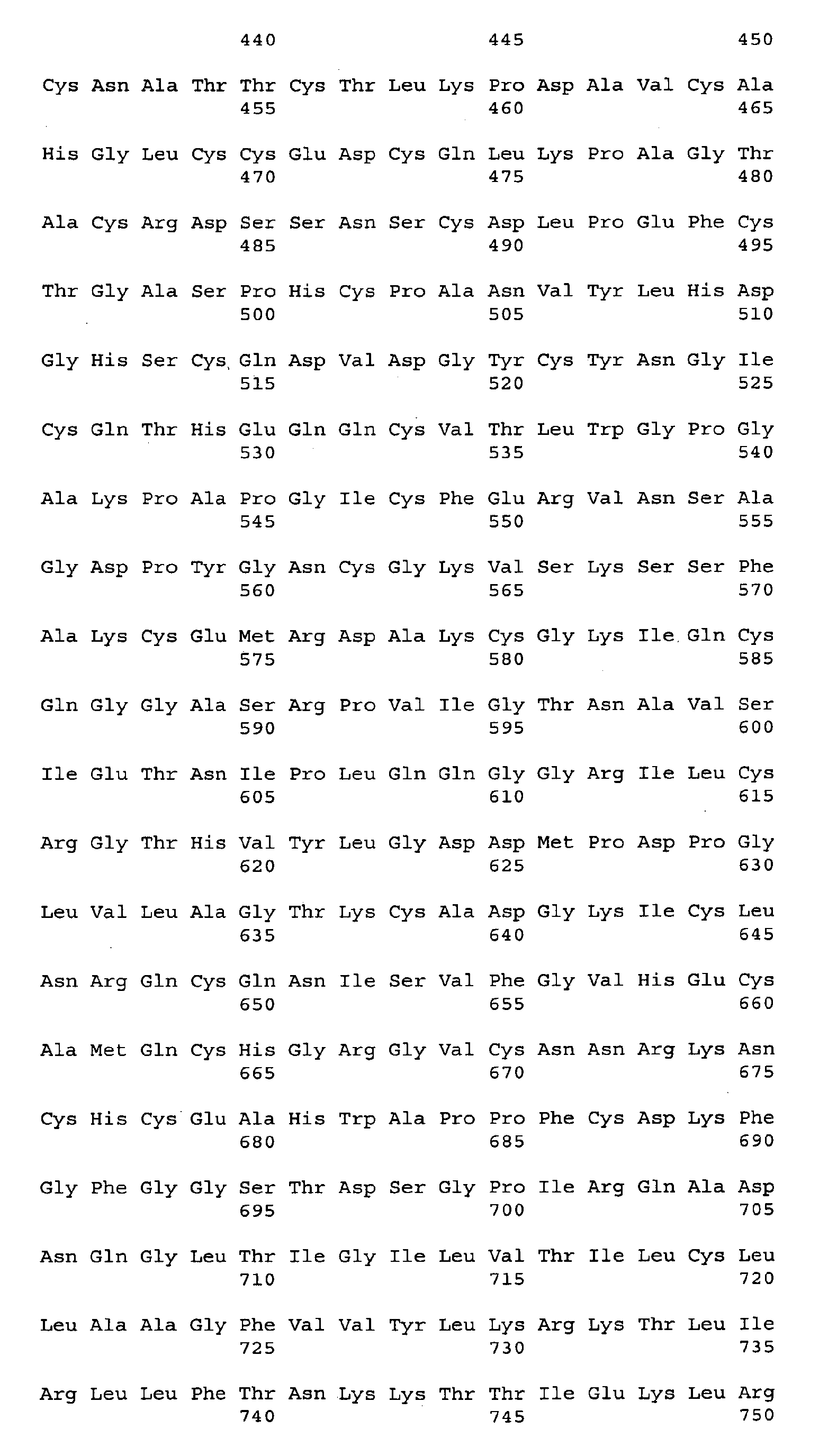


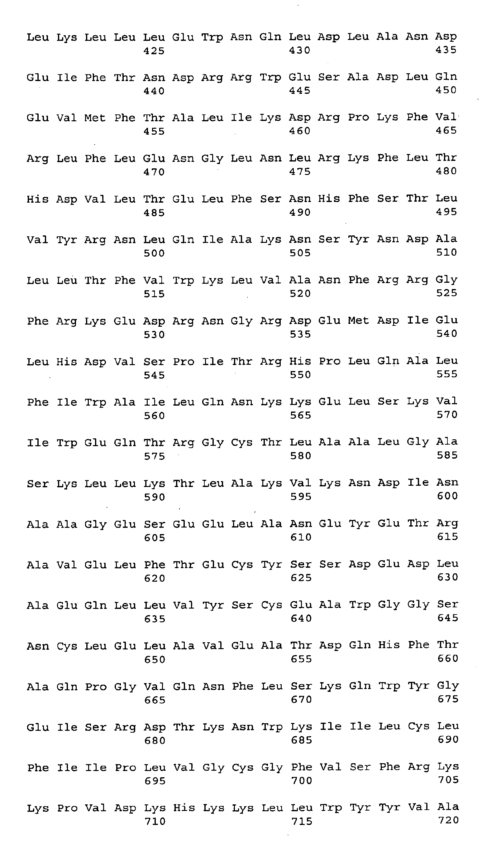


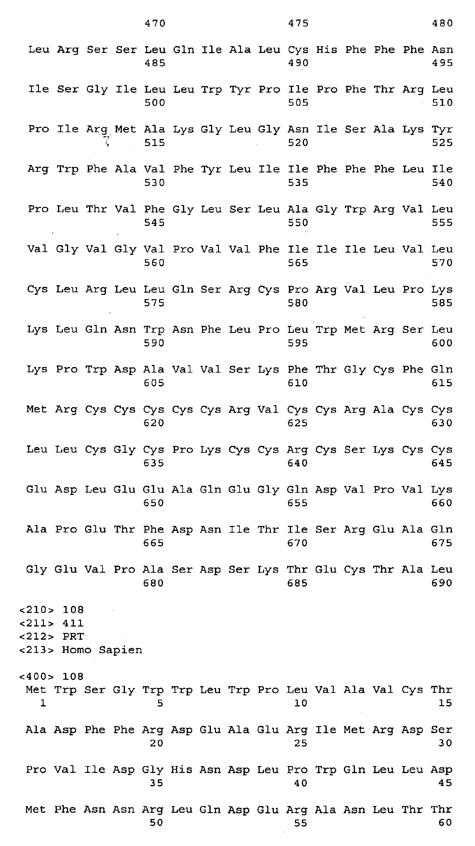


Claims (27)
- An antagonist of a Tumor-associated Antigenic Target (TAT) polypeptide of SEQ ID NO:96, for use in a method of inhibiting the growth of a cancer cell that expresses said Tumor-associated Antigenic Target (TAT) polypeptide of SEQ ID NO:96, said method comprising: contacting said cancer cell with said antagonist.
- An antagonist for use according to claim 1, wherein the cancer cell is a prostate cancer cell.
- An antagonist for use according to claim 1, wherein said inhibition induces death of the cancer cell.
- An antagonist of a Tumor-associated Antigenic Target (TAT) polypeptide of SEQ ID NO:96, for use in a method for the prevention or treatment of a prostate tumor in a mammal wherein the tumor comprises cells expressing said Tumor-associated Antigenic Target (TAT) polypeptide of SEQ ID NO:96, said method comprising: administering to the mammal an effective amount of said antagonist.
- An antagonist for use according to claim 4, wherein said TAT polypeptide of SEQ ID NO:96 is expressed by said prostate tumor cell or by a normal cell in the vicinity of said tumor cell.
- An antagonist for use according to claim 4, wherein said treatment induces shrinkage of the prostate tumor.
- An antagonist for use according to claim 4, wherein said treatment induces death of prostate tumor cells.
- An antagonist of a Tumor-associated Antigenic Target (TAT) polypeptide of SEQ ID NO:96, for use in a method for the prevention or treatment of a prostate proliferative disorder, said method comprising: administering to a subject in need of such treatment, an effective amount of said antagonist.
- An antagonist for use according to claim 8, wherein said prostate proliferative disorder is prostate cancer.
- An antagonist for use according to claim 1, claim 4, or claim 8, wherein said antagonist is selected from the group consisting of anti-TAT antibodies and fragments thereof, fragments and variants of native TAT polypeptides, peptides, antisense oligonucleotides, and small organic molecules.
- An antagonist for use according to claim 1, claim 4, or claim 8, wherein said antagonist is further conjugated to a growth inhibitory agent or cytotoxic agent.
- An antagonist for use according to claim 11 wherein said cytotoxic agent is a toxin.
- An antibody which binds to a Tumor-associated Antigenic Target (TAT) polypeptide of SEQ ID NO:96, for use in a method of inhibiting the growth of a cancer cell that expresses said Tumor-associated Antigenic Target (TAT) polypeptide of SEQ ID NO:96, said method comprising: contacting said cancer cell with said antibody.
- An antibody which binds a Tumor-associated Antigenic Target (TAT) polypeptide of SEQ ID NO:96, for use in a method for the prevention or treatment of a prostate tumor in a mammal wherein the tumor comprises cells expressing said Tumor-associated Antigenic Target (TAT) polypeptide of SEQ ID NO:96, said method comprising: administering to the mammal an effective amount of said antibody.
- An antibody which binds a Tumor-associated Antigenic Target (TAT) polypeptide of SEQ ID NO:96, for use in a method for the prevention or treatment of a prostate proliferative disorder, said method comprising: administering to a subject in need of such treatment, an effective amount of said antibody.
- A method of diagnosing the presence of a tumor in a mammal, said method comprising contacting a test sample of tissue cells obtained from said mammal with an antibody that binds to a protein comprising the amino acid sequence of SEQ ID NO:96, and detecting the formation of a complex between said antibody and said protein in the test sample, wherein the formation of a complex is indicative of the presence of a tumor in said mammal.
- The method of claim 16, wherein said antibody is detectably labeled.
- The method of claim 16, wherein said test sample of tissue cells is obtained from an individual suspected of having a cancerous tumor.
- The method of Claim 18, wherein said tumor is a prostate tumor.
- A method of diagnosing the presence of a tumor in a mammal, said method comprising determining the level of expression of a gene encoding a protein comprising the amino acid sequence of SEQ ID NO:96, in a test sample of tissue cells obtained from said mammal and in a control sample of known normal cells of the same tissue origin, wherein a higher level of expression of said protein in the test sample, as compared to the control sample, is indicative of the presence of tumor in the mammal from which the test sample was obtained.
- The method of claim 20, wherein the step of determining the level of expression of a gene encoding said protein comprises:a) employing an oligonucleotide in an in situ hybridization or RT-PCR analysis; orb) employing an antibody in an immunohistochemistry or Western blot analysis.
- The method of Claim 20, wherein said tumor is a prostate tumor.
- A pharmaceutical composition comprising an effective amount of an antagonist according to any of claims 1 to 12 or an antibody of claims 13 to 15, in admixture with a pharmaceutically acceptable carrier.
- A kit comprising: (a) a pharmaceutical composition of Claim 23; and (b) a package insert or label indicating a beneficial use for said pharmaceutical composition in the treatment of cancer.
- The kit of claim 24 wherein said cancer is prostate cancer.
- The kit of claim 24 wherein the beneficial use is extension of survival.
- The kit of claim 24 wherein the beneficial use is progression-free survival.
Applications Claiming Priority (12)
| Application Number | Priority Date | Filing Date | Title |
|---|---|---|---|
| US29950001P | 2001-06-20 | 2001-06-20 | |
| US30088001P | 2001-06-25 | 2001-06-25 | |
| US30188001P | 2001-06-29 | 2001-06-29 | |
| US30481301P | 2001-07-11 | 2001-07-11 | |
| US31231201P | 2001-08-13 | 2001-08-13 | |
| US31428001P | 2001-08-22 | 2001-08-22 | |
| US32326801P | 2001-09-18 | 2001-09-18 | |
| US33922701P | 2001-10-19 | 2001-10-19 | |
| US33682701P | 2001-11-07 | 2001-11-07 | |
| US36686902P | 2002-03-20 | 2002-03-20 | |
| US37888502P | 2002-05-08 | 2002-05-08 | |
| EP02747926A EP1409549A4 (en) | 2001-06-20 | 2002-06-19 | Compositions and methods for the diagnosis and treatment of tumor |
Related Parent Applications (1)
| Application Number | Title | Priority Date | Filing Date |
|---|---|---|---|
| EP02747926A Division EP1409549A4 (en) | 2001-06-20 | 2002-06-19 | Compositions and methods for the diagnosis and treatment of tumor |
Publications (1)
| Publication Number | Publication Date |
|---|---|
| EP2000148A1 true EP2000148A1 (en) | 2008-12-10 |
Family
ID=27582768
Family Applications (5)
| Application Number | Title | Priority Date | Filing Date |
|---|---|---|---|
| EP08013531A Withdrawn EP1992643A3 (en) | 2001-06-20 | 2002-06-19 | Compositions and methods for the diagnosis and treatment of tumor |
| EP08012996A Expired - Lifetime EP2000545B1 (en) | 2001-06-20 | 2002-06-19 | Compositions and methods for the diagnosis and treatment of lung tumor |
| EP08012997A Withdrawn EP2000482A1 (en) | 2001-06-20 | 2002-06-19 | Compositions and methods for the diagnosis and treatment of tumor |
| EP08012998A Withdrawn EP2000148A1 (en) | 2001-06-20 | 2002-06-19 | Compositions and methods for the diagnosis and treatment of prostate cancer |
| EP02747926A Withdrawn EP1409549A4 (en) | 2001-06-20 | 2002-06-19 | Compositions and methods for the diagnosis and treatment of tumor |
Family Applications Before (3)
| Application Number | Title | Priority Date | Filing Date |
|---|---|---|---|
| EP08013531A Withdrawn EP1992643A3 (en) | 2001-06-20 | 2002-06-19 | Compositions and methods for the diagnosis and treatment of tumor |
| EP08012996A Expired - Lifetime EP2000545B1 (en) | 2001-06-20 | 2002-06-19 | Compositions and methods for the diagnosis and treatment of lung tumor |
| EP08012997A Withdrawn EP2000482A1 (en) | 2001-06-20 | 2002-06-19 | Compositions and methods for the diagnosis and treatment of tumor |
Family Applications After (1)
| Application Number | Title | Priority Date | Filing Date |
|---|---|---|---|
| EP02747926A Withdrawn EP1409549A4 (en) | 2001-06-20 | 2002-06-19 | Compositions and methods for the diagnosis and treatment of tumor |
Country Status (14)
| Country | Link |
|---|---|
| US (11) | US7585953B2 (en) |
| EP (5) | EP1992643A3 (en) |
| JP (3) | JP2004537534A (en) |
| KR (6) | KR100576674B1 (en) |
| AT (1) | ATE522623T1 (en) |
| AU (4) | AU2002318371B2 (en) |
| CA (4) | CA2451239A1 (en) |
| CY (1) | CY1112055T1 (en) |
| DK (1) | DK2000545T3 (en) |
| ES (1) | ES2372321T3 (en) |
| MX (1) | MXPA03011985A (en) |
| PT (1) | PT2000545E (en) |
| WO (1) | WO2003000113A2 (en) |
| ZA (1) | ZA200309808B (en) |
Families Citing this family (51)
| Publication number | Priority date | Publication date | Assignee | Title |
|---|---|---|---|---|
| US6887674B1 (en) | 1998-04-13 | 2005-05-03 | California Institute Of Technology | Artery- and vein-specific proteins and uses therefor |
| CA2385528C (en) | 1999-10-01 | 2013-12-10 | Immunogen, Inc. | Compositions and methods for treating cancer using immunoconjugates and chemotherapeutic agents |
| EP1992643A3 (en) | 2001-06-20 | 2008-12-10 | Genentech, Inc. | Compositions and methods for the diagnosis and treatment of tumor |
| US20050272120A1 (en) | 2001-06-20 | 2005-12-08 | Genentech, Inc. | Compositions and methods for the diagnosis and treatment of tumor |
| US7803915B2 (en) | 2001-06-20 | 2010-09-28 | Genentech, Inc. | Antibody compositions for the diagnosis and treatment of tumor |
| WO2003000012A2 (en) * | 2001-06-21 | 2003-01-03 | Millennium Pharmaceuticals, Inc. | Compositions, kits, and methods for identification, assessment, prevention, and therapy of breast and ovarian cancer |
| CA2496236A1 (en) * | 2002-08-21 | 2004-03-04 | Takeda Pharmaceutical Company Limited | Preventives/remedies for cancer |
| JP4530631B2 (en) * | 2002-08-21 | 2010-08-25 | 武田薬品工業株式会社 | Novel protein and cancer preventive / therapeutic agent |
| AU2002951409A0 (en) * | 2002-09-16 | 2002-09-26 | North Western Adelaide Health Services | Methods for regulating cancer |
| US7381410B2 (en) | 2003-03-12 | 2008-06-03 | Vasgene Therapeutics, Inc. | Polypeptide compounds for inhibiting angiogenesis and tumor growth |
| AU2004220459B2 (en) | 2003-03-12 | 2010-08-05 | Vasgene Therapeutics, Inc. | Polypeptide compounds for inhibiting angiogenesis and tumor growth |
| US8088387B2 (en) | 2003-10-10 | 2012-01-03 | Immunogen Inc. | Method of targeting specific cell populations using cell-binding agent maytansinoid conjugates linked via a non-cleavable linker, said conjugates, and methods of making said conjugates |
| WO2005030805A1 (en) * | 2003-09-25 | 2005-04-07 | Takeda Pharmaceutical Company Limited | Novel protein complex and use thereof |
| KR101520209B1 (en) | 2003-11-06 | 2015-05-13 | 시애틀 지네틱스, 인크. | Monomethylvaline compounds capable of conjugation to ligands |
| AU2005224081B2 (en) | 2004-03-12 | 2011-06-30 | Vasgene Therapeutics, Inc. | Polypeptide compounds for inhibiting angiogenesis and tumor growth |
| CN102746401A (en) | 2004-03-12 | 2012-10-24 | 瓦斯基因治疗公司 | Polypeptide compound for inhibiting angiogenesis and tumor growth |
| NZ551180A (en) * | 2004-06-01 | 2009-10-30 | Genentech Inc | Antibody drug conjugates and methods |
| US20060204512A1 (en) | 2004-09-23 | 2006-09-14 | Vasgene Therapeutics, Inc. | Polypeptide compounds for inhibiting angiogenesis and tumor growth |
| JP4993645B2 (en) | 2004-12-01 | 2012-08-08 | ジェネンテック, インコーポレイテッド | Antibody drug conjugates and methods |
| JP5007238B2 (en) | 2005-01-06 | 2012-08-22 | ジェネンテック, インコーポレイテッド | Methods and compositions useful for cancer prognosis, diagnosis and treatment |
| JP2008537673A (en) * | 2005-01-31 | 2008-09-25 | ジェネンテック・インコーポレーテッド | Anti-EPHB2 antibody and method of use thereof |
| WO2007084388A2 (en) * | 2006-01-12 | 2007-07-26 | Sessa William C | Nogo-b receptor |
| JP5597534B2 (en) | 2007-05-16 | 2014-10-01 | ジェネ シグナル インターナショナル エスエー | Anti-neoplastic drugs, medicaments, compositions and uses thereof |
| US7906621B2 (en) * | 2007-05-16 | 2011-03-15 | Gene Signal International Sa | Anti-tumor drug, medicament, composition, and use thereof |
| AU2012216641B2 (en) * | 2007-07-27 | 2015-02-05 | Immatics Biotechnologies Gmbh | Novel immunogenic epitopes for immunotherapy |
| EP2565204B1 (en) * | 2007-07-27 | 2015-10-07 | immatics biotechnologies GmbH | Novel immunogenic epitopes for immunotherapy |
| DK2644194T3 (en) * | 2008-03-18 | 2017-07-03 | Genentech Inc | Combinations of an anti-HER2 antibody-drug conjugate and docetaxel |
| EP2631302A3 (en) * | 2008-03-31 | 2014-01-08 | Genentech, Inc. | Compositions and methods for treating and diagnosing asthma |
| US9809815B2 (en) * | 2009-02-20 | 2017-11-07 | Ganymed Pharmaceuticals Ag | Methods and compositions for diagnosis and treatment of cancer |
| EP2221063A1 (en) | 2009-02-20 | 2010-08-25 | Ganymed Pharmaceuticals AG | Methods and compositions for diagnosis and treatment of cancer |
| NZ716587A (en) | 2009-11-11 | 2017-10-27 | Ganymed Pharmaceuticals Ag | Antibodies specific for claudin 6 (cldn6) |
| AR079217A1 (en) | 2009-11-30 | 2012-01-04 | Genentech Inc | COMPOSITIONS AND METHODS FOR DIAGNOSIS AND TUMOR TREATMENT |
| EP2404936A1 (en) | 2010-07-06 | 2012-01-11 | Ganymed Pharmaceuticals AG | Cancer therapy using CLDN6 target-directed antibodies in vivo |
| AU2011343570B2 (en) | 2010-12-16 | 2016-11-03 | Genentech, Inc. | Diagnosis and treatments relating to TH2 inhibition |
| PL3026064T3 (en) | 2011-05-13 | 2019-05-31 | Ganymed Pharmaceuticals Gmbh | Antibodies for treatment of cancer expressing claudin 6 |
| EP2849789A4 (en) | 2012-05-14 | 2016-02-24 | Hoffmann La Roche | Compositions and methods for the diagnosis and treatment of tumor |
| US10260089B2 (en) | 2012-10-29 | 2019-04-16 | The Research Foundation Of The State University Of New York | Compositions and methods for recognition of RNA using triple helical peptide nucleic acids |
| US9353150B2 (en) | 2012-12-04 | 2016-05-31 | Massachusetts Institute Of Technology | Substituted pyrazino[1′,2′:1 ,5]pyrrolo[2,3-b]-indole-1,4-diones for cancer treatment |
| JP6482471B2 (en) | 2012-12-21 | 2019-03-13 | バイオアライアンス コマンディテール フェンノートシャップ | Hydrophilic self-destructible linker and conjugate thereof |
| WO2015014376A1 (en) | 2013-07-31 | 2015-02-05 | Biontech Ag | Diagnosis and therapy of cancer involving cancer stem cells |
| GB201319446D0 (en) * | 2013-11-04 | 2013-12-18 | Immatics Biotechnologies Gmbh | Personalized immunotherapy against several neuronal and brain tumors |
| DE112015002791T5 (en) * | 2014-06-13 | 2017-03-16 | Siemens Medical Solutions Usa, Inc. | Intra-reconstruction movement correction |
| US9950077B2 (en) | 2014-06-20 | 2018-04-24 | Bioalliance C.V. | Anti-folate receptor alpha (FRA) antibody-drug conjugates and methods of using thereof |
| US10472422B2 (en) | 2016-01-08 | 2019-11-12 | Abgenomics International Inc. | Tetravalent anti-PSGL-1 antibodies and uses thereof |
| US10918627B2 (en) | 2016-05-11 | 2021-02-16 | Massachusetts Institute Of Technology | Convergent and enantioselective total synthesis of Communesin analogs |
| US11180535B1 (en) | 2016-12-07 | 2021-11-23 | David Gordon Bermudes | Saccharide binding, tumor penetration, and cytotoxic antitumor chimeric peptides from therapeutic bacteria |
| US10640508B2 (en) | 2017-10-13 | 2020-05-05 | Massachusetts Institute Of Technology | Diazene directed modular synthesis of compounds with quaternary carbon centers |
| JP7352547B2 (en) * | 2017-12-13 | 2023-09-28 | イノビオ ファーマシューティカルズ,インコーポレイティド | Cancer vaccines targeting mesothelin and their use |
| US11484279B2 (en) | 2018-09-24 | 2022-11-01 | Siemens Medical Solutions Usa, Inc. | Systems to assess projection data inconsistency |
| KR102341138B1 (en) * | 2020-05-31 | 2021-12-21 | 주식회사 엑소코바이오 | Exosomes comprising exosomal membrane protein variant and manufacturing method thereof |
| US20220296976A1 (en) * | 2021-03-17 | 2022-09-22 | Aviv, Llc | Antimicrobial Basketball Net |
Citations (338)
| Publication number | Priority date | Publication date | Assignee | Title |
|---|---|---|---|---|
| US3687808A (en) | 1969-08-14 | 1972-08-29 | Univ Leland Stanford Junior | Synthetic polynucleotides |
| US3773919A (en) | 1969-10-23 | 1973-11-20 | Du Pont | Polylactide-drug mixtures |
| US3896111A (en) | 1973-02-20 | 1975-07-22 | Research Corp | Ansa macrolides |
| US4137230A (en) | 1977-11-14 | 1979-01-30 | Takeda Chemical Industries, Ltd. | Method for the production of maytansinoids |
| US4151042A (en) | 1977-03-31 | 1979-04-24 | Takeda Chemical Industries, Ltd. | Method for producing maytansinol and its derivatives |
| US4179337A (en) | 1973-07-20 | 1979-12-18 | Davis Frank F | Non-immunogenic polypeptides |
| US4248870A (en) | 1978-10-27 | 1981-02-03 | Takeda Chemical Industries, Ltd. | Maytansinoids and use |
| US4256746A (en) | 1978-11-14 | 1981-03-17 | Takeda Chemical Industries | Dechloromaytansinoids, their pharmaceutical compositions and method of use |
| US4260608A (en) | 1978-11-14 | 1981-04-07 | Takeda Chemical Industries, Ltd. | Maytansinoids, pharmaceutical compositions thereof and methods of use thereof |
| WO1981001145A1 (en) | 1979-10-18 | 1981-04-30 | Univ Illinois | Hydrolytic enzyme-activatible pro-drugs |
| US4265814A (en) | 1978-03-24 | 1981-05-05 | Takeda Chemical Industries | Matansinol 3-n-hexadecanoate |
| US4275149A (en) | 1978-11-24 | 1981-06-23 | Syva Company | Macromolecular environment control in specific receptor assays |
| EP0036776A2 (en) | 1980-03-24 | 1981-09-30 | Genentech, Inc. | A method of creating an expression plasmid |
| US4294757A (en) | 1979-01-31 | 1981-10-13 | Takeda Chemical Industries, Ltd | 20-O-Acylmaytansinoids |
| US4301144A (en) | 1979-07-11 | 1981-11-17 | Ajinomoto Company, Incorporated | Blood substitute containing modified hemoglobin |
| US4307016A (en) | 1978-03-24 | 1981-12-22 | Takeda Chemical Industries, Ltd. | Demethyl maytansinoids |
| US4308269A (en) | 1979-06-11 | 1981-12-29 | Takeda Chemical Industries, Ltd. | Maytansinoids, pharmaceutical compositions thereof and method of use thereof |
| US4308268A (en) | 1979-06-11 | 1981-12-29 | Takeda Chemical Industries, Ltd. | Maytansinoids, pharmaceutical compositions thereof and method of use thereof |
| US4309428A (en) | 1979-07-30 | 1982-01-05 | Takeda Chemical Industries, Ltd. | Maytansinoids |
| US4313946A (en) | 1981-01-27 | 1982-02-02 | The United States Of America As Represented By The Secretary Of Agriculture | Chemotherapeutically active maytansinoids from Trewia nudiflora |
| US4315929A (en) | 1981-01-27 | 1982-02-16 | The United States Of America As Represented By The Secretary Of Agriculture | Method of controlling the European corn borer with trewiasine |
| US4317821A (en) | 1979-06-08 | 1982-03-02 | Takeda Chemical Industries, Ltd. | Maytansinoids, their use and pharmaceutical compositions thereof |
| US4322348A (en) | 1979-06-05 | 1982-03-30 | Takeda Chemical Industries, Ltd. | Maytansinoids |
| US4331598A (en) | 1979-09-19 | 1982-05-25 | Takeda Chemical Industries, Ltd. | Maytansinoids |
| USRE30985E (en) | 1978-01-01 | 1982-06-29 | Serum-free cell culture media | |
| US4362663A (en) | 1979-09-21 | 1982-12-07 | Takeda Chemical Industries, Ltd. | Maytansinoid compound |
| US4364866A (en) | 1979-09-21 | 1982-12-21 | Takeda Chemical Industries, Ltd. | Maytansinoids |
| US4371533A (en) | 1980-10-08 | 1983-02-01 | Takeda Chemical Industries, Ltd. | 4,5-Deoxymaytansinoids, their use and pharmaceutical compositions thereof |
| EP0073657A1 (en) | 1981-08-31 | 1983-03-09 | Genentech, Inc. | Preparation of hepatitis B surface antigen in yeast |
| US4399216A (en) | 1980-02-25 | 1983-08-16 | The Trustees Of Columbia University | Processes for inserting DNA into eucaryotic cells and for producing proteinaceous materials |
| US4424219A (en) | 1981-05-20 | 1984-01-03 | Takeda Chemical Industries, Ltd. | 9-Thiomaytansinoids and their pharmaceutical compositions and use |
| US4426330A (en) | 1981-07-20 | 1984-01-17 | Lipid Specialties, Inc. | Synthetic phospholipid compounds |
| US4450254A (en) | 1980-11-03 | 1984-05-22 | Standard Oil Company | Impact improvement of high nitrile resins |
| EP0117060A2 (en) | 1983-01-19 | 1984-08-29 | Genentech, Inc. | Methods of screening and amplification in eukaryotic host cells, and nucleotide sequences and expression vectors for use therein |
| EP0117058A2 (en) | 1983-01-19 | 1984-08-29 | Genentech, Inc. | Methods for producing mature protein in vertebrate host cells |
| US4469863A (en) | 1980-11-12 | 1984-09-04 | Ts O Paul O P | Nonionic nucleic acid alkyl and aryl phosphonates and processes for manufacture and use thereof |
| WO1984003564A1 (en) | 1983-03-08 | 1984-09-13 | Commw Serum Lab Commission | Method of determining antigenically active amino acid sequences |
| WO1984003506A1 (en) | 1983-03-08 | 1984-09-13 | Commw Serum Lab Commission | Antigenically active amino acid sequences |
| US4476301A (en) | 1982-04-29 | 1984-10-09 | Centre National De La Recherche Scientifique | Oligonucleotides, a process for preparing the same and their application as mediators of the action of interferon |
| US4485045A (en) | 1981-07-06 | 1984-11-27 | Research Corporation | Synthetic phosphatidyl cholines useful in forming liposomes |
| US4496689A (en) | 1983-12-27 | 1985-01-29 | Miles Laboratories, Inc. | Covalently attached complex of alpha-1-proteinase inhibitor with a water soluble polymer |
| EP0139383A1 (en) | 1983-08-16 | 1985-05-02 | Zymogenetics, Inc. | Method for expressing foreign genes in schizosaccharomyces pombe and the use in therapeutic formulations of the products, DNA constructs and transformant strains of schizosaccharomyces pombe usable in such method and their preparation |
| US4534899A (en) | 1981-07-20 | 1985-08-13 | Lipid Specialties, Inc. | Synthetic phospholipid compounds |
| US4544545A (en) | 1983-06-20 | 1985-10-01 | Trustees University Of Massachusetts | Liposomes containing modified cholesterol for organ targeting |
| US4560655A (en) | 1982-12-16 | 1985-12-24 | Immunex Corporation | Serum-free cell culture medium and process for making same |
| US4587044A (en) | 1983-09-01 | 1986-05-06 | The Johns Hopkins University | Linkage of proteins to nucleic acids |
| EP0183070A2 (en) | 1984-10-30 | 1986-06-04 | Phillips Petroleum Company | Transformation of yeasts of the genus pichia |
| US4605735A (en) | 1983-02-14 | 1986-08-12 | Wakunaga Seiyaku Kabushiki Kaisha | Oligonucleotide derivatives |
| WO1987000195A1 (en) | 1985-06-28 | 1987-01-15 | Celltech Limited | Animal cell culture |
| US4640835A (en) | 1981-10-30 | 1987-02-03 | Nippon Chemiphar Company, Ltd. | Plasminogen activator derivatives |
| US4657866A (en) | 1982-12-21 | 1987-04-14 | Sudhir Kumar | Serum-free, synthetic, completely chemically defined tissue culture media |
| US4667025A (en) | 1982-08-09 | 1987-05-19 | Wakunaga Seiyaku Kabushiki Kaisha | Oligonucleotide derivatives |
| US4670417A (en) | 1985-06-19 | 1987-06-02 | Ajinomoto Co., Inc. | Hemoglobin combined with a poly(alkylene oxide) |
| US4676980A (en) | 1985-09-23 | 1987-06-30 | The United States Of America As Represented By The Secretary Of The Department Of Health And Human Services | Target specific cross-linked heteroantibodies |
| WO1987005330A1 (en) | 1986-03-07 | 1987-09-11 | Michel Louis Eugene Bergh | Method for enhancing glycoprotein stability |
| EP0244234A2 (en) | 1986-04-30 | 1987-11-04 | Alko Group Ltd. | Transformation of trichoderma |
| US4708871A (en) | 1983-03-08 | 1987-11-24 | Commonwealth Serum Laboratories Commission | Antigenically active amino acid sequences |
| US4736866B1 (en) | 1984-06-22 | 1988-04-12 | Transgenic non-human mammals | |
| US4762779A (en) | 1985-06-13 | 1988-08-09 | Amgen Inc. | Compositions and methods for functionalizing nucleic acids |
| US4767704A (en) | 1983-10-07 | 1988-08-30 | Columbia University In The City Of New York | Protein-free culture medium |
| WO1988007378A1 (en) | 1987-03-09 | 1988-10-06 | Cancer Research Campaign Technology Ltd. | Improvements relating to drug delivery systems |
| US4791192A (en) | 1986-06-26 | 1988-12-13 | Takeda Chemical Industries, Ltd. | Chemically modified protein with polyethyleneglycol |
| EP0307247A2 (en) | 1987-09-11 | 1989-03-15 | Genentech, Inc. | A method for culturing recombinant cells |
| US4816567A (en) | 1983-04-08 | 1989-03-28 | Genentech, Inc. | Recombinant immunoglobin preparations |
| EP0308936A2 (en) | 1987-09-23 | 1989-03-29 | Bristol-Myers Squibb Company | Antibody heteroconjugates for the killing of HIV-infected cells |
| DD266710A3 (en) | 1983-06-06 | 1989-04-12 | Ve Forschungszentrum Biotechnologie | Process for the biotechnical production of alkaline phosphatase |
| US4824941A (en) | 1983-03-10 | 1989-04-25 | Julian Gordon | Specific antibody to the native form of 2'5'-oligonucleotides, the method of preparation and the use as reagents in immunoassays or for binding 2'5'-oligonucleotides in biological systems |
| US4828979A (en) | 1984-11-08 | 1989-05-09 | Life Technologies, Inc. | Nucleotide analogs for nucleic acid labeling and detection |
| US4833092A (en) | 1985-04-22 | 1989-05-23 | Commonwealth Serum Laboratories Commission | Method for determining mimotopes |
| US4835263A (en) | 1983-01-27 | 1989-05-30 | Centre National De La Recherche Scientifique | Novel compounds containing an oligonucleotide sequence bonded to an intercalating agent, a process for their synthesis and their use |
| WO1989005859A1 (en) | 1987-12-21 | 1989-06-29 | The Upjohn Company | Agrobacterium mediated transformation of germinating plant seeds |
| US4845205A (en) | 1985-01-08 | 1989-07-04 | Institut Pasteur | 2,N6 -disubstituted and 2,N6 -trisubstituted adenosine-3'-phosphoramidites |
| GB2211504A (en) | 1987-10-23 | 1989-07-05 | Nat Res Dev | Fowlpox virus promoters |
| US4870009A (en) | 1982-11-22 | 1989-09-26 | The Salk Institute For Biological Studies | Method of obtaining gene product through the generation of transgenic animals |
| US4876335A (en) | 1986-06-30 | 1989-10-24 | Wakunaga Seiyaku Kabushiki Kaisha | Poly-labelled oligonucleotide derivative |
| US4892538A (en) | 1987-11-17 | 1990-01-09 | Brown University Research Foundation | In vivo delivery of neurotransmitters by implanted, encapsulated cells |
| US4904582A (en) | 1987-06-11 | 1990-02-27 | Synthetic Genetics | Novel amphiphilic nucleic acid conjugates |
| EP0362179A2 (en) | 1988-08-25 | 1990-04-04 | Smithkline Beecham Corporation | Recombinant saccharomyces |
| WO1990003430A1 (en) | 1988-09-23 | 1990-04-05 | Cetus Corporation | Cell culture medium for enhanced cell growth, culture longevity and product expression |
| US4927762A (en) | 1986-04-01 | 1990-05-22 | Cell Enterprises, Inc. | Cell culture medium with antioxidant |
| US4933294A (en) | 1984-01-30 | 1990-06-12 | Icrf Patents Limited | Method of detecting truncated epidermal growth factor receptors |
| US4943529A (en) | 1982-05-19 | 1990-07-24 | Gist-Brocades Nv | Kluyveromyces as a host strain |
| US4946783A (en) | 1987-01-30 | 1990-08-07 | President And Fellows Of Harvard College | Periplasmic protease mutants of Escherichia coli |
| US4948882A (en) | 1983-02-22 | 1990-08-14 | Syngene, Inc. | Single-stranded labelled oligonucleotides, reactive monomers and methods of synthesis |
| WO1990010048A1 (en) | 1989-02-27 | 1990-09-07 | Kerr-Mcgee Corporation | Solvent extraction process |
| US4958013A (en) | 1989-06-06 | 1990-09-18 | Northwestern University | Cholesteryl modified oligonucleotides |
| WO1990010448A2 (en) | 1989-03-07 | 1990-09-20 | Genentech, Inc. | Covalent conjugates of lipid and oligonucleotide |
| EP0394538A1 (en) | 1989-04-28 | 1990-10-31 | Rhein Biotech Gesellschaft Für Neue Biotechnologische Prozesse Und Produkte Mbh | A yeast cell of the genus schwanniomyces |
| WO1990013641A1 (en) | 1989-05-10 | 1990-11-15 | Sloan-Kettering Institute For Cancer Research | Stably transformed eucaryotic cells comprising a foreign transcribable dna under the control of a pol iii promoter |
| WO1990013646A1 (en) | 1989-04-28 | 1990-11-15 | Transgene S.A. | Application of novel dna fragments as a coding sequence for a signal peptide for the secretion of mature proteins by recombinant yeast, expression cassettes, transformed yeasts and corresponding process for the preparation of proteins |
| US4975278A (en) | 1988-02-26 | 1990-12-04 | Bristol-Myers Company | Antibody-enzyme conjugates in combination with prodrugs for the delivery of cytotoxic agents to tumor cells |
| EP0402226A1 (en) | 1989-06-06 | 1990-12-12 | Institut National De La Recherche Agronomique | Transformation vectors for yeast yarrowia |
| EP0404097A2 (en) | 1989-06-22 | 1990-12-27 | BEHRINGWERKE Aktiengesellschaft | Bispecific and oligospecific, mono- and oligovalent receptors, production and applications thereof |
| US4981957A (en) | 1984-07-19 | 1991-01-01 | Centre National De La Recherche Scientifique | Oligonucleotides with modified phosphate and modified carbohydrate moieties at the respective chain termini |
| WO1991000357A1 (en) | 1989-06-30 | 1991-01-10 | Cayla | New strain with filamentous fungi mutants, process for the production of recombinant proteins using said strain, and strains and proteins produced by said process |
| WO1991000360A1 (en) | 1989-06-29 | 1991-01-10 | Medarex, Inc. | Bispecific reagents for aids therapy |
| WO1991004753A1 (en) | 1989-10-02 | 1991-04-18 | Cetus Corporation | Conjugates of antisense oligonucleotides and therapeutic uses thereof |
| WO1991005264A1 (en) | 1989-09-29 | 1991-04-18 | Oncogenetics Partners | Detection and quantification of neu related proteins in the biological fluids of humans |
| US5010182A (en) | 1987-07-28 | 1991-04-23 | Chiron Corporation | DNA constructs containing a Kluyveromyces alpha factor leader sequence for directing secretion of heterologous polypeptides |
| US5013556A (en) | 1989-10-20 | 1991-05-07 | Liposome Technology, Inc. | Liposomes with enhanced circulation time |
| US5013830A (en) | 1986-09-08 | 1991-05-07 | Ajinomoto Co., Inc. | Compounds for the cleavage at a specific position of RNA, oligomers employed for the formation of said compounds, and starting materials for the synthesis of said oligomers |
| WO1991006629A1 (en) | 1989-10-24 | 1991-05-16 | Gilead Sciences, Inc. | Oligonucleotide analogs with novel linkages |
| US5023243A (en) | 1981-10-23 | 1991-06-11 | Molecular Biosystems, Inc. | Oligonucleotide therapeutic agent and method of making same |
| US5034506A (en) | 1985-03-15 | 1991-07-23 | Anti-Gene Development Group | Uncharged morpholino-based polymers having achiral intersubunit linkages |
| US5053394A (en) | 1988-09-21 | 1991-10-01 | American Cyanamid Company | Targeted forms of methyltrithio antitumor agents |
| US5082830A (en) | 1988-02-26 | 1992-01-21 | Enzo Biochem, Inc. | End labeled nucleotide probe |
| US5108921A (en) | 1989-04-03 | 1992-04-28 | Purdue Research Foundation | Method for enhanced transmembrane transport of exogenous molecules |
| US5109124A (en) | 1988-06-01 | 1992-04-28 | Biogen, Inc. | Nucleic acid probe linked to a label having a terminal cysteine |
| US5112963A (en) | 1987-11-12 | 1992-05-12 | Max-Planck-Gesellschaft Zur Foerderung Der Wissenschaften E.V. | Modified oligonucleotides |
| US5118802A (en) | 1983-12-20 | 1992-06-02 | California Institute Of Technology | DNA-reporter conjugates linked via the 2' or 5'-primary amino group of the 5'-terminal nucleoside |
| US5118800A (en) | 1983-12-20 | 1992-06-02 | California Institute Of Technology | Oligonucleotides possessing a primary amino group in the terminal nucleotide |
| US5122469A (en) | 1990-10-03 | 1992-06-16 | Genentech, Inc. | Method for culturing Chinese hamster ovary cells to improve production of recombinant proteins |
| US5130302A (en) | 1989-12-20 | 1992-07-14 | Boron Bilogicals, Inc. | Boronated nucleoside, nucleotide and oligonucleotide compounds, compositions and methods for using same |
| US5134066A (en) | 1989-08-29 | 1992-07-28 | Monsanto Company | Improved probes using nucleosides containing 3-dezauracil analogs |
| US5138045A (en) | 1990-07-27 | 1992-08-11 | Isis Pharmaceuticals | Polyamine conjugated oligonucleotides |
| US5149797A (en) | 1990-02-15 | 1992-09-22 | The Worcester Foundation For Experimental Biology | Method of site-specific alteration of rna and production of encoded polypeptides |
| US5166315A (en) | 1989-12-20 | 1992-11-24 | Anti-Gene Development Group | Sequence-specific binding polymers for duplex nucleic acids |
| WO1992020373A1 (en) | 1991-05-14 | 1992-11-26 | Repligen Corporation | Heteroconjugate antibodies for treatment of hiv infection |
| US5175273A (en) | 1988-07-01 | 1992-12-29 | Genentech, Inc. | Nucleic acid intercalating agents |
| US5177196A (en) | 1990-08-16 | 1993-01-05 | Microprobe Corporation | Oligo (α-arabinofuranosyl nucleotides) and α-arabinofuranosyl precursors thereof |
| US5185444A (en) | 1985-03-15 | 1993-02-09 | Anti-Gene Deveopment Group | Uncharged morpolino-based polymers having phosphorous containing chiral intersubunit linkages |
| US5188897A (en) | 1987-10-22 | 1993-02-23 | Temple University Of The Commonwealth System Of Higher Education | Encapsulated 2',5'-phosphorothioate oligoadenylates |
| US5194599A (en) | 1988-09-23 | 1993-03-16 | Gilead Sciences, Inc. | Hydrogen phosphonodithioate compositions |
| US5208020A (en) | 1989-10-25 | 1993-05-04 | Immunogen Inc. | Cytotoxic agents comprising maytansinoids and their therapeutic use |
| WO1993008829A1 (en) | 1991-11-04 | 1993-05-13 | The Regents Of The University Of California | Compositions that mediate killing of hiv-infected cells |
| US5214136A (en) | 1990-02-20 | 1993-05-25 | Gilead Sciences, Inc. | Anthraquinone-derivatives oligonucleotides |
| US5214134A (en) | 1990-09-12 | 1993-05-25 | Sterling Winthrop Inc. | Process of linking nucleosides with a siloxane bridge |
| US5216141A (en) | 1988-06-06 | 1993-06-01 | Benner Steven A | Oligonucleotide analogs containing sulfur linkages |
| US5218105A (en) | 1990-07-27 | 1993-06-08 | Isis Pharmaceuticals | Polyamine conjugated oligonucleotides |
| WO1993011161A1 (en) | 1991-11-25 | 1993-06-10 | Enzon, Inc. | Multivalent antigen-binding proteins |
| US5220007A (en) | 1990-02-15 | 1993-06-15 | The Worcester Foundation For Experimental Biology | Method of site-specific alteration of RNA and production of encoded polypeptides |
| US5223409A (en) | 1988-09-02 | 1993-06-29 | Protein Engineering Corp. | Directed evolution of novel binding proteins |
| US5227170A (en) | 1989-06-22 | 1993-07-13 | Vestar, Inc. | Encapsulation process |
| US5229275A (en) | 1990-04-26 | 1993-07-20 | Akzo N.V. | In-vitro method for producing antigen-specific human monoclonal antibodies |
| US5235033A (en) | 1985-03-15 | 1993-08-10 | Anti-Gene Development Group | Alpha-morpholino ribonucleoside derivatives and polymers thereof |
| WO1993016185A2 (en) | 1992-02-06 | 1993-08-19 | Creative Biomolecules, Inc. | Biosynthetic binding protein for cancer marker |
| US5245022A (en) | 1990-08-03 | 1993-09-14 | Sterling Drug, Inc. | Exonuclease resistant terminally substituted oligonucleotides |
| US5254469A (en) | 1989-09-12 | 1993-10-19 | Eastman Kodak Company | Oligonucleotide-enzyme conjugate that can be used as a probe in hybridization assays and polymerase chain reaction procedures |
| US5256775A (en) | 1989-06-05 | 1993-10-26 | Gilead Sciences, Inc. | Exonuclease-resistant oligonucleotides |
| WO1993021232A1 (en) | 1992-04-10 | 1993-10-28 | Research Development Foundation | IMMUNOTOXINS DIRECTED AGAINST c-erbB-2 (HER-2/neu) RELATED SURFACE ANTIGENS |
| US5258506A (en) | 1984-10-16 | 1993-11-02 | Chiron Corporation | Photolabile reagents for incorporation into oligonucleotide chains |
| US5262536A (en) | 1988-09-15 | 1993-11-16 | E. I. Du Pont De Nemours And Company | Reagents for the preparation of 5'-tagged oligonucleotides |
| US5264564A (en) | 1989-10-24 | 1993-11-23 | Gilead Sciences | Oligonucleotide analogs with novel linkages |
| US5264562A (en) | 1989-10-24 | 1993-11-23 | Gilead Sciences, Inc. | Oligonucleotide analogs with novel linkages |
| US5264423A (en) | 1987-03-25 | 1993-11-23 | The United States Of America As Represented By The Department Of Health And Human Services | Inhibitors for replication of retroviruses and for the expression of oncogene products |
| US5264221A (en) | 1991-05-23 | 1993-11-23 | Mitsubishi Kasei Corporation | Drug-containing protein-bonded liposome |
| US5272250A (en) | 1992-07-10 | 1993-12-21 | Spielvogel Bernard F | Boronated phosphoramidate compounds |
| WO1993025673A1 (en) | 1992-06-04 | 1993-12-23 | The Regents Of The University Of California | In vivo gene therapy with intron-free sequence of interest |
| US5276019A (en) | 1987-03-25 | 1994-01-04 | The United States Of America As Represented By The Department Of Health And Human Services | Inhibitors for replication of retroviruses and for the expression of oncogene products |
| US5278302A (en) | 1988-05-26 | 1994-01-11 | University Patents, Inc. | Polynucleotide phosphorodithioates |
| US5283187A (en) | 1987-11-17 | 1994-02-01 | Brown University Research Foundation | Cell culture-containing tubular capsule produced by co-extrusion |
| WO1994004690A1 (en) | 1992-08-17 | 1994-03-03 | Genentech, Inc. | Bispecific immunoadhesins |
| US5292873A (en) | 1989-11-29 | 1994-03-08 | The Research Foundation Of State University Of New York | Nucleic acids labeled with naphthoquinone probe |
| WO1994011026A2 (en) | 1992-11-13 | 1994-05-26 | Idec Pharmaceuticals Corporation | Therapeutic application of chimeric and radiolabeled antibodies to human b lymphocyte restricted differentiation antigen for treatment of b cell lymphoma |
| US5317098A (en) | 1986-03-17 | 1994-05-31 | Hiroaki Shizuya | Non-radioisotope tagging of fragments |
| US5319080A (en) | 1991-10-17 | 1994-06-07 | Ciba-Geigy Corporation | Bicyclic nucleosides, oligonucleotides, process for their preparation and intermediates |
| EP0600517A1 (en) | 1992-12-04 | 1994-06-08 | Bristol-Myers Squibb Company | 6,7-Modified paclitaxels |
| US5321131A (en) | 1990-03-08 | 1994-06-14 | Hybridon, Inc. | Site-specific functionalization of oligodeoxynucleotides for non-radioactive labelling |
| EP0616812A1 (en) | 1993-03-24 | 1994-09-28 | Berlex Biosciences | Combination with anti-hormonal compounds and binding molecules for the treatment of cancer |
| US5354844A (en) | 1989-03-16 | 1994-10-11 | Boehringer Ingelheim International Gmbh | Protein-polycation conjugates |
| US5356633A (en) | 1989-10-20 | 1994-10-18 | Liposome Technology, Inc. | Method of treatment of inflamed tissues |
| US5359044A (en) | 1991-12-13 | 1994-10-25 | Isis Pharmaceuticals | Cyclobutyl oligonucleotide surrogates |
| US5364934A (en) | 1991-02-01 | 1994-11-15 | Genentech, Inc. | Plasma carboxypeptidase |
| US5367066A (en) | 1984-10-16 | 1994-11-22 | Chiron Corporation | Oligonucleotides with selectably cleavable and/or abasic sites |
| US5371241A (en) | 1991-07-19 | 1994-12-06 | Pharmacia P-L Biochemicals Inc. | Fluorescein labelled phosphoramidites |
| US5391723A (en) | 1989-05-31 | 1995-02-21 | Neorx Corporation | Oligonucleotide conjugates |
| US5395619A (en) | 1993-03-03 | 1995-03-07 | Liposome Technology, Inc. | Lipid-polymer conjugates and liposomes |
| US5399676A (en) | 1989-10-23 | 1995-03-21 | Gilead Sciences | Oligonucleotides with inverted polarity |
| US5401638A (en) | 1986-06-04 | 1995-03-28 | Oncogene Science, Inc. | Detection and quantification of neu related proteins in the biological fluids of humans |
| US5403711A (en) | 1987-11-30 | 1995-04-04 | University Of Iowa Research Foundation | Nucleic acid hybridization and amplification method for detection of specific sequences in which a complementary labeled nucleic acid probe is cleaved |
| US5405939A (en) | 1987-10-22 | 1995-04-11 | Temple University Of The Commonwealth System Of Higher Education | 2',5'-phosphorothioate oligoadenylates and their covalent conjugates with polylysine |
| US5405938A (en) | 1989-12-20 | 1995-04-11 | Anti-Gene Development Group | Sequence-specific binding polymers for duplex nucleic acids |
| US5414077A (en) | 1990-02-20 | 1995-05-09 | Gilead Sciences | Non-nucleoside linkers for convenient attachment of labels to oligonucleotides using standard synthetic methods |
| US5417978A (en) | 1993-07-29 | 1995-05-23 | Board Of Regents, The University Of Texas System | Liposomal antisense methyl phosphonate oligonucleotides and methods for their preparation and use |
| US5428130A (en) | 1989-02-23 | 1995-06-27 | Genentech, Inc. | Hybrid immunoglobulins |
| US5427908A (en) | 1990-05-01 | 1995-06-27 | Affymax Technologies N.V. | Recombinant library screening methods |
| US5432272A (en) | 1990-10-09 | 1995-07-11 | Benner; Steven A. | Method for incorporating into a DNA or RNA oligonucleotide using nucleotides bearing heterocyclic bases |
| US5432018A (en) | 1990-06-20 | 1995-07-11 | Affymax Technologies N.V. | Peptide library and screening systems |
| US5434257A (en) | 1992-06-01 | 1995-07-18 | Gilead Sciences, Inc. | Binding compentent oligomers containing unsaturated 3',5' and 2',5' linkages |
| US5446137A (en) | 1993-12-09 | 1995-08-29 | Syntex (U.S.A.) Inc. | Oligonucleotides containing 4'-substituted nucleotides |
| US5451463A (en) | 1989-08-28 | 1995-09-19 | Clontech Laboratories, Inc. | Non-nucleoside 1,3-diol reagents for labeling synthetic oligonucleotides |
| US5455233A (en) | 1989-11-30 | 1995-10-03 | University Of North Carolina | Oligoribonucleoside and oligodeoxyribonucleoside boranophosphates |
| US5457187A (en) | 1993-12-08 | 1995-10-10 | Board Of Regents University Of Nebraska | Oligonucleotides containing 5-fluorouracil |
| US5459127A (en) | 1990-04-19 | 1995-10-17 | Vical, Inc. | Cationic lipids for intracellular delivery of biologically active molecules |
| US5459255A (en) | 1990-01-11 | 1995-10-17 | Isis Pharmaceuticals, Inc. | N-2 substituted purines |
| US5462854A (en) | 1993-04-19 | 1995-10-31 | Beckman Instruments, Inc. | Inverse linkage oligonucleotides for chemical and enzymatic processes |
| US5466786A (en) | 1989-10-24 | 1995-11-14 | Gilead Sciences | 2'modified nucleoside and nucleotide compounds |
| US5466677A (en) | 1993-03-06 | 1995-11-14 | Ciba-Geigy Corporation | Dinucleoside phosphinates and their pharmaceutical compositions |
| US5469854A (en) | 1989-12-22 | 1995-11-28 | Imarx Pharmaceutical Corp. | Methods of preparing gas-filled liposomes |
| US5470967A (en) | 1990-04-10 | 1995-11-28 | The Dupont Merck Pharmaceutical Company | Oligonucleotide analogs with sulfamate linkages |
| US5476925A (en) | 1993-02-01 | 1995-12-19 | Northwestern University | Oligodeoxyribonucleotides including 3'-aminonucleoside-phosphoramidate linkages and terminal 3'-amino groups |
| WO1995034683A1 (en) | 1994-06-10 | 1995-12-21 | Symbiotech, Inc. | Method of detecting compounds utilizing genetically modified lambdoid bacteriophage |
| US5484908A (en) | 1991-11-26 | 1996-01-16 | Gilead Sciences, Inc. | Oligonucleotides containing 5-propynyl pyrimidines |
| US5486603A (en) | 1990-01-08 | 1996-01-23 | Gilead Sciences, Inc. | Oligonucleotide having enhanced binding affinity |
| US5489677A (en) | 1990-07-27 | 1996-02-06 | Isis Pharmaceuticals, Inc. | Oligonucleoside linkages containing adjacent oxygen and nitrogen atoms |
| US5491133A (en) | 1987-11-30 | 1996-02-13 | University Of Iowa Research Foundation | Methods for blocking the expression of specifically targeted genes |
| US5498538A (en) | 1990-02-15 | 1996-03-12 | The University Of North Carolina At Chapel Hill | Totally synthetic affinity reagents |
| US5498530A (en) | 1991-10-16 | 1996-03-12 | Affymax Technologies, N.V. | Peptide library and screening method |
| WO1996007321A1 (en) | 1994-09-06 | 1996-03-14 | The Uab Research Foundation | Methods for modulating protein function in cells using intracellular antibody homologues |
| US5500362A (en) | 1987-01-08 | 1996-03-19 | Xoma Corporation | Chimeric antibody with specificity to human B cell surface antigen |
| US5502177A (en) | 1993-09-17 | 1996-03-26 | Gilead Sciences, Inc. | Pyrimidine derivatives for labeled binding partners |
| US5510475A (en) | 1990-11-08 | 1996-04-23 | Hybridon, Inc. | Oligonucleotide multiple reporter precursors |
| US5512295A (en) | 1994-11-10 | 1996-04-30 | The Board Of Trustees Of The Leland Stanford Junior University | Synthetic liposomes for enhanced uptake and delivery |
| US5512667A (en) | 1990-08-28 | 1996-04-30 | Reed; Michael W. | Trifunctional intermediates for preparing 3'-tailed oligonucleotides |
| US5512439A (en) | 1988-11-21 | 1996-04-30 | Dynal As | Oligonucleotide-linked magnetic particles and uses thereof |
| US5514785A (en) | 1990-05-11 | 1996-05-07 | Becton Dickinson And Company | Solid supports for nucleic acid hybridization assays |
| US5519126A (en) | 1988-03-25 | 1996-05-21 | University Of Virginia Alumni Patents Foundation | Oligonucleotide N-alkylphosphoramidates |
| US5519134A (en) | 1994-01-11 | 1996-05-21 | Isis Pharmaceuticals, Inc. | Pyrrolidine-containing monomers and oligomers |
| US5521291A (en) | 1991-09-30 | 1996-05-28 | Boehringer Ingelheim International, Gmbh | Conjugates for introducing nucleic acid into higher eucaryotic cells |
| WO1996016673A1 (en) | 1994-12-02 | 1996-06-06 | Chiron Corporation | Method of promoting an immune response with a bispecific antibody |
| US5525465A (en) | 1987-10-28 | 1996-06-11 | Howard Florey Institute Of Experimental Physiology And Medicine | Oligonucleotide-polyamide conjugates and methods of production and applications of the same |
| US5525711A (en) | 1994-05-18 | 1996-06-11 | The United States Of America As Represented By The Secretary Of The Department Of Health And Human Services | Pteridine nucleotide analogs as fluorescent DNA probes |
| US5527528A (en) | 1989-10-20 | 1996-06-18 | Sequus Pharmaceuticals, Inc. | Solid-tumor treatment method |
| US5534259A (en) | 1993-07-08 | 1996-07-09 | Liposome Technology, Inc. | Polymer compound and coated particle composition |
| US5539082A (en) | 1993-04-26 | 1996-07-23 | Nielsen; Peter E. | Peptide nucleic acids |
| US5541307A (en) | 1990-07-27 | 1996-07-30 | Isis Pharmaceuticals, Inc. | Backbone modified oligonucleotide analogs and solid phase synthesis thereof |
| US5543158A (en) | 1993-07-23 | 1996-08-06 | Massachusetts Institute Of Technology | Biodegradable injectable nanoparticles |
| US5543152A (en) | 1994-06-20 | 1996-08-06 | Inex Pharmaceuticals Corporation | Sphingosomes for enhanced drug delivery |
| US5545730A (en) | 1984-10-16 | 1996-08-13 | Chiron Corporation | Multifunctional nucleic acid monomer |
| US5545807A (en) | 1988-10-12 | 1996-08-13 | The Babraham Institute | Production of antibodies from transgenic animals |
| US5545806A (en) | 1990-08-29 | 1996-08-13 | Genpharm International, Inc. | Ransgenic non-human animals for producing heterologous antibodies |
| US5547932A (en) | 1991-09-30 | 1996-08-20 | Boehringer Ingelheim International Gmbh | Composition for introducing nucleic acid complexes into higher eucaryotic cells |
| US5550111A (en) | 1984-07-11 | 1996-08-27 | Temple University-Of The Commonwealth System Of Higher Education | Dual action 2',5'-oligoadenylate antiviral derivatives and uses thereof |
| US5552540A (en) | 1987-06-24 | 1996-09-03 | Howard Florey Institute Of Experimental Physiology And Medicine | Nucleoside derivatives |
| US5556762A (en) | 1990-11-21 | 1996-09-17 | Houghten Pharmaceutical Inc. | Scanning synthetic peptide combinatorial libraries: oligopeptide mixture sets having a one predetermined residue at a single, predetermined position, methods of making and using the same |
| US5556948A (en) | 1993-01-22 | 1996-09-17 | Mitsubishi Chemical Corporation | Phospholipid derivatized with PEG bifunctional linker and liposome containing it |
| EP0425235B1 (en) | 1989-10-25 | 1996-09-25 | Immunogen Inc | Cytotoxic agents comprising maytansinoids and their therapeutic use |
| US5561225A (en) | 1990-09-19 | 1996-10-01 | Southern Research Institute | Polynucleotide analogs containing sulfonate and sulfonamide internucleoside linkages |
| US5565350A (en) | 1993-12-09 | 1996-10-15 | Thomas Jefferson University | Compounds and methods for site directed mutations in eukaryotic cells |
| US5565332A (en) | 1991-09-23 | 1996-10-15 | Medical Research Council | Production of chimeric antibodies - a combinatorial approach |
| US5565552A (en) | 1992-01-21 | 1996-10-15 | Pharmacyclics, Inc. | Method of expanded porphyrin-oligonucleotide conjugate synthesis |
| US5567610A (en) | 1986-09-04 | 1996-10-22 | Bioinvent International Ab | Method of producing human monoclonal antibodies and kit therefor |
| US5567811A (en) | 1990-05-03 | 1996-10-22 | Amersham International Plc | Phosphoramidite derivatives, their preparation and the use thereof in the incorporation of reporter groups on synthetic oligonucleotides |
| US5569825A (en) | 1990-08-29 | 1996-10-29 | Genpharm International | Transgenic non-human animals capable of producing heterologous antibodies of various isotypes |
| US5571689A (en) | 1988-06-16 | 1996-11-05 | Washington University | Method of N-acylating peptide and proteins with diheteroatom substituted analogs of myristic acid |
| US5571894A (en) | 1991-02-05 | 1996-11-05 | Ciba-Geigy Corporation | Recombinant antibodies specific for a growth factor receptor |
| US5571799A (en) | 1991-08-12 | 1996-11-05 | Basco, Ltd. | (2'-5') oligoadenylate analogues useful as inhibitors of host-v5.-graft response |
| US5573905A (en) | 1992-03-30 | 1996-11-12 | The Scripps Research Institute | Encoded combinatorial chemical libraries |
| US5574142A (en) | 1992-12-15 | 1996-11-12 | Microprobe Corporation | Peptide linkers for improved oligonucleotide delivery |
| US5576427A (en) | 1993-03-30 | 1996-11-19 | Sterling Winthrop, Inc. | Acyclic nucleoside analogs and oligonucleotide sequences containing them |
| US5578718A (en) | 1990-01-11 | 1996-11-26 | Isis Pharmaceuticals, Inc. | Thiol-derivatized nucleosides |
| US5580575A (en) | 1989-12-22 | 1996-12-03 | Imarx Pharmaceutical Corp. | Therapeutic drug delivery systems |
| US5580731A (en) | 1994-08-25 | 1996-12-03 | Chiron Corporation | N-4 modified pyrimidine deoxynucleotides and oligonucleotide probes synthesized therewith |
| US5583020A (en) | 1992-11-24 | 1996-12-10 | Ribozyme Pharmaceuticals, Inc. | Permeability enhancers for negatively charged polynucleotides |
| US5585481A (en) | 1987-09-21 | 1996-12-17 | Gen-Probe Incorporated | Linking reagents for nucleotide probes |
| US5587371A (en) | 1992-01-21 | 1996-12-24 | Pharmacyclics, Inc. | Texaphyrin-oligonucleotide conjugates |
| US5587361A (en) | 1991-10-15 | 1996-12-24 | Isis Pharmaceuticals, Inc. | Oligonucleotides having phosphorothioate linkages of high chiral purity |
| US5587458A (en) | 1991-10-07 | 1996-12-24 | Aronex Pharmaceuticals, Inc. | Anti-erbB-2 antibodies, combinations thereof, and therapeutic and diagnostic uses thereof |
| US5591669A (en) | 1988-12-05 | 1997-01-07 | Genpharm International, Inc. | Transgenic mice depleted in a mature lymphocytic cell-type |
| US5591722A (en) | 1989-09-15 | 1997-01-07 | Southern Research Institute | 2'-deoxy-4'-thioribonucleosides and their antiviral activity |
| US5591721A (en) | 1994-10-25 | 1997-01-07 | Hybridon, Inc. | Method of down-regulating gene expression |
| US5594121A (en) | 1991-11-07 | 1997-01-14 | Gilead Sciences, Inc. | Enhanced triple-helix and double-helix formation with oligomers containing modified purines |
| US5595726A (en) | 1992-01-21 | 1997-01-21 | Pharmacyclics, Inc. | Chromophore probe for detection of nucleic acid |
| US5596086A (en) | 1990-09-20 | 1997-01-21 | Gilead Sciences, Inc. | Modified internucleoside linkages having one nitrogen and two carbon atoms |
| US5596091A (en) | 1994-03-18 | 1997-01-21 | The Regents Of The University Of California | Antisense oligonucleotides comprising 5-aminoalkyl pyrimidine nucleotides |
| US5595756A (en) | 1993-12-22 | 1997-01-21 | Inex Pharmaceuticals Corporation | Liposomal compositions for enhanced retention of bioactive agents |
| US5597909A (en) | 1994-08-25 | 1997-01-28 | Chiron Corporation | Polynucleotide reagents containing modified deoxyribose moieties, and associated methods of synthesis and use |
| US5597696A (en) | 1994-07-18 | 1997-01-28 | Becton Dickinson And Company | Covalent cyanine dye oligonucleotide conjugates |
| US5599923A (en) | 1989-03-06 | 1997-02-04 | Board Of Regents, University Of Tx | Texaphyrin metal complexes having improved functionalization |
| US5602240A (en) | 1990-07-27 | 1997-02-11 | Ciba Geigy Ag. | Backbone modified oligonucleotide analogs |
| US5608046A (en) | 1990-07-27 | 1997-03-04 | Isis Pharmaceuticals, Inc. | Conjugated 4'-desmethyl nucleoside analog compounds |
| US5610289A (en) | 1990-07-27 | 1997-03-11 | Isis Pharmaceuticals, Inc. | Backbone modified oligonucleotide analogues |
| US5610300A (en) | 1992-07-01 | 1997-03-11 | Ciba-Geigy Corporation | Carbocyclic nucleosides containing bicyclic rings, oligonucleotides therefrom, process for their preparation, their use and intermediates |
| WO1997009446A1 (en) | 1995-09-07 | 1997-03-13 | Novo Nordisk A/S | Phage display for detergent enzyme activity |
| US5614617A (en) | 1990-07-27 | 1997-03-25 | Isis Pharmaceuticals, Inc. | Nuclease resistant, pyrimidine modified oligonucleotides that detect and modulate gene expression |
| US5618704A (en) | 1990-07-27 | 1997-04-08 | Isis Pharmacueticals, Inc. | Backbone-modified oligonucleotide analogs and preparation thereof through radical coupling |
| US5623070A (en) | 1990-07-27 | 1997-04-22 | Isis Pharmaceuticals, Inc. | Heteroatomic oligonucleoside linkages |
| US5623065A (en) | 1990-08-13 | 1997-04-22 | Isis Pharmaceuticals, Inc. | Gapped 2' modified oligonucleotides |
| US5625050A (en) | 1994-03-31 | 1997-04-29 | Amgen Inc. | Modified oligonucleotides and intermediates useful in nucleic acid therapeutics |
| US5627024A (en) | 1994-08-05 | 1997-05-06 | The Scripps Research Institute | Lambdoid bacteriophage vectors for expression and display of foreign proteins |
| US5627053A (en) | 1994-03-29 | 1997-05-06 | Ribozyme Pharmaceuticals, Inc. | 2'deoxy-2'-alkylnucleotide containing nucleic acid |
| WO1997017852A1 (en) | 1995-11-15 | 1997-05-22 | Hoechst Schering Agrevo Gmbh | Synergetic herbicidal mixtures |
| US5633360A (en) | 1992-04-14 | 1997-05-27 | Gilead Sciences, Inc. | Oligonucleotide analogs capable of passive cell membrane permeation |
| US5639873A (en) | 1992-02-05 | 1997-06-17 | Centre National De La Recherche Scientifique (Cnrs) | Oligothionucleotides |
| US5641870A (en) | 1995-04-20 | 1997-06-24 | Genentech, Inc. | Low pH hydrophobic interaction chromatography for antibody purification |
| US5645985A (en) | 1991-11-26 | 1997-07-08 | Gilead Sciences, Inc. | Enhanced triple-helix and double-helix formation with oligomers containing modified pyrimidines |
| US5646265A (en) | 1990-01-11 | 1997-07-08 | Isis Pharmceuticals, Inc. | Process for the preparation of 2'-O-alkyl purine phosphoramidites |
| US5646269A (en) | 1994-04-28 | 1997-07-08 | Gilead Sciences, Inc. | Method for oligonucleotide analog synthesis |
| US5648237A (en) | 1991-09-19 | 1997-07-15 | Genentech, Inc. | Expression of functional antibody fragments |
| US5652356A (en) | 1995-08-17 | 1997-07-29 | Hybridon, Inc. | Inverted chimeric and hybrid oligonucleotides |
| US5652355A (en) | 1992-07-23 | 1997-07-29 | Worcester Foundation For Experimental Biology | Hybrid oligonucleotide phosphorothioates |
| US5658873A (en) | 1993-04-10 | 1997-08-19 | Degussa Aktiengesellschaft | Coated sodium percarbonate particles, a process for their production and detergent, cleaning and bleaching compositions containing them |
| US5663143A (en) | 1988-09-02 | 1997-09-02 | Dyax Corp. | Engineered human-derived kunitz domains that inhibit human neutrophil elastase |
| US5663312A (en) | 1993-03-31 | 1997-09-02 | Sanofi | Oligonucleotide dimers with amide linkages replacing phosphodiester linkages |
| WO1997033551A2 (en) | 1996-03-15 | 1997-09-18 | Millennium Pharmaceuticals | Compositions and methods for the diagnosis, prevention, and treatment of neoplastic cell growth and proliferation |
| US5670633A (en) | 1990-01-11 | 1997-09-23 | Isis Pharmaceuticals, Inc. | Sugar modified oligonucleotides that detect and modulate gene expression |
| WO1997035196A1 (en) | 1996-03-20 | 1997-09-25 | Dyax Corp. | Engineering affinity ligands for macromolecules |
| US5672697A (en) | 1991-02-08 | 1997-09-30 | Gilead Sciences, Inc. | Nucleoside 5'-methylene phosphonates |
| US5677439A (en) | 1990-08-03 | 1997-10-14 | Sanofi | Oligonucleotide analogues containing phosphate diester linkage substitutes, compositions thereof, and precursor dinucleotide analogues |
| US5677437A (en) | 1990-07-27 | 1997-10-14 | Isis Pharmaceuticals, Inc. | Heteroatomic oligonucleoside linkages |
| WO1997038731A1 (en) | 1996-04-18 | 1997-10-23 | The Regents Of The University Of California | Immunoliposomes that optimize internalization into target cells |
| US5681941A (en) | 1990-01-11 | 1997-10-28 | Isis Pharmaceuticals, Inc. | Substituted purines and oligonucleotide cross-linking |
| US5688941A (en) | 1990-07-27 | 1997-11-18 | Isis Pharmaceuticals, Inc. | Methods of making conjugated 4' desmethyl nucleoside analog compounds |
| WO1997046251A1 (en) | 1996-06-06 | 1997-12-11 | Lajolla Pharmaceutical Company | aPL IMMUNOREACTIVE PEPTIDES, CONJUGATES THEREOF AND METHODS OF TREATMENT FOR aPL ANTIBODY-MEDIATED PATHOLOGIES |
| US5698426A (en) | 1990-09-28 | 1997-12-16 | Ixsys, Incorporated | Surface expression libraries of heteromeric receptors |
| WO1997047314A1 (en) | 1996-06-10 | 1997-12-18 | The Scripps Research Institute | Use of substrate subtraction libraries to distinguish enzyme specificities |
| US5700922A (en) | 1991-12-24 | 1997-12-23 | Isis Pharmaceuticals, Inc. | PNA-DNA-PNA chimeric macromolecules |
| WO1998002463A1 (en) | 1996-07-11 | 1998-01-22 | Medarex, Inc. | THERAPEUTIC MULTISPECIFIC COMPOUNDS COMPRISED OF ANTI-FCα RECEPTOR ANTIBODIES |
| US5712374A (en) | 1995-06-07 | 1998-01-27 | American Cyanamid Company | Method for the preparation of substantiallly monomeric calicheamicin derivative/carrier conjugates |
| US5714586A (en) | 1995-06-07 | 1998-02-03 | American Cyanamid Company | Methods for the preparation of monomeric calicheamicin derivative/carrier conjugates |
| US5714331A (en) | 1991-05-24 | 1998-02-03 | Buchardt, Deceased; Ole | Peptide nucleic acids having enhanced binding affinity, sequence specificity and solubility |
| US5719262A (en) | 1993-11-22 | 1998-02-17 | Buchardt, Deceased; Ole | Peptide nucleic acids having amino acid side chains |
| US5721218A (en) | 1989-10-23 | 1998-02-24 | Gilead Sciences, Inc. | Oligonucleotides with inverted polarity |
| US5723323A (en) | 1985-03-30 | 1998-03-03 | Kauffman; Stuart Alan | Method of identifying a stochastically-generated peptide, polypeptide, or protein having ligand binding property and compositions thereof |
| US5731168A (en) | 1995-03-01 | 1998-03-24 | Genentech, Inc. | Method for making heteromultimeric polypeptides |
| US5734018A (en) | 1988-05-02 | 1998-03-31 | The Regents Of The University Of California | Peptide mixtures |
| WO1998014277A1 (en) | 1996-10-04 | 1998-04-09 | Whatman, Inc. | Device and method for simultaneous multiple chemical syntheses |
| US5739277A (en) | 1995-04-14 | 1998-04-14 | Genentech Inc. | Altered polypeptides with increased half-life |
| US5739116A (en) | 1994-06-03 | 1998-04-14 | American Cyanamid Company | Enediyne derivatives useful for the synthesis of conjugates of methyltrithio antitumor agents |
| WO1998015833A1 (en) | 1996-10-08 | 1998-04-16 | Universiteit Utrecht | Methods and means for selecting peptides and proteins having specific affinity for a target |
| US5750373A (en) | 1990-12-03 | 1998-05-12 | Genentech, Inc. | Enrichment method for variant proteins having altered binding properties, M13 phagemids, and growth hormone variants |
| US5750692A (en) | 1990-01-11 | 1998-05-12 | Isis Pharmaceuticals, Inc. | Synthesis of 3-deazapurines |
| WO1998020036A1 (en) | 1996-11-06 | 1998-05-14 | Genentech, Inc. | Constrained helical peptides and methods of making same |
| WO1998020159A1 (en) | 1996-11-07 | 1998-05-14 | Ramot University Authority For Applied Research & Industrial Development Ltd. | Representations of bimolecular interactions |
| WO1998020169A1 (en) | 1996-11-07 | 1998-05-14 | Ramot University Authority For Applied Research & Industrial Development Ltd. | Determination and control of bimolecular interactions |
| US5763192A (en) | 1986-11-20 | 1998-06-09 | Ixsys, Incorporated | Process for obtaining DNA, RNA, peptides, polypeptides, or protein, by recombinant DNA technique |
| US5766905A (en) | 1996-06-14 | 1998-06-16 | Associated Universities Inc. | Cytoplasmic bacteriophage display system |
| US5770701A (en) | 1987-10-30 | 1998-06-23 | American Cyanamid Company | Process for preparing targeted forms of methyltrithio antitumor agents |
| US5770710A (en) | 1987-10-30 | 1998-06-23 | American Cyanamid Company | Antitumor and antibacterial substituted disulfide derivatives prepared from compounds possessing a methlytrithio group |
| US5770434A (en) | 1990-09-28 | 1998-06-23 | Ixsys Incorporated | Soluble peptides having constrained, secondary conformation in solution and method of making same |
| US5789199A (en) | 1994-11-03 | 1998-08-04 | Genentech, Inc. | Process for bacterial production of polypeptides |
| US5792608A (en) | 1991-12-12 | 1998-08-11 | Gilead Sciences, Inc. | Nuclease stable and binding competent oligomers and methods for their use |
| US5792747A (en) | 1995-01-24 | 1998-08-11 | The Administrators Of The Tulane Educational Fund | Highly potent agonists of growth hormone releasing hormone |
| WO1998039352A1 (en) | 1997-03-07 | 1998-09-11 | Takeshi Imanishi | Novel bicyclonucleoside and oligonucleotide analogues |
| US5821337A (en) | 1991-06-14 | 1998-10-13 | Genentech, Inc. | Immunoglobulin variants |
| WO1998045479A1 (en) | 1997-04-04 | 1998-10-15 | Albany Medical College | Method for assessing prostate cancer |
| US5830653A (en) | 1991-11-26 | 1998-11-03 | Gilead Sciences, Inc. | Methods of using oligomers containing modified pyrimidines |
| US5837234A (en) | 1995-06-07 | 1998-11-17 | Cytotherapeutics, Inc. | Bioartificial organ containing cells encapsulated in a permselective polyether suflfone membrane |
| US5840523A (en) | 1995-03-01 | 1998-11-24 | Genetech, Inc. | Methods and compositions for secretion of heterologous polypeptides |
| US5869046A (en) | 1995-04-14 | 1999-02-09 | Genentech, Inc. | Altered polypeptides with increased half-life |
| WO1999009166A2 (en) * | 1997-08-20 | 1999-02-25 | Dendreon Corporation | Prostate tumor polynucleotide and antigen compositions |
| WO1999014226A2 (en) | 1997-09-12 | 1999-03-25 | Exiqon A/S | Bi- and tri-cyclic nucleoside, nucleotide and oligonucleotide analogues |
| WO2000000823A1 (en) | 1998-06-26 | 2000-01-06 | Sunesis Pharmaceuticals, Inc. | Methods for rapidly identifying small organic ligands |
| WO2000039585A1 (en) | 1998-12-28 | 2000-07-06 | Sunesis Pharmaceuticals, Inc. | Identifying small organic molecule ligands for binding |
| WO2001051633A2 (en) * | 2000-01-14 | 2001-07-19 | Corixa Corporation | Compositions and methods for the therapy and diagnosis of prostate cancer |
| WO2001075166A2 (en) | 2000-03-31 | 2001-10-11 | Genentech, Inc. | Compositions and methods for detecting and quantifying gene expression |
| WO2002030268A2 (en) * | 2000-10-13 | 2002-04-18 | Eos Biotechnology, Inc. | Methods of diagnosis of prostate cancer, compositions and methods of screening for modulators of prostate cancer |
| US6656730B1 (en) | 1999-06-15 | 2003-12-02 | Isis Pharmaceuticals, Inc. | Oligonucleotides conjugated to protein-binding drugs |
Family Cites Families (50)
| Publication number | Priority date | Publication date | Assignee | Title |
|---|---|---|---|---|
| FR2413974A1 (en) | 1978-01-06 | 1979-08-03 | David Bernard | DRYER FOR SCREEN-PRINTED SHEETS |
| US4208020A (en) * | 1978-08-11 | 1980-06-17 | Brunswick Corporation | Self-centering bail for fishing reel |
| AU625169B2 (en) * | 1987-03-23 | 1992-07-02 | Imperial Chemical Industries Plc | Molecular markers |
| US5223426A (en) * | 1988-12-15 | 1993-06-29 | T Cell Sciences, Inc. | Monoclonal antibodies reactive with defined regions of the t-cell antigen receptor |
| US5185254A (en) * | 1988-12-29 | 1993-02-09 | The Wistar Institute | Gene family of tumor-associated antigens |
| US5776427A (en) * | 1992-03-05 | 1998-07-07 | Board Of Regents, The University Of Texas System | Methods for targeting the vasculature of solid tumors |
| WO1994001026A1 (en) | 1992-07-10 | 1994-01-20 | Randi Bollerup Larsen | A drinking cup |
| GB9317423D0 (en) * | 1993-08-21 | 1993-10-06 | Imp Cancer Res Tech | Monoclonal antibodies |
| US5759776A (en) * | 1995-06-05 | 1998-06-02 | California Pacific Medical Center | Targets for breast cancer diagnosis and treatment |
| WO1996040295A1 (en) * | 1995-06-07 | 1996-12-19 | Sloan-Kettering Institute For Cancer Research | Monoclonal antibody fragment to human ovarian cancers |
| US5968511A (en) * | 1996-03-27 | 1999-10-19 | Genentech, Inc. | ErbB3 antibodies |
| US5945511A (en) * | 1997-02-20 | 1999-08-31 | Zymogenetics, Inc. | Class II cytokine receptor |
| US6620922B1 (en) * | 1997-02-25 | 2003-09-16 | Corixa Corporation | Compositions and methods for the therapy and diagnosis of prostate cancer |
| WO2000078961A1 (en) * | 1999-06-23 | 2000-12-28 | Genentech, Inc. | Secreted and transmembrane polypeptides and nucleic acids encoding the same |
| US6319688B1 (en) * | 1997-04-28 | 2001-11-20 | Smithkline Beecham Corporation | Polynucleotide encoding human sodium dependent phosphate transporter (IPT-1) |
| AU2010699A (en) * | 1997-12-24 | 1999-07-19 | Corixa Corporation | Compounds for immunotherapy and diagnosis of breast cancer and methods for theiruse |
| JP2002523088A (en) * | 1998-08-31 | 2002-07-30 | バイエル コーポレイション | Human genes differentially expressed in colon cancer |
| NZ531664A (en) * | 1998-09-01 | 2005-07-29 | Genentech Inc | Pro1317 polypeptides and sequences thereof with homology to the semaphorin B glycoprotein family |
| JP4689781B2 (en) | 1998-09-03 | 2011-05-25 | 独立行政法人科学技術振興機構 | Amino acid transport protein and its gene |
| IL141426A0 (en) * | 1998-09-10 | 2002-03-10 | Genentech Inc | Compositions and methods for the treatment of tumor |
| WO2001057188A2 (en) | 2000-02-03 | 2001-08-09 | Hyseq, Inc. | Novel nucleic acids and polypeptides |
| CH694588A5 (en) * | 1999-02-09 | 2005-04-15 | Hoffmann La Roche | Human intestinal Npt2B. |
| CA2369319A1 (en) * | 1999-03-15 | 2000-09-21 | Eos Biotechnology, Inc. | Novel methods of diagnosing colorectal cancer, compositions, and methods of screening for colorectal cancer modulators |
| WO2001018046A2 (en) * | 1999-09-10 | 2001-03-15 | Corixa Corporation | Ovarian tumor sequences and methods of use therefor |
| EP2228446A1 (en) * | 1999-12-01 | 2010-09-15 | Genentech, Inc. | Secreted and transmembrane polypeptieds and nucleic acids encoding the same |
| US6610286B2 (en) * | 1999-12-23 | 2003-08-26 | Zymogenetics, Inc. | Method for treating inflammation using soluble receptors to interleukin-20 |
| RU2279441C2 (en) * | 1999-12-23 | 2006-07-10 | Займодженетикс, Инк. | Isolated soluble il-20 receptor (variants) |
| JP2003535037A (en) * | 1999-12-23 | 2003-11-25 | ザイモジェネティクス,インコーポレイティド | How to treat inflammation |
| US20040219521A1 (en) | 2000-01-21 | 2004-11-04 | Tang Y. Tom | Novel nucleic acids and polypeptides |
| AU2001234944A1 (en) * | 2000-02-03 | 2001-08-14 | Hyseq, Inc. | Novel nucleic acids and polypeptides |
| AU2001253140A1 (en) * | 2000-04-03 | 2001-10-15 | The Government Of The United States Of America, As Represented By The Secretary Of The Department Of Health And Human Services | Tumor markers in ovarian cancer |
| EP1358349A2 (en) * | 2000-06-05 | 2003-11-05 | Avalon Pharmaceuticals | Cancer gene determination and therapeutic screening using signature gene sets |
| US6333410B1 (en) * | 2000-08-18 | 2001-12-25 | Immunogen, Inc. | Process for the preparation and purification of thiol-containing maytansinoids |
| WO2002064798A1 (en) | 2001-02-12 | 2002-08-22 | Bionomics Limited | Dna sequences differentially expressed in tumour cell lines |
| US20030087250A1 (en) * | 2001-03-14 | 2003-05-08 | Millennium Pharmaceuticals, Inc. | Nucleic acid molecules and proteins for the identification, assessment, prevention, and therapy of ovarian cancer |
| US20030065157A1 (en) * | 2001-04-04 | 2003-04-03 | Lasek Amy W. | Genes expressed in lung cancer |
| WO2002098356A2 (en) * | 2001-06-05 | 2002-12-12 | Exelixis Inc. | Ppp2cs as modifiers of the p53 pathway and methods of use |
| US20050107595A1 (en) | 2001-06-20 | 2005-05-19 | Genentech, Inc. | Compositions and methods for the diagnosis and treatment of tumor |
| EP1992643A3 (en) | 2001-06-20 | 2008-12-10 | Genentech, Inc. | Compositions and methods for the diagnosis and treatment of tumor |
| WO2003002717A2 (en) * | 2001-06-28 | 2003-01-09 | Schering Corporation | Biological activity of ak155 |
| EP1492523A2 (en) | 2001-11-23 | 2005-01-05 | Chugai Seiyaku Kabushiki Kaisha | Method for identification of tumor targeting enzymes |
| AU2002366951A1 (en) | 2001-12-10 | 2003-07-09 | Nuvelo,Inc. | Novel nucleic acids and polypeptides |
| JP4532902B2 (en) * | 2001-12-17 | 2010-08-25 | ザイモジェネティクス,インコーポレイティド | Treatment method for cervical cancer |
| CA2480176A1 (en) * | 2002-04-11 | 2003-10-23 | Zymogenetics, Inc. | Use of interleukin-24 to treat ovarian cancer |
| US20040063120A1 (en) * | 2002-07-10 | 2004-04-01 | The Regents Of The University Of Michigan | Expression profile of lung cancer |
| CN1964667B (en) | 2004-05-05 | 2013-11-06 | 贝克顿·迪金森公司 | Squeeze-activated medical puncturing device |
| NZ551180A (en) | 2004-06-01 | 2009-10-30 | Genentech Inc | Antibody drug conjugates and methods |
| WO2006010496A1 (en) | 2004-07-28 | 2006-02-02 | Bayer Healthcare Ag | Diagnostics and therapeutics for diseases associated with dipeptidase 1 (dpep1) |
| CN105859886A (en) * | 2005-12-02 | 2016-08-17 | 健泰科生物技术公司 | Compositions and methods associated with antibodies that bind to IL-22 and IL-22R |
| US20070266449A1 (en) * | 2006-05-12 | 2007-11-15 | Zivin Robert A | Generation of animal models |
-
2002
- 2002-06-19 EP EP08013531A patent/EP1992643A3/en not_active Withdrawn
- 2002-06-19 KR KR1020057018834A patent/KR100576674B1/en not_active IP Right Cessation
- 2002-06-19 KR KR1020067001977A patent/KR100603488B1/en active IP Right Grant
- 2002-06-19 WO PCT/US2002/019592 patent/WO2003000113A2/en active Application Filing
- 2002-06-19 KR KR1020037016682A patent/KR100607612B1/en not_active IP Right Cessation
- 2002-06-19 CA CA002451239A patent/CA2451239A1/en not_active Abandoned
- 2002-06-19 PT PT08012996T patent/PT2000545E/en unknown
- 2002-06-19 ES ES08012996T patent/ES2372321T3/en not_active Expired - Lifetime
- 2002-06-19 AT AT08012996T patent/ATE522623T1/en active
- 2002-06-19 CA CA2633171A patent/CA2633171C/en not_active Expired - Fee Related
- 2002-06-19 EP EP08012996A patent/EP2000545B1/en not_active Expired - Lifetime
- 2002-06-19 CA CA2633413A patent/CA2633413C/en not_active Expired - Fee Related
- 2002-06-19 CA CA002633595A patent/CA2633595A1/en not_active Abandoned
- 2002-06-19 EP EP08012997A patent/EP2000482A1/en not_active Withdrawn
- 2002-06-19 AU AU2002318371A patent/AU2002318371B2/en not_active Ceased
- 2002-06-19 MX MXPA03011985A patent/MXPA03011985A/en active IP Right Grant
- 2002-06-19 DK DK08012996.8T patent/DK2000545T3/en active
- 2002-06-19 KR KR1020067011770A patent/KR100628425B1/en active IP Right Grant
- 2002-06-19 EP EP08012998A patent/EP2000148A1/en not_active Withdrawn
- 2002-06-19 EP EP02747926A patent/EP1409549A4/en not_active Withdrawn
- 2002-06-19 JP JP2003506567A patent/JP2004537534A/en active Pending
- 2002-06-19 KR KR1020067011769A patent/KR100788092B1/en not_active IP Right Cessation
- 2002-06-19 KR KR1020067023731A patent/KR20060125923A/en not_active Application Discontinuation
-
2003
- 2003-12-18 ZA ZA2003/09808A patent/ZA200309808B/en unknown
-
2005
- 2005-09-13 US US11/226,554 patent/US7585953B2/en not_active Expired - Fee Related
- 2005-10-11 US US11/248,718 patent/US7390882B2/en not_active Expired - Fee Related
-
2006
- 2006-07-21 AU AU2006203137A patent/AU2006203137B2/en not_active Ceased
- 2006-10-04 US US11/538,552 patent/US20070048326A1/en not_active Abandoned
-
2007
- 2007-10-30 US US11/929,250 patent/US20090136928A1/en not_active Abandoned
- 2007-12-14 JP JP2007323255A patent/JP2008133284A/en active Pending
-
2008
- 2008-03-26 US US12/079,586 patent/US20090081660A1/en not_active Abandoned
- 2008-03-26 US US12/079,623 patent/US20080227106A1/en not_active Abandoned
- 2008-06-05 JP JP2008147696A patent/JP4912356B2/en not_active Expired - Fee Related
-
2009
- 2009-02-03 AU AU2009200401A patent/AU2009200401A1/en not_active Abandoned
- 2009-02-03 AU AU2009200399A patent/AU2009200399B2/en not_active Ceased
- 2009-02-09 US US12/323,000 patent/US20090246129A1/en not_active Abandoned
- 2009-02-23 US US12/380,112 patent/US20090324592A1/en not_active Abandoned
- 2009-02-23 US US12/380,124 patent/US20090324490A1/en not_active Abandoned
-
2010
- 2010-03-22 US US12/661,704 patent/US8278042B2/en not_active Expired - Fee Related
-
2011
- 2011-11-10 US US13/373,367 patent/US8728480B2/en not_active Expired - Fee Related
- 2011-11-22 CY CY20111101123T patent/CY1112055T1/en unknown
Patent Citations (372)
| Publication number | Priority date | Publication date | Assignee | Title |
|---|---|---|---|---|
| US3687808A (en) | 1969-08-14 | 1972-08-29 | Univ Leland Stanford Junior | Synthetic polynucleotides |
| US3773919A (en) | 1969-10-23 | 1973-11-20 | Du Pont | Polylactide-drug mixtures |
| US3896111A (en) | 1973-02-20 | 1975-07-22 | Research Corp | Ansa macrolides |
| US4179337A (en) | 1973-07-20 | 1979-12-18 | Davis Frank F | Non-immunogenic polypeptides |
| US4151042A (en) | 1977-03-31 | 1979-04-24 | Takeda Chemical Industries, Ltd. | Method for producing maytansinol and its derivatives |
| US4137230A (en) | 1977-11-14 | 1979-01-30 | Takeda Chemical Industries, Ltd. | Method for the production of maytansinoids |
| USRE30985E (en) | 1978-01-01 | 1982-06-29 | Serum-free cell culture media | |
| US4265814A (en) | 1978-03-24 | 1981-05-05 | Takeda Chemical Industries | Matansinol 3-n-hexadecanoate |
| US4361650A (en) | 1978-03-24 | 1982-11-30 | Takeda Chemical Industries, Ltd. | Fermentation process of preparing demethyl maytansinoids |
| US4307016A (en) | 1978-03-24 | 1981-12-22 | Takeda Chemical Industries, Ltd. | Demethyl maytansinoids |
| US4248870A (en) | 1978-10-27 | 1981-02-03 | Takeda Chemical Industries, Ltd. | Maytansinoids and use |
| US4260608A (en) | 1978-11-14 | 1981-04-07 | Takeda Chemical Industries, Ltd. | Maytansinoids, pharmaceutical compositions thereof and methods of use thereof |
| US4256746A (en) | 1978-11-14 | 1981-03-17 | Takeda Chemical Industries | Dechloromaytansinoids, their pharmaceutical compositions and method of use |
| US4275149A (en) | 1978-11-24 | 1981-06-23 | Syva Company | Macromolecular environment control in specific receptor assays |
| US4294757A (en) | 1979-01-31 | 1981-10-13 | Takeda Chemical Industries, Ltd | 20-O-Acylmaytansinoids |
| US4322348A (en) | 1979-06-05 | 1982-03-30 | Takeda Chemical Industries, Ltd. | Maytansinoids |
| US4317821A (en) | 1979-06-08 | 1982-03-02 | Takeda Chemical Industries, Ltd. | Maytansinoids, their use and pharmaceutical compositions thereof |
| US4308269A (en) | 1979-06-11 | 1981-12-29 | Takeda Chemical Industries, Ltd. | Maytansinoids, pharmaceutical compositions thereof and method of use thereof |
| US4308268A (en) | 1979-06-11 | 1981-12-29 | Takeda Chemical Industries, Ltd. | Maytansinoids, pharmaceutical compositions thereof and method of use thereof |
| US4301144A (en) | 1979-07-11 | 1981-11-17 | Ajinomoto Company, Incorporated | Blood substitute containing modified hemoglobin |
| US4309428A (en) | 1979-07-30 | 1982-01-05 | Takeda Chemical Industries, Ltd. | Maytansinoids |
| US4331598A (en) | 1979-09-19 | 1982-05-25 | Takeda Chemical Industries, Ltd. | Maytansinoids |
| US4364866A (en) | 1979-09-21 | 1982-12-21 | Takeda Chemical Industries, Ltd. | Maytansinoids |
| US4362663A (en) | 1979-09-21 | 1982-12-07 | Takeda Chemical Industries, Ltd. | Maytansinoid compound |
| WO1981001145A1 (en) | 1979-10-18 | 1981-04-30 | Univ Illinois | Hydrolytic enzyme-activatible pro-drugs |
| US4399216A (en) | 1980-02-25 | 1983-08-16 | The Trustees Of Columbia University | Processes for inserting DNA into eucaryotic cells and for producing proteinaceous materials |
| EP0036776A2 (en) | 1980-03-24 | 1981-09-30 | Genentech, Inc. | A method of creating an expression plasmid |
| US4371533A (en) | 1980-10-08 | 1983-02-01 | Takeda Chemical Industries, Ltd. | 4,5-Deoxymaytansinoids, their use and pharmaceutical compositions thereof |
| US4450254A (en) | 1980-11-03 | 1984-05-22 | Standard Oil Company | Impact improvement of high nitrile resins |
| US4469863A (en) | 1980-11-12 | 1984-09-04 | Ts O Paul O P | Nonionic nucleic acid alkyl and aryl phosphonates and processes for manufacture and use thereof |
| US4313946A (en) | 1981-01-27 | 1982-02-02 | The United States Of America As Represented By The Secretary Of Agriculture | Chemotherapeutically active maytansinoids from Trewia nudiflora |
| US4315929A (en) | 1981-01-27 | 1982-02-16 | The United States Of America As Represented By The Secretary Of Agriculture | Method of controlling the European corn borer with trewiasine |
| US4424219A (en) | 1981-05-20 | 1984-01-03 | Takeda Chemical Industries, Ltd. | 9-Thiomaytansinoids and their pharmaceutical compositions and use |
| US4485045A (en) | 1981-07-06 | 1984-11-27 | Research Corporation | Synthetic phosphatidyl cholines useful in forming liposomes |
| US4534899A (en) | 1981-07-20 | 1985-08-13 | Lipid Specialties, Inc. | Synthetic phospholipid compounds |
| US4426330A (en) | 1981-07-20 | 1984-01-17 | Lipid Specialties, Inc. | Synthetic phospholipid compounds |
| EP0073657A1 (en) | 1981-08-31 | 1983-03-09 | Genentech, Inc. | Preparation of hepatitis B surface antigen in yeast |
| US5023243A (en) | 1981-10-23 | 1991-06-11 | Molecular Biosystems, Inc. | Oligonucleotide therapeutic agent and method of making same |
| US4640835A (en) | 1981-10-30 | 1987-02-03 | Nippon Chemiphar Company, Ltd. | Plasminogen activator derivatives |
| US4476301A (en) | 1982-04-29 | 1984-10-09 | Centre National De La Recherche Scientifique | Oligonucleotides, a process for preparing the same and their application as mediators of the action of interferon |
| US4943529A (en) | 1982-05-19 | 1990-07-24 | Gist-Brocades Nv | Kluyveromyces as a host strain |
| US4667025A (en) | 1982-08-09 | 1987-05-19 | Wakunaga Seiyaku Kabushiki Kaisha | Oligonucleotide derivatives |
| US4789737A (en) | 1982-08-09 | 1988-12-06 | Wakunaga Seiyaku Kabushiki Kaisha | Oligonucleotide derivatives and production thereof |
| US4870009A (en) | 1982-11-22 | 1989-09-26 | The Salk Institute For Biological Studies | Method of obtaining gene product through the generation of transgenic animals |
| US4560655A (en) | 1982-12-16 | 1985-12-24 | Immunex Corporation | Serum-free cell culture medium and process for making same |
| US4657866A (en) | 1982-12-21 | 1987-04-14 | Sudhir Kumar | Serum-free, synthetic, completely chemically defined tissue culture media |
| EP0117058A2 (en) | 1983-01-19 | 1984-08-29 | Genentech, Inc. | Methods for producing mature protein in vertebrate host cells |
| EP0117060A2 (en) | 1983-01-19 | 1984-08-29 | Genentech, Inc. | Methods of screening and amplification in eukaryotic host cells, and nucleotide sequences and expression vectors for use therein |
| US4835263A (en) | 1983-01-27 | 1989-05-30 | Centre National De La Recherche Scientifique | Novel compounds containing an oligonucleotide sequence bonded to an intercalating agent, a process for their synthesis and their use |
| US4605735A (en) | 1983-02-14 | 1986-08-12 | Wakunaga Seiyaku Kabushiki Kaisha | Oligonucleotide derivatives |
| US4948882A (en) | 1983-02-22 | 1990-08-14 | Syngene, Inc. | Single-stranded labelled oligonucleotides, reactive monomers and methods of synthesis |
| US5541313A (en) | 1983-02-22 | 1996-07-30 | Molecular Biosystems, Inc. | Single-stranded labelled oligonucleotides of preselected sequence |
| US4708871A (en) | 1983-03-08 | 1987-11-24 | Commonwealth Serum Laboratories Commission | Antigenically active amino acid sequences |
| WO1984003506A1 (en) | 1983-03-08 | 1984-09-13 | Commw Serum Lab Commission | Antigenically active amino acid sequences |
| WO1984003564A1 (en) | 1983-03-08 | 1984-09-13 | Commw Serum Lab Commission | Method of determining antigenically active amino acid sequences |
| US4824941A (en) | 1983-03-10 | 1989-04-25 | Julian Gordon | Specific antibody to the native form of 2'5'-oligonucleotides, the method of preparation and the use as reagents in immunoassays or for binding 2'5'-oligonucleotides in biological systems |
| US4816567A (en) | 1983-04-08 | 1989-03-28 | Genentech, Inc. | Recombinant immunoglobin preparations |
| DD266710A3 (en) | 1983-06-06 | 1989-04-12 | Ve Forschungszentrum Biotechnologie | Process for the biotechnical production of alkaline phosphatase |
| US4544545A (en) | 1983-06-20 | 1985-10-01 | Trustees University Of Massachusetts | Liposomes containing modified cholesterol for organ targeting |
| EP0139383A1 (en) | 1983-08-16 | 1985-05-02 | Zymogenetics, Inc. | Method for expressing foreign genes in schizosaccharomyces pombe and the use in therapeutic formulations of the products, DNA constructs and transformant strains of schizosaccharomyces pombe usable in such method and their preparation |
| US4587044A (en) | 1983-09-01 | 1986-05-06 | The Johns Hopkins University | Linkage of proteins to nucleic acids |
| US4767704A (en) | 1983-10-07 | 1988-08-30 | Columbia University In The City Of New York | Protein-free culture medium |
| US5118802A (en) | 1983-12-20 | 1992-06-02 | California Institute Of Technology | DNA-reporter conjugates linked via the 2' or 5'-primary amino group of the 5'-terminal nucleoside |
| US5118800A (en) | 1983-12-20 | 1992-06-02 | California Institute Of Technology | Oligonucleotides possessing a primary amino group in the terminal nucleotide |
| US4496689A (en) | 1983-12-27 | 1985-01-29 | Miles Laboratories, Inc. | Covalently attached complex of alpha-1-proteinase inhibitor with a water soluble polymer |
| US4933294A (en) | 1984-01-30 | 1990-06-12 | Icrf Patents Limited | Method of detecting truncated epidermal growth factor receptors |
| US4736866A (en) | 1984-06-22 | 1988-04-12 | President And Fellows Of Harvard College | Transgenic non-human mammals |
| US4736866B1 (en) | 1984-06-22 | 1988-04-12 | Transgenic non-human mammals | |
| US5550111A (en) | 1984-07-11 | 1996-08-27 | Temple University-Of The Commonwealth System Of Higher Education | Dual action 2',5'-oligoadenylate antiviral derivatives and uses thereof |
| US4981957A (en) | 1984-07-19 | 1991-01-01 | Centre National De La Recherche Scientifique | Oligonucleotides with modified phosphate and modified carbohydrate moieties at the respective chain termini |
| US5258506A (en) | 1984-10-16 | 1993-11-02 | Chiron Corporation | Photolabile reagents for incorporation into oligonucleotide chains |
| US5545730A (en) | 1984-10-16 | 1996-08-13 | Chiron Corporation | Multifunctional nucleic acid monomer |
| US5578717A (en) | 1984-10-16 | 1996-11-26 | Chiron Corporation | Nucleotides for introducing selectably cleavable and/or abasic sites into oligonucleotides |
| US5552538A (en) | 1984-10-16 | 1996-09-03 | Chiron Corporation | Oligonucleotides with cleavable sites |
| US5367066A (en) | 1984-10-16 | 1994-11-22 | Chiron Corporation | Oligonucleotides with selectably cleavable and/or abasic sites |
| EP0183070A2 (en) | 1984-10-30 | 1986-06-04 | Phillips Petroleum Company | Transformation of yeasts of the genus pichia |
| US4828979A (en) | 1984-11-08 | 1989-05-09 | Life Technologies, Inc. | Nucleotide analogs for nucleic acid labeling and detection |
| US4845205A (en) | 1985-01-08 | 1989-07-04 | Institut Pasteur | 2,N6 -disubstituted and 2,N6 -trisubstituted adenosine-3'-phosphoramidites |
| US5034506A (en) | 1985-03-15 | 1991-07-23 | Anti-Gene Development Group | Uncharged morpholino-based polymers having achiral intersubunit linkages |
| US5185444A (en) | 1985-03-15 | 1993-02-09 | Anti-Gene Deveopment Group | Uncharged morpolino-based polymers having phosphorous containing chiral intersubunit linkages |
| US5235033A (en) | 1985-03-15 | 1993-08-10 | Anti-Gene Development Group | Alpha-morpholino ribonucleoside derivatives and polymers thereof |
| US5723323A (en) | 1985-03-30 | 1998-03-03 | Kauffman; Stuart Alan | Method of identifying a stochastically-generated peptide, polypeptide, or protein having ligand binding property and compositions thereof |
| US4833092A (en) | 1985-04-22 | 1989-05-23 | Commonwealth Serum Laboratories Commission | Method for determining mimotopes |
| US4762779A (en) | 1985-06-13 | 1988-08-09 | Amgen Inc. | Compositions and methods for functionalizing nucleic acids |
| US4670417A (en) | 1985-06-19 | 1987-06-02 | Ajinomoto Co., Inc. | Hemoglobin combined with a poly(alkylene oxide) |
| WO1987000195A1 (en) | 1985-06-28 | 1987-01-15 | Celltech Limited | Animal cell culture |
| US4676980A (en) | 1985-09-23 | 1987-06-30 | The United States Of America As Represented By The Secretary Of The Department Of Health And Human Services | Target specific cross-linked heteroantibodies |
| WO1987005330A1 (en) | 1986-03-07 | 1987-09-11 | Michel Louis Eugene Bergh | Method for enhancing glycoprotein stability |
| US5317098A (en) | 1986-03-17 | 1994-05-31 | Hiroaki Shizuya | Non-radioisotope tagging of fragments |
| US4927762A (en) | 1986-04-01 | 1990-05-22 | Cell Enterprises, Inc. | Cell culture medium with antioxidant |
| EP0244234A2 (en) | 1986-04-30 | 1987-11-04 | Alko Group Ltd. | Transformation of trichoderma |
| US5401638A (en) | 1986-06-04 | 1995-03-28 | Oncogene Science, Inc. | Detection and quantification of neu related proteins in the biological fluids of humans |
| US4791192A (en) | 1986-06-26 | 1988-12-13 | Takeda Chemical Industries, Ltd. | Chemically modified protein with polyethyleneglycol |
| US4876335A (en) | 1986-06-30 | 1989-10-24 | Wakunaga Seiyaku Kabushiki Kaisha | Poly-labelled oligonucleotide derivative |
| US5567610A (en) | 1986-09-04 | 1996-10-22 | Bioinvent International Ab | Method of producing human monoclonal antibodies and kit therefor |
| US5013830A (en) | 1986-09-08 | 1991-05-07 | Ajinomoto Co., Inc. | Compounds for the cleavage at a specific position of RNA, oligomers employed for the formation of said compounds, and starting materials for the synthesis of said oligomers |
| US5763192A (en) | 1986-11-20 | 1998-06-09 | Ixsys, Incorporated | Process for obtaining DNA, RNA, peptides, polypeptides, or protein, by recombinant DNA technique |
| US5500362A (en) | 1987-01-08 | 1996-03-19 | Xoma Corporation | Chimeric antibody with specificity to human B cell surface antigen |
| US4946783A (en) | 1987-01-30 | 1990-08-07 | President And Fellows Of Harvard College | Periplasmic protease mutants of Escherichia coli |
| WO1988007378A1 (en) | 1987-03-09 | 1988-10-06 | Cancer Research Campaign Technology Ltd. | Improvements relating to drug delivery systems |
| US5286717A (en) | 1987-03-25 | 1994-02-15 | The United States Of America As Represented By The Department Of Health And Human Services | Inhibitors for replication of retroviruses and for the expression of oncogene products |
| US5264423A (en) | 1987-03-25 | 1993-11-23 | The United States Of America As Represented By The Department Of Health And Human Services | Inhibitors for replication of retroviruses and for the expression of oncogene products |
| US5276019A (en) | 1987-03-25 | 1994-01-04 | The United States Of America As Represented By The Department Of Health And Human Services | Inhibitors for replication of retroviruses and for the expression of oncogene products |
| US4904582A (en) | 1987-06-11 | 1990-02-27 | Synthetic Genetics | Novel amphiphilic nucleic acid conjugates |
| US5552540A (en) | 1987-06-24 | 1996-09-03 | Howard Florey Institute Of Experimental Physiology And Medicine | Nucleoside derivatives |
| US5010182A (en) | 1987-07-28 | 1991-04-23 | Chiron Corporation | DNA constructs containing a Kluyveromyces alpha factor leader sequence for directing secretion of heterologous polypeptides |
| EP0307247A2 (en) | 1987-09-11 | 1989-03-15 | Genentech, Inc. | A method for culturing recombinant cells |
| US5585481A (en) | 1987-09-21 | 1996-12-17 | Gen-Probe Incorporated | Linking reagents for nucleotide probes |
| EP0308936A2 (en) | 1987-09-23 | 1989-03-29 | Bristol-Myers Squibb Company | Antibody heteroconjugates for the killing of HIV-infected cells |
| US5188897A (en) | 1987-10-22 | 1993-02-23 | Temple University Of The Commonwealth System Of Higher Education | Encapsulated 2',5'-phosphorothioate oligoadenylates |
| US5405939A (en) | 1987-10-22 | 1995-04-11 | Temple University Of The Commonwealth System Of Higher Education | 2',5'-phosphorothioate oligoadenylates and their covalent conjugates with polylysine |
| GB2211504A (en) | 1987-10-23 | 1989-07-05 | Nat Res Dev | Fowlpox virus promoters |
| US5525465A (en) | 1987-10-28 | 1996-06-11 | Howard Florey Institute Of Experimental Physiology And Medicine | Oligonucleotide-polyamide conjugates and methods of production and applications of the same |
| US5770710A (en) | 1987-10-30 | 1998-06-23 | American Cyanamid Company | Antitumor and antibacterial substituted disulfide derivatives prepared from compounds possessing a methlytrithio group |
| US5770701A (en) | 1987-10-30 | 1998-06-23 | American Cyanamid Company | Process for preparing targeted forms of methyltrithio antitumor agents |
| US5112963A (en) | 1987-11-12 | 1992-05-12 | Max-Planck-Gesellschaft Zur Foerderung Der Wissenschaften E.V. | Modified oligonucleotides |
| US5283187A (en) | 1987-11-17 | 1994-02-01 | Brown University Research Foundation | Cell culture-containing tubular capsule produced by co-extrusion |
| US4892538A (en) | 1987-11-17 | 1990-01-09 | Brown University Research Foundation | In vivo delivery of neurotransmitters by implanted, encapsulated cells |
| US5403711A (en) | 1987-11-30 | 1995-04-04 | University Of Iowa Research Foundation | Nucleic acid hybridization and amplification method for detection of specific sequences in which a complementary labeled nucleic acid probe is cleaved |
| US5491133A (en) | 1987-11-30 | 1996-02-13 | University Of Iowa Research Foundation | Methods for blocking the expression of specifically targeted genes |
| WO1989005859A1 (en) | 1987-12-21 | 1989-06-29 | The Upjohn Company | Agrobacterium mediated transformation of germinating plant seeds |
| US5082830A (en) | 1988-02-26 | 1992-01-21 | Enzo Biochem, Inc. | End labeled nucleotide probe |
| US4975278A (en) | 1988-02-26 | 1990-12-04 | Bristol-Myers Company | Antibody-enzyme conjugates in combination with prodrugs for the delivery of cytotoxic agents to tumor cells |
| US5519126A (en) | 1988-03-25 | 1996-05-21 | University Of Virginia Alumni Patents Foundation | Oligonucleotide N-alkylphosphoramidates |
| US5734018A (en) | 1988-05-02 | 1998-03-31 | The Regents Of The University Of California | Peptide mixtures |
| US5278302A (en) | 1988-05-26 | 1994-01-11 | University Patents, Inc. | Polynucleotide phosphorodithioates |
| US5453496A (en) | 1988-05-26 | 1995-09-26 | University Patents, Inc. | Polynucleotide phosphorodithioate |
| US5109124A (en) | 1988-06-01 | 1992-04-28 | Biogen, Inc. | Nucleic acid probe linked to a label having a terminal cysteine |
| US5216141A (en) | 1988-06-06 | 1993-06-01 | Benner Steven A | Oligonucleotide analogs containing sulfur linkages |
| US5571689A (en) | 1988-06-16 | 1996-11-05 | Washington University | Method of N-acylating peptide and proteins with diheteroatom substituted analogs of myristic acid |
| US5175273A (en) | 1988-07-01 | 1992-12-29 | Genentech, Inc. | Nucleic acid intercalating agents |
| EP0362179A2 (en) | 1988-08-25 | 1990-04-04 | Smithkline Beecham Corporation | Recombinant saccharomyces |
| US5403484A (en) | 1988-09-02 | 1995-04-04 | Protein Engineering Corporation | Viruses expressing chimeric binding proteins |
| US5663143A (en) | 1988-09-02 | 1997-09-02 | Dyax Corp. | Engineered human-derived kunitz domains that inhibit human neutrophil elastase |
| US5223409A (en) | 1988-09-02 | 1993-06-29 | Protein Engineering Corp. | Directed evolution of novel binding proteins |
| US5262536A (en) | 1988-09-15 | 1993-11-16 | E. I. Du Pont De Nemours And Company | Reagents for the preparation of 5'-tagged oligonucleotides |
| US5053394A (en) | 1988-09-21 | 1991-10-01 | American Cyanamid Company | Targeted forms of methyltrithio antitumor agents |
| US5194599A (en) | 1988-09-23 | 1993-03-16 | Gilead Sciences, Inc. | Hydrogen phosphonodithioate compositions |
| US5565555A (en) | 1988-09-23 | 1996-10-15 | Gilead Sciences, Inc. | Nucleoside hydrogen phosphonodithioate diesters and activated phosphonodithioate analogues |
| WO1990003430A1 (en) | 1988-09-23 | 1990-04-05 | Cetus Corporation | Cell culture medium for enhanced cell growth, culture longevity and product expression |
| US5545807A (en) | 1988-10-12 | 1996-08-13 | The Babraham Institute | Production of antibodies from transgenic animals |
| US5512439A (en) | 1988-11-21 | 1996-04-30 | Dynal As | Oligonucleotide-linked magnetic particles and uses thereof |
| US5591669A (en) | 1988-12-05 | 1997-01-07 | Genpharm International, Inc. | Transgenic mice depleted in a mature lymphocytic cell-type |
| US5428130A (en) | 1989-02-23 | 1995-06-27 | Genentech, Inc. | Hybrid immunoglobulins |
| WO1990010048A1 (en) | 1989-02-27 | 1990-09-07 | Kerr-Mcgee Corporation | Solvent extraction process |
| US5599923A (en) | 1989-03-06 | 1997-02-04 | Board Of Regents, University Of Tx | Texaphyrin metal complexes having improved functionalization |
| WO1990010448A2 (en) | 1989-03-07 | 1990-09-20 | Genentech, Inc. | Covalent conjugates of lipid and oligonucleotide |
| US5354844A (en) | 1989-03-16 | 1994-10-11 | Boehringer Ingelheim International Gmbh | Protein-polycation conjugates |
| US5416016A (en) | 1989-04-03 | 1995-05-16 | Purdue Research Foundation | Method for enhancing transmembrane transport of exogenous molecules |
| US5108921A (en) | 1989-04-03 | 1992-04-28 | Purdue Research Foundation | Method for enhanced transmembrane transport of exogenous molecules |
| WO1990013646A1 (en) | 1989-04-28 | 1990-11-15 | Transgene S.A. | Application of novel dna fragments as a coding sequence for a signal peptide for the secretion of mature proteins by recombinant yeast, expression cassettes, transformed yeasts and corresponding process for the preparation of proteins |
| EP0394538A1 (en) | 1989-04-28 | 1990-10-31 | Rhein Biotech Gesellschaft Für Neue Biotechnologische Prozesse Und Produkte Mbh | A yeast cell of the genus schwanniomyces |
| WO1990013641A1 (en) | 1989-05-10 | 1990-11-15 | Sloan-Kettering Institute For Cancer Research | Stably transformed eucaryotic cells comprising a foreign transcribable dna under the control of a pol iii promoter |
| US5391723A (en) | 1989-05-31 | 1995-02-21 | Neorx Corporation | Oligonucleotide conjugates |
| US5256775A (en) | 1989-06-05 | 1993-10-26 | Gilead Sciences, Inc. | Exonuclease-resistant oligonucleotides |
| EP0402226A1 (en) | 1989-06-06 | 1990-12-12 | Institut National De La Recherche Agronomique | Transformation vectors for yeast yarrowia |
| US4958013A (en) | 1989-06-06 | 1990-09-18 | Northwestern University | Cholesteryl modified oligonucleotides |
| US5416203A (en) | 1989-06-06 | 1995-05-16 | Northwestern University | Steroid modified oligonucleotides |
| US5227170A (en) | 1989-06-22 | 1993-07-13 | Vestar, Inc. | Encapsulation process |
| EP0404097A2 (en) | 1989-06-22 | 1990-12-27 | BEHRINGWERKE Aktiengesellschaft | Bispecific and oligospecific, mono- and oligovalent receptors, production and applications thereof |
| WO1991000360A1 (en) | 1989-06-29 | 1991-01-10 | Medarex, Inc. | Bispecific reagents for aids therapy |
| WO1991000357A1 (en) | 1989-06-30 | 1991-01-10 | Cayla | New strain with filamentous fungi mutants, process for the production of recombinant proteins using said strain, and strains and proteins produced by said process |
| US5451463A (en) | 1989-08-28 | 1995-09-19 | Clontech Laboratories, Inc. | Non-nucleoside 1,3-diol reagents for labeling synthetic oligonucleotides |
| US5134066A (en) | 1989-08-29 | 1992-07-28 | Monsanto Company | Improved probes using nucleosides containing 3-dezauracil analogs |
| US5254469A (en) | 1989-09-12 | 1993-10-19 | Eastman Kodak Company | Oligonucleotide-enzyme conjugate that can be used as a probe in hybridization assays and polymerase chain reaction procedures |
| US5591722A (en) | 1989-09-15 | 1997-01-07 | Southern Research Institute | 2'-deoxy-4'-thioribonucleosides and their antiviral activity |
| WO1991005264A1 (en) | 1989-09-29 | 1991-04-18 | Oncogenetics Partners | Detection and quantification of neu related proteins in the biological fluids of humans |
| WO1991004753A1 (en) | 1989-10-02 | 1991-04-18 | Cetus Corporation | Conjugates of antisense oligonucleotides and therapeutic uses thereof |
| US5527528A (en) | 1989-10-20 | 1996-06-18 | Sequus Pharmaceuticals, Inc. | Solid-tumor treatment method |
| US5356633A (en) | 1989-10-20 | 1994-10-18 | Liposome Technology, Inc. | Method of treatment of inflamed tissues |
| US5013556A (en) | 1989-10-20 | 1991-05-07 | Liposome Technology, Inc. | Liposomes with enhanced circulation time |
| US5213804A (en) | 1989-10-20 | 1993-05-25 | Liposome Technology, Inc. | Solid tumor treatment method and composition |
| US5721218A (en) | 1989-10-23 | 1998-02-24 | Gilead Sciences, Inc. | Oligonucleotides with inverted polarity |
| US5527899A (en) | 1989-10-23 | 1996-06-18 | Gilead Sciences, Inc. | Oligonucleotides with inverted polarity |
| US5399676A (en) | 1989-10-23 | 1995-03-21 | Gilead Sciences | Oligonucleotides with inverted polarity |
| US5466786A (en) | 1989-10-24 | 1995-11-14 | Gilead Sciences | 2'modified nucleoside and nucleotide compounds |
| US5466786B1 (en) | 1989-10-24 | 1998-04-07 | Gilead Sciences | 2' Modified nucleoside and nucleotide compounds |
| WO1991006629A1 (en) | 1989-10-24 | 1991-05-16 | Gilead Sciences, Inc. | Oligonucleotide analogs with novel linkages |
| US5264562A (en) | 1989-10-24 | 1993-11-23 | Gilead Sciences, Inc. | Oligonucleotide analogs with novel linkages |
| US5264564A (en) | 1989-10-24 | 1993-11-23 | Gilead Sciences | Oligonucleotide analogs with novel linkages |
| US5208020A (en) | 1989-10-25 | 1993-05-04 | Immunogen Inc. | Cytotoxic agents comprising maytansinoids and their therapeutic use |
| EP0425235B1 (en) | 1989-10-25 | 1996-09-25 | Immunogen Inc | Cytotoxic agents comprising maytansinoids and their therapeutic use |
| US5416064A (en) | 1989-10-25 | 1995-05-16 | Immunogen, Inc. | Cytotoxic agents comprising maytansinoids and their therapeutic use |
| US5292873A (en) | 1989-11-29 | 1994-03-08 | The Research Foundation Of State University Of New York | Nucleic acids labeled with naphthoquinone probe |
| US5455233A (en) | 1989-11-30 | 1995-10-03 | University Of North Carolina | Oligoribonucleoside and oligodeoxyribonucleoside boranophosphates |
| US5405938A (en) | 1989-12-20 | 1995-04-11 | Anti-Gene Development Group | Sequence-specific binding polymers for duplex nucleic acids |
| US5130302A (en) | 1989-12-20 | 1992-07-14 | Boron Bilogicals, Inc. | Boronated nucleoside, nucleotide and oligonucleotide compounds, compositions and methods for using same |
| US5166315A (en) | 1989-12-20 | 1992-11-24 | Anti-Gene Development Group | Sequence-specific binding polymers for duplex nucleic acids |
| US5469854A (en) | 1989-12-22 | 1995-11-28 | Imarx Pharmaceutical Corp. | Methods of preparing gas-filled liposomes |
| US5580575A (en) | 1989-12-22 | 1996-12-03 | Imarx Pharmaceutical Corp. | Therapeutic drug delivery systems |
| US5486603A (en) | 1990-01-08 | 1996-01-23 | Gilead Sciences, Inc. | Oligonucleotide having enhanced binding affinity |
| US5459255A (en) | 1990-01-11 | 1995-10-17 | Isis Pharmaceuticals, Inc. | N-2 substituted purines |
| US5646265A (en) | 1990-01-11 | 1997-07-08 | Isis Pharmceuticals, Inc. | Process for the preparation of 2'-O-alkyl purine phosphoramidites |
| US5681941A (en) | 1990-01-11 | 1997-10-28 | Isis Pharmaceuticals, Inc. | Substituted purines and oligonucleotide cross-linking |
| US5670633A (en) | 1990-01-11 | 1997-09-23 | Isis Pharmaceuticals, Inc. | Sugar modified oligonucleotides that detect and modulate gene expression |
| US5578718A (en) | 1990-01-11 | 1996-11-26 | Isis Pharmaceuticals, Inc. | Thiol-derivatized nucleosides |
| US5750692A (en) | 1990-01-11 | 1998-05-12 | Isis Pharmaceuticals, Inc. | Synthesis of 3-deazapurines |
| US5587469A (en) | 1990-01-11 | 1996-12-24 | Isis Pharmaceuticals, Inc. | Oligonucleotides containing N-2 substituted purines |
| US5366878A (en) | 1990-02-15 | 1994-11-22 | The Worcester Foundation For Experimental Biology | Method of site-specific alteration of RNA and production of encoded polypeptides |
| US5498538A (en) | 1990-02-15 | 1996-03-12 | The University Of North Carolina At Chapel Hill | Totally synthetic affinity reagents |
| US5220007A (en) | 1990-02-15 | 1993-06-15 | The Worcester Foundation For Experimental Biology | Method of site-specific alteration of RNA and production of encoded polypeptides |
| US5149797A (en) | 1990-02-15 | 1992-09-22 | The Worcester Foundation For Experimental Biology | Method of site-specific alteration of rna and production of encoded polypeptides |
| US5414077A (en) | 1990-02-20 | 1995-05-09 | Gilead Sciences | Non-nucleoside linkers for convenient attachment of labels to oligonucleotides using standard synthetic methods |
| US5214136A (en) | 1990-02-20 | 1993-05-25 | Gilead Sciences, Inc. | Anthraquinone-derivatives oligonucleotides |
| US5536821A (en) | 1990-03-08 | 1996-07-16 | Worcester Foundation For Biomedical Research | Aminoalkylphosphorothioamidate oligonucleotide deratives |
| US5563253A (en) | 1990-03-08 | 1996-10-08 | Worcester Foundation For Biomedical Research | Linear aminoalkylphosphoramidate oligonucleotide derivatives |
| US5541306A (en) | 1990-03-08 | 1996-07-30 | Worcester Foundation For Biomedical Research | Aminoalkylphosphotriester oligonucleotide derivatives |
| US5321131A (en) | 1990-03-08 | 1994-06-14 | Hybridon, Inc. | Site-specific functionalization of oligodeoxynucleotides for non-radioactive labelling |
| US5470967A (en) | 1990-04-10 | 1995-11-28 | The Dupont Merck Pharmaceutical Company | Oligonucleotide analogs with sulfamate linkages |
| US5459127A (en) | 1990-04-19 | 1995-10-17 | Vical, Inc. | Cationic lipids for intracellular delivery of biologically active molecules |
| US5229275A (en) | 1990-04-26 | 1993-07-20 | Akzo N.V. | In-vitro method for producing antigen-specific human monoclonal antibodies |
| US5427908A (en) | 1990-05-01 | 1995-06-27 | Affymax Technologies N.V. | Recombinant library screening methods |
| US5580717A (en) | 1990-05-01 | 1996-12-03 | Affymax Technologies N.V. | Recombinant library screening methods |
| US5567811A (en) | 1990-05-03 | 1996-10-22 | Amersham International Plc | Phosphoramidite derivatives, their preparation and the use thereof in the incorporation of reporter groups on synthetic oligonucleotides |
| US5514785A (en) | 1990-05-11 | 1996-05-07 | Becton Dickinson And Company | Solid supports for nucleic acid hybridization assays |
| US5723286A (en) | 1990-06-20 | 1998-03-03 | Affymax Technologies N.V. | Peptide library and screening systems |
| US5432018A (en) | 1990-06-20 | 1995-07-11 | Affymax Technologies N.V. | Peptide library and screening systems |
| US5489677A (en) | 1990-07-27 | 1996-02-06 | Isis Pharmaceuticals, Inc. | Oligonucleoside linkages containing adjacent oxygen and nitrogen atoms |
| US5614617A (en) | 1990-07-27 | 1997-03-25 | Isis Pharmaceuticals, Inc. | Nuclease resistant, pyrimidine modified oligonucleotides that detect and modulate gene expression |
| US5138045A (en) | 1990-07-27 | 1992-08-11 | Isis Pharmaceuticals | Polyamine conjugated oligonucleotides |
| US5541307A (en) | 1990-07-27 | 1996-07-30 | Isis Pharmaceuticals, Inc. | Backbone modified oligonucleotide analogs and solid phase synthesis thereof |
| US5623070A (en) | 1990-07-27 | 1997-04-22 | Isis Pharmaceuticals, Inc. | Heteroatomic oligonucleoside linkages |
| US5688941A (en) | 1990-07-27 | 1997-11-18 | Isis Pharmaceuticals, Inc. | Methods of making conjugated 4' desmethyl nucleoside analog compounds |
| US5618704A (en) | 1990-07-27 | 1997-04-08 | Isis Pharmacueticals, Inc. | Backbone-modified oligonucleotide analogs and preparation thereof through radical coupling |
| US5677437A (en) | 1990-07-27 | 1997-10-14 | Isis Pharmaceuticals, Inc. | Heteroatomic oligonucleoside linkages |
| US5610289A (en) | 1990-07-27 | 1997-03-11 | Isis Pharmaceuticals, Inc. | Backbone modified oligonucleotide analogues |
| US5608046A (en) | 1990-07-27 | 1997-03-04 | Isis Pharmaceuticals, Inc. | Conjugated 4'-desmethyl nucleoside analog compounds |
| US5602240A (en) | 1990-07-27 | 1997-02-11 | Ciba Geigy Ag. | Backbone modified oligonucleotide analogs |
| US5218105A (en) | 1990-07-27 | 1993-06-08 | Isis Pharmaceuticals | Polyamine conjugated oligonucleotides |
| US5245022A (en) | 1990-08-03 | 1993-09-14 | Sterling Drug, Inc. | Exonuclease resistant terminally substituted oligonucleotides |
| US5567810A (en) | 1990-08-03 | 1996-10-22 | Sterling Drug, Inc. | Nuclease resistant compounds |
| US5677439A (en) | 1990-08-03 | 1997-10-14 | Sanofi | Oligonucleotide analogues containing phosphate diester linkage substitutes, compositions thereof, and precursor dinucleotide analogues |
| US5623065A (en) | 1990-08-13 | 1997-04-22 | Isis Pharmaceuticals, Inc. | Gapped 2' modified oligonucleotides |
| US5177196A (en) | 1990-08-16 | 1993-01-05 | Microprobe Corporation | Oligo (α-arabinofuranosyl nucleotides) and α-arabinofuranosyl precursors thereof |
| US5512667A (en) | 1990-08-28 | 1996-04-30 | Reed; Michael W. | Trifunctional intermediates for preparing 3'-tailed oligonucleotides |
| US5545806A (en) | 1990-08-29 | 1996-08-13 | Genpharm International, Inc. | Ransgenic non-human animals for producing heterologous antibodies |
| US5569825A (en) | 1990-08-29 | 1996-10-29 | Genpharm International | Transgenic non-human animals capable of producing heterologous antibodies of various isotypes |
| US5214134A (en) | 1990-09-12 | 1993-05-25 | Sterling Winthrop Inc. | Process of linking nucleosides with a siloxane bridge |
| US5561225A (en) | 1990-09-19 | 1996-10-01 | Southern Research Institute | Polynucleotide analogs containing sulfonate and sulfonamide internucleoside linkages |
| US5596086A (en) | 1990-09-20 | 1997-01-21 | Gilead Sciences, Inc. | Modified internucleoside linkages having one nitrogen and two carbon atoms |
| US5770434A (en) | 1990-09-28 | 1998-06-23 | Ixsys Incorporated | Soluble peptides having constrained, secondary conformation in solution and method of making same |
| US5698426A (en) | 1990-09-28 | 1997-12-16 | Ixsys, Incorporated | Surface expression libraries of heteromeric receptors |
| US5122469A (en) | 1990-10-03 | 1992-06-16 | Genentech, Inc. | Method for culturing Chinese hamster ovary cells to improve production of recombinant proteins |
| US5432272A (en) | 1990-10-09 | 1995-07-11 | Benner; Steven A. | Method for incorporating into a DNA or RNA oligonucleotide using nucleotides bearing heterocyclic bases |
| US5510475A (en) | 1990-11-08 | 1996-04-23 | Hybridon, Inc. | Oligonucleotide multiple reporter precursors |
| US5556762A (en) | 1990-11-21 | 1996-09-17 | Houghten Pharmaceutical Inc. | Scanning synthetic peptide combinatorial libraries: oligopeptide mixture sets having a one predetermined residue at a single, predetermined position, methods of making and using the same |
| US5750373A (en) | 1990-12-03 | 1998-05-12 | Genentech, Inc. | Enrichment method for variant proteins having altered binding properties, M13 phagemids, and growth hormone variants |
| US5364934A (en) | 1991-02-01 | 1994-11-15 | Genentech, Inc. | Plasma carboxypeptidase |
| US5571894A (en) | 1991-02-05 | 1996-11-05 | Ciba-Geigy Corporation | Recombinant antibodies specific for a growth factor receptor |
| US5672697A (en) | 1991-02-08 | 1997-09-30 | Gilead Sciences, Inc. | Nucleoside 5'-methylene phosphonates |
| WO1992020373A1 (en) | 1991-05-14 | 1992-11-26 | Repligen Corporation | Heteroconjugate antibodies for treatment of hiv infection |
| US5264221A (en) | 1991-05-23 | 1993-11-23 | Mitsubishi Kasei Corporation | Drug-containing protein-bonded liposome |
| US5714331A (en) | 1991-05-24 | 1998-02-03 | Buchardt, Deceased; Ole | Peptide nucleic acids having enhanced binding affinity, sequence specificity and solubility |
| US5821337A (en) | 1991-06-14 | 1998-10-13 | Genentech, Inc. | Immunoglobulin variants |
| US5371241A (en) | 1991-07-19 | 1994-12-06 | Pharmacia P-L Biochemicals Inc. | Fluorescein labelled phosphoramidites |
| US5571799A (en) | 1991-08-12 | 1996-11-05 | Basco, Ltd. | (2'-5') oligoadenylate analogues useful as inhibitors of host-v5.-graft response |
| US5648237A (en) | 1991-09-19 | 1997-07-15 | Genentech, Inc. | Expression of functional antibody fragments |
| US5565332A (en) | 1991-09-23 | 1996-10-15 | Medical Research Council | Production of chimeric antibodies - a combinatorial approach |
| US5547932A (en) | 1991-09-30 | 1996-08-20 | Boehringer Ingelheim International Gmbh | Composition for introducing nucleic acid complexes into higher eucaryotic cells |
| US5521291A (en) | 1991-09-30 | 1996-05-28 | Boehringer Ingelheim International, Gmbh | Conjugates for introducing nucleic acid into higher eucaryotic cells |
| US5587458A (en) | 1991-10-07 | 1996-12-24 | Aronex Pharmaceuticals, Inc. | Anti-erbB-2 antibodies, combinations thereof, and therapeutic and diagnostic uses thereof |
| US5587361A (en) | 1991-10-15 | 1996-12-24 | Isis Pharmaceuticals, Inc. | Oligonucleotides having phosphorothioate linkages of high chiral purity |
| US5498530A (en) | 1991-10-16 | 1996-03-12 | Affymax Technologies, N.V. | Peptide library and screening method |
| US5393878A (en) | 1991-10-17 | 1995-02-28 | Ciba-Geigy Corporation | Bicyclic nucleosides, oligonucleotides, process for their preparation and intermediates |
| US5319080A (en) | 1991-10-17 | 1994-06-07 | Ciba-Geigy Corporation | Bicyclic nucleosides, oligonucleotides, process for their preparation and intermediates |
| WO1993008829A1 (en) | 1991-11-04 | 1993-05-13 | The Regents Of The University Of California | Compositions that mediate killing of hiv-infected cells |
| US5594121A (en) | 1991-11-07 | 1997-01-14 | Gilead Sciences, Inc. | Enhanced triple-helix and double-helix formation with oligomers containing modified purines |
| WO1993011161A1 (en) | 1991-11-25 | 1993-06-10 | Enzon, Inc. | Multivalent antigen-binding proteins |
| US5645985A (en) | 1991-11-26 | 1997-07-08 | Gilead Sciences, Inc. | Enhanced triple-helix and double-helix formation with oligomers containing modified pyrimidines |
| US5484908A (en) | 1991-11-26 | 1996-01-16 | Gilead Sciences, Inc. | Oligonucleotides containing 5-propynyl pyrimidines |
| US5830653A (en) | 1991-11-26 | 1998-11-03 | Gilead Sciences, Inc. | Methods of using oligomers containing modified pyrimidines |
| US5792608A (en) | 1991-12-12 | 1998-08-11 | Gilead Sciences, Inc. | Nuclease stable and binding competent oligomers and methods for their use |
| US5359044A (en) | 1991-12-13 | 1994-10-25 | Isis Pharmaceuticals | Cyclobutyl oligonucleotide surrogates |
| US5700922A (en) | 1991-12-24 | 1997-12-23 | Isis Pharmaceuticals, Inc. | PNA-DNA-PNA chimeric macromolecules |
| US5565552A (en) | 1992-01-21 | 1996-10-15 | Pharmacyclics, Inc. | Method of expanded porphyrin-oligonucleotide conjugate synthesis |
| US5587371A (en) | 1992-01-21 | 1996-12-24 | Pharmacyclics, Inc. | Texaphyrin-oligonucleotide conjugates |
| US5595726A (en) | 1992-01-21 | 1997-01-21 | Pharmacyclics, Inc. | Chromophore probe for detection of nucleic acid |
| US5639873A (en) | 1992-02-05 | 1997-06-17 | Centre National De La Recherche Scientifique (Cnrs) | Oligothionucleotides |
| WO1993016185A2 (en) | 1992-02-06 | 1993-08-19 | Creative Biomolecules, Inc. | Biosynthetic binding protein for cancer marker |
| US5573905A (en) | 1992-03-30 | 1996-11-12 | The Scripps Research Institute | Encoded combinatorial chemical libraries |
| WO1993021232A1 (en) | 1992-04-10 | 1993-10-28 | Research Development Foundation | IMMUNOTOXINS DIRECTED AGAINST c-erbB-2 (HER-2/neu) RELATED SURFACE ANTIGENS |
| US5633360A (en) | 1992-04-14 | 1997-05-27 | Gilead Sciences, Inc. | Oligonucleotide analogs capable of passive cell membrane permeation |
| US5434257A (en) | 1992-06-01 | 1995-07-18 | Gilead Sciences, Inc. | Binding compentent oligomers containing unsaturated 3',5' and 2',5' linkages |
| WO1993025673A1 (en) | 1992-06-04 | 1993-12-23 | The Regents Of The University Of California | In vivo gene therapy with intron-free sequence of interest |
| US5610300A (en) | 1992-07-01 | 1997-03-11 | Ciba-Geigy Corporation | Carbocyclic nucleosides containing bicyclic rings, oligonucleotides therefrom, process for their preparation, their use and intermediates |
| US5700920A (en) | 1992-07-01 | 1997-12-23 | Novartis Corporation | Carbocyclic nucleosides containing bicyclic rings, oligonucleotides therefrom, process for their preparation, their use and intermediates |
| US5272250A (en) | 1992-07-10 | 1993-12-21 | Spielvogel Bernard F | Boronated phosphoramidate compounds |
| US5652355A (en) | 1992-07-23 | 1997-07-29 | Worcester Foundation For Experimental Biology | Hybrid oligonucleotide phosphorothioates |
| WO1994004690A1 (en) | 1992-08-17 | 1994-03-03 | Genentech, Inc. | Bispecific immunoadhesins |
| WO1994011026A2 (en) | 1992-11-13 | 1994-05-26 | Idec Pharmaceuticals Corporation | Therapeutic application of chimeric and radiolabeled antibodies to human b lymphocyte restricted differentiation antigen for treatment of b cell lymphoma |
| US5583020A (en) | 1992-11-24 | 1996-12-10 | Ribozyme Pharmaceuticals, Inc. | Permeability enhancers for negatively charged polynucleotides |
| EP0600517A1 (en) | 1992-12-04 | 1994-06-08 | Bristol-Myers Squibb Company | 6,7-Modified paclitaxels |
| US5574142A (en) | 1992-12-15 | 1996-11-12 | Microprobe Corporation | Peptide linkers for improved oligonucleotide delivery |
| US5556948A (en) | 1993-01-22 | 1996-09-17 | Mitsubishi Chemical Corporation | Phospholipid derivatized with PEG bifunctional linker and liposome containing it |
| US5476925A (en) | 1993-02-01 | 1995-12-19 | Northwestern University | Oligodeoxyribonucleotides including 3'-aminonucleoside-phosphoramidate linkages and terminal 3'-amino groups |
| US5395619A (en) | 1993-03-03 | 1995-03-07 | Liposome Technology, Inc. | Lipid-polymer conjugates and liposomes |
| US5466677A (en) | 1993-03-06 | 1995-11-14 | Ciba-Geigy Corporation | Dinucleoside phosphinates and their pharmaceutical compositions |
| EP0616812A1 (en) | 1993-03-24 | 1994-09-28 | Berlex Biosciences | Combination with anti-hormonal compounds and binding molecules for the treatment of cancer |
| US5576427A (en) | 1993-03-30 | 1996-11-19 | Sterling Winthrop, Inc. | Acyclic nucleoside analogs and oligonucleotide sequences containing them |
| US5663312A (en) | 1993-03-31 | 1997-09-02 | Sanofi | Oligonucleotide dimers with amide linkages replacing phosphodiester linkages |
| US5658873A (en) | 1993-04-10 | 1997-08-19 | Degussa Aktiengesellschaft | Coated sodium percarbonate particles, a process for their production and detergent, cleaning and bleaching compositions containing them |
| US5462854A (en) | 1993-04-19 | 1995-10-31 | Beckman Instruments, Inc. | Inverse linkage oligonucleotides for chemical and enzymatic processes |
| US5539082A (en) | 1993-04-26 | 1996-07-23 | Nielsen; Peter E. | Peptide nucleic acids |
| US5534259A (en) | 1993-07-08 | 1996-07-09 | Liposome Technology, Inc. | Polymer compound and coated particle composition |
| US5543158A (en) | 1993-07-23 | 1996-08-06 | Massachusetts Institute Of Technology | Biodegradable injectable nanoparticles |
| US5417978A (en) | 1993-07-29 | 1995-05-23 | Board Of Regents, The University Of Texas System | Liposomal antisense methyl phosphonate oligonucleotides and methods for their preparation and use |
| US5502177A (en) | 1993-09-17 | 1996-03-26 | Gilead Sciences, Inc. | Pyrimidine derivatives for labeled binding partners |
| US5763588A (en) | 1993-09-17 | 1998-06-09 | Gilead Sciences, Inc. | Pyrimidine derivatives for labeled binding partners |
| US6005096A (en) | 1993-09-17 | 1999-12-21 | Gilead Sciences, Inc. | Pyrimidine derivatives |
| US5719262A (en) | 1993-11-22 | 1998-02-17 | Buchardt, Deceased; Ole | Peptide nucleic acids having amino acid side chains |
| US5457187A (en) | 1993-12-08 | 1995-10-10 | Board Of Regents University Of Nebraska | Oligonucleotides containing 5-fluorouracil |
| US5565350A (en) | 1993-12-09 | 1996-10-15 | Thomas Jefferson University | Compounds and methods for site directed mutations in eukaryotic cells |
| US5446137B1 (en) | 1993-12-09 | 1998-10-06 | Behringwerke Ag | Oligonucleotides containing 4'-substituted nucleotides |
| US5446137A (en) | 1993-12-09 | 1995-08-29 | Syntex (U.S.A.) Inc. | Oligonucleotides containing 4'-substituted nucleotides |
| US5595756A (en) | 1993-12-22 | 1997-01-21 | Inex Pharmaceuticals Corporation | Liposomal compositions for enhanced retention of bioactive agents |
| US5519134A (en) | 1994-01-11 | 1996-05-21 | Isis Pharmaceuticals, Inc. | Pyrrolidine-containing monomers and oligomers |
| US5599928A (en) | 1994-02-15 | 1997-02-04 | Pharmacyclics, Inc. | Texaphyrin compounds having improved functionalization |
| US5596091A (en) | 1994-03-18 | 1997-01-21 | The Regents Of The University Of California | Antisense oligonucleotides comprising 5-aminoalkyl pyrimidine nucleotides |
| US5627053A (en) | 1994-03-29 | 1997-05-06 | Ribozyme Pharmaceuticals, Inc. | 2'deoxy-2'-alkylnucleotide containing nucleic acid |
| US5625050A (en) | 1994-03-31 | 1997-04-29 | Amgen Inc. | Modified oligonucleotides and intermediates useful in nucleic acid therapeutics |
| US5646269A (en) | 1994-04-28 | 1997-07-08 | Gilead Sciences, Inc. | Method for oligonucleotide analog synthesis |
| US5525711A (en) | 1994-05-18 | 1996-06-11 | The United States Of America As Represented By The Secretary Of The Department Of Health And Human Services | Pteridine nucleotide analogs as fluorescent DNA probes |
| US5877296A (en) | 1994-06-03 | 1999-03-02 | American Cyanamid Company | Process for preparing conjugates of methyltrithio antitumor agents |
| US5773001A (en) | 1994-06-03 | 1998-06-30 | American Cyanamid Company | Conjugates of methyltrithio antitumor agents and intermediates for their synthesis |
| US5767285A (en) | 1994-06-03 | 1998-06-16 | American Cyanamid Company | Linkers useful for the synthesis of conjugates of methyltrithio antitumor agents |
| US5739116A (en) | 1994-06-03 | 1998-04-14 | American Cyanamid Company | Enediyne derivatives useful for the synthesis of conjugates of methyltrithio antitumor agents |
| WO1995034683A1 (en) | 1994-06-10 | 1995-12-21 | Symbiotech, Inc. | Method of detecting compounds utilizing genetically modified lambdoid bacteriophage |
| US5543152A (en) | 1994-06-20 | 1996-08-06 | Inex Pharmaceuticals Corporation | Sphingosomes for enhanced drug delivery |
| US5597696A (en) | 1994-07-18 | 1997-01-28 | Becton Dickinson And Company | Covalent cyanine dye oligonucleotide conjugates |
| US5627024A (en) | 1994-08-05 | 1997-05-06 | The Scripps Research Institute | Lambdoid bacteriophage vectors for expression and display of foreign proteins |
| US5580731A (en) | 1994-08-25 | 1996-12-03 | Chiron Corporation | N-4 modified pyrimidine deoxynucleotides and oligonucleotide probes synthesized therewith |
| US5597909A (en) | 1994-08-25 | 1997-01-28 | Chiron Corporation | Polynucleotide reagents containing modified deoxyribose moieties, and associated methods of synthesis and use |
| US5591584A (en) | 1994-08-25 | 1997-01-07 | Chiron Corporation | N-4 modified pyrimidine deoxynucleotides and oligonucleotide probes synthesized therewith |
| WO1996007321A1 (en) | 1994-09-06 | 1996-03-14 | The Uab Research Foundation | Methods for modulating protein function in cells using intracellular antibody homologues |
| US5591721A (en) | 1994-10-25 | 1997-01-07 | Hybridon, Inc. | Method of down-regulating gene expression |
| US5789199A (en) | 1994-11-03 | 1998-08-04 | Genentech, Inc. | Process for bacterial production of polypeptides |
| US5512295A (en) | 1994-11-10 | 1996-04-30 | The Board Of Trustees Of The Leland Stanford Junior University | Synthetic liposomes for enhanced uptake and delivery |
| WO1996016673A1 (en) | 1994-12-02 | 1996-06-06 | Chiron Corporation | Method of promoting an immune response with a bispecific antibody |
| US5792747A (en) | 1995-01-24 | 1998-08-11 | The Administrators Of The Tulane Educational Fund | Highly potent agonists of growth hormone releasing hormone |
| US5731168A (en) | 1995-03-01 | 1998-03-24 | Genentech, Inc. | Method for making heteromultimeric polypeptides |
| US5840523A (en) | 1995-03-01 | 1998-11-24 | Genetech, Inc. | Methods and compositions for secretion of heterologous polypeptides |
| US5869046A (en) | 1995-04-14 | 1999-02-09 | Genentech, Inc. | Altered polypeptides with increased half-life |
| US5739277A (en) | 1995-04-14 | 1998-04-14 | Genentech Inc. | Altered polypeptides with increased half-life |
| US5641870A (en) | 1995-04-20 | 1997-06-24 | Genentech, Inc. | Low pH hydrophobic interaction chromatography for antibody purification |
| US5837234A (en) | 1995-06-07 | 1998-11-17 | Cytotherapeutics, Inc. | Bioartificial organ containing cells encapsulated in a permselective polyether suflfone membrane |
| US5714586A (en) | 1995-06-07 | 1998-02-03 | American Cyanamid Company | Methods for the preparation of monomeric calicheamicin derivative/carrier conjugates |
| US5712374A (en) | 1995-06-07 | 1998-01-27 | American Cyanamid Company | Method for the preparation of substantiallly monomeric calicheamicin derivative/carrier conjugates |
| US5652356A (en) | 1995-08-17 | 1997-07-29 | Hybridon, Inc. | Inverted chimeric and hybrid oligonucleotides |
| WO1997009446A1 (en) | 1995-09-07 | 1997-03-13 | Novo Nordisk A/S | Phage display for detergent enzyme activity |
| WO1997017852A1 (en) | 1995-11-15 | 1997-05-22 | Hoechst Schering Agrevo Gmbh | Synergetic herbicidal mixtures |
| WO1997033551A2 (en) | 1996-03-15 | 1997-09-18 | Millennium Pharmaceuticals | Compositions and methods for the diagnosis, prevention, and treatment of neoplastic cell growth and proliferation |
| WO1997035196A1 (en) | 1996-03-20 | 1997-09-25 | Dyax Corp. | Engineering affinity ligands for macromolecules |
| WO1997038731A1 (en) | 1996-04-18 | 1997-10-23 | The Regents Of The University Of California | Immunoliposomes that optimize internalization into target cells |
| WO1997046251A1 (en) | 1996-06-06 | 1997-12-11 | Lajolla Pharmaceutical Company | aPL IMMUNOREACTIVE PEPTIDES, CONJUGATES THEREOF AND METHODS OF TREATMENT FOR aPL ANTIBODY-MEDIATED PATHOLOGIES |
| WO1997047314A1 (en) | 1996-06-10 | 1997-12-18 | The Scripps Research Institute | Use of substrate subtraction libraries to distinguish enzyme specificities |
| US5766905A (en) | 1996-06-14 | 1998-06-16 | Associated Universities Inc. | Cytoplasmic bacteriophage display system |
| WO1998002463A1 (en) | 1996-07-11 | 1998-01-22 | Medarex, Inc. | THERAPEUTIC MULTISPECIFIC COMPOUNDS COMPRISED OF ANTI-FCα RECEPTOR ANTIBODIES |
| WO1998014277A1 (en) | 1996-10-04 | 1998-04-09 | Whatman, Inc. | Device and method for simultaneous multiple chemical syntheses |
| WO1998015833A1 (en) | 1996-10-08 | 1998-04-16 | Universiteit Utrecht | Methods and means for selecting peptides and proteins having specific affinity for a target |
| WO1998020036A1 (en) | 1996-11-06 | 1998-05-14 | Genentech, Inc. | Constrained helical peptides and methods of making same |
| WO1998020169A1 (en) | 1996-11-07 | 1998-05-14 | Ramot University Authority For Applied Research & Industrial Development Ltd. | Determination and control of bimolecular interactions |
| WO1998020159A1 (en) | 1996-11-07 | 1998-05-14 | Ramot University Authority For Applied Research & Industrial Development Ltd. | Representations of bimolecular interactions |
| WO1998039352A1 (en) | 1997-03-07 | 1998-09-11 | Takeshi Imanishi | Novel bicyclonucleoside and oligonucleotide analogues |
| WO1998045479A1 (en) | 1997-04-04 | 1998-10-15 | Albany Medical College | Method for assessing prostate cancer |
| WO1999009166A2 (en) * | 1997-08-20 | 1999-02-25 | Dendreon Corporation | Prostate tumor polynucleotide and antigen compositions |
| WO1999014226A2 (en) | 1997-09-12 | 1999-03-25 | Exiqon A/S | Bi- and tri-cyclic nucleoside, nucleotide and oligonucleotide analogues |
| WO2000000823A1 (en) | 1998-06-26 | 2000-01-06 | Sunesis Pharmaceuticals, Inc. | Methods for rapidly identifying small organic ligands |
| WO2000039585A1 (en) | 1998-12-28 | 2000-07-06 | Sunesis Pharmaceuticals, Inc. | Identifying small organic molecule ligands for binding |
| US6656730B1 (en) | 1999-06-15 | 2003-12-02 | Isis Pharmaceuticals, Inc. | Oligonucleotides conjugated to protein-binding drugs |
| WO2001051633A2 (en) * | 2000-01-14 | 2001-07-19 | Corixa Corporation | Compositions and methods for the therapy and diagnosis of prostate cancer |
| WO2001075166A2 (en) | 2000-03-31 | 2001-10-11 | Genentech, Inc. | Compositions and methods for detecting and quantifying gene expression |
| WO2002030268A2 (en) * | 2000-10-13 | 2002-04-18 | Eos Biotechnology, Inc. | Methods of diagnosis of prostate cancer, compositions and methods of screening for modulators of prostate cancer |
Non-Patent Citations (213)
| Title |
|---|
| "Antibodies, A Laboratory Manual", 1988, COLD SPRING HARBOR LABORATORY |
| "Basic and Clinical Immunology.", 1994, APPLETON & LANGE, pages: 71 |
| "Chemotherapy Service", 1992, WILLIAMS & WILKINS |
| "Mammalian Cell Biotechnology: a Practical Approach", 1991, IRL PRESS |
| "Monoclonal Antibodies in Immunoscintigraphy", 1989, CRC PRESS |
| "Oligodeoxynucleotides as Antisense Inhibitors of Gene Expression", 1988, CRC PRESS |
| "Remington's Pharmaceutical Sciences", 1980 |
| "Reminlrton's Pharmaceutical Sciences", 1980 |
| "The Concise Encyclopedia Of Polymer Science And Engineering", 1990, JOHN WILEY & SONS, pages: 858 - 859 |
| "Virology", vol. 52, 1978, pages: 456 - 457 |
| 0' REILLEY ET AL.: "Baculovirus expression vectors: A Laboratory Manual", 1994, OXFORD UNIVERSITY PRESS |
| ANDERSON ET AL., SCIENCE, vol. 256, 1992, pages 808 - 813 |
| APLIN; WRISTON, CRC CRIT. REV. BIOCHEM., 1981, pages 259 - 306 |
| AUSUBEL ET AL.: "Current Protocols of Molecular Biology", 1997, JOHN WILEY AND SONS |
| AUSUBEL ET AL.: "Current Protocols of Molecular Biology.", 1997, JOHN WILEY AND SONS |
| BALLANCE ET AL., BIOCHEM. BIOPHYS. RES. COMMUN., vol. 112, 1983, pages 284 - 289 |
| BARNES ET AL., ANAL. BIOCHEM., vol. 102, 1980, pages 255 |
| BEACH; NURSE, NATURE, vol. 290, 1981, pages 140 |
| BOLIVAR ET AL., GENE, vol. 2, 1977, pages 95 |
| BORING ET AL., CA CANCEL J. CLIN., vol. 43, 1993, pages 7 |
| BRADLEY: "Teratocarcinomas and Embryonic Stem Cells: A Practical Approach", 1987, IRL, pages: 113 - 152 |
| BRENNAN ET AL., SCIENCE, vol. 229, 1985, pages 81 |
| BRODEUR ET AL.: "Monoclonal Antibody Production Techniques and Applications", 1987, MARCEL DEKKER, INC., pages: 51 - 63 |
| BRUGGEMANN ET AL., YEAR IN IMMUNE, vol. 7, 1993, pages 33 |
| C. ANTHONY, THE BIOCHEMISTRY OF METHLOTROPHS, 1982, pages 269 |
| CAPEL ET AL., IMMUNOMETHODS, vol. 4, 1994, pages 25 - 34 |
| CARLSSON ET AL., BIOCHEM. J., vol. 173, 1978, pages 723 - 737 |
| CARON ET AL., J. EXP MED., vol. 176, 1992, pages 1191 - 1195 |
| CARTER ET AL., BIO/TECHNOLOGY, vol. 10, 1992, pages 163 - 167 |
| CARTER ET AL., PROC. NATL. ACAD. SCI. USA, vol. 89, 1992, pages 4285 |
| CASE ET AL., PROC. NATL. ACAD. SCI. USA, vol. 76, 1979, pages 5259 - 5263 |
| CHANG ET AL., NATURE, vol. 275, 1978, pages 615 |
| CHARI ET AL., CANCER RESEARCH, vol. 52, 1992, pages 127 - 131 |
| CHEVRAY; NATHANS, PROC. NATL. ACAD. SCI. USA, vol. 89, 1991, pages 5789 - 5793 |
| CHIEN ET AL., PROC. NATL. ACAD. SCI. USA, vol. 88, 1991, pages 9578 - 9582 |
| CHOTHIA ET AL., J. MOL. BIOL., vol. 196, 1987, pages 901 |
| CHOTHIA, L., MOL. BIOL., 1976, pages 150 |
| CHOTHIA; LESK L., MOL. BIOL., vol. 196, 1987, pages 901 - 917 |
| CLACKSON ET AL., NATURE, vol. 352, 1991, pages 624 - 628 |
| CLACKSON, T. ET AL., NATURE, vol. 352, 1991, pages 624 |
| CLYNES ET AL., USA, vol. 95, 1998, pages 652 - 656 |
| COLIGAN ET AL.: "Current Protocols in Immun.", vol. 1, 1991 |
| COONEY ET AL., SCIENCE, vol. 241, 1988, pages 456 |
| CREIGHTON: "The Proteins", W.H. FREEMAN & CO. |
| CUNNINGHAM; WELLS, SCIENCE, vol. 244, 1989, pages 1081 - 1085 |
| CWIRLA, S. E. ET AL., PROC. NATL. ACAD. SCI. USA, vol. 87, 1990, pages 6378 |
| DAËRON, ANNU. REV. IMMUNOL., vol. 15, 1997, pages 203 - 234 |
| DE HAAS ET AL., J. LAB. CLIN. MED., vol. 126, 1995, pages 330 - 41 |
| DEBOER ET AL., PROC. NATL. ACAD. SCI. USA, vol. 80, 1983, pages 21 - 25 |
| DERVAN ET AL., SCIENCE, vol. 251, 1991, pages 1360 |
| DEUTSCHER: "Methods in Enzymology", 1990, pages: 182 |
| DIEFFENBACH ET AL.: "PCR Primer: A Laboratorv Manual", 1995, COLD SPRING HARBOR LABORATORY PRESS |
| DZAU ET AL., TRENDS IN BIOTECHNOLOGY, vol. 11, 1993, pages 205 - 210 |
| EDGE ET AL., ANAL. BIOCHEM., vol. 118, 1981, pages 131 |
| EFIMOV, V. P. ET AL., VIRUS GENES, vol. 10, 1995, pages 173 |
| ENGLISCH ET AL., ANGEWANDTE CHEMIE, INTERNATIONAL EDITION, vol. 30, 1991, pages 613 |
| EPSTEIN ET AL., PROC. NATL. ACAD. SCI. USA, vol. 82, 1985, pages 3688 |
| EVAN ET AL., MOLECULAR AND CELLULAR BIOLOGY., vol. 5, 1985, pages 3610 - 3616 |
| FIELD ET AL., MOL. CELL. BIOI., vol. 8, 1988, pages 2159 - 2165 |
| FIELDS; SONG, NATURE (LONDON), vol. 340, 1989, pages 245 - 246 |
| FLEER ET AL., BIO/TECHNOLOGY, vol. 9, 1991, pages 968 - 975 |
| FRAKER ET AL., BIOCHEM. BIOPHYS. RES. COMMUN., vol. 80, 1978, pages 49 - 57 |
| GABIZON ET AL., J. NATIONAL CANCER INST., vol. 81, no. 19, 1989, pages 1484 |
| GETHING ET AL., NATURE, vol. 293, 1981, pages 620 - 625 |
| GEYSEN ET AL., J. IMMUNOL. METH., vol. 102, 1987, pages 259 - 274 |
| GEYSEN ET AL., PROC. NATL. ACAD. SCI. U.S.A., vol. 81, 1984, pages 3998 - 4002 |
| GEYSEN ET AL., PROC. NATL. ACAD. SCI. U.S.A., vol. 82, 1985, pages 178 - 182 |
| GEYSEN ET AL., SYNTHETIC PEPTIDES AS ANTIGENS, 1986, pages 130 - 149 |
| GEYSENET, SYNTHETIC PEPTIDES AS ANTIGENS, 1986, pages 130 - 149 |
| GEYSENETAL., PROC. NATL. ACAD. SCI. U.S.A., vol. 81, 1984, pages 3998 - 4002 |
| GODING: "Monoclonal Antibodies: Principles and Practice", 1986, ACADEMIC PRESS, pages: 59 - 103 |
| GOEDDEL ET AL., NATURE, vol. 281, 1979, pages 544 |
| GOEDDEL, NUCLEIC ACIDS RES., vol. 8, 1980, pages 4057 |
| GRAHAM ET AL., J. GEN VIROL., vol. 36, 1977, pages 59 |
| GRIFFITH ET AL., EMBO J., vol. 12, 1993, pages 725 - 734 |
| GRUBER ET AL., J. IMMUNOL., vol. 152, 1994, pages 5368 |
| GUSS ET AL., EMBO J., vol. 5, 1986 |
| GUYER ET AL., J. IMMUNOL., vol. 117, 1976, pages 587 |
| HAKIMUDDIN ET AL., ARCH. BIOCHEM. BIOPHVS., vol. 259, 1987, pages 52 |
| HAM ET AL., METH. ENZ., vol. 58, 1979, pages 44 |
| HEINJE ET AL., NUCL. ACIDS. RES, vol. 14, 1986, pages 4683 - 4690 |
| HESS ET AL., J. ADV. ENZYME REG., vol. 7, 1968, pages 149 |
| HINMAN ET AL., CANCER RESEARCH, vol. 53, 1993, pages 3336 - 3342 |
| HITZEMAN ET AL., J. BIOL. CHEM., vol. 255, 1980, pages 2073 |
| HOLLAND, BIOCHEMISTRV, vol. 17, 1978, pages 4900 |
| HOLLINGER ET AL., PROC. NATL. ACAD. SCI. USA, vol. 90, 1993, pages 6444 - 6448 |
| HOPP ET AL., BIOTECHNOLOGY, vol. 6, 1988, pages 1204 - 1210 |
| HSIAO ET AL., PROC. NATL. ACAD. SCI. (USA)., vol. 76, 1979, pages 3829 |
| HWANG ET AL., PROC. NATL ACAD. SCI. USA, vol. 77, 1980, pages 4030 |
| JAKOBOVITS ET AL., NATURE, vol. 362, 1993, pages 255 - 258 |
| JAKOBOVITS ET AL., PROC. NATL. ACAD. SCI. USA, vol. 90, 1993, pages 2551 |
| JIANG, J. ET AL., CAN, vol. 128, 1997, pages 44380 |
| JOHNSON, KEVIN S.; CHISWELL, DAVID J., CURRENT OPINION IN STRUCTURAL BIOLOGY, vol. 3, 1993, pages 564 - 571 |
| JONES ET AL., NATURE, vol. 321, 1986, pages 522 - 525 |
| JONES, GENETICS, vol. 85, 1977, pages 12 |
| KABANOV ET AL., FEBS LETT., vol. 259, 1990, pages 327 - 330 |
| KABAT ET AL.: "Sequences of Proteins of Immunological Interest", 1991, PUBLIC HEALTH SERVICE, NATIONAL INSTITUTES OF HEALTH |
| KABAT ET AL.: "Sequences of Proteins ofImmunoloRical Interest", 1991, PUBLIC HEALTH SERVICE, NATIONAL INSTITUTES OF HEALTH |
| KANG, A.S. ET AL., PROC. NATL. ACAD. SCI. USA, vol. 88, 1991, pages 8363 |
| KELLY; HYNES, EMBO J., vol. 4, 1985, pages 475 - 479 |
| KEOWN ET AL., METHODS IN ENZYMOLOAY, vol. 185, 1990, pages 527 - 537 |
| KIM ET AL., J. IMMUNOL., vol. 24, 1994, pages 249 |
| KINGSMAN ET AL., GENE, vol. 7, 1979, pages 141 |
| KOHLER ET AL., NATURE, vol. 256, 1975, pages 495 |
| KOSTELNY ET AL., J. IMMUNOL., vol. 148, no. 5, 1992, pages 1547 - 1553 |
| KOZBOR, J. IMMUNOL., vol. 133, 1984, pages 3001 |
| LEE ET AL., NUCL. ACIDS RES., vol. 6, 1979, pages 3073 |
| LETSINGER ET AL., PROC. NATL. ACAD. SCI. USA, vol. 86, 1989, pages 6553 - 6556 |
| LI ET AL., CELL, vol. 69, 1992, pages 915 |
| LI ET AL., MOL BIOTECH., vol. 9, 1998, pages 187 |
| LINDMARK ET AL., J. IMMUNOL. METH., vol. 62, 1983, pages 1 - 13 |
| LIU ET AL., PROC. NATL. ACAD. SCI. USA, vol. 93, 1996, pages 8618 - 8623 |
| LODE ET AL., CANCER RESEARCH, vol. 58, 1998, pages 2925 - 2928 |
| LOUVENCOURTET, J. BACTERIOL., vol. 154, no. 2, 1983, pages 737 - 742 |
| LOWMAN, H.B. ET AL., BIOCHEMISTRY, vol. 30, 1991, pages 10832 |
| LU; GILLETT, CELL VISION, vol. 1, 1994, pages 169 - 176 |
| LUCAS ET AL., NUCL. ACIDS RES., vol. 24, no. 9, 1996, pages 1774 - 1779 |
| LUTZ-FREYERMUTH ET AL., PROC. NATL. ACAD. SCI. USA, vol. 87, 1990, pages 6393 - 6397 |
| MANOHARAN ET AL., ANN. N.Y. ACAD. SCI., vol. 660, 1992, pages 306 - 309 |
| MANOHARAN ET AL., BIOORG. MED. CHEM. LET., vol. 3, 1993, pages 2765 - 2770 |
| MANOHARAN ET AL., BIOORG. MED. CHEM. LET., vol. 4, 1994, pages 1053 - 1060 |
| MANOHARAN ET AL., NUCLEOSIDES & NUCLEOTIDES, vol. 14, 1995, pages 969 - 973 |
| MANOHARAN ET AL., TETRAHEDRON LETT., vol. 36, 1995, pages 3651 - 3654 |
| MANSOUR ET AL., NATURE, vol. 336, 1988, pages 348 - 352 |
| MANTEI ET AL., NATURE, vol. 281, 1979, pages 40 - 46 |
| MARASCO ET AL., PROC. NATT. ACAD. SCI. USA, vol. 2Q, 1993, pages 7889 - 7893 |
| MARKS ET AL., 2. MOL, BIOL., vol. 222, 1991, pages 581 - 597 |
| MARKS ET AL., BIO/TECHNOLOGY, vol. 10, 1992, pages 779 - 783 |
| MARKS ET AL., J. MOL. BIOL., vol. 222, 1991, pages 581 - 597 |
| MARKS ET AL., J. MOT. BIOL.,, vol. 222, 1991, pages 581 - 597 |
| MARKS, J. D. ET AL., J. MOL. BIOL., vol. 222, 1991, pages 581 |
| MARTIN ET AL., HELV. CHIM. ACTA, vol. 78, 1995, pages 486 - 504 |
| MARTIN ET AL., J. BIOL. CHEM., vol. 257, 1982, pages 286 - 288 |
| MARTIN ET AL., SCIENCE, vol. 255, 1992, pages 192 - 194 |
| MASSEY, NATURE, vol. 328, 1987, pages 457 - 458 |
| MATHER ET AL., ANNALS N.Y. ACAD. SCI., vol. 383, 1982, pages 44 - 68 |
| MATHER, BIOL. REPROD., vol. 23, 1980, pages 243 - 251 |
| MCCAFFERTY ET AL., NATURE, vol. 348, 1990, pages 552 - 553 |
| MCCAFFERTY ET AL., NATURE, vol. 348, 1990, pages 552 - 554 |
| MERRIFIELD, J. AM. CHEM. SOC., vol. 85, 1963, pages 2149 - 2154 |
| MILLSTEIN ET AL., NATURE, vol. 305, 1983, pages 537 - 539 |
| MISHRA ET AL., BIOCHIM. BIOPHYS. ACTA, vol. 1264, 1995, pages 229 - 237 |
| MOORE ET AL., CYTOTECHNOLOGY, vol. 17, 1995, pages 1 - 11 |
| MORIMOTO ET AL., JOURNAL OF BIOCHEMICAL AND BIOPHYSICAL METHODS, vol. 24, 1992, pages 107 - 117 |
| MORRISON ET AL., PROC. NATL ACAD. SCI. USA, vol. 81, 1984, pages 6851 |
| MORRISON ET AL., PROC. NATL. ACAD. SCI. USA, vol. 81, 1984, pages 6851 - 6855 |
| MUNSON ET AL., ANAL. BIOCHEM., vol. 107, 1980, pages 220 |
| MURAKAMI ET AL.: "The Molecular Basis of Cancer", 1995, WB SAUNDERS, article "Cell cycle regulation, oncogenes, and antineoplastic drugs" |
| NEUBERGER ET AL., NATURE, vol. 312, 1984, pages 604 - 608 |
| NIELSEN ET AL., PROT. ENP., vol. 10, 1997, pages 1 - 6 |
| NIELSEN ET AL., SCIENCE, vol. 254, 1991, pages 1497 - 1500 |
| OBERHAUSER ET AL., NUCL. ACIDS RES., vol. 20, 1992, pages 533 - 538 |
| OKANO, NEUROCHEM., vol. 56, 1991, pages 560 |
| PABORSKY ET AL., PROTEIN ENGINEERING. J, vol. 6, 1990, pages 547 - 5S3 |
| PLIICKTHUN, IMMUNOL. REVS., vol. 130, 1992, pages 151 - 188 |
| PLUCKTHUN: "The Pharmacology of Monoclonal Antibodies", vol. 113, 1994, SPRINGER-VERLAG, pages: 269 - 315 |
| PRESTA ET AL., J. IMMUNOL., vol. 151, 1993, pages 2623 |
| PRESTA, CURR. OP. STRUCT. BIOL, vol. 2, 1992, pages 593 - 596 |
| PRESTA, CURR. OP. STRUCT. BIOL., vol. 2, 1992, pages 593 - 596 |
| RAVETCH; KINET, ANNU. REV. IMMUNOL, vol. 9, 1991, pages 457 - 92 |
| RAVETCH; KINET, ANNU. REV. IMMUNOL., vol. 9, 1991, pages 457 - 492 |
| REN, Z-J. ET AL., CAN, vol. 127, 1997, pages 215644 |
| REN, Z-J. ET AL., GENE, vol. 215, 1998, pages 439 |
| REN, Z-J., PROTEIN SCI., vol. 5, 1996, pages 1833 |
| RIECHMANN ET AL., NATURE, vol. 332, 1988, pages 323 - 327 |
| RIECHMANN ET AL., NATURE, vol. 332, 1988, pages 323 - 329 |
| ROSSI, CURRENT BIOLOGY, vol. 4, 1994, pages 469 - 471 |
| RUPERT ET AL., NATURE, vol. 362, 1993, pages 175 - 179 |
| SAISON-BEHMOARAS ET AL., EMBO J., vol. 10, 1991, pages 1111 - 1118 |
| SAMBROOK ET AL.: "Molecular Cloning: A Laboratory Manual", 1989, COLD SPRING HARBOR LABORATORY PRESS |
| SAMBROOK ET AL.: "Molecular Cloning: A Laboratory Manual", 1989, COLD SPRING HARBOR PRESS |
| SANGHVI ET AL.: "Antisense Research and Applications", 1993, CRC PRESS, pages: 276 - 278 |
| SANTORO ET AL., J. IMMUNOL. METHODS, vol. 202, 1996, pages 163 |
| SCHOOFS ET AL., J. IMMUNOL., vol. 140, 1988, pages 611 - 616 |
| SCOPES: "Protein Purification: Principles and Practice", 1982, SPRINGER-VERLAG |
| SCOTT, J.K.; SMITH, G. P., SCIENCE, vol. 249, 1990, pages 386 |
| SHALABY ET AL., J. EXP. MED., vol. 175, 1992, pages 217 - 225 |
| SHAW ET AL., GENE, vol. 23, 1983, pages 315 |
| SHEA ET AL., NUCL. ACIDS RES., vol. 18, 1990, pages 3777 - 3783 |
| SHOPES, B., J. IMMUNOL., vol. 148, 1992, pages 2918 - 2922 |
| SIAS ET AL., J. IMMUNOL. METHODS, vol. 132, 1990, pages 73 - 80 |
| SIMS ET AL., J. IMMUNOL., 1993, pages 2296 |
| SKERRA ET AL., CURR. OPINION IN IMMUNOL., vol. 5, 1993, pages 256 - 262 |
| SKINNER ET AL., J. BIOL. CHEM., vol. 266, 1991, pages 15163 - 15166 |
| SMITH, G. P., CURRENT OPIN. BIOTECHNOL., vol. 2, 1991, pages 668 |
| SMITH, G. P.; SCOTT, J.K., METHODS IN ENZYMOLOGY, vol. 217, 1993, pages 228 - 257 |
| SMITH. G. P., CURRENT OPIN. BIOTECHNOL., vol. 2, 1991, pages 668 |
| SOMPARYRAC ET AL., PROC. NATL. ACAD. SCI., vol. 12, 1981, pages 7575 |
| SREEKRISBNA ET AL., J. BASIC MICROBIOL., vol. 28, 1988, pages 265 - 278 |
| STEVENSON ET AL., ANTI-CANCER DRUG DESIGN, vol. 3, 1989, pages 219 - 230 |
| STEWART ET AL.: "Solid-Phase Peptide Synthesis", 1969, W.H. FREEMAN CO. |
| STINCHCOMB ET AL., NATURE, vol. 282, 1979, pages 39 |
| SVINARCHUK ET AL., BIOCHIMIE, vol. 75, 1993, pages 49 - 54 |
| T.E. CREIGHTON: "Proteins: Structure and Molecular Properties.", 1983, W.H. FREEMAN & CO., pages: 79 - 86 |
| THOMAS, PROC. NATL. ACAD. SCI. USA, vol. 77, 1980, pages 5201 - 5205 |
| THOMAS; CAPECCHI, CELL, vol. 51, 1987, pages 503 |
| THOTAKURA ET AL., METH. ENZYMOL., vol. 138, 1987, pages 350 |
| TILBUM ET AL., GENE, vol. 26, 1983, pages 205 - 221 |
| TRAUNECKER ET AL., EMBO J., vol. 10, 1991, pages 3655 - 3659 |
| TSCHEMPER ET AL., GENE, vol. 10, 1980, pages 157 |
| TUTT ET AL., J. IMMUNOL., vol. 147, 1991, pages 60 |
| URLAUB ET AL., PROC. NATL. ACAD. SCI. USA, vol. 77, 1980, pages 4216 |
| VAN DEN BERG ET AL., BIO/TECHNOLOGY, vol. 8, 1990, pages 135 |
| VAN SOLINGEN ET AL., J. BACT., vol. 130, 1977, pages 946 |
| VERHOEYEN ET AL., SCIENCE, vol. 239, 1988, pages 1534 - 1536 |
| WAGNER ET AL., PROC. NATL. ACAD. SCI. USA, vol. 87, 1990, pages 3410 - 3414 |
| WATERHOUSE ET AL., NUC. ACIDS. RES., vol. 21, 1993, pages 2265 - 2266 |
| WOLFF ET AL., CANCER RESEARCH, vol. 53, 1993, pages 2560 - 2565 |
| WU ET AL., J. BIOL. CHEM., vol. 262, 1987, pages 4429 - 4432 |
| YELTON ET AL., PROC. NATL. ACAD. SCI. USA., vol. 81, 1984, pages 1470 - 1474 |
| ZAMECNIK ET AL., PROC. NATL. ACAD. SCI. USA, vol. 83, 1986, pages 4143 - 4146 |
| ZAPATA ET AL., PROTEIN ENG., vol. 8, no. 10, 1995, pages 1057 - 1062 |
| ZHU, Z., CAN, vol. 33, 1997, pages 534 |
Also Published As
Similar Documents
| Publication | Publication Date | Title |
|---|---|---|
| EP1487877B1 (en) | Compositions and methods for the diagnosis of tumors | |
| EP2000545B1 (en) | Compositions and methods for the diagnosis and treatment of lung tumor | |
| US7767403B2 (en) | Compositions and methods for the diagnosis and treatment of tumor | |
| US8008004B2 (en) | Compositions and methods for the diagnosis and treatment of tumors of glial origin | |
| US20070054361A1 (en) | Compositions and methods for the diagnosis and treatment of tumor | |
| US20050064492A1 (en) | Compositions and methods for the diagnosis and treatment of tumor | |
| EP2067472A1 (en) | Compositions and methods for the diagnosis and treatment of tumor | |
| US20070098634A1 (en) | Compositions and methods for the diagnosis and treatment of tumor | |
| US20080096215A1 (en) | Compositions and methods for the diagnosis and treatment of tumor | |
| US20060147374A1 (en) | Compositions and methods for the diagnosis and treatment of tumor | |
| US20070219350A1 (en) | Compositions and methods for the diagnosis and treatment of tumor | |
| EP1463525B1 (en) | Compositions and methods for the diagnosis and treatment of tumor |
Legal Events
| Date | Code | Title | Description |
|---|---|---|---|
| PUAI | Public reference made under article 153(3) epc to a published international application that has entered the european phase |
Free format text: ORIGINAL CODE: 0009012 |
|
| 17P | Request for examination filed |
Effective date: 20080805 |
|
| AC | Divisional application: reference to earlier application |
Ref document number: 1409549 Country of ref document: EP Kind code of ref document: P |
|
| AK | Designated contracting states |
Kind code of ref document: A1 Designated state(s): AT BE CH CY DE DK ES FI FR GB GR IE IT LI LU MC NL PT SE TR |
|
| 17Q | First examination report despatched |
Effective date: 20090313 |
|
| AKX | Designation fees paid |
Designated state(s): AT BE CH CY DE DK ES FI FR GB GR IE IT LI LU MC NL PT SE TR |
|
| STAA | Information on the status of an ep patent application or granted ep patent |
Free format text: STATUS: THE APPLICATION IS DEEMED TO BE WITHDRAWN |
|
| 18D | Application deemed to be withdrawn |
Effective date: 20090723 |