EP1892302A1 - Nukleinsäurenachweisverfahren mit direkter erzeugung eines messbaren signals - Google Patents
Nukleinsäurenachweisverfahren mit direkter erzeugung eines messbaren signals Download PDFInfo
- Publication number
- EP1892302A1 EP1892302A1 EP05857311A EP05857311A EP1892302A1 EP 1892302 A1 EP1892302 A1 EP 1892302A1 EP 05857311 A EP05857311 A EP 05857311A EP 05857311 A EP05857311 A EP 05857311A EP 1892302 A1 EP1892302 A1 EP 1892302A1
- Authority
- EP
- European Patent Office
- Prior art keywords
- oligonucleotide
- nucleic acid
- activity
- amplification
- probe
- Prior art date
- Legal status (The legal status is an assumption and is not a legal conclusion. Google has not performed a legal analysis and makes no representation as to the accuracy of the status listed.)
- Withdrawn
Links
Images

Classifications
-
- C—CHEMISTRY; METALLURGY
- C12—BIOCHEMISTRY; BEER; SPIRITS; WINE; VINEGAR; MICROBIOLOGY; ENZYMOLOGY; MUTATION OR GENETIC ENGINEERING
- C12Q—MEASURING OR TESTING PROCESSES INVOLVING ENZYMES, NUCLEIC ACIDS OR MICROORGANISMS; COMPOSITIONS OR TEST PAPERS THEREFOR; PROCESSES OF PREPARING SUCH COMPOSITIONS; CONDITION-RESPONSIVE CONTROL IN MICROBIOLOGICAL OR ENZYMOLOGICAL PROCESSES
- C12Q1/00—Measuring or testing processes involving enzymes, nucleic acids or microorganisms; Compositions therefor; Processes of preparing such compositions
- C12Q1/68—Measuring or testing processes involving enzymes, nucleic acids or microorganisms; Compositions therefor; Processes of preparing such compositions involving nucleic acids
- C12Q1/6813—Hybridisation assays
- C12Q1/6816—Hybridisation assays characterised by the detection means
- C12Q1/6823—Release of bound markers
Definitions
- the present invention refers to a nucleic acid detection method involving the direct generation of a measurable signal, by the action of an enzyme with 3'-5' nuclease activity, and applications for it.
- the signal generated can be detected and quantified in real time.
- the nucleic acid is placed in contact with one or more oligonucleotides that do not hybridize perfectly with it, so that the enzyme will split it into unpaired bases generating the signal.
- the oligonucleotide can be labeled.
- nucleic acids are among the most important techniques in molecular biology and are rapidly evolving.
- This amplification is achieved by repeated cycles of denaturisation of the nucleic acid studied, by heat, binding complementary primers to two opposing regions of the nucleic acid to be amplified, and extension of the nucleic acid by the action of a polymerase enzyme. The repetition of successive cycles of this process results in exponential amplification of the nucleic acid.
- polymerases are used to obtain amplification of the nucleic acid studied.
- DNA polymerases catalyze the synthesis of nucleic acids, and in spite of the fact that all of them can polymerize nucleic acids in the 5'-3' region, there are differences among them in relation to the presence or absence of other characteristics, such as: exonuclease activity of the double helix, exonuclease activity of the single strand 3'-5', exonuclease activity of the double strand 3'-5', or reverse transcriptase activity.
- the polymerases that present 3'-5' exonuclease activity perform DNA replication with much better accuracy since they proofread the errors in the replicated bases ( Brutlag, D. And Kornberg, A.J. Biol. Chem. (1972) 247:241-248 ).
- DNA polymerases with 3'-5' exonuclease activity with proofreading activity are used in the replicator system, the DNA obtained includes a smaller proportion of base errors than replicas that do not use these types of enzymes ( Chang, L. M. S., J. Biol.: Chem. (1977) 252:1873-1880 .
- a PCR variant has been developed, real-time PCR, in which the amplification and detection processes are produced simultaneously, without requiring any further operation. Moreover, the amount of DNA synthesized at each moment can be measured by fluorescence detection during the amplification, since the fluorescence emitted during the reaction is proportional to the amount of DNA formed. Hence, the kinetics of the amplification reaction can be determined and known at any moment ( Higuchi R, Fokler C, Dollinger G, Watson R. Kinetic PCR analysis: Real-time monitoring of DNA amplification reactions. Bio/Technology 1993; 11: 1026-30 ).
- thermocyclers that incorporate a fluorescence reader and are designed to measure, at any time, the fluorescence emitted in each of the test-tubes in which the amplification has been carried out.
- the fluorescence detection systems currently most used in real-time PCR are:
- the compound known with the brand name of SYBR Green and protected with the North American patent with publication number US5436134 is a much used fluorescent intercalating agent.
- This compound is a derivative of cyanine that binds to the double stranded nucleic acid, emitting a fluorescent signal that increases proportionally as the PCR product increases.
- these intercalating agents bind to the nucleic acid products of the PCR, they can also bind to primer-dimers and to other non-specific products, and can have non-specific amplification signals resulting in an overestimation of the concentration of the target to be labeled.
- fluorescent labeling systems correspond to hydrolysis probes, such as that with the brand name TaqMan, described in the patent of the invention with publication number US5723591 . These probes are bound to a fluoride reporter at the 5' end and a fluoride blocker or quencher at the 3'end.
- the Taqman probes together with polymerases with 5'-3' exonuclease activity, are used to monitor amplification of the nucleic acids. When both fluorides are bound to the probe, the reporter is diminished by the quencher and no signal is emitted.
- the probe is joined to the nucleic acid strand to be amplified, when this nucleic acid is replicated the 5'-3' exonuclease activity of the DNA polymerase 5'-3' exonuclease, frees the 5' end of the probe where this is joined to the fluoride reporter, producing emission of a fluorescent signal.
- Hairpin probes have inverted repeat sequences at their 5' and 3' ends, permitting a hairpin shaped structure to be formed owing to the complementarity of the two repeat inverted regions, in the absence of the target sequence.
- the internal sequence of the probe is complementary to the target sequence, so that in its presence, the hairpin structure opens, increasing the distance between the fluoride reporter and the fluoride quencher, so that the fluorescent signal is emitted.
- probes are the hybridization probes, the design of which involves the use of two specific oligonucleotide sequences as probes, each labeled with a different fluoride.
- the ends of the probe are complementary and usually the 3' end of one of them is the donor.
- This molecule When this molecule is excited by a light source it transfers its energy to the 5' end of the second probe, the acceptor molecule.
- the two probes are designed to hybridize in their specific targets so that both fluorides are in close proximity, so transfer of the resonance energy only occurs when both probes hybridize to the target, and are very close together.
- probes There are other types of probe with a more limited and incipient use, such as the probes: "Reson-sense”, “Light-up”, “HyBeacon”, “LUX”, “Yin-yang “, “Amplifluor” etc.
- the present invention provides a new method to detect and quantify nucleic acid by the action of an enzyme with 3'-5'nuclease activity.
- the present invention refers to a method to detect specific sequences of DNA or RNA nucleic acids, by generating a detectable and/or quantifiable signal mediated by a 3'-5' nuclease activity.
- This method consists in placing the nucleic acid substrate to be identified in contact with at least one oligonucleotide, designed so that this oligonucleotide can hybridize with the nucleic acid substrate, leaving one or more bases unpaired at the 3' end of the oligonucleotide, or adjacent bases.
- the double banded nucleic acid structure with unpaired bases at the 3' end of the oligonucleotide chain acts as the substrate for the 3'-5' nuclease activity also present that splits the unpaired bases of the oligonucleotide, and the bases are found in position 3' of the unpaired zone, generating a measurable signal.
- This oligonucleotide can include a marker, either in one of the unpaired bases when the oligonucleotide/substrate hybrid is generated, or in a base at position 3' of the unpaired base in the duplex. Moreover, the oligonucleotide can carry additional markers at any number or position along the oligonucleotide chain.
- marker refers to any atom or molecule which can be used to give a detectable and/or quantifiable signal, which is joined to the oligonucleotide.
- Markers can give a signal detectable by fluorescence, an electric signal, an electrochemical or magnetic signal, one detectable by radioactivity, colorimetry, gravimetry, X-ray diffraction or by absorption, enzymatic activity, chemoluminescence, luminous, or vibrational.
- the oligonucleotide chain presents a double labeling with a fluoride quencher and a fluoride reporter.
- the fluoride quencher is at the 5' end of the oligonucleotide chain while the 3' end is labeled with a fluoride reporter.
- oligonucleotides are used that do not incorporate any type of marker in their sequence, to perform assays by gravimetry.
- the signal generated is not determined by splitting a labeled group, but by the mass difference produced when a base or group of bases is split off from the 3' end of the oligonucleotide measured by the 3'-5' nuclease activity.
- the 3'-5' nuclease activity is derived from a polymerase with proofreading activity.
- the 3'-5' nuclease activity, or the polymerase activity with proofreading activity can be resistant to incubations at high temperatures (thermostable enzymes)or may not(thermolabile enzymes).
- the nucleic acid can be derived from any complex sample that includes this type of molecule in its composition, such as sections or extensions of animal or plant tissue, cell cultures or biological material in general, food products, air, soil and water samples. Similarly, it can also derive from previously processed samples, such as in vitro transcription products (cDNA) or gene amplification products (PCR), isothermal amplification or have been generated in rolling-circle amplification systems.
- cDNA in vitro transcription products
- PCR gene amplification products
- both the probe and the substrate can appear in liquid solution, or in systems in which the probe or the substrate are fixed to solid supports, whatever their nature (membranes, glass, plastic or similar).
- this can be used to detect and distinguish between nucleic acid sequences that differ in only one base change (SNPs).
- SNPs base change
- the design of the oligonucleotide is such that, as it hybridizes with the substrate sequence, the position of the relevant mutation is located at the 3' end of the oligonucleotide.
- the sequence of this oligonucleotide should hybridize perfectly with the nucleic acid substrate that does not present the mutation and will, consequently, present an unpaired base at the 3' end when this hybridizes with the substrate sequence of the mutation.
- the oligonucleotide/nucleic acid substrate duplex is perfect, without any unpairing, and does not generate any substrate for the action of the 3'-5' nuclease activity, thus the oligonucleotide remains intact and no signal is generated.
- the nucleic acid/oligonucleotide duplex is not perfect, leaving an unpaired base at the 3' end of the oligonucleotide that is recognized by the activity of the 3'-5' nuclease present in the mixture.
- the unpaired base is spilt from the oligonucleotide chain, releasing a detectable signal.
- this is used to detect the existence of mutations in encoding codons of the nucleic acid.
- the design of the oligonucleotide is such that the codon to be analyzed is located at the 3' end of the oligonucleotide.
- the sequence of this oligonucleotide should be perfectly complementary to the non mutant nucleic acid substrate, which need not be detected.
- the oligonucleotide /nucleic acid substrate duplex presents an unpairing of mutated bases at the 3' end of the oligonucleotide, which is transformed into substrate for the 3'-5' nuclease activity.
- the labeled oligonucleotide can present additional changes in its sequence, such as the joining of bases by non-phosphodiester bonds or the inclusion of spacers with the aim of protecting some positions of the oligonucleotide from the activity of the 3'-5' nuclease, in the case that these positions represent conservative mutations.
- the oligonucleotide or oligonucleotides operate simultaneously to generate the signal mediated by 3'-5' nuclease activity, and as a primer of nucleic acid extension reactions, both in primer extension and in polymerase activity-mediated amplification, so that nucleic acid extension occurs simultaneously to signal generation.
- the DNA substrate is placed in contact with the oligonucleotide, a mixture of dNTPs required for the extension reaction of the nucleic acid, an enzyme with 3'-5' nuclease activity and an enzyme with polymerase activity, together with the appropriate buffers for the enzymatic activities.
- an additional primer is required of opposite polarity that could be a reference oligonucleotide or present unpaired bases in the 3' region when it hybridizes with substrate DNA.
- this second primer can also be labeled, generating a signal identical to or different from the one generated by the first primer.
- the enzyme used can be a polymerase with 3'-5' nuclease proofreading activity.
- the enzyme can be a thermostable polymerase with 3'-5' nuclease proofreading activity.
- the design of the labeled oligonucleotide is such that this hybridizes with the DNA substrate to be detected, except for at the 3' end of the oligonucleotide, or in bases adjacent to this end.
- a duplex with unpaired bases is generated at the 3' terminal of the oligonucleotide.
- the unpaired bases of the oligonucleotide are split by the activity of the 3'-5' nuclease, releasing the marker that was joined to these bases.
- the shortened oligonucleotide now hybridizes perfectly with the DNA substrate, and presents a 3' end with a free OH group, thus this oligonucleotide can now serve as a primer of nucleic acid chain extension mediated by the polymerase activity.
- the oligonucleotide used to generate the signal/primer of nucleic acid synthesis may or may not present additional modifications that block the hydroxyl group of the final base in position 3', to protect the hydrolysis of some positions, or with the aim of stopping the extension reaction of the unmodified oligonucleotide by the activity of the 3'-5' nuclease.
- oligonucleotide with an unblocked 3' end permits the extension or amplification both of the DNA to be detected, and the DNAs that do not need to be identified.
- oligonucleotides with a modified 3' terminal blocks both the amplification and the emission of the signal of the sequence that does not have to be detected.
- the oligonucleotide in nucleic acid extension or amplification systems functions only as a signal generator mediated by 3'-5' nuclease activity, and does not function as a primer of the reaction.
- the oligonucleotide operates as a probe, binding to the nucleic acid of interest and generating a signal mediated by the 3'-5' nuclease activity. Consequently, the system requires the presence of two or more primers of the nucleic acid extension reaction, the labeled oligonucleotide that will act as a probe, a polymerase 3'-5' nuclease proofreading activity, a polymerase with chain displacement activity and/or 5'-3', nuclease activity and a mixture of dNTPs and the appropriate buffers.
- the oligonucleotide primers that prime DNA synthesis may or may not present punctual unpaired bases at the 3' end on hybridization with the substrate.
- this should also present additional modifications at its 3' end, by any technique that blocks the OH free end, such as phosphorylation or addition of any molecule linked to this group.
- oligonucleotides with different markers permits the multiplexation of amplification reactions or the analysis of different changes in the amplified sequence.
- the method is used in gene amplification reactions in which the oligonucleotide labeled with a fluoride quencher is located at the 5' end of the oligonucleotide chain while the 3' end is labeled with a fluoride reporter.
- the amplification reaction is carried out coupled to an instrument that can monitor fluorescence emission in each amplification cycle permitting an amplification assay to be carried out in real-time.
- This application can be done using oligonucleotides that function simultaneously as signal generators and as primers for chain elongation, or with oligonucleotides that function as pure probes, without participating as primers of the elongation reaction.
- direct signal generation is used to detect a specific sequence in a hybridization system in the absence of a polymerization reaction.
- This method requires a high concentration of the nucleic acid substrate to be identified and consists in placing the nucleic acid substrate in contact with the labeled probe and the 3'-5' nuclease activity, and this, in turn, could correspond to a polymerase with 3'-5' nuclease proofreading activity.
- the mixture does not contain dNTPs, since chain elongation is not carried out.
- This method can be carried out in a single hybridization/catalysis step, or in successive cycles in which in one temperature increment step the oligonucleotide and nucleic acid substrate are separated and in a subsequent incubation step at a lower temperature, the nucleic acid substrate hybridizes with a new probe.
- an oligonucleotide is used marked with a fluoride quencher at the 5' end of the chain, while the 3' end is marked with a fluoride reporter.
- the cyclic hybridization/catalysis is performed in an instrument that can monitor fluorescence emission in each amplification cycle permitting the detection assay to be performed in real-time.
- the reagents used in this invention can be presented as nucleic acid detection kits.
- the kits include the oligonucleotides with a sequence complementary to the ones we wish to detect with one or more non-complementary bases at the 3' end of the chain.
- the oligonucleotides can be labeled, if they are not labeled specific marker reagents can also be included in the kit.
- kits also include an enzyme with 3'-5' nuclease activity, such as the polymerase with 3'-5' proofreading activity (pfu), or another.
- the kits can contain additional enzymes with polymerase activity.
- the kit can include at least one oligonucleotide primer of the nucleic acid that may or may not be labeled.
- the kit can also contain other reagents required to carry out the detection and the materials required for the amplification, for example, buffer, dNTPs, magnesium ions, and instructions to carry out the assay.
- other reagents required to carry out the detection and the materials required for the amplification for example, buffer, dNTPs, magnesium ions, and instructions to carry out the assay.
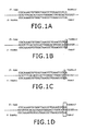
- Figures 1A, 1B, 1C, and 1D show diagrams of the hybridization of the oligonucleotides IS and IS-INV with the DNA substrate.
- Fig 1A pMTB-Control
- Fig.1B Mutant 1F
- Fig.1C Mutant 1P
- Fig1D Mutant 2PF.
- the central sequence of each diagram shows the sequence of the DNA substrate.
- the top sequence corresponds to the primer IS and the bottom sequence to the primer IS-INV. Boxes are used to show the unpaired bases between the probes and the DNA substrate.
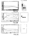
- Figure 2A, 2B, 2C show on the left the amplification results in real-time of different DNA substrates on fluorescence graphs (FAM channel) relative to the number of cycles in the different cases.
- FAM channel fluorescence graphs
- FIG 2A Amplification mixture with IS probe and DNA pfu polymerase
- Fig2B Amplification mixture with IS-INV probe and pfu DNA polymerase
- Fig2C Amplification mixture with IS-INV probe and DNA polymerase 5'-3' exonuclease polymerase.
- Figures 3A and 3B show the results of the comparative amplification assay in real-time using the IS-INV probe as a source of fluorescence in the presence of pfu DNA polymerase, or the intercalating fluoride SYBR Green I in the presence of pfu.
- Figure 3A shows the fluorescence graph versus the number of cycles. The top part of the graph shows the amplification profiles obtained when using the IS-INV probe as a source of fluorescence, the bottom part shows the profiles obtained using intercalating fluoride SYBR Green.
- Figure 2B shows the agarose gel analysis in the amplified products obtained.
- Lane 1 corresponds to a 10 -3 dilution
- lane 2 to a 10 -6 dilution
- lane 3 to a 10 -8 dilution
- lane 4 to a No-DNA negative control
- lane M corresponds to a 100 bp Ladder.
- the lanes on the right correspond to the assay with SYBR Green I, and on the left to the IS-INV probe.
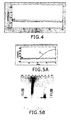
- Figure 4 shows the amplification results in real time on a graph of fluorescence versus the number of cycles, corresponding to example 3 of the present invention.
- Figure 5A shows the amplification results in real time, on a graph of fluorescence versus number of cycles, corresponding to example 4 of the present invention and in Figure 5B , analysis in agarose gel of the amplified products obtained at the end of the process.
- the five lanes on the right correspond to the PCR results without a probe and the five lanes on the left to the PCR results with probe, represented by M: ladder 100 bp, 2: DNA pol 5'-3' exo +, 3:Taq polymerase exo-, 4: "no DNA" control.
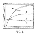
- Figure 6 shows the results of detection by an independent DNA synthesis system shown in example 5, in a graph of fluorescence versus number of cycles.
- Figure 7A shows a profile of fluorescence emission obtained during reverse transcription coupled to gene amplification, of two samples in serum infected with HIV and of a negative "no-RNA" control.
- Figure 7B shows analysis by gel electrophoresis of products amplified in figure 7A.
- oligonucleotides that carry the marker and have been designed to present unpaired bases at the 3' terminal or adjacent bases on hybridization with the nucleic acid substrate, which makes them, consequently, susceptible to partial degradation and to generate a signal mediated by the 3'-5' nuclease activity included in the reaction mixtures are given the generic name of Lion probes.
- a control plasmid (pMTB-Control) was obtained by cloning a 335 bp fragment of the IS6110 region of Mycobacterium tuberculosis (MTB) in the pBlueScript SK(+) plasmid.
- Mutant 1P mutation in the penultimate base of the hybridization region with the primers Lion probe IS and Lion probe IS-INV. This generates an unpaired base at the last base of the 3' end of both primers when it hybridizes with them, as can be seen in Figure 1C .
- Mutant 1F mutation in the last base of the zone of hybridization between the primers Lion probe IS and Lion probe IS-INV. It produces an unpaired base at the 3' end of both primers on hybridization with them, shown in Figure 1B .
- Mutant 2PF mutation of the last two bases of the hybridization region with the primers Lion probe IS and Lion probe IS-INV. It generates unpairing of the last two bases of the 3' end of both primers when it hybridizes with them, as can be observed in Figure 1D .
- oligonucleotides will be used as primers of the amplification reactions:
- Primer MT2 (SEQ ID N°03, 5'-CATCGTGGAAGCGACCCGCCAGCCCAGGAT-3'). Reverse primer, which hybridizes perfectly with the sequences of the four previously described substrates. This primer was used as a reverse primer in all the experiments studied in this example.
- Amplification mixture with Lion probe IS and pfu DNA polymerase performed with the Biotools Pfu DNA polymerase kit (Biotools B & M Labs, Madrid, Spain), including in the mixture 0.1 u/ ⁇ l of Pfu DNA polymerase, reaction buffers, a mixture of dNTPs, Lion probe IS (0.3 ⁇ M final) and oligonucleotide MT2 (0.5 mM final), with a final reaction volume of 20 ⁇ l.
- Amplification mixture with Lion probe IS-INV and pfu-DNA polymerase performed with the kit Biotools Pfu DNA polymerase (Biotools), including in the mixture 0.1 u/ ⁇ l of Pfu DNA polymerase, the reaction buffers, a mixture of dNTPs, Lion probe IS-INV (0.3 ⁇ M final) and oligonucleotide MT2 (0.5 mM final), with a final reaction volume of 20 ⁇ l.
- Amplification mixture with Lion probe IS-INV and DNA polymerase 5'-3' exonuclease performed with the kit Biotools DNA polymerase (Biotools), including in the mixture 0.1 u/ ⁇ l of DNA polymerase 5'-3' exonuclease, reaction buffers, a mixture of dNTPs, Lion probe IS-INV (0.3 ⁇ M final) and oligonucleotide MT2 (0.5 mM final), with a final reaction volume of 20 ⁇ l.
- Biotools Biotools DNA polymerase
- the amplification reaction was performed in a real-time amplification system SmartCycler II (Cepheid) using the following amplification cycles:
- the temperature was maintained for 360 s at 95.0°C.
- Amplification reaction course was monitored in real-time by reading the fluorescence level in the FAM channel, measured in the incubation step at 60° C. Similarly, the amplified products were analyzed in 1.5% agarose gel stained with ethidium bromide. The results are shown in figure 2 .
- Example 1 Since in Example 1 it was observed that the use of DNA amplification systems with pfu DNA polymerase using, as a primer of the reaction, a fluorescent double labeled probe in the presence of substrates that produce unpaired bases at the 3' end of the probe, generates similar fluorescence results to those obtained with intercalating fluorides, such as SYBR Green, a comparative assay was performed of both systems.
- intercalating fluorides such as SYBR Green
- Mutant 2PF mutation in the last two bases of the zone of hybridization with the Lion probes IS and IS-INV. This generates an unpairing of the last two bases of the 3' end of both primers as it hybridizes with them.
- oligonucleotides were used as primers of the amplification reaction:
- the amplification reaction was carried out in a SmartCycler II (Cepheid) real-time amplification system using the following amplification cycles:
- Table 1 of the results shows the Ct values obtained in the samples amplified with probe IS-INV and those obtained with SYBR Green.
- Table 1 Sample Ct Mutant 2PF, dilution 10 -3 , (A1) 8.23 Mutant 2PF, dilution 10 -6 , (A2) 16.39 Mutant 2PF, dilution 10 -8 , (A3) 23.21 No DNA, (A4) 34.33 Mutant 2PF, dilution 10 -3 , (A5) 12.23 Mutant 2PF, dilution 10 -6 , (A6) 23.07 Mutant 2PF, dilution 10 -8 , (A7) 28.92 No DNA, (A8) 31.96
- A1, A2, A3 and A4 Samples amplified with probe IS-INV.A5, A6, A7 and A8: Samples amplified with SYBR-Green.
- the mean delay of the Ct values of the samples amplified with SYBR Green compared to the samples amplified with probe IS-INV was calculated as the arithmetic mean of the differences in Ct obtained in each dilution analyzed.
- the safety margin of each reading was calculated as the difference between the CT value of the no-DNA control and the Ct value of the weakest dilution analyzed (dil 10 -8 ).
- results obtained indicate that although use of double labeled probes as primers in the presence of pfu DNA polymerase generates similar results to those obtained using intercalating fluorides such as SYBR Green, the new method improves both the sensitivity, and potentially the specificity of the detection. Hence, the earliest values of fluorescence appearance, and the highest values of fluorescence obtained indicate a better sensitivity for the system.
- the combination of reduced CT values of the amplified samples and the delayed appearance of the fluorescent signal in no-DNA controls increases the safety range for detection of a given signal in comparison with SYBR Green.
- the difference in cycles between the last sample detectable and the primer-dimer signal in the experiment with IS-INV probe was 11.8 cycles per 3.3 cycles in the amplification experiment with SYBR Green.
- Fluorescence levels were monitored in the FAM channel in each of the incubations at 68° C during the entire process.
- an amplification/detection assay was performed with TaqMan probes that require the presence of 5'-3' exonuclease activity to generate a fluorescent signal.
- the experimental model used in this example is an amplification and detection system with TaqMan probe of a conserved region of the coding region of the human cytomegalovirus polymerase (CMV).
- CMV human cytomegalovirus polymerase
- pCMV plasmid obtained by cloning a conserved fragment of 350 base pairs (bp) of the coding region of the cytomegalovirus polymerase (CMV) in the pBlueScript plasmid SK(+).
- Amplification primers and fluorescent probes are Amplification primers and fluorescent probes.
- CMVF (ID SEQ N°06, 5'-GATAGACACACACTGCAAA-3'). Forward primer that hybridizes perfectly (100% homology) with the region of the CMV genome cloned in the previously described plasmid pCMV.
- Primer CMR (SEQ ID N°07, 5'- GGTGGGACCTATTCGT -3'). Reverse primer that hybridizes perfectly (100% homology) with the region of the CMV genome cloned in the pCMV plasmid described previously.
- CMV probe ID SEQ N° 08 5'-TTCACACCTACGATCAGACGGA-3'.
- TaqMan probe with double fluorescent labeling (5' FAM----TAMRA 3') that hybridizes perfectly (100% homology) with an internal region of the product amplified with the combination of CMVF/CMVR primers described previously.
- the amplification assays were performed in parallel with a DNA polymerase 5'-3' exonuclease enzymatic activity and pfu DNA polymerase, both produced by Biotools B&M Labs, LTD, and an exo Taq polymerase activity - (Clontech).
- the reaction mixtures have the following composition: A mixture containing dNTPs, primer CMVF (0.5 ⁇ M), primer CMVR (0.5 ⁇ M) and probe CMV (0.3 ⁇ M).
- different enzymes were added in each experiment: 5'-3' exonuclease + DNA polymerase (Biotools DNA polymerase), Taq DNA polymerase exo - (Titanium Taq DNA polymerase. Clontech) or Pfu DNA polymerase (Biotools), adding in all cases 0.1 u/ ⁇ l of enzyme, and a supplementing the reaction with specific buffers for each enzyme.
- the final reaction volume was 20 ⁇ l in all cases.
- the amplification reaction was carried out in a SmartCycler II (Cepheid) amplification system in real-time using the following amplification cycles:
- EXAMPLE 5 Detection assay of specific sequences of nucleic acids in fluid hybridization and an absence of polymerization reaction, using probes with double fluorescent labels and pfu DNA polymerase activity.
- pMTB-Control obtained by cloning a 335 bp fragment of the IS6110 region of Mycobacterium tuberculosis (MTB) in the pBlueScript SK(+) plasmid.
- Mutant 1P mutation in the last base of the zone of hybridization with the primers IS and IS-INV. This generates an unlinked base at the 3' end of both primers when it hybridizes with them (See hybridization diagram in figure 1 B) .
- Mutant 1F mutation of the last base of the hybridization zone with primers IS and IS-INV. This generates an unpaired base at the 3' end of both primers when it hybridizes with them (See hybridization diagram in figure 1C ).
- Mutant 2PF mutation in the last two bases of the zone of hybridization zone with the primers IS and IS-INV. This generates unpaired nucleotides at the last two bases of the 3' end of both primers when it hybridizes with them (See hybridization diagram in figure 1D ).
- oligonucleotide was used as a detection probe:
- the expected action mechanism in this non-nucleic acid polymerization dependent detection system consists in the joining of the probe, by base complementarity with the nucleic acid substrate. If the hybridization is perfect, the 3'-5' exonuclease activity does not function, and no fluorescence is emitted. This would be the case of the assay with the control plasmid pMTB-control. By contrast, if on formation of the plasmid an unpairing occurs at the base at the 3' end of the probe, the 3'-5' exonuclease activity will detect the unpaired base and split the bases emitting fluorescence. This would be the case of the assay with each of the mutants included in the example.
- reaction is carried out using the reaction buffers assayed in the previous examples in which the functioning of the pfu 3'-5' exonuclease activity has been demonstrated.
- similar temperature cycles will be applied to those used in the PCT assays to permit denaturisation and hybridization of the probes with the substrate DNA.
- the composition of the reaction mixture was as follows: 0.1 U/ ⁇ l of pfu DNA polymerase (Biotools), Lion probe IS-INV (0.3 ⁇ M) and the specific reaction buffer of pfu. The final volume of the reaction was 20 ⁇ l in all cases.
- the assay results were monitored in a LightCycler amplification system in real-time.
- the results obtained show the functionality of the system to detect specific sequences of nucleic acids in fluid hybridization systems using the Lion type probes with double fluorescent labeling (5' blocker-3' fluoride) that presents unpaired bases at the 3' end on hybridizing with the nucleic acid substrate, and the presence of 3'-5' exonuclease proofreading activity, in an independent polymerization mechanism of nucleic acids.
- results obtained in examples 1 and 2 show the possibility of detecting specific sequences of nucleic acids using probes with double fluorescent labeling (preferably with the quencher in position 5' and the fluoride in position 3') in gene amplification systems with a DNA polymerase with 3'-5' exonuclease proofreading activity and in those in which the 3' end of the probe presents punctual unpaired bases with the DNA substrate to be identified.
- the DNA polymerase recognizes the unpaired bases at the 3' end if the primer, and by the 3'-5' nuclease activity splits the unpaired bases that carry the fluoride. At this point, as the fluoride and the quencher separate fluorescence is emitted.
- the double labeled fluoride operates simultaneously as an identification probe and also as an amplification primer, since as the unpaired bases are eliminated by the action of the 3'-5' exonuclease, the primer is prepared to prime the elongation of a new chain of DNA.
- using a system of these characteristics provides similar information to that obtained with intercalating fluorides such as SYBR Green.
- the non-specific amplification of sequences related to the substrate, or the formation of primer-dimers can generate a non-specific signal.
- the present example describes an amplification method in real time that used a double fluorescent labeled probe to generate the signal that has the following structure: A) a sequence of 20 nucleotides that hybridize perfectly (100% homology) with the sequence to be identified; B) a spacer (dS) in position 3' of the oligonucleotide sequence; C) a sequence of 3 nucleotides including two fluorescent markers (quencher and reporter) that do not hybridize with the sequence to be detected.
- pCMV Cytomegalovirus polymerase
- Amplification primers and fluorescent probes are Amplification primers and fluorescent probes.
- Primer CMVF (ID SEQ N°06). Forward Primer that hybridizes perfectly (100% homology) with the cloned region of the CMV genome in the plasmids pCMV and pMUT-CMV. It functions as an oligonucleotide primer of DNA synthesis.
- Primer CMVR (SEQ ID N°07). Reverse primer that hybridizes perfectly (100% homology) with the cloned region of the CMV genome in plasmids pCMV and pMUT-CMV. It functions as an oligonucleotide primer of DNA synthesis.
- CMV-INV-Blok probe (ID SEQ N° 08, 5'-TCCGTCTGATCGTAGGTGTGAATAA -ds spacer-(TAMRA) tt (FAM-3').
- Probe with double fluorescent labeling (5' TAMRA--FAM 3') with the following structure: A) a sequence of 20 nucleotides that hybridizes perfectly (100% homology) with the sequence to be identified; B) a spacer (dS) in position 3' of the oligonucleotide sequence; C) a sequence of 3 oligonucleotides including the two fluorescent labels (quencher and reporter) that does not hybridize with the sequence to be detected.
- This probe hybridizes with an internal region of the product amplified by the previously described combination of primers CMVF/CMVR.
- Amplification reactions were assayed on 1/10 serial dilutions of the plasmid pCMV using a mixture of pfu DNA polymerase and 5'-3' exonuclease DNA polymerase as a source of polymerase activity.
- the concentration range assayed was of 50,000-50 copies of plasmid/reaction.
- the composition of the reaction mixture was: 0.1 U/ ⁇ l of 5'-3' exonuclease DNA polymerase, 0.1 U/ml of pfu DNA polymerase, a mixture of dNTPs, primer CMVR (final concentration: 0.5 ⁇ M), primer CMVF (final concentration: 0.5 ⁇ M), probe CMV-INV-Block (final concentration: 0.5 ⁇ M) and the reaction buffer (Kit Certamp. Biotools), with a final reaction volume of 20 ⁇ l in all cases.
- the reaction mixture with pfu DNA polymerase was as follows: Performed with the kit Biotools Pfu DNA polymerase (Biotools), incorporating in the mixture 0.1 u/ ⁇ l of Pfu DNA polymerase, the reaction buffers, a mixture of dNTPs, primer CMVR (final concentration: 0.5 uM), primer CMVF (final concentration: 0.5 uM), probe CMV-INV-Block (final concentration: 0.5 ⁇ M) with a final reaction volume of 20 ⁇ l in all cases.
- Biotools Biotools
- CMVR final concentration: 0.5 uM
- primer CMVF final concentration: 0.5 uM
- probe CMV-INV-Block final concentration: 0.5 ⁇ M
- the reaction mixture with DNA polymerase 5'-3' exonuclease was as follows: 0.1 U/ml of DNA polymerase 5'-3' exonuclease (Biotools DNA polymerase), reaction buffer, a mixture of dNTPs, primer CMVR (final concentration: 0.5 ⁇ M), primer CMVF (final concentration: 0.5 ⁇ M), probe CMV-INV-Block (final concentration: 0.5 ⁇ M) and the reaction buffer (Kit Certamp. Biotools), with a final reaction volume of 20 ⁇ l in all cases.
- the amplification reactions were carried out in a SmartCycler II (Cepheid) amplification system in real-time using the following amplification cycles:
- the course of the amplification reaction was monitored in real time by reading in each amplification cycle the fluorescence level in the FAM channel. Likewise, the amplified products were analyzed in 1.5% ethidium bromide- stained agarose gel.
- DNA polymerase 5'-3' exonuclease positivity in gel and negative fluorescence, independently of the substrate analyzed
- this activity can remove the bound probe, by 5'-3' exonuclease activity.
- this activity does not affect the integrity of the tail of unpaired nucleotides at position 3' of the probe, so its activity does not generate a fluorescent signal).
- results obtained confirm the use of nucleotides with two distinct regions separated by a spacer, one of which is used to bind to the substrate DNA, which is susceptible to degradation by 5'-3' nuclease activity, and the other is situated in the 3' region of the oligonucleotide, in which the two fluorescent labels are located that can be degraded by 3'-5' nuclease activity, and function as pure detection probes in gene amplification reactions carried out in the presence of a combination of DNA polymerase 5'-3' exonuclease and pfu DNA polymerase activities.
- Example 7 shows an RNA sequence detection system assay, via an indirect mechanism which links, in a single step, a reverse transcription system (RT), with a subsequent amplification of the cDNA obtained, by an amplification in real time that uses an oligonucleotide with fluorescent double labeling as a primer of the amplification reaction, and a system to generate a fluorescent signal, in the presence of pfu DNA polymerase activity.
- RT reverse transcription system
- Substrate RNA As substrate RNA, two sera of known concentration were used (7.9E+06 and 8.5E+05 copies/ml taken from a panel of sera of HIV of known concentration "HIV-1 RNA Quantification Panel" (Acrometrix). In both cases, RNA was extracted from 200 ⁇ l of serum, using the extraction kit "SpeedTools RNA Virus Extraction Kit” (Biotools, Madrid, Spain) and eluting the RNA extracted in a final volume of 20 ⁇ l. A total of 5 ⁇ l of RNA were used in the reverse transcription/gene amplification reaction.
- Reverse primer for the primer of the reverse transcription reaction and the subsequent gene amplification reaction an antisense oligonucleotide was used that hybridizes perfectly with the HIV-RNA sequence:
- Lion probe As a forward primer in the cDNA amplification reaction, and as a source of fluorescent signal, a Lion probe was used that hybridizes perfectly with the cDNA made in the reverse transcription process, except at the 3' end base, in which the change in substrate from the 3'-5' nuclease activity of the Pfu DNA polymerase was incorporated:
- the reverse transcription/amplification mixture combined in one step is shown below: 0.1 u/ ⁇ l of Pfu DNA polymerase (Biotools B&M Labs, Madrid. Spain), MMLV (Sybenzime), reaction buffers, a mixture of dNTPs, Lion probe HIV-F1 (final 0.3 ⁇ M) and oligonucleotide HIV-R1 (final 0.5 mM), with a final reaction volume of 50 ⁇ l.
- Pfu DNA polymerase Biotools B&M Labs, Madrid. Spain
- MMLV Sybenzime
- reaction buffers a mixture of dNTPs
- Lion probe HIV-F1 final 0.3 ⁇ M
- oligonucleotide HIV-R1 final 0.5 mM
- the amplification reaction was carried out in a Corbett RotorGene 3000 (Corbett) amplification system in real-time using the following amplification cycles:
- reverse phase transcription was carried out by incubating at 42°C for 45 minutes.
- the heat labile enzyme MMLV was inactivated by incubating at 97° C for 3 minutes.
Landscapes
- Chemical & Material Sciences (AREA)
- Organic Chemistry (AREA)
- Life Sciences & Earth Sciences (AREA)
- Zoology (AREA)
- Wood Science & Technology (AREA)
- Proteomics, Peptides & Aminoacids (AREA)
- Health & Medical Sciences (AREA)
- Engineering & Computer Science (AREA)
- Microbiology (AREA)
- Immunology (AREA)
- Physics & Mathematics (AREA)
- Molecular Biology (AREA)
- Biotechnology (AREA)
- Biophysics (AREA)
- Analytical Chemistry (AREA)
- Biochemistry (AREA)
- Bioinformatics & Cheminformatics (AREA)
- General Engineering & Computer Science (AREA)
- General Health & Medical Sciences (AREA)
- Genetics & Genomics (AREA)
- Measuring Or Testing Involving Enzymes Or Micro-Organisms (AREA)
Applications Claiming Priority (1)
| Application Number | Priority Date | Filing Date | Title |
|---|---|---|---|
| PCT/ES2005/070093 WO2006136621A1 (es) | 2005-06-16 | 2005-06-16 | Método de detección de ácidos nucleicos mediante generación directa de una señal medible |
Publications (1)
| Publication Number | Publication Date |
|---|---|
| EP1892302A1 true EP1892302A1 (de) | 2008-02-27 |
Family
ID=37570129
Family Applications (1)
| Application Number | Title | Priority Date | Filing Date |
|---|---|---|---|
| EP05857311A Withdrawn EP1892302A1 (de) | 2005-06-16 | 2005-06-16 | Nukleinsäurenachweisverfahren mit direkter erzeugung eines messbaren signals |
Country Status (6)
| Country | Link |
|---|---|
| US (1) | US7919244B2 (de) |
| EP (1) | EP1892302A1 (de) |
| JP (1) | JP2008543300A (de) |
| AU (1) | AU2005333512A1 (de) |
| CA (1) | CA2579918A1 (de) |
| WO (1) | WO2006136621A1 (de) |
Cited By (1)
| Publication number | Priority date | Publication date | Assignee | Title |
|---|---|---|---|---|
| US11926817B2 (en) | 2019-08-09 | 2024-03-12 | Nutcracker Therapeutics, Inc. | Microfluidic apparatus and methods of use thereof |
Families Citing this family (11)
| Publication number | Priority date | Publication date | Assignee | Title |
|---|---|---|---|---|
| WO2010018245A1 (es) * | 2008-08-12 | 2010-02-18 | Biotools Biotechnological & Medical Laboratories, S.A. | Método para la detección y/o cuantificación de un ácido nucleico substrato |
| JP5299964B2 (ja) * | 2009-03-11 | 2013-09-25 | 独立行政法人産業技術総合研究所 | Dna3’末端の修飾基除去用酵素試薬 |
| US20110136105A1 (en) * | 2009-12-05 | 2011-06-09 | Evogen, Inc. | Methods of quantifying nucleic acids |
| WO2011078441A1 (en) * | 2009-12-21 | 2011-06-30 | Seegene, Inc. | Tsg primer target detection |
| KR20120042100A (ko) | 2010-10-22 | 2012-05-03 | 주식회사 씨젠 | 이중표지 고정화 프로브를 이용한 고상에서의 타겟 핵산서열 검출 |
| EP4361606A2 (de) | 2012-02-03 | 2024-05-01 | California Institute of Technology | Signalkodierung und -dekodierung in multiplexierten biochemischen assays |
| IN2015DN01609A (de) | 2012-08-03 | 2015-07-03 | California Inst Of Techn | |
| KR101403507B1 (ko) * | 2013-03-21 | 2014-06-09 | 주식회사 현일바이오 | 결핵균 및 비결핵 마이코박테리아의 선택적 검출 방법, 그리고 이를 이용한 키트 |
| WO2017218777A1 (en) | 2016-06-17 | 2017-12-21 | California Institute Of Technology | Nucleic acid reactions and related methods and compositions |
| CN107254553A (zh) * | 2017-06-30 | 2017-10-17 | 中国科学院上海巴斯德研究所 | 用于检测多种病原体的荧光实时检测方法及应用 |
| CN107164565A (zh) * | 2017-07-18 | 2017-09-15 | 张梦玲 | 一种用于以实时荧光rt‑pcr检测样品中hiv‑1病毒的引物对和包含其的试剂盒 |
Family Cites Families (25)
| Publication number | Priority date | Publication date | Assignee | Title |
|---|---|---|---|---|
| US4683195A (en) | 1986-01-30 | 1987-07-28 | Cetus Corporation | Process for amplifying, detecting, and/or-cloning nucleic acid sequences |
| US4800159A (en) | 1986-02-07 | 1989-01-24 | Cetus Corporation | Process for amplifying, detecting, and/or cloning nucleic acid sequences |
| US5639611A (en) * | 1988-12-12 | 1997-06-17 | City Of Hope | Allele specific polymerase chain reaction |
| US5352778A (en) | 1990-04-26 | 1994-10-04 | New England Biolabs, Inc. | Recombinant thermostable DNA polymerase from archaebacteria |
| US5500363A (en) | 1990-04-26 | 1996-03-19 | New England Biolabs, Inc. | Recombinant thermostable DNA polymerase from archaebacteria |
| US5948663A (en) | 1990-12-03 | 1999-09-07 | Stratagene | Purified thermostable pyrococcus furiosus DNA polymerase I |
| US5436134A (en) | 1993-04-13 | 1995-07-25 | Molecular Probes, Inc. | Cyclic-substituted unsymmetrical cyanine dyes |
| US5925517A (en) * | 1993-11-12 | 1999-07-20 | The Public Health Research Institute Of The City Of New York, Inc. | Detectably labeled dual conformation oligonucleotide probes, assays and kits |
| US5538848A (en) | 1994-11-16 | 1996-07-23 | Applied Biosystems Division, Perkin-Elmer Corp. | Method for detecting nucleic acid amplification using self-quenching fluorescence probe |
| US5874283A (en) * | 1995-05-30 | 1999-02-23 | John Joseph Harrington | Mammalian flap-specific endonuclease |
| US20020061532A1 (en) * | 1997-02-14 | 2002-05-23 | Mosaic Technologies, Inc. | Method and apparatus for performing amplification of nucleic acids on supports |
| US5846726A (en) * | 1997-05-13 | 1998-12-08 | Becton, Dickinson And Company | Detection of nucleic acids by fluorescence quenching |
| ATE369439T1 (de) * | 1997-12-15 | 2007-08-15 | Csl Behring Gmbh | Markierter primer, geeignet für die detektion von nukleinsäuren |
| US7625705B2 (en) * | 1999-10-29 | 2009-12-01 | Hologic, Inc. | Methods and compositions for detection of a target nucleic acid sequence utilizing a probe with a 3′ flap |
| US20070134686A1 (en) * | 1999-10-29 | 2007-06-14 | Stratagene California | Methods and compositions for detection of a target nucleic acid sequence utilizing a probe with a 3' flap |
| US6528254B1 (en) * | 1999-10-29 | 2003-03-04 | Stratagene | Methods for detection of a target nucleic acid sequence |
| AU2001255448A1 (en) * | 2000-04-17 | 2001-10-30 | Stephen B. Liggett | Alpha-2 adrenergic receptor polymorphisms |
| AU2001259131A1 (en) | 2000-04-25 | 2001-11-07 | Dna Sciences, Inc. | Detection of nucleotide sequence variations via the proofreading activity of polymerases |
| AUPR050700A0 (en) * | 2000-10-03 | 2000-10-26 | Id+Plus Ltd | Detection method |
| US6350580B1 (en) * | 2000-10-11 | 2002-02-26 | Stratagene | Methods for detection of a target nucleic acid using a probe comprising secondary structure |
| WO2002062197A2 (en) * | 2000-12-19 | 2002-08-15 | Hospital For Special Surgery | Markers for disease susceptibility and targets for therapy |
| US6902900B2 (en) * | 2001-10-19 | 2005-06-07 | Prolico, Llc | Nucleic acid probes and methods to detect and/or quantify nucleic acid analytes |
| US7118871B2 (en) * | 2002-04-22 | 2006-10-10 | Lawler Joseph F | Reagents for monitoring nucleic acid amplification and methods of using same |
| CN1506471A (zh) * | 2002-12-09 | 2004-06-23 | 核酸检测方法 | |
| EP2339032B1 (de) * | 2005-04-18 | 2016-12-28 | MDNA Life Sciences Inc. | Mitochondriale Mutationen und Neuanordnungen als Diagnosewerkzeug zum Nachweis von Sonneneinstrahlungsbelastung, Prostatakrebs und andere Krebsarten |
-
2005
- 2005-06-16 WO PCT/ES2005/070093 patent/WO2006136621A1/es not_active Application Discontinuation
- 2005-06-16 EP EP05857311A patent/EP1892302A1/de not_active Withdrawn
- 2005-06-16 JP JP2008516346A patent/JP2008543300A/ja active Pending
- 2005-06-16 US US11/666,468 patent/US7919244B2/en not_active Expired - Fee Related
- 2005-06-16 AU AU2005333512A patent/AU2005333512A1/en not_active Abandoned
- 2005-06-16 CA CA002579918A patent/CA2579918A1/en not_active Abandoned
Non-Patent Citations (1)
| Title |
|---|
| See references of WO2006136621A1 * |
Cited By (1)
| Publication number | Priority date | Publication date | Assignee | Title |
|---|---|---|---|---|
| US11926817B2 (en) | 2019-08-09 | 2024-03-12 | Nutcracker Therapeutics, Inc. | Microfluidic apparatus and methods of use thereof |
Also Published As
| Publication number | Publication date |
|---|---|
| AU2005333512A1 (en) | 2006-12-28 |
| JP2008543300A (ja) | 2008-12-04 |
| US7919244B2 (en) | 2011-04-05 |
| WO2006136621A1 (es) | 2006-12-28 |
| US20070292868A1 (en) | 2007-12-20 |
| CA2579918A1 (en) | 2006-12-28 |
Similar Documents
| Publication | Publication Date | Title |
|---|---|---|
| US7919244B2 (en) | Nucleic acid detection method involving the direct generation of a measurable signal | |
| EP3063292B1 (de) | Nukleinsäuresonde mit einem einzelnen an einem internen zytosin gebundenen fluorophor zur anwendung in loop-vermittelter isothermaler amplifikation | |
| EP2130929B1 (de) | Intern gesteuerte Multiplex-Erkennung und Quantifizierung von mikrobiellen Nukleinsäuren | |
| CA2499360A1 (en) | Methods and compositions for detecting targets | |
| WO2007143097A1 (en) | Detection of nucleic acids | |
| KR20180053743A (ko) | 짧은 호모폴리머릭 반복서열의 개선된 검출법 | |
| EP2722388A1 (de) | Nukleinsäuresonde zur analyse von nukleinsäuren | |
| JP2006500033A (ja) | 標的を検出するための方法および組成物 | |
| EP3805408B1 (de) | Verfahren zur detektion einer zielnukleinsäure mittels rolling-circle-amplifikation und zusammensetzung einer detektion der zielnukleinsäure | |
| CA2925070C (en) | Detecting single nucleotide polymorphism using overlapped primer and melting probe | |
| US20070122827A1 (en) | Target nucleic acid signal detection | |
| CA2967912C (en) | Detecting single nucleotide polymorphism using overlapping hydrolysis probes | |
| WO2021183941A1 (en) | Looped primer and loop-de-loop method for detecting target nucleic acid | |
| CN108291252B (zh) | 稳定特定rna的通用方法 | |
| US7235355B1 (en) | Methods for detecting bacteriophage MS2 | |
| JP5774990B2 (ja) | 蛍光標識及び可溶性消光体を使用する標的変異体の検出 | |
| JP6999645B2 (ja) | 核酸の増幅及び検出/定量の効率を改良するためのヘルパーオリゴヌクレオチド | |
| CA3232224A1 (en) | Looped primer with various internal modifications and loop-de-loop method for target detection | |
| US20160326582A1 (en) | Method and kit for target molecule characterization |
Legal Events
| Date | Code | Title | Description |
|---|---|---|---|
| PUAI | Public reference made under article 153(3) epc to a published international application that has entered the european phase |
Free format text: ORIGINAL CODE: 0009012 |
|
| 17P | Request for examination filed |
Effective date: 20070305 |
|
| AK | Designated contracting states |
Kind code of ref document: A1 Designated state(s): AT BE BG CH CY CZ DE DK EE ES FI FR GB GR HU IE IS IT LI LT LU MC NL PL PT RO SE SI SK TR |
|
| 17Q | First examination report despatched |
Effective date: 20080711 |
|
| DAX | Request for extension of the european patent (deleted) | ||
| STAA | Information on the status of an ep patent application or granted ep patent |
Free format text: STATUS: THE APPLICATION IS DEEMED TO BE WITHDRAWN |
|
| 18D | Application deemed to be withdrawn |
Effective date: 20130103 |