EP1769748A1 - Instrument zur messung der blutgefäss-endothel-reaktion und verfahren zur steuerung des instruments zur messung der blutgefäss-endothel-reaktion - Google Patents
Instrument zur messung der blutgefäss-endothel-reaktion und verfahren zur steuerung des instruments zur messung der blutgefäss-endothel-reaktion Download PDFInfo
- Publication number
- EP1769748A1 EP1769748A1 EP05751367A EP05751367A EP1769748A1 EP 1769748 A1 EP1769748 A1 EP 1769748A1 EP 05751367 A EP05751367 A EP 05751367A EP 05751367 A EP05751367 A EP 05751367A EP 1769748 A1 EP1769748 A1 EP 1769748A1
- Authority
- EP
- European Patent Office
- Prior art keywords
- property
- blood vessel
- blocking
- evaluating
- section
- Prior art date
- Legal status (The legal status is an assumption and is not a legal conclusion. Google has not performed a legal analysis and makes no representation as to the accuracy of the status listed.)
- Withdrawn
Links
Images

Classifications
-
- A—HUMAN NECESSITIES
- A61—MEDICAL OR VETERINARY SCIENCE; HYGIENE
- A61B—DIAGNOSIS; SURGERY; IDENTIFICATION
- A61B5/00—Measuring for diagnostic purposes; Identification of persons
- A61B5/103—Measuring devices for testing the shape, pattern, colour, size or movement of the body or parts thereof, for diagnostic purposes
- A61B5/107—Measuring physical dimensions, e.g. size of the entire body or parts thereof
- A61B5/1075—Measuring physical dimensions, e.g. size of the entire body or parts thereof for measuring dimensions by non-invasive methods, e.g. for determining thickness of tissue layer
-
- A—HUMAN NECESSITIES
- A61—MEDICAL OR VETERINARY SCIENCE; HYGIENE
- A61B—DIAGNOSIS; SURGERY; IDENTIFICATION
- A61B5/00—Measuring for diagnostic purposes; Identification of persons
- A61B5/02—Detecting, measuring or recording for evaluating the cardiovascular system, e.g. pulse, heart rate, blood pressure or blood flow
- A61B5/02007—Evaluating blood vessel condition, e.g. elasticity, compliance
-
- A—HUMAN NECESSITIES
- A61—MEDICAL OR VETERINARY SCIENCE; HYGIENE
- A61B—DIAGNOSIS; SURGERY; IDENTIFICATION
- A61B8/00—Diagnosis using ultrasonic, sonic or infrasonic waves
- A61B8/48—Diagnostic techniques
- A61B8/488—Diagnostic techniques involving Doppler signals
-
- A—HUMAN NECESSITIES
- A61—MEDICAL OR VETERINARY SCIENCE; HYGIENE
- A61B—DIAGNOSIS; SURGERY; IDENTIFICATION
- A61B5/00—Measuring for diagnostic purposes; Identification of persons
- A61B5/22—Ergometry; Measuring muscular strength or the force of a muscular blow
-
- A—HUMAN NECESSITIES
- A61—MEDICAL OR VETERINARY SCIENCE; HYGIENE
- A61B—DIAGNOSIS; SURGERY; IDENTIFICATION
- A61B5/00—Measuring for diagnostic purposes; Identification of persons
- A61B5/68—Arrangements of detecting, measuring or recording means, e.g. sensors, in relation to patient
- A61B5/6801—Arrangements of detecting, measuring or recording means, e.g. sensors, in relation to patient specially adapted to be attached to or worn on the body surface
- A61B5/6813—Specially adapted to be attached to a specific body part
- A61B5/6824—Arm or wrist
-
- A—HUMAN NECESSITIES
- A61—MEDICAL OR VETERINARY SCIENCE; HYGIENE
- A61B—DIAGNOSIS; SURGERY; IDENTIFICATION
- A61B5/00—Measuring for diagnostic purposes; Identification of persons
- A61B5/68—Arrangements of detecting, measuring or recording means, e.g. sensors, in relation to patient
- A61B5/6801—Arrangements of detecting, measuring or recording means, e.g. sensors, in relation to patient specially adapted to be attached to or worn on the body surface
- A61B5/683—Means for maintaining contact with the body
- A61B5/6831—Straps, bands or harnesses
Definitions
- the present invention relates to a vascular endothelial reactivity measuring apparatus for use to make a function test of an arterial blood vessel wall tissue and a method for controlling such a measuring apparatus.
- the pathopoiesis of heart or brain infarction is closely correlated to arterial sclerosis. More specifically, if an atheroma is created on the arterial wall or if no arterial cells are produced anymore due to various factors such as elevated blood pressure, then the artery loses its elasticity to become hard and fragile. Also, if the blood vessel is clogged up where the atheroma has been created or if a vascular tissue covering the atheroma has ruptured, then the atheroma will move itself into the blood vessel to clog up the artery elsewhere or to rupture the hardened portions of the artery. As a result, these diseases are caused. That is why it is important to diagnose the arterial sclerosis as early as possible to prevent or treat these diseases.
- the arterial sclerosis can be diagnosed early enough to administer some medicine to its patient, then the disease can be treated effectively. However, it is said that once the arterial sclerosis has advanced to a certain degree, the farther advancement of that disease can be checked with the administration of medicine but it is difficult to repair the hardened artery completely.
- the lesion of arterial sclerosis is diagnosed by directly observing the inside of the blood vessel with a vascular catheter.
- this diagnosis needs to be carried out with a vascular catheter inserted into the blood vessel of a person under measurement, thus imposing a heavy load on him or her.
- the vascular catheter observation is usually adopted to locate the lesion of arterial sclerosis in a patient who is already known to suffer from that disease but has never been used to make a medical checkup on a supposedly healthy person.
- An ultrasonic diagnostic apparatus or an X-ray diagnostic apparatus has been used in the prior art as a noninvasive medical apparatus that imposes only a light load on a person under measurement.
- geometric information or information about the geometric variation with time of his or her internal body can be acquired without causing pain to him or her.
- the attribute information of the object can be obtained. That is to say, the vascular elastic property of the organism can be known and the degree of advancement of the arterial sclerosis can be detected directly.
- the ultrasonic diagnosis is superior to the X-ray diagnosis because the ultrasonic diagnosis can be made just by putting an ultrasonic probe on a person under measurement. That is to say, in the ultrasonic diagnosis, there is no need to administer a contrast medium to the person under measurement and there is no concern about potential X-ray exposure, either. Also, some ultrasonic diagnostic apparatuses can have significantly improved measuring accuracy thanks to recent remarkable advancement of electronic technologies. As a result, ultrasonic diagnostic apparatuses for measuring the very small motion of a vital tissue have been developed. For example, according to the technique described in Patent Document No.
- vascular endothelial function testing has been researched as a noninvasive method for sensing the degree of advancement of arterial sclerosis.
- vascular endothelial cells There is a layer of cell groups called “vascular endothelial cells” within an arterial blood vessel. These endothelial cells show various physiological reactions in response to a mechanical stress (i.e., shear stress) caused when blood flows through a blood vessel. Production of nitric oxide (NO) is one of those physiological reactions. Nitric oxide is produced and released by a nitric oxide synthetase to relax (i.e., soften) the plain muscle of the vascular tunica media as an endothelium-derived relaxing factor (EDRF). Also, the function of these vascular endothelial cells is called “endothelium dependent relaxation (EDR)".
- EMR endothelium dependent relaxation
- Non-patent Document No. 1 and Patent Document No. 2 Methods for evaluating a variation in the caliber of an arterial blood vessel with an ultrasonic wave before and after blood flow through the arterial blood vessel is blocked are disclosed in Non-patent Document No. 1 and Patent Document No. 2 as EDR checking methods for diagnosing arterial sclerosis.
- EDR checking methods for diagnosing arterial sclerosis.
- the caliber of the blood vessel of a resting examinee is measured and then blood flow through the artery of his or her lower arm is blocked for five minutes. After the blood flow has been unblocked, the maximum vascular caliber is measured intermittently for about two minutes and a flow mediated dilation (FMD) value is obtained based on the vascular caliber and used as an index to arterial sclerosis.
- FMD flow mediated dilation
- the vascular caliber is measured by reading the distances between m-lines (i.e., the distance from the intermediate point between the tunica media and the adventitious tunica of the front wall to the intermediate point between the tunica media and the adventitious tunica of the rear wall) on a 0.1 mm basis in an image obtained by an ultrasonic diagnostic apparatus to show a major-axis cross section of the blood vessel. And the average of four to six measured values is calculated as the vascular caliber.
- FIG. 5 shows the results of tests that were carried out on nine male examinees. In FIG.
- the solid squares plot the rate of increase in the amount of blood flowing through the right upper arm artery after the blood flow in the right forearm was unblocked, while the solid circles plot the rate of increase in the caliber of the right upper arm artery compared to that of a resting examinee.
- the abscissa represents the time passed since the blood flow was unblocked
- the ordinate on the left-hand side represents the rate of increase in the amount of blood flowing
- the ordinate on the right-hand side represents the rate of increase in the vascular caliber.
- Patent Document No. 1 Japanese Patent Application Laid-Open Publication No. 10-5226 Patent Document No.
- Non-patent Document No. 1 Masayoshi Hashimoto, Yasuyoshi Ohuchi, "Vascular Compliance Test", Magazine of the Japan Medical Association, Vol. 120, No. 8, Oct. 15 1998, pp. S93-S96
- Non-patent Document No. 1 and Patent Document No. 2 mentioned above a blood flow blocking period of five minutes and a measuring period of a few more minutes are needed. Thus, it takes about seven minutes in total to make a single measurement. Besides, some more time for preparing for the measurement also needs to be taken into account.
- the vascular caliber is measured on a 0.1 mm basis according to the method disclosed in Non-patent Document No. 1. However, since the vascular caliber of the upper arm artery is about 3 mm, the error is as high as about 3%. That is to say, the measuring accuracy realized by that method is far from satisfactory.
- an object of the present invention is to provide a vascular endothelial reactivity measuring apparatus that can carry out high-reliability measurements in a sufficiently short time.
- a vascular endothelial reactivity measuring apparatus includes: a blood flow blocking section for blocking blood flow through the arterial blood vessel of an organism; an evaluating section for evaluating the geometric property of the arterial blood vessel or its wall; and a control section for controlling the blood flow blocking section such that the blood flow through the arterial blood vessel is repeatedly blocked and unblocked in at least two iterative blocking/unblocking cycles.
- the evaluating section evaluates the geometric property and processes data, representing the geometric property during the partial period, by utilizing the iterative blocking/unblocking cycle.
- the evaluating section further evaluates the attribute property of the blood vessel wall based on the geometric property evaluated.
- the evaluating section extracts components that change synchronously with the iterative blocking/unblocking cycle from the data representing the geometric property and/or attribute property during the partial period.
- the evaluating section superposes multiple sets of data, each representing the geometric property and/or the attribute property to be assessed in every iterative blocking/unblocking cycle, and evaluates the geometric property and/or the attribute property based on the superposed data.
- the evaluating section subjects the data representing the geometric property and/or the attribute property to Fourier transform, extracts only iterative frequency components of the blocking/unblocking cycle, and evaluates the geometric property and/or the attribute property based on the data extracted.
- the evaluating section includes a bandpass filter that has a property to pass frequency components, of which the period is an integral multiple of the iterative blocking/unblocking cycle, to extract the data.
- the evaluating section is an ultrasonic diagnostic apparatus.
- the evaluating section is an X-ray diagnostic apparatus.
- the evaluating section is a nuclear magnetic resonance diagnostic apparatus.
- the geometric property evaluated by the evaluating section is the thickness of the blood vessel wall and/or a variation in the thickness of the blood vessel wall.
- the geometric property evaluated by the evaluating section is the caliber of the blood vessel and/or a variation in the caliber of the blood vessel.
- the attribute property evaluated by the evaluating section is the elastic property of the blood vessel wall.
- a control method is a method for getting a vascular endothelial reactivity measuring apparatus controlled by a control section of the vascular endothelial reactivity measuring apparatus.
- the method includes the steps of: evaluating the geometric property of an arterial blood vessel or its wall during at least a part of a period in which blood flow through the arterial blood vessel is unblocked while the blood flow through the arterial blood vessel is repeatedly blocked and unblocked in at least two iterative blocking/unblocking cycles; and processing data, representing the geometric property during the partial period, by utilizing the iterative blocking/unblocking cycle.
- the step of processing includes extracting components that change synchronously with the iterative blocking/unblocking cycle from the data representing the geometric property.
- the method further includes the step of evaluating the attribute property of the blood vessel or its wall based on the geometric property evaluated during the partial period, and the step of processing includes processing the data representing the geometric property and the data representing the attribute property in the iterative blocking/unblocking cycle.
- the step of processing includes extracting components that change synchronously with the iterative blocking/unblocking cycle from the data representing the geometric property and the data representing the attribute property.
- the step of processing includes superposing multiple sets of data, each representing the geometric property and/or the attribute property to be assessed in every iterative blocking/unblocking cycle, and evaluating the geometric property and/or the attribute property based on the superposed data.
- the step of processing includes subjecting the data representing the geometric property and/or the attribute property to Fourier transform, extracting only iterative frequency components of the blocking/unblocking cycle, and evaluating the geometric property and/or the attribute property based on the data extracted.
- the step of processing includes extracting the data by using a bandpass filter that has a property to pass frequency components, of which the period is an integral multiple of the iterative blocking/unblocking cycle.
- the step of evaluating includes evaluating the geometric property with an ultrasonic diagnostic apparatus, an X-ray diagnostic apparatus or a nuclear magnetic resonance diagnostic apparatus.
- a vascular endothelial reactivity measuring apparatus repeatedly blocks and unblocks the blood flow in at least two iterative cycles and processes the evaluated data by utilizing the blocking/unblocking cycle. Consequently, the resultant geometric property is accurate enough and hardly affected by noise.
- the blood flow blocking period can be shortened. As a result, it takes a shorter time to make a diagnosis using a vascular endothelial reactivity measuring apparatus.
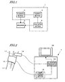
- FIG. 1 is a block diagram schematically showing a configuration for a vascular endothelial reactivity measuring apparatus 1.
- the measuring apparatus 1 includes a blood flow blocking section 2, a control section 3 and an evaluating section 4.
- the blocking section 2 may be a cuff (i.e., a tourniquet) that uses air pressure and is put on an arm of a person under measurement. Specifically, under the control of the control section 3, air is pumped into the cuff, thereby compressing the artery in the arm and blocking the blood flow through the artery. Conversely, also under the control of the control section 3, the air may be pumped out of the cuff, thereby unblocking the blood flow through the artery.
- a cuff i.e., a tourniquet
- This vascular endothelial reactivity measuring apparatus evaluates the geometric property of the vascular wall after the blood flow has been unblocked. That is why the blood flow is preferably unblocked quickly under the control of the control section 3.
- a known blood pressure manometer may be used as the blocking section 2.
- the control section 3 instructs the blocking section 2 to either block or unblock the blood flow at predetermined timings, which will be described in detail later.
- the evaluating section 4 evaluates the geometric property of the arterial blood vessel, in which the blood flow is blocked by the blocking section 2, or its wall.
- the geometric property evaluated includes the caliber of the blood vessel, the thickness of the vascular wall and a variation in the thickness of that vascular wall.
- the elasticity modulus, distortion, viscosity and other attribute properties of the vascular wall may further be obtained based on the geometric property.
- the evaluation is preferably carried out using the ultrasonic wave, the X-ray or the nuclear magnetic resonance. Therefore, an ultrasonic diagnostic apparatus, an X-ray diagnostic apparatus or a nuclear magnetic resonance diagnostic apparatus is preferably used as the evaluating section 4.
- an ultrasonic diagnostic apparatus is particularly preferred as the evaluating section 4 because ultrasonic waves hardly affect human bodies.
- the vascular endothelial reactivity measuring apparatus 1 evaluates the vascular endothelial reactivity (or the EDR among other things). For that purpose, a state in which the production of nitric oxide is checked and a state in which the production of nitric oxide is promoted need to be defined for the endothelial cells. That is to say, the amount of nitric oxide produced is changed by blocking and unblocking the blood flow and the degree of relaxation of the plain muscle caused by nitric oxide is estimated by evaluating the geometric property.
- the amount of time it takes to sense a significant difference in the geometric property of the blood vessel before and after the blood flow is unblocked depends on the manifestation time of the EDR.
- the vascular caliber maximizes in 45 to 60 seconds after the blood flow has been unblocked.
- the unblocking period for obtaining the maximum vascular caliber is preferably about 60 seconds.
- the present invention can be carried out not just in that particular situation where the vascular caliber has maximized but also in a situation where the vascular caliber has just increased. For that reason, there is no need to put measuring on hold for as many as 45 seconds after the blood flow has been unblocked.
- the present inventors discovered that the vascular caliber increased significantly in about 30 seconds after the blood flow was unblocked. That is why the unblocking period is preferably 30 seconds to 60 seconds. During at least a part of this unblocking period, the geometric property of the blood vessel or its wall is evaluated.
- the blocking period is set to several tens of seconds to several minutes in this preferred embodiment. More particularly, the blocking period is preferably 30 seconds to 3 minutes.
- the blocking period and unblocking period are defined as described above and the blocking and unblocking operations are repeatedly carried out in at least two cycles (preferably in four or more cycles). And during at least a part of the unblocking period in every cycle, the geometric property of the blood vessel or its wall is evaluated.
- the geometric property of the blood vessel or its wall may be evaluated during the blocking period, too. Then, the geometric property data thus obtained or data representing the attribute property obtained from the geometric property is processed by utilizing the iterative blocking/unblocking cycle.
- multiple sets of geometric property (or attribute property) data are superposed one upon the other and the geometric property during the unblocking period may be evaluated based on the superposed data.
- the geometric property data acquired during the two or more unblocking periods, and the geometric property value of the blood vessel or its wall during the blocking period is derived from the data extracted.
- the geometric property data may be subjected to Fourier transform to extract only the iterative frequency components of the blocking/unblocking cycle.
- a bandpass filter having a property to pass frequencies of which the period is n times as long as (where n is an integer that is equal to or greater than one) the blocking/unblocking cycle, may be obtained through computations and data may be extracted by passing data through the bandpass filter.
- the data is processed in the blocking/unblocking cycle.
- various external factors that might decrease the reproducibility of measuring including noise caused when the blocking section 2 or evaluating section 4 shifts from the measuring spot of the person under measurement and noise caused by his or her respiration
- the geometric property can be evaluated more accurately.
- a conventional method for evaluating vascular endothelial reactivity is susceptible to these types of noise so easily as to commit significant measuring errors.
- a relatively long blocking period of about five minutes is usually adopted.
- the vascular endothelial reactivity measuring apparatus of the present invention can reduce the influence of noise to such a level as to produce just minor measuring errors.
- the measuring apparatus can still produce significant measuring results.
- the blocking period can be shortened. A long blocking period may be a taboo for some examinees. That is why a shortened blocking period has a great significance.
- the geometric property or attribute property evaluated in this manner shows a variation corresponding to the amount of nitric oxide produced.
- the EDR which is a reaction to the production of nitric oxide, decreases due to a risk factor such as hypertension, hyperlipidemia, smoking or diabetes. Accordingly, evaluation of the geometric property or attribute property may be begun with a property that was diagnosed as arterial sclerosis caused by any of these risk factors.
- FIG. 2 schematically illustrates how to evaluate the geometric property of the arterial blood vessel 17 in an arm 11 of an examinee by using the vascular endothelial reactivity measuring apparatus 1.
- the vascular endothelial reactivity measuring apparatus 1 includes a cuff 10, a cuff pressure control section 12 (which is a control section for controlling the cuff 10 ), and an ultrasonic diagnostic apparatus 13.
- the cuff 10 and the cuff pressure control section 12 together functions as the blood flow blocking section 2, while the ultrasonic diagnostic apparatus 13 functions as the evaluating section 4. Also, the CPU 24 built in the ultrasonic diagnostic apparatus 13 (see FIG. 3 ) functions as the control section 3. A blood pressure manometer or a portion thereof may be used as the cuff 10. An ultrasonic probe 14 is connected to the ultrasonic diagnostic apparatus 13.
- the cuff 10 is wrapped around the upper portion of the arm 11. Also, the ultrasonic probe 14 is arranged closer to the heart than the cuff 10 is so as to evaluate the geometric property of the arterial blood vessel 17, too. In the example illustrated in FIG. 2, the ultrasonic probe 14 is arranged closer to the heart than the cuff 10 is. Alternatively, the cuff 10 may be arranged closer to the heart than the ultrasonic probe 14 is.
- the cuff pressure control section 12 controls the cuff 10 such that the blood flow is repeatedly and cyclically blocked and unblocked at predetermined intervals. For example, air may be pumped into and out of the cuff 10 such that the blood flow is blocked for 60 seconds and then unblocked for another 60 seconds.
- the blocking pressure may be 200 mmHg, for example.
- the ultrasonic diagnostic apparatus 13 receives an unblocking signal from the cuff pressure control section 12 and evaluates the geometric property of the arterial blood vessel 17 by using the ultrasonic probe 14 during at least a part of the unblocking period.
- An echo signal obtained by the ultrasonic probe 14 from the arterial blood vessel 17 is processed by the ultrasonic diagnostic apparatus 13, thereby sensing the variation in the caliber of the arterial blood vessel 17 or the variation in the thickness of the blood vessel wall quantitatively.
- a B-mode diagnostic image may also be generated based on the echo signal.
- a monitor 16 may be connected to the ultrasonic diagnostic apparatus 13 so as to present a B-mode tomographic image thereon.
- the vascular caliber, the thickness of the blood vessel wall or their variations may be either directly read from the B-mode image or figured out by a zero-cross method by which the displacement of an object is calculated based on the zero-cross point shift time of an RF signal.
- a phase difference tracking by a restricted minimum square method as disclosed in Patent Document No. 1 is a preferred measuring method for the ultrasonic diagnostic apparatus 13 of the present invention because the vascular caliber and the variation in the thickness of the blood vessel can be measured highly accurately.
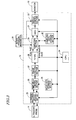
- FIG. 3 is a block diagram showing a configuration for an ultrasonic diagnostic apparatus 13 that uses a high-precision tracking method.
- the ultrasonic diagnostic apparatus 13 includes an ultrasonic wave transmitting/receiving section 23, a CPU 24, a time delay control section 25, a delay data storage section 26, a phase detecting section 27, a filter 28, a computing section 29, a computed data storage section 20, a DSC 21 and a display control section 22.
- the ultrasonic wave transmitting/receiving section 23 includes a driver for driving the ultrasonic probe 14 as an ultrasonic probe driving section and a receiver for amplifying the ultrasonic reflected wave as a receiving section.
- the ultrasonic probe driver applies a predetermined drive pulse signal to the ultrasonic probe 14.
- An ultrasonic transmitted wave, transmitted by the ultrasonic probe 14 in response to the drive pulse signal, is reflected by an organism to produce an ultrasonic reflected wave, which is then received at the ultrasonic probe 14.
- the ultrasonic reflected wave is received by the ultrasonic probe 14 and then amplified by the receiver.
- the ultrasonic transmitting/receiving section 23 further includes an A/D converter for converting the ultrasonic reflected wave, amplified by the receiver, into a digital signal.
- the time delay control section 25 is connected to the ultrasonic wave transmitting/receiving section 23 in order to control the time delay of the drive pulse signal to be supplied from the ultrasonic wave transmitting/receiving section 23 to a group of ultrasonic vibrators in the ultrasonic probe 14. In this manner, the ultrasonic transmitted wave to be transmitted from the ultrasonic probe 14 can have its acoustic line direction and depth of focus changed. Also, by controlling the time delay of an ultrasonic reflected wave signal that has been received by the ultrasonic probe 14 and then amplified by the ultrasonic wave transmitting/receiving section 23, the acoustic line direction of the ultrasonic wave to receive can be changed.
- the phase detecting section 27 detects the phase of the received reflected wave signal, of which the time delay has been controlled by the time delay control section 25, thereby splitting the signal into a real part signal and an imaginary part signal, which are then input to the filter 28.
- the filter 28 filters out components that have not been reflected by the vascular wall.
- the output of the filter 28 is supplied to the computing section 29.
- the computing section 29 includes a motion velocity calculating section, a position calculating section, a shrinkage/stretch calculating section, and an elasticity modulus calculating section.
- the motion velocity calculating section calculates the motion velocity of the blood vessel as the object by using the real part and imaginary part signals of the phase-detected signal. More specifically, the motion velocity calculating section sets a plurality of measuring points on the acoustic line of an ultrasonic wave to be transmitted from the ultrasonic probe 14.
- the computing section 29 calculates the phase difference by a minimum square method so as to minimize the waveform mismatch between the reflected wave signals r(t) and r(t+ ⁇ t).
- the motion velocity of each measuring point is derived from this phase difference and then integrated by the position calculating section and the shrinkage/stretch calculating section, thereby obtaining the magnitudes of positional displacement and shrinkage/stretch.
- the computing section 29 can effectively evaluate the attribute property of the vascular wall tissue based on the geometric property evaluated.
- the cuff of the blood pressure manometer 34 may also function as the cuff 10 for blocking the blood flow.
- the rate of increase in vascular caliber compared to the value of a resting examinee i.e., an FMD value
- FMD ⁇ d ⁇ 100 / d
- d is the vascular caliber of a resting examinee
- ⁇ d is the maximum variation in vascular caliber
- the computing section 29 figures out the geometric property or attribute property of the arterial blood vessel 17 or its wall
- only those components changing synchronously with the iterative blocking/unblocking cycle are preferably extracted by either Fourier transform or bandpass filtering processing.
- the geometric property of the arterial blood vessel 17 or the attribute property of the vascular wall is preferably evaluated based on the data extracted. As a result, the measuring can be carried out even more accurately.
- the geometric property or attribute property thus obtained may be converted by the DSC 21 into an image format suited for presenting it on the display 16 and then presented on the display 16.
- FIGS. 4(a) and 4(b) are graphs showing a variation in-the elastic property E of the vascular wall in the radial direction with time as evaluated by the vascular endothelial reactivity measuring apparatus 1 including the ultrasonic diagnostic apparatus 13 shown in FIG. 3.
- the computing section 29 figured out the elastic property by extracting only components changing synchronously with the iterative blocking/unblocking cycle.
- the elastic property was figured out without extracting those components changing synchronously with the iterative blocking/unblocking cycle for the purpose of comparison. As can be seen from FIG.
- the resultant graph representing the elastic property is a superposition of a lot of small waveforms. Such a graph was obtained due to the displacement of the probe 14 that was once released by, and then got hold of again, by the operator, the examinee's respiration and other subtle motions, or electromagnetic noise.
- T2 represents a period in which blood flow through the blood vessel was blocked
- T1 represents a period in which the blood flow was unblocked.
- the ultrasonic diagnostic apparatus shown in FIG. 3 is used as an exemplary evaluating section.
- the evaluating section may have any other configuration as long as the evaluating section can evaluate the geometric property and attribute property of the arterial blood vessel or its wall.
- the elastic property is evaluated as one of attribute properties of the vascular wall tissue.
- the vascular endothelial reactivity can also be evaluated similarly by compliance, which is the reverse number of the elastic property.
- a vascular endothelial reactivity measuring apparatus realizes high-reliability measurements in just a short time and can be used effectively in the medical and health care fields.
Landscapes
- Health & Medical Sciences (AREA)
- Life Sciences & Earth Sciences (AREA)
- Surgery (AREA)
- Animal Behavior & Ethology (AREA)
- Veterinary Medicine (AREA)
- Biophysics (AREA)
- Pathology (AREA)
- Engineering & Computer Science (AREA)
- Biomedical Technology (AREA)
- Heart & Thoracic Surgery (AREA)
- Medical Informatics (AREA)
- General Health & Medical Sciences (AREA)
- Physics & Mathematics (AREA)
- Public Health (AREA)
- Molecular Biology (AREA)
- Dentistry (AREA)
- Oral & Maxillofacial Surgery (AREA)
- Vascular Medicine (AREA)
- Cardiology (AREA)
- Physiology (AREA)
- Nuclear Medicine, Radiotherapy & Molecular Imaging (AREA)
- Radiology & Medical Imaging (AREA)
- Ultra Sonic Daignosis Equipment (AREA)
- Measuring Pulse, Heart Rate, Blood Pressure Or Blood Flow (AREA)
Applications Claiming Priority (2)
| Application Number | Priority Date | Filing Date | Title |
|---|---|---|---|
| JP2004184767 | 2004-06-23 | ||
| PCT/JP2005/011265 WO2006001252A1 (ja) | 2004-06-23 | 2005-06-20 | 血管内皮反応測定装置および血管内皮反応測定装置の制御方法 |
Publications (2)
| Publication Number | Publication Date |
|---|---|
| EP1769748A1 true EP1769748A1 (de) | 2007-04-04 |
| EP1769748A4 EP1769748A4 (de) | 2010-09-01 |
Family
ID=35781730
Family Applications (1)
| Application Number | Title | Priority Date | Filing Date |
|---|---|---|---|
| EP05751367A Withdrawn EP1769748A4 (de) | 2004-06-23 | 2005-06-20 | Instrument zur messung der blutgefäss-endothel-reaktion und verfahren zur steuerung des instruments zur messung der blutgefäss-endothel-reaktion |
Country Status (5)
| Country | Link |
|---|---|
| US (1) | US20060122489A1 (de) |
| EP (1) | EP1769748A4 (de) |
| JP (1) | JP3987099B2 (de) |
| CN (1) | CN100471459C (de) |
| WO (1) | WO2006001252A1 (de) |
Cited By (2)
| Publication number | Priority date | Publication date | Assignee | Title |
|---|---|---|---|---|
| US8057400B2 (en) | 2009-05-12 | 2011-11-15 | Angiologix, Inc. | System and method of measuring changes in arterial volume of a limb segment |
| US10238306B2 (en) | 2006-02-20 | 2019-03-26 | Everist Genomics, Inc. | Method for non-evasively determining an endothelial function and a device for carrying out said method |
Families Citing this family (14)
| Publication number | Priority date | Publication date | Assignee | Title |
|---|---|---|---|---|
| JPWO2008015761A1 (ja) * | 2006-08-04 | 2009-12-17 | 株式会社島津製作所 | 超音波診断装置 |
| US8016761B2 (en) * | 2006-10-23 | 2011-09-13 | The General Electric Company | Method and apparatus for automated flow mediated dilation |
| US8043223B2 (en) * | 2006-11-22 | 2011-10-25 | The General Electric Company | Method and apparatus for automated vascular function testing |
| JP5016316B2 (ja) * | 2007-01-31 | 2012-09-05 | メディアクロス株式会社 | メタボリックシンドローム血管評価システム |
| JP5446074B2 (ja) * | 2007-06-11 | 2014-03-19 | 株式会社日立製作所 | 血流の計測および評価装置 |
| WO2009028013A1 (ja) * | 2007-08-30 | 2009-03-05 | Shimadzu Corporation | 血管画像撮像装置 |
| US8845542B2 (en) * | 2009-09-09 | 2014-09-30 | Unex Corporation | Blood vessel function inspecting apparatus |
| EP2387949A1 (de) * | 2010-05-17 | 2011-11-23 | Samsung Medison Co., Ltd. | Ultraschallsystem zum Messen eines Bildes mithilfe einer Wertvorlage und Verfahren zum Betrieb des Ultraschallsystems |
| JP5930611B2 (ja) * | 2011-05-26 | 2016-06-08 | キヤノン株式会社 | 被検体情報取得装置 |
| TWM460634U (zh) * | 2013-03-19 | 2013-09-01 | Avita Corp | 監控生理狀態之裝置 |
| CN111031907A (zh) * | 2017-08-24 | 2020-04-17 | 东洋纺株式会社 | 伸缩性电极、伸缩性电极的制造方法、生理信息测量用衣服及生理信息测量方法 |
| CN108992092A (zh) * | 2018-06-12 | 2018-12-14 | 李宏博 | 一种用于动脉硬化闭塞诊断的装置 |
| KR102327662B1 (ko) * | 2019-12-10 | 2021-11-17 | 한양대학교 에리카산학협력단 | 동맥류 파열 예측 장치 및 방법 |
| JP2023156840A (ja) * | 2022-04-13 | 2023-10-25 | キヤノンメディカルシステムズ株式会社 | 超音波診断装置、解析装置、及びプログラム |
Family Cites Families (15)
| Publication number | Priority date | Publication date | Assignee | Title |
|---|---|---|---|---|
| US4771792A (en) * | 1985-02-19 | 1988-09-20 | Seale Joseph B | Non-invasive determination of mechanical characteristics in the body |
| CN1037269A (zh) * | 1989-04-03 | 1989-11-22 | 戚大海 | 动脉硬化诊断仪 |
| US5590649A (en) * | 1994-04-15 | 1997-01-07 | Vital Insite, Inc. | Apparatus and method for measuring an induced perturbation to determine blood pressure |
| US7048716B1 (en) * | 1997-05-15 | 2006-05-23 | Stanford University | MR-compatible devices |
| US6231507B1 (en) * | 1997-06-02 | 2001-05-15 | Vnus Medical Technologies, Inc. | Pressure tourniquet with ultrasound window and method of use |
| US20040015079A1 (en) * | 1999-06-22 | 2004-01-22 | Teratech Corporation | Ultrasound probe with integrated electronics |
| US6267728B1 (en) * | 1999-06-23 | 2001-07-31 | Steven Mark Hayden | Method for evaluating atherosclerosis and its affect on the elasticity of arterial walls |
| US6654628B1 (en) * | 2000-11-03 | 2003-11-25 | The Johns Hopkins University | Methods to assess vascular endothelial function |
| JP3668687B2 (ja) * | 2001-01-30 | 2005-07-06 | アロカ株式会社 | 脈波伝播速度計測装置及び超音波診断装置 |
| US20030049250A1 (en) * | 2001-09-05 | 2003-03-13 | Matti Karvonen | Method for enhancing endothelial function in humans |
| JP3785084B2 (ja) * | 2001-11-09 | 2006-06-14 | フクダ電子株式会社 | 血管内皮機能測定装置 |
| JP3825690B2 (ja) * | 2001-12-19 | 2006-09-27 | メディアクロス株式会社 | 血管径測定システム |
| JP4217023B2 (ja) * | 2002-02-25 | 2009-01-28 | 一郎 佐久間 | 血管内皮計測装置 |
| WO2003086169A2 (en) * | 2002-04-05 | 2003-10-23 | Thermal Technologies, Inc. | System for assessing endothelial function |
| JP3632014B2 (ja) * | 2002-05-14 | 2005-03-23 | コーリンメディカルテクノロジー株式会社 | 血管内皮機能評価装置 |
-
2005
- 2005-06-20 WO PCT/JP2005/011265 patent/WO2006001252A1/ja not_active Ceased
- 2005-06-20 JP JP2006523759A patent/JP3987099B2/ja not_active Expired - Fee Related
- 2005-06-20 CN CN200580000763.5A patent/CN100471459C/zh not_active Expired - Fee Related
- 2005-06-20 EP EP05751367A patent/EP1769748A4/de not_active Withdrawn
- 2005-12-02 US US11/292,858 patent/US20060122489A1/en not_active Abandoned
Cited By (3)
| Publication number | Priority date | Publication date | Assignee | Title |
|---|---|---|---|---|
| US10238306B2 (en) | 2006-02-20 | 2019-03-26 | Everist Genomics, Inc. | Method for non-evasively determining an endothelial function and a device for carrying out said method |
| US8057400B2 (en) | 2009-05-12 | 2011-11-15 | Angiologix, Inc. | System and method of measuring changes in arterial volume of a limb segment |
| US8657755B2 (en) | 2009-05-12 | 2014-02-25 | Angiologix, Inc. | System and method of measuring changes in arterial volume of a limb segment |
Also Published As
| Publication number | Publication date |
|---|---|
| EP1769748A4 (de) | 2010-09-01 |
| US20060122489A1 (en) | 2006-06-08 |
| WO2006001252A1 (ja) | 2006-01-05 |
| JP3987099B2 (ja) | 2007-10-03 |
| JPWO2006001252A1 (ja) | 2008-04-17 |
| CN1838913A (zh) | 2006-09-27 |
| CN100471459C (zh) | 2009-03-25 |
Similar Documents
| Publication | Publication Date | Title |
|---|---|---|
| CN100512764C (zh) | 超声诊断设备和超声诊断方法 | |
| US8771195B2 (en) | Cardiovascular analyzer | |
| US5715826A (en) | Method and device for assessing the state of blood vessels | |
| EP1769748A1 (de) | Instrument zur messung der blutgefäss-endothel-reaktion und verfahren zur steuerung des instruments zur messung der blutgefäss-endothel-reaktion | |
| TWI221407B (en) | Device and method for detecting the location of vein by ultrasound | |
| EP2177165B1 (de) | Ultrasonograph und Verfahren zur Erstellung eines Ultrasonogramms | |
| EP3203966B1 (de) | Vorrichtung und verfahren zur messung des intrakranialen drucks | |
| WO2017051388A1 (en) | Apparatus and methods for detecting increase in brain swelling and/or shifting | |
| EP1806099A1 (de) | Ultraschallgerät und ultraschallgerät-kontrollverfahren | |
| US7708692B2 (en) | Ultrasonic diagnostic apparatus and method for controlling the same | |
| Lee et al. | Reproducibility of regional pulse wave velocity in healthy subjects | |
| JP5014132B2 (ja) | 超音波診断装置 | |
| RU2440018C1 (ru) | Способ диагностики эндотелиальной дисфункции артерий | |
| Zhao | Harmonic Shear Wave Elastography | |
| JP4648700B2 (ja) | 血液レオロジー測定装置、及び血液レオロジー測定方法 | |
| Lee et al. | Reproducibility of Regional Pulse Wave Velocity in Healthy Subjects | |
| JP2007020724A (ja) | 超音波診断装置および超音波診断装置の制御方法 |
Legal Events
| Date | Code | Title | Description |
|---|---|---|---|
| PUAI | Public reference made under article 153(3) epc to a published international application that has entered the european phase |
Free format text: ORIGINAL CODE: 0009012 |
|
| 17P | Request for examination filed |
Effective date: 20060111 |
|
| AK | Designated contracting states |
Kind code of ref document: A1 Designated state(s): AT BE BG CH CY CZ DE DK EE ES FI FR GB GR HU IE IS IT LI LT LU MC NL PL PT RO SE SI SK TR |
|
| AX | Request for extension of the european patent |
Extension state: AL BA HR LV MK YU |
|
| RAP1 | Party data changed (applicant data changed or rights of an application transferred) |
Owner name: PANASONIC CORPORATION |
|
| A4 | Supplementary search report drawn up and despatched |
Effective date: 20100729 |
|
| STAA | Information on the status of an ep patent application or granted ep patent |
Free format text: STATUS: THE APPLICATION HAS BEEN WITHDRAWN |
|
| 18W | Application withdrawn |
Effective date: 20130524 |