EP1747463B1 - Verfahren zum nachweis von infektionen mit mykobakterien - Google Patents
Verfahren zum nachweis von infektionen mit mykobakterien Download PDFInfo
- Publication number
- EP1747463B1 EP1747463B1 EP05736221A EP05736221A EP1747463B1 EP 1747463 B1 EP1747463 B1 EP 1747463B1 EP 05736221 A EP05736221 A EP 05736221A EP 05736221 A EP05736221 A EP 05736221A EP 1747463 B1 EP1747463 B1 EP 1747463B1
- Authority
- EP
- European Patent Office
- Prior art keywords
- sample
- mycolic acid
- antibodies
- antigen
- acid antigen
- Prior art date
- Legal status (The legal status is an assumption and is not a legal conclusion. Google has not performed a legal analysis and makes no representation as to the accuracy of the status listed.)
- Expired - Lifetime
Links
Images



Classifications
-
- G—PHYSICS
- G01—MEASURING; TESTING
- G01N—INVESTIGATING OR ANALYSING MATERIALS BY DETERMINING THEIR CHEMICAL OR PHYSICAL PROPERTIES
- G01N33/00—Investigating or analysing materials by specific methods not covered by groups G01N1/00 - G01N31/00
- G01N33/48—Biological material, e.g. blood, urine; Haemocytometers
- G01N33/50—Chemical analysis of biological material, e.g. blood, urine; Testing involving biospecific ligand binding methods; Immunological testing
- G01N33/53—Immunoassay; Biospecific binding assay; Materials therefor
- G01N33/569—Immunoassay; Biospecific binding assay; Materials therefor for microorganisms, e.g. protozoa, bacteria, viruses
- G01N33/56911—Bacteria
- G01N33/5695—Mycobacteria
-
- G—PHYSICS
- G01—MEASURING; TESTING
- G01N—INVESTIGATING OR ANALYSING MATERIALS BY DETERMINING THEIR CHEMICAL OR PHYSICAL PROPERTIES
- G01N2800/00—Detection or diagnosis of diseases
- G01N2800/12—Pulmonary diseases
-
- G—PHYSICS
- G01—MEASURING; TESTING
- G01N—INVESTIGATING OR ANALYSING MATERIALS BY DETERMINING THEIR CHEMICAL OR PHYSICAL PROPERTIES
- G01N33/00—Investigating or analysing materials by specific methods not covered by groups G01N1/00 - G01N31/00
- G01N33/48—Biological material, e.g. blood, urine; Haemocytometers
- G01N33/50—Chemical analysis of biological material, e.g. blood, urine; Testing involving biospecific ligand binding methods; Immunological testing
- G01N33/53—Immunoassay; Biospecific binding assay; Materials therefor
- G01N33/543—Immunoassay; Biospecific binding assay; Materials therefor with an insoluble carrier for immobilising immunochemicals
- G01N33/54313—Immunoassay; Biospecific binding assay; Materials therefor with an insoluble carrier for immobilising immunochemicals the carrier being characterised by its particulate form
- G01N33/5432—Liposomes or microcapsules
Definitions
- THIS INVENTION relates to a method for detecting active tuberculosis.
- the invention relates to a serodiagnostic method for tuberculosis based on the prevalence of antibodies in mammalian subjects to lipid antigens derived from Mycobacterium tuberculosis. For a disease such as tuberculosis, there has been no acceptable serodiagnostic assay up until now, despite the fact that much progress has been reported in studies of antibodies to various antigens of M.
- Mycobacterial diseases are the cause of a high mortality in humans and other mammals. This is mainly due to the resilience of the pathogenic mycobacteria. These hard to eradicate bacteria have a low multiplication rate and hide intracellularly in the host macrophages. Treatment for mycobacterial diseases comprises combination chemotherapy extending over many months. This increases the frequency of non-compliance and thus may contribute to the emergence of multi-drug resistant strains of mycobacteria (Heym, Honore et al., 1994).
- Tuberculosis is a chronic pulmonary or extra-pulmonary disease caused by infection with Mycobacterium tuberculosis ( M. tuberculosis ).
- M. tuberculosis is spread primarily through aerosolized infectious particles generated from coughing and sneezing by individuals with pulmonary TB (Fenton and Vermeulen, 1996). After being inhaled, the bacilli are able to reach the terminal pulmonary airways and alveoli of uninfected individuals (Lawn et al ., 2002).
- Tuberculosis remains a major global health problem despite advances in medical science and a range of effective drugs, which for some time created the impression that the disease had been conquered.
- One third of the world's population is estimated to be infected with M . tuberculosis. It is further estimated that more than 8 million people contract TB every year, resulting in over 2 million deaths. The global tuberculosis incidence is 61 new cases per 100,000 people (Johnson, 2000).
- TB is responsible for 32% of deaths among HIV-positive people globally, compared to 11% due to septicaemia, 10% to cerebral toxoplasmosis, 8% to pneumonia, 6% to malignancies, 5% to meningitis and 10% due to other infections (Narain et al., 1992). Approximately 8% of the TB patients and one quarter of those who died of TB were already co-infected with HIV, and it is likely that this proportion will increase in future (Kaufmann and Hess, 2000).
- a method of detecting a surrogate marker for active tuberculosis including the steps of taking up isolated mycolic acid antigen of mycobacterial origin or a synthetic analogue thereof in a liposome carrier to produce mycolic acid antigen-containing liposomes; immobilizing isolated mycolic acid antigen of mycobacterial origin to produce immobilised antigens; obtaining a first, a second and a third sample from a human or animal suspected of having active tuberculosis, wherein each sample may contain antibodies to the antigen, the first sample having a lower concentration by dilution than the second and third samples; exposing part of the first sample to the immobilised mycolic acid antigen in a test vessel; exposing part of the first sample to the immobilised mycolic acid antigen in a control vessel; exposing the second sample to mycolic acid antigen-containing liposomes; exposing the third sample to liposomes not containing the mycolic
- the mycobacterial infection may be of the type which causes diseases selected from pulmonary and extra-pulmonary tuberculosis.
- the mycolic acid antigen may be derived from mycobacteria selected from virulent and pathogenic mycobacteria.
- the mycolic acid antigen may be derived from Mycobacterium tuberculosis.
- the mycolic acid antigen may be in a form selected from homogenous and heterogenous compound mixtures.
- the mycolic acid antigen is first mixed with a phospholipid such as phosphatidylcholine to produce liposomes having mycolic acid antigen integrated into the liposome phospholipid surface layer.
- a phospholipid such as phosphatidylcholine
- production of liposomes will involve sonication of the phospholipid-antigen mixture.
- the mycolic acid antigen-containing liposomes in the test and control vessels to which the first sample is exposed may be immobilized mycolic acid antigen.
- Methods of immobilization may include methods known to one skilled in the art.
- the mycolic acid antigen-containing liposomes may be immobilized on an activated surface.
- the activated surface may be a hydrophilic, underivatised biosensor cuvette surface which has been activated by prior incubation with a surface binding agent linked to a hydrophobic hydrocarbon and then washed with a suitable buffer.
- the agent may be a cationic detergent, such as cetyl-pyridinium chloride (CPC).
- the antibody to the mycolic acid antigen serves as a surrogate marker for active tuberculosis.
- the samples from a human or animal suspected of having active tuberculosis may be selected from blood samples, spinal fluid samples and samples that naturally contain antibodies. In the case of a human or animal with active mycobacterial disease the sample will contain antibodies to the mycobacterial pathogen.
- the sample may be from an HIV positive human.
- the antibodies may be antibodies against Mycobacterium tuberculosis, or antibodies against part thereof.
- the antibodies may be of the type that demonstrate cross-reactivity towards sterols such as cholesterol.
- the antibodies may be low affinity antibodies.
- samples from a human or animal suspected of having active tuberculosis may be further prepared such that any antibodies present in the sample are enriched, precipitated or partially purified, and present in non-complexed form.
- Such methods of preparation are known to one skilled in the art.
- the first, second and third samples from a human or animal suspected of having active tuberculosis will preferably be derived from an original sample by dividing the original sample into at least the first, second and third samples before dilution.
- Exposure of the second sample of human or animal origin to mycolic acid antigen may include pre-incubation of the second sample with liposomes containing mycolic acid antigen.
- the control for this pre-incubation will include pre-incubation of the third sample with empty liposomes, or liposomes containing phospholipid only.
- Exposure of the first sample of human or animal origin to mycolic acid antigen in the test and control vessels may include exposing the samples of human or animal origin to a surface prepared by the prior coating of the surface with mycolic acid antigen and a suitable blocking reagent.
- a blocking reagent is saponin or casein.
- the detection of binding of antibodies and/or other material to the mycolic acid antigen may be carried out in an automated device.
- the method will be carried out in a resonant mirror or surface plasmon resonance biosensor, or a sensor based on the monitoring of piezo-electrical signals.
- the detection of the binding of the antibodies and/or other material to the mycolic acid antigen may be carried out in real time.
- the Applicants have found that simply measuring the extent of binding of antibodies to mycolic acids in a sample of human or animal origin to an immoblised mycobacterial antigen is not a reliable diagnostic method for detecting antibodies to the immobilised antigen because of interfering or competing interactions with other components in the sample.
- the Applicant's finding that prior exposure of a part of the sample to free antigen to selectively reduce the amount of free antibodies to the antigen before detecting antibody binding to mycolic acid in the test vessel is a key aspect of the invention. This reduction is detected in the comparison of the degree or extent of binding in the test and control vessels and is a clear indication of the presence of antibodies to the antigen in the original sample.
- the Applicants have also found that simply measuring the extent of binding of antibodies to mycolic acids in a sample of human or animal origin to an immobilised mycobacterial antigen, after washing away excess of unbound antibody, is not a reliable method for detecting antibodies to an immobilised antigen because of the low affinity of the antibodies to mycolic acids in most individuals.
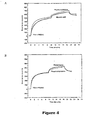
- Figure 1 the comparison is of patient and healthy control sera on mycolic acid and cholesterol-coated wells containing a concentration range of mycolic acid with the addition of 3,75 ⁇ g of cholesterol per well as antigen. All sera were tested in a 1:60 dilution.
- the negative control has sample identity : DR and the positive control has sample identity: Patient 1799 from 1994.
- Mycolic acids are high molecular weight, ⁇ -alkyl, ⁇ -hydroxyl fatty acids and are characteristic components of the cell envelope of mycobacteria and some other bacterial genera.
- MA are present as free lipids, such as trehalose dimycolate (TDM) or cord factor and trehalose monomycolate (TMM), but for the most part, they are esterified to the terminal penta-arabinofuranosyl units of arabinogalactan, a peptidoglycan-linked polysaccaride (Brennan and Nikaido, 1995).
- the presence of such long-chain fatty acids is largely responsible for the high hydrophobicity and very low permeability of the mycobacterial cell envelope (Lee et al., 1996).
- the number of carbon atoms that make up the MA varies from C 20 to C 30 in the genus Corynebacterium to C 60 to C 90 in the genus Mycobacterium, MA of the Nocardia and Rhodococcus species have lengths ranging from C 36 to C 66 (Butler et a /., 1991).
- Mycobacterial MA compose about 40-60% of the dry weight of the cell wall of the bacteria (Brennan and Nikaido, 1995; Lee et al., 1996). Because of the uniqueness of the structures of mycolic acids to the pathogenic Mycobacterium tuberculosis, they would provide ideal antigens for serodiagnosis of tuberculosis.
- WO 9528642 (Verschoor and Bye , 1995 ) relates to the incorporation of mycolic acid in an immunogenic conjugate which elicited specific antibody production in mice. Optimization of a procedure for extraction and purification of mycolic acids of mycobacteria was disclosed in WO 9626288 (Verschoor, 1996 and Goodrum et al., 2001). The immunological properties of the mycolic acids were tested in TB infected animals and in vitro cultures of human cells to explore the potential for broader application of the patented principles and products of tuberculosis This work has been compiled in WO 9839025 (Verschoor et al., 1998 ) and published (Korf et al., 2005).
- Mycolic acid is presented by antigen-presenting cells (APC) through a mechanism that does not involve MHC-class I or MHC-class II molecules.
- APC antigen-presenting cells
- Mycolic acid is a CD1 restricted antigen with the ability to induce proliferation of a T-cell line (Beckman et al., 1994).
- the human CD1 protein is known to mediate T-cell responses by presenting at least the three classes of mycobacterial lipids, i.e.
- the CD1-restricted lipid antigen presentation pathway could probably be the reason why the antibody response to mycolic acids is preserved in HIV-seropositive patients despite a declining CD4 T-lymphocyte count as the T cells that are stimulated by the mycolic acid presentation need not express the CD4 surface protein (Schleicher et al., 2002).
- EXAMPLE 1 ELISA assays for detection of antibodies in serum from tuberculosis patients
- Mycobacterial mycolic acids were isolated from a culture of Mycobacterium tuberculosis H37Rv (American Type Culture Collection 27294) as described by Goodrum et al., (Goodrum et al., 2001).
- PBS buffer 8.0 g NaCl, 0.2 g KCl, 0.2 g KH 2 PO 4 (anhydrous) and 1.05 g Na 2 HPO 4 (anhydrous) per 1l distilled water, adjusted to pH 7.4.
- Diluting buffer 0.5% (m/v) carbohydrate- and fatty acid free casein (Calbiochem, La Jolla, CA) in PBS buffer adjusted to pH 7.4 was used for diluting of the sera and the immunoreagents.
- Blocking buffer same as diluting buffer and used for blocking of ELISA plates.
- Washing buffer same as diluting buffer and used for washing of ELISA plates.
- Coating antigens Mycolic acids originating from Mycobacterium tuberculosis, isolated as described above, and cholesterol (Sigma, St. Louis, MO; Cat No C-8667) were used at final concentrations of 60 ⁇ g/ml and 75 ⁇ /ml, respectively.
- the antigens were heated in PBS buffer for 20 min at 85°C.
- the hot solutions were sonicated at 20% duty cycle and optimal output level for 1 min.
- the solutions were kept at 85°C and loaded into the ELISA plates.
- Goat anti-human IgG (H + L chains) antibody conjugated to peroxidase was obtained from Sigma (Cat No A-8667).
- Substrate o-Phenylenediamine (Sigma; Cat No P-1526) and hydrogen peroxide (Merck, Darmstadt, BRD; Cat No BDR 10366).
- Substrate buffer 0.1 M citrate buffer (0.1 M citric acid and 0.1 M tri-sodium citrate, adjusted to pH 4.5).
- the respective antigens were dissolved in hot PBS and then sonicated, as described above.
- the wells of flat-bottom ELISA plates were coated overnight at 4°C with 50 ⁇ l/well of antigen solution.
- the final antigen load was maximally 3.1 ⁇ g/well for mycolic acids and 3.75 ⁇ g/well for the cholesterol.
- the coating solution was flicked out of the plates and replaced with 400 ⁇ l blocking buffer per well. Blocking was carried out for 2 hours at room temperature.
- the blocking solution was aspirated from the wells before loading of the serum or serum precipitate samples.
- Sera were diluted 20 times in diluting buffer. Aliquots of 50 ⁇ l were introduced into wells in quadruplicate. The plates were incubated at room temperature for 1 hour. The serum samples were removed from the wells, the wells washed three times with washing buffer using an Anthos Autowash automatic ELISA plate washer and then emptied by aspiration.
- Peroxidase-conjugated anti-human IgG diluted 1:1000 in diluting buffer was introduced in aliquots of 50 ⁇ l per well and the plates were incubated for 30 min at room temperature. After removal of the conjugate, the wells were washed three times with the washing buffer and then emptied by aspiration.
- the substrate solution comprising 10.0 mg o-phenylenediamine and 8.0 mg hydrogen peroxide in 10 ml of 0.1 M citrate buffer pH 4.5, was prepared immediately before use and introduced in 50 ⁇ l aliquots per well. The plates were incubated at room temperature and the colour development was monitored at 5, 30 and 60 min after addition of the substrate using an SLT 340 ATC photometer at a wavelength of 450 nm.
- Anti-cholesterol antibodies are commonly found in human sera and may be induced by a variety of diseases (Horvath and Biro, 2003, Horvath et al., 2001, Alving and Wassef, 1999, Dijkstra et al., 1996). This could be problematic for a serodiagnostic assay that is based on the prevalence of anti-mycolic acids antibodies and may be the cause of the many false positive sera that were registered with the ELISA technique to determine such antibodies in hospitalized TB + and TB- patients (Schleicher et al., 2002).
- EXAMPLE 2 Biosensor assays for recognition of antibodies in serum from tuberculosis patients
- the IAsys resonant mirror biosensor system (Buckle et al., 1993) and twin-cell non-derivatized cuvettes were from Affinity Sensors (Cambridge, United Kingdom).
- Serum samples were selected from 102 patients (aged between 18 and 65) half of whom had active pulmonary tuberculosis.
- the study population consisted of a tuberculosis-positive (TB + ) group and a control tuberculosis-negative (TB - ) group.
- the TB + group consisted of patients with newly diagnosed smear-positive pulmonary tuberculosis of which approximately 50% were HIV-seropositive.
- the TB - patients of whom approximately 50% were HIV seropositive had medical conditions other than TB and were recruited from the general medical wards. None of the TB + patients were on anti-TB chemotherapy at the time of serum collection.
- Mycobacterial mycolic acids were isolated from a culture of M. tuberculosis H37Rv (American Type Culture Collection 27294) as described by Goodrum et al., (2001).
- PBS/EA buffer 8.0 g NaCl, 0.2 g KCI, 0.2 g KH 2 PO 4 and 1.05 g Na 2 HPO 4 per 1l ultrapure, distilled water with 1 mM EDTA and 0.025% (m/v) sodium azide, adjusted to pH 7.4 Cetylpyridinium chloride (CPC; Sigma, St.
- phosphatidylcholine Stock solutions of cholesterol and phosphatidylcholine (100 mg/ml) were prepared by dissolving the weighed amounts in chloroform.
- Cholesterol containing liposomes were prepared by combining 30 ⁇ l cholesterol and 60 ⁇ l phosphatidylcholine (Sigma St Louis, MO) stock solutions.
- Mycolic acids containing liposomes were prepared by adding 90 ⁇ l phosphatidylcholine stock to 1 mg dried mycolic acids.
- the liposome ingredients were dried with nitrogen gas in a heat block at 85°C for about 10 minutes. Liposome formation was induced by addition of 2 ml saline (0.9 % NaCl) and placing in a heat block at 85°C for 20 minutes, with vortexing every 5 minutes. The liposomes were then sonicated for 2 minutes at 30 % duty cycle at an output of 3 % with the Model B-30 Branson sonifier (Sonifer Power Company, USA). The sonicator tip was thoroughly washed with chloroform and rinsed with distilled water before and after use. The liposomes (200 ⁇ l) were aliquoted into 10 tubes and kept at -20 °C overnight before freeze-drying. After freeze-drying, 2 ml of PBS/AE was added to each tube containing liposomes. The tubes were placed in a heat block for 20 minutes and sonicated as before.
- IAsys software was used to set the device at a data-sampling interval of 0.4 s, temperature of 25°C and stirring rate of 75 % for all experiments on the IAsys affinity biosensor.
- the cells were rinsed three times prior to use with ethanol (98 %) followed by extensive washing with PBS/AE.
- a 60 ⁇ l volume of PBS/AE was pipetted into each cell of the cuvette to obtain a stable baseline for 1 minute.
- the PBS/AE was subsequently aspirated and the surface activated with 50 ⁇ l of cetyl-pyridinium chloride (CPC) for 10 minutes.
- CPC cetyl-pyridinium chloride
- the surface was then washed 5 times with PBS/AE and each cell's content was substituted with 25 ⁇ l of PBS/AE and allowed to equilibrate for about 5 - 10 minutes to achieve a stable baseline.
- Inhibition studies were performed using patient serum that was first placed at room temperature to thaw completely. After obtaining a stable baseline, a 1:1000 dilution of serum antibodies (10 ⁇ l) in PBS/AE was added, to compare the response of the two cells over 10 minutes. A pre-incubation of 1:500 dilutions of serum antibodies with liposomes containing either mycolic acids or cholesterol, or empty liposomes (phosphatidylcholine alone) were allowed for 20 minutes.
- Regeneration was effected by initial 3 times washing with 96 % ethanol for one minute, followed by 7 times washing with 70 ⁇ l PBS/AE for 1 minute. The surface was then finally treated with 50 ⁇ l potassium hydroxide (12.5 M) for 2 minutes and followed by 7 times washing with 70 ⁇ l PBS/AE for 1 minute.
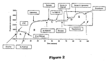
- the six main stages involved to measure the binding of specific antibodies to lipid antigens in liposomes in real time are: (A) the activation of the surface with CPC, (B) immobilization of the liposomes containing cholesterol or mycolic acids to the surface, (C) blocking with saponin to prevent non-specific protein binding, (D) binding (association) of antibodies from a high dilution of serum to calibrate the signal of the two cells of the cuvette, (E) the binding and dissociation of inhibited patient sera at lesser dilution, and finally (F) surface regeneration ( Figure 2 ).
- Remaining false negative scores in the biosensor assay may derive from patients who are under strong immunosuppressive treatment or who suffer immunosuppression due to conditions such as diabetes or cancer.
- the biosensor was able to detect low affinity antibody binding to mycolic acids, which the conventional methods cannot achieve. The reason for this can be demonstrated with the biosensor, by showing that antibodies bound to the mycolic acids coated surface wash away if a washing step is introduced (data not shown). In an ELISA assay, the washing step is essential before the final development and many patients therefore tested false negative with this standard technology.
- the success of the biosensor therefore lies in its reliable detection of antibodies of even low affinity to mycolic acids and in its ability to discriminate between binding to related antigens. Any other technology that can meet these requirements may also be used to diagnose tuberculosis infection based on the detection of anti-mycolic acid antibodies as surrogate markers for tuberculosis.
- the novel serodiagnostic method presented here appears to be particularly amenable to the diagnosis of tuberculosis infection in an environment where there is a high prevalence of AIDS and HIV infection. This appeared to be a major stumbling block in published, state of the art approaches to the serodiagnosis of tuberculosis, even when using lipids from the mycobacterial cell wall as antigens (Boggian et al ., 1996).
- the advantage of the invention is that it provides a highly predictive, simple, accurate and rapid method to detect antibodies to mycolic acids from blood samples as surrogate markers for pathogenic mycobacterial infection in human and animal subjects. This may then be used in:
- the experimental evidence indicates that antibodies to mycolic acids occur in most (78%) of the blood samples that were obtained from seriously ill, hospitalised individuals that were infected with M. tuberculosis and that such antibodies were generally (82%) not found in the blood samples of an equal number of seriously ill, hospitalized individuals that were not infected with M . tuberculosis.
- the invention also takes into account experimental evidence indicating that cholesterol is a cross-reactive antigen with mycolic acids in antibody binding and that cholesterol is a weaker antigen than mycolic acids for binding with anti-mycolic acid antibodies. Specificity of binding of anti mycolic acid antibodies is therefore demonstrated in the method.
- the method therefore measures both the binding of antibodies to the mycolic acids and the inhibition of binding by pre-incubating the blood samples with mycolic acids.
- the method therefore has the advantage in that the endpoint of the assay is the degree of inhibition of binding of serum immunoglobulin to mycolic acids after pre-incubation of the serum with solubilized mycolic acids.
- the endpoint of the method can be determined consistently in an evanescent field biosensor (wave guide or surface plasmon resonance) that allows the binding of low affinity antibodies, and their inhibition of binding when the blood sample is pre-incubated with the mycolic acid antigen. It cannot be consistently determined with standard immunoassays, such as ELISA, that are biased towards higher affinity antibodies and cannot distinguish reliably between binding of antibodies to mycolic acid or cholesterol.
Landscapes
- Health & Medical Sciences (AREA)
- Life Sciences & Earth Sciences (AREA)
- Immunology (AREA)
- Engineering & Computer Science (AREA)
- Urology & Nephrology (AREA)
- Chemical & Material Sciences (AREA)
- Biomedical Technology (AREA)
- Molecular Biology (AREA)
- Hematology (AREA)
- Medicinal Chemistry (AREA)
- General Health & Medical Sciences (AREA)
- Biotechnology (AREA)
- Tropical Medicine & Parasitology (AREA)
- Virology (AREA)
- Food Science & Technology (AREA)
- Microbiology (AREA)
- Analytical Chemistry (AREA)
- Cell Biology (AREA)
- Biochemistry (AREA)
- Physics & Mathematics (AREA)
- General Physics & Mathematics (AREA)
- Pathology (AREA)
- Peptides Or Proteins (AREA)
- Measuring Or Testing Involving Enzymes Or Micro-Organisms (AREA)
Claims (17)
- Verfahren zum Nachweisen eines Ersatzmarkierungsmittels für aktive Tuberkulose, wobei das Verfahren die folgenden Schritte aufweist:- Aufnehmen eines isolierten Mycolsäure-Antigens oder eines synthetischen Analogs davon in einen Liposomenträger zum Erzeugen Mycolsäure-Antigen enthaltender Liposome;- Immobilisieren eines isolierten Mycolsäure-Antigens mykobakteriellen Ursprungs zum Erzeugen immobilisierter Antigene;- Verwenden einer ersten, einer zweiten und einer dritten Probe, die von einem Menschen oder einem Tier erhalten wurde, der bzw. das im Verdacht steht, aktive Tuberkulose zu haben, wobei jede Probe Ersatzmarkierungsmittel-Antikörper zu dem Antigen enthalten kann, wobei die erste Probe durch Verdünnung eine geringere Konzentration aufweist als die zweite und die dritte Probe;- Aussetzen eines Teils der ersten Probe dem immobilisierten Mycolsäure-Antigen in einem Testgefäß;- Aussetzen eines Teils der ersten Probe dem immobilisierten Mycolsäure-Antigen in einem Kontrollgefäß;- Aussetzen der zweiten Probe den Mycolsäure-Antigen enthaltenden Liposomen;- Aussetzen der dritten Probe Liposomen, die das Mycolsäure-Antigen nicht enthalten;- Geben der zweiten Probe in das Testgefäß, nachdem sie den Mycolsäure-Antigen enthaltenden Liposomen ausgesetzt wurde;- Geben der dritten Probe in das Kontrollgefäß, nachdem sie den Liposomen, die das Mycolsäure-Antigen nicht enthalten, ausgesetzt wurde;- Nachweisen eines Bindens von Antikörpern an das Mycolsäure-Antigen sowohl im Testgefäß als auch im Kontrollgefäß in Echtzeit; und- Vergleichen des Grads bzw. des Ausmaßes des Bindens zwischen dem Testgefäß und dem Kontrollgefäß, wobei eine beobachtete geringere Bindung im Testgefäß ein Indikator für die Anwesenheit von Antikörpern für das Mycolsäure-Antigen in den Proben ist, der sich auf aktive Tuberkulose in dem Menschen oder dem Tier bezieht, von dem die Proben stammen.
- Verfahren nach Anspruch 1, wobei die mykobakterielle Infektion des Typs ist, der Krankreiten verursacht, die aus der pulmonalen und extrapulmonaler Tuberkulose ausgewählt sind.
- Verfahren nach Anspruch 1 oder Anspruch 2, wobei das Mycolsäure-Antigen von Mycobakterien erhalten wird, die aus virulenten und pathogenen Mycobakterien ausgewählt sind.
- Verfahren nach Anspruch 3, wobei das Mycolsäure-Antigen vom Mycobacterium tuberculosis erhalten wird.
- Verfahren nach einem der vorhergehenden Ansprüche, wobei das Mycolsäure-Antigen in einer Form vorliegt, die aus homogenen und heterogenen Verbindungsmischungen ausgewählt ist.
- Verfahren nach einem der vorhergehenden Ansprüche, wobei die Mycolsäure-Antigen enthaltenden Liposome auf einer aktivierten Oberfläche immobilisiert werden.
- Verfahren nach Anspruch 6, wobei die aktivierte Oberfläche eine hydrophile, underivatisierte Biosensor-Küvetten-Oberfläche ist, die durch vorhergehende Inkubation mit einem Oberflächenbindemittel, das mit einem hydrophoben Kohlenwasserstoff verbunden ist, aktiviert wurde.
- Verfahren nach einem der vorhergehenden Ansprüche, wobei die Probe menschlichen oder tierischen Ursprungs aus Blutproben, Hirn-Rückenmark-Flüssigkeitsproben sowie Proben, die natürlicherweise Antikörper enthalten, ausgewählt ist.
- Verfahren nach einem der vorhergehenden Ansprüche, wobei die Probe von einem HIV-positiven Menschen ist.
- Verfahren nach einem der vorhergehenden Ansprüche, wobei die Antikörper Antikörper gegen Mycobacterium tuberculosis oder Antikörper gegen einen Teil davon sind.
- Verfahren nach Anspruch 10, wobei die Antikörper des Typs sind, der gegenüber Sterolen eine Kreuzreaktivität aufweist.
- Verfahren nach Anspruch 11, wobei die Antikörper Antikörper mit geringer Affinität sind.
- Verfahren nach einem der vorhergehenden Ansprüche, wobei die erste, zweite und dritte Probe menschlichen oder tierischen Ursprungs dadurch von einer ursprünglichen Probe gewonnen werden, dass die ursprüngliche Probe vor der Verdünnung mindestens in die erste, die zweite und die dritte Probe aufgeteilt wird.
- Verfahren nach einem der vorhergehenden Ansprüche, wobei das Aussetzen der zweiten Probe menschlichen oder tierischen Ursprungs gegenüber Mycolsäure-Antigen eine Präinkubation der zweiten Probe mit Liposomen enthaltendem Mycolsäure-Antigen einschließt.
- Verfahren nach einem der vorhergehenden Ansprüche, das eine Präinkubation der dritten Probe mit leeren Liposomen oder Liposomen, die nur Phospholipid enthalten, einschließt.
- Verfahren nach einem der vorhergehenden Ansprüche, wobei das Aussetzen der ersten Probe menschlichen oder tierischen Ursprungs gegenüber Mycolsäure-Antigen im Testgefäß und im Kontrollgefäß enthält, dass die Proben menschlichen oder tierischen Ursprungs einer Oberfläche ausgesetzt werden, die dadurch präpariert wurde, dass die Oberfläche vorher mit Mycolsäure-Antigen und einem geeigneten Blockierungsreagens beschichtet wird.
- Verfahren nach einem der vorhergehenden Ansprüche, das in einem Resonanzspiegel- oder Oberflächenplasmon-Resonanz-Biosensor oder einem auf dem Überwachen piezoelektrischer Signale basierenden Sensor durchgeführt wird.
Applications Claiming Priority (2)
| Application Number | Priority Date | Filing Date | Title |
|---|---|---|---|
| ZA200403678 | 2004-05-13 | ||
| PCT/IB2005/051548 WO2005116654A1 (en) | 2004-05-13 | 2005-05-11 | A method for detecting mycobacterial infection |
Publications (2)
| Publication Number | Publication Date |
|---|---|
| EP1747463A1 EP1747463A1 (de) | 2007-01-31 |
| EP1747463B1 true EP1747463B1 (de) | 2008-09-17 |
Family
ID=34967106
Family Applications (1)
| Application Number | Title | Priority Date | Filing Date |
|---|---|---|---|
| EP05736221A Expired - Lifetime EP1747463B1 (de) | 2004-05-13 | 2005-05-11 | Verfahren zum nachweis von infektionen mit mykobakterien |
Country Status (7)
| Country | Link |
|---|---|
| US (1) | US7851166B2 (de) |
| EP (1) | EP1747463B1 (de) |
| JP (1) | JP2007537438A (de) |
| AT (1) | ATE408837T1 (de) |
| DE (1) | DE602005009819D1 (de) |
| WO (1) | WO2005116654A1 (de) |
| ZA (1) | ZA200609113B (de) |
Families Citing this family (16)
| Publication number | Priority date | Publication date | Assignee | Title |
|---|---|---|---|---|
| JPWO2009066502A1 (ja) * | 2007-11-20 | 2011-04-07 | コニカミノルタエムジー株式会社 | 固体支持体 |
| KR101644144B1 (ko) | 2008-07-11 | 2016-07-29 | 유니버셜 바이오센서스 피티와이 엘티디. | 향상된 면역분석 센서 |
| US9506913B2 (en) | 2010-07-15 | 2016-11-29 | University Of Pretoria | Method of detecting surrogate markers in a serum sample |
| GB201105436D0 (en) * | 2011-03-31 | 2011-05-18 | Univ Bangor | Method |
| AP2015008200A0 (en) * | 2012-06-11 | 2015-01-31 | Univ Pretoria | A liposomal composition comprising a sterol-modified lipid and a purified mycobacterial lipid cell wall component and it's use in the diagnosis of tuberculosis |
| CN110988081A (zh) * | 2013-05-16 | 2020-04-10 | 比勒陀利亚大学 | 诊断结核病的方法 |
| WO2014210327A1 (en) * | 2013-06-27 | 2014-12-31 | The Brigham And Women's Hospital, Inc. | Methods and systems for determining m. tuberculosis infection |
| GB201414369D0 (en) * | 2014-08-13 | 2014-09-24 | Univ Bangor | Kit and method |
| CN107438767B (zh) * | 2015-01-05 | 2020-05-15 | 凯仪国际有限公司 | 检测活动性结核病标志物的方法 |
| EP3371597A1 (de) | 2015-11-06 | 2018-09-12 | University of Pretoria | Kernelemente zur cor-ort-diagnose von tuberkulose |
| US10337032B2 (en) | 2016-02-01 | 2019-07-02 | The University Of Manchester | Compositions and methods for recombinant biosynthesis of propane |
| NL2016301B1 (en) * | 2016-02-22 | 2017-08-25 | Kei Int Ltd | Method for detecting a marker for active tuberculosis. |
| NL2017204B1 (en) * | 2016-06-08 | 2017-12-18 | Kei International Ltd | Solid substrate comprising antigens immobilised thereto, biosensor comprising said solid substrate and method for detecting the presence of mycobacterial material in a sample |
| WO2017211314A1 (en) | 2016-06-08 | 2017-12-14 | Kei International Limited | Method for detecting presence of mycobacterial material in sample using immobilised mannosyl phosphoketide antigen |
| NL2021443B1 (en) | 2018-08-08 | 2020-02-20 | Kei International Ltd | Synthetic antigens for tuberculosis detection |
| US20240142475A1 (en) * | 2021-02-24 | 2024-05-02 | Washington University | Compositions and methods for the detection of tuberculosis |
Family Cites Families (2)
| Publication number | Priority date | Publication date | Assignee | Title |
|---|---|---|---|---|
| US5721109A (en) * | 1989-01-18 | 1998-02-24 | Sawai Pharmaceutical Co., Ltd. | Method for diagnosing infection caused by acid-fast bacterium |
| PL181579B1 (pl) | 1994-04-14 | 2001-08-31 | Adcock Ingram Ltd | Próba diagnostyczna do wykrywania i identyfikacji gatunków Mycobacterium w próbce, oczyszczony kwas mikolowy lub mieszanina kwasów mikolowych, sposób grupowego rozdzielania i oczyszczania, wydzielone przeciwcialo, koniugat mikobakteryjny i zestaw do wykrywania obecnosci gatunków Mycobacterium PL |
-
2005
- 2005-05-11 WO PCT/IB2005/051548 patent/WO2005116654A1/en not_active Ceased
- 2005-05-11 DE DE602005009819T patent/DE602005009819D1/de not_active Expired - Fee Related
- 2005-05-11 EP EP05736221A patent/EP1747463B1/de not_active Expired - Lifetime
- 2005-05-11 US US11/579,972 patent/US7851166B2/en not_active Expired - Fee Related
- 2005-05-11 JP JP2007512698A patent/JP2007537438A/ja active Pending
- 2005-05-11 AT AT05736221T patent/ATE408837T1/de not_active IP Right Cessation
-
2006
- 2006-11-01 ZA ZA200609113A patent/ZA200609113B/xx unknown
Also Published As
| Publication number | Publication date |
|---|---|
| US20090111125A1 (en) | 2009-04-30 |
| JP2007537438A (ja) | 2007-12-20 |
| DE602005009819D1 (de) | 2008-10-30 |
| EP1747463A1 (de) | 2007-01-31 |
| US7851166B2 (en) | 2010-12-14 |
| ATE408837T1 (de) | 2008-10-15 |
| WO2005116654A1 (en) | 2005-12-08 |
| ZA200609113B (en) | 2008-07-30 |
Similar Documents
| Publication | Publication Date | Title |
|---|---|---|
| US7851166B2 (en) | Method for detecting mycobacterial infection | |
| Chan et al. | Diagnosis of tuberculosis by a visually detectable immunoassay for lipoarabinomannan | |
| Thanyani et al. | A novel application of affinity biosensor technology to detect antibodies to mycolic acid in tuberculosis patients | |
| Koskela et al. | Humoral immunity against Francisella tularensis after natural infection | |
| Sada et al. | Detection of mycobacterial antigens in cerebrospinal fluid of patients with tuberculous meningitis by enzyme-linked immunosorbent assay | |
| EP2593793B1 (de) | Verfahren für den nachweis von surrogatmarkern für aktive tuberkulose in einer serumsprobe | |
| Julián et al. | An ELISA for five glycolipids from the cell wall of Mycobacterium tuberculosis:: Tween 20 interference in the assay | |
| Cho et al. | Mycobacterial aetiology of Crohn's disease: serologic study using common mycobacterial antigens and a species-specific glycolipid antigen from Mycobacterium paratuberculosis. | |
| Loizou et al. | The antiphospholipid syndrome in infectious diseases | |
| Gevaudan et al. | Serological response of tuberculosis patients to antigen 60 of BCG | |
| He et al. | Rapid serodiagnosis of human mycobacteriosis by ELISA using cord factor (trehalose-6, 6′-dimyclate) purified from Mycobacterium tuberculosis as antigen | |
| Yanagihara et al. | Enzyme-linked immunosorbent assay of glycolipid antigens for identification of mycobacteria | |
| WO2008108510A1 (en) | Diagnostic kit for leptospirosis | |
| Baelden et al. | Serological analysis of human tuberculosis by an ELISA with mycobacterial antigen 60 | |
| Simonney et al. | Analysis of the immunological humoral response to Mycobacterium tuberculosis glycolipid antigens (DAT, PGLTb1) for diagnosis of tuberculosis in HIV-seropositive and-seronegative patients | |
| Tiwari et al. | Glycolipids of Mycobacterium tuberculosis strain H37Rv are potential serological markers for diagnosis of active tuberculosis | |
| US6528272B2 (en) | Receptor-based assays for pathogens | |
| CA2313214A1 (en) | A method for identifying a mycobacterium species | |
| Fujita et al. | Differences in serological responses to specific glycopeptidolipid-core and common lipid antigens in patients with pulmonary disease due to Mycobacterium tuberculosis and Mycobacterium avium complex | |
| Tiwari et al. | Rapid liposomal agglutination card test for the detection of antigens in patients with active tuberculosis | |
| Senol et al. | Humoral immune response against 38‐and 16‐kDa mycobacterial antigens in childhood tuberculosis | |
| Coates et al. | Identification of Mycobacterium tuberculosis antigens in Seibert fractions by immunoblotting | |
| JPH07248329A (ja) | 結核感染症検査用膜およびこれを用いる結核感染症関連抗体の検出方法 | |
| Kiraly et al. | Evaluation of the T. pallidum haemagglutination (TPHA) test for syphilis on" problem sera" | |
| Gomes | Mycolic acid as antigen or analyte in tuberculosis |
Legal Events
| Date | Code | Title | Description |
|---|---|---|---|
| PUAI | Public reference made under article 153(3) epc to a published international application that has entered the european phase |
Free format text: ORIGINAL CODE: 0009012 |
|
| 17P | Request for examination filed |
Effective date: 20061109 |
|
| AK | Designated contracting states |
Kind code of ref document: A1 Designated state(s): AT BE BG CH CY CZ DE DK EE ES FI FR GB GR HU IE IS IT LI LT LU MC NL PL PT RO SE SI SK TR |
|
| DAX | Request for extension of the european patent (deleted) | ||
| GRAP | Despatch of communication of intention to grant a patent |
Free format text: ORIGINAL CODE: EPIDOSNIGR1 |
|
| GRAS | Grant fee paid |
Free format text: ORIGINAL CODE: EPIDOSNIGR3 |
|
| GRAA | (expected) grant |
Free format text: ORIGINAL CODE: 0009210 |
|
| AK | Designated contracting states |
Kind code of ref document: B1 Designated state(s): AT BE BG CH CY CZ DE DK EE ES FI FR GB GR HU IE IS IT LI LT LU MC NL PL PT RO SE SI SK TR |
|
| REG | Reference to a national code |
Ref country code: GB Ref legal event code: FG4D |
|
| REG | Reference to a national code |
Ref country code: CH Ref legal event code: EP |
|
| REG | Reference to a national code |
Ref country code: IE Ref legal event code: FG4D |
|
| REF | Corresponds to: |
Ref document number: 602005009819 Country of ref document: DE Date of ref document: 20081030 Kind code of ref document: P |
|
| PG25 | Lapsed in a contracting state [announced via postgrant information from national office to epo] |
Ref country code: LT Free format text: LAPSE BECAUSE OF FAILURE TO SUBMIT A TRANSLATION OF THE DESCRIPTION OR TO PAY THE FEE WITHIN THE PRESCRIBED TIME-LIMIT Effective date: 20080917 |
|
| PG25 | Lapsed in a contracting state [announced via postgrant information from national office to epo] |
Ref country code: SI Free format text: LAPSE BECAUSE OF FAILURE TO SUBMIT A TRANSLATION OF THE DESCRIPTION OR TO PAY THE FEE WITHIN THE PRESCRIBED TIME-LIMIT Effective date: 20080917 Ref country code: FI Free format text: LAPSE BECAUSE OF FAILURE TO SUBMIT A TRANSLATION OF THE DESCRIPTION OR TO PAY THE FEE WITHIN THE PRESCRIBED TIME-LIMIT Effective date: 20080917 Ref country code: AT Free format text: LAPSE BECAUSE OF FAILURE TO SUBMIT A TRANSLATION OF THE DESCRIPTION OR TO PAY THE FEE WITHIN THE PRESCRIBED TIME-LIMIT Effective date: 20080917 |
|
| NLV1 | Nl: lapsed or annulled due to failure to fulfill the requirements of art. 29p and 29m of the patents act | ||
| PG25 | Lapsed in a contracting state [announced via postgrant information from national office to epo] |
Ref country code: BE Free format text: LAPSE BECAUSE OF FAILURE TO SUBMIT A TRANSLATION OF THE DESCRIPTION OR TO PAY THE FEE WITHIN THE PRESCRIBED TIME-LIMIT Effective date: 20080917 |
|
| PG25 | Lapsed in a contracting state [announced via postgrant information from national office to epo] |
Ref country code: ES Free format text: LAPSE BECAUSE OF FAILURE TO SUBMIT A TRANSLATION OF THE DESCRIPTION OR TO PAY THE FEE WITHIN THE PRESCRIBED TIME-LIMIT Effective date: 20081228 Ref country code: BG Free format text: LAPSE BECAUSE OF FAILURE TO SUBMIT A TRANSLATION OF THE DESCRIPTION OR TO PAY THE FEE WITHIN THE PRESCRIBED TIME-LIMIT Effective date: 20081217 |
|
| PG25 | Lapsed in a contracting state [announced via postgrant information from national office to epo] |
Ref country code: IS Free format text: LAPSE BECAUSE OF FAILURE TO SUBMIT A TRANSLATION OF THE DESCRIPTION OR TO PAY THE FEE WITHIN THE PRESCRIBED TIME-LIMIT Effective date: 20090117 Ref country code: RO Free format text: LAPSE BECAUSE OF FAILURE TO SUBMIT A TRANSLATION OF THE DESCRIPTION OR TO PAY THE FEE WITHIN THE PRESCRIBED TIME-LIMIT Effective date: 20080917 Ref country code: SK Free format text: LAPSE BECAUSE OF FAILURE TO SUBMIT A TRANSLATION OF THE DESCRIPTION OR TO PAY THE FEE WITHIN THE PRESCRIBED TIME-LIMIT Effective date: 20080917 Ref country code: NL Free format text: LAPSE BECAUSE OF FAILURE TO SUBMIT A TRANSLATION OF THE DESCRIPTION OR TO PAY THE FEE WITHIN THE PRESCRIBED TIME-LIMIT Effective date: 20080917 Ref country code: PT Free format text: LAPSE BECAUSE OF FAILURE TO SUBMIT A TRANSLATION OF THE DESCRIPTION OR TO PAY THE FEE WITHIN THE PRESCRIBED TIME-LIMIT Effective date: 20090217 Ref country code: CZ Free format text: LAPSE BECAUSE OF FAILURE TO SUBMIT A TRANSLATION OF THE DESCRIPTION OR TO PAY THE FEE WITHIN THE PRESCRIBED TIME-LIMIT Effective date: 20080917 |
|
| PLBE | No opposition filed within time limit |
Free format text: ORIGINAL CODE: 0009261 |
|
| STAA | Information on the status of an ep patent application or granted ep patent |
Free format text: STATUS: NO OPPOSITION FILED WITHIN TIME LIMIT |
|
| PG25 | Lapsed in a contracting state [announced via postgrant information from national office to epo] |
Ref country code: DK Free format text: LAPSE BECAUSE OF FAILURE TO SUBMIT A TRANSLATION OF THE DESCRIPTION OR TO PAY THE FEE WITHIN THE PRESCRIBED TIME-LIMIT Effective date: 20080917 Ref country code: EE Free format text: LAPSE BECAUSE OF FAILURE TO SUBMIT A TRANSLATION OF THE DESCRIPTION OR TO PAY THE FEE WITHIN THE PRESCRIBED TIME-LIMIT Effective date: 20080917 |
|
| 26N | No opposition filed |
Effective date: 20090618 |
|
| PG25 | Lapsed in a contracting state [announced via postgrant information from national office to epo] |
Ref country code: IT Free format text: LAPSE BECAUSE OF FAILURE TO SUBMIT A TRANSLATION OF THE DESCRIPTION OR TO PAY THE FEE WITHIN THE PRESCRIBED TIME-LIMIT Effective date: 20080917 |
|
| PG25 | Lapsed in a contracting state [announced via postgrant information from national office to epo] |
Ref country code: MC Free format text: LAPSE BECAUSE OF NON-PAYMENT OF DUE FEES Effective date: 20090531 |
|
| REG | Reference to a national code |
Ref country code: CH Ref legal event code: PL |
|
| PG25 | Lapsed in a contracting state [announced via postgrant information from national office to epo] |
Ref country code: SE Free format text: LAPSE BECAUSE OF FAILURE TO SUBMIT A TRANSLATION OF THE DESCRIPTION OR TO PAY THE FEE WITHIN THE PRESCRIBED TIME-LIMIT Effective date: 20081217 Ref country code: LI Free format text: LAPSE BECAUSE OF NON-PAYMENT OF DUE FEES Effective date: 20090531 Ref country code: CH Free format text: LAPSE BECAUSE OF NON-PAYMENT OF DUE FEES Effective date: 20090531 |
|
| REG | Reference to a national code |
Ref country code: FR Ref legal event code: ST Effective date: 20100129 |
|
| PG25 | Lapsed in a contracting state [announced via postgrant information from national office to epo] |
Ref country code: FR Free format text: LAPSE BECAUSE OF NON-PAYMENT OF DUE FEES Effective date: 20090602 Ref country code: IE Free format text: LAPSE BECAUSE OF NON-PAYMENT OF DUE FEES Effective date: 20090511 |
|
| PG25 | Lapsed in a contracting state [announced via postgrant information from national office to epo] |
Ref country code: PL Free format text: LAPSE BECAUSE OF FAILURE TO SUBMIT A TRANSLATION OF THE DESCRIPTION OR TO PAY THE FEE WITHIN THE PRESCRIBED TIME-LIMIT Effective date: 20080917 |
|
| PG25 | Lapsed in a contracting state [announced via postgrant information from national office to epo] |
Ref country code: DE Free format text: LAPSE BECAUSE OF NON-PAYMENT OF DUE FEES Effective date: 20091201 |
|
| PG25 | Lapsed in a contracting state [announced via postgrant information from national office to epo] |
Ref country code: GR Free format text: LAPSE BECAUSE OF FAILURE TO SUBMIT A TRANSLATION OF THE DESCRIPTION OR TO PAY THE FEE WITHIN THE PRESCRIBED TIME-LIMIT Effective date: 20081218 |
|
| PG25 | Lapsed in a contracting state [announced via postgrant information from national office to epo] |
Ref country code: LU Free format text: LAPSE BECAUSE OF NON-PAYMENT OF DUE FEES Effective date: 20090511 |
|
| PG25 | Lapsed in a contracting state [announced via postgrant information from national office to epo] |
Ref country code: HU Free format text: LAPSE BECAUSE OF FAILURE TO SUBMIT A TRANSLATION OF THE DESCRIPTION OR TO PAY THE FEE WITHIN THE PRESCRIBED TIME-LIMIT Effective date: 20090318 |
|
| PG25 | Lapsed in a contracting state [announced via postgrant information from national office to epo] |
Ref country code: TR Free format text: LAPSE BECAUSE OF FAILURE TO SUBMIT A TRANSLATION OF THE DESCRIPTION OR TO PAY THE FEE WITHIN THE PRESCRIBED TIME-LIMIT Effective date: 20080917 |
|
| PG25 | Lapsed in a contracting state [announced via postgrant information from national office to epo] |
Ref country code: CY Free format text: LAPSE BECAUSE OF FAILURE TO SUBMIT A TRANSLATION OF THE DESCRIPTION OR TO PAY THE FEE WITHIN THE PRESCRIBED TIME-LIMIT Effective date: 20080917 |
|
| P01 | Opt-out of the competence of the unified patent court (upc) registered |
Effective date: 20230530 |
|
| PGFP | Annual fee paid to national office [announced via postgrant information from national office to epo] |
Ref country code: GB Payment date: 20240411 Year of fee payment: 20 |
|
| REG | Reference to a national code |
Ref country code: GB Ref legal event code: PE20 Expiry date: 20250510 |
|
| PG25 | Lapsed in a contracting state [announced via postgrant information from national office to epo] |
Ref country code: GB Free format text: LAPSE BECAUSE OF EXPIRATION OF PROTECTION Effective date: 20250510 |