EP1731099A1 - Interferometer for quantitative phase contrast imaging and tomography with an incoherent polychromatic x-ray source - Google Patents
Interferometer for quantitative phase contrast imaging and tomography with an incoherent polychromatic x-ray source Download PDFInfo
- Publication number
- EP1731099A1 EP1731099A1 EP05012121A EP05012121A EP1731099A1 EP 1731099 A1 EP1731099 A1 EP 1731099A1 EP 05012121 A EP05012121 A EP 05012121A EP 05012121 A EP05012121 A EP 05012121A EP 1731099 A1 EP1731099 A1 EP 1731099A1
- Authority
- EP
- European Patent Office
- Prior art keywords
- grating
- phase
- detector
- source
- interferometer according
- Prior art date
- Legal status (The legal status is an assumption and is not a legal conclusion. Google has not performed a legal analysis and makes no representation as to the accuracy of the status listed.)
- Withdrawn
Links
Images


Classifications
-
- A—HUMAN NECESSITIES
- A61—MEDICAL OR VETERINARY SCIENCE; HYGIENE
- A61B—DIAGNOSIS; SURGERY; IDENTIFICATION
- A61B6/00—Apparatus for radiation diagnosis, e.g. combined with radiation therapy equipment
- A61B6/42—Apparatus for radiation diagnosis, e.g. combined with radiation therapy equipment with arrangements for detecting radiation specially adapted for radiation diagnosis
- A61B6/4208—Apparatus for radiation diagnosis, e.g. combined with radiation therapy equipment with arrangements for detecting radiation specially adapted for radiation diagnosis characterised by using a particular type of detector
- A61B6/4233—Apparatus for radiation diagnosis, e.g. combined with radiation therapy equipment with arrangements for detecting radiation specially adapted for radiation diagnosis characterised by using a particular type of detector using matrix detectors
-
- A—HUMAN NECESSITIES
- A61—MEDICAL OR VETERINARY SCIENCE; HYGIENE
- A61B—DIAGNOSIS; SURGERY; IDENTIFICATION
- A61B6/00—Apparatus for radiation diagnosis, e.g. combined with radiation therapy equipment
- A61B6/42—Apparatus for radiation diagnosis, e.g. combined with radiation therapy equipment with arrangements for detecting radiation specially adapted for radiation diagnosis
- A61B6/4291—Apparatus for radiation diagnosis, e.g. combined with radiation therapy equipment with arrangements for detecting radiation specially adapted for radiation diagnosis the detector being combined with a grid or grating
-
- A—HUMAN NECESSITIES
- A61—MEDICAL OR VETERINARY SCIENCE; HYGIENE
- A61B—DIAGNOSIS; SURGERY; IDENTIFICATION
- A61B6/00—Apparatus for radiation diagnosis, e.g. combined with radiation therapy equipment
- A61B6/48—Diagnostic techniques
- A61B6/484—Diagnostic techniques involving phase contrast X-ray imaging
-
- G—PHYSICS
- G01—MEASURING; TESTING
- G01N—INVESTIGATING OR ANALYSING MATERIALS BY DETERMINING THEIR CHEMICAL OR PHYSICAL PROPERTIES
- G01N23/00—Investigating or analysing materials by the use of wave or particle radiation, e.g. X-rays or neutrons, not covered by groups G01N3/00 – G01N17/00, G01N21/00 or G01N22/00
- G01N23/02—Investigating or analysing materials by the use of wave or particle radiation, e.g. X-rays or neutrons, not covered by groups G01N3/00 – G01N17/00, G01N21/00 or G01N22/00 by transmitting the radiation through the material
- G01N23/04—Investigating or analysing materials by the use of wave or particle radiation, e.g. X-rays or neutrons, not covered by groups G01N3/00 – G01N17/00, G01N21/00 or G01N22/00 by transmitting the radiation through the material and forming images of the material
- G01N23/041—Phase-contrast imaging, e.g. using grating interferometers
-
- A—HUMAN NECESSITIES
- A61—MEDICAL OR VETERINARY SCIENCE; HYGIENE
- A61B—DIAGNOSIS; SURGERY; IDENTIFICATION
- A61B6/00—Apparatus for radiation diagnosis, e.g. combined with radiation therapy equipment
- A61B6/06—Diaphragms
-
- A—HUMAN NECESSITIES
- A61—MEDICAL OR VETERINARY SCIENCE; HYGIENE
- A61B—DIAGNOSIS; SURGERY; IDENTIFICATION
- A61B6/00—Apparatus for radiation diagnosis, e.g. combined with radiation therapy equipment
- A61B6/40—Apparatus for radiation diagnosis, e.g. combined with radiation therapy equipment with arrangements for generating radiation specially adapted for radiation diagnosis
- A61B6/4064—Apparatus for radiation diagnosis, e.g. combined with radiation therapy equipment with arrangements for generating radiation specially adapted for radiation diagnosis specially adapted for producing a particular type of beam
- A61B6/4092—Apparatus for radiation diagnosis, e.g. combined with radiation therapy equipment with arrangements for generating radiation specially adapted for radiation diagnosis specially adapted for producing a particular type of beam for producing synchrotron radiation
-
- A—HUMAN NECESSITIES
- A61—MEDICAL OR VETERINARY SCIENCE; HYGIENE
- A61B—DIAGNOSIS; SURGERY; IDENTIFICATION
- A61B6/00—Apparatus for radiation diagnosis, e.g. combined with radiation therapy equipment
- A61B6/50—Clinical applications
- A61B6/502—Clinical applications involving diagnosis of breast, i.e. mammography
-
- G—PHYSICS
- G21—NUCLEAR PHYSICS; NUCLEAR ENGINEERING
- G21K—TECHNIQUES FOR HANDLING PARTICLES OR IONISING RADIATION NOT OTHERWISE PROVIDED FOR; IRRADIATION DEVICES; GAMMA RAY OR X-RAY MICROSCOPES
- G21K2207/00—Particular details of imaging devices or methods using ionizing electromagnetic radiation such as X-rays or gamma rays
- G21K2207/005—Methods and devices obtaining contrast from non-absorbing interaction of the radiation with matter, e.g. phase contrast
Definitions
- the present invention relates to an interferometer for X-rays, in particular hard X-rays, for obtaining quantitative phase contrast images.
- the cross section for absorption which generates the contrast in conventional radiography, is usually much smaller than that for elastic scattering.
- the elastic scattering causes a phase shift of the wave passing through matter. If, for example, 17.5-keV x-rays are considered (which are commonly applied for mammography) passing through a 50- ⁇ m-thick sheet of biological tissue, the attenuation will only be a fraction of a percent, while the phase shift is close to n.
- the possibility to record the phase shift of x-rays opens the potential for greatly enhanced contrast and, in consequence, reduction of the applied x-ray dose. Reduction of the dose is desirable i) because of health risks for patients exposed to x-rays, and ii) because of the reduced exposure times.
- phase variations in the radiation behind the sample were developed in the past years. They can be classified into interferometric methods, techniques using an analyzer crystal, and free-space propagation methods. These methods differ vastly in the nature of the signal recorded, the experimental setup, and the requirements on the illuminating radiation (especially its spatial coherence and monochromaticity). Although some of them yield excellent results for specific problems, none is very widely used. In particular, none of them has so far found medical diagnostics applications, which require a large field of view of many centimeters, the efficient use of broadband radiation as provided by laboratory x-ray generators and a reasonably compact setup. In addition to medical applications, any investigation of low contrast objects e.g. in biology or materials science could benefit from exploiting the phase contrast. It should be mentioned that an object embedded in a matrix of the same absorption coefficient is completely invisible in absorption contrast, while for the same sample significant differences in phase shift may occur.
- the phase of a wave cannot be measured directly, so any conversion of a phase shift into an intensity modulation has to depend on interference of two (or more) waves.
- the waves need to have a well-defined phase relation in time and space, i.e. sufficient temporal (longitudinal) coherence and spatial (transverse) coherence.
- Temporal coherence is related to the monochromaticity of the radiation.
- the longitudinal coherence length is ⁇ 2 / ⁇ .
- the spatial coherence is related to the size and distance of the source.
- an intrinsically incoherent and chaotic source e.g. a light bulb or a conventional x-ray tube
- the wave-front phase difference between two points lying in a plane normal to the optical axis and separated by a distance r is only then well defined if the condition r ⁇ 1 / c is fulfilled.
- interference effects like the ones used in the grating-based interferometers, can only occur when the coherence length is larger than the relevant length scale of the diffraction aperture or phase mask.
- the transverse coherence length is again in the order of only one micron.
- an interferometer for X-rays, in particular hard X-rays, for obtaining quantitative phase contrast images comprising:
- This interferometer unifies all the requested requirements in using only a conventional X-ray source instead of a synchrotron source and in using only two different grating structures, one for diffracting the x-rays having passed through the object to be investigated, and the other one for delivering the spatially modulated detection sensitivity.
- the diffractive optical element is a line grating or a two-dimensionally structured grating, such as a phase grating, with low X-ray absorption, but considerable X-ray phase shift, preferably with a phase shift of ⁇ or odd multiples thereof.
- the phase grating may be made by deep etching into silicon or a polymer.
- the modulation of the detector sensitivity can be achieved by a one- or two-dimensional grating structure with high X-ray absorption contrast, placed immediately in front of the detector, preferably this grating structure serves as an anti-scatter grid, or an anti-scatter grid is used as the modulation mask.
- the absorption grating is made by deposition of a heavy metal into the gaps of a low-absorbing structure.
- phase shift of the grating and the distance between the diffractive phase grating and the detection plane can be adapted for a photon energy corresponding to an emission line of the X-ray generator used as the source.
- a mechanism can be comprised to vary the angular orientation around the optical axis of the diffractive optical element with respect to the detector-sensitivity modulation mask, either that moiré fringes of any desired period can be observed, or that moiré fringes can be minimized or altogether suppressed.
- a one-or two-dimensional phase stepping scan can be implemented by lateral transverse movement of the radiation source and/or an aperture located in front of the radiation source, and/or of the diffractive element, and/or of the detector-sensitivity modulation mask, and/or by a tilt of the interferometer with respect to the X-ray source.
- the detector can be designed as being position-sensitive in two dimensions, whereby the lateral dimensions of the diffractive optical element and the transmission mask of the detector cover a significant portion of the active area of the detector.
- a collimator can be placed between the source and the diffractive optical element which limits the spatial extent of the illuminating x rays to a fan beam, a line-array detector and a mechanism can be comprised that allows a linear scan of the sample relative to the rest of the apparatus, perpendicular to the opening angle of the fan.
- the array of sources may be generated by an aperture mask with line- or dot-shaped openings.
- the array of sources is generated by electron optics that creates an array of electron line or dot foci on the anode of the X-ray source. This can be achieved i.e. by generating the array of sources using electron optics that scans a single line or dot focus across the anode surface of the X-ray source.
- the array of sources can be generated by using an X-ray source comprising an anode which is structured either topographically or has assembled in a mosaic manner from different materials.
- means for rotating the sample relatively to the remaining components can be comprised in order to perform data collection for a tomography scan.
- the scanning techniques can be carried out by implementing an analysis procedure for phase-stepping scan data that comprises the steps of calculating for each element of the detector the Fourier transform or at least one Fourier component of the intensity curve measured in the element, and then retaining the phase of one or more Fourier components as a signal for further processing.
- an analysis procedure can be implemented for phase-stepping scan data that comprises the steps of fitting for each element of the detector the intensity curve measured in the element to an intensity curve modeled or measured separately without the beam distortions under study, where at least one of the fit parameters is the shift of the curve along the position axis of the scan.
- the basis of the present invention is the two-grating interferometer as it is shown in figure 1. It comprises the following components: an x-ray source XS (e.g. a stationary or rotating anode with a typical electron energy in the range of 10 to 100 keV), an optional set of appropriate filters F, an object O, a beam splitter phase grating G1, an analyzer amplitude grating G2, and a spatially resolving x-ray detector D.
- the source XS provides some degree of spatial coherence least in the direction perpendicular to the optical axis A.
- the anode material should be chosen to have appropriate lines in the energy range well suited for the specific application.
- the beam splitter grating G1 with a period p 1 should be constructed such, that it divides the incoming beam essentially into the two first diffraction orders.
- the conditions for this are, that the grating lines consist of lines that have little absorption, and that shift the phase of the radiation passing through the grating lines by odd multiples of n, and a width of the phase shifting structures that is close to half of the grating period, i.e. a duty cycle close to 0.5.
- the angle between the two diffracted beams is so small that they overlap almost completely.
- the diffracted beams interfere and form linear periodic fringe patterns in planes perpendicular to the optical axis A at a distance d downstream of G1.
- the period of the interference fringes is about half of the period of G1. Neither the period nor the lateral position of these fringes depends on the wavelength of the radiation used. Perturbations of the incident wave front, such as those induced by refraction on a phase object in the beam, lead to local displacement of the fringes.
- a fundamental idea for the operation of the grating-based interferometer described here is to detect the positions of the fringes and determine from these the shape of the wave front.
- the periodicity of the phase grating (and thus the spacing of the interference fringes) do not exceed a few microns, an area detector placed in the detection plane will generally not have sufficient resolution to resolve the fringes, let alone the exact position of their maxima.
- an analyzer grating G2 with absorbing lines and the same periodicity p 2 and orientation as the fringes is placed in the detection plane, immediately before the detector.
- the transmission of the absorber lines should be as low as possible, and the width of the absorber lines should be close to the width of the gaps in between them to (duty cycle close to 0.5) to give optimum performance.
- This analyzer grating G2 acts as a transmission mask for the detector D and transforms local fringe position into signal intensity variation.
- the detected intensity signal contains quantitative information about the phase shift induced by the object O. At least three methods can be applied to extract this phase information:
- Figure 5 shows processed radiographs a(x,y) and ⁇ (x,y) obtained by phase stepping, the integrated phase shift ⁇ (x,y), as well as reconstructed tomograms of both the absorption and the phase signal and a three-dimensional rendering of the tomographically reconstructed refractive index of the sample, a small spider.
- the edge-enhanced absorption image a(x,y) shows strong contrast for fine details both in projection (fig. 5a) and tomogram (fig. 5d). But low spatial frequencies of the phase distribution are lost, so that image segmentation, i.e., the assignment of each pixel or voxel to one of the constituent materials of the sample, is not easily possible.
- the phase tomogram (fig.
- the movement of the source can either be done mechanically or by deflecting the electron beam of the x-ray source to move the position of the electron impact on the anode.
- the movement of the source point also has a larger component along the optical axis.
- the component perpendicular to the optical axis has an effect on the phase stepping.
- the total deflection of the beam on the anode surface is in this case larger reducing the requirements on the accuracy of the electron beam movement.
- a phase-stepping scan yields, for each pixel in the detector, a series of intensity values from which different quantities can be extracted, especially the phase ⁇ of the intensity oscillation in each pixel, which is related to the derivative of the wavefront phase ⁇ by eq. (2), and the average intensity a over an oscillation period, which essentially corresponds to the non-interferometric X-ray image.
- phase ⁇ of the intensity oscillation in each pixel which is related to the derivative of the wavefront phase ⁇ by eq. (2), and the average intensity a over an oscillation period, which essentially corresponds to the non-interferometric X-ray image.
- the range of the phase stepping scan should cover (at least) one period of the oscillation.
- the optical components are usually designed such that the contrast curve (as shown, e.g., in Fig. 4e) is sinusoidal. In that case it is sufficient to take three points in a scan.
- the tangent of the phase ⁇ of the oscillation is then easily calculated by just taking the ratio of signal differences.
- the situation in the X-ray interferometer differs from the conventional one in that the shape of the contrast curve is not sinusoidal. (For the ideal case of box-profile gratings and fully coherent illumination, it would, for example, be triangular.) There are at least two ways to cope with this problem:
- the first method has the drawback of more necessary points per phase-stepping scan, but the advantage that higher Fourier components can potentially be used to extract additional information.
- the distance d between the two gratings can be chosen freely, although there are values of d for which the contrast is best and others for which it has a minimum (see below).
- the sensitivity of the method increases linearly with d.
- the choice of larger d may result in a loss of spatial resolution due to penumbral blurring, as the distance between the object and the detector is increased.
- a higher degree of lateral coherence is required (see below).
- the contrast of the interference fringes changes periodically as a function of d.
- the expression above for the fractional Talbot distances has to be modified to d n , sph .
- phase shift of the lines of the grating G1 depends on the photon energy due to the dispersion of the grating material.
- the condition to induce a phase shift equal to odd multiples of n is not strict, and a radiation within a certain energy band in the order of ⁇ 10% around the design energy can be accepted.
- a deviation from n only affects the contrast - but it does not change the interference pattern qualitatively.
- the position of the Talbot planes depends on the photon energy.
- the refractive index of the object depends on the used photon energy. Nevertheless, reconstructed phase projections and tomograms will still be quantitative under the condition that the elements contained in the sample have no absorption edges in the range of the used photon energy band. In this case, all parts of the sample have the same dispersion relation, i.e. the real part of the refractive index changes with E -2 .
- phase shift induced by the structures of the grating G2 should be close to odd multiples of n and that the distance between the grating G1 and the grating G2 should be close to an odd fractional Talbot distance d n, sph.
- phase shift and fractional Talbot distance depend on the wavelength of the radiation used. It is therefore important that the grating structures and the distance chosen between the grating G1 and the grating G2 are matched to each other, meaning that both fulfill the conditions for the same wavelength. Moreover, it might be useful if this wavelengths coincides with an emission line of the anode material of the used x-ray source. As mentioned above, the acceptable bandwidth is wide compared to other interferometric methods.
- emission lines e.g. the Cu-K ⁇ 1 , Cu-K ⁇ 2 and Cu-K ® - lines emitting at 8.048, 8.028 and 8.905 keV photon energy, or the Mo-K ⁇ 1 , Mo-K ⁇ 2 and Mo-K ® - lines emitting at 17.479, 17.374 and 19.608 keV photon energy, or the Ag-K ⁇ 1 , Ag-K ⁇ 2 and Ag-K ® - lines emitting at 22.163, 21.990 and 24.942 keV photon energy).
- emission lines e.g. the Cu-K ⁇ 1 , Cu-K ⁇ 2 and Cu-K ® - lines emitting at 8.048, 8.028 and 8.905 keV photon energy
- the use of a smaller source will increase the spatial coherence, resulting in an improvement of the phase contrast.
- a line source can be used.
- the grating G2 comprises structures that are 5 to 10 microns thick - depending on the photon energy used -to provide sufficient x-ray absorption and that have, at the same time, widths on the order of one micron.
- the aspect ratio of the structures is therefore usually much greater than unity.
- the grating G2 can therefore also act as an anti-scatter grid.
- the anti-scatter grid of an x-ray imaging system may be used as the analyzer in a grating based interferometer, although this would require that the period of the anti-scatter grid be smaller than is the case in commonly available systems.
- the divergence angle of the x-ray beam is considerable, as the source cannot be placed at distances too far from the detector. Due to the high aspect ratio of the grating structures in the interferometer, especially of the grating G2, the beams in certain regions close to the edges of the field of view may pass through the gratings under too large angles. Only the angular component within the drawing of the lower part of figure 1 has an effect. To completely avoid this problem, the grating structures have to be bent or curved as depicted in Fig. 7a to d, or a scanning scheme with appropriate orientation of the gratings has to be applied.
- Fig. 7a To cope with this problem that the rays far from the center of the beam cone or fan will be obstructed by the lines of a flat deep grating as shown in Fig. 7a) different solutions can be applied.
- Fig. 7b) illustrated a solution by bending the grating lines on a flat substrate.
- Fig. 7c) schematically depicts the grating lines being placed on a curved surface.
- Fig. 7d) shows a way that segments of a flat grating approximating the curved or bent geometry.
- a slit S placed between the source XS and the sample O generates a fan-shaped beam, and a one-dimensional line detector D 1 can be used, with its elements aligned to the fan.
- the two dimensional projection of the sample O is obtained by either scanning the sample through the fanbeam, or by scanning the beam across the sample and moving the line detector D 1 with the beam.
- the detector D 1 is read out at different positions during the scan and a two-dimensional image is created line by line.
- the scanning scheme has some important advantages compared to a two-dimensional setup: i) a suppression of diffuse scattering without an anti-scatter grid, since the slit S, together with the limited size of the source spot, acts as a collimator, ii) lower cost and higher detection efficiency of one-dimensional detectors in comparison with two-dimensional detectors.
- the grating based phase contrast can also be applied to a scanning scheme, as shown in Figure 8. The advantage is that only gratings over a smaller area are required compared to a full-field imaging scheme, which will significantly reduce the cost.
- Figure 9 shows x-ray images of a test sample containing poly-methylmethacrylate (PMMA, 1.5 mm diameter) and Aluminum (1.0 mm diameter) spheres.
- the detector was a fiber-coupled CCD (model "Hystar", manufacturer Photonic Science) with an effective pixel size of approximately 30 ⁇ m. The data were acquired by phase stepping with 17 steps over 1 oscillation period, with an exposure time of 50 s for each CCD frame.
- the x-ray source that consists of an array of N line sources.
- the line sources are oriented along the grating lines and the width of each source line is chosen narrow enough to provide sufficient spatial coherence in the direction perpendicular to the grating lines.
- each of the line sources is intrinsically coherent, whereas the lines can be mutually incoherent.
- the arrangement also works for source array periods that are integer multiples of p 0 .
- the size c of the source array can be much bigger than the width s of an individual line source.
- the duty cycle DC s / p 0 of the source array determines the degree of transverse coherence in the direction perpendicular to the interferometer grating lines.
- the interference patterns of all N virtual line sources add up incoherently (as the virtual line sources are mutually incoherent) but preserving contrast.
- a source array as described above can be obtained in various ways:
- Figure 12 shows images obtained under the similar conditions as those shown in figure 9, i.e., with a laboratory X-ray source. The important difference is that this time a source with a size of 0.8 mm x 0.4 mm was used, which is far to large to provide sufficient transverse coherence for interferometric phase contrast.
- a source with a size of 0.8 mm x 0.4 mm was used, which is far to large to provide sufficient transverse coherence for interferometric phase contrast.
- phase contrast x-ray imaging configurations particularly suited for phase contrast x-ray imaging are listed in detail. They are designed for two different applications: desktop microtomography and mammography. The latter can be implemented either with a full-field setup or a scanning setup.
- a grating based interferometer for phase contrast desktop tomography system may consist of the following components:
- a grating based interferometer for phase contrast mammography scanning system could consist of the following components:
- the source material, distances and grating periods can be chosen similar or identical to the FFDM example above.
- the following items would be different or additional with respect to the FFDM setup:
- phase grating is four times more efficient. Nonetheless, for a number of applications where these drawbacks can be tolerated and/or where a phase grating is not available, the use of an absorption grating as beam splitter can make sense.
Abstract
An x-ray interferometer arrangement is disclosed comprising only one phase grating (G1) and one amplitude grating (G2). This interferometer can be used to obtain phase contrast images with a standard x-ray tube. Additionall, the new type of interferometer may use a source consisting of an array of individual sub-sources. Each of the sub-sources is individually coherent but mutually incoherent to the other sub-sources. The array of sub-sources may be generated by placing an array of slits, i.e. an additional amplitude grating (G0) close to the source. Such an arrangement makes it possible to use this type of interferometer with a source that provides no spatial or temporal coherence. The setup can therefore be used with larger sources placed at shorter distance of the detector resulting in higher flux densities and thus shorter exposure times. This is of special importance for tomography which requires to acquire images of an object under many (hundreds) viewing angles.
Description
- The present invention relates to an interferometer for X-rays, in particular hard X-rays, for obtaining quantitative phase contrast images.
- For hard X-rays, the cross section for absorption, which generates the contrast in conventional radiography, is usually much smaller than that for elastic scattering. The elastic scattering causes a phase shift of the wave passing through matter. If, for example, 17.5-keV x-rays are considered (which are commonly applied for mammography) passing through a 50-µm-thick sheet of biological tissue, the attenuation will only be a fraction of a percent, while the phase shift is close to n. Thus, the possibility to record the phase shift of x-rays opens the potential for greatly enhanced contrast and, in consequence, reduction of the applied x-ray dose. Reduction of the dose is desirable i) because of health risks for patients exposed to x-rays, and ii) because of the reduced exposure times.
- Several methods to detect phase variations in the radiation behind the sample were developed in the past years. They can be classified into interferometric methods, techniques using an analyzer crystal, and free-space propagation methods. These methods differ vastly in the nature of the signal recorded, the experimental setup, and the requirements on the illuminating radiation (especially its spatial coherence and monochromaticity). Although some of them yield excellent results for specific problems, none is very widely used. In particular, none of them has so far found medical diagnostics applications, which require a large field of view of many centimeters, the efficient use of broadband radiation as provided by laboratory x-ray generators and a reasonably compact setup. In addition to medical applications, any investigation of low contrast objects e.g. in biology or materials science could benefit from exploiting the phase contrast. It should be mentioned that an object embedded in a matrix of the same absorption coefficient is completely invisible in absorption contrast, while for the same sample significant differences in phase shift may occur.
- The use of gratings as optical elements in hard X-ray phase imaging has the potential of overcoming the problems that so far impair the wider use of phase contrast in X-ray radiography and tomography. Several different geometries of grating-based interferometers for hard x-rays have been investigated recently. The following contains a list of topics and results that have already been published, patented or made available in the past:
- The international patent application
WO 2004/071298 A1 describes the use of three gratings (two phase gratings and one amplitude grating) to obtain phase contrast images using a polychromatic, incoherent x-ray source. Further publications contain a description of results obtained with a grating based interferometer using two phase gratings and a Bragg analyzer crystal, or a phase grating together with an amplitude grating. - All the experimental results known in the prior art were obtained at synchrotron x-ray sources which is a highly expensive source being only available at distinct scientific facilities.
- At optical or x-ray wavelengths, the phase of a wave cannot be measured directly, so any conversion of a phase shift into an intensity modulation has to depend on interference of two (or more) waves. In order to be able to interfere constructively or destructively, the waves need to have a well-defined phase relation in time and space, i.e. sufficient temporal (longitudinal) coherence and spatial (transverse) coherence.
- The commercial impact of an invention in context with radiography will greatly depend on whether an x-ray tube is suitable as radiation source or whether the method is restricted a use at very costly synchrotron radiation facilities because of the required degree of coherence. Thus, a thorough understanding of the relevant terms and relations is essential to appreciate the advantages of the interferometer set-up according to the present invention.
- Temporal coherence is related to the monochromaticity of the radiation. For radiation of a bandwidth of δλ around a central wavelength λ, the longitudinal coherence length is λ 2 /δλ. When considering two beams originating from the same source point that are superimposed after taking different paths through an optical set-up, these beams only have a well-defined phase relation if the difference in optical path lengths is shorter than the longitudinal coherence length. While for visible laser light λ 2 /δλ can extend over many meters, it is only in the order of a micron at hard x-ray wavelengths even when a crystal monochromator (λ/δλ = 104) is used.
- The spatial coherence is related to the size and distance of the source. When considering an intrinsically incoherent and chaotic source (e.g. a light bulb or a conventional x-ray tube) of a size c emitting at a wavelength λ, at a
distance 1 from the source, the wave-front phase difference between two points lying in a plane normal to the optical axis and separated by a distance r is only then well defined if the condition r <λ· 1/c is fulfilled. t=λ· 1/c is called the transverse coherence length. Most importantly, interference effects, like the ones used in the grating-based interferometers, can only occur when the coherence length is larger than the relevant length scale of the diffraction aperture or phase mask. For an x-ray tube placed at 1 m distance having a source spot of 0.1 mm emitting at λ=0.1 nm, the transverse coherence length is again in the order of only one micron. - It is therefore the object of the present invention to provide an apparatus for phase contrast x-ray images which does not rely on synchroton x-ray sources and which engages only few efforts in adjusting the optical arrangements.
- This object is achieved according to the present invention by an interferometer for X-rays, in particular hard X-rays, for obtaining quantitative phase contrast images, comprising:
- a) an X-ray source, preferably a standard polychromatic X-ray source,
- b) a diffractive optical element in transmission geometry, and
- c) a position-sensitive detector with spatially modulated detection sensitivity.
- This interferometer unifies all the requested requirements in using only a conventional X-ray source instead of a synchrotron source and in using only two different grating structures, one for diffracting the x-rays having passed through the object to be investigated, and the other one for delivering the spatially modulated detection sensitivity.
- In a preferred embodiment of the present invention the diffractive optical element is a line grating or a two-dimensionally structured grating, such as a phase grating, with low X-ray absorption, but considerable X-ray phase shift, preferably with a phase shift of π or odd multiples thereof. Preferably, the phase grating may be made by deep etching into silicon or a polymer.
- Further, the modulation of the detector sensitivity can be achieved by a one- or two-dimensional grating structure with high X-ray absorption contrast, placed immediately in front of the detector, preferably this grating structure serves as an anti-scatter grid, or an anti-scatter grid is used as the modulation mask. Preferably, the absorption grating is made by deposition of a heavy metal into the gaps of a low-absorbing structure.
- A proper alignment of the optical properties of the detector sensitivity modulation and the phase grating can be achieved when the period of the detector-sensitivity modulation and the period of the phase grating are matched to the radius of curvature of an incident wavefront, given by the
relation 
wherein p1 is the periodicity of the incident X-rays; p2 is the periodicity of the interference pattern; d is the distance between an beam splitter grating and an analyzer grating; and 1 is the distance between the X-ray source and the beam splitter grating. Additionally or optionally, the distance between the diffractive phase grating and the detection plane is chosen to be an odd fractional Talbot distance, given by the equation
wherein n = 1, 3, 5, .... - Furthermore, the phase shift of the grating and the distance between the diffractive phase grating and the detection plane can be adapted for a photon energy corresponding to an emission line of the X-ray generator used as the source.
- In order to increase the sensitivity of the interferometer with respect to distinct transmission lines a mechanism can be comprised to vary the angular orientation around the optical axis of the diffractive optical element with respect to the detector-sensitivity modulation mask, either that moiré fringes of any desired period can be observed, or that moiré fringes can be minimized or altogether suppressed.
- For the purpose of fascilitating fast scanning techniques a one-or two-dimensional phase stepping scan can be implemented by lateral transverse movement of the radiation source and/or an aperture located in front of the radiation source, and/or of the diffractive element, and/or of the detector-sensitivity modulation mask, and/or by a tilt of the interferometer with respect to the X-ray source.
- In order to support the scanning and the position sensitivity of the detector the detector can be designed as being position-sensitive in two dimensions, whereby the lateral dimensions of the diffractive optical element and the transmission mask of the detector cover a significant portion of the active area of the detector.
- Both to perform linear scanning and to arrange for fast detection a collimator can be placed between the source and the diffractive optical element which limits the spatial extent of the illuminating x rays to a fan beam, a line-array detector and a mechanism can be comprised that allows a linear scan of the sample relative to the rest of the apparatus, perpendicular to the opening angle of the fan.
- In another preferred embodiment for enabling a fast scanning the X-ray source may comprise a one- or two-dimensional array of individual physical or virtual sources that may be mutually incoherent and whose lateral separation p0 is preferably given by

- Again in order to improve the scanning properties of the interferometer, means for rotating the sample relatively to the remaining components can be comprised in order to perform data collection for a tomography scan.
- In a preferred embodiment the scanning techniques can be carried out by implementing an analysis procedure for phase-stepping scan data that comprises the steps of calculating for each element of the detector the Fourier transform or at least one Fourier component of the intensity curve measured in the element, and then retaining the phase of one or more Fourier components as a signal for further processing.
- Alternately, an analysis procedure can be implemented for phase-stepping scan data that comprises the steps of fitting for each element of the detector the intensity curve measured in the element to an intensity curve modeled or measured separately without the beam distortions under study, where at least one of the fit parameters is the shift of the curve along the position axis of the scan.
- Additional advantageous features of the present invention can be derived from the remaining depending claims.
- Preferred examples and embodiments of the present invention are described hereinafter with reference to the accompanying drawing which depict in:
- Figure 1
- a schematical view on a two-grating interferometer according to the present invention;
- Figure 2
- a schematical view on Talbot self-imaging of a linear phase grating and moiré fringe formation using the interferometer according to Figure 1;
- Figure 3
- a schematic view on Moiré interferometry;
- Figure 4
- a schematic view on the principles of the phase stepping technique used with the interferometer according to figure 1;
- Figure 5
- tomography images obtained with phase stepping interferometry;
- Figure 6
- a schematic view on four different options to obtain phase stepping movement for the two grating interferometer according to Figure 1;
- Figure 7
- a schematic view on the cone-beam problem and a number of solutions therefore;
- Figure 8
- a schematic view of a two grating interferometer implemented in a scanning x-ray imaging system, whereby two possible orientations of the grating lines with respect to the fan beam are shown;
- Figure 9
- x-ray images of a test sample containing aluminium and plastic spheres;
- Figure 10
- a schematic view on a grating interferometer with an array of line sources;
- Figure 11
- a schematic view on a grating interferometer with a source grating; and
- Figure 12
- X-ray images of a test sample containing aluminum and plastic spheres, recorded with polychromatic radiation from a standard laboratory x-ray source.
- The basis of the present invention is the two-grating interferometer as it is shown in figure 1. It comprises the following components: an x-ray source XS (e.g. a stationary or rotating anode with a typical electron energy in the range of 10 to 100 keV), an optional set of appropriate filters F, an object O, a beam splitter phase grating G1, an analyzer amplitude grating G2, and a spatially resolving x-ray detector D. The source XS provides some degree of spatial coherence least in the direction perpendicular to the optical axis A. The anode material should be chosen to have appropriate lines in the energy range well suited for the specific application. For mammography these could be Nb, Mo, Rh, Pd, Ag, which have K-emission lines in the 15 to 25 kev region. For other applications with thinner or less absorbing samples (e.g. desktop tomography setups) the energy would be in the range of 5-10 keV (e.g. a Cu anode). The spectrum of the source XS may be filtered to reduce the source bandwidth. The coherence requirements are defined in more detail further below.
- Regarding the image formation process the beam splitter grating G1 with a period p 1 should be constructed such, that it divides the incoming beam essentially into the two first diffraction orders. The conditions for this are, that the grating lines consist of lines that have little absorption, and that shift the phase of the radiation passing through the grating lines by odd multiples of n, and a width of the phase shifting structures that is close to half of the grating period, i.e. a duty cycle close to 0.5. Since the wavelength of the illuminating hard x-rays is on the order of 10-10...10-11m, and thus much smaller than the grating pitch (10-5...10-6 m), the angle between the two diffracted beams is so small that they overlap almost completely. In the overlap region downstream of the beam splitter grating G1, the diffracted beams interfere and form linear periodic fringe patterns in planes perpendicular to the optical axis A at a distance d downstream of G1. For a spherical incident wave, i.e. for radiation coming from a source at a distance I the period of the interference pattern is equal to

- If d<<1, which is usually the case, the period of the interference fringes is about half of the period of G1. Neither the period nor the lateral position of these fringes depends on the wavelength of the radiation used. Perturbations of the incident wave front, such as those induced by refraction on a phase object in the beam, lead to local displacement of the fringes. A fundamental idea for the operation of the grating-based interferometer described here is to detect the positions of the fringes and determine from these the shape of the wave front. However, since the periodicity of the phase grating (and thus the spacing of the interference fringes) do not exceed a few microns, an area detector placed in the detection plane will generally not have sufficient resolution to resolve the fringes, let alone the exact position of their maxima. Therefore, an analyzer grating G2 with absorbing lines and the same periodicity p 2 and orientation as the fringes is placed in the detection plane, immediately before the detector. The transmission of the absorber lines should be as low as possible, and the width of the absorber lines should be close to the width of the gaps in between them to (duty cycle close to 0.5) to give optimum performance. This analyzer grating G2 acts as a transmission mask for the detector D and transforms local fringe position into signal intensity variation. The detected intensity signal contains quantitative information about the phase shift induced by the object O. At least three methods can be applied to extract this phase information:
- 1) Simplest method - When the analyzer grating G2 is placed into the interference fringe pattern of the empty set-up (meaning the case without sample) with exactly the same orientation and periodicity of the grating lines and the interference fringes, then the transmission of the transmitted intensity depends on the relative position of fringes and grating lines in the direction perpendicular to the grating lines. In the case when the maxima of the interference fringes coincide with the absorbing grating lines, the transmitted intensity reaches a minimum whereas it will reach a maximum when the maxima of the interference fringes coincide with the transmitting gaps between the absorbing lines. If this position is chosen such that the transmitted intensity is between these extreme values, and ideally close to the average between the maximum and minimum transmission, then a local distortion of the interference fringes will cause a change in the local transmission proportional to fringe distortion. A phase object introduced upstream of the gratings will cause a distortion of the transmitted wave front that in turn will cause a distortion of the interference fringe pattern. It can be shown, that this distortion is proportional to the derivative of the phase shift induced by the object O. The recorded image is therefore a differential phase contrast image. If required, a phase contrast image can be obtained by integration of the differential phase contrast image in the direction perpendicular to the grating lines. The disadvantages of the methods mentioned above are the following: i) the recorded image contains possible absorption contrast and contrast from diffraction of edges inside the object O. Both are difficult to distinguish from the (differential) phase contrast contribution. ii) The method requires an exact alignment of the interference pattern with respect to the analyzer grating lines over the whole field of view. This may be difficult to obtain especially for large fields of view, and even impossible when either the interference fringes or the grating lines are distorted due to mechanical imperfections of the set-up or fabrication errors of the gratings.
- 2) Moiré interferometry - In this mode, the beam-splitter grating G1 and the analyzer grating G2 are not aligned with their lines perfectly parallel to each others', but rather with a deliberate, small rotation angle α of the two gratings with respect to one another, about the optical axis. This results in a set of moiré fringes as shown in Fig. 2. For an incident plane wave (Fig. 2a), the moiré fringes have a spacing

and are perpendicular to the lines of the two gratings (Fig. 2b). For an inclined wave that is still plane, the fringes are displaced with respect to the plane wave parallel to the optical axis (Fig. 2c, d). For a converging or diverging (Fig. 2e) wave, the moiré fringes are inclined (Fig. 2f). From the local positions of the moiré-fringe maxima or from the local moiré-fringe inclination, the derivative of the wave-front phase profile can be obtained relatively simply. The relationship between the lateral moiré-fringe displacement Δy and the wavefront phase ∅ is
Standard fringe-analysis methods and software can be used to obtain the moiré-fringe positions Δy. Figure 3 shows an example of experimental data and a preliminary analysis. The choice of the inclination angle α of the two gratings is determined by a tradeoff between spatial resolution and sensitivity. The spatial resolution in the direction normal to the moiré fringes cannot be much better than one moiré-fringe period p m. Since p m decreases with increasing α (Eq. 2), stronger tilt means better spatial resolution. But Eq. 3 shows that the sensitivity decreases with increasing tilt, i.e., for a given phase gradient the moiré fringes are less displaced for stronger α. The main advantage of moiré operation of the interferometer is that pure phase information, without any unwanted absorption contrast, can be obtained from a single image. The main disadvantage is that the resolution in the direction parallel to the grating lines (normal to the moiré fringes) is at least one order of magnitude poorer than the pixel size of the detector. - 3) Phase stepping - A third method that avoids the disadvantages of the approaches described above is the following: to separate the phase information from other contributions to the signal, such as absorption in the sample, inhomogeneous illumination or imperfections of the gratings, the phase-stepping approach used in visible-light interferometry was adapted to this setup. It is illustrated in Figure 4: one of the gratings is scanned along the transverse direction perpendicular to the grating lines over one period of the grating, and for every point of the scan an image is taken (Figure 4a to d). The intensity signal I(x,y) in each pixel (x,y) in the detector plane oscillates as a function of xg (Figure 4e). The phases of the intensity oscillations in each pixel, ϕ(x,y) (Figure 4f) are related to the wavefront phase profile Φ(x,y) by

ϕ contains no other contributions, particularly no absorption contrast. The phase profile of the object can thus be retrieved from ϕ(x,y) by a simple one-dimensional integration, as shown in Figure 4g. In the general case where the wavefront impinging on the object O already shows some distortion, the background phase distribution Φback (x,y) should be measured with the object O removed from the beam and then subtracted. Even in cases where the range of phase values exceeds 2n by far, such as in the example in Figure 4g, phase unwrapping is generally not necessary because the measured quantity ϕ, essentially the first derivative of Φ (Eq. 4), will not exceed n as long as the phase gradients in the sample are not too steep. - Another quantity contained in the information obtained by a phase-stepping scan, the average signal for each pixel over an entire oscillation, a(x,y) (Figure 4h), is identical to the transmission radiography signal as it would be measured in a radiograph taken without the interferometer. It contains the projected absorption coefficient and, depending on experimental geometry and detector resolution, edge-enhancing Fresnel diffraction contrast. A single phase-stepping scan thus yields both the phase and the absorption image. The full resolution of the detection system can be used in both images. The main disadvantage is the fact, that several images have to be acquired to obtain a phase image. This however does not necessarily mean, that the applied x-ray dose or exposure time has to be increased.
- Figure 5 shows processed radiographs a(x,y) and ϕ(x,y) obtained by phase stepping, the integrated phase shift Φ(x,y), as well as reconstructed tomograms of both the absorption and the phase signal and a three-dimensional rendering of the tomographically reconstructed refractive index of the sample, a small spider. The edge-enhanced absorption image a(x,y) shows strong contrast for fine details both in projection (fig. 5a) and tomogram (fig. 5d). But low spatial frequencies of the phase distribution are lost, so that image segmentation, i.e., the assignment of each pixel or voxel to one of the constituent materials of the sample, is not easily possible. The phase tomogram (fig. 5f) exhibits a clear advantage: its pixel values are a direct measure of the decrement of refractive index δ in the sample, and are therefore directly accessible to quantitative analysis. Moreover the edge enhancing inline Fresnel diffraction contrast in Figure 5a and d would not be observable at lower resolutions of the detector, whereas the interferometric phase contrast is not subject to such a limitation. This is of crucial importance when the interferometric method is scaled up to large fields of view, as the limited number of pixels will inherently limit the spatial resolution.
- It should be mentioned that there are several possibilities to perform the phase stepping: the most obvious is to move either the grating G1 according to Fig. 6a or the grating G2 according to Fig. 6b. The disadvantage is that the movement has to be done with sub-micron accuracy and that the exact parallel orientation of the lines of both gratings may be lost, especially when large gratings are used to provide a large field of view. Instead it is also possible to rotate the grating G1 and the grating G2 together around an axis oriented along the direction of the grating lines by an angle α as indicated in Fig. 6c. This again allows to keep the gratings G1 and G2 in an aligned position with respect to each other or even to mechanically fix both gratings together. The most advantageous possibility is to move the source in the direction perpendicular to the optical axis and the grating lines as shown in Fig. 6d. This may be easier as the movement is coarser and has to be performed with 1/d times less precision as compared to the first two solutions a and b. Again it allows to keep the gratings G1 and G2 in an aligned position and to mechanically fix them so that the alignment cannot be not lost during the phase stepping movement.
- The movement of the source can either be done mechanically or by deflecting the electron beam of the x-ray source to move the position of the electron impact on the anode. In case of the usual geometry of an x-ray tube where the x-rays are extracted under a shallow angle from the anode surface, the movement of the source point also has a larger component along the optical axis. Here only the component perpendicular to the optical axis has an effect on the phase stepping. The total deflection of the beam on the anode surface is in this case larger reducing the requirements on the accuracy of the electron beam movement.
- As mentioned above and illustrated in Fig. 4, a phase-stepping scan yields, for each pixel in the detector, a series of intensity values from which different quantities can be extracted, especially the phase ϕ of the intensity oscillation in each pixel, which is related to the derivative of the wavefront phase Φ by eq. (2), and the average intensity a over an oscillation period, which essentially corresponds to the non-interferometric X-ray image. Hereinafter it is discussed how many phase steps need to be made in one phase-stepping scan, and how the phase-stepping scan is analyzed to extract ϕ.
- The range of the phase stepping scan should cover (at least) one period of the oscillation. In conventional phase-stepping interferometry (i.e. with visible light), the optical components are usually designed such that the contrast curve (as shown, e.g., in Fig. 4e) is sinusoidal. In that case it is sufficient to take three points in a scan. The tangent of the phase ϕ of the oscillation is then easily calculated by just taking the ratio of signal differences.
- The situation in the X-ray interferometer differs from the conventional one in that the shape of the contrast curve is not sinusoidal. (For the ideal case of box-profile gratings and fully coherent illumination, it would, for example, be triangular.) There are at least two ways to cope with this problem:
- 1. Perform phase-stepping scans with a larger number of points per oscillation period. To analyze the phase-stepping scan signal for each pixel, its Fourier transform can be calculated. The phase of the first component of the Fourier transform represents the oscillation phase ϕ (wrapped into a half-open interval of width n). The minimum number of points in the phase-stepping scan for this type of analysis should be the number of the highest Fourier component of non-negligible intensity, plus 2.
- 2. In a calibration measurement without any sample, measure the contrast curve (in each pixel) by taking a phase-stepping scan with very fine steps. In imaging of samples, phase-stepping scans can be performed with few points in the scan, and the phase ϕ determined by using the calibrated contrast curve, for example with a fitting procedure.
- When comparing the two methods, it should be noted that the first method has the drawback of more necessary points per phase-stepping scan, but the advantage that higher Fourier components can potentially be used to extract additional information.
- The distance d between the two gratings can be chosen freely, although there are values of d for which the contrast is best and others for which it has a minimum (see below). As can be seen from equation (2), the sensitivity of the method increases linearly with d. The choice of larger d may result in a loss of spatial resolution due to penumbral blurring, as the distance between the object and the detector is increased. Moreover a higher degree of lateral coherence is required (see below).
- When choosing the inter-grating distance d, the contrast of the interference fringes changes periodically as a function of d. For a plane incoming wave and a pure phase grating of pitch p 1 whose lines shift the phase by n, the contrast is strongest for odd multiples of d = p 1 2 / (8λ) and vanishes for even multiples of d = p 1 2 / (8A), - a phenomenon related to the Talbot self-imaging effect. In the following, one refers to any distance d n = n p 1 2 / (8λ) as the "n-th fractional Talbot distance" and to the planes at a distance d n from the phase grating as the "n-th fractional Talbot plane", d should be set to values giving maximum contrast, i.e. to an odd fractional Talbot distance (n = 1, 3, 5, ...). For a spherical incoming wave instead of a plane wave, the expression above for the fractional Talbot distances has to be modified to

where 1 denotes the distance between the source and G1. For 1 >> d n ,, the difference between d n and d n, sph. is small. - As already mentioned, neither the period nor the lateral position of these fringes depends on the wavelength of the radiation used. In this sense the described setup is achromatic allowing for the use of broadband radiation from an x-ray tube without the need of monochromators that would only cut-out a small fraction of the used radiation and would therefore be not photon efficient.
- However, two aspects of the proposed set-up do depend on the photon energy. Firstly, the phase shift of the lines of the grating G1 depends on the photon energy due to the dispersion of the grating material. The condition to induce a phase shift equal to odd multiples of n is not strict, and a radiation within a certain energy band in the order of ±10% around the design energy can be accepted. A deviation from n only affects the contrast - but it does not change the interference pattern qualitatively. Secondly, the position of the Talbot planes depends on the photon energy. But again the condition that the grating G2 has to be placed in a Talbot plane is also not very strict, allowing the use of radiation within a certain energy band in the order of ±10% for the grating G2 placed in the first Talbot plane. This condition becomes stricter for higher grating distances.
- It should be mentioned, that also the refractive index of the object depends on the used photon energy. Nevertheless, reconstructed phase projections and tomograms will still be quantitative under the condition that the elements contained in the sample have no absorption edges in the range of the used photon energy band. In this case, all parts of the sample have the same dispersion relation, i.e. the real part of the refractive index changes with E -2 .
- The considerations of the previous sections show that the phase shift induced by the structures of the grating G2 should be close to odd multiples of n and that the distance between the grating G1 and the grating G2 should be close to an odd fractional Talbot distance d n, sph. Both, phase shift and fractional Talbot distance, depend on the wavelength of the radiation used. It is therefore important that the grating structures and the distance chosen between the grating G1 and the grating G2 are matched to each other, meaning that both fulfill the conditions for the same wavelength. Moreover, it might be useful if this wavelengths coincides with an emission line of the anode material of the used x-ray source. As mentioned above, the acceptable bandwidth is wide compared to other interferometric methods. In particular, it is wide enough to accept a series of emission lines (e.g. the Cu-K〈1, Cu-K〈2 and Cu-K® - lines emitting at 8.048, 8.028 and 8.905 keV photon energy, or the Mo-K〈1, Mo-K〈2 and Mo-K® - lines emitting at 17.479, 17.374 and 19.608 keV photon energy, or the Ag-K〈1, Ag-K〈2 and Ag-K® - lines emitting at 22.163, 21.990 and 24.942 keV photon energy).
- The setup described requires spatial coherence only in the direction perpendicular to the optical axis A and to the grating lines. In this direction the minimum required coherence length t is

- If the grating G2 is placed in the first fractional Talbot plane, then t=p 1/2. For grating periods in the range of several microns, t is therefore in the order of one micron. As described before, the size of a source emitting at wavelength λ = 0.1 nm and placed at a
distance 1 = 1 m from the beam-splitter grating G1 should be smaller than 0.1 mm along the direction perpendicular to the grating lines. The use of a smaller source will increase the spatial coherence, resulting in an improvement of the phase contrast. As there are no limitations in terms of coherence in the direction along the grating lines, a line source can be used. - In medical x-ray imaging, where samples with thicknesses of many cm are viewed, scattering of x-rays in the sample generates diffuse background intensity on the detector that reduces the obtained contrast and image quality. Therefore, usually, an array of collimating absorbers, called an anti-scatter grid, is mounted in front of the detector to block radiation that has been deflected by a significant angle. This grid can be either a two-dimensional array of absorbing structures or a one-dimensional array. In the grating interferometer setup described here, the grating G2 comprises structures that are 5 to 10 microns thick - depending on the photon energy used -to provide sufficient x-ray absorption and that have, at the same time, widths on the order of one micron. The aspect ratio of the structures is therefore usually much greater than unity. The grating G2 can therefore also act as an anti-scatter grid. Likewise, in principle, the anti-scatter grid of an x-ray imaging system may be used as the analyzer in a grating based interferometer, although this would require that the period of the anti-scatter grid be smaller than is the case in commonly available systems.
- In many X-ray imaging applications that require a field of view of many centimeters (such as in medical diagnostics) the divergence angle of the x-ray beam is considerable, as the source cannot be placed at distances too far from the detector. Due to the high aspect ratio of the grating structures in the interferometer, especially of the grating G2, the beams in certain regions close to the edges of the field of view may pass through the gratings under too large angles. Only the angular component within the drawing of the lower part of figure 1 has an effect. To completely avoid this problem, the grating structures have to be bent or curved as depicted in Fig. 7a to d, or a scanning scheme with appropriate orientation of the gratings has to be applied. To cope with this problem that the rays far from the center of the beam cone or fan will be obstructed by the lines of a flat deep grating as shown in Fig. 7a) different solutions can be applied. Fig. 7b) illustrated a solution by bending the grating lines on a flat substrate. Fig. 7c) schematically depicts the grating lines being placed on a curved surface. Finally, Fig. 7d) shows a way that segments of a flat grating approximating the curved or bent geometry.
- Besides full-field radiography systems, in which a two-dimensional X-ray detector is used to obtain projection images of a sample, scanning systems are also applied in medical X-ray imaging. As illustrated in Figure 8, a slit S placed between the source XS and the sample O generates a fan-shaped beam, and a one-dimensional line detector D1 can be used, with its elements aligned to the fan. The two dimensional projection of the sample O is obtained by either scanning the sample through the fanbeam, or by scanning the beam across the sample and moving the line detector D1 with the beam. The detector D1 is read out at different positions during the scan and a two-dimensional image is created line by line.
- Although the mechanical setup is more complicated, the scanning scheme has some important advantages compared to a two-dimensional setup: i) a suppression of diffuse scattering without an anti-scatter grid, since the slit S, together with the limited size of the source spot, acts as a collimator, ii) lower cost and higher detection efficiency of one-dimensional detectors in comparison with two-dimensional detectors. The grating based phase contrast can also be applied to a scanning scheme, as shown in Figure 8. The advantage is that only gratings over a smaller area are required compared to a full-field imaging scheme, which will significantly reduce the cost. Two possible orientations of the grating lines with respect to the fan beam are shown, either with the grating structures along the slit direction (above) or with the grating lines perpendicular to the slit direction (below). The arrangement show in the upper part of Figure 8 has the advantage that it avoids the cone beam problem (see previous section).
- Figure 9 shows x-ray images of a test sample containing poly-methylmethacrylate (PMMA, 1.5 mm diameter) and Aluminum (1.0 mm diameter) spheres. The images were recorded using a standard, sealed x-ray tube source with a Mo anode operated in line focus mode (8 mm x 0.04 mm) at an electron energy of 40 keV and an anode current of 25 mA anode current, placed at a
distance 1 = 1.7 m from the beam splitter phase grating G1. For the Mo-K( emission lines, the coherence length along the smaller source dimension (c = 0.04 mm) is t= λ. 1/c ⊃ 3 µm at the position of G1. The two gratings G1 and G2 had periods of p 1 = 3.94 µm and p 2=2.0 µm. They were spaced by the first fractional Talbot distance, d 1 = 28.4 mm. Apart from the 500-µm-thick silicon substrates supporting the gratings, no additional filter was inserted in the x-ray beam. This means that the whole Mo K-emission series and large parts of the bremsstrahlung spectrum contributed to image formation. The detector was a fiber-coupled CCD (model "Hystar", manufacturer Photonic Science) with an effective pixel size of approximately 30 µm. The data were acquired by phase stepping with 17 steps over 1 oscillation period, with an exposure time of 50 s for each CCD frame. - In the conventional amplitude contrast image, the aluminum spheres are clearly visible (Figure 9a). The absorption coefficient of the plastic spheres is about 20 times lower than that of Al. In the phase gradient and integrated-phase images (Figure 9b,c) the contrast for the plastic spheres is strongly increased.
- Due to the requirements of spatial coherence described above, the choice of the source geometry is limited to relatively narrow line sources placed at large distances from the sample. This results in low x-ray flux density and thus in long exposure times. The following section describes a setup that can record phase contrast images with much higher flux density. The schematic setup of the proposed design is shown in Figure 10. The difference to the previously described interferometer shown in Figure 1 is the x-ray source that consists of an array of N line sources. The line sources are oriented along the grating lines and the width of each source line is chosen narrow enough to provide sufficient spatial coherence in the direction perpendicular to the grating lines. Thus, each of the line sources is intrinsically coherent, whereas the lines can be mutually incoherent. The spacing of neighboring source lines, i.e. the period p 0 of the source array is chosen such that the shift of the interference patterns created by two neighboring virtual line sources along the direction perpendicular to the grating lines is exactly p 2 . This condition is fulfilled for:

- The relationship between the periods p 0, p 1, and p 2 of the source array, the beam-splitter grating and the analyzer grating is

- Another useful relationship is that for operation in the n-th fractional Talbot distance, the
distance 1 between the source array and the beam-splitter grating is
which is particularly useful when solved for p 0:
- The arrangement also works for source array periods that are integer multiples of p 0. The size c of the source array can be much bigger than the width s of an individual line source. The duty cycle DC = s / p 0 of the source array determines the degree of transverse coherence in the direction perpendicular to the interferometer grating lines. The interference patterns of all N virtual line sources add up incoherently (as the virtual line sources are mutually incoherent) but preserving contrast.
- Therefore, the flux density is increased by a factor N compared to a single line source. It is also useful to note that the setup results in a decoupling of the resolution limit due to penumbral blurring, δpen = c d / 1, from the coherence requirement in equation (6), which, with t =
λ 1 / s, becomes s < 1p 1 / (4d). For plane-wave case, where p 2 = p 1/2, it is obvious from Equation (7) that this results in a maximum duty cycle of DC = 0.5, beyond which contrast will be limited by spatial coherence. - A source array as described above can be obtained in various ways:
- 1) One can generate an array of electron line foci on an anode surface.
- 2) One can use an anode surface that is structured either topographically or that consists of regions of different materials.
- 3) It is also possible to generate a single line focus or spot focus that is scanned across the anode surface to produce - averaged over time - an array of lines. One advantage of such a scheme in comparison to keeping the single spot on the same place is that, due to improved heat dissipation, more power can be deposited without damaging the anode. Another advantage is that the period of the source array can be tuned via the electron beam deflection to match Eq. (7) for different values of 1/d. Moreover the beam deflection could be used to shift the source array across the anode to perform phase stepping.
- 4) The simplest method is to put an array of slits, i.e. an amplitude grating in front of a large x-ray source to generate an array of virtual line sources. A schematic view of such a set-up is shown in figure 11. Here the open fraction, or duty cycle, DC of the source grating determines the degree of spatial coherence. For smaller values of DC, the contrast of the phase images is improved at the cost of reduced flux density. As an example a typical set of parameters is calculated: For a wavelength of λ= 0.1 nm, a period p 1 = 4 µm, and choosing the first fractional Talbot distance for the distance between G1 and G2 we obtain d n ⊃ p 1 2/(8λ) = 20 mm. For a
distance 1 = 1 m between source and beam-splitter grating G1, we obtain p 2 = 2.04 µm and p 0 = 102 µm. If the source size is 1 mm x 1 mm, the slit array generates about N = 10 virtual line sources, which results in aflux density 10 times higher than in a setup with a single, unstructured line source of 0.1 x 1 mm. - The considerations described in the previous sections in context with the two grating interferometer with a single source are also valid for the setup with an arrayed source. The possibility to perform a phase stepping scan by moving the source has to be generalized in this case: it is the entire array of sources that should be moved for phase stepping. When an array of slits is used to create the source array, then that slit-array aperture should be moved together with the source, or it may even be moved alone, with the source remaining fixed in space.
- Figure 12 shows images obtained under the similar conditions as those shown in figure 9, i.e., with a laboratory X-ray source. The important difference is that this time a source with a size of 0.8 mm x 0.4 mm was used, which is far to large to provide sufficient transverse coherence for interferometric phase contrast. By mounting an additional source grating with a period of p 0=127 µm and a duty cycle of approximately DC = 0.25, an array of virtual line sources with sufficient transverse coherence was created.
- In this section, configurations particularly suited for phase contrast x-ray imaging are listed in detail. They are designed for two different applications: desktop microtomography and mammography. The latter can be implemented either with a full-field setup or a scanning setup.
- A grating based interferometer for phase contrast desktop tomography system may consist of the following components:
- An X-ray source of either the rotating-anode or the sealed-tube type with emission lines in the range between 5 and 10 keV. Suitable anode materials include Ti, V, Cr, Mn, Fe, Co, Ni, Cu, and Ge. In this example a design wavelength was chosen to λ = 0.154 nm, which corresponds to the Cu-Kα emission line, at an energy of 8.0 keV. Both of the Kα lines (Cu Kα1 at λ = 0.1540 nm, Cu Kα2 at λ = 0.1544 nm) can be used simultaneously in an efficient way.
- A two-dimensional digital x-ray detector, placed at a distance of 0.3 - 1.5 m, typically 0.8 m from the source. The number of pixels of the detector should be between 500x500 and 4000x4000, typically 2000x2000. The field of view of the detector should be between 5 and 30 mm, typically 10 mm. The pixel size of the detector is thus typically 5 µm x 5 µm.
- The center of the detector and the center of the source define the optical axis. The optical axis can be in any direction, but a horizontal axis is most advantageous.
- A rotatable sample holder with the axis of rotation perpendicular to the optical axis is placed between the source and the detector so that a sample can be mounted with its center of gravity close to the optical axis. The distance between the sample and the source is called a, The distance between the sample and the detector is called b.
- The phase grating and the absorption grating are placed directly in front of the detector. The period p 2 of the analyzer grating should be chosen smaller than the detector pixel size, so that the spatial resolution of the detector can be fully used. For a pixel size of 5 µm, the pitch p 2 should therefore be less than 3 or 4 µm, typically p 2 = 1 µm. In this case, and with A = 0.154 nm and 1 = 0.8 m, the period of the phase grating should be p 1 = 1.992 µm (Equations 8 and 10) for operation in the first fractional Talbot distance, i.e., at an inter-grating distance d = 3.23 mm (Equation 5). If silicon is used as the material for the beam-splitter grating, then a grating depth of 10.2 µm is required to obtain a phase shift of n. The intensity transmission of 10.2 µm of Si is 86.6 %, so that the transmission of an Si grating with that thickness and a duty cycle of 0.5 would be 93.3 %. If poly-methylmethacrylate (PMMA) is used as the material for the beam-splitter grating, then the ideal grating depth is 18.7 µm. The intensity transmission of such a grating would be 99.3 % (for a duty cycle of 0.5). The use of other polymers would result in similar figures. For the absorption grating, if gold is used as the material, a thickness of 6 µm would ensure an absorption of more than 90 %, and 12 µm thickness would result in more than 99 % of absorption in the grating lines.
- An arrayed source, technically realized, e.g., by slit-array aperture. In this case, the slit-array period should be p 0 = 0.248 mm (obtained from Equation 10), max. size of array (in direction perpendicular to slits) c = 1.24 mm, given by the condition that penumbral blurring should not exceed the pixel size, max. gap size of each slit 0.124 mm (i.e., DC=0.5), given by coherence requirement.
- A grating based interferometer for phase contrast mammography scanning system could consist of the following components:
- An x-ray source of either the rotating anode or a sealed tube type with an anode with emission lines in the 15-25 keV region. Anode materials could be Nb, Mo, Rh, Pd, or Ag. In this example we choose a design wavelength λ = 0.056 nm corresponding to the Ag-K〈 emission at 22 keV energy (Ag Kα1 at A = 0.0559 nm, Ag Kα2 at λ = 0.0564 nm). The design of the entire apparatus can be based on that of common (absorption-contrast) FFDM systems, with the source array, beam splitter and analyzer as new, different elements.
- A filter of Pd or Rh or another material with an x-ray absorption edge in the 23-30 keV energy range and a thickness between 20 and 100 µm, typically 50 µm may be used to suppress unwanted parts of the spectrum.
- A gantry with compression paddle as commonly used in mammography screening systems comprises a two-dimensional digital flat-panel x-ray detector, placed at a distance of 0.5-1.0 m from the source, just below (i.e., downstream of) the gantry. Here 1.0 m as the distance was used. The field of view of the detector should be not much smaller than (200 mm)2', preferably around 240 x 300 mm2. (i.e., the size of the largest commonly used film cassette in conventional mammography). The pixel size of the detector is typically between 50 and 100 µm in each dimension, so that the detector has between (2500)2 and (5000)2 pixels. (These are the characteristics of most commercially available detectors for FFDM.) Here, a pixel size of 70 µm is assumed.
- A suitable set of parameters for the grating periods, source array dimensions, and distances is p 2 = 3 µm (period of the analyzer grating), p 1 = 8.4 µm (period of the phase grating), p 0 = 26.6 µm (spacing of the virtual line sources). With a
distance 1 = 1.0 m between the source array and the phase grating, the first fractional Talbot distance is d 1 = 74.8 mm. The total size of the source array should, with these parameters, not exceed 0.4 mm, i.e., the maximum number of virtual line sources is 15. For the phase grating, if nickel is used as the material, the thickness required to achieve a phase shift of π is 7.7 µm, resulting in an average intensity transmission of 92.6 % (for a duty cycle of 0.5). The analyzer grating, if made of gold, should have a minimum thickness of 20 µm, which yields absorption of 90 % in the grating lines. - In a slot-scanning mammography setup (possibly based on existing non-interferometric slot-scanning systems), the source material, distances and grating periods can be chosen similar or identical to the FFDM example above. The following items would be different or additional with respect to the FFDM setup:
- A first collimator (one slit or a sequence of slits), made of a strongly absorbing material, should be mounted between the source and the object position. Together with the spots of an arrayed virtual source, this collimator defines a fan-shaped beam.
- A second collimator slit, made of a strongly absorbing material, should be mounted just above (i.e., upstream of) the interferometer gratings.
- The detector can be a line-array detector, for example a direct-detection detector. The detector elements should have non-square shape, with a size between 25 and 100 µm (corresponding to the desired resolution) along the line of detector elements and several millimeters (corresponding to the width of the collimator slits) in the other dimension.
- The collimator slits, the interferometer gratings and the detector should be parallel to each other. The lines of the interferometer gratings should be parallel to the fan-opening plane.
- The collimators and the detector can be rotated around the source array, perpendicular to the fan-opening plane, over an angular range large enough to cover one dimension of the field of view mentioned in the FFDM setup. For the acquisition of a mammogram, these components will be scanned together.
- All of the considerations above can be applied to an interferometer that resembles the one presented, with the only difference that the beam-splitter grating G1 is not a phase grating, but an absorption grating, i.e., its lines strongly absorb X rays. The use of an absorption grating as the beam splitter is less advantageous than that of a phase grating because even a perfect absorption grating (completely opaque lines, duty cycle exactly 0.5) will only deliver 20 % of the incoming intensity into the negative and positive first orders. 50 % of the incident power are absorbed in the grating, and one-half of the remaining 50 % go into the zeroth order, or undiffracted portion of the beam, contributing to background signal and/or radiation dose deposited in the object or patient. A phase grating is four times more efficient. Nonetheless, for a number of applications where these drawbacks can be tolerated and/or where a phase grating is not available, the use of an absorption grating as beam splitter can make sense.
- In this case, all of the considerations for interferometer design laid out above remain valid with the following modifications:
- The relation between the periods of G1 and G2 is different. Equation 1 (section 3.1) becomes

The Talbot distances at which the contrast is best (section 3.6) are d n' = n p 1 2 / λ for a plane illuminating wave and
for a spherical wave, which replaces Equation (5). - The relationship between p 0, p 1, and p 2 (Equation 8) changes to
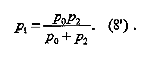
Claims (22)
- An interferometer for X-rays, in particular hard X-rays, for obtaining quantitative phase contrast images, comprising:a) an X-ray source (XS), preferably a standard polychromatic X-ray source,b) a diffractive optical element (G1) in transmission geometry, andc) a position-sensitive detector (D) with spatially modulated detection sensitivity.
- The interferometer according to claim 1, wherein the diffractive optical element is a line grating or a two-dimensionally structured grating (G1), such as a phase grating, with low X-ray absorption, but considerable X-ray phase shift, preferably with a phase shift of n or odd multiples thereof.
- The interferometer according to claim 1 or 2, wherein the phase grating is made by deep etching into silicon or a polymer.
- The interferometer according to claim 1, wherein the modulation of detector sensitivity is achieved by a one- or two-dimensional grating structure (G2) with high X-ray absorption contrast, placed immediately in front of the detector (D, D1), preferably this grating structure serves as an anti-scatter grid, or an anti-scatter grid is used as the modulation mask.
- The interferometer according to claim 1 or 4, wherein the absorption grating (G2) is made by deposition of a heavy metal into the gaps of a low-absorbing structure.
- The interferometer according to claim 1 or claim 2 wherein the period of the detector-sensitivity modulation and the period of the phase grating are matched to the radius of curvature of an incident wavefront, given by the relation

wherein p 1 is the periodicity of the incident X-rays; p2 is the periodicity of the interference pattern; d is the distance between an beam splitter grating (G1) and an analyzer grating (G2); and 1 is the distance between the X-ray source (XS) and the beam splitter grating (G1). - The interferometer according to claim 1 or 2 wherein the distance between the diffractive phase grating and the detection plane is chosen to be an odd fractional Talbot distance, given by the equation

wherein n = 1, 3, 5 ,.... - The interferometer according to claim 1 or 2 or7, wherein the phase shift of the grating and the distance between the diffractive phase grating and the detection plane are adapted for a photon energy corresponding to an emission line of the X-ray generator used as the source.
- The interferometer according to claim 1 wherein a mechanism is comprised to vary the angular orientation around the optical axis of the diffractive optical element with respect to the detector-sensitivity modulation mask, either that moiré fringes of any desired period can be observed, or that moiré fringes can be minimized or altogether suppressed.
- The interferometer according to claim 1 wherein a one- or two-dimensional phase stepping scan is implemented by lateral transverse movement of the radiation source or an aperture located in front of the radiation source, of the diffractive element, or of the detector-sensitivity modulation mask, or by a tilt of the interferometer with respect to the X-ray source.
- The interferometer according to claim 1 wherein a mechanism is comprised to place a sample (O) to be investigated between the source (XS) and the diffractive optical element (G1) or between the diffractive optical element (G1) and the detector (D, D1).
- An interferometer according to claim 1 wherein the detector (D) is position-sensitive in two dimensions, and the lateral dimensions of the diffractive optical element (G1) and the transmission mask of the detector (D) cover at a significant portion of the active area of the detector (D).
- The interferometer according to claim 1 wherein a collimator placed between the source and the diffractive optical element limits the spatial extent of the illuminating X rays to a fan beam, a line-array detector is used, and a mechanism is comprised that allows a linear scan of the sample relative to the rest of the apparatus, perpendicular to the opening angle of the fan.
- The interferometer according to claim 1 wherein the X-ray source comprises a one- or two-dimensional array of individual physical or virtual sources that may be mutually incoherent and whose lateral separation p0 is preferably given by

- The interferometer according to claim 14, wherein the array of sources is generated by an aperture mask (S) with line- or dot-shaped openings.
- The interferometer according to claim 14, wherein the array of sources is generated by electron optics that creates an array of electron line or dot foci on the anode of the X-ray source.
- The interferometer according to claim 14, wherein the array of sources is generated by electron optics that scans a single line or dot focus across the anode surface of the X-ray source.
- The interferometer according to claim 14, wherein the array of sources is generated by using an X-ray source comprising an anode which is structured either topographically or has assembled in a mosaic manner from different materials.
- The interferometer according to claim 1 or 11, comprising means for rotating the sample relatively to the remaining components in order to perform data collection for a tomography scan.
- The interferometer according to claim 1 or 10, wherein an analysis procedure is implemented for phase-stepping scan data that comprises the steps of calculating for each element of the detector the Fourier transform or at least one Fourier component of the intensity curve measured in the element, and then retaining the phase of one or more Fourier components as a signal for further processing.
- The interferometer according to claims 1 or 10, wherein an analysis procedure is implemented for phase-stepping scan data that comprises the steps of fitting for each element of the detector the intensity curve measured in the element to an intensity curve modeled or measured separately without the beam distortions under study, where at least one of the fit parameters is the shift of the curve along the position axis of the scan.
- The interferometer according to anyone of the preceding claims, wherein the beam-splitter grating is an absorption grating.
Priority Applications (8)
| Application Number | Priority Date | Filing Date | Title |
|---|---|---|---|
| EP05012121A EP1731099A1 (en) | 2005-06-06 | 2005-06-06 | Interferometer for quantitative phase contrast imaging and tomography with an incoherent polychromatic x-ray source |
| AU2006257026A AU2006257026B2 (en) | 2005-06-06 | 2006-05-30 | Interferometer for quantative phase contrast imaging and tomography with an incoherent polychromatic x-ray source |
| CN2006800294348A CN101257851B (en) | 2005-06-06 | 2006-05-30 | Interferometer for quantative phase contrast imaging and tomography with an incoherent polychromatic x-ray source |
| CA2610934A CA2610934C (en) | 2005-06-06 | 2006-05-30 | Interferometer for quantative phase contrast imaging and tomography with an incoherent polychromatic x-ray source |
| PCT/EP2006/005119 WO2006131235A1 (en) | 2005-06-06 | 2006-05-30 | Interferometer for quantative phase contrast imaging and tomography with an incoherent polychromatic x-ray source |
| JP2008515098A JP5162453B2 (en) | 2005-06-06 | 2006-05-30 | Interferometer for quantitative phase contrast imaging and tomography using an incoherent polychromatic X-ray source |
| EP06753957.7A EP1887936B1 (en) | 2005-06-06 | 2006-05-30 | Interferometer for quantative phase contrast imaging and tomography with an incoherent polychromatic x-ray source |
| US11/921,709 US7889838B2 (en) | 2005-06-06 | 2006-05-30 | Interferometer for quantitative phase contrast imaging and tomography with an incoherent polychromatic x-ray source |
Applications Claiming Priority (1)
| Application Number | Priority Date | Filing Date | Title |
|---|---|---|---|
| EP05012121A EP1731099A1 (en) | 2005-06-06 | 2005-06-06 | Interferometer for quantitative phase contrast imaging and tomography with an incoherent polychromatic x-ray source |
Publications (1)
| Publication Number | Publication Date |
|---|---|
| EP1731099A1 true EP1731099A1 (en) | 2006-12-13 |
Family
ID=35115941
Family Applications (2)
| Application Number | Title | Priority Date | Filing Date |
|---|---|---|---|
| EP05012121A Withdrawn EP1731099A1 (en) | 2005-06-06 | 2005-06-06 | Interferometer for quantitative phase contrast imaging and tomography with an incoherent polychromatic x-ray source |
| EP06753957.7A Active EP1887936B1 (en) | 2005-06-06 | 2006-05-30 | Interferometer for quantative phase contrast imaging and tomography with an incoherent polychromatic x-ray source |
Family Applications After (1)
| Application Number | Title | Priority Date | Filing Date |
|---|---|---|---|
| EP06753957.7A Active EP1887936B1 (en) | 2005-06-06 | 2006-05-30 | Interferometer for quantative phase contrast imaging and tomography with an incoherent polychromatic x-ray source |
Country Status (7)
| Country | Link |
|---|---|
| US (1) | US7889838B2 (en) |
| EP (2) | EP1731099A1 (en) |
| JP (1) | JP5162453B2 (en) |
| CN (1) | CN101257851B (en) |
| AU (1) | AU2006257026B2 (en) |
| CA (1) | CA2610934C (en) |
| WO (1) | WO2006131235A1 (en) |
Cited By (61)
| Publication number | Priority date | Publication date | Assignee | Title |
|---|---|---|---|---|
| WO2007087789A1 (en) * | 2006-02-01 | 2007-08-09 | Siemens Aktiengesellschaft | Method for producing projective and tomographic images using an x-ray system |
| EP1879020A1 (en) * | 2006-07-12 | 2008-01-16 | Paul Scherrer Institut | X-ray interferometer for phase contrast imaging |
| DE102007024156B3 (en) * | 2007-05-24 | 2008-12-11 | Siemens Ag | X-ray absorption grating |
| DE102007029730A1 (en) * | 2007-06-27 | 2009-01-08 | Siemens Ag | Measuring system and method for the non-invasive determination of properties of an examination subject and contrast agent for the X-ray phase contrast measurement |
| US7522708B2 (en) | 2006-02-01 | 2009-04-21 | Siemens Aktiengesellschaft | Focus/detector system of an X-ray apparatus for generating phase contrast recordings |
| US7522698B2 (en) | 2006-02-01 | 2009-04-21 | Siemens Aktiengesellschaft | Focus/detector system of an X-ray apparatus for generating phase contrast recordings |
| EP2060909A1 (en) | 2007-11-15 | 2009-05-20 | CSEM Centre Suisse d'Electronique et de Microtechnique SA - Recherche et Développement | Interferometer device and method |
| WO2009104560A1 (en) * | 2008-02-20 | 2009-08-27 | 国立大学法人東京大学 | X-ray imaging apparatus and x-ray source used therein |
| WO2009115966A1 (en) | 2008-03-19 | 2009-09-24 | Koninklijke Philips Electronics N.V. | Rotational x ray device for phase contrast imaging |
| FR2935066A1 (en) * | 2008-08-12 | 2010-02-19 | Chabunda Christophe Mwanza | X-ray tube i.e. stereographic X-ray tube, for producing e.g. maxillo-dental image, has crystal for producing splitted conical beam to produce meshing network on flat sensor with compactness of rays and adjustable detector-source distance |
| EP2168488A1 (en) * | 2008-09-30 | 2010-03-31 | Siemens Aktiengesellschaft | X-ray CT system for x-ray phase contrast and/or x-ray dark field imaging |
| WO2010089319A1 (en) * | 2009-02-05 | 2010-08-12 | Institute Of High Energy Physics | Low dose single step grating based x-ray phase contrast imaging |
| CN101467889B (en) * | 2007-12-26 | 2010-08-25 | 中国科学院高能物理研究所 | Grating shearing phase contrast CT image-forming data acquisition and reconstruction method |
| WO2010101920A2 (en) | 2009-03-02 | 2010-09-10 | University Of Rochester | Methods and apparatus for differential phase-contrast fan beam ct, cone-beam ct and hybrid cone-beam ct |
| WO2010119019A1 (en) | 2009-04-17 | 2010-10-21 | Siemens Aktiengesellschaft | Detector arrangement and x-ray tomography device for performing phase-contrast measurements and method for performing a phase-contrast measurement |
| DE102009018882A1 (en) | 2009-04-24 | 2010-10-28 | Siemens Aktiengesellschaft | Method for detecting elastic tissue of patient for tumor diagnosis, involves optically displaying, storing and/or utilizing tomography result-image data sets, which are generated based on positions of interference patterns |
| DE102008049200B4 (en) * | 2008-09-26 | 2010-11-11 | Paul Scherrer Institut | Method for producing X-ray optical grids, X-ray optical grating and X-ray system |
| WO2010146498A1 (en) | 2009-06-16 | 2010-12-23 | Koninklijke Philips Electronics N. V. | Tilted gratings and method for production of tilted gratings |
| WO2010146503A1 (en) * | 2009-06-16 | 2010-12-23 | Koninklijke Philips Electronics N. V. | Correction method for differential phase contrast imaging |
| WO2010150136A1 (en) | 2009-06-22 | 2010-12-29 | Koninklijke Philips Electronics N. V. | Grating-based phase contrast x-ray imaging apparatus and methods |
| EP2311092A2 (en) * | 2008-07-25 | 2011-04-20 | Cornell University | Light field image sensor, method and applications |
| WO2011070489A1 (en) * | 2009-12-10 | 2011-06-16 | Koninklijke Philips Electronics N.V. | Non- parallel grating arrangement with on-the-fly phase stepping, x-ray system and use |
| WO2011070521A1 (en) | 2009-12-10 | 2011-06-16 | Koninklijke Philips Electronics N.V. | Calibration of differential phase-contrast imaging systems |
| WO2011070493A1 (en) * | 2009-12-10 | 2011-06-16 | Koninklijke Philips Electronics N.V. | Apparatus for phase-contrast imaging comprising a displaceable x-ray detector element and method |
| WO2011070519A1 (en) | 2009-12-10 | 2011-06-16 | Koninklijke Philips Electronics N.V. | Scanning system for differential phase contrast imaging |
| WO2011093417A1 (en) * | 2010-01-28 | 2011-08-04 | Canon Kabushiki Kaisha | X-ray imaging apparatus and wavefront measuring apparatus |
| US8009796B2 (en) | 2008-09-24 | 2011-08-30 | Siemens Aktiengesellschaft | X-ray CT system to generate tomographic phase contrast or dark field exposures |
| WO2012000694A1 (en) * | 2010-06-28 | 2012-01-05 | Paul Scherrer Institut | A method for x-ray phase contrast and dark-field imaging using an arrangement of gratings in planar geometry |
| WO2012052900A1 (en) | 2010-10-19 | 2012-04-26 | Koninklijke Philips Electronics N.V. | Differential phase-contrast imaging |
| WO2012052881A1 (en) | 2010-10-19 | 2012-04-26 | Koninklijke Philips Electronics N.V. | Differential phase-contrast imaging |
| WO2012053459A1 (en) * | 2010-10-20 | 2012-04-26 | Canon Kabushiki Kaisha | Imaging apparatus using talbot interference and adjusting method for imaging apparatus |
| US8306183B2 (en) | 2007-11-26 | 2012-11-06 | Koninklijke Philips Electronics N.V. | Detection setup for X-ray phase contrast imaging |
| EP2531105A1 (en) * | 2010-02-04 | 2012-12-12 | FUJIFILM Corporation | Radiation imaging system |
| WO2013030698A1 (en) * | 2011-08-31 | 2013-03-07 | Koninklijke Philips Electronics N.V. | Differential phase contrast imaging with energy sensitive detection |
| WO2013111050A1 (en) | 2012-01-24 | 2013-08-01 | Koninklijke Philips N.V. | Multi-directional phase contrast x-ray imaging |
| JP2014502518A (en) * | 2010-12-13 | 2014-02-03 | コーニンクレッカ フィリップス エヌ ヴェ | Method and apparatus for analyzing region of interest of object using X-ray |
| WO2014027333A1 (en) | 2012-08-17 | 2014-02-20 | Koninklijke Philips N.V. | Correction in x-ray imaging systems for differential phase contrast imaging |
| WO2014027289A1 (en) | 2012-08-17 | 2014-02-20 | Koninklijke Philips N.V. | Handling misalignment in differential phase contrast imaging |
| CN104504710A (en) * | 2014-12-30 | 2015-04-08 | 中国科学技术大学 | Moore stripe recognition method and device for X-ray grating phase-contrast imaging |
| US9084528B2 (en) | 2009-12-10 | 2015-07-21 | Koninklijke Philips N.V. | Phase contrast imaging |
| US9269469B2 (en) | 2012-08-06 | 2016-02-23 | Siemens Aktiengesellschaft | Arrangement and method for inverse X-ray phase contrast imaging |
| CN105612584A (en) * | 2013-10-07 | 2016-05-25 | 西门子医疗有限公司 | Phase contrast X-ray imaging device and phase grating therefor |
| US9412554B2 (en) | 2013-07-23 | 2016-08-09 | Koninklijke Philips N.V. | Anode for an X-ray tube of a differential phase contrast imaging apparatus |
| EP3139156A1 (en) * | 2015-09-04 | 2017-03-08 | Paul Scherrer Institut | Dual phase grating interferometer for x-ray phase contrast imaging |
| WO2017087586A1 (en) * | 2015-11-19 | 2017-05-26 | Carestream Health, Inc. | Phase contrast imaging computed tomography scanner |
| EP3048441A4 (en) * | 2014-11-04 | 2017-06-21 | Tsinghua University | X-ray phase-contrast imaging system and imaging method |
| US9700267B2 (en) | 2012-12-21 | 2017-07-11 | Carestream Health, Inc. | Method and apparatus for fabrication and tuning of grating-based differential phase contrast imaging system |
| US9724063B2 (en) | 2012-12-21 | 2017-08-08 | Carestream Health, Inc. | Surrogate phantom for differential phase contrast imaging |
| DE102016206153A1 (en) * | 2016-04-13 | 2017-10-19 | Siemens Healthcare Gmbh | Method and X-ray system for generating phase-contrast and / or dark-field representations of an examination subject |
| US9795350B2 (en) | 2012-12-21 | 2017-10-24 | Carestream Health, Inc. | Material differentiation with phase contrast imaging |
| US9895117B2 (en) | 2014-10-13 | 2018-02-20 | Koninklijke Philips N.V. | Grating device for phase contrast and/or dark-field imaging of a movable object |
| US9907524B2 (en) | 2012-12-21 | 2018-03-06 | Carestream Health, Inc. | Material decomposition technique using x-ray phase contrast imaging system |
| US10058300B2 (en) | 2013-12-30 | 2018-08-28 | Carestream Health, Inc. | Large FOV phase contrast imaging based on detuned configuration including acquisition and reconstruction techniques |
| US10096098B2 (en) | 2013-12-30 | 2018-10-09 | Carestream Health, Inc. | Phase retrieval from differential phase contrast imaging |
| US10117629B2 (en) | 2014-12-03 | 2018-11-06 | Board Of Supervisors Of Louisiana State University And Agricultural And Mechanical College | High energy grating techniques |
| EP3443904A1 (en) * | 2017-08-14 | 2019-02-20 | Koninklijke Philips N.V. | Multi-spot x-ray imaging |
| EP3459461A1 (en) | 2017-09-25 | 2019-03-27 | Koninklijke Philips N.V. | X-ray imaging reference scan |
| CN110989313A (en) * | 2019-11-18 | 2020-04-10 | 中国科学技术大学 | Holographic microscopic imaging device |
| CN111166363A (en) * | 2014-05-01 | 2020-05-19 | 斯格瑞公司 | X-ray interference imaging system |
| EP4020024A1 (en) | 2020-12-22 | 2022-06-29 | Paul Scherrer Institut | Method for producing high aspect ratio fan-shaped optics |
| DE112009002606B4 (en) | 2008-10-29 | 2024-02-01 | Canon Kabushiki Kaisha | X-ray imaging apparatus and X-ray imaging method |
Families Citing this family (148)
| Publication number | Priority date | Publication date | Assignee | Title |
|---|---|---|---|---|
| DE102006037256B4 (en) * | 2006-02-01 | 2017-03-30 | Paul Scherer Institut | Focus-detector arrangement of an X-ray apparatus for producing projective or tomographic phase contrast recordings and X-ray system, X-ray C-arm system and X-ray CT system |
| CA2643894A1 (en) * | 2006-02-27 | 2007-09-07 | University Of Rochester | Phase contrast cone-beam ct imaging |
| US8009889B2 (en) | 2006-06-27 | 2011-08-30 | Affymetrix, Inc. | Feature intensity reconstruction of biological probe array |
| JP2008145111A (en) * | 2006-12-06 | 2008-06-26 | Univ Of Tokyo | X-ray imaging apparatus, x-ray source used therein and x-ray imaging method |
| JP5056842B2 (en) * | 2007-02-21 | 2012-10-24 | コニカミノルタエムジー株式会社 | Radiation image capturing apparatus and radiation image capturing system |
| US8411816B2 (en) * | 2007-02-21 | 2013-04-02 | Konica Minolta Medical & Graphic, Inc. | Radiological image capturing apparatus and radiological image capturing system |
| JP2009240378A (en) * | 2008-03-28 | 2009-10-22 | Univ Of Tokyo | X-ray imaging apparatus and method of manufacturing slit member used for the same |
| US8809758B2 (en) * | 2008-07-25 | 2014-08-19 | Cornell University | Light field image sensor with an angle-sensitive pixel (ASP) device |
| JP2010063646A (en) * | 2008-09-11 | 2010-03-25 | Fujifilm Corp | Radiation phase image radiographing apparatus |
| DE102008048683A1 (en) * | 2008-09-24 | 2010-04-08 | Siemens Aktiengesellschaft | Method for determining phase and / or amplitude between interfering adjacent X-rays in a detector pixel in a Talbot interferometer |
| CN101413905B (en) * | 2008-10-10 | 2011-03-16 | 深圳大学 | X ray differentiation interference phase contrast imaging system |
| JP4847568B2 (en) * | 2008-10-24 | 2011-12-28 | キヤノン株式会社 | X-ray imaging apparatus and X-ray imaging method |
| BRPI0923198A2 (en) * | 2008-12-01 | 2019-09-24 | Brookhaven Science Ass | systems and methods for detecting an image of an object using multi-beam imaging from an x-ray beam having a polychromatic distribution |
| DK200801722A (en) * | 2008-12-05 | 2010-06-06 | Unisensor As | Optical sectioning of a sample and detection of particles in a sample |
| DE102009004702B4 (en) | 2009-01-15 | 2019-01-31 | Paul Scherer Institut | Arrangement and method for projective and / or tomographic phase-contrast imaging with X-radiation |
| DE102009008793A1 (en) * | 2009-02-13 | 2010-11-11 | Inselspital-Stiftung | Method for generating a tomographic image |
| WO2010109909A1 (en) * | 2009-03-27 | 2010-09-30 | 株式会社リガク | X-ray generating device and examining apparatus using same |
| JP5548189B2 (en) * | 2009-03-27 | 2014-07-16 | 株式会社リガク | X-ray generator target and processing method thereof |
| JP2010236986A (en) * | 2009-03-31 | 2010-10-21 | Fujifilm Corp | Radiation phase contrast imaging apparatus |
| JP2010253194A (en) * | 2009-04-28 | 2010-11-11 | Fujifilm Corp | Radiation phase imaging apparatus |
| US9805834B2 (en) * | 2009-05-19 | 2017-10-31 | Koninklijke Philips N.V. | Grating for phase-contrast imaging |
| JP5675169B2 (en) | 2009-06-18 | 2015-02-25 | キヤノン株式会社 | X-ray imaging apparatus and X-ray imaging method |
| JPWO2011033798A1 (en) * | 2009-09-16 | 2013-02-07 | コニカミノルタエムジー株式会社 | X-ray imaging apparatus, X-ray image system, and X-ray image generation method |
| JP5459659B2 (en) * | 2009-10-09 | 2014-04-02 | キヤノン株式会社 | Phase grating used for imaging X-ray phase contrast image, imaging apparatus using the phase grating, and X-ray computed tomography system |
| JP5538936B2 (en) * | 2010-02-10 | 2014-07-02 | キヤノン株式会社 | Analysis method, program, storage medium, X-ray phase imaging apparatus |
| JP2012090944A (en) | 2010-03-30 | 2012-05-17 | Fujifilm Corp | Radiographic system and radiographic method |
| DE102010019991A1 (en) * | 2010-05-10 | 2011-11-10 | Siemens Aktiengesellschaft | computed Tomography system |
| JP5796976B2 (en) | 2010-05-27 | 2015-10-21 | キヤノン株式会社 | X-ray imaging device |
| DE102010017425A1 (en) | 2010-06-17 | 2011-12-22 | Karlsruher Institut für Technologie | Inclined phase lattice structures |
| JP5646906B2 (en) | 2010-08-06 | 2014-12-24 | キヤノン株式会社 | X-ray apparatus and X-ray measuring method |
| JP5731214B2 (en) * | 2010-08-19 | 2015-06-10 | 富士フイルム株式会社 | Radiation imaging system and image processing method thereof |
| WO2012042924A1 (en) * | 2010-09-29 | 2012-04-05 | コニカミノルタエムジー株式会社 | Method for displaying medical images and medical image display system |
| JP2012103237A (en) * | 2010-10-14 | 2012-05-31 | Canon Inc | Imaging apparatus |
| WO2012056992A1 (en) * | 2010-10-25 | 2012-05-03 | 富士フイルム株式会社 | Radiograph detection device, radiography device, radiography system |
| JP2014014380A (en) * | 2010-10-28 | 2014-01-30 | Fujifilm Corp | Radiographic apparatus and radiographic system |
| JP2012095865A (en) * | 2010-11-02 | 2012-05-24 | Fujifilm Corp | Radiographic apparatus and radiographic system |
| WO2012080125A1 (en) * | 2010-12-13 | 2012-06-21 | Paul Scherrer Institut | A method and a system for image integration using constrained optimization for phase contrast imaging with an arrangement of gratings |
| JP5697430B2 (en) | 2010-12-17 | 2015-04-08 | キヤノン株式会社 | X-ray imaging device |
| JP2012143548A (en) * | 2010-12-21 | 2012-08-02 | Fujifilm Corp | Radiation image obtaining method, and radiation image capturing apparatus |
| JP2012130586A (en) * | 2010-12-22 | 2012-07-12 | Fujifilm Corp | X-ray image detecting apparatus, radiographing apparatus, and radiographing system |
| JP2012157690A (en) * | 2011-01-14 | 2012-08-23 | Fujifilm Corp | Radiation image capturing apparatus and radiation image detecting device |
| JP5792961B2 (en) * | 2011-01-25 | 2015-10-14 | キヤノン株式会社 | Talbot interferometer and imaging method |
| JP5777360B2 (en) * | 2011-03-14 | 2015-09-09 | キヤノン株式会社 | X-ray imaging device |
| US10432871B2 (en) | 2011-04-14 | 2019-10-01 | Yale University | Systems and methods for imaging using a random laser |
| JP2014138625A (en) * | 2011-04-25 | 2014-07-31 | Fujifilm Corp | Radiographic apparatus and image processing method |
| JP5787597B2 (en) * | 2011-04-26 | 2015-09-30 | キヤノン株式会社 | Imaging device |
| US10102612B2 (en) | 2011-05-09 | 2018-10-16 | Koninklijke Philips N.V. | Rotating an object on a screen |
| US20120307970A1 (en) * | 2011-05-31 | 2012-12-06 | General Electric Company | Multispot x-ray phase-contrast imaging system |
| AU2012264596B2 (en) * | 2011-06-01 | 2015-07-30 | Total Sa | An x-ray tomography device |
| WO2012164092A1 (en) | 2011-06-01 | 2012-12-06 | Total Sa | An x-ray tomography device |
| CN102353689B (en) * | 2011-06-03 | 2013-05-08 | 中国科学院上海光学精密机械研究所 | Noncoherent X ray diffraction imaging device |
| CN103827889B (en) | 2011-07-19 | 2018-01-02 | 奥维茨奥成像系统公司 | Method and system for the cancer cell in the cell sample that detects and/or classify |
| EP2594334A1 (en) | 2011-11-21 | 2013-05-22 | Drive O2 | Sample vial for digital holographic analysis of a liquid cell sample |
| WO2013010595A1 (en) * | 2011-07-19 | 2013-01-24 | Ovizio Imaging Systems N.V. | An object database and object database improving method |
| WO2013014083A1 (en) * | 2011-07-28 | 2013-01-31 | Paul Scherrer Institut | Method for image fusion based on principal component analysis |
| US8767915B2 (en) * | 2011-07-29 | 2014-07-01 | The Johns Hopkins University | Differential phase contrast X-ray imaging system and components |
| JP5475737B2 (en) | 2011-10-04 | 2014-04-16 | 富士フイルム株式会社 | Radiation imaging apparatus and image processing method |
| US20150117599A1 (en) * | 2013-10-31 | 2015-04-30 | Sigray, Inc. | X-ray interferometric imaging system |
| JP2013116270A (en) * | 2011-12-05 | 2013-06-13 | Canon Inc | X-ray imaging apparatus |
| EP2626686A1 (en) | 2012-02-13 | 2013-08-14 | Ovizio Imaging Systems NV/SA | Flow cytometer with digital holographic microscope |
| WO2014137325A1 (en) * | 2012-03-05 | 2014-09-12 | University Of Rochester | Methods and apparatus for differential phase-contrast cone-beam ct and hybrid cone-beam ct |
| DE102012005767A1 (en) * | 2012-03-25 | 2013-09-26 | DüRR DENTAL AG | Phase contrast X-ray tomography apparatus |
| US20130259194A1 (en) * | 2012-03-30 | 2013-10-03 | Kwok L. Yip | Hybrid slot-scanning grating-based differential phase contrast imaging system for medical radiographic imaging |
| CN103364839B (en) * | 2012-04-01 | 2015-12-09 | 中国科学院高能物理研究所 | Based on rays safety detection apparatus and the method for grating shearing imaging |
| WO2013184213A2 (en) * | 2012-05-14 | 2013-12-12 | The General Hospital Corporation | A distributed, field emission-based x-ray source for phase contrast imaging |
| WO2014001975A2 (en) | 2012-06-27 | 2014-01-03 | Koninklijke Philips N.V. | Grating-based differential phase contrast imaging |
| JP6173457B2 (en) * | 2012-08-20 | 2017-08-02 | コーニンクレッカ フィリップス エヌ ヴェKoninklijke Philips N.V. | Alignment of source grating versus phase grating distance for multi-order phase adjustment in differential phase contrast imaging |
| KR101378757B1 (en) * | 2012-08-30 | 2014-03-27 | 한국원자력연구원 | Radiation imaging equipment and method available to obtain element date of material and select dimensions of image |
| EP2898310B1 (en) | 2012-09-20 | 2019-05-01 | Ovizio Imaging Systems NV/SA | Digital holographic microscope with fluid systems |
| JP6079204B2 (en) * | 2012-12-18 | 2017-02-15 | コニカミノルタ株式会社 | Medical imaging system |
| TWI552727B (en) * | 2012-12-19 | 2016-10-11 | 財團法人工業技術研究院 | Imaging system |
| US9360439B2 (en) * | 2012-12-19 | 2016-06-07 | Industrial Technology Research Institute | Imaging system |
| US8989347B2 (en) | 2012-12-19 | 2015-03-24 | General Electric Company | Image reconstruction method for differential phase contrast X-ray imaging |
| US9068927B2 (en) | 2012-12-21 | 2015-06-30 | General Electric Company | Laboratory diffraction-based phase contrast imaging technique |
| US9001967B2 (en) * | 2012-12-28 | 2015-04-07 | Carestream Health, Inc. | Spectral grating-based differential phase contrast system for medical radiographic imaging |
| US20140177789A1 (en) * | 2012-12-21 | 2014-06-26 | Pavlo Baturin | Grating-based differential phase contrast imaging system with adjustable capture technique for medical radiographic imaging |
| JP6411364B2 (en) * | 2012-12-21 | 2018-10-24 | ケアストリーム ヘルス インク | Medical grating-based differential phase contrast imaging |
| US9008278B2 (en) | 2012-12-28 | 2015-04-14 | General Electric Company | Multilayer X-ray source target with high thermal conductivity |
| US9014333B2 (en) | 2012-12-31 | 2015-04-21 | General Electric Company | Image reconstruction methods for differential phase contrast X-ray imaging |
| US9364191B2 (en) | 2013-02-11 | 2016-06-14 | University Of Rochester | Method and apparatus of spectral differential phase-contrast cone-beam CT and hybrid cone-beam CT |
| US9439613B2 (en) * | 2013-02-12 | 2016-09-13 | The Johns Hopkins University | System and method for phase-contrast X-ray imaging |
| EP2956954B1 (en) | 2013-02-13 | 2017-03-15 | Koninklijke Philips N.V. | Multiple x-ray beam tube |
| DE102013205406A1 (en) | 2013-03-27 | 2014-10-16 | Siemens Aktiengesellschaft | X-ray imaging system for X-ray imaging at high image frequencies of an examination subject by direct measurement of the interference pattern |
| US9297772B2 (en) | 2013-07-30 | 2016-03-29 | Industrial Technology Research Institute | Apparatus for amplifying intensity during transmission small angle—X-ray scattering measurements |
| US20150055745A1 (en) * | 2013-08-23 | 2015-02-26 | Carl Zeiss X-ray Microscopy, Inc. | Phase Contrast Imaging Using Patterned Illumination/Detector and Phase Mask |
| JP2015072263A (en) * | 2013-09-09 | 2015-04-16 | キヤノン株式会社 | X-ray imaging system |
| US10416099B2 (en) | 2013-09-19 | 2019-09-17 | Sigray, Inc. | Method of performing X-ray spectroscopy and X-ray absorption spectrometer system |
| US10295485B2 (en) | 2013-12-05 | 2019-05-21 | Sigray, Inc. | X-ray transmission spectrometer system |
| US10269528B2 (en) | 2013-09-19 | 2019-04-23 | Sigray, Inc. | Diverging X-ray sources using linear accumulation |
| US10297359B2 (en) | 2013-09-19 | 2019-05-21 | Sigray, Inc. | X-ray illumination system with multiple target microstructures |
| DE102013219553A1 (en) | 2013-09-27 | 2015-04-02 | Friedrich-Alexander-Universität Erlangen-Nürnberg | Examination method of an object with an X-ray system for phase contrast imaging with stochastic phase scanning |
| DE102013219554A1 (en) * | 2013-09-27 | 2015-04-02 | Friedrich-Alexander-Universität Erlangen-Nürnberg | Examination method of an object with an X-ray recording system for phase-contrast imaging with displacement measurement |
| EP2943124B1 (en) * | 2013-09-30 | 2016-08-31 | Koninklijke Philips N.V. | Differential phase contrast imaging device with movable grating(s) |
| US9719947B2 (en) * | 2013-10-31 | 2017-08-01 | Sigray, Inc. | X-ray interferometric imaging system |
| USRE48612E1 (en) | 2013-10-31 | 2021-06-29 | Sigray, Inc. | X-ray interferometric imaging system |
| WO2015066333A1 (en) * | 2013-10-31 | 2015-05-07 | Sigray, Inc. | X-ray interferometric imaging system |
| CN105637351B (en) | 2013-10-31 | 2018-11-13 | 国立大学法人东北大学 | Non-destructive testing apparatus |
| US10304580B2 (en) | 2013-10-31 | 2019-05-28 | Sigray, Inc. | Talbot X-ray microscope |
| CN104622492A (en) * | 2013-11-11 | 2015-05-20 | 中国科学技术大学 | X-ray grating phase-contrast imaging device and method |
| JP2016509872A (en) * | 2014-02-10 | 2016-04-04 | ザ ジョンズ ホプキンス ユニバーシティー | Large field grating interferometer for X-ray phase contrast imaging and CT at high energy |
| CN105992557B (en) * | 2014-02-14 | 2020-01-14 | 佳能株式会社 | X-ray Talbot interferometer and X-ray Talbot interferometer system |
| JP2015166676A (en) * | 2014-03-03 | 2015-09-24 | キヤノン株式会社 | X-ray imaging system |
| DE102014203811B4 (en) * | 2014-03-03 | 2019-07-11 | Siemens Healthcare Gmbh | Supplementary system for interferometric X-ray imaging and projective X-ray device |
| EP2942619A1 (en) * | 2014-05-07 | 2015-11-11 | Paul Scherrer Institut | Tilted-grating approach for scanning-mode X-ray grating interferometry |
| US9746405B2 (en) | 2014-05-09 | 2017-08-29 | General Electric Company | Method for elementally detecting variations in density |
| US10401309B2 (en) | 2014-05-15 | 2019-09-03 | Sigray, Inc. | X-ray techniques using structured illumination |
| EP3143384B1 (en) * | 2014-05-15 | 2020-03-04 | Sigray Inc. | X-ray system and method for measurement, characterization, and analysis of periodic structures |
| WO2015176023A1 (en) * | 2014-05-15 | 2015-11-19 | Sigray, Inc. | X-ray method for measurement, characterization, and analysis of periodic structures |
| RU2674650C2 (en) | 2014-08-05 | 2018-12-12 | Конинклейке Филипс Н.В. | Grating device for x-ray visualization device |
| DE102014221599A1 (en) | 2014-10-23 | 2016-04-28 | Siemens Aktiengesellschaft | Apparatus and method for X-ray phase-contrast imaging |
| EA028468B1 (en) * | 2014-11-27 | 2017-11-30 | Федеральное государственное автономное образовательное учреждение высшего профессионального образования "Балтийский федеральный университет имени Иммануила Канта" | Method for determining optical characteristics of materials based on abnormal refraction in refraction optical elements |
| EA030807B1 (en) * | 2014-11-27 | 2018-09-28 | Федеральное государственное автономное образовательное учреждение высшего профессионального образования "Балтийский федеральный университет имени Иммануила Канта" | Method for determination of optical characteristics from interference fringe pattern |
| JP6451400B2 (en) * | 2015-02-26 | 2019-01-16 | コニカミノルタ株式会社 | Image processing system and image processing apparatus |
| US10352880B2 (en) | 2015-04-29 | 2019-07-16 | Sigray, Inc. | Method and apparatus for x-ray microscopy |
| JP6626517B2 (en) * | 2015-06-26 | 2019-12-25 | コーニンクレッカ フィリップス エヌ ヴェKoninklijke Philips N.V. | Robust reconstruction for darkfield and phase contrast CT |
| DE102015213062A1 (en) * | 2015-07-13 | 2017-01-19 | Siemens Healthcare Gmbh | A method for generating a differential X-ray phase contrast imaging with a Talbot-Lau interferometer array and X-ray phase contrast device |
| JP6604772B2 (en) | 2015-08-05 | 2019-11-13 | キヤノン株式会社 | X-ray Talbot interferometer |
| US10295486B2 (en) | 2015-08-18 | 2019-05-21 | Sigray, Inc. | Detector for X-rays with high spatial and high spectral resolution |
| WO2017046670A1 (en) | 2015-09-16 | 2017-03-23 | Koninklijke Philips N.V. | Dark-field enhanced virtual x-ray colonoscopy |
| CN105590317B (en) * | 2015-12-15 | 2018-12-28 | 中国计量科学研究院 | A kind of image high contrast resolving power method for objectively evaluating and detection operating method |
| EP3196631A1 (en) | 2016-01-19 | 2017-07-26 | Ovizio Imaging Systems NV/SA | Digital holographic microscope with electro-fluidic system, said electro-fluidic system and methods of use |
| CN107807139B (en) * | 2016-09-05 | 2020-04-24 | 天津工业大学 | Dual-energy X-ray phase contrast imaging system without stepping device and implementation method thereof |
| US10247683B2 (en) | 2016-12-03 | 2019-04-02 | Sigray, Inc. | Material measurement techniques using multiple X-ray micro-beams |
| JP6673189B2 (en) * | 2016-12-26 | 2020-03-25 | 株式会社島津製作所 | X-ray phase contrast imaging device |
| EP3403581A1 (en) * | 2017-05-15 | 2018-11-21 | Koninklijke Philips N.V. | Grid-mounting device for slit-scan differential phase contrast imaging |
| CN107238616B (en) * | 2017-06-22 | 2019-10-11 | 合肥工业大学 | Dark-field imaging method based on neutron grating interferometer |
| EP3450967A1 (en) * | 2017-09-01 | 2019-03-06 | Shimadzu Corporation | X-ray imaging apparatus |
| JP6838531B2 (en) * | 2017-09-06 | 2021-03-03 | 株式会社島津製作所 | Radiation phase difference imaging device |
| EP3527138B1 (en) * | 2018-01-30 | 2022-06-29 | Globus Medical, Inc. | Portable medical imaging system with beam scanning |
| WO2019167324A1 (en) * | 2018-03-01 | 2019-09-06 | 株式会社島津製作所 | X-ray phase difference imaging system |
| US10578566B2 (en) | 2018-04-03 | 2020-03-03 | Sigray, Inc. | X-ray emission spectrometer system |
| WO2019236384A1 (en) | 2018-06-04 | 2019-12-12 | Sigray, Inc. | Wavelength dispersive x-ray spectrometer |
| WO2020023408A1 (en) | 2018-07-26 | 2020-01-30 | Sigray, Inc. | High brightness x-ray reflection source |
| US10656105B2 (en) | 2018-08-06 | 2020-05-19 | Sigray, Inc. | Talbot-lau x-ray source and interferometric system |
| DE112019004433T5 (en) | 2018-09-04 | 2021-05-20 | Sigray, Inc. | SYSTEM AND PROCEDURE FOR X-RAY FLUORESCENCE WITH FILTERING |
| DE112019004478T5 (en) | 2018-09-07 | 2021-07-08 | Sigray, Inc. | SYSTEM AND PROCEDURE FOR X-RAY ANALYSIS WITH SELECTABLE DEPTH |
| CN109580667B (en) * | 2018-12-05 | 2020-10-27 | 中国科学技术大学 | Single-grating phase contrast imaging method and system |
| CN111721786B (en) * | 2019-03-22 | 2023-05-26 | 中国科学院深圳先进技术研究院 | X-ray interferometer and imaging system |
| CN110133011B (en) * | 2019-05-28 | 2022-04-15 | 中国科学院苏州生物医学工程技术研究所 | Stepping-free X-ray grating phase contrast imaging method |
| JP7180562B2 (en) * | 2019-07-18 | 2022-11-30 | 株式会社島津製作所 | X-ray phase imaging device |
| DE112020004169T5 (en) | 2019-09-03 | 2022-05-25 | Sigray, Inc. | SYSTEM AND METHODS FOR COMPUTER-ASSISTED LAMINOGRAM X-RAY FLUORESCENCE IMAGING |
| US11175243B1 (en) | 2020-02-06 | 2021-11-16 | Sigray, Inc. | X-ray dark-field in-line inspection for semiconductor samples |
| US11215572B2 (en) | 2020-05-18 | 2022-01-04 | Sigray, Inc. | System and method for x-ray absorption spectroscopy using a crystal analyzer and a plurality of detector elements |
| JP7460577B2 (en) | 2020-06-03 | 2024-04-02 | 株式会社リガク | X-ray image generation device |
| WO2022061347A1 (en) | 2020-09-17 | 2022-03-24 | Sigray, Inc. | System and method using x-rays for depth-resolving metrology and analysis |
| US11686692B2 (en) | 2020-12-07 | 2023-06-27 | Sigray, Inc. | High throughput 3D x-ray imaging system using a transmission x-ray source |
| US11813102B2 (en) * | 2021-10-06 | 2023-11-14 | Houxun Miao | Interferometer for x-ray phase contrast imaging |
| US11885755B2 (en) | 2022-05-02 | 2024-01-30 | Sigray, Inc. | X-ray sequential array wavelength dispersive spectrometer |
Citations (2)
| Publication number | Priority date | Publication date | Assignee | Title |
|---|---|---|---|---|
| US5812629A (en) * | 1997-04-30 | 1998-09-22 | Clauser; John F. | Ultrahigh resolution interferometric x-ray imaging |
| WO2004071298A1 (en) * | 2003-02-14 | 2004-08-26 | Paul Scherrer Institut | Apparatus and method to obtain phase contrast x-ray images |
Family Cites Families (2)
| Publication number | Priority date | Publication date | Assignee | Title |
|---|---|---|---|---|
| JP4445397B2 (en) * | 2002-12-26 | 2010-04-07 | 敦 百生 | X-ray imaging apparatus and imaging method |
| CN1560613A (en) * | 2004-03-04 | 2005-01-05 | 中国科学院上海光学精密机械研究所 | X-ray phasecontrast phase differ amplifying imaging device |
-
2005
- 2005-06-06 EP EP05012121A patent/EP1731099A1/en not_active Withdrawn
-
2006
- 2006-05-30 AU AU2006257026A patent/AU2006257026B2/en not_active Ceased
- 2006-05-30 JP JP2008515098A patent/JP5162453B2/en active Active
- 2006-05-30 CA CA2610934A patent/CA2610934C/en not_active Expired - Fee Related
- 2006-05-30 US US11/921,709 patent/US7889838B2/en active Active
- 2006-05-30 EP EP06753957.7A patent/EP1887936B1/en active Active
- 2006-05-30 WO PCT/EP2006/005119 patent/WO2006131235A1/en active Application Filing
- 2006-05-30 CN CN2006800294348A patent/CN101257851B/en active Active
Patent Citations (2)
| Publication number | Priority date | Publication date | Assignee | Title |
|---|---|---|---|---|
| US5812629A (en) * | 1997-04-30 | 1998-09-22 | Clauser; John F. | Ultrahigh resolution interferometric x-ray imaging |
| WO2004071298A1 (en) * | 2003-02-14 | 2004-08-26 | Paul Scherrer Institut | Apparatus and method to obtain phase contrast x-ray images |
Non-Patent Citations (3)
| Title |
|---|
| MOMOSE A ET AL: "DEMONSTRATION OF X-RAY TALBOT INTERFEROMETRY", JAPANESE JOURNAL OF APPLIED PHYSICS, JAPAN SOCIETY OF APPLIED PHYSICS, TOKYO, JP, vol. 42, no. PART 2, 15 July 2003 (2003-07-15), pages 866 - 868, XP002976586, ISSN: 0021-4922 * |
| MOMOSE A ET AL: "X-ray Talbot interferometry for medical phase imaging", PORTABLE SYNCHROTRON LIGHT SOURCES AND ADVANCED APPLICATIONS. INTERNATIONAL SYMPOSIUM ON PORTABLE SYNCHROTRON LIGHT SOURCES AND ADVANCED APPLICATIONS 13-14 JAN. 2004 SHIGA, JAPAN, no. 716, 13 January 2004 (2004-01-13) - 14 January 2004 (2004-01-14), AIP Conference Proceedings AIP USA, pages 156 - 159, XP002351238, ISSN: 0094-243X * |
| WEITKAMP T ET AL: "X-ray wavefront analysis and optics characterization with a grating interferometer", APPLIED PHYSICS LETTERS AIP USA, vol. 86, no. 5, 24 January 2005 (2005-01-24), pages 54101-1 - 54101-3, XP002351239, ISSN: 0003-6951 * |
Cited By (117)
| Publication number | Priority date | Publication date | Assignee | Title |
|---|---|---|---|---|
| DE102006015358B4 (en) | 2006-02-01 | 2019-08-22 | Paul Scherer Institut | Focus / detector system of an X-ray apparatus for producing phase-contrast images, associated X-ray system and storage medium and method for producing tomographic images |
| DE102006063048B3 (en) | 2006-02-01 | 2018-03-29 | Siemens Healthcare Gmbh | Focus / detector system of an X-ray apparatus for producing phase-contrast images |
| DE102006017290B4 (en) * | 2006-02-01 | 2017-06-22 | Siemens Healthcare Gmbh | Focus / detector system of an X-ray apparatus, X-ray system and method for producing phase-contrast images |
| US7945018B2 (en) | 2006-02-01 | 2011-05-17 | Siemens Aktiengesellschaft | Method for producing projective and tomographic images using an X-ray system |
| US7522698B2 (en) | 2006-02-01 | 2009-04-21 | Siemens Aktiengesellschaft | Focus/detector system of an X-ray apparatus for generating phase contrast recordings |
| US7522708B2 (en) | 2006-02-01 | 2009-04-21 | Siemens Aktiengesellschaft | Focus/detector system of an X-ray apparatus for generating phase contrast recordings |
| WO2007087789A1 (en) * | 2006-02-01 | 2007-08-09 | Siemens Aktiengesellschaft | Method for producing projective and tomographic images using an x-ray system |
| EP1879020A1 (en) * | 2006-07-12 | 2008-01-16 | Paul Scherrer Institut | X-ray interferometer for phase contrast imaging |
| US8041004B2 (en) | 2006-07-12 | 2011-10-18 | Paul Scherrer Institut | X-ray interferometer for phase contrast imaging |
| WO2008006470A1 (en) * | 2006-07-12 | 2008-01-17 | Paul Scherrer Institut | X-ray interferometer for phase contrast imaging |
| US7787596B2 (en) | 2007-05-24 | 2010-08-31 | Siemens Aktiengesellschaft | X-ray absorption grid |
| DE102007024156B3 (en) * | 2007-05-24 | 2008-12-11 | Siemens Ag | X-ray absorption grating |
| DE102007029730A1 (en) * | 2007-06-27 | 2009-01-08 | Siemens Ag | Measuring system and method for the non-invasive determination of properties of an examination subject and contrast agent for the X-ray phase contrast measurement |
| US7653177B2 (en) | 2007-06-27 | 2010-01-26 | Siemens Aktiengesellschaft | Measurement system and method for the noninvasive determination of properties of an object to be examined and contrast medium X-ray phase-contrast measurement |
| DE102007029730B4 (en) * | 2007-06-27 | 2017-06-08 | Paul Scherer Institut | Measuring system with a phase-contrast contrast agent and its use for the non-invasive determination of properties of an examination subject |
| EP2060909A1 (en) | 2007-11-15 | 2009-05-20 | CSEM Centre Suisse d'Electronique et de Microtechnique SA - Recherche et Développement | Interferometer device and method |
| US7924973B2 (en) | 2007-11-15 | 2011-04-12 | Csem Centre Suisse D'electronique Et De Microtechnique Sa | Interferometer device and method |
| US8306183B2 (en) | 2007-11-26 | 2012-11-06 | Koninklijke Philips Electronics N.V. | Detection setup for X-ray phase contrast imaging |
| CN101467889B (en) * | 2007-12-26 | 2010-08-25 | 中国科学院高能物理研究所 | Grating shearing phase contrast CT image-forming data acquisition and reconstruction method |
| JP2009195349A (en) * | 2008-02-20 | 2009-09-03 | Univ Of Tokyo | X-ray imaging apparatus, and x-ray source used therefor |
| WO2009104560A1 (en) * | 2008-02-20 | 2009-08-27 | 国立大学法人東京大学 | X-ray imaging apparatus and x-ray source used therein |
| CN101978257B (en) * | 2008-03-19 | 2014-12-17 | 皇家飞利浦电子股份有限公司 | Rotational x ray device for phase contrast imaging |
| US8565371B2 (en) | 2008-03-19 | 2013-10-22 | Koninklijke Philips N.V. | Rotational X ray device for phase contrast imaging |
| WO2009115966A1 (en) | 2008-03-19 | 2009-09-24 | Koninklijke Philips Electronics N.V. | Rotational x ray device for phase contrast imaging |
| JP2011515143A (en) * | 2008-03-19 | 2011-05-19 | コーニンクレッカ フィリップス エレクトロニクス エヌ ヴィ | Rotating X-ray apparatus for phase contrast imaging |
| EP2311092A4 (en) * | 2008-07-25 | 2013-11-20 | Univ Cornell | Light field image sensor, method and applications |
| EP3836215A1 (en) * | 2008-07-25 | 2021-06-16 | Cornell University | Light field image sensor, method and applications |
| EP2311092A2 (en) * | 2008-07-25 | 2011-04-20 | Cornell University | Light field image sensor, method and applications |
| FR2935066A1 (en) * | 2008-08-12 | 2010-02-19 | Chabunda Christophe Mwanza | X-ray tube i.e. stereographic X-ray tube, for producing e.g. maxillo-dental image, has crystal for producing splitted conical beam to produce meshing network on flat sensor with compactness of rays and adjustable detector-source distance |
| US8009796B2 (en) | 2008-09-24 | 2011-08-30 | Siemens Aktiengesellschaft | X-ray CT system to generate tomographic phase contrast or dark field exposures |
| DE102008049200B4 (en) * | 2008-09-26 | 2010-11-11 | Paul Scherrer Institut | Method for producing X-ray optical grids, X-ray optical grating and X-ray system |
| EP2168488A1 (en) * | 2008-09-30 | 2010-03-31 | Siemens Aktiengesellschaft | X-ray CT system for x-ray phase contrast and/or x-ray dark field imaging |
| DE102009043067A1 (en) | 2008-09-30 | 2010-04-22 | Paul Scherrer Institut | X-ray CT system for X-ray phase contrast and / or X-ray dark field imaging |
| DE112009002606B4 (en) | 2008-10-29 | 2024-02-01 | Canon Kabushiki Kaisha | X-ray imaging apparatus and X-ray imaging method |
| US8972191B2 (en) | 2009-02-05 | 2015-03-03 | Paul Scherrer Institut | Low dose single step grating based X-ray phase contrast imaging |
| AU2010210169B2 (en) * | 2009-02-05 | 2015-04-09 | Paul Scherrer Institut | Low dose single step grating based X-ray phase contrast imaging |
| WO2010089319A1 (en) * | 2009-02-05 | 2010-08-12 | Institute Of High Energy Physics | Low dose single step grating based x-ray phase contrast imaging |
| EP2403411A4 (en) * | 2009-03-02 | 2013-10-02 | Univ Rochester | Methods and apparatus for differential phase-contrast fan beam ct, cone-beam ct and hybrid cone-beam ct |
| EP2403411A2 (en) * | 2009-03-02 | 2012-01-11 | University Of Rochester | Methods and apparatus for differential phase-contrast fan beam ct, cone-beam ct and hybrid cone-beam ct |
| WO2010101920A2 (en) | 2009-03-02 | 2010-09-10 | University Of Rochester | Methods and apparatus for differential phase-contrast fan beam ct, cone-beam ct and hybrid cone-beam ct |
| US8913714B2 (en) | 2009-04-17 | 2014-12-16 | Siemens Aktiengesellschaft | Detector arrangement and X-ray tomography device for performing phase-contrast measurements and method for performing a phase-contrast measurement |
| WO2010119019A1 (en) | 2009-04-17 | 2010-10-21 | Siemens Aktiengesellschaft | Detector arrangement and x-ray tomography device for performing phase-contrast measurements and method for performing a phase-contrast measurement |
| DE102009018882A1 (en) | 2009-04-24 | 2010-10-28 | Siemens Aktiengesellschaft | Method for detecting elastic tissue of patient for tumor diagnosis, involves optically displaying, storing and/or utilizing tomography result-image data sets, which are generated based on positions of interference patterns |
| US9348067B2 (en) | 2009-06-16 | 2016-05-24 | Koninklijke Philips N.V. | Tilted gratings and method for production of tilted gratings |
| WO2010146498A1 (en) | 2009-06-16 | 2010-12-23 | Koninklijke Philips Electronics N. V. | Tilted gratings and method for production of tilted gratings |
| US8855265B2 (en) | 2009-06-16 | 2014-10-07 | Koninklijke Philips N.V. | Correction method for differential phase contrast imaging |
| WO2010146503A1 (en) * | 2009-06-16 | 2010-12-23 | Koninklijke Philips Electronics N. V. | Correction method for differential phase contrast imaging |
| WO2010150136A1 (en) | 2009-06-22 | 2010-12-29 | Koninklijke Philips Electronics N. V. | Grating-based phase contrast x-ray imaging apparatus and methods |
| WO2011070489A1 (en) * | 2009-12-10 | 2011-06-16 | Koninklijke Philips Electronics N.V. | Non- parallel grating arrangement with on-the-fly phase stepping, x-ray system and use |
| US9750465B2 (en) | 2009-12-10 | 2017-09-05 | Koninklijke Philips N.V. | Scanning system for differential phase contrast imaging |
| WO2011070521A1 (en) | 2009-12-10 | 2011-06-16 | Koninklijke Philips Electronics N.V. | Calibration of differential phase-contrast imaging systems |
| US9066649B2 (en) | 2009-12-10 | 2015-06-30 | Koninklijke Philips N.V. | Apparatus for phase-contrast imaging comprising a displaceable X-ray detector element and method |
| RU2562879C2 (en) * | 2009-12-10 | 2015-09-10 | Конинклейке Филипс Электроникс Н.В. | Phase-contrast imaging device, having x-ray detector movable element, and corresponding method |
| WO2011070493A1 (en) * | 2009-12-10 | 2011-06-16 | Koninklijke Philips Electronics N.V. | Apparatus for phase-contrast imaging comprising a displaceable x-ray detector element and method |
| WO2011070519A1 (en) | 2009-12-10 | 2011-06-16 | Koninklijke Philips Electronics N.V. | Scanning system for differential phase contrast imaging |
| CN102656644B (en) * | 2009-12-10 | 2016-11-16 | 皇家飞利浦电子股份有限公司 | There is the most phase stepping non-parallel grating device, x-ray system and use |
| US8848863B2 (en) | 2009-12-10 | 2014-09-30 | Koninklijke Philips N.V. | Non-parallel grating arrangement with on-the-fly phase stepping, X-ray system |
| US9084528B2 (en) | 2009-12-10 | 2015-07-21 | Koninklijke Philips N.V. | Phase contrast imaging |
| CN102655809A (en) * | 2009-12-10 | 2012-09-05 | 皇家飞利浦电子股份有限公司 | Apparatus for phase-contrast imaging comprising a displaceable X-ray detector element and method |
| CN102656644A (en) * | 2009-12-10 | 2012-09-05 | 皇家飞利浦电子股份有限公司 | Non-parallel grating arrangement with on-the-fly phase stepping, X-ray system and use |
| WO2011093417A1 (en) * | 2010-01-28 | 2011-08-04 | Canon Kabushiki Kaisha | X-ray imaging apparatus and wavefront measuring apparatus |
| US9107637B2 (en) | 2010-01-28 | 2015-08-18 | Canon Kabushiki Kaisha | X-ray imaging apparatus and wavefront measuring apparatus |
| EP2531105A1 (en) * | 2010-02-04 | 2012-12-12 | FUJIFILM Corporation | Radiation imaging system |
| EP2531105A4 (en) * | 2010-02-04 | 2013-08-07 | Fujifilm Corp | Radiation imaging system |
| WO2012000694A1 (en) * | 2010-06-28 | 2012-01-05 | Paul Scherrer Institut | A method for x-ray phase contrast and dark-field imaging using an arrangement of gratings in planar geometry |
| US9036773B2 (en) | 2010-06-28 | 2015-05-19 | Paul Scherrer Institut | Method for X-ray phase contrast and dark-field imaging using an arrangement of gratings in planar geometry |
| US10028716B2 (en) | 2010-10-19 | 2018-07-24 | Koniklijke Philips N.V. | Differential phase-contrast imaging |
| CN103168228B (en) * | 2010-10-19 | 2015-11-25 | 皇家飞利浦电子股份有限公司 | Differential phase contrast's imaging |
| US9861330B2 (en) | 2010-10-19 | 2018-01-09 | Koninklijke Philips N.V. | Differential phase-contrast imaging |
| WO2012052881A1 (en) | 2010-10-19 | 2012-04-26 | Koninklijke Philips Electronics N.V. | Differential phase-contrast imaging |
| CN103168228A (en) * | 2010-10-19 | 2013-06-19 | 皇家飞利浦电子股份有限公司 | Differential phase-contrast imaging |
| WO2012052900A1 (en) | 2010-10-19 | 2012-04-26 | Koninklijke Philips Electronics N.V. | Differential phase-contrast imaging |
| US9006656B2 (en) | 2010-10-20 | 2015-04-14 | Canon Kabushiki Kaisha | Imaging apparatus using talbot interference and adjusting method for imaging apparatus |
| WO2012053459A1 (en) * | 2010-10-20 | 2012-04-26 | Canon Kabushiki Kaisha | Imaging apparatus using talbot interference and adjusting method for imaging apparatus |
| JP2014502518A (en) * | 2010-12-13 | 2014-02-03 | コーニンクレッカ フィリップス エヌ ヴェ | Method and apparatus for analyzing region of interest of object using X-ray |
| CN103918005B (en) * | 2011-08-31 | 2017-06-09 | 皇家飞利浦有限公司 | Differential phase with energy-sensitive detection contrasts imaging |
| RU2598310C2 (en) * | 2011-08-31 | 2016-09-20 | Конинклейке Филипс Н.В. | Formation of differential phase-contrast images with help of sensitive to energy detection |
| CN103918005A (en) * | 2011-08-31 | 2014-07-09 | 皇家飞利浦有限公司 | Differential phase contrast imaging with energy sensitive detection |
| WO2013030698A1 (en) * | 2011-08-31 | 2013-03-07 | Koninklijke Philips Electronics N.V. | Differential phase contrast imaging with energy sensitive detection |
| US9430832B2 (en) | 2011-08-31 | 2016-08-30 | Koninklijke Philips N.V. | Differential phase contrast imaging with energy sensitive detection |
| WO2013111050A1 (en) | 2012-01-24 | 2013-08-01 | Koninklijke Philips N.V. | Multi-directional phase contrast x-ray imaging |
| US9597050B2 (en) | 2012-01-24 | 2017-03-21 | Koninklijke Philips N.V. | Multi-directional phase contrast X-ray imaging |
| US9269469B2 (en) | 2012-08-06 | 2016-02-23 | Siemens Aktiengesellschaft | Arrangement and method for inverse X-ray phase contrast imaging |
| WO2014027289A1 (en) | 2012-08-17 | 2014-02-20 | Koninklijke Philips N.V. | Handling misalignment in differential phase contrast imaging |
| WO2014027333A1 (en) | 2012-08-17 | 2014-02-20 | Koninklijke Philips N.V. | Correction in x-ray imaging systems for differential phase contrast imaging |
| US9903827B2 (en) | 2012-08-17 | 2018-02-27 | Koninklijke Philips N.V. | Handling misalignment in differential phase contrast imaging |
| US9724063B2 (en) | 2012-12-21 | 2017-08-08 | Carestream Health, Inc. | Surrogate phantom for differential phase contrast imaging |
| US9795350B2 (en) | 2012-12-21 | 2017-10-24 | Carestream Health, Inc. | Material differentiation with phase contrast imaging |
| US9700267B2 (en) | 2012-12-21 | 2017-07-11 | Carestream Health, Inc. | Method and apparatus for fabrication and tuning of grating-based differential phase contrast imaging system |
| US9907524B2 (en) | 2012-12-21 | 2018-03-06 | Carestream Health, Inc. | Material decomposition technique using x-ray phase contrast imaging system |
| US10578563B2 (en) | 2012-12-21 | 2020-03-03 | Carestream Health, Inc. | Phase contrast imaging computed tomography scanner |
| US9412554B2 (en) | 2013-07-23 | 2016-08-09 | Koninklijke Philips N.V. | Anode for an X-ray tube of a differential phase contrast imaging apparatus |
| US10141081B2 (en) | 2013-10-07 | 2018-11-27 | Siemens Healthcare Gmbh | Phase contrast X-ray imaging device and phase grating therefor |
| CN105612584A (en) * | 2013-10-07 | 2016-05-25 | 西门子医疗有限公司 | Phase contrast X-ray imaging device and phase grating therefor |
| CN105612584B (en) * | 2013-10-07 | 2018-12-04 | 西门子医疗有限公司 | Phase contrast x-ray imaging equipment and its phase grating |
| US10058300B2 (en) | 2013-12-30 | 2018-08-28 | Carestream Health, Inc. | Large FOV phase contrast imaging based on detuned configuration including acquisition and reconstruction techniques |
| US10096098B2 (en) | 2013-12-30 | 2018-10-09 | Carestream Health, Inc. | Phase retrieval from differential phase contrast imaging |
| CN111166363B (en) * | 2014-05-01 | 2023-12-12 | 斯格瑞公司 | X-ray interference imaging system |
| CN111166363A (en) * | 2014-05-01 | 2020-05-19 | 斯格瑞公司 | X-ray interference imaging system |
| US9895117B2 (en) | 2014-10-13 | 2018-02-20 | Koninklijke Philips N.V. | Grating device for phase contrast and/or dark-field imaging of a movable object |
| US10267752B2 (en) | 2014-11-04 | 2019-04-23 | Tsinghua University | X-ray phase-contrast imaging system and imaging method |
| EP3048441A4 (en) * | 2014-11-04 | 2017-06-21 | Tsinghua University | X-ray phase-contrast imaging system and imaging method |
| US10117629B2 (en) | 2014-12-03 | 2018-11-06 | Board Of Supervisors Of Louisiana State University And Agricultural And Mechanical College | High energy grating techniques |
| CN104504710B (en) * | 2014-12-30 | 2017-04-12 | 中国科学技术大学 | Moore stripe recognition method and device for X-ray grating phase-contrast imaging |
| CN104504710A (en) * | 2014-12-30 | 2015-04-08 | 中国科学技术大学 | Moore stripe recognition method and device for X-ray grating phase-contrast imaging |
| EP3139156A1 (en) * | 2015-09-04 | 2017-03-08 | Paul Scherrer Institut | Dual phase grating interferometer for x-ray phase contrast imaging |
| WO2017036726A1 (en) * | 2015-09-04 | 2017-03-09 | Paul Scherrer Institut | Dual phase grating interferometer for x-ray phase contrast imaging |
| WO2017087586A1 (en) * | 2015-11-19 | 2017-05-26 | Carestream Health, Inc. | Phase contrast imaging computed tomography scanner |
| DE102016206153A1 (en) * | 2016-04-13 | 2017-10-19 | Siemens Healthcare Gmbh | Method and X-ray system for generating phase-contrast and / or dark-field representations of an examination subject |
| DE102016206153B4 (en) | 2016-04-13 | 2023-04-27 | Siemens Healthcare Gmbh | Method and X-ray system for generating phase-contrast and/or dark-field images of an examination subject |
| EP3443904A1 (en) * | 2017-08-14 | 2019-02-20 | Koninklijke Philips N.V. | Multi-spot x-ray imaging |
| US11231378B2 (en) | 2017-09-25 | 2022-01-25 | Koninklijke Philips N.V. | X-ray imaging reference scan |
| WO2019057915A1 (en) | 2017-09-25 | 2019-03-28 | Koninklijke Philips N.V. | X-ray imaging reference scan |
| EP3459461A1 (en) | 2017-09-25 | 2019-03-27 | Koninklijke Philips N.V. | X-ray imaging reference scan |
| CN110989313B (en) * | 2019-11-18 | 2021-10-01 | 中国科学技术大学 | Holographic microscopic imaging device |
| CN110989313A (en) * | 2019-11-18 | 2020-04-10 | 中国科学技术大学 | Holographic microscopic imaging device |
| EP4020024A1 (en) | 2020-12-22 | 2022-06-29 | Paul Scherrer Institut | Method for producing high aspect ratio fan-shaped optics |
Also Published As
| Publication number | Publication date |
|---|---|
| JP5162453B2 (en) | 2013-03-13 |
| CN101257851B (en) | 2011-06-15 |
| EP1887936A1 (en) | 2008-02-20 |
| US7889838B2 (en) | 2011-02-15 |
| JP2008545981A (en) | 2008-12-18 |
| CN101257851A (en) | 2008-09-03 |
| CA2610934A1 (en) | 2006-12-14 |
| US20090092227A1 (en) | 2009-04-09 |
| AU2006257026B2 (en) | 2010-03-04 |
| CA2610934C (en) | 2014-09-30 |
| WO2006131235A1 (en) | 2006-12-14 |
| AU2006257026A1 (en) | 2006-12-14 |
| EP1887936B1 (en) | 2013-12-25 |
Similar Documents
| Publication | Publication Date | Title |
|---|---|---|
| EP1887936B1 (en) | Interferometer for quantative phase contrast imaging and tomography with an incoherent polychromatic x-ray source | |
| JP6177306B2 (en) | Hybrid PCI system for medical radiography | |
| US10058300B2 (en) | Large FOV phase contrast imaging based on detuned configuration including acquisition and reconstruction techniques | |
| US7564941B2 (en) | Focus-detector arrangement for generating projective or tomographic phase contrast recordings with X-ray optical gratings | |
| US7639786B2 (en) | X-ray optical transmission grating of a focus-detector arrangement of an X-ray apparatus for generating projective or tomographic phase contrast recordings of a subject | |
| EP2510522B1 (en) | Non-parallel grating arrangement with on-the-fly phase stepping and x-ray system | |
| EP2509503B1 (en) | Apparatus for phase-contrast imaging comprising a displaceable x-ray detector element and method | |
| JP5601909B2 (en) | X-ray imaging apparatus and X-ray imaging method using the same | |
| US8972191B2 (en) | Low dose single step grating based X-ray phase contrast imaging | |
| JP5961614B2 (en) | Grating apparatus for phase contrast imaging, apparatus for phase contrast imaging, X-ray system having the apparatus, and method of using the apparatus | |
| US20100091936A1 (en) | X-ray ct system for x-ray phase contrast and/or x-ray dark field imaging | |
| WO2007125833A1 (en) | X-ray image picking-up device and x-ray image picking-up method | |
| JP5455931B2 (en) | Schlieren radiography using a linear radiation source and focusing optics | |
| US20010038680A1 (en) | X-ray phase-contrast medical micro-imaging methods | |
| US11033243B2 (en) | In-line x-ray focusing optics used for manipulation of x-rays in medical transmission radiography | |
| US10734128B2 (en) | Differential phase-contrast imaging with focussing deflection structure plates | |
| US20160256122A1 (en) | Imaging System and Imaging Method | |
| RU2171628C2 (en) | Device for small-angle mammography (modifications) | |
| JP2020038153A (en) | Radiation image generating device | |
| Viermetz et al. | Dark-Field for Human CT–Realization of a Talbot-Lau Interferometer in a Clinical CT Gantry |
Legal Events
| Date | Code | Title | Description |
|---|---|---|---|
| PUAI | Public reference made under article 153(3) epc to a published international application that has entered the european phase |
Free format text: ORIGINAL CODE: 0009012 |
|
| AK | Designated contracting states |
Kind code of ref document: A1 Designated state(s): AT BE BG CH CY CZ DE DK EE ES FI FR GB GR HU IE IS IT LI LT LU MC NL PL PT RO SE SI SK TR |
|
| AX | Request for extension of the european patent |
Extension state: AL BA HR LV MK YU |
|
| AKX | Designation fees paid | ||
| STAA | Information on the status of an ep patent application or granted ep patent |
Free format text: STATUS: THE APPLICATION IS DEEMED TO BE WITHDRAWN |
|
| 18D | Application deemed to be withdrawn |
Effective date: 20070614 |
|
| REG | Reference to a national code |
Ref country code: DE Ref legal event code: 8566 |