EP1721992B1 - Methods and nucleic acids for analyses of cellular proliferative disorders - Google Patents
Methods and nucleic acids for analyses of cellular proliferative disorders Download PDFInfo
- Publication number
- EP1721992B1 EP1721992B1 EP06007989A EP06007989A EP1721992B1 EP 1721992 B1 EP1721992 B1 EP 1721992B1 EP 06007989 A EP06007989 A EP 06007989A EP 06007989 A EP06007989 A EP 06007989A EP 1721992 B1 EP1721992 B1 EP 1721992B1
- Authority
- EP
- European Patent Office
- Prior art keywords
- seq
- methylation
- nucleic acid
- dna
- sequence
- Prior art date
- Legal status (The legal status is an assumption and is not a legal conclusion. Google has not performed a legal analysis and makes no representation as to the accuracy of the status listed.)
- Expired - Lifetime
Links
Images











Classifications
-
- C—CHEMISTRY; METALLURGY
- C12—BIOCHEMISTRY; BEER; SPIRITS; WINE; VINEGAR; MICROBIOLOGY; ENZYMOLOGY; MUTATION OR GENETIC ENGINEERING
- C12Q—MEASURING OR TESTING PROCESSES INVOLVING ENZYMES, NUCLEIC ACIDS OR MICROORGANISMS; COMPOSITIONS OR TEST PAPERS THEREFOR; PROCESSES OF PREPARING SUCH COMPOSITIONS; CONDITION-RESPONSIVE CONTROL IN MICROBIOLOGICAL OR ENZYMOLOGICAL PROCESSES
- C12Q1/00—Measuring or testing processes involving enzymes, nucleic acids or microorganisms; Compositions therefor; Processes of preparing such compositions
- C12Q1/68—Measuring or testing processes involving enzymes, nucleic acids or microorganisms; Compositions therefor; Processes of preparing such compositions involving nucleic acids
- C12Q1/6876—Nucleic acid products used in the analysis of nucleic acids, e.g. primers or probes
- C12Q1/6883—Nucleic acid products used in the analysis of nucleic acids, e.g. primers or probes for diseases caused by alterations of genetic material
- C12Q1/6886—Nucleic acid products used in the analysis of nucleic acids, e.g. primers or probes for diseases caused by alterations of genetic material for cancer
-
- C—CHEMISTRY; METALLURGY
- C12—BIOCHEMISTRY; BEER; SPIRITS; WINE; VINEGAR; MICROBIOLOGY; ENZYMOLOGY; MUTATION OR GENETIC ENGINEERING
- C12Q—MEASURING OR TESTING PROCESSES INVOLVING ENZYMES, NUCLEIC ACIDS OR MICROORGANISMS; COMPOSITIONS OR TEST PAPERS THEREFOR; PROCESSES OF PREPARING SUCH COMPOSITIONS; CONDITION-RESPONSIVE CONTROL IN MICROBIOLOGICAL OR ENZYMOLOGICAL PROCESSES
- C12Q1/00—Measuring or testing processes involving enzymes, nucleic acids or microorganisms; Compositions therefor; Processes of preparing such compositions
- C12Q1/68—Measuring or testing processes involving enzymes, nucleic acids or microorganisms; Compositions therefor; Processes of preparing such compositions involving nucleic acids
- C12Q1/6806—Preparing nucleic acids for analysis, e.g. for polymerase chain reaction [PCR] assay
-
- C—CHEMISTRY; METALLURGY
- C12—BIOCHEMISTRY; BEER; SPIRITS; WINE; VINEGAR; MICROBIOLOGY; ENZYMOLOGY; MUTATION OR GENETIC ENGINEERING
- C12Q—MEASURING OR TESTING PROCESSES INVOLVING ENZYMES, NUCLEIC ACIDS OR MICROORGANISMS; COMPOSITIONS OR TEST PAPERS THEREFOR; PROCESSES OF PREPARING SUCH COMPOSITIONS; CONDITION-RESPONSIVE CONTROL IN MICROBIOLOGICAL OR ENZYMOLOGICAL PROCESSES
- C12Q1/00—Measuring or testing processes involving enzymes, nucleic acids or microorganisms; Compositions therefor; Processes of preparing such compositions
- C12Q1/68—Measuring or testing processes involving enzymes, nucleic acids or microorganisms; Compositions therefor; Processes of preparing such compositions involving nucleic acids
- C12Q1/6813—Hybridisation assays
-
- G—PHYSICS
- G01—MEASURING; TESTING
- G01N—INVESTIGATING OR ANALYSING MATERIALS BY DETERMINING THEIR CHEMICAL OR PHYSICAL PROPERTIES
- G01N33/00—Investigating or analysing materials by specific methods not covered by groups G01N1/00 - G01N31/00
- G01N33/48—Biological material, e.g. blood, urine; Haemocytometers
- G01N33/50—Chemical analysis of biological material, e.g. blood, urine; Testing involving biospecific ligand binding methods; Immunological testing
- G01N33/53—Immunoassay; Biospecific binding assay; Materials therefor
- G01N33/575—Immunoassay; Biospecific binding assay; Materials therefor for cancer
- G01N33/57525—Immunoassay; Biospecific binding assay; Materials therefor for cancer of the liver or pancreas
-
- C—CHEMISTRY; METALLURGY
- C12—BIOCHEMISTRY; BEER; SPIRITS; WINE; VINEGAR; MICROBIOLOGY; ENZYMOLOGY; MUTATION OR GENETIC ENGINEERING
- C12Q—MEASURING OR TESTING PROCESSES INVOLVING ENZYMES, NUCLEIC ACIDS OR MICROORGANISMS; COMPOSITIONS OR TEST PAPERS THEREFOR; PROCESSES OF PREPARING SUCH COMPOSITIONS; CONDITION-RESPONSIVE CONTROL IN MICROBIOLOGICAL OR ENZYMOLOGICAL PROCESSES
- C12Q2600/00—Oligonucleotides characterized by their use
- C12Q2600/106—Pharmacogenomics, i.e. genetic variability in individual responses to drugs and drug metabolism
-
- C—CHEMISTRY; METALLURGY
- C12—BIOCHEMISTRY; BEER; SPIRITS; WINE; VINEGAR; MICROBIOLOGY; ENZYMOLOGY; MUTATION OR GENETIC ENGINEERING
- C12Q—MEASURING OR TESTING PROCESSES INVOLVING ENZYMES, NUCLEIC ACIDS OR MICROORGANISMS; COMPOSITIONS OR TEST PAPERS THEREFOR; PROCESSES OF PREPARING SUCH COMPOSITIONS; CONDITION-RESPONSIVE CONTROL IN MICROBIOLOGICAL OR ENZYMOLOGICAL PROCESSES
- C12Q2600/00—Oligonucleotides characterized by their use
- C12Q2600/112—Disease subtyping, staging or classification
-
- C—CHEMISTRY; METALLURGY
- C12—BIOCHEMISTRY; BEER; SPIRITS; WINE; VINEGAR; MICROBIOLOGY; ENZYMOLOGY; MUTATION OR GENETIC ENGINEERING
- C12Q—MEASURING OR TESTING PROCESSES INVOLVING ENZYMES, NUCLEIC ACIDS OR MICROORGANISMS; COMPOSITIONS OR TEST PAPERS THEREFOR; PROCESSES OF PREPARING SUCH COMPOSITIONS; CONDITION-RESPONSIVE CONTROL IN MICROBIOLOGICAL OR ENZYMOLOGICAL PROCESSES
- C12Q2600/00—Oligonucleotides characterized by their use
- C12Q2600/154—Methylation markers
-
- C—CHEMISTRY; METALLURGY
- C12—BIOCHEMISTRY; BEER; SPIRITS; WINE; VINEGAR; MICROBIOLOGY; ENZYMOLOGY; MUTATION OR GENETIC ENGINEERING
- C12Q—MEASURING OR TESTING PROCESSES INVOLVING ENZYMES, NUCLEIC ACIDS OR MICROORGANISMS; COMPOSITIONS OR TEST PAPERS THEREFOR; PROCESSES OF PREPARING SUCH COMPOSITIONS; CONDITION-RESPONSIVE CONTROL IN MICROBIOLOGICAL OR ENZYMOLOGICAL PROCESSES
- C12Q2600/00—Oligonucleotides characterized by their use
- C12Q2600/158—Expression markers
Definitions
- the present invention relates to genomic DNA sequences that exhibit altered expression patterns in disease states relative to normal.
- Particular embodiments provide methods, nucleic acids, nucleic acid arrays and kits useful for detecting, or for detecting and differentiating between or among cell proliferative disorders.
- the methods and nucleic acids for the detection and diagnosis of cell proliferative disorders as provided in the present invention are preferably used for the diagnosis of cancer and in particular colorectal and/or liver cancer.
- Cancer is the second leading cause of death of the United States. Mortality rates could be significantly improved if current screening methods would be improved in terms of patient compliance, sensitivity and ease of screening. Current recommended methods for diagnosis of cancer are often expensive and are not suitable for application as population wide screening tests.
- Hepatocellular cancer is the fourth most common cancer in the world, its incidence varies from 2.1 per 100,000 in North America to 80 per 100,000 in China. In the United States, it is estimated that there will be 17,550 new cases diagnosed in 2005 and 15,420 deaths due to this disease. Ultrasound of the liver, alpha fetoprotein levels and conventional CT scan are regularly obtained in the diagnostic evaluation of HCC (hepatocellular cancer or primary liver cancer), but they are often too insensitive to detect multi-focal small lesions and for treatment planing.
- Sensitivity levels for this test are also very low for colorectal adenocarcinoma with wide variability depending on the trial. Sensitivity measurements for detection of adenomas is even less since most adenomas do not bleed. In contrast, sensitivity for more invasive procedures such as sigmoidoscopy and colonoscopy are quite high because of direct visualization of the lumen of the colon. No randomized trials have evaluated the efficacy of these techniques, however, using data from case-control studies and data from the National Polyp Study (U.S.) it has been shown that removal of adenomatous polyps results in a 76-90% reduction in CRC incidence.
- Sigmoidoscopy has the limitation of only visualizing the left side of the colon leaving lesions in the right colon undetected. Both scoping procedures are expensive, require cathartic preparation and have increased risk of morbidity and mortality. Improved tests with increased sensitivity, specificity, ease of use and decreased costs are clearly needed before general widespread screening for colorectal cancer becomes routine.
- Specificity of the FOBT has been calculated at best to be 96%, with a sensitivity of 43% (adenomas) and 50% (colorectal carcinoma). Sensitivity can be improved using an immunoassay FOBT such as that produced under the tradename 'InSure TM ', with an improved sensitivity of 77 % (adenomas) and 88.9% (colorectal carcinoma.
- Molecular disease markers offer several advantages over other types of markers, one advantage being that even samples of very small sizes and/or samples whose tissue architecture has not been maintained can be analyzed quite efficiently.
- a number of genes have been shown to be differentially expressed between normal and colon carcinomas.
- no single or combination of marker has been shown to be sufficient for the diagnosis of colon carcinomas.
- High-dimensional mRNA based approaches have recently been shown to be able to provide a better means to distinguish between different tumor types and benign and malignant lesions.
- its application as a routine diagnostic tool in a clinical environment is impeded by the extreme instability of mRNA, the rapidly occurring expression changes following certain triggers (e.g.
- the current model of colorectal pathogenesis favours a stepwise progression of adenomas, which includes the development of dysplasia and finally signs of invasive cancer.
- the molecular changes underlying this adenoma-carcinoma sequence include genetic and epigenetic alterations of tumor suppressor genes (APC, p53, DCC), the activation of oncogenes (K-ras) and the inactivation of DNA mismatch repair genes. Recently, further molecular changes and genetic defects have been revealed.
- APC tumor suppressor genes
- K-ras oncogenes
- Wnt signalling pathway not only includes mutations of the APC gene, but may also result from ⁇ -catenin mutations.
- alterations in the TGF- ⁇ signalling pathway together with its signal transducers SMAD4 and SMAD2 have been linked to the development of colon cancer.
- the process of metastasis formation also includes the induction and regulation of angiogenesis (VEGF, bFGF), the induction of cell proliferation (EGF, HGF, IGF) and the activation of proteolytic enzymes (MMPs, TIMPs, uPAR), as well as the inhibition of apoptosis (Bcl-2, Bcl-X).
- VEGF angiogenesis
- bFGF the induction of cell proliferation
- MMPs proteolytic enzymes
- TIMPs TIMPs
- uPAR proteolytic enzymes
- Bcl-2, Bcl-X the inhibition of apoptosis
- CpG island methylation Apart from mutations aberrant methylation of CpG islands has been shown to lead to the transcriptional silencing of certain genes that have been previously linked to the pathogenesis of various cancers. CpG islands are short sequences which are rich in CpG dinucleotides and can usually be found in the 5' region of approximately 50% of all human genes. Methylation of the cytosines in these islands leads to the loss of gene expression and has been reported in the inactivation of the X chromosome and genomic imprinting.
- Multifactorial approach Cancer diagnostics has traditionally relied upon the detection of single molecular markers (e.g. , gene mutations, elevated PSA levels). Unfortunately, cancer is a disease state in which single markers have typically failed to detect or differentiate many forms of the disease. Thus, assays that recognize only a single marker have been shown to be of limited predictive value.
- a fundamental aspect of this invention is that methylation-based cancer diagnostics and the screening, diagnosis, and therapeutic monitoring of such diseases will provide significant improvements over the state-of-the-art that uses single marker analyses by the use of a selection of multiple markers.
- the multiplexed analytical approach is particularly well suited for cancer diagnostics since cancer is not a simple disease, this multi-factorial "panel" approach is consistent with the heterogeneous nature of cancer, both cytologically and clinically.
- the present invention describes a plurality of particularly efficient and unique panels of genes, the methylation analysis of one or a combination of the members of the panel enabling the detection of colon cell proliferative disorders with a particularly high sensitivity, specificity and/or predictive value.
- a true positive (TP) result is where the test is positive and the condition is present.
- a false positive (FP) result is where the test is positive but the condition is not present.
- a true negative (TN) result is where the test is negative and the condition is not present.
- a false negative (FN) result is where the test is negative but the condition is not present.
- Sensitivity is a measure of a test's ability to correctly detect the target disease in an individual being tested.
- a test having poor sensitivity produces a high rate of false negatives, i.e. , individuals who have the disease but are falsely identified as being free of that particular disease.
- the potential danger of a false negative is that the diseased individual will remain undiagnosed and untreated for some-period of time, during which the disease may progress to a later stage wherein treatments, if any, may be less effective.
- An example of a test that has low sensitivity is a protein-based blood test for HIV. This type of test exhibits poor sensitivity because it fails to detect the presence of the virus until the disease is well established and the virus has invaded the bloodstream in substantial numbers.
- PCR polymerase chain reaction
- Specificity is a measure of a test's ability to identify accurately patients who are free of the disease state.
- a test having poor specificity produces a high rate of false positives, i.e. , individuals who are falsely identified as having the disease.
- a drawback of false positives is that they force patients to undergo unnecessary medical procedures treatments with their attendant risks, emotional and financial stresses, and which could have adverse effects on the patient's health.
- a feature of diseases which makes it difficult to develop diagnostic tests with high specificity is that disease mechanisms, particularly in cancer, often involve a plurality of genes and proteins. Additionally, certain proteins may be elevated for reasons unrelated to a disease state.
- a test that has high specificity is a gene-based test that can detect a p53 mutation. Specificity is important when the cost or risk associated with further diagnostic procedures or further medical intervention are very high.
- the human Septin 9 gene (also known as MLL septin-like fusion protein, MLL septin-like fusion protein MSF-A, Slpa, Eseptin, Msf, septin-like protein Ovarian/Breast septin (over septin) and Septin D1) is located on chromosome 17q25 within contig AC068594.15.1.168501 and is a member of the Septin gene family.
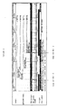
- Figure 1 provides the Ensembl annotation of the Septin 9 gene, and shows 4 transcript variants, the Septin 9 variants and the Q9HC74 variants (which are truncated versions of the Septin 9 transcripts).
- SEQ ID NO: 1 provides the sequence of said gene, comprising regions of both the Septin 9 and Q9HC74 transcripts and promoter regions.
- SEQ ID NO: 2 and SEQ ID NO: 3 are sub-regions thereof that provide the sequence of CpG rich promoter regions of Septin 9 and Q9HC74 transcripts respectively.
- Septin 9 members of the Septin gene family are associated with multiple cellular functions ranging from vesicle transport to cytokinesis. Disruption of the action of Septin 9 results in incomplete cell division, see Surka, M.C., Tsang, C.W., and Trimble, W.S. Mol Biol Cell, 13: 3532-45 (2002 ). Septin 9 and other proteins have been shown to be fusion partners of the proto-oncogene MLL suggesting a role in tumorogenesis, see Osaka, M, Rowley, J.D. and Zeleznik-Le, N.J. PNAS, 96:6428-6433 (1999 ). Burrows et al.
- MSF migration stimulating factor
- WO 200407441 it is claimed that increased copy number and over-expression of the gene is a marker of cancer, and further provides means for diagnosis and treatment thereof according to said observation.
- WO 200407441 is accordingly the closest prior art as it has the greatest number of features in common with the method and nucleic acids of the present invention, and because it relates to the same field (cancer diagnosis).
- the major difference between the present invention and that of WO 200407441 is that the present invention shows for the first time that under-expression of the gene Septin 9 is associated with cancer. More particularly this is illustrated by means of methylation analysis.
- the present invention provides a method for detecting and/or classifying colon or hepatocellular cell proliferative disorders in a subject comprising determining the expression levels of Septin 9 in a biological sample isolated from said subject wherein CpG methylation is indicative of the presence or class of said disorder.
- Various aspects of the present invention provide an efficient and unique genetic marker, whereby expression analysis of said marker enables the detection of cellular proliferative disorders with a particularly high sensitivity, specificity and/or predictive value.
- said marker enables the differentiation of neoplastic cellular proliferative disorders (including pre-cancerous conditions) from benign cellular proliferative disorders.
- the marker of the present invention is particularly suited for detection of colorectal and hepatocellular carcinomas. In the context of colorectal carcinoma the inventive testing methods have particular utility for the screening of at-risk populations.
- the inventive methods have advantages over prior art methods (including the industry standard FOBT), because of improved sensitivity, specificity and likely patient compliance.
- the methods and nucleic acids of the present invention are utilised for detecting liver cancer or distinguishing it from other liver cell proliferative disorders or for detecting colorectal carcinoma or pre-cancerous colorectal cell proliferative disorders.
- the invention provides a method for detecting and/or classifying colon or hepatocellular cell proliferative disorders in a subject comprising determining the expression levels of Septin 9 in a biological sample isolated from said subject wherein CpG methylation is indicative of the presence or class of said disorder. Said expression is determined by detecting the presence or absence of CpG methylation within said gene, wherein the presence of methylation indicates the presence of a cell proliferative disorder.
- Said method comprises the following steps: i) contacting genomic DNA isolated from a biological sample (preferably selected from the group consisting of blood plasma, blood serum, whole blood, isolated blood cells, cells isolated from the blood) obtained from the subject with at least one reagent, or series of reagents that distinguishes between methylated and non-methylated CpG dinucleotides within at least one target region of the genomic DNA, wherein the nucleotide sequence of said target region comprises at least one CpG dinucleotide sequence of the gene Septin 9; and ii) detecting and/or classifying cell proliferative disorders , at least in part.
- the target region comprises, or hybridizes under stringent conditions to a sequence of at least 16 contiguous nucleotides of at least one sequence selected from the group consisting of SEQ ID NO: 1 to SEQ ID NO: 3.
- the sensitivity of said detection is from about 75% to about 96%, or from about 80% to about 90%, or from about 80% to about 85%.
- the specificity is from about 75% to about 96%, or from about 80% to about 90%, or from about 80% to about 85%.
- the method is novel as no methods currently exist that enable the detection of cancer by analysis of body fluids, with a sensitivity and specificity high enough for use in a commercially available and regulatory body approved assay.
- current methods used to detect and diagnose colorectal carcinoma include colonoscopy, sigmoidoscopy, and fecal occult blood colon cancer.
- the disclosed invention is much less invasive than colonoscopy, and as, if not more sensitive than sigmoidoscopy and FOBT.
- the development of a body fluid assay represents a clear technical advantage over current methods known in the art in that it is anticipated that at least for colorectal carcinoma screening patient compliance for a single body fluid based test will be higher than the triplicate analysis of stool currently recommended for FOBT.
- liver cancers include PET and MRI imaging and cytology screening of aspirate or biopsy.
- Radiological screening methods do not usually detect cancers at early stages and are expensive and time consuming to carry out. Cytological screening presents risks associated with biopsy (internal bleeding) and aspiration (needle-track seeding and haemorrhage, bile peritonitis, and pneumothorax) Accordingly, detection of liver cancer at an early stage is currently not possible, furthermore as patient prognosis is greatly improved by early detection there exists a need in the art for such a screening test.
- the method comprises the use of the gene Septin 9 or its truncated transcript Q9HC74 as a marker for the detection and distinguishing of cellular proliferative disorders.
- the present invention is particularly suited for the detection of neoplastic cellular proliferative disorders (including at the pre-neoplastic stage).
- the methods and nucleic acids of the present invention enable the differentiation of malignant from benign cellular proliferative disorders.
- the methods and nucleic acids of the present invention are particularly effective in the detection of colorectal or liver neoplastic disorders and pre-neoplastic. Furthermore they have utility in differentiating between neoplastic and benign cellular proliferative colorectal and hepatocellular disorders.
- the detection, differentiation and distinguishing of colorectal or liver cell proliferative disorders is enabled by means of analysis of the methylation status of the gene Septin 9 or its truncated transcript Q9HC74, and its promoter or regulatory elements.
- the invention provides a method for the analysis of biological samples for features associated with the development of cellular proliferative disorders, the method characterised in that at least one nucleic acid, or a fragment thereof, from the group consisting of SEQ ID NO: I to SEQ ID NO: 3 is contacted with a reagent for series of reagents capable of distinguishing between methylated and non methylated CpG dinucleotides within the genomic sequence, or sequences of interest.
- the present invention provides a method for ascertaining epigenetic parameters of genomic DNA associated with the development of neoplastic cellular proliferative disorders (e.g. cancers).
- the method has utility for the improved diagnosis, treatment and monitoring of said diseases.
- the source of the test sample is selected from the group consisting of cells or cell lines, histological slides, biopsies, paraffin-embedded tissue, body fluids, ejaculate, stool, urine, blood, and combinations thereof. More preferably, the source is selected from the group consisting of stool, blood plasma, blood serum, whole blood, isolated blood cells, cells isolated from the blood obtained from the subject.
- the present invention provides a method for detecting neoplastic cellular proliferative disorders (colorectal and/or liver cell) including at the early pre-cancerous stage, and for differentiating between neoplastic and benign cellular proliferative disorders, comprising: obtaining a biological sample comprising genomic nucleic acid(s); contacting the nucleic acid(s), or a fragment thereof, with one reagent or a plurality of reagents sufficient for distinguishing between methylated and non methylated CpG dinucleotide sequences within a target sequence of the subject nucleic acid, wherein the target sequence comprises, or hybridises under stringent conditions to, a sequence comprising at least 16 contiguous nucleotides of SEQ ID NO: 1 or more preferably SEQ ID NO: 2 or SEQ ID NO: 3, said contiguous nucleotides comprising at least one CpG dinucleotide sequence; and determining, based at least in part on said distinguishing, the methylation
- distinguishing between methylated and non methylated CpG dinucleotide sequences within the target sequence comprises methylation state-dependent conversion or non-conversion of at least one such CpG dinucleotide sequence to the corresponding converted or non-converted dinucleotide sequence within a sequence selected from the group consisting ofSEQ ID Nos: 6-9; 12-15, and contiguous regions thereof corresponding to the target sequence.
- Additional embodiments provide a method for the detection of neoplastic cellular proliferative disorders (or distinguishing them from benign cellular proliferative disorders), most preferably colorectal or hepatocellular, comprising: obtaining a biological sample having subject genomic DNA; extracting the genomic DNA; treating the genomic DNA, or a fragment thereof, with one or more reagents to convert 5-position unmethylated cytosine bases to uracil or to another base that is detectably dissimilar to cytosine in terms of hybridization properties; contacting the treated genomic DNA, or the treated fragment thereof, with an amplification enzyme and at least two primers comprising, in each case a contiguous sequence at least 9 nucleotides in length that is complementary to, or hybridizes under moderately stringent or stringent conditions to a sequence selected from the group consisting SEQ ID Nos: 6-9; 12-15, and complements thereof, wherein the treated DNA or the fragment thereof is either amplified to produce an amplificate, or is not amplified; and determining
- determining comprises use of at least one method selected from the group consisting of: I) hybridizing at least one nucleic acid molecule comprising a contiguous sequence at least 9 nucleotides in length that is complementary to, or hybridizes under moderately stringent or stringent conditions to a sequence selected from the group consisting of SEQ ID NO: 4 to SEQ ID NO: 15, and complements thereof; ii) hybridizing at least one nucleic acid molecule, bound to a solid phase, comprising a contiguous sequence at least 9 nucleotides in length that is complementary to, or hybridizes under moderately stringent or stringent conditions to a sequence selected from the group consisting of SEQ ID Nos: 6-9; 12-15, and complements thereof; iii) hybridizing at least one nucleic acid molecule comprising a contiguous sequence at least 9 nucleotides in length that is complementary to, or hybridizes under moderately stringent or stringent conditions to a sequence selected from the group consisting of SEQ ID Nos:
- the digested or undigested genomic DNA is amplified prior to said determining.
- Additional embodiments provide novel genomic and chemically modified nucleic acid sequences, as well as oligonucleotides and/or PNA-oligomers for analysis of cytosine methylation patterns within sequences from the group consisting of SEQ ID NO: 1 to SEQ ID NO: 3.
- O/E Ratio refers to the frequency of CpG dinucleotides within a particular DNA sequence, and corresponds to the [number of CpG sites / (number of C bases x number of G bases)] / band length for each fragment.
- CpG island refers to a contiguous region of genomic DNA that satisfies the criteria of (1) having a frequency of CpG dinucleotides corresponding to an "Observed/Expected Ratio” >0.6, and (2) having a "GC Content” >0.5.
- CpG islands are typically, but not always, between about 0.2 to about 1 KB, or to about 2kb in length.
- methylation state refers to the presence or absence of 5-methylcytosine ("5-mCyt") at one or a plurality of CpG dinucleotides within a DNA sequence.
- Methylation states at one or more particular CpG methylation sites (each having two CpG dinucleotide sequences) within a DNA sequence include "unmethylated,” “fully-methylated” and "hemi-methylated.”
- hemi-methylation or “hemimethylation” refers to the methylation state of a double stranded DNA wherein only one strand thereof is methylated.
- the term 'AUC' as used herein is an abbreviation for the area under a curve. In particular it refers to the area under a Receiver Operating Characteristic (ROC) curve.
- the ROC curve is a plot of the true positive rate against the false positive rate for the different possible cut points of a diagnostic test. It shows the trade-off between sensitivity and specificity depending on the selected cut point (any increase in sensitivity will be accompanied by a decrease in specificity).
- the area under an ROC curve (AUC) is a measure for the accuracy of a diagnostic test (the larger the area the better, optimum is 1, a random test would have a ROC curve lying on the diagonal with an area of 0.5; for reference: J.P. Egan. Signal Detection Theory and ROC Analysis, Academic Press, New York, 1975 ).
- hypomethylation refers to the average methylation state corresponding to an increased presence of 5-mCyt at one or a plurality of CpG dinucleotides within a DNA sequence of a test DNA sample, relative to the amount of 5-mCyt found at corresponding CpG dinucleotides within a normal control DNA sample.
- hypomethylation refers to the average methylation state corresponding to a decreased presence of 5-mCyt at one or a plurality of CpG dinucleotides within a DNA sequence of a test DNA sample, relative to the amount of 5-mCyt found at corresponding CpG dinucleotides within a normal control DNA sample.
- microarray refers broadly to both "DNA microarrays," and 'DNA chip(s),' as recognized in the art, encompasses all art-recognized solid supports, and encompasses all methods for affixing nucleic acid molecules thereto or synthesis of nucleic acids thereon.
- Genetic parameters are mutations and polymorphisms of genes and sequences further require for their regulation: To be designated as mutations are, in particular, insertions, deletions, point mutations, inversions and polymorphisms and, particularly preferred, SNPs (single nucleotide polymorphisms).
- Epigenetic parameters are, in particular, cytosine methylation. Further epigenetic parameters include, for example, the acetylation of histones which, however, cannot be directly analyzed using the described method but which, in turn, correlate with the DNA methylation.
- bisulfite reagent refers to a reagent comprising bisulfite, disulfite, hydrogen sulfite or combinations thereof, useful as disclosed herein to distinguish between methylated and unmethylated CpG dinucleotide sequences.
- Methods refers to any assay for determining the methylation state of one or more CpG dinucleotide sequences within a sequence of DNA.
- MS.AP-PCR Methods of PCR (Methylation-Sensitive Arbitrarily-Primed Polymerase Chain Reaction) refers to the art-recognized technology that allows for a global scan of the genome using CG-rich primers to focus on the regions most likely to contain CpG dinucleotides, and described by Gonzalgo et al., Cancer Research 57:594-599, 1997 .
- Methods TM refers to the art-recognized fluorescence-based real-time PCR technique described by Eads et al., Cancer Res. 59:2302-2306, 1999 .
- HeavyMethyl TM assay in the embodiment thereof implemented herein, refers to an assay, wherein methylation specific blocking probes (also referred to herein as blockers ) covering CpG positions between, or covered by the amplification primers enable methylation-specific selective amplification of a nucleic acid sample.
- methylation specific blocking probes also referred to herein as blockers
- HeavyMethyl TM MethyLight TM refers to a HeavyMethyl TM MethyLight TM assay, which is a variation of the MethyLight TM assay, wherein the MethyLight TM assay is combined with methylation specific blocking probes covering CpG positions between the amplification primers.
- Ms-SNuPE Metal-sensitive Single Nucleotide Primer Extension
- MSP Metal-specific PCR
- COBRA combined Bisulfite Restriction Analysis
- MCA Metal CpG Island Amplification
- hybridization is to be understood as a bond of an oligonucleotide to a complementary sequence along the lines of the Watson-Crick base pairings in the sample DNA, forming a duplex structure.
- “Stringent hybridization conditions,” as defined herein, involve hybridizing at 68°C in 5x SSC/5x Denhardt's solution/1.0% SDS, and washing in 0.2x SSC/0.1% SDS at room temperature, or involve the art-recognized equivalent thereof (e.g., conditions in which a hybridization is carried out at 60°C in 2.5 x SSC buffer, followed by several washing steps at 37°C in a low buffer concentration, and remains stable).
- Moderately stringent conditions as defined herein, involve including washing in 3x SSC at 42°C, or the art-recognized equivalent thereof.
- the parameters of salt concentration and temperature can be varied to achieve the optimal level of identity between the probe and the target nucleic acid.
- Methods or “methylation-sensitive restriction enzymes” shall be taken to mean an enzyme that selectively digests a nucleic acid dependant on the methylation state of its recognition site.
- restriction enzymes which specifically cut if the recognition site is not methylated or hemimethylated, the cut will not take place, or with a significantly reduced efficiency, if the recognition site is methylated.
- restriction enzymes which specifically cut if the recognition site is methylated, the cut will not take place, or with a significantly reduced efficiency if the recognition site is not methylated.
- methylation-specific restriction enzymes the recognition sequence of which contains a CG dinucleotide (for instance cgcg or cccggg). Further preferred for some embodiments are restriction enzymes that do not cut if the cytosine in this dinucleotide is methylated at the carbon atom C5.
- Non-methylation-specific restriction enzymes or “non-methylation-sensitive restriction enzymes” are restriction enzymes that cut a nucleic acid sequence irrespective of the methylation state with nearly identical efficiency. They are also called “methylation-unspecific restriction enzymes.”
- SNP 9 shall be taken to include all transcript variants thereof (including for example its truncated transcript Q9HC74) and all promoter and regulatory elements thereof. Furthermore as a plurality of SNPs are known within said gene the term shall be taken to include all sequence variants thereof.
- pre-cancerous or "pre-neoplastic” and equivalents thereof shall be taken to mean any cellular proliferative disorder which is undergoing malignant transformation. Examples of such conditions include, in the context of colorectal cellular proliferative disorders, cellular proliferative disorders with a high degree of dysplasia and the following classes of adenomas:
- the present invention provides a method for detecting and/or classifying cell proliferative disorders in a subject comprising determining the expression levels of Septin 9 in a biological sample isolated from said subject wherein underexpression and/or CpG methylation is indicative of the presence or class of said disorder.
- Said markers may be used for the diagnosis of neoplastic cellular proliferative disorders (cancer), including early detection during the pre-cancerous stages of the disease, and furthermore for the differentiation of neoplastic from benign cellular proliferative disorders.
- the present invention discloses a method wherein a neoplastic cell proliferative disorder is distinguished from a benign cell proliferative disorder said method characterized in that underexpression and/or the presence of CpG methylation is indicative of the presence of a neoplastic cell proliferative disorder or pre-neoplastic disorder and the absence thereof is indicative of the presence of a benign cell proliferative disorder.
- the markers of the present invention are particularly efficient in detecting or distinguishing between liver cell proliferative disorders or alternatively for detecting or distinguishing between colorectal cell proliferative disorders, thereby providing improved means for the early detection, classification and treatment of said disorders.
- the invention presents further panels of genes comprising Septin 9 or its truncated transcript Q9HC74 with novel utility for the detection of cancers, in particular liver and/or colorectal cancer.
- sequences of said genes are as according to Table 1.
- 5-methylcytosine is the most frequent covalent base modification in the DNA of eukaryotic cells. It plays a role, for example, in the regulation of the transcription, in genetic imprinting, and in tumorigenesis. Therefore, the identification of 5-methylcytosine as a component of genetic information is of considerable interest.
- 5-methylcytosine positions cannot be identified by sequencing, because 5-methylcytosine has the same base pairing behavior as cytosine.
- the epigenetic information carried by 5-methylcytosine is completely lost during, e.g. , PCR amplification.
- the most frequently used method for analyzing DNA for the presence of 5-methylcytosine is based upon the specific reaction of bisulfite with cytosine whereby, upon subsequent alkaline hydrolysis, cytosine is converted to uracil which corresponds to thymine in its base pairing behavior.
- 5-methylcytosine remains unmodified under these conditions. Consequently, the original DNA is converted in such a manner that methylcytosine, which originally could not be distinguished from cytosine by its hybridization behavior, can now be detected as the only remaining cytosine using standard, art-recognized molecular biological techniques, for example, by amplification and hybridization, or by sequencing. All of these techniques are based on differential base pairing properties, which can now be fully exploited.
- the prior art in terms of sensitivity, is defined by a method comprising enclosing the DNA to be analyzed in an agarose matrix, thereby preventing the diffusion and renaturation of the DNA (bisulfite only reacts with single-stranded DNA), and replacing all precipitation and purification steps with fast dialysis ( Olek A, et al., A modified and improved method for bisulfite based cytosine methylation analysis, Nucleic Acids Res. 24:5064-6, 1996 ). It is thus possible to analyze individual cells for methylation status, illustrating the utility and sensitivity of the method.
- An overview of art-recognized methods for detecting 5-methylcytosine is provided by Rein, T., et al., Nucleic Acids Res., 26:2255, 1998 .
- the present invention provides for the use of the bisulfite technique, in combination with one or more methylation assays, for determination of the methylation status of CpG dinucleotide sequences within SEQ ID NO: 1.
- Genomic CpG dinucleotides can be methylated or unmethylated (alternatively known as up- and down- methylated respectively).
- the methods of the present invention are suitable for the analysis of biological samples of a heterogeneous nature e.g. a low concentration of tumor cells within a background of blood or stool. Accordingly, when analyzing the methylation status of a CpG position within such a sample the person skilled in the art may use a quantitative assay for determining the level (e.g.
- methylation status or methylation state should also be taken to mean a value reflecting the degree of methylation at a CpG position.
- the terms "hypermethylated” or “upmethylated” shall be taken to mean a methylation level above that of a specified cut-off point, wherein said cut-off may be a value representing the average or median methylation level for a given population, or is preferably an optimized cut-off level.
- the "cut-off' is also referred herein as a "threshold".
- methylated In the context of the present invention the terms "methylated”, “hypermethylated” or “upmethylated” shall be taken to include a methylation level above the cut-off be zero (0) % (or equivalents thereof) methylation for all CpG positions within and associated with (e.g. in promoter or regulatory regions) the Septin 9 gene.
- determination of the methylation status of CpG dinucleotide sequences within SEQ ID NO: 1 has utility both in the diagnosis and characterization of cellular proliferative disorders.
- the methylation status of CpG positions within SEQ ID NO: 2 and SEQ ID NO: 3 are determined, SEQ ID NO: 2 and SEQ ID NO: 3 are particularly preferred regions of SEQ ID NO: 1 ( i.e. SEQ ID NO: 1 comprises both SEQ ID NO: 2 and SEQ ID NO: 3).
- Determination of the methylation status of CpG dinucleotide sequences within SEQ ID NO: 2 and SEQ ID NO: 3 also have utility in the diagnosis and characterization of cellular proliferative disorders.
- Methylation Assay Procedures Various methylation assay procedures are known in the art, and can be used in conjunction with the present invention. These assays allow for determination of the methylation state of one or a plurality of CpG dinucleotides (e.g. , CpG islands) within a DNA sequence. Such assays involve, among other techniques, DNA sequencing of bisulfite-treated DNA, PCR (for sequence-specific amplification), Southern blot analysis, and use of methylation-sensitive restriction enzymes.
- genomic sequencing has been simplified for analysis of DNA methylation patterns and 5-methylcytosine distribution by using bisulfite treatment ( Frommer et al., Proc. Natl. Acad. Sci. USA 89:1827-1831, 1992 ).
- restriction enzyme digestion of PCR products amplified from bisulfite-converted DNA is used, e.g. , the method described by Sadri & Hornsby (Nucl. Acids Res. 24:5058-5059, 1996 ), or COBRA (Combined Bisulfite Restriction Analysis) ( Xiong & Laird, Nucleic Acids Res. 25:2532-2534, 1997 ).
- COBRA TM analysis is a quantitative methylation assay useful for determining DNA methylation levels at specific gene loci in small amounts of genomic DNA ( Xiong & Laird, Nucleic Acids Res. 25:2532-2534, 1997 ). Briefly, restriction enzyme digestion is used to reveal methylation-dependent sequence differences in PCR products of sodium bisulfite-treated DNA. Methylation-dependent sequence differences are first introduced into the genomic DNA by standard bisulfite treatment according to the procedure described by Frommer et al. (Proc. Natl. Acad. Sci. USA 89:1827-1831, 1992 ).
- PCR amplification of the bisulfite converted DNA is then performed using primers specific for the CpG islands of interest, followed by restriction endonuclease digestion, gel electrophoresis, and detection using specific, labeled hybridization probes.
- Methylation levels in the original DNA ,sample are represented by the relative amounts of digested and undigested PCR product in a linearly quantitative fashion across a wide spectrum of DNA methylation levels.
- this technique can be reliably applied to DNA obtained from microdissected paraffin-embedded tissue samples.
- Typical reagents for COBRA TM analysis may include, but are not limited to: PCR primers for specific gene (or bisulfite treated DNA sequence or CpG island); restriction enzyme and appropriate buffer; gene-hybridization oligonucleotide; control hybridization oligonucleotide; kinase labeling kit for oligonucleotide probe; and labeled nucleotides.
- bisulfite conversion reagents may include: DNA denaturation buffer; sulfonation buffer; DNA recovery reagents or kits (e.g. , precipitation, ultrafiltration, affinity column); desulfonation buffer; and DNA recovery components.
- assays such as "MethyLight TM” (a fluorescence-based real-time PCR technique) ( Eads et al., Cancer Res. 59:2302-2306, 1999 ), Ms-SNuPE TM (Methylation-sensitive Single Nucleotide Primer Extension) reactions ( Gonzalgo & Jones, Nucleic Acids Res. 25:2529-2531, 1997 ), methylation-specific PCR ("MSP”; Herman et al., Proc. Natl. Acad. Sci. USA 93:9821-9826, 1996 ; US Patent No. 5,786,146 ), and methylated CpG island amplification ("MCA”; Toyota et al., Cancer Res. 59:2307-12, 1999 ) are used alone or in combination with other of these methods.
- MSP methylation-specific PCR
- MCA methylated CpG island amplification
- the "HeavyMethyl TM " assay, technique is a quantitative method for assessing methylation differences based on methylation specific amplification of bisulfite treated DNA.
- Methylation specific blocking probes also referred to herein as blockers ) covering CpG positions between, or covered by the amplification primers enable methylation-specific selective amplification of a nucleic acid sample.
- HeavyMethyl TM MethyLight TM refers to a HeavyMethyl TM MethyLight TM assay, which is a variation of the MethyLight TM assay, wherein the MethyLight TM assay is combined with methylation specific blocking probes covering CpG positions between the amplification primers.
- the HeavyMethyl TM assay may also be used in combination with methylation specific amplification primers.
- Typical reagents for HeavyMethyl TM analysis may include, but are not limited to: PCR primers for specific genes (or bisulfite treated DNA sequence or CpG island); blocking oligonucleotides; optimized PCR buffers and deoxynucleotides; and Taq polymerase.
- MSP methylation-specific PCR
- MSP allows for assessing the methylation status of virtually any group of CpG sites within a CpG island, independent of the use of methylation-sensitive restriction enzymes ( Herman et al. Proc. Natl. Acad. Sci. USA 93:9821-9826, 1996 ; US Patent No. 5,786,146 ). Briefly, DNA is modified by sodium bisulfite converting all unmethylated, but not methylated cytosines to uracil, and subsequently amplified with primers specific for methylated versus unmethylated DNA.
- MSP requires only small quantities of DNA, is sensitive to 0.1% methylated alleles of a given CpG island locus, and can be performed on DNA extracted from paraffin-embedded samples.
- Typical reagents e.g. , as might be found in a typical MSP-based kit
- MSP analysis may include, but are not limited to: methylated and unmethylated PCR primers for specific gene (or bisulfite treated DNA sequence or CpG island), optimized PCR buffers and deoxynucleotides, and specific probes.
- MethyLight TM is a high-throughput quantitative methylation assay that utilizes fluorescence-based real-time PCR (TaqMan TM ) technology that requires no further manipulations after the PCR step ( Eads et al., Cancer Res. 59:2302-2306, 1999 ).
- TaqMan TM fluorescence-based real-time PCR
- the MethyLight TM process begins with a mixed sample of genomic DNA that is converted, in a sodium bisulfite reaction, to a mixed pool of methylation-dependent sequence differences according to standard procedures (the bisulfite process converts unmethylated cytosine residues to uracil).
- Fluorescence-based PCR is then performed in a "biased" (with PCR primers that overlap known CpG dinucleotides) reaction. Sequence discrimination can occur both at the level of the amplification process and at the level of the fluorescence detection process.
- the MethyLight TM assay may be used as a quantitative test for methylation patterns in the genomic DNA sample, wherein sequence discrimination occurs at the level of probe hybridization.
- the PCR reaction provides for a methylation specific amplification in the presence of a fluorescent probe that overlaps a particular putative methylation site.
- An unbiased control for the amount of input DNA is provided by a reaction in which neither the primers, nor the probe overlie any CpG dinucleotides.
- a qualitative test for genomic methylation is achieved by probing of the biased PCR pool with either control oligonucleotides that do not "cover" known methylation sites (a fluorescence-based version of the HeavyMethyl TM and MSP techniques), or with oligonucleotides covering potential methylation sites.
- the MethyLight TM process can by used with any suitable probes e.g. "TaqMan ® " , Lightcycler ® etc....
- double-stranded genomic DNA is treated with sodium bisulfite and subjected to one of two sets of PCR reactions using TaqMan ® probes; e.g. , with MSP primers and/ or HeavyMethyl blocker oligonucleotides and TaqMan ® probe.
- the TaqMan ® probe is dual-labeled with fluorescent "reporter” and "quencher” molecules, and is designed to be specific for a relatively high GC content region so that it melts out at about 10°C higher temperature in the PCR cycle than the forward or reverse primers.
- TaqMan ® probe This allows the TaqMan ® probe to remain fully hybridized during the PCR annealing/extension step. As the Taq polymerase enzymatically synthesizes a new strand during PCR, it will eventually reach the annealed TaqMan ® probe. The Taq polymerase 5' to 3' endonuclease activity will then displace the TaqMan ® probe by digesting it to release the fluorescent reporter molecule for quantitative detection of its now unquenched signal using a real-time fluorescent detection system.
- Typical reagents for MethyLight TM analysis may include, but are not limited to: PCR primers for specific gene (or bisulfite treated DNA sequence or CpG island); TaqMan ® or Lightcycler ® probes; optimized PCR buffers and deoxynucleotides; and Taq polymerase.
- the QM TM (quantitative methylation) assay is an alternative quantitative test for methylation patterns in genomic DNA samples, wherein sequence discrimination occurs at the level of probe hybridization.
- the PCR reaction provides for unbiased amplification in the presence of a fluorescent probe that overlaps a particular putative methylation site.
- An unbiased control for the amount of input DNA is provided by a reaction in which neither the primers, nor the probe overlie any CpG dinucleotides.
- a qualitative test for genomic methylation is achieved by probing of the biased PCR pool with either control oligonucleotides that do not "cover" known methylation sites (a fluorescence-based version of the HeavyMethyl TM and MSP techniques), or with oligonucleotides covering potential methylation sites.
- the QM TM process can by used with any suitable probes e.g. "TaqMan ® " , Lightcycler ® etc... in the amplification process.
- double-stranded genomic DNA is treated with sodium bisulfite and subjected to unbiased primers and the TaqMan ® probe.
- the TaqMan ® probe is dual-labeled with fluorescent "reporter” and “quencher” molecules, and is designed to be specific for a relatively high GC content region so that it melts out at about 10°C higher temperature in the PCR cycle than the forward or reverse primers. This allows the TaqMan ® probe to remain fully hybridized during the PCR annealing/extension step.
- Taq polymerase As the Taq polymerase enzymatically synthesizes a new strand during PCR, it will eventually reach the annealed TaqMan ® probe. The Taq polymerase 5' to 3' endonuclease activity will then displace the TaqMan ® probe by digesting it to release the fluorescent reporter molecule for quantitative detection of its now unquenched signal using a real-time fluorescent detection system.
- Typical reagents for QM TM analysis may include, but are not limited to: PCR primers for specific gene (or bisulfite treated DNA sequence or CpG island); TaqMan ® or Lightcycler ® probes; optimized PCR buffers and deoxynucleotides; and Taq polymerase.
- Ms-SNuPE The Ms-SNuPE TM technique is a quantitative method for assessing methylation differences at specific CpG sites based on bisulfite treatment of DNA, followed by single-nucleotide primer extension ( Gonzalgo & Jones, Nucleic Acids Res. 25:2529-2531, 1997 ). Briefly, genomic DNA is reacted with sodium bisulfite to convert unmethylated cytosine to uracil while leaving 5-methylcytosine unchanged. Amplification of the desired target sequence is then performed using PCR primers specific for bisulfite-converted DNA, and the resulting product is isolated and used as a template for methylation analysis at the CpG site(s) of interest. Small amounts of DNA can be analyzed ( e.g. , microdissected pathology sections), and it avoids utilization of restriction enzymes for determining the methylation status at CpG sites.
- Typical reagents for Ms-SNuPE TM analysis may include, but are not limited to: PCR primers for specific gene (or bisulfite treated DNA sequence or CpG island); optimized PCR buffers and deoxynucleotides; gel extraction kit; positive control primers; Ms-SNuPE TM primers for specific gene; reaction buffer (for the Ms-SNuPE reaction); and labelled nucleotides.
- bisulfite conversion reagents may include: DNA denaturation buffer; sulfonation buffer; DNA recovery regents or kit ( e.g. , precipitation, ultrafiltration, affinity column); desulfonation buffer; and DNA recovery components.
- Genomic Sequence According to SEQ ID NO: 1 to SEQ ID NO: 3, and Non-naturally Occurring Treated Variants Thereof According to SEQ ID NO: 4 TO SEQ ID NO: 15, were Determined to have Novel Utility for the Early Detection, Classification and/or Treatment of Colorectal and/or Liver Cell Proliferative Disorders
- the invention of the method comprises the following steps: i) contacting genomic DNA (preferably isolated from body fluids) obtained from the subject with at least one reagent, or series of reagents that distinguishes between methylated and non-methylated CpG dinucleotides within the gene Septin 9 (including its promoter and regulatory regions); and ii) detecting, or detecting and distinguishing between or among colon or liver cell proliferative disorders afforded with a sensitivity of greater than or equal to 80% and a specificity of greater than or equal to 80%.
- the sensitivity is from about 75% to about 96%, or from about 80% to about 90%, or from about 80% to about 85%.
- the specificity is from about 75% to about 96%, or from about 80% to about 90%, or from about 80% to about 85%.
- Genomic DNA may be isolated by any means standard in the art, including the use of commercially available kits. Briefly, wherein the DNA of interest is encapsulated in by a cellular membrane the biological sample must be disrupted and lysed by enzymatic, chemical or mechanical means. The DNA solution may then be cleared of proteins and other contaminants, e.g. , by digestion with proteinase K. The genomic DNA is then recovered from the solution. This may be carried out by means of a variety of methods including salting out, organic extraction or binding of the DNA to a solid phase support. The choice of method will be affected by several factors including time, expense and required quantity of DNA.
- All clinical sample types comprising neoplastic matter or pre-neoplastic matter are suitable for us e in the present method, preferred are cell lines, histological slides, biopsies, paraffin-embedded tissue, body fluids, stool, colonic effluent, urine, blood plasma, blood serum, whole blood, isolated blood cells, cells isolated from the blood and combinations thereof.
- Body fluids are the preferred source of the DNA; particularly preferred are blood plasma, blood serum, whole blood, isolated blood cells and cells isolated from the blood.
- the genomic DNA sample is then treated with at least one reagent, or series of reagents that distinguishes between methylated and non-methylated CpG dinucleotides within at least one target region of the genomic DNA, wherein the target region comprises, or hybridizes under stringent conditions to a sequence of at least 16 contiguous nucleotides of at least one sequence selected from the group consisting of (SEQ ID NO: 1 to SEQ ID NO: 3 respectively, wherein said contiguous nucleotides comprise at least one CpG dinucleotide sequence.
- said reagent converts cytosine bases which are unmethylated at the 5'-position to uracil, thymine, or another base which is dissimilar to cytosine in terms of hybridization behavior.
- said reagent may be a methylation sensitive restriction enzyme.
- the genomic DNA sample is treated in such a manner that cytosine bases which are unmethylated at the 5'-position are converted to uracil, thymine, or another base which is dissimilar to cytosine in terms of hybridization behavior It is preferred that this treatment is carried out with bisulfite (hydrogen sulfite, disulfite) and subsequent alkaline hydrolysis.
- bisulfite hydrogen sulfite, disulfite
- Such a treatment results in the conversion of SEQ ID NO: 1 to 3 to SEQ ID NO: 4 to SEQ ID NO: 9 (respectively) wherein said CpG dinucleotides are methylated or SEQ ID NO: 10 to SEQ ID NO: 15 wherein said CpG dinucleotides are unmethylated.
- the treated DNA is then analyzed in order to determine the methylation state of the target gene sequences (Septin 9 prior to the treatment). It is particularly preferred that the target region comprises, or hybridizes under stringent conditions to at least 16 contiguous nucleotides of Septin 9 or its truncated transcript Q9HC74. It is preferred that the sequence of said gene according to SEQ ID NO: 1 is analyzed, it is particularly preferred that the sub-regions thereof according to SEQ ID NO: 2 or SEQ ID NO: 3 are analyzed. The method of analysis may be selected from those known in the art, including those listed herein.
- MethyLight TM is particularly preferred, MSP and the use of blocking oligonucleotides (HeavyMethyl TM ) as described herein. It is further preferred that any oligonucleotides used in such analysis (including primers, blocking oligonucleotides and detection probes) should be reverse complementary, identical, or hybridize under stringent or highly stringent conditions to an at least 16-base-pair long segment of the base sequences of one or more of SEQ ID Nos: 6-9; 12-15 and sequences complementary thereto.
- methylation Aberrant methylation, more specifically hypermethylation of Septin 9 (including its truncated transcript Q9HC74, as well as promoter and/or regulatory regions) is associated with the presence of neoplastic cellular proliferative disorders, and is particularly prevalent in colorectal and hepatocellular carcinomas. Accordingly wherein a biological sample presents within any degree of methylation, said sample should be determined as neoplastic.
- Sensitivity is calculated as: ⁇ detected neoplasia/all neoplasia) e.g. : ⁇ detected colon neoplasia/all colon neoplasia); and specificity is calculated as (non-detected negatives/total negatives)
- the sensitivity is from about 75% to about 96%, or from about 80% to about 90%, or from about 80% to about 85%.
- the specificity is from about 75% to about 96%, or from about 80% to about 90%, or from about 80% to about 85%.
- Colon neoplasia is herein defined as all colon malignancies and adenomas greater than 1 cm., or subsets thereof. Negatives can be defined as healthy individuals.
- the method discloses the use of Septin 9 or its truncated transcript Q9HC74 (or promoter and/or regulatory regions thereof) as a marker for the differentiation, detection and distinguishing of cellular proliferative disorders (neoplastic, colon or liver disorders).
- Particular embodiments of the present invention provide a novel application of the analysis of methylation levels and/or patterns within said sequences that enables a precise detection, characterisation and/or treatment of liver and/or colorectal cell proliferative disorders.
- Early detection of cancer is directly linked with disease prognosis, and the disclosed method thereby enables the physician and patient to make better and more informed treatment decisions.
- the present invention provides novel uses for the genomic sequence SEQ ID NO: 1, AND MORE PREFERABLY SEQ ID NO: 2 AND SEQ ID NO: 3. Additional embodiments provide modified variants of SEQ ID NO: 1 TO SEQ ID NO: 3, as well as oligonucleotides and/or PNA-oligomers for analysis of cytosine methylation patterns within SEQ ID NO: 1 TO SEQ ID NO: 3.
- An objective of the invention comprises analysis of the methylation state of one or more CpG dinucleotides within SEQ ID NO: 1 and sequences complementary thereto, and more preferably SEQ ID NO: 2 or SEQ ID NO: 3 and sequences complementary thereto.
- the disclosed invention provides treated nucleic acids, derived from genomic SEQ ID NO: 1 to SEQ ID NO: 3, wherein the treatment is suitable to convert at least one unmethylated cytosine base of the genomic DNA sequence to uracil or another base that is detectably dissimilar to cytosine in terms of hybridization
- the genomic sequences in question may comprise one, or more consecutive methylated CpG positions.
- Said treatment preferably comprises use of a reagent selected from the group consisting of bisulfite, hydrogen sulfite, disulfite, and combinations thereof.
- the invention provides a non-naturally occurring modified nucleic acid comprising a sequence of at least 16 contiguous nucleotide bases in length of a sequence selected from the group consisting of SEQ ID Nos; 6; 7; 12; 13.
- said nucleic acid is at least 50, 100, 150, 200, 250 or 500 base pairs in length of a segment of the nucleic acid sequence disclosed in SEQ ID Nos; 6; 7; 12; 13.
- Particularly preferred is a nucleic acid molecule that is identical or complementary to all or a portion of the sequences SEQ ID Nos; 6; 7; 12; 13 but not SEQ ID NO: 1 to SEQ ID NO: 3 or other naturally occurring DNA.
- sequence comprises at least one CpG, TpA or CpA dinucleotide and sequences complementary thereto.
- the sequences of SEQ ID NO: 4 TO SEQ ID NO: 15 provide non-naturally occurring modified versions of the nucleic acid according to SEQ ID NO: 1 TO SEQ ID NO: 3, wherein the modification of each genomic sequence results in the synthesis of a nucleic acid having a sequence that is unique and distinct from said genomic sequence as follows. For each sense strand genomic DNA, e.g. , SEQ ID NO: 1, four converted versions are disclosed. A first version wherein "C” is converted to "T,” but “CpG” remains “CpG” ( i.e.
- a third chemically converted version of each genomic sequences is provided, wherein "C” is converted to "T” for all “C” residues, including those of "CpG” dinucleotide sequences ( i.e. , corresponds to case where, for the genomic sequences, all "C” residues of CpG dinucleotide sequences are un methylated); a final chemically converted version of each sequence, discloses the complement of the disclosed genomic DNA sequence (i.e. anti sense strand), wherein "C” is converted to "T” for all “C” residues, including those of "CpG” dinucleotide sequences ( i.e.
- SEQ ID NO: 1 to SEQ ID NO: 3 correspond to SEQ ID NO: 10 to SEQ ID NO: 15.
- nucleic acid sequences and molecules according SEQ ID NO: 4 to SEQ ID NO: 15 were not implicated in or connected with the detection, classification or treatment of cellular proliferative disorders.
- the present invention includes nucleic acid molecules (e.g. , oligonucleotides and peptide nucleic acid (PNA) molecules (PNA-oligomers)) that hybridize under moderately stringent and/or stringent hybridization conditions to all or a portion of the sequences SEQ ID NO: 1 to SEQ ID NO: 15 or to the complements thereof.
- nucleic acid molecules e.g. , oligonucleotides and peptide nucleic acid (PNA) molecules (PNA-oligomers)
- PNA-oligomers e.g., oligonucleotides and peptide nucleic acid (PNA) molecules (PNA-oligomers)
- PNA-oligomers peptide nucleic acid
- the identical or hybridizing portion of the hybridizing nucleic acids is typically at least 9, 16, 20, 25, 30 or 35 nucleotides in length. However, longer molecules have inventive utility, and are thus within the scope of the present invention.
- the hybridizing portion of the inventive hybridizing nucleic acids is at least 95%, or at least 98%, or 100% identical to the sequence, or to a portion thereof of SEQ ID NO: 1 to SEQ ID NO: 15, or to the complements thereof.
- Hybridizing nucleic acids of the type described herein can be used, for example, as a primer (e.g. , a PCR primer), or a diagnostic and/or prognostic probe or primer.
- a primer e.g. , a PCR primer
- hybridization of the oligonucleotide probe to a nucleic acid sample is performed under stringent conditions and the probe is 100% identical to the target sequence.
- Nucleic acid duplex or hybrid stability is expressed as the melting temperature or Tm, which is the temperature at which a probe dissociates from a target DNA. This melting temperature is used to define the required stringency conditions.
- target sequences that are related and substantially identical to the corresponding sequence of SEQ ID NO: 1 to SEQ ID NO: 3 (such as allelic variants and SNPs), rather than identical, it is useful to first establish the lowest temperature at which only homologous hybridization occurs with a particular concentration of salt (e.g. , SSC or SSPE). Then, assuming that 1% mismatching results in a 1 °C decrease in the Tm, the temperature of the final wash in the hybridization reaction is reduced accordingly (for example, if sequences having > 95% identity with the probe are sought, the final wash temperature is decreased by 5°C). In practice, the change in Tm can be between 0.5°C and 1.5°C per 1% mismatch.
- a particular concentration of salt e.g. , SSC or SSPE
- inventive oligonucleotides of length X include those corresponding to sets (sense and antisense sets) of consecutively overlapping oligonucleotides of length X, where the oligonucleotides within each consecutively overlapping set (corresponding to a given X value) are defined as the finite set of Z oligonucleotides from nucleotide positions: n to n + X + 1 ;
- the set is limited to those oligomers that comprise at least one CpG, TpG or CpA dinucleotide.
- inventive 20-mer oligonucleotides include the following set of 219890 oligomers (and the antisense set complementary thereto), indicated by polynucleotide positions with reference to SEQ ID NO: 1:
- the set is limited to those oligomers that comprise at least one CpG, TpG or CpA dinucleotide.
- examples of inventive 25-mer oligonucleotides include the following set of 219885 oligomers (and the antisense set complementary thereto), indicated by polynucleotide positions with reference to SEQ ID NO: 1:
- the set is limited to those oligomers that comprise at least one CpG, TpG or CpA dinucleotide.
- oligonucleotides or oligomers according to the present invention constitute effective tools useful to ascertain genetic and epigenetic parameters of the genomic sequence corresponding to SEQ ID NO: 1.
- said oligomers comprise at least one CpG, TpG or CpA dinucleotide.
- the oligonucleotides of the invention can also be modified by chemically linking the oligonucleotide to one or more moieties or conjugates to enhance the activity, stability or detection of the oligonucleotide.
- moieties or conjugates include chromophores, fluorophors, lipids such as cholesterol, cholic acid, thioether, aliphatic chains, phospholipids, polyamines, polyethylene glycol (PEG), palmityl moieties, and others as disclosed in, for example, United States Patent Numbers 5,514,758 , 5,565,552 , 5,567,810 , 5,574,142 , 5,585,481 , 5,587,371 , 5,597,696 and 5,958,773 .
- the probes may also exist in the form of a PNA (peptide nucleic acid) which has particularly preferred pairing properties.
- the oligonucleotide may include other appended groups such as peptides, and may include hybridization-triggered cleavage agents ( Krol et al., BioTechniques 6:958-976, 1988 ) or intercalating agents ( Zon, Pharm. Res. 5:539-549, 1988 ).
- the oligonucleotide may be conjugated to another molecule, e.g. , a chromophore, fluorophor, peptide, hybridization-triggered cross-linking agent, transport agent, hybridization-triggered cleavage agent, etc.
- the oligonucleotide may also comprise at least one art-recognized modified sugar and/or base moiety, or may comprise a modified backbone or non-natural internucleoside linkage.
- oligonucleotides or oligomers according to particular embodiments of the present invention are typically used in 'sets,' which contain at least one oligomer for analysis of each of the CpG dinucleotides of a genomic sequence selected from the group consisting SEQ ID NO: 1 to SEQ ID NO: 3 and sequences complementary thereto, or to the corresponding CpG, TpG or CpA dinucleotide within a sequence of the treated nucleic acids according to SEQ ID NO: 4 to SEQ ID NO: 15 and sequences complementary thereto.
- SEQ ID NO: 4 to SEQ ID NO: 15 and sequences complementary thereto.
- the set of oligomers may also be used for detecting single nucleotide polymorphisms (SNPs) in treated genomic DNA (SEQ ID NO: 4 to SEQ ID NO: 15), or in genomic DNA (SEQ ID NO: 1 to SEQ ID NO: 3 and sequences complementary thereto).
- SNPs single nucleotide polymorphisms
- At least one, and more preferably all members of a set of oligonucleotides is bound to a solid phase.
- the present invention provides a set of at least two (2) oligonucleotides that are used as 'primer' oligonucleotides for amplifying DNA sequences of one of SEQ ID NO: 1 to SEQ ID NO: 15 and sequences complementary thereto, or segments thereof.
- the oligonucleotides may constitute all or part of an "array” or "DNA chip” (i.e. , an arrangement of different oligonucleotides and/or PNA-oligomers bound to a solid phase).
- an array of different oligonucleotide- and/or PNA-oligomer sequences can be characterized, for example, in that it is arranged on the solid phase in the form of a rectangular or hexagonal lattice.
- the solid-phase surface may be composed of silicon, glass, polystyrene, aluminium, steel, iron, copper, nickel, silver, or gold.
- Fluorescently labelled probes are often used for the scanning of immobilized DNA arrays.
- the simple attachment of Cy3 and Cy5 dyes to the 5'-OH of the specific probe are particularly suitable for fluorescence labels.
- the detection of the fluorescence of the hybridised probes may be carried out, for example, via a confocal microscope. Cy3 and Cy5 dyes, besides many others, are commercially available.
- the oligonucleotides, or particular sequences thereof may constitute all or part of an "virtual array" wherein the oligonucleotides, or particular sequences thereof, are used, for example, as 'specifiers' as part of, or in combination with a diverse population of unique labeled probes to analyze a complex mixture of analytes.
- a method for example is described in US 2003/0013091 (United States serial number 09/898,743, published 16 January 2003 ).
- enough labels are generated so that each nucleic acid in the complex mixture (i.e. , each analyte) can be uniquely bound by a unique label and thus detected (each label is directly counted, resulting in a digital read-out of each molecular species in the mixture).
- the oligomers according to the invention are utilized for at least one of: detection of; detection and differentiation between or among subclasses of; diagnosis of; prognosis of; treatment of; monitoring of; and treatment and monitoring of liver and/or colorectal cell proliferative disorders.
- This is enabled by use of said sets for the detection or detection and differentiation of one or more of the following classes of tissues: colorectal carcinoma, colon adenoma, inflammatory colon tissue, grade 2 dysplasia colon adenomas less than 1 cm, grade 3 dysplasia colon adenomas larger than 1 cm, normal colon tissue, non-colon healthy tissue and non-colon cancer tissue.
- the presence or absence of a hepatocellular or colorectal cellular proliferative disorder, most preferably a neoplastic cellular proliferation or differentiation thereof from benign disorders is determined. This is achieved by analysis of the methylation status of at least one target sequence comprising at least one CpG position said sequence comprising, or hybridizing under stringent conditions to at least 16 contiguous nucleotides of a sequence selected from the group consisting SEQ ID NO: 1 to SEQ ID NO: 3 and complements thereof.
- the present invention further provides a method for ascertaining genetic and/or epigenetic parameters of the genomic sequence according to SEQ ID NO: 1 to SEQ ID NO: 3 within a subject by analysing cytosine methylation and single nucleotide polymorphisms.
- Said method comprising contacting a nucleic acid comprising SEQ ID NO: 1 to SEQ ID NO: 3 in a biological sample obtained from said subject with at least one reagent or a series of reagents, wherein said reagent or series of reagents, distinguishes between methylated and non-methylated CpG dinucleotides within the target nucleic acid.
- said method comprises the following steps: In the first step , a sample of the tissue to be analyzed is obtained.
- the source may be any suitable source, such as cell lines, histological slides, biopsies, paraffin-embedded tissue, body fluids, stool, colonic effluent, urine, blood plasma, blood serum, whole blood, isolated blood cells, cells isolated from the blood and all possible combinations thereof. It is preferred that said sources of DNA are stool or body fluids selected from the group consisting colonic effluent, urine, blood plasma, blood serum, whole blood, isolated blood cells, cells isolated from the blood.
- Genomic DNA is then isolated from the sample.
- Genomic DNA may be isolated by any means standard in the art, including the use of commercially available kits. Briefly, wherein the DNA of interest is encapsulated in by a cellular membrane the biological sample must be disrupted and lysed by enzymatic, chemical or mechanical means. The DNA solution may then be cleared of proteins and other contaminants e.g. by digestion with proteinase K. The genomic DNA is then recovered from the solution. This may be carried out by means of a variety of methods including salting out, organic extraction or binding of the DNA to a solid phase support. The choice of method will be affected by several factors including time, expense and required quantity of DNA.
- sample DNA is not enclosed in a membrane (e.g. circulating DNA from a blood sample) methods standard in the art for the isolation and/or purification of DNA may be employed.
- a membrane e.g. circulating DNA from a blood sample
- methods standard in the art for the isolation and/or purification of DNA include the use of a protein degenerating reagent e.g. chaotropic salt e.g. guanidine hydrochloride or urea; or a detergent e.g. sodium dodecyl sulphate (SDS), cyanogen bromide.
- Alternative methods include but are not limited to ethanol precipitation or propanol precipitation, vacuum concentration amongst others by means of a centrifuge.
- filter devices e.g.ultrafiltration , silica surfaces or membranes, magnetic particles, polystyrene particles, polystyrene surfaces, positively charged surfaces, and positively charged membranes, charged membranes, charged surfaces, charged switch membranes, charged switched surfaces.
- the genomic double stranded DNA is used in the analysis.
- the genomic DNA sample is treated in such a manner that cytosine bases which are unmethylated at the 5'-position are converted to uracil, thymine, or another base which is dissimilar to cytosine in terms of hybridization behavior. This will be understood as 'pre-treatment' or 'treatment' herein.
- bisulfite reagent refers to a reagent comprising bisulfite, disulfite, hydrogen sulfite or combinations thereof, useful as disclosed herein to distinguish between methylated and unmethylated CpG dinucleotide sequences. Methods of said treatment are known in the art (e.g. PCT/EP2004/011715 , which is incorporated by reference in its entirety).
- the bisulfite treatment is conducted in the presence of denaturing solvents such as but not limited to n-alkylenglycol, particularly diethylene glycol dimethyl ether (DME), or in the presence of dioxane or dioxane derivatives.
- denaturing solvents are used in concentrations between 1 % and 35% (v/v).
- scavengers such as but not limited to chromane derivatives, e.g., 6-hydroxy-2, 5,7,8, -tetramethylchromane 2-carboxylic acid or trihydroxybenzoe acid and derivates thereof, e.g.
- the bisulfite conversion is preferably carried out at a reaction temperature between 30°C and 70°C, whereby the temperature is increased to over 85°C for short periods of times during the reaction (see: PCT/EP2004/011715 which is incorporated by reference in its entirety).
- the bisulfite treated DNA is preferably purified priori to the quantification. This may be conducted by any means known in the art, such as but not limited to ultrafiltration, preferably carried out by means of Microcon (TM) columns (manufactured by Millipore (TM) . The purification is carried out according to a modified manufacturer's protocol (see: PCT/EP2004/011715 which is incorporated by reference in its entirety).
- fragments of the treated DNA are amplified, using sets of primer oligonucleotides according to the present invention, and an amplification enzyme.
- the amplification of several DNA segments can be carried out simultaneously in one and the same reaction vessel.
- the amplification is carried out using a polymerase chain reaction (PCR).
- PCR polymerase chain reaction
- said amplificates are 100 to 2,000 base pairs in length.
- the set of primer oligonucleotides includes at least two oligonucleotides whose sequences are each reverse complementary, identical, or hybridize under stringent or highly stringent conditions to an at least 16-base-pair long segment of the base sequences of one of SEQ ID NO: 4 to SEQ ID NO: 15 and sequences complementary thereto.
- the methylation status of pre-selected CpG positions within the nucleic acid sequences according to SEQ ID NO: 1, and more preferably SEQ ID NO: 2 or SEQ ID NO: 3 may be detected by use of methylation-specific primer oligonucleotides.
- This technique has been described in United States Patent No. 6,265,171 to Herman .
- the use of methylation status specific primers for the amplification of bisulfite treated DNA allows the differentiation between methylated and unmethylated nucleic acids.
- MSP primers pairs contain at least one primer which hybridizes to a bisulfite treated CpG dinucleotide.
- the sequence of said primers comprises at least one CpG dinucleotide.
- MSP primers specific for non-methylated DNA contain a "T' at the position of the C position in the CpG.
- the base sequence of said primers is required to comprise a sequence having a length of at least 9 nucleotides which hybridizes to a treated nucleic acid sequence according to one of SEQ ID NO: 4 to SEQ ID NO: 15 and sequences complementary thereto, wherein the base sequence of said oligomers comprises at least one CpG dinucleotide .
- a further preferred embodiment of the method comprises the use of blocker oligonucleotides (the HeavyMethyl TM assay).
- Blocking probe oligonucleotides are hybridised to the bisulfite treated nucleic acid concurrently with the PCR primers. PCR amplification of the nucleic acid is terminated at the 5' position of the blocking probe, such that amplification of a nucleic acid is suppressed where the complementary sequence to the blocking probe is present.
- the probes may be designed to hybridize to the bisulfite treated nucleic acid in a methylation status specific manner.
- suppression of the amplification of nucleic acids which are unmethylated at the position in question would be carried out by the use of blocking probes comprising a 'CpA' or 'TpA' at the position in question, as opposed to a 'CpG' if the suppression of amplification of methylated nucleic acids is desired.
- blocker oligonucleotides For PCR methods using blocker oligonucleotides, efficient disruption of polymerase-mediated amplification requires that blocker oligonucleotides not be elongated by the polymerase. Preferably, this is achieved through the use of blockers that are 3'-deoxyoligonucleotides, or oligonucleotides derivitized at the 3' position with other than a "free" hydroxyl group.
- 3'-O-acetyl oligonucleotides are representative of a preferred class of blocker molecule.
- polymerase-mediated decomposition of the blocker oligonucleotides should be precluded.
- such preclusion comprises either use of a polymerase lacking 5'-3' exonuclease activity, or use of modified blocker oligonucleotides having, for example, thioate bridges at the 5'-terminii thereof that render the blocker molecule nuclease-resistant.
- a particularly preferred blocker/PCR embodiment for purposes of the present invention and as implemented herein, comprises the use of peptide nucleic acid (PNA) oligomers as blocking oligonucleotides.
- PNA peptide nucleic acid
- Such PNA blocker oligomers are ideally suited, because they are neither decomposed nor extended by the polymerase.
- the base sequence of said blocking oligonucleotides is required to comprise a sequence having a length of at least 9 nucleotides which hybridizes to a treated nucleic acid sequence according to one of SEQ ID NO: 4 to SEQ ID NO: 15 and sequences complementary thereto, wherein the base sequence of said oligonucleotides comprises at least one CpG, TpG or CpA dinucleotide.
- the fragments obtained by means of the amplification can carry a directly or indirectly detectable label.
- the detection may be carried out and visualized by means of, e.g. , matrix assisted laser desorption/ionization mass spectrometry (MALDI) or using electron spray mass spectrometry (ESI).
- MALDI matrix assisted laser desorption/ionization mass spectrometry
- ESI electron spray mass spectrometry
- Matrix Assisted Laser Desorption/Ionization Mass Spectrometry is a very efficient development for the analysis of biomolecules ( Karas & Hillenkamp, Anal Chem., 60:2299-301, 1988 ).
- An analyte is embedded in a light-absorbing matrix.
- the matrix is evaporated by a short laser pulse thus transporting the analyte molecule into the vapor phase in an unfragmented manner.
- the analyte is ionized by collisions with matrix molecules.
- An applied voltage accelerates the ions into a field-free flight tube. Due to their different masses, the ions are accelerated at different rates. Smaller ions reach the detector sooner than bigger ones.
- MALDI-TOF spectrometry is well suited to the analysis of peptides and proteins.
- the analysis of nucleic acids is somewhat more difficult ( Gut & Beck, Current Innovations and Future Trends, 1:147-57, 1995 ).
- the sensitivity with respect to nucleic acid analysis is approximately 100-times less than for peptides, and decreases disproportionally with increasing fragment size.
- nucleic acids having a multiply negatively charged backbone the ionization process via the matrix is considerably less efficient.
- MALDI-TOF spectrometry the selection of the matrix plays an eminently important role. For desorption of peptides, several very efficient matrixes have been found which produce a very fine crystallization.
- the amplificates obtained during the third step of the method are analyzed in order to ascertain the methylation status of the CpG dinucleotides prior to the treatment.
- the presence or absence of an amplificate is in itself indicative of the methylation state of the CpG positions covered by the primer, according to the base sequences of said primer.
- Amplificates obtained by means of both standard and methylation specific PCR may be further analyzed by means of based-based methods such as, but not limited to, array technology and probe based technologies as well as by means of techniques such as sequencing and template directed extension.
- the amplificates synthesized in step three are subsequently hybridized to an array or a set of oligonucleotides and/or PNA probes.
- the hybridization takes place in the following manner: the set of probes used during the hybridization is preferably composed of at least 2 oligonucleotides or PNA-oligomers; in the process, the amplificates serve as probes which hybridize to oligonucleotides previously bonded to a solid phase; the non-hybridized fragments are subsequently removed; said oligonucleotides contain at least one base sequence having a length of at least 9 nucleotides which is reverse complementary or identical to a segment of the base sequences specified in the present Sequence Listing; and the segment comprises at least one CpG , TpG or CpA dinucleotide.
- the hybridizing portion of the hybridizing nucleic acids is typically at least 9, 15, 20, 25, 30 or 35 nucleotides in length. However, longer
- said dinucleotide is present in the central third of the oligomer.
- said dinucleotide is preferably the fifth to ninth nucleotide from the 5'-end of a 13-mer.
- One oligonucleotide exists for the analysis of each CpG dinucleotide within a sequence selected from the group consisting SEQ ID NO: 1 to SEQ ID NO: 3, and the equivalent positions within SEQ ID NO: 4 to SEQ ID NO: 15.
- Said oligonucleotides may also be present in the form of peptide nucleic acids.
- the non-hybridized amplificates are then removed.
- the hybridized amplificates are then detected.
- labels attached to the amplificates are identifiable at each position of the solid phase at which an oligonucleotide sequence is located.
- the genomic methylation status of the CpG positions may be ascertained by means of oligonucleotide probes (as detailed above) that are hybridized to the bisulfite treated DNA concurrently with the PCR amplification primers (wherein said primers may either be methylation specific or standard).
- a particularly preferred embodiment of this method is the use of fluorescence-based Real Time Quantitative PCR ( Heid et al., Genome Res. 6:986-994, 1996 ; also see United States Patent No. 6,331,393 ) employing a dual-labelled fluorescent oligonucleotide probe (TaqMan TM PCR, using an ABI Prism 7700 Sequence Detection System, Perkin Elmer Applied Biosystems, Foster City, California).
- the TaqMan TM PCR reaction employs the use of a non-extendible interrogating oligonucleotide, called a TaqMan TM probe, which, in preferred embodiments, is designed to hybridise to a CpG-rich sequence located between the forward and reverse amplification primers.
- the TaqMan TM probe further comprises a fluorescent "reporter moiety” and a "quencher moiety” covalently bound to linker moieties (e.g. , phosphoramidites) attached to the nucleotides of the TaqMan TM oligonucleotide.
- the probe For analysis of methylation within nucleic acids subsequent to bisulfite treatment, it is required that the probe be methylation specific, as described in United States Patent No. 6,331,393 , (hereby incorporated by reference in its entirety) also known as the MethyLightTM TM assay.
- Variations on the TaqMan TM detection methodology that are also suitable for use with the described invention include the use of dual-probe technology (Lightcycler TM ) or fluorescent amplification primers (Sunrise TM technology). Both these techniques may be adapted in a manner suitable for use with bisulfite treated DNA, and moreover for methylation analysis within CpG dinucleotides.
- the fourth step of the method comprises the use of template-directed oligonucleotide extension, such as MS-SNuPE as described by Gonzalgo & Jones, Nucleic Acids Res. 25:2529-2531, 1997 .
- template-directed oligonucleotide extension such as MS-SNuPE as described by Gonzalgo & Jones, Nucleic Acids Res. 25:2529-2531, 1997 .
- the fourth step of the method comprises sequencing and subsequent sequence analysis of the amplificates generated in the third step of the method ( Sanger F., et al., Proc Natl Acad Sci USA 74:5463-5467, 1977 ).
- genomic nucleic acids are isolated and treated according to the first three steps of the method outlined above, namely:
- Step e) of the method namely the detection of the specific amplificates indicative of the methylation status of one or more CpG positions according to SEQ ID NO: 1 is carried out by means of real-time detection methods as described above.
- Additional embodiments of the invention provide a method for the analysis of the methylation status of genomic DNA according to the invention (SEQ ID NO: 1 to SEQ ID NO: 3, and complements thereof) without the need for bisulfite conversion.
- Methods are known in the art wherein a methylation sensitive restriction enzyme reagent, or a series of restriction enzyme reagents comprising methylation sensitive restriction enzyme reagents that distinguishes between methylated and non-methylated CpG dinucleotides within a target region are utilized in determining methylation, for example but not limited to DMH.
- Genomic DNA is isolated from tissue or cellular sources.
- Genomic DNA may be isolated by any means standard in the art, including the use of commercially available kits. Briefly, wherein the DNA of interest is encapsulated in by a cellular membrane the biological sample must be disrupted and lysed by enzymatic, chemical or mechanical means. The DNA solution may then be cleared of proteins and other contaminants, e.g ., by digestion with proteinase K. The genomic DNA is then recovered from the solution. This may be carried out by means of a variety of methods including salting out, organic extraction or binding of the DNA to a solid phase support. The choice of method will be affected by several factors including time, expense and required quantity of DNA.
- All clinical sample types comprising neoplastic or potentially neoplastic matter are suitable for us e in the present method, preferred are cell lines, histological slides, biopsies, paraffin-embedded tissue, body fluids, stool, colonic effluent, urine, blood plasma, blood serum, whole blood, isolated blood cells, cells isolated from the blood and combinations thereof.
- Body fluids are the preferred source of the DNA; particularly preferred are blood plasma, blood serum, whole blood, isolated blood cells and cells isolated from the blood.
- the genomic double-stranded DNA is used in the analysis.
- the DNA may be cleaved prior to treatment with methylation sensitive restriction enzymes.
- methylation sensitive restriction enzymes Such methods are known in the art and may include both physical and enzymatic means.
- Particularly preferred is the use of one or a plurality of restriction enzymes which are not methylation sensitive, and whose recognition sites are AT rich and do not comprise CG dinucleotides. The use of such enzymes enables the conservation of CpG islands and CpG rich regions in the fragmented DNA.
- the non-methylation-specific restriction enzymes are preferably selected from the group consisting of MseI, BfaI, Csp6I, Tru1I, Tvu1I, Tru9I, Tvu9I, MaeI and XspI. Particularly preferred is the use of two or three such enzymes. Particularly preferred is the use of a combination of MseI, BfaI and Csp6I.
- the fragmented DNA may then be ligated to adaptor oligonucleotides in order to facilitate subsequent enzymatic amplification.
- the ligation of oligonucleotides to blunt and sticky ended DNA fragments is known in the art, and is carried out by means of dephosphorylation of the ends (e.g. using calf or shrimp alkaline phosphatase) and subsequent ligation using ligase enzymes (e.g. T4 DNA ligase) in the presence of dATPs.
- the adaptor oligonucleotides are typically at least 18 base pairs in length.
- the DNA (or fragments thereof) is then digested with one or more methylation sensitive restriction enzymes.
- the digestion is carried out such that hydrolysis of the DNA at the restriction site is informative of the methylation status of a specific CpG dinucleotide of the Septin 9 gene.
- the methylation-specific restriction enzyme is selected from the group consisting of Bsi E1, Hga I HinPl, Hpy99I , Ava I, Bce AI , Bsa HI, BisI , BstUI, BshI236I, AceII , BstFNI , McrBC, GlaI, MvnI, HpaII ( HapII ) , HhaI, AciI , SmaI , HinP1I , -HpyCH4IV , EagI and mixtures of two or more of the above enzymes.
- Preferred is a mixture containing the restriction enzymes BstUI, HpaII, HpyCH4IV and HinP1I.
- the restriction fragments are amplified.
- This is preferably carried out using a polymerase chain reaction, and said amplificates may carry suitable detectable labels as discussed above, namely fluorophore labels, radionuclides and mass labels.
- amplification by means of an amplification enzyme and at least two primers comprising, in each case a contiguous sequence at least 16 nucleotides in length that is complementary to, or hybridizes under moderately stringent or stringent conditions to a sequence selected from the group consisting SEQ ID NO: 1 to SEQ ID NO: 3, and complements thereof.
- said contiguous sequence is at least 16, 20 or 25 nucleotides in length.
- said primers may be complementary to any adaptors linked to the fragments.
- the amplificates are detected.
- the detection may be by any means standard in the art, for example, but not limited to, gel electrophoresis analysis, hybridization analysis, incorporation of detectable tags within the PCR products, DNA array analysis, MALDI or ESI analysis.
- said detection is carried out by hybridization to at least one nucleic acid or peptide nucleic acid comprising in each case a contiguous sequence at least 16 nucleotides in length that is complementary to, or hybridizes under moderately stringent or stringent conditions to a sequence selected from the group consisting SEQ ID NO: 1 to SEQ ID NO: 3, and complements thereof.
- said contiguous sequence is at least 16, 20 or 25 nucleotides in length.
- the presence, absence or class of cellular proliferative disorder is deduced based upon the methylation state or level of at least one CpG dinucleotide sequence of SEQ ID NO: 1, or an average, or a value reflecting an average methylation state of a plurality of CpG dinucleotide sequences of SEQ ID NO: I wherein methylation is associated with a neoplastic or pre-neoplastic cellular proliferative disorder.
- said methylation is determined by quantitative means the cut-off point for determining said the presence of methylation is preferably zero (i.e.
- said cut-off value may be increased (thus increasing the specificity), said cut off value may be within a range selected form the group consisting of 0%-5%, 5%-10%, 10%-15%, 15%-20%, 20%-30% and 30%-50%. Particularly preferred are the cutoffs 10%, 15%, 25%, and 30%.
- the present invention enables diagnosis of events which are disadvantageous to patients or individuals in which important genetic and/or epigenetic parameters within Septin 9 or its truncated transcript Q9HC74 may be used as markers. Said parameters obtained by means of the present invention may be compared to another set of genetic and/or epigenetic parameters, the differences serving as the basis for a diagnosis and/or prognosis of events which are disadvantageous to patients or individuals.
- the present invention enables the screening of at-risk populations for the early detection of cancers, most preferably liver cancer and/or colorectal carcinomas. Furthermore, the present invention enables the differentiation of neoplastic (e.g. malignant) from benign (i.e. non-cancerous) cellular proliferative disorders. For example, it enables the differentiation of a colorectal carcinoma from small colon adenomas or polyps. Neoplastic cellular proliferative disorders present decreased methylation (i.e. decreased expression) within the Septin 9 gene, as opposed to said benign disorders which do not.
- neoplastic e.g. malignant
- benign i.e. non-cancerous
- Neoplastic cellular proliferative disorders present decreased methylation (i.e. decreased expression) within the Septin 9 gene, as opposed to said benign disorders which do not.
- the present invention provides for diagnostic and classification cancer assays based on measurement of differential expression (preferably methylation) of one or more CpG dinucleotide sequences of SEQ ID NO: 1, or more preferably sub-regions thereof according to SEQ ID NO: 2 or SEQ ID NO: 3 that comprise such a CpG dinucleotide sequence.
- such assays involve obtaining a sample from a subject, performing an assay to measure the expression of the Septin 9 gene, preferably by determining the methylation status of at least one CpG dinucleotide sequences of SEQ ID NO: 1 (more preferably, sub-regions thereof according to SEQ ID NO: 2 or SEQ ID NO: 3), derived from the tissue sample, relative to a control sample, or a known standard and making a diagnosis based thereon.
- inventive oligomers are used to assess the CpG dinucleotide methylation status, such as those based on SEQ ID NO: 1 to SEQ ID NO: 15, or arrays thereof, as well as in kits based thereon and useful for the diagnosis and/or classification of cellular proliferative disorders.
- an additional aspect of the present invention is a kit comprising: a means for determining Septin 9 methylation.
- the means for determining Septin 9 methylation comprise preferably a bisulfite-containing reagent; one or a plurality of oligonucleotides consisting whose sequences in each case are identical, are complementary, or hybridise under stringent or highly stringent conditions to a 9 or more preferably 18 base long segment of a sequence selected from SEQ ID Nos: 6; 7; 12; 13; and optionally instructions for carrying out and evaluating the described method of methylation analysis.
- the base sequence of said oligonucleotides comprises at least one CpG, CpA or TpG dinucleotide.
- said kit may further comprise standard reagents for performing a CpG position-specific methylation analysis, wherein said analysis comprises one or more of the following techniques: MS-SNuPE, MSP, MethyLight TM , HeavyMethyl, COBRA, and nucleic acid sequencing.
- MS-SNuPE MS-SNuPE
- MSP MethyLight TM
- HeavyMethyl COBRA
- nucleic acid sequencing nucleic acid sequencing.
- a kit along the lines of the present invention can also contain only part of the aforementioned components.
- the kit may comprise additional bisulfite conversion reagents selected from the group consisting: DNA denaturation buffer; sulfonation buffer; DNA recovery reagents or kits ( e.g. , precipitation, ultrafiltration, affinity column); desulfonation buffer; and DNA recovery components.
- additional bisulfite conversion reagents selected from the group consisting: DNA denaturation buffer; sulfonation buffer; DNA recovery reagents or kits (e.g. , precipitation, ultrafiltration, affinity column); desulfonation buffer; and DNA recovery components.
- the kit may contain, packaged in separate containers, a polymerase and a reaction buffer optimised for primer extension mediated by the polymerase, such as PCR.
- the kit further comprising means for obtaining a biological sample of the patient.
- a kit which further comprises a container suitable for containing the means for determining methylation of the gene Septin 9 in the biological sample of the patient, and most preferably further comprises instructions for use and interpretation of the kit results.
- the kit comprises: (a) a bisulfite reagent; (b) a container suitable for containing the said bisulfite reagent and the biological sample of the patient; (c) at least one set of primer oligonucleotides containing two oligonucleotides whose sequences in each case are identical, are complementary, or hybridise under stringent or highly stringent conditions to a 9 or more preferably 18 base long segment of a sequence selected from SEQ ID Nos: 6; 7; 12; 13; and optionally (d) instructions for use and interpretation of the kit results.
- the kit comprises: (a) a bisulfite reagent; (b) a container suitable for containing the said bisulfite reagent and the biological sample of the patient; (c) at least one oligonucleotides and/or PNA-oligomer having a length of at least 9 or 16 nucleotides which is identical to or hybridises to a pre-treated nucleic acid sequence according to one of SEQ ID Nos: 6; 7; 12; 13and sequences complementary thereto; and optionally (d) instructions for use and interpretation of the kit results.
- the kit comprises: (a) a bisulfite reagent; (b) a container suitable for containing the said bisulfite reagent and the biological sample of the patient; (c) at least one set of primer oligonucleotides containing two oligonucleotides whose sequences in each case are identical, are complementary, or hybridise under stringent or highly stringent conditions to a 9 or more preferably 18 base long segment of a sequence selected from SEQ ID .
- the kit may also contain other components such as buffers or solutions suitable for blocking, washing or coating, packaged in a separate container.
- kits for use in determining the presence of and/or distinguishing between cell proliferative disorders comprising: a means for measuring the level of transcription of the gene Septin 9 and a means for determining Septin 9 methylation.
- Typical reagents for COBRA TM analysis may include, but are not limited to: PCR primers for Septin 9; restriction enzyme and appropriate buffer; gene-hybridization oligo; control hybridization oligo; kinase labeling kit for oligo probe; and labeled nucleotides.
- Typical reagents e.g. , as might be found in a typical MethyLight TM -based kit
- MethyLight TM analysis may include, but are not limited to: PCR primers for the bisulfite converted sequence of the Septin 9 gene; bisulfite specific probes (e.g. TaqMan TM or Lightcycler TM) ; optimized PCR buffers and deoxynucleotides; and Taq polymerase.
- Typical reagents for Ms-SNuPE TM analysis may include, but are not limited to: PCR primers for specific gene (or bisulfite treated DNA sequence or CpG island); optimized PCR buffers and deoxynucleotides; gel extraction kit; positive control primers; Ms-SNuPE TM primers for the bisulfite converted sequence of the Septin 9 gene; reaction buffer (for the Ms-SNuPE reaction); and labelled nucleotides.
- Typical reagents for MSP analysis may include, but are not limited to: methylated and unmethylated PCR primers for the bisulfite converted sequence of the Septin 9 gene, optimized PCR buffers and deoxynucleotides, and specific probes.
- an additional aspect of the present invention is an alternative kit comprising a means for determining Septin 9 methylation, wherein said means comprise preferably at least one methylation specific restriction enzyme; one or a plurality of primer oligonucleotides (preferably one or a plurality of primer pairs) suitable for the amplification of a sequence comprising at least one CpG dinucleotide of a sequence selected from SEQ ID NO: 1 to 3; and optionally instructions for carrying out and evaluating the described method of methylation analysis.
- the base sequence of said oligonucleotides are identical, are complementary, or hybridise under stringent or highly stringent conditions to an at least 18 base long segment of SEQ ID NO: 2.
- said kit may comprise one or a plurality of oligonucleotide probes for the analysis of the digest fragments, preferably said oligonucleotides are identical, are complementary, or hybridise under stringent or highly stringent conditions to an at least 16 base long segment of SEQ ID NO: 2.
- the kit may comprise additional reagents selected from the group consisting: buffer (e.g . restriction enzyme, PCR, storage or washing buffers); DNA recovery reagents or kits (e.g. , precipitation, ultrafiltration, affinity column) and DNA recovery components.
- buffer e.g . restriction enzyme, PCR, storage or washing buffers
- DNA recovery reagents or kits e.g. , precipitation, ultrafiltration, affinity column
- the kit may contain, packaged in separate containers, a polymerase and a reaction buffer optimized for primer extension mediated by the polymerase, such as PCR.
- the kit further comprising means for obtaining a biological sample of the patient.
- the kit comprises: (a) a methylation sensitive restriction enzyme reagent; (b) a container suitable for containing the said reagent and the biological sample of the patient; (c) at least one set of oligonucleotides one or a plurality of nucleic acids or peptide nucleic acids which are identical, are complementary, or hybridize under stringent or highly stringent conditions to an at least 9 base long segment of SEQ ID NO: 2; and optionally (d) instructions for use and interpretation of the kit results.
- the kit comprises: (a) a methylation sensitive restriction enzyme reagent; (b) a container suitable for containing the said reagent and the biological sample of the patient; (c) at least one set of primer oligonucleotides suitable for the amplification of a sequence comprising at least one CpG dinucleotide of SEQ ID NO: 2; and optionally (d) instructions for use and interpretation of the kit results.
- the kit comprises: (a) a methylation sensitive restriction enzyme reagent; (b) a container suitable for containing the said reagent and the biological sample of the patient; (c) at least one set of primer oligonucleotides suitable for the amplification of a sequence comprising at least one CpG dinucleotide of SEQ ID NO: 2; (d) at least one set of oligonucleotides one or a plurality of nucleic acids or peptide nucleic acids which are identical , are complementary, or hybridize under stringent or highly stringent conditions to an at least 9 base long segment of SEQ ID NO: 2 and optionally (e) instructions for use and interpretation of the kit results.
- the kit may also contain other components such as buffers or solutions suitable for blocking, washing or coating, packaged in a separate container.
- the present invention further provides novel uses of a nucleic acid, comprising essentially of at least 16 contiguous nucleotides of a nucleic acid sequence selected from the group consisting of SEQ ID NO: 8-9; 14-15, and sequences complementary thereto in the diagnosis and/or classification of hepatocellular or colorectal cellular proliferative disorders.
- assays suitable for the methylation analysis of SEQ ID NO: 1 were designed.
- the assays were designed to be run on the LightCycler platform (Roche Diagnostics), but other such instruments commonly used in the art are also suitable.
- the assays were MSP and HeavyMethyl assays.
- MSP amplificates were designed to be detected by means of Taqman style fluorescent labelled detection probes
- HeavyMethyl amplificates were designed to be detected by means of Lightcycler style dual probes.
- the following analysis was performed in order to select preferred panels suitable for colorectal carcinoma screening and/or diagnosis based on analysis of DNA methylation within whole blood, said panel comprising at least the analysis of SEQ ID NO: 1.
- the best performing assays from Example 1 were selected for the analyses, in addition to methylation assays suitable for analysis of the genes according to SEQ ID NO: 24 to SEQ ID NO: 27 of Table 1.
- each marker was analysed using an assay platform (Lightcycler) and real time assays (MSP and/or HeavyMethyl) as would be suitable for use in a reference or clinical laboratory setting.
- the performance of each marker was tested independently in colorectal carcinoma tissue and whole blood, in order to provide an indication of the accuracy of each marker.
- the panels comprised further genes selected from the group of markers consisting:
- Each marker was analysed by means of at least one methylation specific assay, namely MSP and/or HeavyMethyl, as shown in Table 2.
- a further assay (not methylation specific), hereinafter referred to as the C3 assay was performed in order to quantify the total amount of DNA in each sample.
- the C3 assay is a bisulfite DNA assay that detects total DNA irrespective of methylation state.
- the following primers and probes were used:
- the Cp (crossing point values) and intensity curves as calculated by the Lightcycler instrument software were used to determine DNA concentration.
- the DNA concentration was calculated by reference of the CP value of each well to a standard curve for both the methylation assays and the C3 assay.
- Detection of methylation was determined at three different threshold levels, see tables) as well as at all methylation levels (i.e. any samples wherein methylation was detected were deemed positive).
- the sensitivity of each assay was determined from the colorectal carcinoma sample positive detection rate, wherein sensitivity was determined as the % samples wherein methylation was positively detected (i.e. true positives).
- the specificity of each assay was determined from the whole blood sample negative detection rate (i.e. true negative detection rate) wherein false positives were discounted from the total number of analysed samples.
- the proportion of the analysed samples with methylation measured within various thresholds by combinations of assays in colorectal carcinoma and whole blood is shown in Table 8 -11.
- the tables show the proportion of samples within the given threshold, and additionally, the gain in detected samples of using both markers, as opposed to only the first marker.
- the following analysis was performed in order to confirm the gene Septin 9, (including its transcript variant Q9HC74) and panels thereof as a suitable marker for colorectal carcinoma screening and/or diagnosis based on analysis of DNA methylation in whole blood by validating the performance of assays in a large sample set.
- the performance of the marker was analysed using an assay platform (Lightcycler) and real time assays (MSP and/or HeavyMethyl) as would be suitable for use in a reference or clinical laboratory setting.
- the performance of each marker was tested independently in colorectal tissue (normal adjacent tissue), colorectal carcinoma tissue and whole blood, in order to provide an indication of the accuracy of the marker.
- SEQ ID NO: 1 (Assay 7) using the Lightcycler probes according to Table 2 was performed using the following protocol: water Fill up to final volume of 10 ⁇ l MgCl2 3,5 Primer forward 0,3 Primer reverse 0,3 Blocker 4 detect. Probe (fluo) 0,15 detect. Probe (red) 0,15 1a+1b reagent FastStart mix 1 DNA
- SEQ ID NO: I (Assay 7) using the Taqman probes according to Table 2 was performed using the following protocol: protocol: water Fill up to final volume of 10 ⁇ l MgCl2 3,5 Primer 1 0,3 Primer 2 0,3 Blocker 4 TaqMan probe 0,15 1a+1b reagent (FastStart) 1 DNA 10 ⁇ l
- the C3 assay was performed in order to quantify the total amount of DNA in each sample.
- the C3 assay was performed as above in Example 2.
- sample set 1 as shown in Table 13
- sample set 2 as shown in Table 14.
- sample set 1 Only samples with greater than 4ng of DNA were analyzed. In sample set 1 27 blood samples and 91 colorectal cancer samples were analyzed. In sample set 2, 26 blood samples 22 non-adjacent colorectal tissue samples and 81 colorectal cancer samples were analyzed. All assays (C3 and methylation specific) were performed using the Lightcycler platform.
- the DNA was isolated from the all samples by means of the Magna Pure method (Roche) according to the manufacturer's instructions.
- the eluate resulting from the purification was then converted according to the following bisulfite reaction.
- the eluate was mixed with 354 ⁇ l of bisulfite solution (5.89 mol/l) and 146 ⁇ l of dioxane containing a radical scavenger(6-hydroxy-2,5,7,8-tetramethylchromane 2-carboxylic acid, 98.6 mg in 2.5 ml of dioxane).
- the reaction mixture was denatured for 3 min at 99°C and subsequently incubated at the following temperature program for a total of 7 h min 50°C; one thermospike (99.9°C) for 3 min; 1.5 h 50°C; one thermospike (99°C) for 3 min; 3 h 50°C.
- the reaction mixture was subsequently purified by ultrafiltration using a Millipore Microcon TM column. The purification was conducted essentially according to the manufacturer's instructions. For this purpose, the reaction mixture was mixed with 300 ⁇ l of water, loaded onto the ultrafiltration membrane, centrifuged for 15 min and subsequently washed with I x TE buffer. The DNA remains on the membrane in this treatment. Then desulfonation is performed.
- 0.2 mol/l NaOH was added and incubated for 10 min.
- a centrifugation (10 min) was then conducted, followed by a washing step with 1 x TE buffer. After this, the DNA was eluted.
- the membrane was mixed for 10 minutes with 75 ⁇ l of warm 1 x TE buffer (50°C). The membrane was turned over according to the manufacturer's instructions. Subsequently a repeated centrifugation was conducted, with which the DNA was removed from the membrane. 10 ⁇ l of the eluate was utilized for the Lightcycler Real Time PCR assay.
- denat at 95°C 95°C 10min 55 cycles denat at 95°C 10 sec (20°C/s) Annealing 58°C 30 sec (20°C/s) detection extension 72°C 10 sec (20°C/s) melting 95°C 10sec 20 35°C 20sec 20 95°C 0sec 0,1
- denat at 95°C 95°C 10min 55 cycles denat at 95°C 10 sec (20°C/s) Annealing 58°C 30 sec (20°C/s) detection extension 72°C 10 sec (20°C/s) melting 95°C 10sec 20 35°C 20sec 20 95°C 0sec 0,1
- the Cp (crossing point values) as calculated by the Lightcycler instrument software were used to determine DNA concentration.
- the DNA concentration was calculated by reference of the CP value of each well to a standard curve for both the methylation assays and the C3 assay. In most cases each assay was run twice per sample, resulting in multiple measurements per sample.
- the sensitivity of each assay was determined from the colorectal carcinoma sample positive detection rate, wherein sensitivity was determined as the % samples wherein methylation was positively detected (i.e. true positives).
- the specificity of each assay was determined from the whole blood sample negative detection rate (i.e. true negative detection rate) wherein false positives were discounted from the total number of analysed samples.
- the proportion or number of the analysed samples with methylation measured within a given threshold by individual assays are.shown in Tables 15 (Sample set 1) and in Table 16 (Sample set 2). Wherein at least one of the two replicates tested positive within a given threshold the sample was considered as positive.
- the panel data was compiled by determining the proportion or number of the analysed samples with methylation measured within a given threshold using at least one assay of the panel. Wherein at least one of the two replicates tested positive within a given threshold the sample was considered as positive.
- SEQ ID NO: I Assay 2 was further tested in a set of 14 breast cancer samples, 12 colorectal cancer samples and 10 whole blood samples (Sample set 3). The proportion or number of the analysed samples with methylation measured within a given threshold by individual assays are shown in Tables 18.
- Table 20 shows the number of samples tested in each class, and the number of samples wherein both replicates tested positive for methylation.
- Figure 3 shows the methylation levels measured in other cancers, as can be seen the gene is methylated across multiple cancer types. However, only liver cancer is methylated at equal or higher rates than colorectal cancer.
- Figure 4 shows the methylation levels measured in other non-cancerous diseases, as can be seen only pyleonephritis is methylated at equal or higher rates than colorectal cancer.
- the gene Septin 9 has from 4 (see previous discussion regarding the Ensembl database) to at least 6 different transcript variants (at the 5' end, see Russell et al. Oncogene. 2001 Sep 13;20(41):5930-9 ).
- amplicons were designed to cover the CpG islands or CpG rich regions covering for 4 variants (alpha, beta, gamma and epsilon). There are 2 CpG islands overlapping 2 of the variants, epsilon and gamma.
- the beta variant appears to be regulated by the gamma CPG island.
- DNA was isolated with QIAGEN Genomic-Tip 500/G or 100/G according to the manufacturer's instructions. The purified genomic DNA was then converted according to the following bisulfite reaction. 2ug of DNA in 100ul was mixed with 354 ⁇ l of bisulfite solution (10.36g sodium bisulfite & 2.49g sodium sulfite in 22ml nuclease-free water) and 146 ⁇ l of dioxane containing a radical scavenger (6-hydroxy-2,5,7,8-tetramethylchromane 2-carboxylic acid, 323mg in 8.2 ml of dioxane).
- bisulfite solution 10.36g sodium bisulfite & 2.49g sodium sulfite in 22ml nuclease-free water
- dioxane containing a radical scavenger 6-hydroxy-2,5,7,8-tetramethylchromane 2-carboxylic acid, 323mg in 8.2 ml
- the bisulfite reaction was as follows: Time Speed Action 3 min Water bath 99.9°C 30 min 1000 rpm Thermomixer 60°C 3 min Water bath 99.9°C 1.5 hour 1000 rpm Thermomixer 60°C 3 min Water bath 99.9°C 3 hour 1000 rpm Thermomixer 60°C
- the reaction mixture was subsequently purified by ultrafiltration using a Millipore Microcon TM column.
- the purification was conducted according to the manufacturer's instructions. More specifically for desulfonation and washing: Time Volume Speed Action 200 ⁇ l Sterile water to bisulfite reaction; mix, vortex & spin 400 ⁇ l Bisulfite mix to Microcon column 20 min 14,000 g Discard tube with flow through; replace with new tube 400 ⁇ l Remaining bisulfite mix to the same Microcon filter 20 min 14,000 g Discard tube with flow through; replace with new tube 400 ⁇ l 0.2 M NaOH 12 min 14,000 g Discard tube with flow through; replace with new tube 400 ⁇ l 0.1 M NaOH 12 min 14,000 g Discard tube with flow through; replace with new tube 400 ⁇ l ddH 2 O 12 min 14,000 g Discard tube with flow through; replace with new tube 400 ⁇ l ddH 2 O 12 min 14,000 g Discard tube with flow through; replace with new tube 400 ⁇ l ddH 2
- Fragments of interest were amplified using the following conditions in 25ul reactions.
- PCR product was purified with the Montage TM DNA Gel Extraction Kit according the manufacturer's instruction. In brief, PCR reaction was run on 1% modified TAE (containing 0.1 mM EDTA instead of the 1.0 mM EDTA in standard TAE) agarose gel. The DNA band of interest was cut and excised. The gel slice was place in a Montage DNA gel Extraction Device, and span at 5000g for 10 minutes to collect the DNA solution. The purified DNA was further concentrated to 10ul.
- modified TAE containing 0.1 mM EDTA instead of the 1.0 mM EDTA in standard TAE
- the PCR product was cloned and propagated with the Invitrogen TOPO ® TA Cloning kit according to manufacturer's instruction. In brief, 2ul of purified and concentrated PCR product was used in a TOPO cloning reaction to clone it into the vector pCR ® 2.1-TOPO. Transformation was done with the chemically competent E.coli strain TOP10.
- Figures 5 to 29 provide matrices produced from bisulfite sequencing data of the gamma amplicon analyzed by the applicant's proprietary software (See WO 2004/000463 for further information).
- Each column of the matrices represents the sequencing data for a replicate of one sample, all replicates of each sample are grouped together in one block.
- Each row of a matrix represents a single CpG site within the fragment. The CpG number of the amplificate is shown to the left of the matrices.
- the amount of measured methylation at each CpG position is represented by colour from light grey (0% methylation),to medium grey (50% methylation) to dark grey (100% methylation).
- Figures 5 to 29 provide matrices of the bisulfite sequencing data according to Example 5.
- Each column of the matrices represents the sequencing data for a replicate of one sample, all replicates of each sample are grouped together in one block.
- Each row of a matrix represents a single CpG site within the fragment. The CpG number of the amplificate is shown to the left of the matrices.
- the amount of measured methylation at each CpG position is represented by colour from light grey (0% methylation),to medium grey (50% methylation) to dark grey (100% methylation). Some amplificates, samples or CpG positions were not successfully sequenced and these are shown in white.
- Figures 5 to 12 provide an overview of the sequencing of the bisulfite converted amplificates of the genomic sequence according to Table 21 in 4 samples that had previously been quantified (by HeavyMethyl assay) as having between 10% and 20% methylation.
- Figures 13 to 20 provide an overview of the sequencing of the bisulfite converted amplificate of the genomic sequence according to Table 21 in 2 samples that had previously been quantified (by HeavyMethyl assay) as having greater than 20% methylation.
- Figures 21 to 22 provide an overview of the sequencing of the bisulfite converted amplificate of the genomic sequence according to Table 21 in blood samples from 3 healthy subjects.
- Figures 23 to 29 provide an overview of the sequencing of the bisulfite converted amplificate of the genomic sequence according to Table 21 in 6 samples that had previously been quantified (by HeavyMethyl assay) as having less than 10% (but greater than 0%) methylation.
- Table 1 Genomic sequences according to sequence listing SEQ ID NO: Ensembl database* location Ensembl database* genomic location Associated gene transcript( s)* Methylated bisulfite converted sequence (sense) Methylated bisulfite converted sequence (antisense) Unmethylated bisulfite converted sequence (sense) Unmethylated bisulfite converted sequence (antisense) 1 AC068594.15.1. 168501 150580 to 151086 (+) to AC111170.11.1. 158988 137268 to 138151 (+) 1772789082 to 73008258 (+) Septin 9 & Q9HC74 4 5 10 11 2 AC068594.15.1.
- Normal Blood F 68 n.a. n.a. n.a. n.a. n.a. Normal Blood F 70 n.a. n.a. n.a. n.a. n.a. Normal Blood M 58 n.a. n.a. n.a. n.a. Normal Blood M 62 n.a. n.a. n.a. n.a. n.a. Normal Blood F 53 n.a. n.a. n.a. n.a. n.a. n.a. Normal Blood F 42 n.a. n.a. n.a. n.a. n.a. n.a. n.a. n.a. Normal Blood F 51 n.a.
- Table 15 Proportion samples from sample set 1 according to Example 3 with methylation within various thresholds.
- Assays CRC CRC CRC Blood Blood Blood >10%** >20%** >30%** 2 of 2 +* 1 of 2 +** >1% SEQ ID NO: 1 (Assay 2) 75 62 46 1 1 0 % 82,41158 68,13387 50,5945 3,703704 3,703704 0 SEQ ID NO: 6 (Assay 6)/ SEQ ID NO: 1.2 79 69 59 2 11 5 % 86,81319 75,82418 64,83516 7,407407 40,74074 18,51852 SEQ ID NO: 1 (Assay 2)/SEQ ID NO: 4 (Assay 5)/SEQ ID NO: 15174 (Assay 3) 78 62 45 1 1 0 % 85,71429 68,13187 49,4505 3,703704 3,703704 0 SEQ ID NO: 1 (Assay 2)/SEQ ID NO
Landscapes
- Chemical & Material Sciences (AREA)
- Life Sciences & Earth Sciences (AREA)
- Health & Medical Sciences (AREA)
- Organic Chemistry (AREA)
- Proteomics, Peptides & Aminoacids (AREA)
- Engineering & Computer Science (AREA)
- Immunology (AREA)
- Wood Science & Technology (AREA)
- Analytical Chemistry (AREA)
- Zoology (AREA)
- Genetics & Genomics (AREA)
- Pathology (AREA)
- Molecular Biology (AREA)
- Microbiology (AREA)
- Biotechnology (AREA)
- Physics & Mathematics (AREA)
- Biochemistry (AREA)
- General Health & Medical Sciences (AREA)
- General Engineering & Computer Science (AREA)
- Bioinformatics & Cheminformatics (AREA)
- Biophysics (AREA)
- Oncology (AREA)
- Hospice & Palliative Care (AREA)
- Hematology (AREA)
- Urology & Nephrology (AREA)
- Biomedical Technology (AREA)
- Chemical Kinetics & Catalysis (AREA)
- Cell Biology (AREA)
- Food Science & Technology (AREA)
- General Physics & Mathematics (AREA)
- Medicinal Chemistry (AREA)
- Measuring Or Testing Involving Enzymes Or Micro-Organisms (AREA)
- Investigating Or Analysing Biological Materials (AREA)
- Medicines That Contain Protein Lipid Enzymes And Other Medicines (AREA)
Abstract
Description
- The present invention relates to genomic DNA sequences that exhibit altered expression patterns in disease states relative to normal. Particular embodiments provide methods, nucleic acids, nucleic acid arrays and kits useful for detecting, or for detecting and differentiating between or among cell proliferative disorders. The methods and nucleic acids for the detection and diagnosis of cell proliferative disorders as provided in the present invention, are preferably used for the diagnosis of cancer and in particular colorectal and/or liver cancer.
- Cancer is the second leading cause of death of the United States. Mortality rates could be significantly improved if current screening methods would be improved in terms of patient compliance, sensitivity and ease of screening. Current recommended methods for diagnosis of cancer are often expensive and are not suitable for application as population wide screening tests.
- Hepatocellular cancer (HCC) is the fourth most common cancer in the world, its incidence varies from 2.1 per 100,000 in North America to 80 per 100,000 in China. In the United States, it is estimated that there will be 17,550 new cases diagnosed in 2005 and 15,420 deaths due to this disease. Ultrasound of the liver, alpha fetoprotein levels and conventional CT scan are regularly obtained in the diagnostic evaluation of HCC (hepatocellular cancer or primary liver cancer), but they are often too insensitive to detect multi-focal small lesions and for treatment planing.
- In the United States the annual incidence of colorectal cancer is approximately 150,000, with 56,600 individuals dying form colorectal cancer each year. The lifetime risk of colorectal cancer in the general population is about 5 to 6 percent. Despite intensive efforts in recent years in screening and early detection of colon cancer, until today most cases are diagnosed in an advanced stage with regional or distant metastasis. While the therapeutic options include surgery and adjuvant or palliative chemotherapy, most patients die from progression of their cancer within a few months. Identifying the molecular changes that underlie the development of colon cancer may help to develop new monitoring, screening, diagnostic and therapeutic options that could improve the overall poor prognosis of these patients.
- The current guidelines for colorectal screening according to the American Cancer Society utilizes one of five different options for screening in
average risk individuals 50 years of age or older. These options include 1) fecal occult blood test (FOBT) annually, 2) flexible sigmoidoscopy every five years, 3) annual FPBT plus flexible sigmoidoscopy every five years, 4) double contrast barium enema (DCBE) every five years or 5) colonoscopy every ten years. Even though these testing procedures are well accepted by the medical community, the implementation of widespread screening for colorectal cancer has not been realized. Patient compliance is a major factor for limited use due to the discomfort or inconvenience associated with the procedures. FOBT testing, although a non-invasive procedure, requires dietary and other restrictions 3-5 days prior to testing. Sensitivity levels for this test are also very low for colorectal adenocarcinoma with wide variability depending on the trial. Sensitivity measurements for detection of adenomas is even less since most adenomas do not bleed. In contrast, sensitivity for more invasive procedures such as sigmoidoscopy and colonoscopy are quite high because of direct visualization of the lumen of the colon. No randomized trials have evaluated the efficacy of these techniques, however, using data from case-control studies and data from the National Polyp Study (U.S.) it has been shown that removal of adenomatous polyps results in a 76-90% reduction in CRC incidence. Sigmoidoscopy has the limitation of only visualizing the left side of the colon leaving lesions in the right colon undetected. Both scoping procedures are expensive, require cathartic preparation and have increased risk of morbidity and mortality. Improved tests with increased sensitivity, specificity, ease of use and decreased costs are clearly needed before general widespread screening for colorectal cancer becomes routine. - Early colorectal cancer detection is generally based on the fecal occult blood test (FOBT) performed annually on asymptomatic individuals. Current recommendations adapted by several healthcare organizations, including the American Cancer Society, call for fecal occult blood testing beginning at
age 50, repeated annually until such time as the patient would no longer benefit from screening. A positive FOBT leads to colonoscopic examination of the bowel; an expensive and invasive procedure, with a serious complication rate of one per 5,000 examinations. Only 12% of patients with heme positive stool are diagnosed with cancer or large polyps at the time of colonoscopy. A number of studies show that FOBT screening does not improve cancer-related mortality or overall survival. Compliance with occult blood testing has been poor; less than 20 percent of the population is offered or completes FOBT as recommended. If FOBT is properly done, the patient collects a fecal sample from three consecutive bowel movements. Samples are obtained while the patient adheres to dietary guidelines and avoids medications known to induce occult gastrointestinal bleeding. In reality, physicians frequently fail to instruct patients properly, patients frequently fail to adhere to protocol, and some patients find the task of collecting fecal samples difficult or unpleasant, hence compliance with annual occult blood testing is poor. If testing sensitivity and specificity can be improved over current methods, the frequency of testing could be reduced, collection of consecutive samples would be eliminated, dietary and medication schedule modifications would be eliminated, and patient compliance would be enhanced. Compounding the problem of compliance, the sensitivity and specificity of FOBT to detect colon cancer is poor. Poor test specificity leads to unnecessary colonoscopy, adding considerable expense to colon cancer screening. - Specificity of the FOBT has been calculated at best to be 96%, with a sensitivity of 43% (adenomas) and 50% (colorectal carcinoma). Sensitivity can be improved using an immunoassay FOBT such as that produced under the tradename 'InSure™', with an improved sensitivity of 77 % (adenomas) and 88.9% (colorectal carcinoma.
- Molecular disease markers. Molecular disease markers offer several advantages over other types of markers, one advantage being that even samples of very small sizes and/or samples whose tissue architecture has not been maintained can be analyzed quite efficiently. Within the last decade a number of genes have been shown to be differentially expressed between normal and colon carcinomas. However, no single or combination of marker has been shown to be sufficient for the diagnosis of colon carcinomas. High-dimensional mRNA based approaches have recently been shown to be able to provide a better means to distinguish between different tumor types and benign and malignant lesions. However its application as a routine diagnostic tool in a clinical environment is impeded by the extreme instability of mRNA, the rapidly occurring expression changes following certain triggers (e.g., sample collection), and, most importantly, the large amount of mRNA needed for analysis (Lipshutz, R. J. et al., Nature Genetics 21:20-24, 1999; Bowtell, D. D. L. Nature genetics suppl. 21:25-32, 1999), which often cannot be obtained from a routine biopsy.
- The use of biological markers to further improve sensitivity and specificity of FOBT has been suggested, examples of such tests include the PreGen-Plus™ stool analysis assay available from EXACT Sciences which has a sensitivity of 20% (adenoma) and 52% (colorectal carcinoma) and a specificity of 95% in both cases. This test assays for the presence of 23 DNA mutations associated with the development of colon neoplasms. The use of DNA methylation as colon cancer markers is known. For example Sabbioni et al. (Molecular Diagnosis 7:201-207, 2003) detected hypermethylation of a panel of genes consisiting TPEF, HICI, DAPK and MGMT in peripheral blood in 98% of colon carcinoma patients. However, this does provide a suitable basis for a commercially marketable test, as the specificity of such a test must also be sufficiently high.
- The current model of colorectal pathogenesis favours a stepwise progression of adenomas, which includes the development of dysplasia and finally signs of invasive cancer. The molecular changes underlying this adenoma-carcinoma sequence include genetic and epigenetic alterations of tumor suppressor genes (APC, p53, DCC), the activation of oncogenes (K-ras) and the inactivation of DNA mismatch repair genes. Recently, further molecular changes and genetic defects have been revealed. Thus, activation of the Wnt signalling pathway not only includes mutations of the APC gene, but may also result from β-catenin mutations. Furthermore, alterations in the TGF-β signalling pathway together with its signal transducers SMAD4 and SMAD2 have been linked to the development of colon cancer.
- Despite recent progress in the understanding of the pathogenesis of adenomas and carcinomas of the colon and their genetic and molecular changes, the genetic and epigenetic changes underlying the development of metastasis are less well understood. It is, however, generally well accepted that the process of invasion and proteolysis of the extracellular matrix, as well as infiltration of the vascular basement membrane involve adhesive proteins, such as members of the family of integrin receptors, the cadherins, the immunoglobulin superfamily, the laminin binding protein and the CD44 receptor. Apart from adhesion, the process of metastasis formation also includes the induction and regulation of angiogenesis (VEGF, bFGF), the induction of cell proliferation (EGF, HGF, IGF) and the activation of proteolytic enzymes (MMPs, TIMPs, uPAR), as well as the inhibition of apoptosis (Bcl-2, Bcl-X). More recently other groups have compared the genetic and molecular changes in metastatic lesions to the changes found in primary colorectal cancers. Thus, Kleeff et al. reported the loss of DOC-2, a candidate tumor suppressor gene, both in primary and metastatic (colorectal cancer. Furthermore, Zauber et al. reported that in their series of 42 colorectal cancers Ki-ras mutations in the primary cancers were identical in all of the 42 paired primary and synchronous metastatic lesions. Similarly loss of heterozygosity at the APC locus was identical for 39 paired carcinomas and synchronous metastasis. The authors concluded that for Ki-ras and APC genes the genetic changes in metastasis are identical to the primary colorectal cancer. However, other groups have found genetic and molecular changes in metastatic colon cancers, that are not present in the primary cancers. Thus, the development of LOH of chromosome 3p in colorectal metastasis has been reported. In addition, using comparative genomic hybridization several alterations were found in liver metastasis that were unique to metastastic lesions (-9q, -11q, and -17q).
- CpG island methylation. Apart from mutations aberrant methylation of CpG islands has been shown to lead to the transcriptional silencing of certain genes that have been previously linked to the pathogenesis of various cancers. CpG islands are short sequences which are rich in CpG dinucleotides and can usually be found in the 5' region of approximately 50% of all human genes. Methylation of the cytosines in these islands leads to the loss of gene expression and has been reported in the inactivation of the X chromosome and genomic imprinting.
- Recently several groups have also analysed the methylation of various genes in colorectal cancer and reported the transcriptional silencing by promoter methylation for p16INK4, p14ARF, p15INK4b, MGMT, hMLH1, GSTP1, DAPK, CDH1, TIMP-3 and APC among others. Thus apart from mutational inactivation of certain genes, the hypermethylation of these genes also contributes significantly to the pathogenesis of this disease.
- In recent years several genes that are methylated in colon cancer have been identified by MS-APPCR. This group of genes, among others, includes TPEF/HPP1 which is frequently methylated in colon cancers and which was independently identified by two different groups using the MS-APPCR method (see, e.g., Young J, Biden KG, Simms LA, Huggard P, Karamatic R, Eyre HJ, Sutherland GR, Herath N, Barker M, Anderson GJ, Fitzpatrick DR, Ramm GA, Jass JR, Leggett BA. HPP1: a transmembrane protein-encoding gene commonly methylated in colorectal polyps and cancers. Proc Natl Acad Sci USA 98:265-270, 2001).
- Multifactorial approach. Cancer diagnostics has traditionally relied upon the detection of single molecular markers (e.g., gene mutations, elevated PSA levels). Unfortunately, cancer is a disease state in which single markers have typically failed to detect or differentiate many forms of the disease. Thus, assays that recognize only a single marker have been shown to be of limited predictive value. A fundamental aspect of this invention is that methylation-based cancer diagnostics and the screening, diagnosis, and therapeutic monitoring of such diseases will provide significant improvements over the state-of-the-art that uses single marker analyses by the use of a selection of multiple markers. The multiplexed analytical approach is particularly well suited for cancer diagnostics since cancer is not a simple disease, this multi-factorial "panel" approach is consistent with the heterogeneous nature of cancer, both cytologically and clinically.
- Key to the successful implementation of a panel approach to methylation based diagnostic tests is the design and development of optimized panels of markers that can characterize and distinguish disease states. The present invention describes a plurality of particularly efficient and unique panels of genes, the methylation analysis of one or a combination of the members of the panel enabling the detection of colon cell proliferative disorders with a particularly high sensitivity, specificity and/or predictive value.
- Development of medical tests. Two key evaluative measures of any medical screening or diagnostic test are its sensitivity and specificity, which measure how well the test performs to accurately detect all affected individuals without exception, and without falsely including individuals who do not have the target disease (predicitive value). Historically, many diagnostic tests have been criticized due to poor sensitivity and specificity.
- A true positive (TP) result is where the test is positive and the condition is present. A false positive (FP) result is where the test is positive but the condition is not present. A true negative (TN) result is where the test is negative and the condition is not present. A false negative (FN) result is where the test is negative but the condition is not present. In this context: Sensitivity = TP/(TP+FN); Specificity = TN/(FP+TN); and Predictive value = TP/(TP+FP).
- Sensitivity is a measure of a test's ability to correctly detect the target disease in an individual being tested. A test having poor sensitivity produces a high rate of false negatives, i.e., individuals who have the disease but are falsely identified as being free of that particular disease. The potential danger of a false negative is that the diseased individual will remain undiagnosed and untreated for some-period of time, during which the disease may progress to a later stage wherein treatments, if any, may be less effective. An example of a test that has low sensitivity is a protein-based blood test for HIV. This type of test exhibits poor sensitivity because it fails to detect the presence of the virus until the disease is well established and the virus has invaded the bloodstream in substantial numbers. In contrast, an example of a test that has high sensitivity is viral-load detection using the polymerase chain reaction (PCR). High sensitivity is achieved because this type of test can detect very small quantities of the virus. High sensitivity is particularly important when the consequences of missing a diagnosis are high.
- Specificity, on the other hand, is a measure of a test's ability to identify accurately patients who are free of the disease state. A test having poor specificity produces a high rate of false positives, i.e., individuals who are falsely identified as having the disease. A drawback of false positives is that they force patients to undergo unnecessary medical procedures treatments with their attendant risks, emotional and financial stresses, and which could have adverse effects on the patient's health. A feature of diseases which makes it difficult to develop diagnostic tests with high specificity is that disease mechanisms, particularly in cancer, often involve a plurality of genes and proteins. Additionally, certain proteins may be elevated for reasons unrelated to a disease state. n example of a test that has high specificity is a gene-based test that can detect a p53 mutation. Specificity is important when the cost or risk associated with further diagnostic procedures or further medical intervention are very high.
- Pronounced need in the art. It is generally accepted that these is a pronounced need in the art for improved screening and early detection of cancers. As an example, if colon cancer screening specificity can be increased, the problem of false positive test results leading to unnecessary colonoscopic examination would be reduced leading to cost savings and improved safety.
- In view of the incidence of cancers in general and more particularly the disadvantages associated with current colorectal and hepatocelluar cell proliferative disorder screening methods there is a substantial need in the art for improved methods for the early detection of cancer, in particular colon cancer, to be used in addition to or as a substitute for currently available tests.
- Background of the Septin 9 gene. The human Septin 9 gene (also known as MLL septin-like fusion protein, MLL septin-like fusion protein MSF-A, Slpa, Eseptin, Msf, septin-like protein Ovarian/Breast septin (over septin) and Septin D1) is located on chromosome 17q25 within contig AC068594.15.1.168501 and is a member of the Septin gene family.
Figure 1 provides the Ensembl annotation of the Septin 9 gene, and shows 4 transcript variants, the Septin 9 variants and the Q9HC74 variants (which are truncated versions of the Septin 9 transcripts). SEQ ID NO: 1 provides the sequence of said gene, comprising regions of both the Septin 9 and Q9HC74 transcripts and promoter regions. SEQ ID NO: 2 and SEQ ID NO: 3 are sub-regions thereof that provide the sequence of CpG rich promoter regions of Septin 9 and Q9HC74 transcripts respectively. - It has been postulated that members of the Septin gene family are associated with multiple cellular functions ranging from vesicle transport to cytokinesis. Disruption of the action of Septin 9 results in incomplete cell division, see Surka, M.C., Tsang, C.W., and Trimble, W.S. Mol Biol Cell, 13: 3532-45 (2002). Septin 9 and other proteins have been shown to be fusion partners of the proto-oncogene MLL suggesting a role in tumorogenesis, see Osaka, M, Rowley, J.D. and Zeleznik-Le, N.J. PNAS, 96:6428-6433 (1999). Burrows et al. reported an in depth study of expression of the multiple isoforms of the Septin 9 gene in ovarian cancer and showed tissue specific expression of various transcripts, see Burrows, J. F., Chanduloy, et al. S.E.H. Journal of Pathology, 201:581-588 (2003).
- A recent study (post-priority date published prior art) of over 7000 normal and tumor tissues indicates that there is consistent over-expression of Septin 9 isoforms in a number of tumor tissues, see Scott, M., Hyland, P.L., et al. Oncogene, 24: 4688-4700 (2005). The authors speculate that the gene is likely a type II cancer gene where changes in RNA transcript processing control regulation of different protein products, and the levels of these altered protein isoforms may provide answers to the gene's role in malignancy.
- The MSF (migration stimulating factor) protein transcribed from the
FN 1 gene has also been implicated in carcinogenesis (seeWO99/31233 - From the references cited above it can be seen that the biological mechanisms linking said gene to tumorigenesis remain unclear. In
WO 200407441 WO 200407441 WO 200407441 WO 99/28498 WO 200407441 - The present invention provides a method for detecting and/or classifying colon or hepatocellular cell proliferative disorders in a subject comprising determining the expression levels of Septin 9 in a biological sample isolated from said subject wherein CpG methylation is indicative of the presence or class of said disorder. Various aspects of the present invention provide an efficient and unique genetic marker, whereby expression analysis of said marker enables the detection of cellular proliferative disorders with a particularly high sensitivity, specificity and/or predictive value. Furthermore, said marker enables the differentiation of neoplastic cellular proliferative disorders (including pre-cancerous conditions) from benign cellular proliferative disorders. The marker of the present invention is particularly suited for detection of colorectal and hepatocellular carcinomas. In the context of colorectal carcinoma the inventive testing methods have particular utility for the screening of at-risk populations. The inventive methods have advantages over prior art methods (including the industry standard FOBT), because of improved sensitivity, specificity and likely patient compliance.
- The methods and nucleic acids of the present invention are utilised for detecting liver cancer or distinguishing it from other liver cell proliferative disorders or for detecting colorectal carcinoma or pre-cancerous colorectal cell proliferative disorders.
- In one embodiment the invention provides a method for detecting and/or classifying colon or hepatocellular cell proliferative disorders in a subject comprising determining the expression levels of Septin 9 in a biological sample isolated from said subject wherein CpG methylation is indicative of the presence or class of said disorder. Said expression is determined by detecting the presence or absence of CpG methylation within said gene, wherein the presence of methylation indicates the presence of a cell proliferative disorder.
- Said method comprises the following steps: i) contacting genomic DNA isolated from a biological sample (preferably selected from the group consisting of blood plasma, blood serum, whole blood, isolated blood cells, cells isolated from the blood) obtained from the subject with at least one reagent, or series of reagents that distinguishes between methylated and non-methylated CpG dinucleotides within at least one target region of the genomic DNA, wherein the nucleotide sequence of said target region comprises at least one CpG dinucleotide sequence of the gene Septin 9; and ii) detecting and/or classifying cell proliferative disorders , at least in part. Preferably the target region comprises, or hybridizes under stringent conditions to a sequence of at least 16 contiguous nucleotides of at least one sequence selected from the group consisting of SEQ ID NO: 1 to SEQ ID NO: 3.
- Preferably, the sensitivity of said detection is from about 75% to about 96%, or from about 80% to about 90%, or from about 80% to about 85%. Preferably, the specificity is from about 75% to about 96%, or from about 80% to about 90%, or from about 80% to about 85%.
- The method is novel as no methods currently exist that enable the detection of cancer by analysis of body fluids, with a sensitivity and specificity high enough for use in a commercially available and regulatory body approved assay. For example, current methods used to detect and diagnose colorectal carcinoma include colonoscopy, sigmoidoscopy, and fecal occult blood colon cancer. In comparison to these methods, the disclosed invention is much less invasive than colonoscopy, and as, if not more sensitive than sigmoidoscopy and FOBT. The development of a body fluid assay represents a clear technical advantage over current methods known in the art in that it is anticipated that at least for colorectal carcinoma screening patient compliance for a single body fluid based test will be higher than the triplicate analysis of stool currently recommended for FOBT.
- As a further illustration current methods used to detect and diagnose liver cancers include PET and MRI imaging and cytology screening of aspirate or biopsy. Radiological screening methods do not usually detect cancers at early stages and are expensive and time consuming to carry out. Cytological screening presents risks associated with biopsy (internal bleeding) and aspiration (needle-track seeding and haemorrhage, bile peritonitis, and pneumothorax) Accordingly, detection of liver cancer at an early stage is currently not possible, furthermore as patient prognosis is greatly improved by early detection there exists a need in the art for such a screening test.
- A particular embodiment the method comprises the use of the gene Septin 9 or its truncated transcript Q9HC74 as a marker for the detection and distinguishing of cellular proliferative disorders. The present invention is particularly suited for the detection of neoplastic cellular proliferative disorders (including at the pre-neoplastic stage). Furthermore the methods and nucleic acids of the present invention enable the differentiation of malignant from benign cellular proliferative disorders. The methods and nucleic acids of the present invention are particularly effective in the detection of colorectal or liver neoplastic disorders and pre-neoplastic. Furthermore they have utility in differentiating between neoplastic and benign cellular proliferative colorectal and hepatocellular disorders.
- The detection, differentiation and distinguishing of colorectal or liver cell proliferative disorders is enabled by means of analysis of the methylation status of the gene Septin 9 or its truncated transcript Q9HC74, and its promoter or regulatory elements.
- The invention provides a method for the analysis of biological samples for features associated with the development of cellular proliferative disorders, the method characterised in that at least one nucleic acid, or a fragment thereof, from the group consisting of SEQ ID NO: I to SEQ ID NO: 3 is contacted with a reagent for series of reagents capable of distinguishing between methylated and non methylated CpG dinucleotides within the genomic sequence, or sequences of interest.
- The present invention provides a method for ascertaining epigenetic parameters of genomic DNA associated with the development of neoplastic cellular proliferative disorders (e.g. cancers). The method has utility for the improved diagnosis, treatment and monitoring of said diseases.
- Preferably, the source of the test sample is selected from the group consisting of cells or cell lines, histological slides, biopsies, paraffin-embedded tissue, body fluids, ejaculate, stool, urine, blood, and combinations thereof. More preferably, the source is selected from the group consisting of stool, blood plasma, blood serum, whole blood, isolated blood cells, cells isolated from the blood obtained from the subject.
- Specifically, the present invention provides a method for detecting neoplastic cellular proliferative disorders (colorectal and/or liver cell) including at the early pre-cancerous stage, and for differentiating between neoplastic and benign cellular proliferative disorders, comprising: obtaining a biological sample comprising genomic nucleic acid(s); contacting the nucleic acid(s), or a fragment thereof, with one reagent or a plurality of reagents sufficient for distinguishing between methylated and non methylated CpG dinucleotide sequences within a target sequence of the subject nucleic acid, wherein the target sequence comprises, or hybridises under stringent conditions to, a sequence comprising at least 16 contiguous nucleotides of SEQ ID NO: 1 or more preferably SEQ ID NO: 2 or SEQ ID NO: 3, said contiguous nucleotides comprising at least one CpG dinucleotide sequence; and determining, based at least in part on said distinguishing, the methylation state of at least one target CpG dinucleotide sequence, or an average, or a value reflecting an average methylation state of a plurality of target CpG dinucleotide sequences.
- Preferably, distinguishing between methylated and non methylated CpG dinucleotide sequences within the target sequence comprises methylation state-dependent conversion or non-conversion of at least one such CpG dinucleotide sequence to the corresponding converted or non-converted dinucleotide sequence within a sequence selected from the group consisting ofSEQ ID Nos: 6-9; 12-15, and contiguous regions thereof corresponding to the target sequence.
- Additional embodiments provide a method for the detection of neoplastic cellular proliferative disorders (or distinguishing them from benign cellular proliferative disorders), most preferably colorectal or hepatocellular, comprising: obtaining a biological sample having subject genomic DNA; extracting the genomic DNA; treating the genomic DNA, or a fragment thereof, with one or more reagents to convert 5-position unmethylated cytosine bases to uracil or to another base that is detectably dissimilar to cytosine in terms of hybridization properties; contacting the treated genomic DNA, or the treated fragment thereof, with an amplification enzyme and at least two primers comprising, in each case a contiguous sequence at least 9 nucleotides in length that is complementary to, or hybridizes under moderately stringent or stringent conditions to a sequence selected from the group consisting SEQ ID Nos: 6-9; 12-15, and complements thereof, wherein the treated DNA or the fragment thereof is either amplified to produce an amplificate, or is not amplified; and determining, based on a presence or absence of, or on a property of said amplificate, the methylation state or an average, or a value reflecting an average of the methylation level of at least one , but more preferably a plurality of CpG dinucleotides of a sequence selected from the group consisting of SEQ ID NO: 1 to SEQ ID NO: 3.
- Preferably, determining comprises use of at least one method selected from the group consisting of: I) hybridizing at least one nucleic acid molecule comprising a contiguous sequence at least 9 nucleotides in length that is complementary to, or hybridizes under moderately stringent or stringent conditions to a sequence selected from the group consisting of SEQ ID NO: 4 to SEQ ID NO: 15, and complements thereof; ii) hybridizing at least one nucleic acid molecule, bound to a solid phase, comprising a contiguous sequence at least 9 nucleotides in length that is complementary to, or hybridizes under moderately stringent or stringent conditions to a sequence selected from the group consisting of SEQ ID Nos: 6-9; 12-15, and complements thereof; iii) hybridizing at least one nucleic acid molecule comprising a contiguous sequence at least 9 nucleotides in length that is complementary to, or hybridizes under moderately stringent or stringent conditions to a sequence selected from the group consisting of SEQ ID Nos: 6-9; 12-15, and complements thereof, and extending at least one such hybridized nucleic acid molecule by at least one nucleotide base; and iv) sequencing of the amplificate.
- Further embodiments provide a method for the analysis (i.e. detection and/or classification) of cell proliferative disorders, comprising: obtaining a biological sample having subject genomic DNA; extracting the genomic DNA; contacting the genomic DNA, or a fragment thereof, comprising one or more sequences selected from the group consisting of SEQ ID NO: 1 to SEQ ID NO: 3 or a sequence that hybridizes under stringent conditions thereto, with one or more methylation-sensitive restriction enzymes, wherein the genomic DNA is either digested thereby to produce digestion fragments, or is not digested thereby; and determining, based on a presence or absence of, or on property of at least one such fragment, the methylation state of at least one CpG dinucleotide sequence of SEQ ID NO: 1, or an average, or a value reflecting an average methylation state of a plurality of CpG dinucleotide sequences thereof. Preferably, the digested or undigested genomic DNA is amplified prior to said determining. Additional embodiments provide novel genomic and chemically modified nucleic acid sequences, as well as oligonucleotides and/or PNA-oligomers for analysis of cytosine methylation patterns within sequences from the group consisting of SEQ ID NO: 1 to SEQ ID NO: 3.
-
-
Figure 1 shows the Ensembl human genome v31.35d (8 July 2005) annotation of the Septin 9 and Q9HC74 gene transcripts. The relative locations of SEQ ID NO: 2 and SEQ ID NO: 3 are also shown. -
Figure 2 provides three plots. The two plots on the left show the sensitivity of the assay of SEQ ID NO: 1 (Assay 2) in colorectal carcinoma and blood samples in Example 2. The plot to the right provides a ROC of the colorectal carcinoma detection. -
Figure 3 shows the methylation levels measured in other cancers according to Example 4. -
Figure 4 shows the methylation levels measured in other non-cancerous diseases, according to Example 4. -
Figures 5 to 29 provide matrices of the bisulfite sequencing data according to Example 5. Each column of the matrices represents the sequencing data for a replicate of one sample, all replicates of each sample are grouped together in one block. Each row of a matrix represents a single CpG site within the fragment. The CpG number of the amplificate is shown to the left of the matrices. - The term "Observed/Expected Ratio" ("O/E Ratio") refers to the frequency of CpG dinucleotides within a particular DNA sequence, and corresponds to the [number of CpG sites / (number of C bases x number of G bases)] / band length for each fragment.
- The term "CpG island" refers to a contiguous region of genomic DNA that satisfies the criteria of (1) having a frequency of CpG dinucleotides corresponding to an "Observed/Expected Ratio" >0.6, and (2) having a "GC Content" >0.5. CpG islands are typically, but not always, between about 0.2 to about 1 KB, or to about 2kb in length.
- The term "methylation state" or "methylation status" refers to the presence or absence of 5-methylcytosine ("5-mCyt") at one or a plurality of CpG dinucleotides within a DNA sequence. Methylation states at one or more particular CpG methylation sites (each having two CpG dinucleotide sequences) within a DNA sequence include "unmethylated," "fully-methylated" and "hemi-methylated."
- The term "hemi-methylation" or "hemimethylation" refers to the methylation state of a double stranded DNA wherein only one strand thereof is methylated.
- The term 'AUC' as used herein is an abbreviation for the area under a curve. In particular it refers to the area under a Receiver Operating Characteristic (ROC) curve. The ROC curve is a plot of the true positive rate against the false positive rate for the different possible cut points of a diagnostic test. It shows the trade-off between sensitivity and specificity depending on the selected cut point (any increase in sensitivity will be accompanied by a decrease in specificity). The area under an ROC curve (AUC) is a measure for the accuracy of a diagnostic test (the larger the area the better, optimum is 1, a random test would have a ROC curve lying on the diagonal with an area of 0.5; for reference: J.P. Egan. Signal Detection Theory and ROC Analysis, Academic Press, New York, 1975).
- The term "hypermethylation" refers to the average methylation state corresponding to an increased presence of 5-mCyt at one or a plurality of CpG dinucleotides within a DNA sequence of a test DNA sample, relative to the amount of 5-mCyt found at corresponding CpG dinucleotides within a normal control DNA sample.
- The term "hypomethylation" refers to the average methylation state corresponding to a decreased presence of 5-mCyt at one or a plurality of CpG dinucleotides within a DNA sequence of a test DNA sample, relative to the amount of 5-mCyt found at corresponding CpG dinucleotides within a normal control DNA sample.
- The term "microarray" refers broadly to both "DNA microarrays," and 'DNA chip(s),' as recognized in the art, encompasses all art-recognized solid supports, and encompasses all methods for affixing nucleic acid molecules thereto or synthesis of nucleic acids thereon.
- "Genetic parameters" are mutations and polymorphisms of genes and sequences further require for their regulation: To be designated as mutations are, in particular, insertions, deletions, point mutations, inversions and polymorphisms and, particularly preferred, SNPs (single nucleotide polymorphisms).
- "Epigenetic parameters" are, in particular, cytosine methylation. Further epigenetic parameters include, for example, the acetylation of histones which, however, cannot be directly analyzed using the described method but which, in turn, correlate with the DNA methylation.
- The term "bisulfite reagent" refers to a reagent comprising bisulfite, disulfite, hydrogen sulfite or combinations thereof, useful as disclosed herein to distinguish between methylated and unmethylated CpG dinucleotide sequences.
- The term "Methylation assay" refers to any assay for determining the methylation state of one or more CpG dinucleotide sequences within a sequence of DNA.
- The term "MS.AP-PCR" (Methylation-Sensitive Arbitrarily-Primed Polymerase Chain Reaction) refers to the art-recognized technology that allows for a global scan of the genome using CG-rich primers to focus on the regions most likely to contain CpG dinucleotides, and described by Gonzalgo et al., Cancer Research 57:594-599, 1997.
- The term "MethyLight™" refers to the art-recognized fluorescence-based real-time PCR technique described by Eads et al., Cancer Res. 59:2302-2306, 1999.
- The term "HeavyMethyl™" assay, in the embodiment thereof implemented herein, refers to an assay, wherein methylation specific blocking probes (also referred to herein as blockers) covering CpG positions between, or covered by the amplification primers enable methylation-specific selective amplification of a nucleic acid sample.
- The term "HeavyMethyl™ MethyLight™" assay, in the embodiment thereof implemented herein, refers to a HeavyMethyl™ MethyLight™ assay, which is a variation of the MethyLight™ assay, wherein the MethyLight™ assay is combined with methylation specific blocking probes covering CpG positions between the amplification primers.
- The term "Ms-SNuPE" (Methylation-sensitive Single Nucleotide Primer Extension) refers to the art-recognized assay described by Gonzalgo & Jones, Nucleic Acids Res. 25:2529-2531,1997.
- The term "MSP" (Methylation-specific PCR) refers to the art-recognized methylation assay described by Herman et al. Proc. Natl. Acad. Sci. USA 93:9821-9826, 1996, and by
US Patent No. 5,786,146 . - The term "COBRA" (Combined Bisulfite Restriction Analysis) refers to the art-recognized methylation assay described by Xiong & Laird, Nucleic Acids Res. 25:2532-2534, 1997.
- The term "MCA" (Methylated CpG Island Amplification) refers to the methylation assay described by Toyota et al., Cancer Res. 59:2307-12, 1999, and in
WO 00/26401A1 - The term "hybridization" is to be understood as a bond of an oligonucleotide to a complementary sequence along the lines of the Watson-Crick base pairings in the sample DNA, forming a duplex structure.
- "Stringent hybridization conditions," as defined herein, involve hybridizing at 68°C in 5x SSC/5x Denhardt's solution/1.0% SDS, and washing in 0.2x SSC/0.1% SDS at room temperature, or involve the art-recognized equivalent thereof (e.g., conditions in which a hybridization is carried out at 60°C in 2.5 x SSC buffer, followed by several washing steps at 37°C in a low buffer concentration, and remains stable). Moderately stringent conditions, as defined herein, involve including washing in 3x SSC at 42°C, or the art-recognized equivalent thereof. The parameters of salt concentration and temperature can be varied to achieve the optimal level of identity between the probe and the target nucleic acid. Guidance regarding such conditions is available in the art, for example, by Sambrook et al., 1989, Molecular Cloning, A Laboratory Manual, Cold Spring Harbor Press, N.Y.; and Ausubel et al. (eds.), 1995, Current Protocols in Molecular Biology, (John Wiley & Sons, N.Y.) at Unit 2.10.
- The terms "Methylation-specific restriction enzymes" or "methylation-sensitive restriction enzymes" shall be taken to mean an enzyme that selectively digests a nucleic acid dependant on the methylation state of its recognition site. In the case of such restriction enzymes which specifically cut if the recognition site is not methylated or hemimethylated, the cut will not take place, or with a significantly reduced efficiency, if the recognition site is methylated. In the case of such restriction enzymes which specifically cut if the recognition site is methylated, the cut will not take place, or with a significantly reduced efficiency if the recognition site is not methylated. Preferred are methylation-specific restriction enzymes, the recognition sequence of which contains a CG dinucleotide (for instance cgcg or cccggg). Further preferred for some embodiments are restriction enzymes that do not cut if the cytosine in this dinucleotide is methylated at the carbon atom C5.
- "Non-methylation-specific restriction enzymes" or "non-methylation-sensitive restriction enzymes" are restriction enzymes that cut a nucleic acid sequence irrespective of the methylation state with nearly identical efficiency. They are also called "methylation-unspecific restriction enzymes."
- The term "Septin 9" shall be taken to include all transcript variants thereof (including for example its truncated transcript Q9HC74) and all promoter and regulatory elements thereof. Furthermore as a plurality of SNPs are known within said gene the term shall be taken to include all sequence variants thereof.
- The term "pre-cancerous" or "pre-neoplastic" and equivalents thereof shall be taken to mean any cellular proliferative disorder which is undergoing malignant transformation. Examples of such conditions include, in the context of colorectal cellular proliferative disorders, cellular proliferative disorders with a high degree of dysplasia and the following classes of adenomas:
- Level 1: penetration of malignant glands through the muscularis mucosa into the submucosa, within the polyp head
- Level 2: the same submucosal invasion, but present at the junction of the head to the stalk
- Level 3: invasion of the stalk
- Level 4: invasion of the stalk's base at the connection to the colonic wall (this level corresponds to stage Dukes A)
- The present invention provides a method for detecting and/or classifying cell proliferative disorders in a subject comprising determining the expression levels of Septin 9 in a biological sample isolated from said subject wherein underexpression and/or CpG methylation is indicative of the presence or class of said disorder. Said markers may be used for the diagnosis of neoplastic cellular proliferative disorders (cancer), including early detection during the pre-cancerous stages of the disease, and furthermore for the differentiation of neoplastic from benign cellular proliferative disorders. The present invention discloses a method wherein a neoplastic cell proliferative disorder is distinguished from a benign cell proliferative disorder said method characterized in that underexpression and/or the presence of CpG methylation is indicative of the presence of a neoplastic cell proliferative disorder or pre-neoplastic disorder and the absence thereof is indicative of the presence of a benign cell proliferative disorder.
- The markers of the present invention are particularly efficient in detecting or distinguishing between liver cell proliferative disorders or alternatively for detecting or distinguishing between colorectal cell proliferative disorders, thereby providing improved means for the early detection, classification and treatment of said disorders.
- In addition to the embodiments above wherein the methylation analysis of the gene Septin 9 or its truncated transcript Q9HC74 is analysed, the invention presents further panels of genes comprising Septin 9 or its truncated transcript Q9HC74 with novel utility for the detection of cancers, in particular liver and/or colorectal cancer.
- It is further preferred that the sequences of said genes are as according to Table 1.
- Bisulfite modification of DNA is an art-recognized tool used to assess CpG methylation status. 5-methylcytosine is the most frequent covalent base modification in the DNA of eukaryotic cells. It plays a role, for example, in the regulation of the transcription, in genetic imprinting, and in tumorigenesis. Therefore, the identification of 5-methylcytosine as a component of genetic information is of considerable interest. However, 5-methylcytosine positions cannot be identified by sequencing, because 5-methylcytosine has the same base pairing behavior as cytosine. Moreover, the epigenetic information carried by 5-methylcytosine is completely lost during, e.g., PCR amplification.
- The most frequently used method for analyzing DNA for the presence of 5-methylcytosine is based upon the specific reaction of bisulfite with cytosine whereby, upon subsequent alkaline hydrolysis, cytosine is converted to uracil which corresponds to thymine in its base pairing behavior. Significantly, however, 5-methylcytosine remains unmodified under these conditions. Consequently, the original DNA is converted in such a manner that methylcytosine, which originally could not be distinguished from cytosine by its hybridization behavior, can now be detected as the only remaining cytosine using standard, art-recognized molecular biological techniques, for example, by amplification and hybridization, or by sequencing. All of these techniques are based on differential base pairing properties, which can now be fully exploited.
- The prior art, in terms of sensitivity, is defined by a method comprising enclosing the DNA to be analyzed in an agarose matrix, thereby preventing the diffusion and renaturation of the DNA (bisulfite only reacts with single-stranded DNA), and replacing all precipitation and purification steps with fast dialysis (Olek A, et al., A modified and improved method for bisulfite based cytosine methylation analysis, Nucleic Acids Res. 24:5064-6, 1996). It is thus possible to analyze individual cells for methylation status, illustrating the utility and sensitivity of the method. An overview of art-recognized methods for detecting 5-methylcytosine is provided by Rein, T., et al., Nucleic Acids Res., 26:2255, 1998.
- The bisulfite technique, barring few exceptions (e.g., Zeschnigk M, et al., Eur J Hum Genet. 5:94-98, 1997), is currently only used in research. In all instances, short, specific fragments of a known gene are amplified subsequent to a bisulfite treatment, and either completely sequenced (Olek & Walter, Nat Genet. 1997 17:275-6, 1997), subjected to one or more primer extension reactions (Gonzalgo & Jones, Nucleic Acids Res., 25:2529-31, 1997;
WO 95/00669 U.S. Patent No. 6,251,594 ) to analyse individual cytosine positions, or treated by enzymatic digestion (Xiong & Laird, Nucleic Acids Res., 25:2532-4, 1997). Detection by hybridisation has also been described in the art (Olek et al.,WO 99/28498 WO 9746705 WO 95 5373 - The present invention provides for the use of the bisulfite technique, in combination with one or more methylation assays, for determination of the methylation status of CpG dinucleotide sequences within SEQ ID NO: 1. Genomic CpG dinucleotides can be methylated or unmethylated (alternatively known as up- and down- methylated respectively). However the methods of the present invention are suitable for the analysis of biological samples of a heterogeneous nature e.g. a low concentration of tumor cells within a background of blood or stool. Accordingly, when analyzing the methylation status of a CpG position within such a sample the person skilled in the art may use a quantitative assay for determining the level (e.g. percent, fraction, ratio, proportion or degree) of methylation at a particular CpG position as opposed to a methylation state. Accordingly the term methylation status or methylation state should also be taken to mean a value reflecting the degree of methylation at a CpG position. Unless specifically stated the terms "hypermethylated" or "upmethylated" shall be taken to mean a methylation level above that of a specified cut-off point, wherein said cut-off may be a value representing the average or median methylation level for a given population, or is preferably an optimized cut-off level. The "cut-off' is also referred herein as a "threshold". In the context of the present invention the terms "methylated", "hypermethylated" or "upmethylated" shall be taken to include a methylation level above the cut-off be zero (0) % (or equivalents thereof) methylation for all CpG positions within and associated with (e.g. in promoter or regulatory regions) the Septin 9 gene.
- According to the present invention, determination of the methylation status of CpG dinucleotide sequences within SEQ ID NO: 1 has utility both in the diagnosis and characterization of cellular proliferative disorders. In preferred embodiments the methylation status of CpG positions within SEQ ID NO: 2 and SEQ ID NO: 3 are determined, SEQ ID NO: 2 and SEQ ID NO: 3 are particularly preferred regions of SEQ ID NO: 1 (i.e. SEQ ID NO: 1 comprises both SEQ ID NO: 2 and SEQ ID NO: 3). Determination of the methylation status of CpG dinucleotide sequences within SEQ ID NO: 2 and SEQ ID NO: 3 also have utility in the diagnosis and characterization of cellular proliferative disorders.
- Methylation Assay Procedures. Various methylation assay procedures are known in the art, and can be used in conjunction with the present invention. These assays allow for determination of the methylation state of one or a plurality of CpG dinucleotides (e.g., CpG islands) within a DNA sequence. Such assays involve, among other techniques, DNA sequencing of bisulfite-treated DNA, PCR (for sequence-specific amplification), Southern blot analysis, and use of methylation-sensitive restriction enzymes.
- For example, genomic sequencing has been simplified for analysis of DNA methylation patterns and 5-methylcytosine distribution by using bisulfite treatment (Frommer et al., Proc. Natl. Acad. Sci. USA 89:1827-1831, 1992). Additionally, restriction enzyme digestion of PCR products amplified from bisulfite-converted DNA is used, e.g., the method described by Sadri & Hornsby (Nucl. Acids Res. 24:5058-5059, 1996), or COBRA (Combined Bisulfite Restriction Analysis) (Xiong & Laird, Nucleic Acids Res. 25:2532-2534, 1997).
- COBRA. COBRA™ analysis is a quantitative methylation assay useful for determining DNA methylation levels at specific gene loci in small amounts of genomic DNA (Xiong & Laird, Nucleic Acids Res. 25:2532-2534, 1997). Briefly, restriction enzyme digestion is used to reveal methylation-dependent sequence differences in PCR products of sodium bisulfite-treated DNA. Methylation-dependent sequence differences are first introduced into the genomic DNA by standard bisulfite treatment according to the procedure described by Frommer et al. (Proc. Natl. Acad. Sci. USA 89:1827-1831, 1992). PCR amplification of the bisulfite converted DNA is then performed using primers specific for the CpG islands of interest, followed by restriction endonuclease digestion, gel electrophoresis, and detection using specific, labeled hybridization probes. Methylation levels in the original DNA ,sample are represented by the relative amounts of digested and undigested PCR product in a linearly quantitative fashion across a wide spectrum of DNA methylation levels. In addition, this technique can be reliably applied to DNA obtained from microdissected paraffin-embedded tissue samples.
- Typical reagents (e.g., as might be found in a typical COBRA™-based kit) for COBRA™ analysis may include, but are not limited to: PCR primers for specific gene (or bisulfite treated DNA sequence or CpG island); restriction enzyme and appropriate buffer; gene-hybridization oligonucleotide; control hybridization oligonucleotide; kinase labeling kit for oligonucleotide probe; and labeled nucleotides. Additionally, bisulfite conversion reagents may include: DNA denaturation buffer; sulfonation buffer; DNA recovery reagents or kits (e.g., precipitation, ultrafiltration, affinity column); desulfonation buffer; and DNA recovery components.
- Preferably, assays such as "MethyLight™" (a fluorescence-based real-time PCR technique) (Eads et al., Cancer Res. 59:2302-2306, 1999), Ms-SNuPE™ (Methylation-sensitive Single Nucleotide Primer Extension) reactions (Gonzalgo & Jones, Nucleic Acids Res. 25:2529-2531, 1997), methylation-specific PCR ("MSP"; Herman et al., Proc. Natl. Acad. Sci. USA 93:9821-9826, 1996;
US Patent No. 5,786,146 ), and methylated CpG island amplification ("MCA"; Toyota et al., Cancer Res. 59:2307-12, 1999) are used alone or in combination with other of these methods. - The "HeavyMethyl™" assay, technique is a quantitative method for assessing methylation differences based on methylation specific amplification of bisulfite treated DNA. Methylation specific blocking probes (also referred to herein as blockers) covering CpG positions between, or covered by the amplification primers enable methylation-specific selective amplification of a nucleic acid sample.
- The term "HeavyMethyl™ MethyLight™" assay, in the embodiment thereof implemented herein, refers to a HeavyMethyl™ MethyLight™ assay, which is a variation of the MethyLight™ assay, wherein the MethyLight™ assay is combined with methylation specific blocking probes covering CpG positions between the amplification primers. The HeavyMethyl™ assay may also be used in combination with methylation specific amplification primers.
- Typical reagents (e.g., as might be found in a typical MethyLight™-based kit) for HeavyMethyl™ analysis may include, but are not limited to: PCR primers for specific genes (or bisulfite treated DNA sequence or CpG island); blocking oligonucleotides; optimized PCR buffers and deoxynucleotides; and Taq polymerase.
- MSP. MSP (methylation-specific PCR) allows for assessing the methylation status of virtually any group of CpG sites within a CpG island, independent of the use of methylation-sensitive restriction enzymes (Herman et al. Proc. Natl. Acad. Sci. USA 93:9821-9826, 1996;
US Patent No. 5,786,146 ). Briefly, DNA is modified by sodium bisulfite converting all unmethylated, but not methylated cytosines to uracil, and subsequently amplified with primers specific for methylated versus unmethylated DNA. MSP requires only small quantities of DNA, is sensitive to 0.1% methylated alleles of a given CpG island locus, and can be performed on DNA extracted from paraffin-embedded samples. Typical reagents (e.g., as might be found in a typical MSP-based kit) for MSP analysis may include, but are not limited to: methylated and unmethylated PCR primers for specific gene (or bisulfite treated DNA sequence or CpG island), optimized PCR buffers and deoxynucleotides, and specific probes. - MethyLight ™. The MethyLight™ assay is a high-throughput quantitative methylation assay that utilizes fluorescence-based real-time PCR (TaqMan™) technology that requires no further manipulations after the PCR step (Eads et al., Cancer Res. 59:2302-2306, 1999). Briefly, the MethyLight™ process begins with a mixed sample of genomic DNA that is converted, in a sodium bisulfite reaction, to a mixed pool of methylation-dependent sequence differences according to standard procedures (the bisulfite process converts unmethylated cytosine residues to uracil). Fluorescence-based PCR is then performed in a "biased" (with PCR primers that overlap known CpG dinucleotides) reaction. Sequence discrimination can occur both at the level of the amplification process and at the level of the fluorescence detection process.
- The MethyLight™ assay may be used as a quantitative test for methylation patterns in the genomic DNA sample, wherein sequence discrimination occurs at the level of probe hybridization. In this quantitative version, the PCR reaction provides for a methylation specific amplification in the presence of a fluorescent probe that overlaps a particular putative methylation site. An unbiased control for the amount of input DNA is provided by a reaction in which neither the primers, nor the probe overlie any CpG dinucleotides. Alternatively, a qualitative test for genomic methylation is achieved by probing of the biased PCR pool with either control oligonucleotides that do not "cover" known methylation sites (a fluorescence-based version of the HeavyMethyl™ and MSP techniques), or with oligonucleotides covering potential methylation sites.
- The MethyLight™ process can by used with any suitable probes e.g. "TaqMan®" , Lightcycler® etc.... For example, double-stranded genomic DNA is treated with sodium bisulfite and subjected to one of two sets of PCR reactions using TaqMan® probes; e.g., with MSP primers and/ or HeavyMethyl blocker oligonucleotides and TaqMan® probe. The TaqMan® probe is dual-labeled with fluorescent "reporter" and "quencher" molecules, and is designed to be specific for a relatively high GC content region so that it melts out at about 10°C higher temperature in the PCR cycle than the forward or reverse primers. This allows the TaqMan® probe to remain fully hybridized during the PCR annealing/extension step. As the Taq polymerase enzymatically synthesizes a new strand during PCR, it will eventually reach the annealed TaqMan® probe. The Taq polymerase 5' to 3' endonuclease activity will then displace the TaqMan® probe by digesting it to release the fluorescent reporter molecule for quantitative detection of its now unquenched signal using a real-time fluorescent detection system.
- Typical reagents (e.g., as might be found in a typical MethyLight™-based kit) for MethyLight™ analysis may include, but are not limited to: PCR primers for specific gene (or bisulfite treated DNA sequence or CpG island); TaqMan® or Lightcycler® probes; optimized PCR buffers and deoxynucleotides; and Taq polymerase.
- The QM™ (quantitative methylation) assay is an alternative quantitative test for methylation patterns in genomic DNA samples, wherein sequence discrimination occurs at the level of probe hybridization. In this quantitative version, the PCR reaction provides for unbiased amplification in the presence of a fluorescent probe that overlaps a particular putative methylation site. An unbiased control for the amount of input DNA is provided by a reaction in which neither the primers, nor the probe overlie any CpG dinucleotides. Alternatively, a qualitative test for genomic methylation is achieved by probing of the biased PCR pool with either control oligonucleotides that do not "cover" known methylation sites (a fluorescence-based version of the HeavyMethyl™ and MSP techniques), or with oligonucleotides covering potential methylation sites.
- The QM™ process can by used with any suitable probes e.g. "TaqMan®" , Lightcycler® etc... in the amplification process. For example, double-stranded genomic DNA is treated with sodium bisulfite and subjected to unbiased primers and the TaqMan® probe. The TaqMan® probe is dual-labeled with fluorescent "reporter" and "quencher" molecules, and is designed to be specific for a relatively high GC content region so that it melts out at about 10°C higher temperature in the PCR cycle than the forward or reverse primers. This allows the TaqMan® probe to remain fully hybridized during the PCR annealing/extension step. As the Taq polymerase enzymatically synthesizes a new strand during PCR, it will eventually reach the annealed TaqMan® probe. The Taq polymerase 5' to 3' endonuclease activity will then displace the TaqMan® probe by digesting it to release the fluorescent reporter molecule for quantitative detection of its now unquenched signal using a real-time fluorescent detection system.
- Typical reagents (e.g., as might be found in a typical QM™ -based kit) for QM™ analysis may include, but are not limited to: PCR primers for specific gene (or bisulfite treated DNA sequence or CpG island); TaqMan® or Lightcycler® probes; optimized PCR buffers and deoxynucleotides; and Taq polymerase.
- Ms-SNuPE. The Ms-SNuPE™ technique is a quantitative method for assessing methylation differences at specific CpG sites based on bisulfite treatment of DNA, followed by single-nucleotide primer extension (Gonzalgo & Jones, Nucleic Acids Res. 25:2529-2531, 1997). Briefly, genomic DNA is reacted with sodium bisulfite to convert unmethylated cytosine to uracil while leaving 5-methylcytosine unchanged. Amplification of the desired target sequence is then performed using PCR primers specific for bisulfite-converted DNA, and the resulting product is isolated and used as a template for methylation analysis at the CpG site(s) of interest. Small amounts of DNA can be analyzed (e.g., microdissected pathology sections), and it avoids utilization of restriction enzymes for determining the methylation status at CpG sites.
- Typical reagents (e.g., as might be found in a typical Ms-SNuPE™-based kit) for Ms-SNuPE™ analysis may include, but are not limited to: PCR primers for specific gene (or bisulfite treated DNA sequence or CpG island); optimized PCR buffers and deoxynucleotides; gel extraction kit; positive control primers; Ms-SNuPE™ primers for specific gene; reaction buffer (for the Ms-SNuPE reaction); and labelled nucleotides. Additionally, bisulfite conversion reagents may include: DNA denaturation buffer; sulfonation buffer; DNA recovery regents or kit (e.g., precipitation, ultrafiltration, affinity column); desulfonation buffer; and DNA recovery components.
- In one embodiment the invention of the method comprises the following steps: i) contacting genomic DNA (preferably isolated from body fluids) obtained from the subject with at least one reagent, or series of reagents that distinguishes between methylated and non-methylated CpG dinucleotides within the gene Septin 9 (including its promoter and regulatory regions); and ii) detecting, or detecting and distinguishing between or among colon or liver cell proliferative disorders afforded with a sensitivity of greater than or equal to 80% and a specificity of greater than or equal to 80%.
- Preferably, the sensitivity is from about 75% to about 96%, or from about 80% to about 90%, or from about 80% to about 85%. Preferably, the specificity is from about 75% to about 96%, or from about 80% to about 90%, or from about 80% to about 85%.
- Genomic DNA may be isolated by any means standard in the art, including the use of commercially available kits. Briefly, wherein the DNA of interest is encapsulated in by a cellular membrane the biological sample must be disrupted and lysed by enzymatic, chemical or mechanical means. The DNA solution may then be cleared of proteins and other contaminants, e.g., by digestion with proteinase K. The genomic DNA is then recovered from the solution. This may be carried out by means of a variety of methods including salting out, organic extraction or binding of the DNA to a solid phase support. The choice of method will be affected by several factors including time, expense and required quantity of DNA. All clinical sample types comprising neoplastic matter or pre-neoplastic matter are suitable for us e in the present method, preferred are cell lines, histological slides, biopsies, paraffin-embedded tissue, body fluids, stool, colonic effluent, urine, blood plasma, blood serum, whole blood, isolated blood cells, cells isolated from the blood and combinations thereof. Body fluids are the preferred source of the DNA; particularly preferred are blood plasma, blood serum, whole blood, isolated blood cells and cells isolated from the blood.
- The genomic DNA sample is then treated with at least one reagent, or series of reagents that distinguishes between methylated and non-methylated CpG dinucleotides within at least one target region of the genomic DNA, wherein the target region comprises, or hybridizes under stringent conditions to a sequence of at least 16 contiguous nucleotides of at least one sequence selected from the group consisting of (SEQ ID NO: 1 to SEQ ID NO: 3 respectively, wherein said contiguous nucleotides comprise at least one CpG dinucleotide sequence.
- It is particularly preferred that said reagent converts cytosine bases which are unmethylated at the 5'-position to uracil, thymine, or another base which is dissimilar to cytosine in terms of hybridization behavior. However in an alternative embodiment said reagent may be a methylation sensitive restriction enzyme.
- Wherein the genomic DNA sample is treated in such a manner that cytosine bases which are unmethylated at the 5'-position are converted to uracil, thymine, or another base which is dissimilar to cytosine in terms of hybridization behavior It is preferred that this treatment is carried out with bisulfite (hydrogen sulfite, disulfite) and subsequent alkaline hydrolysis. Such a treatment results in the conversion of SEQ ID NO: 1 to 3 to SEQ ID NO: 4 to SEQ ID NO: 9 (respectively) wherein said CpG dinucleotides are methylated or SEQ ID NO: 10 to SEQ ID NO: 15 wherein said CpG dinucleotides are unmethylated.
- The treated DNA is then analyzed in order to determine the methylation state of the target gene sequences (Septin 9 prior to the treatment). It is particularly preferred that the target region comprises, or hybridizes under stringent conditions to at least 16 contiguous nucleotides of Septin 9 or its truncated transcript Q9HC74. It is preferred that the sequence of said gene according to SEQ ID NO: 1 is analyzed, it is particularly preferred that the sub-regions thereof according to SEQ ID NO: 2 or SEQ ID NO: 3 are analyzed. The method of analysis may be selected from those known in the art, including those listed herein. Particularly preferred are MethyLight™, MSP and the use of blocking oligonucleotides (HeavyMethyl™ ) as described herein. It is further preferred that any oligonucleotides used in such analysis (including primers, blocking oligonucleotides and detection probes) should be reverse complementary, identical, or hybridize under stringent or highly stringent conditions to an at least 16-base-pair long segment of the base sequences of one or more of SEQ ID Nos: 6-9; 12-15 and sequences complementary thereto.
- Aberrant methylation, more specifically hypermethylation of Septin 9 (including its truncated transcript Q9HC74, as well as promoter and/or regulatory regions) is associated with the presence of neoplastic cellular proliferative disorders, and is particularly prevalent in colorectal and hepatocellular carcinomas. Accordingly wherein a biological sample presents within any degree of methylation, said sample should be determined as neoplastic.
- Analysis of one the Septin 9 gene enables for the first time detecting, or detecting and distinguishing between or among colon or liver cell proliferative disorders afforded with a sensitivity of greater than or equal to 80% and a specificity of greater than or equal to 80%. Sensitivity is calculated as:{detected neoplasia/all neoplasia) e.g. :{detected colon neoplasia/all colon neoplasia); and specificity is calculated as (non-detected negatives/total negatives)
- Preferably, the sensitivity is from about 75% to about 96%, or from about 80% to about 90%, or from about 80% to about 85%. Preferably, the specificity is from about 75% to about 96%, or from about 80% to about 90%, or from about 80% to about 85%.
- Colon neoplasia is herein defined as all colon malignancies and adenomas greater than 1 cm., or subsets thereof. Negatives can be defined as healthy individuals.
- In one embodiment the method discloses the use of Septin 9 or its truncated transcript Q9HC74 (or promoter and/or regulatory regions thereof) as a marker for the differentiation, detection and distinguishing of cellular proliferative disorders (neoplastic, colon or liver disorders).
- Particular embodiments of the present invention provide a novel application of the analysis of methylation levels and/or patterns within said sequences that enables a precise detection, characterisation and/or treatment of liver and/or colorectal cell proliferative disorders. Early detection of cancer is directly linked with disease prognosis, and the disclosed method thereby enables the physician and patient to make better and more informed treatment decisions.
- The present invention provides novel uses for the genomic sequence SEQ ID NO: 1, AND MORE PREFERABLY SEQ ID NO: 2 AND SEQ ID NO: 3. Additional embodiments provide modified variants of SEQ ID NO: 1 TO SEQ ID NO: 3, as well as oligonucleotides and/or PNA-oligomers for analysis of cytosine methylation patterns within SEQ ID NO: 1 TO SEQ ID NO: 3.
- An objective of the invention comprises analysis of the methylation state of one or more CpG dinucleotides within SEQ ID NO: 1 and sequences complementary thereto, and more preferably SEQ ID NO: 2 or SEQ ID NO: 3 and sequences complementary thereto.
- The disclosed invention provides treated nucleic acids, derived from genomic SEQ ID NO: 1 to SEQ ID NO: 3, wherein the treatment is suitable to convert at least one unmethylated cytosine base of the genomic DNA sequence to uracil or another base that is detectably dissimilar to cytosine in terms of hybridization The genomic sequences in question may comprise one, or more consecutive methylated CpG positions. Said treatment preferably comprises use of a reagent selected from the group consisting of bisulfite, hydrogen sulfite, disulfite, and combinations thereof. In a preferred embodiment of the invention, the invention provides a non-naturally occurring modified nucleic acid comprising a sequence of at least 16 contiguous nucleotide bases in length of a sequence selected from the group consisting of SEQ ID Nos; 6; 7; 12; 13. In further preferred embodiments of the invention said nucleic acid is at least 50, 100, 150, 200, 250 or 500 base pairs in length of a segment of the nucleic acid sequence disclosed in SEQ ID Nos; 6; 7; 12; 13. Particularly preferred is a nucleic acid molecule that is identical or complementary to all or a portion of the sequences SEQ ID Nos; 6; 7; 12; 13 but not SEQ ID NO: 1 to SEQ ID NO: 3 or other naturally occurring DNA.
- It is preferred that said sequence comprises at least one CpG, TpA or CpA dinucleotide and sequences complementary thereto. The sequences of SEQ ID NO: 4 TO SEQ ID NO: 15 provide non-naturally occurring modified versions of the nucleic acid according to SEQ ID NO: 1 TO SEQ ID NO: 3, wherein the modification of each genomic sequence results in the synthesis of a nucleic acid having a sequence that is unique and distinct from said genomic sequence as follows. For each sense strand genomic DNA, e.g., SEQ ID NO: 1, four converted versions are disclosed. A first version wherein "C" is converted to "T," but "CpG" remains "CpG" (i.e., corresponds to case where, for the genomic sequence, all "C" residues of CpG dinucleotide sequences are methylated and are thus not converted); a second version discloses the complement of the disclosed genomic DNA sequence (i.e. antisense strand), wherein "C" is converted to "T," but "CpG" remains "CpG" (i.e., corresponds to case where, for all "C" residues of CpG dinucleotide sequences are methylated and are thus not converted). The 'upmethylated' converted sequences of SEQ ID NO: 1 to SEQ ID NO: 3 correspond to SEQ ID NO: 4 to SEQ ID NO: 9. A third chemically converted version of each genomic sequences is provided, wherein "C" is converted to "T" for all "C" residues, including those of "CpG" dinucleotide sequences (i.e., corresponds to case where, for the genomic sequences, all "C" residues of CpG dinucleotide sequences are unmethylated); a final chemically converted version of each sequence, discloses the complement of the disclosed genomic DNA sequence (i.e. antisense strand), wherein "C" is converted to "T" for all "C" residues, including those of "CpG" dinucleotide sequences (i.e., corresponds to case where, for the complement (antisense strand) of each genomic sequence, all "C" residues of CpG dinucleotide sequences are unmethylated). The 'downmethylated' converted sequences of SEQ ID NO: 1 to SEQ ID NO: 3 correspond to SEQ ID NO: 10 to SEQ ID NO: 15.
- Significantly, heretofore, the nucleic acid sequences and molecules according SEQ ID NO: 4 to SEQ ID NO: 15 were not implicated in or connected with the detection, classification or treatment of cellular proliferative disorders.
- Thus, the present invention includes nucleic acid molecules (e.g., oligonucleotides and peptide nucleic acid (PNA) molecules (PNA-oligomers)) that hybridize under moderately stringent and/or stringent hybridization conditions to all or a portion of the sequences SEQ ID NO: 1 to SEQ ID NO: 15 or to the complements thereof. Particularly preferred is a nucleic acid molecule that hybridizes under moderately stringent and/or stringent hybridization conditions to all or a portion of the sequences SEQ ID Nos; 6; 7; 12; 13but not SEQ ID NO: 1 to SEQ ID NO: 3 or other human genomic DNA.
- The identical or hybridizing portion of the hybridizing nucleic acids is typically at least 9, 16, 20, 25, 30 or 35 nucleotides in length. However, longer molecules have inventive utility, and are thus within the scope of the present invention.
- Preferably, the hybridizing portion of the inventive hybridizing nucleic acids is at least 95%, or at least 98%, or 100% identical to the sequence, or to a portion thereof of SEQ ID NO: 1 to SEQ ID NO: 15, or to the complements thereof.
- Hybridizing nucleic acids of the type described herein can be used, for example, as a primer (e.g., a PCR primer), or a diagnostic and/or prognostic probe or primer. Preferably, hybridization of the oligonucleotide probe to a nucleic acid sample is performed under stringent conditions and the probe is 100% identical to the target sequence. Nucleic acid duplex or hybrid stability is expressed as the melting temperature or Tm, which is the temperature at which a probe dissociates from a target DNA. This melting temperature is used to define the required stringency conditions.
- For target sequences that are related and substantially identical to the corresponding sequence of SEQ ID NO: 1 to SEQ ID NO: 3 (such as allelic variants and SNPs), rather than identical, it is useful to first establish the lowest temperature at which only homologous hybridization occurs with a particular concentration of salt (e.g., SSC or SSPE). Then, assuming that 1% mismatching results in a 1 °C decrease in the Tm, the temperature of the final wash in the hybridization reaction is reduced accordingly (for example, if sequences having > 95% identity with the probe are sought, the final wash temperature is decreased by 5°C). In practice, the change in Tm can be between 0.5°C and 1.5°C per 1% mismatch.
- Examples of inventive oligonucleotides of length X (in nucleotides), as indicated by polynucleotide positions with reference to, e.g., SEQ ID NO: 1, include those corresponding to sets (sense and antisense sets) of consecutively overlapping oligonucleotides of length X, where the oligonucleotides within each consecutively overlapping set (corresponding to a given X value) are defined as the finite set of Z oligonucleotides from nucleotide positions:

- where n=1, 2, 3,...(Y (X-1));
- where Y equals the length (nucleotides or base pairs) of SEQ ID NO: 1 (219909);
- where X equals the common length (in nucleotides) of each oligonucleotide in the set (e.g., X=20 for a set of consecutively overlapping 20-mers); and
- where the number (Z) of consecutively overlapping oligomers of length X for a given SEQ ID NO of length Y is equal to Y (X-1). For example Z= 219909-19= 219890 for either sense or antisense sets of SEQ ID NO: 1, where X=20.
- Preferably, the set is limited to those oligomers that comprise at least one CpG, TpG or CpA dinucleotide.
- Examples of inventive 20-mer oligonucleotides include the following set of 219890 oligomers (and the antisense set complementary thereto), indicated by polynucleotide positions with reference to SEQ ID NO: 1:
- 1-20, 2-21, 3-22, 4-23, 5-24, .............. and 219890-219909.
- Preferably, the set is limited to those oligomers that comprise at least one CpG, TpG or CpA dinucleotide.
- Likewise, examples of inventive 25-mer oligonucleotides include the following set of 219885 oligomers (and the antisense set complementary thereto), indicated by polynucleotide positions with reference to SEQ ID NO: 1:
- 1-25, 2-26, 3-27, 4-28, 5-29, ...... and 219885 -219909.
- Preferably, the set is limited to those oligomers that comprise at least one CpG, TpG or CpA dinucleotide.
- The present invention encompasses, for each of SEQ ID NO: 1 to SEQ ID NO: 15 (sense and antisense), multiple consecutively overlapping sets of oligonucleotides or modified oligonucleotides of length X, where, e.g., X= 9, 10, 17, 20, 22, 23, 25, 27, 30 or 35 nucleotides.
- The oligonucleotides or oligomers according to the present invention constitute effective tools useful to ascertain genetic and epigenetic parameters of the genomic sequence corresponding to SEQ ID NO: 1. Preferably, said oligomers comprise at least one CpG, TpG or CpA dinucleotide.
- The oligonucleotides of the invention can also be modified by chemically linking the oligonucleotide to one or more moieties or conjugates to enhance the activity, stability or detection of the oligonucleotide. Such moieties or conjugates include chromophores, fluorophors, lipids such as cholesterol, cholic acid, thioether, aliphatic chains, phospholipids, polyamines, polyethylene glycol (PEG), palmityl moieties, and others as disclosed in, for example, United States Patent Numbers
5,514,758 ,5,565,552 ,5,567,810 ,5,574,142 ,5,585,481 ,5,587,371 ,5,597,696 and5,958,773 . The probes may also exist in the form of a PNA (peptide nucleic acid) which has particularly preferred pairing properties. Thus, the oligonucleotide may include other appended groups such as peptides, and may include hybridization-triggered cleavage agents (Krol et al., BioTechniques 6:958-976, 1988) or intercalating agents (Zon, Pharm. Res. 5:539-549, 1988). To this end, the oligonucleotide may be conjugated to another molecule, e.g., a chromophore, fluorophor, peptide, hybridization-triggered cross-linking agent, transport agent, hybridization-triggered cleavage agent, etc. - The oligonucleotide may also comprise at least one art-recognized modified sugar and/or base moiety, or may comprise a modified backbone or non-natural internucleoside linkage.
- The oligonucleotides or oligomers according to particular embodiments of the present invention are typically used in 'sets,' which contain at least one oligomer for analysis of each of the CpG dinucleotides of a genomic sequence selected from the group consisting SEQ ID NO: 1 to SEQ ID NO: 3 and sequences complementary thereto, or to the corresponding CpG, TpG or CpA dinucleotide within a sequence of the treated nucleic acids according to SEQ ID NO: 4 to SEQ ID NO: 15 and sequences complementary thereto. However, it is anticipated that for economic or other factors it may be preferable to analyse a limited selection of the CpG dinucleotides within said sequences, and the content of the set of oligonucleotides is altered accordingly.
- These probes enable diagnosis, classification and/or therapy of genetic and epigenetic parameters of liver and/or colorectal cell proliferative disorders. The set of oligomers may also be used for detecting single nucleotide polymorphisms (SNPs) in treated genomic DNA (SEQ ID NO: 4 to SEQ ID NO: 15), or in genomic DNA (SEQ ID NO: 1 to SEQ ID NO: 3 and sequences complementary thereto).
- In preferred embodiments, at least one, and more preferably all members of a set of oligonucleotides is bound to a solid phase.
- In further embodiments, the present invention provides a set of at least two (2) oligonucleotides that are used as 'primer' oligonucleotides for amplifying DNA sequences of one of SEQ ID NO: 1 to SEQ ID NO: 15 and sequences complementary thereto, or segments thereof.
- It is anticipated that the oligonucleotides may constitute all or part of an "array" or "DNA chip" (i.e., an arrangement of different oligonucleotides and/or PNA-oligomers bound to a solid phase). Such an array of different oligonucleotide- and/or PNA-oligomer sequences can be characterized, for example, in that it is arranged on the solid phase in the form of a rectangular or hexagonal lattice. The solid-phase surface may be composed of silicon, glass, polystyrene, aluminium, steel, iron, copper, nickel, silver, or gold. Nitrocellulose as well as plastics such as nylon, which can exist tin the form of pellets or also as resin matrices, may also be used. An overview of the Prior Art in oligomer array manufacturing can be gathered from a special edition of Nature Genetics (Nature Genetics Supplement, , and from the literature cited therein). Fluorescently labelled probes are often used for the scanning of immobilized DNA arrays. The simple attachment of Cy3 and Cy5 dyes to the 5'-OH of the specific probe are particularly suitable for fluorescence labels. The detection of the fluorescence of the hybridised probes may be carried out, for example, via a confocal microscope. Cy3 and Cy5 dyes, besides many others, are commercially available.
- It is also anticipated that the oligonucleotides, or particular sequences thereof, may constitute all or part of an "virtual array" wherein the oligonucleotides, or particular sequences thereof, are used, for example, as 'specifiers' as part of, or in combination with a diverse population of unique labeled probes to analyze a complex mixture of analytes. Such a method, for example is described in
US 2003/0013091 (United States serial number09/898,743, published 16 January 2003 - It is particularly preferred that the oligomers according to the invention are utilized for at least one of: detection of; detection and differentiation between or among subclasses of; diagnosis of; prognosis of; treatment of; monitoring of; and treatment and monitoring of liver and/or colorectal cell proliferative disorders. This is enabled by use of said sets for the detection or detection and differentiation of one or more of the following classes of tissues: colorectal carcinoma, colon adenoma, inflammatory colon tissue,
grade 2 dysplasia colon adenomas less than 1 cm,grade 3 dysplasia colon adenomas larger than 1 cm, normal colon tissue, non-colon healthy tissue and non-colon cancer tissue. - Particularly preferred are those sets of oligomers according to the Examples.
- In the most preferred embodiment of the method, the presence or absence of a hepatocellular or colorectal cellular proliferative disorder, most preferably a neoplastic cellular proliferation or differentiation thereof from benign disorders is determined. This is achieved by analysis of the methylation status of at least one target sequence comprising at least one CpG position said sequence comprising, or hybridizing under stringent conditions to at least 16 contiguous nucleotides of a sequence selected from the group consisting SEQ ID NO: 1 to SEQ ID NO: 3 and complements thereof. The present invention further provides a method for ascertaining genetic and/or epigenetic parameters of the genomic sequence according to SEQ ID NO: 1 to SEQ ID NO: 3 within a subject by analysing cytosine methylation and single nucleotide polymorphisms. Said method comprising contacting a nucleic acid comprising SEQ ID NO: 1 to SEQ ID NO: 3 in a biological sample obtained from said subject with at least one reagent or a series of reagents, wherein said reagent or series of reagents, distinguishes between methylated and non-methylated CpG dinucleotides within the target nucleic acid.
- In a preferred embodiment, said method comprises the following steps: In the first step, a sample of the tissue to be analyzed is obtained. The source may be any suitable source, such as cell lines, histological slides, biopsies, paraffin-embedded tissue, body fluids, stool, colonic effluent, urine, blood plasma, blood serum, whole blood, isolated blood cells, cells isolated from the blood and all possible combinations thereof. It is preferred that said sources of DNA are stool or body fluids selected from the group consisting colonic effluent, urine, blood plasma, blood serum, whole blood, isolated blood cells, cells isolated from the blood.
- The genomic DNA is then isolated from the sample. Genomic DNA may be isolated by any means standard in the art, including the use of commercially available kits. Briefly, wherein the DNA of interest is encapsulated in by a cellular membrane the biological sample must be disrupted and lysed by enzymatic, chemical or mechanical means. The DNA solution may then be cleared of proteins and other contaminants e.g. by digestion with proteinase K. The genomic DNA is then recovered from the solution. This may be carried out by means of a variety of methods including salting out, organic extraction or binding of the DNA to a solid phase support. The choice of method will be affected by several factors including time, expense and required quantity of DNA.
- Wherein the sample DNA is not enclosed in a membrane (e.g. circulating DNA from a blood sample) methods standard in the art for the isolation and/or purification of DNA may be employed. Such methods include the use of a protein degenerating reagent e.g. chaotropic salt e.g. guanidine hydrochloride or urea; or a detergent e.g. sodium dodecyl sulphate (SDS), cyanogen bromide. Alternative methods include but are not limited to ethanol precipitation or propanol precipitation, vacuum concentration amongst others by means of a centrifuge. The person skilled in the art may also make use of devices Such as filter devices e.g.ultrafiltration , silica surfaces or membranes, magnetic particles, polystyrene particles, polystyrene surfaces, positively charged surfaces, and positively charged membranes, charged membranes, charged surfaces, charged switch membranes, charged switched surfaces.
- Once the nucleic acids have been extracted, the genomic double stranded DNA is used in the analysis.
- In the second step of the method, the genomic DNA sample is treated in such a manner that cytosine bases which are unmethylated at the 5'-position are converted to uracil, thymine, or another base which is dissimilar to cytosine in terms of hybridization behavior. This will be understood as 'pre-treatment' or 'treatment' herein.
- This is preferably achieved by means of treatment with a bisulfite reagent. The term "bisulfite reagent" refers to a reagent comprising bisulfite, disulfite, hydrogen sulfite or combinations thereof, useful as disclosed herein to distinguish between methylated and unmethylated CpG dinucleotide sequences. Methods of said treatment are known in the art (e.g.
PCT/EP2004/011715 PCT/EP2004/011715 PCT/EP2004/011715 PCT/EP2004/011715 - In the third step of the method, fragments of the treated DNA are amplified, using sets of primer oligonucleotides according to the present invention, and an amplification enzyme. The amplification of several DNA segments can be carried out simultaneously in one and the same reaction vessel. Typically, the amplification is carried out using a polymerase chain reaction (PCR). Preferably said amplificates are 100 to 2,000 base pairs in length. The set of primer oligonucleotides includes at least two oligonucleotides whose sequences are each reverse complementary, identical, or hybridize under stringent or highly stringent conditions to an at least 16-base-pair long segment of the base sequences of one of SEQ ID NO: 4 to SEQ ID NO: 15 and sequences complementary thereto.
- In an alternate embodiment of the method, the methylation status of pre-selected CpG positions within the nucleic acid sequences according to SEQ ID NO: 1, and more preferably SEQ ID NO: 2 or SEQ ID NO: 3 may be detected by use of methylation-specific primer oligonucleotides. This technique (MSP) has been described in United States Patent No.
6,265,171 to Herman . The use of methylation status specific primers for the amplification of bisulfite treated DNA allows the differentiation between methylated and unmethylated nucleic acids. MSP primers pairs contain at least one primer which hybridizes to a bisulfite treated CpG dinucleotide. Therefore, the sequence of said primers comprises at least one CpG dinucleotide. MSP primers specific for non-methylated DNA contain a "T' at the position of the C position in the CpG. Preferably, therefore, the base sequence of said primers is required to comprise a sequence having a length of at least 9 nucleotides which hybridizes to a treated nucleic acid sequence according to one of SEQ ID NO: 4 to SEQ ID NO: 15 and sequences complementary thereto, wherein the base sequence of said oligomers comprises at least one CpG dinucleotide .A further preferred embodiment of the method comprises the use of blocker oligonucleotides (the HeavyMethyl™ assay). The use of such blocker oligonucleotides has been described by Yu et al., BioTechniques 23:714-720, 1997. Blocking probe oligonucleotides are hybridised to the bisulfite treated nucleic acid concurrently with the PCR primers. PCR amplification of the nucleic acid is terminated at the 5' position of the blocking probe, such that amplification of a nucleic acid is suppressed where the complementary sequence to the blocking probe is present. The probes may be designed to hybridize to the bisulfite treated nucleic acid in a methylation status specific manner. For example, for detection of methylated nucleic acids within a population of unmethylated nucleic acids, suppression of the amplification of nucleic acids which are unmethylated at the position in question would be carried out by the use of blocking probes comprising a 'CpA' or 'TpA' at the position in question, as opposed to a 'CpG' if the suppression of amplification of methylated nucleic acids is desired. - For PCR methods using blocker oligonucleotides, efficient disruption of polymerase-mediated amplification requires that blocker oligonucleotides not be elongated by the polymerase. Preferably, this is achieved through the use of blockers that are 3'-deoxyoligonucleotides, or oligonucleotides derivitized at the 3' position with other than a "free" hydroxyl group. For example, 3'-O-acetyl oligonucleotides are representative of a preferred class of blocker molecule.
- Additionally, polymerase-mediated decomposition of the blocker oligonucleotides should be precluded. Preferably, such preclusion comprises either use of a polymerase lacking 5'-3' exonuclease activity, or use of modified blocker oligonucleotides having, for example, thioate bridges at the 5'-terminii thereof that render the blocker molecule nuclease-resistant. Particular applications may not require such 5' modifications of the blocker. For example, if the blocker- and primer-binding sites overlap, thereby precluding binding of the primer (e.g., with excess blocker), degradation of the blocker oligonucleotide will be substantially precluded. This is because the polymerase will not extend the primer toward, and through (in the 5'=3' direction) the blocker a process that normally results in degradation of the hybridized blocker oligonucleotide.
- A particularly preferred blocker/PCR embodiment, for purposes of the present invention and as implemented herein, comprises the use of peptide nucleic acid (PNA) oligomers as blocking oligonucleotides. Such PNA blocker oligomers are ideally suited, because they are neither decomposed nor extended by the polymerase.
- Preferably, therefore, the base sequence of said blocking oligonucleotides is required to comprise a sequence having a length of at least 9 nucleotides which hybridizes to a treated nucleic acid sequence according to one of SEQ ID NO: 4 to SEQ ID NO: 15 and sequences complementary thereto, wherein the base sequence of said oligonucleotides comprises at least one CpG, TpG or CpA dinucleotide.
- The fragments obtained by means of the amplification can carry a directly or indirectly detectable label. Preferred are labels in the form of fluorescence labels, radionuclides, or detachable molecule fragments having a typical mass which can be detected in a mass spectrometer. Where said labels are mass labels, it is preferred that the labeled amplificates have a single positive or negative net charge, allowing for better delectability in the mass spectrometer. The detection may be carried out and visualized by means of, e.g., matrix assisted laser desorption/ionization mass spectrometry (MALDI) or using electron spray mass spectrometry (ESI).
- Matrix Assisted Laser Desorption/Ionization Mass Spectrometry (MALDI-TOF) is a very efficient development for the analysis of biomolecules (Karas & Hillenkamp, Anal Chem., 60:2299-301, 1988). An analyte is embedded in a light-absorbing matrix. The matrix is evaporated by a short laser pulse thus transporting the analyte molecule into the vapor phase in an unfragmented manner. The analyte is ionized by collisions with matrix molecules. An applied voltage accelerates the ions into a field-free flight tube. Due to their different masses, the ions are accelerated at different rates. Smaller ions reach the detector sooner than bigger ones. MALDI-TOF spectrometry is well suited to the analysis of peptides and proteins. The analysis of nucleic acids is somewhat more difficult (Gut & Beck, Current Innovations and Future Trends, 1:147-57, 1995). The sensitivity with respect to nucleic acid analysis is approximately 100-times less than for peptides, and decreases disproportionally with increasing fragment size. Moreover, for nucleic acids having a multiply negatively charged backbone, the ionization process via the matrix is considerably less efficient. In MALDI-TOF spectrometry, the selection of the matrix plays an eminently important role. For desorption of peptides, several very efficient matrixes have been found which produce a very fine crystallization. There are now several responsive matrixes for DNA, however, the difference in sensitivity between peptides and nucleic acids has not been reduced. This difference in sensitivity can be reduced, however, by chemically modifying the DNA in such a manner that it becomes more similar to a peptide. For example, phosphorothioate nucleic acids, in which the usual phosphates of the backbone are substituted with thiophosphates, can be converted into a charge-neutral DNA using simple alkylation chemistry (Gut & Beck, Nucleic Acids Res. 23: 1367-73, 1995). The coupling of a charge tag to this modified DNA results in an increase in MALDI-TOF sensitivity to the same level as that found for peptides. A further advantage of charge tagging is the increased stability of the analysis against impurities, which makes the detection of unmodified substrates considerably more difficult.
- In the fourth step of the method, the amplificates obtained during the third step of the method are analyzed in order to ascertain the methylation status of the CpG dinucleotides prior to the treatment.
- In embodiments where the amplificates were obtained by means of MSP amplification, the presence or absence of an amplificate is in itself indicative of the methylation state of the CpG positions covered by the primer, according to the base sequences of said primer.
- Amplificates obtained by means of both standard and methylation specific PCR may be further analyzed by means of based-based methods such as, but not limited to, array technology and probe based technologies as well as by means of techniques such as sequencing and template directed extension.
- In one embodiment of the method, the amplificates synthesized in step three are subsequently hybridized to an array or a set of oligonucleotides and/or PNA probes. In this context, the hybridization takes place in the following manner: the set of probes used during the hybridization is preferably composed of at least 2 oligonucleotides or PNA-oligomers; in the process, the amplificates serve as probes which hybridize to oligonucleotides previously bonded to a solid phase; the non-hybridized fragments are subsequently removed; said oligonucleotides contain at least one base sequence having a length of at least 9 nucleotides which is reverse complementary or identical to a segment of the base sequences specified in the present Sequence Listing; and the segment comprises at least one CpG , TpG or CpA dinucleotide. The hybridizing portion of the hybridizing nucleic acids is typically at least 9, 15, 20, 25, 30 or 35 nucleotides in length. However, longer molecules have inventive utility, and are thus within the scope of she present invention.
- In a preferred embodiment, said dinucleotide is present in the central third of the oligomer. For example, wherein the oligomer comprises one CpG dinucleotide, said dinucleotide is preferably the fifth to ninth nucleotide from the 5'-end of a 13-mer. One oligonucleotide exists for the analysis of each CpG dinucleotide within a sequence selected from the group consisting SEQ ID NO: 1 to SEQ ID NO: 3, and the equivalent positions within SEQ ID NO: 4 to SEQ ID NO: 15. Said oligonucleotides may also be present in the form of peptide nucleic acids. The non-hybridized amplificates are then removed. The hybridized amplificates are then detected. In this context, it is preferred that labels attached to the amplificates are identifiable at each position of the solid phase at which an oligonucleotide sequence is located.
- In yet a further embodiment of the method, the genomic methylation status of the CpG positions may be ascertained by means of oligonucleotide probes (as detailed above) that are hybridized to the bisulfite treated DNA concurrently with the PCR amplification primers (wherein said primers may either be methylation specific or standard).
- A particularly preferred embodiment of this method is the use of fluorescence-based Real Time Quantitative PCR (Heid et al., Genome Res. 6:986-994, 1996; also see United States Patent No.
6,331,393 ) employing a dual-labelled fluorescent oligonucleotide probe (TaqMan™ PCR, using an ABI Prism 7700 Sequence Detection System, Perkin Elmer Applied Biosystems, Foster City, California). The TaqMan™ PCR reaction employs the use of a non-extendible interrogating oligonucleotide, called a TaqMan™ probe, which, in preferred embodiments, is designed to hybridise to a CpG-rich sequence located between the forward and reverse amplification primers. The TaqMan™ probe further comprises a fluorescent "reporter moiety" and a "quencher moiety" covalently bound to linker moieties (e.g., phosphoramidites) attached to the nucleotides of the TaqMan™ oligonucleotide. For analysis of methylation within nucleic acids subsequent to bisulfite treatment, it is required that the probe be methylation specific, as described in United States Patent No.6,331,393 , (hereby incorporated by reference in its entirety) also known as the MethyLightTM™ assay. Variations on the TaqMan™ detection methodology that are also suitable for use with the described invention include the use of dual-probe technology (Lightcycler™) or fluorescent amplification primers (Sunrise™ technology). Both these techniques may be adapted in a manner suitable for use with bisulfite treated DNA, and moreover for methylation analysis within CpG dinucleotides. - In a further preferred embodiment of the method, the fourth step of the method comprises the use of template-directed oligonucleotide extension, such as MS-SNuPE as described by Gonzalgo & Jones, Nucleic Acids Res. 25:2529-2531, 1997.
- In yet a further embodiment of the method, the fourth step of the method comprises sequencing and subsequent sequence analysis of the amplificates generated in the third step of the method (Sanger F., et al., Proc Natl Acad Sci USA 74:5463-5467, 1977).
- In the most preferred embodiment of the method the genomic nucleic acids are isolated and treated according to the first three steps of the method outlined above, namely:
- a) obtaining, from a subject, a biological sample having subject genomic DNA;
- b) extracting or otherwise isolating the genomic DNA;
- c) treating the genomic DNA of b), or a fragment thereof, with one or more reagents to convert cytosine bases that are unmethylated in the 5-position thereof to uracil or to another base that is detectably dissimilar to cytosine in terms of hybridization properties; and wherein
- d) amplifying subsequent to treatment in c) is carried out in a methylation specific manner, namely by use of methylation specific primers or blocking oligonucleotides, and further wherein
- e) detecting of the amplificates is carried out by means of a real-time detection probe, as described above.
- Step e) of the method, namely the detection of the specific amplificates indicative of the methylation status of one or more CpG positions according to SEQ ID NO: 1 is carried out by means of real-time detection methods as described above.
- Additional embodiments of the invention provide a method for the analysis of the methylation status of genomic DNA according to the invention (SEQ ID NO: 1 to SEQ ID NO: 3, and complements thereof) without the need for bisulfite conversion. Methods are known in the art wherein a methylation sensitive restriction enzyme reagent, or a series of restriction enzyme reagents comprising methylation sensitive restriction enzyme reagents that distinguishes between methylated and non-methylated CpG dinucleotides within a target region are utilized in determining methylation, for example but not limited to DMH.
- In the first step of such additional embodiments, the genomic DNA sample is isolated from tissue or cellular sources. Genomic DNA may be isolated by any means standard in the art, including the use of commercially available kits. Briefly, wherein the DNA of interest is encapsulated in by a cellular membrane the biological sample must be disrupted and lysed by enzymatic, chemical or mechanical means. The DNA solution may then be cleared of proteins and other contaminants, e.g., by digestion with proteinase K. The genomic DNA is then recovered from the solution. This may be carried out by means of a variety of methods including salting out, organic extraction or binding of the DNA to a solid phase support. The choice of method will be affected by several factors including time, expense and required quantity of DNA. All clinical sample types comprising neoplastic or potentially neoplastic matter are suitable for us e in the present method, preferred are cell lines, histological slides, biopsies, paraffin-embedded tissue, body fluids, stool, colonic effluent, urine, blood plasma, blood serum, whole blood, isolated blood cells, cells isolated from the blood and combinations thereof. Body fluids are the preferred source of the DNA; particularly preferred are blood plasma, blood serum, whole blood, isolated blood cells and cells isolated from the blood.
- Once the nucleic acids have been extracted, the genomic double-stranded DNA is used in the analysis.
- In a preferred embodiment, the DNA may be cleaved prior to treatment with methylation sensitive restriction enzymes. Such methods are known in the art and may include both physical and enzymatic means. Particularly preferred is the use of one or a plurality of restriction enzymes which are not methylation sensitive, and whose recognition sites are AT rich and do not comprise CG dinucleotides. The use of such enzymes enables the conservation of CpG islands and CpG rich regions in the fragmented DNA. The non-methylation-specific restriction enzymes are preferably selected from the group consisting of MseI, BfaI, Csp6I, Tru1I, Tvu1I, Tru9I, Tvu9I, MaeI and XspI. Particularly preferred is the use of two or three such enzymes. Particularly preferred is the use of a combination of MseI, BfaI and Csp6I.
- The fragmented DNA may then be ligated to adaptor oligonucleotides in order to facilitate subsequent enzymatic amplification. The ligation of oligonucleotides to blunt and sticky ended DNA fragments is known in the art, and is carried out by means of dephosphorylation of the ends (e.g. using calf or shrimp alkaline phosphatase) and subsequent ligation using ligase enzymes (e.g. T4 DNA ligase) in the presence of dATPs. The adaptor oligonucleotides are typically at least 18 base pairs in length.
- In the third step, the DNA (or fragments thereof) is then digested with one or more methylation sensitive restriction enzymes. The digestion is carried out such that hydrolysis of the DNA at the restriction site is informative of the methylation status of a specific CpG dinucleotide of the Septin 9 gene.
- Preferably, the methylation-specific restriction enzyme is selected from the group consisting of Bsi E1, Hga I HinPl, Hpy99I, Ava I, Bce AI, Bsa HI, BisI, BstUI, BshI236I, AceII, BstFNI, McrBC, GlaI, MvnI, HpaII (HapII), HhaI, AciI, SmaI, HinP1I, -HpyCH4IV, EagI and mixtures of two or more of the above enzymes. Preferred is a mixture containing the restriction enzymes BstUI, HpaII, HpyCH4IV and HinP1I.
- In the fourth step, which is optional but a preferred embodiment, the restriction fragments are amplified. This is preferably carried out using a polymerase chain reaction, and said amplificates may carry suitable detectable labels as discussed above, namely fluorophore labels, radionuclides and mass labels. Particularly preferred is amplification by means of an amplification enzyme and at least two primers comprising, in each case a contiguous sequence at least 16 nucleotides in length that is complementary to, or hybridizes under moderately stringent or stringent conditions to a sequence selected from the group consisting SEQ ID NO: 1 to SEQ ID NO: 3, and complements thereof. Preferably said contiguous sequence is at least 16, 20 or 25 nucleotides in length. In an alternative embodiment said primers may be complementary to any adaptors linked to the fragments.
- In the fifth step the amplificates are detected. The detection may be by any means standard in the art, for example, but not limited to, gel electrophoresis analysis, hybridization analysis, incorporation of detectable tags within the PCR products, DNA array analysis, MALDI or ESI analysis. Preferably said detection is carried out by hybridization to at least one nucleic acid or peptide nucleic acid comprising in each case a contiguous sequence at least 16 nucleotides in length that is complementary to, or hybridizes under moderately stringent or stringent conditions to a sequence selected from the group consisting SEQ ID NO: 1 to SEQ ID NO: 3, and complements thereof. Preferably said contiguous sequence is at least 16, 20 or 25 nucleotides in length.
- Subsequent to the determination of the methylation state or level of the genomic nucleic acids the presence, absence or class of cellular proliferative disorder is deduced based upon the methylation state or level of at least one CpG dinucleotide sequence of SEQ ID NO: 1, or an average, or a value reflecting an average methylation state of a plurality of CpG dinucleotide sequences of SEQ ID NO: I wherein methylation is associated with a neoplastic or pre-neoplastic cellular proliferative disorder. Wherein said methylation is determined by quantitative means the cut-off point for determining said the presence of methylation is preferably zero (i.e. wherein a sample displays any degree of methylation it is determined as having a methylated status at the analyzed CpG position). Nonetheless, it is foreseen that the person skilled in the art may wish to adjust said cut-off value in order to provide an assay of a particularly preferred sensitivity or specificity. Accordingly said cut-off value may be increased (thus increasing the specificity), said cut off value may be within a range selected form the group consisting of 0%-5%, 5%-10%, 10%-15%, 15%-20%, 20%-30% and 30%-50%. Particularly preferred are the
cutoffs 10%, 15%, 25%, and 30%. - The present invention enables diagnosis of events which are disadvantageous to patients or individuals in which important genetic and/or epigenetic parameters within Septin 9 or its truncated transcript Q9HC74 may be used as markers. Said parameters obtained by means of the present invention may be compared to another set of genetic and/or epigenetic parameters, the differences serving as the basis for a diagnosis and/or prognosis of events which are disadvantageous to patients or individuals.
- More specifically the present invention enables the screening of at-risk populations for the early detection of cancers, most preferably liver cancer and/or colorectal carcinomas. Furthermore, the present invention enables the differentiation of neoplastic (e.g. malignant) from benign (i.e. non-cancerous) cellular proliferative disorders. For example, it enables the differentiation of a colorectal carcinoma from small colon adenomas or polyps. Neoplastic cellular proliferative disorders present decreased methylation (i.e. decreased expression) within the Septin 9 gene, as opposed to said benign disorders which do not.
- Specifically, the present invention provides for diagnostic and classification cancer assays based on measurement of differential expression (preferably methylation) of one or more CpG dinucleotide sequences of SEQ ID NO: 1, or more preferably sub-regions thereof according to SEQ ID NO: 2 or SEQ ID NO: 3 that comprise such a CpG dinucleotide sequence. Typically, such assays involve obtaining a sample from a subject, performing an assay to measure the expression of the Septin 9 gene, preferably by determining the methylation status of at least one CpG dinucleotide sequences of SEQ ID NO: 1 (more preferably, sub-regions thereof according to SEQ ID NO: 2 or SEQ ID NO: 3), derived from the tissue sample, relative to a control sample, or a known standard and making a diagnosis based thereon.
- In particular preferred embodiments, inventive oligomers are used to assess the CpG dinucleotide methylation status, such as those based on SEQ ID NO: 1 to SEQ ID NO: 15, or arrays thereof, as well as in kits based thereon and useful for the diagnosis and/or classification of cellular proliferative disorders.
- Moreover, an additional aspect of the present invention is a kit comprising: a means for determining Septin 9 methylation. The means for determining Septin 9 methylation comprise preferably a bisulfite-containing reagent; one or a plurality of oligonucleotides consisting whose sequences in each case are identical, are complementary, or hybridise under stringent or highly stringent conditions to a 9 or more preferably 18 base long segment of a sequence selected from SEQ ID Nos: 6; 7; 12; 13; and optionally instructions for carrying out and evaluating the described method of methylation analysis. In one embodiment the base sequence of said oligonucleotides comprises at least one CpG, CpA or TpG dinucleotide.
- In a further embodiment, said kit may further comprise standard reagents for performing a CpG position-specific methylation analysis, wherein said analysis comprises one or more of the following techniques: MS-SNuPE, MSP, MethyLight™, HeavyMethyl, COBRA, and nucleic acid sequencing. However, a kit along the lines of the present invention can also contain only part of the aforementioned components.
- In a preferred embodiment the kit may comprise additional bisulfite conversion reagents selected from the group consisting: DNA denaturation buffer; sulfonation buffer; DNA recovery reagents or kits (e.g., precipitation, ultrafiltration, affinity column); desulfonation buffer; and DNA recovery components.
- In a further alternative embodiment, the kit may contain, packaged in separate containers, a polymerase and a reaction buffer optimised for primer extension mediated by the polymerase, such as PCR. In another embodiment of the invention the kit further comprising means for obtaining a biological sample of the patient. Preferred is a kit, which further comprises a container suitable for containing the means for determining methylation of the gene Septin 9 in the biological sample of the patient, and most preferably further comprises instructions for use and interpretation of the kit results. In a preferred embodiment the kit comprises: (a) a bisulfite reagent; (b) a container suitable for containing the said bisulfite reagent and the biological sample of the patient; (c) at least one set of primer oligonucleotides containing two oligonucleotides whose sequences in each case are identical, are complementary, or hybridise under stringent or highly stringent conditions to a 9 or more preferably 18 base long segment of a sequence selected from SEQ ID Nos: 6; 7; 12; 13; and optionally (d) instructions for use and interpretation of the kit results. In an alternative preferred embodiment the kit comprises: (a) a bisulfite reagent; (b) a container suitable for containing the said bisulfite reagent and the biological sample of the patient; (c) at least one oligonucleotides and/or PNA-oligomer having a length of at least 9 or 16 nucleotides which is identical to or hybridises to a pre-treated nucleic acid sequence according to one of SEQ ID Nos: 6; 7; 12; 13and sequences complementary thereto; and optionally (d) instructions for use and interpretation of the kit results.
- In an alternative embodiment the kit comprises: (a) a bisulfite reagent; (b) a container suitable for containing the said bisulfite reagent and the biological sample of the patient; (c) at least one set of primer oligonucleotides containing two oligonucleotides whose sequences in each case are identical, are complementary, or hybridise under stringent or highly stringent conditions to a 9 or more preferably 18 base long segment of a sequence selected from SEQ ID . Nos: 6; 7; 12; 13; (d) at least one oligonucleotides and/or PNA-oligomer having a length of at least 9 or 16 nucleotides which is identical to or hybridises to a pre-treated nucleic acid sequence according to one of SEQ ID NO: 4 to SEQ ID NO: 15 and sequences complementary thereto; and optionally (e) instructions for use and interpretation of the kit results.
- The kit may also contain other components such as buffers or solutions suitable for blocking, washing or coating, packaged in a separate container.
- Another aspect of the invention relates to a kit for use in determining the presence of and/or distinguishing between cell proliferative disorders, said kit comprising: a means for measuring the level of transcription of the gene Septin 9 and a means for determining Septin 9 methylation.
- Typical reagents (e.g., as might be found in a typical COBRA™-based kit) for COBRA™ analysis may include, but are not limited to: PCR primers for Septin 9; restriction enzyme and appropriate buffer; gene-hybridization oligo; control hybridization oligo; kinase labeling kit for oligo probe; and labeled nucleotides. Typical reagents (e.g., as might be found in a typical MethyLight™ -based kit) for MethyLight™ analysis may include, but are not limited to: PCR primers for the bisulfite converted sequence of the Septin 9 gene; bisulfite specific probes (e.g. TaqMan™ or Lightcycler™); optimized PCR buffers and deoxynucleotides; and Taq polymerase.
- Typical reagents (e.g., as might be found in a typical Ms-SNuPE™-based kit) for Ms-SNuPE™ analysis may include, but are not limited to: PCR primers for specific gene (or bisulfite treated DNA sequence or CpG island); optimized PCR buffers and deoxynucleotides; gel extraction kit; positive control primers; Ms-SNuPE™ primers for the bisulfite converted sequence of the Septin 9 gene; reaction buffer (for the Ms-SNuPE reaction); and labelled nucleotides.
- Typical reagents (e.g., as might be found in a typical MSP-based kit) for MSP analysis may include, but are not limited to: methylated and unmethylated PCR primers for the bisulfite converted sequence of the Septin 9 gene, optimized PCR buffers and deoxynucleotides, and specific probes.
- Moreover, an additional aspect of the present invention is an alternative kit comprising a means for determining Septin 9 methylation, wherein said means comprise preferably at least one methylation specific restriction enzyme; one or a plurality of primer oligonucleotides (preferably one or a plurality of primer pairs) suitable for the amplification of a sequence comprising at least one CpG dinucleotide of a sequence selected from SEQ ID NO: 1 to 3; and optionally instructions for carrying out and evaluating the described method of methylation analysis. In one embodiment the base sequence of said oligonucleotides are identical, are complementary, or hybridise under stringent or highly stringent conditions to an at least 18 base long segment of SEQ ID NO: 2.
- In a further embodiment said kit may comprise one or a plurality of oligonucleotide probes for the analysis of the digest fragments, preferably said oligonucleotides are identical, are complementary, or hybridise under stringent or highly stringent conditions to an at least 16 base long segment of SEQ ID NO: 2.
- In a preferred embodiment the kit may comprise additional reagents selected from the group consisting: buffer (e.g. restriction enzyme, PCR, storage or washing buffers); DNA recovery reagents or kits (e.g., precipitation, ultrafiltration, affinity column) and DNA recovery components.
- In a further alternative embodiment, the kit may contain, packaged in separate containers, a polymerase and a reaction buffer optimized for primer extension mediated by the polymerase, such as PCR. In another embodiment of the invention the kit further comprising means for obtaining a biological sample of the patient. In a preferred embodiment the kit comprises: (a) a methylation sensitive restriction enzyme reagent; (b) a container suitable for containing the said reagent and the biological sample of the patient; (c) at least one set of oligonucleotides one or a plurality of nucleic acids or peptide nucleic acids which are identical, are complementary, or hybridize under stringent or highly stringent conditions to an at least 9 base long segment of SEQ ID NO: 2; and optionally (d) instructions for use and interpretation of the kit results.
- In an alternative preferred embodiment the kit comprises: (a) a methylation sensitive restriction enzyme reagent; (b) a container suitable for containing the said reagent and the biological sample of the patient; (c) at least one set of primer oligonucleotides suitable for the amplification of a sequence comprising at least one CpG dinucleotide of SEQ ID NO: 2; and optionally (d) instructions for use and interpretation of the kit results.
- In an alternative embodiment the kit comprises: (a) a methylation sensitive restriction enzyme reagent; (b) a container suitable for containing the said reagent and the biological sample of the patient; (c) at least one set of primer oligonucleotides suitable for the amplification of a sequence comprising at least one CpG dinucleotide of SEQ ID NO: 2; (d) at least one set of oligonucleotides one or a plurality of nucleic acids or peptide nucleic acids which are identical , are complementary, or hybridize under stringent or highly stringent conditions to an at least 9 base long segment of SEQ ID NO: 2 and optionally (e) instructions for use and interpretation of the kit results.
- The kit may also contain other components such as buffers or solutions suitable for blocking, washing or coating, packaged in a separate container.
- The present invention further provides novel uses of a nucleic acid, comprising essentially of at least 16 contiguous nucleotides of a nucleic acid sequence selected from the group consisting of SEQ ID NO: 8-9; 14-15, and sequences complementary thereto in the diagnosis and/or classification of hepatocellular or colorectal cellular proliferative disorders.
- While the present invention has been described with specificity in accordance with certain of its preferred embodiments, the following examples serve only to illustrate the invention and are not intended to limit the invention within the principles and scope of the broadest interpretations and equivalent configurations thereof.
- In the following example a variety of assays suitable for the methylation analysis of SEQ ID NO: 1 were designed. The assays were designed to be run on the LightCycler platform (Roche Diagnostics), but other such instruments commonly used in the art are also suitable. The assays were MSP and HeavyMethyl assays. MSP amplificates were designed to be detected by means of Taqman style fluorescent labelled detection probes, HeavyMethyl amplificates were designed to be detected by means of Lightcycler style dual probes.
- SEQ ID NO: 1
Assay type: MSP
Primers: - aaaatcctctccaacacgtc SEQ ID NO: 121
- cgcgattcgttgtttattag SEQ ID NO: 122
- cggatttcgcggttaacgcgtagtt SEQ ID NO: 123
-
activation: 95°C 10min 55 cycles: 95°C 15 sec (20°C/s) 62°C 45 sec (20°C/s) cooling: 40°C 5 sec - SEQ ID NO: 1
Assay type: MSP
Primers: - aaaatcctctccaacacgtc SEQ ID NO: 124
- cgcgattcgttgtttattag SEQ ID NO: 125
- cggatttcgcggttaacgcgtagtt SEQ ID NO: 126
-
activation: 95°C 10min 55 cycles: 95°C 15 sec (20°C/s) 62°C 45 sec (20°C/s) - SEQ ID NO: 1
Assay type: HeavyMethyl
Primers: - gtagtagttagtttagtatttatttt SEQ ID NO: 127
- cccaccaaccatcatat SEQ ID NO: 128
- catcatatcaaaccccacaatcaacacacaac SEQ ID NO: 54
- gttcgaaatgattttatttagttgc SEQ ID NO: 55
- cgttgatcgcggggttc SEQ ID NO: 56
-
Activation 95° C 10 min 50 cycles 95° C 10 sec (20°C/s) 56°C 30 sec (20°C/s) detection 72° C 10 sec (20°C/s) Melting curve 95° C 10 sec 40° C 10 sec 70° C 0 sec Cooling 40°C 5 sec - SEQ ID NO: 1
Assay type: HeavyMethyl
Primers: - ggggagggttgtttatt SEQ ID NO: 57
- cccctccctttaactct SEQ ID NO: 58
- ttaactctccccaacaactctcaaaccccac SEQ ID NO: 59
- ttagtcggaggtgaggaacgattt SEQ ID NO: 60
- ttatttcgttgtcgggtttaagcg SEQ ID NO: 61
-
Activation 95° C 10 min 50 cycles 95° C 10 sec (20°C/s) 56°C 30 sec (20°C/s) detection 72° C 10 sec (20°C/s) Melting curve 95° C 10 sec 40° C 10 sec 70° C 0 sec Cooling 40°C 5 sec - The following analysis was performed in order to select preferred panels suitable for colorectal carcinoma screening and/or diagnosis based on analysis of DNA methylation within whole blood, said panel comprising at least the analysis of SEQ ID NO: 1. The best performing assays from Example 1 were selected for the analyses, in addition to methylation assays suitable for analysis of the genes according to SEQ ID NO: 24 to SEQ ID NO: 27 of Table 1.
- The performance of each marker was analysed using an assay platform (Lightcycler) and real time assays (MSP and/or HeavyMethyl) as would be suitable for use in a reference or clinical laboratory setting. The performance of each marker was tested independently in colorectal carcinoma tissue and whole blood, in order to provide an indication of the accuracy of each marker.
- In addition to the analysis of SEQ ID NO: 1 the panels comprised further genes selected from the group of markers consisting:
- FOX2 (SEQ ID NO: 24)
- NGFR (SEQ ID NO: 25)
- TMEFF2 (SEQ ID NO: 26)
- SIX6 (SEQ ID NO: 27)
- Each marker was analysed by means of at least one methylation specific assay, namely MSP and/or HeavyMethyl, as shown in Table 2.
- A further assay (not methylation specific), hereinafter referred to as the C3 assay was performed in order to quantify the total amount of DNA in each sample. The C3 assay is a bisulfite DNA assay that detects total DNA irrespective of methylation state. The following primers and probes were used:
- Primer: GGAGTGGAGGAAATTGAGAT SEQ ID NO: 62
- Primer: CCACACAACAAATACTCAAAAC SEQ ID NO: 63
- Probe: TGGGTGTTTGTAATTTTTGTTTTGTGTTAGGTT SEQ ID NO: 64
- Each assay was run in duplicate on colorectal carcinoma, normal adjacent tissue and/or whole blood samples as shown in Table 3. DNA extraction was carried out using commercially available kits, and bisulfite conversion was carried out with minor modifications according to the method described in Olek et al. (1996). All assays (C3 and methylation specific) were performed using the Lightcycler platform.
- The Cp (crossing point values) and intensity curves as calculated by the Lightcycler instrument software were used to determine DNA concentration. The DNA concentration was calculated by reference of the CP value of each well to a standard curve for both the methylation assays and the C3 assay.
- In most cases each assay was run twice per sample, resulting in multiple measurements per sample. For each sample a score is calculated as follows:
- 1. Calculate the ratio v1/v2 for all sample pairs
- 2. If both are below a threshold of 0.1 ng, the ratio is set to =, if one is = and the other is above threshold, set the ratio to 100
- 3. For each assay samples whose ratio exceeds 2.5 are not analysed further
- 4. For samples not having exactly two replicates the average is taken without taking any scores
- All samples that measured less than 1ng DNA using the C3 assay were not further considered. For each sample the detected percentage methylation was calculated as the measured concentration of DNA quantified using the methylation assays over the concentration of DNA in the sample as quantified by the C3 assay.
- Detection of methylation was determined at three different threshold levels, see tables) as well as at all methylation levels (i.e. any samples wherein methylation was detected were deemed positive).
- The sensitivity of each assay was determined from the colorectal carcinoma sample positive detection rate, wherein sensitivity was determined as the % samples wherein methylation was positively detected (i.e. true positives). The specificity of each assay was determined from the whole blood sample negative detection rate (i.e. true negative detection rate) wherein false positives were discounted from the total number of analysed samples.
- The proportion of the analysed samples with methylation measured within various thresholds by individual assays are shown in Table 4 (colorectal carcinoma tissue), 5 (normal adjacent tissue) and 6 (whole blood). Furthermore, sensitivity plots, specificity plots and ROC curves for SEQ ID NO: 1 (Assay 2) are shown in
Figure 2 , illustrating the significance of the difference in methylation between colorectal carcinoma tissue and whole blood, and in some cases normal adjacent tissues. The AUC of each ROC plot and the Wilcoxon p-value are shown in Table 12. - A further analysis of the colorectal carcinoma results according to stage of the carcinoma is shown in Table 7. In said table marker sensitivity based on two different methylation thresholds (>10% and > 20%) is shown for all stages of CRC. For most markers, sensitivity is uniform across all CRC stages so these markers would be suitable for detection of all stages of CRC in a screening or monitoring test. There seems to be a trend for higher sensitivity in Stage II cancers. The less sensitive, more specific markers tend to identify earlier stage cancers (e.g. FOX2 (Assay 3)) and could add to the sensitivity of a screening/monitoring test but also may be useful for other applications (biopsies, stool tests, etc).
- The proportion of the analysed samples with methylation measured within various thresholds by combinations of assays in colorectal carcinoma and whole blood is shown in Table 8 -11. In each case, the tables show the proportion of samples within the given threshold, and additionally, the gain in detected samples of using both markers, as opposed to only the first marker.
- The following analysis was performed in order to confirm the gene Septin 9, (including its transcript variant Q9HC74) and panels thereof as a suitable marker for colorectal carcinoma screening and/or diagnosis based on analysis of DNA methylation in whole blood by validating the performance of assays in a large sample set. The performance of the marker was analysed using an assay platform (Lightcycler) and real time assays (MSP and/or HeavyMethyl) as would be suitable for use in a reference or clinical laboratory setting. The performance of each marker was tested independently in colorectal tissue (normal adjacent tissue), colorectal carcinoma tissue and whole blood, in order to provide an indication of the accuracy of the marker.
- The following primers and probes were used:
SEQ ID NO: 1 (Assay 7) using the Lightcycler probes according to Table 2 was performed using the following protocol:water Fill up to final volume of 10µl MgCl2 3,5 Primer forward 0,3 Primer reverse 0,3 Blocker 4 detect. Probe (fluo) 0,15 detect. Probe (red) 0,15 1a+1b reagent FastStart mix 1 DNA -
activation: 95°C 10min 55 cycles: 95° C 10 sec (20°C/s) 56°C 30 sec (20°C/s) detection 72° C 10 sec (20°C/s) melting curve: 95°C 10sec 20 40°C 10sec 20 70° C 0sec 0,1 cooling: 40°C 5 sec - SEQ ID NO: I (Assay 7) using the Taqman probes according to Table 2 was performed using the following protocol:
protocol:water Fill up to final volume of 10µl MgCl2 3,5 Primer 10,3 Primer 20,3 Blocker 4 TaqMan probe 0,15 1a+1b reagent (FastStart) 1 DNA 10µl -
activation: 95°C 10min 50 cycles: 95° C 10 sec (20°C/s) 56°C 30 sec (20°C/s) detection 72° C 10 sec (20°C/s) melting curve: 95°C 10sec 20 40°C 10sec 20 70° C 0sec 0,1 cooling: 40°C 5 sec - The C3 assay was performed in order to quantify the total amount of DNA in each sample. The C3 assay was performed as above in Example 2.
- Each assay was run in duplicate on colorectal carcinoma, normal adjacent tissue and/or whole blood samples. Two sets of samples were analysed, sample set 1 as shown in Table 13 and sample set 2 as shown in Table 14.
- Sample set 1 was analysed using the following assays detailed in Table 2:
- SEQ ID NO: 1 (Assay 2)
- SEQ ID NO: 26 (Assay 6)
- SEQ ID NO: 24 (Assay 5)
- SEQ ID NO: 25 (Assay 3)
- Sample set 2 was analysed using the following assays as detailed in Table 2:
- SEQ ID NO: 1 (Assay 7) both LightCycler (LC) and Taqman (Taq) variants and the following assays
- SEQ ID NO: 28 (Assay 2)
- SEQ ID NO: 24 (Assay 5b)
- SEQ ID NO: 29 (Assay 2b)
- Only samples with greater than 4ng of DNA were analyzed. In sample set 1 27 blood samples and 91 colorectal cancer samples were analyzed. In sample set 2, 26
blood samples 22 non-adjacent colorectal tissue samples and 81 colorectal cancer samples were analyzed. All assays (C3 and methylation specific) were performed using the Lightcycler platform. - The DNA was isolated from the all samples by means of the Magna Pure method (Roche) according to the manufacturer's instructions. The eluate resulting from the purification was then converted according to the following bisulfite reaction. The eluate was mixed with 354 µl of bisulfite solution (5.89 mol/l) and 146 µl of dioxane containing a radical scavenger(6-hydroxy-2,5,7,8-tetramethylchromane 2-carboxylic acid, 98.6 mg in 2.5 ml of dioxane). The reaction mixture was denatured for 3 min at 99°C and subsequently incubated at the following temperature program for a total of 7
h min 50°C; one thermospike (99.9°C) for 3 min; 1.5h 50°C; one thermospike (99°C) for 3 min; 3h 50°C. The reaction mixture was subsequently purified by ultrafiltration using a Millipore Microcon™ column. The purification was conducted essentially according to the manufacturer's instructions. For this purpose, the reaction mixture was mixed with 300 µl of water, loaded onto the ultrafiltration membrane, centrifuged for 15 min and subsequently washed with I x TE buffer. The DNA remains on the membrane in this treatment. Then desulfonation is performed. For this purpose, 0.2 mol/l NaOH was added and incubated for 10 min. A centrifugation (10 min) was then conducted, followed by a washing step with 1 x TE buffer. After this, the DNA was eluted. For this purpose, the membrane was mixed for 10 minutes with 75 µl of warm 1 x TE buffer (50°C). The membrane was turned over according to the manufacturer's instructions. Subsequently a repeated centrifugation was conducted, with which the DNA was removed from the membrane. 10 µl of the eluate was utilized for the Lightcycler Real Time PCR assay. -
water MgCl2 3,50 mM (buffer include 1mM!) Primer mix 0,30 µM (each) Blocker 4,00 µM detect. probes mix 0,15 µM (each) 1 a+1 b reagent FastStart mix 1,00 x -
activation: 95°C 10min 55 cycles: 95° C 10 sec (20°C/s) 56°C 30 sec (20°C/s) detection 72° C 10 sec (20°C/s) melting curve: 95°C 10sec 20 40°C 10sec 20 70° C 0sec 0,1 cooling: 40°C 5 sec -
water MgCl2 3,50 mM (buffer include 1mM!) Primer mix 0,30 µM (each) Blocker 4,00 µM detect. probes mix 0,15 µM (each) 1a+1 b reagent FastStart mix 1,00 x -
activation: 95°C 10min 55 cycles: 95° C 10 sec (20°C/s) 56°C 30 sec (20°C/s) detection 72° C 10 sec (20°C/s) melting curve: 95°C 10sec 20 40°C 10sec 20 70° C 0sec 0,1 cooling: 40°C 5 sec -
water MgCl2 3,00 MM * Primer forward 0,30 µM Primer reverse 0,30 µM Blocker 4,00 µM detect. probes fluo 0,15 µM detect. probes red 0,15 µM 1a+ 1b reagent mix 1,00 x -
denat at 95° C 95°C 10min 55 cycles: denat at 95° C 10 sec (20°C/s) Annealing 58°C30 sec (20°C/s) detection extension 72° C 10 sec (20°C/s) melting 95°C 10sec 20 35°C 20sec 20 95° C 0sec 0,1 -
water MgCl2 3,00 mM (buffer include mM!) Primer forward 0,30 µM Primer reverse 0,30 µM Blocker 4,00 µM LightCycler Probe 0,15 µM LightCycler Probe 0,15 µM 1a+ 1b reagent mix 1,00 x -
denat at 95° C 95°C 10min 55 cycles: denat at 95° C 10 sec (20°C/s) Annealing 58°C30 sec (20°C/s) detection extension 72° C 10 sec (20°C/s) melting 95°C 10sec 20 35°C 20sec 20 95° C 0sec 0,1 -
Water (3315932) MgCl2 (2239272) 3,50 MM (*) Primer forward 0,60 µM Primer reverse 0,60 µM detect. Probe 0,30 µM 1a+1b reagent FastStart mix 1,00 x -
activation: 95°C 10min 50 cycles: 95°C 15 sec 62°C 45 sec cooling: 40°C 5 sec -
water MgCl2 3,50 mM (buffer include mM!) Primer 10,30 µM Primer 2 0,30 µM Blocker 4,00 µM detect. Probe (fluo) 0,15 µM detect. Probe (red) 0,15 µM 1a+1b reagent (FastStart) 1,00 x -
activation: 95°C 10min 50 cycles: 95° C 10 sec (20°C/s) 56°C 30 sec (20°C/s) detection 72° C 10 sec (20°C/s) melting curve: 95°C 10sec 20 40°C 10sec 20 70° C 0sec 0,1 cooling: 40°C 5 sec -
water MgCl2 3,50 mM (buffer include mM!) Primer 10,30 µM Primer 2 0,30 µM Blocker 4,00 µM detection probe 1 0,15 µM detection probe 2 0,15 µM 1a+ 1b reagent mix 1,00 x -
activation: 95°C 10min 50 cycles: 95° C 10 sec (20°C/s) 56°C 30 sec (20°C/s) detection 72° C 10 sec (20°C/s) melting curve: 95°C 10sec 20 40°C 10sec 20 70° C 0sec 0,1 cooling: 40BC 5 sec -
water MgCl2 3,50 mM (buffer include mM!) Primer 10,30 µM Primer 2 0,30 µM Blocker 4,00 µM detection probe 1 0,15 µM detection probe 2 0,15 µM 1a+ 1b reagent mix 1,00 x -
activation: 95°C 10min 50 cycles: 95° C 10 sec (20°C/s) 56°C 30 sec (20°C/s) detection 72° C 10 sec (20°C/s) melting curve: 95°C 10sec 20 40°C 10sec 20 70° C 0sec 0,1 cooling:_ 40°C 5 sec -
water MgCl2 3,00 mM (buffer include mM!) Primer 10,30 µM Primer 2 0,30 µM Blocker 4,00 µM detect. Probe (fluo) 0,15 µM detect. Probe (red) 0,15 µM 1a+1b reagent (FastStart) 1,00 x -
activation: 95°C 10min 50 cycles: 95° C 10 sec (20°C/s) 58°C 30 sec (20°C/s) detection 72° C 10 sec (20°C/s) melting curve: 95°C 10sec 20 40°C 10sec 20 70° C 0sec 0,1 -
water MgCl2 3,50 mM (buffer include mM!) Primer 10,30 µM Primer 2 0,30 µM Blocker 4,00 µM detection probe 1 0,1 µM detection probe 2 0,15 µM 1a+ 1b reagent mix 1,00 x -
activation: 95°C 10min 55 cycles: 95° C 10 sec (20°C/s) 56°C 30 sec (20°C/s) detection 72° C 10 sec (20°C/s) melting curve: 95°C 10sec 20 40°C 10sec 20 70° C 0sec 0,1 cooling: 40°C 5 sec - The Cp (crossing point values) as calculated by the Lightcycler instrument software were used to determine DNA concentration. The DNA concentration was calculated by reference of the CP value of each well to a standard curve for both the methylation assays and the C3 assay. In most cases each assay was run twice per sample, resulting in multiple measurements per sample.
- All samples that measured less than 4ng DNA using the C3 assay were not further considered. For each sample the detected percentage methylation was calculated as the measured concentration of DNA quantified using the methylation assays over the concentration of DNA in the sample as quantified by the C3 assay. Detection of methylation was determined at multiple different threshold levels, see tables) as well as at all methylation levels (i.e. any samples wherein methylation was detected were deemed positive).
- The sensitivity of each assay was determined from the colorectal carcinoma sample positive detection rate, wherein sensitivity was determined as the % samples wherein methylation was positively detected (i.e. true positives). The specificity of each assay was determined from the whole blood sample negative detection rate (i.e. true negative detection rate) wherein false positives were discounted from the total number of analysed samples.
- The proportion or number of the analysed samples with methylation measured within a given threshold by individual assays are.shown in Tables 15 (Sample set 1) and in Table 16 (Sample set 2). Wherein at least one of the two replicates tested positive within a given threshold the sample was considered as positive. The panel data was compiled by determining the proportion or number of the analysed samples with methylation measured within a given threshold using at least one assay of the panel. Wherein at least one of the two replicates tested positive within a given threshold the sample was considered as positive.
- SEQ ID NO:
I Assay 2 was further tested in a set of 14 breast cancer samples, 12 colorectal cancer samples and 10 whole blood samples (Sample set 3). The proportion or number of the analysed samples with methylation measured within a given threshold by individual assays are shown in Tables 18. - The following analysis was performed in order to confirm the gene Septin 9, (including its transcript variant Q9HC74) and panels thereof as a suitable marker for the screening and/or diagnosis of other cancers, based on analysis of DNA methylation in whole blood by validating the performance of assays in a large sample set.
- The performance of the marker was analysed using
HeavyMethyl Assay 7 of SEQ ID NO: 1 according to Table 2, reactions conditions were as according to Example 2. - Table 20 shows the number of samples tested in each class, and the number of samples wherein both replicates tested positive for methylation.
Figure 3 shows the methylation levels measured in other cancers, as can be seen the gene is methylated across multiple cancer types. However, only liver cancer is methylated at equal or higher rates than colorectal cancer.Figure 4 shows the methylation levels measured in other non-cancerous diseases, as can be seen only pyleonephritis is methylated at equal or higher rates than colorectal cancer. - It has been postulated that the gene Septin 9 has from 4 (see previous discussion regarding the Ensembl database) to at least 6 different transcript variants (at the 5' end, see Russell et al. Oncogene. 2001 Sep 13;20(41):5930-9). Of the variants referred to by Russell et al. amplicons were designed to cover the CpG islands or CpG rich regions covering for 4 variants (alpha, beta, gamma and epsilon). There are 2 CpG islands overlapping 2 of the variants, epsilon and gamma. The beta variant appears to be regulated by the gamma CPG island.
- Samples from 12 patients were analyzed, the level of Septin 9 methylation having been previously quantified by means of HeavyMethyl assay, as described above. Two samples had greater than 20% methylation (Sample C group), 4 samples had 10% to 20% methylation (Sample B group) and 6 samples had previously displayed up to 10% methylation (Sample A group). Furthermore, DNA of 3 whole blood samples from subjects with no apparent disease was also used for alpha and beta amplicons (Sample N group).
- DNA was isolated with QIAGEN Genomic-
Tip 500/G or 100/G according to the manufacturer's instructions. The purified genomic DNA was then converted according to the following bisulfite reaction. 2ug of DNA in 100ul was mixed with 354 µl of bisulfite solution (10.36g sodium bisulfite & 2.49g sodium sulfite in 22ml nuclease-free water) and 146 µl of dioxane containing a radical scavenger (6-hydroxy-2,5,7,8-tetramethylchromane 2-carboxylic acid, 323mg in 8.2 ml of dioxane). The bisulfite reaction was as follows:Time Speed Action 3 min Water bath 99.9°C 30 min 1000 rpm Thermomixer 60° C 3 min Water bath 99.9°C 1.5 hour 1000 rpm Thermomixer 60° C 3 min Water bath 99.9° C 3 hour 1000 rpm Thermomixer 60°C - The reaction mixture was subsequently purified by ultrafiltration using a Millipore Microcon™ column. The purification was conducted according to the manufacturer's instructions. More specifically for desulfonation and washing:
Time Volume Speed Action 200 µl Sterile water to bisulfite reaction; mix, vortex & spin 400 µl Bisulfite mix to Microcon column 20 min 14,000 g Discard tube with flow through; replace with new tube 400 µl Remaining bisulfite mix to the same Microcon filter 20 min 14,000 g Discard tube with flow through; replace with new tube 400 µl 0.2 M NaOH 12 min 14,000 g Discard tube with flow through; replace with new tube 400 µl 0.1 M NaOH 12 min 14,000 g Discard tube with flow through; replace with new tube 400 µl ddH2O 12 min 14,000 g Discard tube with flow through; replace with new tube 400 µl ddH2O 12 min 14,000 g Discard tube with flow through; replace with new tube - Then 50 µl of Bisulfite TE buffer (pre-warmed to 50°C; 0.1 mM EDTA in 10mM Tris) was added to the membrane and incubated for 10 min under agitation (1000rpm). The column was inverted into a 1.7ml low-retention tube and spun at 1000g for for 7 minutes to elute the DNA. The DNA concentration was determined by a control gene (HB14) real-time PCR assay.
- See Table 21 for amplicons and PCR primers. Amplicons with "rc" in their names were amplified from the Bis2 strand, others from the Bis1 strand.
- Fragments of interest were amplified using the following conditions in 25ul reactions.
-
1 x volume (ul) Final conc. 10X DyNAzyme EXT buffer w/ MgCl2 2.5 1X 2 mM dNTPs 2.5 200uM each Rev/For primer combo (10uM stock) 1.25 0.5uM each DyNAzyme EXT polymerase 1 U/ul 0.5 0.5unit total Bisulfite Treated DNA (@10ng/ul) 2.5 - 5 25 - 50ng total DMSO 100% 0 - 0.5 0 - 2% - 3min 94°C;
20s 94°C;30s 54°C;45s 72°C (38 - 42 cycles); 10min 72°C - PCR product was purified with the Montage™ DNA Gel Extraction Kit according the manufacturer's instruction. In brief, PCR reaction was run on 1% modified TAE (containing 0.1 mM EDTA instead of the 1.0 mM EDTA in standard TAE) agarose gel. The DNA band of interest was cut and excised. The gel slice was place in a Montage DNA gel Extraction Device, and span at 5000g for 10 minutes to collect the DNA solution. The purified DNA was further concentrated to 10ul.
- The PCR product was cloned and propagated with the Invitrogen TOPO® TA Cloning kit according to manufacturer's instruction. In brief, 2ul of purified and concentrated PCR product was used in a TOPO cloning reaction to clone it into the vector pCR®2.1-TOPO. Transformation was done with the chemically competent E.coli strain TOP10.
- Individual colonies were picked and cultured in LB (50ug Carbenicillin / ml LB for selection). 1ul of overnight culture were used for colony PCR in a 20ul volume:
- PCR mix
- 2.5ul 10x DyNAzyme buffer
- 2.5ul 2mM dNTPs
- 1.25ul M13 F primer (10uM)
- 1.25ul M13R primer (10uM)
- 0.25ul DyNAzyme Polymerase
- 12.25ul ddH20
- 3min 94°C; 1
min 94°C; 1min 55°C; 1min 72°C (36 cycles); 10min 72°C - Colony PCR amplicon purification and sequencing reads were done using standard protocols. Sequencing primers used were either M13 reverse primer or one of the amplicon specific primers that generated the initial PCR product.
-
Figures 5 to 29 provide matrices produced from bisulfite sequencing data of the gamma amplicon analyzed by the applicant's proprietary software (SeeWO 2004/000463 for further information). Each column of the matrices represents the sequencing data for a replicate of one sample, all replicates of each sample are grouped together in one block. Each row of a matrix represents a single CpG site within the fragment. The CpG number of the amplificate is shown to the left of the matrices. The amount of measured methylation at each CpG position is represented by colour from light grey (0% methylation),to medium grey (50% methylation) to dark grey (100% methylation). Some amplificates, samples or CpG positions were not successfully sequenced and these are shown in white. -
Figures 5 to 29 provide matrices of the bisulfite sequencing data according to Example 5. Each column of the matrices represents the sequencing data for a replicate of one sample, all replicates of each sample are grouped together in one block. Each row of a matrix represents a single CpG site within the fragment. The CpG number of the amplificate is shown to the left of the matrices. - The amount of measured methylation at each CpG position is represented by colour from light grey (0% methylation),to medium grey (50% methylation) to dark grey (100% methylation). Some amplificates, samples or CpG positions were not successfully sequenced and these are shown in white.
-
Figures 5 to 12 provide an overview of the sequencing of the bisulfite converted amplificates of the genomic sequence according to Table 21 in 4 samples that had previously been quantified (by HeavyMethyl assay) as having between 10% and 20% methylation. -
Figures 13 to 20 provide an overview of the sequencing of the bisulfite converted amplificate of the genomic sequence according to Table 21 in 2 samples that had previously been quantified (by HeavyMethyl assay) as having greater than 20% methylation. -
Figures 21 to 22 provide an overview of the sequencing of the bisulfite converted amplificate of the genomic sequence according to Table 21 in blood samples from 3 healthy subjects. -
Figures 23 to 29 provide an overview of the sequencing of the bisulfite converted amplificate of the genomic sequence according to Table 21 in 6 samples that had previously been quantified (by HeavyMethyl assay) as having less than 10% (but greater than 0%) methylation. -
Table 1: Genomic sequences according to sequence listing SEQ ID NO: Ensembl database* location Ensembl database* genomic location Associated gene transcript( s)* Methylated bisulfite converted sequence (sense) Methylated bisulfite converted sequence (antisense) Unmethylated bisulfite converted sequence (sense) Unmethylated bisulfite converted sequence (antisense) 1 AC068594.15.1. 168501 150580 to 151086 (+) to AC111170.11.1. 158988 137268 to 138151 (+) 1772789082 to 73008258 (+) Septin 9 & Q9HC74 4 5 10 11 2 AC068594.15.1. 168501 150580 to 151255 (+) 1772789082 to 72789757 (+) Septin 9 6 7 12 13 3 AC111182.20.1. 171898 127830 to 129168 (+) 1772881422 to 72882760 (+) Q9HC74 8 9 14 15 24 AC092947.12.1. 72207 58709 to 60723 (+) 314013886 2 to 14014087 6 (+). FOXL2 30 31 42 43 25 AC015656.9.1.1 47775 12130 to 12961 (+) 1744929475 to 44930306 (-) NGFR 32 33 44 45 26 AC092644.3.1.1 71099 148656 to 149604 (+) 219288490 9 to 19288585 7 (+) TMEFF2 34 35 46 47 27 AL049874.3.1.19 to 3047 183 to 2782 (+) Chr. 1460045491 to 60048090 (+) SIX6 36 37 48 49 28 AC002094.1.1.1 67101 27574 to 28353 (+) Chr.1723723867 to 23724646 (-) SARM1 & VTN 38 39 50 51 29 AC007375.6.1.1 80331: 23232 to 2924323 (+) Chr. 1476676531 to 76677622 (+) ZDHHC2 2 40 41 52 53 *Ensembl database v31.35d (8 July 2005) 

Table 3: Samples analysed according to Example 2 Assay Total no samples Colorectal carcinoma Normal adjacent tissue Blood SEQ ID NO: 24 (Assay 5) 106 79 0 27 SEQ ID NO: 25 (Assay 3) 109 82 0 27 SEQ ID NO: 26 (Assay 6) 113 86 0 27 SEQ ID NO: 1 (Assay 2) 115 87 0 28 SEQ ID NO: 26 (Assay 2) HM 132 92 16 24 MSP SEQ ID NO: 27 (Assay 2) 128 89 15 24 Table 4: Proportion of colorectal carcinoma samples with methylation within various thresholds Assay above 0.01 above 0.1 above 0.3 above 0.5 SEQ ID NO: 24 (Assay 5) 0,911 0,557 0,152 0,076 SEQ ID NO: 25 (Assay 3) 0,573 0,402 0,232 0,073 SEQ ID NO:2 6 (Assay 6) 0,919 0,756 0,43 0,186 SEQ ID NO: 1 (Assay 2 ) 0,885 0,816 0,506 0,218 SEQ ID NO: 26 (Assay 2) HM 0,924 0,739 0,446 0,228 MSP SEQ ID NO: 127 (Assay 2 ) 0,843 0,551 0,169 0,056 Table 5 Proportion of normal adjacent tissue samples with methylation within various thresholds Assay above 0.001 above 0.01 above 0.1 above 0.3 SEQ ID NO: 26 (Assay 2 ) HM 0,938 0,938 0 0 MSP SEQ ID NO: 27 (Assay 2 ) 0,933 0,533 0,067 0 Table 6 Proportion of whole blood samples with methylation within various thresholds Assay above 0.0001 above 0.001 Above 0.01 above 0.1 SEQ ID NO:24 (Assay 5) 0,074 0 0 0 SEQ ID NO: 25 (Assay 3 ) 0 0 0 0 SEQ ID NO: 26 (Assay 6 ) 0,148 0,037 0 0 SEQ ID NO: 1 (Assay 2) 0,071 0 0 0 SEQ ID NO: 26 (Assay 2 ) HM 0,292 0,083 0 0 SEQ ID NO: 27 (Assay 2 MSP) 0,083 0,042 0 0 Table 7: Proportion of colorectal carcinoma samples within various methylation thresholds according to stage of disease Stage I Stage II Stage III Stage IV Assay > 10% >20% > 10 % >20% > 10% >20% > 10% >20% SEQ ID NO: 24 (Assay 5 HM) 38.5 23.1 90 60 53.8 23.1 57.1 42.9 SEQ ID NO: 25 (Assay 3) 53.8 46.2 50 40 44.4 37 12.5 12.5 SEQ ID NO: 26 (Assay 6) 64.3 64.3 90 70 86.2 62.1 66.7 66.7 SEQ ID NO: 1 (Assay 2 MSP) 71.4 64.3 100 80 79.3 58.6 88.9 88.9 SEQ ID NO: 26 (Assay2) 66.7 66.7 92.3 76.9 75 53.6 72.7 72.7 SEQ ID NO: 27 (Assay2) 55.6 33.3 69.2 38.5 44.4 18. 5 80 50 Table 8: Proportion of colorectal carcinoma samples detected within thresholds 1% to 10% methylation Panel N samples 1% methylation 1% methylation gain 5% methylation 5% methylation gain 10% methylation 10% methylation gain SEQ ID NO: 24 (Assay 5)/SEQ ID NO: 1 (Assay 2) 78 0,987179487 0,075787082 0,884615385 0,045534925 0,88461 5385 0,068523431 SEQ ID NO: 25 (Assay 3)/SEQ ID NO: 1 (Assay 2) 81 0,938271605 0,053214134 0,901234568 0,062154108 0,888888889 0,072796935 SEQ ID NO: 26 (Assay 6)/SEQ ID NO: 1 (Assay 2) 85 0,976470588 0,057865937 0,894117647 0,055037187 0,882352941 0,066260987 SEQ ID NO: 1 (Assay 2)/ SEQ ID NO: 26(Assay 2 HM) 79 4,974683544 0,050770501 0,898734177 0,059653717 0,886075949 0,069983995 SEQ ID NO: 1 (Assay 2)/MSP SEQ ID NO: 27(Assay 2) 76 0,960526316 0,075468845 0,921052632 0,081972172 0,881578947 0,065486993 Table 9 Proportion of colorectalcarcinoma samples detected within thresholds 15% to 25% methylation Panel N samples 15% methylation 15% methylation gain 20% methylation 20% methylation gain 25% methylation 25% methylation gain SEQ ID NO: 24 (Assay 5)/SEQ ID NO: 1 (Assay 2) 78 0,820512821 0,061892131 0,717948718 0,039787798 0,602564103 0,027851459 SEQ ID NO: 25 (Assay 3)/SEQ ID NO: 1 (Assay 2) 81 0,839506173 0,080885483 0,740740741 0,062579821 0,62962963 0,054916986 SEQ ID NO: 26 (Assay 6)/SEQ ID NO: 1 (Assay 2) 85 0,835294118 0,076673428 0,776470588 0,098309669 0,729411765 0,154699121 SEQ ID NO: 1 (Assay 2)/ SEQ ID NO: 26(Assay 2 HM) 79 0,835443038 0,076822348 0,797468354 0,119307435 0,696202532 0,121489888 SEQ ID NO: 1 (Assay 2)/MSP SEQ ID NO: 27(Assay 2) 76 0,815789474 0,057168784 0,684210526 0,006049607 0,605263158 0,030554514 Table 10 Proportion of colorectal carcinoma samples detected within thresholds 30% to 50% methylation Panel N samples 30% methylation 30% methylation gain 50% methylation SEQ ID NO: 24 (Assay 5)/SEQ ID NO: I (Assay 2) 78 0,538461538 0,032714412 0,269230769 SEQ ID NO: 25 (Assay 3)/SEQ ID NO: 1 (Assay 2) 81 0,580246914 0,074499787 0,259259259 SEQ ID NO: 26 (Assay 6)/SEQ ID NO: I (Assay 2) 85 0,635294118 0,129546991 0,305882353 SEQ ID NO: 1 (Assay 2)/ SEQ ID NO: 26(Assay 2 HM) 79 0,620253165 0,114506038 0,303797468 SEQ ID NO: 1 (Assay 2)/MSP SEQ ID NO: 27(Assay 2) 76 0,539473684 0,033726558 0,263157895 Table 11 Proportion of whole blood samples detected within thresholds 0.01 % to 0.1 % methylation Panel N samples 0.01 % methylation 0.1 % methylation SEQ ID NO: 24 (Assay 5)/SEQ ID NO: 1 (Assay 2) 27 0,111111111 0 SEQ ID NO: 25 (Assay 3)/SEQ ID NO: 1 (Assay 2) 27 0,074074074 0 SEQ ID NO: 26 (Assay 6)/SEQ ID NO: 1 (Assay 2) 27 0,185185185 0,037037037 SEQ ID NO: 1 (Assay 2)/SEQ ID NO: 26(Assay 2 HM) 22 0,272727273 0,090909091 SEQ ID NO: 1 (Assay 2)/MSP SEQ ID NO: 27(Assay 2) 22 0,136363636 0,045454545 Table 12: Differentiation between blood and colorectal carcinoma sample as illustrated in Figure 2.* Figure Assay AUC of ROC Sensitivity/Specificity Wilcoxon P-value 2 SEQ ID NO: 1 (Assay 2) 0.99 (0.95/1) 0.98/0.93 0 * confidence intervals are shown in brackets Table 13: Sample set 1 according to Example 3. Sample Type Sex Age Stage T N M Location CRC F 39 III 4 1 0 sigmoid CRC F 65 III 3 2 0 ileo-cecum CRC M 58 IV rectum CRC M 63 III 3 1 0 rectum CRC M 71 II ascending CRC F 69 I 2 0 0 cecum CRC F 54 III 3 2 0 cecum CRC M 44 IV CRC F 75 IV transverse CRC F 60 II rectum CRC M 76 I descending CRC M 69 IV sigmoid CRC M 73 I 1 0 0 rectum CRC M II 3 0 0 ascending CRC M 62 III 3 1 CRC F 49 IV ascending CRC F 58 III 3 1 X ascending CRC M 42 IV 3 0 1 CRC M 64 I 2 0 0 sigmoid CRC F 64 III rectum CRC F 70 III 3 1 0 terminal ileum CRC M 67 CRC M 80 III 3 1 0 rectosigmoid CRC F 72 IV sigmoid CRC M III rectum CRC M 56 I 2 0 0 sigmoid CRC M 72 III 2 1 0 rectum CRC M 45 IV 4 2 1 cecum CRC F II 3 0 0 CRC M 74 III 3 1 0 rectosigmoid CRC F 75 III 4 2 0 cecum wall CRC M III 3 1 0 CRC M I 2 0 0 ascending CRC F 74 I 2 0 0 cecum CRC M 62 I 2 0 0 rectosigmoid CRC F 60 II 3 0 0 rectum CRC F 80 II ascending CRC F 70 111 4 2 0 rectum CRC M III 3 1 0 CRC F 75 III 3 1 0 ascending CRC F 49 IV 4 X 1 rectum CRC F 47 I anus CRC M 81 IV 1 CRC F 89 III 3 1 0 rectum CRC M 85 III 3 1 0 cecum CRC M 52 III 2 1 0 CRC M 75 II sigmoid CRC M CRC F 71 CRC M III rectum CRC M 61 3 x 0 descending CRC F 56 unk sigmoid CRC F 68 IV 3 2 1 sigmoid CRC F 65 III 3 2 0 ileo-cecum CRC M 88 II 3 0 0 flexure CRC F 72 III cecum CRC M 61 IV 3 2 1 rectum CRC M III 3 2 CRC M 52 II 3 0 0 transverse CRC M 66 IV 2 0 1 rectum CRC M 64 III ascending CRC F 65 II 3 0 0 CRC M 61 IV 3 2 1 sigmoid CRC M 64 III 3 1 0 ascending CRC M 76 0 0 sigmoid CRC M 64 I 2 0 0 ascending CRC M 56 I 2 0 0 transverse CRC F 67 II 3 0 0 sigmoid CRC M II 3 0 0 ascending CRC M 66 III 4 I 0 CRC M II 3 0 0 CRC F III CRC F 65 I 2 0 X rectum CRC M II 3 0 0 CRC M 40 I FAP CRC M 77 I 2 0 0 rectosigmoid CRC M 65 III 4 2 0 descending CRC M 68 IV sigmoid CRC M 67 II rectum CRC M unk rectum CRC F 63 3 x 0 CRC M 68 unk descending CRC F 53 III 3 1 0 ascending CRC M II 3 0 0 CRC M 68 I 2 0 0 rectum CRC M 84 III rectum CRC F 53 I 1 0 0 descending CRC M 72 III 4 1 0 CRC F 69 I 1 0 0 sigmoid CRC M II 3 0 0 descending CRC M II 3 0 0 cecum Normal Blood F 62 n.a. n.a. n.a. n.a. n.a. Normal Blood M 62 n.a. n.a. n.a. n.a. n.a. Normal Blood F 44 n.a. n.a. n.a. n.a. n.a. Normal Blood F 57 n.a. n.a. n.a. n.a. n.a. Normal Blood F 51 n.a. n.a. n.a. n.a. n.a. Normal Blood M 66 n.a. n.a. n.a. n.a. n.a. Normal Blood M 65 n.a. n.a. n.a. n.a. n.a. Normal Blood M 55 n.a. n.a. n.a. n.a. n.a. Normal Blood F 70 n.a. n.a. n.a. n.a. n.a. Normal Blood M 40 n.a. n.a. n.a. n.a. n.a. Normal Blood F 42 n.a. n.a. n.a. n.a. n.a. Normal Blood F 68 n.a. n.a. n.a. n.a. n.a. Normal Blood F 67 n.a. n.a. n.a. n.a. n.a. Normal Blood F 53 n.a. n.a. n.a. n.a. n.a. Normal Blood F n.a. n.a. n.a. n.a. n.a. Normal Blood F 50 n.a. n.a. n.a. n.a. n.a. Normal Blood M 50 n.a. n.a. n.a. n.a. n.a. Normal Blood M 51 n.a. n.a. n.a. n.a. n.a. Normal Blood M 56 n.a. n.a. n.a. n.a. n.a. Normal Blood M 58 n.a. n.a. n.a. n.a. n.a. Normal Blood M 67 n.a. n.a. n.a. n.a. n.a. Normal Blood M 55 n.a. n.a. n.a. n.a. n.a. Normal Blood M 62 n.a. n.a. n.a. n.a. n.a. Normal Blood M 66 n.a. n.a. n.a. n.a. n.a. Normal Blood F 56 n.a. n.a. n.a. n.a. n.a. Normal Blood M 56 n.a. n.a. n.a. n.a. n.a. Normal Blood F 69 n.a. n.a. n.a. n.a. n.a. Table 14: Sample set 2 according to Example 3. Sample Type Sex Age Stage T N M Location CRC F 49 IV ascending CRC F 72 IV sigmoid CRC M 69 IV sigmoid CRC F 58 III 3 1 X ascending CRC F 60 II rectum CRC F 74 I 2 0 0 cecum CRC F 70 III 3 1 0 terminal ileum CRC F 69 I 2 0 0 cecum CRC F 39 III 4 1 0 sigmoid CRC M 56 I 2 0 0 sigmoid CRC F II 3 0 0 CRC M 64 I 2 0 0 sigmoid CRC M 45 IV 4 2 1 cecum CRC F 54 III 3 2 0 cecum CRC M 42 IV 3 0 1 CRC M 73 I 1 0 0 rectum CRC M 62 III 3 1 CRC M I 2 0 0 ascending CRC F 75 III 3 1 0 ascending CRC M 74 III 3 1 0 rectosigmoid CRC F 68 IV 3 2 1 sigmoid CRC F 75 IV transverse CRC M 85 III 3 1 0 cecum CRC M 80 III 3 1 0 rectosigmoid CRC M 66 III 4 1 0 CRC F 70 III 4 2 0 rectum CRC F 89 III 3 1 0 rectum CRC M 67 CRC F 67 II 3 0 0 sigmoid CRC M 66 IV 2 0 1 rectum CRC F 56 unk sigmoid CRC M 72 III 2 1 0 rectum CRC F 80 II ascending CRC M 75 II sigmoid CRC F 49 IV 4 X 1 rectum CRC M III rectum CRC F 60 II 3 0 0 rectum CRC M 62 I 2 0 0 rectosigmoid CRC M 88 II 3 0 0 flexure CRC M 61 IV 3 2 1 sigmoid CRC M 61 3 x 0 descending CRC F 64 III rectum CRC M III rectum CRC M 52 II 3 0 0 transverse CRC F 71 CRC M 81 IV 1 CRC F 65 III 3 2 0 ileo-cecum CRC M CRC F 65 II 3 0 0 CRC F 72 III cecum CRC M 61 IV 3 2 1 rectum CRC M 52 III 2 1 0 CRC M II 3 0 0 CRC F 47 I anus CRC M II 3 0 0 ascending CRC M 64 III 3 1 0 ascending CRC M 64 I 2 0 0 ascending CRC M 76 0 0 sigmoid CRC M 56 I 2 0 0 transverse CRC M 65 III 4 2 0 descending CRC M 40 I FAP CRC F 53 I 1 0 0 descending CRC M II 3 0 0 CRC M III 3 2 CRC M unk rectum CRC M 68 I 2 0 0 rectum CRC F 63 3 x 0 CRC F III CRC M 67 II rectum CRC F 65 I 2 0 X rectum CRC M 64 III ascending CRC M 68 IV sigmoid CRC M II 3 0 0 CRC M 72 III 4 1 0 CRC M 77 I 2 0 0 rectosigmoid CRC F 53 III 3 1 0 ascending CRC F 69 I 1 0 0 sigmoid CRC M 84 III rectum CRC M II 3 0 0 descending CRC M 68 unk descending CRC M II 3 0 0 cecum Normal Blood M 55 n.a. n.a. n.a. n.a. n.a. Normal Blood M 62 n.a. n.a. n.a. n.a. n.a. Normal Blood F 57 n.a. n.a. n.a. n.a. n.a. Normal Blood F 62 n.a. n.a. n.a. n.a. n.a. Normal Blood M 65 n.a. n.a. n.a. n.a. n.a. Normal Blood F n.a. n.a. n.a. n.a. n.a. Normal Blood F 44 n.a. n.a. n.a. n.a. n.a. Normal Blood F 68 n.a. n.a. n.a. n.a. n.a. Normal Blood F 70 n.a. n.a. n.a. n.a. n.a. Normal Blood M 58 n.a. n.a. n.a. n.a. n.a. Normal Blood M 62 n.a. n.a. n.a. n.a. n.a. Normal Blood F 53 n.a. n.a. n.a. n.a. n.a. Normal Blood F 42 n.a. n.a. n.a. n.a. n.a. Normal Blood F 51 n.a. n.a. n.a. n.a. n.a. Normal Blood M 66 n.a. n.a. n.a. n.a. n.a. Normal Blood M 51 n.a. n.a. n.a. n.a. n.a. Normal Blood M 40 n.a. n.a. n.a. n.a. n.a. Normal Blood M 56 n.a. n.a. n.a. n.a. n.a. Normal Blood F 56 n.a. n.a. n.a. n.a. n.a. Normal Blood F 50 n.a. n.a. n.a. n.a. n.a. Normal Blood M 50 n.a. n.a. n.a. n.a. n.a. Normal Blood F 67 n.a. n.a. n.a. n.a. n.a. Normal Blood M 67 n.a. n.a. n.a. n.a. n.a. Normal Blood M 55 n.a. n.a. n.a. n.a. n.a. Normal Blood M 66 n.a. n.a. n.a. n.a. n.a. Normal Blood M 56 n.a. n.a. n.a. n.a. n.a. Table 15: Proportion samples from sample set 1 according to Example 3 with methylation within various thresholds. Assays CRC CRC CRC Blood Blood Blood >10%** >20%** >30%** 2 of 2 +* 1 of 2 +** >1% SEQ ID NO: 1 (Assay 2) 75 62 46 1 1 0 % 82,41158 68,13387 50,5945 3,703704 3,703704 0 SEQ ID NO: 6 (Assay 6)/ SEQ ID NO: 1.2 79 69 59 2 11 5 % 86,81319 75,82418 64,83516 7,407407 40,74074 18,51852 SEQ ID NO: 1 (Assay 2)/SEQ ID NO: 4 (Assay 5)/SEQ ID NO: 15174 (Assay 3) 78 62 45 1 1 0 % 85,71429 68,13187 49,4505 3,703704 3,703704 0 SEQ ID NO: 1 (Assay 2)/SEQ ID NO: 5 (Assay 3) 77 66 51 1 1 0 % 84,61538 72,52747 56,04396 3,703704 3,703704 0 SEQ ID NO: 6 (Assay 6)/SEQ ID NO: 1 (Assay 2)/SEQ ID NO: 4 (Assay 5) 79 69 58 2 11 5 % 86,81319 75,82418 63,73626 7,407407 40,74074 18,51852 SEQ ID NO: 1 (Assay 2)/SEQ ID NO: 4 (Assay 5)/SEQ ID NO: 15174 (Assay 3) 78 66 51 1 1 0 % 85,71429 72,52747 56,04396 3,703704 3,703704 0 SEQ ID NO: 1 (Assay 2)/SEQ ID NO: 6 (Assay 6)/SEQ ID NO: 15174 (Assay 3) 79 69 59 2 11 0 % 86,81319 75,82418 64,83516 7,407407 40,74074 0 *Both replicates tested positive
**One of two replicates tested positive or measured within thresholdTable 16: Proportion samples from sample set 2 according to Example 3 with methylation within various thresholds. CRC CRC CRC Blood Blood NAT NAT NAT >10%* >20%* >30%* Positive* >1%* <5%* 5-10%* >10%* SEQ ID NO: 1 (Assay 7)LC 66 54 37 2 1 15 6 1 % 81,48148 66,66667 45,67901 7,692308 3,846154 68,18182 27,27273 4,545455 SEQ ID NO: 1 (Assay 7)-LC/ SEQ ID NO: 28 (Assay 2) 69 57 42 3 2 % 85,18519 70,37037 51,85185 11,53846 7,692308 SEQ ID NO: 1 (Assay 7)-LC/ SEQ ID NO: 24 (Assay 5b) 68 55 39 2 1 % 83,95062 67,90123 48,14815 7,692308 3,846154 SEQ ID NO: 1 (Assay 7)- Taqman 68 58 46 6 5 % 83,95062 71,60494 56,79012 23,07692 19,23077 *One of two replicates tested positive or measured within threshold 
Table 18: Proportion samples from sample set 3 according to Example 3 with methylation within various thresholds. CRC CRC CRC blood blood blood BC BC BC > 10% >20% >30% >0.1 >1 >10 >10% >20% >30% SEQ ID NO: 1 (Assay 2) 6 5 5 0 0 0 4 2 2 % 50 41,66667 41,66667 0 0 0 28,57143 14,28571 14,28571 Table 19: Sample set 3 according to Example 3. Sample Type Year of birth Sex Race Diagnosis CRC 1938 F Asian MO,NO,T3,adenocarci noma,stage II, well differentiated CRC 1941 F Asian M0,N1,T3,adenocarci noma,moderately differentiated, stage III, sigmoid CRC 1956 F Asian M0,N1,T2,adenocarcinoma,stage III, well differentiated CRC 1945 F Asian M0,T2, adenocarcinoma, grade 2,N0 CRC 1961 F Asian M0,N1,T3,adenocarci noma,stage III, well differentiated, sigmoid CRC 1945 F unknown M0,N0,T2,adenocarci noma,stage I, well differentiated, descending CRC 1970 F Asian M0,N0,T3,adenocarci noma,moderately differentiated, stage II, ascending CRC 1941 F Asian M0,N0,T3,adenocarci noma,moderately differentiated, stage II, sigmoid CRC 1952 F White M1,T3,ulcerative,low grade, cancer, sigmoid, stromal CRC 1948 F Asian M0,N1,T3,adenocarci noma,stage III, ascending, grade 1 CRC 1947 F Asian M0,N0,T3,adenocarci noma,stage II, well differentiated, grade 1 CRC 1955 F Asian M0,N0,T3,adenocarci noma,stage 11, well differentiated, grade 1,rectum Sample Type Age Sex Blood 16 F Blood 33 F Blood 33 F Blood 35 F Blood 23 F Blood 35 F Blood 19 F Blood 36 F Blood 24 F Blood 37 F Sample Type Age Sex Menopausal Stage At Time Of Diagnosis BC StageNStageValue Breast Cancer 63 F postmenopausal N1 Breast Cancer 59 F postmenopausal N0 Breast Cancer 56 F postmenopausal N0 Breast Cancer 45 F premenopausal N2 Breast Cancer 85 F postmenopausal N0 Breast Cancer 65 F postmenopausal N0 Breast Cancer 32 F premenopausal N2 Breast Cancer 47 F premenopausal N1 Breast Cancer 44 F premenopausal N0 Breast Cancer 29 F premenopausal N1 Breast Cancer 37 F premenopausal N0 Breast Cancer 44 F premenopausal N0 Breast Cancer 52 F postmenopausal N0 Breast Cancer 54 F premenopausal N0 Table 20: Results of Example 4 Disease type total +samples total sample # Other cancers Bladder 4 10 Breast 5 29 Liver 7 9 Lung 10 26 Prostate 5 29 Stomach 2 7 Pancreas 1 8 Other diseases appendicitis 1 6 cholecystitis 3 10 IBD 4 17 diabetes 3 10 esophagitis 2 10 gastritis 3 11 chronic heart disease 5 10 pancreatitis 3 10 pyelonephritis 5 10 respiratory tract infection 3 10 severe allergy 4 11 diverticulosis/diverticulitis 0 5 rheumatoid arthritis 0 9 chronic renal disease 0 9 non-rheumatoid arthritis 0 10 Table 21 :Primers and genomic equivalents of amplificates according to Example 5. Amplicon name Amplicon size Amplicon name in figures Genomic Ampicon equivalent SEQ ID NO: PCR primer 1Primer 1 SEQ ID NO:PCR primer 2Primer 2 SEQ ID NO:gamma-rc1 493 1 129 gamma- rc 1F 130 gamma-rc1R 131 gamma- rc2 428 2 132 gamma-rc2F 133 gamma- rc2R 134 gamma-3-2 557 3 135 gamma- 3F 2136 gamma- 3R 137 gamma-4 556 4 138 gamma- 4F 139 gamma-4R 140 epsilon-1 529 5 141 epsilon-1F 142 epsilon- 1R 143 epsilon-rc2 550 6 144 epsilon-rc2F 145 epsilon-rc2R 146 epsilon-rc3 423 7 147 epsilon-rc3F 148 epsilon- rc3R 149 beta- rc 1282 8 150 beta- rc1F 151 beta- rc1R 152 alpha-1 459 9 153 alpha- 1F 154 alpha-1R 155 alpha 260 10 156 alpha-F 157 alpha- R 158 Note: Amplicons with "rc" in their names were amplified from the Bis2 strand, others from the Bis1 strand. - <110> Epigenomics AG
- <120> METHODS AND NUCLEIC ACIDS FOR ANALYSES OF COLORECTAL CELL PROLIFERATIVE DISORDERS
- <160> 158
- <210> 1
<211> 219909
<212> DNA
<213> Homo Sapiens - <400> 1


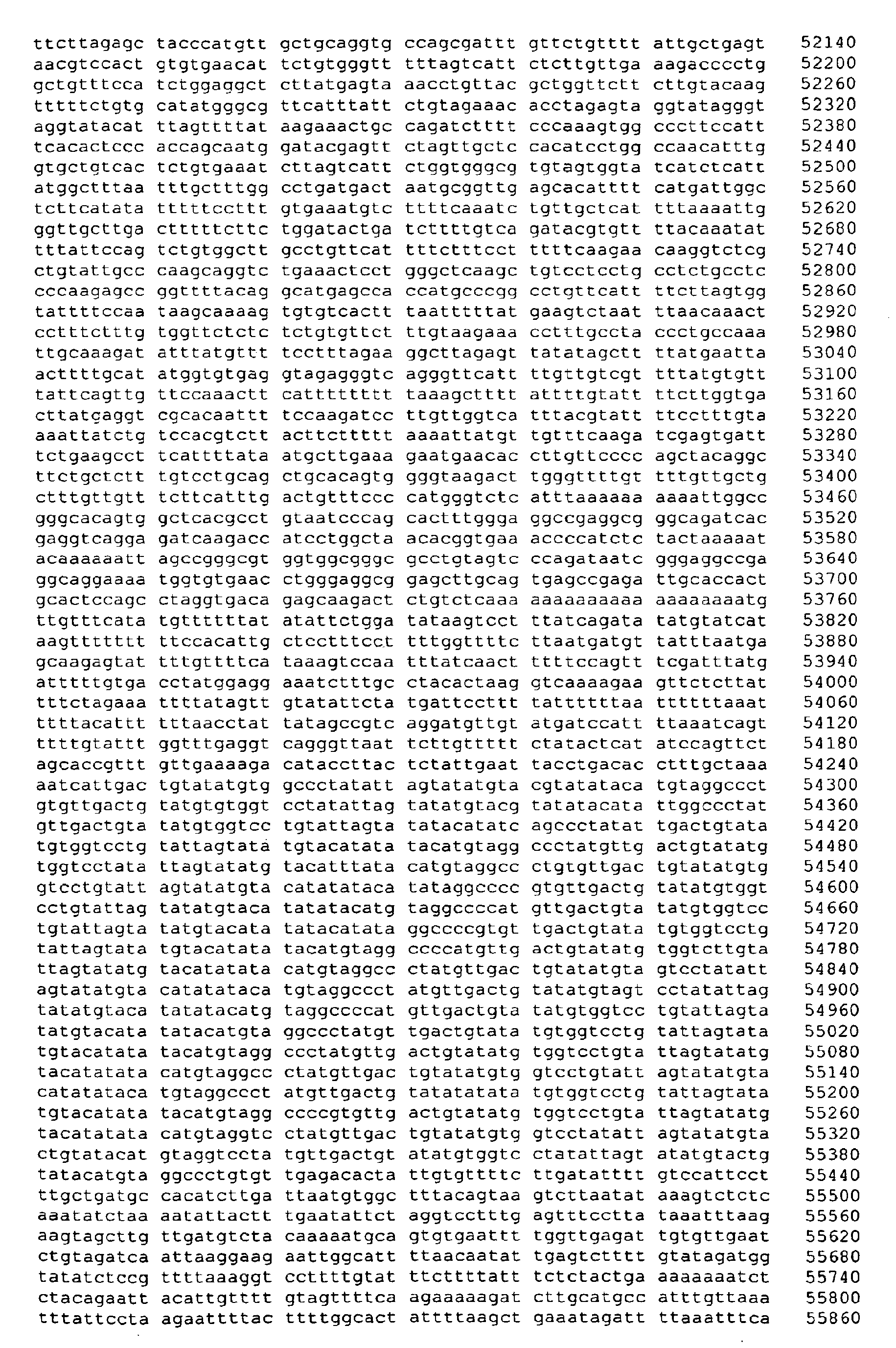


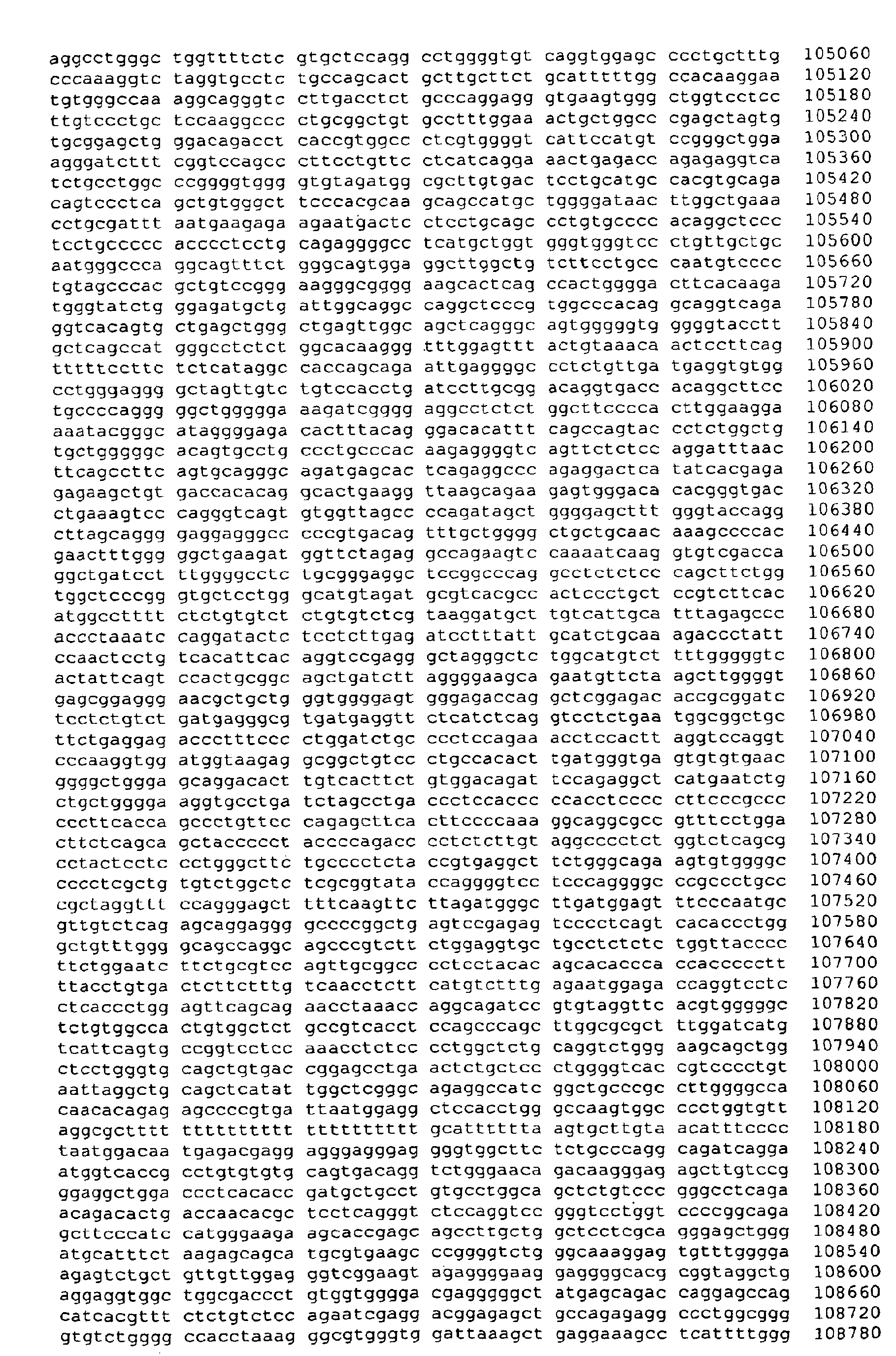


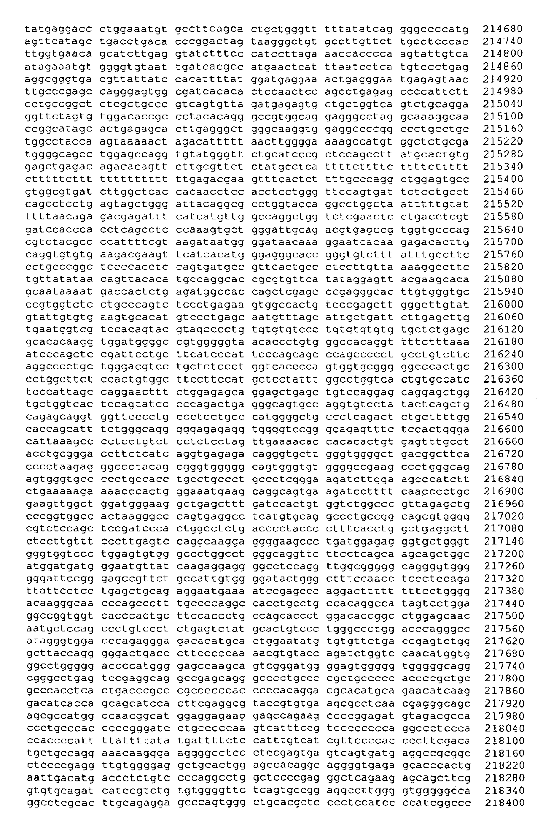

- <210> 2
<211> 1405
<212> DNA
<213> Homo Sapiens - <400> 2

- <210> 3
<211> 2365
<212> DNA
<213> Homo Sapiens - <400> 3

- <210> 4
<211> 219909
<212> DNA
<213> Artificial Sequence - <220>
<223> chemically treated genomic DNA (Homo sapiens) - <400> 4

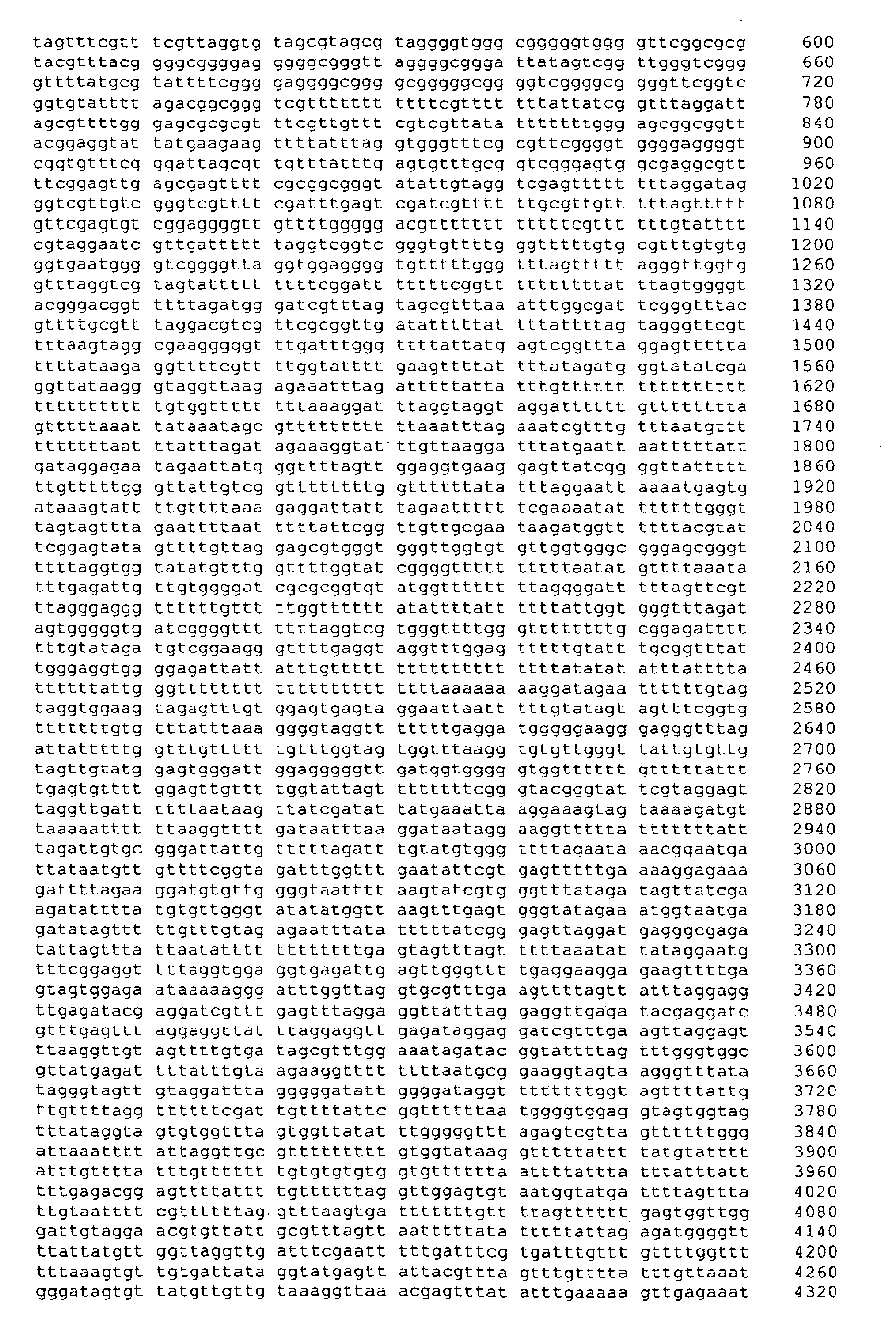


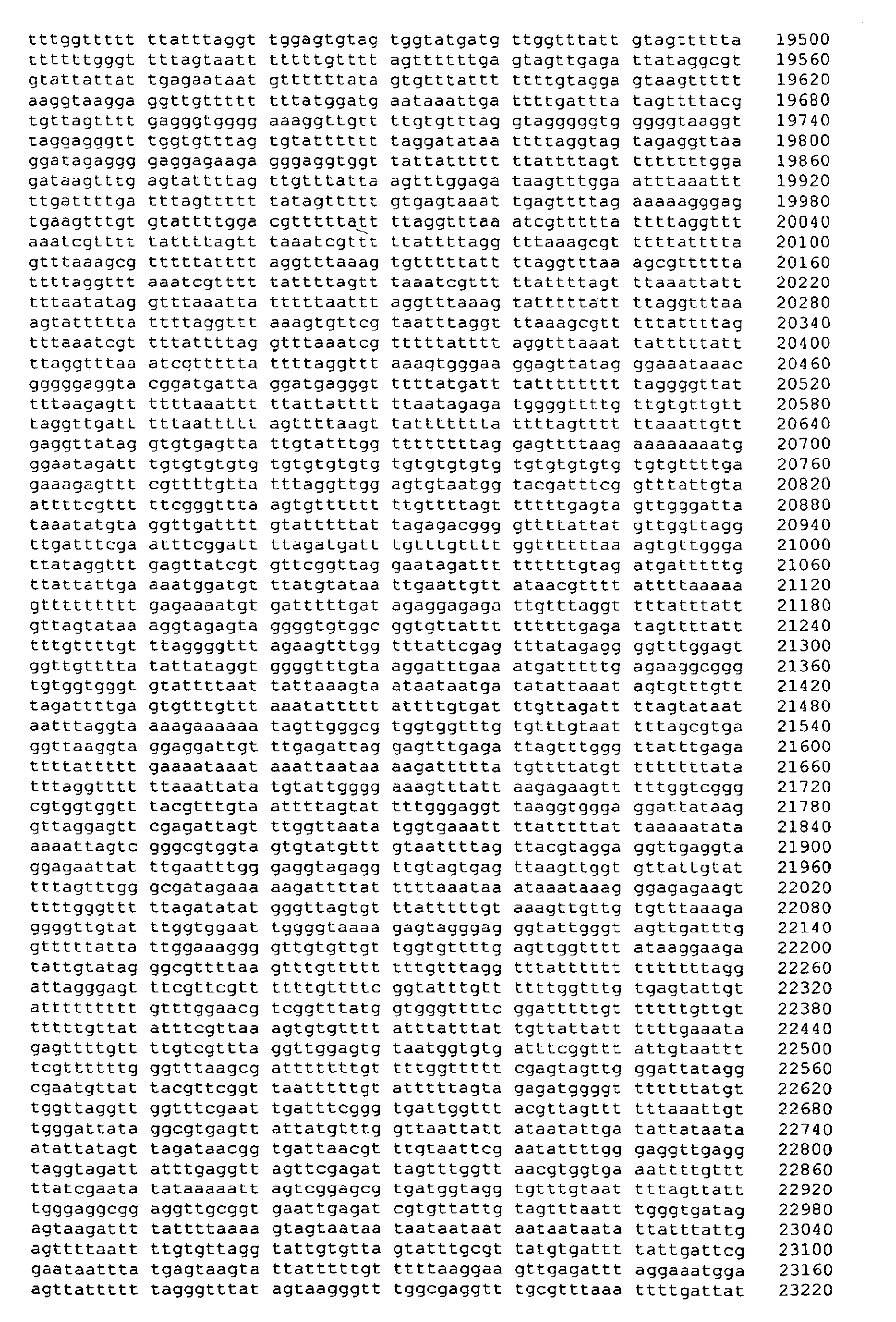
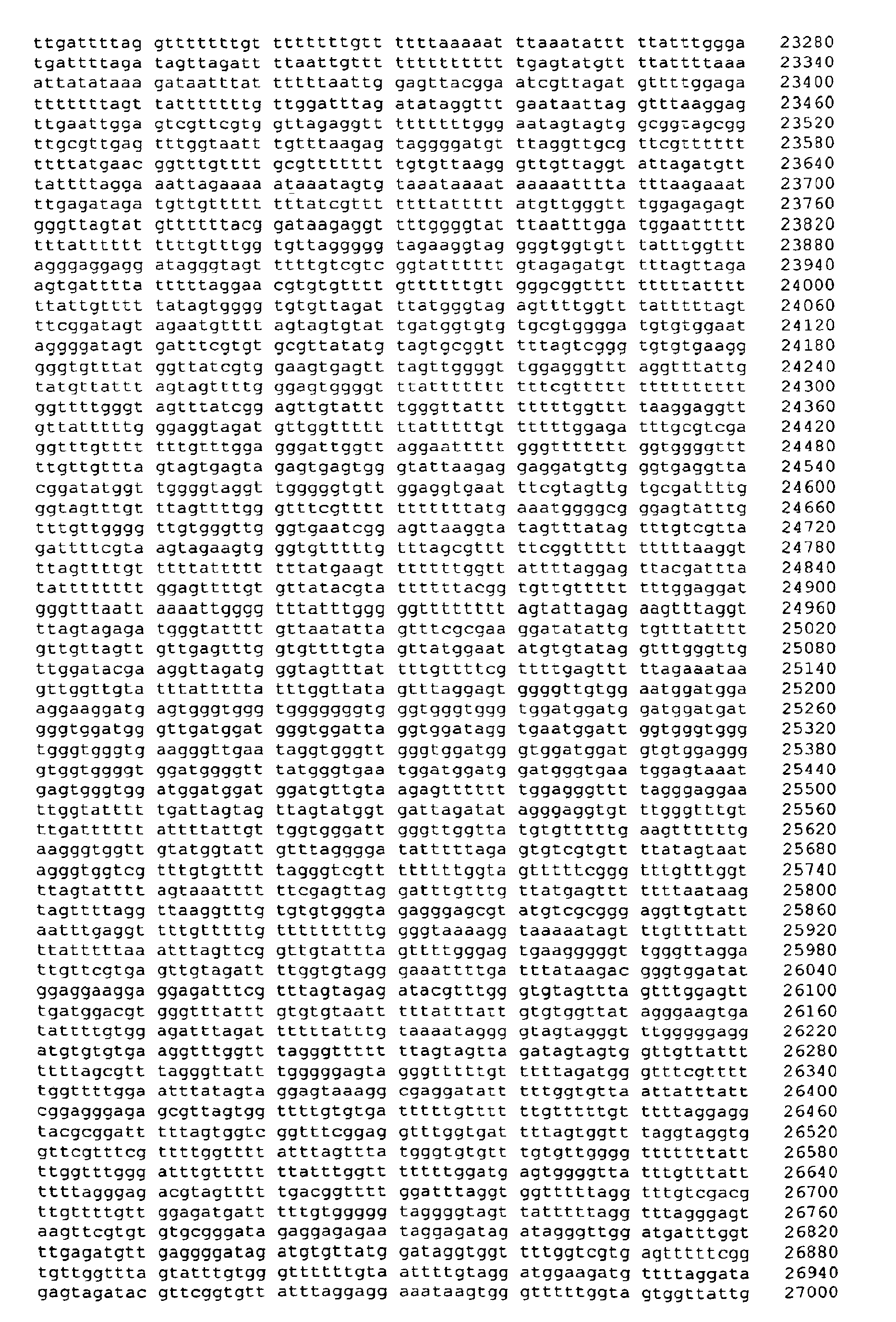


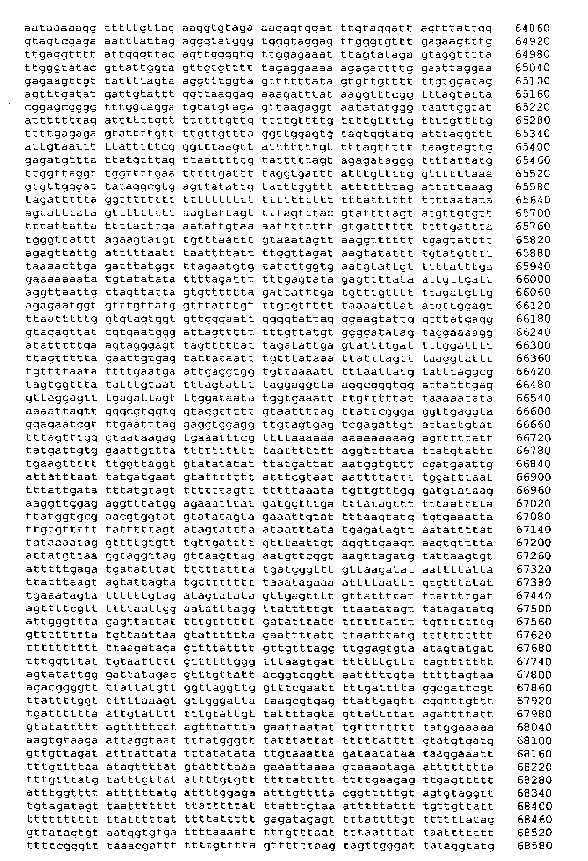


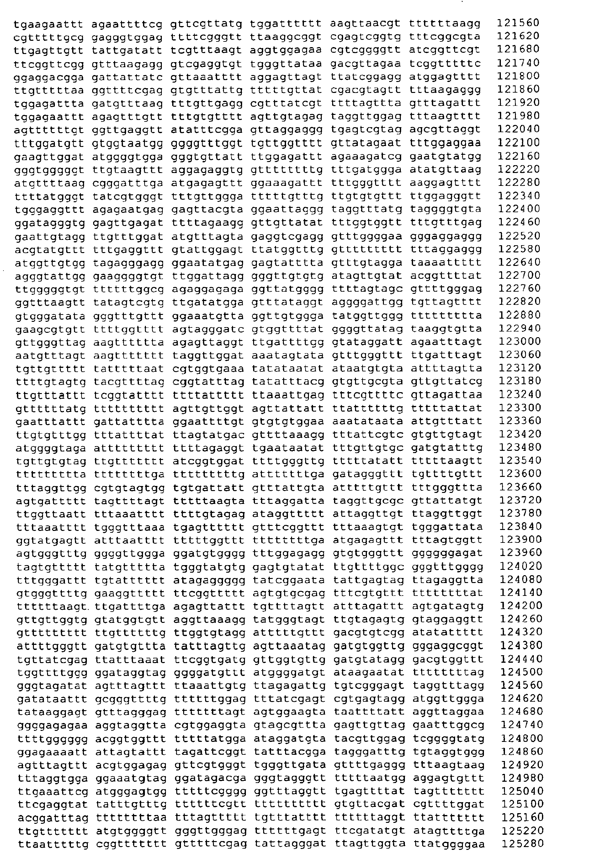


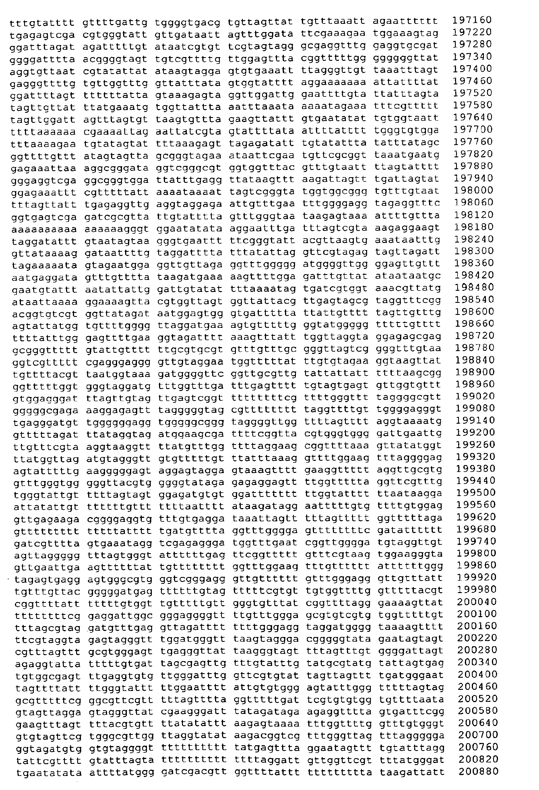


- <210> 5
<211> 219909
<212> DNA
<213> Artificial Sequence - <220>
<223> chemically treated genomic DNA (Homo sapiens) - <400> 5

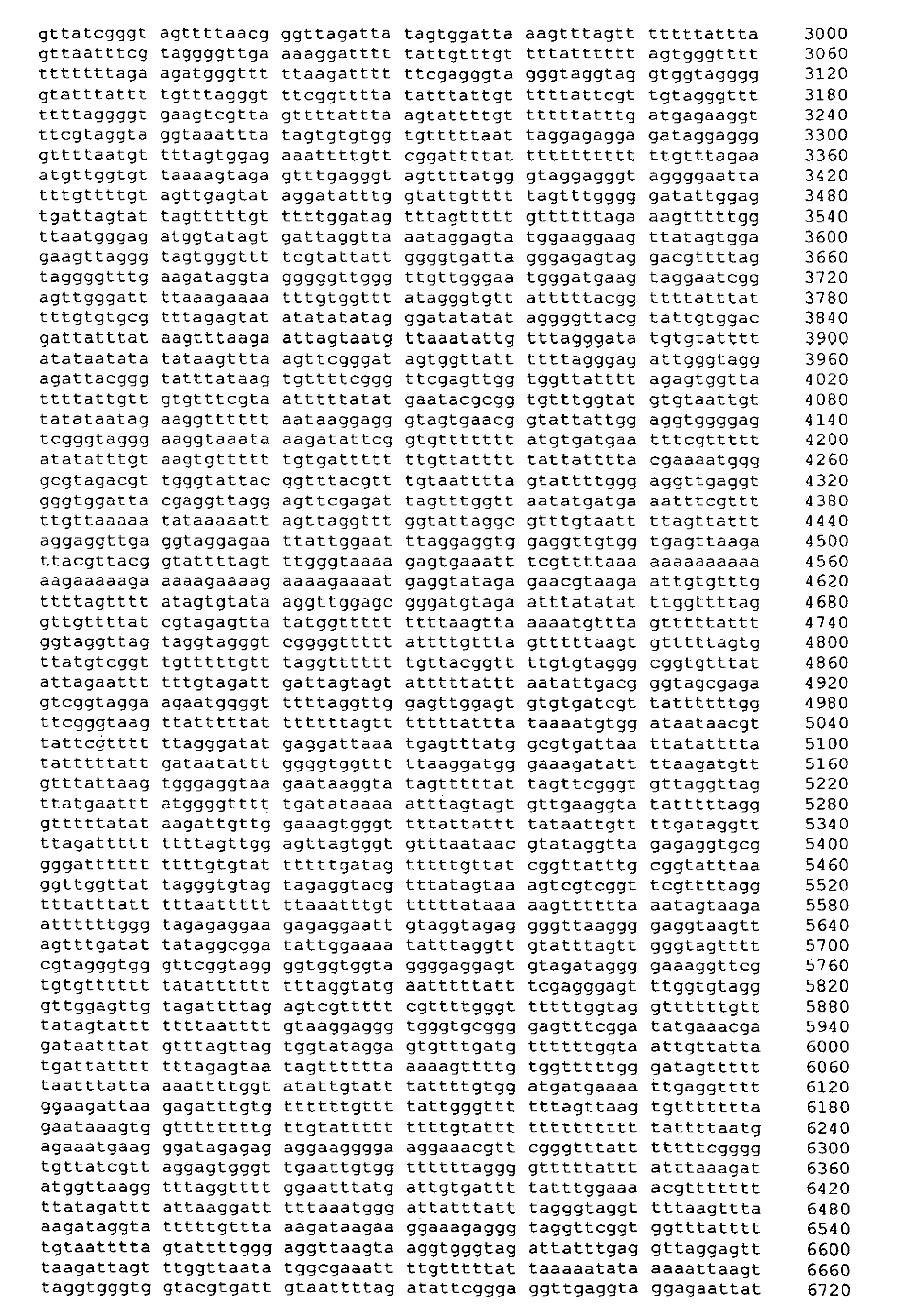
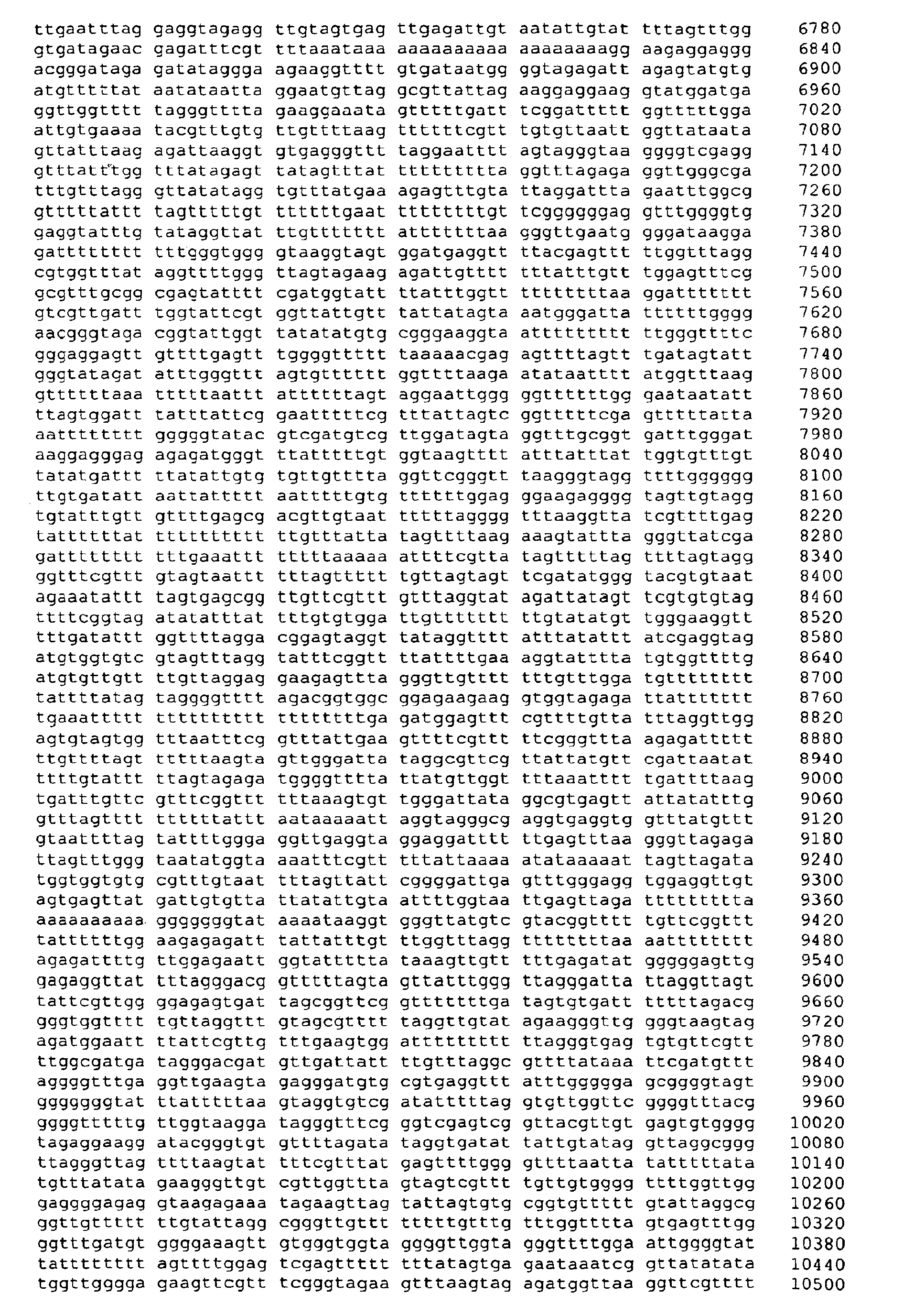


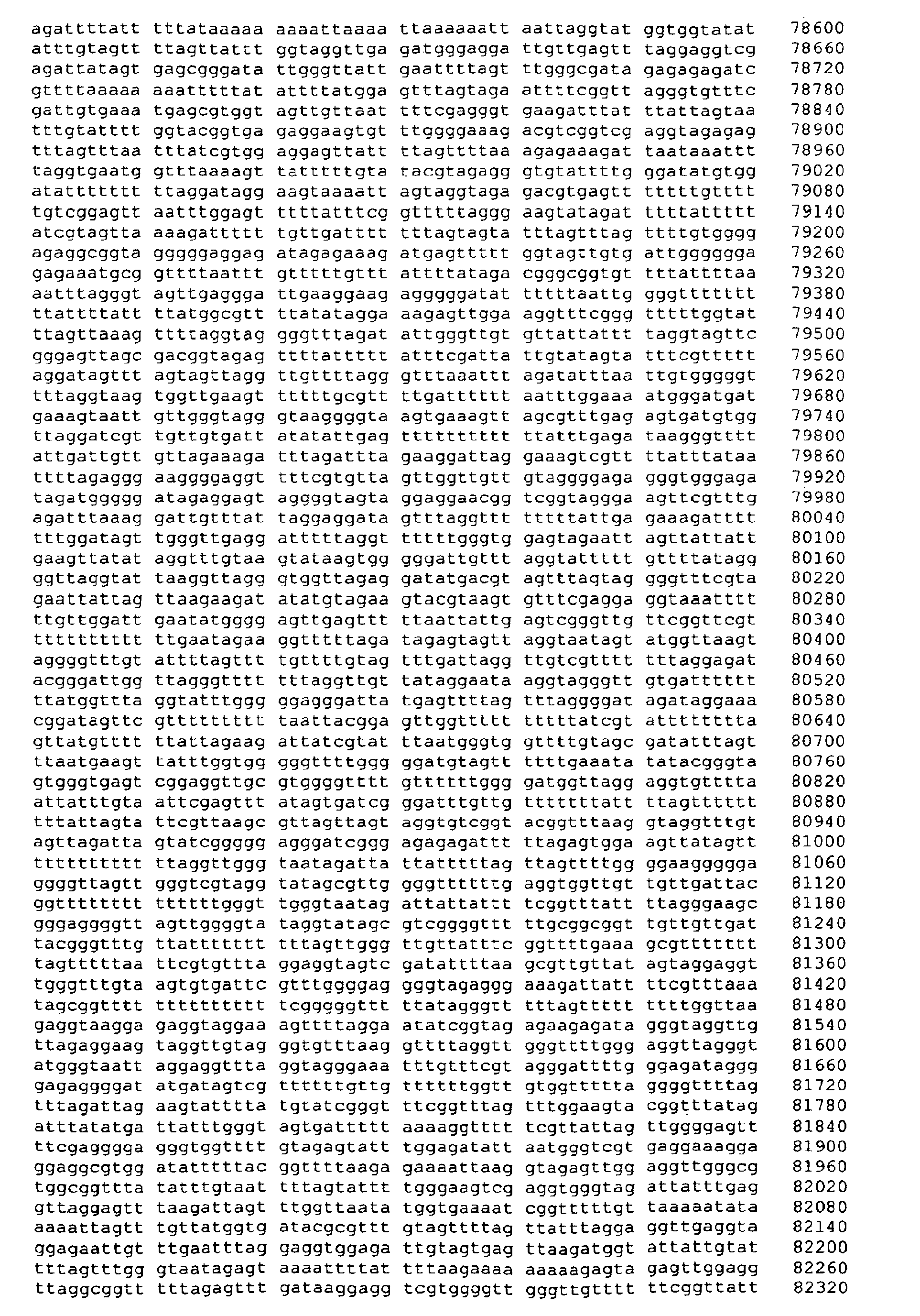


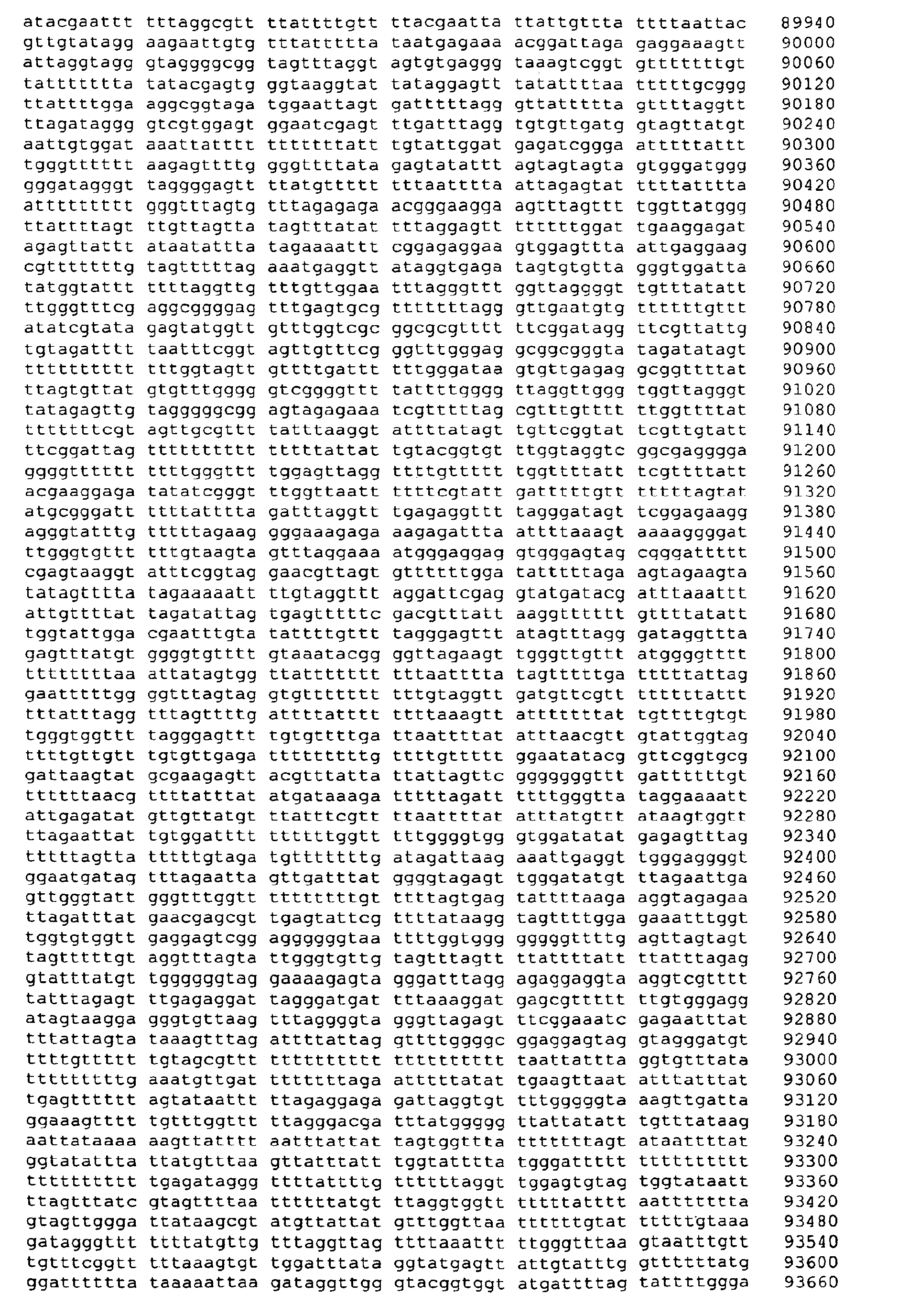


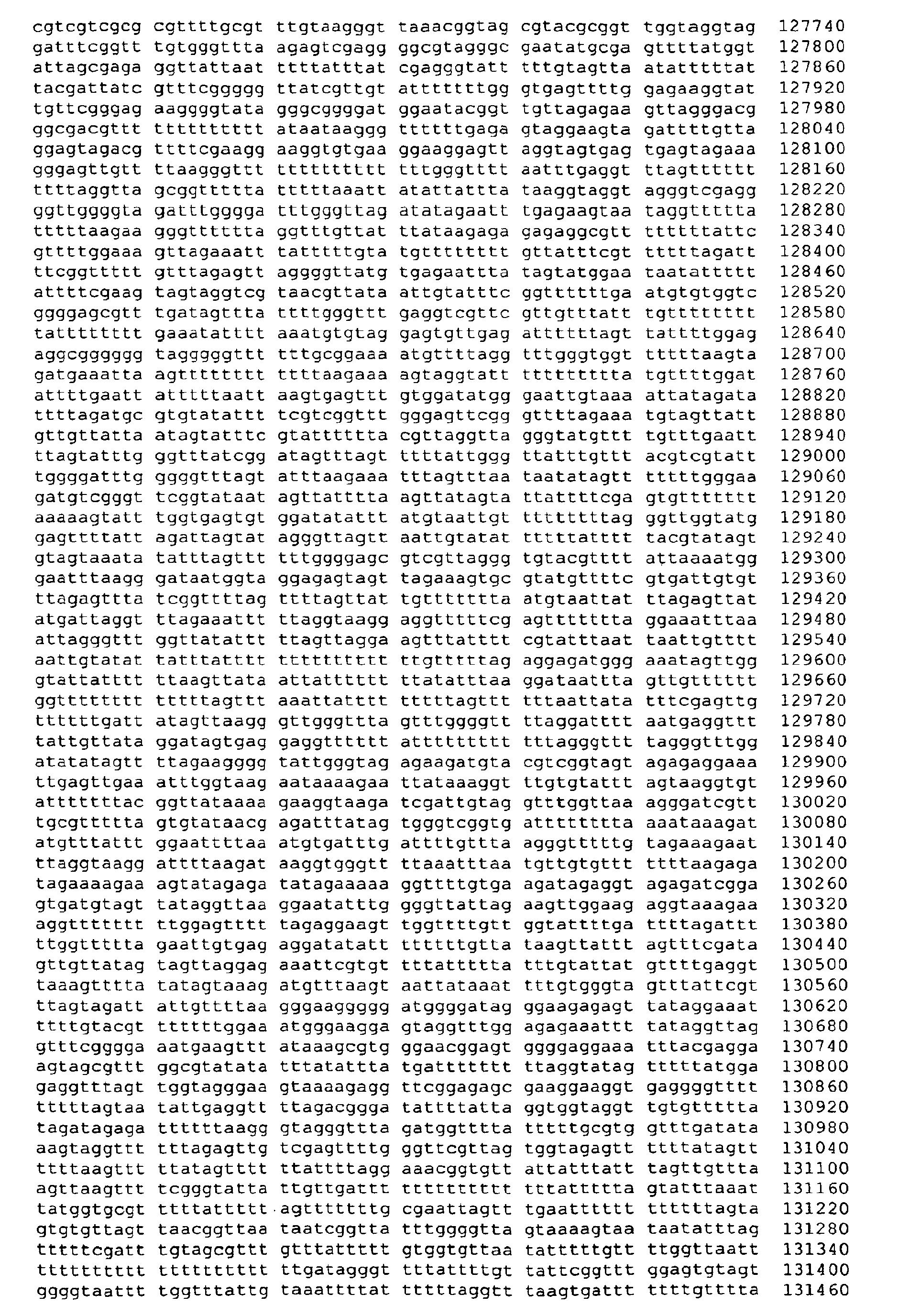


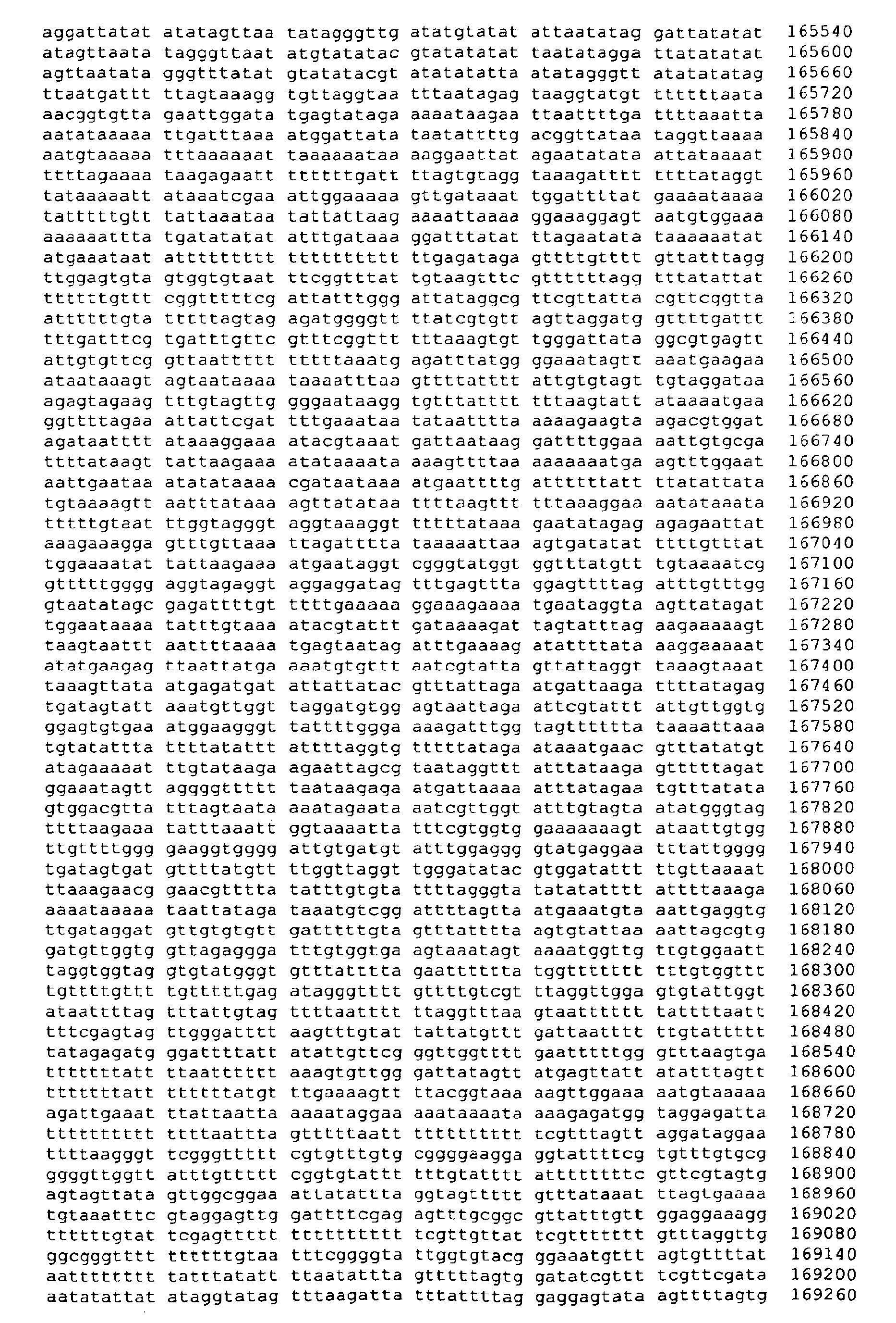


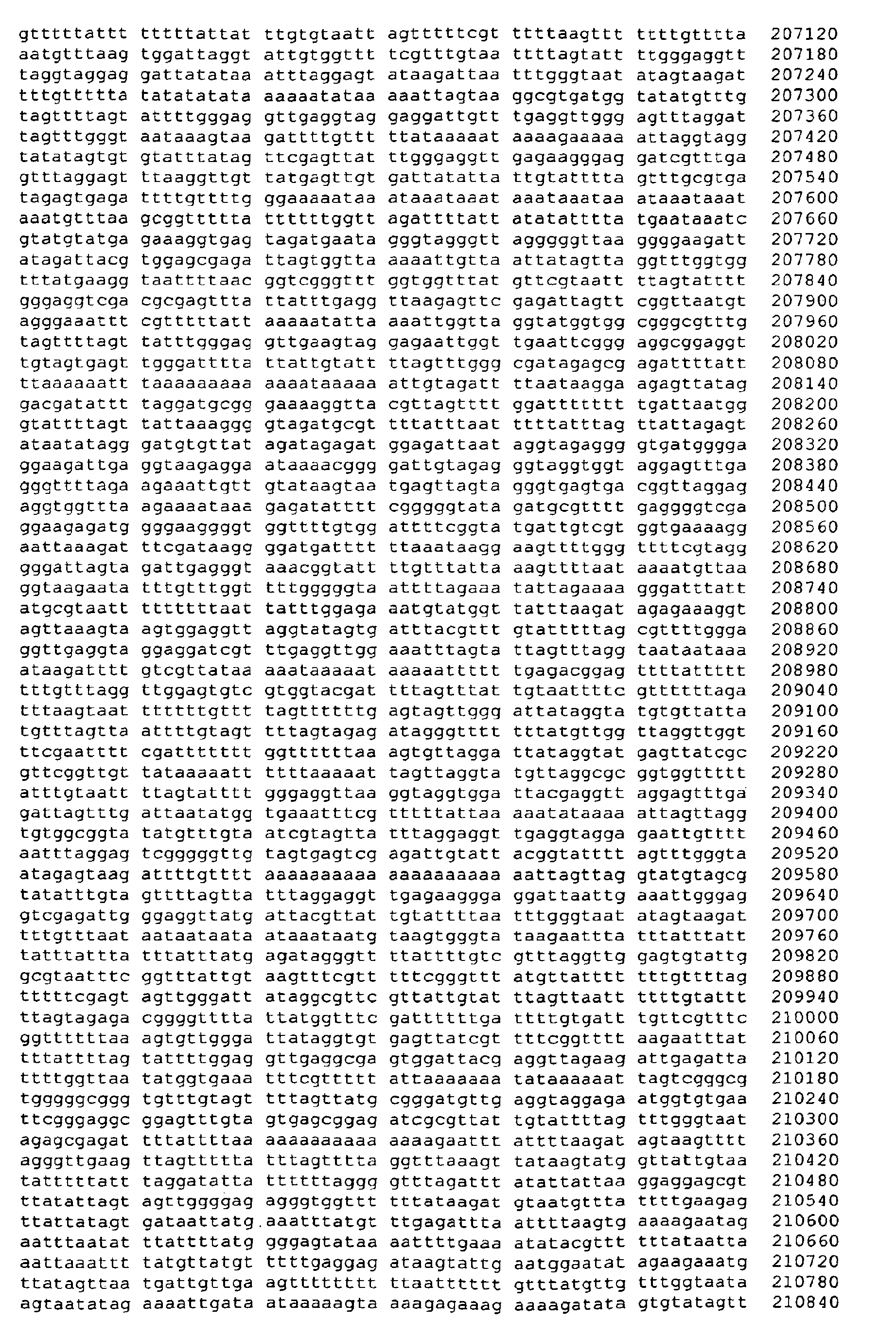


- <210> 6
<211> 1405
<212> DNA
<213> Artificial Sequence - <220>
<223> chemically treated genomic DNA (Homo sapiens) - <400> 6

- <210> 7
<211> 1405
<212> DNA
<213> Artificial Sequence - <220>
<223> chemically treated genomic DNA (Homo sapiens) - <400> 7

- <210> 8
<211> 2365
<212> DNA
<213> Artificial Sequence - <220>
<223> chemically treated genomic DNA (Homo sapiens) - <400> 8


- <210> 9
<211> 2365
<212> DNA
<213> Artificial Sequence - <220>
<223> chemically treated genomic DNA (Homo sapiens) - <400> 9
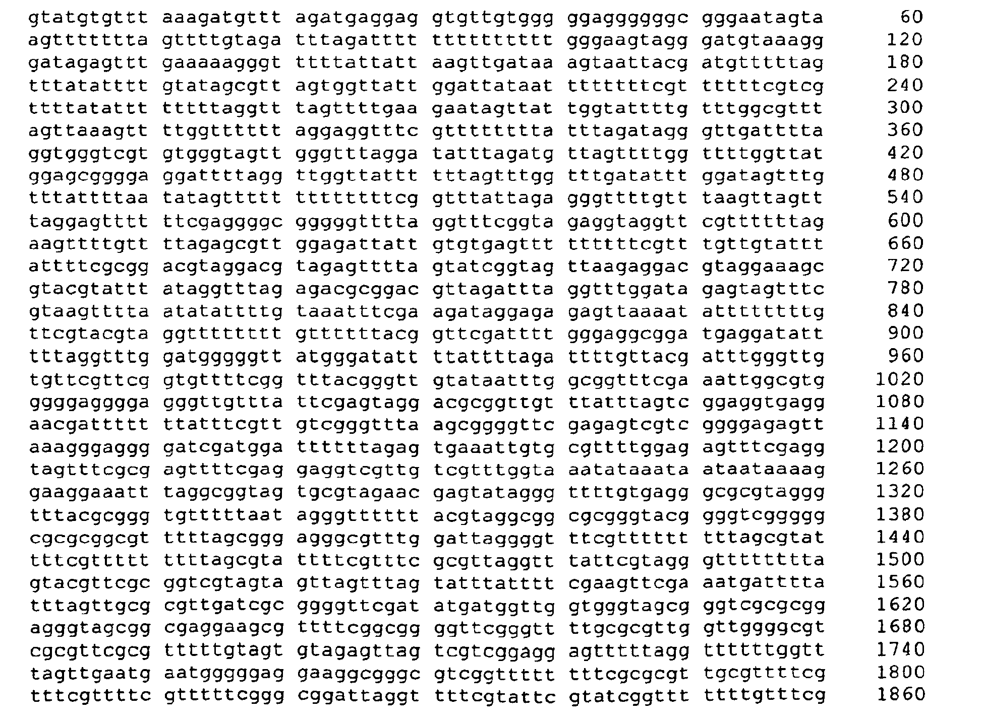

- <210> 10
<211> 219909
<212> DNA
<213> Artificial Sequence - <220>
<223> chemically treated genomic DNA (Homo sapiens) - <400> 10


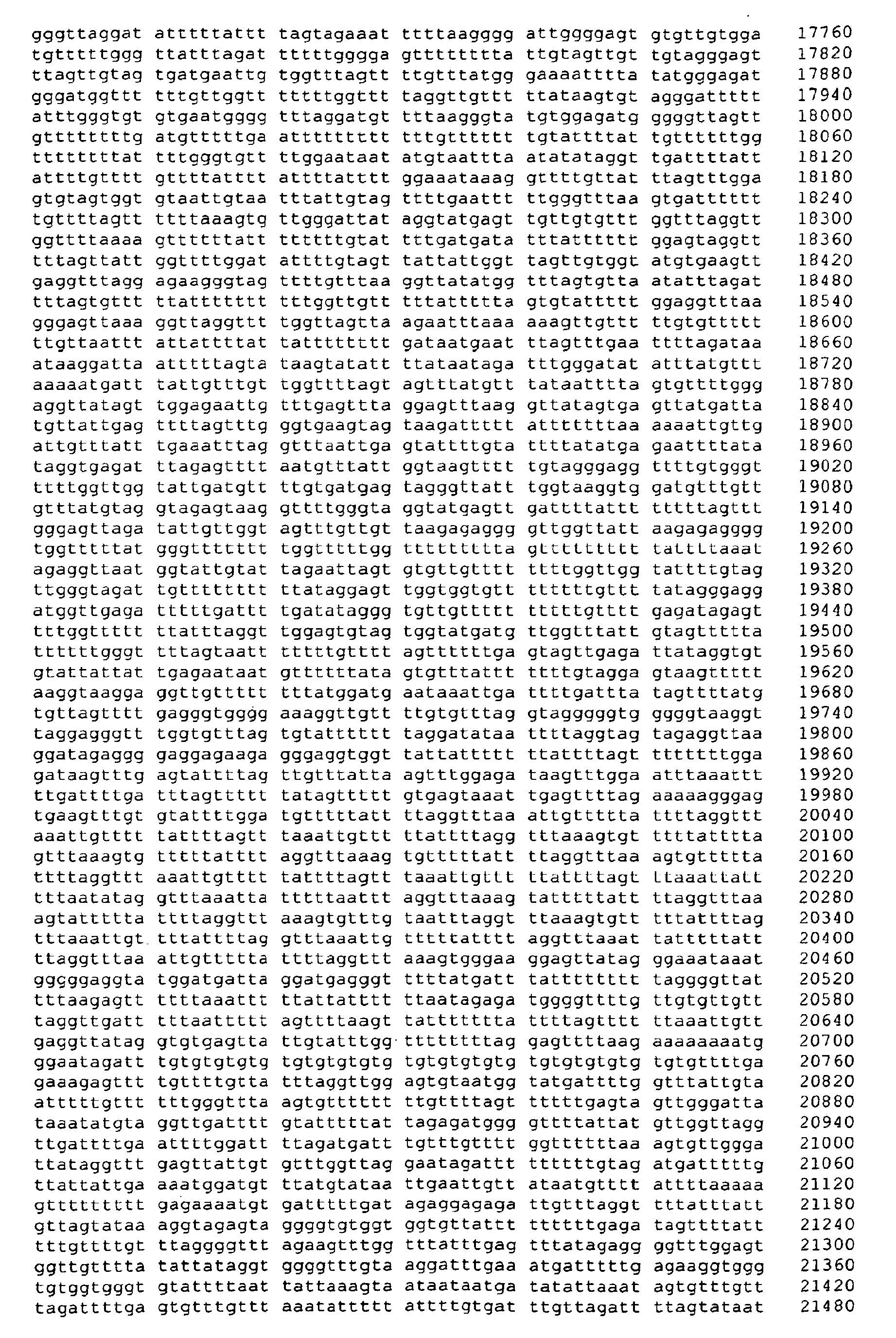





- <210> 11
<211> 219909
<212> DNA
<213> Artificial Sequence - <220>
<223> chemically treated genomic DNA (Homo sapiens) - <400> 11





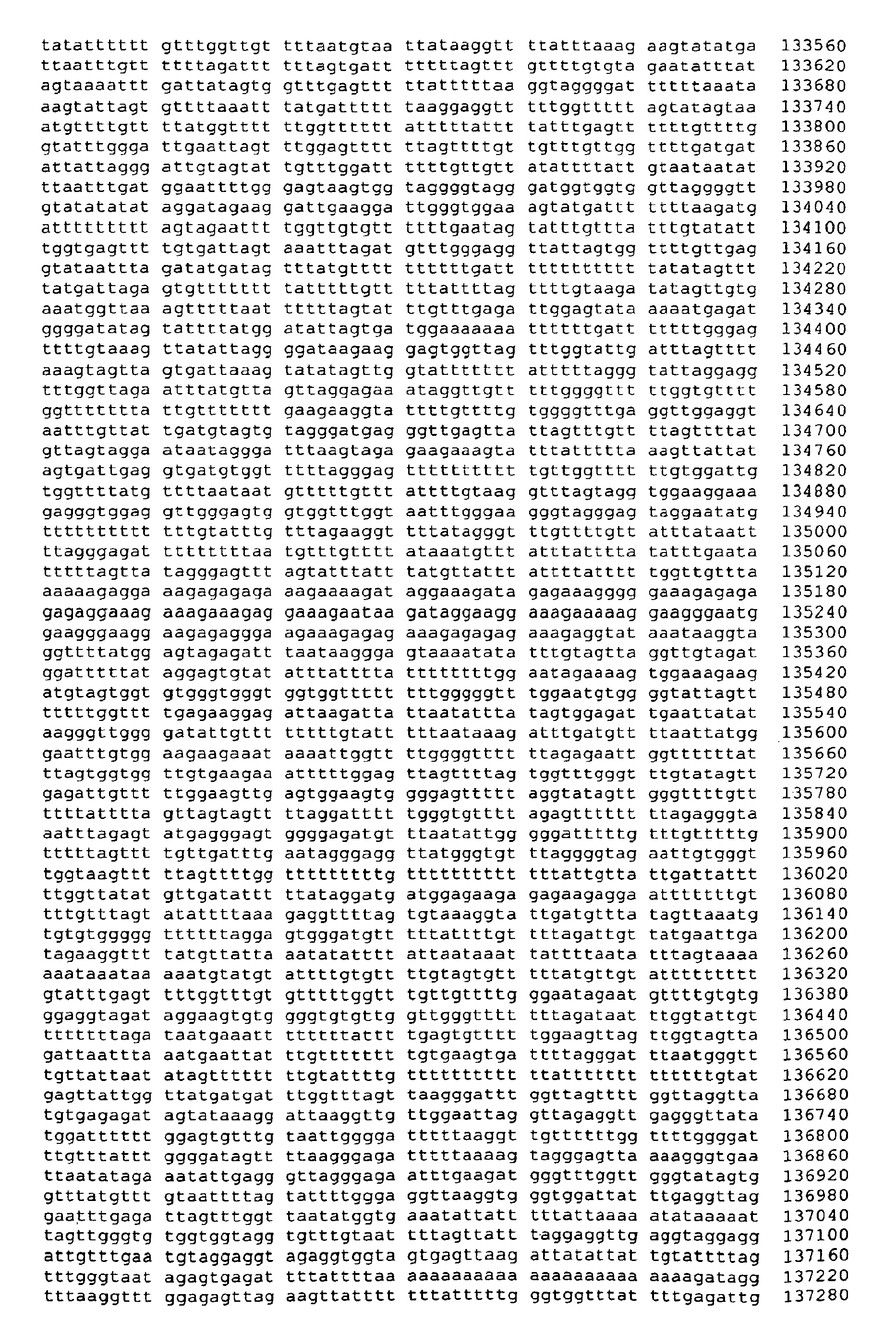


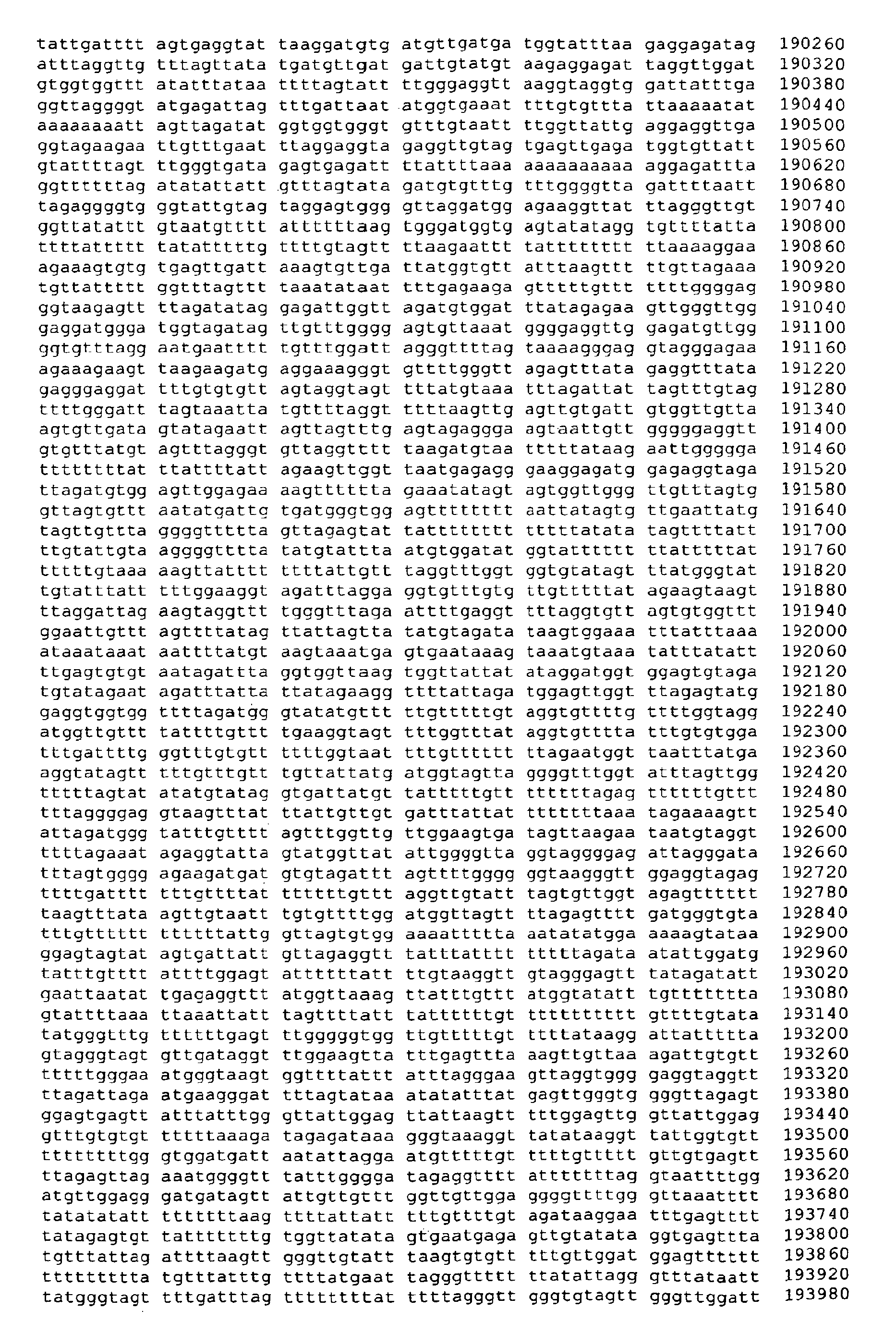


- <210> 12
<211> 1405
<212> DNA
<213> Artificial Sequence - <220>
<223> chemically treated genomic DNA (Homo sapiens) - <400> 12

- <210> 13
<211> 1405
<212> DNA
<213> Artificial Sequence - <220>
<223> chemically treated genomic DNA (Homo sapiens) - <400> 13

- <210> 14
<211> 2365
<212> DNA
<213> Artificial Sequence - <220>
<223> chemically treated genomic DNA (Homo sapiens) - <400> 14

- <210> 15
<211> 2365
<212> DNA
<213> Artificial Sequence - <220>
<223> chemically treated genomic DNA (Homo sapiens) - <400> 15


- <210> 16
<211> 3791
<212> DNA
<213> Homo Sapiens - <400> 16


- <210> 17
<211> 3971
<212> DNA
<213> Homo Sapiens - <400> 17


- <210> 18
<211> 3921
<212> DNA
<213> Homo Sapiens - <400> 18
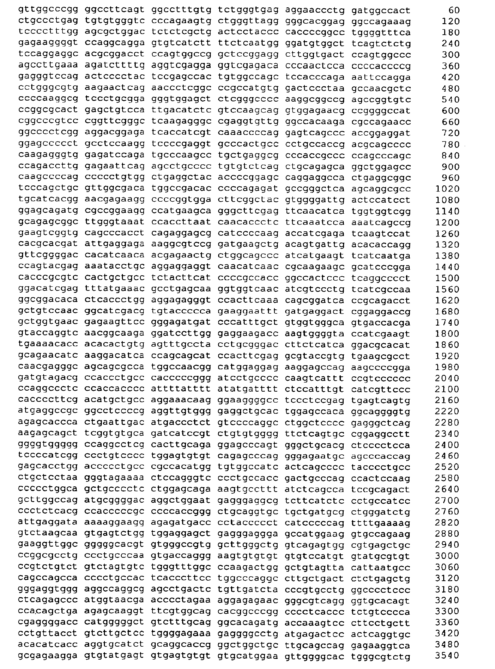

- <210> 19
<211> 1328
<212> DNA
<213> Homo Sapiens - <400> 19

- <210> 20
<211> 586
<212> PRT
<213> Homo sapiens - <400> 20


- <210> 21
<211> 190
<212> PRT
<213> Homo sapiens - <400> 21

- <210> 22
<211> 422
<212> PRT
<213> Homo sapiens - <400> 22


- <210> 23
<211> 197
<212> PRT
<213> Homo sapiens - <400> 23


- <210> 24
<211> 2804
<212> DNA
<213> Homo Sapiens - <400> 24


- <210> 25
<211> 3952
<212> DNA
<213> Homo Sapiens - <400> 25


- <210> 26
<211> 2820
<212> DNA
<213> Homo Sapiens - <400> 26


- <210> 27
<211> 3104
<212> DNA
<213> Homo Sapiens - <400> 27


- <210> 28
<211> 2378
<212> DNA
<213> Homo Sapiens - <400> 28


- <210> 29
<211> 2436
<212> DNA
<213> Homo Sapiens - <400> 29


- <210> 30
<211> 2804
<212> DNA
<213> Artificial Sequence - <220>
<223> chemically treated genomic DNA (Homo sapiens) - <400> 30


- <210> 31
<211> 2804
<212> DNA
<213> Artificial Sequence - <220>
<223> chemically treated genomic DNA (Homo sapiens) - <400> 31

- <210> 32
<211> 3952
<212> DNA
<213> Artificial Sequence - <220>
<223> chemically treated genomic DNA (Homo sapiens) - <400> 32

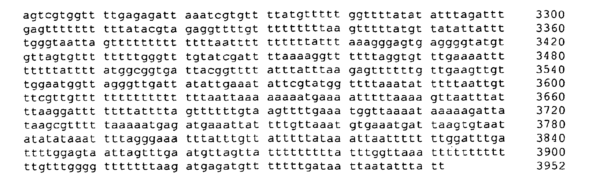
- <210> 33
<211> 3952
<212> DNA
<213> Artificial Sequence - <220>
<223> chemically treated genomic DNA (Homo sapiens) - <400> 33


- <210> 34
<211> 2820
<212> DNA
<213> Artificial Sequence - <220>
<223> chemically treated genomic DNA (Homo sapiens) - <400> 34

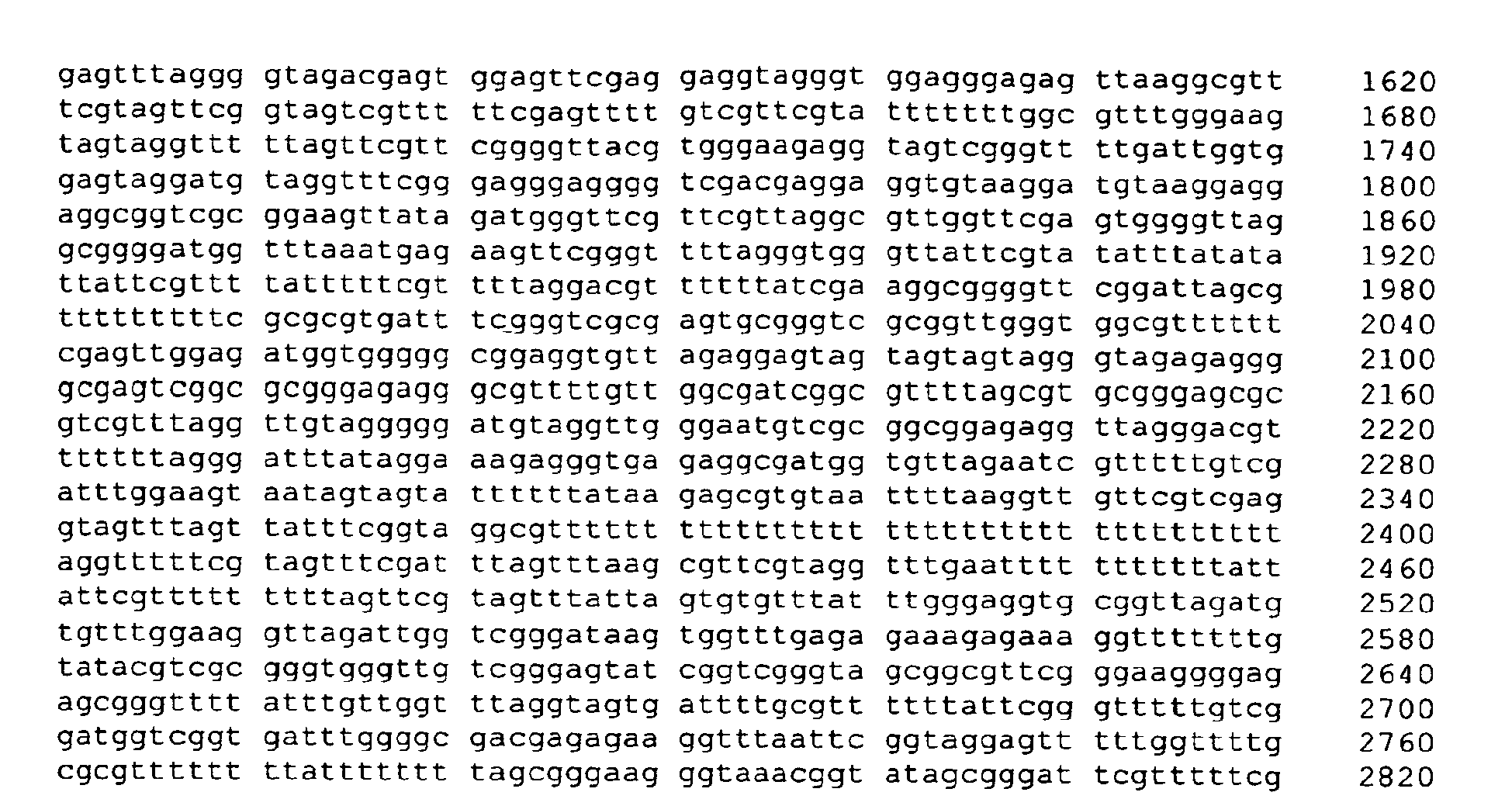
- <210> 35
<211> 2820
<212> DNA
<213> Artificial Sequence - <220>
<223> chemically treated genomic DNA (Homo sapiens) - <400> 35


- <210> 36
<211> 3104
<212> DNA
<213> Artificial Sequence - <220>
<223> chemically treated genomic DNA (Homo sapiens) - <400> 36


- <210> 37
<211> 3104
<212> DNA
<213> Artificial Sequence - <220>
<223> chemically treated genomic DNA (Homo sapiens) - <400> 37


- <210> 38
<211> 2378
<212> DNA
<213> Artificial Sequence - <220>
<223> chemically treated genomic DNA (Homo sapiens) - <400> 38


- <210> 39
<211> 2378
<212> DNA
<213> Artificial Sequence - <220>
<223> chemically treated genomic DNA (Homo sapiens) - <400> 39

- <210> 40
<211> 2436
<212> DNA
<213> Artificial Sequence - <220>
<223> chemically treated genomic DNA (Homo sapiens) - <400> 40

- <210> 41
<211> 2436
<212> DNA
<213> Artificial Sequence - <220>
<223> chemically treated genomic DNA (Homo sapiens) - <400> 41


- <210> 42
<211> 2804
<212> DNA
<213> Artificial Sequence - <220>
<223> chemically treated genomic DNA (Homo sapiens) - <400> 42


- <210> 43
<211> 2804
<212> DNA
<213> Artificial Sequence - <220>
<223> chemically treated genomic DNA (Homo sapiens) - <400> 43


- <210> 44
<211> 3952
<212> DNA
<213> Artificial Sequence - <220>
<223> chemically treated genomic DNA (Homo sapiens) - <400> 44


- <210> 45
<211> 3952
<212> DNA
<213> Artificial Sequence - <220>
<223> chemically treated genomic DNA (Homo sapiens) - <400> 45


- <210> 46
<211> 2820
<212> DNA
<213> Artificial Sequence - <220>
<223> chemically treated genomic DNA (Homo sapiens) - <400> 46


- <210> 47
<211> 2820
<212> DNA
<213> Artificial Sequence - <220>
<223> chemically treated genomic DNA (Homo sapiens) - <400> 47


- <210> 48
<211> 3104
<212> DNA.
<213> Artificial Sequence - <220>
<223> chemically treated genomic DNA (Homo sapiens) - <400> 48


- <210> 49
<211> 3104
<212> DNA
<213> Artificial Sequence - <220>
<223> chemically treated genomic DNA (Homo sapiens) - <400> 49
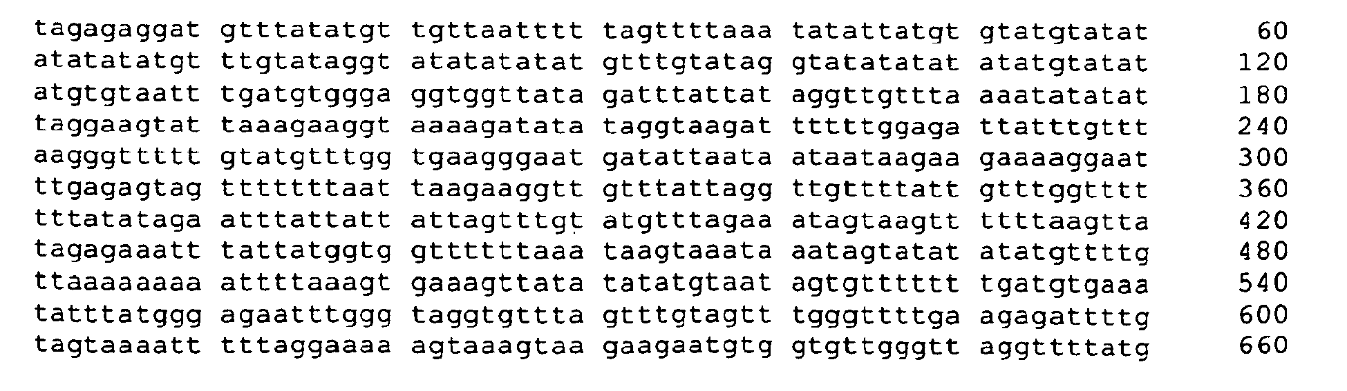

- <210> 50
<211> 2378
<212> DNA
<213> Artificial Sequence - <220>
<223> chemically treated genomic DNA (Homo sapiens) - <400> 50


- <210> 51
<211> 2378
<212> DNA
<213> Artificial Sequence - <220>
<223> chemically treated genomic DNA (Homo sapiens) - <400> 51


- <210> 52
<211> 2436
<212> DNA
<213> Artificial Sequence - <220>
<223> chemically treated genomic DNA (Homo sapiens) - <400> 52


- <210> 53
<211> 2436
<212> DNA
<213> Artificial Sequence - <220>
<223> chemically treated genomic DNA (Homo sapiens) - <400> 53

- <210> 54
<211> 32
<212> DNA
<213> Artificial Sequence - <220>
<223> chemically treated genomic DNA (Homo sapiens) - <400> 54
- catcatatca aaccccacaa tcaacacaca
ac 32
- catcatatca aaccccacaa tcaacacaca
- <210> 55
<211> 25
<212> DNA
<213> Artificial Sequence - <220>
<223> chemically treated genomic DNA (Homo sapiens) - <400> 55
- gttcgaaatg attttattta gttgc 25
- <210> 56
<211> 17
<212> DNA
<213> Artificial Sequence - <220>
<223> chemically treated genomic DNA (Homo sapiens) - <400> 56
- cgttgatcgc ggggttc 17
- <210> 57
<211> 17
<212> DNA
<213> Artificial Sequence - <220>
<223> chemically treated genomic DNA (Homo sapiens) - <400> 57
- ggggagggtt gtttatt 17
- <210> 58
<211> 17
<212> DNA
<213> Artificial Sequence - <220>
<223> chemically treated genomic DNA (Homo sapiens) - <400> 58
- cccctccctt taactct 17
- <210> 59
<211> 31
<212> DNA
<213> Artificial Sequence - <220>
<223> chemically treated genomic DNA (Homo sapiens) - <400> 59
- ttaactctcc ccaacaactc tcaaacccca
c 31
- ttaactctcc ccaacaactc tcaaacccca
- <210> 60
<211> 24
<212> DNA
<213> Artificial Sequence - <220>
<223> chemically treated genomic DNA (Homo sapiens) - <400> 60
- ttagtcggag gtgaggaacg attt 24
- <210> 61
<211> 24
<212> DNA
<213> Artificial Sequence - <220>
<223> chemically treated genomic DNA (Homo sapiens) - <400> 61
- ttatttcgtt gtcgggttta agcg 24
- <210> 62
<211> 20
<212> DNA
<213> Artificial Sequence - <220>
<223> chemically treated genomic DNA (Homo sapiens) - <400> 62
- ggagtggagg aaattgagat 20
- <210> 63
<211> 22
<212> DNA
<213> Artificial Sequence - <220>
<223> chemically treated genomic DNA (Homo sapiens) - <400> 63
- ccacacaaca aatactcaaa
ac 22
- ccacacaaca aatactcaaa
- <210> 64
<211> 33
<212> DNA
<213> Artificial Sequence - <220>
<223> chemically treated genomic DNA (Homo sapiens) - <400> 64
- tgggtgtttg taatttttgt tttgtgttag gtt 33
- <210> 65
<211> 22
<212> DNA
<213> Artificial Sequence - <220>
<223> chemically treated genomic DNA (Homo sapiens) - <400> 65
- gtagtagtag tagggtagag
ag 22
- gtagtagtag tagggtagag
- <210> 66
<211> 19
<212> DNA
<213> Artificial Sequence - <220>
<223> chemically treated genomic DNA (Homo sapiens) - <400> 66
- catcccccta caacctaaa 19
- <210> 67
<211> 35
<212> DNA
<213> Artificial Sequence - <220>
<223> chemically treated genomic DNA (Homo sapiens) - <400> 67
- caacctaaac aacacactcc cacacactaa aacac 35
- <210> 68
<211> 16
<212> DNA
<213> Artificial Sequence - <220>
<223> chemically treated genomic DNA (Homo sapiens) - <400> 68
- cgcgggagag ggcgtt 16
- <210> 69
<211> 19
<212> DNA
<213> Artificial Sequence - <220>
<223> chemically treated genomic DNA (Homo sapiens) - <400> 69
- tgttggcgat cggcgtttt 19
- <210> 70
<211> 16
<212> DNA
<213> Artificial Sequence - <220>
<223> chemically treated genomic DNA (Homo sapiens) - <400> 70
- gggtttcggg cgggta 16
- <210> 71
<211> 21
<212> DNA
<213> Artificial Sequence - <220>
<223> chemically treated genomic DNA (Homo sapiens) - <400> 71
- atatcgcact cgctatcgct a 21
- <210> 72
<211> 23
<212> DNA
<213> Artificial Sequence - <220>
<223> chemically treated genomic DNA (Homo sapiens) - <400> 72
- gagggcgacg gtacgttaga ggt 23
- <210> 73
<211> 24
<212> DNA
<213> Artificial Sequence - <220>
<223> chemically treated genomic DNA (Homo sapiens) - <400> 73
- ttgggcgtcg ttattagttc ggtc 24
- <210> 74
<211> 19
<212> DNA
<213> Artificial Sequence - <220>
<223> chemically treated genomic DNA (Homo sapiens) - <400> 74
- gtcgggttgg agggacgta 19
- <210> 75
<211> 21
<212> DNA
<213> Artificial Sequence - <220>
<223> chemically treated genomic DNA (Homo sapiens) - <400> 75
- atatcgcact cgctatcgct a 21
- <210> 76
<211> 23
<212> DNA
<213> Artificial Sequence - <220>
<223> chemically treated genomic DNA (Homo sapiens) - <400> 76
- gagggcgacg gtacgttaga ggt 23
- <210> 77
<211> 24
<212> DNA
<213> Artificial Sequence - <220>
<223> chemically treated genomic DNA (Homo sapiens) - <400> 77
- ttgggcgtcg ttattagttc ggtc 24
- <210> 78
<211> 21
<212> DNA
<213> Artificial Sequence - <220>
<223> chemically treated genomic DNA (Homo sapiens) - <400> 78
- gaggtgttag aggagtagta
g 21
- gaggtgttag aggagtagta
- <210> 79
<211> 17
<212> DNA
<213> Artificial Sequence - <220>
<223> chemically treated genomic DNA (Homo sapiens) - <400> 79
- tccccctaca acctaaa 17
- <210> 80
<211> 37
<212> DNA
<213> Artificial Sequence - <220>
<223> chemically treated genomic DNA (Homo sapiens) - <400> 80
- acctaaacaa cacactccca cacactaaaa caccaat 37
- <210> 81
<211> 15
<212> DNA
<213> Artificial Sequence - <220>
<223> chemically treated genomic DNA (Homo sapiens) - <400> 81
- cgagtcggcg cggga 15
- <210> 82
<211> 20
<212> DNA
<213> Artificial Sequence - <220>
<223> chemically treated genomic DNA (Homo sapiens) - <400> 82
- agggcgtttt gttggcgatc 20
- <210> 83
<211> 25
<212> DNA
<213> Artificial Sequence - <220>
<223> chemically treated genomic DNA (Homo sapiens) - <400> 83
- aaaaaaaaaa aactcctcta catac 25
- <210> 84
<211> 24
<212> DNA
<213> Artificial Sequence - <220>
<223> chemically treated genomic DNA (Homo sapiens) - <400> 84
- ggttattgtt tgggttaata aatg 24
- <210> 85
<211> 38
<212> DNA
<213> Artificial Sequence - <220>
<223> chemically treated genomic DNA (Homo sapiens) - <400> 85
- acatacacca caaataaatt accaaaaaca tcaaccaa 38
- <210> 86
<211> 21
<212> DNA
<213> Artificial Sequence - <220>
<223> chemically treated genomic DNA (Homo sapiens) - <400> 86
- tttttttttt cggacgtcgt
t 21
- tttttttttt cggacgtcgt
- <210> 87
<211> 20
<212> DNA
<213> Artificial Sequence - <220>
<223> chemically treated genomic DNA (Homo sapiens) - <400> 87
- tcggtcgatg ttttcggtaa 20
- <210> 88
<211> 20
<212> DNA
<213> Artificial Sequence - <220>
<223> chemically treated genomic DNA (Homo sapiens) - <400> 88
- tgagagagag agggttgaaa 20
- <210> 89
<211> 24
<212> DNA
<213> Artificial Sequence - <220>
<223> chemically treated genomic DNA (Homo sapiens) - <400> 89
- tctaaataac aaaatacctc catt 24
- <210> 90
<211> 27
<212> DNA
<213> Artificial Sequence - <220>
<223> chemically treated genomic DNA (Homo sapiens) - <400> 90
- ccattaccaa cacaacccac caaccaa 27
- <210> 91
<211> 16
<212> DNA
<213> Artificial Sequence - <220>
<223> chemically treated genomic DNA (Homo sapiens) - <400> 91
- cgacccgcca accgac 16
- <210> 92
<211> 15
<212> DNA
<213> Artificial Sequence - <220>
<223> chemically treated genomic DNA (Homo sapiens) - <400> 92
- cgccgaaacg cgctc 15
- <210> 93
<211> 26
<212> DNA
<213> Artificial Sequence - <220>
<223> chemically treated genomic DNA (Homo sapiens) - <400> 93
- gtagtagtta gtttagtatt tatttt 26
- <210> 94
<211> 17
<212> DNA
<213> Artificial Sequence - <220>
<223> chemically treated genomic DNA (Homo sapiens) - <400> 94
- cccaccaacc atcatat 17
- <210> 95
<211> 32
<212> DNA
<213> Artificial Sequence - <220>
<223> chemically treated genomic DNA (Homo sapiens) - <400> 95
- catcatatca aaccccacaa tcaacacaca
ac 32
- catcatatca aaccccacaa tcaacacaca
- <210> 96
<211> 19
<212> DNA
<213> Artificial Sequence - <220>
<223> chemically treated genomic DNA (Homo sapiens) - <400> 96
- gaaccccgcg atcaacgcg 19
- <210> 97
<211> 26
<212> DNA
<213> Artificial Sequence - <220>
<223> chemically treated genomic DNA (Homo sapiens) - <400> 97
- gtagtagtta gtttagtatt tatttt 26
- <210> 98
<211> 17
<212> DNA
<213> Artificial Sequence - <220>
<223> chemically treated genomic DNA (Homo sapiens) - <400> 98
- cccaccaacc atcatat 17
- <210> 99
<211> 32
<212> DNA
<213> Artificial Sequence - <220>
<223> chemically treated genomic DNA (Homo sapiens) - <400> 99
- catcatatca aaccccacaa tcaacacaca
ac 32
- catcatatca aaccccacaa tcaacacaca
- <210> 100
<211> 25
<212> DNA
<213> Artificial Sequence - <220>
<223> chemically treated genomic DNA (Homo sapiens) - <400> 100
- gttcgaaatg attttattta gttgc 25
- <210> 101
<211> 17
<212> DNA
<213> Artificial Sequence - <220>
<223> chemically treated genomic DNA (Homo sapiens) - <400> 101
- cgttgatcgc ggggttc 17
- <210> 102
<211> 20
<212> DNA
<213> Artificial Sequence - <220>
<223> chemically treated genomic DNA (Homo sapiens) - <400> 102
- ccaaaaccta aacttacaac 20
- <210> 103
<211> 17
<212> DNA
<213> Artificial Sequence - <220>
<223> chemically treated genomic DNA (Homo sapiens) - <400> 103
- ggaaatttga ggggtaa 17
- <210> 104
<211> 31
<212> DNA
<213> Artificial Sequence - <220>
<223> chemically treated genomic DNA (Homo sapiens) - <400> 104
- tacaacacca ccaacaaacc caaaaacaca a 31
- <210> 105
<211> 19
<212> DNA
<213> Artificial Sequence - <220>
<223> chemically treated genomic DNA (Homo sapiens) - <400> 105
- gttaattgcg ggcgatcga 19
- <210> 106
<211> 17
<212> DNA
<213> Artificial Sequence - <220>
<223> chemically treated genomic DNA (Homo sapiens) - <400> 106
- cgtcgttagc gggtggg 17
- <210> 107
<211> 20
<212> DNA
<213> Artificial Sequence - <220>
<223> chemically treated genomic DNA (Homo sapiens) - <400> 107
- aaaatcctct ccaacacgtc 20
- <210> 108
<211> 20
<212> DNA
<213> Artificial Sequence - <220>
<223> chemically treated genomic DNA (Homo sapiens) - <400> 108
- cgcgattcgt tgtttattag 20
- <210> 109
<211> 25
<212> DNA
<213> Artificial Sequence - <220>
<223> chemically treated genomic DNA (Homo sapiens) - <400> 109
- cggatttcgc ggttaacgcg tagtt 25
- <210> 110
<211> 24
<212> DNA
<213> Artificial Sequence - <220>
<223> chemically treated genomic DNA (Homo sapiens) - <400> 110
- tctaaataac aaaatacctc catt 24
- <210> 111
<211> 25
<212> DNA
<213> Artificial Sequence - <220>
<223> chemically treated genomic DNA (Homo sapiens) - <400> 111
- gtttttttta ttagttggaa gattt 25
- <210> 112
<211> 20
<212> DNA
<213> Artificial Sequence - <220>
<223> chemically treated genomic DNA (Homo sapiens) - <400> 112
- aaactacaac aaaccttatc 20
- <210> 113
<211> 37
<212> DNA
<213> Artificial Sequence - <220>
<223> chemically treated genomic DNA (Homo sapiens) - <400> 113
- ccttatccac actaaaacaa acaaacaaca cacaaac 37
- <210> 114
<211> 17
<212> DNA
<213> Artificial Sequence - <220>
<223> chemically treated genomic DNA (Homo sapiens) - <400> 114
- cgtttacggt tcgcgcg 17
- <210> 115
<211> 20
<212> DNA
<213> Artificial Sequence - <220>
<223> chemically treated genomic DNA (Homo sapiens) - <400> 115
- cgttcgtttg ttttagcgcg 20
- <210> 116
<211> 23
<212> DNA
<213> Artificial Sequence - <220>
<223> chemically treated genomic DNA (Homo sapiens) - <400> 116
- ggtgttgttt attttagaga gtt 23
- <210> 117
<211> 17
<212> DNA
<213> Artificial Sequence - <220>
<223> chemically treated genomic DNA (Homo sapiens) - <400> 117
- ctcccctaac ccctatc 17
- <210> 118
<211> 29
<212> DNA
<213> Artificial Sequence - <220>
<223> chemically treated genomic DNA (Homo sapiens) - <400> 118
- ctatccttca ccaccttccc aacactaca 29
- <210> 119
<211> 16
<212> DNA
<213> Artificial Sequence - <220>
<223> chemically treated genomic DNA (Homo sapiens) - <400> 119
- ttaggggggc gcggga 16
- <210> 120
<211> 21
<212> DNA
<213> Artificial Sequence - <220>
<223> chemically treated genomic DNA (Homo sapiens) - <400> 120
- gttagatgcg tcgtagcgtt
g 21
- gttagatgcg tcgtagcgtt
- <210> 121
<211> 20
<212> DNA
<213> Artificial Sequence - <220>
<223> chemically treated genomic DNA (Homo sapiens) - <400> 121
- aaaatcctct ccaacacgtc 20
- <210> 122
<211> 20
<212> DNA
<213> Artificial Sequence - <220>
<223> chemically treated genomic DNA (Homo sapiens) - <400> 122
- cgcgattcgt tgtttattag 20
- <210> 123
<211> 25
<212> DNA
<213> Artificial Sequence - <220>
<223> chemically treated genomic DNA (Homo sapiens) - <400> 123
- cggatttcgc ggttaacgcg tagtt 25
- <210> 124
<211> 20
<212> DNA
<213> Artificial Sequence - <220>
<223> chemically treated genomic DNA (Homo sapiens) - <400> 124
- aaaatcctct ccaacacgtc 20
- <210> 125
<211> 20
<212> DNA
<213> Artificial Sequence - <220>
<223> chemically treated genomic DNA (Homo sapiens) - <400> 125
- cgcgattcgt tgtttattag 20
- <210> 126
<211> 25
<212> DNA
<213> Artificial Sequence - <220>
<223> chemically treated genomic DNA (Homo sapiens) - <400> 126
- cggatttcgc ggttaacgcg tagtt 25
- <210> 127
<211> 26
<212> DNA
<213> Artificial Sequence - <220>
<223> chemically treated genomic DNA (Homo sapiens) - <400> 127
- gtagtagtta gtttagtatt tatttt 26
- <210> 128
<211> 17
<212> DNA
<213> Artificial Sequence - <220>
<223> chemically treated genomic DNA (Homo sapiens) - <400> 128
- cccaccaacc atcatat 17
- <210> 129
<211> 493
<212> DNA
<213> Artificial Sequence - <220>
<223> chemically treated genomic DNA (Homo sapiens) - <400> 129

- <210> 130
<211> 21
<212> DNA
<213> Artificial Sequence - <220>
<223> chemically treated genomic DNA (Homo sapiens) - <400> 130
- ccttctccaa aactcaccca a 21
- <210> 131
<211> 22
<212> DNA
<213> Artificial Sequence - <220>
<223> chemically treated genomic DNA (Homo sapiens) - <400> 131
- ttgggggata gttttgggta ta 22
- <210> 132
<211> 427
<212> DNA
<213> Artificial Sequence - <220>
<223> chemically treated genomic DNA (Homo sapiens) - <400> 132

- <210> 133
<211> 22
<212> DNA
<213> Artificial Sequence - <220>
<223> chemically treated genomic DNA (Homo sapiens) - <400> 133
- tatacccaaa actatccccc
aa 22
- tatacccaaa actatccccc
- <210> 134
<211> 26
<212> DNA
<213> Artificial Sequence - <220>
<223> chemically treated genomic DNA (Homo sapiens) - <400> 134
- gtagtagtta gtttagtatt tatttt 26
- <210> 135
<211> 556
<212> DNA
<213> Artificial Sequence - <220>
<223> chemically treated genomic DNA (Homo sapiens) - <400> 135

- <210> 136
<211> 21
<212> DNA
<213> Artificial Sequence - <220>
<223> chemically treated genomic DNA (Homo sapiens) - <400> 136
- ggttagtttt gtattgtagg a 21
- <210> 137
<211> 25
<212> DNA
<213> Artificial Sequence - <220>
<223> chemically treated genomic DNA (Homo sapiens) - <400> 137
- ataaatttcc caaaataaaa ctata 25
- <210> 138
<211> 555
<212> DNA
<213> Artificial Sequence - <220>
<223> chemically treated genomic DNA (Homo sapiens) - <400> 138
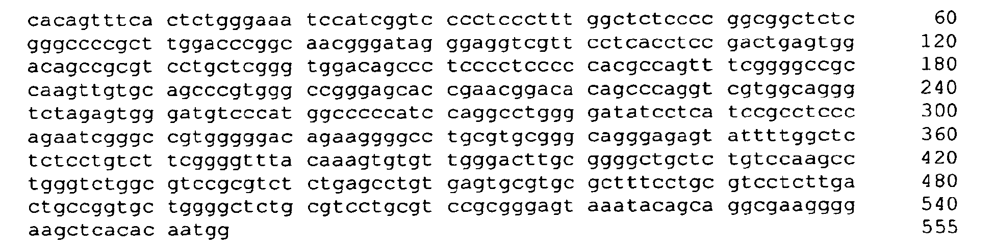
- <210> 139
<211> 24
<212> DNA
<213> Artificial Sequence - <220>
<223> chemically treated genomic DNA (Homo sapiens) - <400> 139
- gtatagtttt attttgggaa attt 24
- <210> 140
<211> 21
<212> DNA
<213> Artificial Sequence - <220>
<223> chemically treated genomic DNA (Homo sapiens) - <400> 140
- ccattatata aacttcccct
t 21
- ccattatata aacttcccct
- <210> 141
<211> 529
<212> DNA
<213> Artificial Sequence - <220>
<223> chemically treated genomic DNA (Homo sapiens) - <400> 141
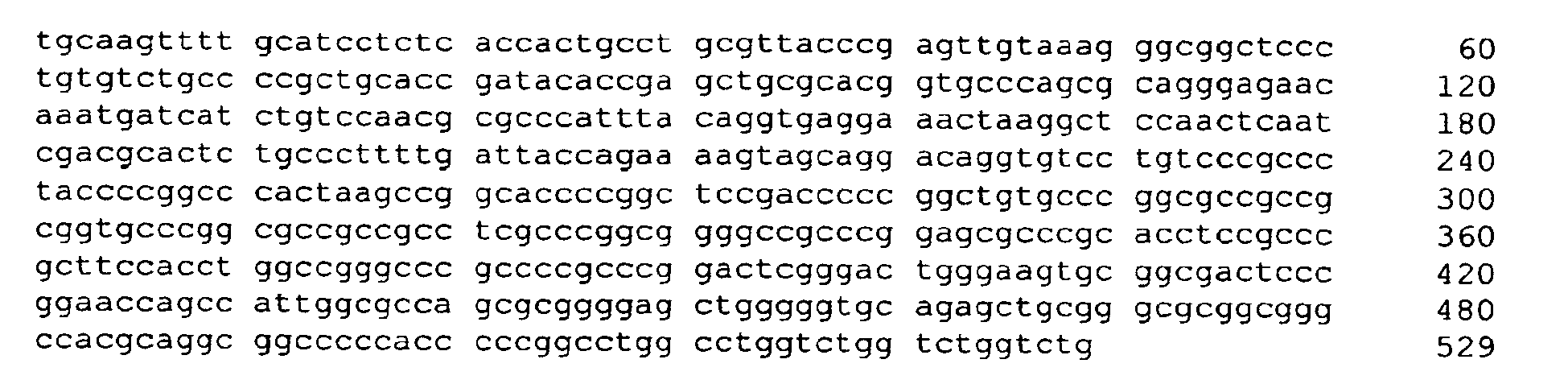
- <210> 142
<211> 28
<212> DNA
<213> Artificial Sequence - <220>
<223> chemically treated genomic DNA (Homo sapiens) - <400> 142
- tgtaagtttt gtattttttt attattgt 28
- <210> 143
<211> 23
<212> DNA
<213> Artificial Sequence - <220>
<223> chemically treated genomic DNA (Homo sapiens) - <400> 143
- caaaccaaac caaaccaaac
caa 23
- caaaccaaac caaaccaaac
- <210> 144
<211> 550
<212> DNA
<213> Artificial Sequence - <220>
<223> chemically treated genomic DNA (Homo sapiens) - <400> 144


- <210> 145
<211> 25
<212> DNA
<213> Artificial Sequence - <220>
<223> chemically treated genomic DNA (Homo sapiens) - <400> 145
- acctaaccta atctaatcta atcta 25
- <210> 196
<211> 20
<212> DNA
<213> Artificial Sequence - <220>
<223> chemically treated genomic DNA (Homo sapiens) - <400> 146
- gttttgtttt gggaggaatt 20
- <210> 147
<211> 423
<212> DNA
<213> Artificial Sequence - <220>
<223> chemically treated genomic DNA (Homo sapiens) - <400> 147

- <210> 148
<211> 20
<212> DNA
<213> Artificial Sequence - <220>
<223> chemically treated genomic DNA (Homo sapiens) - <400> 148
- aattcctccc aaaacaaaac 20
- <210> 149
<211> 20
<212> DNA
<213> Artificial Sequence - <220>
<223> chemically treated genomic DNA (Homo sapiens) - <400> 149
- ggtggagtga ggatgttagt 20
- <210> 150
<211> 282
<212> DNA
<213> Artificial Sequence - <220>
<223> chemically treated genomic DNA (Homo sapiens) - <400> 150

- <210> 151
<211> 27
<212> DNA
<213> Artificial Sequence - <220>
<223> chemically treated genomic DNA (Homo sapiens) - <400> 151
- cctacctata aaaatcatat aaaccaa 27
- <210> 152
<211> 28
<212> DNA
<213> Artificial Sequence - <220>
<223> chemically treated genomic DNA (Homo sapiens) - <400> 152
- gtaatttata tggtgatagg tatttata 28
- <210> 153
<211> 459
<212> DNA
<213> Artificial Sequence - <220>
<223> chemically treated genomic DNA (Homo sapiens) - <400> 153


- <210> 154
<211> 26
<212> DNA
<213> Artificial Sequence - <220>
<223> chemically treated genomic DNA (Homo sapiens) - <400> 154
- gtgtggtagg aattttttag gaagtt 26
- <210> 155
<211> 22
<212> DNA
<213> Artificial Sequence - <220>
<223> chemically treated genomic DNA (Homo sapiens) - <400> 155
- ctcctctctc cacctcctcc
aa 22
- ctcctctctc cacctcctcc
- <210> 156
<211> 260
<212> DNA
<213> Artificial Sequence - <220>
<223> chemically treated genomic DNA (Homo sapiens) - <400> 156

- <210> 157
<211> 27
<212> DNA
<213> Artificial Sequence - <220>
<223> chemically treated genomic DNA (Homo sapiens) - <400> 157
- gaagaaatta gattagataa agagtgt 27
- <210> 158
<211> 23
<212> DNA
<213> Artificial Sequence - <220>
<223> chemically treated genomic DNA (Homo sapiens) - <400> 158
- cctttacata cttattcccc
caa 23
- cctttacata cttattcccc
Claims (25)
- A method for detecting and/or classifying hepatocellular or colorectal cell proliferative disorders by detecting the presence or absence of CpG methylation within the Septin 9 gene and/or its promoter or regulatory elements in a subject wherein CpG methylation is indicative of the presence or class of said disorder.
- The method according to claim 1 wherein a malignant colorectal cell proliferative disorder is distinguished from a benign colorectal cell proliferative disorder said method characterized in that the presence of CpG methylation is indicative of the presence of a malignant cell proliferative disorder and the absence thereof is indicative of the presence of a benign cell proliferative disorder.
- The method according to claim 1 wherein said cell proliferative disorder is cancer.
- The method according to claim 3 wherein said CpG positions are located within SEQ ID NO: 1.
- The method according to any of claims 1 to 4, wherein the presence of methylation indicates the presence of a cell proliferative disorder.
- A method according to claim 1, comprising contacting genomic DNA isolated from a biological sample obtained from said subject with at least one reagent, or series of reagents that distinguishes between methylated and non-methylated CpG dinucleotides within at least one target region of the genomic DNA, wherein the target region comprises essentially of a sequence of at least 16 contiguous nucleotides of at least one sequence selected from the group consisting of SEQ ID NO: 2 to SEQ ID NO: 3 respectively, wherein said contiguous nucleotides comprise at least one CpG dinucleotide sequence, and whereby detecting and/or classifying cell proliferative disorders is, at least in part, afforded.
- A method according to claim 1, comprising:a) extracting or otherwise isolating genomic DNA from a biological sample obtained from the subjectb) treating the genomic DNA of a), or a fragment thereof, with one or more reagents to convert cytosine bases that are unmethylated in the 5-position thereof to uracil or to another base that is detectably dissimilar to cytosine in terms of hybridization properties;c) contacting the treated genomic DNA, or the treated fragment thereof, with an amplification enzyme and at least one primer comprising essentially of, a contiguous sequence of at least 9 nucleotides that is complementary to a sequence selected from the group consisting of SEQ ID NO: 6 to SEQ ID NO: 9 and SEQ ID NO: 12 to SEQ ID NO: 15, and complements thereof, wherein the treated genomic DNA or the fragment thereof is either amplified to produce at least one amplificate, or is not amplified; andd) determining, based on a presence or absence of, or on a property of said amplificate, the methylation state or level of at least one CpG dinucleotide of a sequence selected from the group consisting SEQ ID NO: 2 to SEQ ID NO: 3, or an average, or a value reflecting an average methylation state or level of a plurality of CpG dinucleotides of a sequence selected from the groups consisting of SEQ ID NO: 2 to SEQ ID NO: 3, whereby at least one of detecting and classifying cellular proliferative disorders is, at least in part, afforded.
- The method of claim 7, wherein treating the genomic DNA, or the fragment thereof in b), comprises use of a reagent selected from the group comprising of bisulfite, hydrogen sulfite, disulfite, and combinations thereof.
- The method of claim 7, wherein contacting or amplifying in c) comprises use of at least one method selected from the group comprising: use of a heat-resistant DNA polymerase as the amplification enzyme; use of a polymerase lacking 5'-3' exonuclease activity; use of a polymerase chain reaction (PCR); generation of an amplificate nucleic acid molecule carrying a detectable label.
- The method of any of claim 1 to 9, wherein the biological sample obtained from the subject is selected from the group comprising cell lines, histological slides, biopsies, paraffin-embedded tissue, body fluids, stool, colonic effluent , urine, blood plasma, blood serum, whole blood, isolated blood cells, cells isolated from the blood and combinations thereof.
- The method of claim 7, further comprising in step d) the use of at least one nucleic acid molecule or peptide nucleic acid molecule comprising in each case a contiguous sequence at least 9 nucleotides in length that is complementary to a sequence selected from the group consisting of SEQ ID NO: 6 to SEQ ID NO: 9 and SEQ ID NO: 12 to SEQ ID NO: 15, and complements thereof, wherein said nucleic acid molecule or peptide nucleic acid molecule suppresses amplification of the nucleic acid to which it is hybridized.
- The method of claim 7, wherein determining in d) comprises hybridization of at least one nucleic acid molecule or peptide nucleic acid molecule in each case comprising a contiguous sequence at least 9 nucleotides in length that is complementary to a sequence selected from the group consisting of SEQ ID NO: 6 to SEQ ID NO: 9 and SEQ ID NO: 12 to SEQ ID NO: 15, and complements thereof.
- The method of claim 12, wherein at least one such hybridizing nucleic acid molecule or peptide nucleic acid molecule is bound to a solid phase.
- The method of claim 12, further comprising extending at least one such hybridized nucleic acid molecule by at least one nucleotide base.
- The method of claim 7, wherein determining in d), comprises sequencing of the amplificate.
- The method of claim 7, wherein contacting or amplifying in c), comprises use of methylation-specific primers.
- A method according to claim 1, comprising:a) extracting or otherwise isolating genomic DNA from a biological sample obtained from the subject, .b) digesting the genomic DNA of a), or a fragment thereof, with one or more methylation sensitive restriction enzymes, contacting the DNA restriction enzyme digest of b), with an amplification enzyme and at least two primers comprising, in each case a contiguous sequence at least 16 nucleotides in length that is complementary to a sequence selected from the group consisting SEQ ID NO: 2 to SEQ ID NO: 3, andc) determining, based on a presence or absence of an amplificate the methylation state or level of at least one CpG dinucleotide of a sequence selected from the group consisting SEQ ID NO: 2 to SEQ ID NO: 3, whereby at least one of detecting and classifying cellular proliferative disorders is, at least in part, afforded.
- The method according to claim 17 wherein the presence or absence of an amplificate is determined by means of hybridization to at least one nucleic acid or peptide nucleic acid which is identical or complementary to an at least 16 base long segment of a sequence selected from SEQ ID NO: 2 to SEQ ID NO: 3.
- A nucleic acid, comprising essentially of at least 16 contiguous nucleotides of a nucleic acid sequence selected from the group consisting of SEQ ID NO: 6-7; 12-13, and sequences complementary thereto,
- A nucleic acid, comprising essentially of at least 50 contiguous nucleotides of a DNA sequence selected from the group consisting of SEQ ID NO: 6-7; 12-13, and sequences complementary thereto.
- The nucleic acid of any of claims 19 to 20 wherein the contiguous base sequence comprises at least one CpG, TpG or CpA dinucleotide sequence.
- A kit suitable for performing the method according to claim 9 comprising (a) a bisulfite reagent; (b) a container suitable for containing the said bisulfite reagent and the biological sample of the patient; (c) at least one set of oligonucleotides containing two oligonucleotides whose sequences in each case are identical to a 9 or more preferably 18 base long segment of a sequence selected from SEQ ID NO: 6 to SEQ ID NO: 7 and SEQ ID NO: 12 to SEQ ID NO: 13 or complements thereof.
- A kit suitable for performing the method according to claim 9 comprising (a) a methylation sensitive restriction enzyme reagent; (b) a container suitable for containing the said reagent and the biological sample of the patient; (c) at least one set of oligonucleotides one or a plurality of nucleic acids or peptide nucleic acids which are identical to an at least 9 base long segment of a sequence selected from SEQ ID NO: 2 or complements thereof; and optionally (d) instructions for use and interpretation of the kit results.
- The use of a method according to claims 1 to 18, a nucleic acid according to claims 19 to 21 and/or a kit according to claims 22 to 23 in the diagnosis and/or classification of hepatocellular or colorectal cellular proliferative disorders.
- The use of a nucleic acid, comprising essentially of at least 16 contiguous nucleotides of a nucleic acid sequence selected from the group consisting of SEQ ID NO: 8-9; 14-15 and sequences complementary thereto in the diagnosis and/or classification of hepatocellular or colorectal cellular proliferative disorders.
Priority Applications (3)
| Application Number | Priority Date | Filing Date | Title |
|---|---|---|---|
| PL06007989T PL1721992T3 (en) | 2005-04-15 | 2006-04-18 | Methods and nucleic acids for analyses of cellular proliferative disorders |
| EP08013800A EP2009115A3 (en) | 2005-04-15 | 2006-04-18 | Methods and nucleic acids for analyses of cellular proliferative disorders |
| SI200630135T SI1721992T1 (en) | 2005-04-15 | 2006-04-18 | Methods and nucleic acids for analyses of cellular proliferative disorders |
Applications Claiming Priority (7)
| Application Number | Priority Date | Filing Date | Title |
|---|---|---|---|
| US67224205P | 2005-04-15 | 2005-04-15 | |
| US67699705P | 2005-05-02 | 2005-05-02 | |
| US69752105P | 2005-07-08 | 2005-07-08 | |
| US70486005P | 2005-08-01 | 2005-08-01 | |
| US70931805P | 2005-08-17 | 2005-08-17 | |
| US72360205P | 2005-10-04 | 2005-10-04 | |
| US78740206P | 2006-03-30 | 2006-03-30 |
Related Child Applications (1)
| Application Number | Title | Priority Date | Filing Date |
|---|---|---|---|
| EP08013800A Division EP2009115A3 (en) | 2005-04-15 | 2006-04-18 | Methods and nucleic acids for analyses of cellular proliferative disorders |
Publications (3)
| Publication Number | Publication Date |
|---|---|
| EP1721992A2 EP1721992A2 (en) | 2006-11-15 |
| EP1721992A3 EP1721992A3 (en) | 2006-12-20 |
| EP1721992B1 true EP1721992B1 (en) | 2008-10-08 |
Family
ID=36746665
Family Applications (4)
| Application Number | Title | Priority Date | Filing Date |
|---|---|---|---|
| EP06750221A Withdrawn EP1877577A2 (en) | 2005-04-15 | 2006-04-17 | Methods and nucleic acids for analyses of cellular proliferative disorders |
| EP08021839.9A Expired - Lifetime EP2045335B1 (en) | 2005-04-15 | 2006-04-17 | Methods for the analysis of cellular proliferative disorders |
| EP06007989A Expired - Lifetime EP1721992B1 (en) | 2005-04-15 | 2006-04-18 | Methods and nucleic acids for analyses of cellular proliferative disorders |
| EP08013800A Withdrawn EP2009115A3 (en) | 2005-04-15 | 2006-04-18 | Methods and nucleic acids for analyses of cellular proliferative disorders |
Family Applications Before (2)
| Application Number | Title | Priority Date | Filing Date |
|---|---|---|---|
| EP06750221A Withdrawn EP1877577A2 (en) | 2005-04-15 | 2006-04-17 | Methods and nucleic acids for analyses of cellular proliferative disorders |
| EP08021839.9A Expired - Lifetime EP2045335B1 (en) | 2005-04-15 | 2006-04-17 | Methods for the analysis of cellular proliferative disorders |
Family Applications After (1)
| Application Number | Title | Priority Date | Filing Date |
|---|---|---|---|
| EP08013800A Withdrawn EP2009115A3 (en) | 2005-04-15 | 2006-04-18 | Methods and nucleic acids for analyses of cellular proliferative disorders |
Country Status (20)
| Country | Link |
|---|---|
| US (8) | US7749702B2 (en) |
| EP (4) | EP1877577A2 (en) |
| JP (2) | JP5341506B2 (en) |
| KR (3) | KR101708544B1 (en) |
| CN (3) | CN107663538B (en) |
| AT (1) | ATE410524T1 (en) |
| AU (1) | AU2006236620B2 (en) |
| BR (1) | BRPI0607508B8 (en) |
| CA (1) | CA2606296C (en) |
| DE (1) | DE602006003012D1 (en) |
| DK (1) | DK1721992T3 (en) |
| EA (1) | EA013634B1 (en) |
| ES (2) | ES2533767T3 (en) |
| IL (1) | IL186556A (en) |
| MX (1) | MX2007012782A (en) |
| PL (1) | PL1721992T3 (en) |
| PT (1) | PT1721992E (en) |
| SG (1) | SG183708A1 (en) |
| SI (1) | SI1721992T1 (en) |
| WO (1) | WO2006113466A2 (en) |
Families Citing this family (73)
| Publication number | Priority date | Publication date | Assignee | Title |
|---|---|---|---|---|
| EP1668148B1 (en) | 2003-09-04 | 2008-12-31 | Human Genetic Signatures PTY Ltd. | Nucleic acid detection assay |
| NZ555620A (en) | 2004-12-03 | 2008-08-29 | Human Genetic Signatures Pty | Methods for simplifying microbial nucleic acids by chemical modification of cytosines |
| CA2606296C (en) | 2005-04-15 | 2019-05-21 | Epigenomics Ag | Methods and nucleic acids for analyses of cellular proliferative disorders |
| EP1883707B1 (en) * | 2005-05-26 | 2014-04-09 | Human Genetic Signatures PTY Ltd | Isothermal strand displacement amplification using primers containing a non-regular base |
| CN101292046B (en) | 2005-09-14 | 2013-03-13 | 人类遗传标记控股有限公司 | Assay for a health state |
| EP2484778A3 (en) * | 2006-04-17 | 2012-10-10 | Epigenomics AG | Methods and nucleic acids for the detection of colorectal cell proliferative disorders |
| US20080050738A1 (en) * | 2006-05-31 | 2008-02-28 | Human Genetic Signatures Pty Ltd. | Detection of target nucleic acid |
| ES2546182T3 (en) * | 2006-07-21 | 2015-09-21 | Epigenomics Ag | Methods and kits for methylation analysis in colorectal cell proliferative disorders |
| TWI335354B (en) | 2006-09-27 | 2011-01-01 | Univ Hong Kong Chinese | Methods for the detection of the degree of the methylation of a target dna and kits |
| US7662566B2 (en) * | 2006-11-22 | 2010-02-16 | Myriad Genetics, Inc. | Gene copy number profiling |
| US20080221056A1 (en) * | 2007-02-12 | 2008-09-11 | Johns Hopkins University | Early Detection and Prognosis of Colon Cancers |
| US20100092972A1 (en) * | 2007-03-16 | 2010-04-15 | Human Genetic Signatures Pty Ltd. | Assay for gene expression |
| WO2008122447A2 (en) * | 2007-04-10 | 2008-10-16 | Epigenomics Ag | Dna methylation assay for the diagnosis or prognosis of medical conditions |
| DK3101141T3 (en) | 2007-06-08 | 2020-02-03 | Epigenomics Ag | METHOD OF ANALYZING ANALYSIS |
| FR2919061B1 (en) * | 2007-07-19 | 2009-10-02 | Biomerieux Sa | METHOD OF DOSING PLASTINE-I FOR IN VITRO DIAGNOSIS OF COLORECTAL CANCER. |
| AU2008329549B2 (en) | 2007-11-27 | 2013-08-22 | Human Genetic Signatures Pty Ltd | Enzymes for amplification and copying bisulphite modified nucleic acids |
| AU2008334901A1 (en) * | 2007-12-11 | 2009-06-18 | Epigenomics Ag | Methods and nucleic acids for analyses of lung carcinoma |
| JP2011505845A (en) * | 2007-12-20 | 2011-03-03 | ヒューマン ジェネティック シグネチャーズ ピーティーワイ リミテッド | Eliminate contaminants associated with nucleic acid amplification |
| TW200927925A (en) * | 2007-12-27 | 2009-07-01 | Agriculture And Food Agency Council Of Agriculture | Specific Promoter Region Expresses in Actively Dividing Young Tissues and the Aging Tissues in Plants and its Application |
| WO2009115920A1 (en) * | 2008-03-18 | 2009-09-24 | Epigenomics Ag | A method for optimizing and validating an assay for determining the presence or absence of a medical condition |
| EP2337868A4 (en) * | 2008-10-09 | 2012-03-28 | British Columbia Cancer Agency | DETECTION OF TUMORS OF GRANULOSA |
| US9624530B2 (en) * | 2009-08-03 | 2017-04-18 | Epigenomics Ag | Methods for preservation of genomic DNA sequence complexity |
| TWI434937B (en) * | 2009-10-28 | 2014-04-21 | Univ Nat Chunghsing | Dna sequence from transgenic papaya line 18-2-4 with broad-spectrum resistance to papaya ringspot virus and detection method and use thereof |
| GB201004147D0 (en) * | 2010-03-12 | 2010-04-28 | Dna Electronics Ltd | Method and apparatus for sensing methylation |
| KR101747191B1 (en) | 2011-01-14 | 2017-06-14 | 에스케이하이닉스 주식회사 | Semiconductor Apparatus |
| GB201107466D0 (en) | 2011-05-05 | 2011-06-15 | Loktionov Alexandre | Device and method for non-invasive collection of colorectal mucocellular layer and disease detection |
| WO2012167145A2 (en) * | 2011-06-01 | 2012-12-06 | University Of Southern California | Genome-scale analysis of aberrant dna methylation in colorectal cancer |
| FI20115709A0 (en) * | 2011-07-01 | 2011-07-01 | Helsingin Yliopisto | A method for the diagnosis of hereditary cancers |
| JP6397762B2 (en) * | 2011-07-08 | 2018-09-26 | エピゲノミクス アーゲー | Method for determining prognosis of cancer subject and nucleic acid |
| KR101912488B1 (en) | 2011-09-07 | 2018-10-26 | 휴먼 제네틱 시그너처스 피티와이 엘티디 | Molecular detection assay |
| US9546403B1 (en) | 2011-12-14 | 2017-01-17 | University Of Utah Research Foundation | Substrate for methylated DNA testing |
| EP2828405B1 (en) * | 2012-03-20 | 2017-06-28 | Exact Sciences Corporation | Marker panel for detecting cancer |
| CA2883671A1 (en) * | 2012-08-31 | 2014-03-06 | National Defense Medical Center | Method for screening cancer |
| US10706957B2 (en) | 2012-09-20 | 2020-07-07 | The Chinese University Of Hong Kong | Non-invasive determination of methylome of tumor from plasma |
| US9732390B2 (en) | 2012-09-20 | 2017-08-15 | The Chinese University Of Hong Kong | Non-invasive determination of methylome of fetus or tumor from plasma |
| KR101476769B1 (en) * | 2013-01-02 | 2014-12-26 | 대구대학교 산학협력단 | Biomarker composition for predicting progress from colorectal adenoma to carcinoma of human patients and biokit for dyagnosis thereof comprising the same |
| JP6369857B2 (en) * | 2013-05-29 | 2018-08-08 | シスメックス株式会社 | Method for obtaining information on hepatocellular carcinoma, and marker and kit for obtaining information on hepatocellular carcinoma |
| KR101527283B1 (en) * | 2013-08-13 | 2015-06-10 | 서울대학교산학협력단 | Method for screening cancer marker based on de-glycosylation of glycoproteins and marker for HCC |
| KR101438519B1 (en) * | 2014-07-01 | 2014-09-17 | 대구대학교 산학협력단 | Biomarker composition for predicting progress from colorectal adenoma to carcinoma of human patients and biokit for dyagnosis thereof comprising the same |
| CN106455585A (en) | 2014-07-03 | 2017-02-22 | 伊莱克斯公司 | Household appliance mixing arrangement |
| WO2016000782A1 (en) | 2014-07-03 | 2016-01-07 | Aktiebolaget Electrolux | Household appliance mixing arrangement |
| TWI895765B (en) | 2014-07-18 | 2025-09-01 | 香港中文大學 | Methylation pattern analysis of tissues in a dna mixture |
| CN104745575B (en) | 2014-08-08 | 2019-03-12 | 博诚研究中心 | Gene composition for detecting abnormal cell proliferation or grading disease degree and use thereof |
| ES2896058T3 (en) * | 2014-12-19 | 2022-02-23 | Epigenomics Ag | Methods for detecting CpG methylation and for cancer diagnosis |
| EP3239302A4 (en) * | 2014-12-26 | 2018-05-23 | Peking University | Method for detecting differentially methylated cpg islands associated with abnormal state of human body |
| CN106148501A (en) * | 2015-04-23 | 2016-11-23 | 博诚研究中心 | A kit for detecting Septin9 gene methylation and its application |
| CN104830855A (en) * | 2015-05-21 | 2015-08-12 | 基因科技(上海)有限公司 | Probe for detecting SEPT9 gene methylation and application of probe |
| CA3023335A1 (en) * | 2016-05-04 | 2017-11-30 | Queen's University At Kingston | Cell-free detection of methylated tumour dna |
| RU2630669C1 (en) * | 2016-07-21 | 2017-09-11 | Федеральное бюджетное учреждение науки "Государственный научный центр вирусологии и биотехнологии "Вектор" (ФБУН ГНЦ ВБ "Вектор") | Method for determination of methylation of colorectal cancer tumor marker genes regulatory regions pucgpy sites by glad-pcr analysis, and set of oligonucleotide primers and fluorescence-labelled probes for implementation of this method |
| EP3497220A4 (en) | 2016-08-10 | 2020-04-01 | Grail, Inc. | METHOD FOR PRODUCING DUAL-INDEXED DNA LIBRARIES FOR BISULFIT CONVERSION SEQUENCING |
| CN107723363A (en) * | 2016-08-11 | 2018-02-23 | 博尔诚(北京)科技有限公司 | The combined detection method of tumor markers and its application |
| CA3039685A1 (en) | 2016-11-30 | 2018-06-07 | The Chinese University Of Hong Kong | Analysis of cell-free dna in urine and other samples |
| EP3360971B1 (en) * | 2017-02-09 | 2026-05-20 | Prec For Medicine Gmbh | IMPROVED METHOD FOR EPIGENETIC IMMUNE CELL COUNTING |
| SG10202111266VA (en) | 2017-04-19 | 2021-11-29 | Singlera Genomics Inc | Compositions and methods for library construction and sequence analysis |
| CN109609629A (en) * | 2017-09-30 | 2019-04-12 | 博尔诚(北京)科技有限公司 | Composition and application thereof for detecting liver cancer |
| EP3765637A1 (en) | 2018-03-13 | 2021-01-20 | Grail, Inc. | Anomalous fragment detection and classification |
| AU2019249422B2 (en) | 2018-04-02 | 2025-09-25 | Grail, Inc. | Methylation markers and targeted methylation probe panels |
| CN108866192B (en) * | 2018-07-26 | 2021-06-22 | 上海奕谱生物科技有限公司 | Methylation-modification-based tumor marker STAMP-EP1 |
| CN108866191B (en) | 2018-07-26 | 2021-07-09 | 上海奕谱生物科技有限公司 | Methylation-based tumor marker STAMP-EP2 |
| IL281741B2 (en) | 2018-09-27 | 2026-04-01 | Grail Inc | Methylation markers and targeted methylation probe panel |
| WO2020132572A1 (en) | 2018-12-21 | 2020-06-25 | Grail, Inc. | Source of origin deconvolution based on methylation fragments in cell-free-dna samples |
| CN111560431B (en) * | 2019-02-14 | 2022-11-18 | 博尔诚(北京)科技有限公司 | A composition of health epigenomics and its use |
| CN110211633B (en) * | 2019-05-06 | 2021-08-31 | 臻和精准医学检验实验室无锡有限公司 | MGMT gene promoter methylation detection method, sequencing data processing method and processing device |
| CN112063712A (en) * | 2019-10-28 | 2020-12-11 | 北京康美天鸿生物科技有限公司 | Specific primer pair and kit for detecting septin9 gene methylation based on high-resolution melting curve |
| WO2021122799A1 (en) * | 2019-12-16 | 2021-06-24 | Epigenomics Ag | Methods for detecting colorectal cancer |
| KR102637032B1 (en) * | 2020-01-28 | 2024-02-15 | 주식회사 젠큐릭스 | Composition for diagnosing bladder cancer using CpG methylation status of specific gene and uses thereof |
| AU2021263680A1 (en) * | 2020-04-29 | 2022-11-24 | Freenome Holdings, Inc. | RNA markers and methods for identifying colon cell proliferative disorders |
| WO2023017001A1 (en) | 2021-08-09 | 2023-02-16 | Epigenomics Ag | Improved methods for detecting colorectal cancer |
| CN113584032B (en) * | 2021-08-20 | 2023-08-29 | 深圳市恩辑生物科技有限公司 | Mini promoter pAPP and application thereof |
| CN113862349A (en) * | 2021-10-26 | 2021-12-31 | 山东师范大学 | A diagnostic biomarker for pulmonary fibrosis |
| CN114783511B (en) * | 2022-02-25 | 2025-03-25 | 深圳市硬核酸生物科技有限公司 | Method and device for recommending concentration of nucleic acid molecule solution, and computer equipment |
| WO2024074558A1 (en) | 2022-10-04 | 2024-04-11 | Epigenomics Ag | Improved methods for detecting cancer |
| WO2025019568A1 (en) * | 2023-07-17 | 2025-01-23 | The Penn State Research Foundation | Improving immune cell-based therapies against solid tumors with optogenetically engineered septin proteins |
Family Cites Families (65)
| Publication number | Priority date | Publication date | Assignee | Title |
|---|---|---|---|---|
| US5585481A (en) | 1987-09-21 | 1996-12-17 | Gen-Probe Incorporated | Linking reagents for nucleotide probes |
| US5457183A (en) * | 1989-03-06 | 1995-10-10 | Board Of Regents, The University Of Texas System | Hydroxylated texaphyrins |
| US5245022A (en) | 1990-08-03 | 1993-09-14 | Sterling Drug, Inc. | Exonuclease resistant terminally substituted oligonucleotides |
| US5565552A (en) * | 1992-01-21 | 1996-10-15 | Pharmacyclics, Inc. | Method of expanded porphyrin-oligonucleotide conjugate synthesis |
| US5574142A (en) | 1992-12-15 | 1996-11-12 | Microprobe Corporation | Peptide linkers for improved oligonucleotide delivery |
| SE501439C2 (en) | 1993-06-22 | 1995-02-13 | Pharmacia Lkb Biotech | Method and apparatus for analyzing polynucleotide sequences |
| EP0889122A3 (en) | 1993-11-30 | 1999-03-03 | McGILL UNIVERSITY | Inhibition of DNA Methyltransferase |
| US5597696A (en) | 1994-07-18 | 1997-01-28 | Becton Dickinson And Company | Covalent cyanine dye oligonucleotide conjugates |
| US5514758A (en) | 1994-09-30 | 1996-05-07 | The Goodyear Tire & Rubber Company | Process for making latex for high performance masking tape |
| US5871917A (en) | 1996-05-31 | 1999-02-16 | North Shore University Hospital Research Corp. | Identification of differentially methylated and mutated nucleic acids |
| US6017704A (en) | 1996-06-03 | 2000-01-25 | The Johns Hopkins University School Of Medicine | Method of detection of methylated nucleic acid using agents which modify unmethylated cytosine and distinguishing modified methylated and non-methylated nucleic acids |
| US5786146A (en) * | 1996-06-03 | 1998-07-28 | The Johns Hopkins University School Of Medicine | Method of detection of methylated nucleic acid using agents which modify unmethylated cytosine and distinguishing modified methylated and non-methylated nucleic acids |
| JPH1090169A (en) | 1996-09-19 | 1998-04-10 | Bunshi Bio Photonics Kenkyusho:Kk | Microscope equipment |
| IL129015A0 (en) | 1996-10-01 | 2000-02-17 | Gudrun Rappold Hoerbrand | Human growth gene and short stature gene region |
| WO1998056952A1 (en) | 1997-06-09 | 1998-12-17 | University Of Southern California | A cancer diagnostic method based upon dna methylation differences |
| DE19754482A1 (en) | 1997-11-27 | 1999-07-01 | Epigenomics Gmbh | Process for making complex DNA methylation fingerprints |
| GB9726539D0 (en) | 1997-12-16 | 1998-02-11 | Univ Dundee | Polypeptides, polynucleotides and uses thereof |
| GB9805918D0 (en) * | 1998-03-19 | 1998-05-13 | Nycomed Amersham Plc | Sequencing by hybridisation |
| AUPP312998A0 (en) * | 1998-04-23 | 1998-05-14 | Commonwealth Scientific And Industrial Research Organisation | Diagnostic assay |
| US7700324B1 (en) | 1998-11-03 | 2010-04-20 | The Johns Hopkins University School Of Medicine | Methylated CpG island amplification (MCA) |
| US5958773A (en) * | 1998-12-17 | 1999-09-28 | Isis Pharmaceuticals Inc. | Antisense modulation of AKT-1 expression |
| JP2000221136A (en) | 1999-01-29 | 2000-08-11 | Jasco Corp | Method and apparatus for tracking change over time |
| US6331393B1 (en) * | 1999-05-14 | 2001-12-18 | University Of Southern California | Process for high-throughput DNA methylation analysis |
| US6783933B1 (en) * | 1999-09-15 | 2004-08-31 | The Johns Hopkins University School Of Medicine | CACNA1G polynucleotide, polypeptide and methods of use therefor |
| WO2001029254A2 (en) * | 1999-10-19 | 2001-04-26 | Universiteit Gent | Identification of bpesc1, a gene disrupted by a balanced chromosomal translocation t(3;4) (q23;p15.2) in a patient with bpes |
| JP3783826B2 (en) | 2000-01-17 | 2006-06-07 | 横河電機株式会社 | Biochip reader |
| WO2005000087A2 (en) * | 2003-06-03 | 2005-01-06 | Chiron Corporation | Gene products differentially expressed in cancerous colon cells and their methods of use ii |
| DK1423528T3 (en) * | 2000-09-01 | 2008-05-13 | Epigenomics Ag | Method for determining the degree of methylation of particular cytosines in genomic DNA in the sequence context 5-CPG-3 |
| US6756200B2 (en) * | 2001-01-26 | 2004-06-29 | The Johns Hopkins University School Of Medicine | Aberrantly methylated genes as markers of breast malignancy |
| DE10112515B4 (en) | 2001-03-09 | 2004-02-12 | Epigenomics Ag | Method for the detection of cytosine methylation patterns with high sensitivity |
| US7473767B2 (en) * | 2001-07-03 | 2009-01-06 | The Institute For Systems Biology | Methods for detection and quantification of analytes in complex mixtures |
| JP2005515787A (en) * | 2001-10-18 | 2005-06-02 | ラブレイス レスピレイトリー リサーチ インスティチュート | Monitoring of cancer by abnormal methylation of promoters of transcription factor genes PAX5α, PAX5β, novel loop helix loop protein, novel gene 2, and beta3 gene |
| WO2003042661A2 (en) * | 2001-11-13 | 2003-05-22 | Protein Design Labs, Inc. | Methods of diagnosis of cancer, compositions and methods of screening for modulators of cancer |
| JP3448654B2 (en) | 2001-11-22 | 2003-09-22 | 北陸先端科学技術大学院大学長 | Biochip, biochip array, and screening method using them |
| WO2003048332A2 (en) | 2001-11-30 | 2003-06-12 | The Johns Hopkins University | METHODS FOR ANALYZING METHYLATED CpG ISLANDS AND GC RICH REGIONS |
| JP2003232725A (en) | 2002-02-12 | 2003-08-22 | Japan Science & Technology Corp | Chemical reaction analysis sensor using surface plasmon resonance measurement |
| DE10228917A1 (en) | 2002-06-24 | 2004-01-22 | Epigenomics Ag | Device for handling liquids in a plurality of channels |
| JP4199609B2 (en) | 2002-07-12 | 2008-12-17 | 三菱化学株式会社 | ANALYSIS CHIP, ANALYSIS CHIP UNIT, ANALYSIS DEVICE, AND METHOD FOR PRODUCING ANALYSIS CHIP |
| PT1525188E (en) | 2002-07-16 | 2008-10-16 | Inst Nat Sante Rech Med | Novel derivatives of 4,4`-dithiobis-(3-aminobutane-1-sulphonates) and compositions containing same |
| US20060024684A1 (en) | 2002-10-01 | 2006-02-02 | Epigenomics Ag | Prognostic markers for prediction of treatment response and/or survival of breast cell proliferative disorder patients |
| DE10317955A1 (en) | 2003-04-17 | 2004-11-25 | Epigenomics Ag | Predicting responsiveness of a subject with breast cell proliferative disorder, useful for treating or differentiating breast cell proliferative disorders comprises analyzing methylation pattern of a genomic DNA from the subject |
| CA2500255A1 (en) | 2002-10-01 | 2004-04-29 | Epigenomics Ag | Method and nucleic acids for the treatment of breast cell proliferative disorders |
| DE10245779A1 (en) | 2002-10-01 | 2004-04-29 | Epigenomics Ag | Predicting responsiveness of a subject with breast cell proliferative disorder, useful for treating or differentiating breast cell proliferative disorders comprises analyzing methylation pattern of a genomic DNA from the subject |
| JP2004156911A (en) | 2002-11-01 | 2004-06-03 | Osaka Industrial Promotion Organization | Surface plasmon fluorescence microscope and method for measuring fluorescence excited by surface plasmon |
| JP2004212204A (en) | 2002-12-27 | 2004-07-29 | Olympus Corp | Fluorescence microscope, measuring method of fluorescent lifetime, and measuring program of fluorescent lifetime |
| WO2004074441A2 (en) * | 2003-02-19 | 2004-09-02 | Government Of The United States Of America Represented By The Secretary Department Of Health And Human Services | Amplification or overexpression of mll septin-like fusion (msf) and septin9 and methods related thereto |
| WO2005001141A2 (en) | 2003-06-23 | 2005-01-06 | Epigenomics Ag | Methods and nucleic acids for analyses of colorectal cell proliferative disorders |
| US20040265833A1 (en) * | 2003-06-23 | 2004-12-30 | Cathy Lofton-Day | Methods and nucleic acids for the analysis of colorectal cell proliferative disorders |
| DE602004021192D1 (en) | 2003-10-09 | 2009-07-02 | Epigenomics Ag | IMPROVED BISULFIT CONVERSION OF DNA |
| DE10348407A1 (en) | 2003-10-17 | 2005-05-19 | Widschwendter, Martin, Prof. | Prognostic and diagnostic markers for cell proliferative disorders of breast tissues |
| CN101039951A (en) | 2003-11-03 | 2007-09-19 | 基因信息公司 | Liver cancer biomarkers |
| EP3269826B1 (en) | 2003-12-01 | 2020-03-11 | Epigenomics AG | Methods and nucleic acids for the analysis of gene expression associated with the development of prostate cell proliferative disorders |
| JP2007513621A (en) | 2003-12-11 | 2007-05-31 | エピゲノミクス アーゲー | Methods and nucleic acids for improved treatment of breast cell proliferative disorders |
| EP1561821B1 (en) | 2003-12-11 | 2011-02-16 | Epigenomics AG | Prognostic markers for prediction of treatment response and/or survival of breast cell proliferative disorder patients |
| US20080249118A1 (en) | 2004-05-17 | 2008-10-09 | Heiko Hermeking | Silencing of Tumor-Suppressive Genes by Cpg-Methylation in Prostate Cancer |
| DE102004029700A1 (en) | 2004-06-15 | 2006-02-09 | Epigenomics Ag | Method for quantifying methylated DNA |
| WO2005123945A2 (en) | 2004-06-21 | 2005-12-29 | Epigenomics Ag | Epigenetic markers for the treatment of breast cancer |
| EP1961827A3 (en) * | 2004-07-18 | 2008-09-17 | Epigenomics AG | Epigenetic methods and nucleic acids for the detection of breast cell proliferative disorders |
| JP4192862B2 (en) | 2004-07-20 | 2008-12-10 | ダイソー株式会社 | Method for producing polyether copolymer |
| WO2006060653A2 (en) | 2004-11-30 | 2006-06-08 | Veridex Llc | Lung cancer prognostics |
| CA2593546A1 (en) * | 2004-12-02 | 2006-07-06 | Epigenomics Ag | Methods and nucleic acids for the analysis of gene expression associated with the prognosis of prostate cell proliferative disorders |
| CA2606296C (en) * | 2005-04-15 | 2019-05-21 | Epigenomics Ag | Methods and nucleic acids for analyses of cellular proliferative disorders |
| EP1904649A2 (en) | 2005-07-18 | 2008-04-02 | Epigenomics AG | Compositions and methods for cancer diagnostics comprising pan-cancer markers |
| WO2007115213A2 (en) | 2006-03-30 | 2007-10-11 | Epigenomics Ag | Methods and nucleic acids for analyses of cellular proliferative disorders |
| EP2484778A3 (en) | 2006-04-17 | 2012-10-10 | Epigenomics AG | Methods and nucleic acids for the detection of colorectal cell proliferative disorders |
-
2006
- 2006-04-17 CA CA2606296A patent/CA2606296C/en not_active Expired - Lifetime
- 2006-04-17 WO PCT/US2006/014131 patent/WO2006113466A2/en not_active Ceased
- 2006-04-17 BR BRPI0607508A patent/BRPI0607508B8/en not_active IP Right Cessation
- 2006-04-17 CN CN201710510986.0A patent/CN107663538B/en not_active Expired - Fee Related
- 2006-04-17 EP EP06750221A patent/EP1877577A2/en not_active Withdrawn
- 2006-04-17 EA EA200702203A patent/EA013634B1/en not_active IP Right Cessation
- 2006-04-17 CN CN201410147754.XA patent/CN104046686B/en not_active Expired - Fee Related
- 2006-04-17 EP EP08021839.9A patent/EP2045335B1/en not_active Expired - Lifetime
- 2006-04-17 US US11/405,322 patent/US7749702B2/en active Active
- 2006-04-17 US US11/911,617 patent/US7951563B2/en active Active
- 2006-04-17 KR KR1020147037101A patent/KR101708544B1/en not_active Expired - Fee Related
- 2006-04-17 KR KR1020127031782A patent/KR20130021400A/en not_active Ceased
- 2006-04-17 KR KR1020077026458A patent/KR20080011287A/en not_active Ceased
- 2006-04-17 JP JP2008506752A patent/JP5341506B2/en not_active Expired - Fee Related
- 2006-04-17 SG SG2012059044A patent/SG183708A1/en unknown
- 2006-04-17 CN CN202210279361.9A patent/CN114921544A/en active Pending
- 2006-04-17 MX MX2007012782A patent/MX2007012782A/en active IP Right Grant
- 2006-04-17 AU AU2006236620A patent/AU2006236620B2/en not_active Ceased
- 2006-04-17 ES ES08021839.9T patent/ES2533767T3/en not_active Expired - Lifetime
- 2006-04-18 DK DK06007989T patent/DK1721992T3/en active
- 2006-04-18 EP EP06007989A patent/EP1721992B1/en not_active Expired - Lifetime
- 2006-04-18 DE DE602006003012T patent/DE602006003012D1/en not_active Expired - Lifetime
- 2006-04-18 ES ES06007989T patent/ES2315956T3/en not_active Expired - Lifetime
- 2006-04-18 SI SI200630135T patent/SI1721992T1/en unknown
- 2006-04-18 EP EP08013800A patent/EP2009115A3/en not_active Withdrawn
- 2006-04-18 PL PL06007989T patent/PL1721992T3/en unknown
- 2006-04-18 AT AT06007989T patent/ATE410524T1/en active
- 2006-04-18 PT PT06007989T patent/PT1721992E/en unknown
-
2007
- 2007-10-10 IL IL186556A patent/IL186556A/en active IP Right Grant
-
2010
- 2010-05-13 US US12/779,856 patent/US9695478B2/en not_active Expired - Fee Related
-
2011
- 2011-04-28 US US13/096,932 patent/US8900829B2/en not_active Expired - Lifetime
-
2012
- 2012-11-09 JP JP2012247015A patent/JP5808306B2/en not_active Expired - Fee Related
-
2014
- 2014-11-19 US US14/548,164 patent/US20150086989A1/en not_active Abandoned
-
2016
- 2016-03-23 US US15/078,479 patent/US10385402B2/en active Active
-
2019
- 2019-06-26 US US16/453,688 patent/US11186879B2/en not_active Expired - Fee Related
-
2021
- 2021-10-28 US US17/512,833 patent/US20220154287A1/en not_active Abandoned
Non-Patent Citations (1)
| Title |
|---|
| ECKHARDT F ET AL.: "DNA methylation profiling of human chromosomes 6, 20 and 22", NATURE GENETICS ISSN 1061-4036, vol. 38, no. 12, 2006, pages 1378 - 1385 * |
Also Published As
Similar Documents
| Publication | Publication Date | Title |
|---|---|---|
| US11186879B2 (en) | Methods and nucleic acids for analyses of cellular proliferative disorders | |
| US20220260570A1 (en) | Methods and nucleic acids for the detection of colorectal cell proliferative disorders | |
| US20090317810A1 (en) | Methods and nucleic acids for the detection of colorectal cell proliferative disorders | |
| EP2634265B1 (en) | Methods related to the gene GLI3 for detection of colorectal cancer | |
| US20100184027A1 (en) | Methods and nucleic acids for analyses of cellular proliferative disorders | |
| WO2007115213A2 (en) | Methods and nucleic acids for analyses of cellular proliferative disorders | |
| EP2049681B1 (en) | Methods for analyses of cellular proliferative disorders | |
| HK1172373A (en) | Methods and nucleic acids for the detection of colorectal cell proliferative disorders | |
| HK1167438A (en) | Methods and nucleic acids for the detection of colorectal cell proliferative disorders |
Legal Events
| Date | Code | Title | Description |
|---|---|---|---|
| PUAI | Public reference made under article 153(3) epc to a published international application that has entered the european phase |
Free format text: ORIGINAL CODE: 0009012 |
|
| AK | Designated contracting states |
Kind code of ref document: A2 Designated state(s): AT BE BG CH CY CZ DE DK EE ES FI FR GB GR HU IE IS IT LI LT LU LV MC NL PL PT RO SE SI SK TR |
|
| AX | Request for extension of the european patent |
Extension state: AL BA HR MK YU |
|
| PUAL | Search report despatched |
Free format text: ORIGINAL CODE: 0009013 |
|
| AK | Designated contracting states |
Kind code of ref document: A3 Designated state(s): AT BE BG CH CY CZ DE DK EE ES FI FR GB GR HU IE IS IT LI LT LU LV MC NL PL PT RO SE SI SK TR |
|
| AX | Request for extension of the european patent |
Extension state: AL BA HR MK YU |
|
| 17P | Request for examination filed |
Effective date: 20061212 |
|
| 17Q | First examination report despatched |
Effective date: 20070507 |
|
| AKX | Designation fees paid |
Designated state(s): AT BE BG CH CY CZ DE DK EE ES FI FR GB GR HU IE IS IT LI LT LU LV MC NL PL PT RO SE SI SK TR |
|
| GRAP | Despatch of communication of intention to grant a patent |
Free format text: ORIGINAL CODE: EPIDOSNIGR1 |
|
| GRAS | Grant fee paid |
Free format text: ORIGINAL CODE: EPIDOSNIGR3 |
|
| GRAA | (expected) grant |
Free format text: ORIGINAL CODE: 0009210 |
|
| AK | Designated contracting states |
Kind code of ref document: B1 Designated state(s): AT BE BG CH CY CZ DE DK EE ES FI FR GB GR HU IE IS IT LI LT LU LV MC NL PL PT RO SE SI SK TR |
|
| REG | Reference to a national code |
Ref country code: GB Ref legal event code: FG4D |
|
| REG | Reference to a national code |
Ref country code: CH Ref legal event code: EP |
|
| REG | Reference to a national code |
Ref country code: IE Ref legal event code: FG4D |
|
| REF | Corresponds to: |
Ref document number: 602006003012 Country of ref document: DE Date of ref document: 20081120 Kind code of ref document: P |
|
| REG | Reference to a national code |
Ref country code: SE Ref legal event code: TRGR |
|
| REG | Reference to a national code |
Ref country code: PT Ref legal event code: SC4A Free format text: AVAILABILITY OF NATIONAL TRANSLATION Effective date: 20090106 |
|
| REG | Reference to a national code |
Ref country code: DK Ref legal event code: T3 |
|
| REG | Reference to a national code |
Ref country code: GR Ref legal event code: EP Ref document number: 20090400070 Country of ref document: GR |
|
| REG | Reference to a national code |
Ref country code: CH Ref legal event code: NV Representative=s name: E. BLUM & CO. AG PATENT- UND MARKENANWAELTE VSP |
|
| REG | Reference to a national code |
Ref country code: ES Ref legal event code: FG2A Ref document number: 2315956 Country of ref document: ES Kind code of ref document: T3 |
|
| PG25 | Lapsed in a contracting state [announced via postgrant information from national office to epo] |
Ref country code: BG Free format text: LAPSE BECAUSE OF FAILURE TO SUBMIT A TRANSLATION OF THE DESCRIPTION OR TO PAY THE FEE WITHIN THE PRESCRIBED TIME-LIMIT Effective date: 20090108 Ref country code: LT Free format text: LAPSE BECAUSE OF FAILURE TO SUBMIT A TRANSLATION OF THE DESCRIPTION OR TO PAY THE FEE WITHIN THE PRESCRIBED TIME-LIMIT Effective date: 20081008 |
|
| REG | Reference to a national code |
Ref country code: PL Ref legal event code: T3 |
|
| PG25 | Lapsed in a contracting state [announced via postgrant information from national office to epo] |
Ref country code: IS Free format text: LAPSE BECAUSE OF FAILURE TO SUBMIT A TRANSLATION OF THE DESCRIPTION OR TO PAY THE FEE WITHIN THE PRESCRIBED TIME-LIMIT Effective date: 20090208 Ref country code: LV Free format text: LAPSE BECAUSE OF FAILURE TO SUBMIT A TRANSLATION OF THE DESCRIPTION OR TO PAY THE FEE WITHIN THE PRESCRIBED TIME-LIMIT Effective date: 20081008 |
|
| REG | Reference to a national code |
Ref country code: HU Ref legal event code: AG4A Ref document number: E005052 Country of ref document: HU |
|
| PG25 | Lapsed in a contracting state [announced via postgrant information from national office to epo] |
Ref country code: RO Free format text: LAPSE BECAUSE OF FAILURE TO SUBMIT A TRANSLATION OF THE DESCRIPTION OR TO PAY THE FEE WITHIN THE PRESCRIBED TIME-LIMIT Effective date: 20081008 Ref country code: EE Free format text: LAPSE BECAUSE OF FAILURE TO SUBMIT A TRANSLATION OF THE DESCRIPTION OR TO PAY THE FEE WITHIN THE PRESCRIBED TIME-LIMIT Effective date: 20081008 |
|
| PLBE | No opposition filed within time limit |
Free format text: ORIGINAL CODE: 0009261 |
|
| STAA | Information on the status of an ep patent application or granted ep patent |
Free format text: STATUS: NO OPPOSITION FILED WITHIN TIME LIMIT |
|
| 26N | No opposition filed |
Effective date: 20090709 |
|
| REG | Reference to a national code |
Ref country code: PT Ref legal event code: MM4A Free format text: LAPSE DUE TO NON-PAYMENT OF FEES Effective date: 20100120 |
|
| PG25 | Lapsed in a contracting state [announced via postgrant information from national office to epo] |
Ref country code: MC Free format text: LAPSE BECAUSE OF NON-PAYMENT OF DUE FEES Effective date: 20090430 Ref country code: PT Free format text: LAPSE BECAUSE OF NON-PAYMENT OF DUE FEES Effective date: 20100120 |
|
| REG | Reference to a national code |
Ref country code: PT Ref legal event code: NF4A Free format text: RESTITUTIO IN INTEGRUM Effective date: 20100512 |
|
| PGRI | Patent reinstated in contracting state [announced from national office to epo] |
Ref country code: PT Effective date: 20100512 |
|
| PG25 | Lapsed in a contracting state [announced via postgrant information from national office to epo] |
Ref country code: CY Free format text: LAPSE BECAUSE OF FAILURE TO SUBMIT A TRANSLATION OF THE DESCRIPTION OR TO PAY THE FEE WITHIN THE PRESCRIBED TIME-LIMIT Effective date: 20081008 |
|
| REG | Reference to a national code |
Ref country code: FR Ref legal event code: PLFP Year of fee payment: 11 |
|
| REG | Reference to a national code |
Ref country code: FR Ref legal event code: PLFP Year of fee payment: 12 |
|
| REG | Reference to a national code |
Ref country code: FR Ref legal event code: PLFP Year of fee payment: 13 |
|
| PGFP | Annual fee paid to national office [announced via postgrant information from national office to epo] |
Ref country code: NL Payment date: 20220419 Year of fee payment: 17 |
|
| PGFP | Annual fee paid to national office [announced via postgrant information from national office to epo] |
Ref country code: SK Payment date: 20220404 Year of fee payment: 17 Ref country code: SE Payment date: 20220422 Year of fee payment: 17 Ref country code: PT Payment date: 20220412 Year of fee payment: 17 Ref country code: LU Payment date: 20220420 Year of fee payment: 17 Ref country code: IE Payment date: 20220421 Year of fee payment: 17 Ref country code: DK Payment date: 20220421 Year of fee payment: 17 Ref country code: CZ Payment date: 20220407 Year of fee payment: 17 |
|
| PGFP | Annual fee paid to national office [announced via postgrant information from national office to epo] |
Ref country code: TR Payment date: 20220412 Year of fee payment: 17 Ref country code: SI Payment date: 20220404 Year of fee payment: 17 Ref country code: PL Payment date: 20220407 Year of fee payment: 17 Ref country code: GR Payment date: 20220419 Year of fee payment: 17 Ref country code: FI Payment date: 20220414 Year of fee payment: 17 Ref country code: BE Payment date: 20220419 Year of fee payment: 17 Ref country code: AT Payment date: 20220414 Year of fee payment: 17 |
|
| PGFP | Annual fee paid to national office [announced via postgrant information from national office to epo] |
Ref country code: IT Payment date: 20230426 Year of fee payment: 18 Ref country code: FR Payment date: 20230428 Year of fee payment: 18 Ref country code: ES Payment date: 20230517 Year of fee payment: 18 Ref country code: DE Payment date: 20230424 Year of fee payment: 18 Ref country code: CH Payment date: 20230605 Year of fee payment: 18 |
|
| PGFP | Annual fee paid to national office [announced via postgrant information from national office to epo] |
Ref country code: GB Payment date: 20230427 Year of fee payment: 18 |
|
| REG | Reference to a national code |
Ref country code: DK Ref legal event code: EBP Effective date: 20230430 |
|
| REG | Reference to a national code |
Ref country code: SE Ref legal event code: EUG |
|
| REG | Reference to a national code |
Ref country code: SK Ref legal event code: MM4A Ref document number: E 4886 Country of ref document: SK Effective date: 20230418 Ref country code: NL Ref legal event code: MM Effective date: 20230501 |
|
| REG | Reference to a national code |
Ref country code: AT Ref legal event code: MM01 Ref document number: 410524 Country of ref document: AT Kind code of ref document: T Effective date: 20230418 |
|
| PG25 | Lapsed in a contracting state [announced via postgrant information from national office to epo] |
Ref country code: LU Free format text: LAPSE BECAUSE OF NON-PAYMENT OF DUE FEES Effective date: 20230418 |
|
| REG | Reference to a national code |
Ref country code: BE Ref legal event code: MM Effective date: 20230430 |
|
| PG25 | Lapsed in a contracting state [announced via postgrant information from national office to epo] |
Ref country code: SK Free format text: LAPSE BECAUSE OF NON-PAYMENT OF DUE FEES Effective date: 20230418 |
|
| PG25 | Lapsed in a contracting state [announced via postgrant information from national office to epo] |
Ref country code: GR Free format text: LAPSE BECAUSE OF NON-PAYMENT OF DUE FEES Effective date: 20231110 |
|
| PG25 | Lapsed in a contracting state [announced via postgrant information from national office to epo] |
Ref country code: SK Free format text: LAPSE BECAUSE OF NON-PAYMENT OF DUE FEES Effective date: 20230418 Ref country code: SI Free format text: LAPSE BECAUSE OF NON-PAYMENT OF DUE FEES Effective date: 20230419 Ref country code: SE Free format text: LAPSE BECAUSE OF NON-PAYMENT OF DUE FEES Effective date: 20230419 Ref country code: PT Free format text: LAPSE BECAUSE OF NON-PAYMENT OF DUE FEES Effective date: 20231018 Ref country code: NL Free format text: LAPSE BECAUSE OF NON-PAYMENT OF DUE FEES Effective date: 20230501 Ref country code: HU Free format text: LAPSE BECAUSE OF NON-PAYMENT OF DUE FEES Effective date: 20230419 Ref country code: GR Free format text: LAPSE BECAUSE OF NON-PAYMENT OF DUE FEES Effective date: 20231110 Ref country code: FI Free format text: LAPSE BECAUSE OF NON-PAYMENT OF DUE FEES Effective date: 20230418 Ref country code: CZ Free format text: LAPSE BECAUSE OF NON-PAYMENT OF DUE FEES Effective date: 20230418 Ref country code: AT Free format text: LAPSE BECAUSE OF NON-PAYMENT OF DUE FEES Effective date: 20230418 |
|
| REG | Reference to a national code |
Ref country code: IE Ref legal event code: MM4A |
|
| PG25 | Lapsed in a contracting state [announced via postgrant information from national office to epo] |
Ref country code: BE Free format text: LAPSE BECAUSE OF NON-PAYMENT OF DUE FEES Effective date: 20230430 |
|
| REG | Reference to a national code |
Ref country code: SI Ref legal event code: KO00 Effective date: 20240125 |
|
| PG25 | Lapsed in a contracting state [announced via postgrant information from national office to epo] |
Ref country code: IE Free format text: LAPSE BECAUSE OF NON-PAYMENT OF DUE FEES Effective date: 20230418 |
|
| PG25 | Lapsed in a contracting state [announced via postgrant information from national office to epo] |
Ref country code: IE Free format text: LAPSE BECAUSE OF NON-PAYMENT OF DUE FEES Effective date: 20230418 Ref country code: HU Free format text: LAPSE BECAUSE OF NON-PAYMENT OF DUE FEES Effective date: 20230419 Ref country code: DK Free format text: LAPSE BECAUSE OF NON-PAYMENT OF DUE FEES Effective date: 20230430 |
|
| PGFP | Annual fee paid to national office [announced via postgrant information from national office to epo] |
Ref country code: HU Payment date: 20240116 Year of fee payment: 18 |
|
| PGRI | Patent reinstated in contracting state [announced from national office to epo] |
Ref country code: HU Effective date: 20240129 |
|
| REG | Reference to a national code |
Ref country code: GB Ref legal event code: 732E Free format text: REGISTERED BETWEEN 20240411 AND 20240417 |
|
| PG25 | Lapsed in a contracting state [announced via postgrant information from national office to epo] |
Ref country code: PL Free format text: LAPSE BECAUSE OF NON-PAYMENT OF DUE FEES Effective date: 20230418 |
|
| PG25 | Lapsed in a contracting state [announced via postgrant information from national office to epo] |
Ref country code: PL Free format text: LAPSE BECAUSE OF NON-PAYMENT OF DUE FEES Effective date: 20230418 |
|
| REG | Reference to a national code |
Ref country code: DE Ref legal event code: R119 Ref document number: 602006003012 Country of ref document: DE |
|
| REG | Reference to a national code |
Ref country code: CH Ref legal event code: PL |
|
| GBPC | Gb: european patent ceased through non-payment of renewal fee |
Effective date: 20240418 |
|
| PG25 | Lapsed in a contracting state [announced via postgrant information from national office to epo] |
Ref country code: DE Free format text: LAPSE BECAUSE OF NON-PAYMENT OF DUE FEES Effective date: 20241105 |
|
| PG25 | Lapsed in a contracting state [announced via postgrant information from national office to epo] |
Ref country code: GB Free format text: LAPSE BECAUSE OF NON-PAYMENT OF DUE FEES Effective date: 20240418 |
|
| PG25 | Lapsed in a contracting state [announced via postgrant information from national office to epo] |
Ref country code: FR Free format text: LAPSE BECAUSE OF NON-PAYMENT OF DUE FEES Effective date: 20240430 |
|
| PG25 | Lapsed in a contracting state [announced via postgrant information from national office to epo] |
Ref country code: GB Free format text: LAPSE BECAUSE OF NON-PAYMENT OF DUE FEES Effective date: 20240418 Ref country code: FR Free format text: LAPSE BECAUSE OF NON-PAYMENT OF DUE FEES Effective date: 20240430 Ref country code: DE Free format text: LAPSE BECAUSE OF NON-PAYMENT OF DUE FEES Effective date: 20241105 Ref country code: CH Free format text: LAPSE BECAUSE OF NON-PAYMENT OF DUE FEES Effective date: 20240430 |
|
| PG25 | Lapsed in a contracting state [announced via postgrant information from national office to epo] |
Ref country code: IT Free format text: LAPSE BECAUSE OF NON-PAYMENT OF DUE FEES Effective date: 20240418 |
|
| REG | Reference to a national code |
Ref country code: ES Ref legal event code: FD2A Effective date: 20250529 |
|
| PG25 | Lapsed in a contracting state [announced via postgrant information from national office to epo] |
Ref country code: ES Free format text: LAPSE BECAUSE OF NON-PAYMENT OF DUE FEES Effective date: 20240419 |