EP1706498B1 - Zur Charakterisierung der immunotoxischen Aktivität xenobiotischer Substanzen geeignete Werkzeuge und Verfahren - Google Patents
Zur Charakterisierung der immunotoxischen Aktivität xenobiotischer Substanzen geeignete Werkzeuge und Verfahren Download PDFInfo
- Publication number
- EP1706498B1 EP1706498B1 EP04775181.3A EP04775181A EP1706498B1 EP 1706498 B1 EP1706498 B1 EP 1706498B1 EP 04775181 A EP04775181 A EP 04775181A EP 1706498 B1 EP1706498 B1 EP 1706498B1
- Authority
- EP
- European Patent Office
- Prior art keywords
- expression
- cell line
- egfp
- cells
- reporter
- Prior art date
- Legal status (The legal status is an assumption and is not a legal conclusion. Google has not performed a legal analysis and makes no representation as to the accuracy of the status listed.)
- Expired - Lifetime
Links
- 238000000034 method Methods 0.000 title claims description 49
- 239000000126 substance Substances 0.000 title claims description 45
- 230000002625 immunotoxic effect Effects 0.000 title claims description 10
- 239000002676 xenobiotic agent Substances 0.000 title description 19
- 230000002034 xenobiotic effect Effects 0.000 title description 10
- 230000014509 gene expression Effects 0.000 claims description 150
- 239000013612 plasmid Substances 0.000 claims description 99
- 230000001105 regulatory effect Effects 0.000 claims description 60
- 108020005345 3' Untranslated Regions Proteins 0.000 claims description 58
- 108700008625 Reporter Genes Proteins 0.000 claims description 31
- 150000001875 compounds Chemical class 0.000 claims description 29
- 231100000386 immunotoxicity Toxicity 0.000 claims description 25
- 230000007688 immunotoxicity Effects 0.000 claims description 25
- 102100031181 Glyceraldehyde-3-phosphate dehydrogenase Human genes 0.000 claims description 14
- 108020004445 glyceraldehyde-3-phosphate dehydrogenase Proteins 0.000 claims description 14
- 239000013641 positive control Substances 0.000 claims description 8
- 230000008859 change Effects 0.000 claims description 7
- 231100000110 immunotoxic Toxicity 0.000 claims description 6
- 239000003623 enhancer Substances 0.000 claims description 4
- 210000004027 cell Anatomy 0.000 description 322
- 108010048367 enhanced green fluorescent protein Proteins 0.000 description 107
- 108090000978 Interleukin-4 Proteins 0.000 description 105
- 102000004388 Interleukin-4 Human genes 0.000 description 99
- 108020004414 DNA Proteins 0.000 description 95
- 108010043121 Green Fluorescent Proteins Proteins 0.000 description 94
- 102000004144 Green Fluorescent Proteins Human genes 0.000 description 94
- 239000005090 green fluorescent protein Substances 0.000 description 94
- 108010002350 Interleukin-2 Proteins 0.000 description 85
- 102000000588 Interleukin-2 Human genes 0.000 description 85
- 238000012360 testing method Methods 0.000 description 77
- 108090000695 Cytokines Proteins 0.000 description 75
- 239000013598 vector Substances 0.000 description 67
- 102100037850 Interferon gamma Human genes 0.000 description 58
- 108010074328 Interferon-gamma Proteins 0.000 description 58
- 108060008682 Tumor Necrosis Factor Proteins 0.000 description 51
- PHEDXBVPIONUQT-RGYGYFBISA-N phorbol 13-acetate 12-myristate Chemical compound C([C@]1(O)C(=O)C(C)=C[C@H]1[C@@]1(O)[C@H](C)[C@H]2OC(=O)CCCCCCCCCCCCC)C(CO)=C[C@H]1[C@H]1[C@]2(OC(C)=O)C1(C)C PHEDXBVPIONUQT-RGYGYFBISA-N 0.000 description 51
- 102000000852 Tumor Necrosis Factor-alpha Human genes 0.000 description 50
- 102000004127 Cytokines Human genes 0.000 description 49
- PGHMRUGBZOYCAA-ADZNBVRBSA-N ionomycin Chemical compound O1[C@H](C[C@H](O)[C@H](C)[C@H](O)[C@H](C)/C=C/C[C@@H](C)C[C@@H](C)C(/O)=C/C(=O)[C@@H](C)C[C@@H](C)C[C@@H](CCC(O)=O)C)CC[C@@]1(C)[C@@H]1O[C@](C)([C@@H](C)O)CC1 PGHMRUGBZOYCAA-ADZNBVRBSA-N 0.000 description 49
- PGHMRUGBZOYCAA-UHFFFAOYSA-N ionomycin Natural products O1C(CC(O)C(C)C(O)C(C)C=CCC(C)CC(C)C(O)=CC(=O)C(C)CC(C)CC(CCC(O)=O)C)CCC1(C)C1OC(C)(C(C)O)CC1 PGHMRUGBZOYCAA-UHFFFAOYSA-N 0.000 description 49
- 230000000694 effects Effects 0.000 description 43
- MZOFCQQQCNRIBI-VMXHOPILSA-N (3s)-4-[[(2s)-1-[[(2s)-1-[[(1s)-1-carboxy-2-hydroxyethyl]amino]-4-methyl-1-oxopentan-2-yl]amino]-5-(diaminomethylideneamino)-1-oxopentan-2-yl]amino]-3-[[2-[[(2s)-2,6-diaminohexanoyl]amino]acetyl]amino]-4-oxobutanoic acid Chemical compound OC[C@@H](C(O)=O)NC(=O)[C@H](CC(C)C)NC(=O)[C@H](CCCN=C(N)N)NC(=O)[C@H](CC(O)=O)NC(=O)CNC(=O)[C@@H](N)CCCCN MZOFCQQQCNRIBI-VMXHOPILSA-N 0.000 description 35
- 239000012634 fragment Substances 0.000 description 32
- 238000012163 sequencing technique Methods 0.000 description 32
- 108090000174 Interleukin-10 Proteins 0.000 description 31
- 102000003814 Interleukin-10 Human genes 0.000 description 31
- 102000007469 Actins Human genes 0.000 description 30
- 108010085238 Actins Proteins 0.000 description 30
- 238000010790 dilution Methods 0.000 description 25
- 239000012895 dilution Substances 0.000 description 25
- 238000002474 experimental method Methods 0.000 description 25
- 239000013604 expression vector Substances 0.000 description 25
- 239000002609 medium Substances 0.000 description 25
- 238000011144 upstream manufacturing Methods 0.000 description 25
- 238000001890 transfection Methods 0.000 description 22
- 108090000623 proteins and genes Proteins 0.000 description 20
- 239000013566 allergen Substances 0.000 description 19
- PMATZTZNYRCHOR-CGLBZJNRSA-N Cyclosporin A Chemical compound CC[C@@H]1NC(=O)[C@H]([C@H](O)[C@H](C)C\C=C\C)N(C)C(=O)[C@H](C(C)C)N(C)C(=O)[C@H](CC(C)C)N(C)C(=O)[C@H](CC(C)C)N(C)C(=O)[C@@H](C)NC(=O)[C@H](C)NC(=O)[C@H](CC(C)C)N(C)C(=O)[C@H](C(C)C)NC(=O)[C@H](CC(C)C)N(C)C(=O)CN(C)C1=O PMATZTZNYRCHOR-CGLBZJNRSA-N 0.000 description 18
- 108010036949 Cyclosporine Proteins 0.000 description 18
- IAZDPXIOMUYVGZ-UHFFFAOYSA-N Dimethylsulphoxide Chemical compound CS(C)=O IAZDPXIOMUYVGZ-UHFFFAOYSA-N 0.000 description 18
- 229960001265 ciclosporin Drugs 0.000 description 18
- 238000010367 cloning Methods 0.000 description 18
- 230000008488 polyadenylation Effects 0.000 description 18
- 229930105110 Cyclosporin A Natural products 0.000 description 17
- 230000004913 activation Effects 0.000 description 17
- BLFLLBZGZJTVJG-UHFFFAOYSA-N benzocaine Chemical compound CCOC(=O)C1=CC=C(N)C=C1 BLFLLBZGZJTVJG-UHFFFAOYSA-N 0.000 description 16
- 238000001943 fluorescence-activated cell sorting Methods 0.000 description 16
- 238000000338 in vitro Methods 0.000 description 16
- 108091008146 restriction endonucleases Proteins 0.000 description 16
- 108020004999 messenger RNA Proteins 0.000 description 15
- 230000006698 induction Effects 0.000 description 14
- ZAHRKKWIAAJSAO-UHFFFAOYSA-N rapamycin Natural products COCC(O)C(=C/C(C)C(=O)CC(OC(=O)C1CCCCN1C(=O)C(=O)C2(O)OC(CC(OC)C(=CC=CC=CC(C)CC(C)C(=O)C)C)CCC2C)C(C)CC3CCC(O)C(C3)OC)C ZAHRKKWIAAJSAO-UHFFFAOYSA-N 0.000 description 14
- QFJCIRLUMZQUOT-HPLJOQBZSA-N sirolimus Chemical compound C1C[C@@H](O)[C@H](OC)C[C@@H]1C[C@@H](C)[C@H]1OC(=O)[C@@H]2CCCCN2C(=O)C(=O)[C@](O)(O2)[C@H](C)CC[C@H]2C[C@H](OC)/C(C)=C/C=C/C=C/[C@@H](C)C[C@@H](C)C(=O)[C@H](OC)[C@H](O)/C(C)=C/[C@@H](C)C(=O)C1 QFJCIRLUMZQUOT-HPLJOQBZSA-N 0.000 description 14
- 229960002930 sirolimus Drugs 0.000 description 14
- 238000011161 development Methods 0.000 description 13
- 230000018109 developmental process Effects 0.000 description 13
- 210000003630 histaminocyte Anatomy 0.000 description 13
- JGSARLDLIJGVTE-MBNYWOFBSA-N Penicillin G Chemical compound N([C@H]1[C@H]2SC([C@@H](N2C1=O)C(O)=O)(C)C)C(=O)CC1=CC=CC=C1 JGSARLDLIJGVTE-MBNYWOFBSA-N 0.000 description 12
- 230000002519 immonomodulatory effect Effects 0.000 description 12
- 210000002865 immune cell Anatomy 0.000 description 12
- 230000001939 inductive effect Effects 0.000 description 12
- 238000012216 screening Methods 0.000 description 12
- 239000011550 stock solution Substances 0.000 description 12
- DVKJHBMWWAPEIU-UHFFFAOYSA-N toluene 2,4-diisocyanate Chemical compound CC1=CC=C(N=C=O)C=C1N=C=O DVKJHBMWWAPEIU-UHFFFAOYSA-N 0.000 description 12
- VYZAHLCBVHPDDF-UHFFFAOYSA-N Dinitrochlorobenzene Chemical compound [O-][N+](=O)C1=CC=C(Cl)C([N+]([O-])=O)=C1 VYZAHLCBVHPDDF-UHFFFAOYSA-N 0.000 description 11
- 238000003556 assay Methods 0.000 description 11
- 238000001514 detection method Methods 0.000 description 11
- 230000029087 digestion Effects 0.000 description 11
- 238000000799 fluorescence microscopy Methods 0.000 description 11
- 102000034287 fluorescent proteins Human genes 0.000 description 11
- 108091006047 fluorescent proteins Proteins 0.000 description 11
- 238000010363 gene targeting Methods 0.000 description 11
- 230000001965 increasing effect Effects 0.000 description 11
- 230000005764 inhibitory process Effects 0.000 description 11
- 239000000047 product Substances 0.000 description 11
- 230000004044 response Effects 0.000 description 11
- 108091026890 Coding region Proteins 0.000 description 10
- LFQSCWFLJHTTHZ-UHFFFAOYSA-N Ethanol Chemical compound CCO LFQSCWFLJHTTHZ-UHFFFAOYSA-N 0.000 description 10
- 210000001744 T-lymphocyte Anatomy 0.000 description 10
- 239000002158 endotoxin Substances 0.000 description 10
- 102000004169 proteins and genes Human genes 0.000 description 10
- 108020004705 Codon Proteins 0.000 description 9
- -1 IL-1β Proteins 0.000 description 9
- 108091028043 Nucleic acid sequence Proteins 0.000 description 9
- APQHKWPGGHMYKJ-UHFFFAOYSA-N Tributyltin oxide Chemical compound CCCC[Sn](CCCC)(CCCC)O[Sn](CCCC)(CCCC)CCCC APQHKWPGGHMYKJ-UHFFFAOYSA-N 0.000 description 9
- 210000002950 fibroblast Anatomy 0.000 description 9
- 230000001506 immunosuppresive effect Effects 0.000 description 9
- 210000002510 keratinocyte Anatomy 0.000 description 9
- 230000000638 stimulation Effects 0.000 description 9
- 230000008685 targeting Effects 0.000 description 9
- 230000003827 upregulation Effects 0.000 description 9
- HGUFODBRKLSHSI-UHFFFAOYSA-N 2,3,7,8-tetrachloro-dibenzo-p-dioxin Chemical compound O1C2=CC(Cl)=C(Cl)C=C2OC2=C1C=C(Cl)C(Cl)=C2 HGUFODBRKLSHSI-UHFFFAOYSA-N 0.000 description 8
- UEJJHQNACJXSKW-UHFFFAOYSA-N 2-(2,6-dioxopiperidin-3-yl)-1H-isoindole-1,3(2H)-dione Chemical compound O=C1C2=CC=CC=C2C(=O)N1C1CCC(=O)NC1=O UEJJHQNACJXSKW-UHFFFAOYSA-N 0.000 description 8
- 108700039691 Genetic Promoter Regions Proteins 0.000 description 8
- 101710162684 Glyceraldehyde-3-phosphate dehydrogenase 3 Proteins 0.000 description 8
- 229930193140 Neomycin Natural products 0.000 description 8
- 238000013459 approach Methods 0.000 description 8
- 239000003795 chemical substances by application Substances 0.000 description 8
- 231100000135 cytotoxicity Toxicity 0.000 description 8
- 230000003013 cytotoxicity Effects 0.000 description 8
- 238000005516 engineering process Methods 0.000 description 8
- 239000000463 material Substances 0.000 description 8
- 229960004927 neomycin Drugs 0.000 description 8
- 230000008569 process Effects 0.000 description 8
- 229960003433 thalidomide Drugs 0.000 description 8
- 102000004190 Enzymes Human genes 0.000 description 7
- 108090000790 Enzymes Proteins 0.000 description 7
- DBMJMQXJHONAFJ-UHFFFAOYSA-M Sodium laurylsulphate Chemical compound [Na+].CCCCCCCCCCCCOS([O-])(=O)=O DBMJMQXJHONAFJ-UHFFFAOYSA-M 0.000 description 7
- 229960004784 allergens Drugs 0.000 description 7
- 238000004458 analytical method Methods 0.000 description 7
- 229960005274 benzocaine Drugs 0.000 description 7
- 230000016396 cytokine production Effects 0.000 description 7
- 229910003460 diamond Inorganic materials 0.000 description 7
- 239000010432 diamond Substances 0.000 description 7
- 231100000673 dose–response relationship Toxicity 0.000 description 7
- 238000000684 flow cytometry Methods 0.000 description 7
- 210000000987 immune system Anatomy 0.000 description 7
- 238000001727 in vivo Methods 0.000 description 7
- LWJROJCJINYWOX-UHFFFAOYSA-L mercury dichloride Chemical compound Cl[Hg]Cl LWJROJCJINYWOX-UHFFFAOYSA-L 0.000 description 7
- XDRYMKDFEDOLFX-UHFFFAOYSA-N pentamidine Chemical compound C1=CC(C(=N)N)=CC=C1OCCCCCOC1=CC=C(C(N)=N)C=C1 XDRYMKDFEDOLFX-UHFFFAOYSA-N 0.000 description 7
- 229960004448 pentamidine Drugs 0.000 description 7
- 238000003153 stable transfection Methods 0.000 description 7
- 230000001629 suppression Effects 0.000 description 7
- 108700019146 Transgenes Proteins 0.000 description 6
- 230000009471 action Effects 0.000 description 6
- 208000026935 allergic disease Diseases 0.000 description 6
- 238000010276 construction Methods 0.000 description 6
- 239000002596 immunotoxin Substances 0.000 description 6
- 230000002637 immunotoxin Effects 0.000 description 6
- 229940051026 immunotoxin Drugs 0.000 description 6
- 231100000608 immunotoxin Toxicity 0.000 description 6
- 230000002401 inhibitory effect Effects 0.000 description 6
- 229940056360 penicillin g Drugs 0.000 description 6
- 230000009467 reduction Effects 0.000 description 6
- 235000019333 sodium laurylsulphate Nutrition 0.000 description 6
- 230000009261 transgenic effect Effects 0.000 description 6
- 241001529936 Murinae Species 0.000 description 5
- 108091034117 Oligonucleotide Proteins 0.000 description 5
- 230000005784 autoimmunity Effects 0.000 description 5
- 230000001580 bacterial effect Effects 0.000 description 5
- 230000001413 cellular effect Effects 0.000 description 5
- 238000004520 electroporation Methods 0.000 description 5
- 230000000527 lymphocytic effect Effects 0.000 description 5
- 210000004962 mammalian cell Anatomy 0.000 description 5
- 239000003550 marker Substances 0.000 description 5
- 230000001404 mediated effect Effects 0.000 description 5
- 230000004048 modification Effects 0.000 description 5
- 238000012986 modification Methods 0.000 description 5
- 238000003757 reverse transcription PCR Methods 0.000 description 5
- 239000000523 sample Substances 0.000 description 5
- 238000010200 validation analysis Methods 0.000 description 5
- 206010020751 Hypersensitivity Diseases 0.000 description 4
- 206010062016 Immunosuppression Diseases 0.000 description 4
- 241001465754 Metazoa Species 0.000 description 4
- 241000699666 Mus <mouse, genus> Species 0.000 description 4
- 108700026244 Open Reading Frames Proteins 0.000 description 4
- 238000012408 PCR amplification Methods 0.000 description 4
- 208000000389 T-cell leukemia Diseases 0.000 description 4
- 208000028530 T-cell lymphoblastic leukemia/lymphoma Diseases 0.000 description 4
- 230000004075 alteration Effects 0.000 description 4
- 238000000137 annealing Methods 0.000 description 4
- 210000000988 bone and bone Anatomy 0.000 description 4
- 239000013599 cloning vector Substances 0.000 description 4
- 238000012239 gene modification Methods 0.000 description 4
- 230000005017 genetic modification Effects 0.000 description 4
- 235000013617 genetically modified food Nutrition 0.000 description 4
- 230000003308 immunostimulating effect Effects 0.000 description 4
- 230000001976 improved effect Effects 0.000 description 4
- 238000003780 insertion Methods 0.000 description 4
- 230000037431 insertion Effects 0.000 description 4
- 231100000021 irritant Toxicity 0.000 description 4
- 239000002085 irritant Substances 0.000 description 4
- 230000000670 limiting effect Effects 0.000 description 4
- 210000004698 lymphocyte Anatomy 0.000 description 4
- 238000004519 manufacturing process Methods 0.000 description 4
- 238000005259 measurement Methods 0.000 description 4
- 230000007246 mechanism Effects 0.000 description 4
- 239000000203 mixture Substances 0.000 description 4
- 230000001599 osteoclastic effect Effects 0.000 description 4
- 230000009103 reabsorption Effects 0.000 description 4
- 230000035945 sensitivity Effects 0.000 description 4
- 239000000243 solution Substances 0.000 description 4
- 231100000331 toxic Toxicity 0.000 description 4
- 230000002588 toxic effect Effects 0.000 description 4
- 238000013518 transcription Methods 0.000 description 4
- 230000035897 transcription Effects 0.000 description 4
- 108700028369 Alleles Proteins 0.000 description 3
- 238000002965 ELISA Methods 0.000 description 3
- 241000588724 Escherichia coli Species 0.000 description 3
- 238000000636 Northern blotting Methods 0.000 description 3
- ZLMJMSJWJFRBEC-UHFFFAOYSA-N Potassium Chemical compound [K] ZLMJMSJWJFRBEC-UHFFFAOYSA-N 0.000 description 3
- 239000011543 agarose gel Substances 0.000 description 3
- 230000007815 allergy Effects 0.000 description 3
- 238000010171 animal model Methods 0.000 description 3
- 239000000427 antigen Substances 0.000 description 3
- 108091007433 antigens Proteins 0.000 description 3
- 102000036639 antigens Human genes 0.000 description 3
- 239000003710 calcium ionophore Substances 0.000 description 3
- 238000004113 cell culture Methods 0.000 description 3
- 230000003833 cell viability Effects 0.000 description 3
- 238000003271 compound fluorescence assay Methods 0.000 description 3
- 230000001276 controlling effect Effects 0.000 description 3
- 230000007423 decrease Effects 0.000 description 3
- 210000004443 dendritic cell Anatomy 0.000 description 3
- 239000000428 dust Substances 0.000 description 3
- 238000001976 enzyme digestion Methods 0.000 description 3
- 102000015694 estrogen receptors Human genes 0.000 description 3
- 108010038795 estrogen receptors Proteins 0.000 description 3
- 230000006870 function Effects 0.000 description 3
- 230000028993 immune response Effects 0.000 description 3
- 238000010348 incorporation Methods 0.000 description 3
- 230000003993 interaction Effects 0.000 description 3
- 239000002555 ionophore Substances 0.000 description 3
- 230000000236 ionophoric effect Effects 0.000 description 3
- 230000008774 maternal effect Effects 0.000 description 3
- 229960002523 mercuric chloride Drugs 0.000 description 3
- 238000005457 optimization Methods 0.000 description 3
- 229910052700 potassium Inorganic materials 0.000 description 3
- 239000011591 potassium Substances 0.000 description 3
- 238000002360 preparation method Methods 0.000 description 3
- 230000002829 reductive effect Effects 0.000 description 3
- 230000000284 resting effect Effects 0.000 description 3
- 230000002269 spontaneous effect Effects 0.000 description 3
- 238000007619 statistical method Methods 0.000 description 3
- 239000000725 suspension Substances 0.000 description 3
- 208000008732 thymoma Diseases 0.000 description 3
- 230000035899 viability Effects 0.000 description 3
- 108091032973 (ribonucleotides)n+m Proteins 0.000 description 2
- UPMLOUAZCHDJJD-UHFFFAOYSA-N 4,4'-Diphenylmethane Diisocyanate Chemical compound C1=CC(N=C=O)=CC=C1CC1=CC=C(N=C=O)C=C1 UPMLOUAZCHDJJD-UHFFFAOYSA-N 0.000 description 2
- IJGRMHOSHXDMSA-UHFFFAOYSA-N Atomic nitrogen Chemical compound N#N IJGRMHOSHXDMSA-UHFFFAOYSA-N 0.000 description 2
- 238000001712 DNA sequencing Methods 0.000 description 2
- YQYJSBFKSSDGFO-UHFFFAOYSA-N Epihygromycin Natural products OC1C(O)C(C(=O)C)OC1OC(C(=C1)O)=CC=C1C=C(C)C(=O)NC1C(O)C(O)C2OCOC2C1O YQYJSBFKSSDGFO-UHFFFAOYSA-N 0.000 description 2
- 101000834253 Gallus gallus Actin, cytoplasmic 1 Proteins 0.000 description 2
- 241000282412 Homo Species 0.000 description 2
- 102100020881 Interleukin-1 alpha Human genes 0.000 description 2
- 108010002616 Interleukin-5 Proteins 0.000 description 2
- 102000000743 Interleukin-5 Human genes 0.000 description 2
- 229910020427 K2PtCl4 Inorganic materials 0.000 description 2
- 231100000416 LDH assay Toxicity 0.000 description 2
- 108010086093 Mung Bean Nuclease Proteins 0.000 description 2
- 101001033286 Mus musculus Interleukin-1 beta Proteins 0.000 description 2
- 241000699670 Mus sp. Species 0.000 description 2
- MWUXSHHQAYIFBG-UHFFFAOYSA-N Nitric oxide Chemical compound O=[N] MWUXSHHQAYIFBG-UHFFFAOYSA-N 0.000 description 2
- 238000012181 QIAquick gel extraction kit Methods 0.000 description 2
- 238000002105 Southern blotting Methods 0.000 description 2
- 108010006785 Taq Polymerase Proteins 0.000 description 2
- 108700009124 Transcription Initiation Site Proteins 0.000 description 2
- JLCPHMBAVCMARE-UHFFFAOYSA-N [3-[[3-[[3-[[3-[[3-[[3-[[3-[[3-[[3-[[3-[[3-[[5-(2-amino-6-oxo-1H-purin-9-yl)-3-[[3-[[3-[[3-[[3-[[3-[[5-(2-amino-6-oxo-1H-purin-9-yl)-3-[[5-(2-amino-6-oxo-1H-purin-9-yl)-3-hydroxyoxolan-2-yl]methoxy-hydroxyphosphoryl]oxyoxolan-2-yl]methoxy-hydroxyphosphoryl]oxy-5-(5-methyl-2,4-dioxopyrimidin-1-yl)oxolan-2-yl]methoxy-hydroxyphosphoryl]oxy-5-(6-aminopurin-9-yl)oxolan-2-yl]methoxy-hydroxyphosphoryl]oxy-5-(6-aminopurin-9-yl)oxolan-2-yl]methoxy-hydroxyphosphoryl]oxy-5-(6-aminopurin-9-yl)oxolan-2-yl]methoxy-hydroxyphosphoryl]oxy-5-(6-aminopurin-9-yl)oxolan-2-yl]methoxy-hydroxyphosphoryl]oxyoxolan-2-yl]methoxy-hydroxyphosphoryl]oxy-5-(5-methyl-2,4-dioxopyrimidin-1-yl)oxolan-2-yl]methoxy-hydroxyphosphoryl]oxy-5-(4-amino-2-oxopyrimidin-1-yl)oxolan-2-yl]methoxy-hydroxyphosphoryl]oxy-5-(5-methyl-2,4-dioxopyrimidin-1-yl)oxolan-2-yl]methoxy-hydroxyphosphoryl]oxy-5-(5-methyl-2,4-dioxopyrimidin-1-yl)oxolan-2-yl]methoxy-hydroxyphosphoryl]oxy-5-(6-aminopurin-9-yl)oxolan-2-yl]methoxy-hydroxyphosphoryl]oxy-5-(6-aminopurin-9-yl)oxolan-2-yl]methoxy-hydroxyphosphoryl]oxy-5-(4-amino-2-oxopyrimidin-1-yl)oxolan-2-yl]methoxy-hydroxyphosphoryl]oxy-5-(4-amino-2-oxopyrimidin-1-yl)oxolan-2-yl]methoxy-hydroxyphosphoryl]oxy-5-(4-amino-2-oxopyrimidin-1-yl)oxolan-2-yl]methoxy-hydroxyphosphoryl]oxy-5-(6-aminopurin-9-yl)oxolan-2-yl]methoxy-hydroxyphosphoryl]oxy-5-(4-amino-2-oxopyrimidin-1-yl)oxolan-2-yl]methyl [5-(6-aminopurin-9-yl)-2-(hydroxymethyl)oxolan-3-yl] hydrogen phosphate Polymers Cc1cn(C2CC(OP(O)(=O)OCC3OC(CC3OP(O)(=O)OCC3OC(CC3O)n3cnc4c3nc(N)[nH]c4=O)n3cnc4c3nc(N)[nH]c4=O)C(COP(O)(=O)OC3CC(OC3COP(O)(=O)OC3CC(OC3COP(O)(=O)OC3CC(OC3COP(O)(=O)OC3CC(OC3COP(O)(=O)OC3CC(OC3COP(O)(=O)OC3CC(OC3COP(O)(=O)OC3CC(OC3COP(O)(=O)OC3CC(OC3COP(O)(=O)OC3CC(OC3COP(O)(=O)OC3CC(OC3COP(O)(=O)OC3CC(OC3COP(O)(=O)OC3CC(OC3COP(O)(=O)OC3CC(OC3COP(O)(=O)OC3CC(OC3COP(O)(=O)OC3CC(OC3COP(O)(=O)OC3CC(OC3COP(O)(=O)OC3CC(OC3CO)n3cnc4c(N)ncnc34)n3ccc(N)nc3=O)n3cnc4c(N)ncnc34)n3ccc(N)nc3=O)n3ccc(N)nc3=O)n3ccc(N)nc3=O)n3cnc4c(N)ncnc34)n3cnc4c(N)ncnc34)n3cc(C)c(=O)[nH]c3=O)n3cc(C)c(=O)[nH]c3=O)n3ccc(N)nc3=O)n3cc(C)c(=O)[nH]c3=O)n3cnc4c3nc(N)[nH]c4=O)n3cnc4c(N)ncnc34)n3cnc4c(N)ncnc34)n3cnc4c(N)ncnc34)n3cnc4c(N)ncnc34)O2)c(=O)[nH]c1=O JLCPHMBAVCMARE-UHFFFAOYSA-N 0.000 description 2
- 230000002411 adverse Effects 0.000 description 2
- 238000000246 agarose gel electrophoresis Methods 0.000 description 2
- 230000008901 benefit Effects 0.000 description 2
- 230000004071 biological effect Effects 0.000 description 2
- 230000036755 cellular response Effects 0.000 description 2
- 238000012512 characterization method Methods 0.000 description 2
- 239000013611 chromosomal DNA Substances 0.000 description 2
- 229930182912 cyclosporin Natural products 0.000 description 2
- 231100000263 cytotoxicity test Toxicity 0.000 description 2
- 238000013461 design Methods 0.000 description 2
- CJAONIOAQZUHPN-KKLWWLSJSA-N ethyl 12-[[2-[(2r,3r)-3-[2-[(12-ethoxy-12-oxododecyl)-methylamino]-2-oxoethoxy]butan-2-yl]oxyacetyl]-methylamino]dodecanoate Chemical compound CCOC(=O)CCCCCCCCCCCN(C)C(=O)CO[C@H](C)[C@@H](C)OCC(=O)N(C)CCCCCCCCCCCC(=O)OCC CJAONIOAQZUHPN-KKLWWLSJSA-N 0.000 description 2
- 238000011156 evaluation Methods 0.000 description 2
- 238000001917 fluorescence detection Methods 0.000 description 2
- 239000001963 growth medium Substances 0.000 description 2
- 229910001385 heavy metal Inorganic materials 0.000 description 2
- 230000001900 immune effect Effects 0.000 description 2
- 230000036737 immune function Effects 0.000 description 2
- 239000003018 immunosuppressive agent Substances 0.000 description 2
- 229930027917 kanamycin Natural products 0.000 description 2
- 229960000318 kanamycin Drugs 0.000 description 2
- SBUJHOSQTJFQJX-NOAMYHISSA-N kanamycin Chemical compound O[C@@H]1[C@@H](O)[C@H](O)[C@@H](CN)O[C@@H]1O[C@H]1[C@H](O)[C@@H](O[C@@H]2[C@@H]([C@@H](N)[C@H](O)[C@@H](CO)O2)O)[C@H](N)C[C@@H]1N SBUJHOSQTJFQJX-NOAMYHISSA-N 0.000 description 2
- 229930182823 kanamycin A Natural products 0.000 description 2
- 238000002843 lactate dehydrogenase assay Methods 0.000 description 2
- 210000004185 liver Anatomy 0.000 description 2
- 238000001823 molecular biology technique Methods 0.000 description 2
- 230000007302 negative regulation of cytokine production Effects 0.000 description 2
- PGSADBUBUOPOJS-UHFFFAOYSA-N neutral red Chemical compound Cl.C1=C(C)C(N)=CC2=NC3=CC(N(C)C)=CC=C3N=C21 PGSADBUBUOPOJS-UHFFFAOYSA-N 0.000 description 2
- LGQLOGILCSXPEA-UHFFFAOYSA-L nickel sulfate Chemical compound [Ni+2].[O-]S([O-])(=O)=O LGQLOGILCSXPEA-UHFFFAOYSA-L 0.000 description 2
- 229910000363 nickel(II) sulfate Inorganic materials 0.000 description 2
- 238000001543 one-way ANOVA Methods 0.000 description 2
- 210000000056 organ Anatomy 0.000 description 2
- 230000000144 pharmacologic effect Effects 0.000 description 2
- 230000035790 physiological processes and functions Effects 0.000 description 2
- 239000002244 precipitate Substances 0.000 description 2
- 230000035755 proliferation Effects 0.000 description 2
- 238000001243 protein synthesis Methods 0.000 description 2
- 230000008844 regulatory mechanism Effects 0.000 description 2
- 230000001235 sensitizing effect Effects 0.000 description 2
- 230000000087 stabilizing effect Effects 0.000 description 2
- 230000010473 stable expression Effects 0.000 description 2
- 238000003860 storage Methods 0.000 description 2
- 239000006228 supernatant Substances 0.000 description 2
- 230000001988 toxicity Effects 0.000 description 2
- 231100000419 toxicity Toxicity 0.000 description 2
- 231100000041 toxicology testing Toxicity 0.000 description 2
- 230000002103 transcriptional effect Effects 0.000 description 2
- 230000009466 transformation Effects 0.000 description 2
- 230000014616 translation Effects 0.000 description 2
- 238000012795 verification Methods 0.000 description 2
- 102000040650 (ribonucleotides)n+m Human genes 0.000 description 1
- AZXKALLRCOCGBV-UHFFFAOYSA-N 1-phenyl-2-(propan-2-ylamino)hexan-1-one Chemical compound CCCCC(NC(C)C)C(=O)C1=CC=CC=C1 AZXKALLRCOCGBV-UHFFFAOYSA-N 0.000 description 1
- 229920000936 Agarose Polymers 0.000 description 1
- 108010001857 Cell Surface Receptors Proteins 0.000 description 1
- 102000019034 Chemokines Human genes 0.000 description 1
- 108010012236 Chemokines Proteins 0.000 description 1
- 101001116419 Dermatophagoides pteronyssinus Peptidase 1 Proteins 0.000 description 1
- 101100364969 Dictyostelium discoideum scai gene Proteins 0.000 description 1
- 206010013700 Drug hypersensitivity Diseases 0.000 description 1
- 206010014952 Eosinophilia myalgia syndrome Diseases 0.000 description 1
- 108700039887 Essential Genes Proteins 0.000 description 1
- 108091092566 Extrachromosomal DNA Proteins 0.000 description 1
- 238000012413 Fluorescence activated cell sorting analysis Methods 0.000 description 1
- 108010013216 GATATC-specific type II deoxyribonucleases Proteins 0.000 description 1
- 108010042546 GCGGCCGC-specific type II deoxyribonucleases Proteins 0.000 description 1
- 101150066002 GFP gene Proteins 0.000 description 1
- 238000006595 Griess deamination reaction Methods 0.000 description 1
- 102000012428 Hematopoietic Cell Growth Factors Human genes 0.000 description 1
- 108010022580 Hematopoietic Cell Growth Factors Proteins 0.000 description 1
- 108010050904 Interferons Proteins 0.000 description 1
- 102000014150 Interferons Human genes 0.000 description 1
- 108010002352 Interleukin-1 Proteins 0.000 description 1
- 102000000589 Interleukin-1 Human genes 0.000 description 1
- 108090000193 Interleukin-1 beta Proteins 0.000 description 1
- 108010082786 Interleukin-1alpha Proteins 0.000 description 1
- 238000001282 Kruskal–Wallis one-way analysis of variance Methods 0.000 description 1
- 102000003960 Ligases Human genes 0.000 description 1
- 108090000364 Ligases Proteins 0.000 description 1
- 231100000002 MTT assay Toxicity 0.000 description 1
- 238000000134 MTT assay Methods 0.000 description 1
- 241000124008 Mammalia Species 0.000 description 1
- 101710151803 Mitochondrial intermediate peptidase 2 Proteins 0.000 description 1
- 241000699660 Mus musculus Species 0.000 description 1
- 101000579126 Mus musculus Phosphoglycerate kinase 1 Proteins 0.000 description 1
- 101100364971 Mus musculus Scai gene Proteins 0.000 description 1
- 101000648740 Mus musculus Tumor necrosis factor Proteins 0.000 description 1
- 206010057249 Phagocytosis Diseases 0.000 description 1
- 238000010240 RT-PCR analysis Methods 0.000 description 1
- 241000700584 Simplexvirus Species 0.000 description 1
- 238000000692 Student's t-test Methods 0.000 description 1
- 230000006044 T cell activation Effects 0.000 description 1
- 229940126530 T cell activator Drugs 0.000 description 1
- 206010042971 T-cell lymphoma Diseases 0.000 description 1
- 208000027585 T-cell non-Hodgkin lymphoma Diseases 0.000 description 1
- 101150003725 TK gene Proteins 0.000 description 1
- 108020004440 Thymidine kinase Proteins 0.000 description 1
- 241001149960 Tolypocladium inflatum Species 0.000 description 1
- 102000040945 Transcription factor Human genes 0.000 description 1
- 108091023040 Transcription factor Proteins 0.000 description 1
- 206010067584 Type 1 diabetes mellitus Diseases 0.000 description 1
- 230000005856 abnormality Effects 0.000 description 1
- 238000002835 absorbance Methods 0.000 description 1
- 230000001464 adherent effect Effects 0.000 description 1
- 229940074608 allergen extract Drugs 0.000 description 1
- 208000028004 allergic respiratory disease Diseases 0.000 description 1
- 150000003862 amino acid derivatives Chemical class 0.000 description 1
- 150000001413 amino acids Chemical class 0.000 description 1
- 229960000723 ampicillin Drugs 0.000 description 1
- AVKUERGKIZMTKX-NJBDSQKTSA-N ampicillin Chemical compound C1([C@@H](N)C(=O)N[C@H]2[C@H]3SC([C@@H](N3C2=O)C(O)=O)(C)C)=CC=CC=C1 AVKUERGKIZMTKX-NJBDSQKTSA-N 0.000 description 1
- 239000002260 anti-inflammatory agent Substances 0.000 description 1
- 229940124599 anti-inflammatory drug Drugs 0.000 description 1
- 230000003110 anti-inflammatory effect Effects 0.000 description 1
- 210000000612 antigen-presenting cell Anatomy 0.000 description 1
- 239000003904 antiprotozoal agent Substances 0.000 description 1
- 208000006673 asthma Diseases 0.000 description 1
- 239000012298 atmosphere Substances 0.000 description 1
- LMEKQMALGUDUQG-UHFFFAOYSA-N azathioprine Chemical compound CN1C=NC([N+]([O-])=O)=C1SC1=NC=NC2=C1NC=N2 LMEKQMALGUDUQG-UHFFFAOYSA-N 0.000 description 1
- 229960002170 azathioprine Drugs 0.000 description 1
- 230000010310 bacterial transformation Effects 0.000 description 1
- IYNDLOXRXUOGIU-LQDWTQKMSA-M benzylpenicillin potassium Chemical compound [K+].N([C@H]1[C@H]2SC([C@@H](N2C1=O)C([O-])=O)(C)C)C(=O)CC1=CC=CC=C1 IYNDLOXRXUOGIU-LQDWTQKMSA-M 0.000 description 1
- 230000003115 biocidal effect Effects 0.000 description 1
- 230000008827 biological function Effects 0.000 description 1
- 230000033228 biological regulation Effects 0.000 description 1
- 238000005415 bioluminescence Methods 0.000 description 1
- 230000029918 bioluminescence Effects 0.000 description 1
- 239000000090 biomarker Substances 0.000 description 1
- 238000010804 cDNA synthesis Methods 0.000 description 1
- WKRWUYKLUMMAKG-WYGBAUISSA-L calcium;(4r,6s,8s,10z,12r,14r,16e,18r,19r,20s,21s)-19,21-dihydroxy-22-[(2s,5s)-5-[(2r,5s)-5-[(1r)-1-hydroxyethyl]-5-methyloxolan-2-yl]-5-methyloxolan-2-yl]-4,6,8,12,14,18,20-heptamethyl-11-oxido-9-oxodocosa-10,16-dienoate Chemical compound [Ca+2].O1[C@H](C[C@H](O)[C@H](C)[C@H](O)[C@H](C)/C=C/C[C@@H](C)C[C@@H](C)C(/[O-])=C/C(=O)[C@@H](C)C[C@@H](C)C[C@@H](CCC([O-])=O)C)CC[C@@]1(C)[C@@H]1O[C@](C)([C@@H](C)O)CC1 WKRWUYKLUMMAKG-WYGBAUISSA-L 0.000 description 1
- 238000004364 calculation method Methods 0.000 description 1
- 238000013354 cell banking Methods 0.000 description 1
- 238000010370 cell cloning Methods 0.000 description 1
- 239000013592 cell lysate Substances 0.000 description 1
- 230000011748 cell maturation Effects 0.000 description 1
- 238000001516 cell proliferation assay Methods 0.000 description 1
- 238000003570 cell viability assay Methods 0.000 description 1
- 230000030570 cellular localization Effects 0.000 description 1
- 230000007248 cellular mechanism Effects 0.000 description 1
- 239000003153 chemical reaction reagent Substances 0.000 description 1
- 210000000349 chromosome Anatomy 0.000 description 1
- 230000002596 correlated effect Effects 0.000 description 1
- 230000000875 corresponding effect Effects 0.000 description 1
- 210000004748 cultured cell Anatomy 0.000 description 1
- 238000012258 culturing Methods 0.000 description 1
- 238000005520 cutting process Methods 0.000 description 1
- 238000004163 cytometry Methods 0.000 description 1
- 238000002784 cytotoxicity assay Methods 0.000 description 1
- 230000034994 death Effects 0.000 description 1
- 230000002939 deleterious effect Effects 0.000 description 1
- 230000001419 dependent effect Effects 0.000 description 1
- 230000002074 deregulated effect Effects 0.000 description 1
- 230000000368 destabilizing effect Effects 0.000 description 1
- KORSJDCBLAPZEQ-UHFFFAOYSA-N dicyclohexylmethane-4,4'-diisocyanate Chemical compound C1CC(N=C=O)CCC1CC1CCC(N=C=O)CC1 KORSJDCBLAPZEQ-UHFFFAOYSA-N 0.000 description 1
- 230000003467 diminishing effect Effects 0.000 description 1
- 238000009826 distribution Methods 0.000 description 1
- 239000000975 dye Substances 0.000 description 1
- 238000001962 electrophoresis Methods 0.000 description 1
- 230000007613 environmental effect Effects 0.000 description 1
- 238000010228 ex vivo assay Methods 0.000 description 1
- 230000028023 exocytosis Effects 0.000 description 1
- 108010092809 exonuclease Bal 31 Proteins 0.000 description 1
- 230000002349 favourable effect Effects 0.000 description 1
- 230000008014 freezing Effects 0.000 description 1
- 238000007710 freezing Methods 0.000 description 1
- 238000010230 functional analysis Methods 0.000 description 1
- 238000002825 functional assay Methods 0.000 description 1
- IRSCQMHQWWYFCW-UHFFFAOYSA-N ganciclovir Chemical compound O=C1NC(N)=NC2=C1N=CN2COC(CO)CO IRSCQMHQWWYFCW-UHFFFAOYSA-N 0.000 description 1
- 229960002963 ganciclovir Drugs 0.000 description 1
- 230000002068 genetic effect Effects 0.000 description 1
- 238000010362 genome editing Methods 0.000 description 1
- 230000008821 health effect Effects 0.000 description 1
- CKAPSXZOOQJIBF-UHFFFAOYSA-N hexachlorobenzene Chemical compound ClC1=C(Cl)C(Cl)=C(Cl)C(Cl)=C1Cl CKAPSXZOOQJIBF-UHFFFAOYSA-N 0.000 description 1
- 210000005260 human cell Anatomy 0.000 description 1
- 210000004408 hybridoma Anatomy 0.000 description 1
- 230000009610 hypersensitivity Effects 0.000 description 1
- 208000026278 immune system disease Diseases 0.000 description 1
- 230000036039 immunity Effects 0.000 description 1
- 239000002955 immunomodulating agent Substances 0.000 description 1
- 229940121354 immunomodulator Drugs 0.000 description 1
- 230000002584 immunomodulator Effects 0.000 description 1
- 229960003444 immunosuppressant agent Drugs 0.000 description 1
- 229940124589 immunosuppressive drug Drugs 0.000 description 1
- 238000000099 in vitro assay Methods 0.000 description 1
- 238000005462 in vivo assay Methods 0.000 description 1
- 238000011534 incubation Methods 0.000 description 1
- 230000028709 inflammatory response Effects 0.000 description 1
- 238000002347 injection Methods 0.000 description 1
- 239000007924 injection Substances 0.000 description 1
- 230000008611 intercellular interaction Effects 0.000 description 1
- 230000002452 interceptive effect Effects 0.000 description 1
- 229940047124 interferons Drugs 0.000 description 1
- 230000003834 intracellular effect Effects 0.000 description 1
- 238000002955 isolation Methods 0.000 description 1
- 238000003367 kinetic assay Methods 0.000 description 1
- 238000002372 labelling Methods 0.000 description 1
- 239000007788 liquid Substances 0.000 description 1
- 238000004020 luminiscence type Methods 0.000 description 1
- 230000007257 malfunction Effects 0.000 description 1
- 238000013507 mapping Methods 0.000 description 1
- 230000010534 mechanism of action Effects 0.000 description 1
- 102000006240 membrane receptors Human genes 0.000 description 1
- 230000004060 metabolic process Effects 0.000 description 1
- 238000000329 molecular dynamics simulation Methods 0.000 description 1
- 230000009456 molecular mechanism Effects 0.000 description 1
- 238000012544 monitoring process Methods 0.000 description 1
- 230000000877 morphologic effect Effects 0.000 description 1
- 230000004660 morphological change Effects 0.000 description 1
- 239000002547 new drug Substances 0.000 description 1
- 229910052757 nitrogen Inorganic materials 0.000 description 1
- 108020004707 nucleic acids Proteins 0.000 description 1
- 102000039446 nucleic acids Human genes 0.000 description 1
- 150000007523 nucleic acids Chemical class 0.000 description 1
- 230000003287 optical effect Effects 0.000 description 1
- 235000019371 penicillin G benzathine Nutrition 0.000 description 1
- YBVNFKZSMZGRAD-UHFFFAOYSA-N pentamidine isethionate Chemical compound OCCS(O)(=O)=O.OCCS(O)(=O)=O.C1=CC(C(=N)N)=CC=C1OCCCCCOC1=CC=C(C(N)=N)C=C1 YBVNFKZSMZGRAD-UHFFFAOYSA-N 0.000 description 1
- 230000008782 phagocytosis Effects 0.000 description 1
- 239000002644 phorbol ester Substances 0.000 description 1
- 150000003057 platinum Chemical class 0.000 description 1
- 230000004983 pleiotropic effect Effects 0.000 description 1
- 231100000683 possible toxicity Toxicity 0.000 description 1
- 230000003389 potentiating effect Effects 0.000 description 1
- 230000037452 priming Effects 0.000 description 1
- 230000009696 proliferative response Effects 0.000 description 1
- 230000000644 propagated effect Effects 0.000 description 1
- 238000003908 quality control method Methods 0.000 description 1
- 108020003175 receptors Proteins 0.000 description 1
- 102000005962 receptors Human genes 0.000 description 1
- 230000022532 regulation of transcription, DNA-dependent Effects 0.000 description 1
- 201000004335 respiratory allergy Diseases 0.000 description 1
- 230000000241 respiratory effect Effects 0.000 description 1
- 239000006152 selective media Substances 0.000 description 1
- 239000012679 serum free medium Substances 0.000 description 1
- 230000019491 signal transduction Effects 0.000 description 1
- 238000010572 single replacement reaction Methods 0.000 description 1
- 210000003491 skin Anatomy 0.000 description 1
- MFBOGIVSZKQAPD-UHFFFAOYSA-M sodium butyrate Chemical compound [Na+].CCCC([O-])=O MFBOGIVSZKQAPD-UHFFFAOYSA-M 0.000 description 1
- 159000000000 sodium salts Chemical class 0.000 description 1
- 239000002904 solvent Substances 0.000 description 1
- 210000001082 somatic cell Anatomy 0.000 description 1
- 238000010561 standard procedure Methods 0.000 description 1
- 230000004936 stimulating effect Effects 0.000 description 1
- 208000024891 symptom Diseases 0.000 description 1
- 238000003786 synthesis reaction Methods 0.000 description 1
- 238000012353 t test Methods 0.000 description 1
- 210000001519 tissue Anatomy 0.000 description 1
- 231100000167 toxic agent Toxicity 0.000 description 1
- 230000005030 transcription termination Effects 0.000 description 1
- 238000011830 transgenic mouse model Methods 0.000 description 1
- 230000010474 transient expression Effects 0.000 description 1
- 238000002054 transplantation Methods 0.000 description 1
- 230000032258 transport Effects 0.000 description 1
- 208000035408 type 1 diabetes mellitus 1 Diseases 0.000 description 1
- 231100000747 viability assay Toxicity 0.000 description 1
- 238000003026 viability measurement method Methods 0.000 description 1
- 230000003612 virological effect Effects 0.000 description 1
Images












Classifications
-
- C—CHEMISTRY; METALLURGY
- C07—ORGANIC CHEMISTRY
- C07K—PEPTIDES
- C07K14/00—Peptides having more than 20 amino acids; Gastrins; Somatostatins; Melanotropins; Derivatives thereof
- C07K14/435—Peptides having more than 20 amino acids; Gastrins; Somatostatins; Melanotropins; Derivatives thereof from animals; from humans
- C07K14/52—Cytokines; Lymphokines; Interferons
-
- C—CHEMISTRY; METALLURGY
- C12—BIOCHEMISTRY; BEER; SPIRITS; WINE; VINEGAR; MICROBIOLOGY; ENZYMOLOGY; MUTATION OR GENETIC ENGINEERING
- C12N—MICROORGANISMS OR ENZYMES; COMPOSITIONS THEREOF; PROPAGATING, PRESERVING, OR MAINTAINING MICROORGANISMS; MUTATION OR GENETIC ENGINEERING; CULTURE MEDIA
- C12N15/00—Mutation or genetic engineering; DNA or RNA concerning genetic engineering, vectors, e.g. plasmids, or their isolation, preparation or purification; Use of hosts therefor
- C12N15/09—Recombinant DNA-technology
- C12N15/63—Introduction of foreign genetic material using vectors; Vectors; Use of hosts therefor; Regulation of expression
- C12N15/70—Vectors or expression systems specially adapted for E. coli
Definitions
- the invention relates to methods of in-vitro characterisation of a tested substance in order to ascertain its biological activity, especially in reference to its potential toxicity, and particularly immunotoxicity.
- the invention further relates to products used in the embodiment of such a test, and in particular a collection of cell lines and nucleic acids useful in their procurement.
- Immunotoxicity testing is difficult and it is rather generally accepted that it cannot be accomplished with a single test.
- Strategies for immunotoxicity testing that have been developed and underwent the process of validation are based on a battery of tests. Frequently a "two tier" approach is proposed for immunotoxicity testing.
- the first tier consists of several screening tests detecting general abnormalities in the immune system such as morphological changes, while the second tier represents a more in depth evaluation of immune function following contact with given compound.
- Most of the tests for immunotoxicity that have been developed and validated employed experimental animals. These tests use a combination of in vivo, ex vivo and in vitro assays of immune functions and frequently involve isolated immune cells, usually lymphocytes.
- the difficulties related to predictive testing for immunotoxicity are in part related to multiple molecular and cellular targets of immunotoxin actions that have to be taken into account.
- WO99/37142 concerns the production of transgenic animals for the study of Insulin-dependent Diabetes Mellitus.
- the document describes a method for producing a transgenic non-human mammal carrying a transgene encoding an immuno-inducible autofluorescent protein, said method comprising chromosomally incorporating a first DNA sequence encoding a cytokin promoter operatively connected to a second DNA sequence encoding said autofluorescent protein, such as Green Fluorescent Protein (GFP) or its enhanced variants (EGFP) into the genome of a non-human mammalian animal.
- GFP Green Fluorescent Protein
- EGFP enhanced variants
- WO02/22786 describes a cell line comprising a human cell line capable of producing a selected cytokine associated with an inflammatory response in humans, and transfected with a vector containing DNA encoding a cytokine regulatory factor (CRF) under the control of a promoter, and a vector containing DNA encoding a detectable-marker protein, under the control of a promoter responsive to cytokine induction.
- CRF cytokine regulatory factor
- Disclosed cell line is useful for screening test compounds for anti-inflammatory activity, by its culturing under conditions in which CRF is overproduced in the transfected cells, the selected cytokine is induced, and the detectable-marker protein is produced at detectable levels, adding a test compound to the cultured cells, and observing any diminution in the level of the detectable-marker protein. Furthermore, an amount of dsRNA effective in stimulating cytokine production in the cytokine overproducing cells is added to the culture, and a priming agent such as phorbol myristate acetate (PMA), calcium ionophores, sodium butyrate, endotoxin, and cytokines is also added.
- PMA phorbol myristate acetate
- WO00/75660 provides methods of screening a test agent for the ability to reduce osteoclastic bone reabsorption.
- the methods involve screening the agent for the ability to inhibit tumor necrosis factor (TNF-alpha) expression through activity at an inhibitory TNF-alpha-responsive element (TNF-Re) in the tumor necrosis factor promoter or through activity at a complex formed by an estrogen receptor at TNF-Re.
- TNF-alpha tumor necrosis factor
- TNF-Re inhibitory TNF-alpha-responsive element
- the document discloses screening a test agent for the ability to modulate osteoclastic bone reabsorption comprising: (a) contacting an estrogen receptor (ER) and a gene under the control of a tumor necrosis factor (TNF) modified promoter with a test agent; and (b) detecting a difference in the level of gene expression compared with a control cell.
- the method is useful for screening agents that reduce osteoclastic bone reabsorption, and for the identification of compounds that modulate TNF-alpha expression resulting in reduced osteoclastic bone reabsorption.
- WO0050872 provides systems, methods, screens, reagents and kits for an optical system analysis of cells to rapidly determine the distribution, environment, or activity of fluorescently labeled reporter molecules in cells for the purpose of screening large numbers of compounds for those that specifically affect particular biological functions.
- WO0023615 discloses a method for extracting quantitative information relating to an influence on a cellular response in mechanically intact or permeabilized living cells, which method comprises recording variation in spatially distributed light emitted from a luminophore as a change in light intensity, measured by an instrument designed for the measurement of changes in fluorescence intensity.
- the luminophore which is present in the cells, is capable of being redistributed in a manner which is related with the degree of the influence, and/or of being modulated by a component which is capable of being redistributed in a manner which is related to the degree of the influence.
- Cytokines are generally defined as proteins secreted by cells that affect the behavior of other cells. They are produced by many cell types and through specific receptors affect activity of cells of different origin. It is believed that cytokines are critical regulators that orchestrate immune response by interconnecting dispersed elements of the immune system into one functional entity. They are grouped into families: the hematopoietins, the interferons, the chemokines, and the TNF family. T cells, among which one can distinguish function-related diversity, produce the greatest proportion of cytokines.
- IL-4 the cytokine critical for development of allergic response, was secreted by lymphocytes and mast cells following contact with heavy metals, which represents a good correlation of xenobiotic action in vitro (upregulation of IL-4 in immune cells) and in vivo (upregulation of IgE in experimental animals).
- Certain cytokines expressed by non-immune cells are also important signals modulating immune response. For example, some chemical allergens were reported to stimulate keratinocytes to express particular cytokines such as IP-10, MIP-2, IL-1 ⁇ , and IL-10.
- Some types of immunosuppression can also involve the modulation of cytokine expression like that observed with azathioprine or cyclosporin A, which inhibit IL-2 expression in lymphocytes.
- the main objective of this invention is a development of a new system of characterising the biological activities of xenobiotics in vitro.
- a particular goal of the invention is to facilitate easy and reliable tests for their toxicity, particularly immunotoxicity.
- the present invention relates to a cell reporter line selected from among: cell line deposited in ECACC under Accession No. 3091202 (C/p1-5'3'TNF ⁇ -dEGFP/2), cell line deposited in ECACC under Accession No. 3121901 (EL/pl-5'IL2-dEGFP/6), cell line deposited in ECACC under Accession No. 3091205 (EL/p2-5'IL4-dEGFP/2), cell line deposited in ECACC under Accession No. 3091206 (EL/p1-5'IFN ⁇ -dEGFP/3), cell line deposited in ECACC under Accession No. 3091207 (EL/p2-5'IL10-dEGFP/5), cell line deposited in ECACC under Accession No. 3091208 (J/p4-5'IL1 ⁇ -dEGFP/4).
- a cell reporter line selected from among: cell line deposited in ECACC under Accession No. 3091202 (C/p1-5'3'TNF ⁇ -dEGFP/2), cell line
- Another aspect of the present invention also relates to a collection of cell lines recognizable in that it contains at least one reporter cell line according to the present invention, as mentioned above, as well as well as at least one positive control cell line showing a constitutive expression of the reporter gene sequence, where the reporter gene sequence is operationally bound to the regulatory sequence giving constitutive expression, where possibly it contains at least one element from among the following: 3'UTR GAPDH, promoter/enhancer CMV, promoter-actin or their derivatives.
- the positive control cell line is transformed with a plasmid consisting of the sequence selected from among the following: SEQ ID NO:36 (p1-3'GAPHD/d1EGFP-N1), SEQ ID NO:37 (p2-3'GAPHD/EGFP-F), SEQ ID NO:38 (p3-3'GAPDH/EGFP-F), SEQ ID NO:39 (pCA-EGFP-F), SEQ ID NO:40 (pCA-d1EGFP).
- the positive control cell line is the cell line deposited in ECACC under Accession No. 3091201 (C/pCA-EGFP-F/2) or the cell line deposited in ECACC under Accession No. 3091203 (EL/pCA-dEGFP/9).
- the method is characterised in that stage (a) and (b) are performed on all cell lines belonging to the collection as defined above and that in (b) changes in the expression level of the reporter gene is determined in all cell lines, and that in (c) an expression profile characterizing the immunotoxicity of the substance is obtained and that this profile is compared to the results obtained from known immunotoxic substances.
- Another aspect of the present invention relates to the use of a reporter cell line according to the present invention, or a collection of cell lines according to the present invention, as defined above, to study the immunotoxicity of the tested substance.
- the presented system does not involve experimental animals, but instead is based on a number of immortalized cell lines representing different phenotypes of cells which regulate immune response in vivo. These cell lines have been tested in a uniform high throughput system for the expression of a number of cytokine genes. For this purpose, specialized reporter cell lines have been generated and used to detect signals, which upregulate and downregulate the expression of immunomodulatory cytokines upon contact of these cells with tested compounds e.g. a xenobiotic. Reporter cell lines have been prepared by genetic modification of cell lines in vitro. Each obtained cellular clone has been characterized and tested using a set of model immunotoxins, which have demonstrated adverse effects, in vivo. The entire panel of reporter cell lines was then pre-validated as a tool for testing immunotoxicity using data derived from already established tests as a reference.
- the main achievement of this invention is the construction of a new tool for the detection of possible (immuno)toxicity associated with xenobiotics by performing an in vitro test.
- This tool consists of a series of reporter cell lines that regulate the expression of a transgene coding for fluorescent protein in the same way as they regulate the expression of cytokines. Expression of a fluorescent protein allows for the fast (near real time) detection of intracellular signals leading to changes in cytokine gene expression upon contact of the cells with the tested substance.
- a single assay employing a cell chip allows the detection of possible interference of the tested xenobiotic with different tissue-specific molecular targets, such as signal transduction molecules and transcription factors, and to generate a compound specific pattern of response.
- the present method may also find an application as a facile method of characterising chemical substances, for example at the stage of screening a library of new chemical compounds, or in the search for new drugs.
- reporter cell lines of the desirable phenotype immortalized mammalian cell lines derived from different lineages of the immune system, as well as some non-immune cell types were tranfected with reporter genes encoding fluorescent protein. Reporter genes consist of sequences coding for fluorescent protein and regulatory sequences controlling cytokine expression. Different experimental approaches were used to generate reporter cell lines. Example 1 and Example 2 describe approaches employing a stable transfection system in which reporter genes are randomly incorporated into chromosome. Possible shortcomings detected in reporter cell lines obtained employing this experimental protocol will be corrected later by modifications of the original expression vectors and generation another series of transfectants in Example 3 and Example 4.
- Example 5 In order to reproduce the complex regulatory mechanisms controlling cytokine gene expression in a given lineage of immune cells gene targeting technologies were employed next in Example 5 and Example 6. Reporter cell lines obtained with these genetic modifications were characterized for phenotype and were tested using selected xenobiotics in Example 7. Next, the set of selected cell lines, the fluorescent cell chip, was assembled and tested. This cell chip underwent the process of testing and pre-validation described in Example 8. Patterns of signals detecting the modulation of cytokine expression obtained with a selected xenobiotic in the fluorescent cell chip were then compared to results obtained with existing tests, and with available clinical data.
- the main aspect of the invention involves a preparation of the system for in vitro immunotoxicity testing. This requires preparation of cell lines, in which signals regulating expression of several cytokines will generate increase in specific fluorescence.
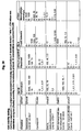
- the following combinations of cytokine genes and cell lines presented in Table I were used as a starting point for the development of a "cell chip”.
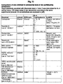
- unique cell lines were obtained and selected, which may serve to embody the method according to the present invention as well as being an example collection of cell lines according to the present invention. Said lines were deposited in the European Collection of Cell Cultures (ECACC). They are presented in Table III.
- ECACC European Collection of Cell Cultures
- IL-2, IFN- ⁇ , IL-1 ⁇ , and TNF- ⁇ were obtained by PCR based cloning using mouse genomic DNA isolated from Balb/c as a template.
- genomic DNA was prepared from the tail tip of a Balb/c mouse. This DNA served as a template for the PCR amplification.
- PCR primers were designed based on the genomic sequences of murine IL-2 (X52618) and IFN- ⁇ (M28381), available from GenBank. The primers were designed to encompass the upstream region of IL-2 from position -2686 to +25, and IFN- ⁇ from the position -2001 to +34, relative to the transcription start site denoted as +1. The rationale was to include a large upstream region in order to include many putative regulatory elements.
- the primers were then used for PCR amplification using the "PCR core kit” (Roche Biochemicals). PCR products of the correct size were excised from an agarose gel, purified using a QiaQuick gel extraction kit (Qiagen), checked for integrity on an agarose gel and ligated into the PCR cloning vector pGEM-T Easy (Promega). The ligation was transformed into JM109 E.coli cells and colonies containing inserts were selected using ampicillin. Plasmid DNA was extracted from a number of clones using a QiaSpin miniprep kit (Qiagen). The plasmids were then screened for the presence and orientation of the PCR product using multiple restriction digests. Finally, the presence and orientation of the IL-2 and IFN- ⁇ 5' upstream regulatory regions were confirmed by automated sequencing (ABI 377 DNA sequenator) from both ends using primers annealing to the vector.
- genomic DNA was prepared from the liver of a Balb/c mouse. This DNA served as a template for PCR amplification. PCR primers were designed based on the genomic sequence of murine TNF- ⁇ (U066950) and IL-1 ⁇ (X04964), available in databases. A 511 bp fragment of mouse IL-1 ⁇ promoter (-500/+11) was amplified by PCR using Taq polymerase (MBI Fermentas). The PCR product was cloned into TA-cloning vector pTAdvance (Clontech).
- a 4104 bp of fragment of IL1- ⁇ promoter (-4093/+11) was amplified by PCR using the high-fidelity AccuTaq LA thermostable polymerase (Sigma).
- the PCR product was ligated into the pCR-Blunt II-TOPO blunt-end cloning vector (Invitrogen).
- the ligation mixture was transformed into JM109 E.coli cells and colonies containing inserts were selected using kanamycin. Bacterial clones were screened for the presence of IL1- ⁇ promoter using PCR.
- Plasmid DNA was extracted from a number of bacterial clones using Plasmid Miniprep Plus kit (A&A Biotechnology) and the presence and orientation of the IL1- ⁇ promoter containing insert was verified using multiple restriction digests. Finally, the sequence and orientation of the IL-1 ⁇ 5' upstream regulatory regions were confirmed by automated sequencing (ABI 377 DNA sequenator) using primers annealing to the vector. A 2276 bp fragment of the mouse TNF- ⁇ promoter (-2013/+263) was obtained using similar experimental protocol. Briefly, PCR was performed using AccuTaq LA polymerase and the PCR product was cloned into pCR-Blunt II-TOPO vector. Following bacterial transformation the bacterial clones were screened for the presence and orientation of the TNF- ⁇ promoter region using PCR and restriction analysis. Finally, the sequence and orientation of the TNF- ⁇ promoter was confirmed by automated sequencing.
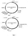
- the first series of reporter constructs consists of a number of plasmids where expression of the reporter gene is driven by promoter sequences of cytokines: IL-4, IL-2, IFN- ⁇ , IL-1 ⁇ , and TNF- ⁇ . All reporter constructs are based either on the plasmid pEGFP-1 (Clontech) containing enhanced green fluorescent protein (EGFP) as a reporter gene or on the plasmid pd1EGFP-N1 (Clontech) containing destabilized enhanced green fluorescent protein (d1EGFP) as a reporter gene.
- a 120 bp DNA fragment containing the minimal IL-4 promoter sequence was excised from -87IL-4 pCAT plasmid using HindIII and XbaI restriction enzymes. This DNA was then ligated into pTAdvance plasmid (Clontech).
- the IL-4 promoter was excised from pTAdvance plasmid using HindIII and ApaI, purified by agarose electrophoresis and ligated into the pEGFP-1 plasmid digested with the same enzymes.
- Plasmid DNA was isolated from kanamycin resistant bacterial clones and the integrity of the reporter construct was confirmed by ApaI / HindIII and HincII / HindIII digestions, and by sequencing. Obtained plasmid was named p1-5'IL4/EGFP-1.
- the PCR cloned IL-2 and IFN- ⁇ 5' regulatory regions were excised from pGEM-T Easy plasmids containing the inserts in negative orientation, using the restriction enzymes ApaI and PstI.
- the obtained DNA fragments were purified by agarose gel electrophoresis and a QiaQuick gel extraction kit (Qiagen), checked for the integrity on an agarose gel and ligated into the vector pEGFP-1 digested with ApaI and PstI in the multiple cloning site immediately upstream of the gene encoding EGFP.
- plasmids were screened for insertion of the IL-2 or IFN- ⁇ promoters using multiple restriction digests. The integrity of the reporter constructs was confirmed by automated sequencing (ABI 377 DNA sequenator) from both ends using primers annealing to the vector. The obtained plasmids were named p1-5'IL2/EGFP-1 and p1-5'INF ⁇ /EGFP-1.
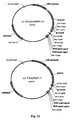
- a 511 bp fragment of mouse IL-1 ⁇ 5' regulatory region excised from pTAdvance plasmid using AseI and EcoRV was used to replace the CMV promoter in the pd1EGFP-N1.
- the CMV promoter was removed using AseI and Eco47III restriction enzymes (NEB, MBI Fermentas).
- the presence of the IL-1 ⁇ 5' regulatory region was confirmed by PCR and BamHI (NEB) digest.
- the integrity of the reporter construct, named p1-5'IL ⁇ /d1EGFP-N1 was confirmed by automated sequencing using primers annealing to the vector.
- a 4104 bp fragment of IL1- ⁇ 5' regulatory region was excised from pCR-Blunt II-TOPO vector using EcoRV and KpnI (NEB).
- the pd1EGFP-N1 vector was modified by removal of the CMV promoter using AseI and NheI (NEB) restriction, treated with Mung Bean Nuclease (NEB) to generate blunt ends, and ligated.
- EcoRV/KpnI IL1- ⁇ promoter was then ligated with modified pd1EGFP-N1 plasmid restricted with SmaI and KpnI.
- the presence of IL-1 ⁇ promoter was confirmed by PCR and restriction with Eco88I (MBI Fermentas).
- the integrity of the obtained plasmid was confirmed by sequencing.
- a 2526 bp TNF- ⁇ 5' regulatory region was excised from pCR-Blunt II-TOPO with the use of AseI/EcoRV (NEB).
- CMV promoter in pd1EGFP-N1 plasmid was excised by Ecl136II/VspI (MBI Fermentas) digestion.
- Both the 2526 bp insert and the resulting 4251 bp promoterless vector were purified by agarose gel electrophoresis and ligated using T4 ligase (Gibco BRL).
- p1-5'TNF ⁇ /d1EGFP-N1 6777 bp plasmid, named p1-5'TNF ⁇ /d1EGFP-N1
- p1-5'TNF ⁇ /d1EGFP-N1 6777 bp plasmid
- the sequence was verified using automated sequencing.
- This plasmid contains the entire TNF- ⁇ promoter region followed by 107 bp of TNF- ⁇ ORF and 80 bp of randomly generated linker at the beginning of d1EGFP ORF.
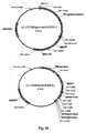
- a CAG promoter containing CMV enhancer and a chicken beta-actin promoter was excised with AseI and Eco47III restriction enzymes from commercially available plasmid pQE-TriSystem (Qiagen) and ligated into pEGFP-F plasmid (Clontech) in the place of CMV promoter (cut out with AseI and Eco47III restriction enzymes), upstream of EGFP coding sequence.
- the sequence of the resultant construct, pCA-EGFP-F was confirmed by restriction enzyme maping.
- 3'UTR regulatory sequences derived from IL-4, IL-2, IFN- ⁇ , IL-1 ⁇ , and TNF- ⁇ the respective sequences were amplified by PCR from genomic DNA obtained from the tail tips (IL-2 and IFN- ⁇ ) or from the livers (IL-4, IL-1 ⁇ , and TNF- ⁇ ) of Balb/c mice.
- primers based on the IL-4 genomic sequence were designed to encompass the entire IL-4 3'UTR, including the polyadenylation signal. Using these primers, a 154 bp long DNA fragment was amplified by PCR and cloned into pTAdvance vector (Clontech). The sequence of the cloned IL-4 3'UTR was verified by automated sequencing.
- the PCR primers were designed to encompass the 3'UTRs, including the translational stop codon and the polyadenylation signal. Primers were designed based on the mRNA sequences of IL-2 (X01772) and IFN- ⁇ (K00083), available in databases. The downstream primers were designed to include AflII restriction sites.
- the 357 bp IL-2 3'UTR and the 645 bp IFN- ⁇ 3'UTR amplified by PCR from genomic DNA were cloned non-directionally into the pGEM-T Easy vector (Promega). The identity and orientation of the inserts were then confirmed using multiple restriction digests.
- primers based on IL-1 ⁇ genomic sequence were designed to encompass the entire IL-1 ⁇ 3'UTR, including the polyadenylation signal. Using these primers, a 455 bp long DNA fragment was amplified by PCR and cloned into the pTAdvance vector (Clontech). The sequence of the cloned IL-1 ⁇ 3'UTR was verified by automated sequencing.
- PCR primers based on the TNF- ⁇ genomic sequence were designed to encompass the entire TNF- ⁇ 3'UTR including the poly-adenylation signal. Using these primers, a 994bp long DNA fragment amplified by PCR was cloned into the pTAdvance vector. The sequence of the cloned TNF- ⁇ 3'UTR was verified by automated sequencing.
- PCR primers based on GAPDH genomic sequence were designed to encompass the entire GAPDH 3'UTR including the polyadenylation signal. Using these primers, a 184bp long DNA fragment amplified by PCR was cloned into the pTAdvance vector. The sequence of the cloned GAPDH 3'UTR was verified by automated sequencing.
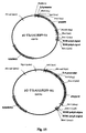
- the second series of reporter constructs consists of a number of plasmids where the expression of the reporter gene is under the regulation of promoter sequences and 3'UTRs of cytokines: IL-4, IL-2, IFN- ⁇ , IL-1 ⁇ , and TNF- ⁇ .
- Reporter constructs are based on the plasmids pEGFP-1, pEGFP-F, and pd1EGFP-N1, pd2EGFP-1 (Clontech), containing enhanced green fluorescent protein (EGFP) and destabilized enhanced green fluorescent protein (dEGFP) as a reporter gene, respectively.
- the pTAdvance vector containing the entire 3'UTR for IL-4 was digested with EcoRI (NEB) and the obtained fragment was ligated downstream of EGFP stop codon into the pEGFP-F vector using the EcoRI site.
- the presence of the IL-4 3'UTR was confirmed by an EcoRI digest and by PCR.
- the right orientation of the IL-4 3'UTR was confirmed by ScaI and BsmI (NEB) digests. Finally, the sequence was confirmed by automated sequencing.
- the constructed vector named p2-3'IL4/EGFP-F , contains SV40 derived 3'UTR sequences including polyadenylation sites. Northern blot experiments showed that these polyadenylation sites were utilized during transcription process. Thus, in the next step, the p2-3'IL4/EGFP-F vector was modified by removing the interfering SV40 polyadenylation signals. This plasmid was digested with BsmI (NEB) and Bal-31 nuclease (NEB), re-ligated and transformed into E . coli cells.
- BsmI BsmI
- NEB Bal-31 nuclease
- IL-4 3'UTR was excised from pTAdvance plasmid with EagI (NEB) and ligated into NotI (NEB) site downstream of EGFP stop codon in the pd1EGFP-N1 vector. Insert presence was confirmed by PCR and an EagI digest, and orientation was checked with a PstI (MBI Fermentas) digest. The sequence of the obtained plasmid, named p1-3'IL4/d1EGFP-N1, was confirmed by sequencing.
- vector p1-5'IL4/d1EGFP-N1, containing IL-4 promoter and d1EGFP ORF and vector p1-3'IL4/d1EGFP-N1 containing d1EGFP ORF and IL-4 3'UTR were both digested with AgeI (MBI Fermentas) and HincII (NEB) enzymes.
- DNA fragment containing d1EGFP ORF and IL-4 3'UTR obtained from p1-3'IL4/d1EGFP-N1 digest was next ligated with DNA fragment containing vector sequence and IL-4 promoter derived from p1-5'IL4/d1EGFP-N1.
- the restriction map of the new construct, named p1-5'3'IL4/EGFP-N1 was verified by digestion with Eco52I (MBI Fermentas), followed by sequencing.
- the following strategy was used.
- the respective 3'UTRs were cloned into the pd2EGFP-1 vector.
- the SV40 derived polyadenylation signals present in the pd2EGFP-1 vector were removed using the restriction enzymes NotI and AflII.
- the 3'UTR's were then excised from the PCR cloning vector pGEM-T Easy using the restriction enzymes NotI and AflII, and the 3'UTRs containing fragments were ligated into NotI/AflII restricted pd2EGFP-1 vector.
- the IL-2 and IFN- ⁇ promoter regions were cut out from the plasmids p1-5'IL2/EGFP-1 and p1-5'INF ⁇ /EGFP-1, respectively, using the restriction enzymes XhoI and BamHI. These fragments were then gel-purified and ligated into the pd2EGFP-1 vectors containing 3'UTRs using XhoI and BamHI sites in the multiple cloning site upstream of d2 EGFP gene. Restriction maps of the new constructs, named p1-5'3'IL2/d2EGFP-1 and p1-5'3'INF ⁇ /d2EGFP-1, were verified by multiple restriction digests and sequencing.
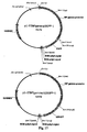
- IL-1 ⁇ 3'UTR was excised from pTAdvance vector with Eco52I (MBI Fermentas) and ligated into p1-5'IL1 ⁇ /d1EGFP-N1 and p3-5'IL1 ⁇ /d1EGFP-N1 vectors digested with NotI (NEB). The presence of the IL-1 ⁇ 3'UTR in both plasmids was confirmed by PCR and its orientation was checked by HincII (NEB) digest. Sequences of new vectors, named p1-5'3'IL1 ⁇ /d1EGFP-N1 and p3-5'3'IL1 ⁇ /d1EGFP-N1, were confirmed by sequencing.
- the pTAdvance vector containing the entire 3'UTR for TNF- ⁇ was digested with HindIII and EcoRV (NEB) and the obtained fragment was ligated downstream of EGFP stop codon into pEGFP-F vector using sites HindIII and SmaI.
- the presence of the TNF- ⁇ 3'UTR was confirmed by EcoRI and SspI (NEB) digest and by PCR.
- the sequence of TNF- ⁇ 3'UTR was confirmed by automated sequencing.
- Resultant vector contains SV-40 derived 3'UTR sequences including polyadenylation sites.
- p2-3'IL4/EGFP-F plasmid showed that SV-40 derived polyadenylation sites are utilized during transcription process the p2-3'TNF ⁇ /EGFP-F vector was modified by removing SV40 polyadenylation sequences.
- Plasmid p3-3'IL4/EGFP-F was digested with AseI and HincII to remove CMV-GFP-3'UTR IL-4 box and ligated with CMV-GFP-3'UTR THF- ⁇ box cut out from the plasmid p2-3'TNF ⁇ /EGFP-F using AseI and HincII.
- TNF- ⁇ 3'UTR was excised from pTAdvance plasmid with EagI (NEB) and ligated into the NotI (NEB) site downstream of EGFP stop codon into the pd1EGFP-N1 vector. Insert presence was confirmed by PCR and by Eco52I digest, and the right orientation of TNF- ⁇ 3'UTR was confirmed BglII and Bsu36I (MBI Fermentas) digest. The sequence of the constructed p1-3'TNF ⁇ /d1EGFP-N1 plasmid was confirmed by sequencing. To obtain EGFP expression vector in which both 5' upstream and 3' downstream regulatory sequences of TNF- ⁇ control the expression of the reporter gene the following strategy was used.
- SV40 3'UTR from p1-5'TNF ⁇ /d1EGFP-N1 plasmid was removed by sequential digestions with the following enzymes: Mva1269I (MBI fermentas), Mung Bean nuclease (NEB) and NotI (NEB).
- Mva1269I MMI fermentas
- NEB Mung Bean nuclease
- NotI NEB
- the 6684 bp vector fragment without SV40 3'UTR was next ligated with the 1039 bp fragment containing TNF- ⁇ 3'UTR excised from the p1-3'TNF ⁇ /d1EGFP-N1 using NotI and EcoRV endonucleases.
- the integrity of constructed vector named p1-5'3'TNF ⁇ /d1EGFP-N1 was confirmed by sequencing.
- the pTAdvance vector containing the entire 3'UTR for GAPDH was digested with EcoRI (NEB) and the obtained fragment was ligated downstream of EGFP stop codon into pEGFP-F vector (Clontech) using the EcoRI site.
- the presence of the GAPDH 3'UTR was confirmed by EcoRI digest and by PCR.
- the right orientation of the GAPDH 3'UTR was confirmed by StyI (NEB) digest.
- sequence of the construct was confirmed by automated sequencing.
- the resulting vector contains SV40 derived 3'UTR sequences, including polyadenylation sites.
- p2-3'GAPDH/EGFP-F contains SV40 derived 3'UTR sequences, including polyadenylation sites.
- Northern blot experiments conducted with p2-3'IL4/EGFP-F plasmid showed that SV-40 derived polyadenylation sites are utilized during transcription process, the p2-3'GAPDH/EGFP-F vector was modified by removing SV40 polyadenylation sequences.
- Plasmid p3-3'IL4/EGFP-F was digested with AseI and HincII to remove CMV-GFP-3'UTR IL-4 cassette and ligated with CMV-GFP-3'UTR GAPDH cassette cut out from the plasmid p2-3'GAPDH/EGFP-F using AseI and HincII.
- the presence of GAPDH 3'UTR in the resulting plasmid named p3-3'GAPDH/EGFP-F was confirmed by PCR and the absence of SV40 polyadenylation sites by BsmI digest.
- GAPDH 3'UTR was excised from the pTAdvance plasmid with EagI (NEB) and ligated into the NotI (NEB) site downstream of the EGFP stop codon into the pd1EGFP-N1 vector (Clontech).
- EagI EagI
- NEB NotI
- PstI MBI Fermentas
- a set of expression vectors was obtained, in which the GFP coding sequence is under the control of 5' upstream regulatory sequences derived from cytokine gene, in a form of highly purified DNA available for transfection of mammalian cells. Moreover, a set of expression vectors was obtained, in which the GFP coding sequence is under the control of 5' upstream and 3'downstream regulatory sequences derived from cytokine genes, in a the form of highly purified DNA available for transfection of mammalian cells.
- T cells and T cell hybridoma, mast cell lines, monocyte-macrophage, fibroblasts and keratinocyte cell lines were used. Plasmids were linearized by digestion and cells were transfected using electroporation.
- Example 7 some of the cell lines tested in Example 7, were used to assemble the panel of reporter cell line i.e.”a prototype of a cell-chip".
- NRU Neutral Red Uptake
- NRR Neuronal Red Release
- MTT MTT Assay
- the electroporation based transfection technique was employed.
- 1 x 10 7 cells grown in an appropriate medium were electroporated with 20 to 50 ⁇ g of DNA. Cells were allowed to recover in normal growth medium for 48 hours before the selecting antibiotic (G-418) was added. Next, the cells were either directly cloned by limiting dilution, or were first cultured in selecting medium for additional 1-2 weeks and then cloned. Resistant clones were expanded, frozen and characterized phenotypically.
- Jurkat To test the transfection protocol and techniques for GFP detection easily transfectable human T cell line Jurkat was used. Jurkat cells were transfected with the pEGFP-F plasmid where EGFP expression is driven by CMV promoter. As a result several cell clones with a high expression of EGFP were obtained. These cell lines were used by DBAPAS to test EGFP expression with a phosphorimager Multimager Typhoon 8600 (Molecular Dynamics).
- C57/CMV-EGFP cells shoed morphology identical to the maternal cell line. An increase in the number of giant cells as compared to the maternal cell line was observed. When tested in functional assays these cells exhibited the normal characteristics of mast cells. They were sensitized with monoclonal IgE in vitro and responded to an antigen by exocytosis and cytokine production. The level of mediator release and cytokine production in C57/CMV-EGFP cells were comparable to those observed with maternal C57.1 mast cells.
- Example 2 DNA constructs developed in Example 1, in which GFP expression was under the control of 5' upstream or 3' UTR regulatory sequences of cytokine genes were used.
- C57.1 mast cells were transfected with the EGFP reporter plasmid for IL-4 gene, p1-5'IL4/EGFP-1.
- the plasmid was linearized and cells were transfected using electroporation.
- Several neomycin resistant clones were obtained and were expanded in large-scale cultures. Following second round of cloning one of these clones has been found to have a proper phenotypic characteristic of mast cells and detectable EGFP expression.
- This resultant cell line in which IL-4 minimal promoter controls EGFP expression was named C57/5'IL4/1 (renamed to C/p1-5'IL4-EGFP/002, in accordance with the labeling system in the reporter cell line database) and is the M1 milestone of the project.
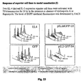
- This cell line was employed in a series of experiments testing the level of EGFP expression in resting cells and cells challenged with ionophore. The conclusion of these tests was that there was a high EGFP expression in resting cells, which was due to the high stability of the EGFP protein. This feature in turn seems to obscure the increase in EGFP expression following cell stimulation. Based on this data the conclusion was drawn to also employ the destabilized EGFP variant, called dEGFP. in addition to EGFP (see Example 3).
- Lymphocytic cell lines EL4 and BW5147.3 were transfected with reporter constructs for IL-2 and IFN- ⁇ , p1-5'INF ⁇ /EGFP-1 and p1-5'IL2/EGFP-1, respectively. Several resistant clones resulting from these transfections have already been frozen. EL4 cells transfected with the p1-5'IL2/EGFP-1 plasmid were used to test spontaneous and phorbol ester mediated expression of EGFP mRNA. For each type of reporter cell line a minimum of 3 clones have been obtained. The clones were initially analyzed on the basis of GFP basal expression and inducibility. Initial analysis was performed using fluorescence microscopy, demonstrating this to be a good method to detect GFP expression.
- Lymphocytic cell line EL4 was transfected with a reporter construct for IL-4 p1-5'IL-4/EGFP-1. Multiple resistant clones resulting from these transfections have been tested using FACS and fluorescence microscopy for basal and inducible EGFP expression. None of the clones showed an increased expression of GFP following activation with TPA and calcium ionophore.
- the GFP constructs obtained in Example 1 were modified using standard molecular biology techniques, by restriction enzyme digestion and directional cloning of desired sequences.
- the sequences of interest were either derived from commercially available expression vectors or were introduced as synthetic oligonucleotides. Positive clones were selected and the sequence of the obtaining plasmids verified. The detailed changes in the design of expression vectors depend of the results obtained. Lowering of the baseline level of GFP expression may require the introduction of additional regulatory elements such as silencers into the upstream of GFP coding sequence. Additional "heterologous" motif destabilizing mRNA can be also introduced into the 3' UTR downstream of GFP open reading frame.
- cytokine promoter can be used and known "heterologous" motifs stabilizing mRNA can also be introduced into the 3' UTR downstream of GFP open reading frame.
- the ladder sequences in the GFP open reading frame can be modified resulting into different trafficking of GFP into cellular compartment.
- the fluorescent protein itself can be replaced with one of several modified fluorescent proteins, which may change the signal to noise ratio in fluorimetric readout of gene expression due to different level of overlap with autofluorescence.
- the bioluminescence based reporter gene can also be tested as a possible alternative for GFP system.
- Two reporter constructs containing dEGFP under control of IL-4 5' upstream regulatory sequences were created. The first one was generated based on the p1-5'IL4/EGFP-1 plasmid (see WP1) digested with SmaI and XhoI enzymes (MBI Fermentas). The excised fragment containing minimal promoter for IL-4 (-87/+5), was cloned into the the pd1EGFP-N1* plasmid (pd1EGFP-N1 modified by removal of CMV promoter, see WP1) digested with the same pair of enzyme. Ligation products were screened with SmaI and Alw44I digestion.
- the sequence of constructed plasmid was confirmed by automated sequencing.
- a longer IL-4 promoter region (-797/+5) was obtained by PCR (Taq Polymerase; MBI Fermentas) using the -797pCAT plasmid as a template (a gift from Dr. Melisa Brown, Atlanta University, Atlanta, USA).
- PCR product was cloned into pTAdvance vector.
- IL-4 promoter was excised from the pTAdvance vector with SmaI and VspI (MBI Fermentas) and cloned into pd1EGFP-N1* digested with the same pair of enzymes. The effect of the ligation was confirmed by HindII and by Eco52I digestions.
- the sequence of the constructed plasmid was verified by sequencing.
- Reporter constructs containing dEGFP under control of IL-2 and IFN- ⁇ 5' upstream regulatory sequences are based on reporter constructs containing EGFP.
- the IL-2 and IFN- ⁇ promoter regions were excised using XhoI and BamHI from plasmids p1-5'IL2/EGFP-1 and p1-5'INF ⁇ /EGFP-1, respectively.
- the obtained DNA fragments were then gel-purified and ligated into the pd2EGFP-1 plasmid (Clontech), digested with the same pair of enzymes. Ligation products were analysed using multiple restriction digests. Sequences of the obtained constructs, named p1-5'IL2/d2EGFP-1 and p1-5'INF ⁇ /d2EGFP-1 , were confirmed by sequencing.
- the IL-1 ⁇ derived sequences were then released from this vector using EcoRV (NEB) and KpnI (NEB) and cloned into pd1EGFP-N1* digested with SmaI (MBI Fermentas) and KpnI (NEB).
- the presence of the -500/+820 IL - 1 ⁇ promoter in the first obtained plasmid, named p2-5'IL1 ⁇ /d1EGFP-N1 was confirmed by PCR and HincII digest.
- p4-5'IL1 ⁇ /d1EGFP-N1 The presence of the -4.093/+820 IL-1 ⁇ promoter in the second obtained plasmid, named p4-5'IL1 ⁇ /d1EGFP-N1 , was confirmed by PCR, and by digestion with Eco88I (MBI Fermentas). Sequences of the p2-5'IL1 ⁇ /d1EGFP-N1 and p4-5'IL1 ⁇ /d1EGFP-N1 plasmids were confirmed by automated sequencing.
- CAG promoter which consists of CMV enhancer and chicken ⁇ -actin proximal promoter was cut out with AseI and Eco47III restriction enzymes from commercially available pQE-TriSystem vector (Qiagen) and ligated into pd1EGFP-N1vector (Clontech) in the place of CMV promoter (cut out with AseI and Eco47III restriction enzymes), upstream of EGFP coding sequence.
- the sequence of resultant construct pCA-d1EGFP was confirmed by restriction enzyme maping.
- the plasmids p1-5'IL4/d1EGFP-N1 and p1-3'IL4/d1EGFP-N1 were digested with AgeI (MBIFermentas) and HincII (NEB)
- AgeI MIFermentas
- HincII NEB
- the fragment containing d1EGFP and 3'UTR excised from p1-3'IL4/d1EGFP-N1 plasmid was next cloned into p1-5'IL4/d1EGFP-N1, immediately downstream of IL-4 promoter.
- Eco52I MBI Fermentas
- the sequence of this plasmid was verified by automated sequencing.
- the plasmids p2-5'IL4/d1EGFP-N1 and p1-3'IL4/d1EGFP-N1 were digested with AgeI (MBI Fermentas) and HincII (NEB).
- the fragment containing d1EGFP and 3'UTR excised from p1-3'IL4/d1EGFP-N1 plasmid was next cloned into p2-5'IL4/d1EGFP-N1, immediately downstream of IL-4 promoter.
- a restriction map of the new construct, named p2-5'3'IL4/d1EGFP-N1 was analysed by Eco52I and SspI (NEB) digestion and the sequence of this plasmid was verified by automated sequencing.
- the IL-1 ⁇ 3'UTR was excised from pTAdvance with EagI (NEB), and ligated into p2-5'IL1 ⁇ /d1EGFP-N1 digested with NotI (NEB). The presence of the insert was confirmed by PCR and its orientation was analysed by Ecl136II (MBI Fermentas) digestion. The sequence of the obtained plasmid, named p2-5'3'IL1 ⁇ /d1EGFP-N1, was confirmed by automated sequencing.
- the IL-1 ⁇ 3'UTR was excised from pTAdvance with EagI (NEB), and ligated into p4-5'IL1 ⁇ /d1EGFP-N1 digested with NotI (NEB). The presence of the insert was confirmed by PCR and its orientation was determined by an Ecl136 (MBI Fermentas) digest. The sequence of the obtained plasmid, named p4-5'3'IL1 ⁇ 3/d1EGFP-N1, was confirmed by automated sequencing.
- lymphocytic cell lines EL4 and BW5147.3 were transfected with dEGFP reporter constructs for IL-2 and IFN- ⁇ , p1-5'IL2/d2EGFP-1 and p1-5'INF ⁇ /d2EGFP-1, respectively (Example 3).
- dEGFP reporter constructs for IL-2 and IFN- ⁇ , p1-5'IL2/d2EGFP-1 and p1-5'INF ⁇ /d2EGFP-1 respectively.
- Several neomycin resistant clones resulting from these transfections were expanded and characterized. Some of them have shown low basal dEGFP expression, which was upregulated upon activation with TPA/ionomycin to the level easily detected by FACS or fluorescence microscopy.
- the reporter cell line EL/p1-5'IL2-dEGFP/7 was obtained.
- clones carrying destabilized variant of EGFP show lower basal expression when compared to EGFP transfected cells the maximum level of inducible dEGFP expression was also lower as compared to activated EL4 clones carrying dEGFP transgene.
- Lymphocytic cell line EL4 was also transfected with a reporter constructs for IL-4 p1-5'ILA/d1EGFP-N1 and p1-5'3'IL4/d1EGFP-N1.
- C57.1 mast cells were transfected with reporter constructs for IL-4 p1-5'IL4/dIEGFP-N1 and pl-5'3'IL4/d1EGFP-N1. Resultant clones were characterized for basal and inducible GFP expression and did not show induction of GFP expression. The transfection protocol using the same plasmids was repeated and generated multiple resistant clones.
- J774.1A monocytes-macrophages were transfected with the reporter constructs for TNF- ⁇ , p1-5'TNF- ⁇ /d1EGFP-N1 . Multiple resistant clones were selected and cloned. Resultant clones were characterized for basal and inducible GFP expression using FACS. One of these clones was found to respond to activation with LPS by upregulation of GFP fluorescence.
- C57.1 mast cells were transfected with two reporter constructs for TNF- ⁇ , p1-5'TNF- ⁇ /d1EGFP-N1 and p1-5'TNF- ⁇ /d1EGFP-N1.
- Multiple resistant clones were selected and cloned. Resultant clones were characterized for basal and inducible GFP expression using fluorescence microscopy and FACS. 10 of these clones have been found to respond to activation with TPA and A23 ionophore by upregulation of GFP fluorescence, easily detected under fluorescence microscopy and FACS.
- EL4 cells and C57 cells were transfected control plasmid pCA-dIEGFP, in which dEGFP expression is under control of the actin promoter. Resultant cell clones were tested for GFP expression and showed a basal expression that is not inducible upon activation.
- 3T3-L1 fibroblasts, Hel-30 keratinocytes, and J774.1A monocytes-macrophages were transfected with the IL-1 ⁇ reporter constructs p2-5'IL1 ⁇ /d1EGFP-N1 and p4-5'IL1 ⁇ /d1EGFP-N1.
- HEL-30 derived resistant cells were found to be difficult to clone and propagate due to very strong adhesion to plastic. An alternative technique for transfection of this cell line is currently being tested. Transfection of J774A.1 yielded multiple resistant clones that were characterized for selected phenotypic features and GFP expression.
- Gene-targeting strategy utilizing a targeting vector with a long contiguous sequence homologous to the targeted loci will be used.
- genomic DNA will be collected, overlapping or mapped close to murine cytokine genes.
- the restriction map of the relevant part of murine chromosomal DNA covering loci for IL-4, IL-5, and IFN- ⁇ are available, and these genes were successfully targeted in mice resulting in a "knockout phenotype".
- the gene targeting constructs will contain a 6 kB fragment of DNA overlapping the coding region of targeted cytokine gene.
- For each targeted locus it is desirable to prepare three vectors, each of them containing the same long homologous sequence but differ in the type of selectable markers.
- targeting vectors will start based on a backbone of a standard pBluescript vector.
- GFP coding region with 3'UTR cytokine sequences adjacent to its Stop codon will be derived from GFP vectors obtained in Example 1.
- GFP coding sequences followed by 3'UTR of cytokine genes will be inserted into the 6 kB long fragment homologous sequence to replace the cytokine open reading frame.
- a Neo or Hyg box flanked with a pair of loxP sequences will be inserted downstream of the transcription termination signal for the cytokine gene followed by long contiguous fragment of targeting homologous sequence.
- Vectors will be constructed using standard DNA manipulation techniques, including restriction enzyme digestion and ligation. The synthetic oligonucleotide adapters and PCR generated DNA fragments will be used if necessary to connect the desired DNA sequences and to introduce particular sequences into the construct. Following transformation the positive E coli clones will be selected with a miniprep analysis. After verification of plasmid sequences with automated sequencing, large quantities of plasmid DNA will be amplified and purified.
- the original plasmid is modified by addition of TK box at the 3' end, which allows the use the positive-negative cell selection technique (PNS).
- PPS positive-negative cell selection technique
- the pPNT plasmid containing the herpes simplex virus thymidine kinase gene under control of the mouse phosphoglycerate kinase-1 promoter BCCM/LMBP plasmid and DNA collection, Ghent, Belgium
- An extensive restriction mapping was performed to identify sites in the pIL-2/GFP plasmid that could be used to clone in the TK-cassette. Only a few single cutters that did not disrupt the IL2/GFP targeting sequence were identified. These restriction sites does not correspond to any available in the pPNT plasmid.
- the 2.8 kb TK cassette was sub-cloned into pBluescript. An alternative PCR based cloning strategy was also tested.
- Vectors obtained in Example 5 will be used to target DNA sequences into the selected cytokine gene loci.
- Vectors designed for positive selection with neomycin (G418) or hygromycin and vectors for positive-negative selection (PNS) will be used. Cutting at the unique restriction enzyme site will linearize the vectors, which will be then electroporated into cells. Cells will be placed in selecting media. Selection of cells will be based on G418 or hygromycin in case of positive selection process or on G418 followed with gancyclovir in a positive-negative selection process. Resistant cells will be cloned by limiting dilution procedure. Clones will be screened for desired genomic modification using PCR.
- the selected clones will be further tested for successful gene targeting using southern blot, and expanded in large-scale cultures. Cell lines obtained at this stage will undergo tests for phenotypic characteristics. In the next step it will be desirable to activate these cells and measure GFP expression. The successful incorporation of a transgenic insert into the specific loci will result in a single allele modified to express GFP instead of cytokine mRNA. In the next step it will be desirable to replace both alleles of the gene with GFP and compare the resulting phenotype with that of a single replacement. To do this, cell lines with single transgenic insertions and which target the other allele will be used. One possible strategy is to use the targeting vector with different selectable markers and to screen transfectants for double resistance.
- An alternative strategy is to remove the neomycin cassette from the transgenic insert and use the neomycin resistance marker for selection once again. Clones will be tested using PCR and clones negative for the neo box and sensitive to neomycin will be selected. From this point the second round of gene targeting is similar for both strategies described above and follows the experimental procedure described for the first gene targeting experiment. The difference will be used for screening in the PCR primers and Southern probe. They will be designed to verify the complete absence of cytokine coding sequences in the genomic DNA.
- the 9 kb IL-2/GFP targetting construct was linearized and used for electroporation-based transfection of EL4 cells.
- Transfected cells underwent G-418 based selection. Limited dilution cloning of neomycin resistant cells yielded 26 clones, which were characterized for the type of insertion.
- the Southern blot of EcoRI digested genomic DNA isolated from these clones has been performed. Although one of these clones gave signal of distinct size other than expected from unmodified IL-2 loci, the final clonclusion was that only random DNA incorporations took place.
- the original plasmid is now being modified by addition of TK cassette at the 3' end, which will allow us to use the positive-negative cell selection technique (PNS).
- PPS positive-negative cell selection technique
- Example 7 Development of the experimental protocol for testing the response of reporter cell lines to xenobiotics.
- the cell culture and assay condition will be optimized to obtain comparable level of baseline fluorescence and the best signal to noise ratio.
- the preferable format for these experiments will be testing cells placed in multiwell plate. Selected immunomodulatory substances will be added at increasing concentrations and the expression of GFP in tested cells will be measured. For that purpose activated or resting cells will be incubated in the presence or absence of tested compounds and the kinetic assay of GFP specific fluorescence will be performed. This fluorescence signal will provide information on possible modulation of cellular response leading to enhancement or inhibition of cytokine expression. Based on obtained data a common experimental protocol will be designed which would allow the usage of several cell lines representing different cell lineage in a single assay. Results will facilitate the selection of cell lines for assembling and testing of the entire panel of reporter cell lines, the prototype of a cell chip. The cell line selected for the cell chip has to fulfill following criteria:
- Cell clones were propagated and transferred for banking. More than fifty reporter GFP transfected cell clones with the proper phenotypic characteristics are stored in multiple aliquots in a cell banking facility.
- Non-transfected EL4 and BW5147.3 cells were activated with TPA/ionomycin or anti-CD3 and RNA was prepared.
- cDNA synthesis PCR was performed to detect expression of IL-2, IFN ⁇ , and GAPDH (control). The results generally agree with the GFP expression data.
- BW5147.3 cells neither IL-2, nor IFN ⁇ mRNA, could be detected in non-induced cells or after activation.
- IL-2 and IFN ⁇ expression could be induced by TPA/ionomycin, but not anti-CD3.
- the work to determine the toxic range of all the chemicals listed in Table II for all the cell lines developed in the project has progressed.
- the basic cytotoxicty assay employed was LDH release.
- Several cytotoxicity assays were performed independently in two laboratories. Data, which are presented in Fig. 20 and Fig. 21 , respectively, were compared and analyzed and will allow us to decide on the range of concentrations to be used for testing of the Fluorescence Cell Chip.
- the cytotoxicity associated with the solvent was also tested to eliminate the possibility of interference in the test outcome.
- Cyclosporin A was shown to completely inhibit the activation induced GFP expression down to basal levels ( Fig. 23 , Fig. 24 ). This inhibitory effect of Cyclosporine was observed independently in all three laboratories performing such experiments. Furthermore, this inhibition of EGFP expression was detected with all three techniques emplyed for EGFP measurements (i.e. FACS, fluorescence microscope, and Fluorostar). Rapamycin or TCDD did not have any effect on the activation induced GFP expression. Thus the important proof of the concept of Fluorescence Cell Chip testing has been obtained, namely the microplate based readout detected the presence of model immunosupresive xenobiotic (Cyclosporin A; Fig. 24 ).
- BW5147.3 derived GFP transfected cell clones have been also used to test the effects of immunomodulatory compounds. Cyclosporin A, Rapamycin and TCDD did not affect the basal GFP expression and since the GFP expression could not be induced, the effect on activated expression could not be analysed.
- Example 8 Pre-validation of the new test against available data on animal and human immunotoxicity. Prototype of cell-chip.
- Comparison of patterns generated by model immunotoxins, substances that may be classified as irritants but do not posses immunomodulatory activity and control inert compounds will be performed. This analysis shall reveal if the new technology is capable to distinguish immunotoxins from other xenobiotics. Testing known and unknown (blind) samples of xenobiotic in parallel experiments will next be performed in all laboratories. A comparison of data obtained independently in participating laboratories will provide preliminary data on reproducibility of responses of reporter cell lines, and the sensitivity of this technology to minor differences in experimental protocols. It is desirable to obtain information in a format of "two dimensional" pattern that describes the action of several "model xenobiotics" (substances already known for their immunomodulatory activities in vivo) on different genes in various cell lineages.
- Fluorstar Galaxy Multiwell (BMG Labtechnologies) fluorescence, luminescence, and an absorbance reader were used for fluorescence detection. Two laboratories received two cell lines 3T3-L1/CMV-EGFP and HEL-30/CMV-EGFP and employed them for testing both the cell culture and the fluorescence assay protocols.
- Cells were plated in 24 well microtiter plates at a density of 0.5x10 6 cells/mL and with a final volume of 1.5 mL/well. Two controls were included for each cell line, in each experiment. One control consisted of cells in growth medium and the second control was cells in the presence of the induction mix (Ionomycin calcium salt, (cat.no#1-0634, Sigma) and 10 ng/mL PMA (Sigma). In some cases, induction mix contained only ionomycin or LPS at concentration 100 ng/ml rather than PMA and ionomycin. Cells were incubated with different chemicals at the concentration that led to 10% cytotoxicity and concentrations 10x and 100x more diluted in the absence and presence of the induction mix.
- the induction mix Ionomycin calcium salt, (cat.no#1-0634, Sigma)
- PMA 10 ng/mL
- induction mix contained only ionomycin or LPS at concentration 100 ng/ml rather than PMA and ionomycin.
- Cyclosporin A (for molecular biology, Tolypocladium inflatum, minimum purity 95%, Sigma) was diluted in ethanol to a stock concentration of 5 mg/mL and kept at -20°C. The stock solution was diluted in medium prior to experiments. Rapamycin (minimum purity 95%, Sigma) was dissolved in DMSO to a concentration of 2 mg/mL and kept at -20°C. The stock solution was diluted in medium prior to experiments. Pentamidine isethionate salt (Sigma) was dissolved in DMSO to a final concentration of 25 mg/mL and kept at -20°C. The stock solution was diluted in medium prior to experiments. (+/-)- Thalidomide (purity >98%, Sigma).
- a 0.2 M stock solution of Thalidomide in DMSO was prepared and kept at -20°C. Prior to experiments, the stock solution was diluted in medium, which led to a white precipitate. The solution was resuspended and used for further dilutions.
- Bis (tri-n-butyltin) oxide (TBTO) (purity 96%, Aldrich) was diluted in ethanol to a stock concentration of 10 mM, kept at -20°C and diluted in medium before use.
- House dust mite D- pteronyssinus (Alutard SQ Depot allergen extract) (suspension for injection, 100 000 SQ-U/mL solution, ALK-Abelló, Horsholm, Denmark) was kept at 4-8°C as instructed by the manufacturer and diluted in medium directly before use.
- 1-Chloro-2,4-dinitrobenzene (DNCB) (purity minimum 98%, Sigma) was diluted in ethanol to a stock solution of 2 mM, kept at -20°C and diluted in medium before use.
- Benzocaine (Ethyl-4-Aminobenzoate, Sigma) was diluted to a 0.5 M stock solution in ethanol, kept at 20°C and diluted in medium before use.
- TDI Tolylene 2,4-diisocyanate
- Penicillin G (Benzylpenicillin) sodium salt (activity >1477 U/mg, Sigma) was diluted in medium to a stock solution of 200 mM, kept at -20°C and further diluted in medium before use.
- SDS purity >85%, Merck
- SDS was diluted in DMSO to a stock solution of 100 mM, kept at -20°C and diluted in medium before use.
- Mercury (II) chloride minimum purity 99.5%, Merck was diluted in ethanol to a stock solution of 6 mM, kept at -20°C and diluted in medium before use.
- EL/pCA-dEGFP/9 The following cell lines: EL/pCA-dEGFP/9, EL/p1-5'IL2-dEGFP/6, EL/p2-5'IL4-dEGFP/2, EL/p1-5'IFN ⁇ -dEGFP/3, and EL/p2-5'IL10-dEGFP/5, obtained by transfection with the DNA constructs containing promoter region derived from ⁇ -actin, IL2, IL4, INF- ⁇ , and IL10 were employed to design the prototype cell chip. The prototype cell chip was used to test the activity of different substances.
- Cyclosporin A and Rapamycin are well-known immunosuppressive drugs used in organ transplantation.
- Pentamidine is used as an antiprotozoal drug, but has also been shown to reduce expression of several cytokines.
- Thalidomide is used as an anti-inflammatory drug.
- Bis(tri-n-butyltin)oxide (TBTO) has also been shown previously to be immunosuppressive, both in vivo and in vitro.
- Figure 26 presents results of testing the effect of Cyclosporin A using the prototype cell chip: cells transfected with the regulatory elements from actin (black bars), IL-2 (grey bars), IL-4 (wide striped bars), IL-10 (diamond bars) and IFN- ⁇ (narrow striped bars) fused to EGFP were exposed to 1 ⁇ M CsA, which led to 10% cytotoxicity, and 1:10 or 1:100 dilution of this for 24 h in the absence (A) or presence (B) of ionomycin/PMA (1 ⁇ M / 10 ng/mL). In non-induced cells (A) at the highest concentration, we found statistically significant differences compared to the control for actin, IL-4 and IL-10.
- Figure 27 presents results of testing the effect of rapamycin using the prototype cell chip: cells transfected with the regulatory elements from actin (black bars), IL-2 (grey bars), IL-4 (wide striped bars), IL-10 (diamond bars) and IFN- ⁇ (narrow striped bars) fused to EGFP were exposed to 10 ⁇ g/mL rapamycin, which led to 10% cytotoxicity, and 1:10 or 1:100 dilution of this for 24 h in the absence (A) or presence (B) of ionomycin/PMA (1 ⁇ M / 10 ng/mL). In non-induced cells (A) at the highest concentration, we found a statistically significant decrease in fluorescence compared to the control for IL-4 and IL-10.
- Figure 28 presents results of testing the effect of rapamycin using the prototype cell chip: cells transfected with the regulatory elements from actin (black bars), IL-2 (grey bars), IL-4 (wide striped bars), IL-10 (diamond bars) and IFN- ⁇ (narrow striped bars) fused to EGFP were exposed to 30 ⁇ g/mL pentamidine, which led to 10% cytotoxicity, and 1:10 or 1:100 dilutions of this for 24 h in the absence (A) or presence (B) of ionomycin/PMA (1 ⁇ M / 10 ng/mL). In non-induced cells (A) at the highest concentration and 1:10 dilution, we found statistically significant differences compared to the control for actin and IL-10.
- IgE-mediated respiratory allergy to protein allergens exemplified by the mite allergen Der p I
- contact allergy to low molecular weight substances mediated by hapten-protein conjugate specific T lymphocytes exemplified by 1-chloro-2,4-dinitrobenzene (strong allergen) and benzocaine (weak allergen)
- low-molecular weight chemical allergens causing clinical symptoms similar to IgE-mediated allergy but with unknown mechanisms, exemplified by tolylene 2,4-diisocyanate where specific IgE is demonstrable only in a minority of cases and potassium tetrachloroplatinate where specific IgE is regularly demonstrable.
- Penicillin G was included because it is known to induce drug hypersensitivity in humans. Furthermore, an irritant (sodium dodecyl sulphate) was included, because the distinction between irritants and contact allergens is an important problem in contact allergy testing. Finally, mercuric chloride was included since it is known to induce Th1 and Th2 subsets leading to autoimmunity.
- Figure 29 presents results of testing the effect of Der p-mite allergen using the prototype cell chip: cells transfected with the regulatory elements from actin (black bars), IL-2 (grey bars), IL-4 (wide striped bars), IL-10 (diamond bars) and IFN- ⁇ (narrow striped bars) fused to EGFP were exposed to 3000 SQU/mL Der p, as the highest concentration, and 1:10 or 1:100 dilution of this for 24 h in the absence (A) or presence (B) of ionomycin/PMA (1 ⁇ M / 10 ng/mL). In non-induced cells (A) at the highest concentration, a statistically significant increase in fluorescence compared to the control was found for all clones except IL-10.
- Figure 30 presents results of testing the effect of DNCB using the prototype cell chip: cells transfected with the regulatory elements from actin (black bars), IL-2 (grey bars), IL-4 (wide striped bars), IL-10 (diamond bars) and IFN- ⁇ (narrow striped bars) fused to EGFP were exposed to 10 ⁇ M DNCB, which led to 10% cytotoxicity, and 1:10 or 1:100 dilutions of this for 24 h in the absence (A) or presence (B) of ionomycin/PMA (1 ⁇ M / 10 ng/mL).
- Figure 31 presents results of testing the effect of benzocaine using the prototype cell chip: cells transfected with the regulatory elements from actin (black bars), IL-2 (grey bars), IL-4 (wide striped bars), IL-10 (diamond bars) and IFN- ⁇ (narrow striped bars) fused to EGFP were exposed to 1 mM benzocaine as the highest concentration and 1:10 or 1:100 dilution of this for 24 h in the absence (A) or presence (B) of ionomycin/PMA (1 ⁇ M / 10 ng/mL). In non-induced cells (A) at the highest concentration, a statistical increase in fluorescence compared to the control for actin is apparent.
- IL-2, IL-10 and IFN- ⁇ are reduced at the highest concentration.
- IL-2 and IL-10 are also reduced at the 1:10 dilution (B).
- Statistically significant findings with p ⁇ 0.05 are noted by *, p ⁇ 0.01 are noted by ** and p ⁇ 0.001 are noted by ***.
- the dashed line represents the control level.
- Figure 32 presents results of testing the effect of TDI using the prototype cell chip: cells transfected with the regulatory elements from actin (black bars), IL-2 (grey bars), IL-4 (wide striped bars), IL-10 (diamond bars) and IFN- ⁇ (narrow striped bars) fused to EGFP were exposed to 500 ⁇ M TDI, which led to 10% cytotoxicity, and 1:10 or 1:100 dilutions of this for 24 h in the absence (A) or presence (B) of ionomycin/PMA (1 ⁇ M / 10 ng/mL). In non-induced cells (A) at the highest concentration, we found a statistically significant increase in fluorescence compared to the control for IFN- ⁇ .
- penicillin G was found both in the presence and absence of ionomycin/PMA.
- Described embodiment of in vitro immunotoxicity screening system has several important advantages. Other methods to measure cytokine expression, such as RT-PCR or ELISA are more time-consuming. Exemplified system is based on fluorescence technology, which enables concurrent viability assessment. Several methods for detection of fluorescence are available, such as Fluostar plate reader, fluorescence microscopy and flow cytometry. Although the project started off employing both flow cytometry and plate-based assays, we soon decided to use flow cytometry based on the advantages of concurrent assessment of viability, the possibility to measure fluorescence intensity per cell and possible effects on cell shape and size.
- the transfected cell lines and the parent cell line have similar gene expression, both basal and in response to stimulation.
- the fluorescence intensity is correlated to EGFP gene expression and parent gene expression (parent meaning e.g. the IL-4 gene in IL-4/EGFP transfected) and finally the cytokine levels correlate to the fluorescence intensities.
- the tested panel consisted of only one type of cells (EL4; T-cells). An increased selection of cell types and cytokines will most probably enhance the precision and sensitivity of the "Cell Chip”.
- One limitation with exemplified in vitro systems is the absence of antigen presenting cells. For a substance to give an effect in exemplified system, it must interact directly with the cells. A high level of protein in the medium might possibly reduce substance-cell interactions. Immunosuppressive compounds often exert their effects directly on T-cells, suggesting that the T-cell lymphoma used in the exemplified embodiment is a suitable target cell line to evaluate immunosuppressive potential.
- Sensitizers however, often exert their effects on other cell types such as keratinocytes (KC) and dendritic cells (DC), suggesting that T-cells may not be a suitable target to assess sensitising potential.
- KC keratinocytes
- DC dendritic cells
Landscapes
- Health & Medical Sciences (AREA)
- Genetics & Genomics (AREA)
- Life Sciences & Earth Sciences (AREA)
- Chemical & Material Sciences (AREA)
- Organic Chemistry (AREA)
- Zoology (AREA)
- Engineering & Computer Science (AREA)
- Biophysics (AREA)
- Biochemistry (AREA)
- Molecular Biology (AREA)
- General Health & Medical Sciences (AREA)
- Bioinformatics & Cheminformatics (AREA)
- General Engineering & Computer Science (AREA)
- Wood Science & Technology (AREA)
- Biotechnology (AREA)
- Biomedical Technology (AREA)
- Microbiology (AREA)
- Plant Pathology (AREA)
- Physics & Mathematics (AREA)
- Toxicology (AREA)
- Gastroenterology & Hepatology (AREA)
- Medicinal Chemistry (AREA)
- Proteomics, Peptides & Aminoacids (AREA)
- Micro-Organisms Or Cultivation Processes Thereof (AREA)
- Measuring Or Testing Involving Enzymes Or Micro-Organisms (AREA)
Claims (7)
- Eine Reporter-Zelllinie, dadurch charakterisiert, dass sie eine Zelllinie ist ausgewählt aus: Zelllinie hinterlegt in ECACC unter Zugriffsnummer 3091202 (C/p1-5'3'TNFα-dEGFP/2), Zelllinie hinterlegt in ECACC unter Zugriffsnummer 3121901 (EL/p1-5'IL2-dEGFP/6), Zelllinie hinterlegt in ECACC unter Zugriffsnummer 3091205 (EL/p2-5'IL4-dEGFP/2), Zelllinie hinterlegt in ECACC unter Zugriffsnummer 3091206 (EL/p1-5'IFNγ-dEGFP/3), Zelllinie hinterlegt in ECACC unter Zugriffsnummer 3091207 (EL/p2-5'IL10-dEGFP/5), Zelllinie hinterlegt in ECACC unter Zugriffsnummer 3091208 (J/p4-5'IL1 β-dEGFP/4).
- Eine Sammlung von Zelllinien, dadurch charakterisiert, dass sie zumindest eine Reporter-Zelllinie nach Anspruch 1 sowie zumindest eine Positiv-Kontroll-Zelllinie enthält, die eine konstitutive Expression der Reporter-Gensequenz aufweist, wobei die Reporter-Gensequenz operationell an die regulatorische Sequenz mit konstitutiver Expression gebunden ist, wobei sie möglicherweise zumindest ein Element aus den folgenden enthält: 3'UTR GAPDH, Promotor/Enhancer-CMV, Promotor-Aktin oder deren Derivate.
- Eine Sammlung von Zelllinien nach Anspruch 2, dadurch charakterisiert, dass die Positiv-Kontroll-Zelllinie mit einem Plasmid transformiert ist, das aus der Sequenz besteht ausgewählt aus den folgenden: SEQ ID NO:36 (p1-3'GAPDH/d1EGFP-N1), SEQ ID NO:37 (p2-3'GAPDH/EGFP-F), SEQ ID NO:38 (p3-3'GAPDH/EGFP-F), SEQ ID NO:39 (pCA-EGFP-F), SEQ ID NO:40 (pCA-d1EGFP).
- Eine Sammlung von Zelllinien nach Anspruch 2, dadurch charakterisiert, dass die Positiv-Kontroll-Zelllinie die Zelllinie ist, die in ECACC unter Zugriffsnummer 3091201 (C/pCA-EGFP-F/2) hinterlegt ist, oder die Zelllinie ist, die in ECACC unter Zugriffsnummer 3091203 (EL/pCAdEGFP/9) hinterlegt ist.
- Ein Verfahren zur Bestimmung von Charakteristika der Testsubstanz, dadurch charakterisiert, dassa) die Testsubstanz mit einer Reporter-Zelllinie nach Anspruch 1 in Kontakt gebracht wird, oder mit zumindest einer Zelllinie nach Anspruch 1 in Kombination mit einer Positiv-Kontroll-Zelllinie wie in Ansprüchen 2 bis 4 definiert,b) eine Veränderung im Expressionsniveau des Reportergens bestimmt wird, die von der Testsubstanz verursacht wird,c) eine Veränderung im Expressionsniveau, wie in (b) bestimmt, als immunotoxisches Charakteristikum der getesteten Substanz akzeptiert wird.
- Das Verfahren nach Anspruch 5, dadurch charakterisiert, dass Stufen (a) und (b) an allen Zelllinien nach Anspruch 1 und Anspruch 4 durchgeführt werden, und dass in (b) Veränderungen im Expressionsniveau des Reportergens in allen Zelllinien bestimmt werden, und dass in (c) ein Expressionsprofil bestimmt wird, das die Immunotoxizität der Substanz charakterisiert, und dass dieses Profil mit den Ergebnissen, die mit bekannten immunotoxischen Substanzen erhalten wurden, verglichen wird.
- Verwendung einer Reporter-Zelllinie nach Anspruch 1 oder einer Sammlung von Zelllinien nach Ansprüchen 2 bis 4, zur Untersuchung der Immunotoxizität einer Testsubstanz.
Priority Applications (1)
| Application Number | Priority Date | Filing Date | Title |
|---|---|---|---|
| PL04775181T PL1706498T3 (pl) | 2003-09-26 | 2004-09-26 | Narzędzia i metody w charakteryzowaniu aktywności immunotoksycznej substancji ksenobiotycznych |
Applications Claiming Priority (2)
| Application Number | Priority Date | Filing Date | Title |
|---|---|---|---|
| PCT/PL2003/000098 WO2005030994A1 (en) | 2003-09-26 | 2003-09-26 | Cells and methods useful in characterising the immunotoxic activity of xenobiotic substances |
| PCT/PL2004/000075 WO2005030970A2 (en) | 2003-09-26 | 2004-09-26 | Tools and methods useful in characterising the immunotoxic activity of xenobiotic substances |
Publications (2)
| Publication Number | Publication Date |
|---|---|
| EP1706498A2 EP1706498A2 (de) | 2006-10-04 |
| EP1706498B1 true EP1706498B1 (de) | 2013-12-25 |
Family
ID=34386884
Family Applications (1)
| Application Number | Title | Priority Date | Filing Date |
|---|---|---|---|
| EP04775181.3A Expired - Lifetime EP1706498B1 (de) | 2003-09-26 | 2004-09-26 | Zur Charakterisierung der immunotoxischen Aktivität xenobiotischer Substanzen geeignete Werkzeuge und Verfahren |
Country Status (6)
| Country | Link |
|---|---|
| US (1) | US8361711B2 (de) |
| EP (1) | EP1706498B1 (de) |
| AU (1) | AU2003304486A1 (de) |
| DK (1) | DK1706498T3 (de) |
| PL (1) | PL1706498T3 (de) |
| WO (2) | WO2005030994A1 (de) |
Families Citing this family (1)
| Publication number | Priority date | Publication date | Assignee | Title |
|---|---|---|---|---|
| MX370604B (es) | 2015-06-19 | 2019-12-18 | Univ Queensland | Composicion. |
Family Cites Families (8)
| Publication number | Priority date | Publication date | Assignee | Title |
|---|---|---|---|---|
| EP0765475A1 (de) * | 1994-05-18 | 1997-04-02 | Ligand Pharmaceuticals, Inc. | Screening für cytokine-modulatoren |
| WO1997039722A2 (en) * | 1996-04-25 | 1997-10-30 | T Cell Sciences, Inc. | Method of isolating regulators of t cell activation |
| AU2512299A (en) * | 1998-01-27 | 1999-08-09 | Novo Nordisk A/S | Method for producing transgenic animals |
| US7090976B2 (en) | 1999-11-10 | 2006-08-15 | Rigel Pharmaceuticals, Inc. | Methods and compositions comprising Renilla GFP |
| US7393944B2 (en) * | 1999-06-02 | 2008-07-01 | President And Fellows Of Harvard College | T-bet compositions and methods of use thereof |
| US6891031B2 (en) * | 2000-02-18 | 2005-05-10 | The Regents Of The University Of California | Coordinate cytokine regulatory sequences |
| US20020015940A1 (en) * | 2000-03-29 | 2002-02-07 | Govind Rao | Method of gene expression screening |
| PE20040015A1 (es) * | 2002-03-26 | 2004-01-29 | Derhsing Lai | Novedoso chip de circuitos integrados para ensayos biologicos |
-
2003
- 2003-09-26 AU AU2003304486A patent/AU2003304486A1/en not_active Abandoned
- 2003-09-26 WO PCT/PL2003/000098 patent/WO2005030994A1/en not_active Ceased
-
2004
- 2004-09-26 WO PCT/PL2004/000075 patent/WO2005030970A2/en not_active Ceased
- 2004-09-26 PL PL04775181T patent/PL1706498T3/pl unknown
- 2004-09-26 US US10/577,268 patent/US8361711B2/en not_active Expired - Fee Related
- 2004-09-26 DK DK04775181.3T patent/DK1706498T3/en active
- 2004-09-26 EP EP04775181.3A patent/EP1706498B1/de not_active Expired - Lifetime
Also Published As
| Publication number | Publication date |
|---|---|
| PL1706498T3 (pl) | 2014-08-29 |
| WO2005030994A1 (en) | 2005-04-07 |
| WO2005030970A2 (en) | 2005-04-07 |
| US20070117187A1 (en) | 2007-05-24 |
| AU2003304486A1 (en) | 2005-04-14 |
| WO2005030970A3 (en) | 2005-06-09 |
| US8361711B2 (en) | 2013-01-29 |
| EP1706498A2 (de) | 2006-10-04 |
| DK1706498T3 (en) | 2014-03-24 |
Similar Documents
| Publication | Publication Date | Title |
|---|---|---|
| Ranger et al. | Delayed lymphoid repopulation with defects in IL-4–driven responses produced by inactivation of NF-ATc | |
| Hodge et al. | Hyperproliferation and dysregulation ofIL-4 expression in NF-ATp-deficient mice | |
| AU664847B2 (en) | Genomic modifications with homologous DNA targeting | |
| US20100166784A1 (en) | Method and compositions for modulating th17 cell development | |
| US20040214192A1 (en) | Methods of testing for allergic diseases and therapeutic agents for treating same | |
| JP2001514510A (ja) | 細胞表現型に影響を与える因子をコードする核酸配列を同定するための方法 | |
| JPH04505104A (ja) | 相同組換え法を用いてのタンパク質の生成 | |
| JP2001522587A (ja) | 光発出足場上に提示されたペプチドライブラリーのための方法および組成物 | |
| JPH09511911A (ja) | インビボ遺伝子発現システム | |
| Koyasu et al. | T cell receptor complexes containing Fc epsilon RI gamma homodimers in lieu of CD3 zeta and CD3 eta components: a novel isoform expressed on large granular lymphocytes. | |
| Dikiy et al. | Terminal differentiation and persistence of effector regulatory T cells essential for preventing intestinal inflammation | |
| US20030143597A1 (en) | Methods for making polynucleotide libraries, polynucleotide arrays, and cell libraries for high-throughput genomics analysis | |
| CN113122574A (zh) | 一种免疫缺陷型人源化非人动物的构建方法 | |
| CN113355355A (zh) | Il23a和/或il12b基因人源化非人动物的构建方法及应用 | |
| EP0694068B1 (de) | Mit chemotherapeutische zusammensetzungenassoziierten genetischen suppressor-elementen | |
| US8361711B2 (en) | Tools and methods useful in characterising the immunotoxic activity of xenobiotic substances | |
| JPWO2004005509A1 (ja) | アレルギー性疾患の検査方法、および治療のための薬剤 | |
| Kim et al. | A subtractive PCR-based cDNA library made from fetal thymic stromal cells | |
| US20040038252A1 (en) | Method of testing for allergic diseases | |
| KR100850846B1 (ko) | 사이토카인 유전자의 보존된 비코딩 서열을 포함하는재조합 리포터 플라스미드를 이용한 면역조절물질의스크리닝 방법 | |
| Otto et al. | The intriguing biology of dictyostlium discoideum meeting report: International dictyostelium conference 2001 | |
| WO2001012213A2 (en) | Ttp-related zinc finger domains and methods of use | |
| US20040137490A1 (en) | Methods for making polynucleotide libraries, polynucleotide arrays, and cell libraries for high-throughput genomics analysis | |
| WO2023118546A2 (en) | METHODS AND MOLECULES FOR RNA INTERFERENCE (RNAi) | |
| JPWO2002026962A1 (ja) | アレルギー性疾患の検査方法 |
Legal Events
| Date | Code | Title | Description |
|---|---|---|---|
| PUAI | Public reference made under article 153(3) epc to a published international application that has entered the european phase |
Free format text: ORIGINAL CODE: 0009012 |
|
| 17P | Request for examination filed |
Effective date: 20060426 |
|
| AK | Designated contracting states |
Kind code of ref document: A2 Designated state(s): AT BE BG CH CY CZ DE DK EE ES FI FR GB GR HU IE IT LI LU MC NL PL PT RO SE SI SK TR |
|
| DAX | Request for extension of the european patent (deleted) | ||
| 17Q | First examination report despatched |
Effective date: 20061220 |
|
| RIN1 | Information on inventor provided before grant (corrected) |
Inventor name: VANDEBRIEL, ROBERT JAN,NAT. INST. PUBLIC HEALTH Inventor name: LOVIK, MARTINUS,NORWEGIAN INST. PUBLIC HEALTH Inventor name: STEPNIK, MACIEJ,NOFER INST. OCCUPATIONAL MED Inventor name: ADAMCZEWSKA, VIOLETTA,INT. INST. MOLECULAR Inventor name: AL-NEDAWI, KHALID,MONTREAL CHILD. HOSP. RES. INST Inventor name: TRZASKA, DOMINKA,INT. INST. MOLECULAR Inventor name: ARKUSZ, JOANNA,NOFER INST. OCCUPATIONAL MED. Inventor name: DASTYCH, JAROSLAW,INT. INST. MOLECULAR Inventor name: WYCZOLKOWSKA, JANINA Inventor name: RINGERIKE, TOVE,NORWEGIAN INST. PUBLIC HEALTH Inventor name: ULLERAS, ERIK,UPPSALA UNIV., DEPT GENETICS AND Inventor name: OLSZEWSKI, MACIEJ,INT. INST. MOLECULAR Inventor name: NILSSON, GUNNAR,ENHETEN KLIN. IMMUN. OCH ALLERGI Inventor name: RYDZYNSKI, KONRAD,NOFER INST OCCUPATIONAL MED. Inventor name: WALCZAK-DRZEWIECKA, AURELIA, Inventor name: BIALEK-WYRZYKOWSKA, URSZULA,INT. INST. MOLECULAR |
|
| RTI1 | Title (correction) |
Free format text: TOOLS AND METHODS USEFUL IN CHARACTERISING THE IMMUNOTOXIC ACTIVITY OF XENOBIOTIC SUBSTANCES |
|
| GRAP | Despatch of communication of intention to grant a patent |
Free format text: ORIGINAL CODE: EPIDOSNIGR1 |
|
| GRAS | Grant fee paid |
Free format text: ORIGINAL CODE: EPIDOSNIGR3 |
|
| RAP1 | Party data changed (applicant data changed or rights of an application transferred) |
Owner name: NOFER INSTITUT FOR OCCUPATIONAL MEDICINE Owner name: PROTEON PHARMACEUTICALS S.A. Owner name: INSTITUTE FOR MEDICAL BIOLOGY OF THE POLISH ACADEM Owner name: INTERNATIONAL INSTITUTE OF MOLECULAR AND CELL BIOL |
|
| GRAA | (expected) grant |
Free format text: ORIGINAL CODE: 0009210 |
|
| AK | Designated contracting states |
Kind code of ref document: B1 Designated state(s): AT BE BG CH CY CZ DE DK EE ES FI FR GB GR HU IE IT LI LU MC NL PL PT RO SE SI SK TR |
|
| REG | Reference to a national code |
Ref country code: GB Ref legal event code: FG4D |
|
| REG | Reference to a national code |
Ref country code: CH Ref legal event code: EP |
|
| REG | Reference to a national code |
Ref country code: AT Ref legal event code: REF Ref document number: 646685 Country of ref document: AT Kind code of ref document: T Effective date: 20140115 |
|
| REG | Reference to a national code |
Ref country code: IE Ref legal event code: FG4D |
|
| REG | Reference to a national code |
Ref country code: DE Ref legal event code: R096 Ref document number: 602004044095 Country of ref document: DE Effective date: 20140213 |
|
| REG | Reference to a national code |
Ref country code: DK Ref legal event code: T3 Effective date: 20140320 |
|
| REG | Reference to a national code |
Ref country code: CH Ref legal event code: NV Representative=s name: ISLER AND PEDRAZZINI AG, CH |
|
| REG | Reference to a national code |
Ref country code: SE Ref legal event code: TRGR |
|
| PG25 | Lapsed in a contracting state [announced via postgrant information from national office to epo] |
Ref country code: FI Free format text: LAPSE BECAUSE OF FAILURE TO SUBMIT A TRANSLATION OF THE DESCRIPTION OR TO PAY THE FEE WITHIN THE PRESCRIBED TIME-LIMIT Effective date: 20131225 |
|
| REG | Reference to a national code |
Ref country code: NL Ref legal event code: VDEP Effective date: 20131225 |
|
| REG | Reference to a national code |
Ref country code: AT Ref legal event code: MK05 Ref document number: 646685 Country of ref document: AT Kind code of ref document: T Effective date: 20131225 |
|
| PG25 | Lapsed in a contracting state [announced via postgrant information from national office to epo] |
Ref country code: BE Free format text: LAPSE BECAUSE OF FAILURE TO SUBMIT A TRANSLATION OF THE DESCRIPTION OR TO PAY THE FEE WITHIN THE PRESCRIBED TIME-LIMIT Effective date: 20131225 Ref country code: EE Free format text: LAPSE BECAUSE OF FAILURE TO SUBMIT A TRANSLATION OF THE DESCRIPTION OR TO PAY THE FEE WITHIN THE PRESCRIBED TIME-LIMIT Effective date: 20131225 |
|
| PG25 | Lapsed in a contracting state [announced via postgrant information from national office to epo] |
Ref country code: NL Free format text: LAPSE BECAUSE OF FAILURE TO SUBMIT A TRANSLATION OF THE DESCRIPTION OR TO PAY THE FEE WITHIN THE PRESCRIBED TIME-LIMIT Effective date: 20131225 Ref country code: AT Free format text: LAPSE BECAUSE OF FAILURE TO SUBMIT A TRANSLATION OF THE DESCRIPTION OR TO PAY THE FEE WITHIN THE PRESCRIBED TIME-LIMIT Effective date: 20131225 Ref country code: CY Free format text: LAPSE BECAUSE OF FAILURE TO SUBMIT A TRANSLATION OF THE DESCRIPTION OR TO PAY THE FEE WITHIN THE PRESCRIBED TIME-LIMIT Effective date: 20131225 Ref country code: RO Free format text: LAPSE BECAUSE OF FAILURE TO SUBMIT A TRANSLATION OF THE DESCRIPTION OR TO PAY THE FEE WITHIN THE PRESCRIBED TIME-LIMIT Effective date: 20131225 Ref country code: SK Free format text: LAPSE BECAUSE OF FAILURE TO SUBMIT A TRANSLATION OF THE DESCRIPTION OR TO PAY THE FEE WITHIN THE PRESCRIBED TIME-LIMIT Effective date: 20131225 Ref country code: CZ Free format text: LAPSE BECAUSE OF FAILURE TO SUBMIT A TRANSLATION OF THE DESCRIPTION OR TO PAY THE FEE WITHIN THE PRESCRIBED TIME-LIMIT Effective date: 20131225 Ref country code: PT Free format text: LAPSE BECAUSE OF FAILURE TO SUBMIT A TRANSLATION OF THE DESCRIPTION OR TO PAY THE FEE WITHIN THE PRESCRIBED TIME-LIMIT Effective date: 20140428 Ref country code: ES Free format text: LAPSE BECAUSE OF FAILURE TO SUBMIT A TRANSLATION OF THE DESCRIPTION OR TO PAY THE FEE WITHIN THE PRESCRIBED TIME-LIMIT Effective date: 20131225 |
|
| REG | Reference to a national code |
Ref country code: PL Ref legal event code: T3 |
|
| REG | Reference to a national code |
Ref country code: DE Ref legal event code: R097 Ref document number: 602004044095 Country of ref document: DE |
|
| PLBE | No opposition filed within time limit |
Free format text: ORIGINAL CODE: 0009261 |
|
| STAA | Information on the status of an ep patent application or granted ep patent |
Free format text: STATUS: NO OPPOSITION FILED WITHIN TIME LIMIT |
|
| 26N | No opposition filed |
Effective date: 20140926 |
|
| REG | Reference to a national code |
Ref country code: DE Ref legal event code: R097 Ref document number: 602004044095 Country of ref document: DE Effective date: 20140926 |
|
| PG25 | Lapsed in a contracting state [announced via postgrant information from national office to epo] |
Ref country code: MC Free format text: LAPSE BECAUSE OF FAILURE TO SUBMIT A TRANSLATION OF THE DESCRIPTION OR TO PAY THE FEE WITHIN THE PRESCRIBED TIME-LIMIT Effective date: 20131225 Ref country code: LU Free format text: LAPSE BECAUSE OF FAILURE TO SUBMIT A TRANSLATION OF THE DESCRIPTION OR TO PAY THE FEE WITHIN THE PRESCRIBED TIME-LIMIT Effective date: 20140926 |
|
| PG25 | Lapsed in a contracting state [announced via postgrant information from national office to epo] |
Ref country code: SI Free format text: LAPSE BECAUSE OF FAILURE TO SUBMIT A TRANSLATION OF THE DESCRIPTION OR TO PAY THE FEE WITHIN THE PRESCRIBED TIME-LIMIT Effective date: 20131225 |
|
| REG | Reference to a national code |
Ref country code: IE Ref legal event code: MM4A |
|
| PG25 | Lapsed in a contracting state [announced via postgrant information from national office to epo] |
Ref country code: IE Free format text: LAPSE BECAUSE OF NON-PAYMENT OF DUE FEES Effective date: 20140926 |
|
| PG25 | Lapsed in a contracting state [announced via postgrant information from national office to epo] |
Ref country code: BG Free format text: LAPSE BECAUSE OF FAILURE TO SUBMIT A TRANSLATION OF THE DESCRIPTION OR TO PAY THE FEE WITHIN THE PRESCRIBED TIME-LIMIT Effective date: 20131225 |
|
| PG25 | Lapsed in a contracting state [announced via postgrant information from national office to epo] |
Ref country code: IT Free format text: LAPSE BECAUSE OF FAILURE TO SUBMIT A TRANSLATION OF THE DESCRIPTION OR TO PAY THE FEE WITHIN THE PRESCRIBED TIME-LIMIT Effective date: 20131225 Ref country code: GR Free format text: LAPSE BECAUSE OF FAILURE TO SUBMIT A TRANSLATION OF THE DESCRIPTION OR TO PAY THE FEE WITHIN THE PRESCRIBED TIME-LIMIT Effective date: 20140326 |
|
| PG25 | Lapsed in a contracting state [announced via postgrant information from national office to epo] |
Ref country code: TR Free format text: LAPSE BECAUSE OF FAILURE TO SUBMIT A TRANSLATION OF THE DESCRIPTION OR TO PAY THE FEE WITHIN THE PRESCRIBED TIME-LIMIT Effective date: 20131225 Ref country code: HU Free format text: LAPSE BECAUSE OF FAILURE TO SUBMIT A TRANSLATION OF THE DESCRIPTION OR TO PAY THE FEE WITHIN THE PRESCRIBED TIME-LIMIT; INVALID AB INITIO Effective date: 20040926 |
|
| REG | Reference to a national code |
Ref country code: FR Ref legal event code: PLFP Year of fee payment: 13 |
|
| PGFP | Annual fee paid to national office [announced via postgrant information from national office to epo] |
Ref country code: CH Payment date: 20160913 Year of fee payment: 13 Ref country code: DK Payment date: 20160923 Year of fee payment: 13 |
|
| PGFP | Annual fee paid to national office [announced via postgrant information from national office to epo] |
Ref country code: SE Payment date: 20160923 Year of fee payment: 13 |
|
| PGFP | Annual fee paid to national office [announced via postgrant information from national office to epo] |
Ref country code: DE Payment date: 20160929 Year of fee payment: 13 |
|
| REG | Reference to a national code |
Ref country code: SE Ref legal event code: RPOT |
|
| REG | Reference to a national code |
Ref country code: FR Ref legal event code: PLFP Year of fee payment: 14 |
|
| PGFP | Annual fee paid to national office [announced via postgrant information from national office to epo] |
Ref country code: GB Payment date: 20170918 Year of fee payment: 14 Ref country code: FR Payment date: 20170725 Year of fee payment: 14 |
|
| PGFP | Annual fee paid to national office [announced via postgrant information from national office to epo] |
Ref country code: PL Payment date: 20170904 Year of fee payment: 14 |
|
| REG | Reference to a national code |
Ref country code: DE Ref legal event code: R119 Ref document number: 602004044095 Country of ref document: DE |
|
| REG | Reference to a national code |
Ref country code: DK Ref legal event code: EBP Effective date: 20170930 |
|
| REG | Reference to a national code |
Ref country code: CH Ref legal event code: PL |
|
| REG | Reference to a national code |
Ref country code: SE Ref legal event code: EUG |
|
| PG25 | Lapsed in a contracting state [announced via postgrant information from national office to epo] |
Ref country code: LI Free format text: LAPSE BECAUSE OF NON-PAYMENT OF DUE FEES Effective date: 20170930 Ref country code: CH Free format text: LAPSE BECAUSE OF NON-PAYMENT OF DUE FEES Effective date: 20170930 Ref country code: DE Free format text: LAPSE BECAUSE OF NON-PAYMENT OF DUE FEES Effective date: 20180404 |
|
| PG25 | Lapsed in a contracting state [announced via postgrant information from national office to epo] |
Ref country code: DK Free format text: LAPSE BECAUSE OF NON-PAYMENT OF DUE FEES Effective date: 20170930 |
|
| GBPC | Gb: european patent ceased through non-payment of renewal fee |
Effective date: 20180926 |
|
| PG25 | Lapsed in a contracting state [announced via postgrant information from national office to epo] |
Ref country code: SE Free format text: LAPSE BECAUSE OF NON-PAYMENT OF DUE FEES Effective date: 20170927 |
|
| PG25 | Lapsed in a contracting state [announced via postgrant information from national office to epo] |
Ref country code: FR Free format text: LAPSE BECAUSE OF NON-PAYMENT OF DUE FEES Effective date: 20180930 |
|
| PG25 | Lapsed in a contracting state [announced via postgrant information from national office to epo] |
Ref country code: GB Free format text: LAPSE BECAUSE OF NON-PAYMENT OF DUE FEES Effective date: 20180926 |
|
| PG25 | Lapsed in a contracting state [announced via postgrant information from national office to epo] |
Ref country code: PL Free format text: LAPSE BECAUSE OF NON-PAYMENT OF DUE FEES Effective date: 20180926 |