EP1690546B1 - Greffons pour cicatrices myocardiques, méthodes et préparations cellulaires associées - Google Patents
Greffons pour cicatrices myocardiques, méthodes et préparations cellulaires associées Download PDFInfo
- Publication number
- EP1690546B1 EP1690546B1 EP06002350A EP06002350A EP1690546B1 EP 1690546 B1 EP1690546 B1 EP 1690546B1 EP 06002350 A EP06002350 A EP 06002350A EP 06002350 A EP06002350 A EP 06002350A EP 1690546 B1 EP1690546 B1 EP 1690546B1
- Authority
- EP
- European Patent Office
- Prior art keywords
- cells
- transplanted
- tissue
- scar
- cardiomyocytes
- Prior art date
- Legal status (The legal status is an assumption and is not a legal conclusion. Google has not performed a legal analysis and makes no representation as to the accuracy of the status listed.)
- Expired - Lifetime
Links
Images


Classifications
-
- A—HUMAN NECESSITIES
- A61—MEDICAL OR VETERINARY SCIENCE; HYGIENE
- A61K—PREPARATIONS FOR MEDICAL, DENTAL OR TOILETRY PURPOSES
- A61K38/00—Medicinal preparations containing peptides
- A61K38/16—Peptides having more than 20 amino acids; Gastrins; Somatostatins; Melanotropins; Derivatives thereof
- A61K38/17—Peptides having more than 20 amino acids; Gastrins; Somatostatins; Melanotropins; Derivatives thereof from animals; from humans
- A61K38/18—Growth factors; Growth regulators
-
- A—HUMAN NECESSITIES
- A61—MEDICAL OR VETERINARY SCIENCE; HYGIENE
- A61K—PREPARATIONS FOR MEDICAL, DENTAL OR TOILETRY PURPOSES
- A61K35/00—Medicinal preparations containing materials or reaction products thereof with undetermined constitution
- A61K35/12—Materials from mammals; Compositions comprising non-specified tissues or cells; Compositions comprising non-embryonic stem cells; Genetically modified cells
- A61K35/33—Fibroblasts
-
- A—HUMAN NECESSITIES
- A61—MEDICAL OR VETERINARY SCIENCE; HYGIENE
- A61K—PREPARATIONS FOR MEDICAL, DENTAL OR TOILETRY PURPOSES
- A61K35/00—Medicinal preparations containing materials or reaction products thereof with undetermined constitution
- A61K35/12—Materials from mammals; Compositions comprising non-specified tissues or cells; Compositions comprising non-embryonic stem cells; Genetically modified cells
- A61K35/34—Muscles; Smooth muscle cells; Heart; Cardiac stem cells; Myoblasts; Myocytes; Cardiomyocytes
-
- A—HUMAN NECESSITIES
- A61—MEDICAL OR VETERINARY SCIENCE; HYGIENE
- A61K—PREPARATIONS FOR MEDICAL, DENTAL OR TOILETRY PURPOSES
- A61K35/00—Medicinal preparations containing materials or reaction products thereof with undetermined constitution
- A61K35/12—Materials from mammals; Compositions comprising non-specified tissues or cells; Compositions comprising non-embryonic stem cells; Genetically modified cells
- A61K35/44—Vessels; Vascular smooth muscle cells; Endothelial cells; Endothelial progenitor cells
-
- A—HUMAN NECESSITIES
- A61—MEDICAL OR VETERINARY SCIENCE; HYGIENE
- A61P—SPECIFIC THERAPEUTIC ACTIVITY OF CHEMICAL COMPOUNDS OR MEDICINAL PREPARATIONS
- A61P9/00—Drugs for disorders of the cardiovascular system
-
- A—HUMAN NECESSITIES
- A61—MEDICAL OR VETERINARY SCIENCE; HYGIENE
- A61P—SPECIFIC THERAPEUTIC ACTIVITY OF CHEMICAL COMPOUNDS OR MEDICINAL PREPARATIONS
- A61P9/00—Drugs for disorders of the cardiovascular system
- A61P9/04—Inotropic agents, i.e. stimulants of cardiac contraction; Drugs for heart failure
-
- C—CHEMISTRY; METALLURGY
- C12—BIOCHEMISTRY; BEER; SPIRITS; WINE; VINEGAR; MICROBIOLOGY; ENZYMOLOGY; MUTATION OR GENETIC ENGINEERING
- C12N—MICROORGANISMS OR ENZYMES; COMPOSITIONS THEREOF; PROPAGATING, PRESERVING, OR MAINTAINING MICROORGANISMS; MUTATION OR GENETIC ENGINEERING; CULTURE MEDIA
- C12N5/00—Undifferentiated human, animal or plant cells, e.g. cell lines; Tissues; Cultivation or maintenance thereof; Culture media therefor
- C12N5/06—Animal cells or tissues; Human cells or tissues
- C12N5/0602—Vertebrate cells
- C12N5/0652—Cells of skeletal and connective tissues; Mesenchyme
- C12N5/0656—Adult fibroblasts
-
- C—CHEMISTRY; METALLURGY
- C12—BIOCHEMISTRY; BEER; SPIRITS; WINE; VINEGAR; MICROBIOLOGY; ENZYMOLOGY; MUTATION OR GENETIC ENGINEERING
- C12N—MICROORGANISMS OR ENZYMES; COMPOSITIONS THEREOF; PROPAGATING, PRESERVING, OR MAINTAINING MICROORGANISMS; MUTATION OR GENETIC ENGINEERING; CULTURE MEDIA
- C12N5/00—Undifferentiated human, animal or plant cells, e.g. cell lines; Tissues; Cultivation or maintenance thereof; Culture media therefor
- C12N5/06—Animal cells or tissues; Human cells or tissues
- C12N5/0602—Vertebrate cells
- C12N5/0652—Cells of skeletal and connective tissues; Mesenchyme
- C12N5/0657—Cardiomyocytes; Heart cells
-
- A—HUMAN NECESSITIES
- A61—MEDICAL OR VETERINARY SCIENCE; HYGIENE
- A61K—PREPARATIONS FOR MEDICAL, DENTAL OR TOILETRY PURPOSES
- A61K48/00—Medicinal preparations containing genetic material which is inserted into cells of the living body to treat genetic diseases; Gene therapy
-
- C—CHEMISTRY; METALLURGY
- C12—BIOCHEMISTRY; BEER; SPIRITS; WINE; VINEGAR; MICROBIOLOGY; ENZYMOLOGY; MUTATION OR GENETIC ENGINEERING
- C12N—MICROORGANISMS OR ENZYMES; COMPOSITIONS THEREOF; PROPAGATING, PRESERVING, OR MAINTAINING MICROORGANISMS; MUTATION OR GENETIC ENGINEERING; CULTURE MEDIA
- C12N2502/00—Coculture with; Conditioned medium produced by
- C12N2502/28—Vascular endothelial cells
-
- C—CHEMISTRY; METALLURGY
- C12—BIOCHEMISTRY; BEER; SPIRITS; WINE; VINEGAR; MICROBIOLOGY; ENZYMOLOGY; MUTATION OR GENETIC ENGINEERING
- C12N—MICROORGANISMS OR ENZYMES; COMPOSITIONS THEREOF; PROPAGATING, PRESERVING, OR MAINTAINING MICROORGANISMS; MUTATION OR GENETIC ENGINEERING; CULTURE MEDIA
- C12N2509/00—Methods for the dissociation of cells, e.g. specific use of enzymes
-
- C—CHEMISTRY; METALLURGY
- C12—BIOCHEMISTRY; BEER; SPIRITS; WINE; VINEGAR; MICROBIOLOGY; ENZYMOLOGY; MUTATION OR GENETIC ENGINEERING
- C12N—MICROORGANISMS OR ENZYMES; COMPOSITIONS THEREOF; PROPAGATING, PRESERVING, OR MAINTAINING MICROORGANISMS; MUTATION OR GENETIC ENGINEERING; CULTURE MEDIA
- C12N2510/00—Genetically modified cells
Definitions
- the present disclosure relates to novel methods of cell transplantation into scar tissue in the heart in order to improve heart function, stimulate angiogenesis, and to salvage myocardium.
- the present disclosure also relates to the preparation and culturing of the subject cells prior to transplantation, a mechanism for the delivery of gene therapy using such transplants, and to grafts comprising such cells.
- Organ transplantation and surgical resection have been used to replace or remove diseased non-functional myocardial tissue. Recently, fetal cellular transplantation has been used to improve neurological deficiencies found in Parkinson's disease ( Tompson, L. et al., Science 257:868-870, 1992 ). In a similar approach, normal myoblasts have been transplanted into the skeletal muscle of patients with Duchenne muscular dystrophy ( Gussoni, E. et al., Nature 356:435-438, 1992 ), where the transplanted cells expressed dystrophin.
- Fetal ventricular cardiomyocytes, atrial tumor cells, and skeletal myoblasts have been transplanted into normal myocardium ( Koh, GY et al., Journal of Clinical Investigation 92:1548-54, 1993 ; Soonpaa, MH et al., Science 264:98-101, 1994 ; U.S. Patent No. 5,602,301 ).
- the cells were transplanted into the middle and thickest layer of the heart, composed of cardiac muscle, which has an excellent blood supply.
- Transplanted atrial tumor cells formed intercalated disc junctions with the host cardiomyocytes. Myocardial function was not assessed.
- Cardiac scar tissue is formed after the ventricular wall of the heart necroses due to damage. In contrast to myocardial tissue, cardiac scar tissue contains no cardiac muscle cells. Instead, it is composed of connective tissue cells, such as fibroblasts, and non-cellular components, such as collagen and fibronectin. Cardiac scar tissue is non-contractile, and, therefore, interferes with normal cardiac function. Mature scar tissue is thought to be an inert tissue having a limited blood supply. Accordingly, the prior art suggests that cultured cells could not be successfully transplanted into mature scar tissue.
- Scar tissue is much thinner than normal myocardium.
- cellular grafts are introduced into the myocardium by injection.
- this method if applied to the much thinner scar tissue, would result in tissue ballooning and an accompanying increase in pressure within the region of cell injection.
- the transplanted cellular material would leak from the puncture point of the injection needle upon withdrawal, and the efficiency of such transplants would be reduced.
- the present disclosure illustrates that atrial myocytes, smooth muscle cells, endothelial cells, and fibroblasts can be successfully transplanted into the scar tissue formed after ventricular necrosis and into tissue membranes and porous synthetic membranes.
- the cell grafts form tissue that survived the three month duration of the study, improved myocardial function, limited myocardial remodeling, and stimulated angiogenesis. The presence of the grafts did not induce overt cardiac arrhythmias. When auto-cell transplantation occurred, immunorejection did not occur.
- the present disclosure features a method of forming a stable myocardial graft in a mammal comprising, transplanting cells into myocardial tissue or scar tissue in the heart;
- Cells are chosen from the group consisting of: adult cardiomyocytes, fetal cardiomyocytes, adult fibroblasts, fetal fibroblasts, smooth muscle cells, endothelial cells, and skeletal myoblasts.
- cells may be chosen from adult or fetal smooth muscle cells and fibroblasts, adult cardiomyocytes and endothelial cells may be co-transplanted, adult cardiomyocytes may be derived from atrial tissue, the graft may be derived from auto-, allo- or xenotransplantation, and the graft may comprise adult cardiomyocytes derived from autotransplantation, such as cardiomyocytes derived from atrial tissue.
- the cells may be directly introduced into the myocardial tissue or the scar tissue, for example, by injection, and the injection site may be sealed with a biological adhesive to prevent leakage of the cells.
- the cells may be suspended on a biodegradable or non-degradable mesh, or may be transfected to deliver recombinant molecules to the myocardial tissue or the scar tissue.
- the cells may be used in myocardial reconstructive surgery, and may be attached to the outer surface of the myocardial tissue or the scar tissue with a biological adhesive, or may be transplanted following an inflammatory response in the myocardial tissue.
- growth factors may be co-transplanted with the cells. Growth factors are chosen from the group consisting of: insulin-like growth factors I and II; transforming growth factor- ⁇ 1, platelet-derived growth factor-B, basic fibroblast growth factor, and, vascular endothelial growth factor.
- the cells are transplanted into scar tissue, and at least 10%, 20%, or 30% of the scar tissue is occupied by transplanted cells four weeks after transplantation.
- the present disclosure features a therapeutic graft for application in mammalian myocardial tissue or scar tissue in the heart, comprising transplanted cells chosen from the group consisting of: adult cardiomyocytes, fetal cardiomyocytes, adult fibroblasts, fetal fibroblasts, smooth muscle cells, endothelial cells, and skeletal myoblasts.
- the graft may comprise adult cardiomyocytes and endothelial cells
- the transplanted cells may be chosen from smooth muscle cells and fetal fibroblasts
- the adult cardiomyocytes may be derived from atrial tissue
- the graft may be derived from auto-, allo- or xenotransplantation.
- the graft may comprise adult cardiomyocytes derived from autotransplantation and the cardiomyocytes may be derived from atrial tissue.
- the cells of the graft may be introduced into myocardial tissue or scar tissue by injection, and the cells may be transfected to deliver recombinant molecules to myocardial tissue or scar tissue.
- the graft may further comprise growth factors, for example, insulin-like growth factors I and II, transforming growth factor- ⁇ 1, platelet-derived growth factor -B, basic fibroblast growth factor, and, vascular endothelial growth factor.
- growth factors for example, insulin-like growth factors I and II, transforming growth factor- ⁇ 1, platelet-derived growth factor -B, basic fibroblast growth factor, and, vascular endothelial growth factor.
- Cells of the graft also may be suspended on a biodegradable mesh.
- the present disclosure features a therapeutic graft, for implantation into mammalian myocardial tissue or scar tissue in the heart, comprising a suitable biodegradable or non-biodegradable scaffolding having cells supported thereon.
- the cells are chosen from the group consisting of: adult cardiomyocytes, fetal cardiomyocytes, adult fibroblasts, fetal fibroblasts, smooth muscle cells, endothelial cells, and skeletal myoblasts.
- adult cardiomyocytes may be derived from atrial tissue, and the graft may comprise adult cardiomyocytes and adult endothelial cells.
- the graft may be used in cardiomyoplasty.
- the scaffolding of the graft may comprise Dacron or polyglycolic acid polymers with or without polylactic acid polymers, the cellular material may consist of cardiomyocytes, smooth muscle cells or endothelial cells, and the graft may further include an implantable pacemaker.
- Grafts according to the third aspect of the present disclosure may be used for closing cardiac defects, and for myocardial reconstructive surgery.
- the present disclosure features a method of culturing cardiomyocytes from pediatric mammalian myocardial tissue comprising: a) comminuting said myocardial tissue; b) digesting said tissue for 15 minutes in a digesting solution containing 0.2% trypsin and 0.1 % collagenase dissolved in phosphate buffered saline and separating the digested tissue solution from the remaining myocardial tissue; c) adding to the digested tissue solution a culture medium comprising Iscove's modified Dulbecco's medium (IMDM), 10% fetal bovine serum, and 0.1mM ⁇ -mercaptoethanol; culture medium being added in a ratio of 20 volumes of culture medium to 1 volume of digesting solution; d) centrifuging the resulting solution at 581 x g for 5 minutes and discarding the supernatant; e) re-suspending the pellet in fresh culture medium; f) culturing the suspension in 10% fetal bovine
- the method may further include passaging cardiomyocytes by sub-culturing with a sub-culturing enzyme solution comprising 0.01% trypsin, 0.02% glucose, and 0.5 mM EDTA.
- the method of the fourth aspect may further include storing the cardiomyocytes by a) dissociating cultured cardiomyocytes from the culture plate using sub-culturing enzyme solution; b) adding culture medium in a ratio of 5 volumes of culture medium to 1 volume of sub-culturing enzyme solution; c) centrifuging the solution at 581 x g for 5 minutes; d) discarding the supernatant and re-suspending the pellet in 1 mL IMDM containing 20% fetal bovine serum and 20% glycerol; and, e) freezing and storing the resulting suspension in liquid nitrogen.
- the method may further include thawing the frozen sample at 37°C and culturing the cardiomyocytes for 3 to 5 days in a solution of IMDM containing
- the present disclosure features a method of culturing cardiomyocytes from adult mammalian myocardial tissue comprising: a) comminuting said myocardial tissue; b) digesting the tissue for 15 minutes in a digesting solution containing 0.2% trypsin and 0.1 % collagenase dissolved in phosphate buffered saline; c) separating the digested tissue solution and digesting the remaining tissue with fresh digesting solution for 10 minutes; d) combining both digested tissue solutions from steps (b) and (c) and adding a culture medium comprising Iscove's modified Dulbecco's medium (IMDM, containing 10% fetal bovine serum, and, 0.1mM ⁇ -mercaptoethanol) in a ratio of 20 volumes of culture medium to 1 volume of said digesting solution; e) centrifuging the resulting solution at 581 x g for 5 minutes and discarding the supernatant; f) re-suspending the pellet in
- the method may further include passaging the cardiomyocytes using sub-culturing enzyme solution comprising 0.01 % trypsin, 0.02% glucose, and 0.5 mM EDTA.
- the method of the fifth aspect also may further include storing cardiomyocytes by a) dissociating cultured cardiomyocytes from the culture plate using sub-culturing enzyme solution; b) adding culture medium in a ratio of 5 volumes of culture medium to 1 volume of sub-culturing enzyme solution; c) centrifuging the solution at 581 x g for 5 minutes; d) discarding the supernatant and re-suspending the pellet in 1 mL IMDM containing 20% fetal bovine serum and 20% glycerol; and, e) freezing and storing the resulting suspension in liquid nitrogen.
- the method may further include thawing the frozen sample at 37°C and culturing the cardiomyocytes for 3 to 5 days in a solution of IMDM containing 20% fetal bovine serum.
- the present disclosure features a method of treating defective, damaged or scarified heart tissue, comprising transplanting into the tissue a graft of cells chosen from the group consisting of: adult cardiomyocytes, fetal cardiomyocytes, adult fibroblasts, fetal fibroblasts, smooth muscle cells, endothelial cells, and skeletal myoblasts.
- the adult cardiomyocytes may be derived from atrial tissue
- cells in the graft may be adult cardiomyocytes and endothelial cells
- the cells may be directly introduced into heart tissue
- the graft may be a patch comprising cells suspended on a biologically acceptable biodegradable or non-biodegradable scaffolding.
- the cells are transplanted into scar tissue, and at least 10%, 20%, or 30% of the scar tissue is occupied by transplanted cells four weeks after transplantation.
- the method may comprise the steps of: (a) surgically removing defective heart tissue thereby creating an opening; and, (b) attaching the graft to the opening to form a water tight seal.
- the present disclosure features isolated cells for transplantation into myocardial scar tissue, selected from the group consisting of: adult cardiomyocytes, adult fibroblasts, fetal fibroblasts, smooth muscle cells, endothelial cells, and skeletal myoblasts, wherein the cells survive in myocardial scar tissue after transplantation and improve cardiac function, relative to cardiac function of a heart having similar myocardial scar tissue that is not transplanted with cells.
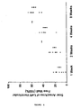
- Cardiac function is assessed by at least one of the criteria in the group consisting of: area occupied by scar tissue; vascularization of scar tissue; blood flow to scar tissue; developed pressure, systolic pressure; end diastolic pressure; and dp/dt.
- the cells for transplantation consist of a combination of adult cardiomyocytes plus endothelial cells.
- the present disclosure features a method for testing a pharmacological agent that is intended to prevent or ameliorate cardiac damage during cardiac surgery.
- the method comprises exposing the pharmacological agent to isolated cells selected from the group consisting of: adult cardiomyocytes, adult fibroblasts, fetal fibroblasts, smooth muscle cells, endothelial cells, and skeletal myoblasts, wherein the cells survive in myocardial scar tissue after transplantation and improve cardiac function, relative to cardiac function of a heart having similar myocardial scar tissue that is not transplanted with cells (cardiac function is assessed by at least one of the criteria in the group consisting of area occupied by scar tissue; vascularization of scar tissue; blood flow to scar tissue; developed pressure, systolic pressure; end diastolic pressure; and dp/dt), wherein cells exposed to the pharmacological agent prevents or ameliorates cardiac damage during cardiac surgery, compared to cells not exposed to the pharmacological agent.
- the present invention provides for the use of skeletal myoblasts for the preparation of a pharmaceutical composition for administering to a mammal with a scarred heart so as to form a therapeutic graft within said heart.
- a pharmaceutical composition for administering to a mammal with a scarred heart so as to form a therapeutic graft within said heart.
- the other technique used for culture purification was the clonal dilution technique.
- viable cells form individual colonies. Any cell not of the desired type that is adjacent to a colony of interest is killed with a sterile needle. Desired colonies are then collected using a sterile Pasteur pipette, and transferred to new culture dishes for culturing and passaging.
- Human cardiomyocytes were used without a purification step.
- Cardiomyocytes The purity of primary cultures of cardiomyocyte was assessed by immunofluorescent staining for cardiac myosin heavy chain (Rougier Bio-Tech Ltd, Quebec) ( Li R-K, et al., Cardiovascular Research 32:362-73, 1996 ).
- the cultured cells were fixed with methanol at -20°C for 15 minutes, washed with phosphate buffered saline, incubated with a monoclonal antibody against cardiac myosin heavy chain for 45 minutes at 37°C, washed three times with phosphate buffered saline for 5 minutes each at room temperature, and then under humid and dark conditions incubated with rabbit anti-mouse IgG conjugated with fluorescein isothiocyanate for 45 minutes at 37°C.
- the cells were washed with phosphate buffered saline, mounted and photographed using a light and UV microscope.
- the purity of cardiomyocyte cultures was determined by counting the percentage of stained cells in 8 random fields/dish. Eight dishes of cardiomyocyte cultures were used in each cardiomyocyte preparation.
- Smooth Muscle Cells The purity of smooth muscle cell cultures was determined by immunofluorescent staining for -smooth muscle cell actin (Sigma) as described in the previous paragraph.
- Vascular Endothelial Cells The purity of endothelial cell cultures was determined by immunofluorescent staining for factor VIII as described in the cardiomyocyte section above.
- Fibroblasts The purity of fibroblast cell cultures was determined by examining cell morphology with a microscope.
- Subject animals were grouped into three categories: sham, control and transplantation.
- the criteria for such grouping was as follows: Group Surgical exposure Scar generation Cell transplantation Sham X Control X X Transplantation X X X
- a latex balloon was passed into the left ventricle through the mitral valve and connected to a pressure transducer (Model p10EZ, Viggo-Spectramed, CA) and transducer amplifier and differentiator amplifier (Model 11-G4113-01, Gould Instrument System Inc., Ohio). After 30 minutes of stabilization, coronary flow in the heart was measured in triplicate by timed collection in the emptying beating state. The balloon size was increased by addition of saline in 0.02 ml increments from 0.04 to 0.8 ml, or the volume at which end diastolic pressure reached to 30 mm Hg, or whichever came first. The systolic and diastolic pressures were recorded at each balloon volume and developed pressure was calculated as the difference between the systolic and diastolic pressures.
- the epicardial and endocardial surface areas of the normal and scar tissue in the left ventricular free wall (LVFW) were measured by the techniques of Pfeffer et al ( Pfeffer JM, et al., American Journal of Physiology 260:H1406-14, 1991 ) and Jugdutt and Khan ( Jugdutt BI, et al., Circulation 89:2297-307, 1994 ). Briefly, the hearts were fixed in distension (30 mm Hg) with 10% phosphate-buffered formalin solution and then cut into 3 mm thick sections.
- the area of normal tissue, scar tissue, and transplanted tissue in the left ventricular free wall were traced onto a transparency and quantified using computerized planimetry (Jandal Scientific Sigma Scan, USA) as described by Wu et al. ( Wu TW, et al., Cardiovascular Research 27:736-39, 1993 ).
- the lengths of left ventricular free wall and scar tissue on both the endocardial and epicardial surfaces of each section were measured.
- the surface areas of the epicardial and endocardial scar tissue and left ventricular free wall were measured as: endocardial length + epicardial length ⁇ section thickness 3 mm .
- the surface area percentage of scar tissue in the left ventricular free wall was calculated as: epicardial scar size + endocardial scar size endocardial L ⁇ V ⁇ F ⁇ W ⁇ 1 + epicardial L ⁇ V ⁇ F ⁇ W ⁇ 100
- Example 1 On day 30 or 45 post-transplantation, the animals were anesthetized as in Example 1 and transplant, control, and sham hearts were exposed through a midline sternotomy and quickly harvested. The animals were euthanised by exsanguination under general anesthesia.
- the transplanted and control myocardial scar tissues were fixed in 5% glacial acetic acid in methanol.
- the tissue was embedded in paraffin and cut into 10 ⁇ m thick sections. After removal of the paraffin by immersing the sections for 3 minutes in xylene and then in 100, 95, 90, 85, and 70% ethanol for 3 minutes each, the samples were stained with haematoxylin and eosin as described by the manufacturer (Sigma Diagnostics, St. Louis, MO), and photographed.
- the heart sections were fixed at 4°C for 12 hours in 2% formaldehyde and 2% glutaraldehyde in phosphate buffer (0.15 M NaCl and 0.015 M NaH 2 PO 4 , pH 7.2).
- the transplanted cardiomyocytes were localized by staining for ⁇ -galactosidase activity as described earlier in Example 2.
- the stained tissue was embedded in paraffin and cut into 10 ⁇ m thick sections that were stained with haematoxylin and eosin as described in the last paragraph.
- tissue sections were washed three times with phosphate buffered saline and fixed with 2 ml 100% cold methanol at -20°C for 15 minutes. After washing three times with phosphate buffered saline and drying by draining, the tissue sections were exposed for 45 minutes at 37°C to monoclonal antibodies against cardiac myosin heavy chain (Rougier Bio-Tech, Montreal, Canada), diluted 1:20 with saline. Control tissues were incubated under the same conditions with phosphate buffered saline.
- the tissues were washed three times with phosphate buffered saline for 15 minutes at room temperature with gentle shaking, after which the secondary antibody, rabbit anti-mouse IgG conjugated with fluorescein isothiocyanate at a concentration of 1:32 dilution with phosphate buffered saline, was added.
- the tissues were incubated with the second antibody under dark and humid conditions for 45 minutes at 37°C. After washing with phosphate buffered saline, the cells in the transplant control tissues were visualized under ultraviolet light using an epi-microscope with a blue filter.
- the smooth muscle cell transplants were identified by staining immuno-fluorescently using a monoclonal antibody for ⁇ -smooth muscle actin as the primary antibody. Endothelial cell transplants were identified by immunofluorescent staining for factor VIII, as described in the next section.
- Fibroblast transplants were identified by the presence of a localized immunorejection within the ventricular scar.
- tissue sections processed as in Section E above were incubated with xylene twice for 5 minutes each, 100% ethanol twice for 2 minutes each and then with 70% ethanol twice for 1 minute each.
- the sections were incubated with rabbit IgG against factor VIII-related antigen (Dimension Lab. Inc., Ontario).
- the control samples were incubated with phosphate buffered saline under the same conditions.
- the test and control samples were incubated with goat anti-rabbit IgG conjugated with peroxidase.
- Cultured cardiomyocytes, smooth muscle cells, endothelial cells, and/or fibroblasts were seeded onto biological mesh, such as a collagen membrane, and on non-biological membranes, such as non-degradable membranes (Dacron) or degradable membranes (polyglycolic acid polymers), and the mesh and cells were cultured in cell culture medium.
- biological mesh such as a collagen membrane
- non-biological membranes such as non-degradable membranes (Dacron) or degradable membranes (polyglycolic acid polymers
- the cell-containing mesh was fixed in 2% formaldehyde and 2% glutaraldehyde in phosphate buffer (0.15 M NaCl and 0.015 M NaH 2 PO 4 , pH 7.2) at 4°C for 12 hours, embedded in paraffin, and cut into 10 ⁇ m thick sections, which were stained with haematoxylin and eosin as described in Section E, and photographed.
- cardiomyocytes must be implanted into the infarcted myocardium. Although this can be done by multiple syringe injections, injecting the cells, unfortunately, limits the number of cells which can be transplanted into myocardial scar tissue.
- Thrombin and cryoprecipitate which are derived from human blood, clot rapidly.
- Our in vitro results showed survival and contraction of cardiomyocytes in fibrin clots. We used this fibrin glue for cell transplantation.
- Function data were evaluated for the sham, control and transplant groups by an analysis of covariance using intracavitary balloon volume as the covariate and systolic, diastolic and developed pressure as dependent variables. Main effects were group, volume, and the interaction between group x volume. If there was an overall difference in the analysis of covariance, multiple pair-wise comparisons were performed to specify which groups were different. Because there were multiple pair-wise comparisons, a Bonferroni correction was performed and the critical alpha level was set at 0.01 for the analysis of covariance.
- Idiopathic hypertrophic cardiomyopathy is a primary cardiac abnormality characterized by regional asymmetrical myocardial hypertrophy.
- the hypertrophic myocardium can result in obstruction of left ventricular ejection as well as systolic and diastolic dysfunction and myocardial ischemia. Symptoms unresponsive to medical therapy can necessitate surgery.
- HCM is described for the most part as a heterogeneous disease of the sarcomeres. At least 34 missense mutations have been described in the ⁇ -myosin heavy chain gene, and 7 mutations in candidate loci also exist.
- family studies suggest that the autosomal dominant trait accounts for only 50% of HCM patients. The remaining HCM patients show no familial transmission and the disease occurs sporadically.
- Myocardial calcium kinetics and sympathetic stimulation have been studied because of diastolic functional abnormalities.
- none of these findings explains the regional myocardial hypertrophy (cardiomyocyte hypertrophy and over production of extracellular matrix proteins) observed in most HCM patients. The etiology of this disease remains unknown. It is thought that growth factors may play an important role in cardiomyocyte proliferation, cell hypertrophy and the overproduction of extracellular matrix.
- TGF ⁇ 1 transforming growth factor ⁇ 1
- IGF-I, -II insulin-like growth factors
- PDGF-B platelet-derived factor-B
- TGF ⁇ 1, IGF-I, IGF-II and PDGF-B transcripts were quantitated using multiplex RT-PCR.
- Glyceraldehyde 3-phosphate dehydrogenase (G3PDH) was used as an internal standard.
- Antibodies against TGF ⁇ 1 and IGF-I were used to localize their peptides within the myocardium.
- mRNA levels (densitometric ratio of GF/G3PDH) of TGF ⁇ 1 and IGF-I in HCM myocardium (0.75 ⁇ 0.05, 0.85 ⁇ 0.15, mean ⁇ 1SE) were significantly (p ⁇ 0.01 for all groups) elevated in comparison to non-HCM myocardium (AS: 0.38 ⁇ 0.07, 0.29 ⁇ 0.06; SA: 0.32 ⁇ 0.04, 0.18 ⁇ 0.05; TM: 0.25 ⁇ 0.03, 0.15 ⁇ 0.03).
- CMs fetal rat cardiomyocytes
- CMs were isolated from 18-day gestation, 5-, 22-, 32-, and 62-day old Sprague-Dawley rat hearts and cultured for 1 day. 2 to 5 x 10 6 cells in saline were injected into the skeletal muscle of one adult rat leg. The other leg (control) was injected with saline alone. Cell viability and function were assessed visually and by ultrasound and electrocardiography (ECG).
- ECG ultrasound and electrocardiography
- Donor age is important for maintaining contractility of CMs after transplantation into skeletal muscle.
- Angiogenesis was induced in the scar tissue by endothelial cell transplantation. Since no myocytes were present in the scar, function did not improve. Endothelial cell transplantation in patients with incomplete infarction may restore perfusion, relieve angina and restore function.
- mice All procedures performed on animals were approved by the Animal Care Committee of The Toronto Hospital. Experiments were performed according to the "Guide to the Care and Use of Experimental Animals" published by the National Institutes of Health (NIH publication 85-23, revised 1985). Sprague-Dawley rats (Charles River Canada Inc, Quebec, Canada) were used for allogeneic transplantation. Male rats, weighing 330 to 360 g, were used as recipients and donors of endothelial cells. Sygenic Lewis rats (Male, 250-300 g, Charles River Canada Inc, Quebec, Canada) were used for autologus transplantation.
- Endothelial cells from rat aorta were isolated and cultured as previously described ( Mickle et al., J. Mol. Cell. Cardiol. 22:1297-1304, 1990 ). In brief, descending thoracic aortic segments were obtained from rats under general anesthesia as described in the next section. The blood vessels were washed with phosphate buffered saline solution (PBS; composition in mmol/L: NaCl 136.9, KCl 2.7, Na 2 HPO 4 8.1, KH 2 PO4 1.5, pH 7.3) and connective tissue was removed.
- PBS phosphate buffered saline solution
- aortic segment was then flushed with enzyme solution (0.1% collagenase and 0.2% trypsin in PBS) and incubated with the enzyme solution for 30 minutes at 37°C.
- enzyme solution (0.1% collagenase and 0.2% trypsin in PBS)
- endothelial cells were isolated by flushing the inside of aortic segments with culture medium (Medium 199 containing 20% fetal bovine serum, 100U/ml of penicillin, 100ug/ml of streptomycin, and 0.5% heparin salt) three to five times.
- culture medium Medium containing 20% fetal bovine serum, 100U/ml of penicillin, 100ug/ml of streptomycin, and 0.5% heparin salt
- the cells were washed three times (15 minutes per wash) with PBS, then incubated with goat anti- rabbit immunoglobulin G conjugated with peroxidase at 37°C for 45 minutes, washed three times (15 minutes per wash) with PBS, and immersed in diaminobenzidine-H 2 O 2 (2mg/ml diaminobenzidine, 0.03% H 2 O 2 in 0.02ml/l phosphate buffer) solution for 15 minutes. After a final wash in PBS, the samples were photographed.
- VEGF protein levels in culture medium and endothelial cells were quantitated by chemiluminescent slot blot analysis.
- Culture medium incubated without cells was used as a control.
- Forty micrograms of protein from culture medium or from cultured endothelial cells were loaded onto a 0.2 ⁇ m nitrocellulose membrane (Schleicher & Schuell Inc., Keene, NH) using the Minifold II Slot Blotting System (Schleicher & Schuell Inc., Keene, NH).
- Standards of VEGF (2, 5, 10, 25 ng) (Sigma, Mississauga, ONT) were loaded on the same membrane.
- TTBS buffer 50 mM Tris-HCl buffer pH 7.4 and 0.1% of Tween-20
- blocking buffer Boehringer Mannhein, GmbH, German.
- Monoclonal antibodies against VEGF (Sigma, Mississauga, ONT) (1:3000, diluted with 0.5 x block buffer) were added and incubated overnight at 4°C.
- TTBS Tris-HCl buffer pH 7.4 and 0.1% of Tween-20
- VEGF proteins were detected by chemiluminescence using the Boehringer Mannheim detection kit (Quebec, Canada).
- a densitometric analysis of the standard and sample bands was performed with a Bio-Rad image analysis system (Bio-Rad Lab. Hercules, CA). Standard curves of VEGF were generated and VEGF quantity in culture medium and cultured endothelial cells was calculated based on the standard curve. The results (mean ⁇ SD) are expressed as ng of VEGF/mg protein.
- Myocardial Scar Formation Rats were anesthesized by intramuscular injection of ketamine hydrochloride (20mg/kg body weight) followed by intraperitoneal injection of sodium pentobarbital (30mg/kg body weight). The anesthesized rats were intubated and positive pressure ventilation was performed with room air supplemented with oxygen and isoflurane (0.2-1.0%) using a Harvard ventilator. The electrocardiogram was monitored during operation.
- the heart was exposed through a 2- to 3-cm left lateral thoracotomy incision. Cryoinjury was produced in the left ventricular free wall(LVFW) with a metal probe (8 X 10 mm in diameter) cooled to -190°C by immersion in liquid nitrogen. It was applied to the LVFW for one minute and this procedure was repeated ten times. The muscle and skin were closed with 3-0 silk sutures. Penlong XL (benzathine penicillin G 150,000 U/ml and procaine penicillin G 150,000 U/ml) was given intramuscularly (1ml/kg) and buprenorphine hydrochloride (0.01mg/kg) was administered after each operation. The cryoinjured rats were randomly divided into two groups: transplantation and control.
- Endothelial Cell Transplantation At two weeks after cryoinjury, cell transplantation was performed. Under general anesthesia, the heart was exposed through a midline sternotomy. Endothelial cell suspension (60ul, 6X10 6 cells) was injected into the center of the scar tissue in the transplantation group using a tuberculin syringe, and same amount of culture media was injected into the scar in the control group. The chest was closed with 3-0 silk sutures. Antibiotics and analgesics were given as previously described. Cyclosporine A (25mg/kg/day) was administered subcutaneously to both transplanted and control groups.
- Ventricles were dissected into normal tissue, scar tissue, and tissue in the borderline area. Each section was weighed and the radioactivity was then determined using a gamma counter at the window setting of 110 to 138 KeV. The data was expressed as counts per minute (cpm)/mg of tissue. The ratio of cpm/mg in scar and borderline tissue to normal tissue was calculated, expressed as the percentage, and compared between transplanted and control hearts.
- Krebs-Hanseleit buffer mmol/L: NaCl 118, KCl 4.7, KH 2 PO 4 1.2, CaCl 2 2.5, MgSO 4 1.2, NaHC0 3 25, glucose 11, pH 7.
- a latex balloon was inserted into the left ventricle through the mitral valve and connected to a pressure transducer (Model p10EZ; Viggo-Spectramed, Oxnard CA) and a transducer amplifier and differentiator amplifier (Model 11-G4113-01; Gould Instrument System Inc, Valley View OH).
- a pressure transducer Model p10EZ; Viggo-Spectramed, Oxnard CA
- a transducer amplifier and differentiator amplifier Model 11-G4113-01; Gould Instrument System Inc, Valley View OH.
- the coronary flow was measured by the timed collection in the empty beating state.
- the balloon size was increased in 0.02 ml increments from 0.04 to 0.6 ml by the addition of saline solution.
- the systolic and end-diastolic pressure and maximal and minimal dp/dt were recorded at each balloon volume.
- the developed pressure was calculated as the difference between the systolic and end-diastolic pressure.
- the epicardial and endocardial surfaces of the normal and scar tissue in the LVFW were measured by the technique of Pfeffer ( Pfeffer Am J Physiol 1991;260:H1406-14 ) and Jugdutt and Khan Circulation 1994;89:2297-307 ). Briefly, the hearts were fixed in distension (30 mmHg) with 10% phosphate-buffered formalin solution for 2 days. The atria were excised, the ventricular weight and the size of the scar on epicardial surface were measured, and ventricular volume was measured by water displacement. After that, the hearts were cut into sections of 3 mm thickness.
- the area of normal and scar tissue in the LVFW were traced onto a transparency and quantified using computed planimetry (Jandal Scientific Sigma-Scan, Corte Madera, CA).
- the lengths of LVFW and scar tissue on both the endocardial and epicardial surfaces of each section were measured.
- the surface areas of the endocardial and epicardial scar tissue and the LVFW were measured as the sum of the endocardial length and epicardial length times the section thickness (3mm).
- the surface area percentage of scar tissue in the LVFW was calculated as follows: (endocardial scar size+epicardial scar size)/(endocardial LVFW+epicardial LVFW) X 100.
- the heart sections were used for immunohistochemical staining of endothelial cells.
- the sections were incubated with xylene twice for 5 minutes each, 100% ethanol twice for 2 minutes each, and then with 70% ethanol twice for 1 minute each.
- the sections were incubated with rabbit immunoglobulin G against factor VIII-related antigen and endothelial cells were identified as described in the Cell Identification section above.
- the stained vasculoendothelial cells in the transplanted and control groups were counted using a light microscope at 400X magnification. The result was expressed as the number of capillary vessels/field.
- Cultured endothelial cells were distinguished from fibroblasts and vascular smooth muscle cells by morphologic criteria and growth characteristics. The endothelial cells were oval shaped, in contrast to the spindle-shape of cultured fibroblasts and vascular smooth muscle cells. Cultured endothelial cells grew in a "cobblestone" pattern whereas fibroblasts grew in a "whirling" pattern and vascular smooth muscle cells grew in a "hill and valley” pattern. In addition, cultured endothelial cells stained positively for factor VIII-related antigen. The purity of the culture to be transplanted was more than 95%.
- transplanted cells In syngeneic cell transplantation the transplanted cells also induced angiogenesis in the myocardial scar tissue.
- the density of capillaries in the scar tissue of the transplanted group was 2.6 ⁇ 1.5 vessels/0.8mm 2 , which was significantly (p ⁇ 0.01) greater than that in the control group (1.1 ⁇ 0.6).
- Transplanted endothelial cells were incorporated into capillaries at the transplant site.
- the transplanted endothelial cells stimulate angiogenesis, demonstrated by an increase in the number of capillaries in the scar tissue and increased blood perfusion in scar tissue as illustrated by microsphere perfusion studies.
- the induced angiogenesis in transmural scar tissue did not improve infarcted heart function.
- angiogenesis by endothelial transplantation: (1) formation of blood vessels by transplanted endothelial cells; (2) angiogenesis stimulated by growth factors secreted by transplanted endothelial cells; (3) angiogenesis stimulated by an inflammatory reaction induced by transplanted endothelial cells.
- Endothelial cells are the most important component for blood vessel formation.
- Our in vivo study showed that transplanted endothelial cells become part of newly formed capillaries in syngeneic cell transplantation. These data suggested that transplanted endothelial cells are involved in blood vessel formation.
- the data obtained from allogeneic endothelial cell transplantation suggested that newly formed capillaries in the scar tissue were grown from recipient heart endothelial cells.
- transplanted cells at 1 and 7 days after transplantation, but did not observe transplanted cells after 14 days post transplantation. Additional evidence of this conclusion is that there was no immunologic reaction around capillary vessels in the scar.
- transplanted allogeneic endothelial cells become part of the newly formed capillaries, lymphocyte infiltration should be observed around the blood vessel.
- Angiogenesis in our animal model might also be stimulated by inflammatory reactions.
- syngeneic endothelial cell transplantation has shown that angiogenesis occurs without inflammatory reaction, factors released by inflammatory cells might play a role in blood vessel formation induced by allogeneic endothelial cell transplantation.
- Capillary density in scar tissue subjected to allogeneic cell transplantation was 1.3 times greater than that subjected to syngeneic cell transplantation. This difference in capillary density may be due to lymphocyte infiltration.
- the left ventricular free wall was divided into three parts: normal area, transmural infarct area, and borderline area.
- normal area normal area
- transmural infarct area transmural infarct area
- borderline area The blood flow to the borderline areas was always between that of the normal and complete transmural infarct area.
- myocardial infarction damaged myocardium undergoes both acute and chronic inflammatory reactions.
- necrosed cardiac cells are replaced with fibrotic tissue and ventricular remodeling progresses.
- Ventricular pressure stretches and thins the healing area and ventricular dilatation occurs.
- a ventricular aneurysm may form and congestive heart failure may result.
- Limitation of post infarction ventricular remodeling is important to prevent expansion and thinning of the infarct region and thinning, and progressive ventricular dilatation, which results in congestive heart failure.
- the addition of cardiac tissure to replace the scar tissue may prevent infarct thinning and preserve chamber size and ventricular function.
- Cardiomyocyte transplantation was most successful after the inflammatory reaction resolved and before significant scar expansion and ventricular dilatation occurred.
- mice All procedures performed on animals were approved by the Animal Care Committee of The Toronto Hospital. Experimental animals used were male Sprague-Dawley rats (Lewis, Charles River Canada Inc. Quebec, Canada), weighing 450 grams. Cardiomyocytes obtained from 18-day gestational rat hearts were cultured prior to transplantation. All experiments were performed according to "A Guide to the Care and Use of Experimental Animals" of the Canadian Council on Animal Care and the "Guide for the Care and Use of Laboratory Animals" NIH publication 85-23, revised 1985).
- Myocardial scar tissue was generated as described in Example I above. Briefly, rats were anesthetized with ketamine (22 mg/kg body weight, intramuscular) followed by an intraperitoneal injection of pentobarbital (30 mg/kg body weight). The anesthetized rats were intubated and positive pressure ventilation was maintained with room air supplemented with oxygen (6 L/minute) using a Harvard ventilator (Model 683, South Natick, MA, USA). The rat heart was exposed through a two cm left lateral thoracotomy. Cryo-injury was produced at the left ventricular free wall (LVFW) with a liquid nitrogen probe. The muscle layer and skin were closed with 5-0 vicryl sutures.
- LVFW left ventricular free wall
- Penlog XL benzathine penicillin G 150,000 U/ml and procaine penicillin G 150,000 U/ml
- Penlog XL benzathine penicillin G 150,000 U/ml and procaine penicillin G 150,000 U/ml
- buprenorphine 0.01-0.05 mg/kg body weight
- the LVFWs of 35 animals were cryo-injured. Immediately after cryo-injury and at 1, 2, 4, and 8 weeks after injury, 7 animals chosen at random were sacrificed under general anesthesia (see Fig. 5 for a schematic diagram of the experimental design). The hearts were then fixed in distension (30 mm Hg) with 10% phosphate-buffered formalin solution for 24-48 hours and then cut into 3 mm thick sections. All sections were used for assessment of myocardial and scar sizes. For each section, the area of scar tissue in the LVFW was traced onto a transparency and quantified using computerized planimetry (Jandal Scientific Sigma Scan, Fairfield, CT., USA). Scar length was calculated as the average of endocardial scar length and epicardial scar length.
- Scar area was calculated as the scar length X section thickness (3mm).
- the total scar area in size was calculated as the sum of the scar areas for all of the tissue sections showing scars.
- the LVFW myocardial size was calculated using the same equation.
- the percentage of the LVFW occupied by the scar was calculated as scar size/LVFW size X 100.
- Histological studies The fixed heart sections were embedded in paraffin and cut to yield 10 ⁇ m thick sections. The sections were stained with haematoxylin and eosin as described by the manufacturer (Sigma Diagnostics, St. Louis, MO).
- Cardiomyocytes from fetal rat hearts were isolated, purified and cultured as previously described ( Li, R-K, et al. J. Tissue Culture Methods 14:93-100, 1992 ). Briefly, the cells were cultured for 24 hours in Iscove's modified Dulbecco's medium containing 10% fetal bovine serum, 100 U/ml penicillin and 100 ug/ml streptomycin at 37EC, 5% CO 2 and 95% air. The cultured cardiomyocytes were detached from the cell culture dish with 0.05% trypsin in phosphate-buffered saline (PBS). After centrifugation at 580 X g for 3 minutes, the cell pellet was resuspended in culture medium at a concentration of 16 X 10 6 cells/ml. A 0.25 ml cell suspension was used for each transplantation.
- PBS phosphate-buffered saline
- the hearts were quickly isolated from rats and perfused in a Langendorff apparatus with filtered Krebs Heinseleit buffer (mmol/L NaCl 118, KCl 4.7,KH 2 PO 4 1.2, CaCl 2 2.5, MgSO 4 1.2, NaHCO 3 25, glucose 11; pH 7.4) equilibrated with a 5% CO 2 and 95% O 2 .
- a latex balloon was passed into the left ventricle through the mitral valve and connected to a pressure transducer (Model p10EZ, Viggo-Spectramed, CA) and transducer amplifier and differentiator amplifier (Model 11-G4113-01, Gould Instrument System Inc., Ohio).
- the balloon size was increased in 0.02 ml increments from 0.04 to a volume, at which end diastolic pressure was 30 mm Hg or more by the addition of saline.
- the systolic and diastolic pressures were recorded at each balloon volume and developed pressure was calculated as the difference between the systolic and diastolic pressures.
- Histological identification of transplanted muscle cells in the scar tissue Fixed heart sections were embedded in paraffin, and cut into 10 ⁇ m thick sections. The sections were stained with hematoxylin and eosin as described in the "Histological Studies" section above.
- Heart sections containing transplanted cells also were stained for myosin heavy chain (MHC) (Rougier Bio-Tech, Montreal). After deparaffination and rehydration, samples were incubated for 30 minutes with a solution of 3% H 2 O 2 in 70% methanol to inhibit endogenous myocardial peroxidase. Triton X-100 (0.2%) was used to treat samples for 10 minutes to enhance cell permeability. After blocking nonspecific protein binding with 2% normal goat serum in 0.05 M Tris buffer (pH 7.4) for 15 minutes, primary antibodies against HCM (1:1000) were added and the samples were incubated at 37°C for 30 minutes followed by an overnight incubation at 4°C. Negative control samples were incubated in PBS under the same conditions.
- MHC myosin heavy chain
- Data Analysis Data are expressed as the mean ⁇ standard error.
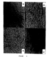
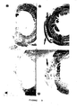
- Fig. 6A Immediately after cryo-injury, 25 ⁇ 3% of the size of the LVFW was transmurally damaged.
- the cardiomyocytes were fragmented ( Fig. 6A ).
- Fig. 6B At one week, most of the necrosed cardiomyocytes were gone and a predominantly mononuclear inflammatory infiltrate was present in the affected area ( Fig. 6B ).
- Fig. 6C At two weeks the inflammatory infiltrate had almost disappeared and fibroblasts and collagen deposition were evident.
- Fig. 6D the scar was composed of fibrotic tissue ( Fig. 6D ). The tissue was less cellular and lymphocytes were not observed.
- the myocardial scar size of the left ventricle expanded over the 8 week study period in the damaged hearts ( Figs. 7 and 8 ). Although the scar sizes at 1 and 2 weeks (13 ⁇ 6 % and 21 ⁇ 4 of LVFW) were not statistically different, the size of 4-week-old scars (39 ⁇ 5 % of LVFW) was larger (p ⁇ 0.01). At 8 weeks there was a further increase (p ⁇ 0.01) in scar size (55 ⁇ 3% of LVFW).
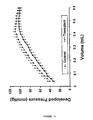
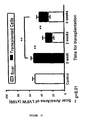
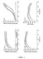
- Cardiomyocytes transplanted at 4 weeks after myocardial injury also improved (p ⁇ 0.001) myocardial function ( Figure 14 ).
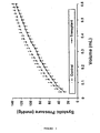
- Figure 15 shows that hearts transplanted at 2 weeks had higher developed pressures at balloon volumes 0.1 ml (p ⁇ 0.05), 0.2 ml (p ⁇ 0.01) and 0.3 ml (p ⁇ 0.01) than hearts transplanted at 4 weeks.
- transplant tissue visibly contracted, but at different rates than did host myocardium.
- the contraction of the transplanted tissue persisted after dissection of the recipient heart, but its rate of contraction continued to be different.
- necrosed ventricular tissue occurs by 2 days after cyro-injury of the myocardium. At this time there is an acute inflammatory reaction with neutrophils accumulating first in the periphery and later in the center of the necrotic region. A chronic inflammatory reaction involving macrophages and lymphocytes follows the acute reaction.
- the inflammatory reaction is less intense two weeks after cryo-injury (and is absent by three weeks): two weeks after cryo-injury, the scars of the control animal hearts showed minimal or no inflammatory reaction.
- Two-week-old scars were less mature and contained more blood vessels and fibroblasts than 4-week-old scars, which comprised firm connective tissue. Compared to one-week-old scars, there was less cardiac tissue in older scars. It is possible that progressive inflammation and lack of oxygen and nutrient supply decreased the myocardial tissue over time.
- the cardiomyocytes transplanted immediately after cryo-injury did not survive in any of the animals studied. We believe the activated neutrophils and macrophages in the inflamed scar area destroyed the transplanted cells.
- the left ventricular chamber of the animals transplanted at the time of cryo-necrosis was similar to that of the control scar. The chamber was dilated due to thinning of the scar. The remaining viable muscle of the left ventricle was hypertrophied. No inflammatory reaction was seen. The function of these transplanted hearts was similar to that of the control hearts.
- a cardiomyocyte transplant forms the equivalent of a viable epicardial rim and prevents ventricular dilatation and over-stretching of the cardiomyocytes during systole. With over-stretching of the cardiomyocytes, cardiac function is diminished (Frank-Starling Law).
- the elastic properties of the contractile apparatus of the transplanted cardiomyocytes may prevent host fibroblast and cardiomyocyte stretching and ventricular enlargement.
- angiogenesis occurred in the course of forming cardiac tissue from transplanted cardiomyocytes. Angiogenesis is most likely necessary to maintain the viability of the transplanted muscle cells. However, an increased blood supply in the scar could also facilitate fibroblast turnover and strengthening of the scar in response to left ventricular free wall stretch.
- the transplanted cardiomyocytes should also increase the contractile capacity of the scar tissue and decrease the contractile requirements of the non-affected host myocardium.
- Patients with angina that cannot be corrected by bypass surgery may benefit from angiogenesis secondary to cardiomyocyte transplantation.
- it may be beneficial to stimulate angiogenesis with growth factors prior to autotransplantation.
- transplantation of fetal cardiomyocytes form cardiac tissue in scar tissue which limits ventricular dilatation and scar thinning.
- the optimal time for transplantation for improved myocardial function is after the acute inflammatory reaction and before significant ventricular dilatation has occurred.
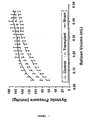
- Cardiac transmural scars were induced by cryo-injury in the left ventricular free wall (LVFW) of Sprague-Dawley rat hearts, as described in the previous examples. Three weeks after cryo-injury, the rats were divided into three groups (eight rats per group), and cardiac transmural scars were transplanted with either medium (no cells), adult cardiomyocytes alone, or adult cardiomyocytes plus endothelial cells. Cardiac function was assessed using a Langandorff preparation five weeks after cell transplantation ( Stephen et al., Annals of Thoracic Surgery 59: 1127-1133, 1995 ).
- Fig. 16 shows a graph of developed pressure in infarcted rat hearts injected with either medium (negative control), adult cardiomyocytes alone (CM), or adult cardiomyocytes plus endothelial cells (CM + EC).
- CM adult cardiomyocytes alone
- CM + EC adult cardiomyocytes plus endothelial cells
Claims (13)
- Utilisation de myoblastes squelettiques pour la préparation d'une composition pharmaceutique devant être administrée à un mammifère dont le coeur présente des cicatrices de sorte à former un greffon thérapeutique à l'intérieur dudit coeur.
- Utilisation selon la revendication 1, dans laquelle ladite composition pharmaceutique comprend en outre des fibroblastes adultes.
- Utilisation selon la revendication 1 ou la revendication 2, dans laquelle lesdites cellules forment un greffon cardiaque stable dans ledit coeur de mammifère.
- Utilisation selon l'une quelconque des revendications 1 à 3, dans laquelle ladite composition pharmaceutique améliore la fonction cardiaque.
- Utilisation selon l'une quelconque des revendications 1 à 4, dans laquelle une ou plusieurs desdites cellules est transfectée ou généralement élaborée pour délivrer des molécules recombinantes au tissu myocardique.
- Utilisation selon l'une quelconque des revendications 1 à 4, dans laquelle une ou plusieurs desdites cellules est transfectée ou élaborée génétiquement pour délivrer des molécules recombinantes au tissu cicatriciel.
- Utilisation selon l'une quelconque des revendications 1 à 6, dans laquelle lesdites cellules sont dérivées d'une auto, allo ou xénotransplantation.
- Utilisation selon l'une quelconque des revendications 1 à 7, dans laquelle ladite composition pharmaceutique comprend en outre un ou plusieurs facteurs de croissance.
- Utilisation selon la revendication 8, dans laquelle ladite composition pharmaceutique comprend un facteur de croissance insulinomimétique de type I ; un facteur de croissance insulinomimétique de type II ; un facteur de croissance de transformation β1 ; un facteur de croissance dérivé des plaquettes β ; un facteur de croissance basique des fibroblastes ; ou un facteur de croissance endothéliale vasculaire.
- Utilisation selon l'une quelconque des revendications 1 à 9, dans laquelle ladite composition pharmaceutique doit être utilisée en chirurgie reconstructive myocardique.
- Utilisation selon l'une quelconque des revendications 1 à 10, dans laquelle lesdites cellules doivent être introduites par injection directe.
- Utilisation selon l'une quelconque des revendications 1 à 10, dans laquelle lesdites cellules sont suspendues sur un échafaudage biodégradable ou non biodégradable.
- Utilisation selon l'une quelconque des revendications 1 à 10, dans laquelle l'administration des cellules comprend l'utilisation d'un adhésif biologique pour attacher les cellules au tissu myocardique.
Applications Claiming Priority (2)
| Application Number | Priority Date | Filing Date | Title |
|---|---|---|---|
| US08/863,882 US6110459A (en) | 1997-05-28 | 1997-05-28 | Transplants for myocardial scars and methods and cellular preparations |
| EP98923950A EP0985028B1 (fr) | 1997-05-28 | 1998-05-28 | Greffons pour cicatrices myocardiques, et methode et preparations cellulaires associees |
Related Parent Applications (2)
| Application Number | Title | Priority Date | Filing Date |
|---|---|---|---|
| EP98923950.4 Division | 1998-05-28 | ||
| EP98923950A Division EP0985028B1 (fr) | 1997-05-28 | 1998-05-28 | Greffons pour cicatrices myocardiques, et methode et preparations cellulaires associees |
Publications (3)
| Publication Number | Publication Date |
|---|---|
| EP1690546A2 EP1690546A2 (fr) | 2006-08-16 |
| EP1690546A3 EP1690546A3 (fr) | 2006-10-11 |
| EP1690546B1 true EP1690546B1 (fr) | 2010-11-17 |
Family
ID=25342008
Family Applications (2)
| Application Number | Title | Priority Date | Filing Date |
|---|---|---|---|
| EP06002350A Expired - Lifetime EP1690546B1 (fr) | 1997-05-28 | 1998-05-28 | Greffons pour cicatrices myocardiques, méthodes et préparations cellulaires associées |
| EP98923950A Expired - Lifetime EP0985028B1 (fr) | 1997-05-28 | 1998-05-28 | Greffons pour cicatrices myocardiques, et methode et preparations cellulaires associees |
Family Applications After (1)
| Application Number | Title | Priority Date | Filing Date |
|---|---|---|---|
| EP98923950A Expired - Lifetime EP0985028B1 (fr) | 1997-05-28 | 1998-05-28 | Greffons pour cicatrices myocardiques, et methode et preparations cellulaires associees |
Country Status (8)
| Country | Link |
|---|---|
| US (2) | US6110459A (fr) |
| EP (2) | EP1690546B1 (fr) |
| JP (1) | JP2002501513A (fr) |
| AT (2) | ATE488244T1 (fr) |
| AU (1) | AU753388B2 (fr) |
| CA (1) | CA2291138C (fr) |
| DE (2) | DE69835354T2 (fr) |
| WO (1) | WO1998054301A2 (fr) |
Families Citing this family (101)
| Publication number | Priority date | Publication date | Assignee | Title |
|---|---|---|---|---|
| US20040087498A1 (en) * | 1991-02-28 | 2004-05-06 | Novo Nordisk Health Care Ag | Modified factor VII |
| US6443974B1 (en) * | 1996-07-28 | 2002-09-03 | Biosense, Inc. | Electromagnetic cardiac biostimulation |
| US6099832A (en) * | 1997-05-28 | 2000-08-08 | Genzyme Corporation | Transplants for myocardial scars |
| US6110459A (en) * | 1997-05-28 | 2000-08-29 | Mickle; Donald A. G. | Transplants for myocardial scars and methods and cellular preparations |
| US6775574B1 (en) * | 1997-11-07 | 2004-08-10 | Medtronic, Inc. | Method and system for myocardial infarction repair |
| US7115417B1 (en) | 1998-05-01 | 2006-10-03 | Chancellor Michael B | Soft tissue and bone augmentation and bulking utilizing muscle-derived progenito compositions, and treatments thereof |
| US6866842B1 (en) | 1998-05-01 | 2005-03-15 | University Of Pittsburgh | Muscle-derived cells (MDCs) for treating muscle-or bone-related injury or dysfunction |
| EP1100870B1 (fr) * | 1998-07-31 | 2008-01-02 | Genzyme Corporation | Amelioration du fonctionnement cardiaque par transplantation de cellules souches mesenchymateuses |
| CA2377541A1 (fr) * | 1999-06-25 | 2001-01-04 | Eduardo N. Mitrani | Procede permettant d'induire une angiogenese au moyen de micro-organes |
| US20030113301A1 (en) * | 1999-07-23 | 2003-06-19 | Albert Edge | Muscle cells and their use in cardiac repair |
| CA2378643A1 (fr) | 1999-07-23 | 2001-02-01 | Diacrin, Inc. | Cellules musculaires et leur utilisation dans la reparation cardiaque |
| DE10003521A1 (de) * | 2000-01-27 | 2001-08-09 | Medigene Ag | Vorrichtung zum Herstellen eines dreidimensionalen Matrixkörpers, Multi-Well-Platte, Lösung zum Kultivieren von Säugerkardiomyocyten, Verfahren zum Kultivieren einer Zellkultur, Vorrichtung für die Messung isometrischer Kraftparameter von Zellkulturen sowie Verfahren zum meßbaren Verfolgen von Kontraktionen eines in eine Trägersubstanz eingelagerten Zellgewebes |
| AU5159901A (en) | 2000-04-14 | 2001-10-30 | Univ Pittsburgh | Soft tissue and bone augmentation and bulking utilizing muscle-derived progenitor cells, compositions and treatments thereof |
| FR2810045B1 (fr) * | 2000-06-07 | 2004-09-03 | Assist Publ Hopitaux De Paris | Procede d'obtention de population cellulaires caracterisees d'origine musculaire et utilisations |
| US7547674B2 (en) | 2001-06-06 | 2009-06-16 | New York Medical College | Methods and compositions for the repair and/or regeneration of damaged myocardium |
| US7862810B2 (en) * | 2000-07-31 | 2011-01-04 | New York Medical College | Methods and compositions for the repair and/or regeneration of damaged myocardium |
| US20020098167A1 (en) * | 2000-07-31 | 2002-07-25 | Piero Anversa | Methods and compositions for the repair and/or regeneration of damaged myocardium |
| US20110091428A1 (en) * | 2000-07-31 | 2011-04-21 | New York Medical College | Compositions of adult organ stem cells and uses thereof |
| US7491385B2 (en) * | 2001-09-05 | 2009-02-17 | Genegrafts Ltd. | Nucleic acid constructs and cells, and methods utilizing same for modifying the electrophysiological function of excitable tissues |
| US7294333B1 (en) | 2000-10-20 | 2007-11-13 | Genegrafts Ltd. | Nucleic acid constructs and cells, and methods utilizing same for modifying the electrophysiological function of excitable tissues |
| US6659995B1 (en) * | 2000-11-17 | 2003-12-09 | Syde A. Taheri | Autologous myocyte micro granual retrieval and implantation (AMMGRI) |
| DE10106512C1 (de) * | 2001-02-13 | 2002-05-23 | Axel Haverich | Verfahren zur Herstellung eines biologischen Gewebes unter Verwendung einer Kollagenunterlage und zugehöriges Gewebekonstrukt |
| EP1372398B1 (fr) | 2001-02-23 | 2013-07-10 | The University of Pittsburgh | Preparation rapide de matrices de cellules souches utilisee pour le traitement et la reparation de tissu et d'organe |
| US7341062B2 (en) * | 2001-03-12 | 2008-03-11 | Bioheart, Inc. | Method of providing a dynamic cellular cardiac support |
| IL158368A0 (en) * | 2001-04-13 | 2004-05-12 | Anterogen Co Ltd | Methods and reagents for cell transplantation |
| JP2004533234A (ja) * | 2001-04-13 | 2004-11-04 | アントロジェン カンパニー リミテッド | カプセル化細胞インジケータシステム |
| US7993365B2 (en) | 2001-06-08 | 2011-08-09 | Morris Innovative, Inc. | Method and apparatus for sealing access |
| AU2002310364B2 (en) * | 2001-06-08 | 2006-02-23 | Morris Innovative Research, Inc. | Method and apparatus for sealing access |
| US20070038244A1 (en) * | 2001-06-08 | 2007-02-15 | Morris Edward J | Method and apparatus for sealing access |
| US7732199B2 (en) | 2001-07-12 | 2010-06-08 | Geron Corporation | Process for making transplantable cardiomyocytes from human embryonic stem cells |
| CA2453438C (fr) * | 2001-07-12 | 2016-04-05 | Geron Corporation | Cellules de la lignee des cardiomyocytes produites a partir de cellules souches humaines pluripotentielles |
| WO2003093433A2 (fr) * | 2002-05-02 | 2003-11-13 | Regents Of The University Of Minnesota | Matrice biologique a base de fibrine |
| US20040106896A1 (en) * | 2002-11-29 | 2004-06-03 | The Regents Of The University Of California | System and method for forming a non-ablative cardiac conduction block |
| US7361368B2 (en) | 2002-06-28 | 2008-04-22 | Advanced Cardiovascular Systems, Inc. | Device and method for combining a treatment agent and a gel |
| US20040018174A1 (en) * | 2002-07-23 | 2004-01-29 | Boston Scientific Corporation | Cell therapy for regeneration |
| WO2004012791A2 (fr) * | 2002-08-06 | 2004-02-12 | Genvec, Inc. | Systeme d'injection ameliore |
| US7829694B2 (en) | 2002-11-26 | 2010-11-09 | Medtronic, Inc. | Treatment of neurodegenerative disease through intracranial delivery of siRNA |
| US7618948B2 (en) * | 2002-11-26 | 2009-11-17 | Medtronic, Inc. | Devices, systems and methods for improving and/or cognitive function through brain delivery of siRNA |
| US7605249B2 (en) * | 2002-11-26 | 2009-10-20 | Medtronic, Inc. | Treatment of neurodegenerative disease through intracranial delivery of siRNA |
| US7732591B2 (en) * | 2003-11-25 | 2010-06-08 | Medtronic, Inc. | Compositions, devices and methods for treatment of huntington's disease through intracranial delivery of sirna |
| US7994149B2 (en) | 2003-02-03 | 2011-08-09 | Medtronic, Inc. | Method for treatment of Huntington's disease through intracranial delivery of sirna |
| US9617516B2 (en) | 2003-04-25 | 2017-04-11 | University Of Pittsburgh-Of The Commonwealth System Of Higher Education | Muscle-derived cells (MDCs) for promoting and enhancing nerve repair and regeneration |
| WO2005072764A2 (fr) * | 2004-01-16 | 2005-08-11 | Novocell, Inc. | Procede d'utilisation de facteurs angiogeniques lies a la fibrine afin de stimuler la vascularisation d'un site de transplantation de cellules encapsulees |
| US7840263B2 (en) | 2004-02-27 | 2010-11-23 | Cardiac Pacemakers, Inc. | Method and apparatus for device controlled gene expression |
| US20050208090A1 (en) * | 2004-03-18 | 2005-09-22 | Medtronic, Inc. | Methods and systems for treatment of neurological diseases of the central nervous system |
| US7452718B2 (en) * | 2004-03-26 | 2008-11-18 | Geron Corporation | Direct differentiation method for making cardiomyocytes from human embryonic stem cells |
| US20050214938A1 (en) * | 2004-03-26 | 2005-09-29 | Gold Joseph D | Cardiac bodies: clusters of spontaneously contracting cells for regenerating cardiac function |
| US7764995B2 (en) * | 2004-06-07 | 2010-07-27 | Cardiac Pacemakers, Inc. | Method and apparatus to modulate cellular regeneration post myocardial infarct |
| US8060219B2 (en) | 2004-12-20 | 2011-11-15 | Cardiac Pacemakers, Inc. | Epicardial patch including isolated extracellular matrix with pacing electrodes |
| US20060134071A1 (en) * | 2004-12-20 | 2006-06-22 | Jeffrey Ross | Use of extracellular matrix and electrical therapy |
| US8874204B2 (en) * | 2004-12-20 | 2014-10-28 | Cardiac Pacemakers, Inc. | Implantable medical devices comprising isolated extracellular matrix |
| US7981065B2 (en) | 2004-12-20 | 2011-07-19 | Cardiac Pacemakers, Inc. | Lead electrode incorporating extracellular matrix |
| US20060263338A1 (en) * | 2005-03-04 | 2006-11-23 | Jacoby Douglas B | Catheter-based delivery of Skeletal Myoblasts to the Myocardium of Damaged Hearts |
| JP4775663B2 (ja) * | 2005-03-30 | 2011-09-21 | 地方独立行政法人 大阪府立病院機構 | 血管新生制御方法 |
| WO2006121532A2 (fr) * | 2005-03-31 | 2006-11-16 | Mytogen, Inc. | Traitement des cardiopathies |
| US8187621B2 (en) | 2005-04-19 | 2012-05-29 | Advanced Cardiovascular Systems, Inc. | Methods and compositions for treating post-myocardial infarction damage |
| US20080125745A1 (en) | 2005-04-19 | 2008-05-29 | Shubhayu Basu | Methods and compositions for treating post-cardial infarction damage |
| US8828433B2 (en) | 2005-04-19 | 2014-09-09 | Advanced Cardiovascular Systems, Inc. | Hydrogel bioscaffoldings and biomedical device coatings |
| US8303972B2 (en) | 2005-04-19 | 2012-11-06 | Advanced Cardiovascular Systems, Inc. | Hydrogel bioscaffoldings and biomedical device coatings |
| US9539410B2 (en) | 2005-04-19 | 2017-01-10 | Abbott Cardiovascular Systems Inc. | Methods and compositions for treating post-cardial infarction damage |
| US20060253068A1 (en) * | 2005-04-20 | 2006-11-09 | Van Bilsen Paul | Use of biocompatible in-situ matrices for delivery of therapeutic cells to the heart |
| WO2006121960A2 (fr) * | 2005-05-06 | 2006-11-16 | Medtronic, Inc. | Procedes et sequences permettant de supprimer l'expression du gene de huntington chez les primates |
| US7902352B2 (en) * | 2005-05-06 | 2011-03-08 | Medtronic, Inc. | Isolated nucleic acid duplex for reducing huntington gene expression |
| EP1885185A4 (fr) * | 2005-05-09 | 2010-04-07 | Mytogen Inc | Cardiomyoplastie cellulaire a titre de therapie de soutien pour des patients atteints d'une maladie du coeur |
| AU2006262329B2 (en) | 2005-06-22 | 2011-04-07 | Asterias Biotherapeutics, Inc. | Differentiation of primate pluripotent stem cells to cardiomyocyte-lineage cells |
| US20080280843A1 (en) * | 2006-05-24 | 2008-11-13 | Van Bilsen Paul | Methods and kits for linking polymorphic sequences to expanded repeat mutations |
| US9133517B2 (en) * | 2005-06-28 | 2015-09-15 | Medtronics, Inc. | Methods and sequences to preferentially suppress expression of mutated huntingtin |
| US8038595B2 (en) * | 2006-01-25 | 2011-10-18 | Beth Israel Deaconess Medical Center | Devices and methods for tissue transplant and regeneration |
| CA2641612A1 (fr) * | 2006-02-07 | 2007-08-16 | Organogenesis, Inc. | Constructions de tissu biologiquement modifie et leurs utilisations cardiaques |
| US9273356B2 (en) | 2006-05-24 | 2016-03-01 | Medtronic, Inc. | Methods and kits for linking polymorphic sequences to expanded repeat mutations |
| US20080039415A1 (en) * | 2006-08-11 | 2008-02-14 | Gregory Robert Stewart | Retrograde transport of sirna and therapeutic uses to treat neurologic disorders |
| US9242005B1 (en) | 2006-08-21 | 2016-01-26 | Abbott Cardiovascular Systems Inc. | Pro-healing agent formulation compositions, methods and treatments |
| US8728819B2 (en) | 2006-08-29 | 2014-05-20 | Fibrocell Technologies, Inc. | Methods for culturing minimally-passaged fibroblasts and uses thereof |
| JP2010504909A (ja) | 2006-09-14 | 2010-02-18 | メドジェニクス・メディカル・イスラエル・リミテッド | 長期持続性の医薬製剤 |
| US20080112927A1 (en) * | 2006-10-23 | 2008-05-15 | Genegrafts Ltd. | Cells and methods utilizing same for modifying the electrophysiological function of excitable tissues |
| US9375440B2 (en) * | 2006-11-03 | 2016-06-28 | Medtronic, Inc. | Compositions and methods for making therapies delivered by viral vectors reversible for safety and allele-specificity |
| US8324367B2 (en) | 2006-11-03 | 2012-12-04 | Medtronic, Inc. | Compositions and methods for making therapies delivered by viral vectors reversible for safety and allele-specificity |
| US9005672B2 (en) | 2006-11-17 | 2015-04-14 | Abbott Cardiovascular Systems Inc. | Methods of modifying myocardial infarction expansion |
| US7819842B2 (en) * | 2006-11-21 | 2010-10-26 | Medtronic, Inc. | Chronically implantable guide tube for repeated intermittent delivery of materials or fluids to targeted tissue sites |
| US7988668B2 (en) * | 2006-11-21 | 2011-08-02 | Medtronic, Inc. | Microsyringe for pre-packaged delivery of pharmaceuticals |
| US7754486B2 (en) * | 2006-12-26 | 2010-07-13 | Institut De Recherche En Hematologie Et Transplantation | Culture medium named MV06 for both endothelial and myocardiac cells |
| US20100303770A1 (en) * | 2006-12-28 | 2010-12-02 | John Maslowski | Methods for culturing dermal cells for treatment of skin injuries such as burns |
| CA2674795C (fr) | 2007-01-11 | 2016-08-23 | University Of Pittsburgh | Cellules musculaires destinees a traiter des pathologies des voies urinaires et procedes de production et d'utilisation associes |
| US20080171906A1 (en) * | 2007-01-16 | 2008-07-17 | Everaerts Frank J L | Tissue performance via hydrolysis and cross-linking |
| US9283302B2 (en) * | 2011-12-16 | 2016-03-15 | Cormatrix Cardiovascular, Inc. | Extracellular matrix encasement structures and methods |
| US9066993B2 (en) * | 2007-05-10 | 2015-06-30 | Cormatrix Cardiovascular, Inc. | Extracellular matrix encasement structures and methods |
| US20090024106A1 (en) * | 2007-07-17 | 2009-01-22 | Morris Edward J | Method and apparatus for maintaining access |
| AU2008323719B2 (en) * | 2007-11-09 | 2013-04-04 | New York Medical College | Methods for the repair and/or regeneration of damaged myocardium using variants of hepatocyte growth factor |
| SG188098A1 (en) | 2008-01-30 | 2013-03-28 | Geron Corp | Synthetic surfaces for culturing stem cell derived cardiomyocytes |
| WO2009155236A1 (fr) | 2008-06-16 | 2009-12-23 | Morris Innovative Research, Inc. | Procédé et appareil destinés à rendre étanche un accès |
| US9199003B2 (en) | 2008-08-18 | 2015-12-01 | University of Pittsburgh—of the Commonwealth System of Higher Education | Bone augmentation utilizing muscle-derived progenitor compositions in biocompatible matrix, and treatments thereof |
| US8529883B2 (en) | 2010-05-07 | 2013-09-10 | Fibrocell Technologies, Inc. | Dosage unit formulations of autologous dermal fibroblasts |
| TW201219573A (en) | 2010-09-22 | 2012-05-16 | Ct Hospitalier Universitaire Vaudois | Anti-fibrotic response provided by fetal cells to implants and delivery systems |
| WO2012051505A1 (fr) | 2010-10-14 | 2012-04-19 | Fibrocell Science, Inc. | Traitement de cordes vocales par une formulation de fibroblastes dermiques autologues |
| US11045500B2 (en) * | 2011-02-14 | 2021-06-29 | Technion Research Development Foundation Ltd. | Tissue engineering construct comprising fibrin |
| JP2015500279A (ja) | 2011-12-08 | 2015-01-05 | イェダ リサーチ アンド デベロップメント カンパニー リミテッド | 哺乳動物胎児肺細胞および該細胞の治療的使用 |
| AU2014277754B2 (en) * | 2012-09-24 | 2017-04-13 | Cormatrix Cardiovascular, Inc. | Extracellular matrix encasement structures and methods |
| EP4026563A1 (fr) | 2015-06-18 | 2022-07-13 | Yeda Research and Development Co. Ltd | Protocoles de conditionnement et utilisation de ceux-ci pour la régénération de tissu |
| GB2546978B (en) * | 2016-02-01 | 2019-01-23 | Siemens Healthcare Gmbh | Methods for visualising heart scar tissue |
| WO2019189545A1 (fr) * | 2018-03-30 | 2019-10-03 | 国立大学法人京都大学 | Procédé de production de cellules |
| CN110358723A (zh) * | 2019-05-08 | 2019-10-22 | 广州医科大学附属第二医院 | 一种成年小鼠心脏成纤维细胞的培养方法 |
Family Cites Families (14)
| Publication number | Priority date | Publication date | Assignee | Title |
|---|---|---|---|---|
| US4963489A (en) * | 1987-04-14 | 1990-10-16 | Marrow-Tech, Inc. | Three-dimensional cell and tissue culture system |
| US5202120A (en) * | 1987-09-11 | 1993-04-13 | Case Western Reserve University | Methods of reducing glial scar formation and promoting axon and blood vessel growth and/or regeneration through the use of activated immature astrocytes |
| US5197985A (en) * | 1990-11-16 | 1993-03-30 | Caplan Arnold I | Method for enhancing the implantation and differentiation of marrow-derived mesenchymal cells |
| US5199942A (en) * | 1991-06-07 | 1993-04-06 | Immunex Corporation | Method for improving autologous transplantation |
| US5543318A (en) * | 1991-06-12 | 1996-08-06 | Smith; David A. | Method of isolation, culture and proliferation of human atrial myocytes |
| CA2111347A1 (fr) * | 1991-06-12 | 1992-12-23 | David A. Smith | Methode d'induction de la proliferation de cellules myocardiques humaines |
| AU4385393A (en) * | 1992-05-29 | 1993-12-30 | General Hospital Corporation, The | Arterial introduction of myoblasts |
| WO1995012979A1 (fr) * | 1993-11-08 | 1995-05-18 | The University Of Southern California | Compositions et methodes pour la transduction de cellules |
| US5602301A (en) * | 1993-11-16 | 1997-02-11 | Indiana University Foundation | Non-human mammal having a graft and methods of delivering protein to myocardial tissue |
| US5785966A (en) * | 1994-06-15 | 1998-07-28 | Coles; John G. | Inhibition of human xenogenic or allogenic antibodies to reduce xenograft or allograft rejection in human recipients |
| US5919449A (en) * | 1995-05-30 | 1999-07-06 | Diacrin, Inc. | Porcine cardiomyocytes and their use in treatment of insufficient cardiac function |
| US6099832A (en) * | 1997-05-28 | 2000-08-08 | Genzyme Corporation | Transplants for myocardial scars |
| US6110459A (en) * | 1997-05-28 | 2000-08-29 | Mickle; Donald A. G. | Transplants for myocardial scars and methods and cellular preparations |
| CA2378643A1 (fr) * | 1999-07-23 | 2001-02-01 | Diacrin, Inc. | Cellules musculaires et leur utilisation dans la reparation cardiaque |
-
1997
- 1997-05-28 US US08/863,882 patent/US6110459A/en not_active Expired - Lifetime
-
1998
- 1998-05-28 EP EP06002350A patent/EP1690546B1/fr not_active Expired - Lifetime
- 1998-05-28 AU AU76331/98A patent/AU753388B2/en not_active Ceased
- 1998-05-28 DE DE69835354T patent/DE69835354T2/de not_active Expired - Lifetime
- 1998-05-28 CA CA2291138A patent/CA2291138C/fr not_active Expired - Lifetime
- 1998-05-28 EP EP98923950A patent/EP0985028B1/fr not_active Expired - Lifetime
- 1998-05-28 DE DE69842009T patent/DE69842009D1/de not_active Expired - Lifetime
- 1998-05-28 AT AT06002350T patent/ATE488244T1/de not_active IP Right Cessation
- 1998-05-28 AT AT98923950T patent/ATE334191T1/de not_active IP Right Cessation
- 1998-05-28 JP JP50004099A patent/JP2002501513A/ja not_active Ceased
- 1998-05-28 WO PCT/CA1998/000520 patent/WO1998054301A2/fr active IP Right Grant
-
2007
- 2007-08-10 US US11/891,387 patent/US20080159997A1/en not_active Abandoned
Also Published As
| Publication number | Publication date |
|---|---|
| ATE488244T1 (de) | 2010-12-15 |
| WO1998054301B1 (fr) | 1999-05-06 |
| US6110459A (en) | 2000-08-29 |
| WO1998054301A2 (fr) | 1998-12-03 |
| WO1998054301A3 (fr) | 1999-04-01 |
| CA2291138C (fr) | 2010-10-26 |
| DE69835354T2 (de) | 2007-08-02 |
| DE69842009D1 (de) | 2010-12-30 |
| CA2291138A1 (fr) | 1998-12-03 |
| EP0985028A2 (fr) | 2000-03-15 |
| AU753388B2 (en) | 2002-10-17 |
| EP0985028B1 (fr) | 2006-07-26 |
| JP2002501513A (ja) | 2002-01-15 |
| DE69835354D1 (de) | 2006-09-07 |
| ATE334191T1 (de) | 2006-08-15 |
| AU7633198A (en) | 1998-12-30 |
| US20080159997A1 (en) | 2008-07-03 |
| EP1690546A3 (fr) | 2006-10-11 |
| EP1690546A2 (fr) | 2006-08-16 |
Similar Documents
| Publication | Publication Date | Title |
|---|---|---|
| EP1690546B1 (fr) | Greffons pour cicatrices myocardiques, méthodes et préparations cellulaires associées | |
| US6099832A (en) | Transplants for myocardial scars | |
| Li et al. | Cardiomyocyte transplantation improves heart function | |
| EP1100870B1 (fr) | Amelioration du fonctionnement cardiaque par transplantation de cellules souches mesenchymateuses | |
| Li et al. | Optimal time for cardiomyocyte transplantation to maximize myocardial function after left ventricular injury | |
| Sakai et al. | Autologous heart cell transplantation improves cardiac function after myocardial injury | |
| WO2009103818A1 (fr) | Procédés d'obtention de cellules progénitrices et leurs utilisations dans le traitement d'un endommagement de tissu ou d'organe | |
| JP5395672B2 (ja) | 心臓病理学を処置するための筋肉由来の細胞およびその作製法および使用法 | |
| RU2312141C2 (ru) | Среда для культивирования аутологических человеческих стволовых клеток-предшественников и способы их применения | |
| CN114984219B (zh) | Pd1抑制剂在制备心脏成纤维细胞转分化抑制剂中的用途 | |
| US20140072611A1 (en) | Methods and Compositions for Treating Post-Cardial Infarction Damage | |
| Krupnick et al. | Myocardial tissue engineering and regeneration as a therapeutic alternative to transplantation | |
| EP1894997A1 (fr) | Amélioration de la fonction cardiaque par transplantation de cellules souches mésenchymateuses |
Legal Events
| Date | Code | Title | Description |
|---|---|---|---|
| PUAI | Public reference made under article 153(3) epc to a published international application that has entered the european phase |
Free format text: ORIGINAL CODE: 0009012 |
|
| 17P | Request for examination filed |
Effective date: 20060206 |
|
| AC | Divisional application: reference to earlier application |
Ref document number: 0985028 Country of ref document: EP Kind code of ref document: P |
|
| AK | Designated contracting states |
Kind code of ref document: A2 Designated state(s): AT BE CH CY DE DK ES FI FR GB GR IE IT LI LU MC NL PT SE |
|
| AX | Request for extension of the european patent |
Extension state: AL LT LV MK RO SI |
|
| PUAL | Search report despatched |
Free format text: ORIGINAL CODE: 0009013 |
|
| AK | Designated contracting states |
Kind code of ref document: A3 Designated state(s): AT BE CH CY DE DK ES FI FR GB GR IE IT LI LU MC NL PT SE |
|
| AX | Request for extension of the european patent |
Extension state: AL LT LV MK RO SI |
|
| 17Q | First examination report despatched |
Effective date: 20061020 |
|
| AKX | Designation fees paid |
Designated state(s): AT BE CH CY DE DK ES FI FR GB GR IE IT LI LU MC NL PT SE |
|
| GRAP | Despatch of communication of intention to grant a patent |
Free format text: ORIGINAL CODE: EPIDOSNIGR1 |
|
| RIC1 | Information provided on ipc code assigned before grant |
Ipc: A61K 35/34 20060101AFI20100324BHEP Ipc: C12N 5/07 20100101ALI20100324BHEP |
|
| GRAS | Grant fee paid |
Free format text: ORIGINAL CODE: EPIDOSNIGR3 |
|
| GRAA | (expected) grant |
Free format text: ORIGINAL CODE: 0009210 |
|
| AC | Divisional application: reference to earlier application |
Ref document number: 0985028 Country of ref document: EP Kind code of ref document: P |
|
| AK | Designated contracting states |
Kind code of ref document: B1 Designated state(s): AT BE CH CY DE DK ES FI FR GB GR IE IT LI LU MC NL PT SE |
|
| REG | Reference to a national code |
Ref country code: GB Ref legal event code: FG4D |
|
| REG | Reference to a national code |
Ref country code: CH Ref legal event code: EP |
|
| REG | Reference to a national code |
Ref country code: IE Ref legal event code: FG4D |
|
| REF | Corresponds to: |
Ref document number: 69842009 Country of ref document: DE Date of ref document: 20101230 Kind code of ref document: P |
|
| REG | Reference to a national code |
Ref country code: NL Ref legal event code: T3 |
|
| PG25 | Lapsed in a contracting state [announced via postgrant information from national office to epo] |
Ref country code: PT Free format text: LAPSE BECAUSE OF FAILURE TO SUBMIT A TRANSLATION OF THE DESCRIPTION OR TO PAY THE FEE WITHIN THE PRESCRIBED TIME-LIMIT Effective date: 20110317 Ref country code: SE Free format text: LAPSE BECAUSE OF FAILURE TO SUBMIT A TRANSLATION OF THE DESCRIPTION OR TO PAY THE FEE WITHIN THE PRESCRIBED TIME-LIMIT Effective date: 20101117 Ref country code: AT Free format text: LAPSE BECAUSE OF FAILURE TO SUBMIT A TRANSLATION OF THE DESCRIPTION OR TO PAY THE FEE WITHIN THE PRESCRIBED TIME-LIMIT Effective date: 20101117 Ref country code: CY Free format text: LAPSE BECAUSE OF FAILURE TO SUBMIT A TRANSLATION OF THE DESCRIPTION OR TO PAY THE FEE WITHIN THE PRESCRIBED TIME-LIMIT Effective date: 20101117 Ref country code: FI Free format text: LAPSE BECAUSE OF FAILURE TO SUBMIT A TRANSLATION OF THE DESCRIPTION OR TO PAY THE FEE WITHIN THE PRESCRIBED TIME-LIMIT Effective date: 20101117 |
|
| PG25 | Lapsed in a contracting state [announced via postgrant information from national office to epo] |
Ref country code: GR Free format text: LAPSE BECAUSE OF FAILURE TO SUBMIT A TRANSLATION OF THE DESCRIPTION OR TO PAY THE FEE WITHIN THE PRESCRIBED TIME-LIMIT Effective date: 20110218 |
|
| PG25 | Lapsed in a contracting state [announced via postgrant information from national office to epo] |
Ref country code: ES Free format text: LAPSE BECAUSE OF FAILURE TO SUBMIT A TRANSLATION OF THE DESCRIPTION OR TO PAY THE FEE WITHIN THE PRESCRIBED TIME-LIMIT Effective date: 20110228 |
|
| PG25 | Lapsed in a contracting state [announced via postgrant information from national office to epo] |
Ref country code: DK Free format text: LAPSE BECAUSE OF FAILURE TO SUBMIT A TRANSLATION OF THE DESCRIPTION OR TO PAY THE FEE WITHIN THE PRESCRIBED TIME-LIMIT Effective date: 20101117 |
|
| PLBE | No opposition filed within time limit |
Free format text: ORIGINAL CODE: 0009261 |
|
| STAA | Information on the status of an ep patent application or granted ep patent |
Free format text: STATUS: NO OPPOSITION FILED WITHIN TIME LIMIT |
|
| 26N | No opposition filed |
Effective date: 20110818 |
|
| REG | Reference to a national code |
Ref country code: DE Ref legal event code: R097 Ref document number: 69842009 Country of ref document: DE Effective date: 20110818 |
|
| PG25 | Lapsed in a contracting state [announced via postgrant information from national office to epo] |
Ref country code: IT Free format text: LAPSE BECAUSE OF FAILURE TO SUBMIT A TRANSLATION OF THE DESCRIPTION OR TO PAY THE FEE WITHIN THE PRESCRIBED TIME-LIMIT Effective date: 20101117 Ref country code: MC Free format text: LAPSE BECAUSE OF NON-PAYMENT OF DUE FEES Effective date: 20110531 |
|
| REG | Reference to a national code |
Ref country code: CH Ref legal event code: PL |
|
| PG25 | Lapsed in a contracting state [announced via postgrant information from national office to epo] |
Ref country code: LI Free format text: LAPSE BECAUSE OF NON-PAYMENT OF DUE FEES Effective date: 20110531 Ref country code: CH Free format text: LAPSE BECAUSE OF NON-PAYMENT OF DUE FEES Effective date: 20110531 |
|
| PG25 | Lapsed in a contracting state [announced via postgrant information from national office to epo] |
Ref country code: LU Free format text: LAPSE BECAUSE OF NON-PAYMENT OF DUE FEES Effective date: 20110528 |
|
| REG | Reference to a national code |
Ref country code: FR Ref legal event code: PLFP Year of fee payment: 18 |
|
| REG | Reference to a national code |
Ref country code: FR Ref legal event code: PLFP Year of fee payment: 19 |
|
| REG | Reference to a national code |
Ref country code: FR Ref legal event code: PLFP Year of fee payment: 20 |
|
| PGFP | Annual fee paid to national office [announced via postgrant information from national office to epo] |
Ref country code: BE Payment date: 20170324 Year of fee payment: 20 |
|
| PGFP | Annual fee paid to national office [announced via postgrant information from national office to epo] |
Ref country code: NL Payment date: 20170512 Year of fee payment: 20 |
|
| PGFP | Annual fee paid to national office [announced via postgrant information from national office to epo] |
Ref country code: IE Payment date: 20170510 Year of fee payment: 20 Ref country code: DE Payment date: 20170523 Year of fee payment: 20 Ref country code: FR Payment date: 20170413 Year of fee payment: 20 Ref country code: GB Payment date: 20170524 Year of fee payment: 20 |
|
| REG | Reference to a national code |
Ref country code: DE Ref legal event code: R071 Ref document number: 69842009 Country of ref document: DE |
|
| REG | Reference to a national code |
Ref country code: NL Ref legal event code: MK Effective date: 20180527 |
|
| REG | Reference to a national code |
Ref country code: GB Ref legal event code: PE20 Expiry date: 20180527 |
|
| REG | Reference to a national code |
Ref country code: IE Ref legal event code: MK9A |
|
| REG | Reference to a national code |
Ref country code: BE Ref legal event code: MK Effective date: 20180528 |
|
| PG25 | Lapsed in a contracting state [announced via postgrant information from national office to epo] |
Ref country code: GB Free format text: LAPSE BECAUSE OF EXPIRATION OF PROTECTION Effective date: 20180527 Ref country code: IE Free format text: LAPSE BECAUSE OF EXPIRATION OF PROTECTION Effective date: 20180528 |