EP1650558B1 - Energy dispersion type x-ray diffraction/spectral device - Google Patents
Energy dispersion type x-ray diffraction/spectral device Download PDFInfo
- Publication number
- EP1650558B1 EP1650558B1 EP04747004A EP04747004A EP1650558B1 EP 1650558 B1 EP1650558 B1 EP 1650558B1 EP 04747004 A EP04747004 A EP 04747004A EP 04747004 A EP04747004 A EP 04747004A EP 1650558 B1 EP1650558 B1 EP 1650558B1
- Authority
- EP
- European Patent Office
- Prior art keywords
- data
- rays
- ray
- specimen
- energy
- Prior art date
- Legal status (The legal status is an assumption and is not a legal conclusion. Google has not performed a legal analysis and makes no representation as to the accuracy of the status listed.)
- Expired - Lifetime
Links
Images


Classifications
-
- G—PHYSICS
- G01—MEASURING; TESTING
- G01N—INVESTIGATING OR ANALYSING MATERIALS BY DETERMINING THEIR CHEMICAL OR PHYSICAL PROPERTIES
- G01N23/00—Investigating or analysing materials by the use of wave or particle radiation, e.g. X-rays or neutrons, not covered by groups G01N3/00 – G01N17/00, G01N21/00 or G01N22/00
- G01N23/22—Investigating or analysing materials by the use of wave or particle radiation, e.g. X-rays or neutrons, not covered by groups G01N3/00 – G01N17/00, G01N21/00 or G01N22/00 by measuring secondary emission from the material
- G01N23/223—Investigating or analysing materials by the use of wave or particle radiation, e.g. X-rays or neutrons, not covered by groups G01N3/00 – G01N17/00, G01N21/00 or G01N22/00 by measuring secondary emission from the material by irradiating the sample with X-rays or gamma-rays and by measuring X-ray fluorescence
-
- G—PHYSICS
- G01—MEASURING; TESTING
- G01N—INVESTIGATING OR ANALYSING MATERIALS BY DETERMINING THEIR CHEMICAL OR PHYSICAL PROPERTIES
- G01N2223/00—Investigating materials by wave or particle radiation
- G01N2223/07—Investigating materials by wave or particle radiation secondary emission
- G01N2223/076—X-ray fluorescence
Definitions
- the present invention relates to an energy dispersion type X-ray diffraction/spectral device which detects diffracted rays and fluorescent X-rays simultaneously using white X-rays and with an X-ray detector fixed.
- An X-ray diffraction device is known as a device which analyzes a crystalline structure of a specimen using X-rays.
- the X-ray diffraction device is constructed of an X-ray generating means disposed at a position from which it can irradiate the surface of a specimen at an angle ⁇ and an X-ray detecting means such as a counter disposed at a position of angle 2 ⁇ formed by the irradiation direction and measuring point of the specimen, and irradiates the specimen with X-rays having a known single wavelength ⁇ by sequentially rotating and moving the X-ray generating means and X-ray detecting means and measures intensity of X-rays at the angle ⁇ , that is, diffraction intensity.
- the X-ray diffraction device requires a precise mechanism for sequentially rotating and moving the specimen and X-ray detecting means, that is, a goniometer, which not only complicates the device and increases the size of the device but also requires a rotation/movement operation time and measuring time at each position of movement, producing a problem that it takes quite a long time to obtain a measurement result.
- an energy dispersion diffraction method as described in Non-Patent Document 1 is proposed.
- an X-ray generation device is disposed at a position of an angle ⁇ with respect to the surface of a specimen, a means for detecting energy and intensity of X-rays simultaneously is disposed at a position of an angle 2 ⁇ formed by the irradiation direction and measuring point of the specimen, white X-rays are irradiated with ⁇ and 2 ⁇ fixed and intensity of X-rays having energy E is measured.
- the X-rays which reach the detecting means also include fluorescent X-rays from the specimen, if these X-rays can be distinguished from diffracted X-rays, it is also possible to determine the kind of atoms of the specimen from fluorescent X-rays.
- the energy at which diffracted rays emerge follows Bragg' s law and also depends on the angle ⁇ , and therefore there is also a proposal to conduct measurement at an appropriate angle at which diffracted rays and fluorescent X-rays do not overlap each other so as to distinguish diffracted rays from fluorescent X-rays.
- DE 199 36 900 A1 discloses a method and device for the determination and separation of fluorescence lines and diffraction lines induced by X-ray irradiation, whereby diffraction lines are assigned to individual measuring positions, characteristic crystallographic properties are determined, monocrystalline portions are identified, the diffraction lines are checked for systematic reoccurrences thereof, and the crystal structure/orientation is determined.
- the probe may be rotated and/or tilted, or the X-ray source and/or the detector may be rotated and/or tilted.
- the present invention has been implemented in view of the above described problems and it is an object of this invention to provide an energy dispersion type X-ray diffraction/spectral device capable of removing interference between fluorescent X-rays and diffracted rays and accurately detecting only diffracted rays without complicating the structure of an X-ray detecting means.
- the present invention only moves the white X-ray generating means and X-ray detecting means to two separated points, and can thereby simplify the structure, eliminate the time required for continuous rotation and measurement times at various angles and obtain both diffraction data and fluorescent X-ray data in a short time. Furthermore, since no specimen rotation mechanism is required, it is possible to extremely simplify the structure and construct the device as a small and portable device.
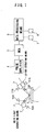
- Figure 1 shows an embodiment of an energy dispersion type X-ray diffraction/spectral device according to the present invention, in which a specimen support 1 is interposed between a white X-ray generating means 2 and an X-ray detecting means 3, and a multichannel analyzer (MCA) 5 is connected to the output end of the X-ray detecting means 3 via a pulse amplifier 4.
- MCA multichannel analyzer
- the specimen support 1 is provided with positioning members 10A, 10B, 11A and 11B which move between a first position and a second position located a few degrees apart therefrom so as to shift only diffracted X-rays, that is, to relatively move the position by an angle ⁇ , this degree enough to prevent a variation in intensity of fluorescent X-rays as much as possible, and constructed so as to be fixed to a predetermined position by a notch mechanism or the like.
- the notch mechanism causes movement by a certain angle between the two points with high accuracy, and can thereby simplify the structure.
- a data processing means 6 is provided with a first storing means 7 for storing intensity data for each level of energy of white X-rays irradiated onto the specimen and absorptivity data which takes into consideration absorption due to the air or the like which exists in the transmission path of X-rays and proportions of various elements constituting the specimen, and a second storing means 8 for storing data measured at the first position and second position.
- the white X-ray generating means 2 and X-ray detecting means 3 are set to the first position (position shown by a solid line in the figure) and a measurement is started, the X-ray detecting means 3 detects diffracted X-rays and fluorescent X-rays corresponding to elements constituting the specimen S and the crystalline structure thereof.
- the data processing means 6 corrects X-ray intensity for each level of energy detected by the X-ray detecting means 3 based on the data of the first storing means 7 and stores the corrected X-ray intensity in the second storing means 8.
- the white X-ray generating means 2 and X-ray detecting means 3 are moved to the second position (position shown by one-dot-chain line in the figure), X-ray intensity for each level of energy detected by the X-ray detecting means 3 is corrected based on the data of the first storing means 7, data equivalent to that obtained by irradiating the specimen with X-rays of the same intensity over the entire energy area of X-rays is obtained and stored in the second storing means 8.
- first measured graphic and second measured graphic include data of the same fluorescent X-rays, if these are canceled out each other, it is possible to obtain third data with only diffracted X-rays.
- the present invention can downsize the device for analyzing various types of specimen, and is thereby applicable as a handy-type specimen analyzer or the like.
Landscapes
- Physics & Mathematics (AREA)
- Health & Medical Sciences (AREA)
- Life Sciences & Earth Sciences (AREA)
- Chemical & Material Sciences (AREA)
- Analytical Chemistry (AREA)
- Biochemistry (AREA)
- General Health & Medical Sciences (AREA)
- General Physics & Mathematics (AREA)
- Immunology (AREA)
- Pathology (AREA)
- Analysing Materials By The Use Of Radiation (AREA)
Description
- The present invention relates to an energy dispersion type X-ray diffraction/spectral device which detects diffracted rays and fluorescent X-rays simultaneously using white X-rays and with an X-ray detector fixed.
- An X-ray diffraction device is known as a device which analyzes a crystalline structure of a specimen using X-rays. The X-ray diffraction device is constructed of an X-ray generating means disposed at a position from which it can irradiate the surface of a specimen at an angle θ and an X-ray detecting means such as a counter disposed at a position of angle 2θ formed by the irradiation direction and measuring point of the specimen, and irradiates the specimen with X-rays having a known single wavelength λ by sequentially rotating and moving the X-ray generating means and X-ray detecting means and measures intensity of X-rays at the angle θ, that is, diffraction intensity.
Since diffracted X-rays are diffracted at the angle θ which satisfies:
according to Bragg's law, it is possible to know a crystal face spacing d and diffraction intensity of the specimen from the diffraction graphic and obtain data for determining an atomic arrangement of the specimen. - However, the X-ray diffraction device requires a precise mechanism for sequentially rotating and moving the specimen and X-ray detecting means, that is, a goniometer, which not only complicates the device and increases the size of the device but also requires a rotation/movement operation time and measuring time at each position of movement, producing a problem that it takes quite a long time to obtain a measurement result.
- To solve such a problem, an energy dispersion diffraction method as described in Non-Patent
Document 1 is proposed.
According to this method, an X-ray generation device is disposed at a position of an angle θ with respect to the surface of a specimen, a means for detecting energy and intensity of X-rays simultaneously is disposed at a position of an angle 2θ formed by the irradiation direction and measuring point of the specimen, white X-rays are irradiated with θ and 2θ fixed and intensity of X-rays having energy E is measured.
Bragg's law is expressed centered on energy E as:
Therefore, it is possible to determine an atomic surface spacing d and diffraction intensity by only measuring the relationship between the detected energy E and intensity of diffracted X-rays by keeping the angles of the X-ray generating means, specimen and X-ray detecting means to fixed positions and using a multichannel analyzer or the like. - Furthermore, since the X-rays which reach the detecting means also include fluorescent X-rays from the specimen, if these X-rays can be distinguished from diffracted X-rays, it is also possible to determine the kind of atoms of the specimen from fluorescent X-rays.
The energy at which diffracted rays emerge follows Bragg' s law and also depends on the angle θ, and therefore there is also a proposal to conduct measurement at an appropriate angle at which diffracted rays and fluorescent X-rays do not overlap each other so as to distinguish diffracted rays from fluorescent X-rays. - Non-Patent Document 1: B.D. Cullity. Elements of X-Ray Diffraction, 2nd ed (Reading, Mass.: Addison-Wesley, 1977.)
-
DE 199 36 900 A1 discloses a method and device for the determination and separation of fluorescence lines and diffraction lines induced by X-ray irradiation, whereby diffraction lines are assigned to individual measuring positions, characteristic crystallographic properties are determined, monocrystalline portions are identified, the diffraction lines are checked for systematic reoccurrences thereof, and the crystal structure/orientation is determined. In order to separate diffraction lines, the probe may be rotated and/or tilted, or the X-ray source and/or the detector may be rotated and/or tilted. - Sutton S.R. et al.: "Synchrotron X-ray Fluorescence: Diffraction Interference", Anal. Chem. 1986, 58, 2167-2171 discloses a method for the determination of fluorescence lines and diffraction lines induced by X-ray irradiation, by changing the specimen orientation with respect to the X-ray source and the detector. As an alternative to specimen reorientation, it is proposed to use a second "off-axis" detector.
- However, when an area of emergence of fluorescent X-rays is limited, it is possible to separate diffracted rays from fluorescent X-rays, whereas such separation is not possible when there are many areas where fluorescent X-rays are generated, and moreover, it is necessary to precisely change 2θ in order to search for an appropriate angle which varies from one specimen to another, which still produces a problem of complicating the structure of the measuring device.
- The present invention has been implemented in view of the above described problems and it is an object of this invention to provide an energy dispersion type X-ray diffraction/spectral device capable of removing interference between fluorescent X-rays and diffracted rays and accurately detecting only diffracted rays without complicating the structure of an X-ray detecting means.
- These problems are solved with the device according to
claim 1. - As described above, the present invention only moves the white X-ray generating means and X-ray detecting means to two separated points, and can thereby simplify the structure, eliminate the time required for continuous rotation and measurement times at various angles and obtain both diffraction data and fluorescent X-ray data in a short time.
Furthermore, since no specimen rotation mechanism is required, it is possible to extremely simplify the structure and construct the device as a small and portable device. - With reference now to the attached drawings, details of the present invention will be explained below.
-
Figure 1 shows an embodiment of an energy dispersion type X-ray diffraction/spectral device according to the present invention, in which aspecimen support 1 is interposed between a white X-ray generating means 2 and an X-ray detecting means 3, and a multichannel analyzer (MCA) 5 is connected to the output end of the X-ray detecting means 3 via apulse amplifier 4. - The
specimen support 1 is provided with positioningmembers - A data processing means 6 is provided with a first storing means 7 for storing intensity data for each level of energy of white X-rays irradiated onto the specimen and absorptivity data which takes into consideration absorption due to the air or the like which exists in the transmission path of X-rays and proportions of various elements constituting the specimen, and a second storing means 8 for storing data measured at the first position and second position.
- In this embodiment, when the specimen S is placed on the
specimen support 1, the white X-ray generating means 2 and X-ray detecting means 3 are set to the first position (position shown by a solid line in the figure) and a measurement is started, the X-ray detecting means 3 detects diffracted X-rays and fluorescent X-rays corresponding to elements constituting the specimen S and the crystalline structure thereof.
The data processing means 6 corrects X-ray intensity for each level of energy detected by the X-ray detecting means 3 based on the data of the first storing means 7 and stores the corrected X-ray intensity in the second storing means 8. - Next, the white X-ray generating means 2 and X-ray detecting means 3 are moved to the second position (position shown by one-dot-chain line in the figure), X-ray intensity for each level of energy detected by the
X-ray detecting means 3 is corrected based on the data of the first storing means 7, data equivalent to that obtained by irradiating the specimen with X-rays of the same intensity over the entire energy area of X-rays is obtained and stored in the second storing means 8. - Thus, when measurements at two types of position are completed, data including diffracted X-rays and fluorescent X-rays as shown in
Figures 2(a), (b) is obtained.
On the other hand, fluorescent X-rays emerge at the same position, and therefore when data of these two measured graphics are canceled out with each other, only peaks of fluorescent X-rays are erased and diffracted X-rays with positive and negative signs remain. Of the data with these signs, if, for example, only the data with positive signs are collected and the data with negative signs are discarded, data with only diffracted X-rays obtained at the first position as shown inFigure 3(a) are obtained. Furthermore, if the data with positive signs are discarded and only the data with negative signs are collected, only data with diffracted X-rays at the second position are obtained. - That is, since the first measured graphic and second measured graphic include data of the same fluorescent X-rays, if these are canceled out each other, it is possible to obtain third data with only diffracted X-rays.
- Next, by canceling out the corresponding third data from any one of the first and second measured graphic data, it is possible to extract data with only fluorescent X-rays as shown in
Figure 3 (b) . - As described above, it is preferable to perform the following processing when obtaining data with only diffracted X-rays and data with only fluorescent X-rays.
That is, it is preferable to measure X-ray intensity for each level of energy at two types of position at different angles first, and then cancel out data at the same energy level as described above and sort the data with only diffracted X-rays obtained and data with only fluorescent X-rays obtained.
More specifically, assuming that m is a positive number (normally 1 to 3) and σ (standard deviation)=√N (N is a measured value at a certain energy level), if mσ is used as a threshold and data with only diffracted X-rays are obtained, only the data with an energy level at which the difference between data with the same energy level equals or exceeds mσ are kept and the rest of the data are set to 0. On the other hand, when data with only fluorescent X-rays are obtained, only the data with an energy level at which the difference between data with the same energy level falls below mσ are kept and the rest of the data are set to 0.
In this way, it is possible to obtain data with only diffracted X-rays and data with only fluorescent X-rays with errors and with high accuracy. - The present invention can downsize the device for analyzing various types of specimen, and is thereby applicable as a handy-type specimen analyzer or the like.
-
-
Figure 1 is a block diagram showing an embodiment of an energy dispersion type X-ray diffraction/spectral device according to the present invention; -
Figure 2 (a) is diagram about data related to diffracted rays and fluorescent X-rays at a first position andFigure 2(b) is a diagram related to data of diffracted X-rays and fluorescent X-rays at a second position; and -
Figure 3 (a) is a diagram related to data of only diffracted X-rays andFigure 3(b) is a diagram related to data of only fluorescent X-rays. -
- 1
- specimen support
- 2
- white X-ray generating means
- 3
- X-ray detecting means
- S
- specimen
Claims (1)
- An energy dispersion type X-ray diffraction/spectral device having means for moving a white X-ray generating means (2) and an X-ray detecting means (3) to a first position and a second position which are separate from each other, and means for using intensities of X-rays detected for each level of energy by said X-ray detecting means at the respective positions as first data and second data,
characterised by
being adapted to obtain third data regarding diffracted X-rays from the difference between said first data and said second data and to obtain data regarding fluorescent X-rays from the difference between the first or second data and third data; and comprising:a storing means (7) for storing intensity data for each level of energy of white X-rays irradiated onto the specimen; anda data processing means (6) for correcting X-ray intensity for each level of energy detected by the X-ray detecting means, based on the data of the storing means.
Applications Claiming Priority (2)
| Application Number | Priority Date | Filing Date | Title |
|---|---|---|---|
| JP2003196155 | 2003-07-11 | ||
| PCT/JP2004/009535 WO2005005969A1 (en) | 2003-07-11 | 2004-07-05 | Energy dispersion type x-ray diffraction/spectral device |
Publications (3)
| Publication Number | Publication Date |
|---|---|
| EP1650558A1 EP1650558A1 (en) | 2006-04-26 |
| EP1650558A4 EP1650558A4 (en) | 2008-01-02 |
| EP1650558B1 true EP1650558B1 (en) | 2010-09-08 |
Family
ID=34055773
Family Applications (1)
| Application Number | Title | Priority Date | Filing Date |
|---|---|---|---|
| EP04747004A Expired - Lifetime EP1650558B1 (en) | 2003-07-11 | 2004-07-05 | Energy dispersion type x-ray diffraction/spectral device |
Country Status (6)
| Country | Link |
|---|---|
| US (1) | US7352845B2 (en) |
| EP (1) | EP1650558B1 (en) |
| JP (1) | JP3928656B2 (en) |
| CN (1) | CN1823270B (en) |
| DE (1) | DE602004029054D1 (en) |
| WO (1) | WO2005005969A1 (en) |
Cited By (1)
| Publication number | Priority date | Publication date | Assignee | Title |
|---|---|---|---|---|
| EP4019951A1 (en) | 2020-12-24 | 2022-06-29 | Inel S.A.S | Apparatuses and methods for combined simultaneous analyses of materials |
Families Citing this family (21)
| Publication number | Priority date | Publication date | Assignee | Title |
|---|---|---|---|---|
| WO2007069639A1 (en) * | 2005-12-13 | 2007-06-21 | Riken Keiki Co., Ltd. | X-ray analyzer |
| JP2008111781A (en) * | 2006-10-31 | 2008-05-15 | Riken Keiki Co Ltd | Measurement processing method of X-ray analyzer |
| JP2008039560A (en) * | 2006-08-04 | 2008-02-21 | Riken Keiki Co Ltd | X-ray analyzer |
| JP4664265B2 (en) * | 2006-10-31 | 2011-04-06 | 理研計器株式会社 | X-ray analyzer |
| JP4619282B2 (en) * | 2005-12-13 | 2011-01-26 | 理研計器株式会社 | X-ray analyzer |
| JP2008039559A (en) * | 2006-08-04 | 2008-02-21 | Riken Keiki Co Ltd | X-ray analyzer |
| JP5455904B2 (en) * | 2007-08-03 | 2014-03-26 | パルセータ,エルエルシー | Pile-up detection method in energy dispersive radiation spectroscopic analysis system, pulse processor for executing the method, and energy dispersive radiation spectroscopic analysis system for detecting pile-up |
| CA2763367C (en) | 2009-06-04 | 2016-09-13 | Nextray, Inc. | Strain matching of crystals and horizontally-spaced monochromator and analyzer crystal arrays in diffraction enhanced imaging systems and related methods |
| US8204174B2 (en) | 2009-06-04 | 2012-06-19 | Nextray, Inc. | Systems and methods for detecting an image of an object by use of X-ray beams generated by multiple small area sources and by use of facing sides of adjacent monochromator crystals |
| TWI407097B (en) * | 2009-09-07 | 2013-09-01 | Nat Univ Chung Hsing | Structural analysis system and method |
| JP4914514B2 (en) | 2010-07-02 | 2012-04-11 | 株式会社リガク | X-ray fluorescence analyzer and method |
| JP5838109B2 (en) * | 2011-05-13 | 2015-12-24 | 株式会社リガク | Compound X-ray analyzer |
| US8903043B2 (en) * | 2011-10-24 | 2014-12-02 | Bruker Axs, Inc. | Method for correcting timing skew in X-ray data read out of an X-ray detector in a rolling shutter mode |
| US20130279653A1 (en) * | 2012-04-19 | 2013-10-24 | Graeme Mark Hansford | Methods and apparatus for x-ray diffraction |
| US9952165B2 (en) | 2012-04-19 | 2018-04-24 | University Of Leicester | Methods and apparatus for X-ray diffraction |
| CN103076352B (en) * | 2012-12-28 | 2015-02-25 | 中国科学院高能物理研究所 | Method for obtaining high-quality X-ray absorption spectrum of thin film sample |
| CN104634799A (en) * | 2013-11-15 | 2015-05-20 | 郑琪 | Device and method for measuring multi-wavelength characteristic X ray diffraction |
| DE102014208295A1 (en) * | 2014-05-02 | 2015-11-05 | Bruker Nano Gmbh | Method and arrangement for identifying crystalline phases and a corresponding computer program and a corresponding computer-readable storage medium |
| DE102014109671B3 (en) | 2014-07-10 | 2015-11-05 | Universität Hamburg | Method and device for X-ray analysis |
| CN105891246B (en) * | 2016-06-23 | 2018-11-16 | 北京至一恒盛技术服务有限公司 | A kind of x-ray fluorescence detection device |
| WO2026107363A1 (en) * | 2024-11-14 | 2026-05-21 | Veracio Ltd. | Assemblies, systems, and methods for analyzing material samples |
Family Cites Families (21)
| Publication number | Priority date | Publication date | Assignee | Title |
|---|---|---|---|---|
| US3440419A (en) * | 1966-02-03 | 1969-04-22 | California Inst Res Found | Dual purpose optical instrument capable of simultaneously acting as spectrometer and diffractometer |
| IL41592A (en) * | 1973-02-20 | 1976-02-29 | Tech Res & Dev Found Ltd | X-ray spectrodiffractometer |
| US3920984A (en) * | 1974-04-08 | 1975-11-18 | Machlett Lab Inc | X-ray energy analyzer |
| DE2632001C3 (en) * | 1976-07-16 | 1980-10-23 | Gkss - Forschungszentrum Geesthacht Gmbh, 2000 Hamburg | Measurement arrangement for X-ray fluorescence analysis |
| US4263510A (en) * | 1979-07-30 | 1981-04-21 | General Electric Company | Combined x-ray diffraction and fluorescence spectroscopy apparatus with environmentally controllable chamber |
| EP0108447A3 (en) | 1982-11-04 | 1985-07-17 | North American Philips Corporation | Simultaneous collection of diffraction and spectrographic data |
| JP2742415B2 (en) * | 1987-11-27 | 1998-04-22 | 株式会社日立製作所 | X-ray analyzer |
| DE69232214T2 (en) * | 1991-07-11 | 2002-05-08 | Fujikura Ltd., Tokio/Tokyo | Device for surface analysis using X-ray spectroscopy |
| GB9223592D0 (en) * | 1992-11-11 | 1992-12-23 | Fisons Plc | X-ray analysis apparatus |
| US5491738A (en) * | 1993-03-15 | 1996-02-13 | The United States Of America As Represented By The Administrator Of The National Aeronautics And Space Administration | X-ray diffraction apparatus |
| JP2912127B2 (en) | 1993-07-15 | 1999-06-28 | 理学電機工業株式会社 | X-ray fluorescence analysis method |
| EP0800647A1 (en) * | 1995-10-03 | 1997-10-15 | Koninklijke Philips Electronics N.V. | Apparatus for simultaneous x-ray diffraction and x-ray fluorescence measurements |
| US6751287B1 (en) * | 1998-05-15 | 2004-06-15 | The Trustees Of The Stevens Institute Of Technology | Method and apparatus for x-ray analysis of particle size (XAPS) |
| FI110820B (en) * | 1998-08-24 | 2003-03-31 | Outokumpu Oy | Procedure for determination of elemental content |
| DE19936900A1 (en) * | 1998-09-22 | 2000-08-24 | Fraunhofer Ges Forschung | X-ray analysis position comparison and spectral analysis, comprises altering spectral position of diffraction lines by single defined change in position of test item, and repeating measurement of emission spectrum |
| JP3519292B2 (en) * | 1998-11-13 | 2004-04-12 | 理学電機株式会社 | X-ray diffraction measurement method for minute area and X-ray diffraction apparatus for minute area |
| JP3610256B2 (en) | 1999-03-29 | 2005-01-12 | 日本電子株式会社 | X-ray fluorescence analyzer |
| US6345086B1 (en) * | 1999-09-14 | 2002-02-05 | Veeco Instruments Inc. | X-ray fluorescence system and method |
| JP2003050115A (en) * | 2001-08-07 | 2003-02-21 | Seiko Instruments Inc | X-ray thickness gauge |
| JP2005509483A (en) * | 2001-11-17 | 2005-04-14 | カウンシル・フォー・ザ・セントラル・ラボラトリー・オブ・ザ・リサーチ・カウンシルズ | Imaging method and apparatus |
| JP2004184314A (en) * | 2002-12-05 | 2004-07-02 | Mitsubishi Electric Corp | X-ray fluorescence analyzer |
-
2004
- 2004-07-05 US US10/563,238 patent/US7352845B2/en not_active Expired - Fee Related
- 2004-07-05 CN CN200480019929.3A patent/CN1823270B/en not_active Expired - Fee Related
- 2004-07-05 EP EP04747004A patent/EP1650558B1/en not_active Expired - Lifetime
- 2004-07-05 JP JP2005511506A patent/JP3928656B2/en not_active Expired - Fee Related
- 2004-07-05 DE DE602004029054T patent/DE602004029054D1/en not_active Expired - Lifetime
- 2004-07-05 WO PCT/JP2004/009535 patent/WO2005005969A1/en not_active Ceased
Cited By (2)
| Publication number | Priority date | Publication date | Assignee | Title |
|---|---|---|---|---|
| EP4019951A1 (en) | 2020-12-24 | 2022-06-29 | Inel S.A.S | Apparatuses and methods for combined simultaneous analyses of materials |
| US11796492B2 (en) | 2020-12-24 | 2023-10-24 | Inel S.A.S. | Apparatuses and methods for combined simultaneous analyses of materials |
Also Published As
| Publication number | Publication date |
|---|---|
| CN1823270A (en) | 2006-08-23 |
| JPWO2005005969A1 (en) | 2006-11-30 |
| EP1650558A1 (en) | 2006-04-26 |
| JP3928656B2 (en) | 2007-06-13 |
| US20060165218A1 (en) | 2006-07-27 |
| WO2005005969A1 (en) | 2005-01-20 |
| CN1823270B (en) | 2011-11-23 |
| EP1650558A4 (en) | 2008-01-02 |
| US7352845B2 (en) | 2008-04-01 |
| DE602004029054D1 (en) | 2010-10-21 |
Similar Documents
| Publication | Publication Date | Title |
|---|---|---|
| EP1650558A1 (en) | Energy dispersion type x-ray diffraction/spectral device | |
| JP7147770B2 (en) | X-ray spectroscopic analyzer and chemical state analysis method using the X-ray spectroscopic analyzer | |
| JP6874835B2 (en) | X-ray spectroscopic analyzer | |
| EP2564186B1 (en) | Method and apparatus for using an area x-ray detector as a point detector in an x-ray diffractometer | |
| GB2083215A (en) | Apparatus for x-ray diffraction | |
| CN102959387B (en) | Fluorescent X-ray analysis device and method | |
| EP1764612B1 (en) | X-ray fluorescence spectrometer and x-ray fluorescence measurement method | |
| CN101566591A (en) | Wavelength dispersion type x ray spectrometer | |
| US11471119B2 (en) | Analysis device and analysis method | |
| JP2007010455A (en) | X-ray diffraction device | |
| JP2009047586A (en) | X-ray analyzer for chemical bond state analysis | |
| EP1653226A1 (en) | Scanning line detector for two-dimensional x-ray diffractometer | |
| JP3968350B2 (en) | X-ray diffraction apparatus and method | |
| JP5013525B2 (en) | X-ray diffraction measurement method and X-ray diffraction apparatus | |
| Tantau et al. | High accuracy energy determination and calibration of synchrotron radiation by powder diffraction | |
| EP3064932B1 (en) | Quantitative x-ray analysis | |
| JP2010286288A (en) | X-ray spectrometer control method and X-ray spectrometer using the control method | |
| JPH0833359B2 (en) | Total reflection X-ray fluorescence analyzer | |
| JP2592723B2 (en) | Apparatus and method for identifying and analyzing X-ray fluorescence spectrum | |
| JPH02266249A (en) | Method for measuring x-ray diffraction of crystal plane | |
| JP3968452B2 (en) | X-ray analyzer | |
| US20260110649A1 (en) | X-ray fluorescence spectrometer | |
| JP3312001B2 (en) | X-ray fluorescence analyzer | |
| JPH06265489A (en) | X-ray diffractometer | |
| CN101014883A (en) | Coherent scatter imaging |
Legal Events
| Date | Code | Title | Description |
|---|---|---|---|
| PUAI | Public reference made under article 153(3) epc to a published international application that has entered the european phase |
Free format text: ORIGINAL CODE: 0009012 |
|
| 17P | Request for examination filed |
Effective date: 20060125 |
|
| AK | Designated contracting states |
Kind code of ref document: A1 Designated state(s): DE FR GB IT |
|
| DAX | Request for extension of the european patent (deleted) | ||
| RBV | Designated contracting states (corrected) |
Designated state(s): DE FR GB IT |
|
| A4 | Supplementary search report drawn up and despatched |
Effective date: 20071130 |
|
| 17Q | First examination report despatched |
Effective date: 20080423 |
|
| GRAP | Despatch of communication of intention to grant a patent |
Free format text: ORIGINAL CODE: EPIDOSNIGR1 |
|
| GRAS | Grant fee paid |
Free format text: ORIGINAL CODE: EPIDOSNIGR3 |
|
| GRAA | (expected) grant |
Free format text: ORIGINAL CODE: 0009210 |
|
| AK | Designated contracting states |
Kind code of ref document: B1 Designated state(s): DE FR GB IT |
|
| REG | Reference to a national code |
Ref country code: GB Ref legal event code: FG4D |
|
| REF | Corresponds to: |
Ref document number: 602004029054 Country of ref document: DE Date of ref document: 20101021 Kind code of ref document: P |
|
| PG25 | Lapsed in a contracting state [announced via postgrant information from national office to epo] |
Ref country code: IT Free format text: LAPSE BECAUSE OF FAILURE TO SUBMIT A TRANSLATION OF THE DESCRIPTION OR TO PAY THE FEE WITHIN THE PRESCRIBED TIME-LIMIT Effective date: 20100908 |
|
| PLBE | No opposition filed within time limit |
Free format text: ORIGINAL CODE: 0009261 |
|
| STAA | Information on the status of an ep patent application or granted ep patent |
Free format text: STATUS: NO OPPOSITION FILED WITHIN TIME LIMIT |
|
| 26N | No opposition filed |
Effective date: 20110609 |
|
| REG | Reference to a national code |
Ref country code: DE Ref legal event code: R097 Ref document number: 602004029054 Country of ref document: DE Effective date: 20110609 |
|
| GBPC | Gb: european patent ceased through non-payment of renewal fee |
Effective date: 20110705 |
|
| REG | Reference to a national code |
Ref country code: FR Ref legal event code: ST Effective date: 20120330 |
|
| PG25 | Lapsed in a contracting state [announced via postgrant information from national office to epo] |
Ref country code: DE Free format text: LAPSE BECAUSE OF NON-PAYMENT OF DUE FEES Effective date: 20120201 Ref country code: FR Free format text: LAPSE BECAUSE OF NON-PAYMENT OF DUE FEES Effective date: 20110801 |
|
| REG | Reference to a national code |
Ref country code: DE Ref legal event code: R119 Ref document number: 602004029054 Country of ref document: DE Effective date: 20120201 |
|
| PG25 | Lapsed in a contracting state [announced via postgrant information from national office to epo] |
Ref country code: GB Free format text: LAPSE BECAUSE OF NON-PAYMENT OF DUE FEES Effective date: 20110705 |