EP1638531B1 - Antigenhaltige mikrosphären zur allergietherapie - Google Patents
Antigenhaltige mikrosphären zur allergietherapie Download PDFInfo
- Publication number
- EP1638531B1 EP1638531B1 EP04729885A EP04729885A EP1638531B1 EP 1638531 B1 EP1638531 B1 EP 1638531B1 EP 04729885 A EP04729885 A EP 04729885A EP 04729885 A EP04729885 A EP 04729885A EP 1638531 B1 EP1638531 B1 EP 1638531B1
- Authority
- EP
- European Patent Office
- Prior art keywords
- microspheres
- antigens
- allergy therapy
- therapy according
- allergy
- Prior art date
- Legal status (The legal status is an assumption and is not a legal conclusion. Google has not performed a legal analysis and makes no representation as to the accuracy of the status listed.)
- Expired - Lifetime
Links
- 239000004005 microsphere Substances 0.000 title claims description 127
- 239000000427 antigen Substances 0.000 title claims description 41
- 108091007433 antigens Proteins 0.000 title claims description 41
- 102000036639 antigens Human genes 0.000 title claims description 41
- 206010020751 Hypersensitivity Diseases 0.000 title claims description 37
- 230000007815 allergy Effects 0.000 title claims description 36
- 238000011282 treatment Methods 0.000 title description 4
- 239000013566 allergen Substances 0.000 claims description 52
- 208000026935 allergic disease Diseases 0.000 claims description 33
- 238000002560 therapeutic procedure Methods 0.000 claims description 28
- 108090001090 Lectins Proteins 0.000 claims description 21
- 102000004856 Lectins Human genes 0.000 claims description 21
- 239000002523 lectin Substances 0.000 claims description 21
- 230000027455 binding Effects 0.000 claims description 20
- 229920000642 polymer Polymers 0.000 claims description 17
- 210000004027 cell Anatomy 0.000 claims description 15
- 108010041181 Aleuria aurantia lectin Proteins 0.000 claims description 12
- 239000000126 substance Substances 0.000 claims description 12
- 210000002919 epithelial cell Anatomy 0.000 claims description 10
- 150000001720 carbohydrates Chemical group 0.000 claims description 8
- 229920001606 poly(lactic acid-co-glycolic acid) Polymers 0.000 claims description 7
- SHZGCJCMOBCMKK-UHFFFAOYSA-N D-mannomethylose Natural products CC1OC(O)C(O)C(O)C1O SHZGCJCMOBCMKK-UHFFFAOYSA-N 0.000 claims description 5
- 229920000954 Polyglycolide Polymers 0.000 claims description 5
- SHZGCJCMOBCMKK-SXUWKVJYSA-N alpha-L-fucose Chemical group C[C@@H]1O[C@@H](O)[C@@H](O)[C@H](O)[C@@H]1O SHZGCJCMOBCMKK-SXUWKVJYSA-N 0.000 claims description 5
- 239000003814 drug Substances 0.000 claims description 4
- 230000000968 intestinal effect Effects 0.000 claims description 4
- 239000004633 polyglycolic acid Substances 0.000 claims description 4
- 229920002988 biodegradable polymer Polymers 0.000 claims description 3
- 239000004621 biodegradable polymer Substances 0.000 claims description 3
- 229920001577 copolymer Polymers 0.000 claims description 3
- 125000000524 functional group Chemical group 0.000 claims description 3
- 238000004519 manufacturing process Methods 0.000 claims description 3
- 231100000252 nontoxic Toxicity 0.000 claims description 3
- 230000003000 nontoxic effect Effects 0.000 claims description 3
- 229920000747 poly(lactic acid) Polymers 0.000 claims description 3
- 239000004626 polylactic acid Substances 0.000 claims description 2
- 229960004784 allergens Drugs 0.000 description 28
- 239000011859 microparticle Substances 0.000 description 28
- 235000018185 Betula X alpestris Nutrition 0.000 description 25
- 235000018212 Betula X uliginosa Nutrition 0.000 description 25
- 235000018102 proteins Nutrition 0.000 description 21
- 102000004169 proteins and genes Human genes 0.000 description 21
- 108090000623 proteins and genes Proteins 0.000 description 21
- 239000000284 extract Substances 0.000 description 18
- 238000002965 ELISA Methods 0.000 description 12
- 108010046516 Wheat Germ Agglutinins Proteins 0.000 description 12
- 238000002360 preparation method Methods 0.000 description 11
- 239000002245 particle Substances 0.000 description 10
- 238000000034 method Methods 0.000 description 9
- 239000002077 nanosphere Substances 0.000 description 9
- 239000000839 emulsion Substances 0.000 description 7
- 210000002490 intestinal epithelial cell Anatomy 0.000 description 7
- 108090000765 processed proteins & peptides Proteins 0.000 description 7
- LMDZBCPBFSXMTL-UHFFFAOYSA-N 1-ethyl-3-(3-dimethylaminopropyl)carbodiimide Chemical group CCN=C=NCCCN(C)C LMDZBCPBFSXMTL-UHFFFAOYSA-N 0.000 description 6
- VHRGRCVQAFMJIZ-UHFFFAOYSA-N cadaverine Chemical compound NCCCCCN VHRGRCVQAFMJIZ-UHFFFAOYSA-N 0.000 description 6
- 230000029087 digestion Effects 0.000 description 6
- 210000002966 serum Anatomy 0.000 description 6
- 238000001694 spray drying Methods 0.000 description 6
- XLYOFNOQVPJJNP-UHFFFAOYSA-N water Substances O XLYOFNOQVPJJNP-UHFFFAOYSA-N 0.000 description 6
- XBDQKXXYIPTUBI-UHFFFAOYSA-M Propionate Chemical compound CCC([O-])=O XBDQKXXYIPTUBI-UHFFFAOYSA-M 0.000 description 5
- 230000008901 benefit Effects 0.000 description 5
- 230000015572 biosynthetic process Effects 0.000 description 5
- 238000006243 chemical reaction Methods 0.000 description 5
- WBJINCZRORDGAQ-UHFFFAOYSA-N formic acid ethyl ester Natural products CCOC=O WBJINCZRORDGAQ-UHFFFAOYSA-N 0.000 description 5
- 238000007306 functionalization reaction Methods 0.000 description 5
- 230000002496 gastric effect Effects 0.000 description 5
- 230000003053 immunization Effects 0.000 description 5
- 238000002649 immunization Methods 0.000 description 5
- 102000004196 processed proteins & peptides Human genes 0.000 description 5
- 210000001519 tissue Anatomy 0.000 description 5
- 101710186708 Agglutinin Proteins 0.000 description 4
- 241000221688 Aleuria aurantia Species 0.000 description 4
- 101710146024 Horcolin Proteins 0.000 description 4
- 101710189395 Lectin Proteins 0.000 description 4
- 101710179758 Mannose-specific lectin Proteins 0.000 description 4
- 101710150763 Mannose-specific lectin 1 Proteins 0.000 description 4
- 101710150745 Mannose-specific lectin 2 Proteins 0.000 description 4
- 239000002671 adjuvant Substances 0.000 description 4
- 239000000910 agglutinin Substances 0.000 description 4
- 150000001413 amino acids Chemical group 0.000 description 4
- 239000002105 nanoparticle Substances 0.000 description 4
- 230000008569 process Effects 0.000 description 4
- 230000004044 response Effects 0.000 description 4
- 208000024891 symptom Diseases 0.000 description 4
- 231100000331 toxic Toxicity 0.000 description 4
- 230000002588 toxic effect Effects 0.000 description 4
- 241000282412 Homo Species 0.000 description 3
- 241000699670 Mus sp. Species 0.000 description 3
- MXRIRQGCELJRSN-UHFFFAOYSA-N O.O.O.[Al] Chemical compound O.O.O.[Al] MXRIRQGCELJRSN-UHFFFAOYSA-N 0.000 description 3
- 102000003992 Peroxidases Human genes 0.000 description 3
- HEMHJVSKTPXQMS-UHFFFAOYSA-M Sodium hydroxide Chemical compound [OH-].[Na+] HEMHJVSKTPXQMS-UHFFFAOYSA-M 0.000 description 3
- 230000000903 blocking effect Effects 0.000 description 3
- 150000001718 carbodiimides Chemical class 0.000 description 3
- 125000003178 carboxy group Chemical group [H]OC(*)=O 0.000 description 3
- 229940079593 drug Drugs 0.000 description 3
- 238000002474 experimental method Methods 0.000 description 3
- MHMNJMPURVTYEJ-UHFFFAOYSA-N fluorescein-5-isothiocyanate Chemical compound O1C(=O)C2=CC(N=C=S)=CC=C2C21C1=CC=C(O)C=C1OC1=CC(O)=CC=C21 MHMNJMPURVTYEJ-UHFFFAOYSA-N 0.000 description 3
- 230000002163 immunogen Effects 0.000 description 3
- 108040007629 peroxidase activity proteins Proteins 0.000 description 3
- 239000000243 solution Substances 0.000 description 3
- 239000002904 solvent Substances 0.000 description 3
- PORMUFZNYQJOEI-UHFFFAOYSA-N sumatriptan succinate Chemical compound OC(=O)CCC(O)=O.CNS(=O)(=O)CC1=CC=C2NC=C(CCN(C)C)C2=C1 PORMUFZNYQJOEI-UHFFFAOYSA-N 0.000 description 3
- 238000012360 testing method Methods 0.000 description 3
- 230000001225 therapeutic effect Effects 0.000 description 3
- 229960005486 vaccine Drugs 0.000 description 3
- 238000012795 verification Methods 0.000 description 3
- 241001295925 Gegenes Species 0.000 description 2
- DHMQDGOQFOQNFH-UHFFFAOYSA-N Glycine Chemical compound NCC(O)=O DHMQDGOQFOQNFH-UHFFFAOYSA-N 0.000 description 2
- NTYJJOPFIAHURM-UHFFFAOYSA-N Histamine Chemical compound NCCC1=CN=CN1 NTYJJOPFIAHURM-UHFFFAOYSA-N 0.000 description 2
- 241000699666 Mus <mouse, genus> Species 0.000 description 2
- 241000283973 Oryctolagus cuniculus Species 0.000 description 2
- 239000004480 active ingredient Substances 0.000 description 2
- 229940074608 allergen extract Drugs 0.000 description 2
- 230000000172 allergic effect Effects 0.000 description 2
- 208000010668 atopic eczema Diseases 0.000 description 2
- 239000000872 buffer Substances 0.000 description 2
- 238000001514 detection method Methods 0.000 description 2
- 238000011161 development Methods 0.000 description 2
- 230000000694 effects Effects 0.000 description 2
- 210000000981 epithelium Anatomy 0.000 description 2
- 238000011156 evaluation Methods 0.000 description 2
- 230000028993 immune response Effects 0.000 description 2
- 238000010166 immunofluorescence Methods 0.000 description 2
- 230000006698 induction Effects 0.000 description 2
- 210000004347 intestinal mucosa Anatomy 0.000 description 2
- JVTAAEKCZFNVCJ-UHFFFAOYSA-N lactic acid Chemical compound CC(O)C(O)=O JVTAAEKCZFNVCJ-UHFFFAOYSA-N 0.000 description 2
- 230000004048 modification Effects 0.000 description 2
- 238000012986 modification Methods 0.000 description 2
- 238000001556 precipitation Methods 0.000 description 2
- 230000002035 prolonged effect Effects 0.000 description 2
- 239000007921 spray Substances 0.000 description 2
- 239000000758 substrate Substances 0.000 description 2
- 238000003786 synthesis reaction Methods 0.000 description 2
- 238000012800 visualization Methods 0.000 description 2
- 238000005406 washing Methods 0.000 description 2
- PJRSUKFWFKUDTH-JWDJOUOUSA-N (2s)-6-amino-2-[[2-[[(2s)-2-[[(2s,3s)-2-[[(2s)-2-[[2-[[(2s)-2-[[(2s)-6-amino-2-[[(2s)-2-[[(2s)-2-[[(2s)-2-[(2-aminoacetyl)amino]-4-methylsulfanylbutanoyl]amino]propanoyl]amino]-3-hydroxypropanoyl]amino]hexanoyl]amino]propanoyl]amino]acetyl]amino]propanoyl Chemical compound CSCC[C@H](NC(=O)CN)C(=O)N[C@@H](C)C(=O)N[C@@H](CO)C(=O)N[C@@H](CCCCN)C(=O)N[C@@H](C)C(=O)NCC(=O)N[C@@H](C)C(=O)N[C@@H]([C@@H](C)CC)C(=O)N[C@@H](C)C(=O)NCC(=O)N[C@@H](CCCCN)C(=O)N[C@@H]([C@@H](C)CC)C(=O)N[C@@H](C)C(=O)N[C@@H](CCCCN)C(=O)N[C@@H](C(C)C)C(=O)N[C@@H](C)C(=O)N[C@@H](CC(C)C)C(=O)N[C@@H](CCCCN)C(=O)N[C@@H](C)C(=O)N[C@@H](CC(C)C)C(N)=O PJRSUKFWFKUDTH-JWDJOUOUSA-N 0.000 description 1
- JKMHFZQWWAIEOD-UHFFFAOYSA-N 2-[4-(2-hydroxyethyl)piperazin-1-yl]ethanesulfonic acid Chemical compound OCC[NH+]1CCN(CCS([O-])(=O)=O)CC1 JKMHFZQWWAIEOD-UHFFFAOYSA-N 0.000 description 1
- 241000251468 Actinopterygii Species 0.000 description 1
- 208000035285 Allergic Seasonal Rhinitis Diseases 0.000 description 1
- 206010002199 Anaphylactic shock Diseases 0.000 description 1
- 240000007087 Apium graveolens Species 0.000 description 1
- 235000015849 Apium graveolens Dulce Group Nutrition 0.000 description 1
- 235000010591 Appio Nutrition 0.000 description 1
- 241000219495 Betulaceae Species 0.000 description 1
- BVKZGUZCCUSVTD-UHFFFAOYSA-L Carbonate Chemical compound [O-]C([O-])=O BVKZGUZCCUSVTD-UHFFFAOYSA-L 0.000 description 1
- 229920001661 Chitosan Polymers 0.000 description 1
- 206010009944 Colon cancer Diseases 0.000 description 1
- 240000009226 Corylus americana Species 0.000 description 1
- 235000001543 Corylus americana Nutrition 0.000 description 1
- 235000007466 Corylus avellana Nutrition 0.000 description 1
- 244000000626 Daucus carota Species 0.000 description 1
- 235000002767 Daucus carota Nutrition 0.000 description 1
- 108010061629 Dermatophagoides pteronyssinus antigen p 1 Proteins 0.000 description 1
- 108010061608 Dermatophagoides pteronyssinus antigen p 2 Proteins 0.000 description 1
- 241000196324 Embryophyta Species 0.000 description 1
- 241000233866 Fungi Species 0.000 description 1
- 239000004471 Glycine Substances 0.000 description 1
- AEMRFAOFKBGASW-UHFFFAOYSA-N Glycolic acid Polymers OCC(O)=O AEMRFAOFKBGASW-UHFFFAOYSA-N 0.000 description 1
- 102000003886 Glycoproteins Human genes 0.000 description 1
- 108090000288 Glycoproteins Proteins 0.000 description 1
- 101000808011 Homo sapiens Vascular endothelial growth factor A Proteins 0.000 description 1
- 241000713772 Human immunodeficiency virus 1 Species 0.000 description 1
- 241001465754 Metazoa Species 0.000 description 1
- 208000009793 Milk Hypersensitivity Diseases 0.000 description 1
- 201000010859 Milk allergy Diseases 0.000 description 1
- 108010058846 Ovalbumin Proteins 0.000 description 1
- 108060005874 Parvalbumin Proteins 0.000 description 1
- 102000001675 Parvalbumin Human genes 0.000 description 1
- 102000057297 Pepsin A Human genes 0.000 description 1
- 108090000284 Pepsin A Proteins 0.000 description 1
- 229920001244 Poly(D,L-lactide) Polymers 0.000 description 1
- 208000005279 Status Asthmaticus Diseases 0.000 description 1
- 108091008874 T cell receptors Proteins 0.000 description 1
- 102000016266 T-Cell Antigen Receptors Human genes 0.000 description 1
- 102000000591 Tight Junction Proteins Human genes 0.000 description 1
- 108010002321 Tight Junction Proteins Proteins 0.000 description 1
- 239000002253 acid Substances 0.000 description 1
- 230000004913 activation Effects 0.000 description 1
- 230000002009 allergenic effect Effects 0.000 description 1
- 235000001014 amino acid Nutrition 0.000 description 1
- 208000003455 anaphylaxis Diseases 0.000 description 1
- 230000033115 angiogenesis Effects 0.000 description 1
- 125000000129 anionic group Chemical group 0.000 description 1
- 230000000890 antigenic effect Effects 0.000 description 1
- 239000007864 aqueous solution Substances 0.000 description 1
- 238000003556 assay Methods 0.000 description 1
- 230000001580 bacterial effect Effects 0.000 description 1
- 210000004369 blood Anatomy 0.000 description 1
- 239000008280 blood Substances 0.000 description 1
- 230000015556 catabolic process Effects 0.000 description 1
- 210000003855 cell nucleus Anatomy 0.000 description 1
- 238000012512 characterization method Methods 0.000 description 1
- 150000001875 compounds Chemical class 0.000 description 1
- 238000010276 construction Methods 0.000 description 1
- 238000013270 controlled release Methods 0.000 description 1
- 230000008878 coupling Effects 0.000 description 1
- 238000010168 coupling process Methods 0.000 description 1
- 238000005859 coupling reaction Methods 0.000 description 1
- 235000018417 cysteine Nutrition 0.000 description 1
- 150000001945 cysteines Chemical group 0.000 description 1
- 238000006731 degradation reaction Methods 0.000 description 1
- 230000003111 delayed effect Effects 0.000 description 1
- 238000009826 distribution Methods 0.000 description 1
- 239000003596 drug target Substances 0.000 description 1
- 239000000975 dye Substances 0.000 description 1
- 239000012636 effector Substances 0.000 description 1
- 230000012202 endocytosis Effects 0.000 description 1
- CCIVGXIOQKPBKL-UHFFFAOYSA-N ethanesulfonic acid Chemical compound CCS(O)(=O)=O CCIVGXIOQKPBKL-UHFFFAOYSA-N 0.000 description 1
- 238000001704 evaporation Methods 0.000 description 1
- 238000000605 extraction Methods 0.000 description 1
- GNBHRKFJIUUOQI-UHFFFAOYSA-N fluorescein Chemical compound O1C(=O)C2=CC=CC=C2C21C1=CC=C(O)C=C1OC1=CC(O)=CC=C21 GNBHRKFJIUUOQI-UHFFFAOYSA-N 0.000 description 1
- 238000001917 fluorescence detection Methods 0.000 description 1
- 239000007850 fluorescent dye Substances 0.000 description 1
- 238000009472 formulation Methods 0.000 description 1
- 108010015750 fucose-binding lectin Proteins 0.000 description 1
- 230000006870 function Effects 0.000 description 1
- 210000001035 gastrointestinal tract Anatomy 0.000 description 1
- 239000011521 glass Substances 0.000 description 1
- 150000004676 glycans Chemical class 0.000 description 1
- 229940046528 grass pollen Drugs 0.000 description 1
- 229960001340 histamine Drugs 0.000 description 1
- 210000003630 histaminocyte Anatomy 0.000 description 1
- 102000058223 human VEGFA Human genes 0.000 description 1
- 210000000987 immune system Anatomy 0.000 description 1
- 230000005847 immunogenicity Effects 0.000 description 1
- 238000009169 immunotherapy Methods 0.000 description 1
- 238000001727 in vivo Methods 0.000 description 1
- 238000011534 incubation Methods 0.000 description 1
- 210000000936 intestine Anatomy 0.000 description 1
- 238000007918 intramuscular administration Methods 0.000 description 1
- 238000002955 isolation Methods 0.000 description 1
- 239000004310 lactic acid Substances 0.000 description 1
- 235000014655 lactic acid Nutrition 0.000 description 1
- 238000011068 loading method Methods 0.000 description 1
- 230000005923 long-lasting effect Effects 0.000 description 1
- 238000012153 long-term therapy Methods 0.000 description 1
- 230000001926 lymphatic effect Effects 0.000 description 1
- 230000010534 mechanism of action Effects 0.000 description 1
- 230000001404 mediated effect Effects 0.000 description 1
- 210000000110 microvilli Anatomy 0.000 description 1
- 239000000203 mixture Substances 0.000 description 1
- 210000002850 nasal mucosa Anatomy 0.000 description 1
- 230000003472 neutralizing effect Effects 0.000 description 1
- 210000000440 neutrophil Anatomy 0.000 description 1
- 230000009871 nonspecific binding Effects 0.000 description 1
- 230000003287 optical effect Effects 0.000 description 1
- 229940126578 oral vaccine Drugs 0.000 description 1
- 210000000056 organ Anatomy 0.000 description 1
- 239000003960 organic solvent Substances 0.000 description 1
- 229940092253 ovalbumin Drugs 0.000 description 1
- 230000000803 paradoxical effect Effects 0.000 description 1
- 229940111202 pepsin Drugs 0.000 description 1
- 108010021753 peptide-Gly-Leu-amide Proteins 0.000 description 1
- 210000001986 peyer's patch Anatomy 0.000 description 1
- 238000005191 phase separation Methods 0.000 description 1
- 239000008363 phosphate buffer Substances 0.000 description 1
- 229940117828 polylactic acid-polyglycolic acid copolymer Drugs 0.000 description 1
- 229920001184 polypeptide Polymers 0.000 description 1
- 239000000843 powder Substances 0.000 description 1
- 239000002244 precipitate Substances 0.000 description 1
- 230000002265 prevention Effects 0.000 description 1
- 238000011321 prophylaxis Methods 0.000 description 1
- 230000009257 reactivity Effects 0.000 description 1
- 229920006395 saturated elastomer Polymers 0.000 description 1
- 238000000935 solvent evaporation Methods 0.000 description 1
- 238000005507 spraying Methods 0.000 description 1
- 238000010186 staining Methods 0.000 description 1
- 238000010561 standard procedure Methods 0.000 description 1
- 230000000638 stimulation Effects 0.000 description 1
- 238000007920 subcutaneous administration Methods 0.000 description 1
- 229940031626 subunit vaccine Drugs 0.000 description 1
- 230000001629 suppression Effects 0.000 description 1
- 230000009885 systemic effect Effects 0.000 description 1
- 230000008685 targeting Effects 0.000 description 1
- 229960000814 tetanus toxoid Drugs 0.000 description 1
- 210000001578 tight junction Anatomy 0.000 description 1
- 230000036962 time dependent Effects 0.000 description 1
- 239000003053 toxin Substances 0.000 description 1
- 231100000765 toxin Toxicity 0.000 description 1
- 108700012359 toxins Proteins 0.000 description 1
- 238000012546 transfer Methods 0.000 description 1
- 230000003612 virological effect Effects 0.000 description 1
- 230000003442 weekly effect Effects 0.000 description 1
Images


Classifications
-
- A—HUMAN NECESSITIES
- A61—MEDICAL OR VETERINARY SCIENCE; HYGIENE
- A61K—PREPARATIONS FOR MEDICAL, DENTAL OR TOILETRY PURPOSES
- A61K9/00—Medicinal preparations characterised by special physical form
- A61K9/48—Preparations in capsules, e.g. of gelatin, of chocolate
- A61K9/50—Microcapsules having a gas, liquid or semi-solid filling; Solid microparticles or pellets surrounded by a distinct coating layer, e.g. coated microspheres, coated drug crystals
- A61K9/51—Nanocapsules; Nanoparticles
- A61K9/5107—Excipients; Inactive ingredients
- A61K9/513—Organic macromolecular compounds; Dendrimers
- A61K9/5146—Organic macromolecular compounds; Dendrimers obtained otherwise than by reactions only involving carbon-to-carbon unsaturated bonds, e.g. polyethylene glycol, polyamines, polyanhydrides
- A61K9/5153—Polyesters, e.g. poly(lactide-co-glycolide)
-
- A—HUMAN NECESSITIES
- A61—MEDICAL OR VETERINARY SCIENCE; HYGIENE
- A61K—PREPARATIONS FOR MEDICAL, DENTAL OR TOILETRY PURPOSES
- A61K39/00—Medicinal preparations containing antigens or antibodies
- A61K39/35—Allergens
- A61K39/36—Allergens from pollen
-
- A—HUMAN NECESSITIES
- A61—MEDICAL OR VETERINARY SCIENCE; HYGIENE
- A61K—PREPARATIONS FOR MEDICAL, DENTAL OR TOILETRY PURPOSES
- A61K9/00—Medicinal preparations characterised by special physical form
- A61K9/14—Particulate form, e.g. powders, Processes for size reducing of pure drugs or the resulting products, Pure drug nanoparticles
- A61K9/16—Agglomerates; Granulates; Microbeadlets ; Microspheres; Pellets; Solid products obtained by spray drying, spray freeze drying, spray congealing,(multiple) emulsion solvent evaporation or extraction
- A61K9/1605—Excipients; Inactive ingredients
- A61K9/1629—Organic macromolecular compounds
- A61K9/1641—Organic macromolecular compounds obtained otherwise than by reactions only involving carbon-to-carbon unsaturated bonds, e.g. polyethylene glycol, poloxamers
- A61K9/1647—Polyesters, e.g. poly(lactide-co-glycolide)
-
- A—HUMAN NECESSITIES
- A61—MEDICAL OR VETERINARY SCIENCE; HYGIENE
- A61K—PREPARATIONS FOR MEDICAL, DENTAL OR TOILETRY PURPOSES
- A61K9/00—Medicinal preparations characterised by special physical form
- A61K9/48—Preparations in capsules, e.g. of gelatin, of chocolate
- A61K9/50—Microcapsules having a gas, liquid or semi-solid filling; Solid microparticles or pellets surrounded by a distinct coating layer, e.g. coated microspheres, coated drug crystals
- A61K9/51—Nanocapsules; Nanoparticles
- A61K9/5192—Processes
-
- A—HUMAN NECESSITIES
- A61—MEDICAL OR VETERINARY SCIENCE; HYGIENE
- A61P—SPECIFIC THERAPEUTIC ACTIVITY OF CHEMICAL COMPOUNDS OR MEDICINAL PREPARATIONS
- A61P37/00—Drugs for immunological or allergic disorders
- A61P37/08—Antiallergic agents
-
- A—HUMAN NECESSITIES
- A61—MEDICAL OR VETERINARY SCIENCE; HYGIENE
- A61P—SPECIFIC THERAPEUTIC ACTIVITY OF CHEMICAL COMPOUNDS OR MEDICINAL PREPARATIONS
- A61P43/00—Drugs for specific purposes, not provided for in groups A61P1/00-A61P41/00
-
- A—HUMAN NECESSITIES
- A61—MEDICAL OR VETERINARY SCIENCE; HYGIENE
- A61K—PREPARATIONS FOR MEDICAL, DENTAL OR TOILETRY PURPOSES
- A61K39/00—Medicinal preparations containing antigens or antibodies
- A61K2039/54—Medicinal preparations containing antigens or antibodies characterised by the route of administration
- A61K2039/541—Mucosal route
- A61K2039/542—Mucosal route oral/gastrointestinal
-
- A—HUMAN NECESSITIES
- A61—MEDICAL OR VETERINARY SCIENCE; HYGIENE
- A61K—PREPARATIONS FOR MEDICAL, DENTAL OR TOILETRY PURPOSES
- A61K39/00—Medicinal preparations containing antigens or antibodies
- A61K2039/555—Medicinal preparations containing antigens or antibodies characterised by a specific combination antigen/adjuvant
- A61K2039/55511—Organic adjuvants
- A61K2039/55555—Liposomes; Vesicles, e.g. nanoparticles; Spheres, e.g. nanospheres; Polymers
Definitions
- the present invention relates to microspheres containing antigens, especially allergens, for allergy therapy.
- IgE-mediated allergies which may range from milder symptoms such as hay fever to severe episodes of asthma or anaphylactic shock.
- mast cells and other allergy effector cells are loaded with IgE antibodies.
- triggering of the cells that is, to the release of mediators such as histamine, which is responsible for the allergic symptoms.
- the common standard method of allergy treatment is the so-called hyposensitization with natural extracts of allergens, which after prolonged treatment leads to the suppression of symptoms.
- the mechanism of action is not yet fully understood, however, one observes an induction of IgG antibodies that can act as blocking antibodies. Furthermore, a modulation of the TH response is discussed after long-term therapy.
- adjuvants such as aluminum trihydroxide. This adsorbs the allergen protein and thereby increases its immunogenicity.
- micro- or nanoparticles containing immunogenic substances are described in the literature.
- Johansen et al. the controlled antigen supply by means of microspheres of polylactic acid-polyglycolic acid copolymers (so-called PLGA microparticles).
- the invention is based on the finding that such an administration form has a good effect, in particular, when the microspheres release their substances, targeted and over a prolonged period, on their target tissue.
- the present invention therefore provides microspheres for allergy therapy which contain antigens and / or DNA of antigens, in particular allergens and / or DNA of allergens, which are characterized in that the microspheres have a binding constant K B of at least 1 ⁇ 10 3 M - 1 , preferably at least 1 x 10 4 M -1, and most preferably at least 6 x 10 4 M -1 , over the specific carbohydrate moiety, preferably alpha-L fucose, of intestinal and / or nasal epithelial cells.
- Antigens in this invention are understood to mean not only substances which are recognized as foreign by the organism and which trigger an immune response or react with an antibody or T cell receptors, but also their derivatives.
- allergens defined below, in particular the following antigens are preferred: viral, bacterial, protozoan antigens as well as toxins and worm antigens.
- antigens be included in the microspheres, but also the DNA of antigens. It is possible to incorporate antigens or DNA from antigens alone or together in the microspheres. It is preferred that the antigens are allergens. Furthermore, it is preferred that the allergens and / or the DNA of allergens be incorporated into the microspheres.
- microspheres is understood to mean particles which are preferably of spherical or spherical construction. By evaporating the solvent used in the production of the microspheres for the polymer, the microspheres precipitate, including entrained antigens.
- nanoparticles or “nanospheres” which is frequently used in the literature is thus likewise subsumed under the term “microspheres” or “microparticles”.
- allergen in addition to naturally occurring allergen extracts and allergen molecules and mutants of allergens, hypoallergens or parts of allergen molecules, such as peptides or allergen mimotopes understood. Allergen mimotopes may also be peptides, such as peptides of an amino acid sequence length of 5 to 25 amino acids. Allergens are capable of triggering an allergy, that is, an immediate-type hypersensitivity reaction induced by the synthesis of IgE antibodies. Hypoallergens are natural or recombinant derivatives of an allergen molecule, which, due to slight differences in the amino acid sequence of the allergen, adopt a conformation in which IgE-binding properties are lost.
- allergens are preferred for microspheres: birch pollen (Bet v 1), carrot (Dau c 1), celery (Api g 1), hazelnut (Cor a 1), alder pollen (Aln g 1) and grass pollen (including Ph1 p 5, Phl p 1, Phl p 6, Phl p 7) as well as house mite (Der p 1, Der p 2) and fish (Parvalbumin).
- the following mimotopes of the allergen Phl p 5 are preferred:
- the following mimotope of the allergen Bet v 1 is preferred: CRSDKDGWRLWC
- the mimotopes were analyzed by means of a random peptide 10-mer phage library (Mazzucchelli et al. Mazzucchelli, L., Burritt, JB, Jesaitis, AJ, Nusrat, A., Liang, TW, Gewirtz, AT, Schnell, FJ, and Parkos, CA Cell-specific peptide binding by human neutrophils. Blood, 93: 1738-1748, 1999 ) by means of human specific IgE against Phl p 5, or Bet v 1 selected.
- the structure of the microspheres allows the antigens and / or DNA contained in the microspheres to be slowly and evenly released from antigens, in particular allergens and / or DNA from allergens.
- the adhesiveness of the microspheres to mucosal target tissue is quantified by their adhesiveness to Caco-2 cells.
- Caco-2 cells (American Type Culture Catalog No. HTB-37) are highly differentiated intestinal epithelial cells (Poalrized growth, microvilli, tight junction formation), originally isolated from a human colon carcinoma. They have surface properties that are representative of the intestinal epithelium and therefore serve to study the adhesion of microparticles.
- fluorescence-labeled (FITC-cadaverine) microparticles are produced, by covalently binding fluorescein cadaverine by the carbodiimide activation method via the free carboxyl group of the polymer and spraying this compound into microspheres in the spray-drying plant.
- a desired lectin see below
- the temperature 4 ° C is chosen to eliminate internalization into the epithelial cells by endocytosis and to assess purely binding ability.
- the cell-bound microparticles are dissolved together with the Caco-2 cells and the fluorescence contained in the sample is determined in a fluorescence spectrometer.
- microspheres according to the invention preferably have substances on their surface which increase the adhesiveness to mucosal cells and are not toxic to humans.
- These substances located on the surface of the microspheres not only serve for improved adhesion, but also for the selection of the mucosal target tissue. In allergy therapy, it is further desirable that the substances on the surface of the microspheres are not toxic to humans and thus do not cause any harmful side effects.
- the substances on the microsphere surface are preferably lectins. It is preferred that the lectins are not toxic. It is particularly preferred that the lectins are edible.
- Lectins are proteins or glycoproteins that recognize and bind very specific (poly) saccharides even in lipid- or protein-bound form.
- microspheres according to the invention for allergy therapy preferably have Aleuria aurantia lectin (AAL) on their microsphere surface.
- AAL Aleuria aurantia lectin
- Aleuria aurantia lectin comes from an edible fungus and binds to ⁇ -L-fucose of the so-called M cells. These M cells are derived from intestinal epithelial cells, with which they share surface properties such as carbohydrate structures (example, alpha-L Fucose), and are part of Peyer's patches, lymphatic organs of the intestine, which for the local antigen uptake and stimulation of the immune system Play a key role.
- M cells can be detected in nasal epithelium and used for mucosal targeting ( Clark et al .: Adv Drug Deliv Rev 2000 Sep 30; 43 (2-3): 207-23 ; or Brookyn et al .: J Drug Target 2001; 9 (4): 267-79 ).
- AAL Aleuria aurantia lectin
- the avidity (the functional affinity) may be between 10 3 and 10 7 times stronger than the corresponding affinity of a single binding site by multivalent bonds.
- the adhesion of the microspheres thus depends on the number of binding possibilities between microspheres and epithelial cells, preferably on the number of lectin-cell bonds. In other words, if the microsphere offers more than one binding possibility, the binding strength automatically increases as, in the case of detachment, all binding sites would have to be simultaneously detached from epithelial cells.
- the avidity K B is preferably from at least 1x 10 10 M -1 , more preferably at least 1x 10 11 M -1, and most preferably at least 1x 10 12 M -1 . It is further preferred that the avidity of the microparticles be achieved by the plurality of AAL on the microparticles.
- microspheres of the invention are used in allergy therapy. They are characterized in particular by the fact that they attach themselves targeted in an increased amount of mucosal surfaces of the gastrointestinal and / or nasal area and there develop their therapeutic effect. Therefore, the possibility of oral or nasal immunization is given.
- the advantages of such an immunization are in a simple handling, which is also preferred by the patient. Because AAL is non-toxic, it is safe to use in humans and is particularly preferred.
- the microspheres have a diameter of 0.1-100 ⁇ m, more preferably 0.1-10 ⁇ m, and most preferably 0.2-5 ⁇ m.
- the size distribution of the microspheres was determined by laser diffraction (Shimadzu Laser Diffraction Type Particle Size Analyzer SALD-1100). It can be seen from which maximum size 50 or 90% of the microspheres have.
- microspheres smaller than 10 microns are generated because they are more easily absorbed through intestinal epithelium. More preferred are microspheres smaller than 8 microns and most preferred are microspheres smaller than 5 microns.
- antigen-loaded microparticles are prepared by dispersing the antigen in the aqueous solution into an organic solvent in which the polymer has been dissolved.
- solvent evaporation McClean, E. Prosser, E. Meehan, D. O'Malley, N. Clarke, Z., Ramtoola and D.
- the backbone of the microspheres is composed of polymers having functional groups.
- the functional groups serve, in particular, to bind the force-imparting substances, particularly preferably lectins and most preferably AAL, chemically via a covalent bond to the surface of the particles, for example via an amide bond.
- the antigens and / or DNA of antigens can be physically or chemically incorporated into the micro- or nanospheres. Preferably, they are physically incorporated.
- the preparation of the micro- and / or nanospheres is preferably carried out by double emulsion technique or spray drying. In the process of spray-drying, a solution of the polymer with a dispersed, dispersed or emulsified active ingredient dissolved in it is sprayed in a stream of hot air. The solvent evaporates and causes precipitation of the polymer, thereby embedding the drug. The formed microparticles are deposited in a cyclone and recovered as a powder.
- the polymer and active ingredient concentration, the spray solution delivery rate, the inlet temperature, the atomizing air quantity and the aspirator performance can be varied as parameters. The finer the drops produced, the larger the active surface and the better the heat and mass transfer.
- an emulsion of ethyl formate / water with 5% polymer and 0.2% protein is sprayed.
- the inlet temperature of the spray dryer preferably contains 45 ° C and the aspirator is preferably set to 100%.
- the polymer is preferably dissolved in ethyl formate and the proteins of the birch pollen extract preferably in water.
- the functionalization of the micro and / or nanospheres preferably proceeds in two steps.
- the free carboxy groups on the surface of the microspheres, preferably PLGA microspheres are replaced with 1-ethyl-3- (3-dimethyl-aminopropyl) carbodiimide (EDAC) and (N- [2-hydroxyethyl-piperazine-N '- [2 -ethanesulfonic acid]) (NHS) activated via a carbodiimide reaction. Thereafter, the coupling of the lectin takes place to form a stable amide bond.
- EDAC 1-ethyl-3- (3-dimethyl-aminopropyl) carbodiimide
- NHS N- [2-hydroxyethyl-piperazine-N '- [2 -ethanesulfonic acid]
- the microspheres for allergy therapy consist of a skeleton of a biodegradable polymer or copolymer. This ensures that the particles in the body can be broken down. However, degradation must be delayed until the particles reach their target location and can continuously deliver the antigens and / or DNA from antigens, preferably allergens and / or DNA from allergens, over an extended period of time.
- the biodegradability of the polymer or copolymer ensures that the microparticles or nanoparticles do not accumulate in the body at an unwanted elevated concentration.
- the basic structure of the microspheres for allergy therapy from polylactic acid (PLA), which is also known under the name polylactide, polyglycolic acid (PGA), also under the name Polyglycolide known or from polylactic acid-polyglycolic acid copolymer (PGLA).
- PLA polylactic acid
- PGA polyglycolic acid
- PGLA polylactic acid-polyglycolic acid copolymer
- chitosan would also be conceivable as microsphere backbone, especially for anionic allergens.
- the micro- and / or nanospheres preferably contain 0.1-20% by weight, more preferably 0.2-3% by weight, of antigens and / or DNA of antigens. It is preferred that the wt.% Data are indications for allergens and / or DNA of allergens.
- the micro- and / or nanospheres contain mimotopes of allergens, more preferably mimotopes of the allergen Phl p 5 and / or Bet v 1. It is particularly preferred that they are mimotopes of the allergen Phl p 5 and / or Bet v 1 with the amino acid sequences as defined above.
- the present invention relates to a method for producing microspheres described above, characterized in that the microspheres have a binding constant K B of at least 1 x 10 4 M -1 over the specific carbohydrate residue of intestenal and / or nasal epithelial cells.
- the microspheres are constructed as described above.
- the present invention relates to the use of microspheres for allergy therapy, characterized in that the microspheres have a binding constant K B of at least 1 x 10 4 M -1 over the specific carbohydrate moiety of intestenal and / or nasal epithelial cells.
- the microspheres have a structure as described above.
- the avidity K B is preferably at least 10 10 M -1 , more preferably 1 ⁇ 10 11 M 1, and most preferably 1 ⁇ 10 12 M -1 .
- Aleuria Aurantia lectin has the advantage over many lectins of being non-toxic.
- Another, but toxic lectin is Wheat Germ Agglutinin (WGA), which is thought to bind to intestinal epithelial cells because of its glycan-binding properties, specifically by binding to N-acetylglycosamine.
- AAL has the further and unexpected advantage over WGA of binding significantly more antigen to the mucosal surface. This may be due to improved adhesion, but is definitely considered an advantage, since this property ultimately ensures that the incorporated into the AALoberfest microparticles allergens can target and in increased amount to get to their therapeutic site. In addition, the repeated administration of increasing amounts of allergen at frequent intervals is unnecessary because the microspheres are able to release the allergens slowly and steadily into the target tissue.
- Example 1 Allergen-containing, lectin-coupled PLGA microspheres for oral allergy prophylaxis and therapy.
- PLGA microspheres were recovered by spray drying. In this process, an emulsion of water and ethyl formate with birch pollen extract was sprayed in a hot air stream. The median of the microspheres was determined by means of laser diffraction and was 5.5 ⁇ m. The protein level of the microparticles was determined with 40 ⁇ g / mg microparticles. The functionalization of the microspheres with the respective lectins was carried out to form an amide bond. In producing the microspheres, an emulsion of ethyl formate / water is sprayed with 5% (w / v) polymer and 0.2% (w / v) protein.
- the polymer is dissolved in the ethyl formate, the proteins of the birch pollen extract in water.
- the emulsion is sprayed in a stream of hot air.
- the solvent evaporates and causes precipitation of the polymer, thereby embedding the proteins of the birch pollen extract.
- the microspheres were then dissolved in 0.05 N NaOH / 1% SDS and quantified by means of a staining test (bichinchoninic acid assay).
- the functionalization of the micro- and / or nanosphere filled with proteins of the birch pollen extract proceeds in two steps.
- the particles are taken up in Hepes buffer pH 7.2 and activated by addition of EDAC / NHS via a carbodiimide reaction.
- AAL is added and the lectin is coupled to the micro and / or nanospheres via formation of a stable amide bond.
- the remaining activated carboxy groups are saturated with glycine.
- mice were intragastrically immunized with microparticles twice (day 0 and day 21) for three consecutive days with microparticles in PBS (volume 150 microL).
- the microparticles were either empty or packed with birch pollen proteins.
- Part of the microparticles packed with birch pollen proteins were additionally functionalized with Aleuria aurantia lectin or wheat germ agglutinin.
- the mice were bled and examined for the presence of specific anti-Bet v 1 antibodies. This happened in an ELISA.
- Birch pollen extract was coated at a concentration of 10 microm / ml of carbonate buffer, pH 9.6. After blocking with PBS / 0.1% BSA mouse sera (diluted 1: 100 in blocking buffer) were incubated. After washing with PBS, bound IgG was detected with anti-mouse antibodies, peroxidase conjugated. The reaction was developed by subtracting and in an ELISA reader, the evaluation was carried out by reading at OD 405-490 nm.
- Example 2 Gastric Digestion.
- microsphere-packed birch pollen extract BP-MS
- pepsin pH 1.5
- unpackaged birch pollen protein was destroyed within seconds.
- AALcoupled microparticles reacted as well in this experiment as other microsphere preparations (MS: microspheres alone, MS-WGA: microgranules coupled with wheatgerm agglutinin) and protected the protein from digestion.
- MS-WGA microgranules coupled with wheatgerm agglutinin
- the evaluation of the experiment was carried out in an ELISA: the birch pollen protein was extracted from microspheres and the solution used to coat ELISA plates. Bound birch pollen major allergen Bet v 1 was detected with a polyclonal rabbit anti-Bet v 1 antibody and then with a second peroxidase-labeled anti-rabbit IgG antibody. The reaction was visualized by substrate addition and the signal measured in an ELISA reader. The bound antigen is reactive and therefore directly proportional to the signal. The results are in FIG. 2 shown graphically.
- Example 3 Qualitative adhesion of different microsphere preparations.
- the microspheres were loaded with the fluorescent dye fluorescein isothiocyanate (FITC) cadaverine.
- FITC fluorescent dye fluorescein isothiocyanate
- the epithelial cells were visualized with a rabbit anti-PLAP antibody followed by ALEXA 568-conjugated anti-rabbit IgG.
- the cell nuclei were stained with Hoechst Dye.
- microspheres labeled with FITC cadaverine were used.
- the epithelial cells were visualized by fluorescence detection at 485/535 nm. The signal is directly proportional to the number of bound particles.
Landscapes
- Health & Medical Sciences (AREA)
- Engineering & Computer Science (AREA)
- Bioinformatics & Cheminformatics (AREA)
- Chemical & Material Sciences (AREA)
- Life Sciences & Earth Sciences (AREA)
- Medicinal Chemistry (AREA)
- Pharmacology & Pharmacy (AREA)
- Animal Behavior & Ethology (AREA)
- General Health & Medical Sciences (AREA)
- Public Health (AREA)
- Veterinary Medicine (AREA)
- Epidemiology (AREA)
- Nanotechnology (AREA)
- Physics & Mathematics (AREA)
- Optics & Photonics (AREA)
- Biomedical Technology (AREA)
- Immunology (AREA)
- Microbiology (AREA)
- Chemical Kinetics & Catalysis (AREA)
- General Chemical & Material Sciences (AREA)
- Nuclear Medicine, Radiotherapy & Molecular Imaging (AREA)
- Organic Chemistry (AREA)
- Mycology (AREA)
- Pulmonology (AREA)
- Medicines Containing Antibodies Or Antigens For Use As Internal Diagnostic Agents (AREA)
- Medicinal Preparation (AREA)
- Pharmaceuticals Containing Other Organic And Inorganic Compounds (AREA)
- Medicines That Contain Protein Lipid Enzymes And Other Medicines (AREA)
Description
- Die vorliegende Erfindung betrifft Mikrosphären, die Antigene, insbesondere Allergene, enthalten und zur Allergietherapie dienen.
- Etwa 20% der Bevölkerung leiden an IgE-vermittelten Allergien, die von leichteren Symptomen wie Heufieber bis zu schweren Episoden von Asthma oder anaphylaktischem Schock führen können. In diesen Patienten sind Mastzellen und andere Effektorzellen der Allergie mit IgE-Antikörpern beladen. Bei einem Kontakt mit dem Allergen kann es zum sogenannten Triggern der Zellen kommen, das heißt zur Freisetzung von Mediatoren wie Histamin, welches für die allergische Symptomatik verantwortlich ist.
- Die gängige Standardmethode der Allergiebehandlung ist die sogenannte Hyposensibilisierung mit natürlichen Extrakten von Allergenen, welche nach langandauernder Behandlung zur Unterdrückung der Symptome führt. Der Wirkmechanismus ist dabei noch nicht völlig geklärt, jedoch beobachtet man eine Induktion von IgG Antikörpern, die als blockierende Antikörper fungieren können. Des weiteren wird nach langandauernder Therapie eine Modulation der TH Antwort diskutiert.
- Um die oft gut löslichen Allergene besser immunogen zu machen, werden in der Regel sogenannte Adjuvantien verwendet, wie zum Beispiel Aluminium-Trihydroxid. Dieses adsorbiert das Allergenprotein und erhöht dadurch dessen Immunogenität.
- Als eine weitere Möglichkeit zur Applikation werden in der Literatur sogenannte Mikro- oder Nanopärtikel beschrieben, die immunogene Substanzen enthalten. Beispielsweise beschreiben Johansen et al. die kontrollierte Antigenzufuhr mittels Mikrosphären aus Polymilchsäure-Polyglycolsäure-Copolymeren (sogenannte PLGA-Mikropartikel).
- Die Applikation von Allergenen erfolgt meistens subkutan oder intramuskulär. Einige Literaturstellen berichten von Versuchen einer oralen Allergietherapie (European Journal of Allergy and Clinical Immunology, WHO Position Paper 44;53:20-21). Die sublinguale Immunotherapie ist bereits in klinischer Anwendung, jedoch mit moderatem Erfolg (Rakoski J, Wessner D, Int. Arch. Allergy Immunol. 2001, Nov; 126(3): 185-7).
- Jedoch ist auch bereits die orale Gabe von immunogenen Substanzen, eingebunden in Mikrosphären, bekannt. So wird etwa von K.J. Maloy et al. die Induktion einer mukosalen und systemischen Immunantwort durch die orale Gabe von in PLGA-Mikropartikeln eingebundenem Ovalbumin beschrieben (K.J. Maloy et al., Immunology 1994, Apr, 81(4): 661-7). Weiterhin zeigte eine Arbeit von Pecquet et al., dass ein in PLGA-Mikrosphären eingebundenes Milchsäureallergen Mäusen zur Prophylaxe von Milchallergie oral verabreicht werden kann (Pecquel et al., Vaccine 2000, 18: 1196-1202).
- Leider sind die bisherigen Behandlungsmethoden von Allergien nicht immer erfolgreich, es werden manchmal Verschlimmerungen der Symptome festgestellt, und es gibt Daten die zeigen, dass eine Hyposensibilisierung die IgE Synthese sogar anregen kann. Dies kann an der Verwendung von Aluminium-Trihydroxid (Al(OH)3) als Adjuvans liegen, ein für viele Impfstoffe gängiges Adjuvans. Aus Tierversuchen weiß man seit langem, dass Aluminium-Trihydroxid sogar eine TH2 Antwort fördert, also eine IgE Bildung und damit eine Allergisierung bewirkt. Daraus ergibt sich die paradoxe Situation, dass allergische Patienten mit einem allergisierenden Adjuvans in Kombination mit Allergenextrakten immunisiert werden. Zusätzlich sind wiederholte Gaben von steigenden Allergenmengen erforderlich, anfangs in sogar wöchentlichen Abständen, was von Patienten häufig als lästig empfunden wird. Orale Gaben des Antigens würden dem Patienten erlauben, diese in Art einer Schluckimpfung vorteilhaft zu Hause selbst durchzuführen.
- Demgemäß ist es Aufgabe der vorliegenden Erfindung, geeignete Substanzen, insbesondere Allergene, in einer Form zur Allergietherapie zur Verfügung zu stellen, die die obengenannten Nachteile bekannter Verabreichungsweisen von Allergenen vermeidet und ein zuverlässigeres Ansprechen des Patienten ermöglicht.
- Der Erfindung liegt die Erkenntnis zugrunde, dass eine solche Verabreichungsform insbesondere dann eine gute Wirkung entfaltet, wenn die Mikrosphären ihre Substanzen, gezielt und über einen längeren Zeitraum an ihrem Zielgewebe freisetzen.
- Gegenstand der vorliegenden Erfindung sind daher Mikrosphären zur Allergietherapie, die Antigene und/oder DNA von Antigenen, insbesondere Allergene und/oder DNA von Allergenen, enthalten, die dadurch gekennzeichnet sind, dass die Mikrosphären eine Bindungskonstante KB von mindestens 1 x 10³ M-1, bevorzugt mindestens 1 x 10⁴ M-1 und am meisten bevorzugt mindestens 6 x 10⁴ M-1, gegenüber dem spezifischen Carbohydratrest, bevorzugt alpha-L Fucose, von intestinalen und/oder nasalen Epithelzellen aufweisen.
- Unter Antigene werden in dieser Erfindung nicht nur Substanzen verstanden, die vom Organismus als fremd erkannt werden und die eine Immunantwort auslösen bzw. mit einem Antikörper oder T-Zellrezeptoren reagieren, sondern auch deren Derivate. Neben den weiter unten definierten Allergenen sind insbesondere folgende Antigene bevorzugt: Virale, bakterielle, Protozoon Antigene als auch Toxine und Wurmantigene.
- Es ist bevorzugt, daß nicht nur Antigene in die Mikrosphären eingeschlossen werden können sondern auch die DNA von Antigenen. Dabei besteht die Möglichkeit, Antigene oder DNA von Antigenen alleine oder gemeinsam in die Mikrosphären einzubauen. Es ist bevorzugt, daß die Antigene Allergene sind. Weiterhin ist es bevorzugt, daß die Allergene und/oder die DNA von Allergenen in die Mikrosphären einbaut werden.
- Unter dem Begriff "Mikrosphären" werden dabei Partikel verstanden, die vorzugsweise kugelförmig bzw. sphärisch aufgebaut sind. Indem das bei der Produktion der Mikrosphären verwendete Lösungsmittel für das Polymer verdunstet, fallen die Mikrosphären aus und schließen dabei mitversprühte Antigene ein.
- Der in der Literatur häufig verwendete Begriff der "Nanopartikel" bzw. "Nanosphären" wird somit ebenfalls unter dem Begriff "Mikrosphären" bzw. "Mikropartikel" subsumiert.
- Unter dem Begriff "Allergen" werden neben natürlich vorkommenden Allergenextrakten und Allergenmolekülen auch Mutanten von Allergenen, Hypoallergene oder Teile aus Allergenmolekülen, wie etwa Peptide oder aber auch Allergenmimotope verstanden. Allergenmimotope können dabei ebenfalls Peptide sein, etwa Peptide einer Aminosäuresequenzlänge von 5 bis 25 Aminosäuren. Allergene sind hierbei in der Lage, eine Allergie, das heißt eine Überempfindlichkeitsreaktion vom Soforttyp auszulösen, die durch die Synthese von IgE Antikörpern induziert wird. Hypoallergene sind natürliche oder rekombinante Derivate eines Allergenmoleküls, welche auf Grund geringfügiger Unterschiede zur Aminosäuresequenz des Allergens eine Konformation einnehmen, bei der IgE-bindende Eigenschaften eingebüsst werden.
- Insbesondere sind folgende Allergene für Mikrosphären bevorzugt: Birkenpollen (Bet v 1), Karotte (Dau c 1), Sellerie (Api g 1), Haselnuß (Cor a 1), Erlenpollen (Aln g 1) und Gräserpollen (u.a. Ph1 p 5, Phl p 1, Phl p 6, Phl p 7) sowie Haustaubmilbe (Der p 1, Der p 2) und Fische (Parvalbumin).
- Insbesondere sind folgende Mimotope des Allergens Phl p 5 bevorzugt:


- Insbesondere ist folgendes Mimotop des Allergens Bet v 1 bevorzugt:
CRSDKDGWRLWC - In allen Fällen erfolgt eine Disulfidbrückenbildung zwischen den endständigen Cysteinen. Die Mimotope wurden mittels einer Zufalls-Peptid 10-mer Phagenbibliothek (Mazzucchelli et al., Mazzucchelli, L., Burritt, J. B., Jesaitis, A. J., Nusrat, A., Liang, T. W., Gewirtz, A. T., Schnell, F. J., and Parkos, C. A. Cell-specific peptide binding by human neutrophils. Blood, 93: 1738-1748, 1999) mittels humanem spezifischen IgE gegen Phl p 5, bzw. Bet v 1 selektioniert.
- Die Struktur der Mikrosphären erlaubt es, die in den Mikrosphären enthaltenen Antigene und/oder DNA von Antigenen, insbesondere Allergene und/oder DNA von Allergenen, langsam und gleichmäßig freizusetzen. Ein Vorteil dieser stetigen Antigen-, insbesondere Allergen-Freisetzung ist es, dass keine wiederholten Gaben von steigenden Antigen-, insbesondere Allergen-Mengen wie in der konventionellen Hyposensibilisierung angewandt, verabreicht werden müssen. Das oben Ausgeführte gilt natürlich auch für die Freisetzung von DNA von Antigenen bzw. DNA von Allergenen.
- Die Haftfähigkeit der Mikrosphären an mukosalem Zielgewebe wird durch deren Haftfähigkeit an Caco-2-Zellen quantifiziert.
- Caco-2 Zellen (American Type Culture Catalogue Nr. HTB-37) sind intestinale Epithelzellen mit hoher Differenzierung (Poalrisiertes Wachstum, Mikrovilli, Ausbildung von Tight Junctions), ursprünglich aus einem humanen Colon Carzinom isoliert. Sie haben Oberflächeneigenschaften, die repräsentativ für das Darmepithel sind und dienen daher zum Studium der Haftfähigkeit von Mikropartikeln. Dazu werden fluoreszenzmarkierte (FITC-Cadaverin) Mikropartikel hergestellt, indem Fluorescein-Cadaverin durch die Carbodümidaktivierungsmethode covalent über die freie Carboxylgruppe des Polymers gebunden wird und diese Verbindung in der Sprühtrocknungsanlage zu Mikrosphären versprüht wird. Diese werden vorzugsweise mit einem gewünschten Lektin funktionalisiert (siehe unten) und dann mit den Caco-2 Zellen bei vorzugsweise 4°C inkubiert. Die Temperatur 4°C wird gewählt, um Internalisierung in die Epithelzellen durch Endozytosevorgänge auszuschalten und rein Bindungsfähigkeit zu beurteilen. Nach ausgiebigen Waschen mit eiskaltem Phosphatpuffer, um unspezifische Bindungen zu subtrahieren, werden die Zell-gebundenen Mikropartikel zusammen mit den Caco-2 Zellen aufgelöst und die in der Probe enthaltene Fluoreszenz in einem Fluoreszenzspektrometer bestimmt.
- Bevorzugt weisen die erfindungsgemäßen Mikrosphären an ihrer Oberfläche Substanzen auf, die die Haftfähigkeit an mukosalen Zellen erhöhen und für Menschen nicht toxisch sind.
- Diese sich an der Oberfläche der Mikrosphären befindlichen Substanzen dienen dabei nicht nur der verbesserten Haftung, sondern auch der Selektion des mukosalen Zielgewebes. In der Allergentherapie ist es weiterhin wünschenswert, dass die Substanzen an der Oberfläche der Mikrosphären für den Menschen nicht toxisch sind und somit keine schädlichen Nebenwirkungen hervorrufen.
- Weiterhin handelt es sich bei den Substanzen an der Mikrosphärenoberfläche bevorzugt um Lektine. Dabei ist bevorzugt, daß die Lektine nicht toxisch sind. Besonders bevorzugt wird, daß die Lektine eßbar sind.
- Lektine sind Proteine oder Glykoproteine, die sehr spezifisch (Poly)Saccharide auch in Lipid- oder Proteingebundener Form erkennen und binden.
- Bevorzugt weisen die erfindungsgemäßen Mikrosphären zur Allergietherapie Aleuria Aurantia Lektin (AAL) an ihrer Mikrosphärenoberfläche auf.
- Durch die Oberflächenmodifizierung der Mikrosphären mit Aleuria Aurantia Lektin wird die selektive Anreicherung und die Verlängerung der Verweilzeit der Mikropartikel im Darm bewirkt, wodurch eine besondere therapeutische Wirkung der Mikropartikel erzielt wird. Aleuria Aurantia Lektin (AAL) stammt aus einem eßbaren Pilz und bindet an α-L-Fucose der sogenannten M-Zellen. Diese M-Zellen leiten sich von intestinalen Epithelzellen ab, mit denen sie Oberflächeneigenschaften wie z.B. Carbohydratstrukturen (Beispiel alpha-L Fucose) teilen, und sind Bestandteil der Peyer'schen Patches, Lymphorgane des Darmes, die für die lokale Antigenaufnahme und Stimulation des Immunsystems eine Schlüsselrolle spielen. Weiterhin kann eine Art von M-Zellen in nasalen Epithel nachgewiesen und für mukosales Targeting genutzt werden (Clark et al: Adv Drug Deliv Rev 2000 Sep 30;43(2-3):207-23; oder Brookyn et al: J Drug Target 2001;9(4):267-79).
- Die Isolierung und Charakterisierung von Aleuria Aurantia Lektin (AAL) wird im Buch "The Lektins: Properties, Functions and Applications in Biology and Medicine" von I. E. Liener, N. Sharon und I. J. Goldstein (Academic Press 1986) beschrieben. Das Fucose-bindende Lektin wird aus dem Fruchtkörper des Orangebecherling isoliert und wurde für das Beispiel von Vektor Laboratories (Burlingame, USA) bezogen. Es hat ein Molekulargewicht von 72,000, der isoelektrische Punkt ist zwischen 9.0-9.2, und das Lektin besteht aus 2 Untereinheiten einer einzigen Polypeptidketten aus 31 kDa. AAL besteht aus zwei gleichen Untereinheiten, die jeweils eine Bindungskonstante, KB, von 6.1 x 10⁴ M-1 gegenüber alpha-L Fucose aufweist.
- Die Avidität (die funktionelle Affinität) kann durch multivalente Bindungen zwischen 103 und 107 mal stärker als die entsprechende Affinität einer einzelnen Bindungsstelle sein. Die Haftfähigkeit der Mikrosphären hängt somit von der Anzahl der Bindungsmöglichkeiten zwischen Mikrosphären und Epithelzellen ab, bevorzugt von der Anzahl der Lektin - Zell-Bindungen. D. h., bietet die Mikrosphäre mehr als eine Bindungsmöglichkeit an, erhöht sich automatisch auch die Bindungsstärke, da im Falle einer Ablösung sämtliche Bindungsstellen gleichzeitig von Epithelzellen gelöst werden müssten. Die Avidität KB ist bevorzugt von mindestens 1x 10¹⁰ M-1, noch bevorzugter von mindestens 1x 10¹¹ M-1 und am meisten bevorzugt von mindestens 1x 10¹² M-1 ab. Es ist des weiteren bevorzugt, daß die Avidität der Mikropartikel durch die Vielzahl von AAL auf den Mikropartikeln erreicht wird.
- Die erfindungsgemäßen Mikrosphären finden Verwendung in der Allergietherapie. Sie zeichnen sich insbesondere dadurch aus, dass sie sich zielgerichtet in erhöhter Menge an mukosalen Oberflächen des gastrointestinalen und/oder nasalen Bereiches anlagern und dort ihre therapeutische Wirkung entfalten können. Daher ist die Möglichkeit der oralen oder nasalen Immunisierung gegeben. Die Vorteile einer solchen Immunisierung liegen in einer einfachen Handhabung, die auch vom Patienten bevorzugt wird. Da AAL nicht toxisch ist, ist es im Menschen unbedenklich anzuwenden und besonders bevorzugt.
- Bevorzugt besitzen die Mikrosphären einen Durchmesser von 0.1-100µm, mehr bevorzugt 0.1-10µm und am meisten bevorzugt 0,2-5µm. Die Größenverteilung der Mikrosphären wurde mittels Laserdiffraktion bestimmt (Shimadzu Laser Diffraction Type Particle Size Analyzer SALD-1100). Aus der ist ersichtlich, welche Maximalgröße 50 bzw. 90% der Mikrosphären haben. Bevorzugt werden Mikrosphären, die kleiner als 10µm sind erzeugt, weil diese leichter über intestinales Epithel aufgenommen werden können. Mehr bevorzugt werden Mikrosphären, die kleiner als 8 µm sind und am meisten bevorzugt sind Mikrosphären, die kleiner sind als 5 µm.
- Die Herstellung der Mikro- und/oder Nanosphären erfolgt meist durch Doppelemulsionstechnik beziehungsweise Sprühtrocknung. In einem der nachfolgenden Beispiele wird die Herstellung entsprechender Partikel exemplarisch beschrieben. Vorzugsweise werden Antigen-beladene Mikropartikeln hergestellt, indem das Antigen in der wäßrigen Lösung in ein organisches Lösungsmittel, in dem das Polymer gelöst wurde, dispergiert wird. Verschiedene Modifikationen dieses Prozesses wurden schon als Lösungsmittelverdampfungs- (S. McClean, E. Prosser, E. Meehan, D. O'Malley, N. Clarke, Z.,Ramtoola and D. Brayden, Binding and uptake of biodegradable poly-DL-lactide micro- and nanoparticles in intestinal epithelia, Eur J Pharm Sci 6 (1998) 153-163., R.K. Gupta, A.C. Chang, P. Griffin, R. Rivera, Y.Y. Guo and G.R. Siber, Determination of protein loading in biodegradable polymer microspheres containing tetanus toxoid, Vaccine 15 (1997) 672-678. ), Extraktion von Wasser-in Öl-in Wasser-Emulsionen -( J.L. Cleland, E.T. Duenas, A. Park, A. Daugherty, J. Kahn, J. Kowalski and A. Cuthbertson, Development of poly-(D,L-lactide-coglycolide) microsphere formulations containing recombinant human vascular endothelial growth factor to promote local angiogenesis, J Control Release 72 (2001) 13-24., J.L. Cleland, A. Lim, A. Daugherty, L. Barron, N. Desjardin, E.T.,Duenas, D.J. Eastman, J.C. Vennari, T., Wrin, P., Berman, K.K. Murthy and M.F. Powell, Development of a single-shot subunit vaccine for HIV-1. 5. programmable in vivo autoboost and long lasting neutralizing response, J Pharm Sci 87 (1998) 1489-1495.), Sprühtrocknungs-(N.E.S.N.W. A., Controlled release microparticles comprising core coated microparticles, US Patent, Biotek Inc., Woburn, MA, November 18, 1986.) oder Phasentrennungs-Methoden (F.J. W. Processes for preparation of microspheres, US Patent, Sandoz, Inc., E.Hanover, NJ, US, 1979.)beschrieben.
- Vorzugsweise ist das Grundgerüst der Mikrosphären aus Polymeren mit funktionellen Gruppen aufgebaut. Die funktionellen Gruppen dienen insbesondere dazu, die Kraft vermittelnden Substanzen, besonders bevorzugt Lektine und am meisten bevorzugt AAL, chemisch über eine kovalente Bindung an die Oberfläche der Partikel zu binden, beispielsweise über eine Amidbindung.
- Die Antigene und/oder DNA von Antigenen, bevorzugt die Allergene und/oder DNA von Allergenen können physikalisch oder chemisch in die Mikro- oder Nanosphären eingebaut werden. Vorzugsweise werden sie physikalisch eingebaut. Wie schon oben erwähnt, erfolgt die Herstellung der Mikro- und/ oder Nanosphären vorzugsweise durch Doppelemulsionstechnik bzw. Sprühtrocknung. Beim Verfahren der Sprühtrocknung wird eine Lösung des Polymers mit einem darin gelösten, dispergierten oder emulgierten Wirkstoff in einem heißen Luftstrom versprüht. Das Lösungsmittel verdunstet und verursacht eine Ausfällung des Polymers, wodurch der Wirkstoff eingebettet bzw. umhüllt wird. Die entstandenen Mikropartikel werden in einem Zyklon abgeschieden und als Pulver gewonnen. Die Polymer- und Wirkstoffkonzentration, die Sprühlösungs-förderrate, die Einlasstemperatur, die Zerstäuberluftmenge und die Aspiratorleistung können dabei als Parameter variiert werden. Je feiner die erzeugten Tropfen, desto größer ist die aktive Oberfläche und desto besser ist der Wärme- und Stoffübergang.
- Bei der Erzeugung der Mikrosphären wird vorzugsweise eine Emulsion aus Ethylformiat/Wasser mit 5% Polymer und 0,2% Protein versprüht. Die Einlasstemperatur des Sprühtrockner enthält dabei vorzugsweise 45 °C und die Aspiratorleistung wird vorzugsweise auf 100% gestellt. Das Polymer ist vorzugsweise in Ehylformiat und die Proteine des Birkenpollenextrakt vorzugsweise in Wasser gelöst.
- Die Funktionalisierung der Mikro- und/ oder Nanosphären verläuft vorzugsweise in zwei Schritten. Beim ersten Schritt werden die freie Carboxygruppen an der Oberfläche der Mikrosphären, vorzugsweise PLGA Mikrosphären mittels 1-Ethyl-3-(3-Dimethyl-aminopropyl)carbodiimide (EDAC) und (N-[2-Hydroxyethyl-piperazine-N'-[2-ethansulfonsäure]) (NHS) über eine Carbodiimidreaktion aktiviert. Danach erfolgt die Kopplung des Lektins unter Ausbildung einer stabilen Amidbindung.
- Weiterhin bevorzugt bestehen die Mikrosphären zur Allergietherapie aus einem Grundgerüst aus einem biologisch abbaubarem Polymer oder Copolymer. Dadurch wird gewährleistet, dass die Partikel im Körper abgebaut werden können. Der Abbau muß jedoch solange hinausgezögert werden, dass die Partikel Ihren Zielort erreichen und die Antigene und/öder DNA von Antigenen, bevorzugt Allergene und/oder DNA von Allergenen kontinuierlich über einen längeren Zeitraum abgeben können. Die biologische Abbaubarkeit des Polymers oder Copolymers stellt sicher, dass sich die Mikro- beziehungsweise Nanopartikel nicht in ungewollter erhöhter Konzentration im Körper einlagern.
- Besonders bevorzugt besteht das Grundgerüst der Mikrosphären zur Allergietherapie aus Polymilchsäure (PLA), welche auch unter dem Namen Polylactid bekannt ist, Polyglykolsäure (PGA), auch unter dem Namen Polyglycolid bekannt, oder aus Polymilchsäure-Polyglykolsäure-Copolymer (PGLA). Diese Polymere können im Körper langsam zu ungefährlichen Substanzen abgebaut werden. Polylaktide als Verabreichungssysteme für Peptide und Proteine werden beschrieben in Johannsen et al., European J. of Pharmaceutics and Biopharmaceutics 50 (2000): 129-146.
- Denkbar als Mikrosphären-Grundgerüst wäre jedoch auch Chitosan, und zwar insbesondere für anionische Allergene.
- Die Mikro- und/oder Nanosphären enthalten bevorzugt 0,1-20 Gw.-%, besonders bevorzugt 0,2-3% Gw.% Antigene und/oder DNA von Antigenen. Es ist bevorzugt, daß sich bei den Gew.%-Angaben um Angaben für Allergene und/oder DNA von Allergenen handelt.
- Es ist bevorzugt, daß die Mikro- und/oder Nanosphären Mimotope von Allergenen, mehr bevorzugt Mimotope des Allergens Phl p 5 und/oder Bet v 1 enthalten. Es ist besonders bevorzugt, daß es sich um Mimotope des Allergens Phl p 5 und/oder Bet v 1 mit den Aminosäuresequenzen wie oben definiert handelt.
- Weiterhin bezieht sich die vorliegende Erfindung auf ein Verfahren zur Herstellung von oben beschriebenen Mikrosphären, dadurch gekennzeichnet, dass die Mikrosphären eine Bindungskonstante KB von mindestens 1 x 10⁴ M-1 gegenüber dem spezifischen Carbohydratrest von intestenalen und/oder nasalen Epithelzellen aufweisen. Die Mikrosphären sind dabei wie oben beschrieben aufgebaut.
- Weiterhin bezieht sich die vorliegende Erfindung auf die Verwendung von Mikrosphären zur Allergietherapie, dadurch gekennzeichnet, dass die Mikrosphären eine Bindungskonstante KB von mindestens 1 x 10⁴ M-1 gegenüber dem spezifischen Carbohydratrest von intestenalen und/oder nasalen Epithelzellen aufweisen. Die Mikrosphären besitzen dabei einen Aufbau wie oben beschrieben. Dabei ist die Avidität KB ist vorzugsweise mindestens 1010 M-1, noch mehr bevorzugt 1 x 10¹¹ M1 und am meisten bevorzugt 1 x 10¹² M-1.
- Nachstehend ist eine Ausführungsform der erfindungsgemäßen Mikrosphären anhand der Figuren beispielhaft näher erläutert. Dabei zeigen:
-
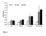
Figur 1 : IgG Titer gegen Bet v 1 nach 2-maliger orale Immunisierung mit Mikropartikeln; aufgetragen ist die IgG-Detektion gegen Bet v 1. ELISA-Platten wurden mit rekombinantem Bet v 1 beschichtet, mit Mausseren (1:100 verdünnt) inkubiert, und gebundenes IgG durch einen Peroxidase-markierten Anti-Maus IgG detektiert. Die Reaktion wurde durch Substratzugabe visualisiert und die Reaktivität mit einem ELISA Reader gemessen. Die Werte sind direkt proportional der Menge des gebundenen Antikörpers.
Die Abkürzungen bedeuten: MS = Mikrosphären, BP = Birkenpollenextrakt, AAL = Aleuria Aurantia Lektin, WGA = Weizenkeim Agglutinin, PIS = Präimmunserum, 1.MIS = erstes Immunserum, 2.MIS = zweites Immunserum, OD = Optische Dichte -
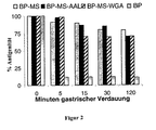
Figur 2 : Überprüfung der Antigenität von in Mikrosphären verpacktem Birkenpollenextrakt nach gastrischem Verdau; die graphische Darstellung zeigt die Antigenität der verschiedenen Mikropartikel in zeitlicher Abhängigkeit gastrischer Verdauung. Die Abkürzungen haben folgende Bedeutung:- BP: Birkenpollenextrakt, MS: Mikrosphären, AAL: Aleuria Aurantia Lektin, WGA: Weizenkeim Agglutinin.
-
Figur 3 : Qualitative Überprüfung der Haftfähigkeit unterschiedlicher Mikrosphärenpräparationen an humanen intestinalen Epithelzellen in der Immunofluoreszenz. -
Figur 4 : Quantitative Überprüfung der Haftfähigkeit unterschiedlicher Mikrosphärenpräparationen an humanen intestinalen Epithelzellen im Fluoreszenz-ELISA. - Aleuria Aurantia Lektin (AAL) besitzt den Vorteil gegenüber vielen Lektinen, nicht toxisch zu sein. Ein weiteres, allerdings toxisches Lektin ist Weizenkeim Agglutinin (WGA), von dem vermutet wird, dass es aufgrund seiner Glykan-bindenden Eigenschaften an intestinale Epitelzellen bindet, und zwar insbesondere durch Bindung an N-Acetylglykosamin.
- Wie in den unten beschriebenen Beispielen gezeigt wird, besitzt AAL jedoch den weiteren und unerwarteten Vorteil gegenüber WGA, signifikant mehr Antigen an die mukosale Oberfläche zu binden. Dies mag an einer verbesserten Haftung liegen, wird aber auf jeden Fall als Vorteil angesehen, da diese Eigenschaft letztlich dafür sorgt, dass die in die AALoberflächenmodifizierten Mikropartikel eingebauten Allergene zielgerichtet und in erhöhter Menge an ihren therapeutischen Einsatzort gelangen können. Außerdem erübrigt sich dabei die wiederholte Gabe von steigenden Allergenmengen in kurzen Abständen, weil die Mikrosphären in der Lage sind, die Allergene langsam und stetig in das Zielgewebe freizusetzen.
- PLGA-Mikrosphären (MS) wurden mittels Sprühtrocknung gewonnen. Bei diesem Verfahren wurde eine Emulsion aus Wasser und Ethylformiat mit Birkenpollenextrakt in einem heißen Luftstrom versprüht. Der Median der Mikrosphären wurde mittels Laser-Diffraktion ermittelt und betrug 5,5 µm. Die Proteinmenge der Mikropartikel wurde mit 40 ug/mg Mikropartikel bestimmt. Die Funktionalisierung der Mikrosphären mit den jeweiligen Lektinen erfolgte unter Ausbildung einer Amidbindung. Bei der Erzeugung der Mikrosphären wird eine Emulsion aus Ethylformiat/Wasser mit 5% (w/v) Polymer und 0,2% (w/v) Protein versprüht. Das Polymer wird dabei im Ethylformiat gelöst, die Proteine des Birkenpollenextraktes in Wasser. Die Emulsion wird in einem heißen Luftstrom versprüht. Das Lösungsmittel verdunstet und verursacht eine Ausfällung des Polymers, wodurch die Proteine des Birkenpollenextraktes eingebettet werden. Für die Proteinbestimmung wurden dann die Mikrosphären in 0,05N NaOH/1%SDS gelöst und mittels eines Färbetest (Bichinchoninsäure-Assay) quantifiziert.
- Die Funktionalisierung der mit Proteinen des Birkenpollenextraktes gefüllten Mikro- und/ oder Nanosphären verläuft in zwei Schritten. Dazu werden die Partikel in Hepes-Puffer pH 7,2 aufgenommen und durch Zugabe von EDAC/NHS über eine Carbodiimidreaktion aktiviert. AAL wird zugeben und das Lektin über eine Ausbildung einer stabilen Amidbindung an die Mikro- und/ oder Nanosphären gekoppelt. Die restlichen aktivierten Carboxygruppen werden mit Glycin abgesättigt.
- Die Ergebnisse des Immunisierungsmodells sind in
Figur 1 gezeigt. 4 Gruppen von je 5 Balb/c Mäusen wurden mit Mikropartikeln zweimal (Tag 0 und Tag 21) an jeweils drei aufeinanderfolgenden Tagen mit Mikropartikeln in PBS (Volumen 150 microL) intragastral immunisiert. Die Mikropartikel waren entweder leer oder mit Birkenpollenproteinen bepackt. Ein Teil der Mikropartikel, die mit Birkenpollenproteinen bepackt waren, war außerdem zusätzlich mit Aleuria Aurantia Lektin oder Weizenkeim Agglutinin funktionalisiert. Vor und nach zwei Wochen nach den Immunisierungen wurde den Mäusen Blut abgenommen und auf das Vorliegen von spezifischen Anti-Bet v 1 Antikörpern untersucht. Dies geschah in einem ELISA. Dabei wurde Birkenpollenextrakt in einer Konzentration von 10 micorg/ml Carbonatpuffer, pH 9,6 beschichtet. Nach Blockierung mit PBS/0,1% BSA wurden Maussera (1:100 in Blockierungspuffer verdünnt) inkubiert. Nach Waschen mit PBS wurde gebundenes IgG mit anti-Maus Antikörpern, Peroxidase konjugiert, detektiert. Die Reaktion wurde durch Substrazzugabe entwickelt und in einem ELISA Reader erfolgte die Auswertung durch Ablesung bei OD 405-490 nm. - Wie in
Figur 1 zu sehen ist, zeigten die Gruppen, die mit leeren beziehungsweise mit Birkenpollenproteinen bepackten Mikropartikeln gefüttert wurden, im ELISA weder im ersten Immunserum (1. MIS-grauer Balken) noch im zweiten Immunserum (2. MIS-grau-schwarzer Balken) einen relevanten Anstieg an Bet v 1 spezifischen IgG-Antikörpern gegenüber dem Präimmunserum. Hingegen zeigten beide Gruppen, bei denen die bepackten Mikropartikel zusätzlich außen noch mit Lektinen funktionalisiert wurden, einen deutlichen Anstieg der gegen Bet v 1 gerichteten IgG-Antikörper. - Um die Antigenität von mikrosphären-verpacktem Birkenpollenextrakt (BP-MS) nach gastrischem Verdau zu überprüfen, wurden Mikrosphären mit Pepsin bei pH 1,5 für unterschiedliche Zeitintervalle inkubiert. In mikrosphären-verpacktes Birkenpollenprotein konnte auch noch nach zwei Stunden Verdau antigenetisch detektiert werden, hingegen wurde nicht verpacktes Birkenpollenprotein innerhalb von Sekunden zerstört. AALgekoppelte Mikropartikel reagierten in diesem Versuch genauso vorteilhaft wie andere Mikrosphären-Präparationen (MS: Mikrosphären allein, MS-WGA: mit Weizenkeim Agglutinin gekoppelte Mikrosphären) und schützten das Protein vor dem Verdau. Weiterhin zeigt dieses Beispiel, dass Birkenpollenprotein beim Einpacken in Mikrosphären nichts an Antigenität gegenüber unbehandelten Extrakten verliert.
- Die Auswertung des Versuchs erfolgte in einem ELISA: das Birkenpollenprotein wurde aus Mikrosphären extrahiert und die Lösung zur Beschichtung von ELISA-Platten verwendet. Gebundenes Birkenpollen-Hauptallergen Bet v 1 wurde mit einem polyklonalen Kaninchen Anti-Bet v 1-Antikörper und dann mittels eines zweiten Peroxidase markierten Anti-Kaninchen IgG-Antikörpers detektiert. Die Reaktion wurde durch Substratzugabe visualisiert und das Signal in einem ELISA Reader gemessen. Das gebundene Antigen ist der Reaktivität und daher dem Signal direkt proportional. Die Ergebnisse sind in
Figur 2 graphisch dargestellt. - Die qualitative Überprüfung der Haftfähigkeit unterschiedlicher Mikrosphären-Präparationen an humanen intestinalen Epithelzellen erfolgte mit der Immunofluoreszenz-Methode. Die Zelllinie Caco-2 wurde auf Glasplättchen ausgesät und bei 4 °C inkubiert mit:
- 1. Birkenpollenextrakt verpackt in Mikrosphären (BP-MS), oder
- 2. Birkenpollenextrakt verpackt in Mikrosphären, gekoppelt mit Weizenkeim Agglutinin (BP-WGA-MS), oder
- 3. Birkenpollenextrakt verpackt in Mikrosphären, gekoppelt mit Aleuria Aurantia Lektin (BP-AAL-MS).
- Zum Sichtbarmachen wurden die Mikrosphären mit dem Fluoreszenz-Farbstoff Fluorescein-Isothiocyanat(FITC)-Cadaverin beladen. Die Visualisierung der Epithelzellen erfolgte mit einem Kaninchen anti-PLAP Antikörper, gefolgt von ALEXA 568-konjugiertem Anti-Kaninchen IgG. Die Zellkerne wurden mit Hoechst Dye gefärbt.
- Gegenüber nicht-funktionalisierten Mikrosphären wird in diesem Versuch deutlich, dass Lektin-Funktionalisierung die Anbindung der Partikel an das Epithel signifikant verbessert. Die Ergebnisse sind in
Figur 3 gezeigt. - Beispiel 4: Quantitative Überprüfung der Haftfähigkeit unterschiedlicher Mikrosphären-Präparationen.
- Die quantitative Überprüfung der Haftfähigkeit unterschiedlicher Mikrosphären-Präparationen an humane intestinalen Epithelzellen erfolgte im Fuoreszenz-ELISA. Die Zelllinie Caco-2 wurde in 96-well Gewebekulturplatten gezüchtet, bis sie geschlossene einschichtige Lagen bildete. Dann erfolgte eine Inkubation bei 4 °C mit:
- 1. Birkenpollenextrakt verpackt in Mikrosphären (BP-MS), oder
- 2. Birkenpollenextrakt verpackt in Mikrosphären, gekoppelt mit Weizenkeim Agglutinin (BP-WGA-MS), oder
- 3. Birkenpollenextrakt verpackt in Mikrosphären, gekoppelt mit Aleuria Aurantia Lektin (BP-AAL-MS).
- Zur Sichtbarmachung wurden mit FITC-Cadaverin markierte Mikrosphären verwendet. Die Visualisierung der Epithelzellen erfolgte durch Fluoreszenz-Detektion bei 485/535 nm. Das Signal ist der Anzahl gebundener Partikel direkt proportional.
- Gegenüber nicht-funktionalisierten Mikrosphären wird in diesem Versuch deutlich, dass Lektin-Funktionalisierung die Anbindung der Partikel an das Epithel signifikant verbessert. Vor allem aber besitzt AAL eine deutlich erhöhte Haftfähigkeit gegenüber WGA. Die Ergebnisse sind in
Figur 4 dargestellt.
Claims (18)
- Mikrosphären zur Allergietherapie, die Antigene und/oder DNA von Antigenen enthalten, dadurch gekennzeichnet, dass die Mikrosphären eine Bindungskonstante KB von mindestens 1 x 10⁴ M-1 gegenüber dem spezifischen Carbohydratrest von intestinalen und/oder nasalen Epithelzellen aufweisen.
- Mikrosphären zur Allergietherapie nach Anspruch 1, dadurch gekennzeichnet, daß die Mikrosphären eine Avidität KB von mindestens 1 x 10¹⁰ M-1 gegenüber dem spezifischen Carbohydratrest von intestinalen und/oder nasalen Epithelzellen aufweisen.
- Mikrosphären zur Allergietherapie nach Anspruch 1 oder 2, dadurch gekennzeichnet, dass die Mikrosphären an ihrer Oberfläche zektine aufweisen, die die Haftfähigkeit an mukosalen Zellen erhöhen.
- Mikrosphären zur Allergietherapie nach einem der vorstehenden Ansprüche, dadurch gekennzeichnet, dass der spezifische Carbohydratrest α-L Fucose ist.
- Mikrosphären zur Allergietherapie nach Anspruch 4, dadurch gekennzeichnet, dass die Substanz an der Mikrosphärenoberfläche ein nicht toxisches Lektin ist.
- Mikrosphären zur Allergietherapie nach Anspruch 4 oder 5, dadurch gekennzeichnet, dass Lektin eßbar ist.
- Mikrosphären zur Allergietherapie nach Anspruch 4-6, dadurch gekennzeichnet, dass das Lektin Aleuria-Aurentia-Lektin ist.
- Mikrosphären zur Allergietherapie nach einem der vorstehenden Ansprüche, dadurch gekennzeichnet, dass die Mikrosphären einen Durchmesser von 0,1 bis 100 µm haben.
- Mikrosphären zur Allergietherapie nach einem der vorstehenden Ansprüche, dadurch gekennzeichnet, dass das Grundgerüst der Mikrosphären aus Polymeren besteht.
- Mikrosphären zur Allergietherapie nach Anspruch 9, dadurch gekennzeichnet, dass das Grundgerüst der Mikrosphären aus Polymeren mit funktionellen Gruppen besteht.
- Mikrosphären zur Allergietherapie nach einem der Ansprüche 9 oder 11, dadurch gekennzeichnet, dass das Grundgerüst der Mikrosphären aus biologisch abbaubaren Polymeren oder Copolymeren besteht.
- Mikrosphären zur Allergietherapie nach einem der Ansprüche 8-11, dadurch gekennzeichnet, dass das Grundgerüst der Mikrosphären aus Polymilchsäure, Polyglycolsäure oder aus Polymilchsäure-Polyglycolsäure-Copolymer besteht.
- Mikrosphären zur Allergietherapie nach einem der Ansprüche 9 bis 12, dadurch gekennzeichnet, dass das Aleuria Aurantia Lektin über eine kovalente Bindung an die Polymere gebunden ist.
- Mikrosphären zur Allergietherapie nach einem der vorstehenden Ansprüche, dadurch gekennzeichnet, dass die Mikrosphären 0,1-20 Gw.-% Antigene und/oder DNA von Antigenen enthalten.
- Mikrosphären zur Allergietherapie nach einem der vorhergehenden Ansprüche, dadurch gekennzeichnet, daß die Antigene und/oder DNA von Antigenen Allergene und/oder DNA von Allergenen sind.
- Mikrosphären zur Allergietherapie nach einem der vorhergehenden Ansprüche, dadurch gekennzeichnet, daß die Antigene Mimotope des Allergens Phl p 5 und/oder des Allergens Bet v 1 sind.
- Verfahren zur Herstellung von Mikrosphären nach einem der vorstehenden Ansprüche, dadurch gekennzeichnet, dass die Mikrosphären zunächst mir Antigenen und/oder DNA von Antigenen beladen werden und anschließend die Mikrosphären funktionalisiert werden.
- Verwendung von Mikrosphären nach einem der vorstehenden Ansprüche 1-17 zur Herstelling mies Aizneimittels für die Allergietherapie.
Applications Claiming Priority (2)
| Application Number | Priority Date | Filing Date | Title |
|---|---|---|---|
| DE10329087.7A DE10329087B4 (de) | 2003-06-27 | 2003-06-27 | Antigenhaltige Mikrosphären zur Allergietherapie |
| PCT/EP2004/004480 WO2005000274A1 (de) | 2003-06-27 | 2004-04-28 | Antigenhaltige mikrosphären zur allergietherapie |
Publications (2)
| Publication Number | Publication Date |
|---|---|
| EP1638531A1 EP1638531A1 (de) | 2006-03-29 |
| EP1638531B1 true EP1638531B1 (de) | 2013-02-13 |
Family
ID=33546691
Family Applications (1)
| Application Number | Title | Priority Date | Filing Date |
|---|---|---|---|
| EP04729885A Expired - Lifetime EP1638531B1 (de) | 2003-06-27 | 2004-04-28 | Antigenhaltige mikrosphären zur allergietherapie |
Country Status (8)
| Country | Link |
|---|---|
| US (1) | US8420095B2 (de) |
| EP (1) | EP1638531B1 (de) |
| JP (1) | JP4900799B2 (de) |
| AU (1) | AU2004250998B2 (de) |
| CA (1) | CA2530625C (de) |
| DE (1) | DE10329087B4 (de) |
| ES (1) | ES2407137T3 (de) |
| WO (1) | WO2005000274A1 (de) |
Families Citing this family (2)
| Publication number | Priority date | Publication date | Assignee | Title |
|---|---|---|---|---|
| EP2123261A1 (de) * | 2008-05-20 | 2009-11-25 | Stallergenes S.A. | Speichelklebende Partikelformulierung zur Induzierung von antigenspezifischer Immuntoleranz |
| WO2012156376A1 (en) * | 2011-05-13 | 2012-11-22 | Merrion Pharmaceuticals Ireland Ltd. | Use of plant lectins to target leukocytes |
Family Cites Families (10)
| Publication number | Priority date | Publication date | Assignee | Title |
|---|---|---|---|---|
| NZ215865A (en) * | 1985-04-22 | 1988-10-28 | Commw Serum Lab Commission | Method of determining the active site of a receptor-binding analogue |
| US5075109A (en) | 1986-10-24 | 1991-12-24 | Southern Research Institute | Method of potentiating an immune response |
| DE4120760A1 (de) * | 1991-06-24 | 1993-03-04 | 3 M Medica Gmbh | Traegersysteme fuer arzneimittel |
| JP3451783B2 (ja) | 1995-04-18 | 2003-09-29 | 東ソー株式会社 | コリンエステラーゼの測定法及び肝硬変と肝炎の識別法 |
| GB2330532A (en) | 1996-07-10 | 1999-04-28 | Danbiosyst Uk | Compositions suitable for delivery of genes to epithelial cells |
| CA2362593A1 (en) * | 1999-02-10 | 2000-08-17 | Curis, Inc. | Pancreatic progenitor cells, methods and uses related thereto |
| US6610535B1 (en) * | 2000-02-10 | 2003-08-26 | Es Cell International Pte Ltd. | Progenitor cells and methods and uses related thereto |
| EP1356826A1 (de) | 2002-04-22 | 2003-10-29 | BIOMAY Produktions- und Handels- Aktiengesellschaft | Mikropartikel die Kohlenhydratkugeln enthalten an welche ein Allergen kovalent gebunden sind |
| CA2521904A1 (en) * | 2003-04-01 | 2004-10-21 | Proactive Oral Solutions, Inc. | Caries risk test for predicting and assessing the risk of disease |
| DE10325752A1 (de) * | 2003-06-06 | 2004-12-30 | Faustus Forschungs Cie. Translational Cancer Research Gmbh | Lektin-Konjugate |
-
2003
- 2003-06-27 DE DE10329087.7A patent/DE10329087B4/de not_active Expired - Fee Related
-
2004
- 2004-04-28 ES ES04729885T patent/ES2407137T3/es not_active Expired - Lifetime
- 2004-04-28 JP JP2006515761A patent/JP4900799B2/ja not_active Expired - Fee Related
- 2004-04-28 EP EP04729885A patent/EP1638531B1/de not_active Expired - Lifetime
- 2004-04-28 AU AU2004250998A patent/AU2004250998B2/en not_active Ceased
- 2004-04-28 US US10/562,066 patent/US8420095B2/en not_active Expired - Fee Related
- 2004-04-28 CA CA2530625A patent/CA2530625C/en not_active Expired - Fee Related
- 2004-04-28 WO PCT/EP2004/004480 patent/WO2005000274A1/de not_active Ceased
Also Published As
| Publication number | Publication date |
|---|---|
| US8420095B2 (en) | 2013-04-16 |
| ES2407137T3 (es) | 2013-06-11 |
| DE10329087A1 (de) | 2005-02-03 |
| EP1638531A1 (de) | 2006-03-29 |
| WO2005000274A1 (de) | 2005-01-06 |
| CA2530625C (en) | 2012-08-14 |
| AU2004250998B2 (en) | 2009-05-28 |
| DE10329087B4 (de) | 2014-02-13 |
| US20070275079A1 (en) | 2007-11-29 |
| JP2007506650A (ja) | 2007-03-22 |
| AU2004250998A1 (en) | 2005-01-06 |
| CA2530625A1 (en) | 2005-01-06 |
| JP4900799B2 (ja) | 2012-03-21 |
Similar Documents
| Publication | Publication Date | Title |
|---|---|---|
| DE69221568T2 (de) | Systeme zur oralen freisetzung von mikropartikeln | |
| DE69507339T2 (de) | Polymer-mikrokugeln und verfahren zu deren herstellung | |
| DE69222306T2 (de) | Oral verabreichbare therapeutische proteine und herstellungsverfahren | |
| Schöll et al. | Allergen‐loaded biodegradable poly (d, l‐lactic‐co‐glycolic) acid nanoparticles down‐regulate an ongoing Th2 response in the BALB/c mouse model | |
| EP2299980B1 (de) | Siliciumdioxid-nanopartikel und deren verwendung zur vakzinierung | |
| DE69812887T2 (de) | Mikropartikel aus chitosan-gelatin a | |
| DE69305313T3 (de) | Orale Verabreichung von immunologischen aktiven Biomolekülen und anderen therapeutischen Proteinen | |
| DE69529054T2 (de) | System zur gezielten wirkstoffzufuhr, verfahren zu seiner herstellung und verwendung | |
| DE202008006598U1 (de) | Allergie-Impfstoff-Formulierung zur mucosalen Verabreichung | |
| DE69505949T2 (de) | Arzneimittel, impfstoffe und hormone in polylaktidbeschichteten mikropartikel | |
| DE69934179T2 (de) | Zusammensetzung enthaltend ein Antigen, ein TH-1 induzierendes Adjuvans und eine im Wasser schwer lösliche Aminosäure zur Verwendung in der Immunisierung | |
| EP2819659B1 (de) | Verfahren zur herstellung wirkstoffbeladener nanopartikel | |
| DE60125961T2 (de) | Modulierung einer allergischen antwort | |
| HK1200019A1 (en) | Use of a liquid allergy vaccine formulation for oromucosal administration | |
| DE69528074T2 (de) | Chitosan induzierte verstärkung | |
| EP0686030A1 (de) | Verfahren zur potenzierung der immunologischen antwort | |
| CN110290780A (zh) | 使用小檗碱纳米颗粒的免疫疾病治疗 | |
| DE10329087B4 (de) | Antigenhaltige Mikrosphären zur Allergietherapie | |
| DE60004334T2 (de) | Impfstoffzusammensetzung auf basis von partikeln | |
| Di Felice et al. | Nanoparticles adjuvants in allergology: new challenges and pitfalls | |
| WO2002000191A2 (de) | Bdellosomen | |
| DE69926531T2 (de) | Gegen gastrointestinale transport rezeptoren gerichtete retro-invertierte peptide und deren verwendung | |
| DE10253623B4 (de) | Bioabbaubare kolloidale Partikel, insbesondere für pulmonale Applikationen | |
| AU2019422413B2 (en) | Methods of purifying an allergen extract | |
| DE60313593T2 (de) | Chemische igf-i-zusammensetzung zur behandlung und prävention von neurodegenerativen erkrankungen |
Legal Events
| Date | Code | Title | Description |
|---|---|---|---|
| PUAI | Public reference made under article 153(3) epc to a published international application that has entered the european phase |
Free format text: ORIGINAL CODE: 0009012 |
|
| 17P | Request for examination filed |
Effective date: 20060103 |
|
| AK | Designated contracting states |
Kind code of ref document: A1 Designated state(s): AT BE BG CH CY CZ DE DK EE ES FI FR GB GR HU IE IT LI LU MC NL PL PT RO SE SI SK TR |
|
| DAX | Request for extension of the european patent (deleted) | ||
| 17Q | First examination report despatched |
Effective date: 20091218 |
|
| RAP1 | Party data changed (applicant data changed or rights of an application transferred) |
Owner name: BIOMEDICAL INTERNATIONAL R + D GMBH |
|
| GRAP | Despatch of communication of intention to grant a patent |
Free format text: ORIGINAL CODE: EPIDOSNIGR1 |
|
| GRAS | Grant fee paid |
Free format text: ORIGINAL CODE: EPIDOSNIGR3 |
|
| GRAA | (expected) grant |
Free format text: ORIGINAL CODE: 0009210 |
|
| AK | Designated contracting states |
Kind code of ref document: B1 Designated state(s): AT BE BG CH CY CZ DE DK EE ES FI FR GB GR HU IE IT LI LU MC NL PL PT RO SE SI SK TR |
|
| REG | Reference to a national code |
Ref country code: GB Ref legal event code: FG4D Free format text: NOT ENGLISH |
|
| REG | Reference to a national code |
Ref country code: AT Ref legal event code: REF Ref document number: 596086 Country of ref document: AT Kind code of ref document: T Effective date: 20130215 |
|
| REG | Reference to a national code |
Ref country code: IE Ref legal event code: FG4D Free format text: LANGUAGE OF EP DOCUMENT: GERMAN |
|
| REG | Reference to a national code |
Ref country code: DE Ref legal event code: R096 Ref document number: 502004014021 Country of ref document: DE Effective date: 20130411 |
|
| REG | Reference to a national code |
Ref country code: CH Ref legal event code: NV Representative=s name: E. BLUM AND CO. AG PATENT- UND MARKENANWAELTE , CH |
|
| REG | Reference to a national code |
Ref country code: ES Ref legal event code: FG2A Ref document number: 2407137 Country of ref document: ES Kind code of ref document: T3 Effective date: 20130611 |
|
| REG | Reference to a national code |
Ref country code: NL Ref legal event code: T3 |
|
| PG25 | Lapsed in a contracting state [announced via postgrant information from national office to epo] |
Ref country code: CY Free format text: LAPSE BECAUSE OF FAILURE TO SUBMIT A TRANSLATION OF THE DESCRIPTION OR TO PAY THE FEE WITHIN THE PRESCRIBED TIME-LIMIT Effective date: 20130213 Ref country code: SE Free format text: LAPSE BECAUSE OF FAILURE TO SUBMIT A TRANSLATION OF THE DESCRIPTION OR TO PAY THE FEE WITHIN THE PRESCRIBED TIME-LIMIT Effective date: 20130213 Ref country code: BG Free format text: LAPSE BECAUSE OF FAILURE TO SUBMIT A TRANSLATION OF THE DESCRIPTION OR TO PAY THE FEE WITHIN THE PRESCRIBED TIME-LIMIT Effective date: 20130513 |
|
| PG25 | Lapsed in a contracting state [announced via postgrant information from national office to epo] |
Ref country code: PL Free format text: LAPSE BECAUSE OF FAILURE TO SUBMIT A TRANSLATION OF THE DESCRIPTION OR TO PAY THE FEE WITHIN THE PRESCRIBED TIME-LIMIT Effective date: 20130213 Ref country code: FI Free format text: LAPSE BECAUSE OF FAILURE TO SUBMIT A TRANSLATION OF THE DESCRIPTION OR TO PAY THE FEE WITHIN THE PRESCRIBED TIME-LIMIT Effective date: 20130213 Ref country code: PT Free format text: LAPSE BECAUSE OF FAILURE TO SUBMIT A TRANSLATION OF THE DESCRIPTION OR TO PAY THE FEE WITHIN THE PRESCRIBED TIME-LIMIT Effective date: 20130613 Ref country code: SI Free format text: LAPSE BECAUSE OF FAILURE TO SUBMIT A TRANSLATION OF THE DESCRIPTION OR TO PAY THE FEE WITHIN THE PRESCRIBED TIME-LIMIT Effective date: 20130213 Ref country code: GR Free format text: LAPSE BECAUSE OF FAILURE TO SUBMIT A TRANSLATION OF THE DESCRIPTION OR TO PAY THE FEE WITHIN THE PRESCRIBED TIME-LIMIT Effective date: 20130514 |
|
| BERE | Be: lapsed |
Owner name: BIOMEDICAL INTERNATIONAL R + D GMBH Effective date: 20130430 |
|
| PG25 | Lapsed in a contracting state [announced via postgrant information from national office to epo] |
Ref country code: RO Free format text: LAPSE BECAUSE OF FAILURE TO SUBMIT A TRANSLATION OF THE DESCRIPTION OR TO PAY THE FEE WITHIN THE PRESCRIBED TIME-LIMIT Effective date: 20130213 Ref country code: DK Free format text: LAPSE BECAUSE OF FAILURE TO SUBMIT A TRANSLATION OF THE DESCRIPTION OR TO PAY THE FEE WITHIN THE PRESCRIBED TIME-LIMIT Effective date: 20130213 Ref country code: EE Free format text: LAPSE BECAUSE OF FAILURE TO SUBMIT A TRANSLATION OF THE DESCRIPTION OR TO PAY THE FEE WITHIN THE PRESCRIBED TIME-LIMIT Effective date: 20130213 Ref country code: CZ Free format text: LAPSE BECAUSE OF FAILURE TO SUBMIT A TRANSLATION OF THE DESCRIPTION OR TO PAY THE FEE WITHIN THE PRESCRIBED TIME-LIMIT Effective date: 20130213 Ref country code: SK Free format text: LAPSE BECAUSE OF FAILURE TO SUBMIT A TRANSLATION OF THE DESCRIPTION OR TO PAY THE FEE WITHIN THE PRESCRIBED TIME-LIMIT Effective date: 20130213 |
|
| PG25 | Lapsed in a contracting state [announced via postgrant information from national office to epo] |
Ref country code: MC Free format text: LAPSE BECAUSE OF FAILURE TO SUBMIT A TRANSLATION OF THE DESCRIPTION OR TO PAY THE FEE WITHIN THE PRESCRIBED TIME-LIMIT Effective date: 20130213 |
|
| PLBE | No opposition filed within time limit |
Free format text: ORIGINAL CODE: 0009261 |
|
| STAA | Information on the status of an ep patent application or granted ep patent |
Free format text: STATUS: NO OPPOSITION FILED WITHIN TIME LIMIT |
|
| 26N | No opposition filed |
Effective date: 20131114 |
|
| REG | Reference to a national code |
Ref country code: IE Ref legal event code: MM4A |
|
| PG25 | Lapsed in a contracting state [announced via postgrant information from national office to epo] |
Ref country code: BE Free format text: LAPSE BECAUSE OF NON-PAYMENT OF DUE FEES Effective date: 20130430 |
|
| REG | Reference to a national code |
Ref country code: DE Ref legal event code: R097 Ref document number: 502004014021 Country of ref document: DE Effective date: 20131114 |
|
| PG25 | Lapsed in a contracting state [announced via postgrant information from national office to epo] |
Ref country code: IE Free format text: LAPSE BECAUSE OF NON-PAYMENT OF DUE FEES Effective date: 20130428 |
|
| PG25 | Lapsed in a contracting state [announced via postgrant information from national office to epo] |
Ref country code: TR Free format text: LAPSE BECAUSE OF FAILURE TO SUBMIT A TRANSLATION OF THE DESCRIPTION OR TO PAY THE FEE WITHIN THE PRESCRIBED TIME-LIMIT Effective date: 20130213 |
|
| PG25 | Lapsed in a contracting state [announced via postgrant information from national office to epo] |
Ref country code: LU Free format text: LAPSE BECAUSE OF NON-PAYMENT OF DUE FEES Effective date: 20130428 Ref country code: HU Free format text: LAPSE BECAUSE OF FAILURE TO SUBMIT A TRANSLATION OF THE DESCRIPTION OR TO PAY THE FEE WITHIN THE PRESCRIBED TIME-LIMIT; INVALID AB INITIO Effective date: 20040428 |
|
| REG | Reference to a national code |
Ref country code: FR Ref legal event code: PLFP Year of fee payment: 13 |
|
| REG | Reference to a national code |
Ref country code: FR Ref legal event code: PLFP Year of fee payment: 14 |
|
| PGFP | Annual fee paid to national office [announced via postgrant information from national office to epo] |
Ref country code: NL Payment date: 20170424 Year of fee payment: 14 |
|
| PGFP | Annual fee paid to national office [announced via postgrant information from national office to epo] |
Ref country code: DE Payment date: 20170529 Year of fee payment: 14 Ref country code: GB Payment date: 20170425 Year of fee payment: 14 Ref country code: CH Payment date: 20170425 Year of fee payment: 14 Ref country code: FR Payment date: 20170424 Year of fee payment: 14 |
|
| PGFP | Annual fee paid to national office [announced via postgrant information from national office to epo] |
Ref country code: IT Payment date: 20170420 Year of fee payment: 14 Ref country code: AT Payment date: 20170420 Year of fee payment: 14 Ref country code: ES Payment date: 20170503 Year of fee payment: 14 |
|
| REG | Reference to a national code |
Ref country code: DE Ref legal event code: R119 Ref document number: 502004014021 Country of ref document: DE |
|
| REG | Reference to a national code |
Ref country code: CH Ref legal event code: PL |
|
| REG | Reference to a national code |
Ref country code: NL Ref legal event code: MM Effective date: 20180501 |
|
| REG | Reference to a national code |
Ref country code: AT Ref legal event code: MM01 Ref document number: 596086 Country of ref document: AT Kind code of ref document: T Effective date: 20180428 |
|
| GBPC | Gb: european patent ceased through non-payment of renewal fee |
Effective date: 20180428 |
|
| PG25 | Lapsed in a contracting state [announced via postgrant information from national office to epo] |
Ref country code: DE Free format text: LAPSE BECAUSE OF NON-PAYMENT OF DUE FEES Effective date: 20181101 Ref country code: AT Free format text: LAPSE BECAUSE OF NON-PAYMENT OF DUE FEES Effective date: 20180428 Ref country code: NL Free format text: LAPSE BECAUSE OF NON-PAYMENT OF DUE FEES Effective date: 20180501 |
|
| PG25 | Lapsed in a contracting state [announced via postgrant information from national office to epo] |
Ref country code: CH Free format text: LAPSE BECAUSE OF NON-PAYMENT OF DUE FEES Effective date: 20180430 Ref country code: GB Free format text: LAPSE BECAUSE OF NON-PAYMENT OF DUE FEES Effective date: 20180428 Ref country code: LI Free format text: LAPSE BECAUSE OF NON-PAYMENT OF DUE FEES Effective date: 20180430 |
|
| PG25 | Lapsed in a contracting state [announced via postgrant information from national office to epo] |
Ref country code: FR Free format text: LAPSE BECAUSE OF NON-PAYMENT OF DUE FEES Effective date: 20180430 Ref country code: IT Free format text: LAPSE BECAUSE OF NON-PAYMENT OF DUE FEES Effective date: 20180428 |
|
| REG | Reference to a national code |
Ref country code: ES Ref legal event code: FD2A Effective date: 20190912 |
|
| PG25 | Lapsed in a contracting state [announced via postgrant information from national office to epo] |
Ref country code: ES Free format text: LAPSE BECAUSE OF NON-PAYMENT OF DUE FEES Effective date: 20180429 |