EP1631684B1 - Verfahren und vorrichtung zum sporenaufschluss und/oder -nachweis - Google Patents
Verfahren und vorrichtung zum sporenaufschluss und/oder -nachweis Download PDFInfo
- Publication number
- EP1631684B1 EP1631684B1 EP04736409A EP04736409A EP1631684B1 EP 1631684 B1 EP1631684 B1 EP 1631684B1 EP 04736409 A EP04736409 A EP 04736409A EP 04736409 A EP04736409 A EP 04736409A EP 1631684 B1 EP1631684 B1 EP 1631684B1
- Authority
- EP
- European Patent Office
- Prior art keywords
- sample
- vial
- structured
- micro
- collection chamber
- Prior art date
- Legal status (The legal status is an assumption and is not a legal conclusion. Google has not performed a legal analysis and makes no representation as to the accuracy of the status listed.)
- Expired - Lifetime
Links
Images
Classifications
-
- A—HUMAN NECESSITIES
- A61—MEDICAL OR VETERINARY SCIENCE; HYGIENE
- A61L—METHODS OR APPARATUS FOR STERILISING MATERIALS OR OBJECTS IN GENERAL; DISINFECTION, STERILISATION OR DEODORISATION OF AIR; CHEMICAL ASPECTS OF BANDAGES, DRESSINGS, ABSORBENT PADS OR SURGICAL ARTICLES; MATERIALS FOR BANDAGES, DRESSINGS, ABSORBENT PADS OR SURGICAL ARTICLES
- A61L2/00—Methods or apparatus for disinfecting or sterilising materials or objects other than foodstuffs or contact lenses; Accessories therefor
- A61L2/02—Methods or apparatus for disinfecting or sterilising materials or objects other than foodstuffs or contact lenses; Accessories therefor using physical phenomena
- A61L2/08—Radiation
- A61L2/084—Visible light
-
- A—HUMAN NECESSITIES
- A61—MEDICAL OR VETERINARY SCIENCE; HYGIENE
- A61L—METHODS OR APPARATUS FOR STERILISING MATERIALS OR OBJECTS IN GENERAL; DISINFECTION, STERILISATION OR DEODORISATION OF AIR; CHEMICAL ASPECTS OF BANDAGES, DRESSINGS, ABSORBENT PADS OR SURGICAL ARTICLES; MATERIALS FOR BANDAGES, DRESSINGS, ABSORBENT PADS OR SURGICAL ARTICLES
- A61L2/00—Methods or apparatus for disinfecting or sterilising materials or objects other than foodstuffs or contact lenses; Accessories therefor
- A61L2/02—Methods or apparatus for disinfecting or sterilising materials or objects other than foodstuffs or contact lenses; Accessories therefor using physical phenomena
- A61L2/08—Radiation
- A61L2/10—Ultraviolet radiation
-
- A61L2103/05—
-
- C—CHEMISTRY; METALLURGY
- C12—BIOCHEMISTRY; BEER; SPIRITS; WINE; VINEGAR; MICROBIOLOGY; ENZYMOLOGY; MUTATION OR GENETIC ENGINEERING
- C12M—APPARATUS FOR ENZYMOLOGY OR MICROBIOLOGY; APPARATUS FOR CULTURING MICROORGANISMS FOR PRODUCING BIOMASS, FOR GROWING CELLS OR FOR OBTAINING FERMENTATION OR METABOLIC PRODUCTS, i.e. BIOREACTORS OR FERMENTERS
- C12M47/00—Means for after-treatment of the produced biomass or of the fermentation or metabolic products, e.g. storage of biomass
- C12M47/06—Hydrolysis; Cell lysis; Extraction of intracellular or cell wall material
Definitions
- This invention relates to a method and apparatus for disrupting spores to aid subsequent analysis, especially to a method and apparatus for fast release of intrasporal DNA.
- MALDI Matrix Assisted Laser Desorption and lonisation
- the analyte of interest is mixed with a suitable matrix material and a solvent on a substrate.
- the solvent is then evaporated to leave the analyte co-crystallised with the matrix material.
- a pulsed UV laser source is then directed to irradiate the sample.
- the matrix material absorbs the laser light and a rapid temperature increase causes disintegration of the matrix ejecting a plume of sample.
- the ejection plume is input to a mass spectrometer to analyse the ionised biomolecules and hence the irradiation step is performed in a high vacuum.
- biomarkers associated with the exosporium can then be identified.
- the method is not good at discriminating between different Bacillus species as the origin and identity of the biomarkers may be unclear and different species may have similar biomarkers.
- corona plasma discharge may be used or sonication pre-treatments could be used but discrimination between species is still relatively poor.
- Sonication may also be used to modify the surface of spores so as to aid subsequent detection in an immunoassay, for instance immunoassays involve the binding of an analyte to a specific antibody contained on the surface of a sensor. Detection sensitivity can be improved by modification of the surface of the species to be detected so as to improve subsequent binding to the antibodies on the biosensor.
- Another method of screening for spores is to completely disrupt the spore so as to release intrasporal DNA for subsequent analysis via polymerase chain reaction (PCR) assays.
- PCR polymerase chain reaction
- ultrasonication has recently been proposed to completely disrupt spores in Belgrader P.; Hansford D.; Kovacs G.T.A.; Venkateswaran, K; Mariella, R.; Milanovich, F.; Nasarabadi, s.;Okuzumi, m; Pourahmadi, F.; Northrup, M.A. Analytical Chemistry 1999, 71, 4232-4236' .
- the samples can require pretreatments of up to 90 minutes and so far the amount of intracellular DNA released has been low so the technique would not currently be sensitive enough for most applications.
- the method according to the present invention therefore uses laser energy to disrupt any spores present in the sample and then collects the disrupted material for subsequent analysis.
- the method of the present invention allows very rapid disruption of any present spores for subsequent analysis - for instance screening for certain bacteria.
- the method may involve enclosing the sample in a collection chamber and irradiating the sample within the collection chamber. This can provide a sealed environment for spore disruption.
- the method may also be arranged to irradiate the sample so as to disrupt any spores present in the sample and eject material into the collection chamber. This can allow for efficient collection of the disrupted material within the collection chamber.
- the collection step may comprise the step, subsequent to irradiating the sample, of flushing the sample within an extraction fluid.
- the present invention provides a rapid and simple means of disrupting spores in a sample.
- the irradiation may be sufficient to eject material into a collection chamber where it can be easily collected for subsequent analysis.
- the irradiated sample will also contain disrupted material and flushing the sample with an extraction fluid will collect such material. It is therefore possible to use laser light which is intense enough to disrupt the spores but which is below the threshold for ejecting material into the vapour phase. Depending upon the material used this could have advantages as a less intense irradiation step may cause less damage to any material released from the spore.
- the environment for irradiation does not need to be in vacuo.
- the chamber is at atmospheric pressure and could just contain air. This removes the need for vacuum pumps etc. and means that samples can be easily collected in situ.
- the collection chamber could, if required, be filled at least partly with another fluid other than air. It may even be possible to fill the collection chamber with a liquid to aid transfer of ejected material to an analysis chamber.
- disruption means to completely disrupt the spore so as to release the DNA contained within the spore.
- Intracellular DNA can be used to uniquely identify the particular bacterium by any known method such as PCR. Therefore the irradiation step is arranged to disrupt the spore so as to eject material from within the spore, such as DNA, into the collection chamber.
- the sample is disposed within a matrix material.
- Disposing the sample within a matrix material can aid in energy transfer from the laser as the matrix material can be arranged to hold any spores in a specified location and to absorb the incident radiation and undergo an explosive type decomposition to disrupt the spores.
- the matrix material is chosen so that it has maximum disruptive effect but does not destroy the released material of interest. To release intrasporal DNA it has been found that matrix materials used in standard MALDI techniques where DNA is the sample material are advantageous, resulting in complete disruption of the spore but no damage to the released DNA.
- the method includes the step of locating the sample on a substrate. Conveniently this involves mixing the sample, and any matrix material, with a solvent. The solvent mixture is then placed on the substrate and the solvent evaporated to leave the sample. Where a matrix material is used the sample is left co-crystallised with the matrix material.
- the step of locating the sample on the substrate comprises the step of locating the sample in at least one micro-structured vial on the substrate. Locating the sample in a micro-structured vial on the substrate tends to localise the sample in one place providing inherent enrichment of the sample which can increase the sensitivity of the process. Further as the sample is located in the micro-structured vial a high power laser can be focussed just on the micro-structured vial to cause spore disruption resulting in enhanced disruption. Conveniently the sample is mixed with a solvent as discussed and applied to the micro-structured vial. Upon solvent evaporation the sample remains pinned to the micro-structured vial due to surface tension. The sample then crystallises into the vial. Given that the solvent-sample droplet will be larger than the micro-structured vial the method therefore enriches the sample in the micro-structured vial. Repeating the process with more drops provides yet further enrichment of sample.
- the at least one micro-structured vial may have a volume of less than 100nL or less than 10nl or less than 1 nl or less than 0.1 nL.
- the method involves locating samples in a plurality of micro-structured vial on the substrate.
- the plurality of micro-structured vial may be arranged as an array.
- each micro-structured vial may be arranged to be co-located with a separate collection chamber thereby allowing several samples of material to be disrupted and collected separately, say by sequential irradiation by a laser.
- a parallel illumination arrangement could be used if desired. This allows a plurality of disrupted samples to be collected. In this way the same assay may be performed several times to verify results and reduce susceptibility to error. Additionally or alternatively more than one assay may be performed on the material so as to improve detection and identification.
- the wavelength of illuminating radiation is substantially matched to the absorption maximum of the chosen matrix material, when used.
- the skilled person will be aware of the absorption maximum for the chosen matrix material, i.e. the wavelength at which radiation is most strongly absorbed, and therefore the wavelength of illuminating radiation is preferably chosen to be at or near to this maximum.
- the matrix material may be selected for low fragmentation of intrasporal DNA samples.
- the wavelength of illumination is within the ultraviolet range, for instance within the range 400 - 10 nm.
- the sample is illuminated with pulses of laser radiation, having a duration in the range 1 ns to 100ns.
- pulses of laser radiation having a duration in the range 1 ns to 100ns.
- relatively low power laser illumination is used with power in the region of 75kW peak power. This gives pulse energies of 300 microJoules in energy and at a repetition rate of 35Hz corresponds to an average power or 6mW.
- the method may also comprise an initial step of washing the sample prior to irradiating with laser light so as to remove low molecular weight acid soluble proteins associated with the outer spore layers, this could render the spore more susceptible to disruption by irradiation, especially irradiation with UV radiation.
- Spores can be coated with proteins which protect the spore from radiation such as UV radiation.
- pre-treating the sample to remove or reduce such protective proteins the power and/or duration of laser illumination required to disrupt the spore can be reduced as compared to illuminating a sample without a pre-treatment. This can allow shorter illumination times which may be useful for ultra rapid detection systems.
- the sample is washed with an acidic aqeous based solvent to release and remove at least some of the protective proteins. This could be achieved in a sample pre-treatment step just prior to the laser irradiation.
- the step of enclosing the sample within a collection chamber conveniently comprises the step of securing a collection plate to the substrate, the substrate and collection plate defining a cavity within which the sample is located.
- a collection plate By securing a collection plate to the substrate when the sample is illuminated the ejected material may be ejected as a plume and will be deposited on the collection plate. The collected material could then be used in a detection step as will be described later.
- the collection plate has a microchannel formed therein and the microchannel at least partly forms the cavity of the collection chamber.
- the microchannel is configured so that it may be used as part of a microfluidic circuit - in this way the collection plate may be removed from the substrate and used as part of a microfluidic circuit to aid subsequent detection steps.
- the substrate and collection plate could together be used as part of a microfluidic circuit.
- the method may involve filling the collection chamber at least partly with an extraction fluid, which may be water for instance, in order to better collect the ejected material.
- an extraction fluid which may be water for instance
- the ejected material is not only more efficiently collected but the extraction fluid can be flushed through without needing to remove the original sample which reduces the possibility of contamination.
- a solid matrix material it may be necessary to keep an air gap between the extraction fluid and the sample/matrix so as to prevent the sample dissolving.
- liquid matrix materials may be used in which case a liquid-liquid interface needs to be maintained.
- Parts of the collection chamber may have surface treatments to keep the extraction fluid from mixing with the sample.
- the disrupted sample material can then be subjected to any of a number of assays to detect and/or identify certain bacteria in spore form. Therefore the method comprises performing a test on the collected ejected material to determine the presence or otherwise of a known bacteria.
- the collected material is intrasporal DNA in which case the step of testing the collected material may involve performing a PCR based assay.
- PCR assays on intrasporal DNA can uniquely identify the bacteria or detect the presence of particular bacteria.
- PCR analysis is a well known and relatively quick analysis technique.
- the disruption method of the present invention is a very quick and efficient way of obtaining undamaged intrasporal DNA with relatively simple equipment.
- the method of the invention offers an extremely quick and simple analysis equipment which can offer in situ analysis of material to determine the presence or otherwise of particular bacterial agents.
- the detection method may involve the first step of disrupting the sample according to the first aspect of the invention, removing the collection plate from the substrate and introducing it to a microfluidic circuit and performing the required assay in the microfluidic circuit.
- the method according to the invention provides for very fast spore disruption.
- the irradiation step may last for one second or less.
- An apparatus for disrupting spores located on a substrate can comprise a collection plate releasably securable to the substrate to define a collection chamber and a laser apparatus arranged to illuminate a sample within the collection chamber so as to disrupt any spores present in the.
- the apparatus provides a rapid and simple method of disrupting spores to aid subsequent detection or to release intrasporal material for use in subsequent assays.
- the laser may be arranged to disrupt any spores present and eject material in the collection chamber.
- the laser apparatus comprises an ultraviolet laser.
- the laser preferably has an emission wavelength matched to the excitation region of the absorbing matrix material, ideally at the maximum absorption wavelength.
- the laser is a pulsed laser.
- the substrate has at least one micro-structured vial located therein to hold sample material.
- a micro-structured vial can usefully contain sample material for irradiation and can also serve to enrich the sample giving enhanced sensitivity.
- the collection chamber comprises a microchannel formed in the collection plate and the collection plate can be used as part of a microfluidic circuit.
- kits for screening for the presence or otherwise of spore forming bacteria comprising a substrate having at least one micro-structured vial for holding a sample and a collection plate relesably securable to the substrate, the substrate and collection plate defining a collection chamber adapted, in use, to collect material ejected from a sample disposed in the micro-structured vial upon illumination with a laser.
- sample material is mixed with a solvent and deposited on the substrate over the micro-structured vial. The solvent then evaporates and surface tension keeps the sample material at the micro-structured vial.
- a matrix material is also mixed with the solvent and the sample and after the solvent has evaporated the matrix material and sample are co-crystallised in the micro-structured vial.
- the kit therefore preferably comprises a solvent and possibly a matrix material.
- the kit may also comprise means for illuminating a sample disposed in the collection chamber with laser light so as to disrupt any spores in the sample and eject material into the collection chamber.
- the kit also comprises a test means for performing an assay on the collected material.
- This test means preferably comprises a microfluidic circuit which the collection plate can be releasably secured to, either together with the substrate or having been detached therefrom.
- the test means may perform a PCR based assay on released intrasporal DNA.
- the kit therefore provides a simple and easy to use screening kit which can be used in situ and yield rapid results for detection or otherwise of known agents.
- a method of enriching a sample of material comprising the steps of dissolving the sample in a solvent, placing a droplet of solution on a micro-structured vial in a substrate and evaporating the solvent so as to crystallise the sample in the micro-structured vial.
- dissolving and solution should be read broadly.
- the sample may not actually be dissolve in the solution but may be suspended in a suspension. For example spore forming bacteria will not be dissolved as such but will be held in suspension.
- analytes may actually dissolve. Therefore throughout this specification the terms dissolve and suspend and solution and suspension should be read interchangeably depending upon the analyte of interest. Also it should be noted that the enrichment process is applicable to a wide range of possible analytes and is not limited to spore forming bacteria. A similar enrichment effect would be achieved with a range of low abundance materials where the surface chemistry of the target plate and micro-structured vial are such so as to draw the sample into the vial. For instance when the substrate is PDMS the method would work well with analytes that are hydrophobic in nature, for instance peptides/proteins.
- the method involves adding a matrix material to the solution so that the sample co-crystallises with the matrix material in the micro-structured vial.
- a matrix material to be added so that the sample co-crystallises with the matrix material in the micro-structured vial.
- This allows a large droplet of sample material to be crystallised in the micro-structured vial resulting in an enriched sample as compared to that had a flat substrate been used.
- the enriched sample may then be irradiated in one go by a high power laser with a narrow beam cross section resulting in efficient energy transfer and maximising the amount of disrupted spore material that may be collected.
- the method of enrichment may also involve the step of repeatedly applying a drop of solvent/ material mix onto the micro-structured vial so as to further enrich the sample in the micro-structured vial.
- the sample is enriched by a factor of 10 or greater, 100 or greater, 1000 or greater or 10,000 or greater.
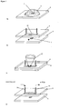
- FIG 1 schematically is shown a laser based spore enrichment and disruption process with integrated extraction of desorbed intrasporal content in a microchip format at atmospheric pressure. Amplification and identification of any released DNA may then be performed in a second step by PCR.
- a sample containing, or thought to contain, spores is first mixed with a laser light absorbing matrix and a solvent and a drop 2 of the mixture is applied onto a micro-structured vial such as nanovial 4 in an elastomer plate 6 - see figure 1a .
- a micro-structured vial such as nanovial 4 in an elastomer plate 6 - see figure 1a .
- the drop remains pinned to the nanovial 4 due to surface effects and eventually crystallises into the nanovial.
- a second elastomer plate which comprises a collection plate 8 with a microstructured channel 10 is then attached to the sample zone plate 6 - figure 1 b.
- the explosive disintegration of the matrix breaks open the protective spore layers.
- released intrasporal content is ejected in a plume and deposited onto the inner walls of the microchannel 10 in the collection plate 8. Since attachment of the two plates is adhesion based, the top plate 8 can readily be removed after illumination. Any released intrasporal DNA is then recovered by flushing the channel in the top plate 8 with water and the collected volume is subjected to PCR - figure 1 d.
- the process described above allows ultrafast spore disruption in the second to sub-second range. Furthermore, the sample enrichment and amplification in the PCR step yields high sensitivity. It should be noted however that other assays than PCR based assays could equally be applied and that the disruption process may be designed to modify the surface of the spores for subsequent analysis rather than break open the outer spore layers.
- the first step is to mix the sample with a suitable matrix material and a solvent.
- matrix material known for Matrix Assisted Laser Desorption and lonisation (MALDI) may be appropriate. Where it is wished to break open the spore to release intrasporal DNA a matrix known for MALDI DNA analysis may be used.
- MALDI DNA analysis the most commonly used matrix materials are 2,4,6-trihydroxyacetophenone (THAP), 6-aza-2-thiothymine (ATT) and 3-hydroxypicolinic acid (3-HPA). It should be noted however that when used in a MALDI apparatus it is necessary that the resultant material be ionised. This is not necessary in the present invention and hence the choice of matrix material is less constrained.
- any matrix which is soluble in an aqueous medium and absorbs highly at the wavelength of operation will be sufficient.
- 3-HPA was used in tests as a suitable candidate based on its solubility in water and comparatively low fragmentation effects on target DNA.
- the laser source used was a nitrogen laser with a 4ns pulsed output at 337nm.
- the emission wavelength of 337nm is strongly absorbed by 3-HPA.
- the energy density of the illuminating radiation will vary depending on the optical set-up used for illumination.
- the skilled person will be aware of various optical arrangements that could be used. For instance optic fibre coupling or use of appropriate lenses (open beam configuration).
- a range of output energies of between 0.4microJoule (equivalent to 0.04 mJ/cm2, which is below the threshold energy density of ⁇ 10 mJ/cm2 required for desorption and plume ejection - obtained using the optic fibre set-up) and 68 microJoules (energy density of 170 mJ/cm2, obtained using the open beam configuration) were achieved.
- spores contain dipicolinic acid in the outer layers and as such a matrix material might not be required in all embodiments and absorption of laser light by the spores themselves may be sufficient to cause disruption.
- Solvent choice is governed by the need to maintain analyte suspension and promote partitioning of the analyte into the matrix crystals during drying of the analyte/matrix mix.
- the solvent of choice is water (both DNA and the above mentioned matrices are water soluble).
- more volatile organic solvents such as acetone or acetonitrile can be added.
- the reduced surface tension of the resulting solvent systems can adversely affect the confinement of the evaporating drop into the microstructured sample vial 4 (high surface tension keeps the evaporating drop pinned to the vial, "corralling effect").
- Sample spot confinement is important to get a well defined target for the laser beam 12 and as will be described later is important in a two phase flow system for extraction of potentially released spore DNA.
- the sample plate and collection plate may be fabricated from polydimethylsiloxane (PDMS) by moulding from a photoresist master.
- PDMS polydimethylsiloxane
- other methods may be more appropriate and the skilled person would know of a range of methods that could be applied to fabricate the plates.
- the microfluidic layout of the collection plate may vary depending upon the required use as will the dimensions.
- the main channel may be in the region of 5 mm long.
- the width of the main channel may vary between 600-1000 ⁇ m, but might be more if an array of nanovials is used.
- the depth may be approximately 10 ⁇ m to minimise the extraction volume (main channel: 30-50 nL).
- the sample spot size, and hence the size of the nanovial, is limited by the minimum diameter of the laser beam.
- a beam diameter of the order of 200 ⁇ m may be achieved with a power density of approximately 8mJ cm -2 (with a coupling loss of approximately 90%). Therefore sample vials with dimensions ranging from 200-500 ⁇ m were designed (volume: 0.4-1 nL).
- Sample vial arrays should allow multiple shots without need for repositioning of the microfluidic chip.
- sample zone and extraction liquid are arranged in a vertical arrangement, as shown in Figure 1 , to exploit best directionality of the plume (normal to the surface).
- Collection plate 8 may then be detached from sample plate 6 and have another flat plate attached as a seal (not shown). Water may then be fed into the channel through inlet 14 and out through outlet 16 to flush the deposited material out where it may be used in a subsequent analysis.
- the laser illumination is sufficient to disrupt the spores but is not sufficient to eject any material out of the nanovial, i.e. no material is volatilized. In this case all the disrupted material will be left in the nanovial following illumination. Illuminating below the threshold required to eject material into the vapour phase can be advantageous where the material of interest, for instance intracellular DNA, is relatively fragile and could be damaged by intense irradiation.
- Prior art MALDI techniques all work on material ionised in the vapour phase and so illumination has to be above the threshold to eject material into the vapour phase.
- the method may also involve a pre-treatment step of washing the sample thought to contain spores to remove or reduce any radiation protective proteins, so called low molecular weight acid soluble proteins from the spore outer layers.
- the spore layer may contain various proteins which act to protect the spore from radiation damage or disruption, such as UV radiation. When irradiated these proteins in the layer/s may serve to protect the spore.
- a suitable treatment such as an acidic aqueous based solvent
- the amount of protective proteins on the spore layer/s can be reduced. This can reduce the power and/or duration of radiation exposure needed to disrupt the spore.
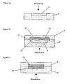
- Figure 2 shows a range of possible geometries.
- Figure 2a shows the situation where spores alone are irradiated in a nanovial with the disrupted material being left in the nanovial for collection. The same arrangement would equally apply were the spore mixed with a matrix material which may be solid or liquid.
- Figure 2b shows a layout that may be used when the matrix used is a solid.
- the PDMS sample plate 6 would comprise a channel 20 with lower level sample nanovials 4.
- This sample plate 6 would be placed below a PDMS collection plate 8 comprising a second channel 22.
- the PDMS material in the second plate 8 is plasma-treated to improve its hydrophilicity (or another treatment such as silanisation could be employed).
- Introducing the aqueous liquid phase extraction fluid 30 into the assembled microchip would then result in selective filling of the channel in the collection plate due to the higher hydrophilicity. This should leave the channel 20 on top of the sample vial filled with air 32, resulting in an air-liquid interface. Illumination of the sample zone 4 through the sample plate could then be used to generate a plume directed towards the liquid extraction phase. This should greatly enhance the extraction yield. Alternatively using a reflection geometry the sample could be illuminated through the collection plate 8.
- a liquid phase UV matrix 34 could be employed in a liquid/liquid configuration. Again illumination would be in a transmission geometry through the UV matrix. Such an arrangement could be implemented in a parallel or layered flow configuration (illumination from side or top, respectively).
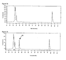
- a first test experiment the PDMS flowcell was not attached and the zone was directly illuminated for 1 second at 30 Hz with a 6 mW 337nm nitrogen laser via fibre optics (output ⁇ 50 ⁇ J/cm 2 ). Desorption was not observed and the sample zone was redissolved in 1 ⁇ L sterile water and subjected to PCR. It should be noted that the spores were pre-treated with chloros (sodium hypochlorite) prior to illumination to remove any extrasporal DNA.
- chloros sodium hypochlorite
- Electron micrographs of the illuminated zone confirmed spore damage.
Landscapes
- Health & Medical Sciences (AREA)
- Life Sciences & Earth Sciences (AREA)
- General Health & Medical Sciences (AREA)
- Veterinary Medicine (AREA)
- Animal Behavior & Ethology (AREA)
- Public Health (AREA)
- Engineering & Computer Science (AREA)
- Epidemiology (AREA)
- Wood Science & Technology (AREA)
- Organic Chemistry (AREA)
- Chemical & Material Sciences (AREA)
- Biotechnology (AREA)
- Bioinformatics & Cheminformatics (AREA)
- Zoology (AREA)
- Biomedical Technology (AREA)
- Sustainable Development (AREA)
- Microbiology (AREA)
- Molecular Biology (AREA)
- Biochemistry (AREA)
- General Engineering & Computer Science (AREA)
- Genetics & Genomics (AREA)
- Cell Biology (AREA)
- Measuring Or Testing Involving Enzymes Or Micro-Organisms (AREA)
- Micro-Organisms Or Cultivation Processes Thereof (AREA)
- Apparatus Associated With Microorganisms And Enzymes (AREA)
- Investigating Or Analyzing Materials By The Use Of Electric Means (AREA)
Claims (23)
- Verfahren zum Nachweis bakterieller Sporen in einer Probe, das die folgenden Schritte umfasst:Bestrahlen einer Probe mit Laserstrahlung bei in etwa Atmosphärendruck, um alle in der Probe vorhandenen Sporen aufzuschließen und dadurch aus den Sporen intrasporale DNA freizusetzen;Gewinnen des zerstörten Materials; undDurchführen eines Tests an der intrasporalen DNA, um das Bakterium zu identifizieren.
- Verfahren nach Anspruch 1, wobei die Laserstrahlung eine Energieabgabe über etwa 1 mJ/cm2 aufweist, um die Desorption des zerstörten Materials auszulösen.
- Verfahren nach Anspruch 1 oder 2, wobei das Verfahren einen Schritt umfasst, in dem die Probe in eine Auffangkammer gegeben wird, und die Probe in der Auffangkammer bestrahlt wird.
- Verfahren nach einem der Ansprüche 1 bis 3, wobei der Schritt der Bestrahlung der Probe umfasst, die Probe in einer solchen ausreichenden Weise zu bestrahlen, dass alle in der Probe enthaltenen Sporen zerstört werden und Material in die Auffangkammer abgegeben wird.
- Verfahren nach Anspruch 4, wobei das abgegebene Material in der Auffangkammer gesammelt wird.
- Verfahren nach einem der vorhergehenden Ansprüche, wobei der Schritt, das aufgeschlossene Material zu sammeln, einen Schritt umfasst, bei dem die Probe nach dem Bestrahlen der Probe mit einer Extraktionsflüssigkeit gespült wird.
- Verfahren nach einem der vorhergehenden Ansprüche, wobei das Verfahren den Schritt umfasst, die Probe vor der Bestrahlung in einem Matrixmaterial anzuordnen.
- Verfahren nach einem der vorhergehenden Ansprüche, wobei das Verfahren den anfänglichen Schritt umfasst, die Probe auf einem Substrat zu fixieren.
- Verfahren nach Anspruch 8, wobei der Schritt, die Probe auf einem Substrat zu fixieren, umfasst, die Probe in ein Lösemittel zu geben, das Lösemittel-Probe-Gemisch auf das Substrat aufzubringen und das Lösemittel zu verdampfen.
- Verfahren nach Anspruch 8 oder 9, wobei der Schritt, die Probe auf einem Substrat zu fixieren, den Schritt umfasst, die Probe in zumindest einem mikrostrukturierten Behälter auf dem Substrat zu fixieren.
- Verfahren nach Anspruch 10, wobei der Schritt, die Probe in zumindest einem mikrostrukturierten Behälter auf dem Substrat zu fixieren, die Probe in dem mikrostrukturierten Behälter anreichert.
- Verfahren nach Anspruch 10 oder 11, wobei der mikrostrukturierte Behälter ein Volumen von weniger als 100 nl oder 10 nl oder 1 nl oder 0,1 nl aufweist.
- Verfahren nach einem der Ansprüche 10 bis 12, wobei das Substrat eine Vielzahl von mikrostrukturierten Behälter umfasst und das Verfahren einen Schritt beinhaltet, eine Probe in jeden mikrostrukturierten Behälter zu geben.
- Verfahren nach Anspruch 13, wobei das Verfahren umfasst, jeden mikrostrukturierte Behälter in eine getrennte Auffangkammer zu geben und jede Probe in einem mikrostrukturierten Behälter gesondert zu bestrahlen.
- Verfahren nach einem der Ansprüche 8 bis 14, wobei, wenn abhängig von Anspruch 3, der Schritt die Probe in eine Auffangkammer zu geben, den Schritt umfasst, eine Sammelplatte in geeigneter Weise an dem Substrat zu fixieren.
- Verfahren nach Anspruch 15, wobei in der Sammelplatte ein Mikrokanal ausgebildet ist und der Mikrokanal zumindest einen Teil der Auffangkammer bildet.
- Verfahren nach Anspruch 16, wobei der Mikrokanal so konfiguriert ist, dass er als Teil eines Mikrofluidsystems verwendet werden kann.
- Verfahren nach einem der vorhergehenden Ansprüche, wobei, wenn abhängig von Anspruch 3, die Auffangkammer zumindest zum Teil mit einem Extraktionsfluid gefüllt ist, um das gesamte aufgeschlossene Material zu sammeln.
- Verfahren nach einem der vorhergehenden Ansprüche, wobei der Test zur Identifizierung des Bakteriums einen PCR-basierten Assay umfasst.
- Verfahren nach Anspruch 19, wobei, wenn abhängig von einem der Ansprüche 15 bis 17, der Schritt, einen Test an dem gesammelten Material durchzuführen, umfasst, die Sammelplatte von dem Substrat zu entfernen, sie in einen Mikrofluidkreislauf einzubringen und den Test in einem Mikrofluidkreislauf durchzuführen.
- Verfahren nach einem der vorhergehenden Ansprüche, wobei der Schritt, in dem die Probe bestrahlt wird, eine Sekunde oder weniger dauert.
- Verfahren nach einem der vorhergehenden Ansprüche, wobei die zur Bestrahlung verwendete Strahlung eine Wellenlänge im ultravioletten Bereich aufweist.
- Verfahren nach einem der vorhergehenden Ansprüche, wobei die zur Bestrahlung verwendete Strahlung gepulst ist, wobei jeder Puls eine Dauer von 1 bis 100 ns hat.
Applications Claiming Priority (2)
| Application Number | Priority Date | Filing Date | Title |
|---|---|---|---|
| GBGB0313170.3A GB0313170D0 (en) | 2003-06-09 | 2003-06-09 | Method and apparatus for spore disruption and/or detection |
| PCT/GB2004/002472 WO2004108959A2 (en) | 2003-06-09 | 2004-06-09 | Method and apparatus for spore disruption and/or detection |
Publications (2)
| Publication Number | Publication Date |
|---|---|
| EP1631684A2 EP1631684A2 (de) | 2006-03-08 |
| EP1631684B1 true EP1631684B1 (de) | 2012-03-07 |
Family
ID=27589664
Family Applications (1)
| Application Number | Title | Priority Date | Filing Date |
|---|---|---|---|
| EP04736409A Expired - Lifetime EP1631684B1 (de) | 2003-06-09 | 2004-06-09 | Verfahren und vorrichtung zum sporenaufschluss und/oder -nachweis |
Country Status (5)
| Country | Link |
|---|---|
| US (1) | US8338092B2 (de) |
| EP (1) | EP1631684B1 (de) |
| AT (1) | ATE548053T1 (de) |
| GB (1) | GB0313170D0 (de) |
| WO (1) | WO2004108959A2 (de) |
Families Citing this family (34)
| Publication number | Priority date | Publication date | Assignee | Title |
|---|---|---|---|---|
| US7998418B1 (en) * | 2006-06-01 | 2011-08-16 | Nanotek, Llc | Evaporator and concentrator in reactor and loading system |
| US10400280B2 (en) | 2012-08-14 | 2019-09-03 | 10X Genomics, Inc. | Methods and systems for processing polynucleotides |
| IN2015DN01126A (de) | 2012-08-14 | 2015-06-26 | 10X Genomics Inc | |
| US10273541B2 (en) | 2012-08-14 | 2019-04-30 | 10X Genomics, Inc. | Methods and systems for processing polynucleotides |
| US10323279B2 (en) | 2012-08-14 | 2019-06-18 | 10X Genomics, Inc. | Methods and systems for processing polynucleotides |
| US11591637B2 (en) | 2012-08-14 | 2023-02-28 | 10X Genomics, Inc. | Compositions and methods for sample processing |
| US9701998B2 (en) | 2012-12-14 | 2017-07-11 | 10X Genomics, Inc. | Methods and systems for processing polynucleotides |
| US9951386B2 (en) | 2014-06-26 | 2018-04-24 | 10X Genomics, Inc. | Methods and systems for processing polynucleotides |
| US10752949B2 (en) | 2012-08-14 | 2020-08-25 | 10X Genomics, Inc. | Methods and systems for processing polynucleotides |
| EP3567116A1 (de) | 2012-12-14 | 2019-11-13 | 10X Genomics, Inc. | Verfahren und systeme zur verarbeitung von polynukleotiden |
| US10533221B2 (en) | 2012-12-14 | 2020-01-14 | 10X Genomics, Inc. | Methods and systems for processing polynucleotides |
| EP2954104B1 (de) | 2013-02-08 | 2020-09-16 | 10X Genomics, Inc. | Polynukleotid-strichcodegenerierung |
| CN114534806B (zh) | 2014-04-10 | 2024-03-29 | 10X基因组学有限公司 | 用于封装和分割试剂的流体装置、系统和方法及其应用 |
| EP4053292A1 (de) | 2014-06-26 | 2022-09-07 | 10X Genomics, Inc. | Verfahren zur analyse von nukleinsäuren aus einzelzellen oder zellpopulationen |
| US12312640B2 (en) | 2014-06-26 | 2025-05-27 | 10X Genomics, Inc. | Analysis of nucleic acid sequences |
| JP2017522866A (ja) | 2014-06-26 | 2017-08-17 | 10エックス ジェノミクス, インコーポレイテッド | 核酸配列の分析 |
| KR102002451B1 (ko) | 2014-09-18 | 2019-07-23 | 제넥스 디스인펙션 서비시즈 인코퍼레이티드 | 조절된 파워 플럭스를 갖는 펄스화된 광을 사용하는 룸 및 구역 살균 장치 및 펄스 간의 가시 광 보상을 갖는 광 시스템 |
| JP6769969B2 (ja) | 2015-01-12 | 2020-10-14 | 10エックス ジェノミクス, インコーポレイテッド | 核酸配列決定ライブラリーを作製するためのプロセス及びシステム、並びにこれらを使用して作製したライブラリー |
| US10697000B2 (en) | 2015-02-24 | 2020-06-30 | 10X Genomics, Inc. | Partition processing methods and systems |
| US9850479B2 (en) * | 2015-03-03 | 2017-12-26 | The Board Of Regents Of The University Of Oklahoma | Method and apparatus for sampling macromolecules from a biological specimen |
| CN110268108B (zh) | 2016-12-12 | 2022-09-06 | 埃克切拉生物科学公司 | 用于使用微毛细管阵列进行筛选的方法和系统 |
| US11085039B2 (en) | 2016-12-12 | 2021-08-10 | xCella Biosciences, Inc. | Methods and systems for screening using microcapillary arrays |
| US10550429B2 (en) | 2016-12-22 | 2020-02-04 | 10X Genomics, Inc. | Methods and systems for processing polynucleotides |
| US10815525B2 (en) | 2016-12-22 | 2020-10-27 | 10X Genomics, Inc. | Methods and systems for processing polynucleotides |
| WO2018125832A1 (en) | 2016-12-30 | 2018-07-05 | xCella Biosciences, Inc. | Multi-stage sample recovery system |
| US12264411B2 (en) | 2017-01-30 | 2025-04-01 | 10X Genomics, Inc. | Methods and systems for analysis |
| CN117512066A (zh) | 2017-01-30 | 2024-02-06 | 10X基因组学有限公司 | 用于基于微滴的单细胞条形编码的方法和系统 |
| US10844372B2 (en) | 2017-05-26 | 2020-11-24 | 10X Genomics, Inc. | Single cell analysis of transposase accessible chromatin |
| EP3445876B1 (de) | 2017-05-26 | 2023-07-05 | 10X Genomics, Inc. | Einzelzellenanalyse von transposasezugänglichem chromatin |
| EP3625361A1 (de) | 2017-11-15 | 2020-03-25 | 10X Genomics, Inc. | Funktionalisierte gelperlen |
| US10829815B2 (en) | 2017-11-17 | 2020-11-10 | 10X Genomics, Inc. | Methods and systems for associating physical and genetic properties of biological particles |
| CN112262218B (zh) | 2018-04-06 | 2024-11-08 | 10X基因组学有限公司 | 用于单细胞处理中的质量控制的系统和方法 |
| WO2020118106A1 (en) | 2018-12-06 | 2020-06-11 | xCella Biosciences, Inc. | Lateral loading of microcapillary arrays |
| CN111024768B (zh) * | 2019-11-25 | 2025-04-18 | 中国计量大学 | 一种微流控阻抗式生物在线检测装置 |
Family Cites Families (3)
| Publication number | Priority date | Publication date | Assignee | Title |
|---|---|---|---|---|
| US3941670A (en) * | 1970-11-12 | 1976-03-02 | Massachusetts Institute Of Technology | Method of altering biological and chemical activity of molecular species |
| US5777324A (en) | 1996-09-19 | 1998-07-07 | Sequenom, Inc. | Method and apparatus for maldi analysis |
| GB0120131D0 (en) * | 2001-08-17 | 2001-10-10 | Micromass Ltd | Maldi target plate |
-
2003
- 2003-06-09 GB GBGB0313170.3A patent/GB0313170D0/en not_active Ceased
-
2004
- 2004-06-09 EP EP04736409A patent/EP1631684B1/de not_active Expired - Lifetime
- 2004-06-09 US US10/558,966 patent/US8338092B2/en not_active Expired - Fee Related
- 2004-06-09 AT AT04736409T patent/ATE548053T1/de active
- 2004-06-09 WO PCT/GB2004/002472 patent/WO2004108959A2/en not_active Ceased
Also Published As
| Publication number | Publication date |
|---|---|
| US20070026401A1 (en) | 2007-02-01 |
| WO2004108959A3 (en) | 2005-03-24 |
| GB0313170D0 (en) | 2003-07-16 |
| EP1631684A2 (de) | 2006-03-08 |
| US8338092B2 (en) | 2012-12-25 |
| ATE548053T1 (de) | 2012-03-15 |
| WO2004108959A2 (en) | 2004-12-16 |
Similar Documents
| Publication | Publication Date | Title |
|---|---|---|
| EP1631684B1 (de) | Verfahren und vorrichtung zum sporenaufschluss und/oder -nachweis | |
| CA2788852C (en) | Soft ablative desorption method and system | |
| US10847357B2 (en) | Structured biological samples for analysis by mass cytometry | |
| JP3601834B2 (ja) | Maldi分析の方法および装置 | |
| US10596570B2 (en) | Apparatus, system and method for performing automated centrifugal separation | |
| US9920352B2 (en) | Acoustic radiation for ejecting and monitoring pathogenic fluids | |
| CN101291736A (zh) | 用于流体的可编程微量操作的剂量计 | |
| US6451616B1 (en) | Analysis of molecules bound to solid surfaces using selective bond cleavage processes | |
| EP1836698B1 (de) | Generation von pulsierenden druckwellen, z.b. für zellyse | |
| Hofmann et al. | Laser induced disruption of bacterial spores on a microchip | |
| JP4378649B2 (ja) | 生体高分子の非共有結合等を選択的に切断して分析する方法および装置 | |
| US9850479B2 (en) | Method and apparatus for sampling macromolecules from a biological specimen | |
| WO2005074003A1 (ja) | レーザ分析装置及び方法 | |
| US20110049354A1 (en) | Method and device for detecting at least one target substance | |
| US20230398535A1 (en) | Sample analysis cartridge and system | |
| US8974731B2 (en) | Sample carrier and/or sample carrier processing apparatus | |
| US20220072550A1 (en) | Cell marking systems | |
| HK40119682A (en) | Structured biological samples for analysis by mass cytometry | |
| JP2007303840A (ja) | 液滴イオン化法、質量分析法及びそれらの装置 | |
| Huang | Laser-induced fluorescence spectroscopy and collection into liquid solution of aerosol particles |
Legal Events
| Date | Code | Title | Description |
|---|---|---|---|
| PUAI | Public reference made under article 153(3) epc to a published international application that has entered the european phase |
Free format text: ORIGINAL CODE: 0009012 |
|
| 17P | Request for examination filed |
Effective date: 20051128 |
|
| AK | Designated contracting states |
Kind code of ref document: A2 Designated state(s): AT BE BG CH CY CZ DE DK EE ES FI FR GB GR HU IE IT LI LU MC NL PL PT RO SE SI SK TR |
|
| DAX | Request for extension of the european patent (deleted) | ||
| 17Q | First examination report despatched |
Effective date: 20090622 |
|
| RAP1 | Party data changed (applicant data changed or rights of an application transferred) |
Owner name: QINETIQ LIMITED |
|
| REG | Reference to a national code |
Ref country code: DE Ref legal event code: R079 Ref document number: 602004036814 Country of ref document: DE Free format text: PREVIOUS MAIN CLASS: C12Q0001680000 Ipc: A61L0002100000 |
|
| RIC1 | Information provided on ipc code assigned before grant |
Ipc: C12Q 1/68 20060101ALI20110823BHEP Ipc: H01J 49/16 20060101ALI20110823BHEP Ipc: A61L 2/10 20060101AFI20110823BHEP Ipc: A61L 2/08 20060101ALI20110823BHEP Ipc: A61L 2/00 20060101ALI20110823BHEP Ipc: C12M 1/33 20060101ALI20110823BHEP |
|
| GRAP | Despatch of communication of intention to grant a patent |
Free format text: ORIGINAL CODE: EPIDOSNIGR1 |
|
| GRAS | Grant fee paid |
Free format text: ORIGINAL CODE: EPIDOSNIGR3 |
|
| GRAA | (expected) grant |
Free format text: ORIGINAL CODE: 0009210 |
|
| AK | Designated contracting states |
Kind code of ref document: B1 Designated state(s): AT BE BG CH CY CZ DE DK EE ES FI FR GB GR HU IE IT LI LU MC NL PL PT RO SE SI SK TR |
|
| REG | Reference to a national code |
Ref country code: GB Ref legal event code: FG4D |
|
| REG | Reference to a national code |
Ref country code: CH Ref legal event code: EP Ref country code: AT Ref legal event code: REF Ref document number: 548053 Country of ref document: AT Kind code of ref document: T Effective date: 20120315 |
|
| REG | Reference to a national code |
Ref country code: IE Ref legal event code: FG4D |
|
| REG | Reference to a national code |
Ref country code: DE Ref legal event code: R096 Ref document number: 602004036814 Country of ref document: DE Effective date: 20120503 |
|
| REG | Reference to a national code |
Ref country code: NL Ref legal event code: VDEP Effective date: 20120307 |
|
| PG25 | Lapsed in a contracting state [announced via postgrant information from national office to epo] |
Ref country code: NL Free format text: LAPSE BECAUSE OF FAILURE TO SUBMIT A TRANSLATION OF THE DESCRIPTION OR TO PAY THE FEE WITHIN THE PRESCRIBED TIME-LIMIT Effective date: 20120307 |
|
| PGFP | Annual fee paid to national office [announced via postgrant information from national office to epo] |
Ref country code: DE Payment date: 20120622 Year of fee payment: 9 |
|
| PG25 | Lapsed in a contracting state [announced via postgrant information from national office to epo] |
Ref country code: FI Free format text: LAPSE BECAUSE OF FAILURE TO SUBMIT A TRANSLATION OF THE DESCRIPTION OR TO PAY THE FEE WITHIN THE PRESCRIBED TIME-LIMIT Effective date: 20120307 Ref country code: GR Free format text: LAPSE BECAUSE OF FAILURE TO SUBMIT A TRANSLATION OF THE DESCRIPTION OR TO PAY THE FEE WITHIN THE PRESCRIBED TIME-LIMIT Effective date: 20120608 |
|
| PGFP | Annual fee paid to national office [announced via postgrant information from national office to epo] |
Ref country code: FR Payment date: 20120705 Year of fee payment: 9 Ref country code: GB Payment date: 20120622 Year of fee payment: 9 |
|
| REG | Reference to a national code |
Ref country code: AT Ref legal event code: MK05 Ref document number: 548053 Country of ref document: AT Kind code of ref document: T Effective date: 20120307 |
|
| PG25 | Lapsed in a contracting state [announced via postgrant information from national office to epo] |
Ref country code: CY Free format text: LAPSE BECAUSE OF FAILURE TO SUBMIT A TRANSLATION OF THE DESCRIPTION OR TO PAY THE FEE WITHIN THE PRESCRIBED TIME-LIMIT Effective date: 20120307 |
|
| PG25 | Lapsed in a contracting state [announced via postgrant information from national office to epo] |
Ref country code: SI Free format text: LAPSE BECAUSE OF FAILURE TO SUBMIT A TRANSLATION OF THE DESCRIPTION OR TO PAY THE FEE WITHIN THE PRESCRIBED TIME-LIMIT Effective date: 20120307 Ref country code: CZ Free format text: LAPSE BECAUSE OF FAILURE TO SUBMIT A TRANSLATION OF THE DESCRIPTION OR TO PAY THE FEE WITHIN THE PRESCRIBED TIME-LIMIT Effective date: 20120307 Ref country code: BE Free format text: LAPSE BECAUSE OF FAILURE TO SUBMIT A TRANSLATION OF THE DESCRIPTION OR TO PAY THE FEE WITHIN THE PRESCRIBED TIME-LIMIT Effective date: 20120307 Ref country code: PL Free format text: LAPSE BECAUSE OF FAILURE TO SUBMIT A TRANSLATION OF THE DESCRIPTION OR TO PAY THE FEE WITHIN THE PRESCRIBED TIME-LIMIT Effective date: 20120307 Ref country code: EE Free format text: LAPSE BECAUSE OF FAILURE TO SUBMIT A TRANSLATION OF THE DESCRIPTION OR TO PAY THE FEE WITHIN THE PRESCRIBED TIME-LIMIT Effective date: 20120307 Ref country code: RO Free format text: LAPSE BECAUSE OF FAILURE TO SUBMIT A TRANSLATION OF THE DESCRIPTION OR TO PAY THE FEE WITHIN THE PRESCRIBED TIME-LIMIT Effective date: 20120307 Ref country code: SE Free format text: LAPSE BECAUSE OF FAILURE TO SUBMIT A TRANSLATION OF THE DESCRIPTION OR TO PAY THE FEE WITHIN THE PRESCRIBED TIME-LIMIT Effective date: 20120307 |
|
| PG25 | Lapsed in a contracting state [announced via postgrant information from national office to epo] |
Ref country code: SK Free format text: LAPSE BECAUSE OF FAILURE TO SUBMIT A TRANSLATION OF THE DESCRIPTION OR TO PAY THE FEE WITHIN THE PRESCRIBED TIME-LIMIT Effective date: 20120307 Ref country code: PT Free format text: LAPSE BECAUSE OF FAILURE TO SUBMIT A TRANSLATION OF THE DESCRIPTION OR TO PAY THE FEE WITHIN THE PRESCRIBED TIME-LIMIT Effective date: 20120709 |
|
| PLBE | No opposition filed within time limit |
Free format text: ORIGINAL CODE: 0009261 |
|
| STAA | Information on the status of an ep patent application or granted ep patent |
Free format text: STATUS: NO OPPOSITION FILED WITHIN TIME LIMIT |
|
| PG25 | Lapsed in a contracting state [announced via postgrant information from national office to epo] |
Ref country code: AT Free format text: LAPSE BECAUSE OF FAILURE TO SUBMIT A TRANSLATION OF THE DESCRIPTION OR TO PAY THE FEE WITHIN THE PRESCRIBED TIME-LIMIT Effective date: 20120307 Ref country code: DK Free format text: LAPSE BECAUSE OF FAILURE TO SUBMIT A TRANSLATION OF THE DESCRIPTION OR TO PAY THE FEE WITHIN THE PRESCRIBED TIME-LIMIT Effective date: 20120307 Ref country code: MC Free format text: LAPSE BECAUSE OF NON-PAYMENT OF DUE FEES Effective date: 20120630 |
|
| REG | Reference to a national code |
Ref country code: CH Ref legal event code: PL |
|
| 26N | No opposition filed |
Effective date: 20121210 |
|
| REG | Reference to a national code |
Ref country code: CH Ref legal event code: PL |
|
| PG25 | Lapsed in a contracting state [announced via postgrant information from national office to epo] |
Ref country code: IT Free format text: LAPSE BECAUSE OF FAILURE TO SUBMIT A TRANSLATION OF THE DESCRIPTION OR TO PAY THE FEE WITHIN THE PRESCRIBED TIME-LIMIT Effective date: 20120307 |
|
| REG | Reference to a national code |
Ref country code: IE Ref legal event code: MM4A |
|
| REG | Reference to a national code |
Ref country code: DE Ref legal event code: R097 Ref document number: 602004036814 Country of ref document: DE Effective date: 20121210 |
|
| PG25 | Lapsed in a contracting state [announced via postgrant information from national office to epo] |
Ref country code: IE Free format text: LAPSE BECAUSE OF NON-PAYMENT OF DUE FEES Effective date: 20120609 Ref country code: ES Free format text: LAPSE BECAUSE OF FAILURE TO SUBMIT A TRANSLATION OF THE DESCRIPTION OR TO PAY THE FEE WITHIN THE PRESCRIBED TIME-LIMIT Effective date: 20120618 Ref country code: CH Free format text: LAPSE BECAUSE OF NON-PAYMENT OF DUE FEES Effective date: 20120630 Ref country code: LI Free format text: LAPSE BECAUSE OF NON-PAYMENT OF DUE FEES Effective date: 20120630 |
|
| PG25 | Lapsed in a contracting state [announced via postgrant information from national office to epo] |
Ref country code: BG Free format text: LAPSE BECAUSE OF FAILURE TO SUBMIT A TRANSLATION OF THE DESCRIPTION OR TO PAY THE FEE WITHIN THE PRESCRIBED TIME-LIMIT Effective date: 20120607 |
|
| GBPC | Gb: european patent ceased through non-payment of renewal fee |
Effective date: 20130609 |
|
| REG | Reference to a national code |
Ref country code: DE Ref legal event code: R119 Ref document number: 602004036814 Country of ref document: DE Effective date: 20140101 |
|
| REG | Reference to a national code |
Ref country code: FR Ref legal event code: ST Effective date: 20140228 |
|
| PG25 | Lapsed in a contracting state [announced via postgrant information from national office to epo] |
Ref country code: TR Free format text: LAPSE BECAUSE OF FAILURE TO SUBMIT A TRANSLATION OF THE DESCRIPTION OR TO PAY THE FEE WITHIN THE PRESCRIBED TIME-LIMIT Effective date: 20120307 Ref country code: GB Free format text: LAPSE BECAUSE OF NON-PAYMENT OF DUE FEES Effective date: 20130609 Ref country code: DE Free format text: LAPSE BECAUSE OF NON-PAYMENT OF DUE FEES Effective date: 20140101 |
|
| PG25 | Lapsed in a contracting state [announced via postgrant information from national office to epo] |
Ref country code: FR Free format text: LAPSE BECAUSE OF NON-PAYMENT OF DUE FEES Effective date: 20130701 Ref country code: LU Free format text: LAPSE BECAUSE OF NON-PAYMENT OF DUE FEES Effective date: 20120609 |
|
| PG25 | Lapsed in a contracting state [announced via postgrant information from national office to epo] |
Ref country code: HU Free format text: LAPSE BECAUSE OF FAILURE TO SUBMIT A TRANSLATION OF THE DESCRIPTION OR TO PAY THE FEE WITHIN THE PRESCRIBED TIME-LIMIT Effective date: 20040609 |