EP1601767B1 - USE OF siRNA TO SUPPRESS EXPRESSION OF EIF-5A1 IN THE TREATMENT OF GLAUCOMA - Google Patents
USE OF siRNA TO SUPPRESS EXPRESSION OF EIF-5A1 IN THE TREATMENT OF GLAUCOMA Download PDFInfo
- Publication number
- EP1601767B1 EP1601767B1 EP04717933A EP04717933A EP1601767B1 EP 1601767 B1 EP1601767 B1 EP 1601767B1 EP 04717933 A EP04717933 A EP 04717933A EP 04717933 A EP04717933 A EP 04717933A EP 1601767 B1 EP1601767 B1 EP 1601767B1
- Authority
- EP
- European Patent Office
- Prior art keywords
- cells
- apoptosis
- eif
- sirna
- transfected
- Prior art date
- Legal status (The legal status is an assumption and is not a legal conclusion. Google has not performed a legal analysis and makes no representation as to the accuracy of the status listed.)
- Expired - Lifetime
Links
- 108020004459 Small interfering RNA Proteins 0.000 title claims abstract description 10
- 208000010412 Glaucoma Diseases 0.000 title claims description 19
- 230000014509 gene expression Effects 0.000 title abstract description 115
- 238000011282 treatment Methods 0.000 title description 59
- 101150061075 EIF5A1 gene Proteins 0.000 title 1
- 102000016614 Autophagy-Related Protein 5 Human genes 0.000 claims abstract description 82
- 108010092776 Autophagy-Related Protein 5 Proteins 0.000 claims abstract description 82
- 238000000034 method Methods 0.000 claims abstract description 59
- 102100026761 Eukaryotic translation initiation factor 5A-1 Human genes 0.000 claims description 7
- 239000008194 pharmaceutical composition Substances 0.000 claims description 2
- 101710126270 Eukaryotic translation initiation factor 5A-1 Proteins 0.000 claims 2
- 230000006907 apoptotic process Effects 0.000 abstract description 294
- 108010074105 Factor Va Proteins 0.000 abstract description 73
- 239000002773 nucleotide Substances 0.000 abstract description 54
- 125000003729 nucleotide group Chemical group 0.000 abstract description 54
- 102000004127 Cytokines Human genes 0.000 abstract description 41
- 108090000695 Cytokines Proteins 0.000 abstract description 41
- 230000000692 anti-sense effect Effects 0.000 abstract description 41
- 108090000765 processed proteins & peptides Proteins 0.000 abstract description 23
- 150000007523 nucleic acids Chemical class 0.000 abstract description 18
- 102000004196 processed proteins & peptides Human genes 0.000 abstract description 18
- 230000002401 inhibitory effect Effects 0.000 abstract description 17
- 229920001184 polypeptide Polymers 0.000 abstract description 17
- 102000039446 nucleic acids Human genes 0.000 abstract description 16
- 108020004707 nucleic acids Proteins 0.000 abstract description 16
- 230000000770 proinflammatory effect Effects 0.000 abstract description 7
- 108010044843 Peptide Initiation Factors Proteins 0.000 abstract description 4
- 102000005877 Peptide Initiation Factors Human genes 0.000 abstract description 4
- 210000004027 cell Anatomy 0.000 description 602
- 108090000623 proteins and genes Proteins 0.000 description 106
- 241000700159 Rattus Species 0.000 description 102
- 108060008682 Tumor Necrosis Factor Proteins 0.000 description 98
- 102000000852 Tumor Necrosis Factor-alpha Human genes 0.000 description 98
- 239000000074 antisense oligonucleotide Substances 0.000 description 98
- 238000012230 antisense oligonucleotides Methods 0.000 description 98
- MZOFCQQQCNRIBI-VMXHOPILSA-N (3s)-4-[[(2s)-1-[[(2s)-1-[[(1s)-1-carboxy-2-hydroxyethyl]amino]-4-methyl-1-oxopentan-2-yl]amino]-5-(diaminomethylideneamino)-1-oxopentan-2-yl]amino]-3-[[2-[[(2s)-2,6-diaminohexanoyl]amino]acetyl]amino]-4-oxobutanoic acid Chemical compound OC[C@@H](C(O)=O)NC(=O)[C@H](CC(C)C)NC(=O)[C@H](CCCN=C(N)N)NC(=O)[C@H](CC(O)=O)NC(=O)CNC(=O)[C@@H](N)CCCCN MZOFCQQQCNRIBI-VMXHOPILSA-N 0.000 description 96
- 239000012528 membrane Substances 0.000 description 86
- 102000004169 proteins and genes Human genes 0.000 description 83
- 108091034117 Oligonucleotide Proteins 0.000 description 80
- 235000018102 proteins Nutrition 0.000 description 79
- 238000001890 transfection Methods 0.000 description 65
- KLWPJMFMVPTNCC-UHFFFAOYSA-N Camptothecin Natural products CCC1(O)C(=O)OCC2=C1C=C3C4Nc5ccccc5C=C4CN3C2=O KLWPJMFMVPTNCC-UHFFFAOYSA-N 0.000 description 63
- VSJKWCGYPAHWDS-FQEVSTJZSA-N camptothecin Chemical compound C1=CC=C2C=C(CN3C4=CC5=C(C3=O)COC(=O)[C@]5(O)CC)C4=NC2=C1 VSJKWCGYPAHWDS-FQEVSTJZSA-N 0.000 description 63
- 229940127093 camptothecin Drugs 0.000 description 63
- VSJKWCGYPAHWDS-UHFFFAOYSA-N dl-camptothecin Natural products C1=CC=C2C=C(CN3C4=CC5=C(C3=O)COC(=O)C5(O)CC)C4=NC2=C1 VSJKWCGYPAHWDS-UHFFFAOYSA-N 0.000 description 63
- 239000002953 phosphate buffered saline Substances 0.000 description 59
- 230000001640 apoptogenic effect Effects 0.000 description 52
- 238000002474 experimental method Methods 0.000 description 46
- 238000001262 western blot Methods 0.000 description 46
- 108020000948 Antisense Oligonucleotides Proteins 0.000 description 42
- 102100025064 Cellular tumor antigen p53 Human genes 0.000 description 37
- 102000048231 Deoxyhypusine synthases Human genes 0.000 description 37
- 108700023218 Deoxyhypusine synthases Proteins 0.000 description 37
- 101000721661 Homo sapiens Cellular tumor antigen p53 Proteins 0.000 description 37
- 108091003079 Bovine Serum Albumin Proteins 0.000 description 36
- 210000004246 corpus luteum Anatomy 0.000 description 36
- 210000004940 nucleus Anatomy 0.000 description 36
- 230000001965 increasing effect Effects 0.000 description 35
- 108091032973 (ribonucleotides)n+m Proteins 0.000 description 34
- 239000002299 complementary DNA Substances 0.000 description 34
- 239000000243 solution Substances 0.000 description 32
- 239000000203 mixture Substances 0.000 description 31
- RJURFGZVJUQBHK-UHFFFAOYSA-N actinomycin D Natural products CC1OC(=O)C(C(C)C)N(C)C(=O)CN(C)C(=O)C2CCCN2C(=O)C(C(C)C)NC(=O)C1NC(=O)C1=C(N)C(=O)C(C)=C2OC(C(C)=CC=C3C(=O)NC4C(=O)NC(C(N5CCCC5C(=O)N(C)CC(=O)N(C)C(C(C)C)C(=O)OC4C)=O)C(C)C)=C3N=C21 RJURFGZVJUQBHK-UHFFFAOYSA-N 0.000 description 30
- 239000000523 sample Substances 0.000 description 30
- FAPWRFPIFSIZLT-UHFFFAOYSA-M Sodium chloride Chemical compound [Na+].[Cl-] FAPWRFPIFSIZLT-UHFFFAOYSA-M 0.000 description 29
- PXGPLTODNUVGFL-BRIYLRKRSA-N (E,Z)-(1R,2R,3R,5S)-7-(3,5-Dihydroxy-2-((3S)-(3-hydroxy-1-octenyl))cyclopentyl)-5-heptenoic acid Chemical compound CCCCC[C@H](O)C=C[C@H]1[C@H](O)C[C@H](O)[C@@H]1CC=CCCCC(O)=O PXGPLTODNUVGFL-BRIYLRKRSA-N 0.000 description 28
- 206010028980 Neoplasm Diseases 0.000 description 28
- ATHGHQPFGPMSJY-UHFFFAOYSA-N spermidine Chemical compound NCCCCNCCCN ATHGHQPFGPMSJY-UHFFFAOYSA-N 0.000 description 28
- 230000000875 corresponding effect Effects 0.000 description 27
- 238000013467 fragmentation Methods 0.000 description 26
- 238000006062 fragmentation reaction Methods 0.000 description 26
- 101000971171 Homo sapiens Apoptosis regulator Bcl-2 Proteins 0.000 description 25
- 238000001514 detection method Methods 0.000 description 25
- 210000005003 heart tissue Anatomy 0.000 description 25
- 238000011534 incubation Methods 0.000 description 25
- 239000007924 injection Substances 0.000 description 25
- 238000002347 injection Methods 0.000 description 25
- 108020004999 messenger RNA Proteins 0.000 description 25
- 210000002966 serum Anatomy 0.000 description 25
- 210000001519 tissue Anatomy 0.000 description 25
- 102100021569 Apoptosis regulator Bcl-2 Human genes 0.000 description 24
- 239000013615 primer Substances 0.000 description 24
- 230000004044 response Effects 0.000 description 24
- 101001054354 Homo sapiens Eukaryotic translation initiation factor 5A-1 Proteins 0.000 description 23
- 239000012634 fragment Substances 0.000 description 23
- 125000003275 alpha amino acid group Chemical group 0.000 description 22
- 239000012091 fetal bovine serum Substances 0.000 description 22
- LFQSCWFLJHTTHZ-UHFFFAOYSA-N Ethanol Chemical compound CCO LFQSCWFLJHTTHZ-UHFFFAOYSA-N 0.000 description 21
- 238000010240 RT-PCR analysis Methods 0.000 description 21
- 239000000834 fixative Substances 0.000 description 21
- 239000006144 Dulbecco’s modified Eagle's medium Substances 0.000 description 20
- 230000000694 effects Effects 0.000 description 20
- 108010085279 eukaryotic translation initiation factor 5A Proteins 0.000 description 20
- 238000002372 labelling Methods 0.000 description 19
- QKNYBSVHEMOAJP-UHFFFAOYSA-N 2-amino-2-(hydroxymethyl)propane-1,3-diol;hydron;chloride Chemical compound Cl.OCC(N)(CO)CO QKNYBSVHEMOAJP-UHFFFAOYSA-N 0.000 description 18
- 108090000171 Interleukin-18 Proteins 0.000 description 18
- 102000003810 Interleukin-18 Human genes 0.000 description 18
- OKKJLVBELUTLKV-UHFFFAOYSA-N Methanol Chemical compound OC OKKJLVBELUTLKV-UHFFFAOYSA-N 0.000 description 18
- 238000010790 dilution Methods 0.000 description 18
- 239000012895 dilution Substances 0.000 description 18
- 230000006882 induction of apoptosis Effects 0.000 description 18
- JLCPHMBAVCMARE-UHFFFAOYSA-N [3-[[3-[[3-[[3-[[3-[[3-[[3-[[3-[[3-[[3-[[3-[[5-(2-amino-6-oxo-1H-purin-9-yl)-3-[[3-[[3-[[3-[[3-[[3-[[5-(2-amino-6-oxo-1H-purin-9-yl)-3-[[5-(2-amino-6-oxo-1H-purin-9-yl)-3-hydroxyoxolan-2-yl]methoxy-hydroxyphosphoryl]oxyoxolan-2-yl]methoxy-hydroxyphosphoryl]oxy-5-(5-methyl-2,4-dioxopyrimidin-1-yl)oxolan-2-yl]methoxy-hydroxyphosphoryl]oxy-5-(6-aminopurin-9-yl)oxolan-2-yl]methoxy-hydroxyphosphoryl]oxy-5-(6-aminopurin-9-yl)oxolan-2-yl]methoxy-hydroxyphosphoryl]oxy-5-(6-aminopurin-9-yl)oxolan-2-yl]methoxy-hydroxyphosphoryl]oxy-5-(6-aminopurin-9-yl)oxolan-2-yl]methoxy-hydroxyphosphoryl]oxyoxolan-2-yl]methoxy-hydroxyphosphoryl]oxy-5-(5-methyl-2,4-dioxopyrimidin-1-yl)oxolan-2-yl]methoxy-hydroxyphosphoryl]oxy-5-(4-amino-2-oxopyrimidin-1-yl)oxolan-2-yl]methoxy-hydroxyphosphoryl]oxy-5-(5-methyl-2,4-dioxopyrimidin-1-yl)oxolan-2-yl]methoxy-hydroxyphosphoryl]oxy-5-(5-methyl-2,4-dioxopyrimidin-1-yl)oxolan-2-yl]methoxy-hydroxyphosphoryl]oxy-5-(6-aminopurin-9-yl)oxolan-2-yl]methoxy-hydroxyphosphoryl]oxy-5-(6-aminopurin-9-yl)oxolan-2-yl]methoxy-hydroxyphosphoryl]oxy-5-(4-amino-2-oxopyrimidin-1-yl)oxolan-2-yl]methoxy-hydroxyphosphoryl]oxy-5-(4-amino-2-oxopyrimidin-1-yl)oxolan-2-yl]methoxy-hydroxyphosphoryl]oxy-5-(4-amino-2-oxopyrimidin-1-yl)oxolan-2-yl]methoxy-hydroxyphosphoryl]oxy-5-(6-aminopurin-9-yl)oxolan-2-yl]methoxy-hydroxyphosphoryl]oxy-5-(4-amino-2-oxopyrimidin-1-yl)oxolan-2-yl]methyl [5-(6-aminopurin-9-yl)-2-(hydroxymethyl)oxolan-3-yl] hydrogen phosphate Polymers Cc1cn(C2CC(OP(O)(=O)OCC3OC(CC3OP(O)(=O)OCC3OC(CC3O)n3cnc4c3nc(N)[nH]c4=O)n3cnc4c3nc(N)[nH]c4=O)C(COP(O)(=O)OC3CC(OC3COP(O)(=O)OC3CC(OC3COP(O)(=O)OC3CC(OC3COP(O)(=O)OC3CC(OC3COP(O)(=O)OC3CC(OC3COP(O)(=O)OC3CC(OC3COP(O)(=O)OC3CC(OC3COP(O)(=O)OC3CC(OC3COP(O)(=O)OC3CC(OC3COP(O)(=O)OC3CC(OC3COP(O)(=O)OC3CC(OC3COP(O)(=O)OC3CC(OC3COP(O)(=O)OC3CC(OC3COP(O)(=O)OC3CC(OC3COP(O)(=O)OC3CC(OC3COP(O)(=O)OC3CC(OC3COP(O)(=O)OC3CC(OC3CO)n3cnc4c(N)ncnc34)n3ccc(N)nc3=O)n3cnc4c(N)ncnc34)n3ccc(N)nc3=O)n3ccc(N)nc3=O)n3ccc(N)nc3=O)n3cnc4c(N)ncnc34)n3cnc4c(N)ncnc34)n3cc(C)c(=O)[nH]c3=O)n3cc(C)c(=O)[nH]c3=O)n3ccc(N)nc3=O)n3cc(C)c(=O)[nH]c3=O)n3cnc4c3nc(N)[nH]c4=O)n3cnc4c(N)ncnc34)n3cnc4c(N)ncnc34)n3cnc4c(N)ncnc34)n3cnc4c(N)ncnc34)O2)c(=O)[nH]c1=O JLCPHMBAVCMARE-UHFFFAOYSA-N 0.000 description 17
- 201000011510 cancer Diseases 0.000 description 17
- 239000002609 medium Substances 0.000 description 17
- 210000003994 retinal ganglion cell Anatomy 0.000 description 17
- 239000011780 sodium chloride Substances 0.000 description 17
- XLYOFNOQVPJJNP-UHFFFAOYSA-N water Substances O XLYOFNOQVPJJNP-UHFFFAOYSA-N 0.000 description 17
- 108010085238 Actins Proteins 0.000 description 16
- 102000007469 Actins Human genes 0.000 description 16
- 102100031181 Glyceraldehyde-3-phosphate dehydrogenase Human genes 0.000 description 16
- 235000001014 amino acid Nutrition 0.000 description 16
- 208000037265 diseases, disorders, signs and symptoms Diseases 0.000 description 16
- 239000012737 fresh medium Substances 0.000 description 16
- 108020004445 glyceraldehyde-3-phosphate dehydrogenase Proteins 0.000 description 16
- 210000003733 optic disk Anatomy 0.000 description 16
- 108010092160 Dactinomycin Proteins 0.000 description 15
- IAZDPXIOMUYVGZ-UHFFFAOYSA-N Dimethylsulphoxide Chemical compound CS(C)=O IAZDPXIOMUYVGZ-UHFFFAOYSA-N 0.000 description 15
- RJURFGZVJUQBHK-IIXSONLDSA-N actinomycin D Chemical compound C[C@H]1OC(=O)[C@H](C(C)C)N(C)C(=O)CN(C)C(=O)[C@@H]2CCCN2C(=O)[C@@H](C(C)C)NC(=O)[C@H]1NC(=O)C1=C(N)C(=O)C(C)=C2OC(C(C)=CC=C3C(=O)N[C@@H]4C(=O)N[C@@H](C(N5CCC[C@H]5C(=O)N(C)CC(=O)N(C)[C@@H](C(C)C)C(=O)O[C@@H]4C)=O)C(C)C)=C3N=C21 RJURFGZVJUQBHK-IIXSONLDSA-N 0.000 description 15
- 239000003795 chemical substances by application Substances 0.000 description 15
- 229960000640 dactinomycin Drugs 0.000 description 15
- 239000000499 gel Substances 0.000 description 15
- 208000028867 ischemia Diseases 0.000 description 15
- 229920002981 polyvinylidene fluoride Polymers 0.000 description 15
- 238000010186 staining Methods 0.000 description 15
- 239000012591 Dulbecco’s Phosphate Buffered Saline Substances 0.000 description 14
- ZDXPYRJPNDTMRX-VKHMYHEASA-N L-glutamine Chemical compound OC(=O)[C@@H](N)CCC(N)=O ZDXPYRJPNDTMRX-VKHMYHEASA-N 0.000 description 14
- 108700020796 Oncogene Proteins 0.000 description 14
- 229920001213 Polysorbate 20 Polymers 0.000 description 14
- 239000004904 UV filter Substances 0.000 description 14
- 230000005775 apoptotic pathway Effects 0.000 description 14
- 229940098773 bovine serum albumin Drugs 0.000 description 14
- 235000013336 milk Nutrition 0.000 description 14
- 239000008267 milk Substances 0.000 description 14
- 210000004080 milk Anatomy 0.000 description 14
- 102000013415 peroxidase activity proteins Human genes 0.000 description 14
- 108040007629 peroxidase activity proteins Proteins 0.000 description 14
- 239000000256 polyoxyethylene sorbitan monolaurate Substances 0.000 description 14
- 235000010486 polyoxyethylene sorbitan monolaurate Nutrition 0.000 description 14
- 229940063673 spermidine Drugs 0.000 description 14
- QFVHZQCOUORWEI-UHFFFAOYSA-N 4-[(4-anilino-5-sulfonaphthalen-1-yl)diazenyl]-5-hydroxynaphthalene-2,7-disulfonic acid Chemical compound C=12C(O)=CC(S(O)(=O)=O)=CC2=CC(S(O)(=O)=O)=CC=1N=NC(C1=CC=CC(=C11)S(O)(=O)=O)=CC=C1NC1=CC=CC=C1 QFVHZQCOUORWEI-UHFFFAOYSA-N 0.000 description 13
- WSFSSNUMVMOOMR-UHFFFAOYSA-N Formaldehyde Chemical compound O=C WSFSSNUMVMOOMR-UHFFFAOYSA-N 0.000 description 13
- 102000014150 Interferons Human genes 0.000 description 13
- 108010050904 Interferons Proteins 0.000 description 13
- 241001465754 Metazoa Species 0.000 description 13
- 102000001708 Protein Isoforms Human genes 0.000 description 13
- 108010029485 Protein Isoforms Proteins 0.000 description 13
- 150000001413 amino acids Chemical class 0.000 description 13
- 210000001130 astrocyte Anatomy 0.000 description 13
- 230000007423 decrease Effects 0.000 description 13
- 239000000284 extract Substances 0.000 description 13
- GNBHRKFJIUUOQI-UHFFFAOYSA-N fluorescein Chemical compound O1C(=O)C2=CC=CC=C2C21C1=CC=C(O)C=C1OC1=CC(O)=CC=C21 GNBHRKFJIUUOQI-UHFFFAOYSA-N 0.000 description 13
- 238000009396 hybridization Methods 0.000 description 13
- 229940079322 interferon Drugs 0.000 description 13
- 238000004519 manufacturing process Methods 0.000 description 13
- 102100040247 Tumor necrosis factor Human genes 0.000 description 12
- 239000000872 buffer Substances 0.000 description 12
- 230000030833 cell death Effects 0.000 description 12
- KRKNYBCHXYNGOX-UHFFFAOYSA-N citric acid Chemical compound OC(=O)CC(O)(C(O)=O)CC(O)=O KRKNYBCHXYNGOX-UHFFFAOYSA-N 0.000 description 12
- 239000013612 plasmid Substances 0.000 description 12
- 239000000725 suspension Substances 0.000 description 12
- 238000005406 washing Methods 0.000 description 12
- 101710154606 Hemagglutinin Proteins 0.000 description 11
- 101710093908 Outer capsid protein VP4 Proteins 0.000 description 11
- 101710135467 Outer capsid protein sigma-1 Proteins 0.000 description 11
- 239000002033 PVDF binder Substances 0.000 description 11
- 101710176177 Protein A56 Proteins 0.000 description 11
- 101001002417 Rattus norvegicus Eukaryotic translation initiation factor 5A-1 Proteins 0.000 description 11
- 230000001413 cellular effect Effects 0.000 description 11
- 238000006243 chemical reaction Methods 0.000 description 11
- 230000006870 function Effects 0.000 description 11
- 239000000185 hemagglutinin Substances 0.000 description 11
- 230000002062 proliferating effect Effects 0.000 description 11
- 238000002415 sodium dodecyl sulfate polyacrylamide gel electrophoresis Methods 0.000 description 11
- 230000014616 translation Effects 0.000 description 11
- 210000004881 tumor cell Anatomy 0.000 description 11
- KCXVZYZYPLLWCC-UHFFFAOYSA-N EDTA Chemical compound OC(=O)CN(CC(O)=O)CCN(CC(O)=O)CC(O)=O KCXVZYZYPLLWCC-UHFFFAOYSA-N 0.000 description 10
- 102000000589 Interleukin-1 Human genes 0.000 description 10
- 108010002352 Interleukin-1 Proteins 0.000 description 10
- 108090001007 Interleukin-8 Proteins 0.000 description 10
- 239000008367 deionised water Substances 0.000 description 10
- 229910021641 deionized water Inorganic materials 0.000 description 10
- 201000010099 disease Diseases 0.000 description 10
- MHMNJMPURVTYEJ-UHFFFAOYSA-N fluorescein-5-isothiocyanate Chemical compound O1C(=O)C2=CC(N=C=S)=CC=C2C21C1=CC=C(O)C=C1OC1=CC(O)=CC=C21 MHMNJMPURVTYEJ-UHFFFAOYSA-N 0.000 description 10
- ZDXPYRJPNDTMRX-UHFFFAOYSA-N glutamine Natural products OC(=O)C(N)CCC(N)=O ZDXPYRJPNDTMRX-UHFFFAOYSA-N 0.000 description 10
- 239000000047 product Substances 0.000 description 10
- TZCPCKNHXULUIY-RGULYWFUSA-N 1,2-distearoyl-sn-glycero-3-phosphoserine Chemical compound CCCCCCCCCCCCCCCCCC(=O)OC[C@H](COP(O)(=O)OC[C@H](N)C(O)=O)OC(=O)CCCCCCCCCCCCCCCCC TZCPCKNHXULUIY-RGULYWFUSA-N 0.000 description 9
- 108020005345 3' Untranslated Regions Proteins 0.000 description 9
- 102000011727 Caspases Human genes 0.000 description 9
- 108010076667 Caspases Proteins 0.000 description 9
- ZWZWYGMENQVNFU-UHFFFAOYSA-N Glycerophosphorylserin Natural products OC(=O)C(N)COP(O)(=O)OCC(O)CO ZWZWYGMENQVNFU-UHFFFAOYSA-N 0.000 description 9
- 102000008070 Interferon-gamma Human genes 0.000 description 9
- 108010074328 Interferon-gamma Proteins 0.000 description 9
- 108091023040 Transcription factor Proteins 0.000 description 9
- 102000040945 Transcription factor Human genes 0.000 description 9
- 230000000903 blocking effect Effects 0.000 description 9
- 239000006167 equilibration buffer Substances 0.000 description 9
- 229960003130 interferon gamma Drugs 0.000 description 9
- 230000002829 reductive effect Effects 0.000 description 9
- 238000006467 substitution reaction Methods 0.000 description 9
- 230000003827 upregulation Effects 0.000 description 9
- QTBSBXVTEAMEQO-UHFFFAOYSA-N Acetic acid Chemical compound CC(O)=O QTBSBXVTEAMEQO-UHFFFAOYSA-N 0.000 description 8
- 102000053171 Glial Fibrillary Acidic Human genes 0.000 description 8
- 101710193519 Glial fibrillary acidic protein Proteins 0.000 description 8
- 230000004913 activation Effects 0.000 description 8
- 230000001464 adherent effect Effects 0.000 description 8
- 239000011543 agarose gel Substances 0.000 description 8
- 102000055102 bcl-2-Associated X Human genes 0.000 description 8
- 108700000707 bcl-2-Associated X Proteins 0.000 description 8
- 230000027455 binding Effects 0.000 description 8
- 230000004069 differentiation Effects 0.000 description 8
- 229940079593 drug Drugs 0.000 description 8
- 239000003814 drug Substances 0.000 description 8
- 239000012636 effector Substances 0.000 description 8
- 210000005046 glial fibrillary acidic protein Anatomy 0.000 description 8
- 230000012010 growth Effects 0.000 description 8
- 210000002216 heart Anatomy 0.000 description 8
- 210000001616 monocyte Anatomy 0.000 description 8
- 230000035772 mutation Effects 0.000 description 8
- 210000003819 peripheral blood mononuclear cell Anatomy 0.000 description 8
- 238000011160 research Methods 0.000 description 8
- 239000004017 serum-free culture medium Substances 0.000 description 8
- 235000020183 skimmed milk Nutrition 0.000 description 8
- UCSJYZPVAKXKNQ-HZYVHMACSA-N streptomycin Chemical compound CN[C@H]1[C@H](O)[C@@H](O)[C@H](CO)O[C@H]1O[C@@H]1[C@](C=O)(O)[C@H](C)O[C@H]1O[C@@H]1[C@@H](NC(N)=N)[C@H](O)[C@@H](NC(N)=N)[C@H](O)[C@H]1O UCSJYZPVAKXKNQ-HZYVHMACSA-N 0.000 description 8
- 238000013519 translation Methods 0.000 description 8
- CURLTUGMZLYLDI-UHFFFAOYSA-N Carbon dioxide Chemical compound O=C=O CURLTUGMZLYLDI-UHFFFAOYSA-N 0.000 description 7
- 102000004190 Enzymes Human genes 0.000 description 7
- 108090000790 Enzymes Proteins 0.000 description 7
- 101710135898 Myc proto-oncogene protein Proteins 0.000 description 7
- 102100038895 Myc proto-oncogene protein Human genes 0.000 description 7
- 241000283973 Oryctolagus cuniculus Species 0.000 description 7
- 241000282320 Panthera leo Species 0.000 description 7
- 240000004808 Saccharomyces cerevisiae Species 0.000 description 7
- 101710150448 Transcriptional regulator Myc Proteins 0.000 description 7
- 238000004458 analytical method Methods 0.000 description 7
- 230000015556 catabolic process Effects 0.000 description 7
- 239000013592 cell lysate Substances 0.000 description 7
- 238000010367 cloning Methods 0.000 description 7
- 238000006731 degradation reaction Methods 0.000 description 7
- 239000002158 endotoxin Substances 0.000 description 7
- 229940088598 enzyme Drugs 0.000 description 7
- 238000000338 in vitro Methods 0.000 description 7
- 230000001939 inductive effect Effects 0.000 description 7
- 230000005764 inhibitory process Effects 0.000 description 7
- 229920006008 lipopolysaccharide Polymers 0.000 description 7
- 210000004498 neuroglial cell Anatomy 0.000 description 7
- 239000008188 pellet Substances 0.000 description 7
- 230000002207 retinal effect Effects 0.000 description 7
- DGVVWUTYPXICAM-UHFFFAOYSA-N β‐Mercaptoethanol Chemical compound OCCS DGVVWUTYPXICAM-UHFFFAOYSA-N 0.000 description 7
- 102000040650 (ribonucleotides)n+m Human genes 0.000 description 6
- IJGRMHOSHXDMSA-UHFFFAOYSA-N Atomic nitrogen Chemical compound N#N IJGRMHOSHXDMSA-UHFFFAOYSA-N 0.000 description 6
- 208000023275 Autoimmune disease Diseases 0.000 description 6
- 201000004569 Blindness Diseases 0.000 description 6
- HEDRZPFGACZZDS-UHFFFAOYSA-N Chloroform Chemical compound ClC(Cl)Cl HEDRZPFGACZZDS-UHFFFAOYSA-N 0.000 description 6
- 102000016359 Fibronectins Human genes 0.000 description 6
- 108010067306 Fibronectins Proteins 0.000 description 6
- PEDCQBHIVMGVHV-UHFFFAOYSA-N Glycerine Chemical compound OCC(O)CO PEDCQBHIVMGVHV-UHFFFAOYSA-N 0.000 description 6
- 239000012097 Lipofectamine 2000 Substances 0.000 description 6
- 241000699670 Mus sp. Species 0.000 description 6
- MWUXSHHQAYIFBG-UHFFFAOYSA-N Nitric oxide Chemical compound O=[N] MWUXSHHQAYIFBG-UHFFFAOYSA-N 0.000 description 6
- 238000000636 Northern blotting Methods 0.000 description 6
- 101710163270 Nuclease Proteins 0.000 description 6
- 229910002092 carbon dioxide Inorganic materials 0.000 description 6
- 231100000504 carcinogenesis Toxicity 0.000 description 6
- 238000004113 cell culture Methods 0.000 description 6
- 239000003153 chemical reaction reagent Substances 0.000 description 6
- 238000010276 construction Methods 0.000 description 6
- 230000006378 damage Effects 0.000 description 6
- 208000035475 disorder Diseases 0.000 description 6
- VHJLVAABSRFDPM-QWWZWVQMSA-N dithiothreitol Chemical compound SC[C@@H](O)[C@H](O)CS VHJLVAABSRFDPM-QWWZWVQMSA-N 0.000 description 6
- 238000000799 fluorescence microscopy Methods 0.000 description 6
- 230000006698 induction Effects 0.000 description 6
- PHTQWCKDNZKARW-UHFFFAOYSA-N isoamylol Chemical compound CC(C)CCO PHTQWCKDNZKARW-UHFFFAOYSA-N 0.000 description 6
- 230000000877 morphologic effect Effects 0.000 description 6
- 239000012120 mounting media Substances 0.000 description 6
- 208000010125 myocardial infarction Diseases 0.000 description 6
- 210000001328 optic nerve Anatomy 0.000 description 6
- 210000001672 ovary Anatomy 0.000 description 6
- 230000037361 pathway Effects 0.000 description 6
- 102000040430 polynucleotide Human genes 0.000 description 6
- 108091033319 polynucleotide Proteins 0.000 description 6
- 239000002157 polynucleotide Substances 0.000 description 6
- 239000000843 powder Substances 0.000 description 6
- 230000008569 process Effects 0.000 description 6
- 239000011541 reaction mixture Substances 0.000 description 6
- 108020003175 receptors Proteins 0.000 description 6
- 102000005962 receptors Human genes 0.000 description 6
- 108091008146 restriction endonucleases Proteins 0.000 description 6
- 230000002441 reversible effect Effects 0.000 description 6
- 206010039073 rheumatoid arthritis Diseases 0.000 description 6
- 241000894007 species Species 0.000 description 6
- 239000012192 staining solution Substances 0.000 description 6
- 239000000126 substance Substances 0.000 description 6
- 239000006228 supernatant Substances 0.000 description 6
- 230000004083 survival effect Effects 0.000 description 6
- 238000013518 transcription Methods 0.000 description 6
- 230000035897 transcription Effects 0.000 description 6
- 238000003146 transient transfection Methods 0.000 description 6
- 238000011144 upstream manufacturing Methods 0.000 description 6
- 239000013598 vector Substances 0.000 description 6
- 102100035793 CD83 antigen Human genes 0.000 description 5
- 241000283707 Capra Species 0.000 description 5
- 208000005623 Carcinogenesis Diseases 0.000 description 5
- 108010008286 DNA nucleotidylexotransferase Proteins 0.000 description 5
- 102100033215 DNA nucleotidylexotransferase Human genes 0.000 description 5
- 238000002965 ELISA Methods 0.000 description 5
- 101000946856 Homo sapiens CD83 antigen Proteins 0.000 description 5
- 239000007836 KH2PO4 Substances 0.000 description 5
- 206010027476 Metastases Diseases 0.000 description 5
- 239000004372 Polyvinyl alcohol Substances 0.000 description 5
- 238000002105 Southern blotting Methods 0.000 description 5
- 210000001744 T-lymphocyte Anatomy 0.000 description 5
- 238000010171 animal model Methods 0.000 description 5
- 239000002246 antineoplastic agent Substances 0.000 description 5
- QVGXLLKOCUKJST-UHFFFAOYSA-N atomic oxygen Chemical compound [O] QVGXLLKOCUKJST-UHFFFAOYSA-N 0.000 description 5
- 230000036952 cancer formation Effects 0.000 description 5
- 230000024245 cell differentiation Effects 0.000 description 5
- SUYVUBYJARFZHO-RRKCRQDMSA-N dATP Chemical compound C1=NC=2C(N)=NC=NC=2N1[C@H]1C[C@H](O)[C@@H](COP(O)(=O)OP(O)(=O)OP(O)(O)=O)O1 SUYVUBYJARFZHO-RRKCRQDMSA-N 0.000 description 5
- SUYVUBYJARFZHO-UHFFFAOYSA-N dATP Natural products C1=NC=2C(N)=NC=NC=2N1C1CC(O)C(COP(O)(=O)OP(O)(=O)OP(O)(O)=O)O1 SUYVUBYJARFZHO-UHFFFAOYSA-N 0.000 description 5
- 230000007850 degeneration Effects 0.000 description 5
- 210000004443 dendritic cell Anatomy 0.000 description 5
- 238000011161 development Methods 0.000 description 5
- 230000018109 developmental process Effects 0.000 description 5
- BNIILDVGGAEEIG-UHFFFAOYSA-L disodium hydrogen phosphate Chemical compound [Na+].[Na+].OP([O-])([O-])=O BNIILDVGGAEEIG-UHFFFAOYSA-L 0.000 description 5
- 229910000397 disodium phosphate Inorganic materials 0.000 description 5
- 230000003619 fibrillary effect Effects 0.000 description 5
- BZUIJMCJNWUGKQ-BDAKNGLRSA-N hypusine Chemical compound NCC[C@@H](O)CNCCCC[C@H](N)C(O)=O BZUIJMCJNWUGKQ-BDAKNGLRSA-N 0.000 description 5
- 230000004410 intraocular pressure Effects 0.000 description 5
- 238000011068 loading method Methods 0.000 description 5
- 210000002540 macrophage Anatomy 0.000 description 5
- 230000004048 modification Effects 0.000 description 5
- 238000012986 modification Methods 0.000 description 5
- 229910000402 monopotassium phosphate Inorganic materials 0.000 description 5
- 239000001301 oxygen Substances 0.000 description 5
- 229910052760 oxygen Inorganic materials 0.000 description 5
- 229920002451 polyvinyl alcohol Polymers 0.000 description 5
- GNSKLFRGEWLPPA-UHFFFAOYSA-M potassium dihydrogen phosphate Chemical compound [K+].OP(O)([O-])=O GNSKLFRGEWLPPA-UHFFFAOYSA-M 0.000 description 5
- 238000000751 protein extraction Methods 0.000 description 5
- 230000028327 secretion Effects 0.000 description 5
- 230000000638 stimulation Effects 0.000 description 5
- 230000001629 suppression Effects 0.000 description 5
- FWMNVWWHGCHHJJ-SKKKGAJSSA-N 4-amino-1-[(2r)-6-amino-2-[[(2r)-2-[[(2r)-2-[[(2r)-2-amino-3-phenylpropanoyl]amino]-3-phenylpropanoyl]amino]-4-methylpentanoyl]amino]hexanoyl]piperidine-4-carboxylic acid Chemical compound C([C@H](C(=O)N[C@H](CC(C)C)C(=O)N[C@H](CCCCN)C(=O)N1CCC(N)(CC1)C(O)=O)NC(=O)[C@H](N)CC=1C=CC=CC=1)C1=CC=CC=C1 FWMNVWWHGCHHJJ-SKKKGAJSSA-N 0.000 description 4
- 102000002260 Alkaline Phosphatase Human genes 0.000 description 4
- 108020004774 Alkaline Phosphatase Proteins 0.000 description 4
- 102000000412 Annexin Human genes 0.000 description 4
- 108050008874 Annexin Proteins 0.000 description 4
- 208000010839 B-cell chronic lymphocytic leukemia Diseases 0.000 description 4
- 208000011231 Crohn disease Diseases 0.000 description 4
- 241000287826 Gallus Species 0.000 description 4
- 208000003098 Ganglion Cysts Diseases 0.000 description 4
- 208000031422 Lymphocytic Chronic B-Cell Leukemia Diseases 0.000 description 4
- 241000699666 Mus <mouse, genus> Species 0.000 description 4
- 229930182555 Penicillin Natural products 0.000 description 4
- JGSARLDLIJGVTE-MBNYWOFBSA-N Penicillin G Chemical group N([C@H]1[C@H]2SC([C@@H](N2C1=O)C(O)=O)(C)C)C(=O)CC1=CC=CC=C1 JGSARLDLIJGVTE-MBNYWOFBSA-N 0.000 description 4
- ISWSIDIOOBJBQZ-UHFFFAOYSA-N Phenol Chemical compound OC1=CC=CC=C1 ISWSIDIOOBJBQZ-UHFFFAOYSA-N 0.000 description 4
- 102000000574 RNA-Induced Silencing Complex Human genes 0.000 description 4
- 108010016790 RNA-Induced Silencing Complex Proteins 0.000 description 4
- 206010040047 Sepsis Diseases 0.000 description 4
- VMHLLURERBWHNL-UHFFFAOYSA-M Sodium acetate Chemical compound [Na+].CC([O-])=O VMHLLURERBWHNL-UHFFFAOYSA-M 0.000 description 4
- PXIPVTKHYLBLMZ-UHFFFAOYSA-N Sodium azide Chemical compound [Na+].[N-]=[N+]=[N-] PXIPVTKHYLBLMZ-UHFFFAOYSA-N 0.000 description 4
- 208000005400 Synovial Cyst Diseases 0.000 description 4
- 229960000583 acetic acid Drugs 0.000 description 4
- 238000007792 addition Methods 0.000 description 4
- 230000004075 alteration Effects 0.000 description 4
- 230000003367 anti-collagen effect Effects 0.000 description 4
- 238000003556 assay Methods 0.000 description 4
- AFYNADDZULBEJA-UHFFFAOYSA-N bicinchoninic acid Chemical compound C1=CC=CC2=NC(C=3C=C(C4=CC=CC=C4N=3)C(=O)O)=CC(C(O)=O)=C21 AFYNADDZULBEJA-UHFFFAOYSA-N 0.000 description 4
- 239000012148 binding buffer Substances 0.000 description 4
- 230000033228 biological regulation Effects 0.000 description 4
- 210000004369 blood Anatomy 0.000 description 4
- 239000008280 blood Substances 0.000 description 4
- 230000008859 change Effects 0.000 description 4
- 208000032852 chronic lymphocytic leukemia Diseases 0.000 description 4
- 230000000295 complement effect Effects 0.000 description 4
- 230000002596 correlated effect Effects 0.000 description 4
- 210000000805 cytoplasm Anatomy 0.000 description 4
- 230000034994 death Effects 0.000 description 4
- 206010012601 diabetes mellitus Diseases 0.000 description 4
- 238000007865 diluting Methods 0.000 description 4
- 238000002565 electrocardiography Methods 0.000 description 4
- 238000005516 engineering process Methods 0.000 description 4
- ZMMJGEGLRURXTF-UHFFFAOYSA-N ethidium bromide Chemical compound [Br-].C12=CC(N)=CC=C2C2=CC=C(N)C=C2[N+](CC)=C1C1=CC=CC=C1 ZMMJGEGLRURXTF-UHFFFAOYSA-N 0.000 description 4
- 229960005542 ethidium bromide Drugs 0.000 description 4
- 239000012362 glacial acetic acid Substances 0.000 description 4
- 238000001727 in vivo Methods 0.000 description 4
- 102000044166 interleukin-18 binding protein Human genes 0.000 description 4
- 230000000302 ischemic effect Effects 0.000 description 4
- 238000002955 isolation Methods 0.000 description 4
- 239000002502 liposome Substances 0.000 description 4
- 239000012139 lysis buffer Substances 0.000 description 4
- 230000001575 pathological effect Effects 0.000 description 4
- 229940049954 penicillin Drugs 0.000 description 4
- 229920002401 polyacrylamide Polymers 0.000 description 4
- 230000003389 potentiating effect Effects 0.000 description 4
- 230000035755 proliferation Effects 0.000 description 4
- 239000011734 sodium Substances 0.000 description 4
- 229960005322 streptomycin Drugs 0.000 description 4
- 239000012096 transfection reagent Substances 0.000 description 4
- 230000005945 translocation Effects 0.000 description 4
- 239000003981 vehicle Substances 0.000 description 4
- LOGFVTREOLYCPF-KXNHARMFSA-N (2s,3r)-2-[[(2r)-1-[(2s)-2,6-diaminohexanoyl]pyrrolidine-2-carbonyl]amino]-3-hydroxybutanoic acid Chemical compound C[C@@H](O)[C@@H](C(O)=O)NC(=O)[C@H]1CCCN1C(=O)[C@@H](N)CCCCN LOGFVTREOLYCPF-KXNHARMFSA-N 0.000 description 3
- QTBSBXVTEAMEQO-UHFFFAOYSA-M Acetate Chemical compound CC([O-])=O QTBSBXVTEAMEQO-UHFFFAOYSA-M 0.000 description 3
- 102000010565 Apoptosis Regulatory Proteins Human genes 0.000 description 3
- 108010063104 Apoptosis Regulatory Proteins Proteins 0.000 description 3
- 108010035532 Collagen Proteins 0.000 description 3
- 102000008186 Collagen Human genes 0.000 description 3
- 108010022452 Collagen Type I Proteins 0.000 description 3
- 102000012422 Collagen Type I Human genes 0.000 description 3
- CEAZRRDELHUEMR-URQXQFDESA-N Gentamicin Chemical compound O1[C@H](C(C)NC)CC[C@@H](N)[C@H]1O[C@H]1[C@H](O)[C@@H](O[C@@H]2[C@@H]([C@@H](NC)[C@@](C)(O)CO2)O)[C@H](N)C[C@@H]1N CEAZRRDELHUEMR-URQXQFDESA-N 0.000 description 3
- 229930182566 Gentamicin Natural products 0.000 description 3
- WQZGKKKJIJFFOK-GASJEMHNSA-N Glucose Natural products OC[C@H]1OC(O)[C@H](O)[C@@H](O)[C@@H]1O WQZGKKKJIJFFOK-GASJEMHNSA-N 0.000 description 3
- 101000844963 Homo sapiens Deoxyhypusine synthase Proteins 0.000 description 3
- 241000713772 Human immunodeficiency virus 1 Species 0.000 description 3
- 206010061218 Inflammation Diseases 0.000 description 3
- 102000003777 Interleukin-1 beta Human genes 0.000 description 3
- 108090000193 Interleukin-1 beta Proteins 0.000 description 3
- 108090001005 Interleukin-6 Proteins 0.000 description 3
- 102000004889 Interleukin-6 Human genes 0.000 description 3
- 229930182816 L-glutamine Natural products 0.000 description 3
- 108700026244 Open Reading Frames Proteins 0.000 description 3
- HEMHJVSKTPXQMS-UHFFFAOYSA-M Sodium hydroxide Chemical compound [OH-].[Na+] HEMHJVSKTPXQMS-UHFFFAOYSA-M 0.000 description 3
- 230000006044 T cell activation Effects 0.000 description 3
- 230000024932 T cell mediated immunity Effects 0.000 description 3
- 108091036066 Three prime untranslated region Proteins 0.000 description 3
- 239000007983 Tris buffer Substances 0.000 description 3
- 239000013504 Triton X-100 Substances 0.000 description 3
- 229920004890 Triton X-100 Polymers 0.000 description 3
- 230000005856 abnormality Effects 0.000 description 3
- 239000004480 active ingredient Substances 0.000 description 3
- 125000000539 amino acid group Chemical group 0.000 description 3
- 230000002424 anti-apoptotic effect Effects 0.000 description 3
- 229940041181 antineoplastic drug Drugs 0.000 description 3
- 238000010009 beating Methods 0.000 description 3
- 230000004071 biological effect Effects 0.000 description 3
- 230000015572 biosynthetic process Effects 0.000 description 3
- 230000004663 cell proliferation Effects 0.000 description 3
- 239000006285 cell suspension Substances 0.000 description 3
- GVPFVAHMJGGAJG-UHFFFAOYSA-L cobalt dichloride Chemical compound [Cl-].[Cl-].[Co+2] GVPFVAHMJGGAJG-UHFFFAOYSA-L 0.000 description 3
- 229920001436 collagen Polymers 0.000 description 3
- 208000029742 colonic neoplasm Diseases 0.000 description 3
- 238000009833 condensation Methods 0.000 description 3
- 230000005494 condensation Effects 0.000 description 3
- 230000001276 controlling effect Effects 0.000 description 3
- 231100000433 cytotoxic Toxicity 0.000 description 3
- 230000001472 cytotoxic effect Effects 0.000 description 3
- 230000004041 dendritic cell maturation Effects 0.000 description 3
- 238000013461 design Methods 0.000 description 3
- 239000012153 distilled water Substances 0.000 description 3
- 230000003828 downregulation Effects 0.000 description 3
- 239000003937 drug carrier Substances 0.000 description 3
- 238000001962 electrophoresis Methods 0.000 description 3
- 210000002919 epithelial cell Anatomy 0.000 description 3
- 238000011067 equilibration Methods 0.000 description 3
- 230000002518 glial effect Effects 0.000 description 3
- 238000007654 immersion Methods 0.000 description 3
- 230000028993 immune response Effects 0.000 description 3
- 230000002757 inflammatory effect Effects 0.000 description 3
- 230000004054 inflammatory process Effects 0.000 description 3
- 230000003993 interaction Effects 0.000 description 3
- 108010070145 interleukin-18 binding protein Proteins 0.000 description 3
- 230000005865 ionizing radiation Effects 0.000 description 3
- 239000007788 liquid Substances 0.000 description 3
- 210000004962 mammalian cell Anatomy 0.000 description 3
- 230000001404 mediated effect Effects 0.000 description 3
- 238000002156 mixing Methods 0.000 description 3
- 208000015122 neurodegenerative disease Diseases 0.000 description 3
- 229910052757 nitrogen Inorganic materials 0.000 description 3
- 210000000056 organ Anatomy 0.000 description 3
- 230000008506 pathogenesis Effects 0.000 description 3
- 229920003023 plastic Polymers 0.000 description 3
- 239000004033 plastic Substances 0.000 description 3
- HJRIWDYVYNNCFY-UHFFFAOYSA-M potassium;dimethylarsinate Chemical compound [K+].C[As](C)([O-])=O HJRIWDYVYNNCFY-UHFFFAOYSA-M 0.000 description 3
- 230000037452 priming Effects 0.000 description 3
- 230000000861 pro-apoptotic effect Effects 0.000 description 3
- 238000001243 protein synthesis Methods 0.000 description 3
- 238000011002 quantification Methods 0.000 description 3
- 230000009467 reduction Effects 0.000 description 3
- 230000001105 regulatory effect Effects 0.000 description 3
- 108020004418 ribosomal RNA Proteins 0.000 description 3
- 238000000926 separation method Methods 0.000 description 3
- 210000002460 smooth muscle Anatomy 0.000 description 3
- 239000001509 sodium citrate Substances 0.000 description 3
- NLJMYIDDQXHKNR-UHFFFAOYSA-K sodium citrate Chemical compound O.O.[Na+].[Na+].[Na+].[O-]C(=O)CC(O)(CC([O-])=O)C([O-])=O NLJMYIDDQXHKNR-UHFFFAOYSA-K 0.000 description 3
- 238000012360 testing method Methods 0.000 description 3
- LENZDBCJOHFCAS-UHFFFAOYSA-N tris Chemical compound OCC(N)(CO)CO LENZDBCJOHFCAS-UHFFFAOYSA-N 0.000 description 3
- 230000006433 tumor necrosis factor production Effects 0.000 description 3
- 125000004042 4-aminobutyl group Chemical group [H]C([*])([H])C([H])([H])C([H])([H])C([H])([H])N([H])[H] 0.000 description 2
- 208000030507 AIDS Diseases 0.000 description 2
- 241000894006 Bacteria Species 0.000 description 2
- 210000001239 CD8-positive, alpha-beta cytotoxic T lymphocyte Anatomy 0.000 description 2
- 108091026890 Coding region Proteins 0.000 description 2
- 206010009900 Colitis ulcerative Diseases 0.000 description 2
- 102000029816 Collagenase Human genes 0.000 description 2
- 108060005980 Collagenase Proteins 0.000 description 2
- 206010009944 Colon cancer Diseases 0.000 description 2
- 101150013449 DHS gene Proteins 0.000 description 2
- 238000007399 DNA isolation Methods 0.000 description 2
- 239000003155 DNA primer Substances 0.000 description 2
- 206010013801 Duchenne Muscular Dystrophy Diseases 0.000 description 2
- 238000012286 ELISA Assay Methods 0.000 description 2
- 241000196324 Embryophyta Species 0.000 description 2
- 108010067770 Endopeptidase K Proteins 0.000 description 2
- 241000206602 Eukaryota Species 0.000 description 2
- 102100021002 Eukaryotic translation initiation factor 5A-2 Human genes 0.000 description 2
- ZHNUHDYFZUAESO-UHFFFAOYSA-N Formamide Chemical compound NC=O ZHNUHDYFZUAESO-UHFFFAOYSA-N 0.000 description 2
- 241000287828 Gallus gallus Species 0.000 description 2
- -1 Hoescht 33258 Chemical class 0.000 description 2
- 241000701024 Human betaherpesvirus 5 Species 0.000 description 2
- 241000725303 Human immunodeficiency virus Species 0.000 description 2
- 206010021143 Hypoxia Diseases 0.000 description 2
- 208000022559 Inflammatory bowel disease Diseases 0.000 description 2
- 102000015696 Interleukins Human genes 0.000 description 2
- 108010063738 Interleukins Proteins 0.000 description 2
- QNAYBMKLOCPYGJ-REOHCLBHSA-N L-alanine Chemical compound C[C@H](N)C(O)=O QNAYBMKLOCPYGJ-REOHCLBHSA-N 0.000 description 2
- 108060001084 Luciferase Proteins 0.000 description 2
- 239000005089 Luciferase Substances 0.000 description 2
- 235000007688 Lycopersicon esculentum Nutrition 0.000 description 2
- KDXKERNSBIXSRK-UHFFFAOYSA-N Lysine Natural products NCCCCC(N)C(O)=O KDXKERNSBIXSRK-UHFFFAOYSA-N 0.000 description 2
- TWRXJAOTZQYOKJ-UHFFFAOYSA-L Magnesium chloride Chemical compound [Mg+2].[Cl-].[Cl-] TWRXJAOTZQYOKJ-UHFFFAOYSA-L 0.000 description 2
- 241000124008 Mammalia Species 0.000 description 2
- 240000004658 Medicago sativa Species 0.000 description 2
- 235000017587 Medicago sativa ssp. sativa Nutrition 0.000 description 2
- 241000711408 Murine respirovirus Species 0.000 description 2
- 101001002432 Mus musculus Eukaryotic translation initiation factor 5A-1 Proteins 0.000 description 2
- 229930193140 Neomycin Natural products 0.000 description 2
- 102000008299 Nitric Oxide Synthase Human genes 0.000 description 2
- 108010021487 Nitric Oxide Synthase Proteins 0.000 description 2
- 108091028043 Nucleic acid sequence Proteins 0.000 description 2
- 239000004677 Nylon Substances 0.000 description 2
- 208000001132 Osteoporosis Diseases 0.000 description 2
- 101710150344 Protein Rev Proteins 0.000 description 2
- 102000013535 Proto-Oncogene Proteins c-bcl-2 Human genes 0.000 description 2
- 108010090931 Proto-Oncogene Proteins c-bcl-2 Proteins 0.000 description 2
- 201000004681 Psoriasis Diseases 0.000 description 2
- 101100232702 Rattus norvegicus Eif5a gene Proteins 0.000 description 2
- 102000007056 Recombinant Fusion Proteins Human genes 0.000 description 2
- 108010008281 Recombinant Fusion Proteins Proteins 0.000 description 2
- 102000006382 Ribonucleases Human genes 0.000 description 2
- 108010083644 Ribonucleases Proteins 0.000 description 2
- 229920005654 Sephadex Polymers 0.000 description 2
- 239000012507 Sephadex™ Substances 0.000 description 2
- 206010040070 Septic Shock Diseases 0.000 description 2
- 238000012300 Sequence Analysis Methods 0.000 description 2
- UIIMBOGNXHQVGW-UHFFFAOYSA-M Sodium bicarbonate Chemical compound [Na+].OC([O-])=O UIIMBOGNXHQVGW-UHFFFAOYSA-M 0.000 description 2
- 240000003768 Solanum lycopersicum Species 0.000 description 2
- 101710105083 Translation initiation factor 5A Proteins 0.000 description 2
- 208000030886 Traumatic Brain injury Diseases 0.000 description 2
- GLNADSQYFUSGOU-GPTZEZBUSA-J Trypan blue Chemical compound [Na+].[Na+].[Na+].[Na+].C1=C(S([O-])(=O)=O)C=C2C=C(S([O-])(=O)=O)C(/N=N/C3=CC=C(C=C3C)C=3C=C(C(=CC=3)\N=N\C=3C(=CC4=CC(=CC(N)=C4C=3O)S([O-])(=O)=O)S([O-])(=O)=O)C)=C(O)C2=C1N GLNADSQYFUSGOU-GPTZEZBUSA-J 0.000 description 2
- 108090000631 Trypsin Proteins 0.000 description 2
- 102000004142 Trypsin Human genes 0.000 description 2
- 102000044209 Tumor Suppressor Genes Human genes 0.000 description 2
- 108700025716 Tumor Suppressor Genes Proteins 0.000 description 2
- 201000006704 Ulcerative Colitis Diseases 0.000 description 2
- 108010067390 Viral Proteins Proteins 0.000 description 2
- 208000027418 Wounds and injury Diseases 0.000 description 2
- 238000009825 accumulation Methods 0.000 description 2
- 235000004279 alanine Nutrition 0.000 description 2
- 238000012867 alanine scanning Methods 0.000 description 2
- 206010002026 amyotrophic lateral sclerosis Diseases 0.000 description 2
- 230000033115 angiogenesis Effects 0.000 description 2
- 238000000137 annealing Methods 0.000 description 2
- 230000002022 anti-cellular effect Effects 0.000 description 2
- 230000003092 anti-cytokine Effects 0.000 description 2
- 230000001857 anti-mycotic effect Effects 0.000 description 2
- 239000002543 antimycotic Substances 0.000 description 2
- 208000006673 asthma Diseases 0.000 description 2
- 230000001746 atrial effect Effects 0.000 description 2
- 230000008335 axon cargo transport Effects 0.000 description 2
- 102000055574 bcl-2 Homologous Antagonist-Killer Human genes 0.000 description 2
- 108700039689 bcl-2 Homologous Antagonist-Killer Proteins 0.000 description 2
- 230000009286 beneficial effect Effects 0.000 description 2
- 230000003115 biocidal effect Effects 0.000 description 2
- 230000037396 body weight Effects 0.000 description 2
- 230000032823 cell division Effects 0.000 description 2
- 230000010261 cell growth Effects 0.000 description 2
- 238000005119 centrifugation Methods 0.000 description 2
- 238000012512 characterization method Methods 0.000 description 2
- 238000002512 chemotherapy Methods 0.000 description 2
- 239000012829 chemotherapy agent Substances 0.000 description 2
- 210000003837 chick embryo Anatomy 0.000 description 2
- 235000013330 chicken meat Nutrition 0.000 description 2
- 230000010428 chromatin condensation Effects 0.000 description 2
- 229960002424 collagenase Drugs 0.000 description 2
- 201000010897 colon adenocarcinoma Diseases 0.000 description 2
- 238000004737 colorimetric analysis Methods 0.000 description 2
- 238000007796 conventional method Methods 0.000 description 2
- 210000004351 coronary vessel Anatomy 0.000 description 2
- 238000012937 correction Methods 0.000 description 2
- 230000009260 cross reactivity Effects 0.000 description 2
- RGWHQCVHVJXOKC-SHYZEUOFSA-J dCTP(4-) Chemical compound O=C1N=C(N)C=CN1[C@@H]1O[C@H](COP([O-])(=O)OP([O-])(=O)OP([O-])([O-])=O)[C@@H](O)C1 RGWHQCVHVJXOKC-SHYZEUOFSA-J 0.000 description 2
- HAAZLUGHYHWQIW-KVQBGUIXSA-N dGTP Chemical compound C1=NC=2C(=O)NC(N)=NC=2N1[C@H]1C[C@H](O)[C@@H](COP(O)(=O)OP(O)(=O)OP(O)(O)=O)O1 HAAZLUGHYHWQIW-KVQBGUIXSA-N 0.000 description 2
- NHVNXKFIZYSCEB-XLPZGREQSA-N dTTP Chemical compound O=C1NC(=O)C(C)=CN1[C@@H]1O[C@H](COP(O)(=O)OP(O)(=O)OP(O)(O)=O)[C@@H](O)C1 NHVNXKFIZYSCEB-XLPZGREQSA-N 0.000 description 2
- 230000003247 decreasing effect Effects 0.000 description 2
- 238000012217 deletion Methods 0.000 description 2
- 230000037430 deletion Effects 0.000 description 2
- 230000004064 dysfunction Effects 0.000 description 2
- 108010045839 eIF-5A2 Proteins 0.000 description 2
- 101150074736 eif5a gene Proteins 0.000 description 2
- 239000003623 enhancer Substances 0.000 description 2
- 239000013604 expression vector Substances 0.000 description 2
- 235000013861 fat-free Nutrition 0.000 description 2
- 210000002950 fibroblast Anatomy 0.000 description 2
- 239000007850 fluorescent dye Substances 0.000 description 2
- 230000002538 fungal effect Effects 0.000 description 2
- 238000001502 gel electrophoresis Methods 0.000 description 2
- 239000008103 glucose Substances 0.000 description 2
- 239000001963 growth medium Substances 0.000 description 2
- 210000002443 helper t lymphocyte Anatomy 0.000 description 2
- 210000003958 hematopoietic stem cell Anatomy 0.000 description 2
- 206010073071 hepatocellular carcinoma Diseases 0.000 description 2
- 231100000844 hepatocellular carcinoma Toxicity 0.000 description 2
- 210000005260 human cell Anatomy 0.000 description 2
- 229940034998 human von willebrand factor Drugs 0.000 description 2
- 230000007954 hypoxia Effects 0.000 description 2
- 230000001900 immune effect Effects 0.000 description 2
- 238000010166 immunofluorescence Methods 0.000 description 2
- 238000003125 immunofluorescent labeling Methods 0.000 description 2
- 230000001976 improved effect Effects 0.000 description 2
- 238000011065 in-situ storage Methods 0.000 description 2
- 230000002779 inactivation Effects 0.000 description 2
- 230000028709 inflammatory response Effects 0.000 description 2
- 206010022000 influenza Diseases 0.000 description 2
- 239000003112 inhibitor Substances 0.000 description 2
- 208000014674 injury Diseases 0.000 description 2
- 230000021995 interleukin-8 production Effects 0.000 description 2
- 210000003734 kidney Anatomy 0.000 description 2
- 210000002596 lutein cell Anatomy 0.000 description 2
- 230000035800 maturation Effects 0.000 description 2
- 201000001441 melanoma Diseases 0.000 description 2
- 238000010369 molecular cloning Methods 0.000 description 2
- 239000003068 molecular probe Substances 0.000 description 2
- 201000006417 multiple sclerosis Diseases 0.000 description 2
- 210000003205 muscle Anatomy 0.000 description 2
- 229960004927 neomycin Drugs 0.000 description 2
- 230000000926 neurological effect Effects 0.000 description 2
- 238000006386 neutralization reaction Methods 0.000 description 2
- 229920001778 nylon Polymers 0.000 description 2
- 230000036961 partial effect Effects 0.000 description 2
- 230000004983 pleiotropic effect Effects 0.000 description 2
- 230000008488 polyadenylation Effects 0.000 description 2
- 230000001124 posttranscriptional effect Effects 0.000 description 2
- 239000002243 precursor Substances 0.000 description 2
- 230000001292 preischemic effect Effects 0.000 description 2
- 230000000750 progressive effect Effects 0.000 description 2
- 230000001737 promoting effect Effects 0.000 description 2
- 238000002731 protein assay Methods 0.000 description 2
- 239000003531 protein hydrolysate Substances 0.000 description 2
- 230000002797 proteolythic effect Effects 0.000 description 2
- 230000010076 replication Effects 0.000 description 2
- 210000001525 retina Anatomy 0.000 description 2
- 230000036303 septic shock Effects 0.000 description 2
- 238000002864 sequence alignment Methods 0.000 description 2
- 230000019491 signal transduction Effects 0.000 description 2
- 238000002741 site-directed mutagenesis Methods 0.000 description 2
- 239000001632 sodium acetate Substances 0.000 description 2
- 235000017281 sodium acetate Nutrition 0.000 description 2
- DAEPDZWVDSPTHF-UHFFFAOYSA-M sodium pyruvate Chemical compound [Na+].CC(=O)C([O-])=O DAEPDZWVDSPTHF-UHFFFAOYSA-M 0.000 description 2
- 210000000130 stem cell Anatomy 0.000 description 2
- 238000010254 subcutaneous injection Methods 0.000 description 2
- 239000007929 subcutaneous injection Substances 0.000 description 2
- 239000000829 suppository Substances 0.000 description 2
- 238000001356 surgical procedure Methods 0.000 description 2
- 238000003786 synthesis reaction Methods 0.000 description 2
- 201000000596 systemic lupus erythematosus Diseases 0.000 description 2
- 239000003826 tablet Substances 0.000 description 2
- 230000008685 targeting Effects 0.000 description 2
- 230000001225 therapeutic effect Effects 0.000 description 2
- RYYWUUFWQRZTIU-UHFFFAOYSA-K thiophosphate Chemical compound [O-]P([O-])([O-])=S RYYWUUFWQRZTIU-UHFFFAOYSA-K 0.000 description 2
- 230000001988 toxicity Effects 0.000 description 2
- 231100000419 toxicity Toxicity 0.000 description 2
- 238000012546 transfer Methods 0.000 description 2
- 230000010474 transient expression Effects 0.000 description 2
- 239000012588 trypsin Substances 0.000 description 2
- 238000012800 visualization Methods 0.000 description 2
- RVNZEJNWTUDQSC-JOCHJYFZSA-N (2r)-n-(6-aminohexyl)-1-tridecanoylpyrrolidine-2-carboxamide Chemical compound CCCCCCCCCCCCC(=O)N1CCC[C@@H]1C(=O)NCCCCCCN RVNZEJNWTUDQSC-JOCHJYFZSA-N 0.000 description 1
- QRXMUCSWCMTJGU-UHFFFAOYSA-L (5-bromo-4-chloro-1h-indol-3-yl) phosphate Chemical compound C1=C(Br)C(Cl)=C2C(OP([O-])(=O)[O-])=CNC2=C1 QRXMUCSWCMTJGU-UHFFFAOYSA-L 0.000 description 1
- JKMHFZQWWAIEOD-UHFFFAOYSA-N 2-[4-(2-hydroxyethyl)piperazin-1-yl]ethanesulfonic acid Chemical compound OCC[NH+]1CCN(CCS([O-])(=O)=O)CC1 JKMHFZQWWAIEOD-UHFFFAOYSA-N 0.000 description 1
- 101800000263 Acidic protein Proteins 0.000 description 1
- 108090000672 Annexin A5 Proteins 0.000 description 1
- 102000004121 Annexin A5 Human genes 0.000 description 1
- 241000203069 Archaea Species 0.000 description 1
- 208000036490 Arterial inflammations Diseases 0.000 description 1
- 241000972773 Aulopiformes Species 0.000 description 1
- 238000012935 Averaging Methods 0.000 description 1
- 206010007558 Cardiac failure chronic Diseases 0.000 description 1
- 102000014914 Carrier Proteins Human genes 0.000 description 1
- 102000004046 Caspase-2 Human genes 0.000 description 1
- 108090000552 Caspase-2 Proteins 0.000 description 1
- 108050005259 Caveolae-associated protein 2 Proteins 0.000 description 1
- 102100032231 Caveolae-associated protein 2 Human genes 0.000 description 1
- 241000282552 Chlorocebus aethiops Species 0.000 description 1
- 108010077544 Chromatin Proteins 0.000 description 1
- 208000017667 Chronic Disease Diseases 0.000 description 1
- 108700010070 Codon Usage Proteins 0.000 description 1
- 108020004635 Complementary DNA Proteins 0.000 description 1
- 108091035707 Consensus sequence Proteins 0.000 description 1
- 102000005927 Cysteine Proteases Human genes 0.000 description 1
- 108010005843 Cysteine Proteases Proteins 0.000 description 1
- 201000003883 Cystic fibrosis Diseases 0.000 description 1
- 102100030497 Cytochrome c Human genes 0.000 description 1
- 108010075031 Cytochromes c Proteins 0.000 description 1
- 102000004594 DNA Polymerase I Human genes 0.000 description 1
- 108010017826 DNA Polymerase I Proteins 0.000 description 1
- 230000005778 DNA damage Effects 0.000 description 1
- 231100000277 DNA damage Toxicity 0.000 description 1
- 238000001712 DNA sequencing Methods 0.000 description 1
- 102000016911 Deoxyribonucleases Human genes 0.000 description 1
- 108010053770 Deoxyribonucleases Proteins 0.000 description 1
- 201000004624 Dermatitis Diseases 0.000 description 1
- 206010012438 Dermatitis atopic Diseases 0.000 description 1
- 239000012594 Earle’s Balanced Salt Solution Substances 0.000 description 1
- 102000002045 Endothelin Human genes 0.000 description 1
- 108050009340 Endothelin Proteins 0.000 description 1
- 208000009386 Experimental Arthritis Diseases 0.000 description 1
- 102000010834 Extracellular Matrix Proteins Human genes 0.000 description 1
- 108010037362 Extracellular Matrix Proteins Proteins 0.000 description 1
- 229920001917 Ficoll Polymers 0.000 description 1
- 208000003807 Graves Disease Diseases 0.000 description 1
- 208000015023 Graves' disease Diseases 0.000 description 1
- 239000007995 HEPES buffer Substances 0.000 description 1
- 208000010496 Heart Arrest Diseases 0.000 description 1
- 241000282412 Homo Species 0.000 description 1
- 101000929319 Homo sapiens Actin, aortic smooth muscle Proteins 0.000 description 1
- 101000782195 Homo sapiens von Willebrand factor Proteins 0.000 description 1
- 108010001336 Horseradish Peroxidase Proteins 0.000 description 1
- 241000714260 Human T-lymphotropic virus 1 Species 0.000 description 1
- 206010020751 Hypersensitivity Diseases 0.000 description 1
- 102000039996 IL-1 family Human genes 0.000 description 1
- 108091069196 IL-1 family Proteins 0.000 description 1
- 101150101999 IL6 gene Proteins 0.000 description 1
- 201000002287 Keratoconus Diseases 0.000 description 1
- 101710128836 Large T antigen Proteins 0.000 description 1
- 208000005777 Lupus Nephritis Diseases 0.000 description 1
- 206010025323 Lymphomas Diseases 0.000 description 1
- 239000004472 Lysine Substances 0.000 description 1
- 208000001940 Massive Hepatic Necrosis Diseases 0.000 description 1
- 208000034578 Multiple myelomas Diseases 0.000 description 1
- 101100325747 Mus musculus Bak1 gene Proteins 0.000 description 1
- 101000960949 Mus musculus Interleukin-18 Proteins 0.000 description 1
- 238000005481 NMR spectroscopy Methods 0.000 description 1
- 241000244206 Nematoda Species 0.000 description 1
- 208000028389 Nerve injury Diseases 0.000 description 1
- 208000012902 Nervous system disease Diseases 0.000 description 1
- 108010069196 Neural Cell Adhesion Molecules Proteins 0.000 description 1
- 102100027347 Neural cell adhesion molecule 1 Human genes 0.000 description 1
- 208000036110 Neuroinflammatory disease Diseases 0.000 description 1
- 208000025966 Neurological disease Diseases 0.000 description 1
- 108091005461 Nucleic proteins Chemical group 0.000 description 1
- 208000023715 Ocular surface disease Diseases 0.000 description 1
- 102000043276 Oncogene Human genes 0.000 description 1
- 108020002230 Pancreatic Ribonuclease Proteins 0.000 description 1
- 102000005891 Pancreatic ribonuclease Human genes 0.000 description 1
- 108010033276 Peptide Fragments Proteins 0.000 description 1
- 102000007079 Peptide Fragments Human genes 0.000 description 1
- 108091000080 Phosphotransferase Proteins 0.000 description 1
- 235000014676 Phragmites communis Nutrition 0.000 description 1
- 206010035226 Plasma cell myeloma Diseases 0.000 description 1
- 101710150336 Protein Rex Proteins 0.000 description 1
- 208000032225 Proximal spinal muscular atrophy type 1 Diseases 0.000 description 1
- 238000002123 RNA extraction Methods 0.000 description 1
- 229940122277 RNA polymerase inhibitor Drugs 0.000 description 1
- 102000044126 RNA-Binding Proteins Human genes 0.000 description 1
- 108700020471 RNA-Binding Proteins Proteins 0.000 description 1
- 239000012980 RPMI-1640 medium Substances 0.000 description 1
- 101000928655 Rattus norvegicus Deoxyhypusine synthase Proteins 0.000 description 1
- 208000001647 Renal Insufficiency Diseases 0.000 description 1
- 208000017442 Retinal disease Diseases 0.000 description 1
- 208000007014 Retinitis pigmentosa Diseases 0.000 description 1
- 102000003661 Ribonuclease III Human genes 0.000 description 1
- 108010057163 Ribonuclease III Proteins 0.000 description 1
- 101001010097 Shigella phage SfV Bactoprenol-linked glucose translocase Proteins 0.000 description 1
- UIIMBOGNXHQVGW-DEQYMQKBSA-M Sodium bicarbonate-14C Chemical compound [Na+].O[14C]([O-])=O UIIMBOGNXHQVGW-DEQYMQKBSA-M 0.000 description 1
- 244000019194 Sorbus aucuparia Species 0.000 description 1
- 208000003954 Spinal Muscular Atrophies of Childhood Diseases 0.000 description 1
- 241000187747 Streptomyces Species 0.000 description 1
- 208000006011 Stroke Diseases 0.000 description 1
- 206010042573 Superovulation Diseases 0.000 description 1
- 230000006052 T cell proliferation Effects 0.000 description 1
- 101710137500 T7 RNA polymerase Proteins 0.000 description 1
- 241000255588 Tephritidae Species 0.000 description 1
- 206010052779 Transplant rejections Diseases 0.000 description 1
- 108060008683 Tumor Necrosis Factor Receptor Proteins 0.000 description 1
- 108091023045 Untranslated Region Proteins 0.000 description 1
- 206010047115 Vasculitis Diseases 0.000 description 1
- 108020000999 Viral RNA Proteins 0.000 description 1
- 241000700605 Viruses Species 0.000 description 1
- LEBBDRXHHNYZIA-LDUWYPJVSA-N [(2s,3r,4s,5r,6r)-3,4,5-trihydroxy-6-(hydroxymethyl)oxan-2-yl] n-[(z)-1,3-dihydroxyoctadec-4-en-2-yl]carbamate Chemical compound CCCCCCCCCCCCC\C=C/C(O)C(CO)NC(=O)O[C@@H]1O[C@H](CO)[C@H](O)[C@H](O)[C@H]1O LEBBDRXHHNYZIA-LDUWYPJVSA-N 0.000 description 1
- 239000002253 acid Substances 0.000 description 1
- 230000002378 acidificating effect Effects 0.000 description 1
- 150000007513 acids Chemical class 0.000 description 1
- 230000009471 action Effects 0.000 description 1
- 230000003213 activating effect Effects 0.000 description 1
- 208000024340 acute graft versus host disease Diseases 0.000 description 1
- 208000038016 acute inflammation Diseases 0.000 description 1
- 230000006022 acute inflammation Effects 0.000 description 1
- 239000000443 aerosol Substances 0.000 description 1
- 238000000246 agarose gel electrophoresis Methods 0.000 description 1
- 230000007815 allergy Effects 0.000 description 1
- 150000001408 amides Chemical group 0.000 description 1
- 229960000723 ampicillin Drugs 0.000 description 1
- 239000002269 analeptic agent Substances 0.000 description 1
- 230000003042 antagnostic effect Effects 0.000 description 1
- 239000000427 antigen Substances 0.000 description 1
- 210000000612 antigen-presenting cell Anatomy 0.000 description 1
- 102000036639 antigens Human genes 0.000 description 1
- 108091007433 antigens Proteins 0.000 description 1
- 230000009925 apoptotic mechanism Effects 0.000 description 1
- 238000013459 approach Methods 0.000 description 1
- 125000003118 aryl group Chemical group 0.000 description 1
- 238000003149 assay kit Methods 0.000 description 1
- 201000008937 atopic dermatitis Diseases 0.000 description 1
- 208000010668 atopic eczema Diseases 0.000 description 1
- 230000005784 autoimmunity Effects 0.000 description 1
- 210000003050 axon Anatomy 0.000 description 1
- 210000003719 b-lymphocyte Anatomy 0.000 description 1
- 230000001580 bacterial effect Effects 0.000 description 1
- 101150024147 bax gene Proteins 0.000 description 1
- 230000008901 benefit Effects 0.000 description 1
- WQZGKKKJIJFFOK-VFUOTHLCSA-N beta-D-glucose Chemical compound OC[C@H]1O[C@@H](O)[C@H](O)[C@@H](O)[C@@H]1O WQZGKKKJIJFFOK-VFUOTHLCSA-N 0.000 description 1
- 108091008324 binding proteins Proteins 0.000 description 1
- 210000000601 blood cell Anatomy 0.000 description 1
- 239000001045 blue dye Substances 0.000 description 1
- 238000009835 boiling Methods 0.000 description 1
- 108010006025 bovine growth hormone Proteins 0.000 description 1
- UDSAIICHUKSCKT-UHFFFAOYSA-N bromophenol blue Chemical compound C1=C(Br)C(O)=C(Br)C=C1C1(C=2C=C(Br)C(O)=C(Br)C=2)C2=CC=CC=C2S(=O)(=O)O1 UDSAIICHUKSCKT-UHFFFAOYSA-N 0.000 description 1
- AIYUHDOJVYHVIT-UHFFFAOYSA-M caesium chloride Chemical compound [Cl-].[Cs+] AIYUHDOJVYHVIT-UHFFFAOYSA-M 0.000 description 1
- 239000002775 capsule Substances 0.000 description 1
- 239000001569 carbon dioxide Substances 0.000 description 1
- 230000000747 cardiac effect Effects 0.000 description 1
- 230000020411 cell activation Effects 0.000 description 1
- 239000006143 cell culture medium Substances 0.000 description 1
- 230000003915 cell function Effects 0.000 description 1
- 210000000170 cell membrane Anatomy 0.000 description 1
- 239000002458 cell surface marker Substances 0.000 description 1
- 230000008614 cellular interaction Effects 0.000 description 1
- 230000033077 cellular process Effects 0.000 description 1
- 230000002490 cerebral effect Effects 0.000 description 1
- 229940044683 chemotherapy drug Drugs 0.000 description 1
- 210000003483 chromatin Anatomy 0.000 description 1
- 210000000349 chromosome Anatomy 0.000 description 1
- 208000037976 chronic inflammation Diseases 0.000 description 1
- 230000006020 chronic inflammation Effects 0.000 description 1
- 208000019425 cirrhosis of liver Diseases 0.000 description 1
- 238000003776 cleavage reaction Methods 0.000 description 1
- 150000001875 compounds Chemical class 0.000 description 1
- 230000006835 compression Effects 0.000 description 1
- 238000007906 compression Methods 0.000 description 1
- 210000002808 connective tissue Anatomy 0.000 description 1
- 208000029078 coronary artery disease Diseases 0.000 description 1
- 239000006059 cover glass Substances 0.000 description 1
- 238000002425 crystallisation Methods 0.000 description 1
- 230000008025 crystallization Effects 0.000 description 1
- 238000012258 culturing Methods 0.000 description 1
- 230000016396 cytokine production Effects 0.000 description 1
- 230000009089 cytolysis Effects 0.000 description 1
- 238000011393 cytotoxic chemotherapy Methods 0.000 description 1
- 230000007547 defect Effects 0.000 description 1
- 230000005860 defense response to virus Effects 0.000 description 1
- 230000003111 delayed effect Effects 0.000 description 1
- 238000000432 density-gradient centrifugation Methods 0.000 description 1
- PGPFBXMCOQNMJO-VIFPVBQESA-N deoxyhypusine Chemical group NCCCCNCCCC[C@H](N)C(O)=O PGPFBXMCOQNMJO-VIFPVBQESA-N 0.000 description 1
- 102000006735 deoxyhypusine hydroxylase Human genes 0.000 description 1
- 108010028753 deoxyhypusine hydroxylase Proteins 0.000 description 1
- 230000001419 dependent effect Effects 0.000 description 1
- 239000008121 dextrose Substances 0.000 description 1
- 239000003085 diluting agent Substances 0.000 description 1
- LOKCTEFSRHRXRJ-UHFFFAOYSA-I dipotassium trisodium dihydrogen phosphate hydrogen phosphate dichloride Chemical compound P(=O)(O)(O)[O-].[K+].P(=O)(O)([O-])[O-].[Na+].[Na+].[Cl-].[K+].[Cl-].[Na+] LOKCTEFSRHRXRJ-UHFFFAOYSA-I 0.000 description 1
- 208000037765 diseases and disorders Diseases 0.000 description 1
- 239000006185 dispersion Substances 0.000 description 1
- 239000003534 dna topoisomerase inhibitor Substances 0.000 description 1
- 231100000673 dose–response relationship Toxicity 0.000 description 1
- 239000012154 double-distilled water Substances 0.000 description 1
- 230000009977 dual effect Effects 0.000 description 1
- 239000003995 emulsifying agent Substances 0.000 description 1
- ZUBDGKVDJUIMQQ-UBFCDGJISA-N endothelin-1 Chemical compound C([C@@H](C(=O)N[C@@H](CC(C)C)C(=O)N[C@@H](CC(O)=O)C(=O)N[C@@H]([C@@H](C)CC)C(=O)N[C@@H]([C@@H](C)CC)C(=O)N[C@@H](CC=1C2=CC=CC=C2NC=1)C(O)=O)NC(=O)[C@H]1NC(=O)[C@H](CC=2C=CC=CC=2)NC(=O)[C@@H](CC=2C=CC(O)=CC=2)NC(=O)[C@H](C(C)C)NC(=O)[C@H]2CSSC[C@@H](C(N[C@H](CO)C(=O)N[C@@H](CO)C(=O)N[C@H](CC(C)C)C(=O)N[C@@H](CCSC)C(=O)N[C@H](CC(O)=O)C(=O)N[C@@H](CCCCN)C(=O)N[C@@H](CCC(O)=O)C(=O)N2)=O)NC(=O)[C@@H](CO)NC(=O)[C@H](N)CSSC1)C1=CNC=N1 ZUBDGKVDJUIMQQ-UBFCDGJISA-N 0.000 description 1
- 230000002255 enzymatic effect Effects 0.000 description 1
- 208000002854 epidermolysis bullosa simplex superficialis Diseases 0.000 description 1
- 230000010502 episomal replication Effects 0.000 description 1
- 239000003797 essential amino acid Substances 0.000 description 1
- 235000020776 essential amino acid Nutrition 0.000 description 1
- 238000000605 extraction Methods 0.000 description 1
- 208000030533 eye disease Diseases 0.000 description 1
- 238000000684 flow cytometry Methods 0.000 description 1
- 230000004927 fusion Effects 0.000 description 1
- 239000007903 gelatin capsule Substances 0.000 description 1
- 238000001415 gene therapy Methods 0.000 description 1
- 239000011521 glass Substances 0.000 description 1
- 230000002414 glycolytic effect Effects 0.000 description 1
- ZRALSGWEFCBTJO-UHFFFAOYSA-O guanidinium Chemical compound NC(N)=[NH2+] ZRALSGWEFCBTJO-UHFFFAOYSA-O 0.000 description 1
- ZJYYHGLJYGJLLN-UHFFFAOYSA-N guanidinium thiocyanate Chemical compound SC#N.NC(N)=N ZJYYHGLJYGJLLN-UHFFFAOYSA-N 0.000 description 1
- 230000010247 heart contraction Effects 0.000 description 1
- 208000019622 heart disease Diseases 0.000 description 1
- 208000006454 hepatitis Diseases 0.000 description 1
- 231100000283 hepatitis Toxicity 0.000 description 1
- 210000003494 hepatocyte Anatomy 0.000 description 1
- 230000003118 histopathologic effect Effects 0.000 description 1
- 235000003642 hunger Nutrition 0.000 description 1
- 230000002706 hydrostatic effect Effects 0.000 description 1
- 125000002887 hydroxy group Chemical group [H]O* 0.000 description 1
- 230000033444 hydroxylation Effects 0.000 description 1
- 238000005805 hydroxylation reaction Methods 0.000 description 1
- 210000000987 immune system Anatomy 0.000 description 1
- 230000006872 improvement Effects 0.000 description 1
- 208000015181 infectious disease Diseases 0.000 description 1
- 208000027866 inflammatory disease Diseases 0.000 description 1
- 230000000977 initiatory effect Effects 0.000 description 1
- 238000003780 insertion Methods 0.000 description 1
- 230000037431 insertion Effects 0.000 description 1
- 230000031037 interleukin-18 production Effects 0.000 description 1
- 229940047122 interleukins Drugs 0.000 description 1
- 230000003834 intracellular effect Effects 0.000 description 1
- 238000011835 investigation Methods 0.000 description 1
- 230000001788 irregular Effects 0.000 description 1
- 210000001503 joint Anatomy 0.000 description 1
- 208000017169 kidney disease Diseases 0.000 description 1
- 201000006370 kidney failure Diseases 0.000 description 1
- 238000011813 knockout mouse model Methods 0.000 description 1
- 230000003902 lesion Effects 0.000 description 1
- 231100000636 lethal dose Toxicity 0.000 description 1
- 208000032839 leukemia Diseases 0.000 description 1
- 239000003446 ligand Substances 0.000 description 1
- 230000000670 limiting effect Effects 0.000 description 1
- 239000008297 liquid dosage form Substances 0.000 description 1
- 239000011344 liquid material Substances 0.000 description 1
- 239000006193 liquid solution Substances 0.000 description 1
- 210000004185 liver Anatomy 0.000 description 1
- 210000005229 liver cell Anatomy 0.000 description 1
- 208000019423 liver disease Diseases 0.000 description 1
- 239000012160 loading buffer Substances 0.000 description 1
- 230000033001 locomotion Effects 0.000 description 1
- 230000005923 long-lasting effect Effects 0.000 description 1
- 239000007937 lozenge Substances 0.000 description 1
- 206010025135 lupus erythematosus Diseases 0.000 description 1
- 208000002780 macular degeneration Diseases 0.000 description 1
- 229910001629 magnesium chloride Inorganic materials 0.000 description 1
- 230000036210 malignancy Effects 0.000 description 1
- 230000003211 malignant effect Effects 0.000 description 1
- 238000005259 measurement Methods 0.000 description 1
- 238000002844 melting Methods 0.000 description 1
- 230000008018 melting Effects 0.000 description 1
- 230000009401 metastasis Effects 0.000 description 1
- 208000037819 metastatic cancer Diseases 0.000 description 1
- 208000011575 metastatic malignant neoplasm Diseases 0.000 description 1
- 239000013586 microbial product Substances 0.000 description 1
- 230000003278 mimic effect Effects 0.000 description 1
- 239000007758 minimum essential medium Substances 0.000 description 1
- 230000002438 mitochondrial effect Effects 0.000 description 1
- 230000002297 mitogenic effect Effects 0.000 description 1
- 230000003990 molecular pathway Effects 0.000 description 1
- 208000005264 motor neuron disease Diseases 0.000 description 1
- 238000010172 mouse model Methods 0.000 description 1
- 229940126619 mouse monoclonal antibody Drugs 0.000 description 1
- 238000002887 multiple sequence alignment Methods 0.000 description 1
- 210000000663 muscle cell Anatomy 0.000 description 1
- 230000002107 myocardial effect Effects 0.000 description 1
- 208000031225 myocardial ischemia Diseases 0.000 description 1
- 210000004165 myocardium Anatomy 0.000 description 1
- 230000010309 neoplastic transformation Effects 0.000 description 1
- 230000008764 nerve damage Effects 0.000 description 1
- 230000000626 neurodegenerative effect Effects 0.000 description 1
- 230000003959 neuroinflammation Effects 0.000 description 1
- 210000002569 neuron Anatomy 0.000 description 1
- 230000001067 neuroprotector Effects 0.000 description 1
- 231100000189 neurotoxic Toxicity 0.000 description 1
- 230000002887 neurotoxic effect Effects 0.000 description 1
- 230000003472 neutralizing effect Effects 0.000 description 1
- JPXMTWWFLBLUCD-UHFFFAOYSA-N nitro blue tetrazolium(2+) Chemical compound COC1=CC(C=2C=C(OC)C(=CC=2)[N+]=2N(N=C(N=2)C=2C=CC=CC=2)C=2C=CC(=CC=2)[N+]([O-])=O)=CC=C1[N+]1=NC(C=2C=CC=CC=2)=NN1C1=CC=C([N+]([O-])=O)C=C1 JPXMTWWFLBLUCD-UHFFFAOYSA-N 0.000 description 1
- 230000030147 nuclear export Effects 0.000 description 1
- 210000004492 nuclear pore Anatomy 0.000 description 1
- 239000002674 ointment Substances 0.000 description 1
- 231100000590 oncogenic Toxicity 0.000 description 1
- 230000002246 oncogenic effect Effects 0.000 description 1
- 201000008482 osteoarthritis Diseases 0.000 description 1
- 230000002018 overexpression Effects 0.000 description 1
- 244000045947 parasite Species 0.000 description 1
- 230000007170 pathology Effects 0.000 description 1
- 239000000816 peptidomimetic Substances 0.000 description 1
- 201000001245 periodontitis Diseases 0.000 description 1
- 239000000546 pharmaceutical excipient Substances 0.000 description 1
- 150000003904 phospholipids Chemical class 0.000 description 1
- 102000020233 phosphotransferase Human genes 0.000 description 1
- 238000005222 photoaffinity labeling Methods 0.000 description 1
- 239000006187 pill Substances 0.000 description 1
- 230000036470 plasma concentration Effects 0.000 description 1
- 229920000768 polyamine Polymers 0.000 description 1
- 210000002729 polyribosome Anatomy 0.000 description 1
- 230000001323 posttranslational effect Effects 0.000 description 1
- 239000003755 preservative agent Substances 0.000 description 1
- 230000002265 prevention Effects 0.000 description 1
- 238000012545 processing Methods 0.000 description 1
- 238000011321 prophylaxis Methods 0.000 description 1
- PXGPLTODNUVGFL-UHFFFAOYSA-N prostaglandin F2alpha Natural products CCCCCC(O)C=CC1C(O)CC(O)C1CC=CCCCC(O)=O PXGPLTODNUVGFL-UHFFFAOYSA-N 0.000 description 1
- 150000003180 prostaglandins Chemical class 0.000 description 1
- 230000001681 protective effect Effects 0.000 description 1
- 230000018883 protein targeting Effects 0.000 description 1
- 238000001959 radiotherapy Methods 0.000 description 1
- 238000011552 rat model Methods 0.000 description 1
- 239000011535 reaction buffer Substances 0.000 description 1
- 238000003753 real-time PCR Methods 0.000 description 1
- 230000021419 recognition of apoptotic cell Effects 0.000 description 1
- 238000010188 recombinant method Methods 0.000 description 1
- 238000011084 recovery Methods 0.000 description 1
- 230000024060 regulation of tumor necrosis factor production Effects 0.000 description 1
- 230000010410 reperfusion Effects 0.000 description 1
- 230000028617 response to DNA damage stimulus Effects 0.000 description 1
- 208000032253 retinal ischemia Diseases 0.000 description 1
- 238000003757 reverse transcription PCR Methods 0.000 description 1
- 238000012552 review Methods 0.000 description 1
- 235000019515 salmon Nutrition 0.000 description 1
- 150000003839 salts Chemical class 0.000 description 1
- 230000007017 scission Effects 0.000 description 1
- 239000012056 semi-solid material Substances 0.000 description 1
- 239000008299 semisolid dosage form Substances 0.000 description 1
- 230000035945 sensitivity Effects 0.000 description 1
- 230000001235 sensitizing effect Effects 0.000 description 1
- 238000012163 sequencing technique Methods 0.000 description 1
- 235000006414 serbal de cazadores Nutrition 0.000 description 1
- 239000012679 serum free medium Substances 0.000 description 1
- 238000004904 shortening Methods 0.000 description 1
- 235000017557 sodium bicarbonate Nutrition 0.000 description 1
- 229910000030 sodium bicarbonate Inorganic materials 0.000 description 1
- 229940054269 sodium pyruvate Drugs 0.000 description 1
- 239000007787 solid Substances 0.000 description 1
- 239000007909 solid dosage form Substances 0.000 description 1
- 239000011343 solid material Substances 0.000 description 1
- 230000009870 specific binding Effects 0.000 description 1
- 230000037351 starvation Effects 0.000 description 1
- 239000012058 sterile packaged powder Substances 0.000 description 1
- 238000012916 structural analysis Methods 0.000 description 1
- 239000000758 substrate Substances 0.000 description 1
- 239000013589 supplement Substances 0.000 description 1
- 230000002459 sustained effect Effects 0.000 description 1
- 230000002195 synergetic effect Effects 0.000 description 1
- 230000009885 systemic effect Effects 0.000 description 1
- 102000055501 telomere Human genes 0.000 description 1
- 108091035539 telomere Proteins 0.000 description 1
- 210000003411 telomere Anatomy 0.000 description 1
- 230000002123 temporal effect Effects 0.000 description 1
- 238000002560 therapeutic procedure Methods 0.000 description 1
- 230000025366 tissue development Effects 0.000 description 1
- 230000030968 tissue homeostasis Effects 0.000 description 1
- 230000000699 topical effect Effects 0.000 description 1
- 229940044693 topoisomerase inhibitor Drugs 0.000 description 1
- 231100000027 toxicology Toxicity 0.000 description 1
- 238000010361 transduction Methods 0.000 description 1
- 230000026683 transduction Effects 0.000 description 1
- 230000001052 transient effect Effects 0.000 description 1
- 230000009529 traumatic brain injury Effects 0.000 description 1
- 230000001960 triggered effect Effects 0.000 description 1
- 230000001228 trophic effect Effects 0.000 description 1
- 230000005748 tumor development Effects 0.000 description 1
- 102000003298 tumor necrosis factor receptor Human genes 0.000 description 1
- 238000013042 tunel staining Methods 0.000 description 1
- 210000003462 vein Anatomy 0.000 description 1
- 238000009736 wetting Methods 0.000 description 1
- 239000000080 wetting agent Substances 0.000 description 1
Images
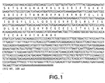
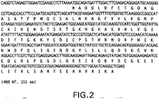


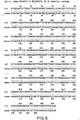

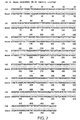


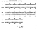


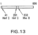
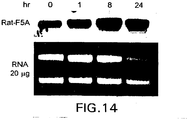
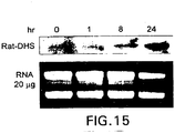
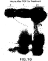


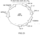
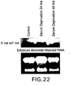
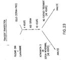
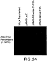
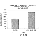
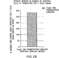

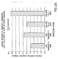


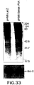
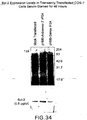
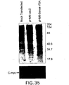

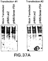


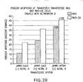
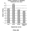
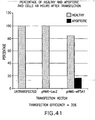
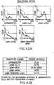
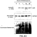

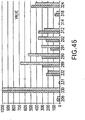
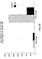

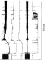


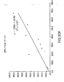
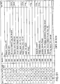
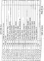

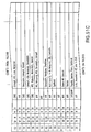


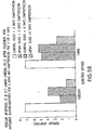


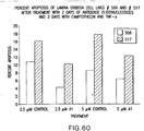


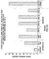
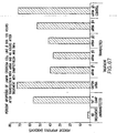


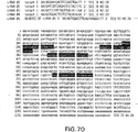


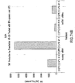
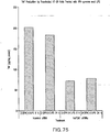


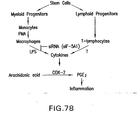
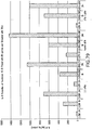
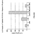

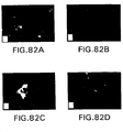
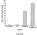

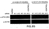
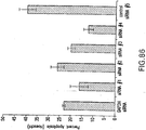
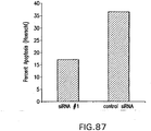
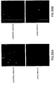

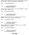
Classifications
-
- C—CHEMISTRY; METALLURGY
- C12—BIOCHEMISTRY; BEER; SPIRITS; WINE; VINEGAR; MICROBIOLOGY; ENZYMOLOGY; MUTATION OR GENETIC ENGINEERING
- C12N—MICROORGANISMS OR ENZYMES; COMPOSITIONS THEREOF; PROPAGATING, PRESERVING, OR MAINTAINING MICROORGANISMS; MUTATION OR GENETIC ENGINEERING; CULTURE MEDIA
- C12N15/00—Mutation or genetic engineering; DNA or RNA concerning genetic engineering, vectors, e.g. plasmids, or their isolation, preparation or purification; Use of hosts therefor
- C12N15/09—Recombinant DNA-technology
- C12N15/11—DNA or RNA fragments; Modified forms thereof; Non-coding nucleic acids having a biological activity
- C12N15/113—Non-coding nucleic acids modulating the expression of genes, e.g. antisense oligonucleotides; Antisense DNA or RNA; Triplex- forming oligonucleotides; Catalytic nucleic acids, e.g. ribozymes; Nucleic acids used in co-suppression or gene silencing
-
- A—HUMAN NECESSITIES
- A61—MEDICAL OR VETERINARY SCIENCE; HYGIENE
- A61K—PREPARATIONS FOR MEDICAL, DENTAL OR TOILETRY PURPOSES
- A61K31/00—Medicinal preparations containing organic active ingredients
- A61K31/70—Carbohydrates; Sugars; Derivatives thereof
- A61K31/7088—Compounds having three or more nucleosides or nucleotides
-
- A—HUMAN NECESSITIES
- A61—MEDICAL OR VETERINARY SCIENCE; HYGIENE
- A61P—SPECIFIC THERAPEUTIC ACTIVITY OF CHEMICAL COMPOUNDS OR MEDICINAL PREPARATIONS
- A61P1/00—Drugs for disorders of the alimentary tract or the digestive system
-
- A—HUMAN NECESSITIES
- A61—MEDICAL OR VETERINARY SCIENCE; HYGIENE
- A61P—SPECIFIC THERAPEUTIC ACTIVITY OF CHEMICAL COMPOUNDS OR MEDICINAL PREPARATIONS
- A61P1/00—Drugs for disorders of the alimentary tract or the digestive system
- A61P1/02—Stomatological preparations, e.g. drugs for caries, aphtae, periodontitis
-
- A—HUMAN NECESSITIES
- A61—MEDICAL OR VETERINARY SCIENCE; HYGIENE
- A61P—SPECIFIC THERAPEUTIC ACTIVITY OF CHEMICAL COMPOUNDS OR MEDICINAL PREPARATIONS
- A61P11/00—Drugs for disorders of the respiratory system
- A61P11/06—Antiasthmatics
-
- A—HUMAN NECESSITIES
- A61—MEDICAL OR VETERINARY SCIENCE; HYGIENE
- A61P—SPECIFIC THERAPEUTIC ACTIVITY OF CHEMICAL COMPOUNDS OR MEDICINAL PREPARATIONS
- A61P13/00—Drugs for disorders of the urinary system
- A61P13/12—Drugs for disorders of the urinary system of the kidneys
-
- A—HUMAN NECESSITIES
- A61—MEDICAL OR VETERINARY SCIENCE; HYGIENE
- A61P—SPECIFIC THERAPEUTIC ACTIVITY OF CHEMICAL COMPOUNDS OR MEDICINAL PREPARATIONS
- A61P17/00—Drugs for dermatological disorders
- A61P17/04—Antipruritics
-
- A—HUMAN NECESSITIES
- A61—MEDICAL OR VETERINARY SCIENCE; HYGIENE
- A61P—SPECIFIC THERAPEUTIC ACTIVITY OF CHEMICAL COMPOUNDS OR MEDICINAL PREPARATIONS
- A61P17/00—Drugs for dermatological disorders
- A61P17/06—Antipsoriatics
-
- A—HUMAN NECESSITIES
- A61—MEDICAL OR VETERINARY SCIENCE; HYGIENE
- A61P—SPECIFIC THERAPEUTIC ACTIVITY OF CHEMICAL COMPOUNDS OR MEDICINAL PREPARATIONS
- A61P19/00—Drugs for skeletal disorders
-
- A—HUMAN NECESSITIES
- A61—MEDICAL OR VETERINARY SCIENCE; HYGIENE
- A61P—SPECIFIC THERAPEUTIC ACTIVITY OF CHEMICAL COMPOUNDS OR MEDICINAL PREPARATIONS
- A61P19/00—Drugs for skeletal disorders
- A61P19/02—Drugs for skeletal disorders for joint disorders, e.g. arthritis, arthrosis
-
- A—HUMAN NECESSITIES
- A61—MEDICAL OR VETERINARY SCIENCE; HYGIENE
- A61P—SPECIFIC THERAPEUTIC ACTIVITY OF CHEMICAL COMPOUNDS OR MEDICINAL PREPARATIONS
- A61P25/00—Drugs for disorders of the nervous system
-
- A—HUMAN NECESSITIES
- A61—MEDICAL OR VETERINARY SCIENCE; HYGIENE
- A61P—SPECIFIC THERAPEUTIC ACTIVITY OF CHEMICAL COMPOUNDS OR MEDICINAL PREPARATIONS
- A61P27/00—Drugs for disorders of the senses
-
- A—HUMAN NECESSITIES
- A61—MEDICAL OR VETERINARY SCIENCE; HYGIENE
- A61P—SPECIFIC THERAPEUTIC ACTIVITY OF CHEMICAL COMPOUNDS OR MEDICINAL PREPARATIONS
- A61P27/00—Drugs for disorders of the senses
- A61P27/02—Ophthalmic agents
-
- A—HUMAN NECESSITIES
- A61—MEDICAL OR VETERINARY SCIENCE; HYGIENE
- A61P—SPECIFIC THERAPEUTIC ACTIVITY OF CHEMICAL COMPOUNDS OR MEDICINAL PREPARATIONS
- A61P27/00—Drugs for disorders of the senses
- A61P27/02—Ophthalmic agents
- A61P27/06—Antiglaucoma agents or miotics
-
- A—HUMAN NECESSITIES
- A61—MEDICAL OR VETERINARY SCIENCE; HYGIENE
- A61P—SPECIFIC THERAPEUTIC ACTIVITY OF CHEMICAL COMPOUNDS OR MEDICINAL PREPARATIONS
- A61P29/00—Non-central analgesic, antipyretic or antiinflammatory agents, e.g. antirheumatic agents; Non-steroidal antiinflammatory drugs [NSAID]
-
- A—HUMAN NECESSITIES
- A61—MEDICAL OR VETERINARY SCIENCE; HYGIENE
- A61P—SPECIFIC THERAPEUTIC ACTIVITY OF CHEMICAL COMPOUNDS OR MEDICINAL PREPARATIONS
- A61P3/00—Drugs for disorders of the metabolism
- A61P3/08—Drugs for disorders of the metabolism for glucose homeostasis
- A61P3/10—Drugs for disorders of the metabolism for glucose homeostasis for hyperglycaemia, e.g. antidiabetics
-
- A—HUMAN NECESSITIES
- A61—MEDICAL OR VETERINARY SCIENCE; HYGIENE
- A61P—SPECIFIC THERAPEUTIC ACTIVITY OF CHEMICAL COMPOUNDS OR MEDICINAL PREPARATIONS
- A61P35/00—Antineoplastic agents
-
- A—HUMAN NECESSITIES
- A61—MEDICAL OR VETERINARY SCIENCE; HYGIENE
- A61P—SPECIFIC THERAPEUTIC ACTIVITY OF CHEMICAL COMPOUNDS OR MEDICINAL PREPARATIONS
- A61P37/00—Drugs for immunological or allergic disorders
-
- A—HUMAN NECESSITIES
- A61—MEDICAL OR VETERINARY SCIENCE; HYGIENE
- A61P—SPECIFIC THERAPEUTIC ACTIVITY OF CHEMICAL COMPOUNDS OR MEDICINAL PREPARATIONS
- A61P37/00—Drugs for immunological or allergic disorders
- A61P37/02—Immunomodulators
-
- A—HUMAN NECESSITIES
- A61—MEDICAL OR VETERINARY SCIENCE; HYGIENE
- A61P—SPECIFIC THERAPEUTIC ACTIVITY OF CHEMICAL COMPOUNDS OR MEDICINAL PREPARATIONS
- A61P37/00—Drugs for immunological or allergic disorders
- A61P37/08—Antiallergic agents
-
- A—HUMAN NECESSITIES
- A61—MEDICAL OR VETERINARY SCIENCE; HYGIENE
- A61P—SPECIFIC THERAPEUTIC ACTIVITY OF CHEMICAL COMPOUNDS OR MEDICINAL PREPARATIONS
- A61P43/00—Drugs for specific purposes, not provided for in groups A61P1/00-A61P41/00
-
- A—HUMAN NECESSITIES
- A61—MEDICAL OR VETERINARY SCIENCE; HYGIENE
- A61P—SPECIFIC THERAPEUTIC ACTIVITY OF CHEMICAL COMPOUNDS OR MEDICINAL PREPARATIONS
- A61P7/00—Drugs for disorders of the blood or the extracellular fluid
-
- A—HUMAN NECESSITIES
- A61—MEDICAL OR VETERINARY SCIENCE; HYGIENE
- A61P—SPECIFIC THERAPEUTIC ACTIVITY OF CHEMICAL COMPOUNDS OR MEDICINAL PREPARATIONS
- A61P9/00—Drugs for disorders of the cardiovascular system
-
- A—HUMAN NECESSITIES
- A61—MEDICAL OR VETERINARY SCIENCE; HYGIENE
- A61P—SPECIFIC THERAPEUTIC ACTIVITY OF CHEMICAL COMPOUNDS OR MEDICINAL PREPARATIONS
- A61P9/00—Drugs for disorders of the cardiovascular system
- A61P9/10—Drugs for disorders of the cardiovascular system for treating ischaemic or atherosclerotic diseases, e.g. antianginal drugs, coronary vasodilators, drugs for myocardial infarction, retinopathy, cerebrovascula insufficiency, renal arteriosclerosis
-
- C—CHEMISTRY; METALLURGY
- C12—BIOCHEMISTRY; BEER; SPIRITS; WINE; VINEGAR; MICROBIOLOGY; ENZYMOLOGY; MUTATION OR GENETIC ENGINEERING
- C12N—MICROORGANISMS OR ENZYMES; COMPOSITIONS THEREOF; PROPAGATING, PRESERVING, OR MAINTAINING MICROORGANISMS; MUTATION OR GENETIC ENGINEERING; CULTURE MEDIA
- C12N2310/00—Structure or type of the nucleic acid
- C12N2310/10—Type of nucleic acid
- C12N2310/11—Antisense
-
- C—CHEMISTRY; METALLURGY
- C12—BIOCHEMISTRY; BEER; SPIRITS; WINE; VINEGAR; MICROBIOLOGY; ENZYMOLOGY; MUTATION OR GENETIC ENGINEERING
- C12N—MICROORGANISMS OR ENZYMES; COMPOSITIONS THEREOF; PROPAGATING, PRESERVING, OR MAINTAINING MICROORGANISMS; MUTATION OR GENETIC ENGINEERING; CULTURE MEDIA
- C12N2310/00—Structure or type of the nucleic acid
- C12N2310/10—Type of nucleic acid
- C12N2310/14—Type of nucleic acid interfering nucleic acids [NA]
-
- C—CHEMISTRY; METALLURGY
- C12—BIOCHEMISTRY; BEER; SPIRITS; WINE; VINEGAR; MICROBIOLOGY; ENZYMOLOGY; MUTATION OR GENETIC ENGINEERING
- C12N—MICROORGANISMS OR ENZYMES; COMPOSITIONS THEREOF; PROPAGATING, PRESERVING, OR MAINTAINING MICROORGANISMS; MUTATION OR GENETIC ENGINEERING; CULTURE MEDIA
- C12N2310/00—Structure or type of the nucleic acid
- C12N2310/50—Physical structure
- C12N2310/53—Physical structure partially self-complementary or closed
Definitions
- the present invention relates to apoptosis-specific eucaryotic initiation Factor-5A (eIF-5A) or referred to as apoptosis Factor 5A or Factor 5A1 and deoxyhypusine synthase (DHS).
- eIF-5A eucaryotic initiation Factor-5A
- DHS deoxyhypusine synthase
- the present invention relates to apoptosis Factor 5A and DHS nucleic acids and polypeptides and methods for inhibiting expression of apoptosis Factor 5A and DHS.
- Apoptosis is a genetically programmed cellular event that is characterized by well-defined morphological features, such as cell shrinkage, chromatin condensation, nuclear fragmentation, and membrane blebbing. Kerr et al. (1972) Br. J. Cancer, 26, 239-257 ; Wyllie et al. (1980) Int. Rev. Cytol., 68, 251-306 . It plays an important role in normal tissue development and homeostasis, and defects in the apoptotic program are thought to contribute to a wide range of human disorders ranging from neurodegenerative and autoimmunity disorders to neoplasms. Thompson (1995) Science, 267, 1456-1462 ; Mullauer et al. (2001) Mutat. Res, 488, 211-231 . Although the morphological characteristics of apoptotic cells are well characterized, the molecular pathways that regulate this process have only begun to be elucidated.
- caspases cysteine proteases
- Creagh & Martin 2001) Biochem. Soc. Trans, 29, 696-701 ; Dales et al. (2001) Leuk. Lymphoma, 41, 247-253 .
- Caspases trigger apoptosis in response to apoptotic stimuli by cleaving various cellular proteins, which results in classic manifestations of apoptosis, including cell shrinkage, membrane blebbing and DNA fragmentation. Chang & Yang (2000) Microbiol. Mol. Biol. Rev., 64, 821-846 .
- Pro-apoptotic proteins such as Bax or Bak
- caspase-activating molecules such as mitochondrial cytochrome c
- Anti-apoptotic proteins such as Bcl-2, promote cell survival by antagonizing the activity of the pro-apoptotic proteins, Bax and Bak. Tsujimoto (1998) Genes Cells, 3, 697-707 ; Kroemer (1997) Nature Med., 3, 614-620 .
- the ratio of Bax:Bcl-2 is thought to be one way in which cell fate is determined; an excess of Bax promotes apoptosis and an excess of Bcl-2 promotes cell survival.
- Another key protein involved in apoptosis is that encoded by the tumor suppressor gene p53.
- This protein is a transcription factor that regulates cell growth and induces apoptosis in cells that are damaged and genetically unstable, presumably through upregulation of Bax.
- Bold et al. (1997) Surgical Oncology, 6,133-142 ; Ronen et al., 1996; Schuler & Green (2001) Biochem. Soc. Trans., 29, 684-688 ; Ryan et al. (2001) Curr. Opin. Cell Biol., 13, 332-337 ; Zörnig et al. (2001) Biochem. Biophys. Acta, 1551, F1-F37 .
- apoptotic cells that can be exploited for their detection is activation of a flippase, which results in externalization of phosphatidylserine, a phospholipid normally localized to the inner leaflet of the plasma membrane. Fadok et al. (1992) J. Immunol., 149, 4029-4035 . Apoptotic cells bearing externalized phosphatidylserine can be detected by staining with a phosphatidylserine-binding protein, Annexin V, conjugated to a fluorescent dye.
- a phosphatidylserine-binding protein Annexin V
- the characteristic DNA fragmentation that occurs during the apoptotic process can be detected by labeling the exposed 3'-OH ends of the DNA fragments with fluorescein-labeled deoxynucleotides.
- Fluorescent dyes that bind nucleic acids such as Hoescht 33258, can be used to detect chromatin condensation and nuclear fragmentation in apoptotic cells.
- the degree of apoptosis in a cell population can also be inferred from the extent of caspase proteolytic activity present in cellular extracts.
- apoptosis like any other developmental program, can be disrupted by mutation. Alterations in the apoptotic pathways are believed to play a key role in a number of disease processes, including cancer.
- Apoptosis can be triggered during tumor development by a variety of signals.
- Extracellular signals include growth or survival factor depletion, hypoxia and ionizing radiation.
- Internal signals that can trigger apoptosis include DNA damage, shortening telomeres, and oncogenic mutations that produce inappropriate proliferative signals.
- Ionizing radiation and nearly all cytotoxic chemotherapy agents used to treat malignancies are thought to act by triggering endogenous apoptotic mechanisms to induce cell death.
- Rowan & Fisher 1997) Leukemia, 11, 457-465 ; Kerr et al. (1994) Cancer, 73, 2013-2026 ; Martin & Schwartz (1997) Oncology Research, 9,1-5 .
- apoptosis is intimately involved in the development and progression of neoplastic transformation and metastases, and a better understanding of the apoptotic pathways involved may lead to new potential targets for the treatment of cancer by the modulation of apoptotic pathways through gene therapy approaches.
- Bold et al. (1997) Surgical Oncology, 6, 133-142 .
- the present invention relates to cloning of an eIF-5A cDNA that is up regulated immediately before the induction of apoptosis.
- This apoptosis-specific eIF-5A is likely to be a suitable target for intervention in apoptosis-causing disease states since it appears to act at the level of post-transcriptional regulation of downstream effectors and transcription factors involved in the apoptotic pathway.
- the apoptosis-specific eIF-5A appears to selectively facilitate the translocation of mRNAs encoding downstream effectors and transcription factors of apoptosis from the nucleus to the cytoplasm, where they are subsequently translated.
- the ultimate decision to initiate apoptosis appears to stem from a complex interaction between internal and external pro- and anti-apoptotic signals.
- the apoptosis-related eIF-5A appears to tip the balance between these signals in favor of apoptosis.
- Induction of apoptosis-related eIF-5A has the potential to selectively target cancer cells while having little or no effect on normal neighboring cells. This arises because mitogenic oncogenes expressed in tumor cells provide an apoptotic signal in the form of specific species of mRNA that are not present in normal cells. Lowe et al. (1993) Cell, 74, 954-967 ; Lowe & Lin (2000) Carcinogenesis, 21, 485-495 . For example, restoration of wild-type p53 in p53-mutant tumor cells can directly induce apoptosis as well as increase drug sensitivity in tumor cell lines and xenographs. (Spitz et al., 1996; Badie et al. 1998).
- apoptosis-eIF-5A arises from the fact that it selectively facilitates translation of mRNAs for downstream apoptosis effectors and transcription factors by mediating their translocation from the nucleus into the cytoplasm.
- mRNAs for these effectors and transcription factors have to be transcribed.
- apoptosis eIF-5A would promote apoptosis in cancer cells but have minimal, if any, effect on normal cells.
- restoration of apoptotic potential in tumor cells with apoptosis-related eIF-5A may decrease the toxicity and side effects experienced by cancer patients due to selective targeting of tumor cells.
- Induction of apoptotic eIF-5A also has the potential to potentiate the response of tumor cells to anti-cancer drugs and thereby improve the effectiveness of these agents against drug-resistant tumors. This in turn could result in lower doses of anti-cancer drugs for efficacy and reduced toxicity to the patient.
- Glaucoma describes a group of eye conditions in which increased intra-ocular pressure (IOP) leads to damage of the optic nerve and progressive blindness.
- IOP intra-ocular pressure
- Cytokines have also been implicated in the apoptotic pathway. Biological systems require cellular interactions for their regulation, and cross-talk between cells generally involves a large variety of cytokines. Cytokines are mediators that are produced in response to a wide variety of stimuli by many different cell types. Cytokines are pleiotropic molecules that can exert many different effects on many different cell types, but are especially important in regulation of the immune response and hematopoietic cell proliferation and differentiation. The actions of cytokines on target cells can promote cell survival, proliferation, activation, differentiation, or apoptosis depending on the particular cytokine, relative concentration, and presence of other mediators.
- anti-cytokines to treat autoimmune disorders (psoriasis, rheumatoid arthritis, Crohn's disease) is gaining popularity.
- Interleukin 1 is an important cytokine that mediates local and systemic inflammatory reactions and which can synergize with TNF in the pathogenesis of many disorders, including vasculitis, osteoporosis, neurodegenerative disorders, diabetes, lupus nephritis, and autoimmune disorders such as rheumatoid arthritis.
- IL-1 ⁇ Interleukin 1
- the importance of IL-1 ⁇ in tumour angiogenesis and invasiveness was also recently demonstrated by the resistance of IL-1 ⁇ knockout mice to metastases and angiogenesis when injected with melanoma cells (Voronov et al ., 2003).
- Interleukin 18 is a recently discovered member of the IL-1 family and is related by structure, receptors, and function to IL-1.
- IL-18 is a central cytokine involved in inflammatory and autoimmune disorders as a result of its ability to induce interferon-gamma (IFN- ⁇ ), TNF- ⁇ , and IL-1.
- IFN- ⁇ interferon-gamma
- TNF- ⁇ TNF- ⁇
- IL-1 ⁇ and IL-18 are both capable of inducing production of TNF- ⁇ , a cytokine known to contribute to cardiac dysfunction during myocardial ischemia (Maekawa et al ., 2002).
- Inhibition of IL-18 by neutralization with an IL-18 binding protein was found to reduce ischemia-induced myocardial dysfunction in an ischemia/reperfusion model of suprafused human atrial myocardium (Dinarello, 2001).
- Neutralization of IL-18 using a mouse IL-18 binding protein was also able to decrease IFN- ⁇ , TNF- ⁇ , and IL-1 ⁇ transcript levels and reduce joint damage in a collagen-induced arthritis mouse model (Banda et al ., 2003).
- a reduction of IL-18 production or availability may also prove beneficial to control metastatic cancer as injection of IL-18 binding protein in a mouse melanoma model successfully inhibited metastases (Carrascal et al ., 2003).
- IL-18 As a further indication of its importance as a pro-inflammatory cytokine, plasma levels of IL-18 were elevated in patients with chronic liver disease and increased levels were correlated with the severity of the disease (Ludwiczek et al ., 2002). Similarly, IL-18 and TNF- ⁇ were elevated in the serum of diabetes mellitus patients with nephropathy (Moriwaki et al., 2003). Neuroinflammation following traumatic brain injury is also mediated by pro-inflammatory cytokines and inhibition of IL-18 by the IL-18 binding protein improved neurological recovery in mice following brain trauma (Yatsiv et al ., 2002).
- TNF- ⁇ a member of the TNF family of cytokines, is a pro-inflammatory cytokine with pleiotropic effects ranging from co-mitogenic effects on hematopoietic cells, induction of inflammatory responses, and induction of cell death in many cell types.
- TNF- ⁇ is normally induced by bacterial lipopolysaccharides, parasites, viruses, malignant cells and cytokines and usually acts beneficially to protect cells from infection and cancer.
- inappropriate induction of TNF- ⁇ is a major contributor to disorders resulting from acute and chronic inflammation such as autoimmune disorders and can also contribute to cancer, AIDS, heart disease, and sepsis (reviewed by Aggarwal and Natarajan, 1996; Sharma and Anker, 2002).
- TNF- ⁇ TNF- ⁇
- B-CLL B-cell chronic lymphocytic leukemia
- IL-1 ⁇ Interleukin-1 ⁇
- DHS Deoxyhypusine synthase
- eIF-5A hypusine-containing eucaryotic translation initiation Factor-5A
- Hypusine a unique amino acid, is found in all examined eucaryotes and archaebacteria, but not in eubacteria, and eIF-5A is the only known hypusine-containing protein.
- Active eIF-5A is formed in two post-translational steps: the first step is the formation of a deoxyhypusine residue by the transfer of the 4-aminobutyl moiety of spermidine to the ⁇ -amino group of a specific lysine of the precursor eIF-5A catalyzed by deoxyhypusine synthase; the second step involves the hydroxylation of this 4-aminobutyl moiety by deoxyhypusine hydroxylase to form hypusine.
- eIF-5A intracellular depletion results in a significant accumulation of specific mRNAs in the nucleus, indicating that eIF-5A may be responsible for shuttling specific classes of mRNAs from the nucleus to the cytoplasm.
- the accumulation of eIF-5A at nuclear pore-associated intranuclear filaments and its interaction with a general nuclear export receptor further suggest that eIF-5A is a nucleocytoplasmic shuttle protein, rather than a component of polysomes. Rosorius et al. (1999) J. Cell Science, 112, 2369-2380 .
- the first cDNA for eIF-5A was cloned from human in 1989 by Smit-McBride et al., and since then cDNAs or genes for eIF-5A have been cloned from various eukaryotes including yeast, rat, chick embryo, alfalfa, and tomato.
- eIF-5A mRNA has been explored in various human tissues and mammalian cell lines. For example, changes in eIF-5A expression have been observed in human fibroblast cells after addition of serum following serum deprivation. Pang & Chen (1994) J. Cell Physiol., 160, 531-538 . Age-related decreases in deoxyhypusine synthase activity and abundance of precursor eIF-5A have also been observed in senescing fibroblast cells, although the possibility that this reflects averaging of differential changes in isoforms was not determined. Chen & Chen (1997b) J. Cell Physiol., 170, 248-254 .
- eIF-5A may be the cellular target of viral proteins such as the human immunodeficiency virus type 1 Rev protein and human T cell leukemia virus type 1 Rex protein. Ruhl et al. (1993) J. Cell Biol..,123, 1309-1320 ; Katahira et al. (1995) J. Virol., 69, 3125-3133 . Preliminary studies indicate that eIF-5A may target RNA by interacting with other RNA-binding proteins such as Rev, suggesting that these viral proteins may recruit eIF-5A for viral RNA processing. Liu et al. (1997) Biol. Signals, 6, 166-174 .
- eIF5A and DHS are known, there remains a need in understanding how these proteins are involved in apoptotic pathways as well as cytokine stimulation to be able to modulate apoptosis and cytokine expression.
- the present invention fulfills this need.
- the present invention relates to apoptosis specific eucaryotic initiation factor 5A (eIF-5A), referred to as apoptosis factor 5A1 or simply factor 5A1.
- eIF-5A eucaryotic initiation factor 5A
- the present invention relates to methods for inhibiting or suppressing apoptosis in cells using siRNAs to inhibit expression of factor 5A1. Also described herein are methods of suppressing or inhibiting expression of pro-inflammatory cytokines or expression of p53 by inhibiting expression of apoptosis factor 5A1, and a method of increasing Bcl-2 expression by inhibiting or suppressing expression of apoptosis factor 5A1 using antisense nucleotides or siRNAs.
- a method of inhibiting production of cytokines, especially TNF- ⁇ in human epithelial cells provides methods of preventing retinal ganglion cell death in a glaucomatous eye.
- eukaryotic initiation factor 5a eukaryotic initiation factor 5a
- eIF-5A eukaryotic initiation factor 5a
- Apoptosis factor 5A is likely to be a suitable target for intervention in apoptosis-causing disease states since it appears to act at the level of post-transcriptional regulation of downstream effectors and transcription factors involved in the apoptotic pathway. Specifically, apoptosis factor 5A appears to selectively facilitate the translocation of mRNAs encoding downstream effectors and transcription factors of apoptosis from the nucleus to the cytoplasm, where they are subsequently translated. The ultimate decision to initiate apoptosis appears to stem from a complex interaction between internal and external pro- and anti-apoptotic signals. Lowe & Lin (2000) Carcinogenesis, 21, 485-495 . Through its ability to facilitate the translation of downstream apoptosis effectors and transcription factors, the apoptosis factor 5A appears to tip the balance between these signals in favor of apoptosis.
- Described herein is a method of suppressing or reducing apoptosis in a cell by administering an agent that inhibits or reduces expression of either apoptosis factor 5A or DHS.
- an agent that can inhibit or reduce expression of apoptosis factor 5A or DHS is an antisense oligonucleotide.
- Antisense oligonucleotides have been successfully used to accomplish both in vitro as well as in vivo gene-specific suppression.
- Antisense oligonucleotides are short, synthetic strands of DNA (or DNA analogs) that are antisense (or complimentary) to a specific DNA or RNA target.
- Antisense oligonucleotides are designed to block expression of the protein encoded by the DNA or RNA target by binding to the target and halting expression at the level of transcription, translation, or splicing.
- the antisense oligonucleotides of the present invention have a nucleotide sequence encoding a portion of an apoptosis factor 5A polypeptide or an apoptosis-specific DHS polypeptide.
- the inventors have transfected various cell lines with antisense nucleotides encoding a portion of an apoptosis factor 5A polypeptide as described below and measured the number of cells undergoing apoptosis.
- the cells that were transfected with the antisense oligonucleotides showed a decrease in the number of cells undergoing apoptosis as compared to like cells not having been transfected with the antisense oligos.
- Figures 54 -58 show a decrease in the percentage of cells undergoing apoptosis in the cells having being treated with antisense apoptosis factor 5A oligonucleotides as compared to cells not having been transfected with the antisense apoptosis factor 5A oligonucleotides.
- apoptosis factor 5A polypeptide or DHS polypeptide encoding an apoptosis factor 5A polypeptide or DHS polypeptide.
- SEQ ID NOS:1, 3, 4, 5, 11, 15, 19, 20, and 21 apoptosis-factor 5A nucleic acid sequences
- SEQ ID NOS:6 and 8 apoptosis-specific DHS nucleic acid sequences
- SEQ ID NOS:12 and 16 apoptosis factor 5A sequences
- SEQ ID NO:7 apoptosis-specific DHS polypeptide sequences
- Other preferred apoptosis factor 5A antisense polynucleotide sequences include SEQ ID NO: 35, 37, and 39.
- Additional antisense nucleotides include those that have substantial sequence identity to those enumerated above (i.e. 90% homology) or those having sequences that hybridize under highly stringent conditions to the enumerated SEQ ID NOs. Additionally, other suitable sequences can be found using the known sequences as probes according to methods known in the art.
- the antisense oligonucleotides may be single stranded, double stranded, DNA, RNA or a hybrid.
- the oligonucleotides may be modified by methods known in the art to increase stability, increase resistance to nuclease degradation or the like. These modifications are known in the art and include, but are not limited to modifying the backbone of the oligonucleotide, modifying the sugar moieties, or modifying the base. Also inclusive in these modifications are various DNA-RNA hybrids or constructs commonly referred to as "gapped" oligonucleotides.
- the present invention provides SiRNAs that can inhibit or reduce expression of apoptosis factor 5A or DHS.
- Small Inhibitory RNAs (siRNA) have been emerging as a viable alternative to antisense oligonucleotides since lower concentrations are required to achieve levels of suppression that are equivalent or superior to those achieved with antisense oligonucleotides (Thompson, 2002).
- Long doublestranded RNAs have been used to silence the expression of specific genes in a variety of organisms such as plants, nematodes, and fruit flies.
- Dicer processes these long double stranded RNAs into 21-23 nucleotide small interfering RNAs which are then incorporated into an RNA-induced silencing complex (RISC). Unwinding of the siRNA activates RISC and allows the single-stranded siRNA to guide the complex to the endogenous mRNA by base pairing. Recognition of the endogenous mRNA by RISC results in its cleavage and consequently makes it unavailable for translation. Introduction of long double stranded RNA into mammalian cells results in a potent antiviral response which can be bypassed by use of siRNAs. (Elbashir et al ., 2001). SiRNA has been widely used in cell cultures and routinely achieves a reduction in specific gene expression of 90% or more.
- siRNAs have also been gaining popularity in inhibiting gene expression in animal models of disease.
- a recent study that demonstrated that an siRNA against luciferase was able to block luciferase expression from a co-transfected plasmid in a wide variety of organs in post-natal mice using a hydrodynamic injection delivery technique (Lewis et al ., 2002).
- An siRNA against Fas, a receptor in the TNF family, injected hydrodynamically into the tail vein of mice was able to transfect greater than 80% of hepatocytes and decrease Fas expression in the liver by 90% for up to 10 days after the last injection (Song et al ., 2003).
- the Fas siRNA was also able to protect mice from liver fibrosis and fulminant hepatitis.
- the development of sepsis in mice treated with a lethal dose of lipopolysaccharide was inhibited by the use of an siRNA directed against TNF ⁇ (S ⁇ rensen et al ., 2003).
- SiRNA has the potential to be a very potent drug for the inhibition of specific gene expression in vivo in light of their long-lasting effectiveness in cell cultures and in vivo , their ability to transfect cells in vivo , and their resistance to degradation in serum (Bertrand et al ., 2002).
- the present inventors have transfected cells with apoptosis factor 5A siRNAs and studied the effects on expression of apoptosis factor 5A.
- Figure 64 shows that cells transfected with apoptosis factor 5a siRNA produced less apoptosis factor 5a protein.
- Figures 64 -67 show that cells transfected with apoptosis factor 5A siRNAs have a lower percentage of cells undergoing apoptosis after exposure to camptothecin and TNF- ⁇ as compared to cells not having been transfected with apoptosis factor 5A siRNAs.
- Preferred siRNAs include those that have SEQ ID NO: 30, 31, 32, 33, and 34. Additional siRNAs include those that have substantial sequence identity to those enumerated (i.e. 90% homology) or those having sequences that hybridize under highly stringent conditions to the enumerated SEQ ID NOs.
- Figure 64 shows that cells transfected with apoptosis factor 5A siRNA produced less apoptosis factor 5A protein.
- apoptosis-associated diseases and disorders include neurological/ neurodegenerative disorders (e.g., Alzheimer's, Parkinson's, Huntington's, Amyotrophic Lateral Sclerosis (Lou Gehrig's Disease), autoimmune disorders (e.g., rheumatoid arthritis, systemic lupus erythematosus (SLE), multiple sclerosis), Duchenne Muscular Dystrophy (DMD), motor neuron disorders, ischemia, heart ischemia, chronic heart failure, stroke, infantile spinal muscular atrophy, cardiac arrest, renal failure, atopic dermatitis, sepsis and septic shock, AIDS, hepatitis, glaucoma, diabetes (type 1 and type 2), asthma, retinit
- Glaucoma is a group of eye conditions arising from damage to the optic nerve that results in progressive blindness. Apoptosis has been shown to be a direct cause of this optic nerve damage.
- the interruption of axonal transport as a result of increased IOP may contribute to retinal ganglion cell death by deprivation of trophic factors.
- Optic nerve head astrocytes in glaucomatous eyes have also been found to produce increased levels of some neurotoxic substances. For example, increased production of tumor necrosis factor- ⁇ (TNF- ⁇ ) ( Yan et al., (2000) Arch. Ophthalmol., 118, 666-673 ), and nitric oxide synthase ( Neufeld et al., (1997) Arch.
- TNF- ⁇ tumor necrosis factor- ⁇
- nitric oxide synthase Neufeld et al., (1997) Arch.
- Ophthalmol., 115, 497-503 the enzyme which gives rise to nitric oxide, has been found in the optic nerve head of glaucomatous eyes. Furthermore, increased expression of the inducible form of nitric oxide synthase (iNOS) and TNF- ⁇ by activated retinal glial cells have been observed in rat models of hereditary retinal diseases. Cotinet et al., (1997)Glia, 20, 59-69 ; de Kozak et al., (1997) Ocul. Immunol. Inflamm., 5, 85-94 ; Goureau et al., (1999) J. Neurochem, 72, 2506-2515 .
- iNOS inducible form of nitric oxide synthase
- Antisense oligonucleotides have been used by several groups to target key proteins in the apoptotic process in order to protect retinal ganglion cells from apoptotic cell death.
- Antisense oligonucleotides are short, synthetic strands of DNA (or DNA analogs) that are antisense (or complimentary) to a specific DNA or RNA target.
- Antisense oligonucleotides are designed to block expression of the protein encoded by the DNA or RNA target by binding to the target and halting expression at the level of transcription, translation, or splicing.
- Antisense oligonucleotides have been used successfully in animal models of eye disease.
- expression of caspase 2 was increased during ischemia, primarily in the inner nuclear and ganglion cell layers of the retina. Suppression of caspase using an antisense oligonucleotide led to significant histopathologic and functional improvement as determined by electroretinogram.
- Singh et al., (2001) J. Neurochem., 77(2), 466-75 Another study demonstrated that, upon transection of the optic nerve, retinal ganglion cells upregulate the pro-apoptotic protein Bax and undergo apoptosis.
- HVJ inactivated hemagglutinating virus of Japan
- HVJ liposomes Intravitreal injection into mice of FITC-labeled antisense oligonucleotides encapsulated in HVJ liposomes resulted in high fluorescence within 44% of the cells in the ganglion layer which lasted three days while fluorescence with naked FITC-labeled antisense oligonucleotide disappeared after one day.
- HVJ liposomes Intravitreal injection into mice of FITC-labeled antisense oligonucleotides encapsulated in HVJ liposomes
- a method of preventing or modulating apoptosis is directed to modulating apoptosis in the cells of the eye, such as but not limited to, astrocytes, retinal ganglion, retinal glial cells and lamina cribosa. Death of retinal ganglion cells in glaucoma occurs by apoptosis.
- providing a method of inhibiting apoptosis in retinal ganglion cells or by protecting retinal ganglion cells from degeneration by apoptosis provides a novel treatment for prevention of blindness due to glaucoma.
- the present invention provides a method for preventing retinal ganglion cell death in a glaucomatous eye, by suppressing expression of apoptosis-specific eIF-5A1. Inhibiting the expression of apoptosis-specific eIF-5A1 reduces apoptosis.
- Apoptosis-specific eIF-5A1 is a powerful gene that appears to regulate the entire apoptic process. Thus, controlling apoptosis in the optic nerve head indicates that blocking expression of apoptosis-specific eIF-5A1 provides a treatment for glaucoma.
- Suppression of expression of apoptosis-specific eIF-5A1 is accomplished by administering siRNA targeted against human apoptosis-specific eIF-5A1 to cells of the eye such as, but not limited to lamina crobosa, astrocytes, retinal ganglion, or retinal glial cells.
- antisense oligonucleotides as defined above i.e. have a nucleotide sequence encoding at least a portion of an apoptosis-specific eIF-5A1 polypeptide.
- Antisense oligonucleotides targeted against human apoptosis-specific eIF-5A1 have a nucleotide sequence encoding at least a portion of human apoptosis-specific eIF-5A1 polypeptide.
- Preferred antisense oligonucleotides comprise SEQ ID NO:26 or 27 or oligonucleotides that bind to a sequence complementary to SEQ ID NO:26 or 27 under high stringency conditions.
- Also disclosed herein is a method of suppressing expression of apoptosis-specific eIF-5A1 in lamina cribosa cells, astrocyte cells, retinal ganglion cells or retinal glial cells.
- Antisense oligonucleotides such as but not limited to, SEQ ID NO:26 and 27, targeted against human apoptosis-specific eIF-5A1 are administered to lamina cribosa cells, astrocyte cells, retinal ganglion cells or retinal glial cells.
- the cells may be human.
- eIF-5A may also have a role in the immune response.
- the present inventors have discovered that apoptosis factor 5A levels correlate with elevated levels of two cytokines (Interleukin 1-beta "IL-1 ⁇ ” and interleukin 18 "IL-18") in ischemic heart tissue, thus further proving that apoptosis factor 5A is involved in cell death as it is present in ischemic heart tissue. Further, this apoptosis factor 5A/interleukin correlation has not been seen in non-ischemic heart tissue. See Figures 50A-F and 51 .
- apoptosis factor 5A proliferating eIF-5a (eIF-5A2 - the other known isoform), IL-1 ⁇ , and IL-18 were measured and compared in various ischemic heart tissue (from coronary bypass graft and valve (mitral and atrial valve) replacement patients).
- apoptosis factor 5A is involved in the immune response is suggested by the fact that human peripheral blood mononuclear cells (PBMCs) normally express very low levels of eIF-5A, but upon stimulation with T-lymphocyte-specific stimuli expression of eIF-5A increases dramatically (Bevec et al., 1994). This suggests a role for apoptosis factor 5A in T-cell proliferation and/or activation. Since activated T cells are capable of producing a wide variety of cytokines, it is also possible that apoptosis factor 5A may be required as a nucleocytoplasmic shuttle for cytokine mRNAs.
- PBMCs peripheral blood mononuclear cells
- PBMCs peripheral blood mononuclear cells
- elF-5A mRNA expression was induced in the PBMCs by numerous stimuli that also induced T-cell activation (Bevec et al., 1994). Higher levels of PBMC eIF-5A mRNA expression were observed in HIV-1 patients than in healthy donors.
- the present inventors have studied the ability of eIF-5A1 to promote translation of cytokines by acting as a nucleocytoplasmic shuttle for cytokine mRNAs in vitro using a cell line known to predictably produce cytokine(s) in response to a specific stimulus. Some recent studies have found that human liver cell lines can respond to cytokine stimulation by inducing production of other cytokines. HepG2 is a well characterized human hepatocellular carcinoma cell line found to be sensitive to cytokines.
- HepG2 cells In response to IL-1 ⁇ , HepG2 cells rapidly produce TNF- ⁇ mRNA and protein in a dose-dependent manner (Frede et al., 1996; Rowell et al., 1997; Wordemann et al., 1998). Thus, HepG2 cells were used as a model system to study the regulation of TNF- ⁇ production. The present inventors have shown that inhibition of eIF-5A1 expression in HepG2 cells caused the cells to produce less TNF- ⁇ after having been transfected with antisense nucleotides directed toward apoptosis factor 5A.
- a method for reducing levels of a cytokine in a cell involves administering an agent to the cell capable of reducing expression of apoptosis factor 5A1. Reducing expression of apoptosis factor 5A1 causes a reduction in the expression of the cytokine, and thus leads to a decreased amount of the cytokine produced by cell.
- the cytokine is a preferably a pro-inflammatory cytokine, including, but not limited to IL-1, IL-18, IL-6 and TNF- ⁇ .
- An agent capable of reducing expression of apoptosis factor 5A may be an antisense nucleotide having a sequence complementary to apoptosis factor 5A.
- the antisense nucleotide has a sequence selected from the group consisting of SEQ ID NO: 35, 37, and 39 or is an antisense nucleotide that hybridizes under highly stringent conditions to a sequence selected from the group consisting of SEQ ID NO: 35, 37, and 39.
- An agent may also comprise a siRNA having a sequence complementary to apoptosis factor 5A.
- the siRNA has a sequence selected from the group consisting of SEQ ID NO: 30, 31, 32, and 33, or is a siRNA that hybridizes under highly stringent conditions to a sequence selected from the group consisting of SEQ ID NO: 30, 31, 32, and 33.
- Figures 65 -67 show that cells transfected with apoptosis factor 5A siRNAs have a lower percentage of cells undergoing apoptosis after exposure to camptothecin and TNF- ⁇ .
- An agent may also comprise antisense DHS nucleotides.
- a polynucleotide having a sequence selected from the group consisting of SEQ ID NO: 35, 37, and 39 or is an antisense nucleotide that hybridizes under highly stringent conditions to a sequence selected from the group consisting of SEQ ID NO: 35, 37, and 39.
- Also disclosed herein is a method for reducing the expression of p53.
- This method involves administering an agent capable of reducing expression of apoptosis factor 5A, such as the antisense polynucleotides or the siRNAs described above. Reducing expression of apoptosis factor 5A1 reduces expression of p53 as shown in figure 52 and example 11.
- Also disclosed herein is a method for increasing the expression of Bcl-2.
- This method entails administering an agent capable of reducing expression of apoptosis factor 5A.
- Preferred agents include the antisense oligonucleotides and siRNAs described above. Reducing of expression of apoptosis factor 5A1 increases expression of Bcl-2 as shown in figure 63 and example 13.
- Figure 63 shows that cells transfected with apoptosis factor 5a siRNA produced less apoptosis factor 5A1 protein and in addition, produced more Bcl-2 protein. Decrease in apoptosis factor 5A1 expression correlates with an increase in BCL-2 expression.
- Also disclosed herein is a method for reducing levels of TNF-alpha in a patient in need thereof comprising administering to said patient either the antisense polynucleotide or siRNAs described above.
- a method for reducing levels of TNF-alpha in a patient in need thereof comprising administering to said patient either the antisense polynucleotide or siRNAs described above.
- cells transfected with antisense factor 5A oligonucleotides of the present invention produced less TNF- ⁇ after induction with IL-1 than cells not transfected with such antisense oligonucleotides.
- Also disclosed herein is a method of reducing levels of TNF-alpha in human epithelial cells.
- reducing or inhibiting the expression of eIF-5A1 causes a decrease, if not complete inhibition of the production of TNF-alpha in a human epithelial cell line.
- siRNAs against eIF-5A1 were used to inhibit expression of eIF-5A1. This inhibition of expression not only reduced or inhibited the production of TNF-alpha, but it also protected the cells from cytokine-induced apoptosis.
- reducing expression of eIF-5A1 the production of TNF- ⁇ is reduced. This dual effect provides a method of treating patients suffering from inflammatory bowel disorders such as Crohn's disease and ulcerative colitis, which are associated with an increased inflammation caused by TNF- ⁇ .
- a method of treating pathological conditions characterized by an increased IL-1, TNF-alpha, IL-6 or IL-18 level comprising administering to a mammal having said pathological condition, an agent to reduce expression of apoptosis Factor 5A.
- IL-1, TNF-alpha, or Il-6 levels include, but are not limited to, arthritis-rheumatoid and osteo arthritis, asthma, allergies, arterial inflammation, Crohn's disease, inflammatory bowel disease (ibd), ulcerative colitis, coronary heart disease, cystic fibrosis, diabetes, lupus, multiple sclerosis, graves disease, periodontitis, glaucoma & macular degeneration, ocular surface diseases including keratoconus, organ ischemia-heart, kidney, repurfusion injury, sepsis, multiple myeloma, organ transplant rejection, psoriasis and eczema.
- Dendritic cells are antigen-presenting cells that sensitize helper and killer T cells to induce T cell-mediated immunity (Steinman, 1991). Immature dendritic cells lack the ability to stimulate T cells and require appropriate stimuli (i.e. inflammatory cytokines and/or microbial products) to mature into cells capable of activating T cells.
- the synthesis and surface expression of CD83 on mature dendritic cells is importantly involved in sensitizing helper and killer T cells and in inducing T cell-mediated immunity.
- an inhibitor GC7 of hypusination and thus an inhibitor of eIF-5A activation
- the surface expression of CD83 was prevented (Kruse et al., 2000).
- the authors of this study interpreted their results that the eIF-5A was essential for the nucleocytoplasmic translocation of the CD83 mRNA and that by blocking hypusination and thus eIF-5A, CD83 expression and dendritic cell maturation were blocked (Kruse et al., 2000).
- the disclosure herein relates to controlling the expression of eIF-5A1 to control the rate of dendritic cell maturation and PBMC activation, which in turn may control the rate of T cell-mediated immunity.
- the present inventors studied the role of eIF-5A1 in the differentiation of monocytes into adherent macrophages using the U-937 cell line, as U-937 is known to express eIF-5A mRNA (Bevec et al., 1994).
- U-937 is a human monocyte cell line that grows in suspension and will become adherent and differentiate into macrophages upon stimulation with PMA. When PMA is removed by changing the media, the cells become quiescent and are then capable of producing cytokines ( Barrios-Rodiles et al., J.
- eIF-5A1 is upregulated during monocyte differentiation and TNF- ⁇ secretion. See Figure 77 . Accordingly, disclosed herein is a method of inhibiting or delaying maturation of macrophages to inhibit or reduce the production of cytokines. This method involves providing an agent that is capable of reducing the expression of either DHS or eIF-5A1. By reducing or eliminating expression of DHS, eIF-5A1 activation will be reduced or eliminated. Since, eIF-5A1 is upregulated during monocyte differentiation and TNF- ⁇ secretion, it is believed that if is necessary for these events to occur.
- eIF-5A1 By reducing or eliminating activation of eIF-5A1 or by directly reducing or eliminating eIF-5A1 expression, monocyte differentiation and TNF- ⁇ secretion can be reduced or eliminated.
- Any agent capable of reducing the expression of DHS or eIF-5Al may be used and includes, but is not limited to antisense oligonucleotides or siRNAs as described herein.
- the term "substantial sequence identity” or “substantial homology” is used to indicate that a sequence exhibits substantial structural or functional equivalence with another sequence. Any structural or functional differences between sequences having substantial sequence identity or substantial homology will be de minimus; that is, they will not affect the ability of the sequence to function as indicated in the desired application. Differences may be due to inherent variations in codon usage among different species, for example. Structural differences are considered de minimus if there is a significant amount of sequence overlap or similarity between two or more different sequences or if the different sequences exhibit similar physical characteristics even if the sequences differ in length or structure. Such characteristics include, for example, the ability to hybridize under defined conditions, or in the case of proteins, immunological crossreactivity, similar enzymatic activity, etc. The skilled practitioner can readily determine each of these characteristics by art known methods.
- two nucleotide sequences are "substantially complementary” if the sequences have at least about 70 percent or greater, more preferably 80 percent or greater, even more preferably about 90 percent or greater, and most preferably about 95 percent or greater sequence similarity between them.
- Two amino acid sequences are substantially homologous if they have at least 50%, preferably at least 70%, more preferably at least 80%, even more preferably at least 90%, and most preferably at least 95% similarity between the active, or functionally relevant, portions of the polypeptides.
- the sequences are aligned for optimal comparison purposes (e.g., gaps can be introduced in one or both of a first and a second amino acid or nucleic acid sequence for optimal alignment and non-homologous sequences can be disregarded for comparison purposes).
- at least 30%, 40%, 50%, 60%, 70%, 80%, or 90% or more of the length of a reference sequence is aligned for comparison purposes.
- the amino acid residues or nucleotides at corresponding amino acid positions or nucleotide positions are then compared.
- amino acid or nucleic acid “identity” is equivalent to amino acid or nucleic acid "homology”).
- the percent identity between the two sequences is a function of the number of identical positions shared by the sequences, taking into account the number of gaps, and the length of each gap, which need to be introduced for optimal alignment of the two sequences.
- the nucleic acid and protein sequences of the present invention can further be used as a "query sequence" to perform a search against sequence databases to, for example, identify other family members or related sequences.
- Such searches can be performed using the NBLAST and XBLAST programs (version 2.0) of Altschul, et al. (1990) J. Mol Biol. 215:403-10 .
- BLAST nucleotide searches can be performed with the NBLAST program.
- BLAST protein searches can be performed with the XBLAST program to obtain amino acid sequences homologous to the proteins of the invention.
- Gapped BLAST can be utilized as described in Altschul et al. (1997) Nucleic Acids Res. 25(17):3389-3402 .
- the default parameters of the respective programs e.g., XBLAST and NBLAST
- the default parameters of the respective programs e.g., XBLAST and NBLAST
- apoptosis factor 5A polypeptide as described herein are fragments, variants, analogs, or chemical derivatives of apoptosis factor 5A that retain at least a portion of the apoptosis factor 5A activity or immunological cross reactivity with an antibody specific for apoptosis factor 5A.
- a fragment of the apoptosis factor 5A polypeptide refers to any subset of the molecule.
- Functional variants can also contain substitutions of similar amino acids that result in no change or an insignificant change in function.
- Amino acids that are essential for function can be identified by methods known in the art, such as site-directed mutagenesis or alanine-scanning mutagenesis ( Cunningham et al. (1959) Science 244:1081-1085 ). The latter procedure introduces single alanine mutations at every residue in the molecule. The resulting mutant molecules are then tested for biological activity such as kinase activity or in assays such as an in vitro proliferative activity. Sites that are critical for binding partner/substrate binding can also be determined by structural analysis such as crystallization, nuclear magnetic resonance or photoaffinity labeling ( Smith et al. (1992) J. Mol. Biol. 224:899-904 ; de Vos et al. (1992) Science 255:306-312 ).
- a “variant” refers to a molecule substantially similar to either the entire gene or a fragment thereof, such as a nucleotide substitution variant having one or more substituted nucleotides, but which maintains the ability to hybridize with the particular gene or to encode mRNA transcript which hybridizes with the native DNA.
- a “homolog” refers to a fragment or variant sequence from a different animal genus or species.
- An “analog” refers to a non-natural molecule substantially similar to or functioning in relation to the entire molecule, a variant or a fragment thereof.
- Variant peptides include naturally occurring variants as well as those manufactured by methods well known in the art. Such variants can readily be identified/made using molecular techniques and the sequence information disclosed herein. Further, such variants can readily be distinguished from other proteins based on sequence and/or structural homology to the eIF-5A or DHS proteins of the present invention. The degree of homology/identity present will be based primarily on whether the protein is a functional variant or non-functional variant, the amount of divergence present in the paralog family and the evolutionary distance between the orthologs.
- Non-naturally occurring variants of the eIF-5A or DHS proteins of the present invention can readily be generated using recombinant techniques.
- Such variants include, but are not limited to deletions, additions and substitutions in the amino acid sequence of the proteins.
- one class of substitutions are conserved amino acid substitution.
- Such substitutions are those that substitute a given amino acid in a protein by another amino acid of like characteristics.
- conservative substitutions are the replacements, one for another, among the aliphatic amino acids Ala, Val, Leu, and Ile; interchange of the hydroxyl residues Ser and Thr; exchange of the acidic residues Asp and Glu; substitution between the amide residues Asn and Gln; exchange of the basic residues Lys and Arg; and replacements among the aromatic residues Phe and Tyr.
- Guidance concerning which amino acid changes are likely to be phenotypically silent are found in Bowie et al., Science 247:1306-1310 (1990 ).
- hybridization is generally used to mean hybridization of nucleic acids at appropriate conditions of stringency as would be readily evident to those skilled in the art depending upon the nature of the probe sequence and target sequences.
- Conditions of hybridization and washing are well known in the art, and the adjustment of conditions depending upon the desired stringency by varying incubation time, temperature and/or ionic strength of the solution are readily accomplished. See, e.g. Sambrook, J. et al., Molecular Cloning: A Laboratory Manual, 2nd edition, Cold Spring Harbour Press, Cold Spring Harbor, New York, 1989 .
- the choice of conditions is dictated by the length of the sequences being hybridized, in particular, the length of the probe sequence, the relative G-C content of the nucleic acids and the amount of mismatches to be permitted.
- Low stringency conditions are preferred when partial hybridization between strands that have lesser degrees of complementarity is desired.
- high stringency conditions are preferred.
- the hybridization solution contains 6X S.S.C., 0.01 M EDTA, 1X Denhardt's solution and 0.5% SDS.
- Hybridization is carried out at about 68°C for about 3 to 4 hours for fragments of cloned DNA and for about 12 to 16 hours for total eucaryotic DNA.
- the temperature of hybridization is reduced to about 42° C below the melting temperature (T m ) of the duplex.
- T m melting temperature
- hybridizes to a corresponding portion of a DNA or RNA molecule means that the molecule that hybridizes, e.g., oligonucleotide, polynucleotide, or any nucleotide sequence (in sense or antisense orientation) recognizes and hybridizes to a sequence in another nucleic acid molecule that is of approximately the same size and has enough sequence similarity thereto to effect hybridization under appropriate conditions. For example, a 100 nucleotide long sense molecule will recognize and hybridize to an approximately 100 nucleotide portion of a nucleotide sequence, so long as there is about 70% or more sequence similarity between the two sequences.
- the size of the "corresponding portion” will allow for some mismatches in hybridization such that the "corresponding portion” may be smaller or larger than the molecule which hybridizes to it, for example 20-30% larger or smaller, preferably no more than about 12-15% larger or smaller.
- amino acids that are essential for function can be identified by methods known in the art, such as site-directed mutagenesis or alanine-scanning mutagenesis ( Cunningham et al., Science 244:1081-1085 (1989 )). The latter procedure introduces single alanine mutations at every residue in the molecule. The resulting mutant molecules are then tested for biological activity or in assays.
- an analog of apoptosis factor 5A refers to a non-natural protein or peptidomimetic substantially similar to either the entire protein or a fragment thereof.
- Chemical derivatives of apoptosis factor 5A contain additional chemical moieties not normally a part of the peptide or peptide fragment. Modifications can be introduced into peptide or fragment thereof by reacting targeted amino acid residues of the peptide with an organic derivatizing agent that is capable of reacting with selected side chains or terminal residues.
- nucleic acids and polypeptides where used in an animal for the purpose of prophylaxis or treatment, will be administered in the form of a composition additionally comprising a pharmaceutically acceptable carrier.
- suitable pharmaceutically acceptable carriers include, for example, one or more of water, saline, phosphate buffered saline, dextrose, glycerol, ethanol and the like, as well as combinations thereof.
- Pharmaceutically acceptable carriers can further comprise minor amounts of auxiliary substances such as wetting or emulsifying agents, preservatives or buffers, which enhance the shelf life or effectiveness of the binding proteins.
- the compositions of the injection can, as is well known in the art, be formulated so as to provide quick, sustained or delayed release of the active ingredient after administration to the mammal.
- compositions of this invention can be in a variety of forms. These include, for example, solid, semi-solid and liquid dosage forms, such as tablets, pills, powders, liquid solutions, dispersions or suspensions, liposomes, suppositories, injectable and infusible solutions.
- solid, semi-solid and liquid dosage forms such as tablets, pills, powders, liquid solutions, dispersions or suspensions, liposomes, suppositories, injectable and infusible solutions.
- the preferred form depends on the intended mode of administration and therapeutic application.
- compositions can be prepared in a manner well known in the pharmaceutical art.
- the active ingredient will usually be mixed with a carrier, or diluted by a carrier, and/or enclosed within a carrier which can, for example, be in the form of a capsule, sachet, paper or other container.
- a carrier which can, for example, be in the form of a capsule, sachet, paper or other container.
- the carrier serves as a diluent, it can be a solid, semi-solid, or liquid material, which acts as a vehicle, excipient or medium for the active ingredient.
- the composition can be in the form of tablets, lozenges, sachets, cachets, elixirs, suspensions, aerosols (as a solid or in a liquid medium), ointments containing for example up to 10% by weight of the active compound, soft and hard gelatin capsules, suppositories, injection solutions, suspensions, sterile packaged powders and as a topical patch.
- the degree of apoptosis was determined by DNA laddering. Genomic DNA was isolated from dispersed corpus luteal cells or from excised corpus luteum tissue using the QIAamp DNA Blood Kit (Qiagen) according to the manufacturer's instructions. Corpus luteum tissue was excised before the induction of apoptosis by treatment with PGF-2 ⁇ , 1 and 24 hours after induction of apoptosis.
- the isolated DNA was end-labeled by incubating 500 ng of DNA with 0.2 ⁇ Ci [ ⁇ - 32 P]dCTP, 1 mM Tris, 0.5 mM EDTA, 3 units of Klenow enzyme, and 0.2 pM each of dATP, dGTP, and dTTP at room temperature for 30 minutes. Unincorporated nucleotides were removed by passing the sample through a 1 ml Sephadex G-50 column according to Sambrook et al. The samples were then resolved by Tris-acetate-EDTA (1.8 %) gel electrophoresis. The gel was dried for 30 minutes at room temperature under vacuum and exposed to x-ray film at - 80° C for 24 hours.
- the degree of apoptosis in superovulated rat corpus lutea was examined either 0, 1, or 24 hours after injection with PGF-2 ⁇ .
- the ovaries were removed without PGF-2 ⁇ injection.
- Laddering of low molecular weight DNA fragments reflecting nuclease activity associated with apoptosis is not evident in control corpus luteum tissue excised before treatment with PGF-2 ⁇ , but is discernible within 1 hour after induction of apoptosis and is pronounced by 24 hours after induction of apoptosis, which is shown in FIG. 16 .
- the top panel is an autoradiograph of the Northern blot probed with the 32 P-dCTP-labeled 3'-untranslated region of rat corpus luteum apoptosis-specific DHS cDNA.
- the lower panel is the ethidium bromide stained gel of total RNA. Each lane contains 10 ⁇ g RNA. The data indicates that there is down-regulation of eIF-5A transcript following serum withdrawal.
- Control cells from rats treated with saline were dispersed with collagenase and incubated for 1 hour and a further 5 hours in glutamine only (lane 1).
- Five hundred nanograms of DNA from each sample was labeled with [ ⁇ - 32 P]-dCTP using Klenow enzyme, separated on a 1.8 % agarose gel, and exposed to film for 24 hours. Results are shown in FIG. 18 .
- superovulated rats were injected subcutaneously with 1 mg/100 g body weight of spermidine, delivered in three equal doses of 0.333 mg/100 g body weight, 24, 12, and 2 hours prior to a subcutaneous injection with 500 ⁇ g PGF-2 ⁇ .
- Control rats were divided into three sets: no injections, three injections of spermidine but no PGF-2 ⁇ ; and three injections with an equivalent volume of saline prior to PGF-2 ⁇ treatment.
- Ovaries were removed front the rats either 1 hour and 35 minutes or 3 hours and 45 minutes after prostaglandin treatment and used for the isolation of DNA.
- lane 1 Five hundred nanograms of DNA from each sample was labeled with [ ⁇ - 32 P]-dCTP using Klenow enzyme, separated on a 1.8 % agarose gel, and exposed to film for 24 hours: lane 1, no injections (animals were sacrificed at the same time as for lanes 3-5); lane 2, three injections with spermidine (animals were sacrificed at the same time as for lanes 3-5); lane 3, three injections with saline followed by injection with PGF-2 ⁇ (animals were sacrificed 1 h and 35 min after treatment with PGF-2 ⁇ ); lane 4, three injections with spermidine followed by injection with PGF-2 ⁇ (animals were sacrificed 1 h and 35 min after treatment with PGF-2 ⁇ ); lane 5, three injections with spermidine followed by injection with PGF-2 ⁇ (animals were sacrificed 1 h and 35 min after treatment with PGF-2 ⁇ ); lane 6, three injections with spermidine followed by injection with PGF-2 ⁇ (animals were sacrificed 3 h and 45 min after
- guanidinium buffer 4 M guanidinium isothiocyanate, 2.5 mM NaOAc pH 8.5, 0.8% ⁇ -mercaptoethanol
- RNA was rinsed with 75% ethanol, resuspended in 600 ml DEPC-treated water and the RNA precipitated at -70° C with 1.5 ml 95% ethanol and 60 ml of 3M NaOAc.
- Genomic DNA was isolated from extracted corpus luteum tissue or dispersed corpus luteal cells using the QIAamp DNA Blood Kit (Qiagen) according to the manufacturer's instructions.
- the DNA was end-labeled by incubating 500 ng of DNA with 0.2 ⁇ Ci [ ⁇ - 32 P]dCTP, 1 mM Tris, 0.5 mM EDTA, 3 units of Klenow enzyme, and 0.2 pM each of dATP, dGTP, and dTTP, at room temperature for 30 minutes. Unincorporated nucleotides were removed by passing the sample through a 1-ml Sephadex G-50 column according to the method described by Maniatis et al. The samples were then resolved by Tris-acetate-EDTA (2 %) gel electrophoresis. The gel was dried for 30 minutes at room temperature under vacuum and exposed to x-ray film at - 80° C for 24 hours.
- RNA isolated from rat corpus luteum at various stages of apoptosis were separated on 1% denatured formaldehyde agarose gels and immobilized on nylon membranes.
- the full-length rat apoptosis-specific eIF-5A cDNA (SEQ ID NO:1) labeled with 32 P-dCTP using a random primer kit (Boehringer) was used to probe the membranes 7 x 10 7 .
- full length rat apoptosis-specific DHS cDNA (SEQ ID NO:6) labeled with 32 P-dCTP using a random primer kit (Boehringer) was used to probe the membranes (7 x 10 7 cpm).
- the membranes were washed once with 1x SSC, 0.1% SDS at room temperature and three times with 0.2x SSC, 0.1% SDS at 65° C.
- the membranes were dried and exposed to X-ray film overnight at -70° C.
- eIF-5A and DHS are both upregulated in apoptosing corpus luteum tissue.
- Expression of apoptosis-specific eIF-5A is significantly enhanced after induction of apoptosis by treatment with PGF-2 ⁇ - low at time zero, increased substantially within 1 hour of treatment, increased still more within 8 hours of treatment and increased slightly within 24 hours of treatment ( FIG. 14 ).
- Expression of DHS was low at time zero, increased substantially within 1 hour of treatment, increased still more within 8 hours of treatment and increased again slightly within 24 hours of treatment ( FIG. 15 ).
- a partial-length apoptosis-specific eIF-5A sequence (SEQ ID NO:11) corresponding to the 3' end of the gene was generated from apoptosing rat corpus luteum RNA template by RT-PCR using a pair of oligonucleotide primers designed from yeast, fungal and human eIF-5A sequences.
- the upstream primer used to isolate the 3'end of the rat eIF-5A gene is a 20 nucleotide degenerate primer: 5' TCSAARACHGGNAAGCAYGG 3' (SEQ ID NO:9), wherein S is selected from C and G; R is selected from A and G; H is selected from A, T, and C; Y is selected from C and T; and N is any nucleic acid.
- the downstream primer used to isolate the 3'end of the rat eIF-5A gene contains 42 nucleotides: 5' GCGAAGCTTCCATGG CTCGAGTTTTTTTTTTTTTTTTTTTTTTTTTTTTTTTTTTTTTTTTTTTTTTTTTTTTTTTTTTTTTTTTTTTTTTTTTTTTTTTTTTTTTTTTTTTTTTTTTTTTTTTTTTTTTTTTTTTTTTTTTTTTTTTTTTTTTTTTTTTTTTTTTTTTTTTTTTTTTTTTTTTTTTTTTTTTTTTTTTTTTTTTTTTTTTTTTTTTTTTTTTTTTTTTTTT
- a partial-length apoptosis-specific eIF-5A sequence (SEQ ID NO:15) corresponding to the 5' end of the gene and overlapping with the 3' end was generated from apoptosing rat corpus luteum RNA template by RT-PCR.
- the 5' primer is a 24-mer having the sequence, 5' CAGGTCTAGAGTTGGAATCGAAGC 3' (SEQ ID NO:13), that was designed from human eIF-5A sequences.
- the 3' primer is a 30-mer having the sequence, 5' ATATCTCGAGCCTT GATTGCAACAGCTGCC 3' (SEQ ID NO:14) that was designed according to the 3' end RT-PCR fragment.
- RT-PCR reverse transcriptase-polymerase chain reaction
- the sequences of the 3' and 5' ends of the rat apoptosis-specific eIF-5A overlapped and gave rise to the full-length cDNA sequence (SEQ ID NO: 1). This full-length sequence was aligned and compared with sequences in the GeneBank data base. See FIGS. 1-2 .
- the cDNA clone encodes a 154 amino acid polypeptide (SEQ ID NO:2) having a calculated molecular mass of 16.8 KDa.
- the nucleotide sequence, SEQ ID NO:1, for the full length cDNA of the rat apoptosis-specific corpus luteum eIF-5A gene obtained by RT-PCR is depicted in FIG. 3 and the corresponding derived amino acid sequence is SEQ ID NO:9.
- the derived full-length amino acid sequence of eIF-5A was aligned with human and mouse eIF-5a sequences. See FIG. 7-9 .
- a partial-length apoptosis-specific DHS sequence (SEQ ID NO:6) corresponding to the 3' end of the gene was generated from apoptosing rat corpus luteum RNA template by RT-PCR using a pair of oligonucleotide primers designed from a human DHS sequence.
- the 5' primer is a 20-mer having the sequence, 5' GTCTGTGTATTATTGGGCCC 3' (SEQ ID NO. 17).
- the 3' primer is a 42-mer having the sequence, 5' GCGAAGCTTCCATGGC TCGAGTTTTTTTTTTTTTTTTTTTTTTTTTTTTTTTTTTTTTTTTTTTTTTTTTTTTTTTTTTTTTTTTTTTTTTTTTTTTTTTTTTTTTTTTTTTTTTTTTTTTTTTTTTTTTTTTTTTTTTTTTTTTTTTTTTTTTTTTTTTTTTTTTTTTTTTTTTTTTTTTTTTTTTTTTTTTTTTTTTTTTTTTTTTTTTTTTTTTTTTTTTTTTTTTTTTTT
- Genomic DNA for southern blotting was isolated from excised rat ovaries. Approximately 100 mg of ovary tissue was divided into small pieces and placed into a 15 ml tube. The tissue was washed twice with 1 ml of PBS by gently shaking the tissue suspension and then removing the PBS using a pipette. The tissue was resuspended in 2.06 ml of DNA-buffer (0.2 M Tris-HCl pH 8.0 and 0.1 mM EDTA) and 240 ⁇ l of 10 % SDS and 100 ⁇ l of proteinase K (Boehringer Manheim; 10 mg/ml) was added. The tissue was placed in a shaking water bath at 45° C overnight.
- DNA-buffer 0.2 M Tris-HCl pH 8.0 and 0.1 mM EDTA
- tissue suspension was incubated in a water-bath at 45 °C for an additional 4 hours. After the incubation the tissue suspension was extracted once with an equal volume of phenol:chloroform:iso-amyl alcohol (25:24:1) and once with an equal volume of chloroform:iso-amyl alcohol (24:1). Following the extractions 1/10th volume of 3M sodium acetate (pH 5.2) and 2 volumes of ethanol were added. A glass pipette sealed and formed into a hook using a Bunsen burner was used to pull the DNA threads out of solution and to transfer the DNA into a clean microcentrifuge tube.
- the DNA was washed once in 70 % ethanol and air-dried for 10 minutes.
- the DNA pellet was dissolved in 500 ⁇ l of 10 mM Tris-HCl (pH 8.0), 10 ⁇ l of RNase A (10 mg/ml) was added, and the DNA was incubated for 1 hour at 37 °C.
- the DNA was extracted once with phenol:chloroform:iso-amyl alcohol (25:24:1) and the DNA was precipitated by adding 1/10th volume of 3 M sodium acetate (pH 5.2) and 2 volumes of ethanol.
- the DNA was pelleted by centrifugation for 10 minutes at 13,000 x g at 4° C.
- the DNA pellet was washed once in 70% ethanol and dissolved in 200 ⁇ l 10 mM Tris-HCl (pH 8.0) by rotating the DNA at 4 °C overnight.
- genomic DNA isolated from rat ovaries was digested with various restriction enzymes that either do not cut in the endogenous gene or cut only once. To achieve this, 10 ⁇ g genomic DNA, 20 ⁇ l 10X reaction buffer and 100 U restriction enzyme were reacted for five to six hours in a total reaction volume of 200 ⁇ l. Digested DNA was loaded onto a 0.7% agarose gel and subjected to electrophoresis for 6 hours at 40 volts or overnight at 15 volts.
- the gel was depurinated for 10 minutes in 0.2 N HCl followed by two 15-minute washes in denaturing solution (0.5 M NaOH, 1.5 M NaCl) and two 15 minute washes in neutralizing buffer (1.5 M NaCl, 0.5 M Tris-HCl pH 7.4).
- the DNA was transferred to a nylon membrane, and the membrane was prehybridized in hybridization solution (40% formamide, 6 X SSC, 5 X Denhart's, solution (1 X Denhart's solution is 0.02% Ficoll, 0.02% PVP, and 0.02% BSA), 0.5% SDS, and 1.5 mg of denatured salmon sperm DNA).
- a 700 bp PCR fragment of the 3' UTR of rat eIF-5A cDNA (650 bp of 3' UTR and 50 bp of coding) was labeled with [a-32P]-dCTP by random priming and added to the membrane at 1 X 10 6 cpm/ml.
- a 606 bp PCR fragment of the rat DHS cDNA (450 bp coding and 156 bp 3' UTR) was random prime labeled with [ ⁇ - 32 P]-dCTP and added at 1 X 10 6 cpm/ml to a second identical membrane.
- the blots were hybridized overnight at 42° C and then washed twice with 2 X SSC and 0.1 % SDS at 42° C and twice with 1 X SSC and 0.1 % SDS at 42° C. The blots were then exposed to film for 3-10 days.
- Rat corpus genomic DNA was cut with restriction enzymes as indicated on FIG. 20 and probed with 32 P-dCTP-labeled full-length eIF-5A cDNA. Hybridization under high stringency conditions revealed hybridization of the full-length cDNA probe to several restriction fragments for each restriction enzyme digested DNA sample, indicating the presence of several isoforms of eIF-5A.
- EcoRV EcoRV
- two restriction fragments of the apoptosis-specific isoform of eIF-5A were detectable in the Southern blot. The two fragments are indicated with double arrows in FIG. 20 .
- the restriction fragment corresponding to the apoptosis-specific isoform of eIF-5A is indicated by a single arrow in the lanes labeled EcoR1 and BamH1, restriction enzymes for which there are no cut sites within the open reading frame.
- Figure 21 shows a Southern blot of rat genomic DNA probed with 32 P-dCTP-labeled partial-length rat corpus luteum apoptosis-specific DHS cDNA.
- the genomic DNA was cut with EcoRV, a restriction enzyme that does not cut the partial-length cDNA used as a probe. Two restriction fragments are evident indicating that there are two copies of the gene or that the gene contains an intron with an EcoRV site.
- the present example demonstrates modulation of apoptosis with apoptosis factor 5A and DHS.
- COS-7 an African green monkey kidney fibroblast-like cell line transformed with a mutant of SV40 that codes for wild-type T antigen, was used for all transfection-based experiments.
- COS-7 cells were cultured in Dulbecco's Modified Eagle's medium (DMEM) with 0.584 grams per liter of L-glutamine, 4.5 g of glucose per liter, and 0.37% sodium bicarbonate. The culture media was supplemented with 10% fetal bovine serum (FBS) and 100 units of penicillin/streptomycin. The cells were grown at 37° C in a humidified environment of 5% CO 2 and 95% air.
- DMEM Dulbecco's Modified Eagle's medium
- FBS fetal bovine serum
- the cells were subcultured every 3 to 4 days by detaching the adherent cells with a solution of 0.25% trypsin and 1 mM EDTA.
- the detached cells were dispensed at a split ratio of 1:10 in a new culture dish with fresh media.
- COS-7 cells to be used for isolation of RNA were grown in 150-mm tissue culture treated dishes (Corning). The cells were harvested by detaching them with a solution of trypsin-EDTA. The detached cells were collected in a centrifuge tube, and the cells were pelleted by centrifugation at 3000 rpm for 5 minutes. The supernatant was removed, and the cell pellet was flash-frozen in liquid nitrogen. RNA was isolated from the frozen cells using the GenElute Mammalian Total RNA Miniprep kit (Sigma) according to the manufacturer's instructions.
- Recombinant plasmids carrying the full-length coding sequence of rat apoptosis eIF-5A in the sense orientation and the 3' untranslated region (UTR) of rat apoptosis eIF-5A in the antisense orientation were constructed using the mammalian epitope tag expression vector, pHM6 (Roche Molecular Biochemicals), which is illustrated in FIG. 21 .
- the vector contains the following: CMV promoter - human cytomegalovirus immediate-early promoter/enhancer; HA - nonapeptide epitope tag from influenza hemagglutinin; BGH pA - Bovine growth hormone polyadenylation signal; f1 ori - f1 origin; SV40 ori - SV40 early promoter and origin; Neomycin - Neomycin resistance (G418) gene; SV40 pA - SV40 polyadenylation signal; Col E1- ColE1 origin; Ampicillin- Ampicillin resistance gene.
- the full-length coding sequence of rat apoptosis eIF-5A and the 3' UTR of rat apoptosis eIF-5A were amplified by PCR from the original rat eIF-5A RT-PCR fragment in pBluescript (SEQ ID NO:1).
- the primers used were as follows: Forward 5' GCCAAGCTTAATGGCAGATGATTT GG 3' (SEQ ID NO: 59) (Hind3) and Reverse 5' CTGAATTCCAGT TATTTTGCCATGG 3' (SEQ ID NO: 60) (EcoR1).
- the primers used were as follows: forward 5' AATGAATTCCGCCATGACAGAGGAGGC 3' (SEQ ID NO: 61) (EcoR1) and reverse 5' GCG AAGCTT CCATGG CTCGAG TTTTTTTTTTTTTTTTTTTTTTTTTTTTTTTTTTTTTTTTTTTTTTTTTTTTTTTTTTTTTTTTTTTTTTTTTTTTTTTTTTTTTTTTTTTTTTTTTTTTTTTTTTTTTTTTTTTTTTTTTTTTTTTTTTTTTTTTTTTTTTTTTTTTTTTTTTTTTTTTTTTTTTTTTTTTTTTTTTTTTTTTTTTTTTTTTTTTTTTTTTTTTTTTTTTTTTTTTTTTTTTTTTTTTTTTTTTTTTTTTTTTTTTTTTTTTTTTTTTTTTTTTTTTTTTTTTTTTTTTTTTTTTTTTTTTTTTTTTTTTTTTTTTTTTTTTTTTTTTTTTTTTTTTTTTTTTTTTTTTTTTTTTTTTTTTTTTTTTTTTTTTTTTTTTTTTTTTTTTTTTTTTTTTTTTT
- the full-length rat eIF-5A PCR product isolated after agarose gel electrophoresis was 430 bp in length while the 3' UTR rat eIF-5A PCR product was 697 bp in length. Both PCR products were subcloned into the Hind 3 and EcoR1 sites of pHM6 to create pHM6-full-length eIF-5A and pHM6-antisense 3'UTReIF-5A.
- the full-length rat eIF-5A PCR product was subcloned in frame with the nonapeptide epitope tag from influenza hemagglutinin (HA) present upstream of the multiple cloning site to allow for detection of the recombinant protein using an anti-[HA]-peroxidase antibody.
- Expression is driven by the human cytomegalovirus immediate-early promoter/enhancer to ensure high level expression in mammalian cell lines.
- the plasmid also features a neomycin-resistance (G418) gene, which allows for selection of stable transfectants, and a SV40 early promoter and origin, which allows episomal replication in cells expressing SV40 large T antigen, such as COS-7.
- COS-7 cells to be used in transfection experiments were cultured in either 24 well cell culture plates (Coming) for cells to be used for protein extraction, or 4 chamber culture slides (Falcon) for cells to be used for staining.
- the cells were grown in DMEM media supplemented with 10% FBS, but lacking penicillin/streptomycin, to 50 to 70% confluency.
- Transfection medium sufficient for one well of a 24-well plate or culture slide was prepared by diluting 0.32 ⁇ g of plasmid DNA in 42.5 ⁇ l of serum-free DMEM and incubating the mixture at room temperature for 15 minutes.
- LipofectAMINE (Gibco, BRL) was diluted in 42.5 ⁇ l of serum-free DMEM and incubated for 5 minutes at room temperature. After 5 minutes the LipofectAMINE mixture was added to the DNA mixture and incubated together at room temperature for 30 to 60 minutes. The cells to be transfected were washed once with serum-free DMEM before overlaying the transfection medium and the cells were placed back in the growth chamber for 4 hours.
- DMEM + 20% FBS was added to the cells.
- the cells were then cultured for a further 40 hours before either being induced to undergo apoptosis prior to staining or harvested for Western blot analysis.
- mock transfections were also performed in which the plasmid DNA was omitted from the transfection medium.
- Protein was isolated for Western blotting from transfected cells by washing the cells twice in PBS (8 g/L NaCl, 0.2 g/L KCl, 1.44 g/L Na 2 HPO 4 , and 0.24 g/L KH 2 PO 4 ) and then adding 150 ⁇ l of hot SDS gel-loading buffer (50 mM Tris-HCl pH 6.8, 100 mM dithiothreitol, 2% SDS, 0.1 % bromophenol blue, and 10% glycerol). The cell lysate was collected in a microcentrifuge tube, heated at 95° C for 10 minutes, and then centrifuged at 13,000 x g for 10 minutes. The supernatant was transferred to a fresh microcentrifuge tube and stored at -20° C until ready for use.
- PBS g/L NaCl, 0.2 g/L KCl, 1.44 g/L Na 2 HPO 4 , and 0.24 g/L KH 2 PO 4
- the monoclonal antibody to p53 was used at a dilution of 0.1 ⁇ g/ml, and the monoclonal antibodies to BcI-2 and c-Myc were both used at a dilution of 0.83 ⁇ g/ml. After incubation with primary antibody for 60 to 90 minutes, the membrane was washed 3 times for 15 minutes in PBS-T. Secondary antibody was then diluted in 1% milk in PBS and incubated with the membrane for 60 to 90 minutes. When p53 (Ab-6) was used as the primary antibody, the secondary antibody used was a goat anti-mouse IgG conjugated to alkaline phosphatase (Rockland) at a dilution of 1:1000.
- Bcl-2 (Ab-1) and c-Myc (Ab-2) were used as the primary antibody
- a rabbit anti-mouse IgG conjugated to peroxidase (Sigma) was used at a dilution of 1:5000. After incubation with the secondary antibody, the membrane was washed 3 times in PBS-T.
- the colorimetric method was used only when p53 (Ab-6) was used as the primary antibody in conjunction with the alkaline phosphatase-conjugated secondary antibody. Bound antibody was visualized by incubating the blot in the dark in a solution of 0.33 mg/mL nitro blue tetrazolium, 0.165 mg/mL 5-bromo-4-chloro-3-indolyl phosphate, 100 mM NaCl, 5 mM MgCl 2 , and 100 mM Tris-HCl (pH 9.5).
- the color reaction was stopped by incubating the blot in 2 mM EDTA in PBS.
- a chemiluminescent detection method was used for all other primary antibodies, including anti-[HA]-peroxidase, Bcl-2 (Ab-1), and c-Myc (Ab-2).
- the ECL Plus Western blotting detection kit (Amersham Pharmacia Biotech) was used to detect peroxidase-conjugated bound antibodies.
- the membrane was lightly blotted dry and then incubated in the dark with a 40:1 mix of reagent A and reagent B for 5 minutes.
- the membrane was blotted dry, placed between sheets of acetate, and exposed to X-ray film for time periods varying from 10 seconds to 10 minutes.
- the percentage of apoptotic cells was determined 48 hours later by staining with either Hoescht or Annexin V-Cy3. Induction of apoptosis was also confirmed by Northern blot analyses, as shown in FIG. 22 .
- the nuclear stain was used to label the nuclei of transfected COS-7 cells in order to identify apoptotic cells based on morphological features such as nuclear fragmentation and condensation.
- a fixative consisting of a 3:1 mixture of absolute methanol and glacial acetic acid, was prepared immediately before use. An equal volume of fixative was added to the media of COS-7 cells growing on a culture slide and incubated for 2 minutes. The media/fixative mixture was removed from the cells and discarded, and 1 ml of fixative was added to the cells. After 5 minutes the fixative was discarded, and 1 ml of fresh fixative was added to the cells and incubated for 5 minutes.
- the fixative was discarded, and the cells were air-dried for 4 minutes before adding 1 ml of Hoescht stain (0.5 ⁇ g/ml Hoescht 33258 in PBS). After a 10-minute incubation in the dark, the staining solution was discarded and the slide was washed 3 times for 1 minute with deionized water. After washing, 1 ml of McIlvaine's buffer (0.021 M citric acid, 0.058 M Na 2 HPO 4 .7H 2 O; pH 5.6) was added to the cells, and they were incubated in the dark for 20 minutes. The buffer was discarded, the cells were air-dried for 5 minutes in the dark and the chambers separating the wells of the culture slide were removed.
- Hoescht stain 0.5 ⁇ g/ml Hoescht 33258 in PBS. After a 10-minute incubation in the dark, the staining solution was discarded and the slide was washed 3 times for 1 minute with deionized water. After
- Vectashield mounting media for fluorescence Vector Laboratories
- Vector Laboratories A few drops of Vectashield mounting media for fluorescence (Vector Laboratories) was added to the slide and overlaid with a coverslip.
- the stained cells were viewed under a fluorescence microscope using a UV filter. Cells with brightly stained or fragmented nuclei were scored as apoptotic.
- An Annexin V-Cy3 apoptosis detection kit (Sigma) was used to fluorescently label externalized phosphatidylserine on apoptotic cells.
- the kit was used according to the manufacturer's protocol with the following modifications.
- transfected COS-7 cells growing on four chamber culture slides were washed twice with PBS and three times with 1 X Binding Buffer.
- 150 ⁇ l of staining solution (1 ⁇ g/ml AnnCy3 in 1 X Binding Buffer) was added, and the cells were incubated in the dark for 10 minutes. The staining solution was then removed, and the cells were washed 5 times with 1 X Binding Buffer.
- the chamber walls were removed from the culture slide, and several drops of 1 X Binding Buffer were placed on the cells and overlaid with a coverslip.
- the stained cells were analyzed by fluorescence microscopy using a green filter to visualize the red fluorescence of positively stained (apoptotic) cells.
- the total cell population was determined by counting the cell number under visible light.
- the present example demonstrates modulation of apoptosis with apoptosis factor 5A and DHS.
- FIG. 23 is a flow chart illustrating the procedure for transient transfection of COS-7 cells, in which cells in serum-free medium were incubated in plasmid DNA in lipofectAMINE for 4 hours, serum was added, and the cells were incubated for a further 40 hours. The cells were then either incubated in regular medium containing serum for a further 48 hours before analysis (i.e. no further treatment), deprived of serum for 48 hours to induce apoptosis before analysis, or treated with actinomycin D for 48 hours to induce apoptosis before analysis.
- FIG. 22 is a Western blot illustrating transient expression of foreign proteins in COS-7 cells following transfection with pHM6.
- Protein was isolated from COS-7 cells 48 hours after either mock transfection, or transfection with pHM6-LacZ, pHM6-Antisense 3' rF5A (pHM6-Antisense 3' UTR rat apoptosis eIF-5A), or pHM6-Sense rF5A (pHM6-Full length rat apoptosis eIF-5A).
- Five ⁇ g of protein from each sample was fractionated by SDS-PAGE, transferred to a PVDF membrane, and Western blotted with anti-[HA]-peroxidase. The bound antibody was detected by chemiluminescence and exposed to x-ray film for 30 seconds. Expression of LacZ (lane 2) and of sense rat apoptosis eIF-5A (lane 4) is clearly visible.
- COS-7 cells were either mock transfected or transfected with pHM6-Sense rF5A (pHM6-Full length rat eIF-5A). Forty hours after transfection, the cells were induced to undergo apoptosis by withdrawal of serum for 48 hours.
- the caspase proteolytic activity in the transfected cell extract was measured using a fluorometric homogenous caspase assay kit (Roche Diagnostics). DNA fragmentation was also measured using the FragEL DNA Fragmentation Apoptosis Detection kit (Oncogene) which labels the exposed 3'-OH ends of DNA fragments with fluorescein-labeled deoxynucleotides.
- Additional COS-7 cells were either mock transfected or transfected with pHM6-Sense rF5A (pHM6-Full length rat eIF-5A). Forty hours after transfection, the cells were either grown for an additional 48 hours in regular medium containing serum (no further treatment), induced to undergo apoptosis by withdrawal of serum for 48 hours or induced to undergo apoptosis by treatment with 0.5 ⁇ g/ml of Actinomycin D for 48 hours. The cells were either stained with Hoescht 33258, which depicts nuclear fragmentation accompanying apoptosis, or stained with Annexin V-Cy3, which depicts phosphatidylserine exposure accompanying apoptosis. Stained cells were also viewed by fluorescence microscopy using a green filter and counted to determine the percentage of cells undergoing apoptosis. The total cell population was counted under visible light.
- FIG. 25 illustrates enhanced apoptosis as reflected by increased caspase activity when COS-7 cells were transiently transfected with pHM6 containing full-length rat apoptosis-induced eIF-5A in the sense orientation. Expression of rat apoptosis-induced eIF-5A resulted in a 60% increase in caspase activity.
- FIG. 26 illustrates enhanced apoptosis as reflected by increased DNA fragmentation when COS-7 cells were transiently transfected with pHM6 containing full-length rat apoptosis-induced eIF-5A in the sense orientation. Expression of rat apoptosis-induced eIF-5A resulted in a 273% increase in DNA fragmentation.
- FIG. 27 illustrates detection of apoptosis as reflected by increased nuclear fragmentation when COS-7 cells were transiently transfected with pHM6 containing full-length rat apoptosis-induced eIF-5A in the sense orientation. There is a greater incidence of fragmented nuclei in cells expressing rat apoptosis-induced eIF-5A.
- FIG. 28 illustrates enhanced apoptosis as reflected by increased nuclear fragmentation when COS-7 cells were transiently transfected with pHM6 containing full-length rat apoptosis-induced eIF-5A in the sense orientation.
- Expression of rat apoptosis-induced eIF-5A resulted in a 27% and 63% increase in nuclear fragmentation over control in non-serum starved and serum starved samples, respectively.
- FIG. 29 illustrates detection of apoptosis as reflected by phosphatidylserine exposure when COS-7 cells were transiently transfected with pHM6 containing full-length rat apoptosis-induced eIF-5A in the sense orientation.
- FIG. 30 illustrates enhanced apoptosis as reflected by increased phosphatidylserine exposure when COS-7 cells were transiently transfected with pHM6 containing full-length rat apoptosis-induced eIF-5A in the sense orientation.
- Expression of rat apoptosis-induced eIF-5A resulted in a 140% and 198% increase in phosphatidylserine exposure over control, in non-serum starved and serum starved samples, respectively.
- FIG. 31 illustrates enhanced apoptosis as reflected by increased nuclear fragmentation when COS-7 cells were transiently transfected with pHM6 containing full-length rat apoptosis-induced eIF-5A in the sense orientation. Expression of rat apoptosis-induced eIF-5A resulted in a 115% and 62% increase in nuclear fragmentation over control in untreated and treated samples, respectively.
- FIG. 32 illustrates a comparison of enhanced apoptosis under conditions in which COS-7 cells transiently transfected with pHM6 containing full-length rat apoptosis-induced eIF-5A in the sense orientation were either given no further treatment or treatment to induce apoptosis.
- the present example demonstrates modulation of apoptotic activity following administration of apoptosis factor 5A and DHS.
- COS-7 cells were either mock transfected, transfected with pHM6-LacZ or transfected with pHM6-Sense rF5A (pHM6-Full length rat eIF-5A) and incubated for 40 hours.
- Five ⁇ g samples of protein extract from each sample were fractionated by SDS-PAGE, transferred to a PVDF membrane, and Western blotted with a monoclonal antibody that recognizes Bcl-2.
- Rabbit anti-mouse IgG conjugated to peroxidase was used as a secondary antibody, and bound antibody was detected by chemiluminescence and exposure to x-ray film. Results are shown in FIG. 32 . Less Bcl-2 is detectable in cells transfected with pHM6-Sense rF5A than in those transfected with pHM6-LacZ; therefore, Bcl-2 is down-regulated.
- Additional COS-7 cells were either mock transfected, transfected with pHM6-antisense 3' rF5A (pHM6-antisense 3' UTR of rat apoptosis-specific eIF-5A) or transfected with pHM6-Sense rF5A (pHM6-Full length rat apoptosis-specific eIF-5A). Forty hours after transfection, the cells were induced to undergo apoptosis by withdrawal of serum for 48 hours. Five ⁇ g samples of protein extract from each sample were fractionated by SDS-PAGE, transferred to a PVDF membrane, and Western blotted with a monoclonal antibody that recognizes Bcl-2. Rabbit anti-mouse IgG conjugated to peroxidase was used as a secondary antibody, and bound antibody was detected by chemiluminescence and exposure to x-ray film.
- COS-7 cells were either mock transfected, transfected with pHM6-LacZ or transfected with pHM6-Sense rF5A (pHM6-Full length rat eIF-5A) and incubated for 40 hours.
- Five ⁇ g samples of protein extract from each sample were fractionated by SDS-PAGE, transferred to a PVDF membrane, and Western blotted with a monoclonal antibody that recognizes p53.
- Goat anti-mouse IgG conjugated to alkaline phosphatase was used as a secondary antibody, and bound antibody was detected colorimetrically.
- COS-7 cells were either mock transfected, transfected with pHM6-LacZ or transfected with pHM6-Sense rF5A (pHM6-Full length rat eIF-5A) and incubated for 40 hours.
- Five ⁇ g samples of protein extract from each sample were fractionated by SDS-PAGE, transferred to a PVDF membrane, and probed with a monoclonal antibody that recognizes p53.
- Corresponding protein blots were probed with anti-[HA]-peroxidase to determine the level of rat apoptosis-specific eIF-5A expression.
- Goat anti-mouse IgG conjugated to alkaline phosphatase was used as a secondary antibody, and bound antibody was detected by chemiluminescence.
- FIG. 33 illustrates downregulation of Bcl-2 when COS-7 cells were transiently transfected with pHM6 containing full-length rat apoptosis-induced eIF-5A in the sense orientation.
- the upper panel illustrates the Coomassie-blue-stained protein blot; the lower panel illustrates the corresponding Western blot. Less Bcl-2 is detectable in cells transfected with pHM6-Sense rF5A than in those transfected with pHM6-LacZ.
- FIG. 34 illustrates upregulation of Bcl-2 when COS-7 cells were transiently transfected with pHM6 containing full-length rat apoptosis-induced eIF-5A in the antisense orientation.
- the upper panel illustrates the Coomassie-blue-stained protein blot; the lower panel illustrates the corresponding Western blot. More Bcl-2 is detectable in cells transfected with pHM6-antisense 3' rF5A than in those mock transfected or transfected with pHM6-Sense rF5A.
- FIG. 35 illustrates upregulation of c-Myc when COS-7 cells were transiently transfected with pHM6 containing full-length rat apoptosis-induced eIF-5A in the sense orientation.
- the upper panel illustrates the Coomassie-blue-stained protein blot; the lower panel illustrates the corresponding Western blot. More c-Myc is detectable in cells transfected with pHM6-Sense rF5A than in those transfected with pHM6-LacZ or the mock control.
- FIG. 36 illustrates upregulation of p53 when COS-7 cells were transiently transfected with pHM6 containing full-length rat apoptosis-induced eIF-5A in the sense orientation.
- the upper panel illustrates the Coomassie-blue-stained protein blot; the lower panel illustrates the corresponding Western blot. More p53 is detectable in cells transfected with pHM6-Sense rF5A than in those transfected with pHM6-LacZ or the mock control.
- FIG. 37 illustrates the dependence of p53 upregulation upon the expression of pHM6-full length rat apoptosis-induced eIF-5A in COS-7 cells.
- the upper panel illustrates the Coomassie-blue-stained protein blot and the lower panel illustrates the corresponding Western blot. More rat apoptosis-induced eIF-5A is detectable in the first transfection than in the second transfection.
- the upper panel in A illustrates a corresponding Coomassie-blue-stained protein blot and the lower panel illustrates the Western blot with p53.
- Figure 47 depicts an experiment run on heart tissue to mimic the beating of a human heart and the subsequent induced heart attack.
- Figure 49 shows the laboratory bench set up. A slice of human heart tissue removed during valve replacement surgery was hooked up to electrodes. A small weight was attached to the heart tissue for ease in measuring the strength of the heart beats. The electrodes provided an electrical stimulus to get the tissue to start beating. The levels of gene expression for both apoptosis-specific eIF-5A (eIF-5a) and proliferating eIF-5A (eIF-5b) were measured in the heart tissue before ischemia was induced. See Figure 46 . In the pre-ischemic heart tissue low levels of both eIF-5a and eIF-5b were produced and their levels were in relative balance.
- the heart did not beat as strongly, as indicated by less compression/movement of the attached weight, thus indicating that the heart tissue cells were being killed rapidly due to the presence of apoptosis-specific eIF-5A.
- the EKG results are depicted in Figure 48 .
- a normal heart beat is shown (the pre-ischemic heart tissue).
- the EKG shows decreased activity due to muscle cell death.
- the EKG shows relative loss in strength of heart beat.
- Optic nerve heads (with attached pole) were removed and placed in Dulbecco's modified Eagle's medium (DMEM) supplemented with antibiotic/antimycotic, glutamine, and 10% FBS for 3 hours.
- DMEM Dulbecco's modified Eagle's medium
- the optic nerve head (ONH) button was retrieved from each tissue sample and minced with fine dissecting scissors into four small pieces.
- Explants were cultured in 12.5 cm 2 plastic culture flasks in DMEM medium. Growth was observed within one month in viable explants. Once the cells reached 90% confluence, they were trypsinized and subjected to differential subculturing to produce lamina cribrosa (LC) and astrocyte cell populations.
- LC lamina cribrosa
- LC cells were subcultured in 25 cm 2 flasks in DMEM supplemented with gentamycin, glutamine, and 10% FBS, whereas astrocytes were expanded in 25cm 2 flasks containing EBM complete medium (Clonetics) with no FBS. FBS was added to astrocyte cultures following 10 days of subculture. Cells were maintained and subcultured as per this protocol.
- Cell populations obtained by differential subculturing were characterized for identity and population purity using differential fluorescent antibody staining on 8 well culture slides.
- Cells were fixed in 10% formalin solution and washed three times with Dulbecco's Phosphate Buffered Saline (DPBS).
- DPBS Dulbecco's Phosphate Buffered Saline
- antibodies were diluted in 1% BSA in DPBS and applied to the cells in 6 of the wells. The remaining two wells were treated with only 1% bovine serum albumin (BSA) solution and no primary antibody as controls.
- BSA bovine serum albumin
- Cells were incubated with the primary antibodies for one hour at room temperature and then washed three times with DPBS.
- Appropriate secondary antibodies were diluted in 1% BSA in DPBS, added to each well and incubated for 1 hour.
- Immunofluorescent staining was viewed under a fluorescent microscope with appropriate filters and compared to the control wells that were not treated with primary antibody. All primary antibodies were obtained from Sigma unless otherwise stated. All secondary antibodies were purchased from Molecular Probes. Primary antibodies used to identify LC cells were: anti-collagen I, anti-collagen IV, anti-laminin, anti-cellular fibronectin. Primary antibodies used to identify astrocytes were: anti-galactocerebroside (Chemicon International), anti-A2B5 (Chemicon International), anti-NCAM, anti-human Von willebrand Factor. Additional antibodies used for both cell populations included anti-glial fibrillary (GFAP) and anti-alpha-smooth muscle actin.
- GFAP anti-glial fibrillary
- Cell populations were determined to be comprised of LC cells if they stained positively for collagen I, collagen IV, laminin, cellular fibronectin, alpha smooth muscle actin and negatively for glial fibrillary (GFAP).
- Cell populations were determined to be comprised of astrocytes if they stained positively for NCAM, glial fibrillary (GFAP), and negatively for galactocerebroside, A2B5, human Von willebrand Factor, and alpha smooth muscle actin.
- LC cell lines # 506, # 517, and # 524 were established from the optic nerve heads of an 83-year old male, a 17-year old male, and a 26-year old female, respectively. All LC cell lines have been fully characterized and found to contain greater than 90% LC cells.
- RKO American Type Culture Collection CRL-2577
- a human colon carcinoma cell line expressing wild-type p53 was used to test the antisense oligonucleotides for the ability to suppress eIF-5A1 protein expression.
- RKO were cultured in Minimum Essential Medium Eagle (MEM) with non-essential amino acids, Earle's salts, and L-glutamine. The culture media was supplemented with 10% fetal bovine serum (FBS) and 100 units of penicillin/streptomycin. The cells were grown at 37 °C in a humidified environment of 5% CO 2 and 95% air. The cells were subcultured every 3 to 4 days by detaching the adherent cells with a solution of 0.25% trypsin and 1 mM EDTA. The detached cells were dispensed at a split ratio of 1:10 to 1:12 into a new culture dish with fresh media.
- MEM Minimum Essential Medium Eagle
- FBS fetal bovine serum
- HepG2 a human hepatocellular carcinoma cell line, was used to test the ability of an antisense oligo directed against human eIF-5A1 to block production of TNF- ⁇ in response to treatment with IL-1 ⁇ .
- HepG2 cells were cultured in DMEM supplemented with gentamycin, glutamine, and 10% FBS and grown at 37 °C in a humidified environment of 5% CO 2 and 95% air.
- Apoptosis was induced in RKO and lamina cribrosa cells using Actinomycin D, an RNA polymerase inhibitor, and camptothecin, a topoisomerase inhibitor, respectively. Actinomycin D was used at a concentration of 0.25 ⁇ g/ml and camptothecin was used at a concentration of 20, 40, or 50 ⁇ M. Apoptosis was also induced in lamina cribrosa cells using a combination of camptothecin (50 ⁇ M) and TNF- ⁇ (10 ng/ml). The combination of camptothecin and TNF- ⁇ was found to be more effective at inducing apoptosis than either camptothecin or TNF- ⁇ alone.
- a set of three antisense oligonucleotides targeted against human eIF-5A1 were designed by, and purchased from, Molecula Research Labs.
- the sequence of the first antisense oligonucleotide targeted against human eIF-5A1 (#1) was 5' CCT GTC TCG AAG TCC AAG TC 3' (SEQ ID NO: 63).
- the sequence of the second antisense oligonucleotide targeted against human eIF-5A1 (#2) was 5' GGA CCT TGG CGT GGC CGT GC 3' (SEQ ID NO: 64).
- the sequence of the third antisense oligonucleotide targeted against human eIF-5A1 was 5' CTC GTA CCT CCC CGC TCT CC 3' (SEQ ID NO: 65).
- the control oligonucleotide had the sequence 5' CGT ACC GGT ACG GTT CCA GG 3' (SEQ ID NO: 66).
- a fluorescein isothiocyanate (FITC)-labeled antisense oligonucleotide (Molecula Research Labs) was used to monitor transfection efficiency and had the sequence 5' GGA CCT TGG CGT GGC CGT GCX 3' (SEQ ID NO: 67), where X is the FITC label. All antisense oligonucleotides were fully phosphorothioated.
- RKO cells were transfected with antisense oligonucleotides using the transfection reagent, Oligofectamine (Invitrogen). Twenty four hours prior to transfection, the cells were split onto a 24 well plate at 157,000 per well in MEM media supplemented with 10% FBS but lacking penicillin/streptomycin. Twenty four hours later the cells had generally reached a confluency of approximately 50%. RKO cells were either mock transfected, or transfected with 100 nM or 200 nM antisense oligonucleotide.
- Transfection medium sufficient for one well of an 24 well plate was prepared by diluting 0, 1.25, or 2.5 ⁇ l of a 20 ⁇ M stock of antisense oligonucleotide with serum-free MEM to a final volume of 42.5 ⁇ l and incubating the mixture at room temperature for 15 minutes.
- 1.5 ⁇ l of Oligofectamine was diluted in 6 ⁇ l of serum-free MEM and incubated for 7.5 minutes at room temperature. After 5 minutes the diluted Oligofectamine mixture was added to the DNA mixture and incubated together at room temperature for 20 minutes.
- the cells were washed once with serum-free MEM before adding 200 ⁇ l of MEM to the cells and overlaying 50 ⁇ l of transfection medium. The cells were placed back in the growth chamber for 4 hours.
- Transfection of lamina cribrosa cells was also tested using 100 and 200 nM antisense oligonucleotide and Oligofectamine using the same procedure described for RKO cells.
- effective transfection of lamina cribrosa cells was achieved by simply adding antisense oligonucleotide, diluted from 1 ⁇ M to 10 ⁇ M in serum-free media, to the cells for 24 hours and thereafter replacing the media with fresh antisense oligonucleotides diluted in serum-containing media every 24 hours for a total of two to five days.
- the efficiency of antisense oligonucleotide transfection was optimized and monitored by performing transfections with an FITC-labeled antisense oligonucleotide having the same sequence as eIF-5A1 antisense oligonucleotide # 2 but conjugated to FITC at the 3' end.
- RKO and lamina cribrosa cells were transfected with the FITC-labeled antisense oligonucleotide on an 8-well culture slide. Forty-eight hours later the cells were washed with PBS and fixed for 10 minutes in 3.7% formaldehyde in PBS. The wells were removed and mounting media (Vectashield) was added, followed by a coverslip. The cells were then visualized under UV light on a fluorescent microscope nucleus using a fluorescein filter (Green H546, filter set 48915) and cells fluorescing bright green were determined to have taken up the oligonucleotide.
- the percentage of cells undergoing apoptosis in cells treated with either control antisense oligonucleotide or antisense oligonucleotide eIF-5A1 SEQ ID NO:26 was determined.
- Two methods were used to detect apoptotic lamina cribosa cells - Hoescht staining and DeadEndTM Fluorometric TUNEL.
- the nuclear stain, Hoescht was used to label the nuclei of lamina cribosa cells in order to identify apoptotic cells based on morphological features such as nuclear fragmentation and condensation.
- a fixative consisting of a 3:1 mixture of absolute methanol and glacial acetic acid, was prepared immediately before use. An equal volume of fixative was added to the media of cells growing on a culture slide and incubated for 2 minutes. The media/fixative mixture was removed from the cells and discarded and 1 ml of fixative was added to the cells. After 5 minutes the fixative was discarded and 1 ml of fresh fixative was added to the cells and incubated for 5 minutes. The fixative was discarded and the cells were air-dried for 4 minutes before adding 1 ml of Hoescht stain (0.5 ⁇ g/ml Hoescht 33258 in PBS).
- the DeadEndTM Fluorometric TUNEL (Promega) was used to detect the DNA fragmentation that is a characteristic feature of apoptotic cells. Following Hoescht staining, the culture slide was washed briefly with distilled water, and further washed by immersing the slide twice for 5 minutes in PBS (137mM NaCl, 2.68mM KCl, 1.47mM KH 2 PO 4 , 8.1mM Na 2 HPO 4 ), blotting the slide on paper towel between washes. The cells were permeabilized by immersing them in 0.2% Triton X-100 in PBS for 5 minutes.
- the cells were then washed again by immersing the slide twice for 5 minutes in PBS and blotting the slide on paper towel between washes. 25 ⁇ l of equilibration buffer [200 mM potassium cacodylate (pH 6.6), 25 mM Tris-HCl (pH 6.6), 0.2 mM dithiothreitol, 0.25 mg/ml bovine serum albumin, and 2.5 mM cobalt chloride] was added per well and incubated for 5 to 10 minutes.
- equilibration buffer 200 mM potassium cacodylate (pH 6.6), 25 mM Tris-HCl (pH 6.6), 0.2 mM dithiothreitol, 0.25 mg/ml bovine serum albumin, and 2.5 mM cobalt chloride
- reaction mixture was prepared for each well by mixing in a ratio of 45:5:1, respectively, equilibration buffer, nucleotide mix [50 ⁇ M fluorescein-12-dUTP, 100 ⁇ M dATP, 10 mM Tris-HCl (pH 7.6), and 1 mM EDTA], and terminal deoxynucleotidyl transferase enzyme (Tdt, 25 U/ ⁇ l).
- nucleotide mix 50 ⁇ M fluorescein-12-dUTP, 100 ⁇ M dATP, 10 mM Tris-HCl (pH 7.6), and 1 mM EDTA
- Tdt terminal deoxynucleotidyl transferase enzyme
- the reaction was terminated by immersing the slide in 2 X SSC [0.3 M NaCl, and 30mM sodium citrate (pH 7.0)] and incubating for 15 minutes. The slide was then washed by immersion in PBS three times for 5 minutes. The PBS was removed by sponging around the wells with a Kim wipe, a drop of mounting media (Oncogene research project, JA1750-4ML) was added to each well, and the slide was overlayed with a coverslip. The cells were viewed under a fluorescent microscope using a UV filter (UV-G 365, filter set 487902) in order to count the Hoescht-stained nuclei. Any cells with brightly stained or fragmented nuclei were scored as apoptotic.
- 2 X SSC 0.3 M NaCl, and 30mM sodium citrate (pH 7.0)
- the cells were then viewed using a fluorescein filter (Green H546, filter set 48915) and any nuclei fluorescing bright green were scored as apoptotic.
- the percentage of apoptotic cells in the field of view was calculated by dividing the number of bright green nuclei counted using the fluorescein filter by the total number of nuclei counted under the UV filter. A minimum of 200 cells were counted per well.
- Figures 54-57 depict the results of these studies.
- the percentage of apoptotic cells in samples having been transfected with apoptosis-specific eIF-5A1 is clearly much less than seen in cells having been transfected with the control oligonucleotide.
- Protein from transfected RKO cells was harvested for Western blot analysis by washing the cells with PBS, adding 40 ⁇ l of hot lysis buffer [0.5% SDS, 1 mM dithiothreitol, 50 mM Tris-HCl (pH 8.0)] per well. The cells were scraped and the resulting extract was transferred to a microfuge tube, boiled for 5 minutes, and stored at - 20°C. The protein was quantitated using the Bio-Rad Protein Assay (Bio-Rad) according to the manufacturer's instructions.
- a monoclonal antibody from Oncogene which recognizes p53 (Ab-6) and a polyclonal antibody directed against a synthetic peptide (amino-CRLPEGDLGKEIEQKYD-carboxy) (SEQ ID NO:68) homologous to the c-terminal end of human eIF-5A1 that was raised in chickens (Gallus Immunotech).
- An anti- ⁇ -actin antibody (Oncogene) was also used to demonstrate equal loading of protein.
- the monoclonal antibody to p53 was used at a dilution of 0.05 ⁇ g/ml, the antibody against eIF-5A1 was used at a dilution of 1:1000, and the antibody against actin was used at a dilution of 1:20,000. After incubation with primary antibody for 60 to 90 minutes, the membrane was washed 3 times for 15 minutes in 0.05% Tween-20/PBS. Secondary antibody was then diluted in 1% milk in 0.025% Tween-20/PBS and incubated with the membrane for 60 to 90 minutes.
- the secondary antibody used was a rabbit anti-mouse IgG conjugated to peroxidase (Sigma) at a dilution of 1:5000.
- anti-eIF-5A1 was used as the primary antibody
- a rabbit anti-chicken IgY conjugated to peroxidase was used at a dilution of 1:5000.
- the secondary antibody used with actin was a goat anti-mouse IgM conjugated to peroxidase (Calbiochem) used at a dilution of 1:5000. After incubation with the secondary antibody, the membrane was washed 3 times in PBS-T.
- the ECL Plus Western blotting detection kit (Amersham Pharmacia Biotech) was used to detect peroxidase-conjugated bound antibodies.
- the membrane was lightly blotted dry and then incubated in the dark with a 40:1 mix of reagent A and reagent B for 5 minutes.
- the membrane was blotted dry, placed between sheets of acetate, and exposed to X-ray film for time periods varying from 10 seconds to 30 minutes.
- the membrane was stripped by submerging the membrane in stripping buffer [100 mM 2-Mercaptoethanol, 2% SDS, and 62.5 mM Tris-HCl (pH 6.7)], and incubating at 50°C for 30 minutes.
- the membrane was then rinsed in deionized water and washed twice for 10 minutes in large volumes of 0.05% Tween-20/PBS. Membranes were stripped and re-blotted up to three times.
- siRNAs Small inhibitory RNAs directed against human eIF-5A1 were used to specifically suppress expression of eIF-5A1 in RKO and lamina cribrosa cells.
- Six siRNAs were generated by in vitro transcription using the Silencer TM siRNA Construction Kit (Ambion Inc.).
- siRNAs were generated against human eIF-5A1 (siRNAs # 1 to # 4) (SEQ ID NO:30-33). See Figure 70 .
- siRNA # 5 siRNA directed against GAPDH provided in the kit
- siRNA # 5 siRNA directed against GAPDH
- siRNA # 5 siRNA directed against GAPDH
- siRNA # 5 siRNA directed against GAPDH
- siRNA # 5 siRNA directed against GAPDH
- siRNA # 5 siRNA which has the reverse sequence of the eIF-5A1-specific siRNA # 1 (SEQ ID NO:30) but does not itself target eIF-5A1.
- the siRNAs were generated according to the manufacturer's protocol.
- DNA oligonucleotides encoding the desired siRNA strands were used as templates for T7 RNA polymerase to generate individual strands of the siRNA following annealing of a T7 promoter primer and a fill-in reaction with Klenow fragment.
- siRNA # 1 antisense 5' AAAGGAATGACTTCCAGCTGA CCTGTCTC 3' SEQ ID NO: 69
- siRNA # 1 sense 5' AATCAGCTGGAAGTCATTCCT CCTGTCTC 3' SEQ ID NO: 70
- siRNA # 2 antisense 5' AAGATCGTCGAGATGTCTACT CCTGTCTC 3' SEQ ID NO: 71
- siRNAs # 2 sense 5' AAAGTAGACATCTCGACGATC CCTGTCTC 3' SEQ ID NO: 72
- siRNA # 3 antisense 5' AAGGTCCATCTGGTTGGTATT CCTGTCTC 3' SEQ ID NO:
- the Silencer TM siRNA Labeling Kit - FAM (Ambion) was used to label GAPDH siRNA with FAM in order to monitor the uptake of siRNA into RKO and lamina cribrosa cells. After transfection on 8-well culture slides, cells were washed with PBS and fixed for 10 minutes in 3.7% formaldehyde in PBS. The wells were removed and mounting media (Vectashield) was added, followed by a coverslip. Uptake of the FAM-labeled siRNA was visualized under a fluorescent microscope under UV light using a fluorescein filter. The GAPDH siRNA was labeled according to the manufacturer's protocol.
- RKO cells and lamina cribrosa cells were transfected with siRNA using the same transfection protocol.
- RKO cells were seeded the day before transfection onto 8-well culture slides or 24-well plates at a density of 46,000 and 105,800 cells per well, respectively.
- Lamina cribrosa cells were transfected when cell confluence was at 40 to 70% and were generally seeded onto 8-well culture slides at 7500 to 10,000 cells per well three days prior to transfection.
- Transfection medium sufficient for one well of an 8-well culture slide was prepared by diluting 25.5 pmoles of siRNA stock to a final volume of 21.2 ⁇ l in Opti-Mem (Sigma).
- Lipofectamine 2000 was diluted to a final volume of 21.2 ⁇ l in Opti-Mem and incubated for 7 to 10 minutes at room temperature. The diluted Lipofectamine 2000 mixture was then added to the diluted siRNA mixture and incubated together at room temperature for 20 to 30 minutes. The cells were washed once with serum-free media before adding 135 ⁇ l of serum-free media to the cells and overlaying the 42.4 ⁇ l of transfection medium. The cells were placed back in the growth chamber for 4 hours. After the incubation, 65 ⁇ l of serum-free media + 30% FBS was added to the cells. Transfection of siRNA into cells to be used for Western blot analysis were performed in 24-well plates using the same conditions as the transfections in 8-well slides except that the volumes were increased by 2.3 fold.
- lamina cribrosa cells were incubated for 72 hours prior to collection of cellular extract for Western blot analysis.
- lamina cribrosa cells were treated with 50 ⁇ M of camptothecin (Sigma) and 10 ng/ml of TNF- ⁇ (Leinco Technologies) to induce apoptosis either 48 or 72 hours after transfection.
- the cells were stained with Hoescht either 24 or 48 hours later in order to determine the percentage of cells undergoing apoptosis.
- the percentage of cells undergoing apoptosis in cells treated with either control antisense oligonucleotide or antisense oligonucleotide eIF-5A1 # 2 was determined.
- Two methods were used to detect apoptotic lamina cribrosa cells - Hoescht staining and DeadEndTM Fluorometric TUNEL.
- the nuclear stain, Hoescht was used to label the nuclei of lamina cribrosa cells in order to identify apoptotic cells based on morphological features such as nuclear fragmentation and condensation.
- a fixative consisting of a 3:1 mixture of absolute methanol and glacial acetic acid, was prepared immediately before use. An equal volume of fixative was added to the media of cells growing on a culture slide and incubated for 2 minutes. The media/fixative mixture was removed from the cells and discarded and 1 ml of fixative was added to the cells. After 5 minutes the fixative was discarded and 1 ml of fresh fixative was added to the cells and incubated for 5 minutes. The fixative was discarded and the cells were air-dried for 4 minutes before adding 1 ml of Hoescht stain (0.5 ⁇ g/ml Hoescht 33258 in PBS).
- the DeadEndTM Fluorometric TUNEL (Promega) was used to detect the DNA fragmentation that is a characteristic feature of apoptotic cells. Following Hoescht staining, the culture slide was washed briefly with distilled water, and further washed by immersing the slide twice for 5 minutes in PBS (137mM NaCl, 2.68mM KCl, 1.47mM KH 2 PO 4 , 8.1mom Na 2 HPO 4 ), blotting the slide on paper towel between washes. The cells were permeabilized by immersing them in 0.2% Triton X-100 in PBS for 5 minutes.
- the cells were then washed again by immersing the slide twice for 5 minutes in PBS and blotting the slide on paper towel between washes. 25 ⁇ l of equilibration buffer [200 mM potassium cacodylate (pH 6.6), 25 mM Tris-HCl (pH 6.6), 0.2 mM dithiothreitol, 0.25 mg/ml bovine serum albumin, and 2.5 mM cobalt chloride] was added per well and incubated for 5 to 10 minutes.
- equilibration buffer 200 mM potassium cacodylate (pH 6.6), 25 mM Tris-HCl (pH 6.6), 0.2 mM dithiothreitol, 0.25 mg/ml bovine serum albumin, and 2.5 mM cobalt chloride
- reaction mixture was prepared for each well by mixing in a ratio of 45:5:1, respectively, equilibration buffer, nucleotide mix [50 ⁇ M fluorescein-12-dUTP, 100 ⁇ M dATP, 10 mM Tris-HCl (pH 7.6), and 1 mM EDTA], and terminal deoxynucleotidyl transferase enzyme (Tdt, 25 U/ ⁇ l).
- nucleotide mix 50 ⁇ M fluorescein-12-dUTP, 100 ⁇ M dATP, 10 mM Tris-HCl (pH 7.6), and 1 mM EDTA
- Tdt terminal deoxynucleotidyl transferase enzyme
- the reaction was terminated by immersing the slide in 2 X SSC [0.3 M NaCl, and 30mM sodium citrate (pH 7.0)] and incubating for 15 minutes. The slide was then washed by immersion in PBS three times for 5 minutes. The PBS was removed by sponging around the wells with a Kim wipe, a drop of mounting media (Oncogene research project, JA1750-4ML) was added to each well, and the slide was overlayed with a coverslip. The cells were viewed under a fluorescent microscope using a UV filter (UV-G 365, filter set 487902) in order to count the Hoescht-stained nuclei. Any cells with brightly stained or fragmented nuclei were scored as apoptotic.
- 2 X SSC 0.3 M NaCl, and 30mM sodium citrate (pH 7.0)
- the cells were then viewed using a fluorescein filter (Green H546, filter set 48915) and any nuclei fluorescing bright green were scored as apoptotic.
- the percentage of apoptotic cells in the field of view was calculated by dividing the number of bright green nuclei counted using the fluorescein filter by the total number of nuclei counted under the UV filter. A minimum of 200 cells were counted per well.
- Protein from transfected RKO cells was harvested for Western blot analysis by washing the cells with PBS, adding 40 ⁇ l of hot lysis buffer [0.5% SDS, 1 mM dithiothreitol, 50 mM Tris-HCl (pH 8.0)] per well. The cells were scraped and the resulting extract was transferred to an eppendorf, boiled for 5 minutes, and stored at - 20°C. The protein was quantitated using the Bio-Rad Protein Assay (Bio-Rad) according to the manufacturer's instructions.
- a monoclonal antibody from Oncogene which recognizes p53 (Ab-6; Oncogene), a monoclonal which recognizes human bcl-2 (Oncogene), and a polyclonal antibody directed against a synthetic peptide (amino-CRLPEGDLGKEIEQKYD-carboxy) (SEQ ID NO: 68) homologous to the c-terminal end of human eIF-5A1 that was raised in chickens (Gallus Immunotech).
- An anti- ⁇ -action antibody (Oncogene) was also used to demonstrate equal loading of protein.
- the monoclonal antibody to p53 was used at a dilution of 0.05 ⁇ g/ml, the antibody against bcl-2 was used at a dilution of 1:3500, the antibody against eIF-5A1 was used at a dilution of 1:1000, and the antibody against actin was used at a dilution of 1:20,000.
- the membrane was washed 3 times for 15 minutes in 0.05% Tween-20/PBS. Secondary antibody was then diluted in 1% milk in 0.025% Tween-20/PBS and incubated with the membrane for 60 to 90 minutes.
- the secondary antibody used was a rabbit anti-mouse IgG conjugated to peroxidase (Sigma) at a dilution of 1:5000.
- anti-eIF-5A1 was used as the primary antibody
- a rabbit anti-chicken IgY conjugated to peroxidase was used at a dilution of 1:5000.
- the secondary antibody used with actin was a goat anti-mouse IgM conjugated to peroxidase (Calbiochem) used at a dilution of 1:5000. After incubation with the secondary antibody, the membrane was washed 3 times in PBS-T.
- the ECL Plus Western blotting detection kit (Amersham Pharmacia Biotech) was used to detect peroxidase-conjugated bound antibodies.
- the membrane was lightly blotted dry and then incubated in the dark with a 40:1 mix of reagent A and reagent B for 5 minutes.
- the membrane was blotted dry, placed between sheets of acetate, and exposed to X-ray film for time periods varying from 10 seconds to 30 minutes.
- the membrane was stripped by submerging the membrane in stripping buffer [100 mM 2-Mercaptoethanol, 2% SDS, and 62.5 mM Tris-HCl (pH 6.7)], and incubating at 50°C for 30 minutes.
- the membrane was then rinsed in deionized water and washed twice for 10 minutes in large volumes of 0.05% Tween-20/PBS. Membranes were stripped and re-probed up to three times.
- HepG2 cells were plated at 20,000 cells per well onto 48-well plates. Seventy two hours later the media was removed and fresh media containing either 2.5 ⁇ M control antisense oligonucleotide or 2.5 ⁇ M antisense oligonucleotide eIF-5A1 # 2 (SEQ ID NO:26) was added to the cells. Fresh media containing antisense oligonucleotides was added after twenty four hours. After a total of 48 hours incubation with the oligonucleotides, the media was replaced with media containing interleukin 1 ⁇ (IL-1 ⁇ , 1000 pg/ml; Leinco Technologies) and incubated for 6 hours. The media was collected and frozen (-20°C) for TNF- ⁇ quantification.
- IL-1 ⁇ interleukin 1 ⁇
- RKO cells were either left untransfected, mock transfected, or transfected with 200 nM of antisense oligonucleotides eIF-5A1 # 1, # 2, or # 3 (SEQ ID NO: 25, 26, and 27) / RKO cells were also transfected with 100 nM of antisense oligonucleotide eIF-5A1 # 2 (SEQ ID NO:26). Forty-eight hours after transfection, the cells were treated with 0.25 ⁇ g/ml Actinomycin D. Twenty-four hours later, the cell extract was harvested and 5 ⁇ g of protein from each sample was separated on an SDS-PAGE gel, transferred to a PVDF membrane, and Western blotted with an antibody against eIF-5A1.
- FIG. 52 shows the levels of protein produced by RKO cells after being treated with antisense oligo 1, 2 and 3 (to apoptosis factor 5A) (SEQ ID NO:25,26, and 27, respectively).
- the RKO cells produced less apoptosis factor 5A as well as less p53 after having been transfected with the antisense apoptosis factor 5A nucleotides.
- the lamina cribrosa cell line # 506 was either (A) transfected with 100 nM of FITC-labeled antisense oligonucleotide using Oligofectamine transfection reagent or (B) transfected with 10 ⁇ M of naked FITC-labeled antisense oligonucleotide diluted directly in serum-free media. After 24 hours fresh media containing 10% FBS and fresh antisense oligonucleotide diluted to 10 ⁇ M was added to the cells. The cells, (A) and (B), were fixed after a total of 48 hours and visualized on a fluorescent microscope under UV light using a fluorescein filter. Figure 53 shows uptake of the fluorescently labeled antisense oligonucleotide.
- the lamina cribrosa cell line # 506 was transfected with 10 ⁇ M of either the control antisense oligonucleotide or antisense oligonucleotide eIF-5A1 # 2 (SEQ ID NO:26) for a total of 4 days. Forty-eight hours after beginning antisense oligonucleotide treatment, the cells were treated with either 20 ⁇ M or 40 ⁇ M camptothecin for 48 hours. Antisense oligonucleotide and camptothecin-containing media was changed daily. The percentage of apoptotic cells was determined by labeling the cells with Hoescht and TUNEL. See figure 54 .
- the lamina cribrosa cell line # 506 was transfected with 10 ⁇ M of either the control antisense oligonucleotide or antisense oligonucleotide eIF-5A # 2 (SEQ ID NO:26). Twenty-four hours later the media was changed and fresh antisense oligonucleotides were added. Forty-eight hours after beginning antisense oligonucleotide treatment, the antisense-oligonucleotides were removed and the cells were treated with 20 ⁇ M camptothecin for 3 days. The camptothecin-containing media was changed daily. The percentage of apoptotic cells was determined by labeling the cells with Hoescht and TUNEL. See Figure 55 .
- the lamina cribrosa cell line # 517 was transfected with 1 ⁇ M of either the control antisense oligonucleotide or antisense oligonucleotide eLF-5A1 # 2 (SEQ ID NO:26) for a total of five days. Forty-eight hours after beginning antisense oligonucleotide treatment, the cells were treated with 20 ⁇ M camptothecin for either 3 or 4 days. Antisense oligonucleotide and camptothecin-containing media was changed daily. The percentage of apoptotic cells was determined by labeling the cells with Hoescht and TUNEL. See Figure 56 .
- the lamina cribrosa cell line # 517 was transfected with 2.5 ⁇ M of either the control antisense oligonucleotide or antisense oligonucleotide eIF-5A1 # 2 (SEQ ID NO:26) for a total of five days. Forty-eight hours after beginning antisense oligonucleotide treatment, the cells were treated with 40 ⁇ M camptothecin for 3 days. Antisense oligonucleotide and camptothecin-containing media was changed daily. The percentage of apoptotic cells was determined by labeling the cells with Hoescht. See figure 57 .
- the lamina cribrosa cell line # 517 was transfected with either 1 ⁇ M or 2.5 ⁇ M of either the control antisense oligonucleotide or antisense oligonucleotide eIF-5A1 # 2 (SEQ ID NO:26) for a total of five days. Forty-eight hours after beginning antisense oligonucleotide treatment, the cells were treated with 40 ⁇ M camptothecin for 3 days. Antisense oligonucleotide and camptothecin-containing media was changed daily. The percentage of apoptotic cells was determined by labeling the cells with Hoescht. See figure 58 .
- the lamina cribrosa cell line # 517 was left either untreated, or was treated with 10 ng/ml TNF- ⁇ , 50 ⁇ M camptothecin, or 10 ng/ml TNF- ⁇ and 50 ⁇ M camptothecin.
- the percentage of apoptotic cells was determined by labeling the cells with Hoescht. See figure 59 .
- the lamina cribrosa cell lines # 506 and # 517 were transfected with either 2.5 ⁇ M or 5 ⁇ M of either the control antisense oligonucleotide or antisense oligonucleotide eIF-5A1 # 2 (SEQ ID NO:26) for a total of two days.
- Fresh media containing antisense oligonucleotides was added after 24 hours.
- Forty-eight hours after beginning antisense oligonucleotide treatment the cells were treated with 50 ⁇ M camptothecin and 10 ng/ml TNF- ⁇ for 2 days. The percentage of apoptotic cells was determined by labeling the cells with Hoescht. See figure 60 .
- the lamina cribrosa cell lines # 506, # 517, and # 524 were transfected with 2.5 ⁇ M of either the control antisense oligonucleotide or antisense oligonucleotide eIF-5A1 # 2 (SEQ ID NO:26) for a total of two days.
- Fresh media containing antisense oligonucleotides was added after 24 hours.
- Forty-eight hours after beginning antisense oligonucleotide treatment the cells were treated with 50 ⁇ M camptothecin and 10 ng/ml TNF- ⁇ for 2 days. The percentage of apoptotic cells was determined by labeling the cells with Hoescht. See figure 61 .
- the following experiments show that cells transfected with siRNAs targeted against apoptosis factor 5A expressed less apoptosis factor 5A. The experiments also show that siRNAs targeted against apoptosis factor 5A were able to reduce apoptosis.
- the lamina cribrosa cell line # 517 was transfected with 100 nM of FAM-labeled siRNA using Lipofectamine 2000 transfection reagent either with serum (A) or without serum (B) during transfection.
- the cells, (A) and (B), were fixed after a total of 24 hours and visualized on a fluorescent microscope under UV light using a fluorescein filter. See figure 62 .
- RKO cells were transfected with 100 nM of siRNA either in the presence or absence of serum during the transfection.
- Six siRNAs were transfected, two control siRNAs (siRNA # 5 (SEQ ID NO: 34) and one targeted against GAPDH) and four targeted against eIF-5A1 (siRNA # 1 to # 4)(SEQ ID NO:30-34).
- the cell extract was harvested and 5 ⁇ g of protein from each sample was separated on an SDS-PAGE gel, transferred to a PVDF membrane, and Western blotted with an antibody against eIF-5A1. After chemiluminescent detection, the membrane was stripped and re-probed with an antibody against bcl-2. After chemiluminescent detection, the membrane was stripped again and re-probed with an antibody against actin. See figure 63 .
- lamina Cribrosa cell lines # 506 and # 517 were transfected with 100 nM of siRNA.
- Six siRNAs were transfected, two control siRNAs (siRNA # 5 (SEQ ID NO:34)and one targeted against GAPDH) and four targeted against eIF-5A1 (siRNA # 1 to # 4)(SEQ ID NO:30-33).
- the cell extract was harvested and 5 ⁇ g of protein from each sample was separated on an SDS-PAGE gel, transferred to a PVDF membrane, and Western blotted with an antibody against eIF-5A1. After chemiluminescent detection, the membrane was stripped and re-probed with an antibody against actin. See figure 64 .
- the lamina cribrosa cell line # 506 was transfected with 100 nm of siRNA.
- Six siRNAs were transfected, two control siRNAs (siRNA # 5 (SEQ ID NO:34)and one targeted against GAPDH) and four targeted against eIF-5A1 (siRNA # 1 to # 4)(SEQ ID NO:30-33).
- the media was replaced with media containing 50 ⁇ M camptothecin and 10 ng/ml TNF- ⁇ .
- the percentage of apoptotic cells was determined by labeling the cells with Hoescht. See figure 65 .
- the lamina cribrosa cell line # 506 was transfected with 100 nm of siRNA.
- Six siRNAs were transfected, two control siRNAs (siRNA # 5 (SEQ ID NO:34)and one targeted against GAPDH) and four targeted against eIF-5A1 (siRNA # 1 to # 4)(SEQ ID NO:30-33).
- the media was replaced with media containing 50 ⁇ M camptothecin and 10 ng/ml TNF- ⁇ .
- the percentage of apoptotic cells was determined by labeling the cells with Hoescht. See figure 66 .
- the lamina cribrosa cell line # 506 was either left untransfected or was transfected with 100 nm of siRNA.
- Six siRNAs were transfected, two control siRNAs (siRNA # 5 (SEQ ID NO:34)and one targeted against GAPDH) and four targeted against eIF-5A1 (siRNA # 1 to # 4)(SEQ ID NO:30-33).
- the media was replaced with media containing 50 ⁇ M camptothecin and 10 ng/ml TNF- ⁇ . Fresh media was also added to the untransfected, untreated control cells. Forty-eight hours later, the percentage of apoptotic cells was determined by labeling the cells with Hoescht. See figure 67 .
- This example shows that treating a human cell line with antisense oligonucleotides directed against apoptosis factor 5A causes the cells to produce less TNF- ⁇ .
- HepG2 cells were treated with 2.5 ⁇ M of either the control antisense oligonucleotide or antisense oligonucleotide eIF-5A1 # 2 for a total of two days.
- Fresh media containing antisense oligonucleotides was added after 24 hours. Additional cells were left untreated for two days. Forty-eight hours after the beginning of treatment, the cells were treated with IL-1 ⁇ (1000 pg/ml) in fresh media for 6 hours. At the end of the experiment, the media was collected and frozen (-20°C) for TNF- ⁇ quantification. TNF- ⁇ released into the media was measured using ELISA assays purchased from Assay Designs Inc. See figure 69 .
- HT-29 cells human colon adenocarcinoma
- siRNA against eIF-5A1
- control siRNA with the reverse sequence.
- the siRNA used is as follows:
- control siRNA used is as follows:
- U-937 is a human monocyte cell line that grows in suspension and will become adherent and differentiate into macrophages upon stimulation with PMA (ATCC Number CRL-1593.2)(cells not obtained directly from ATCC).
- Cells were maintained in RPMI 1640 media with 2 mM L-glutamine, 1.5 g/L sodium bicarbonate, 4.5 g/L glucose, 10 mM HEPES, 1.0 mM sodium pyruvate and 10% fetal bovine serum in a 37°C CO 2 (5%) incubator.
- Cells were split into fresh media (1:4 or 1:5 split ratio) twice a week and the cell density was always kept between 105 and 2 x 10⁶ cells/ml.
- Cells were cultured in suspension in tissue culture-treated plastic T-25 flasks and experiments were conducted in 24-well plates.
- the cell density was adjusted to 3 x 10⁵ cells/ml media.
- the cells were harvested in log phase.
- the cell suspension was transferred to 15ml tubes and centrifuged at 400 x g for 10 mins at room temperature.
- the supernatant was aspirated and the cell pellet was washed/resuspended with fresh media.
- the cells were again centrifuged at 400 x g for 10 mins, the supernatant was aspirated, and the cell pellet was finally resuspended in fresh media.
- Equal volumes of cell suspension and trypan blue solution (0.4% trypan blue dye in PBS) were mixed and the live cells were counted using a haemocytometer and a microscope.
- the cells were diluted to 4 x 10⁵ cells/ml.
- a 24-well plate was prepared by adding either PMA or DMSO (vehicle control) to each well. 1ml of cell suspension was added to each well so that each well contained 400,000 cells, 0.1% DMSO +/- 162 nM PMA. The cells were maintained in a 37°C CO 2 (5%) incubator. Separate wells of cells were harvested at times 0, 24, 48, 72, 96, 99 and 102 h. See Figure 76 for a summary of the experimental time points and additions.
- the media was changed at 72 h. Since some cells were adherent and others were in suspension, care was taken to avoid disrupting the adherent cells.
- the media from each well was carefully transferred into corresponding microcentrifuge tubes and the tubes were centrifuged at 14,000 xg for 3 min. The tubes were aspirated, the cell pellets were resuspended in fresh media (1 ml, (-) DMSO, (-) PMA), and returned to their original wells. The cells become quiescent in this fresh media without PMA.
- LPS 100 ng/ml
- the suspension cells and media were transferred from each well into microcentrifuge tubes.
- the cells were pelleted at 14,000 xg for 3 min.
- the media (supernatant) was transferred to clean tubes and stored (-20°C) for ELISA/cytokine analysis.
- the cells remaining in the wells were washed with PBS (1ml, 37°C) and this PBS was also used to wash the cell pellets in the corresponding microcentrifuge tubes.
- the cells were pelleted again at 14,000 xg for 3 min.
- the cells were lysed with boiling lysis buffer (50 mM Tris pH 7.4 and 2% SDS).
- the adherent cells and the suspension cells from each well were pooled.
- the samples were boiled and then stored at -20°C.
- the protein concentration in each cell sample was determined by the BCA (bicinchoninic acid) method using BSA (bovine serum albumin) as the standard protein. Protein samples (5 ⁇ g total protein) were separated by 12% SDS-PAGE electrophoresis and transferred to PVDF membranes. The membranes were blocked with polyvinyl alcohol (1 ⁇ g/ml, 30 sec) and with 5% skim milk in PBS-t (1 h). The membranes were probed with a mouse monoclonal antibody raised against human eIF-5A (BD Biosciences cat # 611976; 1:20,000 in 5% skim milk, 1 h). The membranes were washed 3x10 mins PBS-t.
- the secondary antibody was a horseradish peroxidase-conjugated antimouse antibody (Sigma, 1:5000 in 1% skim milk, 1 h). The membranes were washed 3x10 mins PBS-t. The protein bands were visualized by chemiluminescence (ECL detection system, Amersham Pharmacia Biotech).
- the membranes were stripped and reprobed for actin.
- Membranes were stripped (100 mM 2-mercaptoethanol, 2% SDS, 62.5 mM Tris-HCl pH 6.7; 50°C for 30 mins), washed, and then blocked as above.
- actin primary antibody actin monoclonal antibody made in mouse; Oncogene, Ab-1; 1:20,000 in 5% skim milk.
- the secondary antibody, washing, and detection were the same as above.
- Figure 77 shows that eIF-5A is upregulated during monocyte (U-397) differentiation and subsequent TNF- ⁇ secretion.
- EXAMPLE 17 Suppression of Il-8 production in response to interferon gammage by eIF-5A siRNA
- HT-29 (human colon adenocarcinoma) cells were transfected with siRNA directed to apoptosis eIF-5A. Approximately 48 hours after transfection the media was changed so that some of the test samples had media with interferon gamma and some of the samples had media without interferon gamma. 16 hours after interferon gamma addition, the cells were washed, and the media, with or without TNF-alpha, was placed on the cells. The media (used for ELISA detection of IL-8) and the cell lysate was harvested 8 or 24 hours later.
- Figures 79 and 80 show that IL-8 is produced in response to TNF-alpha as well as in response to interferon.
- Priming the cells with interferon gamma prior to TNF treatment causes the cells to produce more IL-8 than either treatment alone. This may be due to the known upregulation of the TNF receptor 1 in response to interferon, so 'priming' the cells with interferon allows them to respond to TNF better since the cells have more receptors.
- siRNA against eIF-5A had no effect on IL-8 production in response to TNF alone (previous experiment) however, the siRNA blocked almost all IL-8 produced in response to interferon as well as a significant amount of the IL-8 produced as a result of the combined treatment of interferon and TNF.
- Figure 81 is a western showing upregulation (4 fold at 8 hours) of apoptosis eIF-5A in response to interferon gamma in HT-29 cells.
- LC cells were enriched by subculture in 25 cm 2 flasks in DMEM supplemented with gentamycin, glutamine, and 10% FBS. Cells were maintained and subcultured as per this protocol.
- the identity and population purity of cells populations obtained by differential subculturing was characterized using differential fluorescent antibody staining on 8 well culture slides.
- Cells were fixed in 10% formalin solution and washed three times with Dulbecco's Phosphate Buffered Saline (DPBS).
- DPBS Dulbecco's Phosphate Buffered Saline
- antibodies were diluted in 1% BSA in DPBS and applied to the cells in 6 of the wells.
- the remaining two wells were treated with only 1% bovine serum albumin (BSA) solution and only secondary antibody as controls.
- BSA bovine serum albumin
- LC cells Appropriate secondary antibodies were diluted in 1% BSA in DPBS, added to each well and incubated for 1 hour. Following washing with DPBS, the slide was washed in water, air-dried, and overlayed with Fluoromount (Vector Laboratories). Immunofluorescent staining was viewed under a fluorescent microscope with appropriate filters and compared to the control wells that were not treated with primary antibody. All primary antibodies were obtained from Sigma unless otherwise stated. All secondary antibodies were purchased from Molecular Probes. Primary antibodies used to identify LC cells were: anti-collagen I, anti-collagen IV, anti-laminin, anti-cellular fibronectin, anti-glial fibrillary acidic protein (GFAP), and anti-alpha-smooth muscle actin.
- GFAP anti-alpha-smooth muscle actin.
- LC cell populations were determined to be comprised of LC cells if they stained positively for collagen I, collagen IV, laminin, cellular fibronectin, alpha smooth muscle actin and negatively for glial fibrillary (GFAP).
- GFAP glial fibrillary
- Apoptosis was induced in lamina cribrosa cells using a combination of 50 ⁇ M camptothecin (Sigma) and 10 ng/ml TNF- ⁇ (Leinco Technologies). The combination of camptothecin and TNF- ⁇ was found to be more effective at inducing apoptosis than either camptothecin or TNF- ⁇ alone.
- siRNAs Small inhibitory RNAs directed against human eIF-5A were used to specifically suppress expression of eIF-5A in lamina cribrosa cells.
- Six siRNAs were generated by in vitro transcription using the Silencer TM siRNA Construction Kit (Ambion Inc.). Four siRNAs were generated against human eIF-5A1 (siRNAs # 1 to # 4). Two siRNAs were used as controls; an siRNA directed against GAPDH provided in the kit, and an siRNA (siRNA # 5), which had the reverse sequence of the eIF-5A-specific siRNA # 1, but does not itself target eIF-5A. The siRNAs were generated according to the manufacturer's protocol.
- siRNA # 1 5' AAAGGAATGACTTCCAGCTGA 3' SEQ ID NO: 81
- siRNA # 2 5' AAGATCGTCGAGATGTCTACT 3' SEQ ID NO: 82
- siRNA # 3 5' AAGGTCCATCTGGTTGGTATT 3' SEQ ID NO: 83
- siRNA # 4 5' AAGCTGGACTCCTCCTACACA 3' SEQ ID NO: 84
- siRNA # 5' AAAGTCGACCTTCAGTAAGGA 3 ' (SEQ ID NO: 85).
- Lamina cribrosa cells were transfected with siRNA using LipofectAMINE 2000.
- Lamina cribrosa cells were transfected when cell confluence was at 40 to 70% and were generally seeded onto 8-well culture slides at 7500 cells per well three days prior to transfection.
- Transfection medium sufficient for one well of an 8-well culture slide was prepared by diluting 25.5 pmoles of siRNA to a final volume of 21.2 ⁇ l in Opti-Mem (Sigma).
- 0.425 ⁇ l of Lipofectamine 2000 was diluted to a final volume of 21.2 ⁇ l in Opti-Mem and incubated for 7 to 10 minutes at room temperature.
- the diluted Lipofectamine 2000 mixture was then added to the diluted siRNA mixture and incubated together at room temperature for 20 to 30 minutes.
- the cells were washed once with serum-free media before adding 135 ⁇ l of serum-free media to the cells and overlaying 42.4 ⁇ l of transfection medium. The cells were placed back in the growth chamber for 4 hours. After the incubation, 65 ⁇ l of serum-free media plus 30% FBS was added to the cells.
- Transfection of siRNA into cells to be used for Western blot analysis were performed in 24-well plates using the same conditions as the transfections in 8-well slides except that the volumes were increased by 2.3 fold. Following transfection, lamina cribrosa cells were incubated for 72 hours prior to treatment with 50 ⁇ M of camptothecin (Sigma) and 10 ng/ml of TNF- ⁇ (Leinco Technologies) to induce apoptosis. Cell lysates were then harvested for Western blotting or the cells were examined for apoptosis
- Transfected cells that had been treated with TNF- ⁇ and camptothecin for 24 hours were stained with Hoescht 33258 in order to determine the percentage of cells undergoing apoptosis. Briefly, cells were fixed with a 3:1 mixture of absolute methanol and glacial acetic acid and then incubated with Hoescht stain (0.5 ⁇ g/ml Hoescht 33258 in PBS). After a 10 minute incubation in the dark, the staining solution was discarded, the chambers separating the wells of the culture slide were removed, and the slide was washed 3 times for 1 minute with deionized water.
- Hoescht stain 0.5 ⁇ g/ml Hoescht 33258 in PBS
- McIlvaine's buffer 0.021 M citric acid, 0.058 M Na 2 HPO 4 .7H 2 O; pH 5.6
- the stained cells were viewed under a fluorescent microscope using a UV filter. Cells with brightly stained or fragmented nuclei were scored as apoptotic. A minimum of 200 cells were counted per well.
- the DeadEndTM Fluorometric TUNEL (Promega) was also used to detect the DNA fragmentation that is a characteristic feature of apoptotic cells.
- the culture slide was washed briefly with distilled water, and further washed by immersing the slide twice for 5 minutes in PBS (137mM NaCl, 2.68mM KCl, 1.47mM KH 2 PO 4 , 8.lmM Na 2 HPO 4 ), blotting the slide on paper towel between washes.
- PBS 137mM NaCl, 2.68mM KCl, 1.47mM KH 2 PO 4 , 8.lmM Na 2 HPO 4
- the cells were permeabilized by immersing them in 0.2% Triton X-100 in PBS for 5 minutes.
- the cells were then washed again by immersing the slide twice for 5 minutes in PBS and blotting the slide on paper towel between washes.
- equilibration buffer 200 mM potassium cacodylate (pH 6.6), 25 mM Tris-HCl (pH 6.6), 0.2 mM dithiothreitol, 0.25 mg/ml bovine serum albumin, and 2.5 mM cobalt chloride] was added per well and incubated for 5 to 10 minutes.
- reaction mixture was prepared for each well by mixing in a ratio of 45:5:1, respectively, equilibration buffer, nucleotide mix [50 ⁇ M fluorescein-12-dUTP, 100 ⁇ M dATP, 10 mM Tris-HCl (pH 7.6), and 1 mM EDTA], and terminal deoxynucleotidyl transferase enzyme (Tdt, 25 U/ ⁇ l).
- nucleotide mix 50 ⁇ M fluorescein-12-dUTP, 100 ⁇ M dATP, 10 mM Tris-HCl (pH 7.6), and 1 mM EDTA
- Tdt terminal deoxynucleotidyl transferase enzyme
- the reaction was terminated by immersing the slide in 2 X SSC [0.3 M NaCl, and 30mM sodium citrate (pH 7.0)] and incubating for 15 minutes. The slide was then washed by immersion in PBS three times for 5 minutes. The PBS was removed by sponging around the wells with a Kim wipe, a drop of mounting media (Oncogene research project, JA1750-4ML) was added to each well, and the slide was overlayed with a coverslip. The cells were viewed under a fluorescent microscope using a UV filter (UV-G 365, filter set 487902) in order to count the Hoescht-stained nuclei. Any cells with brightly stained or fragmented nuclei were scored as apoptotic.
- 2 X SSC 0.3 M NaCl, and 30mM sodium citrate (pH 7.0)
- the cells were then viewed using a fluorescein filter (Green H546, filter set 48915) and any nuclei fluorescing bright green were scored as apoptotic.
- the percentage of apoptotic cells in the field of view was calculated by dividing the number of bright green nuclei counted using the fluorescein filter by the total number of nuclei counted under the UV filter. A minimum of 200 cells were counted per well.
- Protein was isolated for Western blotting from lamina cribrosa cells growing on 24-well plates by washing the cells twice in PBS (8 g/L NaCl, 0.2 g/L KCl, 1.44 g/L Na 2 HPO 4 , and 0.24 g/L KH 2 PO 4 ) and then adding 50 ⁇ l of lysis buffer [2% SDS, 50 mM Tris-HCl (pH 7.4)]. The cell lysate was collected in a microcentrifuge tube, boiled for 5 minutes and stored at - 20 °C until ready for use. Protein concentrations were determined using the Bicinchoninic Acid Kit (BCA; Sigma).
- the primary antibody was preincubated for 30 minutes in a solution of 5% milk in PBS prior to incubation with the membrane.
- the primary antibodies used were anti-eIF-5A (BD Transduction Laboratories) at 1:20,000 and anti- ⁇ -actin (Oncogene).
- the membranes were washed three times in PBS-T and incubated for 1 hour with the appropriate HRP-conjugated secondary antibodies diluted in 1% milk in PBS.
- the blot was washed and the ECL Plus Western blotting detection kit (Amersham Pharmacia Biotech) was used to detect the peroxidase-conjugated bound antibodies.
- LC cell lines Two lamina cribrosa (LC) cell lines were established from optic nerve heads obtained from male donors ranging in age from 83 years (#506) to 17 years (#517).
- the cells isolated from the human lamina cribrosa had the same broad, flat morphology with prominent nucleus observed in other studies (Lambert et al. , 2001). Consistent with the characterizations of other groups, the LC cells showed immunoreactivity to alpha smooth muscle actin ( Fig. 82a ) as well as to a number of extracellular matrix proteins including cellular fibronectin ( Fig. 82b ), laminin ( Fig.82c ), collagen I, and collagen IV (data not shown) (Clark et al. , 1995; Hernandez et al.
- TNF- ⁇ is believed to play an important role during the glaucomatous process
- the susceptibility of LC cells to the cytotoxic effects of TNF- ⁇ was examined.
- Confluent LC cells were exposed to either camptothecin, TNF- ⁇ , or a combination of camptothecin and TNF- ⁇ for 48 hours ( Fig. 83 ).
- Hoescht staining revealed that TNF- ⁇ alone was not cytotoxic to LC cells.
- Treatment with camptothecin resulted in approximately 30% cell death of the LC cells.
- EIF-5A is a nucleocytoplasmic shuttle protein known to be necessary for cell division and recently suggested to also be involved during apoptosis.
- camptothecin or camptothecin plus TNF- ⁇ .
- the expression of eIF-5A was not altered significantly upon treatment with camptothecin except perhaps to decrease slightly ( Fig. 84A ).
- a significant upregulation of eIF-5A protein was observed after 8 and 24 hours of camptothecin plus TNF- ⁇ treatment ( Fig. 84B ).
- siRNAs # 1 to # 4 In order to examine the importance of eIF-5A expression during TNF- ⁇ -induced apoptosis in LC cells, a series of four siRNAs (siRNAs # 1 to # 4) targeting eIF-5A were designed and synthesized by in vitro transcription. To determine the effectiveness of the siRNAs in suppressing eIF-5A protein expression, LC cell lines # 506 and # 517 were transfected with each of the siRNAs and expression of eIF-5A protein in the cell lysate was examined 72 hours later ( Fig. 85 ). For comparison, cells were also transfected with either an siRNA against GAPDH and/or a control siRNA (siRNA # 5) having the same chemical composition as siRNA #1 but which does not recognize eIF-5A.
- siRNAs directed against eIF-5A were capable of significantly suppressing eIF-5A expression in both LC cell lines ( Fig. 85 ).
- the GAPDH siRNA was used as an additional control because, unlike the control siRNA # 5 which simply has the reverse sequence of siRNA # 1 and does not have a cellular target, it is an active siRNA capable of suppressing the expression of its target protein, GAPDH (data not shown).
- All four siRNAs against eIF-5A were also capable of protecting transfected LC cells (# 506) from apoptosis induced by 24 hour treatment with TNF- ⁇ and camptothecin ( Fig. 86 ).
- siRNAs # 1 to # 4 were found to be able to reduce apoptosis of LC cells by 59% (siRNA # 1), 35% (siRNA # 2), 50% (siRNA # 3), and 69% (siRNA # 4).
- siRNA against GAPDH was also able to reduce apoptosis of LC cells by 42% ( Fig. 86 ).
- GAPDH is known to have cellular functions outside of its role as a glycolytic enzyme, including a proposed function during apoptosis of cerebellar neurons (Ishitani and Chuang, 1996; Ishitani et al.
- siRNA # 1 was able to reduce apoptosis of the LC line # 517 by 53% in response to TNF- ⁇ and camptothecin indicating that eIF-5A siRNAs are protective for LC cells isolated from different optic nerve heads ( Fig. 87 ). These results indicate that eIF-5A does have a function during apoptosis and may be an important intermediate in the pathway leading to TNF- ⁇ -induced apoptosis in LC cells.
- LC cells exposed to TNF- ⁇ and camptothecin were dying by classical apoptosis
- DNA fragmentation was evaluated in situ using the terminal deoxynucleotidyl transferase-mediated dUTP-digoxigenin nick end labeling (TUNEL) method.
- LC cells (# 506) were treated with TNF- ⁇ and camptothecin for 24 hours, 3 days after transfection with either an eIF-5A siRNA (siRNA # 1) or a control siRNA (siRNA # 5). The cells were also stained with Hoescht to facilitate visualization of the nuclei.
Landscapes
- Health & Medical Sciences (AREA)
- Life Sciences & Earth Sciences (AREA)
- Engineering & Computer Science (AREA)
- Chemical & Material Sciences (AREA)
- Organic Chemistry (AREA)
- General Health & Medical Sciences (AREA)
- Bioinformatics & Cheminformatics (AREA)
- Veterinary Medicine (AREA)
- Public Health (AREA)
- Medicinal Chemistry (AREA)
- Animal Behavior & Ethology (AREA)
- Pharmacology & Pharmacy (AREA)
- General Chemical & Material Sciences (AREA)
- Chemical Kinetics & Catalysis (AREA)
- Nuclear Medicine, Radiotherapy & Molecular Imaging (AREA)
- Genetics & Genomics (AREA)
- Biomedical Technology (AREA)
- Molecular Biology (AREA)
- Immunology (AREA)
- Zoology (AREA)
- Wood Science & Technology (AREA)
- Biotechnology (AREA)
- General Engineering & Computer Science (AREA)
- Diabetes (AREA)
- Pulmonology (AREA)
- Ophthalmology & Optometry (AREA)
- Plant Pathology (AREA)
- Microbiology (AREA)
- Biophysics (AREA)
- Physics & Mathematics (AREA)
- Biochemistry (AREA)
- Urology & Nephrology (AREA)
- Heart & Thoracic Surgery (AREA)
- Physical Education & Sports Medicine (AREA)
- Rheumatology (AREA)
- Epidemiology (AREA)
- Dermatology (AREA)
- Hematology (AREA)
- Cardiology (AREA)
- Vascular Medicine (AREA)
Applications Claiming Priority (11)
| Application Number | Priority Date | Filing Date | Title |
|---|---|---|---|
| US45167703P | 2003-03-05 | 2003-03-05 | |
| US451677P | 2003-03-05 | ||
| US10/383,614 US7381708B2 (en) | 2001-07-23 | 2003-03-10 | Suppression of eIF5A1 expression by the use of antisense oligonucleotides for the prevention of retinal cell death in the glaucomatous eye |
| US383614 | 2003-03-10 | ||
| US47619403P | 2003-06-06 | 2003-06-06 | |
| US476194P | 2003-06-06 | ||
| US50473103P | 2003-09-22 | 2003-09-22 | |
| US504731P | 2003-09-22 | ||
| US52824903P | 2003-12-10 | 2003-12-10 | |
| US528249P | 2003-12-10 | ||
| PCT/US2004/006598 WO2004078940A2 (en) | 2003-03-05 | 2004-03-05 | USE OF ANTISENSE OLIGONUCLEOTIDES OR siRNA TO SUPPRESS EXPRESSION OF eIF-5A1 |
Publications (2)
| Publication Number | Publication Date |
|---|---|
| EP1601767A2 EP1601767A2 (en) | 2005-12-07 |
| EP1601767B1 true EP1601767B1 (en) | 2012-04-25 |
Family
ID=32966688
Family Applications (1)
| Application Number | Title | Priority Date | Filing Date |
|---|---|---|---|
| EP04717933A Expired - Lifetime EP1601767B1 (en) | 2003-03-05 | 2004-03-05 | USE OF siRNA TO SUPPRESS EXPRESSION OF EIF-5A1 IN THE TREATMENT OF GLAUCOMA |
Country Status (9)
| Country | Link |
|---|---|
| US (5) | US20100168047A9 (enExample) |
| EP (1) | EP1601767B1 (enExample) |
| JP (1) | JP2006520611A (enExample) |
| KR (1) | KR20050098954A (enExample) |
| AU (1) | AU2004217437B2 (enExample) |
| CA (1) | CA2517974A1 (enExample) |
| NZ (1) | NZ542665A (enExample) |
| TW (1) | TW200427695A (enExample) |
| WO (1) | WO2004078940A2 (enExample) |
Families Citing this family (40)
| Publication number | Priority date | Publication date | Assignee | Title |
|---|---|---|---|---|
| US7381708B2 (en) | 2001-07-23 | 2008-06-03 | Sensco Technologies, Inc. | Suppression of eIF5A1 expression by the use of antisense oligonucleotides for the prevention of retinal cell death in the glaucomatous eye |
| CN101014708B (zh) * | 2003-03-05 | 2011-11-02 | 森尼斯科技术公司 | 反义寡核苷酸或siRNA在抑制eIF-5A1表达中的应用 |
| AU2004217437B2 (en) | 2003-03-05 | 2009-11-19 | Senesco Technologies, Inc. | Use of antisense oligonucleotides or siRNA to suppress expression of eIF-5A1 |
| NZ574973A (en) * | 2003-06-06 | 2010-10-29 | Senesco Technologies Inc | Inhibition of apoptosis-specific eIF-5A ("eIF-5A1") with antisense oligonucleotides and siRNAs as anti-inflammatory therapeutics |
| US20140371299A1 (en) * | 2004-06-07 | 2014-12-18 | Senesco Technologies, Inc. | Use of Apoptosis-Specific elF-5A siRNA to Down Regulate Expression of Proinflammatory Cytokines to Treat Sepsis |
| AR049999A1 (es) * | 2004-07-20 | 2006-09-20 | Senesco Technologies Inc | Uso de siarn de eif-5a y polinucleotidos antisentido especificos de la apoptosis para inhibir/suprimir una respuesta inflamatoria. |
| EP2019142A3 (en) * | 2004-12-03 | 2009-04-01 | Senesco Technologies, Inc. | Isoforms of elF-5A and methods of using same |
| AR051786A1 (es) * | 2004-12-03 | 2007-02-07 | Senesco Technologies Inc | Eif-5a especifico de la apoptosis y polinucleotidos que codifican el mismo |
| NZ560320A (en) | 2005-01-25 | 2011-02-25 | Prolexys Pharmaceuticals Inc | Quinoxaline derivatives as antitumor agents |
| DE102005005528A1 (de) * | 2005-01-30 | 2006-08-03 | Eberhard-Karls-Universität Tübingen Universitätsklinikum | Injizierbares Mittel zur zielgerichteten Behandlung von retinalen Ganglienzellen |
| US8105611B2 (en) * | 2005-06-17 | 2012-01-31 | Allergan, Inc. | Treatment of autoimmune disorder with a neurotoxin |
| US20070154457A1 (en) * | 2005-12-13 | 2007-07-05 | Senesco Technologies, Inc. | Use of eIF-5A to kill multiple myeloma cells |
| CA2637137A1 (en) * | 2006-03-20 | 2007-09-27 | Senesco Technologies, Inc. | A novel method of protecting islet cells from apoptosis during the donor harvesting process |
| WO2007109674A2 (en) * | 2006-03-20 | 2007-09-27 | Senesco Technologies, Inc. | Use of eif-5a1 sirna and antisense to treat sepsis or haemorrhagic shock |
| US20070238691A1 (en) * | 2006-03-29 | 2007-10-11 | Senesco Technologies, Inc. | Inhibition of HIV replication and expression of p24 with eIF-5A |
| ATE508191T1 (de) | 2006-11-01 | 2011-05-15 | Medical Res And Infrastructure Fund Of The Tel Aviv Sourasky Medical Ct | Adipozytenspezifische konstrukte und verfahren zur hemmung der expression von blutplättchen-typ- 12-lipoxygenase |
| WO2008065429A1 (en) * | 2006-11-30 | 2008-06-05 | Ucl Business Plc | Method for delivering gene therapy vectors to the optic nerve head |
| CN101126098B (zh) * | 2007-07-05 | 2012-07-11 | 深圳市第二人民医院 | 特异性抑制gfap蛋白表达的小干扰rna分子表达载体及其构建方法与用途 |
| US8703929B2 (en) | 2008-03-07 | 2014-04-22 | Senesco Technologies, Inc. | Compositions comprising siRNA and plasmids |
| JP5578522B2 (ja) | 2008-05-27 | 2014-08-27 | 義規 世古 | アポトーシス誘導薬 |
| US8946239B2 (en) | 2008-07-10 | 2015-02-03 | Duquesne University Of The Holy Spirit | Substituted pyrrolo, -furano, and cyclopentylpyrimidines having antimitotic and/or antitumor activity and methods of use thereof |
| US8445638B2 (en) | 2008-09-03 | 2013-05-21 | Senesco Technologies, Inc. | Use of a truncated eIF-5A1 polynucleotide to induce apoptosis in cancer cells |
| WO2010048352A2 (en) * | 2008-10-22 | 2010-04-29 | Quark Pharmaceuticals, Inc. | Methods for treating eye disorders |
| WO2010118224A1 (en) * | 2009-04-08 | 2010-10-14 | Indiana University Research And Technology Corporation | Preventing islet inflammation and dysfunction and maintaining proper glucose levels by controlling elf5a and its hypusination |
| RS58405B1 (sr) | 2009-12-01 | 2019-04-30 | Translate Bio Inc | Stereoidni derivati za isporuku irnk u humanim genetskim oboljenjima |
| JP6184945B2 (ja) | 2011-06-08 | 2017-08-23 | シャイアー ヒューマン ジェネティック セラピーズ インコーポレイテッド | mRNA送達のための脂質ナノ粒子組成物および方法 |
| US20150267192A1 (en) | 2012-06-08 | 2015-09-24 | Shire Human Genetic Therapies, Inc. | Nuclease resistant polynucleotides and uses thereof |
| WO2014153052A2 (en) | 2013-03-14 | 2014-09-25 | Shire Human Genetic Therapies, Inc. | Cftr mrna compositions and related methods and uses |
| EP2970955B1 (en) | 2013-03-14 | 2018-11-14 | Translate Bio, Inc. | Methods for purification of messenger rna |
| EP2986599A1 (en) | 2013-04-17 | 2016-02-24 | Pfizer Inc. | N-piperidin-3-ylbenzamide derivatives for treating cardiovascular diseases |
| CN103421791B (zh) * | 2013-06-24 | 2015-04-15 | 广西医科大学 | 一种抑制人-单核巨噬细胞TLR2表达的siRNA及其应用 |
| BR112016009014B1 (pt) | 2013-10-22 | 2024-02-06 | Translate Bio, Inc | USO DE COMPOSIÇÃO COMPREENDENDO mRNA PARA DEFICIÊNCIA DE ARGININOSSUCINATO SINTETASE |
| MX2016005239A (es) | 2013-10-22 | 2016-08-12 | Shire Human Genetic Therapies | Tratamiento con acido ribonucleico mensajero para la fenilcetonuria. |
| SG11201608725YA (en) | 2014-04-25 | 2016-11-29 | Shire Human Genetic Therapies | Methods for purification of messenger rna |
| AU2018224326B2 (en) | 2017-02-27 | 2024-01-04 | Translate Bio, Inc. | Novel codon-optimized CFTR mRNA |
| IL270631B2 (en) | 2017-05-16 | 2024-03-01 | Translate Bio Inc | Treatment of cystic fibrosis through the administration of mRNA with an optimal codon encoding ctfr |
| CN111565752A (zh) * | 2017-11-18 | 2020-08-21 | 恩佐拉·德马加尔海斯 | 在肿瘤治疗中使用基于gc7(n1-甲脒基-1,7-二氨基庚烷)的抗原结合缀合物的产品和方法 |
| CN112930396B (zh) | 2018-08-24 | 2024-05-24 | 川斯勒佰尔公司 | 用于纯化信使rna的方法 |
| US20200157157A1 (en) | 2018-11-21 | 2020-05-21 | Translate Bio, Inc. | TREATMENT OF CYSTIC FIBROSIS BY DELIVERY OF NEBULIZED mRNA ENCODING CFTR |
| CN114231551B (zh) * | 2021-12-24 | 2023-09-29 | 云南大学 | 蛋白在促进昆虫淋巴细胞凋亡和/或防治害虫中的应用 |
Family Cites Families (18)
| Publication number | Priority date | Publication date | Assignee | Title |
|---|---|---|---|---|
| HUP9801681A3 (en) | 1995-02-13 | 2000-10-30 | Novartis Ag | Mutant proteins |
| ATE294583T1 (de) * | 1996-09-13 | 2005-05-15 | Univ Florida | Verfahren zur hemmung von eif5a-biosynthese |
| EP1002079A2 (en) | 1997-06-30 | 2000-05-24 | Max-Planck-Gesellschaft zur Förderung der Wissenschaften e.V. | Novel inhibitor of cellular proliferation |
| US6506559B1 (en) * | 1997-12-23 | 2003-01-14 | Carnegie Institute Of Washington | Genetic inhibition by double-stranded RNA |
| US7358418B2 (en) | 1999-07-06 | 2008-04-15 | Senesco Technologies, Inc. | Isoforms of eIF-5A: senescence-induced eLF5A; wounding-induced eIF-4A; growth eIF-5A; and DHS |
| AU6401200A (en) | 1999-08-06 | 2001-03-05 | Pharmacia & Upjohn Company | Crystallization and structure determination of staphylococcus aureus elongation factor |
| KR20080023768A (ko) * | 2000-03-30 | 2008-03-14 | 화이트헤드 인스티튜트 포 바이오메디칼 리서치 | Rna 간섭의 rna 서열 특이적인 매개체 |
| KR100909681B1 (ko) * | 2000-12-01 | 2009-07-29 | 막스-플랑크-게젤샤프트 츄어 푀르더룽 데어 비쎈샤프텐 에.파우. | Rna 간섭을 매개하는 작은 rna 분자 |
| US7166467B2 (en) * | 2001-07-23 | 2007-01-23 | Senesco Technologies, Inc. | Nucleic acids, polypeptides, compositions, and methods for modulating apoptosis |
| US7381708B2 (en) * | 2001-07-23 | 2008-06-03 | Sensco Technologies, Inc. | Suppression of eIF5A1 expression by the use of antisense oligonucleotides for the prevention of retinal cell death in the glaucomatous eye |
| US7968523B2 (en) * | 2001-07-23 | 2011-06-28 | Senesco Technologies, Inc. | Method for inducing apoptosis using apoptosis-specific EIF5-A |
| JP2003037457A (ja) * | 2001-07-23 | 2003-02-07 | Niigata Seimitsu Kk | 増幅回路 |
| US7217517B2 (en) * | 2001-07-23 | 2007-05-15 | Senesco Technologies, Inc. | Nucleic acids, polypeptides, and methods for modulating apoptosis |
| WO2003016572A1 (en) * | 2001-08-17 | 2003-02-27 | Eli Lilly And Company | Oligonucleotide therapeutics for treating hepatitis c virus infections |
| WO2003064621A2 (en) * | 2002-02-01 | 2003-08-07 | Ambion, Inc. | HIGH POTENCY siRNAS FOR REDUCING THE EXPRESSION OF TARGET GENES |
| EP2213738B1 (en) * | 2002-11-14 | 2012-10-10 | Dharmacon, Inc. | siRNA molecules targeting Bcl-2 |
| AU2004217437B2 (en) * | 2003-03-05 | 2009-11-19 | Senesco Technologies, Inc. | Use of antisense oligonucleotides or siRNA to suppress expression of eIF-5A1 |
| NZ574973A (en) | 2003-06-06 | 2010-10-29 | Senesco Technologies Inc | Inhibition of apoptosis-specific eIF-5A ("eIF-5A1") with antisense oligonucleotides and siRNAs as anti-inflammatory therapeutics |
-
2004
- 2004-03-05 AU AU2004217437A patent/AU2004217437B2/en not_active Ceased
- 2004-03-05 TW TW093105908A patent/TW200427695A/zh unknown
- 2004-03-05 CA CA002517974A patent/CA2517974A1/en not_active Abandoned
- 2004-03-05 JP JP2006509126A patent/JP2006520611A/ja active Pending
- 2004-03-05 NZ NZ542665A patent/NZ542665A/en unknown
- 2004-03-05 KR KR1020057015276A patent/KR20050098954A/ko not_active Ceased
- 2004-03-05 EP EP04717933A patent/EP1601767B1/en not_active Expired - Lifetime
- 2004-03-05 WO PCT/US2004/006598 patent/WO2004078940A2/en not_active Ceased
-
2005
- 2005-05-23 US US11/134,445 patent/US20100168047A9/en not_active Abandoned
- 2005-11-28 US US11/287,460 patent/US7662796B2/en not_active Expired - Fee Related
-
2009
- 2009-12-17 US US12/640,148 patent/US8242256B2/en not_active Expired - Fee Related
-
2011
- 2011-05-02 US US13/099,171 patent/US8754057B2/en not_active Expired - Fee Related
-
2012
- 2012-08-10 US US13/572,123 patent/US20130066058A1/en not_active Abandoned
Also Published As
| Publication number | Publication date |
|---|---|
| US20060094677A1 (en) | 2006-05-04 |
| NZ542665A (en) | 2008-05-30 |
| WO2004078940A2 (en) | 2004-09-16 |
| US20120129910A1 (en) | 2012-05-24 |
| TW200427695A (en) | 2004-12-16 |
| WO2004078940A8 (en) | 2005-10-27 |
| US8754057B2 (en) | 2014-06-17 |
| US20060178330A1 (en) | 2006-08-10 |
| US7662796B2 (en) | 2010-02-16 |
| US20110098460A1 (en) | 2011-04-28 |
| US8242256B2 (en) | 2012-08-14 |
| US20130066058A1 (en) | 2013-03-14 |
| AU2004217437A1 (en) | 2004-09-16 |
| WO2004078940A3 (en) | 2004-12-16 |
| CA2517974A1 (en) | 2004-09-16 |
| EP1601767A2 (en) | 2005-12-07 |
| AU2004217437B2 (en) | 2009-11-19 |
| KR20050098954A (ko) | 2005-10-12 |
| JP2006520611A (ja) | 2006-09-14 |
| US20100168047A9 (en) | 2010-07-01 |
Similar Documents
| Publication | Publication Date | Title |
|---|---|---|
| EP1601767B1 (en) | USE OF siRNA TO SUPPRESS EXPRESSION OF EIF-5A1 IN THE TREATMENT OF GLAUCOMA | |
| AU2004258070B2 (en) | Inhibition of apoptosis-specific elF-5A ("elF-5A1") with antisense oligonucleotides and siRNAs as anti-inflammatory therapeutics | |
| JP5566250B2 (ja) | eIF−5A1の発現を抑制するための、アンチセンス・オリゴヌクレオチド又はsiRNAの使用 | |
| AU2005269647B2 (en) | Use of apoptosis-specific eIF-5A siRNAs and antisense polynucleotides to inhibit/suppress an inflammatory response | |
| EP1959010A2 (en) | Apoptosis-specific eif-5A and polynucleotides encoding same | |
| US20060287265A1 (en) | Apoptosis-specific eIF-5A and polynucleotides encoding same | |
| KR20070018146A (ko) | 염증 반응을 저해/억제하기 위한 아폽토시스-특이적eIF-5A siRNA 및 안티센스 폴리뉴클레오타이드의이용 | |
| HK1110889A (en) | Use of apoptosis-specific eif-5a sirnas and antisense polynucleotides to inhibit/suppress an inflammatory response | |
| HK1107369A (en) | Use of antisense oligonucleotides or sirna to suppress expression of eif-5a1 |
Legal Events
| Date | Code | Title | Description |
|---|---|---|---|
| PUAI | Public reference made under article 153(3) epc to a published international application that has entered the european phase |
Free format text: ORIGINAL CODE: 0009012 |
|
| 17P | Request for examination filed |
Effective date: 20051005 |
|
| AK | Designated contracting states |
Kind code of ref document: A2 Designated state(s): AT BE BG CH CY CZ DE DK EE ES FI FR GB GR HU IE IT LI LU MC NL PL PT RO SE SI SK TR |
|
| AX | Request for extension of the european patent |
Extension state: AL LT LV MK |
|
| RIN1 | Information on inventor provided before grant (corrected) |
Inventor name: SENCHYNA, DIANA MICHELLE Inventor name: THOMPSON, JOHN, E. Inventor name: CLICHE, DOMINIC Inventor name: HEIKKILA, ELISABETH, M., C/O UNIVERSITY WATERLOO Inventor name: FLANAGAN, JOHN GERARD Inventor name: TAYLOR, CATHERINE |
|
| RIN1 | Information on inventor provided before grant (corrected) |
Inventor name: CLICHE, DOMINIC Inventor name: HEIKKILA, ELISABETH, M. Inventor name: THOMPSON, JOHN, E. Inventor name: SENCHYNA, DIANA MICHELLE Inventor name: FLANAGAN, JOHN GERARD Inventor name: TAYLOR, CATHERINE |
|
| DAX | Request for extension of the european patent (deleted) | ||
| 17Q | First examination report despatched |
Effective date: 20071129 |
|
| REG | Reference to a national code |
Ref country code: DE Ref legal event code: R079 Ref document number: 602004037519 Country of ref document: DE Free format text: PREVIOUS MAIN CLASS: C12N0015110000 Ipc: C12N0015113000 |
|
| GRAP | Despatch of communication of intention to grant a patent |
Free format text: ORIGINAL CODE: EPIDOSNIGR1 |
|
| RIC1 | Information provided on ipc code assigned before grant |
Ipc: C12N 15/113 20100101AFI20111005BHEP Ipc: A61K 31/713 20060101ALI20111005BHEP Ipc: A61P 27/06 20060101ALN20111005BHEP |
|
| RTI1 | Title (correction) |
Free format text: USE OF SIRNA TO SUPPRESS EXPRESSION OF EIF-5A1 IN THE TREATMENT OF GLAUCOMA |
|
| GRAS | Grant fee paid |
Free format text: ORIGINAL CODE: EPIDOSNIGR3 |
|
| GRAA | (expected) grant |
Free format text: ORIGINAL CODE: 0009210 |
|
| AK | Designated contracting states |
Kind code of ref document: B1 Designated state(s): AT BE BG CH CY CZ DE DK EE ES FI FR GB GR HU IE IT LI LU MC NL PL PT RO SE SI SK TR |
|
| REG | Reference to a national code |
Ref country code: GB Ref legal event code: FG4D |
|
| REG | Reference to a national code |
Ref country code: CH Ref legal event code: EP |
|
| REG | Reference to a national code |
Ref country code: AT Ref legal event code: REF Ref document number: 555201 Country of ref document: AT Kind code of ref document: T Effective date: 20120515 |
|
| REG | Reference to a national code |
Ref country code: IE Ref legal event code: FG4D |
|
| REG | Reference to a national code |
Ref country code: DE Ref legal event code: R096 Ref document number: 602004037519 Country of ref document: DE Effective date: 20120614 |
|
| REG | Reference to a national code |
Ref country code: NL Ref legal event code: VDEP Effective date: 20120425 |
|
| REG | Reference to a national code |
Ref country code: AT Ref legal event code: MK05 Ref document number: 555201 Country of ref document: AT Kind code of ref document: T Effective date: 20120425 |
|
| PG25 | Lapsed in a contracting state [announced via postgrant information from national office to epo] |
Ref country code: CY Free format text: LAPSE BECAUSE OF FAILURE TO SUBMIT A TRANSLATION OF THE DESCRIPTION OR TO PAY THE FEE WITHIN THE PRESCRIBED TIME-LIMIT Effective date: 20120425 Ref country code: PL Free format text: LAPSE BECAUSE OF FAILURE TO SUBMIT A TRANSLATION OF THE DESCRIPTION OR TO PAY THE FEE WITHIN THE PRESCRIBED TIME-LIMIT Effective date: 20120425 Ref country code: SE Free format text: LAPSE BECAUSE OF FAILURE TO SUBMIT A TRANSLATION OF THE DESCRIPTION OR TO PAY THE FEE WITHIN THE PRESCRIBED TIME-LIMIT Effective date: 20120425 Ref country code: FI Free format text: LAPSE BECAUSE OF FAILURE TO SUBMIT A TRANSLATION OF THE DESCRIPTION OR TO PAY THE FEE WITHIN THE PRESCRIBED TIME-LIMIT Effective date: 20120425 |
|
| PG25 | Lapsed in a contracting state [announced via postgrant information from national office to epo] |
Ref country code: GR Free format text: LAPSE BECAUSE OF FAILURE TO SUBMIT A TRANSLATION OF THE DESCRIPTION OR TO PAY THE FEE WITHIN THE PRESCRIBED TIME-LIMIT Effective date: 20120726 Ref country code: PT Free format text: LAPSE BECAUSE OF FAILURE TO SUBMIT A TRANSLATION OF THE DESCRIPTION OR TO PAY THE FEE WITHIN THE PRESCRIBED TIME-LIMIT Effective date: 20120827 Ref country code: SI Free format text: LAPSE BECAUSE OF FAILURE TO SUBMIT A TRANSLATION OF THE DESCRIPTION OR TO PAY THE FEE WITHIN THE PRESCRIBED TIME-LIMIT Effective date: 20120425 |
|
| PG25 | Lapsed in a contracting state [announced via postgrant information from national office to epo] |
Ref country code: BE Free format text: LAPSE BECAUSE OF FAILURE TO SUBMIT A TRANSLATION OF THE DESCRIPTION OR TO PAY THE FEE WITHIN THE PRESCRIBED TIME-LIMIT Effective date: 20120425 |
|
| PG25 | Lapsed in a contracting state [announced via postgrant information from national office to epo] |
Ref country code: DK Free format text: LAPSE BECAUSE OF FAILURE TO SUBMIT A TRANSLATION OF THE DESCRIPTION OR TO PAY THE FEE WITHIN THE PRESCRIBED TIME-LIMIT Effective date: 20120425 Ref country code: EE Free format text: LAPSE BECAUSE OF FAILURE TO SUBMIT A TRANSLATION OF THE DESCRIPTION OR TO PAY THE FEE WITHIN THE PRESCRIBED TIME-LIMIT Effective date: 20120425 Ref country code: RO Free format text: LAPSE BECAUSE OF FAILURE TO SUBMIT A TRANSLATION OF THE DESCRIPTION OR TO PAY THE FEE WITHIN THE PRESCRIBED TIME-LIMIT Effective date: 20120425 Ref country code: NL Free format text: LAPSE BECAUSE OF FAILURE TO SUBMIT A TRANSLATION OF THE DESCRIPTION OR TO PAY THE FEE WITHIN THE PRESCRIBED TIME-LIMIT Effective date: 20120425 Ref country code: SK Free format text: LAPSE BECAUSE OF FAILURE TO SUBMIT A TRANSLATION OF THE DESCRIPTION OR TO PAY THE FEE WITHIN THE PRESCRIBED TIME-LIMIT Effective date: 20120425 Ref country code: AT Free format text: LAPSE BECAUSE OF FAILURE TO SUBMIT A TRANSLATION OF THE DESCRIPTION OR TO PAY THE FEE WITHIN THE PRESCRIBED TIME-LIMIT Effective date: 20120425 Ref country code: CZ Free format text: LAPSE BECAUSE OF FAILURE TO SUBMIT A TRANSLATION OF THE DESCRIPTION OR TO PAY THE FEE WITHIN THE PRESCRIBED TIME-LIMIT Effective date: 20120425 |
|
| PG25 | Lapsed in a contracting state [announced via postgrant information from national office to epo] |
Ref country code: IT Free format text: LAPSE BECAUSE OF FAILURE TO SUBMIT A TRANSLATION OF THE DESCRIPTION OR TO PAY THE FEE WITHIN THE PRESCRIBED TIME-LIMIT Effective date: 20120425 |
|
| PLBE | No opposition filed within time limit |
Free format text: ORIGINAL CODE: 0009261 |
|
| STAA | Information on the status of an ep patent application or granted ep patent |
Free format text: STATUS: NO OPPOSITION FILED WITHIN TIME LIMIT |
|
| 26N | No opposition filed |
Effective date: 20130128 |
|
| PG25 | Lapsed in a contracting state [announced via postgrant information from national office to epo] |
Ref country code: ES Free format text: LAPSE BECAUSE OF FAILURE TO SUBMIT A TRANSLATION OF THE DESCRIPTION OR TO PAY THE FEE WITHIN THE PRESCRIBED TIME-LIMIT Effective date: 20120805 |
|
| REG | Reference to a national code |
Ref country code: DE Ref legal event code: R097 Ref document number: 602004037519 Country of ref document: DE Effective date: 20130128 |
|
| PG25 | Lapsed in a contracting state [announced via postgrant information from national office to epo] |
Ref country code: BG Free format text: LAPSE BECAUSE OF FAILURE TO SUBMIT A TRANSLATION OF THE DESCRIPTION OR TO PAY THE FEE WITHIN THE PRESCRIBED TIME-LIMIT Effective date: 20120725 |
|
| PG25 | Lapsed in a contracting state [announced via postgrant information from national office to epo] |
Ref country code: MC Free format text: LAPSE BECAUSE OF NON-PAYMENT OF DUE FEES Effective date: 20130331 |
|
| REG | Reference to a national code |
Ref country code: CH Ref legal event code: PL |
|
| GBPC | Gb: european patent ceased through non-payment of renewal fee |
Effective date: 20130305 |
|
| REG | Reference to a national code |
Ref country code: FR Ref legal event code: ST Effective date: 20131129 |
|
| REG | Reference to a national code |
Ref country code: IE Ref legal event code: MM4A |
|
| REG | Reference to a national code |
Ref country code: DE Ref legal event code: R119 Ref document number: 602004037519 Country of ref document: DE Effective date: 20131001 |
|
| PG25 | Lapsed in a contracting state [announced via postgrant information from national office to epo] |
Ref country code: IE Free format text: LAPSE BECAUSE OF NON-PAYMENT OF DUE FEES Effective date: 20130305 Ref country code: LI Free format text: LAPSE BECAUSE OF NON-PAYMENT OF DUE FEES Effective date: 20130331 Ref country code: GB Free format text: LAPSE BECAUSE OF NON-PAYMENT OF DUE FEES Effective date: 20130305 Ref country code: DE Free format text: LAPSE BECAUSE OF NON-PAYMENT OF DUE FEES Effective date: 20131001 Ref country code: CH Free format text: LAPSE BECAUSE OF NON-PAYMENT OF DUE FEES Effective date: 20130331 Ref country code: FR Free format text: LAPSE BECAUSE OF NON-PAYMENT OF DUE FEES Effective date: 20130402 |
|
| PG25 | Lapsed in a contracting state [announced via postgrant information from national office to epo] |
Ref country code: TR Free format text: LAPSE BECAUSE OF FAILURE TO SUBMIT A TRANSLATION OF THE DESCRIPTION OR TO PAY THE FEE WITHIN THE PRESCRIBED TIME-LIMIT Effective date: 20120425 |
|
| PG25 | Lapsed in a contracting state [announced via postgrant information from national office to epo] |
Ref country code: LU Free format text: LAPSE BECAUSE OF NON-PAYMENT OF DUE FEES Effective date: 20130305 Ref country code: HU Free format text: LAPSE BECAUSE OF FAILURE TO SUBMIT A TRANSLATION OF THE DESCRIPTION OR TO PAY THE FEE WITHIN THE PRESCRIBED TIME-LIMIT; INVALID AB INITIO Effective date: 20040305 |