EP1601416B1 - Detektion einer tachykardiebeendigung - Google Patents
Detektion einer tachykardiebeendigung Download PDFInfo
- Publication number
- EP1601416B1 EP1601416B1 EP04712822A EP04712822A EP1601416B1 EP 1601416 B1 EP1601416 B1 EP 1601416B1 EP 04712822 A EP04712822 A EP 04712822A EP 04712822 A EP04712822 A EP 04712822A EP 1601416 B1 EP1601416 B1 EP 1601416B1
- Authority
- EP
- European Patent Office
- Prior art keywords
- tachyarrhythmia
- rate
- episode
- morphology
- medical device
- Prior art date
- Legal status (The legal status is an assumption and is not a legal conclusion. Google has not performed a legal analysis and makes no representation as to the accuracy of the status listed.)
- Expired - Lifetime
Links
Images




Classifications
-
- A—HUMAN NECESSITIES
- A61—MEDICAL OR VETERINARY SCIENCE; HYGIENE
- A61N—ELECTROTHERAPY; MAGNETOTHERAPY; RADIATION THERAPY; ULTRASOUND THERAPY
- A61N1/00—Electrotherapy; Circuits therefor
- A61N1/18—Applying electric currents by contact electrodes
- A61N1/32—Applying electric currents by contact electrodes alternating or intermittent currents
- A61N1/36—Applying electric currents by contact electrodes alternating or intermittent currents for stimulation
- A61N1/362—Heart stimulators
- A61N1/3621—Heart stimulators for treating or preventing abnormally high heart rate
- A61N1/3622—Heart stimulators for treating or preventing abnormally high heart rate comprising two or more electrodes co-operating with different heart regions
Definitions
- the invention relates to cardiac pacing systems and, more particularly, to systems for delivering pacing therapies to treat tachyarrhythmia.
- An arrhythmia is a disturbance in the normal rate, rhythm or conduction of the heartbeat.
- a tachyarrhythmia is a condition in which an arrhythmia is occurring at a high rate, e.g., 100 or more beats per minute.
- Arrhythmias may be classified broadly by the chamber of the heart in which the arrhythmia originates. Such classification includes supraventricular tachycardia, ventricular tachycardia, or concomitant supraventricular and ventricular tachycardia, defined here as "double tachycardia.”
- Tachycardias can lead to fibrillation, which is characterized by a chaotic activation of myocardial tissue. Ventricular tachycardia and ventricular fibrillation can be life threatening over a period of minutes.
- Treatment for tachyarrhythmias may include anti-tachycardia pacing (ATP), cardioversion, or defibrillation in which a train of high rate pulses or one or more high-energy pulses is delivered to the heart in an attempt to restore a more normal rhythm.
- ATP is typically effective in converting stable tachyarrhythmias to normal rhythm, and is often delivered via an implanted device.
- a sequence of increasingly aggressive ATP therapies are applied until an episode of tachyarrhythmia is terminated.
- Some implanted devices can be configured to discontinue ATP and immediately apply cardioversion in the event the tachyarrhythmia degrades into fibrillation.
- Document US-A-6 078 837 discloses a device according to the preamble of claim 1.
- the threshold includes a number of consecutive beats.
- the threshold includes a portion of consecutive beats.
- calculating the threshold tachyarrhythmia rate as a function of the metric of the tachyarrhythmia rate includes subtracting an offset from the metric of the rate of the tachyarrhythmia episode to calculate the threshold tachyarrhythmia rate.
- the metric of the tachyarrhythmia rate includes one of a median of the tachyarrhythmia rate, a mean of the tachyarrhythmia rate, a maximum of the tachyarrhythmia rate and a minimum of the tachyarrhythmia rate.
- redetecting the tachyarrhythmia episode includes determining that a tachyarrhythmia rate of a successive heart beat does not decrease; comparing the tachyarrhythmia rate of the successive heart beat to a threshold tachyarrhythmia rate; and redetecting the tachyarrhythmia episode when the tachyarrhythmia rate of the successive heart beat exceeds the threshold.
- redetecting the tachyarrhythmia episode includes determining that a tachyarrhythmia rate of a successive heart beat decreases; comparing the tachyarrhythmia rate of the successive heart beat to a threshold tachyarrhythmia rate; comparing a morphology of the successive heart beat to a template morphology; and redetecting the tachyarrhythmia episode when the tachyarrhythmia rate of the successive heart beat exceeds the threshold and the morphology of the successive heart beat categorizes as normal.
- the techniques continue to analyze the cardiac activity to determine whether the episode has terminated.
- the techniques may determine whether the tachyarrhythmia episode has terminated based on a relative decrease in tachyarrhythmia rate, a normalization of electrogram morphology criteria, or a combination thereof.
- An implanted medical device is configured to obtain both a tachyarrhythmia rate and a morphology of a cardiac waveform.
- the implanted medical device uses the obtained tachyarrhythmia rate and morphology to determine whether the heart beat indicates termination of the identified tachyarrhythmia episode.
- the tachyarrhythmia may be atrial or ventricular.
- a processor compares the tachyarrhythmia rate to a relative threshold rate and compares the morphology to a template morphology.
- the processor classifies the beat as indicating termination of the tachyarrhythmia episode when the tachyarrhythmia rate drops below the relative threshold rate and the morphology of the cardiac waveform substantially matches the template morphology.
- the processor Upon determining that the tachyarrhythmia episode has spontaneously terminated, the processor reclassifies the episode by monitoring atrial and ventricular events immediately prior to deceleration of the tachyarrhythmia rate. In other words, the processor verifies, for example, whether the identified episode was truly a ventricular tachycardia or whether the episode was incorrectly identified.
- the processor classifies the tachyarrhythmia episode as a ventricular tachycardia.
- a ventricular event is the last event prior to the deceleration, however, the processor classifies the tachyarrhythmia as a supraventricular tachycardia
- the processor may be configured to store information about the tachyarrhythmia episode, such as a type of tachyarrhythmia episode, a relative threshold rate, atrial and ventricular events captured during the tachyarrhythmia episode, and electrogram morphologies obtained during the tachyarrhythmia episode.
- the processor may reclassify the episode, thereby providing appropriate use of the memory resources.
- the processor may use the information obtained at termination to reclassify the stored information according to whether a ventricular tachycardia or a supraventricular tachycardia is detected.
- the processor also may be configured to selectively deliver therapy in accordance with the tachyarrhythmia rate and morphology. For example, if the implanted medical device is programmed to deliver a cardioversion shock in the case of ventricular tachycardia, the processor may determine whether the identified tachyarrhythmia episode has terminated during a period of charging of capacitors or a confirmation period and, if so, forego the cardioversion shock. This feature can conserve battery resources and avoid painful, unnecessary therapies.
- the processor may also be configured to observe changes in the tachyarrhythmia rate immediately after delivery of a therapy relative to the tachyarrhythmia rate immediately before delivery of a therapy. Based upon the detected change in tachyarrhythmia rate, the device may either proceed rapidly to a more aggressive therapy, proceed rapidly to the next sequence of programmed therapy, or proceed more slowly by taking time to re-analyze the rhythm to determine the appropriateness of additional therapies.
- the invention can provide a number of advantages.
- the invention may be capable of more accurately determining whether an identified tachyarrhythmia episode has terminated.
- the tachyarrhythmia may be atrial or ventricular.
- Increased accuracy can be achieved by comparing a tachyarrhythmia rate with a relative threshold rate instead of an absolute threshold rate. In this manner, the threshold rate is relative to the particular tachyarrhythmia episode.
- the accuracy and sensitivity of detected terminations may be improved by comparing a morphology of a cardiac waveform associated with the heart beat to a template morphology. For example, the criteria for categorizing a morphology as normal compared to the template may be adjusted to increase or decrease the sensitivity.
- Improvements in the detection of arrhythmia termination may benefit three areas.
- the invention may permit more selective storage of episodic information, thereby conserving memory space. Further, selective storage of episodic information prevents filling up memory space with false detections.
- Second, the use of a relative tachyarrhythmia rate may aid in redetection of the tachyarrhythmia episode after applying therapy. In this manner, the invention leads to a determination of the urgency for applying subsequent therapies.
- an observed slowing of the arrhythmia after a first therapy may lead to the application of slower and more specific criteria for redetection of the arrhythmia before application of a second therapy.
- the invention may permit delivery of cardioversion shocks to be aborted upon tachyarrhythmia termination, thereby conserving battery resources, avoiding painful and unnecessary therapies, and extending device longevity.
- FIG. 1 is a diagram illustrating an implanted medical device 10 useful in delivering anti-tachyarrhythmia therapies.
- Device 10 shown in conjunction with a human heart 12, may be configured to deliver anti-tachycardia pacing (ATP) therapy as well as cardioversion and defibrillation shocks, and monitor the effect of the delivered therapy.
- implanted medical device 10 can be configured to determine whether a tachyarrhythmia episode has terminated based on a relative decrease in tachyarrhythmia rate, a normalization of electrogram morphology criteria, or a combination thereof.
- implanted medical device 10 may determine tachyarrhythmia termination based on both a relative decrease in tachyarrhythmia rate and application of electrogram morphology criteria. Implanted medical device 10 may adjust the delivered therapy or the criteria for redetection of arrhythmia after a therapy based on the determination, as well as abort cardioversion or defibrillation shocks. In addition, implanted medical device 10 may selectively store episodic information based on the determination, thereby conserving memory resources.
- the specific structure of device 10 is described below for purposes of example, and should not be considered limiting of the invention as broadly embodied herein.
- device 10 may include a ventricular lead 14 having an elongated insulative lead body 16, carrying three concentric coiled conductors, separated from one another by tubular insulative sheaths.
- a ring electrode 18 Located adjacent the distal end of ventricular lead 14 are a ring electrode 18, an extendable helix electrode 20, mounted retractably within an insulative electrode head 22 and an elongated coil electrode 24.
- Each of electrodes 18, 20, and 24 is coupled to one of the coiled conductors within lead body 16.
- Electrodes 18, 20, and 24 can be used for both cardiac pacing and sensing of ventricular depolarizations, often referred to as ventricular events.
- a bifurcated connector 26 At the proximal end of ventricular lead 14 is a bifurcated connector 26 that carries three electrical connectors, each coupled to one of the coiled conductors.
- An atrial lead 28 includes an elongated insulative lead body 30, carrying three concentric coiled conductors, separated from one another by tubular insulative sheaths. Located adjacent the J-shaped distal end of atrial lead 28 are a ring electrode 32 and an extendable helix electrode 34, mounted retractably within an insulative electrode head 36 and an elongated coil electrode 38. Each of electrodes 32, 34, and 38 is coupled to one of the coiled conductors within lead body 30. Electrodes 32, 34, and 38 are employed for atrial pacing and for sensing atrial depolarizations, often referred to as atrial events. Elongated coil electrode 38 is provided proximal to ring electrode 32 and coupled to the third conductor within lead body 30. At the proximal end of lead 28 is a bifurcated connector 40 that carries three electrical connectors, each coupled to one of the coiled conductors.
- a coronary sinus lead 42 includes an elongated insulative lead body 44, carrying one coiled conductor coupled to an elongated coiled defibrillation electrode 46. Electrode 46, illustrated in broken outline, is located within the coronary sinus and great vein of the heart. At the proximal end of lead 42 is a connector plug 48 that carries an electrical connector, coupled to the coiled conductor. Leads 14, 28, and 42 are inserted into a connector block 50 associated with device 10. Device 10 has an outer housing 52 that may function as a subcutaneous defibrillation electrode that defibrillates either the atria or ventricles.
- FIG. 2 is a functional schematic diagram illustrating a system 53 capable of delivering anti-tachyarrhythmia therapies such as ATP therapies, defibrillation, cardioversion, or a combination thereof.
- System 53 may further be capable of determining whether a tachyarrhythmia episode has terminated based on a relative decrease in tachyarrhythmia rate, a normalization of electrogram morphology criteria, or a combination thereof.
- the tachyarrhythmia may be atrial or ventricular.
- the system may be implemented within device 10 of FIG. 1 , and may take the form of an implantable device that integrates various pacemaker/cardioverter/defibrillator functions. The diagrams of FIGS.
- the invention may be practiced in a wide variety of device implementations, including devices providing therapies for treating atrial arrhythmias, ventricular arrhythmias, or both atrial and ventricular arrhythmias.
- the invention may be practiced in anti-tachycardia or anti-bradycardia pacemakers that do not provide cardioversion or defibrillation, as well as devices that deliver different forms of anti-arrhythmia therapies such as nerve stimulation or drug administration.
- the invention may be practiced in devices that provide no therapy, but only provide monitoring capabilities.
- electrode 52 represents the uninsulated portion of the housing of device 10, i.e., the "can,” which may function as a defibrillation electrode. Electrodes 24, 38, 46, and 52 are coupled to high voltage output circuit 54. Electrodes 18 and 20 are coupled to R-wave amplifier 56, which preferably takes the form of an automatic gain controlled amplifier providing an adjustable sensing threshold as a function of the measured R-wave amplitude. A signal is generated on R-out line 58 whenever the signal sensed between electrodes 18 and 20 exceeds the present sensing threshold.
- Electrodes 32 and 34 are coupled to a P-wave amplifier 60, which also may take the form of an automatic gain controlled amplifier providing an adjustable sensing threshold as a function of the measured P-wave amplitude.
- a signal is generated on Pout line 62 when the signal sensed between electrodes 32 and 34 exceeds the sensing threshold.
- Switch matrix 64 selects which of the available electrodes are coupled to wide band amplifier 66 for use in digital signal analysis. Selection of electrodes is controlled by a controller, which may take the form of a microprocessor 68.
- Microprocessor 68 controls selection of electrodes by switch matrix 64 via data/address bus 70.
- Signals from the electrodes selected for coupling to bandpass amplifier 66 are provided to multiplexer 72 and thereafter converted to multi-bit digital signals by A/D converter 74, for storage in random access memory (RAM) 76 under control of direct memory access circuit 78.
- RAM random access memory
- Microprocessor 68 may employ digital signal analysis techniques to characterize the digitized signals stored in random access memory 76 to recognize and classify the heart rhythm using any of a variety of known signal processing methods.
- microprocessor 68 may implement a detector that tracks the cycle length and regularity of the heart rhythm during a tachyarrhythmia episode. For example, the detector may track a tachyarrhythmia rate of a patient and morphologies of cardiac waveforms to monitor the tachyarrhythmia episode. From the tachyarrhythmia rate, morphologies, or both, microprocessor 68 may, for example, detect the termination of an identified tachyarrhythmia episode. The remainder of the circuitry illustrated in FIG. 2 is dedicated to the provision of cardiac pacing, cardioversion, and defibrillation therapies.
- Pacer timing/control circuitry 80 may include programmable digital counters that control the basic time intervals associated with DDD, VVI, DVI, VDD, AAI, DDI and other modes of single and dual chamber pacing well known to the art. More particularly, circuitry 80 is configured to control escape intervals associated with ATP in the atrium, the ventricle, or both the atrium and ventricle. To treat an episode of tachyarrhythmia, circuitry 80 may employ known ATP, defibrillation, or cardioversion therapies.
- Intervals defined by pacing circuitry 80 include atrial and ventricular pacing escape intervals, the refractory periods during which sensed P-waves and R-waves are ineffective to restart timing of the escape intervals and the pulse widths of the pacing pulses.
- the durations of these intervals are determined by microprocessor 68, in response to stored data in memory 76, and are communicated to the pacing circuitry 80 via address/data bus 70.
- Circuitry 80 also determines the amplitude of the cardiac pacing pulses under control of microprocessor 68.
- the escape interval counters within pacer timing/control circuitry 80 are reset upon sensing of R-waves and P-waves, and in accordance with the selected mode of pacing on time-out trigger generation of pacing pulses by pacer output circuits 82 and 84, which are coupled to electrodes 18, 20, 32, and 34.
- the escape interval counters are also reset upon generation of pacing pulses, and thereby control the basic timing of cardiac pacing functions, including anti-tachyarrhythmia pacing.
- the durations of the intervals defined by the escape interval timers are determined by microprocessor 68 via data/address bus 70.
- the value of the count present in the escape interval counters when reset by sensed R-waves and P-waves may be used to measure the durations of R-R intervals, P-P intervals, P-R intervals and R-P intervals, also known as VV intervals, AA intervals, AV intervals, and VA intervals, respectively.
- An R-R interval for example, is the length of time between a first ventricular event and a subsequent ventricular event.
- the resulting measurements can be stored in memory 76 and may be used to detect tachyarrhythmias.
- the invention may employ any of a variety of known tachycardia detection algorithms, e,g., comparison of the R-R interval to a tachycardia detection interval (TDI). Further, the durations above may be measured during a tachyarrhythmia episode and used alone or in conjunction with obtained electrogram morphologies to detect termination of the tachyarrhythmia episode.
- tachycardia detection algorithms e.g., comparison of the R-R interval to a tachycardia detection interval (TDI).
- TDI tachycardia detection interval
- microprocessor 68 prescribes appropriate timing intervals for controlling generation of ATP therapies by pacer timing and control circuitry 80.
- the timing intervals control the operation of the escape interval counters in circuitry 80 and define refractory periods during which detection of R-waves and P-waves are ineffective to restart the escape interval counters.
- microprocessor 68 selects one of several ATP therapy sequences based on characteristics of the heart rhythm, such as cycle length and regularity.
- microprocessor 68 employs the escape interval counter to control timing of cardioversion and defibrillation shocks, as well as associated refractory periods.
- microprocessor 68 activates cardioversion/defibrillation control circuitry 86, which initiates charging of the high voltage capacitors 88 and 90 via charging circuit 92, under control of high voltage charging control line 94.
- the voltage on high voltage capacitors 88, 90 is monitored via VCAP line 96, which is passed through multiplexer 72 and in response to reaching a predetermined value set by microprocessor 68, results in generation of a logic signal on Cap Full (CF) line 98, terminating charging.
- VCAP line 96 which is passed through multiplexer 72 and in response to reaching a predetermined value set by microprocessor 68, results in generation of a logic signal on Cap Full (CF) line 98, terminating charging.
- timing of the delivery of the defibrillation or cardioversion shock is controlled by circuitry 86 via control bus 97.
- Microprocessor 68 may detect termination of the tachyarrhythmia episode during the charging of capacitors 88 and 90 or during a subsequent confirmation period after charging is completed, and abort delivery of the cardioversion shock in response to the detected termination. Following delivery of the fibrillation or tachycardia therapy, microprocessor 68 then returns device 10 to cardiac pacing and awaits the next pacing event or sensed atrial or ventricular depolarization.
- Device 10 may be configured to apply an increasingly aggressive regimen of therapies.
- a sequence of ATP therapies may be selected and delivered to a heart of the patient.
- the sequence defines an order of ATP therapies to be applied in attempts to terminate the tachyarrhythmia episode.
- the ATP therapies will typically be ordered from least to most aggressive. If the rate of the tachyarrhythmia episode increases following application of the first ATP therapy, microprocessor 68 controls circuitry 80 to apply the next ATP therapy in the sequence, i.e., a more aggressive therapy. If the rate of the tachyarrhythmia episode does not increase, microprocessor 68 determines whether the tachyarrhythmia episode terminates and continues applying therapies when the tachyarrhythmia episode is redetected.
- FIG. 3 is a schematic diagram illustrating an exemplary electrogram 100 of a tachyarrhythmia episode.
- Electrogram 100 of FIG. 3 is for purposes of example.
- a medical device may use an electrogram, an electrocardiogram, or combination thereof in order to track a tachyarrhythmia episode.
- electrogram 100 of FIG. 3 a heart rate of a patient goes from a normal sinus rhythm 102A into a tachyarrhythmia episode 104.
- Tachyarrhythmia episode 104 terminates, and the patient returns to normal sinus rhythm 102B.
- tachyarrhythmia episode 104 may terminate as a result of provided therapy.
- Tachyarrhythmia episode 104 may be a ventricular tachycardia episode, a supraventricular tachycardia episode, or a concomitant supraventricular and ventricular tachycardia, defined here as "double tachycardia”.
- microprocessor 68 may obtain a morphology of a cardiac waveform 106 associated with a "normal" heart beat.
- Cardiac waveform 106 may be defined by a series of peaks.
- cardiac waveform 106 is defined by a P-wave 108 associated with an atrial depolarization, a QRS complex 110 that represents the time it takes for depolarization of ventricles of the heart, and a T-wave 112 associated with a ventricular repolarization.
- the peaks associated with P-wave 108, QRS complex 110, and T-wave 112 each have a set of characteristics, such as an amplitude, a polarity, and a width that define the shape.
- Microprocessor 68 may analyze the characteristics of P-wave 108, QRS complex 110, and T-wave 112 to generate a template morphology of cardiac waveform 106. Microprocessor 68 may compare the template morphology to morphologies of other cardiac waveforms and classify the morphologies as normal or abnormal. For example, a morphology may be compared to the template morphology and receive a score based on the similarities and differences in peak amplitudes, width, and polarity.
- Implanted medical device 10 may detect tachyarrhythmia episode 104 via any of a number of conventional detection algorithms.
- Implanted medical device 10 may be configured, for example, to detect a tachyarrhythmia episode when a heart rate of a patient exceeds a threshold heart rate, e.g., when the R-R interval is less than the TDI for a programmed duration.
- implanted medical device 10 Upon detection of tachyarrhythmia episode 104, or at other times throughout the episode, implanted medical device 10 measures a metric of a tachyarrhythmia rate of tachyarrhythmia episode 104, such as a median of the tachyarrhythmia rate, a mean of the tachyarrhythmia rate, a maximum of the tachyarrhythmia rate, or a minimum of the tachyarrhythmia rate, and determines a relative threshold tachyarrhythmia rate.
- the relative threshold rate may, for example, be the metric of the tachyarrhythmia rate of the tachyarrhythmia episode 104.
- the relative threshold rate may be the mean or median rate of the tachyarrhythmia episode minus an offset.
- the offset may be a defined value or may be calculated as a function of the metric of the rate of the tachyarrhythmia episode 104, the metric of the heart rate of sinus rhythm 102A, or combination thereof.
- the threshold tachyarrhythmia rate is relative to the particular tachyarrhythmia episode experienced by the patient. Determining termination of a tachyarrhythmia episode based on a relative tachyarrhythmia rate increases the accuracy of determining whether an identified tachyarrhythmia episode has terminated.
- implanted medical device 10 may obtain a rate and a morphology of a cardiac waveform associated with the tachyarrhythmia episode.
- Implanted medical device 10 may obtain the tachyarrhythmia rate associated with the heart beat as durations between atrial depolarizations or durations between ventricular depolarizations, e.g., P-P intervals 113 or R-R intervals 114, respectively.
- obtaining the tachyarrhythmia rate associated with the heart beat may include measuring other atrial or ventricular intervals such as P-R intervals or R-P intervals.
- Implanted medical device 10 determines whether tachyarrhythmia episode 104 has terminated based on the tachyarrhythmia rate and the morphology associated with the beat. The accuracy and sensitivity of detected terminations may further be improved by using both a relative tachyarrhythmia rate and a morphology of a cardiac waveform associated with the heart beat.
- FIG. 4 is a flow diagram illustrating a technique for aborting a shock based on detecting termination of a tachyarrhythmia episode.
- a medical device such as implanted medical device 10, obtains a morphology of a cardiac waveform while a patient is in sinus rhythm and analyzes the morphology to develop a template morphology (116).
- Implanted medical device 10 detects a tachyarrhythmia episode (118). Implanted medical device 10 may detect the tachyarrhythmia episode via any of a number of conventional detection algorithms. Implanted medical device 10 may be configured, for example, to detect a tachyarrhythmia episode when a tachyarrhythmia rate of a patient exceeds a threshold tachyarrhythmia rate for a defined number of beats. The tachyarrhythmia rate may be determined according to a ventricular rate determined by an R-R interval, an atrial rate determined by an P-P interval, or similar interval.
- Implanted medical device 10 determines a threshold tachyarrhythmia rate upon detection of the tachyarrhythmia episode or at other times throughout the episode (120).
- the threshold tachyarrhythmia rate may be a metric of the rate of the tachyarrhythmia episode, such as a median of the tachyarrhythmia rate, a mean of the tachyarrhythmia rate, a maximum of the tachyarrhythmia rate or a minimum of the tachyarrhythmia rate.
- implanted medical device 10 may subtract an offset from the metric of the tachyarrhythmia rate of the tachyarrhythmia episode to obtain the threshold tachyarrhythmia rate.
- the threshold tachyarrhythmia rate may be a previously defined tachyarrhythmia rate.
- Implanted medical device 10 begins charging capacitors 88 and 90 (122).
- the energy stored in capacitors 88 and 90 may be used to provide anti-tachycardia therapy such as a shock or a pulse to a heart of the patient.
- implanted medical device 10 monitors a tachyarrhythmia rate associated with a heart beat during the tachyarrhythmia episode (124).
- Obtaining the tachyarrhythmia rate associated with the heart beat may include measuring an R-R interval.
- obtaining the tachyarrhythmia rate associated with the heart beat may include measuring other atrial or ventricular intervals, such as a P-P interval, a P-R interval, or an R-P interval.
- Implanted medical device 10 may further monitor an electrogram morphology of a cardiac waveform associated with the heart beat of the patient (126). Implanted medical device 10 determines whether the tachyarrhythmia episode has terminated during a charging period of capacitors 88 and 90 and/or during a subsequent "confirmation period" after the charging is completed (128). The confirmation period aids in preventing inadvertent application of tachycardia therapy to the patient, which may cause significant patient discomfort. Implanted medical device 10 may determine termination of the tachyarrhythmia episode based on the tachyarrhythmia rate of the patient, the morphology of the cardiac waveform, or a combination thereof. For purposes of example, FIG.
- implanted medical device 10 may compare the measured R-R interval to a threshold R-R interval and the obtained morphology to the template morphology and determine whether the tachyarrhythmia episode has terminated based on the comparisons.
- implanted medical device 10 determines that the tachyarrhythmia episode has terminated, implanted medical device 10 aborts the anti-tachycardia therapy to be applied to the patient (132).
- Implanted medical device 10 may, for example, detect termination when the measured R-R interval is greater than the threshold R-R interval and, optionally, the obtained morphology substantially matches the template morphology.
- implanted medical device 10 determines whether the charging period and the subsequent confirmation period has expired (129). When the charging period and the subsequent confirmation period has expired, implanted medical device 10 may apply the anti-tachycardia therapy, e.g., the shock or pulse, in attempts to terminate the tachyarrhythmia episode (130). When the charging period and the subsequent confirmation period has not expired, implanted medical device 10 continues to monitor the tachyarrhythmia rate and morphologies.
- the anti-tachycardia therapy e.g., the shock or pulse
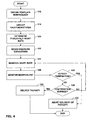
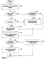
- FIGS. 5 and 6 are flow diagrams illustrating techniques for determining termination of a tachyarrhythmia episode and may be viewed as an expansion of step 128 of FIG. 4 .
- FIG. 5 is a flow diagram illustrating detection of termination of a tachyarrhythmia episode based on a relative decrease in tachyarrhythmia rate and a normalization of electrogram morphology criteria. Although termination of the tachyarrhythmia episode may be detected based on a relative decrease in tachyarrhythmia rate, a normalization of electrogram morphology criteria, or a combination thereof, for purposes of illustration, the termination detection technique of FIG.
- termination may be detected based on the relative decrease in tachyarrhythmia rate alone or, optionally, in combination with a normalization of the electrogram morphology criteria.
- Implanted medical device 10 determines whether an R-R interval associated with a heart beat exceeds a threshold R-R interval (134).
- a threshold R-R interval is relative to the particular tachyarrhythmia episode experienced by the patient.
- the threshold R-R interval may be a metric of a number of R-R intervals of the tachyarrhythmia episode.
- the threshold R-R interval may be a mean or median of the first ten R-R intervals or a rolling median of the most recent ten R-R intervals of the tachyarrhythmia episode. Atrial episodes can be very long and the rate at detection can be very different than the rate later into the episode. For this reason, the use of a rolling median may be desirable for atrial tachyarrhythmia episodes.
- the threshold R-R interval may be the mean or median of a number of R-R intervals plus an offset.
- the threshold R-R interval may be the median of the first ten R-R intervals of the tachyarrhythmia episode plus fifty milliseconds.
- the threshold R-R interval may also be a defined value, such as 400 milliseconds.
- implanted medical device 10 compares an obtained morphology of the cardiac waveform associated with the beat to a template morphology to determine whether the obtained morphology categorizes as normal (138). When implanted medical device 10 categorizes the morphology as abnormal compared to the template morphology, implanted medical device 10 classifies the beat associated with the morphology as indicating continuation of the tachyarrhythmia episode (136) and stores the classification information (141).
- implanted medical device 10 categorizes the obtained morphology as normal compared to the template morphology, implanted medical device 10 classifies the beat associated with the morphology as indicating termination of the tachyarrhythmia episode (140) and stores the classification information (141). Implanted medical device 10 then determines whether a number of beats indicating termination of the tachyarrhythmia episode exceed a threshold (142). When the number of beats indicating termination of the tachyarrhythmia episode exceeds the threshold, implanted medical device 10 detects termination of the tachyarrhythmia episode (144). For example, implanted medical device 10 may consider the tachyarrhythmia episode terminated when four out of the last five beats indicate termination.
- a successive number of beats may need to indicate termination of the tachyarrhythmia episode in order to detect termination of the tachyarrhythmia episode.
- implanted medical device 10 may consider the tachyarrhythmia episode terminated when five consecutive beats indicate termination.
- implanted medical device 10 determines that the tachyarrhythmia episode has not terminated (146).
- FIG. 6 is another flow diagram illustrating detection of termination of a tachyarrhythmia episode based a relative decrease in tachyarrhythmia rate and a normalization of electrogram morphology criteria.
- termination of the tachyarrhythmia episode may be detected based on a relative decrease in tachyarrhythmia rate, a normalization of electrogram morphology criteria, or a combination thereof, for purposes of illustration, the termination detection technique of FIG. 6 is based on both a relative decrease in tachyarrhythmia rate and a normalization of electrogram morphology criteria.
- Implanted medical device 10 determines a range of R-R intervals (148). Although in the example of FIG. 6 the R-R interval is used as a measure of a tachyarrhythmia rate, any atrial or ventricular interval may be used as discussed above. Implanted medical device 10 may calculate, for example, a metric of a number of R-R intervals of the tachyarrhythmia episode and identify a range using the calculated metric of the R-R intervals. For instance, the R-R interval range may extend from a mean or median R-R interval value plus sixty milliseconds to a mean or median R-R interval plus one hundred milliseconds. In this manner, the R-R interval range is relative to each tachyarrhythmia episode. Alternatively, the R-R interval range may be defined, in which case, the range is common to all tachyarrhythmia episodes.
- Implanted medical device 10 determines whether an R-R interval associated with a heart beat is within the R-R interval range (150). When the R-R interval is outside of the R-R interval range, implanted medical device 10 determines whether the R-R interval falls below the R-R interval range (152). When the R-R interval falls below the R-R interval range, implanted medical device 10 classifies the beat associated with the R-R interval as indicating continuation of the tachyarrhythmia episode (154) and stores the classification information (159).
- implanted medical device 10 compares a morphology of a cardiac waveform associated with the beat to a template morphology to determine whether the obtained morphology categorizes as normal (156). When implanted medical device 10 categorizes the morphology as abnormal, implanted medical device 10 classifies the beat as indicating continuation of the tachyarrhythmia episode (154) and stores the classification information (159).
- implanted medical device 10 categorizes the morphology as normal, implanted medical device 10 classifies the beat as indicating termination of the tachyarrhythmia episode (158) and stores the classification information (159). Implanted medical device 10 then determines whether a number of beats indicating termination of the tachyarrhythmia episode exceed a threshold (160). When the number of beats indicating termination of the tachyarrhythmia episode exceeds the threshold, implanted medical device 10 detects termination of the tachyarrhythmia episode (162).
- implanted medical device 10 classifies the tachyarrhythmia episode as not terminated (164).
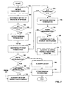
- FIG. 7 is a flow diagram illustrating a technique for classifying the type of a tachyarrhythmia episode based on information obtained at a spontaneous termination of the episode.
- the technique illustrated by FIG. 7 is directed to cases of ventricular tachyarrhythmia for purposes example. In other embodiments, however, the invention may be applied to atrial tachyarrhythmia.
- a medical device such as implanted medical device 10, detects a tachyarrhythmia episode (166).
- Implanted medical device 10 tracks ventricular and atrial events to determine a metric of a tachyarrhythmia rate of the tachyarrhythmia episode (168).
- implanted medical device 10 may calculate a mean tachyarrhythmia rate of the tachyarrhythmia episode, a median tachyarrhythmia rate of the tachyarrhythmia episode, a maximum of the tachyarrhythmia rate, a minimum of the tachyarrhythmia rate, or similar metric of the tachyarrhythmia rate.
- Implanted medical device 10 determines whether the tachyarrhythmia episode has an atrial to ventricular (A:V) conduction ratio that is less than 1:1 (170). When the A:V conduction ratio of the tachyarrhythmia episode is less than 1:1, implanted medical device 10 waits to detect episode termination (172) and classifies the tachyarrhythmia episode as a ventricular tachycardia (VT) or double tachycardia (192). Implanted medical device 10 may detect termination of the tachyarrhythmia episode based on an absolute or relative decrease in tachyarrhythmia rate, a normalization of electrogram morphology criteria, or a combination thereof.
- A:V atrial to ventricular
- implanted medical device 10 waits to detect episode termination (174, 176). After detecting episode termination, implanted medical device 10 determines whether the rate of the tachyarrhythmia episode rapidly decelerates at termination (178). For example, implanted medical device 10 may determine a metric of deceleration, such as a difference between two consecutive R-R intervals being larger than a threshold. Other metrics of deceleration may also be used. For example, the metric of deceleration may include the difference between two median heart rates or two mean heart rates.
- implanted medical device 10 classifies the tachyarrhythmia episode as a ventricular tachycardia (VT) or a double tachycardia (180).
- implanted medical device 10 classifies the tachyarrhythmia episode as atrial fibrillation or atrial flutter (182).
- implanted medical device 10 waits to detect episode termination (174, 184) and determines whether the rate of the tachyarrhythmia episode rapidly decelerates at termination (186). When the tachyarrhythmia episode does not rapidly decelerate, implanted medical device 10 classifies the tachyarrhythmia episode as a supraventricular tachycardia (SVT) (188).
- SVT supraventricular tachycardia
- implanted medical device 10 determines from the tracked ventricular and atrial events whether a last event prior to the deceleration was a ventricular event (190). When the last event prior to the deceleration was a ventricular event, implanted medical device 10 classifies the tachyarrhythmia episode as a supraventricular tachycardia (SVT) (188). When the last event prior to the deceleration was not a ventricular event, i.e., was an atrial event, implanted medical device 10 classifies the tachyarrhythmia episode as a ventricular tachycardia (VT) or a double tachycardia (192).
- SVT supraventricular tachycardia
- Implanted medical device 10 stores tachyarrhythmia episode information upon classifying the tachyarrhythmia episode (194).
- Tachyarrhythmia episode information may include, for example, the type of tachyarrhythmia episode, the threshold tachyarrhythmia rate of the patient, atrial and ventricular events during the tachyarrhythmia episode, the morphologies obtained during the tachyarrhythmia episode, and the like.
- implanted medical device 10 may conserve memory by storing certain classes of tachyarrhythmia episodes.
- FIG. 8 is a flow diagram illustrating a technique for providing therapy to treat a tachyarrhythmia episode based on a rate of the tachyarrhythmia episode and optionally a morphology of a cardiac waveform.
- the technique illustrated by FIG. 8 is directed to cases of ventricular tachyarrhythmia for purposes example. In other embodiments, however, the invention may be applied to atrial tachyarrhythmia.
- a medical device such as implanted medical device 10, detects a tachyarrhythmia episode (196).
- Implanted medical device 10 tracks ventricular and atrial events to determine a metric of a rate of the tachyarrhythmia episode (198).
- Implanted medical device 10 applies a therapy in an attempt to terminate the tachyarrhythmia episode (200).
- implanted medical device 10 determines a rate of the current rhythm (202).
- Implanted medical device 10 may, for example, determine the tachyarrhythmia rate from the tracked atrial and ventricular events. For instance, the rate of the tachyarrhythmia episode may be a metric of a series of measured R-R intervals, P-P intervals, or the like.
- Implanted medical device 10 determines whether the rate of the tachyarrhythmia episode increased (204). When heart the rate of the tachyarrhythmia episode increases, implanted medical device 10 may select a more aggressive therapy, such as cardioversion or defibrillation, and apply the more aggressive therapy (206, 200).
- a more aggressive therapy such as cardioversion or defibrillation
- implanted medical device 10 determines whether the rate of the tachyarrhythmia episode decreased (208). When the rate of the tachyarrhythmia episode does not decrease, implanted medical device 10 applies fast redetection criteria in attempts to redetect the tachyarrhythmia episode (210, 212).
- the fast redetection criteria may include comparing the rate of the tachyarrhythmia episode to a relative threshold tachyarrhythmia rate, e.g., over a range of 6 to 12 beats.
- implanted medical device 10 redetects the tachyarrhythmia episode, implanted medical device 10 applies another therapy (200).
- implanted medical device 10 determines whether the tachyarrhythmia episode has terminated (214). When the tachyarrhythmia episode has not terminated, implanted medical device 10 reapplies the fast redetection criteria.
- implanted medical device 10 applies slow redetection criteria in attempts to redetect the tachyarrhythmia episode (216, 212).
- the slow redetection criteria may include comparing the rate of the tachyarrhythmia episode to a threshold tachyarrhythmia rate, comparing the pattern of atrial and ventricular depolarization events to known patterns of VT or SVT rhythms, and/or also comparing a morphology of a cardiac waveform associated with a heart beat with a template morphology.
- the slow detection criteria may be applied within an extended range, e.g., 18 to 60 beats.
- implanted medical device 10 When implanted medical device 10 redetects the tachyarrhythmia episode, implanted medical device 10 applies another therapy (200). When implanted medical device 10 does not redetect the tachyarrhythmia episode, implanted medical device 10 determines whether the tachyarrhythmia episode has terminated (214). When the tachyarrhythmia episode has not terminated, implanted medical device 10 reapplies the slow redetection criteria.
Landscapes
- Health & Medical Sciences (AREA)
- Cardiology (AREA)
- Heart & Thoracic Surgery (AREA)
- Engineering & Computer Science (AREA)
- Biomedical Technology (AREA)
- Nuclear Medicine, Radiotherapy & Molecular Imaging (AREA)
- Radiology & Medical Imaging (AREA)
- Life Sciences & Earth Sciences (AREA)
- Animal Behavior & Ethology (AREA)
- General Health & Medical Sciences (AREA)
- Public Health (AREA)
- Veterinary Medicine (AREA)
- Electrotherapy Devices (AREA)
- Measuring Pulse, Heart Rate, Blood Pressure Or Blood Flow (AREA)
Claims (2)
- Vorrichtung, mit:einem ersten Detektor (18, 20, 24), der dazu ausgelegt ist, eine Tachyarrhythmie-Rate bzw. -Frequenz, die einem Herzschlag zugeordnet ist, zu detektieren;einem zweiten Detektor (32, 34), der dazu ausgelegt ist, eine Morphologie einer Herzsignalform, die dem Herzschlag zugeordnet ist, zu detektieren; undeinem Prozessor (68), der dazu ausgelegt ist, auf der Grundlage der Tachyarrhythmie-Rate und der Morphologie zu bestimmen, ob eine detektierte Tachyarrhythmie-Episode geendet hat, dadurch gekennzeichnet, dass der Prozessor dazu ausgelegt ist, den Schlag als das Ende der Tachyarrhythmie-Episode angebend zu klassifizieren und zu bestimmen, dass die detektierte Tachyarrhythmie-Episode geendet hat, wenn eine Anzahl von Schlägen, die als das Ende der Tachyarrhythmie-Episode angebend klassifiziert werden, einen Schwellenwert übersteigt, wobei der Prozessor dazu ausgelegt ist, bei der Bestimmung, dass die Tachyarrhythmie-Episode spontan geendet hat, die Tachyarrhythmie-Episode durch Überwachen atrialer und ventrikulärer Ereignisse unmittelbar vor der Verlangsamung der Tachyarrhythmie-Rate neu zu klassifizieren, derart, dass dann, wenn ein atriales Ereignis das letzte Ereignis vor der Verlangsamung ist, der Prozessor die Tachyarrhythmie-Episode als eine ventrikuläre Tachycardie klassifiziert, und dann, wenn ein ventrikuläres Ereignis das letzte Ereignis vor der Verlangsamung ist, der Prozessor die Tachyarrhythmie-Episode als eine supraventrikuläre Tachycardie klassifiziert.
- Vorrichtung nach Anspruch 1, wobei der Prozessor dazu ausgelegt ist, die Tachyarrhythmie-Rate mit einer Schwellen-Tachyarrhythmie-Rate zu vergleichen, die Morphologie der Herzsignalform mit einer Schablonenmorphologie der Herzsignalform zu vergleichen und den Schlag als das Ende der Tachyarrhythmie-Episode angebend zu klassifizieren, wenn die Tachyarrhythmie-Rate geringer als die Schwellen-Tachyarrhythmie-Rate ist und wenn die Morphologie als normal kategorisiert wird.
Applications Claiming Priority (3)
| Application Number | Priority Date | Filing Date | Title |
|---|---|---|---|
| US375457 | 1995-01-20 | ||
| US10/375,457 US7103404B2 (en) | 2003-02-27 | 2003-02-27 | Detection of tachyarrhythmia termination |
| PCT/US2004/004805 WO2004075983A1 (en) | 2003-02-27 | 2004-02-19 | Detection of tachyarrhythmia termination |
Publications (2)
| Publication Number | Publication Date |
|---|---|
| EP1601416A1 EP1601416A1 (de) | 2005-12-07 |
| EP1601416B1 true EP1601416B1 (de) | 2010-12-15 |
Family
ID=32907822
Family Applications (1)
| Application Number | Title | Priority Date | Filing Date |
|---|---|---|---|
| EP04712822A Expired - Lifetime EP1601416B1 (de) | 2003-02-27 | 2004-02-19 | Detektion einer tachykardiebeendigung |
Country Status (6)
| Country | Link |
|---|---|
| US (1) | US7103404B2 (de) |
| EP (1) | EP1601416B1 (de) |
| JP (1) | JP2006519057A (de) |
| CA (1) | CA2517293A1 (de) |
| DE (1) | DE602004030540D1 (de) |
| WO (1) | WO2004075983A1 (de) |
Families Citing this family (72)
| Publication number | Priority date | Publication date | Assignee | Title |
|---|---|---|---|---|
| US7191004B2 (en) * | 2002-12-31 | 2007-03-13 | Cardiac Pacemakers, Inc. | Capture verification using an evoked response reference |
| US7162301B2 (en) * | 2002-12-31 | 2007-01-09 | Cardiac Pacemakers, Inc. | Method and system for detecting capture with cancellation of pacing artifact |
| US7536224B2 (en) * | 2003-04-30 | 2009-05-19 | Medtronic, Inc. | Method for elimination of ventricular pro-arrhythmic effect caused by atrial therapy |
| US7477932B2 (en) | 2003-05-28 | 2009-01-13 | Cardiac Pacemakers, Inc. | Cardiac waveform template creation, maintenance and use |
| US20060247693A1 (en) | 2005-04-28 | 2006-11-02 | Yanting Dong | Non-captured intrinsic discrimination in cardiac pacing response classification |
| US7319900B2 (en) | 2003-12-11 | 2008-01-15 | Cardiac Pacemakers, Inc. | Cardiac response classification using multiple classification windows |
| US8521284B2 (en) | 2003-12-12 | 2013-08-27 | Cardiac Pacemakers, Inc. | Cardiac response classification using multisite sensing and pacing |
| US7774064B2 (en) | 2003-12-12 | 2010-08-10 | Cardiac Pacemakers, Inc. | Cardiac response classification using retriggerable classification windows |
| US7706866B2 (en) | 2004-06-24 | 2010-04-27 | Cardiac Pacemakers, Inc. | Automatic orientation determination for ECG measurements using multiple electrodes |
| ATE517658T1 (de) * | 2004-09-30 | 2011-08-15 | Cardiac Pacemakers Inc | Arrythmie-klassifizierung und therapiewahl |
| US7917196B2 (en) | 2005-05-09 | 2011-03-29 | Cardiac Pacemakers, Inc. | Arrhythmia discrimination using electrocardiograms sensed from multiple implanted electrodes |
| US7805185B2 (en) | 2005-05-09 | 2010-09-28 | Cardiac Pacemakers, In. | Posture monitoring using cardiac activation sequences |
| US7277747B2 (en) * | 2004-11-23 | 2007-10-02 | Cardiac Pacemakers, Inc. | Arrhythmia memory for tachyarrhythmia discrimination |
| US7457664B2 (en) | 2005-05-09 | 2008-11-25 | Cardiac Pacemakers, Inc. | Closed loop cardiac resynchronization therapy using cardiac activation sequence information |
| US7797036B2 (en) | 2004-11-30 | 2010-09-14 | Cardiac Pacemakers, Inc. | Cardiac activation sequence monitoring for ischemia detection |
| US7509170B2 (en) | 2005-05-09 | 2009-03-24 | Cardiac Pacemakers, Inc. | Automatic capture verification using electrocardiograms sensed from multiple implanted electrodes |
| US7894893B2 (en) * | 2004-09-30 | 2011-02-22 | Cardiac Pacemakers, Inc. | Arrhythmia classification and therapy selection |
| US7890159B2 (en) | 2004-09-30 | 2011-02-15 | Cardiac Pacemakers, Inc. | Cardiac activation sequence monitoring and tracking |
| US7477935B2 (en) * | 2004-11-29 | 2009-01-13 | Cameron Health, Inc. | Method and apparatus for beat alignment and comparison |
| US7280869B2 (en) * | 2004-12-03 | 2007-10-09 | Medtronic, Inc. | Arrhythmia termination detection based on beat pattern |
| US7761162B2 (en) * | 2004-12-13 | 2010-07-20 | Cardiac Pacemakers, Inc. | Capture verification with intrinsic response discrimination |
| US8229563B2 (en) * | 2005-01-25 | 2012-07-24 | Cameron Health, Inc. | Devices for adapting charge initiation for an implantable cardioverter-defibrillator |
| AU2012211492B2 (en) * | 2005-01-25 | 2014-05-15 | Cameron Health, Inc. | Devices for adapting charge initiation for an implantable cardioverter-defribrillator |
| US8160697B2 (en) | 2005-01-25 | 2012-04-17 | Cameron Health, Inc. | Method for adapting charge initiation for an implantable cardioverter-defibrillator |
| WO2006081027A2 (en) * | 2005-01-25 | 2006-08-03 | Cameron Health, Inc. | Devices for adapting charge initiation for an implantable cardioverter-defibrillator |
| US7392086B2 (en) | 2005-04-26 | 2008-06-24 | Cardiac Pacemakers, Inc. | Implantable cardiac device and method for reduced phrenic nerve stimulation |
| US7574260B2 (en) * | 2005-04-28 | 2009-08-11 | Cardiac Pacemakers, Inc. | Adaptive windowing for cardiac waveform discrimination |
| US7499751B2 (en) * | 2005-04-28 | 2009-03-03 | Cardiac Pacemakers, Inc. | Cardiac signal template generation using waveform clustering |
| US7765004B2 (en) * | 2005-04-28 | 2010-07-27 | Cardiac Pacemakers, Inc. | Methods and systems for managing fusion and noise in cardiac pacing response classification |
| US7392088B2 (en) * | 2005-04-28 | 2008-06-24 | Cardiac Pacemakers, Inc. | Capture detection for multi-chamber pacing |
| US7529578B2 (en) * | 2005-07-12 | 2009-05-05 | Cardiac Pacemakers, Inc. | Multi channel approach to capture verification |
| US7908001B2 (en) * | 2005-08-23 | 2011-03-15 | Cardiac Pacemakers, Inc. | Automatic multi-level therapy based on morphologic organization of an arrhythmia |
| US8831725B2 (en) * | 2005-11-28 | 2014-09-09 | Medtronic, Inc. | Method and apparatus for post-processing of episodes detected by a medical device |
| US8532762B2 (en) * | 2005-12-20 | 2013-09-10 | Cardiac Pacemakers, Inc. | Discriminating polymorphic and monomorphic cardiac rhythms using template generation |
| US7653431B2 (en) * | 2005-12-20 | 2010-01-26 | Cardiac Pacemakers, Inc. | Arrhythmia discrimination based on determination of rate dependency |
| US7937137B2 (en) * | 2006-01-30 | 2011-05-03 | Medtronic, Inc. | Methods of identifying and/or assessing cardiac arrhythmias using an implantable medical device |
| US7761142B2 (en) * | 2006-03-29 | 2010-07-20 | Medtronic, Inc. | Method and apparatus for detecting arrhythmias in a medical device |
| US8437850B2 (en) * | 2006-05-18 | 2013-05-07 | Medtronic, Inc. | Automatic enabling of post long pause overdrive pacing |
| US7729763B2 (en) * | 2006-05-18 | 2010-06-01 | Medtronic, Inc. | Post long pause overdrive pacing in response to atrial tachyarrythmia episode |
| DE102006023517A1 (de) * | 2006-05-19 | 2007-11-22 | Biotronik Crm Patent Ag | Herzstimulator |
| US8527048B2 (en) | 2006-06-29 | 2013-09-03 | Cardiac Pacemakers, Inc. | Local and non-local sensing for cardiac pacing |
| US7580741B2 (en) | 2006-08-18 | 2009-08-25 | Cardiac Pacemakers, Inc. | Method and device for determination of arrhythmia rate zone thresholds using a probability function |
| US8712507B2 (en) | 2006-09-14 | 2014-04-29 | Cardiac Pacemakers, Inc. | Systems and methods for arranging and labeling cardiac episodes |
| US8209013B2 (en) | 2006-09-14 | 2012-06-26 | Cardiac Pacemakers, Inc. | Therapeutic electrical stimulation that avoids undesirable activation |
| US7941208B2 (en) | 2006-11-29 | 2011-05-10 | Cardiac Pacemakers, Inc. | Therapy delivery for identified tachyarrhythmia episode types |
| JP4649429B2 (ja) * | 2007-03-12 | 2011-03-09 | 株式会社大成 | 心拍測定システム及び方法 |
| US9037239B2 (en) | 2007-08-07 | 2015-05-19 | Cardiac Pacemakers, Inc. | Method and apparatus to perform electrode combination selection |
| US8265736B2 (en) | 2007-08-07 | 2012-09-11 | Cardiac Pacemakers, Inc. | Method and apparatus to perform electrode combination selection |
| US8649866B2 (en) | 2008-02-14 | 2014-02-11 | Cardiac Pacemakers, Inc. | Method and apparatus for phrenic stimulation detection |
| WO2010042364A2 (en) | 2008-10-06 | 2010-04-15 | Cardiac Pacemakers, Inc. | Dynamic cardiac resynchronization therapy by tracking intrinsic conduction |
| DE102008043480A1 (de) * | 2008-11-05 | 2010-05-06 | Biotronik Crm Patent Ag | Einkammer-Herzstimulator |
| US8428697B2 (en) * | 2009-01-22 | 2013-04-23 | Medtronic, Inc. | “Blurred template” approach for arrhythmia detection |
| US8483808B2 (en) | 2009-09-25 | 2013-07-09 | Yanting Dong | Methods and systems for characterizing cardiac signal morphology using K-fit analysis |
| US8437840B2 (en) * | 2011-09-26 | 2013-05-07 | Medtronic, Inc. | Episode classifier algorithm |
| US8774909B2 (en) | 2011-09-26 | 2014-07-08 | Medtronic, Inc. | Episode classifier algorithm |
| CN105208928B (zh) | 2013-03-11 | 2018-10-19 | 卡梅伦保健公司 | 实施心律失常检测双重标准的设备 |
| WO2014210527A1 (en) | 2013-06-28 | 2014-12-31 | The General Hospital Corporation | System and method to infer brain state during burst suppression |
| US9643025B2 (en) | 2014-09-08 | 2017-05-09 | Medtronic, Inc. | Multi-primary transformer charging circuits for implantable medical devices |
| US9604071B2 (en) | 2014-09-08 | 2017-03-28 | Medtronic, Inc. | Implantable medical devices having multi-cell power sources |
| US9861827B2 (en) | 2014-09-08 | 2018-01-09 | Medtronic, Inc. | Implantable medical devices having multi-cell power sources |
| US9724528B2 (en) | 2014-09-08 | 2017-08-08 | Medtronic, Inc. | Multiple transformer charging circuits for implantable medical devices |
| US9861828B2 (en) | 2014-09-08 | 2018-01-09 | Medtronic, Inc. | Monitoring multi-cell power source of an implantable medical device |
| US9539435B2 (en) | 2014-09-08 | 2017-01-10 | Medtronic, Inc. | Transthoracic protection circuit for implantable medical devices |
| US9579517B2 (en) | 2014-09-08 | 2017-02-28 | Medtronic, Inc. | Transformer-based charging circuits for implantable medical devices |
| CN107072580B (zh) * | 2014-10-22 | 2020-08-25 | 美敦力公司 | 心脏医疗设备中的心房心律失常发病检测 |
| ES2713231T3 (es) | 2015-02-06 | 2019-05-20 | Cardiac Pacemakers Inc | Sistemas para el suministro seguro de una terapia de estimulación eléctrica |
| US10159847B2 (en) | 2015-05-20 | 2018-12-25 | Medtronic, Inc. | Implantable medical devices with active component monitoring |
| CN110573068B (zh) * | 2017-03-07 | 2023-01-03 | 心脏起搏器股份公司 | 用于房性心律失常检测的系统和方法 |
| JP6938778B2 (ja) | 2017-09-20 | 2021-09-22 | カーディアック ペースメイカーズ, インコーポレイテッド | 複数の作動モードを備えた移植式医療用装置 |
| US10967187B2 (en) * | 2017-12-01 | 2021-04-06 | Pacesetter, Inc. | Method and device for managing a self-termination period for ventricular arrhythmias |
| JP2024506138A (ja) * | 2021-02-11 | 2024-02-09 | バイオトロニック エスエー アンド カンパニー カーゲー | 心臓内機能を提供するように構成された植込み型医療装置 |
| US12594019B2 (en) | 2021-06-04 | 2026-04-07 | Medtronic, Inc. | Detection of atrial tachycardia based on regularity of cardiac rhythm |
Family Cites Families (19)
| Publication number | Priority date | Publication date | Assignee | Title |
|---|---|---|---|---|
| US4830006B1 (en) * | 1986-06-17 | 1997-10-28 | Intermedics Inc | Implantable cardiac stimulator for detection and treatment of ventricular arrhythmias |
| US4949719A (en) * | 1989-04-26 | 1990-08-21 | Ventritex, Inc. | Method for cardiac defibrillation |
| US5193536A (en) | 1990-12-20 | 1993-03-16 | Medtronic, Inc. | Paced cardioversion |
| US5240009A (en) | 1991-03-25 | 1993-08-31 | Ventritex, Inc. | Medical device with morphology discrimination |
| US5312441A (en) * | 1992-04-13 | 1994-05-17 | Medtronic, Inc. | Method and apparatus for discrimination of ventricular tachycardia from supraventricular tachycardia and for treatment thereof |
| US5342402A (en) | 1993-01-29 | 1994-08-30 | Medtronic, Inc. | Method and apparatus for detection and treatment of tachycardia and fibrillation |
| US5400795A (en) | 1993-10-22 | 1995-03-28 | Telectronics Pacing Systems, Inc. | Method of classifying heart rhythms by analyzing several morphology defining metrics derived for a patient's QRS complex |
| WO1997043002A1 (en) | 1996-05-14 | 1997-11-20 | Medtronic, Inc. | Prioritized rule based method and apparatus for diagnosis and treatment of arrhythmias |
| US5730141A (en) | 1996-06-07 | 1998-03-24 | Pacesetter, Inc. | Tachyarrhythmia detection method |
| US5779645A (en) * | 1996-12-17 | 1998-07-14 | Pacesetter, Inc. | System and method for waveform morphology comparison |
| US5941831A (en) * | 1998-04-03 | 1999-08-24 | Pacesetter, Inc. | Method for diagnosing cardiac arrhythmias using interval irregularity |
| US6275732B1 (en) | 1998-06-17 | 2001-08-14 | Cardiac Pacemakers, Inc. | Multiple stage morphology-based system detecting ventricular tachycardia and supraventricular tachycardia |
| US6078837A (en) | 1999-01-27 | 2000-06-20 | Medtronic, Inc. | Method and apparatus for treatment of fibrillation |
| US6169923B1 (en) * | 1999-04-23 | 2001-01-02 | Pacesetter, Inc. | Implantable cardioverter-defibrillator with automatic arrhythmia detection criteria adjustment |
| WO2000069517A1 (en) | 1999-05-12 | 2000-11-23 | Medtronic, Inc. | Monitoring apparatus using wavelet transforms for the analysis of heart rhythms |
| JP4295459B2 (ja) | 2000-02-18 | 2009-07-15 | ストライカー コーポレイション | 外科用器具の切断アクセサリー |
| US6400986B1 (en) * | 2000-04-10 | 2002-06-04 | Cardiac Pacemakers, Inc. | Adaptive anti-tachycardia therapy apparatus and method |
| US6636764B1 (en) * | 2000-11-28 | 2003-10-21 | Pacesetter, Inc. | Safety backup in arrhythmia discrimination algorithm |
| US7149577B2 (en) | 2002-12-02 | 2006-12-12 | Medtronic, Inc. | Apparatus and method using ATP return cycle length for arrhythmia discrimination |
-
2003
- 2003-02-27 US US10/375,457 patent/US7103404B2/en not_active Expired - Lifetime
-
2004
- 2004-02-19 JP JP2006503680A patent/JP2006519057A/ja active Pending
- 2004-02-19 DE DE602004030540T patent/DE602004030540D1/de not_active Expired - Lifetime
- 2004-02-19 WO PCT/US2004/004805 patent/WO2004075983A1/en not_active Ceased
- 2004-02-19 CA CA002517293A patent/CA2517293A1/en not_active Abandoned
- 2004-02-19 EP EP04712822A patent/EP1601416B1/de not_active Expired - Lifetime
Also Published As
| Publication number | Publication date |
|---|---|
| DE602004030540D1 (de) | 2011-01-27 |
| WO2004075983A1 (en) | 2004-09-10 |
| CA2517293A1 (en) | 2004-09-10 |
| EP1601416A1 (de) | 2005-12-07 |
| JP2006519057A (ja) | 2006-08-24 |
| US20040171959A1 (en) | 2004-09-02 |
| US7103404B2 (en) | 2006-09-05 |
Similar Documents
| Publication | Publication Date | Title |
|---|---|---|
| US7103404B2 (en) | Detection of tachyarrhythmia termination | |
| EP1622678B1 (de) | System zur entfernung des pro-arrhythmischen effekts verursacht durch atrialtherapie | |
| EP1677673B1 (de) | Verfahren und gerät zum nachweis und zur diskriminierung von arrhythmien | |
| EP1339455B1 (de) | Automatische erzeugung von mustern | |
| US5275621A (en) | Method and apparatus for terminating tachycardia | |
| US6959212B2 (en) | System and method for arrhythmia discrimination | |
| EP1515778B1 (de) | Vorrichtung zur verhütung von arrhythmie-häufungen unter verwendung von schnellgang-impulsgebung | |
| US20050288725A1 (en) | Combination of electrogram and intra-cardiac pressure to discriminate between fibrillation and tachycardia | |
| US20110152957A1 (en) | Chaos-based detection of atrial fibrillation using an implantable medical device | |
| EP1737535B1 (de) | Automatische vorlagengeneration beim implantierbaren gerät | |
| JP4865713B2 (ja) | 頻拍性不整脈のためのatp治療 | |
| CA2562088A1 (en) | Automated template generation algorithm for implantable device | |
| US20060122650A1 (en) | Arrhythmia termination detection based on beat pattern | |
| CN107072580B (zh) | 心脏医疗设备中的心房心律失常发病检测 | |
| US7065398B2 (en) | Atrial tachycardia termination sensing |
Legal Events
| Date | Code | Title | Description |
|---|---|---|---|
| PUAI | Public reference made under article 153(3) epc to a published international application that has entered the european phase |
Free format text: ORIGINAL CODE: 0009012 |
|
| 17P | Request for examination filed |
Effective date: 20050927 |
|
| AK | Designated contracting states |
Kind code of ref document: A1 Designated state(s): AT BE BG CH CY CZ DE DK EE ES FI FR GB GR HU IE IT LI LU MC NL PT RO SE SI SK TR |
|
| AX | Request for extension of the european patent |
Extension state: AL LT LV MK |
|
| DAX | Request for extension of the european patent (deleted) | ||
| RBV | Designated contracting states (corrected) |
Designated state(s): CH DE FR LI NL SE |
|
| RAP3 | Party data changed (applicant data changed or rights of an application transferred) |
Owner name: MEDTRONIC, INC. |
|
| 17Q | First examination report despatched |
Effective date: 20080813 |
|
| GRAP | Despatch of communication of intention to grant a patent |
Free format text: ORIGINAL CODE: EPIDOSNIGR1 |
|
| GRAS | Grant fee paid |
Free format text: ORIGINAL CODE: EPIDOSNIGR3 |
|
| GRAA | (expected) grant |
Free format text: ORIGINAL CODE: 0009210 |
|
| AK | Designated contracting states |
Kind code of ref document: B1 Designated state(s): CH DE FR LI NL SE |
|
| REG | Reference to a national code |
Ref country code: CH Ref legal event code: EP |
|
| REF | Corresponds to: |
Ref document number: 602004030540 Country of ref document: DE Date of ref document: 20110127 Kind code of ref document: P |
|
| REG | Reference to a national code |
Ref country code: NL Ref legal event code: VDEP Effective date: 20101215 |
|
| PG25 | Lapsed in a contracting state [announced via postgrant information from national office to epo] |
Ref country code: SE Free format text: LAPSE BECAUSE OF FAILURE TO SUBMIT A TRANSLATION OF THE DESCRIPTION OR TO PAY THE FEE WITHIN THE PRESCRIBED TIME-LIMIT Effective date: 20101215 Ref country code: NL Free format text: LAPSE BECAUSE OF FAILURE TO SUBMIT A TRANSLATION OF THE DESCRIPTION OR TO PAY THE FEE WITHIN THE PRESCRIBED TIME-LIMIT Effective date: 20101215 |
|
| REG | Reference to a national code |
Ref country code: CH Ref legal event code: PL |
|
| PLBE | No opposition filed within time limit |
Free format text: ORIGINAL CODE: 0009261 |
|
| STAA | Information on the status of an ep patent application or granted ep patent |
Free format text: STATUS: NO OPPOSITION FILED WITHIN TIME LIMIT |
|
| PG25 | Lapsed in a contracting state [announced via postgrant information from national office to epo] |
Ref country code: CH Free format text: LAPSE BECAUSE OF NON-PAYMENT OF DUE FEES Effective date: 20110228 Ref country code: LI Free format text: LAPSE BECAUSE OF NON-PAYMENT OF DUE FEES Effective date: 20110228 |
|
| 26N | No opposition filed |
Effective date: 20110916 |
|
| REG | Reference to a national code |
Ref country code: DE Ref legal event code: R097 Ref document number: 602004030540 Country of ref document: DE Effective date: 20110916 |
|
| REG | Reference to a national code |
Ref country code: FR Ref legal event code: PLFP Year of fee payment: 13 |
|
| REG | Reference to a national code |
Ref country code: FR Ref legal event code: PLFP Year of fee payment: 14 |
|
| REG | Reference to a national code |
Ref country code: FR Ref legal event code: PLFP Year of fee payment: 15 |
|
| PGFP | Annual fee paid to national office [announced via postgrant information from national office to epo] |
Ref country code: FR Payment date: 20190123 Year of fee payment: 16 Ref country code: DE Payment date: 20190122 Year of fee payment: 16 |
|
| REG | Reference to a national code |
Ref country code: DE Ref legal event code: R119 Ref document number: 602004030540 Country of ref document: DE |
|
| PG25 | Lapsed in a contracting state [announced via postgrant information from national office to epo] |
Ref country code: DE Free format text: LAPSE BECAUSE OF NON-PAYMENT OF DUE FEES Effective date: 20200901 Ref country code: FR Free format text: LAPSE BECAUSE OF NON-PAYMENT OF DUE FEES Effective date: 20200229 |