EP1580558A1 - Procede d'analyse du staphylococcus aureus - Google Patents
Procede d'analyse du staphylococcus aureus Download PDFInfo
- Publication number
- EP1580558A1 EP1580558A1 EP03774149A EP03774149A EP1580558A1 EP 1580558 A1 EP1580558 A1 EP 1580558A1 EP 03774149 A EP03774149 A EP 03774149A EP 03774149 A EP03774149 A EP 03774149A EP 1580558 A1 EP1580558 A1 EP 1580558A1
- Authority
- EP
- European Patent Office
- Prior art keywords
- dnase
- aureus
- antibody
- enzyme
- specimen
- Prior art date
- Legal status (The legal status is an assumption and is not a legal conclusion. Google has not performed a legal analysis and makes no representation as to the accuracy of the status listed.)
- Withdrawn
Links
- 238000000034 method Methods 0.000 title claims abstract description 25
- 241000191967 Staphylococcus aureus Species 0.000 title claims abstract description 11
- 102000016911 Deoxyribonucleases Human genes 0.000 claims abstract description 49
- 108010053770 Deoxyribonucleases Proteins 0.000 claims abstract description 49
- 238000003018 immunoassay Methods 0.000 claims abstract description 35
- 238000012360 testing method Methods 0.000 claims abstract description 23
- 238000010998 test method Methods 0.000 claims abstract description 12
- 102000004190 Enzymes Human genes 0.000 claims description 44
- 108090000790 Enzymes Proteins 0.000 claims description 44
- 239000000126 substance Substances 0.000 claims description 11
- 102000003992 Peroxidases Human genes 0.000 claims description 5
- 108040007629 peroxidase activity proteins Proteins 0.000 claims description 5
- 210000001124 body fluid Anatomy 0.000 claims description 4
- 239000010839 body fluid Substances 0.000 claims description 4
- 239000003153 chemical reaction reagent Substances 0.000 claims description 4
- RJQXTJLFIWVMTO-TYNCELHUSA-N Methicillin Chemical compound COC1=CC=CC(OC)=C1C(=O)N[C@@H]1C(=O)N2[C@@H](C(O)=O)C(C)(C)S[C@@H]21 RJQXTJLFIWVMTO-TYNCELHUSA-N 0.000 claims description 3
- HWYHZTIRURJOHG-UHFFFAOYSA-N luminol Chemical compound O=C1NNC(=O)C2=C1C(N)=CC=C2 HWYHZTIRURJOHG-UHFFFAOYSA-N 0.000 claims description 3
- 229960003085 meticillin Drugs 0.000 claims description 3
- 238000001514 detection method Methods 0.000 abstract description 12
- 230000035945 sensitivity Effects 0.000 abstract description 12
- 229940088598 enzyme Drugs 0.000 description 40
- 241000894006 Bacteria Species 0.000 description 16
- 238000006243 chemical reaction Methods 0.000 description 15
- 238000005259 measurement Methods 0.000 description 15
- MHAJPDPJQMAIIY-UHFFFAOYSA-N Hydrogen peroxide Chemical compound OO MHAJPDPJQMAIIY-UHFFFAOYSA-N 0.000 description 8
- 239000000758 substrate Substances 0.000 description 8
- 239000000243 solution Substances 0.000 description 7
- 210000002700 urine Anatomy 0.000 description 7
- 238000003556 assay Methods 0.000 description 6
- 238000012767 chemiluminescent enzyme immunoassay Methods 0.000 description 6
- 210000002966 serum Anatomy 0.000 description 6
- 239000007787 solid Substances 0.000 description 6
- 241000283973 Oryctolagus cuniculus Species 0.000 description 5
- 241001494479 Pecora Species 0.000 description 5
- 238000010790 dilution Methods 0.000 description 5
- 239000012895 dilution Substances 0.000 description 5
- 230000000694 effects Effects 0.000 description 5
- 239000008055 phosphate buffer solution Substances 0.000 description 5
- 108010065152 Coagulase Proteins 0.000 description 4
- 238000002965 ELISA Methods 0.000 description 4
- QAOWNCQODCNURD-UHFFFAOYSA-N Sulfuric acid Chemical compound OS(O)(=O)=O QAOWNCQODCNURD-UHFFFAOYSA-N 0.000 description 4
- 239000000427 antigen Substances 0.000 description 4
- 108091007433 antigens Proteins 0.000 description 4
- 102000036639 antigens Human genes 0.000 description 4
- 210000004369 blood Anatomy 0.000 description 4
- 239000008280 blood Substances 0.000 description 4
- 238000002372 labelling Methods 0.000 description 4
- 108090000623 proteins and genes Proteins 0.000 description 4
- GEYOCULIXLDCMW-UHFFFAOYSA-N 1,2-phenylenediamine Chemical compound NC1=CC=CC=C1N GEYOCULIXLDCMW-UHFFFAOYSA-N 0.000 description 3
- 102000002260 Alkaline Phosphatase Human genes 0.000 description 3
- 108020004774 Alkaline Phosphatase Proteins 0.000 description 3
- 206010003445 Ascites Diseases 0.000 description 3
- 108091003079 Bovine Serum Albumin Proteins 0.000 description 3
- 208000002151 Pleural effusion Diseases 0.000 description 3
- 206010036790 Productive cough Diseases 0.000 description 3
- 241000191940 Staphylococcus Species 0.000 description 3
- 230000001580 bacterial effect Effects 0.000 description 3
- 229940098773 bovine serum albumin Drugs 0.000 description 3
- 239000007853 buffer solution Substances 0.000 description 3
- 238000000354 decomposition reaction Methods 0.000 description 3
- 238000007865 diluting Methods 0.000 description 3
- 239000012634 fragment Substances 0.000 description 3
- 102000004169 proteins and genes Human genes 0.000 description 3
- 210000003802 sputum Anatomy 0.000 description 3
- 208000024794 sputum Diseases 0.000 description 3
- 238000005406 washing Methods 0.000 description 3
- YBJHBAHKTGYVGT-ZKWXMUAHSA-N (+)-Biotin Chemical compound N1C(=O)N[C@@H]2[C@H](CCCCC(=O)O)SC[C@@H]21 YBJHBAHKTGYVGT-ZKWXMUAHSA-N 0.000 description 2
- XQXPVVBIMDBYFF-UHFFFAOYSA-N 4-hydroxyphenylacetic acid Chemical compound OC(=O)CC1=CC=C(O)C=C1 XQXPVVBIMDBYFF-UHFFFAOYSA-N 0.000 description 2
- 230000004544 DNA amplification Effects 0.000 description 2
- VEXZGXHMUGYJMC-UHFFFAOYSA-N Hydrochloric acid Chemical compound Cl VEXZGXHMUGYJMC-UHFFFAOYSA-N 0.000 description 2
- UIIMBOGNXHQVGW-UHFFFAOYSA-M Sodium bicarbonate Chemical compound [Na+].OC([O-])=O UIIMBOGNXHQVGW-UHFFFAOYSA-M 0.000 description 2
- FAPWRFPIFSIZLT-UHFFFAOYSA-M Sodium chloride Chemical compound [Na+].[Cl-] FAPWRFPIFSIZLT-UHFFFAOYSA-M 0.000 description 2
- 208000033809 Suppuration Diseases 0.000 description 2
- 102000005936 beta-Galactosidase Human genes 0.000 description 2
- 108010005774 beta-Galactosidase Proteins 0.000 description 2
- 239000000872 buffer Substances 0.000 description 2
- 230000003197 catalytic effect Effects 0.000 description 2
- 239000007979 citrate buffer Substances 0.000 description 2
- 239000012228 culture supernatant Substances 0.000 description 2
- 238000005516 engineering process Methods 0.000 description 2
- 239000012530 fluid Substances 0.000 description 2
- 208000015181 infectious disease Diseases 0.000 description 2
- 239000004816 latex Substances 0.000 description 2
- 229920000126 latex Polymers 0.000 description 2
- KNJDBYZZKAZQNG-UHFFFAOYSA-N lucigenin Chemical compound [O-][N+]([O-])=O.[O-][N+]([O-])=O.C12=CC=CC=C2[N+](C)=C(C=CC=C2)C2=C1C1=C(C=CC=C2)C2=[N+](C)C2=CC=CC=C12 KNJDBYZZKAZQNG-UHFFFAOYSA-N 0.000 description 2
- 238000004020 luminiscence type Methods 0.000 description 2
- 238000004519 manufacturing process Methods 0.000 description 2
- 101150008979 mecA gene Proteins 0.000 description 2
- 239000012528 membrane Substances 0.000 description 2
- 102000039446 nucleic acids Human genes 0.000 description 2
- 108020004707 nucleic acids Proteins 0.000 description 2
- 150000007523 nucleic acids Chemical class 0.000 description 2
- 239000008363 phosphate buffer Substances 0.000 description 2
- 238000002360 preparation method Methods 0.000 description 2
- 238000001179 sorption measurement Methods 0.000 description 2
- 238000003756 stirring Methods 0.000 description 2
- 239000006228 supernatant Substances 0.000 description 2
- 239000003053 toxin Substances 0.000 description 2
- 231100000765 toxin Toxicity 0.000 description 2
- 108700012359 toxins Proteins 0.000 description 2
- BCHIXGBGRHLSBE-UHFFFAOYSA-N (4-methyl-2-oxochromen-7-yl) dihydrogen phosphate Chemical compound C1=C(OP(O)(O)=O)C=CC2=C1OC(=O)C=C2C BCHIXGBGRHLSBE-UHFFFAOYSA-N 0.000 description 1
- ORIIXCOYEOIFSN-UHFFFAOYSA-N 1,3-benzothiazol-6-ol Chemical compound OC1=CC=C2N=CSC2=C1 ORIIXCOYEOIFSN-UHFFFAOYSA-N 0.000 description 1
- KUWPCJHYPSUOFW-YBXAARCKSA-N 2-nitrophenyl beta-D-galactoside Chemical compound O[C@@H]1[C@@H](O)[C@@H](O)[C@@H](CO)O[C@H]1OC1=CC=CC=C1[N+]([O-])=O KUWPCJHYPSUOFW-YBXAARCKSA-N 0.000 description 1
- XMTQQYYKAHVGBJ-UHFFFAOYSA-N 3-(3,4-DICHLOROPHENYL)-1,1-DIMETHYLUREA Chemical compound CN(C)C(=O)NC1=CC=C(Cl)C(Cl)=C1 XMTQQYYKAHVGBJ-UHFFFAOYSA-N 0.000 description 1
- XZKIHKMTEMTJQX-UHFFFAOYSA-N 4-Nitrophenyl Phosphate Chemical compound OP(O)(=O)OC1=CC=C([N+]([O-])=O)C=C1 XZKIHKMTEMTJQX-UHFFFAOYSA-N 0.000 description 1
- HUDPLKWXRLNSPC-UHFFFAOYSA-N 4-aminophthalhydrazide Chemical class O=C1NNC(=O)C=2C1=CC(N)=CC=2 HUDPLKWXRLNSPC-UHFFFAOYSA-N 0.000 description 1
- VSMDINRNYYEDRN-UHFFFAOYSA-N 4-iodophenol Chemical compound OC1=CC=C(I)C=C1 VSMDINRNYYEDRN-UHFFFAOYSA-N 0.000 description 1
- YUDPTGPSBJVHCN-DZQJYWQESA-N 4-methylumbelliferyl beta-D-galactoside Chemical compound C1=CC=2C(C)=CC(=O)OC=2C=C1O[C@@H]1O[C@H](CO)[C@H](O)[C@H](O)[C@H]1O YUDPTGPSBJVHCN-DZQJYWQESA-N 0.000 description 1
- 108010077805 Bacterial Proteins Proteins 0.000 description 1
- 208000035143 Bacterial infection Diseases 0.000 description 1
- BVKZGUZCCUSVTD-UHFFFAOYSA-L Carbonate Chemical compound [O-]C([O-])=O BVKZGUZCCUSVTD-UHFFFAOYSA-L 0.000 description 1
- 206010011409 Cross infection Diseases 0.000 description 1
- 206010016952 Food poisoning Diseases 0.000 description 1
- 208000019331 Foodborne disease Diseases 0.000 description 1
- 102000002464 Galactosidases Human genes 0.000 description 1
- 108010093031 Galactosidases Proteins 0.000 description 1
- 108010010803 Gelatin Proteins 0.000 description 1
- 108010015776 Glucose oxidase Proteins 0.000 description 1
- 239000004366 Glucose oxidase Substances 0.000 description 1
- SXRSQZLOMIGNAQ-UHFFFAOYSA-N Glutaraldehyde Chemical compound O=CCCCC=O SXRSQZLOMIGNAQ-UHFFFAOYSA-N 0.000 description 1
- -1 HSA Proteins 0.000 description 1
- 241000124008 Mammalia Species 0.000 description 1
- 206010029719 Nonspecific reaction Diseases 0.000 description 1
- 206010029803 Nosocomial infection Diseases 0.000 description 1
- 102000057297 Pepsin A Human genes 0.000 description 1
- 108090000284 Pepsin A Proteins 0.000 description 1
- BJGNCJDXODQBOB-SSDOTTSWSA-N Photinus luciferin Chemical compound OC(=O)[C@H]1CSC(C=2SC3=CC(O)=CC=C3N=2)=N1 BJGNCJDXODQBOB-SSDOTTSWSA-N 0.000 description 1
- 229920001213 Polysorbate 20 Polymers 0.000 description 1
- 241000295644 Staphylococcaceae Species 0.000 description 1
- 230000003698 anagen phase Effects 0.000 description 1
- 230000000844 anti-bacterial effect Effects 0.000 description 1
- 208000022362 bacterial infectious disease Diseases 0.000 description 1
- 244000052616 bacterial pathogen Species 0.000 description 1
- 239000011324 bead Substances 0.000 description 1
- 230000003115 biocidal effect Effects 0.000 description 1
- 229960002685 biotin Drugs 0.000 description 1
- 235000020958 biotin Nutrition 0.000 description 1
- 239000011616 biotin Substances 0.000 description 1
- 150000001718 carbodiimides Chemical class 0.000 description 1
- 238000005119 centrifugation Methods 0.000 description 1
- 239000003593 chromogenic compound Substances 0.000 description 1
- 239000012568 clinical material Substances 0.000 description 1
- 238000004040 coloring Methods 0.000 description 1
- 238000012875 competitive assay Methods 0.000 description 1
- 238000011109 contamination Methods 0.000 description 1
- 239000003431 cross linking reagent Substances 0.000 description 1
- 238000011161 development Methods 0.000 description 1
- 230000018109 developmental process Effects 0.000 description 1
- 230000004069 differentiation Effects 0.000 description 1
- 201000010099 disease Diseases 0.000 description 1
- 208000037265 diseases, disorders, signs and symptoms Diseases 0.000 description 1
- 230000002255 enzymatic effect Effects 0.000 description 1
- 239000000835 fiber Substances 0.000 description 1
- 239000000706 filtrate Substances 0.000 description 1
- 238000002795 fluorescence method Methods 0.000 description 1
- 229920000159 gelatin Polymers 0.000 description 1
- 239000008273 gelatin Substances 0.000 description 1
- 235000019322 gelatine Nutrition 0.000 description 1
- 235000011852 gelatine desserts Nutrition 0.000 description 1
- 239000011521 glass Substances 0.000 description 1
- 229940116332 glucose oxidase Drugs 0.000 description 1
- 235000019420 glucose oxidase Nutrition 0.000 description 1
- PCHJSUWPFVWCPO-UHFFFAOYSA-N gold Chemical compound [Au] PCHJSUWPFVWCPO-UHFFFAOYSA-N 0.000 description 1
- 230000005283 ground state Effects 0.000 description 1
- 230000012010 growth Effects 0.000 description 1
- 238000013095 identification testing Methods 0.000 description 1
- 230000003100 immobilizing effect Effects 0.000 description 1
- 238000003317 immunochromatography Methods 0.000 description 1
- 238000001802 infusion Methods 0.000 description 1
- 238000011835 investigation Methods 0.000 description 1
- 230000001788 irregular Effects 0.000 description 1
- 238000002955 isolation Methods 0.000 description 1
- 239000006249 magnetic particle Substances 0.000 description 1
- 239000002245 particle Substances 0.000 description 1
- 229940111202 pepsin Drugs 0.000 description 1
- 239000004033 plastic Substances 0.000 description 1
- 235000010486 polyoxyethylene sorbitan monolaurate Nutrition 0.000 description 1
- 239000000256 polyoxyethylene sorbitan monolaurate Substances 0.000 description 1
- 238000001556 precipitation Methods 0.000 description 1
- 238000000926 separation method Methods 0.000 description 1
- 229910000030 sodium bicarbonate Inorganic materials 0.000 description 1
- 235000017557 sodium bicarbonate Nutrition 0.000 description 1
- 239000011780 sodium chloride Substances 0.000 description 1
- JJGWLCLUQNFDIS-GTSONSFRSA-M sodium;1-[6-[5-[(3as,4s,6ar)-2-oxo-1,3,3a,4,6,6a-hexahydrothieno[3,4-d]imidazol-4-yl]pentanoylamino]hexanoyloxy]-2,5-dioxopyrrolidine-3-sulfonate Chemical compound [Na+].O=C1C(S(=O)(=O)[O-])CC(=O)N1OC(=O)CCCCCNC(=O)CCCC[C@H]1[C@H]2NC(=O)N[C@H]2CS1 JJGWLCLUQNFDIS-GTSONSFRSA-M 0.000 description 1
- 241000894007 species Species 0.000 description 1
- 238000011895 specific detection Methods 0.000 description 1
- 230000006641 stabilisation Effects 0.000 description 1
- 238000011105 stabilization Methods 0.000 description 1
- GEVPIWPYWJZSPR-UHFFFAOYSA-N tcpo Chemical compound ClC1=CC(Cl)=CC(Cl)=C1OC(=O)C(=O)OC1=C(Cl)C=C(Cl)C=C1Cl GEVPIWPYWJZSPR-UHFFFAOYSA-N 0.000 description 1
- 230000002087 whitening effect Effects 0.000 description 1
- 229910000859 α-Fe Inorganic materials 0.000 description 1
Images
Classifications
-
- G—PHYSICS
- G01—MEASURING; TESTING
- G01N—INVESTIGATING OR ANALYSING MATERIALS BY DETERMINING THEIR CHEMICAL OR PHYSICAL PROPERTIES
- G01N33/00—Investigating or analysing materials by specific methods not covered by groups G01N1/00 - G01N31/00
- G01N33/48—Biological material, e.g. blood, urine; Haemocytometers
- G01N33/50—Chemical analysis of biological material, e.g. blood, urine; Testing involving biospecific ligand binding methods; Immunological testing
- G01N33/53—Immunoassay; Biospecific binding assay; Materials therefor
- G01N33/569—Immunoassay; Biospecific binding assay; Materials therefor for microorganisms, e.g. protozoa, bacteria, viruses
- G01N33/56911—Bacteria
- G01N33/56938—Staphylococcus
Definitions
- Staphylococcus aureus is a bacterium of Staphylococcus spp. which is gram-positive and which usually appears under the microscope as irregular grape-like clusters, and it is also a major pathogenic bacterium which produces various toxins and enzymes that induce purulent diseases in human and mammals. S. aureus is also a typical bacterial strainwhich causes food poisoning in human.
- DNase test is also used for differentiation of S . aureus from CNS since DNase is produced by S. aureus but not by CNS next to coagulase.
- S. aureus is identified by using the phenomenon that, when a streaked DNAplate culture is cultivated overnight and to which 1.5N hydrochloric acid is added, white clouding occurs in the part where DNA is present while such whitening is not observed near the bacterial plaque where DNA decomposition by the DNase has taken place.
- mecA gene is sometimes found in Staphylococcus other than S . aureus such as CNS, and detection of mecA gene has been associated with the difficulty of clearly differentiating between MRSA and CNS.
- An object of the present invention is to provide a method which is capable of detecting S . aureus in a specimen in a short time and at a high sensitivity without cultivating S . aureus.
- the inventors of the present invention have made an intensive investigation on quick tests for detecting S. aureus in a specimen, and found that an immunoassay of high sensitivity and high precision using an antibody against DNase specifically produced by S . aureus is capable of directly detecting or quantitatively determining the DNase antigen in the blood, urine, sputum, spinal fluid, pleural effusion, ascites, pus, or other body fluid components from patients infected by S. aureus without any need for cultivating the bacterium, and that S . aureus in a specimen can be tested in a short time by using such method.
- the present invention has been completed on such a finding.
- the present invention provides a method for testing S. aureus in a specimen wherein DNase produced by the S. aureus is directly detected or quantitatively determined by an immunoassay without cultivating S . aureus.
- the present invention also provides a test kit for testing S. aureus in a specimen by the test method as described above wherein the kit at least comprises an antibody for the DNase and a reagent for detecting the labeled DNase.
- the present invention has enabled to test S . aureus in a body fluid component without any need for cultivating the S . aureus, and to conveniently and quickly test whether or not the patient is infected by S . aureus.
- the present invention can also determine the infection by S . aureus including MRSA.
- the method for testing S. aureus of the present invention is a method wherein DNase produced by S. aureus is directly detected in the specimen by an immunoassay.
- the present invention does not require precultivation of the specimen (test specimen) or increase in the concentration of the DNase in the specimen by centrifugation, column separation, precipitation, or the like.
- S . aureus includs methicillin-resistant S . aureus .
- the specimen used in the present invention is not limited as long as there is possibility that S . aureus is present, and exemplary specimens include blood, urine, sputum, spinal fluid, pleural effusion, ascites, pus, and other body fluid components. More specifically, the present invention is capable of directly detecting or quantitatively determining the DNase without precultivation of the test specimen or increase in the concentration of the DNase of the test specimen not only when the specimen are those containing the bacteria at a high concentration such as sputum or urine but also when the specimen are those containing the bacteria at a low concentration (for example, 10 to 10 3 cfu/mL) such as blood, pleural effusion, or ascites.
- a low concentration for example, 10 to 10 3 cfu/mL
- the specimen may also be used after diluting with a buffer such as phosphate buffer.
- a buffer such as phosphate buffer.
- the buffer used in the dilution of the specimen may simultaneously contain an adequate protein such as BSA, HSA, gelatin, or the like for stabilization of the antigen.
- the immunoassay used in the present invention is not particularly limited as long as it is an immunoassay of high sensitivity which can measure the DNase produced by S . aureus to the order of 100 pg/mL or less, and more preferably 10 pg/mL or less, and exemplary immunoassays include enzyme immunoassay, radioimmmunoassay, immunochromatography, fluorescent immunoassay, and other immunoassays known in the art.
- the preferred, however, is an enzyme immunoassay in view of the safety and the sensitivity.
- An enzyme immunoassay is an immunoassay wherein amount of the relevant antibody or antigen is measured by detecting enzyme activity of the enzyme labeled antibody using an enzyme such as peroxidase, alkaline phosphatase, or ⁇ -galactosidase.
- Enzyme immunoassays using various enzyme detection means are known such as colorimetric enzyme immunoassay, fluorescent enzyme immunoassay, and chemiluminescent enzyme immunoassay (CLEIA).
- enzyme activity is quantitatively determined by measuring coloring of the chromogenic substance generated by decomposition of the enzymatic substrate such as hydrogen peroxide/o-phenylenediamine, p-nitrophenylphosphate,or o-nitrophenyl- ⁇ -D-galactopyranoside by the enzyme.
- the fluorescent enzyme immunoassay fluorescence is generated by decomposition of the fluorescent substrate such as 4-hydroxyphenyl acetic acid, 4-methyl-umbelliferyl phosphate, or 4-methyl-umbelliferyl- ⁇ -galactoside by the catalytic activity of the enzyme, and the fluorescence intensity is measured to quantitatively determine the enzyme activity.
- chemiluminescent enzyme immunoassay In the chemiluminescent enzyme immunoassay (CLEIA), chemiluminescent substance is excited by the catalytic activity of the enzyme, and amount of the light emitted when it returns to the ground state is measured to quantitatively determine the enzyme activity.
- chemiluminescent enzyme immunoassay is particularly preferable in the present invention since no light source is required for the chemiluminescence of the chemiluminescent substance, and detection at a sensitivity higher than the colorimetric or the fluorescent immunoassays is possible.
- Exemplary enzymes used in the CLEIA include peroxidase, alkaline phosphatase, ⁇ -galactosidase, glucose oxidase, and glucose-6-phosphodehydrogenase, and exemplary chemiluminescent substances include luminols such as luminol derivatives and isoluminol derivatives, lucigenin(N,N'-dimethyl-9,9'-bisacridinium nitrate), and bis(2,4,6-trichlorophenyl)oxalate.
- luminols such as luminol derivatives and isoluminol derivatives, lucigenin(N,N'-dimethyl-9,9'-bisacridinium nitrate), and bis(2,4,6-trichlorophenyl)oxalate.
- the immunoassay of the present invention is not particularly limited for its measurement procedure, and the assay may be conducted either by a competitive assay or a sandwich assay. However, use of a sandwich assay is preferable in view of conducing the assay at a high sensitivity.
- test method for S . aureus using sandwich assay is described in detail.
- the antibody against the DNase used in the test method of the present invention may be either a polyclonal antibody or a monoclonal antibody, and this antibody can be produced by the method known in the art using the purified antigen.
- the secondary antibody may comprise an antibody in its entirety. However, in view of reducing the nonspecific reactions to the lowest possible level, the secondary antibody may preferably comprise a fragment of the antibody such as Fab, Fab' , or F(ab')2 produced by treating the antibody with an enzyme such as pepsin or by reduction treatment.
- the combination of the primary antibody and the secondary antibody may be a combination of a monoclonal antibody or a fragment thereof recognizing different epitopes, or a combination of a polyclonal antibody and a monoclonal antibody or a fragment thereof.
- Non-limiting examples of the solid carrier used for immobilizing the primary antibody include glass or plastic tube, plate, well, or beads, magnetic particles, latex, membrane, and fibers.
- the immobilization can be accomplished by physical adsorption, or alternatively, by chemical bonding using a crosslinking agent such as glutaraldehyde or carbodiimide.
- immobilization may be conducted by contacting the antibody diluted with phosphate buffer with the surface of the solid carrier, and incubating the carrier at 37°C for 60 minutes.
- the labeling of the secondary antibody may be accomplished by direct labeling with a radioisotope, an enzyme, biotin, a fluorescent substance, a chemiluminescent substance, gold colloid, latex, ferrite particle, or the like.
- the secondary antibody is preferably labeled with an enzyme as described above in view of high safety and expectation for good measurement results.
- Exemplary preferable enzymes used for the labeling include peroxidase, alkaline phosphatase, and galactosidase which are highly stable and whose enzyme activity can be readily determined.
- the secondary antibody bonded can be detected or quantitatively determined by any method known in the art, for example, by directly detecting or quantitatively determining the secondary antibody itself that has been labeled with an enzyme, a luminescent substance, a fluorescent substance, or the like, or by using a tertiary antibody which specifically binds to the secondary antibody and labeling this tertiary antibody by various methods and detecting or quantitatively determining the label of the tertiary antibody.
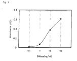
- Example 1(1) 50 ⁇ L/well of purified DNase (manufactured by Toxin Technology) diluted with 0.2% BSA-PBS to 0. 1, 1, 10, and 100 ng/mL was added to the wells of the microtiter plate having the sheep anti-DNase polyclonal antibody immobilized thereto produced in Example 1(1). After allowing the reaction to take place at room temperature for 1 hour, the wells were washed with PBS-T, and 50 ⁇ L/well of rabbit anti-DNase polyclonal antibody diluted with 0.2% BSA-PBS to 2 ⁇ g/mL was added as the secondary antibody. The reaction was then allowed to proceed at room temperature for 1 hour, and the wells were washed with PBS-T.
- DNase manufactured by Toxin Technology
- HRP-labeled pig anti-rabbit IgG antibody manufactured by DAKO was diluted with 0.2% BSA-PBS to 4000 folds, and 50 ⁇ L/well of this dilution was added to the well, and the reaction was allowed to proceed at room temperature for 1 hour. After washing, 50 ⁇ L/well of 0.1M citrate buffer solution containing 2 mg/mL o-phenylenediamine and 9. 5 mM hydrogen peroxide, pH 5 was added, and the reaction was allowed to proceed at room temperature for 30 minutes, and the reaction was ceased by adding 50 ⁇ L/well of 1.5N sulfuric acid. The measurement was conducted by using a plate reader for ELISA at a wavelength of 492 nm.
- the resulting standard curve is shown in FIG. 3.
- the measurement was possible in the DNase concentration range of 0.1 to 1000 pg/mL.
- Example 3 Measurement of the amount of DNase in the culture supernatant over time
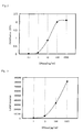
- DNase in the serum and urine of the patients suffering from S. aureus and the healthy donors were measured according to the procedure described in Example 2 (3) . The results are shown in FIG. 5. While no DNase was detected in the serum of the healthy donor, presence of DNase was confirmed in the serum and the urine of the infected patients.
Landscapes
- Health & Medical Sciences (AREA)
- Life Sciences & Earth Sciences (AREA)
- Immunology (AREA)
- Engineering & Computer Science (AREA)
- Urology & Nephrology (AREA)
- Chemical & Material Sciences (AREA)
- Biomedical Technology (AREA)
- Molecular Biology (AREA)
- Hematology (AREA)
- Medicinal Chemistry (AREA)
- Analytical Chemistry (AREA)
- Biotechnology (AREA)
- Tropical Medicine & Parasitology (AREA)
- Virology (AREA)
- Food Science & Technology (AREA)
- Microbiology (AREA)
- Physics & Mathematics (AREA)
- Cell Biology (AREA)
- Biochemistry (AREA)
- General Health & Medical Sciences (AREA)
- General Physics & Mathematics (AREA)
- Pathology (AREA)
- Measuring Or Testing Involving Enzymes Or Micro-Organisms (AREA)
- Investigating Or Analysing Materials By The Use Of Chemical Reactions (AREA)
Applications Claiming Priority (3)
| Application Number | Priority Date | Filing Date | Title |
|---|---|---|---|
| JP2002339525 | 2002-11-22 | ||
| JP2002339525 | 2002-11-22 | ||
| PCT/JP2003/014912 WO2004048976A1 (fr) | 2002-11-22 | 2003-11-21 | Procede d'analyse du staphylococcus aureus |
Publications (2)
| Publication Number | Publication Date |
|---|---|
| EP1580558A1 true EP1580558A1 (fr) | 2005-09-28 |
| EP1580558A4 EP1580558A4 (fr) | 2006-10-25 |
Family
ID=32375776
Family Applications (1)
| Application Number | Title | Priority Date | Filing Date |
|---|---|---|---|
| EP03774149A Withdrawn EP1580558A4 (fr) | 2002-11-22 | 2003-11-21 | Procede d'analyse du staphylococcus aureus |
Country Status (5)
| Country | Link |
|---|---|
| US (1) | US20060024765A1 (fr) |
| EP (1) | EP1580558A4 (fr) |
| JP (1) | JPWO2004048976A1 (fr) |
| AU (1) | AU2003284636A1 (fr) |
| WO (1) | WO2004048976A1 (fr) |
Families Citing this family (4)
| Publication number | Priority date | Publication date | Assignee | Title |
|---|---|---|---|---|
| DE19983691T1 (de) * | 1998-10-29 | 2001-11-29 | Cell Works Inc | Charakterisierung mehrerer Marker von Einzelzellen |
| JP4675671B2 (ja) * | 2005-05-11 | 2011-04-27 | 学校法人北里研究所 | 核酸増幅反応の新規短時間検出方法 |
| WO2013033436A1 (fr) | 2011-09-01 | 2013-03-07 | University Of Iowa Research Foundation | Sondes à base d'oligonucléotide pour la détection de nucléases bactériennes |
| EP3680348B1 (fr) | 2014-02-07 | 2023-04-12 | University of Iowa Research Foundation | Sondes à base d'oligonucléotides et procédés de détection de microbes |
-
2003
- 2003-11-21 US US10/535,915 patent/US20060024765A1/en not_active Abandoned
- 2003-11-21 EP EP03774149A patent/EP1580558A4/fr not_active Withdrawn
- 2003-11-21 WO PCT/JP2003/014912 patent/WO2004048976A1/fr not_active Application Discontinuation
- 2003-11-21 JP JP2004555007A patent/JPWO2004048976A1/ja not_active Withdrawn
- 2003-11-21 AU AU2003284636A patent/AU2003284636A1/en not_active Abandoned
Non-Patent Citations (8)
| Title |
|---|
| BRAKSTAD O G ET AL: "CHARACTERIZATION OF MONOCLONAL ANTIBODIES AGAINST STAPHYLOCOCCUS-AUREUS THERMONUCLEASE" SCANDINAVIAN JOURNAL OF IMMUNOLOGY, vol. 28, no. 2, 1988, page 252, XP002396703 & NINETEENTH ANNUAL GENERAL MEETING OF THE SCANDINAVIAN SOCIETY FOR IMMUNOLOGY, TRONDHEIM, NORWAY, JUN ISSN: 0300-9475 * |
| BRAKSTAD O G ET AL: "COMPARISON OF VARIOUS METHODS AND REAGENTS FOR SPECIES IDENTIFICATION OF STAPHYLOCOCCUS AUREUS POSITIVE OR NEGATIVE FOR THE MECA GENE" APMIS, COPENHAGEN, DK, vol. 101, no. 8, August 1993 (1993-08), pages 651-654, XP001058106 ISSN: 0903-4641 * |
| BRAKSTAD O G ET AL: "Detection of Staphylococcus aureus thermostable nuclease (TNase) by a rapid sandwich enzyme-linked immunofiltration assay (sELIFA)" ZENTRALBLATT FUER BAKTERIOLOGIE SUPPLEMENT, vol. 26, no. 0, 1994, pages 488-490, XP008068183 ISSN: 0941-018X * |
| BRAKSTAD O G ET AL: "Detection of Staphylococcus aureus with biotinylated monoclonal antibodies directed against staphylococcal TNase complexed to avidin-peroxidase in a rapid sandwich enzyme-linked immunofiltration assay (sELIFA)" JOURNAL OF MEDICAL MICROBIOLOGY, vol. 39, no. 2, 1993, pages 128-134, XP008068184 ISSN: 0022-2615 * |
| BRAKSTAD O G ET AL: "GENERATION AND CHARACTERIZATION OF MONOCLONAL ANTIBODIES AGAINST STAPHYLOCOCCUS-AUREUS THERMONUCLEASE" APMIS, vol. 97, no. 2, 1989, pages 166-174, XP008068181 ISSN: 0903-4641 * |
| BRAKSTAD ODD G ET AL: "Comparison of tests designed to identify Staphylococcus aureus thermostable nuclease" APMIS, vol. 103, no. 3, 1995, pages 219-224, XP008068182 ISSN: 0903-4641 * |
| BRAKSTAD ODD G ET AL: "Direct identification of Staphylococcus aureus in blood cultures by detection of the gene encoding the thermostable nuclease or the gene product" APMIS, vol. 103, no. 3, 1995, pages 209-218, XP008068201 ISSN: 0903-4641 * |
| See also references of WO2004048976A1 * |
Also Published As
| Publication number | Publication date |
|---|---|
| US20060024765A1 (en) | 2006-02-02 |
| WO2004048976A1 (fr) | 2004-06-10 |
| EP1580558A4 (fr) | 2006-10-25 |
| JPWO2004048976A1 (ja) | 2006-03-23 |
| AU2003284636A1 (en) | 2004-06-18 |
Similar Documents
| Publication | Publication Date | Title |
|---|---|---|
| US11255854B2 (en) | Signal amplification in lateral flow and related immunoassays | |
| US3992631A (en) | Fluorometric system, method and test article | |
| US9372192B2 (en) | Method and device for combined detection of viral and bacterial infections | |
| US20190219569A1 (en) | Fluorescence immunochromatographic detection card and a preparation method therefor and use thereof | |
| US20100221747A1 (en) | Immunochromatography detection of multidrug-resistant staphylococcus and diagnostic kit | |
| US8679812B2 (en) | Method for extracting Staphylococcus aureus antigen, reagent for extracting Staphylococcus aureus antigen, and method for assessing Staphylococcus aureus | |
| EP1580557A1 (fr) | Procede de detection de staphylococcus aureus | |
| JP6858784B2 (ja) | サブトラクティブイムノアッセイ方法およびその方法を実施するためのラテラルフローイムノクロマトグラフィーアッセイストリップ | |
| CN113777326A (zh) | 一种高特异检测肝素结合蛋白的试剂盒及其应用 | |
| US4795702A (en) | Diagnostic method for gonorrhea by assay of IgA1 fragments | |
| EP1580558A1 (fr) | Procede d'analyse du staphylococcus aureus | |
| EP0217583B1 (fr) | Extraction simultanée d'un ligand d'un échantillon et capture par un antiligand à cet effet, dans des essais ligands/antiligands | |
| JP3337575B2 (ja) | 抗ストレプトリジンo抗体の決定方法 | |
| EP0233048A2 (fr) | Procédé pour la détection d'infection ou inflammation du faisceau urinaire | |
| CN116804674A (zh) | 一种肺炎链球菌尿抗原elisa试剂盒及其制备方法 | |
| JP2002275199A (ja) | 抗体作製方法及び抗酸菌の免疫学的検出方法 |
Legal Events
| Date | Code | Title | Description |
|---|---|---|---|
| PUAI | Public reference made under article 153(3) epc to a published international application that has entered the european phase |
Free format text: ORIGINAL CODE: 0009012 |
|
| 17P | Request for examination filed |
Effective date: 20050519 |
|
| AK | Designated contracting states |
Kind code of ref document: A1 Designated state(s): AT BE BG CH CY CZ DE DK EE ES FI FR GB GR HU IE IT LI LU MC NL PT RO SE SI SK TR |
|
| AX | Request for extension of the european patent |
Extension state: AL LT LV MK |
|
| DAX | Request for extension of the european patent (deleted) | ||
| A4 | Supplementary search report drawn up and despatched |
Effective date: 20060925 |
|
| 17Q | First examination report despatched |
Effective date: 20070309 |
|
| STAA | Information on the status of an ep patent application or granted ep patent |
Free format text: STATUS: THE APPLICATION IS DEEMED TO BE WITHDRAWN |
|
| 18D | Application deemed to be withdrawn |
Effective date: 20070720 |