EP1563809A2 - Zwischenwirbel-Nucleus-Prothese und chirurgisches Verfahren zu seiner Implantation - Google Patents
Zwischenwirbel-Nucleus-Prothese und chirurgisches Verfahren zu seiner Implantation Download PDFInfo
- Publication number
- EP1563809A2 EP1563809A2 EP05075508A EP05075508A EP1563809A2 EP 1563809 A2 EP1563809 A2 EP 1563809A2 EP 05075508 A EP05075508 A EP 05075508A EP 05075508 A EP05075508 A EP 05075508A EP 1563809 A2 EP1563809 A2 EP 1563809A2
- Authority
- EP
- European Patent Office
- Prior art keywords
- nucleus
- cage
- prosthesis
- ball
- intervertebral
- Prior art date
- Legal status (The legal status is an assumption and is not a legal conclusion. Google has not performed a legal analysis and makes no representation as to the accuracy of the status listed.)
- Granted
Links
- 238000001356 surgical procedure Methods 0.000 title 1
- 230000001590 oxidative effect Effects 0.000 claims abstract description 6
- 239000000560 biocompatible material Substances 0.000 claims abstract description 4
- 239000007787 solid Substances 0.000 claims abstract description 4
- 238000003860 storage Methods 0.000 claims description 2
- 238000000034 method Methods 0.000 abstract description 6
- -1 non-oxidizing Substances 0.000 abstract description 3
- 238000000465 moulding Methods 0.000 abstract 1
- 239000000835 fiber Substances 0.000 description 9
- 238000002513 implantation Methods 0.000 description 5
- 230000006378 damage Effects 0.000 description 4
- 230000004927 fusion Effects 0.000 description 4
- 238000006073 displacement reaction Methods 0.000 description 3
- 239000007943 implant Substances 0.000 description 3
- 210000004446 longitudinal ligament Anatomy 0.000 description 3
- RTAQQCXQSZGOHL-UHFFFAOYSA-N Titanium Chemical compound [Ti] RTAQQCXQSZGOHL-UHFFFAOYSA-N 0.000 description 2
- 241000826860 Trapezium Species 0.000 description 2
- 238000005452 bending Methods 0.000 description 2
- 230000008901 benefit Effects 0.000 description 2
- 230000007850 degeneration Effects 0.000 description 2
- 230000018109 developmental process Effects 0.000 description 2
- 210000003041 ligament Anatomy 0.000 description 2
- 210000000056 organ Anatomy 0.000 description 2
- 230000001568 sexual effect Effects 0.000 description 2
- 229910052719 titanium Inorganic materials 0.000 description 2
- 239000010936 titanium Substances 0.000 description 2
- 230000002792 vascular Effects 0.000 description 2
- 229920002683 Glycosaminoglycan Polymers 0.000 description 1
- 239000004698 Polyethylene Substances 0.000 description 1
- 206010038967 Retrograde ejaculation Diseases 0.000 description 1
- 201000001880 Sexual dysfunction Diseases 0.000 description 1
- 208000027418 Wounds and injury Diseases 0.000 description 1
- 230000003187 abdominal effect Effects 0.000 description 1
- 230000032683 aging Effects 0.000 description 1
- 210000000709 aorta Anatomy 0.000 description 1
- 208000037873 arthrodesis Diseases 0.000 description 1
- 230000000740 bleeding effect Effects 0.000 description 1
- 210000000988 bone and bone Anatomy 0.000 description 1
- 239000002775 capsule Substances 0.000 description 1
- 238000005520 cutting process Methods 0.000 description 1
- 238000013016 damping Methods 0.000 description 1
- 230000007423 decrease Effects 0.000 description 1
- 230000006866 deterioration Effects 0.000 description 1
- 201000010099 disease Diseases 0.000 description 1
- 208000037265 diseases, disorders, signs and symptoms Diseases 0.000 description 1
- 201000001881 impotence Diseases 0.000 description 1
- 238000001727 in vivo Methods 0.000 description 1
- 208000014674 injury Diseases 0.000 description 1
- 238000003780 insertion Methods 0.000 description 1
- 230000037431 insertion Effects 0.000 description 1
- 210000000281 joint capsule Anatomy 0.000 description 1
- 230000005923 long-lasting effect Effects 0.000 description 1
- 238000004519 manufacturing process Methods 0.000 description 1
- 239000000463 material Substances 0.000 description 1
- 210000005036 nerve Anatomy 0.000 description 1
- 230000003204 osmotic effect Effects 0.000 description 1
- 201000008482 osteoarthritis Diseases 0.000 description 1
- 230000001575 pathological effect Effects 0.000 description 1
- 230000002688 persistence Effects 0.000 description 1
- 229920000573 polyethylene Polymers 0.000 description 1
- 238000002271 resection Methods 0.000 description 1
- 230000000087 stabilizing effect Effects 0.000 description 1
- 229910001220 stainless steel Inorganic materials 0.000 description 1
- 239000010935 stainless steel Substances 0.000 description 1
- 239000000126 substance Substances 0.000 description 1
- 210000003462 vein Anatomy 0.000 description 1
- XLYOFNOQVPJJNP-UHFFFAOYSA-N water Substances O XLYOFNOQVPJJNP-UHFFFAOYSA-N 0.000 description 1
Images
Classifications
-
- A—HUMAN NECESSITIES
- A61—MEDICAL OR VETERINARY SCIENCE; HYGIENE
- A61F—FILTERS IMPLANTABLE INTO BLOOD VESSELS; PROSTHESES; DEVICES PROVIDING PATENCY TO, OR PREVENTING COLLAPSING OF, TUBULAR STRUCTURES OF THE BODY, e.g. STENTS; ORTHOPAEDIC, NURSING OR CONTRACEPTIVE DEVICES; FOMENTATION; TREATMENT OR PROTECTION OF EYES OR EARS; BANDAGES, DRESSINGS OR ABSORBENT PADS; FIRST-AID KITS
- A61F2/00—Filters implantable into blood vessels; Prostheses, i.e. artificial substitutes or replacements for parts of the body; Appliances for connecting them with the body; Devices providing patency to, or preventing collapsing of, tubular structures of the body, e.g. stents
- A61F2/02—Prostheses implantable into the body
- A61F2/30—Joints
- A61F2/44—Joints for the spine, e.g. vertebrae, spinal discs
- A61F2/442—Intervertebral or spinal discs, e.g. resilient
-
- A—HUMAN NECESSITIES
- A61—MEDICAL OR VETERINARY SCIENCE; HYGIENE
- A61F—FILTERS IMPLANTABLE INTO BLOOD VESSELS; PROSTHESES; DEVICES PROVIDING PATENCY TO, OR PREVENTING COLLAPSING OF, TUBULAR STRUCTURES OF THE BODY, e.g. STENTS; ORTHOPAEDIC, NURSING OR CONTRACEPTIVE DEVICES; FOMENTATION; TREATMENT OR PROTECTION OF EYES OR EARS; BANDAGES, DRESSINGS OR ABSORBENT PADS; FIRST-AID KITS
- A61F2/00—Filters implantable into blood vessels; Prostheses, i.e. artificial substitutes or replacements for parts of the body; Appliances for connecting them with the body; Devices providing patency to, or preventing collapsing of, tubular structures of the body, e.g. stents
- A61F2/02—Prostheses implantable into the body
- A61F2/30—Joints
- A61F2002/30001—Additional features of subject-matter classified in A61F2/28, A61F2/30 and subgroups thereof
- A61F2002/30108—Shapes
- A61F2002/3011—Cross-sections or two-dimensional shapes
- A61F2002/30112—Rounded shapes, e.g. with rounded corners
- A61F2002/30133—Rounded shapes, e.g. with rounded corners kidney-shaped or bean-shaped
-
- A—HUMAN NECESSITIES
- A61—MEDICAL OR VETERINARY SCIENCE; HYGIENE
- A61F—FILTERS IMPLANTABLE INTO BLOOD VESSELS; PROSTHESES; DEVICES PROVIDING PATENCY TO, OR PREVENTING COLLAPSING OF, TUBULAR STRUCTURES OF THE BODY, e.g. STENTS; ORTHOPAEDIC, NURSING OR CONTRACEPTIVE DEVICES; FOMENTATION; TREATMENT OR PROTECTION OF EYES OR EARS; BANDAGES, DRESSINGS OR ABSORBENT PADS; FIRST-AID KITS
- A61F2/00—Filters implantable into blood vessels; Prostheses, i.e. artificial substitutes or replacements for parts of the body; Appliances for connecting them with the body; Devices providing patency to, or preventing collapsing of, tubular structures of the body, e.g. stents
- A61F2/02—Prostheses implantable into the body
- A61F2/30—Joints
- A61F2002/30001—Additional features of subject-matter classified in A61F2/28, A61F2/30 and subgroups thereof
- A61F2002/30108—Shapes
- A61F2002/3011—Cross-sections or two-dimensional shapes
- A61F2002/30138—Convex polygonal shapes
- A61F2002/30158—Convex polygonal shapes trapezoidal
-
- A—HUMAN NECESSITIES
- A61—MEDICAL OR VETERINARY SCIENCE; HYGIENE
- A61F—FILTERS IMPLANTABLE INTO BLOOD VESSELS; PROSTHESES; DEVICES PROVIDING PATENCY TO, OR PREVENTING COLLAPSING OF, TUBULAR STRUCTURES OF THE BODY, e.g. STENTS; ORTHOPAEDIC, NURSING OR CONTRACEPTIVE DEVICES; FOMENTATION; TREATMENT OR PROTECTION OF EYES OR EARS; BANDAGES, DRESSINGS OR ABSORBENT PADS; FIRST-AID KITS
- A61F2/00—Filters implantable into blood vessels; Prostheses, i.e. artificial substitutes or replacements for parts of the body; Appliances for connecting them with the body; Devices providing patency to, or preventing collapsing of, tubular structures of the body, e.g. stents
- A61F2/02—Prostheses implantable into the body
- A61F2/30—Joints
- A61F2002/30001—Additional features of subject-matter classified in A61F2/28, A61F2/30 and subgroups thereof
- A61F2002/30108—Shapes
- A61F2002/30199—Three-dimensional shapes
- A61F2002/30242—Three-dimensional shapes spherical
-
- A—HUMAN NECESSITIES
- A61—MEDICAL OR VETERINARY SCIENCE; HYGIENE
- A61F—FILTERS IMPLANTABLE INTO BLOOD VESSELS; PROSTHESES; DEVICES PROVIDING PATENCY TO, OR PREVENTING COLLAPSING OF, TUBULAR STRUCTURES OF THE BODY, e.g. STENTS; ORTHOPAEDIC, NURSING OR CONTRACEPTIVE DEVICES; FOMENTATION; TREATMENT OR PROTECTION OF EYES OR EARS; BANDAGES, DRESSINGS OR ABSORBENT PADS; FIRST-AID KITS
- A61F2/00—Filters implantable into blood vessels; Prostheses, i.e. artificial substitutes or replacements for parts of the body; Appliances for connecting them with the body; Devices providing patency to, or preventing collapsing of, tubular structures of the body, e.g. stents
- A61F2/02—Prostheses implantable into the body
- A61F2/30—Joints
- A61F2002/30001—Additional features of subject-matter classified in A61F2/28, A61F2/30 and subgroups thereof
- A61F2002/30316—The prosthesis having different structural features at different locations within the same prosthesis; Connections between prosthetic parts; Special structural features of bone or joint prostheses not otherwise provided for
- A61F2002/30329—Connections or couplings between prosthetic parts, e.g. between modular parts; Connecting elements
- A61F2002/30331—Connections or couplings between prosthetic parts, e.g. between modular parts; Connecting elements made by longitudinally pushing a protrusion into a complementarily-shaped recess, e.g. held by friction fit
- A61F2002/30378—Spherically-shaped protrusion and recess
-
- A—HUMAN NECESSITIES
- A61—MEDICAL OR VETERINARY SCIENCE; HYGIENE
- A61F—FILTERS IMPLANTABLE INTO BLOOD VESSELS; PROSTHESES; DEVICES PROVIDING PATENCY TO, OR PREVENTING COLLAPSING OF, TUBULAR STRUCTURES OF THE BODY, e.g. STENTS; ORTHOPAEDIC, NURSING OR CONTRACEPTIVE DEVICES; FOMENTATION; TREATMENT OR PROTECTION OF EYES OR EARS; BANDAGES, DRESSINGS OR ABSORBENT PADS; FIRST-AID KITS
- A61F2/00—Filters implantable into blood vessels; Prostheses, i.e. artificial substitutes or replacements for parts of the body; Appliances for connecting them with the body; Devices providing patency to, or preventing collapsing of, tubular structures of the body, e.g. stents
- A61F2/02—Prostheses implantable into the body
- A61F2/30—Joints
- A61F2002/30001—Additional features of subject-matter classified in A61F2/28, A61F2/30 and subgroups thereof
- A61F2002/30621—Features concerning the anatomical functioning or articulation of the prosthetic joint
- A61F2002/30639—Features concerning the anatomical functioning or articulation of the prosthetic joint having rolling elements between both articulating surfaces
-
- A—HUMAN NECESSITIES
- A61—MEDICAL OR VETERINARY SCIENCE; HYGIENE
- A61F—FILTERS IMPLANTABLE INTO BLOOD VESSELS; PROSTHESES; DEVICES PROVIDING PATENCY TO, OR PREVENTING COLLAPSING OF, TUBULAR STRUCTURES OF THE BODY, e.g. STENTS; ORTHOPAEDIC, NURSING OR CONTRACEPTIVE DEVICES; FOMENTATION; TREATMENT OR PROTECTION OF EYES OR EARS; BANDAGES, DRESSINGS OR ABSORBENT PADS; FIRST-AID KITS
- A61F2/00—Filters implantable into blood vessels; Prostheses, i.e. artificial substitutes or replacements for parts of the body; Appliances for connecting them with the body; Devices providing patency to, or preventing collapsing of, tubular structures of the body, e.g. stents
- A61F2/02—Prostheses implantable into the body
- A61F2/30—Joints
- A61F2/3094—Designing or manufacturing processes
- A61F2002/30975—Designing or manufacturing processes made of two halves
-
- A—HUMAN NECESSITIES
- A61—MEDICAL OR VETERINARY SCIENCE; HYGIENE
- A61F—FILTERS IMPLANTABLE INTO BLOOD VESSELS; PROSTHESES; DEVICES PROVIDING PATENCY TO, OR PREVENTING COLLAPSING OF, TUBULAR STRUCTURES OF THE BODY, e.g. STENTS; ORTHOPAEDIC, NURSING OR CONTRACEPTIVE DEVICES; FOMENTATION; TREATMENT OR PROTECTION OF EYES OR EARS; BANDAGES, DRESSINGS OR ABSORBENT PADS; FIRST-AID KITS
- A61F2/00—Filters implantable into blood vessels; Prostheses, i.e. artificial substitutes or replacements for parts of the body; Appliances for connecting them with the body; Devices providing patency to, or preventing collapsing of, tubular structures of the body, e.g. stents
- A61F2/02—Prostheses implantable into the body
- A61F2/30—Joints
- A61F2/44—Joints for the spine, e.g. vertebrae, spinal discs
- A61F2/442—Intervertebral or spinal discs, e.g. resilient
- A61F2002/444—Intervertebral or spinal discs, e.g. resilient for replacing the nucleus pulposus
-
- A—HUMAN NECESSITIES
- A61—MEDICAL OR VETERINARY SCIENCE; HYGIENE
- A61F—FILTERS IMPLANTABLE INTO BLOOD VESSELS; PROSTHESES; DEVICES PROVIDING PATENCY TO, OR PREVENTING COLLAPSING OF, TUBULAR STRUCTURES OF THE BODY, e.g. STENTS; ORTHOPAEDIC, NURSING OR CONTRACEPTIVE DEVICES; FOMENTATION; TREATMENT OR PROTECTION OF EYES OR EARS; BANDAGES, DRESSINGS OR ABSORBENT PADS; FIRST-AID KITS
- A61F2220/00—Fixations or connections for prostheses classified in groups A61F2/00 - A61F2/26 or A61F2/82 or A61F9/00 or A61F11/00 or subgroups thereof
- A61F2220/0025—Connections or couplings between prosthetic parts, e.g. between modular parts; Connecting elements
- A61F2220/0033—Connections or couplings between prosthetic parts, e.g. between modular parts; Connecting elements made by longitudinally pushing a protrusion into a complementary-shaped recess, e.g. held by friction fit
-
- A—HUMAN NECESSITIES
- A61—MEDICAL OR VETERINARY SCIENCE; HYGIENE
- A61F—FILTERS IMPLANTABLE INTO BLOOD VESSELS; PROSTHESES; DEVICES PROVIDING PATENCY TO, OR PREVENTING COLLAPSING OF, TUBULAR STRUCTURES OF THE BODY, e.g. STENTS; ORTHOPAEDIC, NURSING OR CONTRACEPTIVE DEVICES; FOMENTATION; TREATMENT OR PROTECTION OF EYES OR EARS; BANDAGES, DRESSINGS OR ABSORBENT PADS; FIRST-AID KITS
- A61F2230/00—Geometry of prostheses classified in groups A61F2/00 - A61F2/26 or A61F2/82 or A61F9/00 or A61F11/00 or subgroups thereof
- A61F2230/0002—Two-dimensional shapes, e.g. cross-sections
- A61F2230/0004—Rounded shapes, e.g. with rounded corners
- A61F2230/0015—Kidney-shaped, e.g. bean-shaped
-
- A—HUMAN NECESSITIES
- A61—MEDICAL OR VETERINARY SCIENCE; HYGIENE
- A61F—FILTERS IMPLANTABLE INTO BLOOD VESSELS; PROSTHESES; DEVICES PROVIDING PATENCY TO, OR PREVENTING COLLAPSING OF, TUBULAR STRUCTURES OF THE BODY, e.g. STENTS; ORTHOPAEDIC, NURSING OR CONTRACEPTIVE DEVICES; FOMENTATION; TREATMENT OR PROTECTION OF EYES OR EARS; BANDAGES, DRESSINGS OR ABSORBENT PADS; FIRST-AID KITS
- A61F2230/00—Geometry of prostheses classified in groups A61F2/00 - A61F2/26 or A61F2/82 or A61F9/00 or A61F11/00 or subgroups thereof
- A61F2230/0002—Two-dimensional shapes, e.g. cross-sections
- A61F2230/0017—Angular shapes
- A61F2230/0026—Angular shapes trapezoidal
-
- A—HUMAN NECESSITIES
- A61—MEDICAL OR VETERINARY SCIENCE; HYGIENE
- A61F—FILTERS IMPLANTABLE INTO BLOOD VESSELS; PROSTHESES; DEVICES PROVIDING PATENCY TO, OR PREVENTING COLLAPSING OF, TUBULAR STRUCTURES OF THE BODY, e.g. STENTS; ORTHOPAEDIC, NURSING OR CONTRACEPTIVE DEVICES; FOMENTATION; TREATMENT OR PROTECTION OF EYES OR EARS; BANDAGES, DRESSINGS OR ABSORBENT PADS; FIRST-AID KITS
- A61F2230/00—Geometry of prostheses classified in groups A61F2/00 - A61F2/26 or A61F2/82 or A61F9/00 or A61F11/00 or subgroups thereof
- A61F2230/0063—Three-dimensional shapes
- A61F2230/0071—Three-dimensional shapes spherical
-
- A—HUMAN NECESSITIES
- A61—MEDICAL OR VETERINARY SCIENCE; HYGIENE
- A61F—FILTERS IMPLANTABLE INTO BLOOD VESSELS; PROSTHESES; DEVICES PROVIDING PATENCY TO, OR PREVENTING COLLAPSING OF, TUBULAR STRUCTURES OF THE BODY, e.g. STENTS; ORTHOPAEDIC, NURSING OR CONTRACEPTIVE DEVICES; FOMENTATION; TREATMENT OR PROTECTION OF EYES OR EARS; BANDAGES, DRESSINGS OR ABSORBENT PADS; FIRST-AID KITS
- A61F2310/00—Prostheses classified in A61F2/28 or A61F2/30 - A61F2/44 being constructed from or coated with a particular material
- A61F2310/00005—The prosthesis being constructed from a particular material
- A61F2310/00011—Metals or alloys
- A61F2310/00017—Iron- or Fe-based alloys, e.g. stainless steel
-
- A—HUMAN NECESSITIES
- A61—MEDICAL OR VETERINARY SCIENCE; HYGIENE
- A61F—FILTERS IMPLANTABLE INTO BLOOD VESSELS; PROSTHESES; DEVICES PROVIDING PATENCY TO, OR PREVENTING COLLAPSING OF, TUBULAR STRUCTURES OF THE BODY, e.g. STENTS; ORTHOPAEDIC, NURSING OR CONTRACEPTIVE DEVICES; FOMENTATION; TREATMENT OR PROTECTION OF EYES OR EARS; BANDAGES, DRESSINGS OR ABSORBENT PADS; FIRST-AID KITS
- A61F2310/00—Prostheses classified in A61F2/28 or A61F2/30 - A61F2/44 being constructed from or coated with a particular material
- A61F2310/00005—The prosthesis being constructed from a particular material
- A61F2310/00011—Metals or alloys
- A61F2310/00023—Titanium or titanium-based alloys, e.g. Ti-Ni alloys
Definitions
- the invention relates to an intervertebral-nucleus prosthesis whose The task is to restore the mobility in the area of an intervertebral disc manufacture. This mobility becomes physiologically through the nucleus produced, considered as a mechanical joint element of the vertebral body can be.
- the nucleus is characterized by a substantially spherical, not malleable, but deformable body formed like a ball is inserted between two vertebral surfaces, essentially in its center, and movements in the form of inclination, rotation and the shift allows.
- the vortex end surfaces are in turn of concentric fiber layers which are called annulus and contain the fibers of one layer to the other to cross at an angle.
- the nucleus which is actually one deformable but non-distensible capsule containing a hydrophilic, jelly-like substance is filled (mucopolysaccharides) in the flexion of the Spine forward or backward even to the rear respectively forward, this movement of the nucleus through the posterior and the front fibers of the annulus and by different longitudinal bands is limited.
- the disc Upon rotation of the spine, the disc is with Shear forces applied.
- the nucleus which is a hydrophilic body, is at rest a certain osmotic pressure. When loaded, the nucleus loses Water. The thickness of the disc decreases as soon as the pressure returns is reduced, rehydration occurs.
- the pathological conditions affecting the reciprocal movements of the disturbing various vertebral bodies are essentially overloaded or too strong and too frequent loads or natural aging processes.
- LVCA anterior longitudinal ligament
- LVCP posterior longitudinal ligament
- nucleus prosthesis allowed without injury to the annulus or the Longitudinal bands (LVCA and LVCP) by displacement of the replaced nucleus to exert pressure on the annulus and therefore in the movement of the Spine so that it restores the equilibrium position.
- the center of rotation that has been restored in this way is, actually remains mobile and able to adapt to the different Beugerisonen forward and backward, on stretching movements and adjust lateral tilting movements.
- the nucleus prosthesis of the present invention consists of one or several balls that are hard, smooth and non-oxidizing and that move freely are arranged inside a fixed cage, which in turn is not oxidizing is. This prosthesis can be accommodated in the volume which after the removal of the nucleus between two vertebral end faces is free.
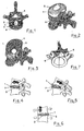
- FIG. 1 shows a plan view of a vertebral body with the respective positions of the nucleus, the annulus and the anterior and posterior Longitudinal bands.
- FIG. 2 is a perspective view of a vertebral body and FIG shows the annulus and the nucleus it surrounds, which is located on the Vertebral body supports.
- FIG. 3 is a perspective schematic view of one diametrically cut annulus, showing the concentric, intersecting Fiber layers.
- Figures 4 and 5 are schematic side views of two adjacent vertebral bodies in a bending movement forward or behind.
- FIG. 6 is a schematic front view of two adjacent ones Vertebral bodies during lateral flexion of the spine.
- FIG. 7 is a plan view of a vertebral body in FIG Rotational movement.
- FIGS. 8, 9 and 10 are schematic side views of FIG above and from the front of a nucleus prosthesis with only one ball.
- FIG. 11 is a schematic, exemplary longitudinal sectional view along line A-A in Figure 8.
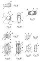
- FIGS. 12, 13 and 14 are schematic top views of FIG on the front and on the side of a multi-sphere nucleus prosthesis.
- FIGS. 15, 16 and 17 are schematic top views of FIG in front and from the side of a nucleus prosthesis with a Damping device.
- Figures 1 and 2 show well an intervertebral plane formed is characterized by a multitude of concentric layers of intersecting fibers 1 and 2, clearly visible in Figure 3, and in the center of each of the nucleus 3 is arranged, while on the outside of the front longitudinal belt (LVCA) 4th and the rear longitudinal band (LVCP) 5 are arranged.
- LVCA front longitudinal belt
- LVCP rear longitudinal band
- FIGS. 4 and 5 show the compressive forces generated by the Nucleus 3 are exerted on the concentric Annuluslagen, namely each on the outside of the bending angle at the bend forward or to the rear.
- the nucleus 3 exerts in the same way a pressure on the rings of Annulus off, on the outside of mall ⁇ ichen tilt angle ( Figure 6).
- Each of these shifts of the nucleus 3 represents a return to the Balance position sure.
- the nucleus plays the role of one in all these cases Movement damper and represents in this movement the return to the Balance position sure. You can see from this the consequences that arise when the nucleus is changed.
- the inventive nucleus prosthesis has none of these disadvantages on. It allows the injured nucleus both the form as well to replace the function. It is suitable for the degeneration of End intervertebral disc by stabilizing it again.
- the surgical implant procedure is extremely simple.
- the implantation can be performed endoscopically. she is fast and gentle.
- the nucleus prosthesis consists essentially of at least one movable body 8, made of a solid, non-oxidizing, biocompatible Material that can move in two axes of a plane (For example, a stainless steel ball or a ball of titanium), whose Volume is adapted to that of the biological nucleus.
- This mobile Body or ball B is referred to as a cage in the following Body 7, which comprises a housing 14 (for example in Figure 11 shown), which consists of a light, solid, non-oxidizing and biocompatible material is made (for example, titanium), this case 14 contains a mass 15 of a material with a minimum Coefficients of friction (for example polyethylene).
- the volume of the cage 7 housing the mobile body 8 is in turn adapted as closely as possible to the volume of the biological nucleus, of course, it is considered that the cage 7 as a storage for the ball. 8 must serve. This allows the prosthesis to position itself, so it is her possible to always be in the anatomical position and thereby the restore natural movement between two vertebral bodies.
- the ball 8 superimposed cage 7 can in plan view ( Figure 8) a more or less curved shape, symmetrical with respect to is a transverse median plane.
- the cage In cross section ( Figure 10), the cage preferably the shape of a trapezium whose small end face the Ends of more or less strongly bent form absorbs.
- the cage 7 which in cross section substantially the Has an isosceles trapezoid shape (Figure 13), several identical Balls 8 ( Figure 14) record on the outside a common fictitious Level touching on both sides of the horizontal median plane of the cage are arranged.
- the balls are located on both sides of the cage 7 the three vertices of an isosceles triangle.
- the cage 7 can according to the Embodiments of Figures 15, 16 and 17 of two identical Single bodies 10, 11 are the test, non-oxidizable and biocompatible are formed and have the shape of an isosceles trapezium. They are arranged so that their perpendicular to the parallel base surfaces standing, along the center lines extending center planes substantially parallel to each other and that the large base surfaces of trapezoidal single body 10 and 11 substantially in the same vertical plane lie.
- the cage 7, a ball 8 or more Contains balls 8 and for the above several embodiments have been proposed without departing from the inventive Thoughts have other equivalent forms, it's just important that it stores the ball or balls 8, that they are freely rotatable in it and that its volume is adapted to the volume which, after removal of the biological nucleus is available.
- the described nucleus prosthesis can be used in all of the above described embodiments spontaneously in the anatomical environment assume your own position, so that all natural movements between two vertebral bodies can be restored (lateral inclination, Inclination forwards and backwards, rotation).
- the conical shape (in Cross-section trapezoidal) of the cage 7 facilitates the shift in the Plane of the vertebral end faces and at the same time prevents the rotation of the cage about its central axis, as well as the sinking of the implant in the Vertebral end.
Landscapes
- Health & Medical Sciences (AREA)
- Engineering & Computer Science (AREA)
- Biomedical Technology (AREA)
- Neurology (AREA)
- Orthopedic Medicine & Surgery (AREA)
- Cardiology (AREA)
- Oral & Maxillofacial Surgery (AREA)
- Transplantation (AREA)
- Heart & Thoracic Surgery (AREA)
- Vascular Medicine (AREA)
- Life Sciences & Earth Sciences (AREA)
- Animal Behavior & Ethology (AREA)
- General Health & Medical Sciences (AREA)
- Public Health (AREA)
- Veterinary Medicine (AREA)
- Prostheses (AREA)
Abstract
Description
Claims (4)
- Zwischenwirbel-Nucleus-Prothese, deren Volumen an das des biologischen Nucleus angepaßt ist, dadurch gekennzeichnet, dass sie einen Käfig (10, 11) besitzt, der folgende Elemente aufweist:mehrere identische Kugeln (8) aus einem festen, nicht oxidierenden, biokompatiblen Material, die in zwei Achsen einer Ebene auf beiden Seiten einer horizontalen Mittelebene des Käfigs beweglich sind, um eine gleiche, fiktive, außerhalb des Käfigs liegenden Ebene zu berühren, und die auf einer der Seiten des Käfigs (10, 11) in der Form einer Kugelkalotte hervorstehen;eine Vielzahl von Hohlräumen, die das Einschließen jeweils einer Kugel (8) ermöglicht, die frei drehbar um ihren Mittelpunkt gelagert ist.
- Prothese nach Anspruch 1, bei welcher die Kugeln (8) unverschiebbar in dem jeweiligen Hohlraum gelagert sind.
- Prothese nach Anspruch 1 oder 2, bei welcher der Käfig (7) aus zwei identischen, gleichschenkligen trapezförmigen Einzelkörpern (10, 11) gebildet ist.
- Prothese nach einem der Ansprüche 1 bis 3, bei welcher der mit Kugeln (8) versehene Käfig ein Volumen aufweist, der unter Berücksichtigung seiner Aufgabe als Lagerung der Kugeln so gut wie möglich dem Volumen des biologischen Nucleus angepaßt ist, so dass damit eine Selbstpositionierung der Prothese gesichert ist, durch die sich diese immer in der anatomischen Position befindet, um die natürlichen Bewegungen zwischen zwei Wirbelkörpern wiederherzustellen.
Applications Claiming Priority (3)
| Application Number | Priority Date | Filing Date | Title |
|---|---|---|---|
| FR9910167 | 1999-08-03 | ||
| FR9910167A FR2797179B1 (fr) | 1999-08-03 | 1999-08-03 | Prothese nucleaire intervertebrale et son procede chirurgical d'implantation |
| EP00962288A EP1198209B1 (de) | 1999-08-03 | 2000-08-02 | Zwischenwirbel-nucleus-prothese |
Related Parent Applications (1)
| Application Number | Title | Priority Date | Filing Date |
|---|---|---|---|
| EP00962288A Division EP1198209B1 (de) | 1999-08-03 | 2000-08-02 | Zwischenwirbel-nucleus-prothese |
Publications (3)
| Publication Number | Publication Date |
|---|---|
| EP1563809A2 true EP1563809A2 (de) | 2005-08-17 |
| EP1563809A3 EP1563809A3 (de) | 2009-04-15 |
| EP1563809B1 EP1563809B1 (de) | 2014-10-01 |
Family
ID=9548908
Family Applications (2)
| Application Number | Title | Priority Date | Filing Date |
|---|---|---|---|
| EP05075508.1A Expired - Lifetime EP1563809B1 (de) | 1999-08-03 | 2000-08-02 | Zwischenwirbel-Nucleus-Prothese und chirurgisches Verfahren zu seiner Implantation |
| EP00962288A Expired - Lifetime EP1198209B1 (de) | 1999-08-03 | 2000-08-02 | Zwischenwirbel-nucleus-prothese |
Family Applications After (1)
| Application Number | Title | Priority Date | Filing Date |
|---|---|---|---|
| EP00962288A Expired - Lifetime EP1198209B1 (de) | 1999-08-03 | 2000-08-02 | Zwischenwirbel-nucleus-prothese |
Country Status (6)
| Country | Link |
|---|---|
| US (2) | US7037340B2 (de) |
| EP (2) | EP1563809B1 (de) |
| DE (1) | DE50010295D1 (de) |
| ES (2) | ES2241656T3 (de) |
| FR (1) | FR2797179B1 (de) |
| WO (1) | WO2001008612A1 (de) |
Cited By (5)
| Publication number | Priority date | Publication date | Assignee | Title |
|---|---|---|---|---|
| US7713303B2 (en) | 2002-09-18 | 2010-05-11 | Warsaw Orthopedic, Inc. | Collagen-based materials and methods for augmenting intervertebral discs |
| US7731981B2 (en) | 2002-11-15 | 2010-06-08 | Warsaw Orthopedic, Inc. | Collagen-based materials and methods for treating synovial joints |
| US7744651B2 (en) | 2002-09-18 | 2010-06-29 | Warsaw Orthopedic, Inc | Compositions and methods for treating intervertebral discs with collagen-based materials |
| US8118779B2 (en) | 2006-06-30 | 2012-02-21 | Warsaw Orthopedic, Inc. | Collagen delivery device |
| US8399619B2 (en) | 2006-06-30 | 2013-03-19 | Warsaw Orthopedic, Inc. | Injectable collagen material |
Families Citing this family (88)
| Publication number | Priority date | Publication date | Assignee | Title |
|---|---|---|---|---|
| AU780719B2 (en) | 1999-07-02 | 2005-04-14 | Spine Solutions Inc. | Intervertebral implant |
| FR2897259B1 (fr) | 2006-02-15 | 2008-05-09 | Ldr Medical Soc Par Actions Si | Cage intersomatique transforaminale a greffon de fusion intervetebrale et instrument d'implantation de la cage |
| FR2797179B1 (fr) | 1999-08-03 | 2002-03-08 | Michel Gau | Prothese nucleaire intervertebrale et son procede chirurgical d'implantation |
| US6899716B2 (en) | 2000-02-16 | 2005-05-31 | Trans1, Inc. | Method and apparatus for spinal augmentation |
| US7727263B2 (en) | 2000-02-16 | 2010-06-01 | Trans1, Inc. | Articulating spinal implant |
| US6805695B2 (en) | 2000-04-04 | 2004-10-19 | Spinalabs, Llc | Devices and methods for annular repair of intervertebral discs |
| US6478822B1 (en) * | 2001-03-20 | 2002-11-12 | Spineco, Inc. | Spherical spinal implant |
| FR2824261B1 (fr) | 2001-05-04 | 2004-05-28 | Ldr Medical | Prothese de disque intervertebral et procede et outils de mise en place |
| US7085827B2 (en) * | 2001-09-20 | 2006-08-01 | Hitachi, Ltd. | Integrated service management system for remote customer support |
| US7001433B2 (en) * | 2002-05-23 | 2006-02-21 | Pioneer Laboratories, Inc. | Artificial intervertebral disc device |
| US8388684B2 (en) * | 2002-05-23 | 2013-03-05 | Pioneer Signal Technology, Inc. | Artificial disc device |
| BR0313499A (pt) | 2002-08-15 | 2005-07-05 | David Gerber | Disco intervertebral |
| DE60322066D1 (de) | 2002-08-15 | 2008-08-21 | Hfsc Co | Bandscheibenimplantat |
| DE10242329B4 (de) | 2002-09-12 | 2005-03-17 | Biedermann Motech Gmbh | Bandscheibenprothese |
| DE10242331B4 (de) | 2002-09-12 | 2005-10-20 | Biedermann Motech Gmbh | Platzhalter für Wirbelkörper oder Bandscheiben |
| US20040068321A1 (en) * | 2002-10-04 | 2004-04-08 | Ferree Bret A. | Reduced-friction artificial disc replacements |
| FR2846550B1 (fr) | 2002-11-05 | 2006-01-13 | Ldr Medical | Prothese de disque intervertebral |
| DE10253170A1 (de) * | 2002-11-14 | 2004-06-03 | Sepitec Foundation | Druckfester Hohlkörper zum Einsatz bei Versteifungsoperationen an der Wirbelsäule |
| US7473276B2 (en) * | 2002-12-17 | 2009-01-06 | Synthes (U.S.A.) | Intervertebral implant with joint parts mounted on roller bodies |
| US7887591B2 (en) * | 2002-12-17 | 2011-02-15 | Synthes Usa, Llc | Intervertebral implant comprising joint parts that are mounted to form a universal joint |
| US7192447B2 (en) * | 2002-12-19 | 2007-03-20 | Synthes (Usa) | Intervertebral implant |
| GB0301085D0 (en) * | 2003-01-17 | 2003-02-19 | Krishna Manoj | Articulating spinal disc prosthesis |
| US7824444B2 (en) | 2003-03-20 | 2010-11-02 | Spineco, Inc. | Expandable spherical spinal implant |
| US7491204B2 (en) | 2003-04-28 | 2009-02-17 | Spine Solutions, Inc. | Instruments and method for preparing an intervertebral space for receiving an artificial disc implant |
| FR2859095B1 (fr) | 2003-09-01 | 2006-05-12 | Ldr Medical | Implant d'ancrage osseux a tete polyaxiale et procede de mise en place de l'implant |
| US7550010B2 (en) | 2004-01-09 | 2009-06-23 | Warsaw Orthopedic, Inc. | Spinal arthroplasty device and method |
| US7771479B2 (en) | 2004-01-09 | 2010-08-10 | Warsaw Orthopedic, Inc. | Dual articulating spinal device and method |
| US20050171610A1 (en) * | 2004-01-09 | 2005-08-04 | Sdgi Holdings, Inc. | Mobile bearing spinal device and method |
| FR2865629B1 (fr) | 2004-02-04 | 2007-01-26 | Ldr Medical | Prothese de disque intervertebral |
| PT1711133E (pt) | 2004-02-04 | 2011-06-01 | Ldr Medical | Prótese de disco intervertebral |
| US7806933B2 (en) * | 2004-03-15 | 2010-10-05 | Warsaw Orthopedic, Inc. | System and method for stabilizing a prosthetic device |
| FR2869528B1 (fr) | 2004-04-28 | 2007-02-02 | Ldr Medical | Prothese de disque intervertebral |
| US7497854B2 (en) * | 2004-05-07 | 2009-03-03 | Ethicon Endo-Surgery, Inc. | Method and instrument for effecting anastomosis of respective tissues defining two body lumens |
| US7763024B2 (en) * | 2004-09-23 | 2010-07-27 | Spine Solutions, Inc. | Adjustable cutting of cutout in vertebral bone |
| US7780731B2 (en) * | 2004-11-26 | 2010-08-24 | Spine Solutions, Inc. | Intervertebral implant |
| US20060085076A1 (en) * | 2004-10-15 | 2006-04-20 | Manoj Krishna | Posterior spinal arthroplasty-development of a new posteriorly inserted artificial disc and an artificial facet joint |
| WO2006042484A1 (de) * | 2004-10-18 | 2006-04-27 | Buettner-Janz Karin | Gewinkelter gleitkern als teil einer bandscheibenendoprothese |
| USD598105S1 (en) | 2004-10-21 | 2009-08-11 | Disc Motion Technologies, Inc. | Convex intervertebral disc prosthesis |
| USD597674S1 (en) | 2004-10-21 | 2009-08-04 | Disc Motion Technologies, Inc. | Concave intervertebral disc prosthesis |
| US20060265074A1 (en) | 2004-10-21 | 2006-11-23 | Manoj Krishna | Posterior spinal arthroplasty-development of a new posteriorly inserted artificial disc, a new anteriorly inserted artifical disc and an artificial facet joint |
| US7717960B2 (en) * | 2004-10-22 | 2010-05-18 | Michael Schneier | Dynamic spinal implant or joint replacement |
| FR2879436B1 (fr) | 2004-12-22 | 2007-03-09 | Ldr Medical | Prothese de disque intervertebral |
| US8911498B2 (en) * | 2005-02-10 | 2014-12-16 | DePuy Synthes Products, LLC | Intervertebral prosthetic disc |
| US8083798B2 (en) * | 2005-04-04 | 2011-12-27 | Warsaw Orthopedic, Inc. | Non-circular stabilization sphere and method |
| FR2887762B1 (fr) | 2005-06-29 | 2007-10-12 | Ldr Medical Soc Par Actions Si | Instrumentation d'insertion de prothese de disque intervertebral entre des vertebres |
| FR2891135B1 (fr) | 2005-09-23 | 2008-09-12 | Ldr Medical Sarl | Prothese de disque intervertebral |
| US20070093906A1 (en) * | 2005-10-26 | 2007-04-26 | Zimmer Spine, Inc. | Nucleus implant and method |
| FR2893838B1 (fr) | 2005-11-30 | 2008-08-08 | Ldr Medical Soc Par Actions Si | Prothese de disque intervertebral et instrumentation d'insertion de la prothese entre les vertebres |
| US7645301B2 (en) * | 2006-01-13 | 2010-01-12 | Zimmer Spine, Inc. | Devices and methods for disc replacement |
| US7811326B2 (en) | 2006-01-30 | 2010-10-12 | Warsaw Orthopedic Inc. | Posterior joint replacement device |
| US7635389B2 (en) | 2006-01-30 | 2009-12-22 | Warsaw Orthopedic, Inc. | Posterior joint replacement device |
| US8282641B2 (en) | 2006-03-28 | 2012-10-09 | Depuy Spine, Inc. | Methods and instrumentation for disc replacement |
| US8137404B2 (en) | 2006-03-28 | 2012-03-20 | Depuy Spine, Inc. | Artificial disc replacement using posterior approach |
| US7905906B2 (en) * | 2006-06-08 | 2011-03-15 | Disc Motion Technologies, Inc. | System and method for lumbar arthroplasty |
| WO2008014258A2 (en) | 2006-07-24 | 2008-01-31 | Spine Solutions, Inc. | Intervertebral implant with keel |
| WO2008016872A2 (en) | 2006-07-31 | 2008-02-07 | Synthes (U.S.A.) | Drilling/milling guide and keel cut preparation system |
| US8715350B2 (en) | 2006-09-15 | 2014-05-06 | Pioneer Surgical Technology, Inc. | Systems and methods for securing an implant in intervertebral space |
| EP2063817A4 (de) | 2006-09-15 | 2012-04-18 | Pioneer Surgical Technology Inc | Gelenkarthroplastie-vorrichtung mit beweglichen elementen |
| US8092534B2 (en) * | 2006-11-16 | 2012-01-10 | Warsaw Orthopedic, Inc. | Revision device |
| US8715352B2 (en) * | 2006-12-14 | 2014-05-06 | Depuy Spine, Inc. | Buckling disc replacement |
| FR2910267B1 (fr) | 2006-12-21 | 2009-01-23 | Ldr Medical Soc Par Actions Si | Dispositif de soutien vertebral |
| US20080167686A1 (en) * | 2007-01-05 | 2008-07-10 | Warsaw Orthopedic, Inc. | Non-Rigid Intervertebral Spacers |
| US20080183292A1 (en) * | 2007-01-29 | 2008-07-31 | Warsaw Orthopedic, Inc. | Compliant intervertebral prosthetic devices employing composite elastic and textile structures |
| US8465546B2 (en) | 2007-02-16 | 2013-06-18 | Ldr Medical | Intervertebral disc prosthesis insertion assemblies |
| US20080228275A1 (en) * | 2007-03-14 | 2008-09-18 | Heather Cannon | Intervertebral implant component with three points of contact |
| US8864832B2 (en) | 2007-06-20 | 2014-10-21 | Hh Spinal Llc | Posterior total joint replacement |
| FR2916956B1 (fr) | 2007-06-08 | 2012-12-14 | Ldr Medical | Cage intersomatique,prothese intervertebrale,dispositif d'ancrage et instrumentation d'implantation |
| FR2917287B1 (fr) | 2007-06-15 | 2010-09-03 | Ldr Medical | Prothese intervertebrale |
| US10821003B2 (en) | 2007-06-20 | 2020-11-03 | 3Spline Sezc | Spinal osteotomy |
| FR2918555B1 (fr) | 2007-07-12 | 2010-04-02 | Ldr Medical | Dispositif et systeme de liaison rachidienne transverse |
| EP2209443A4 (de) * | 2007-09-17 | 2012-08-29 | Linares Medical Devices Llc | Künstliche gelenkstütze zwischen ersten und zweiten knochen |
| US9011539B2 (en) * | 2009-01-21 | 2015-04-21 | Warsaw Orthopedic, Inc. | Spinal nucleus replacement implant |
| US9011538B2 (en) | 2009-01-21 | 2015-04-21 | Warsaw Orthopedic, Inc. | Methods of spinal nucleus replacemennt |
| US8292962B2 (en) * | 2009-03-04 | 2012-10-23 | Warsaw Orthopedic, Inc. | Spinal nucleus replacement implants |
| US9408715B2 (en) | 2009-04-15 | 2016-08-09 | DePuy Synthes Products, Inc. | Arcuate fixation member |
| US8641766B2 (en) | 2009-04-15 | 2014-02-04 | DePuy Synthes Products, LLC | Arcuate fixation member |
| US20110040331A1 (en) * | 2009-05-20 | 2011-02-17 | Jose Fernandez | Posterior stabilizer |
| USD631966S1 (en) * | 2009-11-10 | 2011-02-01 | Globus Medical, Inc. | Basilar invagination implant |
| MX2012007642A (es) | 2009-12-31 | 2012-08-01 | Ldr Medical | Dispositivo de anclaje, implante intervertebral e instrumento de implantacion. |
| US9358122B2 (en) | 2011-01-07 | 2016-06-07 | K2M, Inc. | Interbody spacer |
| US9241807B2 (en) | 2011-12-23 | 2016-01-26 | Pioneer Surgical Technology, Inc. | Systems and methods for inserting a spinal device |
| WO2014159225A2 (en) | 2013-03-14 | 2014-10-02 | Baxano Surgical, Inc. | Spinal implants and implantation system |
| KR101555317B1 (ko) | 2013-09-11 | 2015-10-06 | 주식회사 솔고 바이오메디칼 | 스파이크를 가진 케이지 |
| KR101524532B1 (ko) * | 2014-12-01 | 2015-06-01 | 조대진 | 척추 임플란트용 추간 케이지 |
| JP7521152B2 (ja) | 2017-09-08 | 2024-07-24 | エクスタント メディカル ホールディングス,インコーポレイテッド. | 椎間インプラント、器具、及び方法 |
| USD907771S1 (en) | 2017-10-09 | 2021-01-12 | Pioneer Surgical Technology, Inc. | Intervertebral implant |
| IT201900003947A1 (it) * | 2019-03-19 | 2020-09-19 | Mt Ortho S R L | Granulo in materiale metallico biocompatibile per vertebroplastica. |
| US12409046B2 (en) | 2022-04-12 | 2025-09-09 | 3Spine, Inc. | Total spinal joint systems with motion moderators |
Citations (1)
| Publication number | Priority date | Publication date | Assignee | Title |
|---|---|---|---|---|
| EP0621020A1 (de) | 1993-04-21 | 1994-10-26 | SULZER Medizinaltechnik AG | Zwischenwirbelprothese und Verfahren zum Implantieren einer derartigen Prothese |
Family Cites Families (26)
| Publication number | Priority date | Publication date | Assignee | Title |
|---|---|---|---|---|
| DE3726969C1 (de) * | 1987-08-13 | 1989-03-16 | Friedrichsfeld Gmbh | Kniegelenk-Endoprothese |
| DE59100448D1 (de) | 1990-04-20 | 1993-11-11 | Sulzer Ag | Implantat, insbesondere Zwischenwirbelprothese. |
| US5033607A (en) * | 1990-09-20 | 1991-07-23 | Otis Elevator Company | Handrail newel guide assembly for an escalator |
| US5192326A (en) | 1990-12-21 | 1993-03-09 | Pfizer Hospital Products Group, Inc. | Hydrogel bead intervertebral disc nucleus |
| US5320644A (en) * | 1991-08-30 | 1994-06-14 | Sulzer Brothers Limited | Intervertebral disk prosthesis |
| DE4208115A1 (de) * | 1992-03-13 | 1993-09-16 | Link Waldemar Gmbh Co | Bandscheibenendoprothese |
| DE4220215C2 (de) * | 1992-06-20 | 1994-09-22 | S & G Implants Gmbh | Implantat zum Fixieren benachbarter Wirbelknochen |
| US5534023A (en) | 1992-12-29 | 1996-07-09 | Henley; Julian L. | Fluid filled prosthesis excluding gas-filled beads |
| DE9304368U1 (de) * | 1993-03-18 | 1993-05-13 | AAP GmbH & Co. Betriebs KG, 1000 Berlin | Wirbelsäulenimplantat |
| DE4417629B4 (de) * | 1993-06-24 | 2006-03-16 | SDGI Holdings, Inc., Wilmington | Implantat für den Ersatz von Wirbelkörpern |
| US5622014A (en) * | 1993-11-08 | 1997-04-22 | Weiss; Hali J. | Columbarium with movable element |
| DE4409836A1 (de) * | 1994-03-22 | 1995-09-28 | Draenert Klaus | Vorrichtung zum mechanischen Schutz eines Implantats oder Transplantats beim Einführen in einen und/oder Verbleiben in einem lebenden Körper |
| US5824093A (en) | 1994-10-17 | 1998-10-20 | Raymedica, Inc. | Prosthetic spinal disc nucleus |
| US5562736A (en) | 1994-10-17 | 1996-10-08 | Raymedica, Inc. | Method for surgical implantation of a prosthetic spinal disc nucleus |
| US5702391A (en) * | 1995-05-16 | 1997-12-30 | Lin; Chih-I | Intervertebral fusion device |
| DE19527975C1 (de) * | 1995-07-24 | 1997-04-24 | Uwe Ahrens | Wälzkörperlager für Gelenke für den Gelenktotalersatz |
| US6273914B1 (en) * | 1995-09-28 | 2001-08-14 | Sparta, Inc. | Spinal implant |
| US6022376A (en) | 1997-06-06 | 2000-02-08 | Raymedica, Inc. | Percutaneous prosthetic spinal disc nucleus and method of manufacture |
| AU754516B2 (en) * | 1998-09-04 | 2002-11-21 | Warsaw Orthopedic, Inc. | Peanut spectacle multi discoid thoraco-lumbar disc prosthesis |
| US6602291B1 (en) | 1999-04-05 | 2003-08-05 | Raymedica, Inc. | Prosthetic spinal disc nucleus having a shape change characteristic |
| US6110210A (en) | 1999-04-08 | 2000-08-29 | Raymedica, Inc. | Prosthetic spinal disc nucleus having selectively coupled bodies |
| FR2797179B1 (fr) | 1999-08-03 | 2002-03-08 | Michel Gau | Prothese nucleaire intervertebrale et son procede chirurgical d'implantation |
| US6264695B1 (en) | 1999-09-30 | 2001-07-24 | Replication Medical, Inc. | Spinal nucleus implant |
| US6689125B1 (en) | 2000-04-04 | 2004-02-10 | Spinalabs, Llc | Devices and methods for the treatment of spinal disorders |
| US6579291B1 (en) | 2000-10-10 | 2003-06-17 | Spinalabs, Llc | Devices and methods for the treatment of spinal disorders |
| US6620196B1 (en) | 2000-08-30 | 2003-09-16 | Sdgi Holdings, Inc. | Intervertebral disc nucleus implants and methods |
-
1999
- 1999-08-03 FR FR9910167A patent/FR2797179B1/fr not_active Expired - Fee Related
-
2000
- 2000-08-02 WO PCT/EP2000/007494 patent/WO2001008612A1/de not_active Ceased
- 2000-08-02 ES ES00962288T patent/ES2241656T3/es not_active Expired - Lifetime
- 2000-08-02 DE DE50010295T patent/DE50010295D1/de not_active Expired - Lifetime
- 2000-08-02 ES ES05075508.1T patent/ES2523849T3/es not_active Expired - Lifetime
- 2000-08-02 EP EP05075508.1A patent/EP1563809B1/de not_active Expired - Lifetime
- 2000-08-02 EP EP00962288A patent/EP1198209B1/de not_active Expired - Lifetime
-
2002
- 2002-01-30 US US10/060,862 patent/US7037340B2/en not_active Expired - Lifetime
-
2006
- 2006-03-27 US US11/390,711 patent/US7695518B2/en not_active Expired - Lifetime
Patent Citations (1)
| Publication number | Priority date | Publication date | Assignee | Title |
|---|---|---|---|---|
| EP0621020A1 (de) | 1993-04-21 | 1994-10-26 | SULZER Medizinaltechnik AG | Zwischenwirbelprothese und Verfahren zum Implantieren einer derartigen Prothese |
Cited By (5)
| Publication number | Priority date | Publication date | Assignee | Title |
|---|---|---|---|---|
| US7713303B2 (en) | 2002-09-18 | 2010-05-11 | Warsaw Orthopedic, Inc. | Collagen-based materials and methods for augmenting intervertebral discs |
| US7744651B2 (en) | 2002-09-18 | 2010-06-29 | Warsaw Orthopedic, Inc | Compositions and methods for treating intervertebral discs with collagen-based materials |
| US7731981B2 (en) | 2002-11-15 | 2010-06-08 | Warsaw Orthopedic, Inc. | Collagen-based materials and methods for treating synovial joints |
| US8118779B2 (en) | 2006-06-30 | 2012-02-21 | Warsaw Orthopedic, Inc. | Collagen delivery device |
| US8399619B2 (en) | 2006-06-30 | 2013-03-19 | Warsaw Orthopedic, Inc. | Injectable collagen material |
Also Published As
| Publication number | Publication date |
|---|---|
| EP1563809A3 (de) | 2009-04-15 |
| US20020156528A1 (en) | 2002-10-24 |
| FR2797179B1 (fr) | 2002-03-08 |
| EP1198209B1 (de) | 2005-05-11 |
| DE50010295D1 (de) | 2005-06-16 |
| US20060173544A1 (en) | 2006-08-03 |
| FR2797179A1 (fr) | 2001-02-09 |
| US7695518B2 (en) | 2010-04-13 |
| ES2241656T3 (es) | 2005-11-01 |
| WO2001008612A1 (de) | 2001-02-08 |
| EP1198209A1 (de) | 2002-04-24 |
| ES2523849T3 (es) | 2014-12-02 |
| US7037340B2 (en) | 2006-05-02 |
| EP1563809B1 (de) | 2014-10-01 |
Similar Documents
| Publication | Publication Date | Title |
|---|---|---|
| EP1563809B1 (de) | Zwischenwirbel-Nucleus-Prothese und chirurgisches Verfahren zu seiner Implantation | |
| DE60015955T2 (de) | Zwischenwirbel-discuspulposus-prothese mit wahlweise koppelbaren körpern | |
| DE69828605T2 (de) | Chirurgisches implantat | |
| EP0420794B1 (de) | Fingergelenkprothese | |
| DE69112425T2 (de) | Hydrogel-zwischenwirbel-discus-pulposus. | |
| DE60030313T2 (de) | Vorrichtung zur zwischenwirbelstabilisierung | |
| DE60033790T2 (de) | Nucleusersatzprothese für die chirurgische rekonstruktion der bandscheibe sowie behandlungsmethode | |
| DE69209494T2 (de) | Implantat aus einer expandierbaren zwischenwirbelscheibe | |
| DE69935425T2 (de) | Perkutane Bandscheiben-Prothese und Herstellverfahren | |
| DE60225982T2 (de) | Zwischenwirbelkäfig für Operationen mit posteriorem Zugang zur Wirbelsäule | |
| DE2203242C3 (de) | Prothese zum Ersatz einer beschädigten oder degenerierten Bandscheibe und Verfahren zu ihrer Herstellung | |
| EP1287795B1 (de) | Künstliche Bandscheibe | |
| EP1539051B1 (de) | Implantat mit zweiteiligem gelenk | |
| DE69522060T2 (de) | Bandscheibenkern aus Hydrogel | |
| EP0717609B1 (de) | Gelenkprothese für kleine gelenke | |
| DE69429579T2 (de) | Prothese für eine intervertebrale fusion | |
| DE60023284T2 (de) | Zwischenwirbel-discuspulposus-prothese mit formveränderungseigenschaften | |
| DE2605180B2 (de) | Endoapparat zur gelenkigen Abstützung eines Schulter- oder eines Hüftgelenkes zur Knochen- und Knorpelgeweberegeneration der Gelenkelemente | |
| WO2005007040A1 (de) | Zwischenwirbelimplantat mit kalottenartigen gelenkflächen | |
| EP1534193A1 (de) | Zwischenwirbelimplantat mit dreiteiligem gelenk | |
| DE2908898A1 (de) | Prothesengelenk | |
| EP2116211A1 (de) | Zwischenwirbelprothese | |
| DE202007003420U1 (de) | Zwischenwirbelprothese | |
| DE10061975C2 (de) | Bandscheibenteilersatzimplantat | |
| DE102015112799A1 (de) | Implantat zum Verschließen eines Defekts im Anulus fibrosus einer Bandscheibe |
Legal Events
| Date | Code | Title | Description |
|---|---|---|---|
| PUAI | Public reference made under article 153(3) epc to a published international application that has entered the european phase |
Free format text: ORIGINAL CODE: 0009012 |
|
| AC | Divisional application: reference to earlier application |
Ref document number: 1198209 Country of ref document: EP Kind code of ref document: P |
|
| AK | Designated contracting states |
Kind code of ref document: A2 Designated state(s): CH DE ES FR GB IT LI |
|
| 17P | Request for examination filed |
Effective date: 20060113 |
|
| PUAL | Search report despatched |
Free format text: ORIGINAL CODE: 0009013 |
|
| AK | Designated contracting states |
Kind code of ref document: A3 Designated state(s): CH DE ES FR GB IT LI |
|
| AKX | Designation fees paid |
Designated state(s): CH DE ES FR GB IT LI |
|
| 17Q | First examination report despatched |
Effective date: 20130422 |
|
| GRAP | Despatch of communication of intention to grant a patent |
Free format text: ORIGINAL CODE: EPIDOSNIGR1 |
|
| INTG | Intention to grant announced |
Effective date: 20140307 |
|
| GRAS | Grant fee paid |
Free format text: ORIGINAL CODE: EPIDOSNIGR3 |
|
| GRAA | (expected) grant |
Free format text: ORIGINAL CODE: 0009210 |
|
| AC | Divisional application: reference to earlier application |
Ref document number: 1198209 Country of ref document: EP Kind code of ref document: P |
|
| AK | Designated contracting states |
Kind code of ref document: B1 Designated state(s): CH DE ES FR GB IT LI |
|
| REG | Reference to a national code |
Ref country code: GB Ref legal event code: FG4D Free format text: NOT ENGLISH |
|
| REG | Reference to a national code |
Ref country code: CH Ref legal event code: EP |
|
| REG | Reference to a national code |
Ref country code: DE Ref legal event code: R096 Ref document number: 50016389 Country of ref document: DE Effective date: 20141113 |
|
| REG | Reference to a national code |
Ref country code: CH Ref legal event code: NV Representative=s name: R.A. EGLI AND CO, PATENTANWAELTE, CH |
|
| REG | Reference to a national code |
Ref country code: ES Ref legal event code: FG2A Ref document number: 2523849 Country of ref document: ES Kind code of ref document: T3 Effective date: 20141202 |
|
| REG | Reference to a national code |
Ref country code: DE Ref legal event code: R097 Ref document number: 50016389 Country of ref document: DE |
|
| PLBE | No opposition filed within time limit |
Free format text: ORIGINAL CODE: 0009261 |
|
| STAA | Information on the status of an ep patent application or granted ep patent |
Free format text: STATUS: NO OPPOSITION FILED WITHIN TIME LIMIT |
|
| 26N | No opposition filed |
Effective date: 20150702 |
|
| REG | Reference to a national code |
Ref country code: FR Ref legal event code: PLFP Year of fee payment: 17 |
|
| REG | Reference to a national code |
Ref country code: FR Ref legal event code: PLFP Year of fee payment: 18 |
|
| PGFP | Annual fee paid to national office [announced via postgrant information from national office to epo] |
Ref country code: FR Payment date: 20170628 Year of fee payment: 18 |
|
| PGFP | Annual fee paid to national office [announced via postgrant information from national office to epo] |
Ref country code: ES Payment date: 20170901 Year of fee payment: 18 Ref country code: IT Payment date: 20170824 Year of fee payment: 18 Ref country code: CH Payment date: 20170814 Year of fee payment: 18 Ref country code: GB Payment date: 20170802 Year of fee payment: 18 Ref country code: DE Payment date: 20170725 Year of fee payment: 18 |
|
| REG | Reference to a national code |
Ref country code: DE Ref legal event code: R119 Ref document number: 50016389 Country of ref document: DE |
|
| REG | Reference to a national code |
Ref country code: CH Ref legal event code: PL |
|
| GBPC | Gb: european patent ceased through non-payment of renewal fee |
Effective date: 20180802 |
|
| PG25 | Lapsed in a contracting state [announced via postgrant information from national office to epo] |
Ref country code: CH Free format text: LAPSE BECAUSE OF NON-PAYMENT OF DUE FEES Effective date: 20180831 Ref country code: LI Free format text: LAPSE BECAUSE OF NON-PAYMENT OF DUE FEES Effective date: 20180831 |
|
| PG25 | Lapsed in a contracting state [announced via postgrant information from national office to epo] |
Ref country code: IT Free format text: LAPSE BECAUSE OF NON-PAYMENT OF DUE FEES Effective date: 20180802 Ref country code: DE Free format text: LAPSE BECAUSE OF NON-PAYMENT OF DUE FEES Effective date: 20190301 |
|
| PG25 | Lapsed in a contracting state [announced via postgrant information from national office to epo] |
Ref country code: FR Free format text: LAPSE BECAUSE OF NON-PAYMENT OF DUE FEES Effective date: 20180831 |
|
| REG | Reference to a national code |
Ref country code: ES Ref legal event code: FD2A Effective date: 20190918 |
|
| PG25 | Lapsed in a contracting state [announced via postgrant information from national office to epo] |
Ref country code: GB Free format text: LAPSE BECAUSE OF NON-PAYMENT OF DUE FEES Effective date: 20180802 Ref country code: ES Free format text: LAPSE BECAUSE OF NON-PAYMENT OF DUE FEES Effective date: 20180803 |