EP1514129B1 - Blutströmungs-gate-mri - Google Patents
Blutströmungs-gate-mri Download PDFInfo
- Publication number
- EP1514129B1 EP1514129B1 EP03756077A EP03756077A EP1514129B1 EP 1514129 B1 EP1514129 B1 EP 1514129B1 EP 03756077 A EP03756077 A EP 03756077A EP 03756077 A EP03756077 A EP 03756077A EP 1514129 B1 EP1514129 B1 EP 1514129B1
- Authority
- EP
- European Patent Office
- Prior art keywords
- cardiac cycle
- region
- blood
- navigator
- set forth
- Prior art date
- Legal status (The legal status is an assumption and is not a legal conclusion. Google has not performed a legal analysis and makes no representation as to the accuracy of the status listed.)
- Expired - Lifetime
Links
Images
Classifications
-
- A—HUMAN NECESSITIES
- A61—MEDICAL OR VETERINARY SCIENCE; HYGIENE
- A61B—DIAGNOSIS; SURGERY; IDENTIFICATION
- A61B5/00—Measuring for diagnostic purposes; Identification of persons
- A61B5/72—Signal processing specially adapted for physiological signals or for diagnostic purposes
- A61B5/7271—Specific aspects of physiological measurement analysis
- A61B5/7285—Specific aspects of physiological measurement analysis for synchronizing or triggering a physiological measurement or image acquisition with a physiological event or waveform, e.g. an ECG signal
-
- A—HUMAN NECESSITIES
- A61—MEDICAL OR VETERINARY SCIENCE; HYGIENE
- A61B—DIAGNOSIS; SURGERY; IDENTIFICATION
- A61B5/00—Measuring for diagnostic purposes; Identification of persons
- A61B5/05—Detecting, measuring or recording for diagnosis by means of electric currents or magnetic fields; Measuring using microwaves or radio waves
- A61B5/055—Detecting, measuring or recording for diagnosis by means of electric currents or magnetic fields; Measuring using microwaves or radio waves involving electronic [EMR] or nuclear [NMR] magnetic resonance, e.g. magnetic resonance imaging
-
- G—PHYSICS
- G01—MEASURING; TESTING
- G01R—MEASURING ELECTRIC VARIABLES; MEASURING MAGNETIC VARIABLES
- G01R33/00—Arrangements or instruments for measuring magnetic variables
- G01R33/20—Arrangements or instruments for measuring magnetic variables involving magnetic resonance
- G01R33/44—Arrangements or instruments for measuring magnetic variables involving magnetic resonance using nuclear magnetic resonance [NMR]
- G01R33/48—NMR imaging systems
- G01R33/54—Signal processing systems, e.g. using pulse sequences ; Generation or control of pulse sequences; Operator console
- G01R33/56—Image enhancement or correction, e.g. subtraction or averaging techniques, e.g. improvement of signal-to-noise ratio and resolution
- G01R33/567—Image enhancement or correction, e.g. subtraction or averaging techniques, e.g. improvement of signal-to-noise ratio and resolution gated by physiological signals, i.e. synchronization of acquired MR data with periodical motion of an object of interest, e.g. monitoring or triggering system for cardiac or respiratory gating
- G01R33/5676—Gating or triggering based on an MR signal, e.g. involving one or more navigator echoes for motion monitoring and correction
-
- A—HUMAN NECESSITIES
- A61—MEDICAL OR VETERINARY SCIENCE; HYGIENE
- A61B—DIAGNOSIS; SURGERY; IDENTIFICATION
- A61B5/00—Measuring for diagnostic purposes; Identification of persons
- A61B5/02—Detecting, measuring or recording for evaluating the cardiovascular system, e.g. pulse, heart rate, blood pressure or blood flow
- A61B5/026—Measuring blood flow
- A61B5/0263—Measuring blood flow using NMR
-
- A—HUMAN NECESSITIES
- A61—MEDICAL OR VETERINARY SCIENCE; HYGIENE
- A61B—DIAGNOSIS; SURGERY; IDENTIFICATION
- A61B5/00—Measuring for diagnostic purposes; Identification of persons
- A61B5/72—Signal processing specially adapted for physiological signals or for diagnostic purposes
- A61B5/7235—Details of waveform analysis
- A61B5/7253—Details of waveform analysis characterised by using transforms
- A61B5/7257—Details of waveform analysis characterised by using transforms using Fourier transforms
-
- A—HUMAN NECESSITIES
- A61—MEDICAL OR VETERINARY SCIENCE; HYGIENE
- A61B—DIAGNOSIS; SURGERY; IDENTIFICATION
- A61B5/00—Measuring for diagnostic purposes; Identification of persons
- A61B5/72—Signal processing specially adapted for physiological signals or for diagnostic purposes
- A61B5/7271—Specific aspects of physiological measurement analysis
- A61B5/7285—Specific aspects of physiological measurement analysis for synchronizing or triggering a physiological measurement or image acquisition with a physiological event or waveform, e.g. an ECG signal
- A61B5/7289—Retrospective gating, i.e. associating measured signals or images with a physiological event after the actual measurement or image acquisition, e.g. by simultaneously recording an additional physiological signal during the measurement or image acquisition
-
- A—HUMAN NECESSITIES
- A61—MEDICAL OR VETERINARY SCIENCE; HYGIENE
- A61B—DIAGNOSIS; SURGERY; IDENTIFICATION
- A61B5/00—Measuring for diagnostic purposes; Identification of persons
- A61B5/72—Signal processing specially adapted for physiological signals or for diagnostic purposes
- A61B5/7271—Specific aspects of physiological measurement analysis
- A61B5/7285—Specific aspects of physiological measurement analysis for synchronizing or triggering a physiological measurement or image acquisition with a physiological event or waveform, e.g. an ECG signal
- A61B5/7292—Prospective gating, i.e. predicting the occurrence of a physiological event for use as a synchronisation signal
-
- G—PHYSICS
- G01—MEASURING; TESTING
- G01R—MEASURING ELECTRIC VARIABLES; MEASURING MAGNETIC VARIABLES
- G01R33/00—Arrangements or instruments for measuring magnetic variables
- G01R33/20—Arrangements or instruments for measuring magnetic variables involving magnetic resonance
- G01R33/44—Arrangements or instruments for measuring magnetic variables involving magnetic resonance using nuclear magnetic resonance [NMR]
- G01R33/48—NMR imaging systems
- G01R33/54—Signal processing systems, e.g. using pulse sequences ; Generation or control of pulse sequences; Operator console
- G01R33/56—Image enhancement or correction, e.g. subtraction or averaging techniques, e.g. improvement of signal-to-noise ratio and resolution
- G01R33/563—Image enhancement or correction, e.g. subtraction or averaging techniques, e.g. improvement of signal-to-noise ratio and resolution of moving material, e.g. flow contrast angiography
- G01R33/56308—Characterization of motion or flow; Dynamic imaging
- G01R33/56316—Characterization of motion or flow; Dynamic imaging involving phase contrast techniques
Definitions
- the present invention relates to the magnetic resonance imaging arts. It finds particular application in conjunction with imaging triggered from the cardiac cycle and will be described with particular reference thereto. It is to be appreciated, however, that the present invention may also find application in other cardiac gated applications, gated imaging triggered by other moving fluids or tissues, and the like.
- a substantially uniform main magnetic field is generated within an examination region.
- the main magnetic field polarizes the nuclear spin system of a patient being imaged within the examination region.
- Magnetic resonance is excited in the polarized dipoles by B 1 fields generated from radio frequency excitation signals throughout the examination region.
- radio frequency pulses tip the dipoles out of alignment with the main magnetic field and cause the macroscopic magnetic moment to precess around an axis parallel to the main magnetic field.
- the precessing magnetic moment in turn, generates a corresponding radio frequency magnetic resonance signal as the magnetic moment transverse to the direction of the main field relaxes.
- Magnetic field gradients are applied during this process to encode spatial information in the phase and frequency of the resonance signal.
- the radio frequency magnetic resonance signal is received by the radio frequency coil assembly. From the spatial encoding of the received signals, an image representation is reconstructed for display on a human viewable display.
- cardiac imaging In cardiac imaging, one of the biggest problems is collecting data when the heart is not moving. If image data gathering is distributed over the whole cardiac cycle, the resultant image is reconstructed from all positions of the heart over the cardiac cycle. Therefore, it is desirable to take multiple image data snapshots from a fixed time segment within the cardiac cycle, so that when the snapshots are combined into a complete data set, the reconstructed image appears as if a still shot were taken of the heart.
- Selective imaging based on the phase of the cardiac cycle is known as cardiac gating.
- cardiac gating is also useful in imaging regions remote from the heart that are affected by blood flow surges. For instance, an image of a region of the brain that includes an artery, an aneurysm, or other structure that changes with the cardiac cycle is gated.
- cardiac activity is monitored and data collection is synchronized with some feature of a monitored signal.
- the pulse of the subject is monitored with an optical transducer placed on a finger of the subject. Light transmission through the finger varies with blood flow. A light signal maximum triggers the collection of data. A fixed delay is introduced to center data collection in other parts of the cardiac cycle.
- timing of the cardiac cycle for instance, comfort of the subject, health of the subject, distance of the imaged region from the heart, and other factors.
- ECG electro-cardio-graph
- three or more electrodes are positioned on the chest of the subject to detect the electrical signals from the brain that control the heart.
- Each cycle of a normal ECG signal has an acute spike that represents the signal directing the left ventricle to contract. Shortly thereafter, the left ventricle contracts.
- image data is collected commencing at a time point and continuing for a selected time interval.
- Electrodes and lead wires are used to detect the electrical signal.
- the electrodes and lead wires cause local abnormalities in the magnetic field, and can distort images.
- the radio frequency and gradient pulses can induce currents that generate electrode heating which can burn the patient. Extreme electrode heating can burn the patient.
- the ECG signal is subject to weakening and distortion from several sources, notably, surface resistance, physical condition of the subject, quality of pre-amplifiers, and other external sources. These sources weaken or corrupt the ECG signal making it non-representative of the heart activity in some cases. Additionally, the subject may be sick and not have a strong ECG signal.
- ECG signal cannot be used as an accurate trigger, for instance in the time immediately preceding the main R-wave spike. It is difficult to obtain an ECG signal during data acquisition, when gradient and RF fields are active. Often the ECG signal is blanked at these times. Also, 1.5 and 3 Tesla magnetic fields distort and interact with the electrical activity making it difficult to detect a useful ECG signal. All of these factors may manifest in a changing baseline, distortion of the signal, experimenting with different electrode arrangements, and others.
- the international application WO 02/42788 A2 discloses a method of signal analysis for navigated magnetic resonance imaging.
- the method includes imaging a region of interest of the a patient within the magnetic resonance imaging scanner and acquiring image data resulting from the imaging.
- the method also includes generating navigator echoes during the imaging, collecting the navigator echoes and deriving, from each navigator echo, a measurement of patient motion experience during the imaging.
- the present invention contemplates a new and improved cardiac gating method and apparatus which overcomes the above referenced disadvantages and others.
- a method of magnetic resonance is provided.
- a region of interest and a region with flowing blood are disposed in a main magnetic field.
- At least one phase contrast navigator sequence is applied to the flowing blood region to generate navigator echoes.
- a measurement of blood movement is determined from the navigator echoes and an imaging sequence is gated in accordance with the blood movement measurement.
- a magnetic resonance apparatus applies a main magnetic field to an imaging region in which at least a portion of a subject including a flowing blood region is disposed.
- An application means applies at least one phase contrast navigator sequence to the flowing blood region.
- a measuring means measures blood movement, and a gating means gates the imaging sequence in accordance with the blood movement.
- One advantage of the present invention resides in the opportunity to perform prospective gating.
- Another advantage of the present invention resides in the opportunity to perform retrospective gating.
- Another advantage is the elimination of ECG electrodes and other cardiac monitoring hardware from the magnetic field region.
- Another advantage resides in gating from a local flow related signal instead of a remote electrical signal.
- Another advantage resides in the ability to trigger imaging in all phases of the cardiac cycle.
- Another advantage resides in the ability to gate from the blood flow of a fetus.
- Another advantage resides in shorter imaging times due to more frequent sampling.
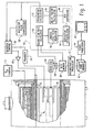
- a main magnetic field control 10 controls superconducting or resistive magnets 12 such that a substantially uniform, temporally constant main magnetic field is created along a z axis through an examination region 14.
- a magnetic resonance generation and manipulation system applies a series of radio frequency (RF) and magnetic gradient field pulses to re-orient the magnetization or excite magnetic spins, induce magnetic resonance, refocus magnetic resonance, manipulate magnetic resonance, spatially and otherwise encode the magnetic resonance, to saturate spin, and the like to generate magnetic resonance imaging and spectroscopy sequences.
- gradient pulse amplifiers 20 apply current pulses to selected ones or pairs of whole-body gradient coils 22 to create magnetic field gradients along x, y and z-axes of the examination region 14.
- a digital radio frequency transmitter 24 transmits radio frequency pulses or pulse packets to a whole-body RF coil 26 to transmit radio frequency B 1 fields in the examination region.
- a typical radio frequency pulse is composed of a packet of immediately contiguous pulse segments of short duration which taken together with each other and any applied gradients achieve a selected magnetic resonance manipulation.
- the RF pulses are used to saturate, excite resonance, invert magnetization, refocus resonance, or manipulate resonance in selected portions of the examination region.
- the resonance signals are commonly picked up by the whole-body RF coil 26.
- localized coils are disposed in the bore more closely adjacent the imaged region.
- a sequence control circuit 30 controls the gradient pulse amplifiers 20 and the transmitter 24 to generate any of a plurality of multiple echo sequences such as echo planar imaging, echo volume imaging, gradient and spin echo imaging, fast spin echo imaging, and the like.
- a receiver 32 receives magnetic resonance signals from RF coil 26 and demodulates the signals into a plurality of data lines. If the receiver is analog, an analog-to-digital converter 34 converts each data line to a digital format. Alternately, the analog-to-digital converter is disposed between the radio frequency receiving coil 26 and the receiver 32 for digital receivers. The data lines are stored or buffered in a data memory 36.
- the data lines are reconstructed into an image representation by a reconstruction processor 40 which applies an inverse Fourier transform or other appropriate reconstruction algorithm.
- the image may represent a planar slice through the patient, an array of parallel planar slices, a three-dimensional volume, or the like.
- the image is then stored in an image memory 42 where it is selectively accessed by a video processor 44 that converts slices, projections, or other portions of the image representation into appropriate format for a display, such as a monitor 46 which provides a man-readable display of the resultant image.
- diagnostic imaging sequence is gated off of blood flow.
- a diagnostic imaging sequence is gated off blood flow in a selected region or position.
- a navigator pulse synthesizer 50 synthesizes a cardiac cycle navigator that measures a distribution of flow velocity in a selected region, perhaps a major vessel.
- the selected region for the phase contrast navigator can be one-dimensional for a slice, two-dimensional or three-dimensional for a selected volume, preferably with the selected region aligned with the flow encoding gradient.
- phase contrast RF pulse sequence When a phase contrast RF pulse sequence is applied, it excites resonance in a region that includes flowing blood. If the excited region is totally within flowing blood, the phase of the RF signal is proportional to the velocity of the flowing blood. If there is other tissue in the excited region, a reference scan is taken and used to subtract out the static tissue background.
- a series of excitations are performed to time map the velocity or flow rate over the cardiac cycle.
- a conventional imaging sequence is initiated or continued for the selected portion of the flow cycle.
- the navigator sequence is then applied again to locate the same point in the next cardiac cycle.
- the cardiac cycle plot represents the velocity of the blood flow through the navigation region.
- the cardiac cycle plot In regions close to the heart, such as the ascending aorta, the cardiac cycle plot resembles an ECG signal.
- the cardiac cycle plot has a large peak that corresponds to the contraction of the left ventricle, at which time blood is flowing quickly through the navigation region. In regions more distal from the heart, the cardiac cycle plot resembles a pulse smoothed by the elasticity of the vessels.
- the cardiac cycle plot can be compared to a characteristic ECG signal or a measure of the ECG signal of the subject to refine the correlation therebetween.
- the cardiac cycle plot is analyzed by a cardiac cycle plot analyzer 52.
- a blood flow calculator 54 analyzes the navigator echoes and determines blood flow rates.
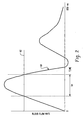
- a cardiac cycle plot truncator 56 establishes a flow rate threshold or window. For instance, a diagnostician may only want an image of the region that includes sample times with high blood flow. With reference to FIGURE 2, a typical cardiac cycle plot 60 is truncated by a threshold value 62 that sets an imaging time window 64 where the blood flow rate is greater than the threshold value 62. Similarly, the diagnostician might want an image with only low blood flow. Optionally, an independent, fixed time window may be established.
- a derivative processor 66 takes a first derivative of the cardiac cycle plot 60. This allows the analyzer 52 to determine at any given point in time whether the flow rate is increasing or decreasing.
- An optional ECG comparator 68 as ghosted in FIGURE 1, compares the cardiac cycle plot to an ECG signal to determine correlations there between.
- a delay factor calculator 70 calculates a delay factor that accounts for blood pulse travel time between the navigation region and the imaging region. The length of the delay is largely a factor of the distance between the navigation region and the imaging region. If the imaging region is relatively close to the navigation region, then the delay is relatively short. Likewise, the farther away the two regions spatially, the longer the delay. In prospective gating, the navigation region is typically upstream of the imaging region.
- the delay represents the time it takes for a pulse of blood (not the blood itself) to travel from the navigation region to the imaging region.
- an image triggering calculator 72 directs the sequence controller 30 to cease generating navigator echoes and commence diagnostic imaging. Alternately the triggering point can be extrapolated from the amplitude and/or slope of the cardiac cycle plot without setting a threshold or triggering window.
- a fast spin echo imaging sequence is preferred because only fractions of a second are available for imaging a narrow window in each cardiac cycle, but other sequences are contemplated.
- Navigator echoes are optionally interspersed between fast spin echo repetitions to determine whether the blood flow velocity has moved out of the window 64.
- Other imaging sequences such as echo planar imaging sequences are contemplated.
- the preferred data collection results in substantially the same blood flow rate through the imaging region throughout the sampling time.
- navigator echoes are applied over a few cardiac cycles to establish a base line cardiac cycle.
- navigator echoes are interspersed among imaging sequence segments to mark where in the cardiac cycle each data line or group of data lines was collected.
- navigator and single spin echo sequences are alternated to mark the location of each data line in the cardiac cycle.
- a plurality of data lines are collected between each navigator echo and the cardiac phase of each data line is extrapolated from the base line cardiac cycle. Echo planar, fast spin echo, and other multi-echo sequences are contemplated.
- Each collected data line and its point in the cardiac cycle are stored in the data memory 36. The operator selects a segment of the cardiac cycle and the corresponding data lines are reconstructed.
- the cardiac cycle can be divided into a plurality of segments and the reconstructed images displayed sequentially in a ciné mode.
- navigator echoes are preferably applied two or more per cycle to monitor cardiac cycle location. More monitoring points per cycle improve accuracy. Few points may be acceptable for patients with very stable cardiac cycles.
- Each cardiac cycle, as estimated from the monitored points is compared with the base line cardiac cycle. If a monitored cycle is abnormal, the data collected during it is discarded.
Landscapes
- Health & Medical Sciences (AREA)
- Life Sciences & Earth Sciences (AREA)
- Physics & Mathematics (AREA)
- Engineering & Computer Science (AREA)
- Biophysics (AREA)
- Nuclear Medicine, Radiotherapy & Molecular Imaging (AREA)
- General Health & Medical Sciences (AREA)
- Radiology & Medical Imaging (AREA)
- Public Health (AREA)
- Physiology (AREA)
- Biomedical Technology (AREA)
- Signal Processing (AREA)
- Veterinary Medicine (AREA)
- High Energy & Nuclear Physics (AREA)
- Animal Behavior & Ethology (AREA)
- Surgery (AREA)
- Molecular Biology (AREA)
- Medical Informatics (AREA)
- Heart & Thoracic Surgery (AREA)
- Pathology (AREA)
- Pulmonology (AREA)
- Psychiatry (AREA)
- Computer Vision & Pattern Recognition (AREA)
- Artificial Intelligence (AREA)
- General Physics & Mathematics (AREA)
- Condensed Matter Physics & Semiconductors (AREA)
- Power Engineering (AREA)
- Cardiology (AREA)
- Magnetic Resonance Imaging Apparatus (AREA)
- Medicines Containing Material From Animals Or Micro-Organisms (AREA)
- Medicines Containing Antibodies Or Antigens For Use As Internal Diagnostic Agents (AREA)
Claims (16)
- Verfahren der Magnetresonanzbildgebung, das Folgendes umfasst:Anordnen einer interessierenden Region und einer Region mit strömendem Blut in einem Hauptmagnetfeld;Zuführen von mindestens einer Navigatorsequenz zu der Region mit strömendem Blut, um Navigatorechos zu erzeugen;Ermitteln eines Messwertes für die Blutbewegung anhand der Navigatorechos; undGating einer Bildgebungssequenz der interessierenden Region in Übereinstimmung mit dem Messwert der Blutbewegung;dadurch gekennzeichnet, dass die mindestens eine Navigatorsequenz eine Phasenkontrast-Navigatorsequenz ist.
- Verfahren nach Anspruch 1, wobei die Navigatorsequenz Navigatorechos erzeugt, die die Blutströmungsgeschwindigkeit angeben.
- Verfahren nach einem der Ansprüche 1 und 2, wobei die Region mit strömendem Blut auch Nicht-Blut-Gewebe enthält, und das Verfahren weiterhin Folgendes umfasst:Erfassen eines grundlegenden Navigator-Messwertes, der den Beitrag des Nicht-Blut-Gewebes zum Navigatorecho angibt.
- Verfahren nach einem der vorhergehenden Ansprüche, das weiterhin Folgendes umfasst:Wiederholtes Anlegen von Phasenkontrast-Navigatoren über mindestens einen Herzzyklus und Bestimmen einer Vielzahl von Blutbewegungs-Messwerten über den Herzzyklus;anhand der Blutströmungsmesswerte Erzeugen eines Herzzyklus-Diagramms der Blutbewegung als Funktion der Zeit, das den Herzzyklus angibt.
- Verfahren nach Anspruch 4, das weiterhin Folgendes umfasst:Einstellen eines Schwellenwertes für den Blutströmungs-Messwert relativ zum Herzzyklus-Diagramm, um einen abzubildenden Bereich des Herzzyklus auszuwählen.
- Verfahren nach einem der Ansprüche 4 und 5, das weiterhin Folgendes umfasst:Korrelieren des Herzzyklus-Diagramms und der EKG-Signale.
- Verfahren nach einem der Ansprüche 4 bis 6, das weiterhin Folgendes umfasst:Erlangen erster Ableitungen des Herzzyklus-Diagramms.
- Verfahren nach Anspruch 7, wobei die angelegten Navigatorsequenzen Navigatorechos erzeugen, die geschwindigkeits- und beschleunigungscodiert sind, und das weiterhin Folgendes umfasst:Vergleichen der Blutströmungsgeschwindigkeit und -beschleunigung mit Amplituden und Ableitungen des Herzzyklus-Diagramms, um Punkte im Herzzyklus zu bestimmen.
- Verfahren nach Anspruch 8, das weiterhin Folgendes umfasst:Auslösen der Bildgebungssequenz-Datenerfassung in Reaktion auf die ermittelten Punkte im Herzzyklus.
- Bildgebungsverfahren nach einem der Ansprüche 7 bis 9, das weiterhin Folgendes umfasst:Anwenden von mindestens einem Extrapolationsalgorithmus auf das Herzzyklus-Diagramm basierend auf der ersten Ableitung des Herzzyklus-Diagramms, um einen Auslösezeitpunkt zu bestimmen; undAuslösen einer Datenerfassung für die interessierende Region zum Auslösezeitpunkt.
- Verfahren nach einem der vorhergehenden Ansprüche, das weiterhin Folgendes umfasst:Justieren der Navigatorsequenz, um die Ausrichtung der Region mit strömendem Blut auf ein Blutgefäß zu justieren.
- Magnetresonanz-Bildgebungsgerät, das Folgendes umfasst:ein Hauptmagnetfeldmittel (12) zum Erzeugen eines Hauptmagnetfeldes in einer Bildgebungsregion (14), in der sich eine interessierende Region und eine Region mit strömendem Blut befinden;ein Mittel (30, 22, 26) zum Zuführen mindestens einer Navigatorsequenz zu der Region mit strömendem Blut, um Navigatorechos zu erzeugen, und einer Bildgebungssequenz zu der interessierenden Region;ein Mittel (54) zum Messen der Blutbewegung in der Region mit strömendem Blut anhand der Navigatorechos; undein Gating-Mittel (52) für das Gating der Bildgebungssequenz in Übereinstimmung mit den Blutbewegungs-Messwerten,dadurch gekennzeichnet, dass die mindestens eine Navigatorsequenz eine Phasenkontrast-Navigatorsequenz ist.
- Magnetresonanzgerät nach Anspruch 12, das weiterhin Folgendes umfasst:ein Mittel (54) zum Erzeugen eines Herzzyklus-Diagramms der Blutbewegung als Funktion der Zeit, das den Herzzyklus anhand der Blutbewegungs-Messwerte angibt.
- Magnetresonanzgerät nach Anspruch 13, das weiterhin Folgendes umfasst:ein Ableitungsmittel (66) zum Erlangen einer ersten Ableitung des Herzzyklus-Diagramms.
- Magnetresonanzgerät nach einem der Ansprüche 12, 13 und 14, das weiterhin Folgendes umfasst:ein Komparatormittel (68) zum Vergleichen der Strömungsrate mit einem EKG und zum Ermitteln von Ähnlichkeiten zwischen beiden.
- Magnetresonanzgerät nach einem der Ansprüche 12 bis 15, das weiterhin Folgendes umfasst:ein Auslösezeitpunkt-Berechnungsmittel (72) zum:Anwenden eines Extrapolationsalgorithmus auf das Herzzyklus-Diagramm;Kombinieren der extrapolierten Daten mit Kriterien der ersten Ableitung;Berechnen eines Auslösezeitpunktes; undAuslösen der Bildgebungssequenz für die interessierende Region zu dem Auslösezeitpunkt.
Applications Claiming Priority (3)
| Application Number | Priority Date | Filing Date | Title |
|---|---|---|---|
| US162379 | 2002-06-04 | ||
| US10/162,379 US6922580B2 (en) | 2002-06-04 | 2002-06-04 | Blood flow gated MRI |
| PCT/IB2003/002058 WO2003102618A1 (en) | 2002-06-04 | 2003-05-28 | Blood flow gated mri |
Publications (2)
| Publication Number | Publication Date |
|---|---|
| EP1514129A1 EP1514129A1 (de) | 2005-03-16 |
| EP1514129B1 true EP1514129B1 (de) | 2008-01-02 |
Family
ID=29583595
Family Applications (1)
| Application Number | Title | Priority Date | Filing Date |
|---|---|---|---|
| EP03756077A Expired - Lifetime EP1514129B1 (de) | 2002-06-04 | 2003-05-28 | Blutströmungs-gate-mri |
Country Status (7)
| Country | Link |
|---|---|
| US (1) | US6922580B2 (de) |
| EP (1) | EP1514129B1 (de) |
| JP (1) | JP4376779B2 (de) |
| AT (1) | ATE382870T1 (de) |
| AU (1) | AU2003232383A1 (de) |
| DE (1) | DE60318438T2 (de) |
| WO (1) | WO2003102618A1 (de) |
Families Citing this family (30)
| Publication number | Priority date | Publication date | Assignee | Title |
|---|---|---|---|---|
| US7561909B1 (en) * | 2002-09-16 | 2009-07-14 | The United States Of America As Represented By The Department Of Health And Human Services | MRI navigator methods and systems |
| DE10256209B4 (de) * | 2002-12-02 | 2007-06-21 | Siemens Ag | Verfahren zur automatischen Bestimmung des tatsächlichen Geschwindigkeitsintervalls eines fließenden Mediums bei Flussmessungen in der Magnetresonanz-Tomographie sowie Kernspintomographiegerät und Computersoftwareprodukt |
| US6798199B2 (en) * | 2003-02-06 | 2004-09-28 | Siemens Medical Solutions Usa, Inc. | Method for synchronizing magnetic resonance imaging data to body motion |
| US9301704B2 (en) | 2004-03-26 | 2016-04-05 | Kabushiki Kaisha Toshiba | Magnetic resonance imaging system for non-contrast MRA and magnetic resonance signal acquisition method employed by the same |
| JP4820567B2 (ja) * | 2004-03-26 | 2011-11-24 | 株式会社東芝 | 磁気共鳴イメージング装置及び磁気共鳴信号の収集方法 |
| US7238027B2 (en) * | 2004-07-20 | 2007-07-03 | Cardiac Pacemakers, Inc. | Device functionality representation tool |
| FR2880252A1 (fr) * | 2005-01-06 | 2006-07-07 | Yves Darlas | " nouvelle methode d'analyse et de mesure de la distribution ou de la reserve sanguine coronarienne " |
| US8000768B2 (en) * | 2005-01-10 | 2011-08-16 | Vassol Inc. | Method and system for displaying blood flow |
| DE102005027438B4 (de) * | 2005-06-14 | 2011-12-22 | Siemens Ag | Verfahren zur EKG-Triggerung einer Messsequenz eines Magnetresonanzgeräts |
| US7613496B2 (en) * | 2005-09-22 | 2009-11-03 | Kabushiki Kaisha Toshiba | Magnetic resonance imaging apparatus and magnetic resonance imaging method |
| JP4702107B2 (ja) * | 2006-03-03 | 2011-06-15 | 株式会社日立製作所 | 生体光計測装置 |
| US7693568B2 (en) | 2006-03-30 | 2010-04-06 | Medtronic, Inc. | Medical device sensing and detection during MRI |
| US8190244B2 (en) * | 2007-01-23 | 2012-05-29 | Case Western Reserve University | Gated optical coherence tomography (OCT) environmental chamber |
| US8320647B2 (en) | 2007-11-20 | 2012-11-27 | Olea Medical | Method and system for processing multiple series of biological images obtained from a patient |
| BRPI0907547B8 (pt) | 2008-02-28 | 2021-07-27 | Koninklijke Philips Electronics Nv | sistema de ressonância magnética, monitor seguro de pressão sanguínea para a mri, e, método para monitorar um objeto em um campo magnético |
| JP2010063871A (ja) * | 2008-08-12 | 2010-03-25 | Toshiba Corp | 磁気共鳴イメージング装置 |
| DE102008060049A1 (de) * | 2008-12-02 | 2010-06-17 | Siemens Aktiengesellschaft | Verfahren zur Bestimmung einer Kodierung für eine Flussmessung und Verfahren zur Flussmessung sowie entsprechend ausgestaltete Magnetresonanzanlage |
| JP5542495B2 (ja) * | 2009-06-08 | 2014-07-09 | 株式会社東芝 | 磁気共鳴イメージング装置 |
| JP2011156078A (ja) * | 2010-01-29 | 2011-08-18 | Ge Medical Systems Global Technology Co Llc | 磁気共鳴イメージング装置およびプログラム |
| US9226673B2 (en) | 2011-01-10 | 2016-01-05 | East Carolina University | Methods, systems and computer program products for non-invasive determination of blood flow distribution using speckle imaging techniques and hemodynamic modeling |
| EP2663222B1 (de) | 2011-01-10 | 2021-10-27 | East Carolina University | Verfahren und systeme zur nichtinvasiven bestimmung der blutflussverteilung unter verwendung von specklebildgebungsverfahren und hämodynamischer modellierung |
| JP5284422B2 (ja) * | 2011-06-27 | 2013-09-11 | 学校法人東海大学 | 磁気共鳴イメージング装置 |
| DE102012216248A1 (de) * | 2012-09-13 | 2014-03-13 | Siemens Aktiengesellschaft | Medizinische Bildgebungsvorrichtung mit einer Sensoreinheit zu einem Erfassen eines physiologischen Signals sowie ein Verfahren zu einem Erfassen eines Herzzyklus eines Patienten |
| DE102014206724B4 (de) * | 2014-04-08 | 2015-11-12 | Siemens Aktiengesellschaft | Dynamische Bildgebung mit variablem Kontrast |
| EP3188651A4 (de) | 2014-10-14 | 2018-07-04 | East Carolina University | Verfahren, systeme und computerprogrammprodukte zur visualisierung anatomischer strukturen und des blutflusses und der perfusionsphysiologie durch verwendung von bildgebungstechniken |
| US11553844B2 (en) | 2014-10-14 | 2023-01-17 | East Carolina University | Methods, systems and computer program products for calculating MetaKG signals for regions having multiple sets of optical characteristics |
| CN107257655B (zh) | 2014-10-14 | 2020-06-16 | 东卡罗莱娜大学 | 用于利用从多谱段血液流动和灌注成像获取的信号确定血液动力学状态参数的方法、系统和计算机程序产品 |
| US10390718B2 (en) | 2015-03-20 | 2019-08-27 | East Carolina University | Multi-spectral physiologic visualization (MSPV) using laser imaging methods and systems for blood flow and perfusion imaging and quantification in an endoscopic design |
| US10058256B2 (en) | 2015-03-20 | 2018-08-28 | East Carolina University | Multi-spectral laser imaging (MSLI) methods and systems for blood flow and perfusion imaging and quantification |
| JP6996930B2 (ja) * | 2017-10-13 | 2022-01-17 | キヤノンメディカルシステムズ株式会社 | 磁気共鳴イメージング装置 |
Family Cites Families (7)
| Publication number | Priority date | Publication date | Assignee | Title |
|---|---|---|---|---|
| US5070876A (en) * | 1989-08-07 | 1991-12-10 | The Board Of Trustees Of The Leland Stanford Junior University | Flow-independent magnetic resonance projection angiography |
| US5031624A (en) * | 1990-08-17 | 1991-07-16 | Wisconsin Alumni Research Foundation | Phase contrast, line-scanned method for NMR angiography |
| US5133357A (en) * | 1991-02-07 | 1992-07-28 | General Electric Company | Quantitative measurement of blood flow using cylindrically localized fourier velocity encoding |
| US5233298A (en) * | 1992-02-20 | 1993-08-03 | General Electric Company | Quantitative measurement of blood flow at multiple positions using comb excitation and fourier velocity encoding |
| US5435303A (en) * | 1993-08-04 | 1995-07-25 | General Electric Company | MRA image produced by temporal flow data sharing |
| US6268730B1 (en) | 1999-05-24 | 2001-07-31 | Ge Medical Systems Global Technology Company Llc | Multi-slab multi-window cardiac MR imaging |
| US6516210B1 (en) * | 2000-11-22 | 2003-02-04 | Koninklijke Philips Electronics N.V. | Signal analysis for navigated magnetic resonance imaging |
-
2002
- 2002-06-04 US US10/162,379 patent/US6922580B2/en not_active Expired - Fee Related
-
2003
- 2003-05-28 DE DE60318438T patent/DE60318438T2/de not_active Expired - Lifetime
- 2003-05-28 AT AT03756077T patent/ATE382870T1/de not_active IP Right Cessation
- 2003-05-28 JP JP2004509446A patent/JP4376779B2/ja not_active Expired - Lifetime
- 2003-05-28 EP EP03756077A patent/EP1514129B1/de not_active Expired - Lifetime
- 2003-05-28 WO PCT/IB2003/002058 patent/WO2003102618A1/en not_active Ceased
- 2003-05-28 AU AU2003232383A patent/AU2003232383A1/en not_active Abandoned
Also Published As
| Publication number | Publication date |
|---|---|
| ATE382870T1 (de) | 2008-01-15 |
| JP2005528175A (ja) | 2005-09-22 |
| US6922580B2 (en) | 2005-07-26 |
| EP1514129A1 (de) | 2005-03-16 |
| WO2003102618A1 (en) | 2003-12-11 |
| US20030225328A1 (en) | 2003-12-04 |
| AU2003232383A1 (en) | 2003-12-19 |
| DE60318438T2 (de) | 2008-12-18 |
| JP4376779B2 (ja) | 2009-12-02 |
| DE60318438D1 (de) | 2008-02-14 |
Similar Documents
| Publication | Publication Date | Title |
|---|---|---|
| EP1514129B1 (de) | Blutströmungs-gate-mri | |
| US6380740B1 (en) | Method for acquiring time-resolved and location-resolved, three-dimensional data sets with magnetic resonance and apparatus for the implementation of the method | |
| EP1113288B1 (de) | Messung von atmungsbedingter Bewegung und Geschwindigkeit unter Verwendung von Navigator-Echosignalen der bildgebenden magnetischen Resonanz | |
| US6408201B1 (en) | Method and apparatus for efficient stenosis identification in peripheral arterial vasculature using MR imaging | |
| US6195579B1 (en) | Contrast detection and guided reconstruction in contrast-enhanced magnetic resonance angiography | |
| US6268730B1 (en) | Multi-slab multi-window cardiac MR imaging | |
| US6064203A (en) | Method and apparatus for determining or imaging longitudinal spin relaxation time or producing images which substantially reflect longitudinal spin relaxation time contrast | |
| US7027854B2 (en) | Magnetic resonance imaging utilizing a microcoil | |
| RU2580189C2 (ru) | Интервенционная мр-томография с компенсацией движения | |
| EP0994363A2 (de) | Verfahren der atmungsabhängigen Triggerung für die Magnetresonanzbildgebung | |
| US5492123A (en) | Diffusion weighted magnetic resonance imaging | |
| JP3992973B2 (ja) | 高時間分解能の自由呼吸mr画像の収集 | |
| US20030036693A1 (en) | Method to obtain the cardiac gating signal using a cardiac displacement sensor | |
| EP0948929B1 (de) | Verfahren zur Berechnung von Wellengeschwindigkeiten in Blutgefässen | |
| EP1221624A2 (de) | Verfahren und Gerät zur schnellen dreidimensionalen Datenaufnahme mittels magnetischer Resonanz mit Atemanhalten und unter Verwendung einer variablen Probennahme | |
| CN112394311B (zh) | 具有改进的导航器的mri系统 | |
| US20050272997A1 (en) | MRI method for assessing myocardial viability | |
| US6201986B1 (en) | Synchronized K-space sampling in magnetic resonance angiography | |
| CN1226415A (zh) | 用epi脉冲序列采集分段的mri心脏数据 | |
| US6198960B1 (en) | Flip angle modulated magnetic resonance angiography | |
| JP3967210B2 (ja) | 磁気共鳴イメージング装置 | |
| WO2003103491A1 (ja) | 磁気共鳴イメージング装置 | |
| US6288541B1 (en) | MRI measurement of blood vessel wall compliance | |
| JP4349647B2 (ja) | 磁気共鳴イメージング装置 | |
| JP2889871B1 (ja) | 磁気共鳴診断装置 |
Legal Events
| Date | Code | Title | Description |
|---|---|---|---|
| PUAI | Public reference made under article 153(3) epc to a published international application that has entered the european phase |
Free format text: ORIGINAL CODE: 0009012 |
|
| 17P | Request for examination filed |
Effective date: 20050104 |
|
| AK | Designated contracting states |
Kind code of ref document: A1 Designated state(s): AT BE BG CH CY CZ DE DK EE ES FI FR GB GR HU IE IT LI LU MC NL PT RO SE SI SK TR |
|
| AX | Request for extension of the european patent |
Extension state: AL LT LV MK |
|
| DAX | Request for extension of the european patent (deleted) | ||
| GRAP | Despatch of communication of intention to grant a patent |
Free format text: ORIGINAL CODE: EPIDOSNIGR1 |
|
| GRAS | Grant fee paid |
Free format text: ORIGINAL CODE: EPIDOSNIGR3 |
|
| GRAA | (expected) grant |
Free format text: ORIGINAL CODE: 0009210 |
|
| AK | Designated contracting states |
Kind code of ref document: B1 Designated state(s): AT BE BG CH CY CZ DE DK EE ES FI FR GB GR HU IE IT LI LU MC NL PT RO SE SI SK TR |
|
| REG | Reference to a national code |
Ref country code: GB Ref legal event code: FG4D |
|
| REG | Reference to a national code |
Ref country code: IE Ref legal event code: FG4D |
|
| REG | Reference to a national code |
Ref country code: CH Ref legal event code: EP |
|
| REF | Corresponds to: |
Ref document number: 60318438 Country of ref document: DE Date of ref document: 20080214 Kind code of ref document: P |
|
| PG25 | Lapsed in a contracting state [announced via postgrant information from national office to epo] |
Ref country code: SI Free format text: LAPSE BECAUSE OF FAILURE TO SUBMIT A TRANSLATION OF THE DESCRIPTION OR TO PAY THE FEE WITHIN THE PRESCRIBED TIME-LIMIT Effective date: 20080102 Ref country code: NL Free format text: LAPSE BECAUSE OF FAILURE TO SUBMIT A TRANSLATION OF THE DESCRIPTION OR TO PAY THE FEE WITHIN THE PRESCRIBED TIME-LIMIT Effective date: 20080102 |
|
| NLV1 | Nl: lapsed or annulled due to failure to fulfill the requirements of art. 29p and 29m of the patents act | ||
| PG25 | Lapsed in a contracting state [announced via postgrant information from national office to epo] |
Ref country code: FI Free format text: LAPSE BECAUSE OF FAILURE TO SUBMIT A TRANSLATION OF THE DESCRIPTION OR TO PAY THE FEE WITHIN THE PRESCRIBED TIME-LIMIT Effective date: 20080102 Ref country code: LI Free format text: LAPSE BECAUSE OF FAILURE TO SUBMIT A TRANSLATION OF THE DESCRIPTION OR TO PAY THE FEE WITHIN THE PRESCRIBED TIME-LIMIT Effective date: 20080102 Ref country code: CH Free format text: LAPSE BECAUSE OF FAILURE TO SUBMIT A TRANSLATION OF THE DESCRIPTION OR TO PAY THE FEE WITHIN THE PRESCRIBED TIME-LIMIT Effective date: 20080102 Ref country code: ES Free format text: LAPSE BECAUSE OF FAILURE TO SUBMIT A TRANSLATION OF THE DESCRIPTION OR TO PAY THE FEE WITHIN THE PRESCRIBED TIME-LIMIT Effective date: 20080413 |
|
| REG | Reference to a national code |
Ref country code: CH Ref legal event code: PL |
|
| ET | Fr: translation filed | ||
| PG25 | Lapsed in a contracting state [announced via postgrant information from national office to epo] |
Ref country code: BG Free format text: LAPSE BECAUSE OF FAILURE TO SUBMIT A TRANSLATION OF THE DESCRIPTION OR TO PAY THE FEE WITHIN THE PRESCRIBED TIME-LIMIT Effective date: 20080402 Ref country code: AT Free format text: LAPSE BECAUSE OF FAILURE TO SUBMIT A TRANSLATION OF THE DESCRIPTION OR TO PAY THE FEE WITHIN THE PRESCRIBED TIME-LIMIT Effective date: 20080102 |
|
| PG25 | Lapsed in a contracting state [announced via postgrant information from national office to epo] |
Ref country code: PT Free format text: LAPSE BECAUSE OF FAILURE TO SUBMIT A TRANSLATION OF THE DESCRIPTION OR TO PAY THE FEE WITHIN THE PRESCRIBED TIME-LIMIT Effective date: 20080602 Ref country code: BE Free format text: LAPSE BECAUSE OF FAILURE TO SUBMIT A TRANSLATION OF THE DESCRIPTION OR TO PAY THE FEE WITHIN THE PRESCRIBED TIME-LIMIT Effective date: 20080102 |
|
| PG25 | Lapsed in a contracting state [announced via postgrant information from national office to epo] |
Ref country code: SE Free format text: LAPSE BECAUSE OF FAILURE TO SUBMIT A TRANSLATION OF THE DESCRIPTION OR TO PAY THE FEE WITHIN THE PRESCRIBED TIME-LIMIT Effective date: 20080402 Ref country code: CZ Free format text: LAPSE BECAUSE OF FAILURE TO SUBMIT A TRANSLATION OF THE DESCRIPTION OR TO PAY THE FEE WITHIN THE PRESCRIBED TIME-LIMIT Effective date: 20080102 Ref country code: DK Free format text: LAPSE BECAUSE OF FAILURE TO SUBMIT A TRANSLATION OF THE DESCRIPTION OR TO PAY THE FEE WITHIN THE PRESCRIBED TIME-LIMIT Effective date: 20080102 Ref country code: SK Free format text: LAPSE BECAUSE OF FAILURE TO SUBMIT A TRANSLATION OF THE DESCRIPTION OR TO PAY THE FEE WITHIN THE PRESCRIBED TIME-LIMIT Effective date: 20080102 |
|
| PLBE | No opposition filed within time limit |
Free format text: ORIGINAL CODE: 0009261 |
|
| STAA | Information on the status of an ep patent application or granted ep patent |
Free format text: STATUS: NO OPPOSITION FILED WITHIN TIME LIMIT |
|
| PG25 | Lapsed in a contracting state [announced via postgrant information from national office to epo] |
Ref country code: RO Free format text: LAPSE BECAUSE OF FAILURE TO SUBMIT A TRANSLATION OF THE DESCRIPTION OR TO PAY THE FEE WITHIN THE PRESCRIBED TIME-LIMIT Effective date: 20080102 |
|
| 26N | No opposition filed |
Effective date: 20081003 |
|
| PG25 | Lapsed in a contracting state [announced via postgrant information from national office to epo] |
Ref country code: MC Free format text: LAPSE BECAUSE OF NON-PAYMENT OF DUE FEES Effective date: 20080531 |
|
| GBPC | Gb: european patent ceased through non-payment of renewal fee |
Effective date: 20080528 |
|
| PG25 | Lapsed in a contracting state [announced via postgrant information from national office to epo] |
Ref country code: EE Free format text: LAPSE BECAUSE OF FAILURE TO SUBMIT A TRANSLATION OF THE DESCRIPTION OR TO PAY THE FEE WITHIN THE PRESCRIBED TIME-LIMIT Effective date: 20080102 Ref country code: IE Free format text: LAPSE BECAUSE OF NON-PAYMENT OF DUE FEES Effective date: 20080528 |
|
| PG25 | Lapsed in a contracting state [announced via postgrant information from national office to epo] |
Ref country code: GB Free format text: LAPSE BECAUSE OF NON-PAYMENT OF DUE FEES Effective date: 20080528 |
|
| PG25 | Lapsed in a contracting state [announced via postgrant information from national office to epo] |
Ref country code: CY Free format text: LAPSE BECAUSE OF FAILURE TO SUBMIT A TRANSLATION OF THE DESCRIPTION OR TO PAY THE FEE WITHIN THE PRESCRIBED TIME-LIMIT Effective date: 20080102 |
|
| PG25 | Lapsed in a contracting state [announced via postgrant information from national office to epo] |
Ref country code: IT Free format text: LAPSE BECAUSE OF FAILURE TO SUBMIT A TRANSLATION OF THE DESCRIPTION OR TO PAY THE FEE WITHIN THE PRESCRIBED TIME-LIMIT Effective date: 20080102 |
|
| PG25 | Lapsed in a contracting state [announced via postgrant information from national office to epo] |
Ref country code: LU Free format text: LAPSE BECAUSE OF NON-PAYMENT OF DUE FEES Effective date: 20080528 Ref country code: HU Free format text: LAPSE BECAUSE OF FAILURE TO SUBMIT A TRANSLATION OF THE DESCRIPTION OR TO PAY THE FEE WITHIN THE PRESCRIBED TIME-LIMIT Effective date: 20080703 |
|
| PG25 | Lapsed in a contracting state [announced via postgrant information from national office to epo] |
Ref country code: TR Free format text: LAPSE BECAUSE OF FAILURE TO SUBMIT A TRANSLATION OF THE DESCRIPTION OR TO PAY THE FEE WITHIN THE PRESCRIBED TIME-LIMIT Effective date: 20080102 |
|
| PG25 | Lapsed in a contracting state [announced via postgrant information from national office to epo] |
Ref country code: GR Free format text: LAPSE BECAUSE OF FAILURE TO SUBMIT A TRANSLATION OF THE DESCRIPTION OR TO PAY THE FEE WITHIN THE PRESCRIBED TIME-LIMIT Effective date: 20080403 |
|
| PGFP | Annual fee paid to national office [announced via postgrant information from national office to epo] |
Ref country code: FR Payment date: 20110615 Year of fee payment: 9 |
|
| PGFP | Annual fee paid to national office [announced via postgrant information from national office to epo] |
Ref country code: DE Payment date: 20110728 Year of fee payment: 9 |
|
| REG | Reference to a national code |
Ref country code: FR Ref legal event code: ST Effective date: 20130131 |
|
| REG | Reference to a national code |
Ref country code: DE Ref legal event code: R119 Ref document number: 60318438 Country of ref document: DE Effective date: 20121201 |
|
| PG25 | Lapsed in a contracting state [announced via postgrant information from national office to epo] |
Ref country code: FR Free format text: LAPSE BECAUSE OF NON-PAYMENT OF DUE FEES Effective date: 20120531 |
|
| PG25 | Lapsed in a contracting state [announced via postgrant information from national office to epo] |
Ref country code: DE Free format text: LAPSE BECAUSE OF NON-PAYMENT OF DUE FEES Effective date: 20121201 |