EP1500697A2 - Bioartifizielles, primär vaskularisiertes Testgewebe für pharmakologische Testverfahren, Verfahren zu dessen Herstellung und Testverfahren unter dessen Verwendung sowie Bioreaktor zur Durchführung der Verfahren - Google Patents
Bioartifizielles, primär vaskularisiertes Testgewebe für pharmakologische Testverfahren, Verfahren zu dessen Herstellung und Testverfahren unter dessen Verwendung sowie Bioreaktor zur Durchführung der Verfahren Download PDFInfo
- Publication number
- EP1500697A2 EP1500697A2 EP04017315A EP04017315A EP1500697A2 EP 1500697 A2 EP1500697 A2 EP 1500697A2 EP 04017315 A EP04017315 A EP 04017315A EP 04017315 A EP04017315 A EP 04017315A EP 1500697 A2 EP1500697 A2 EP 1500697A2
- Authority
- EP
- European Patent Office
- Prior art keywords
- tissue
- test
- matrix
- cell
- bioartificial
- Prior art date
- Legal status (The legal status is an assumption and is not a legal conclusion. Google has not performed a legal analysis and makes no representation as to the accuracy of the status listed.)
- Granted
Links
Images

Classifications
-
- C—CHEMISTRY; METALLURGY
- C12—BIOCHEMISTRY; BEER; SPIRITS; WINE; VINEGAR; MICROBIOLOGY; ENZYMOLOGY; MUTATION OR GENETIC ENGINEERING
- C12N—MICROORGANISMS OR ENZYMES; COMPOSITIONS THEREOF; PROPAGATING, PRESERVING, OR MAINTAINING MICROORGANISMS; MUTATION OR GENETIC ENGINEERING; CULTURE MEDIA
- C12N5/00—Undifferentiated human, animal or plant cells, e.g. cell lines; Tissues; Cultivation or maintenance thereof; Culture media therefor
- C12N5/06—Animal cells or tissues; Human cells or tissues
- C12N5/0697—Artificial constructs associating cells of different lineages, e.g. tissue equivalents
-
- C—CHEMISTRY; METALLURGY
- C12—BIOCHEMISTRY; BEER; SPIRITS; WINE; VINEGAR; MICROBIOLOGY; ENZYMOLOGY; MUTATION OR GENETIC ENGINEERING
- C12M—APPARATUS FOR ENZYMOLOGY OR MICROBIOLOGY; APPARATUS FOR CULTURING MICROORGANISMS FOR PRODUCING BIOMASS, FOR GROWING CELLS OR FOR OBTAINING FERMENTATION OR METABOLIC PRODUCTS, i.e. BIOREACTORS OR FERMENTERS
- C12M25/00—Means for supporting, enclosing or fixing the microorganisms, e.g. immunocoatings
- C12M25/14—Scaffolds; Matrices
-
- C—CHEMISTRY; METALLURGY
- C12—BIOCHEMISTRY; BEER; SPIRITS; WINE; VINEGAR; MICROBIOLOGY; ENZYMOLOGY; MUTATION OR GENETIC ENGINEERING
- C12M—APPARATUS FOR ENZYMOLOGY OR MICROBIOLOGY; APPARATUS FOR CULTURING MICROORGANISMS FOR PRODUCING BIOMASS, FOR GROWING CELLS OR FOR OBTAINING FERMENTATION OR METABOLIC PRODUCTS, i.e. BIOREACTORS OR FERMENTERS
- C12M29/00—Means for introduction, extraction or recirculation of materials, e.g. pumps
- C12M29/10—Perfusion
-
- C—CHEMISTRY; METALLURGY
- C12—BIOCHEMISTRY; BEER; SPIRITS; WINE; VINEGAR; MICROBIOLOGY; ENZYMOLOGY; MUTATION OR GENETIC ENGINEERING
- C12N—MICROORGANISMS OR ENZYMES; COMPOSITIONS THEREOF; PROPAGATING, PRESERVING, OR MAINTAINING MICROORGANISMS; MUTATION OR GENETIC ENGINEERING; CULTURE MEDIA
- C12N5/00—Undifferentiated human, animal or plant cells, e.g. cell lines; Tissues; Cultivation or maintenance thereof; Culture media therefor
- C12N5/06—Animal cells or tissues; Human cells or tissues
- C12N5/0602—Vertebrate cells
- C12N5/0634—Cells from the blood or the immune system
- C12N5/0645—Macrophages, e.g. Kuepfer cells in the liver; Monocytes
-
- C—CHEMISTRY; METALLURGY
- C12—BIOCHEMISTRY; BEER; SPIRITS; WINE; VINEGAR; MICROBIOLOGY; ENZYMOLOGY; MUTATION OR GENETIC ENGINEERING
- C12N—MICROORGANISMS OR ENZYMES; COMPOSITIONS THEREOF; PROPAGATING, PRESERVING, OR MAINTAINING MICROORGANISMS; MUTATION OR GENETIC ENGINEERING; CULTURE MEDIA
- C12N5/00—Undifferentiated human, animal or plant cells, e.g. cell lines; Tissues; Cultivation or maintenance thereof; Culture media therefor
- C12N5/06—Animal cells or tissues; Human cells or tissues
- C12N5/0602—Vertebrate cells
- C12N5/0693—Tumour cells; Cancer cells
-
- C—CHEMISTRY; METALLURGY
- C12—BIOCHEMISTRY; BEER; SPIRITS; WINE; VINEGAR; MICROBIOLOGY; ENZYMOLOGY; MUTATION OR GENETIC ENGINEERING
- C12N—MICROORGANISMS OR ENZYMES; COMPOSITIONS THEREOF; PROPAGATING, PRESERVING, OR MAINTAINING MICROORGANISMS; MUTATION OR GENETIC ENGINEERING; CULTURE MEDIA
- C12N2503/00—Use of cells in diagnostics
- C12N2503/04—Screening or testing on artificial tissues
-
- C—CHEMISTRY; METALLURGY
- C12—BIOCHEMISTRY; BEER; SPIRITS; WINE; VINEGAR; MICROBIOLOGY; ENZYMOLOGY; MUTATION OR GENETIC ENGINEERING
- C12N—MICROORGANISMS OR ENZYMES; COMPOSITIONS THEREOF; PROPAGATING, PRESERVING, OR MAINTAINING MICROORGANISMS; MUTATION OR GENETIC ENGINEERING; CULTURE MEDIA
- C12N2533/00—Supports or coatings for cell culture, characterised by material
- C12N2533/90—Substrates of biological origin, e.g. extracellular matrix, decellularised tissue
Definitions
- the invention relates to a bioartificial, primary vascularized, human test tissue for pharmacological Test method, a method for its production and a Test method using such a test fabric and a bioreactor for carrying out the manufacturing and Test procedure.
- the invention is based on the object, compared to known Systems to create improvements and in particular To enable testing of pharmaceuticals as possible largely be transmitted to the human body can.
- test fabric Claim 1 This task is solved by a test fabric Claim 1 and a method for producing such Test fabric according to claim 5 and a test method according to Claim 9. Furthermore, a bioreactor according to claim 17 intended.
- the dependent claims describe preferred developments.
- the test fabric according to the invention is in particular producible by the manufacturing method according to the invention.
- the invention is based on the finding that missing Matches between ex vivo cell culture systems from human cells and the human body in particular to the two-dimensional environment of such cell culture systems are attributed. These new cells will be ex vivo in the two-dimensional or quasi-two-dimensional Environment increases and can be due to its two-dimensional Microenviroment Dedifferentiation processes be subjected. Such dedifferentiation processes in vivo, in particular by the cell culture medium or extracellular matrix components are prevented.
- Tissue Engineering used. Endothelial cells, human primary or tumor cells and a natural vascularized support structure, d. H. ECM extracellular matrix, are used as a substitute for a used native three-dimensional tissue association.
- a test substance or a physical treatment can act on the cells in vitro, so that the influence of a Drug or physical treatment on cell cycle, Proliferation and rates of synthesis in a natural or quasi-natural Mikroenviroment be examined can.
- changes in the Creation rate of new blood vessels and variations in the expression pattern endothelial adhesion molecules are assessed.
- special cell-cell and cell-matrix interactions at mRNA and protein level to investigate. Found changes can be made with the used pharmaceutical concentration and the duration of application be correlated, whereupon the corresponding Evaluation the corresponding parameters are changed can.
- a biological Output tissue by decellularizing a bioartificial, vascularized, biological tissue matrix essentially collagen formed.
- the tissue completely decellularized for this purpose; if autologous Tissue is taken, z. B. also only partially Decellularization be made.
- Such Making a tissue matrix is already taken in itself from the field of implant production or tissue Engineering, z. Example of WO 02/064179 A2. in this connection can z. B. a xenogeneic tissue from z.
- a pig, an autologous tissue of the patient himself or an allogeneic Tissues of another human patient are used wherein the tissue at least one vessel branch, preferably having an arterial and a venous vessel branch.
- endothelial cells or stem cells or Progeniturzellen inflicted in the primary vascularized Tissue matrix form a vascular network.
- relevant human cells in particular supplied to tumor cells.
- liver cells are supplied. Have liver cells in this case, the property which is advantageous for the test methods, the supplied drugs and degradation products too metabolize.
- human strain or Precursor cells are supplied, since these are located in the Tissue according to the culture conditions to different Form cell types, e.g. Muscle or cartilage cells.
- Form cell types e.g. Muscle or cartilage cells.
- a fresh tissue is produced according to the invention, particularly due to its higher division and regeneration rate and due to missing previous Influences good for the subsequent test procedure suitable.
- This can be embryonic or adult stem cells be used.
- test fabric according to the invention makes it possible in particular two applications:
- the delivery of the patient's own Blood during colonization and / or during the test method advantageous. It can thus be up on possibly a non-autologous, essentially collagen-based Tissue matrix by colonization with autologous primary Cells or tumor cells and autologous supply Blood is a very realistic, bio-artificial, biological ex vivo quasi-organ system are formed.
- According to the invention can advantageously metabolism controlled and possibly adjusted. Especially This results in significant advantages over known two-dimensional cell culture systems. It can in particular Series of measurements depending on pharmacological concentrations, Conditions of supply, if necessary other radiotherapeutic Treatments and supply of other nutrients are investigated become.
- the perfusion of the quasi-organ system preferably takes place in a bioreactor according to the invention.
- the perfusion system has at least one, preferably Five adapter / connection systems for perfusion lines.
- the Lumen of the organ system is about an inlet and a drain perfused with culture solution.
- the vascular branches of the biological Matrix are connected separately to a perfusion unit and perfused by this.
- the perfusion is possible under laminar and pulsatile perfusion conditions. Under perfusion and during culture conditions samples can be obtained from the perfusate at any time and drugs, cell solutions or other drugs added become.
- test tissue in the same bioreactor can advantageously the production of the test tissue in the same bioreactor be like the following test procedure; in this connection can the connections of the arterial and venous vessel branch of the test fabric to the supply pipe of the bioreactor remain, so that adverse effects by a temporary removal of the entire test tissue can be largely avoided.
- the example relates to the production and cultivation of human 3D tumor tissue in vitro from tumor biopsies.
- a Tumor patients with a rare or refractory Malignancy is a tissue biopsy of the tumor area as well 200 ml of blood taken.
- the blood sample is 1: 1 with PBS diluted and 10 ml each to 20 ml Bicoll solution given up.
- the mononuclear cells are replaced by a Centrifugation for 20 min at 300g without brake isolated, and washed twice with PBS.
- the obtained mononuclear cells serve for re-endothelialization one of acellularized pig intestine fabricated biological support structure with preserved Vascular bed with an arterial and a venous vessel branch.
- the tissue matrix obtained by acellularization becomes in an incubation container, also called bioreactor is fixed and with a nutrient medium and Endothelial cells or stem and Progeniturzellen perfused and thus forms a functional intramural capillary and Vascular network that extends three-dimensionally through the tissue.
- an incubation container also called bioreactor
- a nutrient medium and Endothelial cells or stem and Progeniturzellen perfused forms a functional intramural capillary and Vascular network that extends three-dimensionally through the tissue.
- d. H. used autologous blood become for perfusion, in particular blood, preferably patient's own, d. H. used autologous blood become.
- the tumor tissue is placed in a Petri dish after sterile collection, moistened with 2 ml of RPMI and cut into small pieces. Necrotic and fatty tissue are removed. The chopped fragments of the tumor tissue are incubated with 400 IU Collagnease / ml Hank's Solution for 4 hours at 37 ° C. Depending on the consistency of the tumor tissue, the collagenase concentration can be increased up to 1300-1500 IU / ml Hank's and the incubation time up to 15-24 hours. However, to avoid cell agregate formation, 2 IU / ml heparin should then be added to Hank's solution. After the incubation phase, the solution is filtered through a cell strainer to separate the cell debris.
- the resulting cell suspension is centrifuged for 10 min at 200 g, thus sedimenting the tumor cells.
- the supernatant is discarded and the cell pellet washed twice with 20 ml of fresh RPMI medium.
- the number of cells is determined in a Neugebaurheim, and the tumor cells are applied in a cell density of 1x10 5 on the tissue matrix and cultured under perfusion.
- the bioartificial Tumor tissue After two weeks of cultivation, the bioartificial Tumor tissue incubated with a drug.
- the drug is the nutrient medium, preferably the patient's own blood, supplied via the vascular branches.
- the effect of the pharmaceutical exposure can be determined by means of histological (eg HE, pentachrome), immunohistochemical, protein biochemical (eg western blot) or molecular biological (eg RT-PCR) studies prove.
- histological eg HE, pentachrome
- immunohistochemical eg western blot
- protein biochemical eg western blot
- molecular biological eg RT-PCR
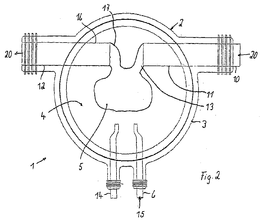
- a bioreactor 1 has a container 2 made of glass a container base 3 and a lid 3 ', which has a Tissue culture chamber 4 for receiving a tissue matrix or surrounded the hieraüs formed test tissue 5.
- a tissue culture chamber 4 externally connected lines 6 run, 10, 12, 14, one at the bottom or bottom of the side wall entering culture medium supply line 6 for feeding a culture medium 15, an arterial supply line 10 with a nozzle 11 for connecting an arterial vessel branch 13 of the test fabric 5 and a venous discharge line 12 with a connecting piece 16 for connecting a venous vessel branch 17 of the test tissue 5.
- the arterial supply line 10 is used as a nutrient medium
- the patient's blood is supplied to the patient Pharmacon - which may also be a mixture of several active ingredients can - is added.
- the nutrient medium becomes laminar or pulsating supplied.
- the temperature of the added and removed solutions as well also, the culture medium 15 in the chamber 4 are regulated. Furthermore, the pH of the culture medium 15 in the Chamber 4 is measured and can be regulated.
Landscapes
- Health & Medical Sciences (AREA)
- Engineering & Computer Science (AREA)
- Life Sciences & Earth Sciences (AREA)
- Biomedical Technology (AREA)
- Wood Science & Technology (AREA)
- Zoology (AREA)
- Organic Chemistry (AREA)
- Chemical & Material Sciences (AREA)
- Bioinformatics & Cheminformatics (AREA)
- Biotechnology (AREA)
- Genetics & Genomics (AREA)
- Biochemistry (AREA)
- General Health & Medical Sciences (AREA)
- Microbiology (AREA)
- General Engineering & Computer Science (AREA)
- Immunology (AREA)
- Cell Biology (AREA)
- Hematology (AREA)
- Gastroenterology & Hepatology (AREA)
- Sustainable Development (AREA)
- Oncology (AREA)
- Micro-Organisms Or Cultivation Processes Thereof (AREA)
- Measuring Or Testing Involving Enzymes Or Micro-Organisms (AREA)
- Medicines Containing Material From Animals Or Micro-Organisms (AREA)
- Apparatus Associated With Microorganisms And Enzymes (AREA)
Abstract
- Zellzyklus, Poliferation, Syntheserate, Stoffwechsel, Entstehungsrate neuer Blutgefäße, Variationen im Expressionsmuster von Adhäsionsmolekülen, Zell-Zell- und Zell-Matrix-Interaktionen auf RNA- und Proteinebene.
Description
- Fig. 1
- eine Seitenansicht des erfindungsgemäßen Bioreaktors und
- Fig. 2
- eine Draufsicht auf den Bioreaktor nach Fig. 1 mit abgenommenem Deckel.
Claims (20)
- Bioartifizielles, primär vaskularisiertes Testgewebe für pharmakologische oder strahlentherapeutische Testverfahren, aufweisend
eine dezellularisierte, primär vaskularisierte, biologische Gewebematrix mit wenigstens einem Gefäßast, wobei die Gewebematrix mit humanen Tumor-, Stamm-, Vorläufer- oder Leberzellen besiedelt ist. - Bioartifizielles, primär vaskularisiertes Testgewebe nach Anspruch 1, dadurch gekennzeichnet, dass die Gewebematrix wenigstens eine Matrix eines arteriellen Gefäßastes und wenigstens eine Matrix eines venösen Gefäßastes aufweist.
- Bioartifizielles, primär vaskularisiertes Testgewebe nach Anspruch 1 oder 2, dadurch gekennzeichnet, dass der wenigstens eine Gefäßast mit einem in der Gewebematrix ausgebildeten intramuralen Kapillarnetz verbunden ist.
- Bioartifizielles, primär vaskularisiertes Testgewebe nach einem der vorherigen Ansprüche, dadurch gekennzeichnet, dass es durchblutet wird.
- Verfahren zum Herstellen eines bioartifiziellen, primär vaskularisierten (biologischen) Testgewebes, das mindestens folgende Schritte aufweist:Bereitstellen eines autologen, allogenen oder xenogenen Ausgangsgewebes mit wenigstens einem Gefäßast, zumindest teilweises Dezellularisieren des bereitgestellten Ausgangsgewebes zu einer bioartifiziellen, primär vaskularisierten, biologischen Gewebematrix mit wenigstens einer Matrix eines Gefäßasts und einem intramuralen Kapillarnetz,
Perfusion der Gewebematrix mit einem Nährstoffmedium über die Matrix des wenigstens einen Gefäßastes;Auskleiden des Gefäßastes und des Kapillarnetzes mit Endothel-, Stamm- oder Vorläuferzellen und Besiedeln der Gewebematrix mit humanen Tumor-, Stamm-, Vorläufer- oder Leberzellen. - Verfahren nach Anspruch 5, dadurch gekennzeichnet, dass das Nährstoffmedium Blut, vorzugsweise autologes Blut, ist.
- Verfahren nach einem der Ansprüche 5 oder 6, dadurch gekennzeichnet, dass das Ausgangsgewebe vollständig dezellularisiert wird.
- Verfahren nach einem der Ansprüche 5 bis 7, dadurch gekennzeichnet, dass die Gewebematrix während der Besiedelung pulsierend perfundiert wird.
- Verfahren zum Testen eines Pharmakons mit mindestens folgenden Schritten:Bereitstellen eines nach einem der Ansprüche 5 bis 8 hergestellten bioartifiziellen, primär vaskularisierten Testgewebes oder eines bioartifiziellen, primär vaskularisierten Testgewebes nach einem der Ansprüche 1 bis 4,Zuführen des Pharmakons in einem Nährstoffmedium, das dem mindestens einen Gefäßast zugeführt wird, undUntersuchen des Testgewebes.
- Verfahren nach Anspruch 9, dadurch gekennzeichnet, dass das dem mindestens einen Gefäßast zugeführte Nährstoffmedium Blut, vorzugsweise autologes Blut ist.
- Verfahren nach Anspruch 9 oder 10, dadurch gekennzeichnet, dass das Nährstoffmedium gepulst zugeführt wird.
- Verfahren nach einem der Ansprüche 9 bis 11, dadurch gekennzeichnet, dass zusätzlich zu der Zuführung des Pharmakons gleichzeitig und/oder zeitlich getrennt eine strahlentherapeutische Behandlung durchgeführt wird.
- Verfahren nach einem der Ansprüche 9 bis 12, dadurch gekennzeichnet, dass Zellzyklus, Proliferation und Syntheseraten des Testgewebes untersucht werden.
- Verfahren nach einem der Ansprüche 9 bis 13, dadurch gekennzeichnet, dass Veränderungen in der Entstehungsrate neuer Blutgefäße und Variationen im Expressionsmuster von Adhäsionsmolekülen untersucht werden.
- Verfahren nach einem der Ansprüche 9 bis 14, dadurch gekennzeichnet, dass Zell-Zell- und Zell-Matrix-Interaktionen auf RNA- und Proteinebene untersucht werden.
- Verfahren nach einem der Ansprüche 9 bis 15, dadurch gekennzeichnet, dass ein Stoffwechsel des Testgewebes durch Überprüfung des zugeführten und abgeführten Nährstoffmediums untersucht wird.
- Verfahren nach einem der Ansprüche 9 bis 16, dadurch gekennzeichnet, dass eines oder mehrere der folgenden Merkmale des Testgewebes in Abhängigkeit der Untersuchung durch Änderung der Pharmaka-Konzentration und/oder der Applikationsdauer der Pharmazufuhr und/oder der strahlentherapeutischen Behandlung gezielt verändert werden:Zellzyklus, Poliferation, Syntheserate, Stoffwechsel, Entstehungsrate neuer Blutgefäße, Variationen im Expressionsmuster von Adhäsionsmolekülen, Zell-Zell- und Zell-Matrix-Interaktionen auf RNA- und Proteinebene.
- Bioreaktor zum Durchführen eines Verfahrens nach einem der Ansprüche 5 bis 8 und/oder 9 bis 17, mit mindestens
einem Behälter (2) mit einer Gewebekulturkammer (4) zur Aufnahme der Gewebematrix und/oder des Testgewebes (5),
einer in einen unteren Bereich des Behälters (2) führenden Kulturmedium-Zuleitung (6) zur Zuführung eines Kulturmediums (15),
einer arteriellen Zuleitung (10) zum Anschluss eines arteriellen Gefäßastes (13) des Testgewebes (5), und
einer venösen Abführleitung (12) zum Anschluss eines venösen Gefäßastes (17) des Testgewebes (5). - Bioreaktor nach Anspruch 18, dadurch gekennzeichnet, dass eine weitere Zuleitung (14) zur Zuführung von Wirkstoffen und/oder Entnahme von Kulturmedium vorgesehen ist.
- Biorektor nach Anspruch 18 oder 19, dadurch gekennzeichnet, dass er eine Perfundiereinrichtung zur wahlweise statischen oder pulsativen Zuführung des Nährstoffmediums aufweist.
Applications Claiming Priority (2)
| Application Number | Priority Date | Filing Date | Title |
|---|---|---|---|
| DE10333863 | 2003-07-24 | ||
| DE10333863A DE10333863A1 (de) | 2003-07-24 | 2003-07-24 | Bioartifizielles, primär vaskularisiertes Testgewebe für pharmakologische Testverfahren, Verfahren zu dessen Herstellung und Testverfahren unter dessen Verwendung sowie Bioreaktor zur Durchführung der Verfahren |
Publications (3)
| Publication Number | Publication Date |
|---|---|
| EP1500697A2 true EP1500697A2 (de) | 2005-01-26 |
| EP1500697A3 EP1500697A3 (de) | 2006-05-10 |
| EP1500697B1 EP1500697B1 (de) | 2015-10-21 |
Family
ID=33483063
Family Applications (1)
| Application Number | Title | Priority Date | Filing Date |
|---|---|---|---|
| EP04017315.5A Expired - Lifetime EP1500697B1 (de) | 2003-07-24 | 2004-07-22 | Bioartifizielles, primär vaskularisiertes Testgewebe für pharmakologische Testverfahren, Verfahren zu dessen Herstellung und Testverfahren unter dessen Verwendung sowie Bioreaktor zur Durchführung der Verfahren |
Country Status (3)
| Country | Link |
|---|---|
| EP (1) | EP1500697B1 (de) |
| DE (1) | DE10333863A1 (de) |
| ES (1) | ES2555123T3 (de) |
Cited By (4)
| Publication number | Priority date | Publication date | Assignee | Title |
|---|---|---|---|---|
| WO2010033820A1 (en) * | 2008-09-19 | 2010-03-25 | University Of Louisville Research Foundation, Inc. | Bioreactors, systems, and methods for vascularizing tissue constructs |
| US10004826B2 (en) | 2010-10-06 | 2018-06-26 | Massachusetts Institute Of Technology | Implantable human liver tissue constructs and uses thereof |
| US11266766B2 (en) | 2015-10-08 | 2022-03-08 | Massachusetts Institute Of Technology | Situ expansion of engineered devices for regeneration |
| US11324858B2 (en) | 2012-07-10 | 2022-05-10 | The Trustees Of The University Of Pennsylvania | Biomaterials for enhanced implant-host integration |
Families Citing this family (1)
| Publication number | Priority date | Publication date | Assignee | Title |
|---|---|---|---|---|
| GB2555787A (en) | 2016-11-07 | 2018-05-16 | Landberg Goran | Diagnostic methods |
Family Cites Families (6)
| Publication number | Priority date | Publication date | Assignee | Title |
|---|---|---|---|---|
| US6479064B1 (en) * | 1999-12-29 | 2002-11-12 | Children's Medical Center Corporation | Culturing different cell populations on a decellularized natural biostructure for organ reconstruction |
| DE10021627B4 (de) * | 2000-05-04 | 2009-11-19 | Corlife Gbr (Vertretungsberechtigte Gesellschafter: Prof. Dr. Alex Haverich | Verfahren zur Herstellung eines vaskularisierten bioartifiziellen Gewebes und zugehöriger Versuchsreaktor |
| AU2001284968B2 (en) * | 2000-08-16 | 2006-12-21 | Duke University | Decellularized tissue engineered constructs and tissues |
| CA2426819A1 (en) * | 2000-10-30 | 2002-05-10 | Children's Medical Center Corporation | Tissue-engineered vascular structures |
| DE50111147D1 (de) * | 2001-02-13 | 2006-11-16 | Corlife Gbr | Bioartifizielle, primär vaskularisierte Gewebematrix und bioartifizielles, primär vaskularisiertes Gewebe |
| EP1448247B1 (de) * | 2001-11-16 | 2011-02-16 | Children's Medical Center Corporation | Erzeugung gewebetechnologisch hergestellter weiblicher fortpflanzungsorgane |
-
2003
- 2003-07-24 DE DE10333863A patent/DE10333863A1/de not_active Withdrawn
-
2004
- 2004-07-22 EP EP04017315.5A patent/EP1500697B1/de not_active Expired - Lifetime
- 2004-07-22 ES ES04017315.5T patent/ES2555123T3/es not_active Expired - Lifetime
Cited By (7)
| Publication number | Priority date | Publication date | Assignee | Title |
|---|---|---|---|---|
| WO2010033820A1 (en) * | 2008-09-19 | 2010-03-25 | University Of Louisville Research Foundation, Inc. | Bioreactors, systems, and methods for vascularizing tissue constructs |
| US9226494B2 (en) | 2008-09-19 | 2016-01-05 | University Of Louisville Research Foundation, Inc. | Bioreactors, systems, and methods for vascularizing tissue constructs |
| US10004826B2 (en) | 2010-10-06 | 2018-06-26 | Massachusetts Institute Of Technology | Implantable human liver tissue constructs and uses thereof |
| US11324858B2 (en) | 2012-07-10 | 2022-05-10 | The Trustees Of The University Of Pennsylvania | Biomaterials for enhanced implant-host integration |
| US11617817B2 (en) | 2012-07-10 | 2023-04-04 | The Trustees Of The University Of Pennsylvania | Biomaterials for enhanced implant-host integration |
| US11266766B2 (en) | 2015-10-08 | 2022-03-08 | Massachusetts Institute Of Technology | Situ expansion of engineered devices for regeneration |
| US11338065B2 (en) | 2015-10-08 | 2022-05-24 | Massachusetts Institute Of Technology | In situ expansion of engineered devices for regeneration |
Also Published As
| Publication number | Publication date |
|---|---|
| DE10333863A1 (de) | 2005-02-17 |
| ES2555123T3 (es) | 2015-12-29 |
| EP1500697A3 (de) | 2006-05-10 |
| EP1500697B1 (de) | 2015-10-21 |
Similar Documents
| Publication | Publication Date | Title |
|---|---|---|
| DE10013223C2 (de) | Verfahren zur in vitro-Herstellung von dreidimensionalem, vitalem Knorpel- oder Knochengewebe und dessen Verwendung als Transplantationsmaterial | |
| Sun et al. | Polylysine-decorated macroporous microcarriers laden with adipose-derived stem cells promote nerve regeneration in vivo | |
| TWI278517B (en) | Methods and compositions for the differentiation of human preadipocytes into adipocytes | |
| DE3751843T2 (de) | Herstellung von körperorganen durch kontroliertes zellwachstum auf künstlicher matrix | |
| DE69219613T2 (de) | Vorvaskularisierte polymerimplantate für organtransplantation | |
| EP1152053B1 (de) | Verfahren und Vorrichtung zur Herstellung eines vaskularisierten bioartifiziellen Gewebes | |
| DE10026789B4 (de) | Knorpelersatz und Verfahren zu dessen Herstellung | |
| Phang et al. | Advancements in extracellular matrix-based biomaterials and biofabrication of 3D organotypic skin models | |
| PT91676A (pt) | Three-dimensional cell and tissue culture system | |
| Li et al. | Preparation and characterization of a novel acellular swim bladder as dura mater substitute | |
| Padma et al. | Towards a bioengineered uterus: bioactive sheep uterus scaffolds are effectively recellularized by enzymatic preconditioning | |
| Ding et al. | Rapid repair of rat sciatic nerve injury using a nanosilver-embedded collagen scaffold coated with laminin and fibronectin | |
| DE10214095C1 (de) | Dedifferenzierte, programmierbare Stammzellen monozytären Ursprungs, sowie deren Herstellung und Verwendung | |
| Moroni et al. | Tissue engineering: an introduction | |
| WO2007090575A1 (de) | Hautmodell mit dendritischen zellen | |
| EP1500697B1 (de) | Bioartifizielles, primär vaskularisiertes Testgewebe für pharmakologische Testverfahren, Verfahren zu dessen Herstellung und Testverfahren unter dessen Verwendung sowie Bioreaktor zur Durchführung der Verfahren | |
| Li et al. | BDNF-loaded chitosan-based mimetic mussel polymer conduits for repair of peripheral nerve injury | |
| EP1706157B1 (de) | Verfahren zur herstellung von bandscheibenzelltransplantaten und deren anwendung als transplantationsmaterial | |
| EP2864474B1 (de) | Verfahren zur herstellung von funktionalem fusionsgewebe | |
| CN118892581B (zh) | 骨缺损修复材料的制备方法及其应用 | |
| WO2004042038A1 (de) | Verfahren zur behandlung von erkranktem,degeneriertem oder geschädigtem gewebe unter verwendung von in vitro hergestelltem dreidimensionalem gewebe in kombination mit gewebezellen und/oder exogenen faktoren | |
| Jiang et al. | Construction of 3D tumor in vitro models with an immune microenvironment exhibiting similar tumor properties and biomimetic physiological functionality | |
| Gershlak et al. | Biocompatibility of decellularized spinach leaves | |
| DE4229013C2 (de) | Verfahren und Vorrichtung zur in-vitro-Prüfung von Einwirkungen auf biologische Strukturen | |
| Ghafari et al. | Mechanical reinforcement of urinary bladder matrix by electrospun polycaprolactone nanofibers |
Legal Events
| Date | Code | Title | Description |
|---|---|---|---|
| PUAI | Public reference made under article 153(3) epc to a published international application that has entered the european phase |
Free format text: ORIGINAL CODE: 0009012 |
|
| AK | Designated contracting states |
Kind code of ref document: A2 Designated state(s): AT BE BG CH CY CZ DE DK EE ES FI FR GB GR HU IE IT LI LU MC NL PL PT RO SE SI SK TR |
|
| AX | Request for extension of the european patent |
Extension state: AL HR LT LV MK |
|
| RTI1 | Title (correction) |
Free format text: BIOARTIFICIAL, PRIMARILY VASCULARIZED TISSUE FOR PHARMACEUTICAL TESTS, METHOD FOR ITS PRODUCTION, USE OF IT IN A BIOREACTOR. |
|
| PUAL | Search report despatched |
Free format text: ORIGINAL CODE: 0009013 |
|
| AK | Designated contracting states |
Kind code of ref document: A3 Designated state(s): AT BE BG CH CY CZ DE DK EE ES FI FR GB GR HU IE IT LI LU MC NL PL PT RO SE SI SK TR |
|
| AX | Request for extension of the european patent |
Extension state: AL HR LT LV MK |
|
| 17P | Request for examination filed |
Effective date: 20060718 |
|
| RAP1 | Party data changed (applicant data changed or rights of an application transferred) |
Owner name: FRAUNHOFER-GESELLSCHAFT ZUR FOERDERUNG DER ANGEWAN |
|
| RIN1 | Information on inventor provided before grant (corrected) |
Inventor name: WALLES, THORSTEN, DR. |
|
| 17Q | First examination report despatched |
Effective date: 20061127 |
|
| AKX | Designation fees paid |
Designated state(s): AT BE BG CH CY CZ DE DK EE ES FI FR GB GR HU IE IT LI LU MC NL PL PT RO SE SI SK TR |
|
| REG | Reference to a national code |
Ref country code: DE Ref legal event code: R079 Ref document number: 502004015057 Country of ref document: DE Free format text: PREVIOUS MAIN CLASS: C12N0005060000 Ipc: G01N0033500000 |
|
| GRAP | Despatch of communication of intention to grant a patent |
Free format text: ORIGINAL CODE: EPIDOSNIGR1 |
|
| RIC1 | Information provided on ipc code assigned before grant |
Ipc: G01N 33/50 20060101AFI20150630BHEP |
|
| GRAS | Grant fee paid |
Free format text: ORIGINAL CODE: EPIDOSNIGR3 |
|
| INTG | Intention to grant announced |
Effective date: 20150722 |
|
| GRAA | (expected) grant |
Free format text: ORIGINAL CODE: 0009210 |
|
| AK | Designated contracting states |
Kind code of ref document: B1 Designated state(s): AT BE BG CH CY CZ DE DK EE ES FI FR GB GR HU IE IT LI LU MC NL PL PT RO SE SI SK TR |
|
| REG | Reference to a national code |
Ref country code: GB Ref legal event code: FG4D Free format text: NOT ENGLISH |
|
| REG | Reference to a national code |
Ref country code: CH Ref legal event code: EP Ref country code: CH Ref legal event code: NV Representative=s name: E. BLUM AND CO. AG PATENT- UND MARKENANWAELTE , CH |
|
| REG | Reference to a national code |
Ref country code: AT Ref legal event code: REF Ref document number: 756916 Country of ref document: AT Kind code of ref document: T Effective date: 20151115 |
|
| REG | Reference to a national code |
Ref country code: IE Ref legal event code: FG4D Free format text: LANGUAGE OF EP DOCUMENT: GERMAN |
|
| REG | Reference to a national code |
Ref country code: DE Ref legal event code: R096 Ref document number: 502004015057 Country of ref document: DE |
|
| REG | Reference to a national code |
Ref country code: ES Ref legal event code: FG2A Ref document number: 2555123 Country of ref document: ES Kind code of ref document: T3 Effective date: 20151229 |
|
| REG | Reference to a national code |
Ref country code: SE Ref legal event code: TRGR |
|
| REG | Reference to a national code |
Ref country code: NL Ref legal event code: FP |
|
| PG25 | Lapsed in a contracting state [announced via postgrant information from national office to epo] |
Ref country code: FI Free format text: LAPSE BECAUSE OF FAILURE TO SUBMIT A TRANSLATION OF THE DESCRIPTION OR TO PAY THE FEE WITHIN THE PRESCRIBED TIME-LIMIT Effective date: 20151021 Ref country code: PL Free format text: LAPSE BECAUSE OF FAILURE TO SUBMIT A TRANSLATION OF THE DESCRIPTION OR TO PAY THE FEE WITHIN THE PRESCRIBED TIME-LIMIT Effective date: 20151021 Ref country code: PT Free format text: LAPSE BECAUSE OF FAILURE TO SUBMIT A TRANSLATION OF THE DESCRIPTION OR TO PAY THE FEE WITHIN THE PRESCRIBED TIME-LIMIT Effective date: 20160222 Ref country code: GR Free format text: LAPSE BECAUSE OF FAILURE TO SUBMIT A TRANSLATION OF THE DESCRIPTION OR TO PAY THE FEE WITHIN THE PRESCRIBED TIME-LIMIT Effective date: 20160122 |
|
| REG | Reference to a national code |
Ref country code: FR Ref legal event code: PLFP Year of fee payment: 13 Ref country code: DE Ref legal event code: R097 Ref document number: 502004015057 Country of ref document: DE |
|
| PG25 | Lapsed in a contracting state [announced via postgrant information from national office to epo] |
Ref country code: CZ Free format text: LAPSE BECAUSE OF FAILURE TO SUBMIT A TRANSLATION OF THE DESCRIPTION OR TO PAY THE FEE WITHIN THE PRESCRIBED TIME-LIMIT Effective date: 20151021 |
|
| PLBE | No opposition filed within time limit |
Free format text: ORIGINAL CODE: 0009261 |
|
| STAA | Information on the status of an ep patent application or granted ep patent |
Free format text: STATUS: NO OPPOSITION FILED WITHIN TIME LIMIT |
|
| PG25 | Lapsed in a contracting state [announced via postgrant information from national office to epo] |
Ref country code: DK Free format text: LAPSE BECAUSE OF FAILURE TO SUBMIT A TRANSLATION OF THE DESCRIPTION OR TO PAY THE FEE WITHIN THE PRESCRIBED TIME-LIMIT Effective date: 20151021 Ref country code: EE Free format text: LAPSE BECAUSE OF FAILURE TO SUBMIT A TRANSLATION OF THE DESCRIPTION OR TO PAY THE FEE WITHIN THE PRESCRIBED TIME-LIMIT Effective date: 20151021 Ref country code: SK Free format text: LAPSE BECAUSE OF FAILURE TO SUBMIT A TRANSLATION OF THE DESCRIPTION OR TO PAY THE FEE WITHIN THE PRESCRIBED TIME-LIMIT Effective date: 20151021 Ref country code: RO Free format text: LAPSE BECAUSE OF FAILURE TO SUBMIT A TRANSLATION OF THE DESCRIPTION OR TO PAY THE FEE WITHIN THE PRESCRIBED TIME-LIMIT Effective date: 20151021 |
|
| 26N | No opposition filed |
Effective date: 20160722 |
|
| PG25 | Lapsed in a contracting state [announced via postgrant information from national office to epo] |
Ref country code: SI Free format text: LAPSE BECAUSE OF FAILURE TO SUBMIT A TRANSLATION OF THE DESCRIPTION OR TO PAY THE FEE WITHIN THE PRESCRIBED TIME-LIMIT Effective date: 20151021 |
|
| PG25 | Lapsed in a contracting state [announced via postgrant information from national office to epo] |
Ref country code: MC Free format text: LAPSE BECAUSE OF FAILURE TO SUBMIT A TRANSLATION OF THE DESCRIPTION OR TO PAY THE FEE WITHIN THE PRESCRIBED TIME-LIMIT Effective date: 20151021 |
|
| REG | Reference to a national code |
Ref country code: IE Ref legal event code: MM4A |
|
| REG | Reference to a national code |
Ref country code: FR Ref legal event code: PLFP Year of fee payment: 14 |
|
| PG25 | Lapsed in a contracting state [announced via postgrant information from national office to epo] |
Ref country code: IE Free format text: LAPSE BECAUSE OF NON-PAYMENT OF DUE FEES Effective date: 20160722 |
|
| PG25 | Lapsed in a contracting state [announced via postgrant information from national office to epo] |
Ref country code: LU Free format text: LAPSE BECAUSE OF NON-PAYMENT OF DUE FEES Effective date: 20160722 |
|
| PG25 | Lapsed in a contracting state [announced via postgrant information from national office to epo] |
Ref country code: CY Free format text: LAPSE BECAUSE OF FAILURE TO SUBMIT A TRANSLATION OF THE DESCRIPTION OR TO PAY THE FEE WITHIN THE PRESCRIBED TIME-LIMIT Effective date: 20151021 Ref country code: HU Free format text: LAPSE BECAUSE OF FAILURE TO SUBMIT A TRANSLATION OF THE DESCRIPTION OR TO PAY THE FEE WITHIN THE PRESCRIBED TIME-LIMIT; INVALID AB INITIO Effective date: 20040722 |
|
| PG25 | Lapsed in a contracting state [announced via postgrant information from national office to epo] |
Ref country code: TR Free format text: LAPSE BECAUSE OF FAILURE TO SUBMIT A TRANSLATION OF THE DESCRIPTION OR TO PAY THE FEE WITHIN THE PRESCRIBED TIME-LIMIT Effective date: 20151021 |
|
| REG | Reference to a national code |
Ref country code: FR Ref legal event code: PLFP Year of fee payment: 15 |
|
| PG25 | Lapsed in a contracting state [announced via postgrant information from national office to epo] |
Ref country code: BG Free format text: LAPSE BECAUSE OF FAILURE TO SUBMIT A TRANSLATION OF THE DESCRIPTION OR TO PAY THE FEE WITHIN THE PRESCRIBED TIME-LIMIT Effective date: 20151021 |
|
| PGFP | Annual fee paid to national office [announced via postgrant information from national office to epo] |
Ref country code: NL Payment date: 20200729 Year of fee payment: 17 |
|
| PGFP | Annual fee paid to national office [announced via postgrant information from national office to epo] |
Ref country code: FR Payment date: 20200727 Year of fee payment: 17 Ref country code: GB Payment date: 20200724 Year of fee payment: 17 Ref country code: DE Payment date: 20200723 Year of fee payment: 17 Ref country code: ES Payment date: 20200818 Year of fee payment: 17 |
|
| PGFP | Annual fee paid to national office [announced via postgrant information from national office to epo] |
Ref country code: SE Payment date: 20200724 Year of fee payment: 17 Ref country code: IT Payment date: 20200731 Year of fee payment: 17 Ref country code: CH Payment date: 20200724 Year of fee payment: 17 Ref country code: BE Payment date: 20200722 Year of fee payment: 17 Ref country code: AT Payment date: 20200720 Year of fee payment: 17 |
|
| REG | Reference to a national code |
Ref country code: DE Ref legal event code: R119 Ref document number: 502004015057 Country of ref document: DE |
|
| REG | Reference to a national code |
Ref country code: CH Ref legal event code: PL |
|
| REG | Reference to a national code |
Ref country code: NL Ref legal event code: MM Effective date: 20210801 |
|
| REG | Reference to a national code |
Ref country code: AT Ref legal event code: MM01 Ref document number: 756916 Country of ref document: AT Kind code of ref document: T Effective date: 20210722 |
|
| GBPC | Gb: european patent ceased through non-payment of renewal fee |
Effective date: 20210722 |
|
| REG | Reference to a national code |
Ref country code: BE Ref legal event code: MM Effective date: 20210731 |
|
| PG25 | Lapsed in a contracting state [announced via postgrant information from national office to epo] |
Ref country code: LI Free format text: LAPSE BECAUSE OF NON-PAYMENT OF DUE FEES Effective date: 20210731 Ref country code: GB Free format text: LAPSE BECAUSE OF NON-PAYMENT OF DUE FEES Effective date: 20210722 Ref country code: DE Free format text: LAPSE BECAUSE OF NON-PAYMENT OF DUE FEES Effective date: 20220201 Ref country code: CH Free format text: LAPSE BECAUSE OF NON-PAYMENT OF DUE FEES Effective date: 20210731 Ref country code: AT Free format text: LAPSE BECAUSE OF NON-PAYMENT OF DUE FEES Effective date: 20210722 |
|
| PG25 | Lapsed in a contracting state [announced via postgrant information from national office to epo] |
Ref country code: SE Free format text: LAPSE BECAUSE OF NON-PAYMENT OF DUE FEES Effective date: 20210723 Ref country code: NL Free format text: LAPSE BECAUSE OF NON-PAYMENT OF DUE FEES Effective date: 20210801 Ref country code: FR Free format text: LAPSE BECAUSE OF NON-PAYMENT OF DUE FEES Effective date: 20210731 |
|
| PG25 | Lapsed in a contracting state [announced via postgrant information from national office to epo] |
Ref country code: IT Free format text: LAPSE BECAUSE OF NON-PAYMENT OF DUE FEES Effective date: 20210722 Ref country code: BE Free format text: LAPSE BECAUSE OF NON-PAYMENT OF DUE FEES Effective date: 20210731 |
|
| REG | Reference to a national code |
Ref country code: ES Ref legal event code: FD2A Effective date: 20220829 |
|
| PG25 | Lapsed in a contracting state [announced via postgrant information from national office to epo] |
Ref country code: ES Free format text: LAPSE BECAUSE OF NON-PAYMENT OF DUE FEES Effective date: 20210723 |