EP1435957B1 - Inhibitors of gsk-3 and crystal structures of gsk-3beta protein and protein complexes - Google Patents
Inhibitors of gsk-3 and crystal structures of gsk-3beta protein and protein complexes Download PDFInfo
- Publication number
- EP1435957B1 EP1435957B1 EP02729056A EP02729056A EP1435957B1 EP 1435957 B1 EP1435957 B1 EP 1435957B1 EP 02729056 A EP02729056 A EP 02729056A EP 02729056 A EP02729056 A EP 02729056A EP 1435957 B1 EP1435957 B1 EP 1435957B1
- Authority
- EP
- European Patent Office
- Prior art keywords
- phenyl
- compound
- pyrazolo
- pyridazin
- gsk
- Prior art date
- Legal status (The legal status is an assumption and is not a legal conclusion. Google has not performed a legal analysis and makes no representation as to the accuracy of the status listed.)
- Expired - Lifetime
Links
- 239000003112 inhibitor Substances 0.000 title abstract description 117
- 239000013078 crystal Substances 0.000 title abstract description 77
- 108090000623 proteins and genes Proteins 0.000 title abstract description 77
- 102000004169 proteins and genes Human genes 0.000 title abstract description 75
- 102000007474 Multiprotein Complexes Human genes 0.000 title abstract description 7
- 108010085220 Multiprotein Complexes Proteins 0.000 title abstract description 7
- 101150090422 gsk-3 gene Proteins 0.000 title 1
- 150000001875 compounds Chemical class 0.000 claims abstract description 157
- 238000000034 method Methods 0.000 claims abstract description 80
- 102000002254 Glycogen Synthase Kinase 3 Human genes 0.000 claims abstract description 64
- 108010014905 Glycogen Synthase Kinase 3 Proteins 0.000 claims abstract description 64
- 239000000203 mixture Substances 0.000 claims abstract description 61
- 239000008194 pharmaceutical composition Substances 0.000 claims abstract description 22
- 206010012601 diabetes mellitus Diseases 0.000 claims abstract description 15
- 208000024827 Alzheimer disease Diseases 0.000 claims abstract description 10
- -1 2-tetrahydrofuranyl Chemical group 0.000 claims description 514
- 125000001931 aliphatic group Chemical group 0.000 claims description 67
- 125000000623 heterocyclic group Chemical group 0.000 claims description 56
- 125000003118 aryl group Chemical group 0.000 claims description 55
- 125000004429 atom Chemical group 0.000 claims description 51
- 125000001072 heteroaryl group Chemical group 0.000 claims description 51
- 238000006366 phosphorylation reaction Methods 0.000 claims description 51
- 230000026731 phosphorylation Effects 0.000 claims description 50
- 125000003710 aryl alkyl group Chemical group 0.000 claims description 39
- 125000004475 heteroaralkyl group Chemical group 0.000 claims description 36
- 125000004452 carbocyclyl group Chemical group 0.000 claims description 33
- 125000003785 benzimidazolyl group Chemical group N1=C(NC2=C1C=CC=C2)* 0.000 claims description 28
- 230000000694 effects Effects 0.000 claims description 28
- 125000001997 phenyl group Chemical group [H]C1=C([H])C([H])=C(*)C([H])=C1[H] 0.000 claims description 28
- 125000005843 halogen group Chemical group 0.000 claims description 24
- 125000002023 trifluoromethyl group Chemical group FC(F)(F)* 0.000 claims description 22
- 230000002401 inhibitory effect Effects 0.000 claims description 21
- 229910052736 halogen Inorganic materials 0.000 claims description 19
- 125000000449 nitro group Chemical group [O-][N+](*)=O 0.000 claims description 19
- 229910006074 SO2NH2 Inorganic materials 0.000 claims description 18
- 230000015572 biosynthetic process Effects 0.000 claims description 17
- 125000001637 1-naphthyl group Chemical group [H]C1=C([H])C([H])=C2C(*)=C([H])C([H])=C([H])C2=C1[H] 0.000 claims description 16
- 125000001622 2-naphthyl group Chemical group [H]C1=C([H])C([H])=C2C([H])=C(*)C([H])=C([H])C2=C1[H] 0.000 claims description 16
- 238000003786 synthesis reaction Methods 0.000 claims description 16
- 125000003349 3-pyridyl group Chemical group N1=C([H])C([*])=C([H])C([H])=C1[H] 0.000 claims description 15
- 229920002527 Glycogen Polymers 0.000 claims description 15
- 229940096919 glycogen Drugs 0.000 claims description 15
- 125000001712 tetrahydronaphthyl group Chemical group C1(CCCC2=CC=CC=C12)* 0.000 claims description 15
- LBUJPTNKIBCYBY-UHFFFAOYSA-N 1,2,3,4-tetrahydroquinoline Chemical compound C1=CC=C2CCCNC2=C1 LBUJPTNKIBCYBY-UHFFFAOYSA-N 0.000 claims description 14
- YJUFGFXVASPYFQ-UHFFFAOYSA-N 2,3-dihydro-1-benzothiophene Chemical compound C1=CC=C2SCCC2=C1 YJUFGFXVASPYFQ-UHFFFAOYSA-N 0.000 claims description 14
- HBEDSQVIWPRPAY-UHFFFAOYSA-N 2,3-dihydrobenzofuran Chemical compound C1=CC=C2OCCC2=C1 HBEDSQVIWPRPAY-UHFFFAOYSA-N 0.000 claims description 14
- 125000002941 2-furyl group Chemical group O1C([*])=C([H])C([H])=C1[H] 0.000 claims description 14
- 125000004105 2-pyridyl group Chemical group N1=C([*])C([H])=C([H])C([H])=C1[H] 0.000 claims description 14
- 125000004485 2-pyrrolidinyl group Chemical group [H]N1C([H])([H])C([H])([H])C([H])([H])C1([H])* 0.000 claims description 14
- 125000000389 2-pyrrolyl group Chemical group [H]N1C([*])=C([H])C([H])=C1[H] 0.000 claims description 14
- 125000000175 2-thienyl group Chemical group S1C([*])=C([H])C([H])=C1[H] 0.000 claims description 14
- WPWNEKFMGCWNPR-UHFFFAOYSA-N 3,4-dihydro-2h-thiochromene Chemical compound C1=CC=C2CCCSC2=C1 WPWNEKFMGCWNPR-UHFFFAOYSA-N 0.000 claims description 14
- 125000003682 3-furyl group Chemical group O1C([H])=C([*])C([H])=C1[H] 0.000 claims description 14
- 125000004575 3-pyrrolidinyl group Chemical group [H]N1C([H])([H])C([H])([H])C([H])(*)C1([H])[H] 0.000 claims description 14
- 125000001397 3-pyrrolyl group Chemical group [H]N1C([H])=C([*])C([H])=C1[H] 0.000 claims description 14
- 125000001541 3-thienyl group Chemical group S1C([H])=C([*])C([H])=C1[H] 0.000 claims description 14
- 125000000339 4-pyridyl group Chemical group N1=C([H])C([H])=C([*])C([H])=C1[H] 0.000 claims description 14
- KDDQRKBRJSGMQE-UHFFFAOYSA-N 4-thiazolyl Chemical group [C]1=CSC=N1 KDDQRKBRJSGMQE-UHFFFAOYSA-N 0.000 claims description 14
- CWDWFSXUQODZGW-UHFFFAOYSA-N 5-thiazolyl Chemical group [C]1=CN=CS1 CWDWFSXUQODZGW-UHFFFAOYSA-N 0.000 claims description 14
- 125000000641 acridinyl group Chemical group C1(=CC=CC2=NC3=CC=CC=C3C=C12)* 0.000 claims description 14
- 125000002393 azetidinyl group Chemical group 0.000 claims description 14
- 125000000499 benzofuranyl group Chemical group O1C(=CC2=C1C=CC=C2)* 0.000 claims description 14
- 125000001164 benzothiazolyl group Chemical group S1C(=NC2=C1C=CC=C2)* 0.000 claims description 14
- 125000004196 benzothienyl group Chemical group S1C(=CC2=C1C=CC=C2)* 0.000 claims description 14
- 125000004541 benzoxazolyl group Chemical group O1C(=NC2=C1C=CC=C2)* 0.000 claims description 14
- VZWXIQHBIQLMPN-UHFFFAOYSA-N chromane Chemical compound C1=CC=C2CCCOC2=C1 VZWXIQHBIQLMPN-UHFFFAOYSA-N 0.000 claims description 14
- 125000000532 dioxanyl group Chemical group 0.000 claims description 14
- 125000005879 dioxolanyl group Chemical group 0.000 claims description 14
- 125000005883 dithianyl group Chemical group 0.000 claims description 14
- 125000002962 imidazol-1-yl group Chemical group [*]N1C([H])=NC([H])=C1[H] 0.000 claims description 14
- 125000002636 imidazolinyl group Chemical group 0.000 claims description 14
- LPAGFVYQRIESJQ-UHFFFAOYSA-N indoline Chemical compound C1=CC=C2NCCC2=C1 LPAGFVYQRIESJQ-UHFFFAOYSA-N 0.000 claims description 14
- 125000001041 indolyl group Chemical group 0.000 claims description 14
- 125000000904 isoindolyl group Chemical group C=1(NC=C2C=CC=CC12)* 0.000 claims description 14
- 125000002183 isoquinolinyl group Chemical group C1(=NC=CC2=CC=CC=C12)* 0.000 claims description 14
- 125000003551 oxepanyl group Chemical group 0.000 claims description 14
- 125000000466 oxiranyl group Chemical group 0.000 claims description 14
- 125000000587 piperidin-1-yl group Chemical group [H]C1([H])N(*)C([H])([H])C([H])([H])C([H])([H])C1([H])[H] 0.000 claims description 14
- 125000004483 piperidin-3-yl group Chemical group N1CC(CCC1)* 0.000 claims description 14
- 125000004482 piperidin-4-yl group Chemical group N1CCC(CC1)* 0.000 claims description 14
- 125000004309 pyranyl group Chemical group O1C(C=CC=C1)* 0.000 claims description 14
- 125000003072 pyrazolidinyl group Chemical group 0.000 claims description 14
- 125000002755 pyrazolinyl group Chemical group 0.000 claims description 14
- 125000002206 pyridazin-3-yl group Chemical group [H]C1=C([H])C([H])=C(*)N=N1 0.000 claims description 14
- 125000001422 pyrrolinyl group Chemical group 0.000 claims description 14
- 125000002943 quinolinyl group Chemical group N1=C(C=CC2=CC=CC=C12)* 0.000 claims description 14
- 125000004621 quinuclidinyl group Chemical group N12C(CC(CC1)CC2)* 0.000 claims description 14
- 125000001583 thiepanyl group Chemical group 0.000 claims description 14
- 125000005455 trithianyl group Chemical group 0.000 claims description 14
- 125000002632 imidazolidinyl group Chemical group 0.000 claims description 13
- 125000004423 acyloxy group Chemical group 0.000 claims description 12
- 125000003178 carboxy group Chemical group [H]OC(*)=O 0.000 claims description 12
- 239000003814 drug Substances 0.000 claims description 12
- 125000005842 heteroatom Chemical group 0.000 claims description 12
- 150000003839 salts Chemical class 0.000 claims description 12
- 108060000903 Beta-catenin Proteins 0.000 claims description 10
- 125000000217 alkyl group Chemical group 0.000 claims description 10
- 125000001424 substituent group Chemical group 0.000 claims description 10
- 102000015735 Beta-catenin Human genes 0.000 claims description 9
- WQZGKKKJIJFFOK-GASJEMHNSA-N Glucose Natural products OC[C@H]1OC(O)[C@H](O)[C@@H](O)[C@@H]1O WQZGKKKJIJFFOK-GASJEMHNSA-N 0.000 claims description 8
- 239000008103 glucose Substances 0.000 claims description 8
- 239000003981 vehicle Substances 0.000 claims description 8
- SEXDCZOTUATPCJ-UHFFFAOYSA-N 5-(3-fluorophenyl)-4-phenyl-1h-pyrazolo[3,4-c]pyridazin-3-amine Chemical compound C=1C=CC=CC=1C1=C2C(N)=NNC2=NN=C1C1=CC=CC(F)=C1 SEXDCZOTUATPCJ-UHFFFAOYSA-N 0.000 claims description 7
- 239000002671 adjuvant Substances 0.000 claims description 7
- 239000003937 drug carrier Substances 0.000 claims description 7
- 102000013498 tau Proteins Human genes 0.000 claims description 7
- 108010026424 tau Proteins Proteins 0.000 claims description 7
- JHXDLIAMKPRADS-UHFFFAOYSA-N 4-(3-fluorophenyl)-5-phenyl-1h-pyrazolo[3,4-c]pyridazin-3-amine Chemical compound C=1C=CC(F)=CC=1C1=C2C(N)=NNC2=NN=C1C1=CC=CC=C1 JHXDLIAMKPRADS-UHFFFAOYSA-N 0.000 claims description 6
- 239000012472 biological sample Substances 0.000 claims description 6
- 230000036765 blood level Effects 0.000 claims description 6
- 125000002887 hydroxy group Chemical group [H]O* 0.000 claims description 6
- 238000004519 manufacturing process Methods 0.000 claims description 6
- 238000002360 preparation method Methods 0.000 claims description 6
- 201000000980 schizophrenia Diseases 0.000 claims description 6
- JWVJFQWLTBIMEV-UHFFFAOYSA-N 4-phenyl-5-pyridin-4-yl-1h-pyrazolo[3,4-c]pyridazin-3-amine Chemical compound C=1C=CC=CC=1C1=C2C(N)=NNC2=NN=C1C1=CC=NC=C1 JWVJFQWLTBIMEV-UHFFFAOYSA-N 0.000 claims description 5
- 125000000113 cyclohexyl group Chemical group [H]C1([H])C([H])([H])C([H])([H])C([H])(*)C([H])([H])C1([H])[H] 0.000 claims description 5
- 150000002367 halogens Chemical class 0.000 claims description 5
- PHHUDUOMJNIKIV-UHFFFAOYSA-N n-(4,5-diphenyl-1h-pyrazolo[3,4-c]pyridazin-3-yl)acetamide Chemical compound C=1C=CC=CC=1C1=C2C(NC(=O)C)=NNC2=NN=C1C1=CC=CC=C1 PHHUDUOMJNIKIV-UHFFFAOYSA-N 0.000 claims description 5
- 125000002098 pyridazinyl group Chemical group 0.000 claims description 5
- 125000002047 benzodioxolyl group Chemical group O1OC(C2=C1C=CC=C2)* 0.000 claims description 4
- 125000004093 cyano group Chemical group *C#N 0.000 claims description 4
- 230000002708 enhancing effect Effects 0.000 claims description 4
- 125000002541 furyl group Chemical group 0.000 claims description 4
- 125000001624 naphthyl group Chemical group 0.000 claims description 4
- 125000004076 pyridyl group Chemical group 0.000 claims description 4
- 125000000714 pyrimidinyl group Chemical group 0.000 claims description 4
- 125000001544 thienyl group Chemical group 0.000 claims description 4
- JMAGLEWPHWZICN-UHFFFAOYSA-N 5-(4-fluorophenyl)-4-phenyl-1h-pyrazolo[3,4-c]pyridazin-3-amine Chemical compound C=1C=CC=CC=1C1=C2C(N)=NNC2=NN=C1C1=CC=C(F)C=C1 JMAGLEWPHWZICN-UHFFFAOYSA-N 0.000 claims description 3
- 229940124597 therapeutic agent Drugs 0.000 claims description 3
- GLGNXYJARSMNGJ-VKTIVEEGSA-N (1s,2s,3r,4r)-3-[[5-chloro-2-[(1-ethyl-6-methoxy-2-oxo-4,5-dihydro-3h-1-benzazepin-7-yl)amino]pyrimidin-4-yl]amino]bicyclo[2.2.1]hept-5-ene-2-carboxamide Chemical compound CCN1C(=O)CCCC2=C(OC)C(NC=3N=C(C(=CN=3)Cl)N[C@H]3[C@H]([C@@]4([H])C[C@@]3(C=C4)[H])C(N)=O)=CC=C21 GLGNXYJARSMNGJ-VKTIVEEGSA-N 0.000 claims description 2
- SZUVGFMDDVSKSI-WIFOCOSTSA-N (1s,2s,3s,5r)-1-(carboxymethyl)-3,5-bis[(4-phenoxyphenyl)methyl-propylcarbamoyl]cyclopentane-1,2-dicarboxylic acid Chemical compound O=C([C@@H]1[C@@H]([C@](CC(O)=O)([C@H](C(=O)N(CCC)CC=2C=CC(OC=3C=CC=CC=3)=CC=2)C1)C(O)=O)C(O)=O)N(CCC)CC(C=C1)=CC=C1OC1=CC=CC=C1 SZUVGFMDDVSKSI-WIFOCOSTSA-N 0.000 claims description 2
- GHYOCDFICYLMRF-UTIIJYGPSA-N (2S,3R)-N-[(2S)-3-(cyclopenten-1-yl)-1-[(2R)-2-methyloxiran-2-yl]-1-oxopropan-2-yl]-3-hydroxy-3-(4-methoxyphenyl)-2-[[(2S)-2-[(2-morpholin-4-ylacetyl)amino]propanoyl]amino]propanamide Chemical compound C1(=CCCC1)C[C@@H](C(=O)[C@@]1(OC1)C)NC([C@H]([C@@H](C1=CC=C(C=C1)OC)O)NC([C@H](C)NC(CN1CCOCC1)=O)=O)=O GHYOCDFICYLMRF-UTIIJYGPSA-N 0.000 claims description 2
- QFLWZFQWSBQYPS-AWRAUJHKSA-N (3S)-3-[[(2S)-2-[[(2S)-2-[5-[(3aS,6aR)-2-oxo-1,3,3a,4,6,6a-hexahydrothieno[3,4-d]imidazol-4-yl]pentanoylamino]-3-methylbutanoyl]amino]-3-(4-hydroxyphenyl)propanoyl]amino]-4-[1-bis(4-chlorophenoxy)phosphorylbutylamino]-4-oxobutanoic acid Chemical compound CCCC(NC(=O)[C@H](CC(O)=O)NC(=O)[C@H](Cc1ccc(O)cc1)NC(=O)[C@@H](NC(=O)CCCCC1SC[C@@H]2NC(=O)N[C@H]12)C(C)C)P(=O)(Oc1ccc(Cl)cc1)Oc1ccc(Cl)cc1 QFLWZFQWSBQYPS-AWRAUJHKSA-N 0.000 claims description 2
- ONBQEOIKXPHGMB-VBSBHUPXSA-N 1-[2-[(2s,3r,4s,5r)-3,4-dihydroxy-5-(hydroxymethyl)oxolan-2-yl]oxy-4,6-dihydroxyphenyl]-3-(4-hydroxyphenyl)propan-1-one Chemical compound O[C@@H]1[C@H](O)[C@@H](CO)O[C@H]1OC1=CC(O)=CC(O)=C1C(=O)CCC1=CC=C(O)C=C1 ONBQEOIKXPHGMB-VBSBHUPXSA-N 0.000 claims description 2
- UNILWMWFPHPYOR-KXEYIPSPSA-M 1-[6-[2-[3-[3-[3-[2-[2-[3-[[2-[2-[[(2r)-1-[[2-[[(2r)-1-[3-[2-[2-[3-[[2-(2-amino-2-oxoethoxy)acetyl]amino]propoxy]ethoxy]ethoxy]propylamino]-3-hydroxy-1-oxopropan-2-yl]amino]-2-oxoethyl]amino]-3-[(2r)-2,3-di(hexadecanoyloxy)propyl]sulfanyl-1-oxopropan-2-yl Chemical compound O=C1C(SCCC(=O)NCCCOCCOCCOCCCNC(=O)COCC(=O)N[C@@H](CSC[C@@H](COC(=O)CCCCCCCCCCCCCCC)OC(=O)CCCCCCCCCCCCCCC)C(=O)NCC(=O)N[C@H](CO)C(=O)NCCCOCCOCCOCCCNC(=O)COCC(N)=O)CC(=O)N1CCNC(=O)CCCCCN\1C2=CC=C(S([O-])(=O)=O)C=C2CC/1=C/C=C/C=C/C1=[N+](CC)C2=CC=C(S([O-])(=O)=O)C=C2C1 UNILWMWFPHPYOR-KXEYIPSPSA-M 0.000 claims description 2
- VQISKCBTGNMDTP-UHFFFAOYSA-N 4,5-bis(4-fluorophenyl)-1h-pyrazolo[3,4-c]pyridazin-3-amine Chemical compound C=1C=C(F)C=CC=1C1=C2C(N)=NNC2=NN=C1C1=CC=C(F)C=C1 VQISKCBTGNMDTP-UHFFFAOYSA-N 0.000 claims description 2
- FPHUKEUMXVJBDX-UHFFFAOYSA-N 4-(4-chlorophenyl)-5-phenyl-1h-pyrazolo[3,4-c]pyridazin-3-amine Chemical compound C=1C=C(Cl)C=CC=1C1=C2C(N)=NNC2=NN=C1C1=CC=CC=C1 FPHUKEUMXVJBDX-UHFFFAOYSA-N 0.000 claims description 2
- IVRPJGSBYHTNHT-UHFFFAOYSA-N 4-(4-fluorophenyl)-5-phenyl-1h-pyrazolo[3,4-c]pyridazin-3-amine Chemical compound C=1C=C(F)C=CC=1C1=C2C(N)=NNC2=NN=C1C1=CC=CC=C1 IVRPJGSBYHTNHT-UHFFFAOYSA-N 0.000 claims description 2
- OCHNFYUNDYIYRF-UHFFFAOYSA-N 5-(2-chlorophenyl)-4-phenyl-1h-pyrazolo[3,4-c]pyridazin-3-amine Chemical compound C=1C=CC=CC=1C1=C2C(N)=NNC2=NN=C1C1=CC=CC=C1Cl OCHNFYUNDYIYRF-UHFFFAOYSA-N 0.000 claims description 2
- KJXWMOPZSGTTGD-UHFFFAOYSA-N 5-(2-fluorophenyl)-4-phenyl-1h-pyrazolo[3,4-c]pyridazin-3-amine Chemical compound C=1C=CC=CC=1C1=C2C(N)=NNC2=NN=C1C1=CC=CC=C1F KJXWMOPZSGTTGD-UHFFFAOYSA-N 0.000 claims description 2
- GQLPYDMYNRVHKC-UHFFFAOYSA-N 5-(2-methylphenyl)-4-phenyl-1h-pyrazolo[3,4-c]pyridazin-3-amine Chemical compound CC1=CC=CC=C1C1=NN=C(NN=C2N)C2=C1C1=CC=CC=C1 GQLPYDMYNRVHKC-UHFFFAOYSA-N 0.000 claims description 2
- SJHHGUYUTFSVME-UHFFFAOYSA-N 5-(3-methylphenyl)-4-phenyl-1h-pyrazolo[3,4-c]pyridazin-3-amine Chemical compound CC1=CC=CC(C=2C(=C3C(N)=NNC3=NN=2)C=2C=CC=CC=2)=C1 SJHHGUYUTFSVME-UHFFFAOYSA-N 0.000 claims description 2
- SFFFIHJZANPZKT-UHFFFAOYSA-N 5-(4-chlorophenyl)-4-phenyl-1h-pyrazolo[3,4-c]pyridazin-3-amine Chemical compound C=1C=CC=CC=1C1=C2C(N)=NNC2=NN=C1C1=CC=C(Cl)C=C1 SFFFIHJZANPZKT-UHFFFAOYSA-N 0.000 claims description 2
- 229940125773 compound 10 Drugs 0.000 claims description 2
- 229940125797 compound 12 Drugs 0.000 claims description 2
- 229940126543 compound 14 Drugs 0.000 claims description 2
- 229940125758 compound 15 Drugs 0.000 claims description 2
- 229940126142 compound 16 Drugs 0.000 claims description 2
- 229940125782 compound 2 Drugs 0.000 claims description 2
- 229940126214 compound 3 Drugs 0.000 claims description 2
- 229940125898 compound 5 Drugs 0.000 claims description 2
- 238000000338 in vitro Methods 0.000 claims description 2
- ZLVXBBHTMQJRSX-VMGNSXQWSA-N jdtic Chemical compound C1([C@]2(C)CCN(C[C@@H]2C)C[C@H](C(C)C)NC(=O)[C@@H]2NCC3=CC(O)=CC=C3C2)=CC=CC(O)=C1 ZLVXBBHTMQJRSX-VMGNSXQWSA-N 0.000 claims description 2
- CBJZXSKIVHGDPX-UHFFFAOYSA-N n-(4,5-diphenyl-1h-pyrazolo[3,4-c]pyridazin-3-yl)benzamide Chemical compound C=1C=CC=CC=1C(=O)NC(C1=C2C=3C=CC=CC=3)=NNC1=NN=C2C1=CC=CC=C1 CBJZXSKIVHGDPX-UHFFFAOYSA-N 0.000 claims description 2
- 125000006711 (C2-C12) alkynyl group Chemical group 0.000 claims 13
- 125000004400 (C1-C12) alkyl group Chemical group 0.000 claims 12
- 125000006710 (C2-C12) alkenyl group Chemical group 0.000 claims 12
- LETVJWLLIMJADE-UHFFFAOYSA-N pyridazin-3-amine Chemical compound NC1=CC=CN=N1 LETVJWLLIMJADE-UHFFFAOYSA-N 0.000 claims 10
- JFDZBHWFFUWGJE-UHFFFAOYSA-N benzonitrile Substances N#CC1=CC=CC=C1 JFDZBHWFFUWGJE-UHFFFAOYSA-N 0.000 claims 3
- AKNGRUGMIBUNNX-UHFFFAOYSA-N 3-(3-amino-4-phenyl-2h-pyrazolo[3,4-c]pyridazin-5-yl)benzamide Chemical compound NC(=O)C1=CC=CC(C=2C(=C3C(N)=NNC3=NN=2)C=2C=CC=CC=2)=C1 AKNGRUGMIBUNNX-UHFFFAOYSA-N 0.000 claims 2
- IDFNMLSYDALSTJ-UHFFFAOYSA-N 3-[3-amino-4-(2-methylphenyl)-2h-pyrazolo[3,4-c]pyridazin-5-yl]benzamide Chemical compound CC1=CC=CC=C1C1=C(C=2C=C(C=CC=2)C(N)=O)N=NC2=C1C(N)=NN2 IDFNMLSYDALSTJ-UHFFFAOYSA-N 0.000 claims 2
- LQJWCMRWFCJIQE-UHFFFAOYSA-N 3-[3-amino-5-(3-phenoxyphenyl)-1h-pyrazolo[3,4-c]pyridazin-4-yl]benzonitrile Chemical compound C=1C=CC(C#N)=CC=1C1=C2C(N)=NNC2=NN=C1C(C=1)=CC=CC=1OC1=CC=CC=C1 LQJWCMRWFCJIQE-UHFFFAOYSA-N 0.000 claims 2
- XBVVEHAQBBQDGO-UHFFFAOYSA-N 4-(2-methylphenyl)-5-(3-nitrophenyl)-2h-pyrazolo[3,4-c]pyridazin-3-amine Chemical compound CC1=CC=CC=C1C1=C(C=2C=C(C=CC=2)[N+]([O-])=O)N=NC2=C1C(N)=NN2 XBVVEHAQBBQDGO-UHFFFAOYSA-N 0.000 claims 2
- LVEJQOZXZPSRLV-UHFFFAOYSA-N 4-(4-chlorophenyl)-5-(3-methylphenyl)-2h-pyrazolo[3,4-c]pyridazin-3-amine Chemical compound CC1=CC=CC(C=2C(=C3C(N)=NNC3=NN=2)C=2C=CC(Cl)=CC=2)=C1 LVEJQOZXZPSRLV-UHFFFAOYSA-N 0.000 claims 2
- FNXMBCWYAGWKSG-UHFFFAOYSA-N 4-[3-amino-4-(2-methylphenyl)-2h-pyrazolo[3,4-c]pyridazin-5-yl]benzamide Chemical compound CC1=CC=CC=C1C1=C(C=2C=CC(=CC=2)C(N)=O)N=NC2=C1C(N)=NN2 FNXMBCWYAGWKSG-UHFFFAOYSA-N 0.000 claims 2
- REFIMIGHWYMMPC-UHFFFAOYSA-N 4-[3-amino-5-(2-chlorophenyl)-1h-pyrazolo[3,4-c]pyridazin-4-yl]benzonitrile Chemical compound C=1C=C(C#N)C=CC=1C1=C2C(N)=NNC2=NN=C1C1=CC=CC=C1Cl REFIMIGHWYMMPC-UHFFFAOYSA-N 0.000 claims 2
- BCTNHAUKHDCNGB-UHFFFAOYSA-N 5-(1,3-benzodioxol-5-yl)-4-(4-chlorophenyl)-1h-pyrazolo[3,4-c]pyridazin-3-amine Chemical compound C=12C(N)=NNC2=NN=C(C=2C=C3OCOC3=CC=2)C=1C1=CC=C(Cl)C=C1 BCTNHAUKHDCNGB-UHFFFAOYSA-N 0.000 claims 2
- LLWXDAQULUCWDC-UHFFFAOYSA-N 5-(1,3-benzodioxol-5-yl)-4-phenyl-1h-pyrazolo[3,4-c]pyridazin-3-amine Chemical compound C=12C(N)=NNC2=NN=C(C=2C=C3OCOC3=CC=2)C=1C1=CC=CC=C1 LLWXDAQULUCWDC-UHFFFAOYSA-N 0.000 claims 2
- YYGUSAHCDWSGJE-UHFFFAOYSA-N 5-(3-chlorophenyl)-4-phenyl-1h-pyrazolo[3,4-c]pyridazin-3-amine Chemical compound C=1C=CC=CC=1C1=C2C(N)=NNC2=NN=C1C1=CC=CC(Cl)=C1 YYGUSAHCDWSGJE-UHFFFAOYSA-N 0.000 claims 2
- YTBPMMBYXWEXTC-UHFFFAOYSA-N 5-(3-phenoxyphenyl)-4-phenyl-2h-pyrazolo[3,4-c]pyridazin-3-amine Chemical compound C=1C=CC=CC=1C1=C2C(N)=NNC2=NN=C1C(C=1)=CC=CC=1OC1=CC=CC=C1 YTBPMMBYXWEXTC-UHFFFAOYSA-N 0.000 claims 2
- 125000004940 pyridazin-4-yl group Chemical group N1=NC=C(C=C1)* 0.000 claims 2
- ASGMFNBUXDJWJJ-JLCFBVMHSA-N (1R,3R)-3-[[3-bromo-1-[4-(5-methyl-1,3,4-thiadiazol-2-yl)phenyl]pyrazolo[3,4-d]pyrimidin-6-yl]amino]-N,1-dimethylcyclopentane-1-carboxamide Chemical compound BrC1=NN(C2=NC(=NC=C21)N[C@H]1C[C@@](CC1)(C(=O)NC)C)C1=CC=C(C=C1)C=1SC(=NN=1)C ASGMFNBUXDJWJJ-JLCFBVMHSA-N 0.000 claims 1
- UAOUIVVJBYDFKD-XKCDOFEDSA-N (1R,9R,10S,11R,12R,15S,18S,21R)-10,11,21-trihydroxy-8,8-dimethyl-14-methylidene-4-(prop-2-enylamino)-20-oxa-5-thia-3-azahexacyclo[9.7.2.112,15.01,9.02,6.012,18]henicosa-2(6),3-dien-13-one Chemical compound C([C@@H]1[C@@H](O)[C@@]23C(C1=C)=O)C[C@H]2[C@]12C(N=C(NCC=C)S4)=C4CC(C)(C)[C@H]1[C@H](O)[C@]3(O)OC2 UAOUIVVJBYDFKD-XKCDOFEDSA-N 0.000 claims 1
- AOSZTAHDEDLTLQ-AZKQZHLXSA-N (1S,2S,4R,8S,9S,11S,12R,13S,19S)-6-[(3-chlorophenyl)methyl]-12,19-difluoro-11-hydroxy-8-(2-hydroxyacetyl)-9,13-dimethyl-6-azapentacyclo[10.8.0.02,9.04,8.013,18]icosa-14,17-dien-16-one Chemical compound C([C@@H]1C[C@H]2[C@H]3[C@]([C@]4(C=CC(=O)C=C4[C@@H](F)C3)C)(F)[C@@H](O)C[C@@]2([C@@]1(C1)C(=O)CO)C)N1CC1=CC=CC(Cl)=C1 AOSZTAHDEDLTLQ-AZKQZHLXSA-N 0.000 claims 1
- ABJSOROVZZKJGI-OCYUSGCXSA-N (1r,2r,4r)-2-(4-bromophenyl)-n-[(4-chlorophenyl)-(2-fluoropyridin-4-yl)methyl]-4-morpholin-4-ylcyclohexane-1-carboxamide Chemical compound C1=NC(F)=CC(C(NC(=O)[C@H]2[C@@H](C[C@@H](CC2)N2CCOCC2)C=2C=CC(Br)=CC=2)C=2C=CC(Cl)=CC=2)=C1 ABJSOROVZZKJGI-OCYUSGCXSA-N 0.000 claims 1
- GCTFTMWXZFLTRR-GFCCVEGCSA-N (2r)-2-amino-n-[3-(difluoromethoxy)-4-(1,3-oxazol-5-yl)phenyl]-4-methylpentanamide Chemical compound FC(F)OC1=CC(NC(=O)[C@H](N)CC(C)C)=CC=C1C1=CN=CO1 GCTFTMWXZFLTRR-GFCCVEGCSA-N 0.000 claims 1
- IUSARDYWEPUTPN-OZBXUNDUSA-N (2r)-n-[(2s,3r)-4-[[(4s)-6-(2,2-dimethylpropyl)spiro[3,4-dihydropyrano[2,3-b]pyridine-2,1'-cyclobutane]-4-yl]amino]-3-hydroxy-1-[3-(1,3-thiazol-2-yl)phenyl]butan-2-yl]-2-methoxypropanamide Chemical compound C([C@H](NC(=O)[C@@H](C)OC)[C@H](O)CN[C@@H]1C2=CC(CC(C)(C)C)=CN=C2OC2(CCC2)C1)C(C=1)=CC=CC=1C1=NC=CS1 IUSARDYWEPUTPN-OZBXUNDUSA-N 0.000 claims 1
- YJLIKUSWRSEPSM-WGQQHEPDSA-N (2r,3r,4s,5r)-2-[6-amino-8-[(4-phenylphenyl)methylamino]purin-9-yl]-5-(hydroxymethyl)oxolane-3,4-diol Chemical compound C=1C=C(C=2C=CC=CC=2)C=CC=1CNC1=NC=2C(N)=NC=NC=2N1[C@@H]1O[C@H](CO)[C@@H](O)[C@H]1O YJLIKUSWRSEPSM-WGQQHEPDSA-N 0.000 claims 1
- VIJSPAIQWVPKQZ-BLECARSGSA-N (2s)-2-[[(2s)-2-[[(2s)-2-[[(2s)-2-[[(2s)-2-[[(2s)-2-acetamido-5-(diaminomethylideneamino)pentanoyl]amino]-4-methylpentanoyl]amino]-4,4-dimethylpentanoyl]amino]-4-methylpentanoyl]amino]propanoyl]amino]-5-(diaminomethylideneamino)pentanoic acid Chemical compound NC(=N)NCCC[C@@H](C(O)=O)NC(=O)[C@H](C)NC(=O)[C@H](CC(C)C)NC(=O)[C@H](CC(C)(C)C)NC(=O)[C@H](CC(C)C)NC(=O)[C@H](CCCNC(N)=N)NC(C)=O VIJSPAIQWVPKQZ-BLECARSGSA-N 0.000 claims 1
- WWTBZEKOSBFBEM-SPWPXUSOSA-N (2s)-2-[[2-benzyl-3-[hydroxy-[(1r)-2-phenyl-1-(phenylmethoxycarbonylamino)ethyl]phosphoryl]propanoyl]amino]-3-(1h-indol-3-yl)propanoic acid Chemical compound N([C@@H](CC=1C2=CC=CC=C2NC=1)C(=O)O)C(=O)C(CP(O)(=O)[C@H](CC=1C=CC=CC=1)NC(=O)OCC=1C=CC=CC=1)CC1=CC=CC=C1 WWTBZEKOSBFBEM-SPWPXUSOSA-N 0.000 claims 1
- STBLNCCBQMHSRC-BATDWUPUSA-N (2s)-n-[(3s,4s)-5-acetyl-7-cyano-4-methyl-1-[(2-methylnaphthalen-1-yl)methyl]-2-oxo-3,4-dihydro-1,5-benzodiazepin-3-yl]-2-(methylamino)propanamide Chemical compound O=C1[C@@H](NC(=O)[C@H](C)NC)[C@H](C)N(C(C)=O)C2=CC(C#N)=CC=C2N1CC1=C(C)C=CC2=CC=CC=C12 STBLNCCBQMHSRC-BATDWUPUSA-N 0.000 claims 1
- IWZSHWBGHQBIML-ZGGLMWTQSA-N (3S,8S,10R,13S,14S,17S)-17-isoquinolin-7-yl-N,N,10,13-tetramethyl-2,3,4,7,8,9,11,12,14,15,16,17-dodecahydro-1H-cyclopenta[a]phenanthren-3-amine Chemical compound CN(C)[C@H]1CC[C@]2(C)C3CC[C@@]4(C)[C@@H](CC[C@@H]4c4ccc5ccncc5c4)[C@@H]3CC=C2C1 IWZSHWBGHQBIML-ZGGLMWTQSA-N 0.000 claims 1
- UDQTXCHQKHIQMH-KYGLGHNPSA-N (3ar,5s,6s,7r,7ar)-5-(difluoromethyl)-2-(ethylamino)-5,6,7,7a-tetrahydro-3ah-pyrano[3,2-d][1,3]thiazole-6,7-diol Chemical compound S1C(NCC)=N[C@H]2[C@@H]1O[C@H](C(F)F)[C@@H](O)[C@@H]2O UDQTXCHQKHIQMH-KYGLGHNPSA-N 0.000 claims 1
- OOKAZRDERJMRCJ-KOUAFAAESA-N (3r)-7-[(1s,2s,4ar,6s,8s)-2,6-dimethyl-8-[(2s)-2-methylbutanoyl]oxy-1,2,4a,5,6,7,8,8a-octahydronaphthalen-1-yl]-3-hydroxy-5-oxoheptanoic acid Chemical compound C1=C[C@H](C)[C@H](CCC(=O)C[C@@H](O)CC(O)=O)C2[C@@H](OC(=O)[C@@H](C)CC)C[C@@H](C)C[C@@H]21 OOKAZRDERJMRCJ-KOUAFAAESA-N 0.000 claims 1
- HUWSZNZAROKDRZ-RRLWZMAJSA-N (3r,4r)-3-azaniumyl-5-[[(2s,3r)-1-[(2s)-2,3-dicarboxypyrrolidin-1-yl]-3-methyl-1-oxopentan-2-yl]amino]-5-oxo-4-sulfanylpentane-1-sulfonate Chemical compound OS(=O)(=O)CC[C@@H](N)[C@@H](S)C(=O)N[C@@H]([C@H](C)CC)C(=O)N1CCC(C(O)=O)[C@H]1C(O)=O HUWSZNZAROKDRZ-RRLWZMAJSA-N 0.000 claims 1
- MPDDTAJMJCESGV-CTUHWIOQSA-M (3r,5r)-7-[2-(4-fluorophenyl)-5-[methyl-[(1r)-1-phenylethyl]carbamoyl]-4-propan-2-ylpyrazol-3-yl]-3,5-dihydroxyheptanoate Chemical compound C1([C@@H](C)N(C)C(=O)C2=NN(C(CC[C@@H](O)C[C@@H](O)CC([O-])=O)=C2C(C)C)C=2C=CC(F)=CC=2)=CC=CC=C1 MPDDTAJMJCESGV-CTUHWIOQSA-M 0.000 claims 1
- OMBVEVHRIQULKW-DNQXCXABSA-M (3r,5r)-7-[3-(4-fluorophenyl)-8-oxo-7-phenyl-1-propan-2-yl-5,6-dihydro-4h-pyrrolo[2,3-c]azepin-2-yl]-3,5-dihydroxyheptanoate Chemical compound O=C1C=2N(C(C)C)C(CC[C@@H](O)C[C@@H](O)CC([O-])=O)=C(C=3C=CC(F)=CC=3)C=2CCCN1C1=CC=CC=C1 OMBVEVHRIQULKW-DNQXCXABSA-M 0.000 claims 1
- YQOLEILXOBUDMU-KRWDZBQOSA-N (4R)-5-[(6-bromo-3-methyl-2-pyrrolidin-1-ylquinoline-4-carbonyl)amino]-4-(2-chlorophenyl)pentanoic acid Chemical compound CC1=C(C2=C(C=CC(=C2)Br)N=C1N3CCCC3)C(=O)NC[C@H](CCC(=O)O)C4=CC=CC=C4Cl YQOLEILXOBUDMU-KRWDZBQOSA-N 0.000 claims 1
- STPKWKPURVSAJF-LJEWAXOPSA-N (4r,5r)-5-[4-[[4-(1-aza-4-azoniabicyclo[2.2.2]octan-4-ylmethyl)phenyl]methoxy]phenyl]-3,3-dibutyl-7-(dimethylamino)-1,1-dioxo-4,5-dihydro-2h-1$l^{6}-benzothiepin-4-ol Chemical compound O[C@H]1C(CCCC)(CCCC)CS(=O)(=O)C2=CC=C(N(C)C)C=C2[C@H]1C(C=C1)=CC=C1OCC(C=C1)=CC=C1C[N+]1(CC2)CCN2CC1 STPKWKPURVSAJF-LJEWAXOPSA-N 0.000 claims 1
- VUEGYUOUAAVYAS-JGGQBBKZSA-N (6ar,9s,10ar)-9-(dimethylsulfamoylamino)-7-methyl-6,6a,8,9,10,10a-hexahydro-4h-indolo[4,3-fg]quinoline Chemical compound C1=CC([C@H]2C[C@@H](CN(C)[C@@H]2C2)NS(=O)(=O)N(C)C)=C3C2=CNC3=C1 VUEGYUOUAAVYAS-JGGQBBKZSA-N 0.000 claims 1
- DEVSOMFAQLZNKR-RJRFIUFISA-N (z)-3-[3-[3,5-bis(trifluoromethyl)phenyl]-1,2,4-triazol-1-yl]-n'-pyrazin-2-ylprop-2-enehydrazide Chemical compound FC(F)(F)C1=CC(C(F)(F)F)=CC(C2=NN(\C=C/C(=O)NNC=3N=CC=NC=3)C=N2)=C1 DEVSOMFAQLZNKR-RJRFIUFISA-N 0.000 claims 1
- KKHFRAFPESRGGD-UHFFFAOYSA-N 1,3-dimethyl-7-[3-(n-methylanilino)propyl]purine-2,6-dione Chemical compound C1=NC=2N(C)C(=O)N(C)C(=O)C=2N1CCCN(C)C1=CC=CC=C1 KKHFRAFPESRGGD-UHFFFAOYSA-N 0.000 claims 1
- MHSLDASSAFCCDO-UHFFFAOYSA-N 1-(5-tert-butyl-2-methylpyrazol-3-yl)-3-(4-pyridin-4-yloxyphenyl)urea Chemical compound CN1N=C(C(C)(C)C)C=C1NC(=O)NC(C=C1)=CC=C1OC1=CC=NC=C1 MHSLDASSAFCCDO-UHFFFAOYSA-N 0.000 claims 1
- KQZLRWGGWXJPOS-NLFPWZOASA-N 1-[(1R)-1-(2,4-dichlorophenyl)ethyl]-6-[(4S,5R)-4-[(2S)-2-(hydroxymethyl)pyrrolidin-1-yl]-5-methylcyclohexen-1-yl]pyrazolo[3,4-b]pyrazine-3-carbonitrile Chemical compound ClC1=C(C=CC(=C1)Cl)[C@@H](C)N1N=C(C=2C1=NC(=CN=2)C1=CC[C@@H]([C@@H](C1)C)N1[C@@H](CCC1)CO)C#N KQZLRWGGWXJPOS-NLFPWZOASA-N 0.000 claims 1
- WZZBNLYBHUDSHF-DHLKQENFSA-N 1-[(3s,4s)-4-[8-(2-chloro-4-pyrimidin-2-yloxyphenyl)-7-fluoro-2-methylimidazo[4,5-c]quinolin-1-yl]-3-fluoropiperidin-1-yl]-2-hydroxyethanone Chemical compound CC1=NC2=CN=C3C=C(F)C(C=4C(=CC(OC=5N=CC=CN=5)=CC=4)Cl)=CC3=C2N1[C@H]1CCN(C(=O)CO)C[C@@H]1F WZZBNLYBHUDSHF-DHLKQENFSA-N 0.000 claims 1
- WGFNXGPBPIJYLI-UHFFFAOYSA-N 2,6-difluoro-3-[(3-fluorophenyl)sulfonylamino]-n-(3-methoxy-1h-pyrazolo[3,4-b]pyridin-5-yl)benzamide Chemical compound C1=C2C(OC)=NNC2=NC=C1NC(=O)C(C=1F)=C(F)C=CC=1NS(=O)(=O)C1=CC=CC(F)=C1 WGFNXGPBPIJYLI-UHFFFAOYSA-N 0.000 claims 1
- FQMZXMVHHKXGTM-UHFFFAOYSA-N 2-(1-adamantyl)-n-[2-[2-(2-hydroxyethylamino)ethylamino]quinolin-5-yl]acetamide Chemical compound C1C(C2)CC(C3)CC2CC13CC(=O)NC1=CC=CC2=NC(NCCNCCO)=CC=C21 FQMZXMVHHKXGTM-UHFFFAOYSA-N 0.000 claims 1
- VCUXVXLUOHDHKK-UHFFFAOYSA-N 2-(2-aminopyrimidin-4-yl)-4-(2-chloro-4-methoxyphenyl)-1,3-thiazole-5-carboxamide Chemical compound ClC1=CC(OC)=CC=C1C1=C(C(N)=O)SC(C=2N=C(N)N=CC=2)=N1 VCUXVXLUOHDHKK-UHFFFAOYSA-N 0.000 claims 1
- QEBYEVQKHRUYPE-UHFFFAOYSA-N 2-(2-chlorophenyl)-5-[(1-methylpyrazol-3-yl)methyl]-4-[[methyl(pyridin-3-ylmethyl)amino]methyl]-1h-pyrazolo[4,3-c]pyridine-3,6-dione Chemical compound C1=CN(C)N=C1CN1C(=O)C=C2NN(C=3C(=CC=CC=3)Cl)C(=O)C2=C1CN(C)CC1=CC=CN=C1 QEBYEVQKHRUYPE-UHFFFAOYSA-N 0.000 claims 1
- DADYNQDOZIGUGB-UHFFFAOYSA-N 2-(3-amino-5-phenyl-1h-pyrazolo[3,4-c]pyridazin-4-yl)benzonitrile Chemical compound C=1C=CC=C(C#N)C=1C1=C2C(N)=NNC2=NN=C1C1=CC=CC=C1 DADYNQDOZIGUGB-UHFFFAOYSA-N 0.000 claims 1
- PYRKKGOKRMZEIT-UHFFFAOYSA-N 2-[6-(2-cyclopropylethoxy)-9-(2-hydroxy-2-methylpropyl)-1h-phenanthro[9,10-d]imidazol-2-yl]-5-fluorobenzene-1,3-dicarbonitrile Chemical compound C1=C2C3=CC(CC(C)(O)C)=CC=C3C=3NC(C=4C(=CC(F)=CC=4C#N)C#N)=NC=3C2=CC=C1OCCC1CC1 PYRKKGOKRMZEIT-UHFFFAOYSA-N 0.000 claims 1
- FMKGJQHNYMWDFJ-CVEARBPZSA-N 2-[[4-(2,2-difluoropropoxy)pyrimidin-5-yl]methylamino]-4-[[(1R,4S)-4-hydroxy-3,3-dimethylcyclohexyl]amino]pyrimidine-5-carbonitrile Chemical compound FC(COC1=NC=NC=C1CNC1=NC=C(C(=N1)N[C@H]1CC([C@H](CC1)O)(C)C)C#N)(C)F FMKGJQHNYMWDFJ-CVEARBPZSA-N 0.000 claims 1
- VVCMGAUPZIKYTH-VGHSCWAPSA-N 2-acetyloxybenzoic acid;[(2s,3r)-4-(dimethylamino)-3-methyl-1,2-diphenylbutan-2-yl] propanoate;1,3,7-trimethylpurine-2,6-dione Chemical compound CC(=O)OC1=CC=CC=C1C(O)=O.CN1C(=O)N(C)C(=O)C2=C1N=CN2C.C([C@](OC(=O)CC)([C@H](C)CN(C)C)C=1C=CC=CC=1)C1=CC=CC=C1 VVCMGAUPZIKYTH-VGHSCWAPSA-N 0.000 claims 1
- YSUIQYOGTINQIN-UZFYAQMZSA-N 2-amino-9-[(1S,6R,8R,9S,10R,15R,17R,18R)-8-(6-aminopurin-9-yl)-9,18-difluoro-3,12-dihydroxy-3,12-bis(sulfanylidene)-2,4,7,11,13,16-hexaoxa-3lambda5,12lambda5-diphosphatricyclo[13.2.1.06,10]octadecan-17-yl]-1H-purin-6-one Chemical compound NC1=NC2=C(N=CN2[C@@H]2O[C@@H]3COP(S)(=O)O[C@@H]4[C@@H](COP(S)(=O)O[C@@H]2[C@@H]3F)O[C@H]([C@H]4F)N2C=NC3=C2N=CN=C3N)C(=O)N1 YSUIQYOGTINQIN-UZFYAQMZSA-N 0.000 claims 1
- TVTJUIAKQFIXCE-HUKYDQBMSA-N 2-amino-9-[(2R,3S,4S,5R)-4-fluoro-3-hydroxy-5-(hydroxymethyl)oxolan-2-yl]-7-prop-2-ynyl-1H-purine-6,8-dione Chemical compound NC=1NC(C=2N(C(N(C=2N=1)[C@@H]1O[C@@H]([C@H]([C@H]1O)F)CO)=O)CC#C)=O TVTJUIAKQFIXCE-HUKYDQBMSA-N 0.000 claims 1
- LFOIDLOIBZFWDO-UHFFFAOYSA-N 2-methoxy-6-[6-methoxy-4-[(3-phenylmethoxyphenyl)methoxy]-1-benzofuran-2-yl]imidazo[2,1-b][1,3,4]thiadiazole Chemical compound N1=C2SC(OC)=NN2C=C1C(OC1=CC(OC)=C2)=CC1=C2OCC(C=1)=CC=CC=1OCC1=CC=CC=C1 LFOIDLOIBZFWDO-UHFFFAOYSA-N 0.000 claims 1
- DFRAKBCRUYUFNT-UHFFFAOYSA-N 3,8-dicyclohexyl-2,4,7,9-tetrahydro-[1,3]oxazino[5,6-h][1,3]benzoxazine Chemical compound C1CCCCC1N1CC(C=CC2=C3OCN(C2)C2CCCCC2)=C3OC1 DFRAKBCRUYUFNT-UHFFFAOYSA-N 0.000 claims 1
- TXXVFAPDJYIMHK-UHFFFAOYSA-N 3-(3-amino-4-phenyl-1h-pyrazolo[3,4-c]pyridazin-5-yl)benzonitrile Chemical compound C=1C=CC=CC=1C1=C2C(N)=NNC2=NN=C1C1=CC=CC(C#N)=C1 TXXVFAPDJYIMHK-UHFFFAOYSA-N 0.000 claims 1
- SNSHBMAWEIOBBA-UHFFFAOYSA-N 3-(3-amino-5-phenyl-1h-pyrazolo[3,4-c]pyridazin-4-yl)benzonitrile Chemical compound C=1C=CC(C#N)=CC=1C1=C2C(N)=NNC2=NN=C1C1=CC=CC=C1 SNSHBMAWEIOBBA-UHFFFAOYSA-N 0.000 claims 1
- QBWKPGNFQQJGFY-QLFBSQMISA-N 3-[(1r)-1-[(2r,6s)-2,6-dimethylmorpholin-4-yl]ethyl]-n-[6-methyl-3-(1h-pyrazol-4-yl)imidazo[1,2-a]pyrazin-8-yl]-1,2-thiazol-5-amine Chemical compound N1([C@H](C)C2=NSC(NC=3C4=NC=C(N4C=C(C)N=3)C3=CNN=C3)=C2)C[C@H](C)O[C@H](C)C1 QBWKPGNFQQJGFY-QLFBSQMISA-N 0.000 claims 1
- WFOVEDJTASPCIR-UHFFFAOYSA-N 3-[(4-methyl-5-pyridin-4-yl-1,2,4-triazol-3-yl)methylamino]-n-[[2-(trifluoromethyl)phenyl]methyl]benzamide Chemical compound N=1N=C(C=2C=CN=CC=2)N(C)C=1CNC(C=1)=CC=CC=1C(=O)NCC1=CC=CC=C1C(F)(F)F WFOVEDJTASPCIR-UHFFFAOYSA-N 0.000 claims 1
- YEWCUDXJAZHYNA-UHFFFAOYSA-N 3-[3-amino-4-(3-cyanophenyl)-1h-pyrazolo[3,4-c]pyridazin-5-yl]benzamide Chemical compound NC(=O)C1=CC=CC(C=2C(=C3C(N)=NNC3=NN=2)C=2C=C(C=CC=2)C#N)=C1 YEWCUDXJAZHYNA-UHFFFAOYSA-N 0.000 claims 1
- GIJRWUZFJIRXAR-UHFFFAOYSA-N 3-[3-amino-4-(4-chlorophenyl)-1h-pyrazolo[3,4-c]pyridazin-5-yl]benzamide Chemical compound NC(=O)C1=CC=CC(C=2C(=C3C(N)=NNC3=NN=2)C=2C=CC(Cl)=CC=2)=C1 GIJRWUZFJIRXAR-UHFFFAOYSA-N 0.000 claims 1
- LJPOACMPUDYQMX-UHFFFAOYSA-N 3-[3-amino-5-(3-chlorophenyl)-1h-pyrazolo[3,4-c]pyridazin-4-yl]benzonitrile Chemical compound C=1C=CC(C#N)=CC=1C1=C2C(N)=NNC2=NN=C1C1=CC=CC(Cl)=C1 LJPOACMPUDYQMX-UHFFFAOYSA-N 0.000 claims 1
- CBLZGWZPLHBHGZ-UHFFFAOYSA-N 3-[3-amino-5-(4-methylphenyl)-1h-pyrazolo[3,4-c]pyridazin-4-yl]benzonitrile Chemical compound C1=CC(C)=CC=C1C1=NN=C(NN=C2N)C2=C1C1=CC=CC(C#N)=C1 CBLZGWZPLHBHGZ-UHFFFAOYSA-N 0.000 claims 1
- AFUYIFQOSMYFMM-UHFFFAOYSA-N 4-(2,6-difluorophenyl)-5-(3-methoxyphenyl)-1h-pyrazolo[3,4-c]pyridazin-3-amine Chemical compound COC1=CC=CC(C=2C(=C3C(N)=NNC3=NN=2)C=2C(=CC=CC=2F)F)=C1 AFUYIFQOSMYFMM-UHFFFAOYSA-N 0.000 claims 1
- NGDWEFADVAZLSJ-UHFFFAOYSA-N 4-(2,6-difluorophenyl)-5-(4-methylphenyl)-1h-pyrazolo[3,4-c]pyridazin-3-amine Chemical compound C1=CC(C)=CC=C1C1=NN=C(NN=C2N)C2=C1C1=C(F)C=CC=C1F NGDWEFADVAZLSJ-UHFFFAOYSA-N 0.000 claims 1
- FMCWKXHIJSDXEL-UHFFFAOYSA-N 4-(2,6-difluorophenyl)-5-phenyl-2h-pyrazolo[3,4-c]pyridazin-3-amine Chemical compound FC=1C=CC=C(F)C=1C1=C2C(N)=NNC2=NN=C1C1=CC=CC=C1 FMCWKXHIJSDXEL-UHFFFAOYSA-N 0.000 claims 1
- PAWVWFMQKUCLBU-UHFFFAOYSA-N 4-(2-methylphenyl)-5-(4-methylphenyl)-1h-pyrazolo[3,4-c]pyridazin-3-amine Chemical compound C1=CC(C)=CC=C1C1=NN=C(NN=C2N)C2=C1C1=CC=CC=C1C PAWVWFMQKUCLBU-UHFFFAOYSA-N 0.000 claims 1
- VFQPENAFQXTJPO-UHFFFAOYSA-N 4-(2-methylphenyl)-5-phenyl-2h-pyrazolo[3,4-c]pyridazin-3-amine Chemical compound CC1=CC=CC=C1C1=C(C=2C=CC=CC=2)N=NC2=C1C(N)=NN2 VFQPENAFQXTJPO-UHFFFAOYSA-N 0.000 claims 1
- FQNDOWUEYWVOAP-UHFFFAOYSA-N 4-(2-methylsulfanylpyrimidin-4-yl)-5-phenyl-2h-pyrazolo[3,4-c]pyridazin-3-amine Chemical compound CSC1=NC=CC(C=2C=3C(N)=NNC=3N=NC=2C=2C=CC=CC=2)=N1 FQNDOWUEYWVOAP-UHFFFAOYSA-N 0.000 claims 1
- BBJIJJMMCOBPEM-UHFFFAOYSA-N 4-(3,4-dimethoxyphenyl)-5-(3-methoxyphenyl)-2h-pyrazolo[3,4-c]pyridazin-3-amine Chemical compound COC1=CC=CC(C=2C(=C3C(N)=NNC3=NN=2)C=2C=C(OC)C(OC)=CC=2)=C1 BBJIJJMMCOBPEM-UHFFFAOYSA-N 0.000 claims 1
- QHJSCCOFWOOMKJ-UHFFFAOYSA-N 4-(3,4-dimethoxyphenyl)-5-phenyl-2h-pyrazolo[3,4-c]pyridazin-3-amine Chemical compound C1=C(OC)C(OC)=CC=C1C1=C(C=2C=CC=CC=2)N=NC2=C1C(N)=NN2 QHJSCCOFWOOMKJ-UHFFFAOYSA-N 0.000 claims 1
- ULURETCOZARWGS-UHFFFAOYSA-N 4-(3-amino-4-phenyl-1h-pyrazolo[3,4-c]pyridazin-5-yl)benzamide Chemical compound C1=CC(C(=O)N)=CC=C1C1=NN=C(NN=C2N)C2=C1C1=CC=CC=C1 ULURETCOZARWGS-UHFFFAOYSA-N 0.000 claims 1
- RMBOVCQVFGEEFW-UHFFFAOYSA-N 4-(3-amino-4-phenyl-1h-pyrazolo[3,4-c]pyridazin-5-yl)benzonitrile Chemical compound C=1C=CC=CC=1C1=C2C(N)=NNC2=NN=C1C1=CC=C(C#N)C=C1 RMBOVCQVFGEEFW-UHFFFAOYSA-N 0.000 claims 1
- HCVXRDDSMDRLCW-UHFFFAOYSA-N 4-(3-amino-5-phenyl-1h-pyrazolo[3,4-c]pyridazin-4-yl)benzenesulfonamide Chemical compound C=1C=C(S(N)(=O)=O)C=CC=1C1=C2C(N)=NNC2=NN=C1C1=CC=CC=C1 HCVXRDDSMDRLCW-UHFFFAOYSA-N 0.000 claims 1
- ZKEAANKBXLTXFU-UHFFFAOYSA-N 4-(3-amino-5-phenyl-1h-pyrazolo[3,4-c]pyridazin-4-yl)benzonitrile Chemical compound C=1C=C(C#N)C=CC=1C1=C2C(N)=NNC2=NN=C1C1=CC=CC=C1 ZKEAANKBXLTXFU-UHFFFAOYSA-N 0.000 claims 1
- ZSQXHOXZNRXTIU-UHFFFAOYSA-N 4-(3-fluoro-4-methylphenyl)-5-phenyl-2h-pyrazolo[3,4-c]pyridazin-3-amine Chemical compound C1=C(F)C(C)=CC=C1C1=C(C=2C=CC=CC=2)N=NC2=C1C(N)=NN2 ZSQXHOXZNRXTIU-UHFFFAOYSA-N 0.000 claims 1
- NVEFXRVYEGUSHL-UHFFFAOYSA-N 4-(3-fluorophenyl)-5-(3-methoxyphenyl)-1h-pyrazolo[3,4-c]pyridazin-3-amine Chemical compound COC1=CC=CC(C=2C(=C3C(N)=NNC3=NN=2)C=2C=C(F)C=CC=2)=C1 NVEFXRVYEGUSHL-UHFFFAOYSA-N 0.000 claims 1
- VGZLTKPGMRRCPM-UHFFFAOYSA-N 4-(3-fluorophenyl)-5-(4-methylphenyl)-2h-pyrazolo[3,4-c]pyridazin-3-amine Chemical compound C1=CC(C)=CC=C1C1=NN=C(NN=C2N)C2=C1C1=CC=CC(F)=C1 VGZLTKPGMRRCPM-UHFFFAOYSA-N 0.000 claims 1
- LFUGBGYDHZLWHY-UHFFFAOYSA-N 4-(3-methylphenyl)-5-phenyl-2h-pyrazolo[3,4-c]pyridazin-3-amine Chemical compound CC1=CC=CC(C=2C=3C(N)=NNC=3N=NC=2C=2C=CC=CC=2)=C1 LFUGBGYDHZLWHY-UHFFFAOYSA-N 0.000 claims 1
- ULXSVGLXWUJFMM-UHFFFAOYSA-N 4-(3-nitrophenyl)-5-phenyl-2h-pyrazolo[3,4-c]pyridazin-3-amine Chemical compound C=1C=CC([N+]([O-])=O)=CC=1C1=C2C(N)=NNC2=NN=C1C1=CC=CC=C1 ULXSVGLXWUJFMM-UHFFFAOYSA-N 0.000 claims 1
- XPBYJLKHIDDTDA-UHFFFAOYSA-N 4-(4-chlorophenyl)-5-(3-nitrophenyl)-2h-pyrazolo[3,4-c]pyridazin-3-amine Chemical compound C=1C=C(Cl)C=CC=1C1=C2C(N)=NNC2=NN=C1C1=CC=CC([N+]([O-])=O)=C1 XPBYJLKHIDDTDA-UHFFFAOYSA-N 0.000 claims 1
- HQWZIIPPRFMBGS-UHFFFAOYSA-N 4-(4-methylphenyl)-5-phenyl-2h-pyrazolo[3,4-c]pyridazin-3-amine Chemical compound C1=CC(C)=CC=C1C1=C(C=2C=CC=CC=2)N=NC2=C1C(N)=NN2 HQWZIIPPRFMBGS-UHFFFAOYSA-N 0.000 claims 1
- XOHBTHIOTSLLQR-UHFFFAOYSA-N 4-(4-nitrophenyl)-5-phenyl-2h-pyrazolo[3,4-c]pyridazin-3-amine Chemical compound C=1C=C([N+]([O-])=O)C=CC=1C1=C2C(N)=NNC2=NN=C1C1=CC=CC=C1 XOHBTHIOTSLLQR-UHFFFAOYSA-N 0.000 claims 1
- VJIJPMQRIUHTSC-UHFFFAOYSA-N 4-(furan-2-yl)-5-phenyl-1h-pyrazolo[3,4-c]pyridazin-3-amine Chemical compound C=1C=COC=1C1=C2C(N)=NNC2=NN=C1C1=CC=CC=C1 VJIJPMQRIUHTSC-UHFFFAOYSA-N 0.000 claims 1
- FVINWOOTUNSPHX-UHFFFAOYSA-N 4-[3-amino-4-(2-methylphenyl)-1h-pyrazolo[3,4-c]pyridazin-5-yl]benzonitrile Chemical compound CC1=CC=CC=C1C1=C(C=2C=CC(=CC=2)C#N)N=NC2=C1C(N)=NN2 FVINWOOTUNSPHX-UHFFFAOYSA-N 0.000 claims 1
- HSVROBQENKSWQY-UHFFFAOYSA-N 4-[3-amino-4-(3-methoxyphenyl)-2h-pyrazolo[3,4-c]pyridazin-5-yl]benzonitrile Chemical compound COC1=CC=CC(C=2C=3C(N)=NNC=3N=NC=2C=2C=CC(=CC=2)C#N)=C1 HSVROBQENKSWQY-UHFFFAOYSA-N 0.000 claims 1
- PXHJOMBQTNICGI-UHFFFAOYSA-N 4-[3-amino-4-(4-chlorophenyl)-1h-pyrazolo[3,4-c]pyridazin-5-yl]benzamide Chemical compound C1=CC(C(=O)N)=CC=C1C1=NN=C(NN=C2N)C2=C1C1=CC=C(Cl)C=C1 PXHJOMBQTNICGI-UHFFFAOYSA-N 0.000 claims 1
- DFCHEEXWPJUSIE-UHFFFAOYSA-N 4-[3-amino-4-(4-chlorophenyl)-1h-pyrazolo[3,4-c]pyridazin-5-yl]benzonitrile Chemical compound C=1C=C(Cl)C=CC=1C1=C2C(N)=NNC2=NN=C1C1=CC=C(C#N)C=C1 DFCHEEXWPJUSIE-UHFFFAOYSA-N 0.000 claims 1
- PHXKMDKSQFAQSP-UHFFFAOYSA-N 4-[3-amino-5-(3-methoxyphenyl)-1h-pyrazolo[3,4-c]pyridazin-4-yl]benzonitrile Chemical compound COC1=CC=CC(C=2C(=C3C(N)=NNC3=NN=2)C=2C=CC(=CC=2)C#N)=C1 PHXKMDKSQFAQSP-UHFFFAOYSA-N 0.000 claims 1
- NAINQYDPJPZKEN-UHFFFAOYSA-N 4-[4-(dimethylamino)phenyl]-5-(3-methoxyphenyl)-1h-pyrazolo[3,4-c]pyridazin-3-amine Chemical compound COC1=CC=CC(C=2C(=C3C(N)=NNC3=NN=2)C=2C=CC(=CC=2)N(C)C)=C1 NAINQYDPJPZKEN-UHFFFAOYSA-N 0.000 claims 1
- WYFCZWSWFGJODV-MIANJLSGSA-N 4-[[(1s)-2-[(e)-3-[3-chloro-2-fluoro-6-(tetrazol-1-yl)phenyl]prop-2-enoyl]-5-(4-methyl-2-oxopiperazin-1-yl)-3,4-dihydro-1h-isoquinoline-1-carbonyl]amino]benzoic acid Chemical compound O=C1CN(C)CCN1C1=CC=CC2=C1CCN(C(=O)\C=C\C=1C(=CC=C(Cl)C=1F)N1N=NN=C1)[C@@H]2C(=O)NC1=CC=C(C(O)=O)C=C1 WYFCZWSWFGJODV-MIANJLSGSA-N 0.000 claims 1
- MPMKMQHJHDHPBE-RUZDIDTESA-N 4-[[(2r)-1-(1-benzothiophene-3-carbonyl)-2-methylazetidine-2-carbonyl]-[(3-chlorophenyl)methyl]amino]butanoic acid Chemical compound O=C([C@@]1(N(CC1)C(=O)C=1C2=CC=CC=C2SC=1)C)N(CCCC(O)=O)CC1=CC=CC(Cl)=C1 MPMKMQHJHDHPBE-RUZDIDTESA-N 0.000 claims 1
- ASOMIQVGUVVNQC-UHFFFAOYSA-N 4-cyclohexyl-5-phenyl-2h-pyrazolo[3,4-c]pyridazin-3-amine Chemical compound C1CCCCC1C1=C2C(N)=NNC2=NN=C1C1=CC=CC=C1 ASOMIQVGUVVNQC-UHFFFAOYSA-N 0.000 claims 1
- ZKJXDBAZVHYAMJ-UHFFFAOYSA-N 4-phenyl-5-pyridin-2-yl-1h-pyrazolo[3,4-c]pyridazin-3-amine Chemical compound C=1C=CC=CC=1C1=C2C(N)=NNC2=NN=C1C1=CC=CC=N1 ZKJXDBAZVHYAMJ-UHFFFAOYSA-N 0.000 claims 1
- MIOSBBQQWYTJDC-UHFFFAOYSA-N 5-(1,3-benzodioxol-5-yl)-4-(2-methylphenyl)-2h-pyrazolo[3,4-c]pyridazin-3-amine Chemical compound CC1=CC=CC=C1C1=C(C=2C=C3OCOC3=CC=2)N=NC2=C1C(N)=NN2 MIOSBBQQWYTJDC-UHFFFAOYSA-N 0.000 claims 1
- IQWSYYGJQMPLSX-UHFFFAOYSA-N 5-(2-aminophenyl)-4-phenyl-1h-pyrazolo[3,4-c]pyridazin-3-amine Chemical compound C=1C=CC=CC=1C1=C2C(N)=NNC2=NN=C1C1=CC=CC=C1N IQWSYYGJQMPLSX-UHFFFAOYSA-N 0.000 claims 1
- PKJJAKUIWJRDIL-UHFFFAOYSA-N 5-(2-chlorophenyl)-4-(3,4-dimethoxyphenyl)-2h-pyrazolo[3,4-c]pyridazin-3-amine Chemical compound C1=C(OC)C(OC)=CC=C1C1=C(C=2C(=CC=CC=2)Cl)N=NC2=C1C(N)=NN2 PKJJAKUIWJRDIL-UHFFFAOYSA-N 0.000 claims 1
- LBTGNUQBZIWTMZ-UHFFFAOYSA-N 5-(2-chlorophenyl)-4-(3-fluorophenyl)-2h-pyrazolo[3,4-c]pyridazin-3-amine Chemical compound C=1C=CC(F)=CC=1C1=C2C(N)=NNC2=NN=C1C1=CC=CC=C1Cl LBTGNUQBZIWTMZ-UHFFFAOYSA-N 0.000 claims 1
- HDPBVGWBZRVMDA-UHFFFAOYSA-N 5-(2-chlorophenyl)-4-(3-nitrophenyl)-1h-pyrazolo[3,4-c]pyridazin-3-amine Chemical compound C=1C=CC([N+]([O-])=O)=CC=1C1=C2C(N)=NNC2=NN=C1C1=CC=CC=C1Cl HDPBVGWBZRVMDA-UHFFFAOYSA-N 0.000 claims 1
- CSKBGPYQHKVMEA-UHFFFAOYSA-N 5-(2-chlorophenyl)-4-[4-(dimethylamino)phenyl]-2h-pyrazolo[3,4-c]pyridazin-3-amine Chemical compound C1=CC(N(C)C)=CC=C1C1=C(C=2C(=CC=CC=2)Cl)N=NC2=C1C(N)=NN2 CSKBGPYQHKVMEA-UHFFFAOYSA-N 0.000 claims 1
- OVAGBTYPAGXINN-UHFFFAOYSA-N 5-(2-chlorophenyl)-4-pyridin-3-yl-1h-pyrazolo[3,4-c]pyridazin-3-amine Chemical compound C=1C=CN=CC=1C1=C2C(N)=NNC2=NN=C1C1=CC=CC=C1Cl OVAGBTYPAGXINN-UHFFFAOYSA-N 0.000 claims 1
- YRXVOXOMKHQVSF-UHFFFAOYSA-N 5-(2-chlorophenyl)-4-pyridin-4-yl-1h-pyrazolo[3,4-c]pyridazin-3-amine Chemical compound C=1C=NC=CC=1C1=C2C(N)=NNC2=NN=C1C1=CC=CC=C1Cl YRXVOXOMKHQVSF-UHFFFAOYSA-N 0.000 claims 1
- OZYJUCYUGKRUAK-UHFFFAOYSA-N 5-(3-chlorophenyl)-4-(2-methylphenyl)-2h-pyrazolo[3,4-c]pyridazin-3-amine Chemical compound CC1=CC=CC=C1C1=C(C=2C=C(Cl)C=CC=2)N=NC2=C1C(N)=NN2 OZYJUCYUGKRUAK-UHFFFAOYSA-N 0.000 claims 1
- LSHSIQDFYHPADI-UHFFFAOYSA-N 5-(3-methoxyphenyl)-4-(3-nitrophenyl)-1h-pyrazolo[3,4-c]pyridazin-3-amine Chemical compound COC1=CC=CC(C=2C(=C3C(N)=NNC3=NN=2)C=2C=C(C=CC=2)[N+]([O-])=O)=C1 LSHSIQDFYHPADI-UHFFFAOYSA-N 0.000 claims 1
- IONQCZAIDZHHME-UHFFFAOYSA-N 5-(3-methoxyphenyl)-4-pyridin-3-yl-1h-pyrazolo[3,4-c]pyridazin-3-amine Chemical compound COC1=CC=CC(C=2C(=C3C(N)=NNC3=NN=2)C=2C=NC=CC=2)=C1 IONQCZAIDZHHME-UHFFFAOYSA-N 0.000 claims 1
- OQEWMTRXRDPPTE-UHFFFAOYSA-N 5-(3-methoxyphenyl)-4-pyridin-4-yl-1h-pyrazolo[3,4-c]pyridazin-3-amine Chemical compound COC1=CC=CC(C=2C(=C3C(N)=NNC3=NN=2)C=2C=CN=CC=2)=C1 OQEWMTRXRDPPTE-UHFFFAOYSA-N 0.000 claims 1
- KOMGFKVGLCEPAA-UHFFFAOYSA-N 5-(4-methylphenyl)-4-(3-nitrophenyl)-1h-pyrazolo[3,4-c]pyridazin-3-amine Chemical compound C1=CC(C)=CC=C1C1=NN=C(NN=C2N)C2=C1C1=CC=CC([N+]([O-])=O)=C1 KOMGFKVGLCEPAA-UHFFFAOYSA-N 0.000 claims 1
- WEPOYBFBJQJFSP-UHFFFAOYSA-N 5-(4-methylphenyl)-4-pyridin-3-yl-1h-pyrazolo[3,4-c]pyridazin-3-amine Chemical compound C1=CC(C)=CC=C1C1=NN=C(NN=C2N)C2=C1C1=CC=CN=C1 WEPOYBFBJQJFSP-UHFFFAOYSA-N 0.000 claims 1
- LOOHNRBNAKCSME-UHFFFAOYSA-N 5-(4-methylphenyl)-4-pyridin-4-yl-1h-pyrazolo[3,4-c]pyridazin-3-amine Chemical compound C1=CC(C)=CC=C1C1=NN=C(NN=C2N)C2=C1C1=CC=NC=C1 LOOHNRBNAKCSME-UHFFFAOYSA-N 0.000 claims 1
- JOHHRARENYRKKG-UHFFFAOYSA-N 5-(4-tert-butylphenyl)-4-phenyl-2h-pyrazolo[3,4-c]pyridazin-3-amine Chemical compound C1=CC(C(C)(C)C)=CC=C1C1=NN=C(NN=C2N)C2=C1C1=CC=CC=C1 JOHHRARENYRKKG-UHFFFAOYSA-N 0.000 claims 1
- VKLKXFOZNHEBSW-UHFFFAOYSA-N 5-[[3-[(4-morpholin-4-ylbenzoyl)amino]phenyl]methoxy]pyridine-3-carboxamide Chemical compound O1CCN(CC1)C1=CC=C(C(=O)NC=2C=C(COC=3C=NC=C(C(=O)N)C=3)C=CC=2)C=C1 VKLKXFOZNHEBSW-UHFFFAOYSA-N 0.000 claims 1
- XFJBGINZIMNZBW-CRAIPNDOSA-N 5-chloro-2-[4-[(1r,2s)-2-[2-(5-methylsulfonylpyridin-2-yl)oxyethyl]cyclopropyl]piperidin-1-yl]pyrimidine Chemical compound N1=CC(S(=O)(=O)C)=CC=C1OCC[C@H]1[C@@H](C2CCN(CC2)C=2N=CC(Cl)=CN=2)C1 XFJBGINZIMNZBW-CRAIPNDOSA-N 0.000 claims 1
- RVLITJSYPGBNAJ-UHFFFAOYSA-N 5-phenyl-4-pyridin-2-yl-1h-pyrazolo[3,4-c]pyridazin-3-amine Chemical compound C=1C=CC=NC=1C1=C2C(N)=NNC2=NN=C1C1=CC=CC=C1 RVLITJSYPGBNAJ-UHFFFAOYSA-N 0.000 claims 1
- AAYSYKFZALTLMB-UHFFFAOYSA-N 5-phenyl-4-pyridin-4-yl-1h-pyrazolo[3,4-c]pyridazin-3-amine Chemical compound C=1C=NC=CC=1C1=C2C(N)=NNC2=NN=C1C1=CC=CC=C1 AAYSYKFZALTLMB-UHFFFAOYSA-N 0.000 claims 1
- RSIWALKZYXPAGW-NSHDSACASA-N 6-(3-fluorophenyl)-3-methyl-7-[(1s)-1-(7h-purin-6-ylamino)ethyl]-[1,3]thiazolo[3,2-a]pyrimidin-5-one Chemical compound C=1([C@@H](NC=2C=3N=CNC=3N=CN=2)C)N=C2SC=C(C)N2C(=O)C=1C1=CC=CC(F)=C1 RSIWALKZYXPAGW-NSHDSACASA-N 0.000 claims 1
- GDUANFXPOZTYKS-UHFFFAOYSA-N 6-bromo-8-[(2,6-difluoro-4-methoxybenzoyl)amino]-4-oxochromene-2-carboxylic acid Chemical compound FC1=CC(OC)=CC(F)=C1C(=O)NC1=CC(Br)=CC2=C1OC(C(O)=O)=CC2=O GDUANFXPOZTYKS-UHFFFAOYSA-N 0.000 claims 1
- XASOHFCUIQARJT-UHFFFAOYSA-N 8-methoxy-6-[7-(2-morpholin-4-ylethoxy)imidazo[1,2-a]pyridin-3-yl]-2-(2,2,2-trifluoroethyl)-3,4-dihydroisoquinolin-1-one Chemical compound C(N1C(=O)C2=C(OC)C=C(C=3N4C(=NC=3)C=C(C=C4)OCCN3CCOCC3)C=C2CC1)C(F)(F)F XASOHFCUIQARJT-UHFFFAOYSA-N 0.000 claims 1
- JQUCWIWWWKZNCS-LESHARBVSA-N C(C1=CC=CC=C1)(=O)NC=1SC[C@H]2[C@@](N1)(CO[C@H](C2)C)C=2SC=C(N2)NC(=O)C2=NC=C(C=C2)OC(F)F Chemical compound C(C1=CC=CC=C1)(=O)NC=1SC[C@H]2[C@@](N1)(CO[C@H](C2)C)C=2SC=C(N2)NC(=O)C2=NC=C(C=C2)OC(F)F JQUCWIWWWKZNCS-LESHARBVSA-N 0.000 claims 1
- PKMUHQIDVVOXHQ-HXUWFJFHSA-N C[C@H](C1=CC(C2=CC=C(CNC3CCCC3)S2)=CC=C1)NC(C1=C(C)C=CC(NC2CNC2)=C1)=O Chemical compound C[C@H](C1=CC(C2=CC=C(CNC3CCCC3)S2)=CC=C1)NC(C1=C(C)C=CC(NC2CNC2)=C1)=O PKMUHQIDVVOXHQ-HXUWFJFHSA-N 0.000 claims 1
- 229940126657 Compound 17 Drugs 0.000 claims 1
- 229940127007 Compound 39 Drugs 0.000 claims 1
- LVDRREOUMKACNJ-BKMJKUGQSA-N N-[(2R,3S)-2-(4-chlorophenyl)-1-(1,4-dimethyl-2-oxoquinolin-7-yl)-6-oxopiperidin-3-yl]-2-methylpropane-1-sulfonamide Chemical compound CC(C)CS(=O)(=O)N[C@H]1CCC(=O)N([C@@H]1c1ccc(Cl)cc1)c1ccc2c(C)cc(=O)n(C)c2c1 LVDRREOUMKACNJ-BKMJKUGQSA-N 0.000 claims 1
- AVYVHIKSFXVDBG-UHFFFAOYSA-N N-benzyl-N-hydroxy-2,2-dimethylbutanamide Chemical compound C(C1=CC=CC=C1)N(C(C(CC)(C)C)=O)O AVYVHIKSFXVDBG-UHFFFAOYSA-N 0.000 claims 1
- OPFJDXRVMFKJJO-ZHHKINOHSA-N N-{[3-(2-benzamido-4-methyl-1,3-thiazol-5-yl)-pyrazol-5-yl]carbonyl}-G-dR-G-dD-dD-dD-NH2 Chemical compound S1C(C=2NN=C(C=2)C(=O)NCC(=O)N[C@H](CCCN=C(N)N)C(=O)NCC(=O)N[C@H](CC(O)=O)C(=O)N[C@H](CC(O)=O)C(=O)N[C@H](CC(O)=O)C(N)=O)=C(C)N=C1NC(=O)C1=CC=CC=C1 OPFJDXRVMFKJJO-ZHHKINOHSA-N 0.000 claims 1
- QOVYHDHLFPKQQG-NDEPHWFRSA-N N[C@@H](CCC(=O)N1CCC(CC1)NC1=C2C=CC=CC2=NC(NCC2=CN(CCCNCCCNC3CCCCC3)N=N2)=N1)C(O)=O Chemical compound N[C@@H](CCC(=O)N1CCC(CC1)NC1=C2C=CC=CC2=NC(NCC2=CN(CCCNCCCNC3CCCCC3)N=N2)=N1)C(O)=O QOVYHDHLFPKQQG-NDEPHWFRSA-N 0.000 claims 1
- LJOOWESTVASNOG-UFJKPHDISA-N [(1s,3r,4ar,7s,8s,8as)-3-hydroxy-8-[2-[(4r)-4-hydroxy-6-oxooxan-2-yl]ethyl]-7-methyl-1,2,3,4,4a,7,8,8a-octahydronaphthalen-1-yl] (2s)-2-methylbutanoate Chemical compound C([C@H]1[C@@H](C)C=C[C@H]2C[C@@H](O)C[C@@H]([C@H]12)OC(=O)[C@@H](C)CC)CC1C[C@@H](O)CC(=O)O1 LJOOWESTVASNOG-UFJKPHDISA-N 0.000 claims 1
- SPXSEZMVRJLHQG-XMMPIXPASA-N [(2R)-1-[[4-[(3-phenylmethoxyphenoxy)methyl]phenyl]methyl]pyrrolidin-2-yl]methanol Chemical compound C(C1=CC=CC=C1)OC=1C=C(OCC2=CC=C(CN3[C@H](CCC3)CO)C=C2)C=CC=1 SPXSEZMVRJLHQG-XMMPIXPASA-N 0.000 claims 1
- LNUFLCYMSVYYNW-ZPJMAFJPSA-N [(2r,3r,4s,5r,6r)-2-[(2r,3r,4s,5r,6r)-6-[(2r,3r,4s,5r,6r)-6-[(2r,3r,4s,5r,6r)-6-[[(3s,5s,8r,9s,10s,13r,14s,17r)-10,13-dimethyl-17-[(2r)-6-methylheptan-2-yl]-2,3,4,5,6,7,8,9,11,12,14,15,16,17-tetradecahydro-1h-cyclopenta[a]phenanthren-3-yl]oxy]-4,5-disulfo Chemical compound O([C@@H]1[C@@H](COS(O)(=O)=O)O[C@@H]([C@@H]([C@H]1OS(O)(=O)=O)OS(O)(=O)=O)O[C@@H]1[C@@H](COS(O)(=O)=O)O[C@@H]([C@@H]([C@H]1OS(O)(=O)=O)OS(O)(=O)=O)O[C@@H]1[C@@H](COS(O)(=O)=O)O[C@H]([C@@H]([C@H]1OS(O)(=O)=O)OS(O)(=O)=O)O[C@@H]1C[C@@H]2CC[C@H]3[C@@H]4CC[C@@H]([C@]4(CC[C@@H]3[C@@]2(C)CC1)C)[C@H](C)CCCC(C)C)[C@H]1O[C@H](COS(O)(=O)=O)[C@@H](OS(O)(=O)=O)[C@H](OS(O)(=O)=O)[C@H]1OS(O)(=O)=O LNUFLCYMSVYYNW-ZPJMAFJPSA-N 0.000 claims 1
- PSLUFJFHTBIXMW-WYEYVKMPSA-N [(3r,4ar,5s,6s,6as,10s,10ar,10bs)-3-ethenyl-10,10b-dihydroxy-3,4a,7,7,10a-pentamethyl-1-oxo-6-(2-pyridin-2-ylethylcarbamoyloxy)-5,6,6a,8,9,10-hexahydro-2h-benzo[f]chromen-5-yl] acetate Chemical compound O([C@@H]1[C@@H]([C@]2(O[C@](C)(CC(=O)[C@]2(O)[C@@]2(C)[C@@H](O)CCC(C)(C)[C@@H]21)C=C)C)OC(=O)C)C(=O)NCCC1=CC=CC=N1 PSLUFJFHTBIXMW-WYEYVKMPSA-N 0.000 claims 1
- XRWSZZJLZRKHHD-WVWIJVSJSA-N asunaprevir Chemical compound O=C([C@@H]1C[C@H](CN1C(=O)[C@@H](NC(=O)OC(C)(C)C)C(C)(C)C)OC1=NC=C(C2=CC=C(Cl)C=C21)OC)N[C@]1(C(=O)NS(=O)(=O)C2CC2)C[C@H]1C=C XRWSZZJLZRKHHD-WVWIJVSJSA-N 0.000 claims 1
- KGNDCEVUMONOKF-UGPLYTSKSA-N benzyl n-[(2r)-1-[(2s,4r)-2-[[(2s)-6-amino-1-(1,3-benzoxazol-2-yl)-1,1-dihydroxyhexan-2-yl]carbamoyl]-4-[(4-methylphenyl)methoxy]pyrrolidin-1-yl]-1-oxo-4-phenylbutan-2-yl]carbamate Chemical compound C1=CC(C)=CC=C1CO[C@H]1CN(C(=O)[C@@H](CCC=2C=CC=CC=2)NC(=O)OCC=2C=CC=CC=2)[C@H](C(=O)N[C@@H](CCCCN)C(O)(O)C=2OC3=CC=CC=C3N=2)C1 KGNDCEVUMONOKF-UGPLYTSKSA-N 0.000 claims 1
- 229940125810 compound 20 Drugs 0.000 claims 1
- 229940126086 compound 21 Drugs 0.000 claims 1
- 229940126208 compound 22 Drugs 0.000 claims 1
- 229940125833 compound 23 Drugs 0.000 claims 1
- 229940125961 compound 24 Drugs 0.000 claims 1
- 229940125846 compound 25 Drugs 0.000 claims 1
- 229940125851 compound 27 Drugs 0.000 claims 1
- 229940127204 compound 29 Drugs 0.000 claims 1
- 229940125877 compound 31 Drugs 0.000 claims 1
- 229940125878 compound 36 Drugs 0.000 claims 1
- 229940125807 compound 37 Drugs 0.000 claims 1
- 229940127573 compound 38 Drugs 0.000 claims 1
- 229940126540 compound 41 Drugs 0.000 claims 1
- 229940125936 compound 42 Drugs 0.000 claims 1
- 229940125844 compound 46 Drugs 0.000 claims 1
- 229940127271 compound 49 Drugs 0.000 claims 1
- 229940126545 compound 53 Drugs 0.000 claims 1
- 229940127113 compound 57 Drugs 0.000 claims 1
- 229940125900 compound 59 Drugs 0.000 claims 1
- 229940126179 compound 72 Drugs 0.000 claims 1
- BJXYHBKEQFQVES-NWDGAFQWSA-N enpatoran Chemical compound N[C@H]1CN(C[C@H](C1)C(F)(F)F)C1=C2C=CC=NC2=C(C=C1)C#N BJXYHBKEQFQVES-NWDGAFQWSA-N 0.000 claims 1
- GWNFQAKCJYEJEW-UHFFFAOYSA-N ethyl 3-[8-[[4-methyl-5-[(3-methyl-4-oxophthalazin-1-yl)methyl]-1,2,4-triazol-3-yl]sulfanyl]octanoylamino]benzoate Chemical compound CCOC(=O)C1=CC(NC(=O)CCCCCCCSC2=NN=C(CC3=NN(C)C(=O)C4=CC=CC=C34)N2C)=CC=C1 GWNFQAKCJYEJEW-UHFFFAOYSA-N 0.000 claims 1
- JAXFJECJQZDFJS-XHEPKHHKSA-N gtpl8555 Chemical compound OC(=O)C[C@H](N)C(=O)N[C@@H](CCC(O)=O)C(=O)N[C@@H](C(C)C)C(=O)N[C@@H](C(C)C)C(=O)N1CCC[C@@H]1C(=O)N[C@H](B1O[C@@]2(C)[C@H]3C[C@H](C3(C)C)C[C@H]2O1)CCC1=CC=C(F)C=C1 JAXFJECJQZDFJS-XHEPKHHKSA-N 0.000 claims 1
- ZBELDPMWYXDLNY-UHFFFAOYSA-N methyl 9-(4-bromo-2-fluoroanilino)-[1,3]thiazolo[5,4-f]quinazoline-2-carboximidate Chemical compound C12=C3SC(C(=N)OC)=NC3=CC=C2N=CN=C1NC1=CC=C(Br)C=C1F ZBELDPMWYXDLNY-UHFFFAOYSA-N 0.000 claims 1
- YGBMCLDVRUGXOV-UHFFFAOYSA-N n-[6-[6-chloro-5-[(4-fluorophenyl)sulfonylamino]pyridin-3-yl]-1,3-benzothiazol-2-yl]acetamide Chemical compound C1=C2SC(NC(=O)C)=NC2=CC=C1C(C=1)=CN=C(Cl)C=1NS(=O)(=O)C1=CC=C(F)C=C1 YGBMCLDVRUGXOV-UHFFFAOYSA-N 0.000 claims 1
- YXSOEVGTYLQJGE-UHFFFAOYSA-N n-benzyl-4,5-diphenyl-1h-pyrazolo[3,4-c]pyridazin-3-amine Chemical compound C=1C=CC=CC=1CNC(C1=C2C=3C=CC=CC=3)=NNC1=NN=C2C1=CC=CC=C1 YXSOEVGTYLQJGE-UHFFFAOYSA-N 0.000 claims 1
- IOMMMLWIABWRKL-WUTDNEBXSA-N nazartinib Chemical compound C1N(C(=O)/C=C/CN(C)C)CCCC[C@H]1N1C2=C(Cl)C=CC=C2N=C1NC(=O)C1=CC=NC(C)=C1 IOMMMLWIABWRKL-WUTDNEBXSA-N 0.000 claims 1
- PIDFDZJZLOTZTM-KHVQSSSXSA-N ombitasvir Chemical compound COC(=O)N[C@@H](C(C)C)C(=O)N1CCC[C@H]1C(=O)NC1=CC=C([C@H]2N([C@@H](CC2)C=2C=CC(NC(=O)[C@H]3N(CCC3)C(=O)[C@@H](NC(=O)OC)C(C)C)=CC=2)C=2C=CC(=CC=2)C(C)(C)C)C=C1 PIDFDZJZLOTZTM-KHVQSSSXSA-N 0.000 claims 1
- 125000004941 pyridazin-5-yl group Chemical group N1=NC=CC(=C1)* 0.000 claims 1
- PBMFSQRYOILNGV-UHFFFAOYSA-N pyridazine Chemical compound C1=CC=NN=C1 PBMFSQRYOILNGV-UHFFFAOYSA-N 0.000 claims 1
- FDBYIYFVSAHJLY-UHFFFAOYSA-N resmetirom Chemical compound N1C(=O)C(C(C)C)=CC(OC=2C(=CC(=CC=2Cl)N2C(NC(=O)C(C#N)=N2)=O)Cl)=N1 FDBYIYFVSAHJLY-UHFFFAOYSA-N 0.000 claims 1
- 208000037265 diseases, disorders, signs and symptoms Diseases 0.000 abstract description 23
- 238000013461 design Methods 0.000 abstract description 18
- 238000011282 treatment Methods 0.000 abstract description 7
- 238000013500 data storage Methods 0.000 abstract description 2
- 230000002265 prevention Effects 0.000 abstract description 2
- 108010051975 Glycogen Synthase Kinase 3 beta Proteins 0.000 description 356
- 102000019058 Glycogen Synthase Kinase 3 beta Human genes 0.000 description 356
- 125000000539 amino acid group Chemical group 0.000 description 176
- 239000000758 substrate Substances 0.000 description 105
- 235000018102 proteins Nutrition 0.000 description 83
- 108090000765 processed proteins & peptides Proteins 0.000 description 68
- 235000001014 amino acid Nutrition 0.000 description 52
- 229940024606 amino acid Drugs 0.000 description 51
- 150000001413 amino acids Chemical class 0.000 description 51
- DHMQDGOQFOQNFH-UHFFFAOYSA-N Glycine Chemical compound NCC(O)=O DHMQDGOQFOQNFH-UHFFFAOYSA-N 0.000 description 47
- 150000005829 chemical entities Chemical class 0.000 description 44
- 229910052739 hydrogen Inorganic materials 0.000 description 40
- 108010001483 Glycogen Synthase Proteins 0.000 description 38
- CYADPSMQNARVII-UHFFFAOYSA-N 2-[2-(cyclopropanecarbonylamino)pyridin-4-yl]-4-methoxy-1,3-thiazole-5-carboxamide Chemical class S1C(C(N)=O)=C(OC)N=C1C1=CC=NC(NC(=O)C2CC2)=C1 CYADPSMQNARVII-UHFFFAOYSA-N 0.000 description 37
- MTCFGRXMJLQNBG-REOHCLBHSA-N (2S)-2-Amino-3-hydroxypropansäure Chemical compound OC[C@H](N)C(O)=O MTCFGRXMJLQNBG-REOHCLBHSA-N 0.000 description 34
- IJGRMHOSHXDMSA-UHFFFAOYSA-N Atomic nitrogen Chemical compound N#N IJGRMHOSHXDMSA-UHFFFAOYSA-N 0.000 description 33
- 239000001257 hydrogen Substances 0.000 description 33
- 235000004400 serine Nutrition 0.000 description 29
- 108091000080 Phosphotransferase Proteins 0.000 description 27
- 210000004027 cell Anatomy 0.000 description 27
- 102000020233 phosphotransferase Human genes 0.000 description 27
- LFQSCWFLJHTTHZ-UHFFFAOYSA-N Ethanol Chemical compound CCO LFQSCWFLJHTTHZ-UHFFFAOYSA-N 0.000 description 25
- MTCFGRXMJLQNBG-UHFFFAOYSA-N Serine Natural products OCC(N)C(O)=O MTCFGRXMJLQNBG-UHFFFAOYSA-N 0.000 description 25
- 108010024986 Cyclin-Dependent Kinase 2 Proteins 0.000 description 24
- 102100036239 Cyclin-dependent kinase 2 Human genes 0.000 description 24
- 239000004471 Glycine Substances 0.000 description 23
- 230000004913 activation Effects 0.000 description 23
- YMWUJEATGCHHMB-UHFFFAOYSA-N Dichloromethane Chemical compound ClCCl YMWUJEATGCHHMB-UHFFFAOYSA-N 0.000 description 22
- 102100023038 WD and tetratricopeptide repeats protein 1 Human genes 0.000 description 22
- 229910052757 nitrogen Inorganic materials 0.000 description 22
- 239000000243 solution Substances 0.000 description 22
- ZKHQWZAMYRWXGA-KQYNXXCUSA-N Adenosine triphosphate Chemical class C1=NC=2C(N)=NC=NC=2N1[C@@H]1O[C@H](COP(O)(=O)OP(O)(=O)OP(O)(O)=O)[C@@H](O)[C@H]1O ZKHQWZAMYRWXGA-KQYNXXCUSA-N 0.000 description 20
- 201000010099 disease Diseases 0.000 description 20
- 235000002639 sodium chloride Nutrition 0.000 description 20
- OKKJLVBELUTLKV-UHFFFAOYSA-N Methanol Chemical compound OC OKKJLVBELUTLKV-UHFFFAOYSA-N 0.000 description 18
- 102000001253 Protein Kinase Human genes 0.000 description 18
- 108060006633 protein kinase Proteins 0.000 description 18
- IAZDPXIOMUYVGZ-UHFFFAOYSA-N Dimethylsulphoxide Chemical compound CS(C)=O IAZDPXIOMUYVGZ-UHFFFAOYSA-N 0.000 description 17
- 229930194542 Keto Natural products 0.000 description 17
- 238000004128 high performance liquid chromatography Methods 0.000 description 17
- 229910019142 PO4 Inorganic materials 0.000 description 16
- FAPWRFPIFSIZLT-UHFFFAOYSA-M Sodium chloride Chemical compound [Na+].[Cl-] FAPWRFPIFSIZLT-UHFFFAOYSA-M 0.000 description 16
- 238000002425 crystallisation Methods 0.000 description 16
- 230000008025 crystallization Effects 0.000 description 16
- NOESYZHRGYRDHS-UHFFFAOYSA-N insulin Chemical compound N1C(=O)C(NC(=O)C(CCC(N)=O)NC(=O)C(CCC(O)=O)NC(=O)C(C(C)C)NC(=O)C(NC(=O)CN)C(C)CC)CSSCC(C(NC(CO)C(=O)NC(CC(C)C)C(=O)NC(CC=2C=CC(O)=CC=2)C(=O)NC(CCC(N)=O)C(=O)NC(CC(C)C)C(=O)NC(CCC(O)=O)C(=O)NC(CC(N)=O)C(=O)NC(CC=2C=CC(O)=CC=2)C(=O)NC(CSSCC(NC(=O)C(C(C)C)NC(=O)C(CC(C)C)NC(=O)C(CC=2C=CC(O)=CC=2)NC(=O)C(CC(C)C)NC(=O)C(C)NC(=O)C(CCC(O)=O)NC(=O)C(C(C)C)NC(=O)C(CC(C)C)NC(=O)C(CC=2NC=NC=2)NC(=O)C(CO)NC(=O)CNC2=O)C(=O)NCC(=O)NC(CCC(O)=O)C(=O)NC(CCCNC(N)=N)C(=O)NCC(=O)NC(CC=3C=CC=CC=3)C(=O)NC(CC=3C=CC=CC=3)C(=O)NC(CC=3C=CC(O)=CC=3)C(=O)NC(C(C)O)C(=O)N3C(CCC3)C(=O)NC(CCCCN)C(=O)NC(C)C(O)=O)C(=O)NC(CC(N)=O)C(O)=O)=O)NC(=O)C(C(C)CC)NC(=O)C(CO)NC(=O)C(C(C)O)NC(=O)C1CSSCC2NC(=O)C(CC(C)C)NC(=O)C(NC(=O)C(CCC(N)=O)NC(=O)C(CC(N)=O)NC(=O)C(NC(=O)C(N)CC=1C=CC=CC=1)C(C)C)CC1=CN=CN1 NOESYZHRGYRDHS-UHFFFAOYSA-N 0.000 description 16
- 230000003993 interaction Effects 0.000 description 16
- 238000000524 positive electrospray ionisation mass spectrometry Methods 0.000 description 16
- ZKHQWZAMYRWXGA-UHFFFAOYSA-N Adenosine triphosphate Natural products C1=NC=2C(N)=NC=NC=2N1C1OC(COP(O)(=O)OP(O)(=O)OP(O)(O)=O)C(O)C1O ZKHQWZAMYRWXGA-UHFFFAOYSA-N 0.000 description 15
- PEDCQBHIVMGVHV-UHFFFAOYSA-N Glycerine Chemical compound OCC(O)CO PEDCQBHIVMGVHV-UHFFFAOYSA-N 0.000 description 15
- XLYOFNOQVPJJNP-UHFFFAOYSA-N water Substances O XLYOFNOQVPJJNP-UHFFFAOYSA-N 0.000 description 15
- 102000004190 Enzymes Human genes 0.000 description 14
- 108090000790 Enzymes Proteins 0.000 description 14
- CKLJMWTZIZZHCS-REOHCLBHSA-N L-aspartic acid Chemical compound OC(=O)[C@@H](N)CC(O)=O CKLJMWTZIZZHCS-REOHCLBHSA-N 0.000 description 14
- 125000002915 carbonyl group Chemical group [*:2]C([*:1])=O 0.000 description 14
- 229940088598 enzyme Drugs 0.000 description 14
- 239000002904 solvent Substances 0.000 description 14
- KDXKERNSBIXSRK-YFKPBYRVSA-N L-lysine Chemical compound NCCCC[C@H](N)C(O)=O KDXKERNSBIXSRK-YFKPBYRVSA-N 0.000 description 13
- KDXKERNSBIXSRK-UHFFFAOYSA-N Lysine Natural products NCCCCC(N)C(O)=O KDXKERNSBIXSRK-UHFFFAOYSA-N 0.000 description 13
- 238000009510 drug design Methods 0.000 description 13
- OUYCCCASQSFEME-QMMMGPOBSA-N L-tyrosine Chemical compound OC(=O)[C@@H](N)CC1=CC=C(O)C=C1 OUYCCCASQSFEME-QMMMGPOBSA-N 0.000 description 12
- 125000002467 phosphate group Chemical group [H]OP(=O)(O[H])O[*] 0.000 description 12
- 229910052727 yttrium Inorganic materials 0.000 description 12
- 101000628954 Homo sapiens Mitogen-activated protein kinase 12 Proteins 0.000 description 11
- 102100026932 Mitogen-activated protein kinase 12 Human genes 0.000 description 11
- 239000012634 fragment Substances 0.000 description 11
- 238000012900 molecular simulation Methods 0.000 description 11
- 239000012071 phase Substances 0.000 description 11
- NBIIXXVUZAFLBC-UHFFFAOYSA-K phosphate Chemical compound [O-]P([O-])([O-])=O NBIIXXVUZAFLBC-UHFFFAOYSA-K 0.000 description 11
- 239000010452 phosphate Substances 0.000 description 11
- 238000006467 substitution reaction Methods 0.000 description 11
- UFHFLCQGNIYNRP-UHFFFAOYSA-N Hydrogen Chemical compound [H][H] UFHFLCQGNIYNRP-UHFFFAOYSA-N 0.000 description 10
- ZDXPYRJPNDTMRX-VKHMYHEASA-N L-glutamine Chemical compound OC(=O)[C@@H](N)CCC(N)=O ZDXPYRJPNDTMRX-VKHMYHEASA-N 0.000 description 10
- KZSNJWFQEVHDMF-UHFFFAOYSA-N Valine Natural products CC(C)C(N)C(O)=O KZSNJWFQEVHDMF-UHFFFAOYSA-N 0.000 description 10
- 210000004899 c-terminal region Anatomy 0.000 description 10
- 230000003197 catalytic effect Effects 0.000 description 10
- 239000003446 ligand Substances 0.000 description 10
- 235000021317 phosphate Nutrition 0.000 description 10
- 239000000047 product Substances 0.000 description 10
- 239000000126 substance Substances 0.000 description 10
- 101000876610 Dictyostelium discoideum Extracellular signal-regulated kinase 2 Proteins 0.000 description 9
- 101001052493 Homo sapiens Mitogen-activated protein kinase 1 Proteins 0.000 description 9
- 102100024193 Mitogen-activated protein kinase 1 Human genes 0.000 description 9
- AYFVYJQAPQTCCC-UHFFFAOYSA-N Threonine Natural products CC(O)C(N)C(O)=O AYFVYJQAPQTCCC-UHFFFAOYSA-N 0.000 description 9
- 239000004473 Threonine Substances 0.000 description 9
- 238000013480 data collection Methods 0.000 description 9
- 230000001965 increasing effect Effects 0.000 description 9
- 230000005764 inhibitory process Effects 0.000 description 9
- BZQFBWGGLXLEPQ-REOHCLBHSA-N phosphoserine Chemical compound OC(=O)[C@@H](N)COP(O)(O)=O BZQFBWGGLXLEPQ-REOHCLBHSA-N 0.000 description 9
- 239000003909 protein kinase inhibitor Substances 0.000 description 9
- 239000011780 sodium chloride Substances 0.000 description 9
- 102000004877 Insulin Human genes 0.000 description 8
- 108090001061 Insulin Proteins 0.000 description 8
- COLNVLDHVKWLRT-QMMMGPOBSA-N L-phenylalanine Chemical compound OC(=O)[C@@H](N)CC1=CC=CC=C1 COLNVLDHVKWLRT-QMMMGPOBSA-N 0.000 description 8
- AYFVYJQAPQTCCC-GBXIJSLDSA-N L-threonine Chemical compound C[C@@H](O)[C@H](N)C(O)=O AYFVYJQAPQTCCC-GBXIJSLDSA-N 0.000 description 8
- 230000006870 function Effects 0.000 description 8
- 235000004554 glutamine Nutrition 0.000 description 8
- 229940125396 insulin Drugs 0.000 description 8
- AAILEWXSEQLMNI-UHFFFAOYSA-N 1h-pyridazin-6-one Chemical compound OC1=CC=CN=N1 AAILEWXSEQLMNI-UHFFFAOYSA-N 0.000 description 7
- AGPKZVBTJJNPAG-WHFBIAKZSA-N L-isoleucine Chemical compound CC[C@H](C)[C@H](N)C(O)=O AGPKZVBTJJNPAG-WHFBIAKZSA-N 0.000 description 7
- VYPSYNLAJGMNEJ-UHFFFAOYSA-N Silicium dioxide Chemical compound O=[Si]=O VYPSYNLAJGMNEJ-UHFFFAOYSA-N 0.000 description 7
- 238000002441 X-ray diffraction Methods 0.000 description 7
- 238000007792 addition Methods 0.000 description 7
- 206010002026 amyotrophic lateral sclerosis Diseases 0.000 description 7
- 238000003556 assay Methods 0.000 description 7
- 229910052799 carbon Inorganic materials 0.000 description 7
- 238000006243 chemical reaction Methods 0.000 description 7
- 238000001816 cooling Methods 0.000 description 7
- 230000001404 mediated effect Effects 0.000 description 7
- 125000003729 nucleotide group Chemical group 0.000 description 7
- 229920001223 polyethylene glycol Polymers 0.000 description 7
- OUYCCCASQSFEME-UHFFFAOYSA-N tyrosine Natural products OC(=O)C(N)CC1=CC=C(O)C=C1 OUYCCCASQSFEME-UHFFFAOYSA-N 0.000 description 7
- WVDDGKGOMKODPV-UHFFFAOYSA-N Benzyl alcohol Chemical compound OCC1=CC=CC=C1 WVDDGKGOMKODPV-UHFFFAOYSA-N 0.000 description 6
- OAKJQQAXSVQMHS-UHFFFAOYSA-N Hydrazine Chemical compound NN OAKJQQAXSVQMHS-UHFFFAOYSA-N 0.000 description 6
- DCXYFEDJOCDNAF-REOHCLBHSA-N L-asparagine Chemical compound OC(=O)[C@@H](N)CC(N)=O DCXYFEDJOCDNAF-REOHCLBHSA-N 0.000 description 6
- 229920002562 Polyethylene Glycol 3350 Polymers 0.000 description 6
- 239000002202 Polyethylene glycol Substances 0.000 description 6
- WYURNTSHIVDZCO-UHFFFAOYSA-N Tetrahydrofuran Chemical compound C1CCOC1 WYURNTSHIVDZCO-UHFFFAOYSA-N 0.000 description 6
- 108090000190 Thrombin Proteins 0.000 description 6
- 238000013459 approach Methods 0.000 description 6
- 238000004364 calculation method Methods 0.000 description 6
- 125000002837 carbocyclic group Chemical group 0.000 description 6
- ZDXPYRJPNDTMRX-UHFFFAOYSA-N glutamine Natural products OC(=O)C(N)CCC(N)=O ZDXPYRJPNDTMRX-UHFFFAOYSA-N 0.000 description 6
- 125000002950 monocyclic group Chemical group 0.000 description 6
- 239000002773 nucleotide Substances 0.000 description 6
- 230000037361 pathway Effects 0.000 description 6
- 230000008569 process Effects 0.000 description 6
- 238000000746 purification Methods 0.000 description 6
- 229920006395 saturated elastomer Polymers 0.000 description 6
- 238000002864 sequence alignment Methods 0.000 description 6
- 239000000725 suspension Substances 0.000 description 6
- 238000012360 testing method Methods 0.000 description 6
- 229960004072 thrombin Drugs 0.000 description 6
- 0 **C(NC**=*)=O Chemical compound **C(NC**=*)=O 0.000 description 5
- NPHKXSDZIKFIRU-UHFFFAOYSA-N 4,5-diphenyl-2h-pyrazolo[3,4-c]pyridazin-3-amine Chemical group C=1C=CC=CC=1C=1C2=C(N)NN=C2N=NC=1C1=CC=CC=C1 NPHKXSDZIKFIRU-UHFFFAOYSA-N 0.000 description 5
- PFHKAGAZRDERTK-UHFFFAOYSA-N 4-(2-anilino-5-methylpyrimidin-4-yl)-n-(2-hydroxy-1-phenylethyl)-1h-pyrrole-2-carboxamide Chemical compound N1=C(C=2C=C(NC=2)C(=O)NC(CO)C=2C=CC=CC=2)C(C)=CN=C1NC1=CC=CC=C1 PFHKAGAZRDERTK-UHFFFAOYSA-N 0.000 description 5
- 108010046277 H 179 Proteins 0.000 description 5
- QNAYBMKLOCPYGJ-REOHCLBHSA-N L-alanine Chemical compound C[C@H](N)C(O)=O QNAYBMKLOCPYGJ-REOHCLBHSA-N 0.000 description 5
- 230000002159 abnormal effect Effects 0.000 description 5
- 235000004279 alanine Nutrition 0.000 description 5
- 239000003795 chemical substances by application Substances 0.000 description 5
- 238000004587 chromatography analysis Methods 0.000 description 5
- 239000002552 dosage form Substances 0.000 description 5
- 229940079593 drug Drugs 0.000 description 5
- 230000009881 electrostatic interaction Effects 0.000 description 5
- 229910052731 fluorine Inorganic materials 0.000 description 5
- 230000004190 glucose uptake Effects 0.000 description 5
- 108010049611 glycogen synthase kinase 3 alpha Proteins 0.000 description 5
- 229940043355 kinase inhibitor Drugs 0.000 description 5
- 239000007788 liquid Substances 0.000 description 5
- 235000018977 lysine Nutrition 0.000 description 5
- 239000011777 magnesium Substances 0.000 description 5
- 229910001425 magnesium ion Inorganic materials 0.000 description 5
- 201000006417 multiple sclerosis Diseases 0.000 description 5
- QJGQUHMNIGDVPM-UHFFFAOYSA-N nitrogen group Chemical group [N] QJGQUHMNIGDVPM-UHFFFAOYSA-N 0.000 description 5
- XHXFXVLFKHQFAL-UHFFFAOYSA-N phosphoryl trichloride Chemical compound ClP(Cl)(Cl)=O XHXFXVLFKHQFAL-UHFFFAOYSA-N 0.000 description 5
- USRGIUJOYOXOQJ-GBXIJSLDSA-N phosphothreonine Chemical compound OP(=O)(O)O[C@H](C)[C@H](N)C(O)=O USRGIUJOYOXOQJ-GBXIJSLDSA-N 0.000 description 5
- 235000013930 proline Nutrition 0.000 description 5
- 239000001226 triphosphate Substances 0.000 description 5
- 235000011178 triphosphate Nutrition 0.000 description 5
- 125000001493 tyrosinyl group Chemical group [H]OC1=C([H])C([H])=C(C([H])=C1[H])C([H])([H])C([H])(N([H])[H])C(*)=O 0.000 description 5
- 238000011179 visual inspection Methods 0.000 description 5
- PVKSNHVPLWYQGJ-KQYNXXCUSA-N AMP-PNP Chemical compound C1=NC=2C(N)=NC=NC=2N1[C@@H]1O[C@H](COP(O)(=O)OP(O)(=O)NP(O)(O)=O)[C@@H](O)[C@H]1O PVKSNHVPLWYQGJ-KQYNXXCUSA-N 0.000 description 4
- DCXYFEDJOCDNAF-UHFFFAOYSA-N Asparagine Natural products OC(=O)C(N)CC(N)=O DCXYFEDJOCDNAF-UHFFFAOYSA-N 0.000 description 4
- ONIBWKKTOPOVIA-BYPYZUCNSA-N L-Proline Chemical compound OC(=O)[C@@H]1CCCN1 ONIBWKKTOPOVIA-BYPYZUCNSA-N 0.000 description 4
- 239000004472 Lysine Substances 0.000 description 4
- 102000043136 MAP kinase family Human genes 0.000 description 4
- 108091054455 MAP kinase family Proteins 0.000 description 4
- ONIBWKKTOPOVIA-UHFFFAOYSA-N Proline Natural products OC(=O)C1CCCN1 ONIBWKKTOPOVIA-UHFFFAOYSA-N 0.000 description 4
- 125000003342 alkenyl group Chemical group 0.000 description 4
- 125000000304 alkynyl group Chemical group 0.000 description 4
- 150000001408 amides Chemical class 0.000 description 4
- 229940045988 antineoplastic drug protein kinase inhibitors Drugs 0.000 description 4
- 235000009697 arginine Nutrition 0.000 description 4
- 235000009582 asparagine Nutrition 0.000 description 4
- 229960001230 asparagine Drugs 0.000 description 4
- 239000002585 base Substances 0.000 description 4
- 239000000872 buffer Substances 0.000 description 4
- 238000004422 calculation algorithm Methods 0.000 description 4
- 125000004432 carbon atom Chemical group C* 0.000 description 4
- 239000000969 carrier Substances 0.000 description 4
- 230000036755 cellular response Effects 0.000 description 4
- 239000011549 crystallization solution Substances 0.000 description 4
- 238000012217 deletion Methods 0.000 description 4
- 230000037430 deletion Effects 0.000 description 4
- 239000002270 dispersing agent Substances 0.000 description 4
- 238000009472 formulation Methods 0.000 description 4
- 125000003630 glycyl group Chemical group [H]N([H])C([H])([H])C(*)=O 0.000 description 4
- 230000002209 hydrophobic effect Effects 0.000 description 4
- 229920002521 macromolecule Polymers 0.000 description 4
- 125000002496 methyl group Chemical group [H]C([H])([H])* 0.000 description 4
- 230000035772 mutation Effects 0.000 description 4
- NRHHPDMHIVEBSS-UHFFFAOYSA-N n-(1h-indazol-3-yl)-2-[2-(trifluoromethyl)phenyl]quinazolin-4-amine Chemical compound FC(F)(F)C1=CC=CC=C1C1=NC(NC=2C3=CC=CC=C3NN=2)=C(C=CC=C2)C2=N1 NRHHPDMHIVEBSS-UHFFFAOYSA-N 0.000 description 4
- LGWDGIALCCNBQU-UHFFFAOYSA-N n-(5-methyl-1h-pyrazol-3-yl)-2-pyridin-4-ylquinazolin-4-amine Chemical group N1N=C(C)C=C1NC1=NC(C=2C=CN=CC=2)=NC2=CC=CC=C12 LGWDGIALCCNBQU-UHFFFAOYSA-N 0.000 description 4
- 230000004770 neurodegeneration Effects 0.000 description 4
- 208000015122 neurodegenerative disease Diseases 0.000 description 4
- 229910052760 oxygen Inorganic materials 0.000 description 4
- 108010068338 p38 Mitogen-Activated Protein Kinases Proteins 0.000 description 4
- 102000002574 p38 Mitogen-Activated Protein Kinases Human genes 0.000 description 4
- 229940085991 phosphate ion Drugs 0.000 description 4
- DCWXELXMIBXGTH-UHFFFAOYSA-N phosphotyrosine Chemical compound OC(=O)C(N)CC1=CC=C(OP(O)(O)=O)C=C1 DCWXELXMIBXGTH-UHFFFAOYSA-N 0.000 description 4
- 239000012460 protein solution Substances 0.000 description 4
- 150000003355 serines Chemical class 0.000 description 4
- 239000000741 silica gel Substances 0.000 description 4
- 229910002027 silica gel Inorganic materials 0.000 description 4
- 238000012546 transfer Methods 0.000 description 4
- 230000014616 translation Effects 0.000 description 4
- UNXRWKVEANCORM-UHFFFAOYSA-N triphosphoric acid Chemical compound OP(O)(=O)OP(O)(=O)OP(O)(O)=O UNXRWKVEANCORM-UHFFFAOYSA-N 0.000 description 4
- 208000001072 type 2 diabetes mellitus Diseases 0.000 description 4
- DDFHBQSCUXNBSA-UHFFFAOYSA-N 5-(5-carboxythiophen-2-yl)thiophene-2-carboxylic acid Chemical compound S1C(C(=O)O)=CC=C1C1=CC=C(C(O)=O)S1 DDFHBQSCUXNBSA-UHFFFAOYSA-N 0.000 description 3
- 239000004475 Arginine Substances 0.000 description 3
- 208000023275 Autoimmune disease Diseases 0.000 description 3
- OKTJSMMVPCPJKN-UHFFFAOYSA-N Carbon Chemical group [C] OKTJSMMVPCPJKN-UHFFFAOYSA-N 0.000 description 3
- 208000024172 Cardiovascular disease Diseases 0.000 description 3
- 102000052052 Casein Kinase II Human genes 0.000 description 3
- 108010010919 Casein Kinase II Proteins 0.000 description 3
- 102100025698 Cytosolic carboxypeptidase 4 Human genes 0.000 description 3
- 208000031124 Dementia Alzheimer type Diseases 0.000 description 3
- WHUUTDBJXJRKMK-UHFFFAOYSA-N Glutamic acid Natural products OC(=O)C(N)CCC(O)=O WHUUTDBJXJRKMK-UHFFFAOYSA-N 0.000 description 3
- 101000932590 Homo sapiens Cytosolic carboxypeptidase 4 Proteins 0.000 description 3
- 101001059454 Homo sapiens Serine/threonine-protein kinase MARK2 Proteins 0.000 description 3
- VEXZGXHMUGYJMC-UHFFFAOYSA-N Hydrochloric acid Chemical compound Cl VEXZGXHMUGYJMC-UHFFFAOYSA-N 0.000 description 3
- 206010020751 Hypersensitivity Diseases 0.000 description 3
- 206010020880 Hypertrophy Diseases 0.000 description 3
- ROHFNLRQFUQHCH-YFKPBYRVSA-N L-leucine Chemical compound CC(C)C[C@H](N)C(O)=O ROHFNLRQFUQHCH-YFKPBYRVSA-N 0.000 description 3
- KZSNJWFQEVHDMF-BYPYZUCNSA-N L-valine Chemical compound CC(C)[C@H](N)C(O)=O KZSNJWFQEVHDMF-BYPYZUCNSA-N 0.000 description 3
- 101001033003 Mus musculus Granzyme F Proteins 0.000 description 3
- 208000012902 Nervous system disease Diseases 0.000 description 3
- 208000025966 Neurological disease Diseases 0.000 description 3
- BZQFBWGGLXLEPQ-UHFFFAOYSA-N O-phosphoryl-L-serine Natural products OC(=O)C(N)COP(O)(O)=O BZQFBWGGLXLEPQ-UHFFFAOYSA-N 0.000 description 3
- DNIAPMSPPWPWGF-UHFFFAOYSA-N Propylene glycol Chemical compound CC(O)CO DNIAPMSPPWPWGF-UHFFFAOYSA-N 0.000 description 3
- 102000001708 Protein Isoforms Human genes 0.000 description 3
- 108010029485 Protein Isoforms Proteins 0.000 description 3
- 102100028904 Serine/threonine-protein kinase MARK2 Human genes 0.000 description 3
- 102000013814 Wnt Human genes 0.000 description 3
- 108050003627 Wnt Proteins 0.000 description 3
- 239000002253 acid Substances 0.000 description 3
- 239000000556 agonist Substances 0.000 description 3
- 230000007815 allergy Effects 0.000 description 3
- 239000005557 antagonist Substances 0.000 description 3
- ODKSFYDXXFIFQN-UHFFFAOYSA-N arginine Natural products OC(=O)C(N)CCCNC(N)=N ODKSFYDXXFIFQN-UHFFFAOYSA-N 0.000 description 3
- 125000006615 aromatic heterocyclic group Chemical group 0.000 description 3
- 208000006673 asthma Diseases 0.000 description 3
- WPYMKLBDIGXBTP-UHFFFAOYSA-N benzoic acid Chemical compound OC(=O)C1=CC=CC=C1 WPYMKLBDIGXBTP-UHFFFAOYSA-N 0.000 description 3
- 210000004369 blood Anatomy 0.000 description 3
- 239000008280 blood Substances 0.000 description 3
- 150000001721 carbon Chemical group 0.000 description 3
- 238000006555 catalytic reaction Methods 0.000 description 3
- 238000003776 cleavage reaction Methods 0.000 description 3
- 238000005094 computer simulation Methods 0.000 description 3
- 229950006137 dexfosfoserine Drugs 0.000 description 3
- 239000003085 diluting agent Substances 0.000 description 3
- SLPJGDQJLTYWCI-UHFFFAOYSA-N dimethyl-(4,5,6,7-tetrabromo-1h-benzoimidazol-2-yl)-amine Chemical compound BrC1=C(Br)C(Br)=C2NC(N(C)C)=NC2=C1Br SLPJGDQJLTYWCI-UHFFFAOYSA-N 0.000 description 3
- 239000000284 extract Substances 0.000 description 3
- 125000000291 glutamic acid group Chemical group N[C@@H](CCC(O)=O)C(=O)* 0.000 description 3
- 208000027866 inflammatory disease Diseases 0.000 description 3
- 230000004155 insulin signaling pathway Effects 0.000 description 3
- 150000002500 ions Chemical class 0.000 description 3
- 210000002682 neurofibrillary tangle Anatomy 0.000 description 3
- 230000000926 neurological effect Effects 0.000 description 3
- 235000019271 petrolatum Nutrition 0.000 description 3
- OJUGVDODNPJEEC-UHFFFAOYSA-N phenylglyoxal Chemical compound O=CC(=O)C1=CC=CC=C1 OJUGVDODNPJEEC-UHFFFAOYSA-N 0.000 description 3
- 102000004196 processed proteins & peptides Human genes 0.000 description 3
- 102000005962 receptors Human genes 0.000 description 3
- 108020003175 receptors Proteins 0.000 description 3
- 230000007017 scission Effects 0.000 description 3
- 230000035939 shock Effects 0.000 description 3
- 150000003384 small molecules Chemical class 0.000 description 3
- 238000002791 soaking Methods 0.000 description 3
- 239000011734 sodium Substances 0.000 description 3
- 239000004299 sodium benzoate Substances 0.000 description 3
- YLQBMQCUIZJEEH-UHFFFAOYSA-N tetrahydrofuran Natural products C=1C=COC=1 YLQBMQCUIZJEEH-UHFFFAOYSA-N 0.000 description 3
- 230000000699 topical effect Effects 0.000 description 3
- 238000013519 translation Methods 0.000 description 3
- 241000701447 unidentified baculovirus Species 0.000 description 3
- 239000004474 valine Substances 0.000 description 3
- PUPZLCDOIYMWBV-UHFFFAOYSA-N (+/-)-1,3-Butanediol Chemical compound CC(O)CCO PUPZLCDOIYMWBV-UHFFFAOYSA-N 0.000 description 2
- JKMHFZQWWAIEOD-UHFFFAOYSA-N 2-[4-(2-hydroxyethyl)piperazin-1-yl]ethanesulfonic acid Chemical compound OCC[NH+]1CCN(CCS([O-])(=O)=O)CC1 JKMHFZQWWAIEOD-UHFFFAOYSA-N 0.000 description 2
- QKNYBSVHEMOAJP-UHFFFAOYSA-N 2-amino-2-(hydroxymethyl)propane-1,3-diol;hydron;chloride Chemical compound Cl.OCC(N)(CO)CO QKNYBSVHEMOAJP-UHFFFAOYSA-N 0.000 description 2
- 125000000981 3-amino-3-oxopropyl group Chemical group [H]C([*])([H])C([H])([H])C(=O)N([H])[H] 0.000 description 2
- GWKKHJDIEUWFHO-UHFFFAOYSA-N 3-chloro-5-phenylpyridazine-4-carbonitrile Chemical compound ClC1=NN=CC(C=2C=CC=CC=2)=C1C#N GWKKHJDIEUWFHO-UHFFFAOYSA-N 0.000 description 2
- GSDXVXFWNRXPFN-UHFFFAOYSA-N 4-phenyl-1h-pyrazolo[3,4-c]pyridazin-3-amine Chemical compound C12=C(N)NN=C2N=NC=C1C1=CC=CC=C1 GSDXVXFWNRXPFN-UHFFFAOYSA-N 0.000 description 2
- RLJLXKXLYCEMMA-UHFFFAOYSA-N 6-oxo-4-phenyl-1h-pyridazine-5-carbonitrile Chemical compound O=C1NN=CC(C=2C=CC=CC=2)=C1C#N RLJLXKXLYCEMMA-UHFFFAOYSA-N 0.000 description 2
- 208000030507 AIDS Diseases 0.000 description 2
- 101150030271 AXIN1 gene Proteins 0.000 description 2
- QTBSBXVTEAMEQO-UHFFFAOYSA-N Acetic acid Chemical compound CC(O)=O QTBSBXVTEAMEQO-UHFFFAOYSA-N 0.000 description 2
- GFFGJBXGBJISGV-UHFFFAOYSA-N Adenine Chemical compound NC1=NC=NC2=C1N=CN2 GFFGJBXGBJISGV-UHFFFAOYSA-N 0.000 description 2
- 201000004384 Alopecia Diseases 0.000 description 2
- 108700012045 Axin Proteins 0.000 description 2
- 229920002261 Corn starch Polymers 0.000 description 2
- 206010012289 Dementia Diseases 0.000 description 2
- 102100030013 Endoribonuclease Human genes 0.000 description 2
- 101710199605 Endoribonuclease Proteins 0.000 description 2
- OHCQJHSOBUTRHG-KGGHGJDLSA-N FORSKOLIN Chemical compound O=C([C@@]12O)C[C@](C)(C=C)O[C@]1(C)[C@@H](OC(=O)C)[C@@H](O)[C@@H]1[C@]2(C)[C@@H](O)CCC1(C)C OHCQJHSOBUTRHG-KGGHGJDLSA-N 0.000 description 2
- 102000018233 Fibroblast Growth Factor Human genes 0.000 description 2
- 108050007372 Fibroblast Growth Factor Proteins 0.000 description 2
- 108010017213 Granulocyte-Macrophage Colony-Stimulating Factor Proteins 0.000 description 2
- 102100039620 Granulocyte-macrophage colony-stimulating factor Human genes 0.000 description 2
- 239000007995 HEPES buffer Substances 0.000 description 2
- 108010093488 His-His-His-His-His-His Proteins 0.000 description 2
- 241000282414 Homo sapiens Species 0.000 description 2
- 101001032567 Homo sapiens Glycogen synthase kinase-3 beta Proteins 0.000 description 2
- 208000023105 Huntington disease Diseases 0.000 description 2
- 102000000589 Interleukin-1 Human genes 0.000 description 2
- 108010002352 Interleukin-1 Proteins 0.000 description 2
- WHUUTDBJXJRKMK-VKHMYHEASA-N L-glutamic acid Chemical compound OC(=O)[C@@H](N)CCC(O)=O WHUUTDBJXJRKMK-VKHMYHEASA-N 0.000 description 2
- GUBGYTABKSRVRQ-QKKXKWKRSA-N Lactose Natural products OC[C@H]1O[C@@H](O[C@H]2[C@H](O)[C@@H](O)C(O)O[C@@H]2CO)[C@H](O)[C@@H](O)[C@H]1O GUBGYTABKSRVRQ-QKKXKWKRSA-N 0.000 description 2
- ROHFNLRQFUQHCH-UHFFFAOYSA-N Leucine Natural products CC(C)CC(N)C(O)=O ROHFNLRQFUQHCH-UHFFFAOYSA-N 0.000 description 2
- FYYHWMGAXLPEAU-UHFFFAOYSA-N Magnesium Chemical compound [Mg] FYYHWMGAXLPEAU-UHFFFAOYSA-N 0.000 description 2
- TWRXJAOTZQYOKJ-UHFFFAOYSA-L Magnesium chloride Chemical compound [Mg+2].[Cl-].[Cl-] TWRXJAOTZQYOKJ-UHFFFAOYSA-L 0.000 description 2
- 206010028980 Neoplasm Diseases 0.000 description 2
- 229910019213 POCl3 Inorganic materials 0.000 description 2
- 208000018737 Parkinson disease Diseases 0.000 description 2
- 102000005877 Peptide Initiation Factors Human genes 0.000 description 2
- 108010044843 Peptide Initiation Factors Proteins 0.000 description 2
- 239000004264 Petrolatum Substances 0.000 description 2
- 101710113029 Serine/threonine-protein kinase Proteins 0.000 description 2
- PMZURENOXWZQFD-UHFFFAOYSA-L Sodium Sulfate Chemical compound [Na+].[Na+].[O-]S([O-])(=O)=O PMZURENOXWZQFD-UHFFFAOYSA-L 0.000 description 2
- 108060008682 Tumor Necrosis Factor Proteins 0.000 description 2
- 102000000852 Tumor Necrosis Factor-alpha Human genes 0.000 description 2
- QYYHVKMYGVFMAD-UHFFFAOYSA-N [C].N1=CN=CC2=CC=CC=C21 Chemical group [C].N1=CN=CC2=CC=CC=C21 QYYHVKMYGVFMAD-UHFFFAOYSA-N 0.000 description 2
- BOGSOFADOWIECK-UHFFFAOYSA-N [N].C=1C=NNC=1 Chemical compound [N].C=1C=NNC=1 BOGSOFADOWIECK-UHFFFAOYSA-N 0.000 description 2
- 208000026935 allergic disease Diseases 0.000 description 2
- 125000003275 alpha amino acid group Chemical group 0.000 description 2
- 230000009435 amidation Effects 0.000 description 2
- 238000007112 amidation reaction Methods 0.000 description 2
- VZTDIZULWFCMLS-UHFFFAOYSA-N ammonium formate Chemical compound [NH4+].[O-]C=O VZTDIZULWFCMLS-UHFFFAOYSA-N 0.000 description 2
- 238000004458 analytical method Methods 0.000 description 2
- 239000003472 antidiabetic agent Substances 0.000 description 2
- 239000007900 aqueous suspension Substances 0.000 description 2
- 235000019445 benzyl alcohol Nutrition 0.000 description 2
- 125000002619 bicyclic group Chemical group 0.000 description 2
- 230000004071 biological effect Effects 0.000 description 2
- 230000037396 body weight Effects 0.000 description 2
- 239000007853 buffer solution Substances 0.000 description 2
- 201000011510 cancer Diseases 0.000 description 2
- 239000002775 capsule Substances 0.000 description 2
- 239000011203 carbon fibre reinforced carbon Substances 0.000 description 2
- 239000001768 carboxy methyl cellulose Substances 0.000 description 2
- 230000004663 cell proliferation Effects 0.000 description 2
- 239000003153 chemical reaction reagent Substances 0.000 description 2
- 239000003638 chemical reducing agent Substances 0.000 description 2
- 238000004590 computer program Methods 0.000 description 2
- 239000008120 corn starch Substances 0.000 description 2
- 229940099112 cornstarch Drugs 0.000 description 2
- 235000018417 cysteine Nutrition 0.000 description 2
- XUJNEKJLAYXESH-UHFFFAOYSA-N cysteine Natural products SCC(N)C(O)=O XUJNEKJLAYXESH-UHFFFAOYSA-N 0.000 description 2
- 230000001086 cytosolic effect Effects 0.000 description 2
- 230000003247 decreasing effect Effects 0.000 description 2
- 230000002939 deleterious effect Effects 0.000 description 2
- 238000011161 development Methods 0.000 description 2
- 238000010586 diagram Methods 0.000 description 2
- 235000014113 dietary fatty acids Nutrition 0.000 description 2
- 238000009792 diffusion process Methods 0.000 description 2
- 208000035475 disorder Diseases 0.000 description 2
- 238000006073 displacement reaction Methods 0.000 description 2
- 238000007876 drug discovery Methods 0.000 description 2
- 239000003995 emulsifying agent Substances 0.000 description 2
- ZIUSEGSNTOUIPT-UHFFFAOYSA-N ethyl 2-cyanoacetate Chemical compound CCOC(=O)CC#N ZIUSEGSNTOUIPT-UHFFFAOYSA-N 0.000 description 2
- 229930195729 fatty acid Natural products 0.000 description 2
- 239000000194 fatty acid Substances 0.000 description 2
- 150000004665 fatty acids Chemical class 0.000 description 2
- 230000002349 favourable effect Effects 0.000 description 2
- 229940126864 fibroblast growth factor Drugs 0.000 description 2
- 238000001914 filtration Methods 0.000 description 2
- 210000001035 gastrointestinal tract Anatomy 0.000 description 2
- 235000013922 glutamic acid Nutrition 0.000 description 2
- 239000004220 glutamic acid Substances 0.000 description 2
- 150000002309 glutamines Chemical class 0.000 description 2
- 125000005456 glyceride group Chemical group 0.000 description 2
- 230000003676 hair loss Effects 0.000 description 2
- 229940088597 hormone Drugs 0.000 description 2
- 239000005556 hormone Substances 0.000 description 2
- 229930195733 hydrocarbon Natural products 0.000 description 2
- 150000002430 hydrocarbons Chemical class 0.000 description 2
- 125000001165 hydrophobic group Chemical group 0.000 description 2
- 230000001771 impaired effect Effects 0.000 description 2
- 238000000099 in vitro assay Methods 0.000 description 2
- 238000001727 in vivo Methods 0.000 description 2
- 238000011534 incubation Methods 0.000 description 2
- 208000028867 ischemia Diseases 0.000 description 2
- 229960000310 isoleucine Drugs 0.000 description 2
- AGPKZVBTJJNPAG-UHFFFAOYSA-N isoleucine Natural products CCC(C)C(N)C(O)=O AGPKZVBTJJNPAG-UHFFFAOYSA-N 0.000 description 2
- 239000008101 lactose Substances 0.000 description 2
- 230000000670 limiting effect Effects 0.000 description 2
- 125000005647 linker group Chemical group 0.000 description 2
- 239000007791 liquid phase Substances 0.000 description 2
- HQKMJHAJHXVSDF-UHFFFAOYSA-L magnesium stearate Chemical compound [Mg+2].CCCCCCCCCCCCCCCCCC([O-])=O.CCCCCCCCCCCCCCCCCC([O-])=O HQKMJHAJHXVSDF-UHFFFAOYSA-L 0.000 description 2
- 239000000463 material Substances 0.000 description 2
- 208000030159 metabolic disease Diseases 0.000 description 2
- 230000002503 metabolic effect Effects 0.000 description 2
- MYWUZJCMWCOHBA-VIFPVBQESA-N methamphetamine Chemical compound CN[C@@H](C)CC1=CC=CC=C1 MYWUZJCMWCOHBA-VIFPVBQESA-N 0.000 description 2
- 229930182817 methionine Natural products 0.000 description 2
- 235000006109 methionine Nutrition 0.000 description 2
- 125000001360 methionine group Chemical group N[C@@H](CCSC)C(=O)* 0.000 description 2
- 239000002480 mineral oil Substances 0.000 description 2
- 235000010446 mineral oil Nutrition 0.000 description 2
- 238000002156 mixing Methods 0.000 description 2
- 230000004048 modification Effects 0.000 description 2
- 238000012986 modification Methods 0.000 description 2
- 238000003032 molecular docking Methods 0.000 description 2
- IDVRZFQYOSGBAF-UHFFFAOYSA-N monophenyl phosphonate Chemical group O=P(=O)OC1=CC=CC=C1 IDVRZFQYOSGBAF-UHFFFAOYSA-N 0.000 description 2
- 229930027945 nicotinamide-adenine dinucleotide Natural products 0.000 description 2
- BOPGDPNILDQYTO-NNYOXOHSSA-N nicotinamide-adenine dinucleotide Chemical compound C1=CCC(C(=O)N)=CN1[C@H]1[C@H](O)[C@H](O)[C@@H](COP(O)(=O)OP(O)(=O)OC[C@@H]2[C@H]([C@@H](O)[C@@H](O2)N2C3=NC=NC(N)=C3N=C2)O)O1 BOPGDPNILDQYTO-NNYOXOHSSA-N 0.000 description 2
- 231100000252 nontoxic Toxicity 0.000 description 2
- 230000003000 nontoxic effect Effects 0.000 description 2
- 239000000346 nonvolatile oil Substances 0.000 description 2
- 239000003921 oil Substances 0.000 description 2
- 235000019198 oils Nutrition 0.000 description 2
- 239000002674 ointment Substances 0.000 description 2
- 210000000056 organ Anatomy 0.000 description 2
- 229940066842 petrolatum Drugs 0.000 description 2
- COLNVLDHVKWLRT-UHFFFAOYSA-N phenylalanine Natural products OC(=O)C(N)CC1=CC=CC=C1 COLNVLDHVKWLRT-UHFFFAOYSA-N 0.000 description 2
- YBYRMVIVWMBXKQ-UHFFFAOYSA-N phenylmethanesulfonyl fluoride Chemical compound FS(=O)(=O)CC1=CC=CC=C1 YBYRMVIVWMBXKQ-UHFFFAOYSA-N 0.000 description 2
- 229910052698 phosphorus Inorganic materials 0.000 description 2
- 125000003367 polycyclic group Chemical group 0.000 description 2
- NROKBHXJSPEDAR-UHFFFAOYSA-M potassium fluoride Chemical compound [F-].[K+] NROKBHXJSPEDAR-UHFFFAOYSA-M 0.000 description 2
- 239000011698 potassium fluoride Substances 0.000 description 2
- WFIZEGIEIOHZCP-UHFFFAOYSA-M potassium formate Chemical compound [K+].[O-]C=O WFIZEGIEIOHZCP-UHFFFAOYSA-M 0.000 description 2
- 239000002244 precipitate Substances 0.000 description 2
- 239000003755 preservative agent Substances 0.000 description 2
- 239000000651 prodrug Substances 0.000 description 2
- 229940002612 prodrug Drugs 0.000 description 2
- 125000001500 prolyl group Chemical group [H]N1C([H])(C(=O)[*])C([H])([H])C([H])([H])C1([H])[H] 0.000 description 2
- LJXQPZWIHJMPQQ-UHFFFAOYSA-N pyrimidin-2-amine Chemical compound NC1=NC=CC=N1 LJXQPZWIHJMPQQ-UHFFFAOYSA-N 0.000 description 2
- 238000005956 quaternization reaction Methods 0.000 description 2
- 239000011541 reaction mixture Substances 0.000 description 2
- 230000010410 reperfusion Effects 0.000 description 2
- 230000004044 response Effects 0.000 description 2
- 238000012216 screening Methods 0.000 description 2
- 230000019491 signal transduction Effects 0.000 description 2
- QDRKDTQENPPHOJ-UHFFFAOYSA-N sodium ethoxide Chemical compound [Na+].CC[O-] QDRKDTQENPPHOJ-UHFFFAOYSA-N 0.000 description 2
- 229910052938 sodium sulfate Inorganic materials 0.000 description 2
- 235000011152 sodium sulphate Nutrition 0.000 description 2
- 239000007787 solid Substances 0.000 description 2
- 239000007790 solid phase Substances 0.000 description 2
- 241000894007 species Species 0.000 description 2
- 230000006641 stabilisation Effects 0.000 description 2
- 238000011105 stabilization Methods 0.000 description 2
- 239000000375 suspending agent Substances 0.000 description 2
- 239000003826 tablet Substances 0.000 description 2
- VZCYOOQTPOCHFL-UHFFFAOYSA-N trans-butenedioic acid Natural products OC(=O)C=CC(O)=O VZCYOOQTPOCHFL-UHFFFAOYSA-N 0.000 description 2
- 238000011144 upstream manufacturing Methods 0.000 description 2
- JWZZKOKVBUJMES-UHFFFAOYSA-N (+-)-Isoprenaline Chemical compound CC(C)NCC(O)C1=CC=C(O)C(O)=C1 JWZZKOKVBUJMES-UHFFFAOYSA-N 0.000 description 1
- LSPHULWDVZXLIL-UHFFFAOYSA-N (+/-)-Camphoric acid Chemical compound CC1(C)C(C(O)=O)CCC1(C)C(O)=O LSPHULWDVZXLIL-UHFFFAOYSA-N 0.000 description 1
- MZOFCQQQCNRIBI-VMXHOPILSA-N (3s)-4-[[(2s)-1-[[(2s)-1-[[(1s)-1-carboxy-2-hydroxyethyl]amino]-4-methyl-1-oxopentan-2-yl]amino]-5-(diaminomethylideneamino)-1-oxopentan-2-yl]amino]-3-[[2-[[(2s)-2,6-diaminohexanoyl]amino]acetyl]amino]-4-oxobutanoic acid Chemical compound OC[C@@H](C(O)=O)NC(=O)[C@H](CC(C)C)NC(=O)[C@H](CCCN=C(N)N)NC(=O)[C@H](CC(O)=O)NC(=O)CNC(=O)[C@@H](N)CCCCN MZOFCQQQCNRIBI-VMXHOPILSA-N 0.000 description 1
- 125000004178 (C1-C4) alkyl group Chemical group 0.000 description 1
- WRIDQFICGBMAFQ-UHFFFAOYSA-N (E)-8-Octadecenoic acid Natural products CCCCCCCCCC=CCCCCCCC(O)=O WRIDQFICGBMAFQ-UHFFFAOYSA-N 0.000 description 1
- IIZPXYDJLKNOIY-JXPKJXOSSA-N 1-palmitoyl-2-arachidonoyl-sn-glycero-3-phosphocholine Chemical compound CCCCCCCCCCCCCCCC(=O)OC[C@H](COP([O-])(=O)OCC[N+](C)(C)C)OC(=O)CCC\C=C/C\C=C/C\C=C/C\C=C/CCCCC IIZPXYDJLKNOIY-JXPKJXOSSA-N 0.000 description 1
- 102100027831 14-3-3 protein theta Human genes 0.000 description 1
- CHHHXKFHOYLYRE-UHFFFAOYSA-M 2,4-Hexadienoic acid, potassium salt (1:1), (2E,4E)- Chemical compound [K+].CC=CC=CC([O-])=O CHHHXKFHOYLYRE-UHFFFAOYSA-M 0.000 description 1
- HPHBOJANXDKUQD-UHFFFAOYSA-N 2-cyanoacetohydrazide Chemical compound NNC(=O)CC#N HPHBOJANXDKUQD-UHFFFAOYSA-N 0.000 description 1
- 229940080296 2-naphthalenesulfonate Drugs 0.000 description 1
- LEACJMVNYZDSKR-UHFFFAOYSA-N 2-octyldodecan-1-ol Chemical compound CCCCCCCCCCC(CO)CCCCCCCC LEACJMVNYZDSKR-UHFFFAOYSA-N 0.000 description 1
- 125000000094 2-phenylethyl group Chemical group [H]C1=C([H])C([H])=C(C([H])=C1[H])C([H])([H])C([H])([H])* 0.000 description 1
- LQJBNNIYVWPHFW-UHFFFAOYSA-N 20:1omega9c fatty acid Natural products CCCCCCCCCCC=CCCCCCCCC(O)=O LQJBNNIYVWPHFW-UHFFFAOYSA-N 0.000 description 1
- 125000003974 3-carbamimidamidopropyl group Chemical group C(N)(=N)NCCC* 0.000 description 1
- ZRPLANDPDWYOMZ-UHFFFAOYSA-N 3-cyclopentylpropionic acid Chemical compound OC(=O)CCC1CCCC1 ZRPLANDPDWYOMZ-UHFFFAOYSA-N 0.000 description 1
- XMIIGOLPHOKFCH-UHFFFAOYSA-M 3-phenylpropionate Chemical compound [O-]C(=O)CCC1=CC=CC=C1 XMIIGOLPHOKFCH-UHFFFAOYSA-M 0.000 description 1
- SUTWDDBXZVUAQG-UHFFFAOYSA-N 5-(4-methylphenyl)-4-phenyl-1h-pyrazolo[3,4-c]pyridazin-3-amine Chemical compound C1=CC(C)=CC=C1C1=NN=C(NN=C2N)C2=C1C1=CC=CC=C1 SUTWDDBXZVUAQG-UHFFFAOYSA-N 0.000 description 1
- FHVDTGUDJYJELY-UHFFFAOYSA-N 6-{[2-carboxy-4,5-dihydroxy-6-(phosphanyloxy)oxan-3-yl]oxy}-4,5-dihydroxy-3-phosphanyloxane-2-carboxylic acid Chemical compound O1C(C(O)=O)C(P)C(O)C(O)C1OC1C(C(O)=O)OC(OP)C(O)C1O FHVDTGUDJYJELY-UHFFFAOYSA-N 0.000 description 1
- QSBYPNXLFMSGKH-UHFFFAOYSA-N 9-Heptadecensaeure Natural products CCCCCCCC=CCCCCCCCC(O)=O QSBYPNXLFMSGKH-UHFFFAOYSA-N 0.000 description 1
- PWJFNRJRHXWEPT-UHFFFAOYSA-N ADP ribose Natural products C1=NC=2C(N)=NC=NC=2N1C1OC(COP(O)(=O)OP(O)(=O)OCC(O)C(O)C(O)C=O)C(O)C1O PWJFNRJRHXWEPT-UHFFFAOYSA-N 0.000 description 1
- SRNWOUGRCWSEMX-TYASJMOZSA-N ADP-D-ribose Chemical compound C([C@H]1O[C@H]([C@@H]([C@@H]1O)O)N1C=2N=CN=C(C=2N=C1)N)OP(O)(=O)OP(O)(=O)OC[C@H]1OC(O)[C@H](O)[C@@H]1O SRNWOUGRCWSEMX-TYASJMOZSA-N 0.000 description 1
- 102000004146 ATP citrate synthases Human genes 0.000 description 1
- 108090000662 ATP citrate synthases Proteins 0.000 description 1
- 108091006112 ATPases Proteins 0.000 description 1
- QTBSBXVTEAMEQO-UHFFFAOYSA-M Acetate Chemical compound CC([O-])=O QTBSBXVTEAMEQO-UHFFFAOYSA-M 0.000 description 1
- 229930024421 Adenine Natural products 0.000 description 1
- 102000057290 Adenosine Triphosphatases Human genes 0.000 description 1
- QGZKDVFQNNGYKY-UHFFFAOYSA-O Ammonium Chemical compound [NH4+] QGZKDVFQNNGYKY-UHFFFAOYSA-O 0.000 description 1
- QJHOOKBAHRJPPX-QWRGUYRKSA-N Asp-Phe-Gly Chemical group OC(=O)C[C@H](N)C(=O)N[C@H](C(=O)NCC(O)=O)CC1=CC=CC=C1 QJHOOKBAHRJPPX-QWRGUYRKSA-N 0.000 description 1
- 239000005711 Benzoic acid Substances 0.000 description 1
- 208000020925 Bipolar disease Diseases 0.000 description 1
- 201000004569 Blindness Diseases 0.000 description 1
- 108010017384 Blood Proteins Proteins 0.000 description 1
- 102000004506 Blood Proteins Human genes 0.000 description 1
- FERIUCNNQQJTOY-UHFFFAOYSA-M Butyrate Chemical compound CCCC([O-])=O FERIUCNNQQJTOY-UHFFFAOYSA-M 0.000 description 1
- FERIUCNNQQJTOY-UHFFFAOYSA-N Butyric acid Natural products CCCC(O)=O FERIUCNNQQJTOY-UHFFFAOYSA-N 0.000 description 1
- 102100034808 CCAAT/enhancer-binding protein alpha Human genes 0.000 description 1
- 101710168309 CCAAT/enhancer-binding protein alpha Proteins 0.000 description 1
- BQXUPNKLZNSUMC-YUQWMIPFSA-N CCN(CCCCCOCC(=O)N[C@H](C(=O)N1C[C@H](O)C[C@H]1C(=O)N[C@@H](C)c1ccc(cc1)-c1scnc1C)C(C)(C)C)CCOc1ccc(cc1)C(=O)c1c(sc2cc(O)ccc12)-c1ccc(O)cc1 Chemical compound CCN(CCCCCOCC(=O)N[C@H](C(=O)N1C[C@H](O)C[C@H]1C(=O)N[C@@H](C)c1ccc(cc1)-c1scnc1C)C(C)(C)C)CCOc1ccc(cc1)C(=O)c1c(sc2cc(O)ccc12)-c1ccc(O)cc1 BQXUPNKLZNSUMC-YUQWMIPFSA-N 0.000 description 1
- VAQADBKYWMFAFD-SPWGYTGBSA-O C[ClH]C(/C(/C#N)=C(\C=[NH2+])/c1ccccc1)=N Chemical compound C[ClH]C(/C(/C#N)=C(\C=[NH2+])/c1ccccc1)=N VAQADBKYWMFAFD-SPWGYTGBSA-O 0.000 description 1
- 229920002134 Carboxymethyl cellulose Polymers 0.000 description 1
- GHPODDMCSOYWNE-UHFFFAOYSA-N Cc(cc1)cc2c1OCO2 Chemical compound Cc(cc1)cc2c1OCO2 GHPODDMCSOYWNE-UHFFFAOYSA-N 0.000 description 1
- RZWNTQUHKANWQG-UHFFFAOYSA-N Cc(cc1)cc2c1O[IH]O2 Chemical compound Cc(cc1)cc2c1O[IH]O2 RZWNTQUHKANWQG-UHFFFAOYSA-N 0.000 description 1
- ZPTVNYMJQHSSEA-UHFFFAOYSA-N Cc(cc1)ccc1[N+]([O-])=O Chemical compound Cc(cc1)ccc1[N+]([O-])=O ZPTVNYMJQHSSEA-UHFFFAOYSA-N 0.000 description 1
- QZYHIOPPLUPUJF-UHFFFAOYSA-N Cc1cc([N+]([O-])=O)ccc1 Chemical compound Cc1cc([N+]([O-])=O)ccc1 QZYHIOPPLUPUJF-UHFFFAOYSA-N 0.000 description 1
- OSIGJGFTADMDOB-UHFFFAOYSA-N Cc1cccc(OC)c1 Chemical compound Cc1cccc(OC)c1 OSIGJGFTADMDOB-UHFFFAOYSA-N 0.000 description 1
- VEXZGXHMUGYJMC-UHFFFAOYSA-M Chloride anion Chemical compound [Cl-] VEXZGXHMUGYJMC-UHFFFAOYSA-M 0.000 description 1
- KRKNYBCHXYNGOX-UHFFFAOYSA-K Citrate Chemical compound [O-]C(=O)CC(O)(CC([O-])=O)C([O-])=O KRKNYBCHXYNGOX-UHFFFAOYSA-K 0.000 description 1
- 206010010144 Completed suicide Diseases 0.000 description 1
- 102000005636 Cyclic AMP Response Element-Binding Protein Human genes 0.000 description 1
- 108010045171 Cyclic AMP Response Element-Binding Protein Proteins 0.000 description 1
- 102000008130 Cyclic AMP-Dependent Protein Kinases Human genes 0.000 description 1
- 108010049894 Cyclic AMP-Dependent Protein Kinases Proteins 0.000 description 1
- 101710105094 Cyclic AMP-responsive element-binding protein Proteins 0.000 description 1
- 108010068192 Cyclin A Proteins 0.000 description 1
- 108010058546 Cyclin D1 Proteins 0.000 description 1
- 102100025191 Cyclin-A2 Human genes 0.000 description 1
- 102000000578 Cyclin-Dependent Kinase Inhibitor p21 Human genes 0.000 description 1
- 108010016788 Cyclin-Dependent Kinase Inhibitor p21 Proteins 0.000 description 1
- 102000004127 Cytokines Human genes 0.000 description 1
- 108090000695 Cytokines Proteins 0.000 description 1
- CKLJMWTZIZZHCS-UWTATZPHSA-N D-aspartic acid Chemical compound OC(=O)[C@H](N)CC(O)=O CKLJMWTZIZZHCS-UWTATZPHSA-N 0.000 description 1
- SUZLHDUTVMZSEV-UHFFFAOYSA-N Deoxycoleonol Natural products C12C(=O)CC(C)(C=C)OC2(C)C(OC(=O)C)C(O)C2C1(C)C(O)CCC2(C)C SUZLHDUTVMZSEV-UHFFFAOYSA-N 0.000 description 1
- YZCKVEUIGOORGS-OUBTZVSYSA-N Deuterium Chemical compound [2H] YZCKVEUIGOORGS-OUBTZVSYSA-N 0.000 description 1
- FEWJPZIEWOKRBE-JCYAYHJZSA-N Dextrotartaric acid Chemical compound OC(=O)[C@H](O)[C@@H](O)C(O)=O FEWJPZIEWOKRBE-JCYAYHJZSA-N 0.000 description 1
- 101000826389 Dictyostelium discoideum Signal transducer and activator of transcription A Proteins 0.000 description 1
- 241000792859 Enema Species 0.000 description 1
- 241000588724 Escherichia coli Species 0.000 description 1
- BDAGIHXWWSANSR-UHFFFAOYSA-M Formate Chemical compound [O-]C=O BDAGIHXWWSANSR-UHFFFAOYSA-M 0.000 description 1
- VZCYOOQTPOCHFL-OWOJBTEDSA-N Fumaric acid Chemical compound OC(=O)\C=C\C(O)=O VZCYOOQTPOCHFL-OWOJBTEDSA-N 0.000 description 1
- 102000003688 G-Protein-Coupled Receptors Human genes 0.000 description 1
- 108090000045 G-Protein-Coupled Receptors Proteins 0.000 description 1
- 102100024165 G1/S-specific cyclin-D1 Human genes 0.000 description 1
- AEMRFAOFKBGASW-UHFFFAOYSA-M Glycolate Chemical compound OCC([O-])=O AEMRFAOFKBGASW-UHFFFAOYSA-M 0.000 description 1
- 101001050288 Homo sapiens Transcription factor Jun Proteins 0.000 description 1
- 102000008100 Human Serum Albumin Human genes 0.000 description 1
- 108091006905 Human Serum Albumin Proteins 0.000 description 1
- CPELXLSAUQHCOX-UHFFFAOYSA-N Hydrogen bromide Chemical compound Br CPELXLSAUQHCOX-UHFFFAOYSA-N 0.000 description 1
- DGAQECJNVWCQMB-PUAWFVPOSA-M Ilexoside XXIX Chemical compound C[C@@H]1CC[C@@]2(CC[C@@]3(C(=CC[C@H]4[C@]3(CC[C@@H]5[C@@]4(CC[C@@H](C5(C)C)OS(=O)(=O)[O-])C)C)[C@@H]2[C@]1(C)O)C)C(=O)O[C@H]6[C@@H]([C@H]([C@@H]([C@H](O6)CO)O)O)O.[Na+] DGAQECJNVWCQMB-PUAWFVPOSA-M 0.000 description 1
- ODKSFYDXXFIFQN-BYPYZUCNSA-P L-argininium(2+) Chemical compound NC(=[NH2+])NCCC[C@H]([NH3+])C(O)=O ODKSFYDXXFIFQN-BYPYZUCNSA-P 0.000 description 1
- 102000003855 L-lactate dehydrogenase Human genes 0.000 description 1
- 108700023483 L-lactate dehydrogenases Proteins 0.000 description 1
- FFEARJCKVFRZRR-BYPYZUCNSA-N L-methionine Chemical compound CSCC[C@H](N)C(O)=O FFEARJCKVFRZRR-BYPYZUCNSA-N 0.000 description 1
- JVTAAEKCZFNVCJ-UHFFFAOYSA-M Lactate Chemical compound CC(O)C([O-])=O JVTAAEKCZFNVCJ-UHFFFAOYSA-M 0.000 description 1
- OFOBLEOULBTSOW-UHFFFAOYSA-L Malonate Chemical compound [O-]C(=O)CC([O-])=O OFOBLEOULBTSOW-UHFFFAOYSA-L 0.000 description 1
- 241000124008 Mammalia Species 0.000 description 1
- 206010027476 Metastases Diseases 0.000 description 1
- AFVFQIVMOAPDHO-UHFFFAOYSA-N Methanesulfonic acid Chemical compound CS(O)(=O)=O AFVFQIVMOAPDHO-UHFFFAOYSA-N 0.000 description 1
- 102100040243 Microtubule-associated protein tau Human genes 0.000 description 1
- 101710115937 Microtubule-associated protein tau Proteins 0.000 description 1
- 102100038895 Myc proto-oncogene protein Human genes 0.000 description 1
- 101710135898 Myc proto-oncogene protein Proteins 0.000 description 1
- 230000005725 N terminal phosphorylation Effects 0.000 description 1
- KVZRVEOJSBHYFY-GVRCZNHESA-O NC(/C(/C#N)=C(\C=[NH2+])/c1ccccc1)=O Chemical compound NC(/C(/C#N)=C(\C=[NH2+])/c1ccccc1)=O KVZRVEOJSBHYFY-GVRCZNHESA-O 0.000 description 1
- 229910002651 NO3 Inorganic materials 0.000 description 1
- PVNIIMVLHYAWGP-UHFFFAOYSA-N Niacin Chemical compound OC(=O)C1=CC=CN=C1 PVNIIMVLHYAWGP-UHFFFAOYSA-N 0.000 description 1
- NHNBFGGVMKEFGY-UHFFFAOYSA-N Nitrate Chemical compound [O-][N+]([O-])=O NHNBFGGVMKEFGY-UHFFFAOYSA-N 0.000 description 1
- WSDRAZIPGVLSNP-UHFFFAOYSA-N O.P(=O)(O)(O)O.O.O.P(=O)(O)(O)O Chemical group O.P(=O)(O)(O)O.O.O.P(=O)(O)(O)O WSDRAZIPGVLSNP-UHFFFAOYSA-N 0.000 description 1
- PCKPVGOLPKLUHR-UHFFFAOYSA-N OH-Indolxyl Natural products C1=CC=C2C(O)=CNC2=C1 PCKPVGOLPKLUHR-UHFFFAOYSA-N 0.000 description 1
- 239000005642 Oleic acid Substances 0.000 description 1
- ZQPPMHVWECSIRJ-UHFFFAOYSA-N Oleic acid Natural products CCCCCCCCC=CCCCCCCCC(O)=O ZQPPMHVWECSIRJ-UHFFFAOYSA-N 0.000 description 1
- MUBZPKHOEPUJKR-UHFFFAOYSA-N Oxalic acid Chemical compound OC(=O)C(O)=O MUBZPKHOEPUJKR-UHFFFAOYSA-N 0.000 description 1
- 229910018828 PO3H2 Inorganic materials 0.000 description 1
- 229920003171 Poly (ethylene oxide) Polymers 0.000 description 1
- 229920001214 Polysorbate 60 Polymers 0.000 description 1
- ZLMJMSJWJFRBEC-UHFFFAOYSA-N Potassium Chemical compound [K] ZLMJMSJWJFRBEC-UHFFFAOYSA-N 0.000 description 1
- XBDQKXXYIPTUBI-UHFFFAOYSA-M Propionate Chemical compound CCC([O-])=O XBDQKXXYIPTUBI-UHFFFAOYSA-M 0.000 description 1
- 102000007327 Protamines Human genes 0.000 description 1
- 108010007568 Protamines Proteins 0.000 description 1
- 108091008611 Protein Kinase B Proteins 0.000 description 1
- 108010087776 Proto-Oncogene Proteins c-myb Proteins 0.000 description 1
- 102000009096 Proto-Oncogene Proteins c-myb Human genes 0.000 description 1
- 102000013009 Pyruvate Kinase Human genes 0.000 description 1
- 108020005115 Pyruvate Kinase Proteins 0.000 description 1
- 108020004511 Recombinant DNA Proteins 0.000 description 1
- 208000001647 Renal Insufficiency Diseases 0.000 description 1
- 229920002684 Sepharose Polymers 0.000 description 1
- 238000012300 Sequence Analysis Methods 0.000 description 1
- XUIMIQQOPSSXEZ-UHFFFAOYSA-N Silicon Chemical compound [Si] XUIMIQQOPSSXEZ-UHFFFAOYSA-N 0.000 description 1
- HVUMOYIDDBPOLL-XWVZOOPGSA-N Sorbitan monostearate Chemical compound CCCCCCCCCCCCCCCCCC(=O)OC[C@@H](O)[C@H]1OC[C@H](O)[C@H]1O HVUMOYIDDBPOLL-XWVZOOPGSA-N 0.000 description 1
- QAOWNCQODCNURD-UHFFFAOYSA-L Sulfate Chemical compound [O-]S([O-])(=O)=O QAOWNCQODCNURD-UHFFFAOYSA-L 0.000 description 1
- 229940100389 Sulfonylurea Drugs 0.000 description 1
- NINIDFKCEFEMDL-UHFFFAOYSA-N Sulfur Chemical compound [S] NINIDFKCEFEMDL-UHFFFAOYSA-N 0.000 description 1
- 239000012505 Superdex™ Substances 0.000 description 1
- ZMZDMBWJUHKJPS-UHFFFAOYSA-M Thiocyanate anion Chemical compound [S-]C#N ZMZDMBWJUHKJPS-UHFFFAOYSA-M 0.000 description 1
- 102000040945 Transcription factor Human genes 0.000 description 1
- 108091023040 Transcription factor Proteins 0.000 description 1
- 102100023132 Transcription factor Jun Human genes 0.000 description 1
- 101710150448 Transcriptional regulator Myc Proteins 0.000 description 1
- 102000004887 Transforming Growth Factor beta Human genes 0.000 description 1
- 108090001012 Transforming Growth Factor beta Proteins 0.000 description 1
- 239000007983 Tris buffer Substances 0.000 description 1
- YZCKVEUIGOORGS-NJFSPNSNSA-N Tritium Chemical compound [3H] YZCKVEUIGOORGS-NJFSPNSNSA-N 0.000 description 1
- QIVBCDIJIAJPQS-UHFFFAOYSA-N Tryptophan Natural products C1=CC=C2C(CC(N)C(O)=O)=CNC2=C1 QIVBCDIJIAJPQS-UHFFFAOYSA-N 0.000 description 1
- 229940124532 absorption promoter Drugs 0.000 description 1
- LPQOADBMXVRBNX-UHFFFAOYSA-N ac1ldcw0 Chemical compound Cl.C1CN(C)CCN1C1=C(F)C=C2C(=O)C(C(O)=O)=CN3CCSC1=C32 LPQOADBMXVRBNX-UHFFFAOYSA-N 0.000 description 1
- 238000009825 accumulation Methods 0.000 description 1
- DPXJVFZANSGRMM-UHFFFAOYSA-N acetic acid;2,3,4,5,6-pentahydroxyhexanal;sodium Chemical compound [Na].CC(O)=O.OCC(O)C(O)C(O)C(O)C=O DPXJVFZANSGRMM-UHFFFAOYSA-N 0.000 description 1
- 150000007513 acids Chemical class 0.000 description 1
- 230000009471 action Effects 0.000 description 1
- 239000004480 active ingredient Substances 0.000 description 1
- 230000001154 acute effect Effects 0.000 description 1
- 229960000643 adenine Drugs 0.000 description 1
- 229960001456 adenosine triphosphate Drugs 0.000 description 1
- 102000030621 adenylate cyclase Human genes 0.000 description 1
- 108060000200 adenylate cyclase Proteins 0.000 description 1
- WNLRTRBMVRJNCN-UHFFFAOYSA-L adipate(2-) Chemical compound [O-]C(=O)CCCCC([O-])=O WNLRTRBMVRJNCN-UHFFFAOYSA-L 0.000 description 1
- 238000001042 affinity chromatography Methods 0.000 description 1
- 229940072056 alginate Drugs 0.000 description 1
- 235000010443 alginic acid Nutrition 0.000 description 1
- 229920000615 alginic acid Polymers 0.000 description 1
- 229910052783 alkali metal Inorganic materials 0.000 description 1
- 150000001340 alkali metals Chemical class 0.000 description 1
- 229910052784 alkaline earth metal Inorganic materials 0.000 description 1
- 150000001342 alkaline earth metals Chemical class 0.000 description 1
- AWUCVROLDVIAJX-UHFFFAOYSA-N alpha-glycerophosphate Natural products OCC(O)COP(O)(O)=O AWUCVROLDVIAJX-UHFFFAOYSA-N 0.000 description 1
- PNEYBMLMFCGWSK-UHFFFAOYSA-N aluminium oxide Inorganic materials [O-2].[O-2].[O-2].[Al+3].[Al+3] PNEYBMLMFCGWSK-UHFFFAOYSA-N 0.000 description 1
- CEGOLXSVJUTHNZ-UHFFFAOYSA-K aluminium tristearate Chemical compound [Al+3].CCCCCCCCCCCCCCCCCC([O-])=O.CCCCCCCCCCCCCCCCCC([O-])=O.CCCCCCCCCCCCCCCCCC([O-])=O CEGOLXSVJUTHNZ-UHFFFAOYSA-K 0.000 description 1
- 229940063655 aluminum stearate Drugs 0.000 description 1
- 125000003277 amino group Chemical group 0.000 description 1
- DZHSAHHDTRWUTF-SIQRNXPUSA-N amyloid-beta polypeptide 42 Chemical compound C([C@@H](C(=O)N[C@@H](C)C(=O)N[C@@H](CCC(O)=O)C(=O)N[C@@H](CC(O)=O)C(=O)N[C@H](C(=O)NCC(=O)N[C@@H](CO)C(=O)N[C@@H](CC(N)=O)C(=O)N[C@@H](CCCCN)C(=O)NCC(=O)N[C@@H](C)C(=O)N[C@H](C(=O)N[C@@H]([C@@H](C)CC)C(=O)NCC(=O)N[C@@H](CC(C)C)C(=O)N[C@@H](CCSC)C(=O)N[C@@H](C(C)C)C(=O)NCC(=O)NCC(=O)N[C@@H](C(C)C)C(=O)N[C@@H](C(C)C)C(=O)N[C@@H]([C@@H](C)CC)C(=O)N[C@@H](C)C(O)=O)[C@@H](C)CC)C(C)C)NC(=O)[C@H](CC=1C=CC=CC=1)NC(=O)[C@@H](NC(=O)[C@H](CC(C)C)NC(=O)[C@H](CCCCN)NC(=O)[C@H](CCC(N)=O)NC(=O)[C@H](CC=1N=CNC=1)NC(=O)[C@H](CC=1N=CNC=1)NC(=O)[C@@H](NC(=O)[C@H](CCC(O)=O)NC(=O)[C@H](CC=1C=CC(O)=CC=1)NC(=O)CNC(=O)[C@H](CO)NC(=O)[C@H](CC(O)=O)NC(=O)[C@H](CC=1N=CNC=1)NC(=O)[C@H](CCCNC(N)=N)NC(=O)[C@H](CC=1C=CC=CC=1)NC(=O)[C@H](CCC(O)=O)NC(=O)[C@H](C)NC(=O)[C@@H](N)CC(O)=O)C(C)C)C(C)C)C1=CC=CC=C1 DZHSAHHDTRWUTF-SIQRNXPUSA-N 0.000 description 1
- 238000010171 animal model Methods 0.000 description 1
- 230000003178 anti-diabetic effect Effects 0.000 description 1
- 229940125708 antidiabetic agent Drugs 0.000 description 1
- 239000007864 aqueous solution Substances 0.000 description 1
- 125000000637 arginyl group Chemical class N[C@@H](CCCNC(N)=N)C(=O)* 0.000 description 1
- 229940009098 aspartate Drugs 0.000 description 1
- 235000003704 aspartic acid Nutrition 0.000 description 1
- 235000013871 bee wax Nutrition 0.000 description 1
- 239000012166 beeswax Substances 0.000 description 1
- PXXJHWLDUBFPOL-UHFFFAOYSA-N benzamidine Chemical compound NC(=N)C1=CC=CC=C1 PXXJHWLDUBFPOL-UHFFFAOYSA-N 0.000 description 1
- 229940077388 benzenesulfonate Drugs 0.000 description 1
- SRSXLGNVWSONIS-UHFFFAOYSA-M benzenesulfonate Chemical compound [O-]S(=O)(=O)C1=CC=CC=C1 SRSXLGNVWSONIS-UHFFFAOYSA-M 0.000 description 1
- 229940050390 benzoate Drugs 0.000 description 1
- 235000010233 benzoic acid Nutrition 0.000 description 1
- 125000001797 benzyl group Chemical group [H]C1=C([H])C([H])=C(C([H])=C1[H])C([H])([H])* 0.000 description 1
- OQFSQFPPLPISGP-UHFFFAOYSA-N beta-carboxyaspartic acid Natural products OC(=O)C(N)C(C(O)=O)C(O)=O OQFSQFPPLPISGP-UHFFFAOYSA-N 0.000 description 1
- XMIIGOLPHOKFCH-UHFFFAOYSA-N beta-phenylpropanoic acid Natural products OC(=O)CCC1=CC=CC=C1 XMIIGOLPHOKFCH-UHFFFAOYSA-N 0.000 description 1
- 230000007321 biological mechanism Effects 0.000 description 1
- 230000008236 biological pathway Effects 0.000 description 1
- 210000001124 body fluid Anatomy 0.000 description 1
- 239000010839 body fluid Substances 0.000 description 1
- 229910052794 bromium Inorganic materials 0.000 description 1
- 229940045348 brown mixture Drugs 0.000 description 1
- MIOPJNTWMNEORI-UHFFFAOYSA-N camphorsulfonic acid Chemical compound C1CC2(CS(O)(=O)=O)C(=O)CC1C2(C)C MIOPJNTWMNEORI-UHFFFAOYSA-N 0.000 description 1
- 235000010948 carboxy methyl cellulose Nutrition 0.000 description 1
- 239000008112 carboxymethyl-cellulose Substances 0.000 description 1
- 210000004413 cardiac myocyte Anatomy 0.000 description 1
- 239000012876 carrier material Substances 0.000 description 1
- 239000004359 castor oil Substances 0.000 description 1
- 235000019438 castor oil Nutrition 0.000 description 1
- 238000004113 cell culture Methods 0.000 description 1
- 230000024245 cell differentiation Effects 0.000 description 1
- 230000010261 cell growth Effects 0.000 description 1
- 210000003855 cell nucleus Anatomy 0.000 description 1
- 230000023715 cellular developmental process Effects 0.000 description 1
- 230000008727 cellular glucose uptake Effects 0.000 description 1
- 230000019522 cellular metabolic process Effects 0.000 description 1
- 230000005754 cellular signaling Effects 0.000 description 1
- 239000001913 cellulose Substances 0.000 description 1
- 229920002678 cellulose Polymers 0.000 description 1
- 235000010980 cellulose Nutrition 0.000 description 1
- 208000015114 central nervous system disease Diseases 0.000 description 1
- 229940081733 cetearyl alcohol Drugs 0.000 description 1
- 230000005591 charge neutralization Effects 0.000 description 1
- 229910052801 chlorine Inorganic materials 0.000 description 1
- 238000010367 cloning Methods 0.000 description 1
- 229940110456 cocoa butter Drugs 0.000 description 1
- 235000019868 cocoa butter Nutrition 0.000 description 1
- OHCQJHSOBUTRHG-UHFFFAOYSA-N colforsin Natural products OC12C(=O)CC(C)(C=C)OC1(C)C(OC(=O)C)C(O)C1C2(C)C(O)CCC1(C)C OHCQJHSOBUTRHG-UHFFFAOYSA-N 0.000 description 1
- 239000008119 colloidal silica Substances 0.000 description 1
- 239000003086 colorant Substances 0.000 description 1
- 230000000295 complement effect Effects 0.000 description 1
- 239000002299 complementary DNA Substances 0.000 description 1
- 229940125904 compound 1 Drugs 0.000 description 1
- 239000000470 constituent Substances 0.000 description 1
- 238000010276 construction Methods 0.000 description 1
- 239000006071 cream Substances 0.000 description 1
- 238000002447 crystallographic data Methods 0.000 description 1
- 238000012866 crystallographic experiment Methods 0.000 description 1
- 229940126513 cyclase activator Drugs 0.000 description 1
- 125000001995 cyclobutyl group Chemical group [H]C1([H])C([H])([H])C([H])(*)C1([H])[H] 0.000 description 1
- 125000000596 cyclohexenyl group Chemical group C1(=CCCCC1)* 0.000 description 1
- 125000002433 cyclopentenyl group Chemical group C1(=CCCC1)* 0.000 description 1
- 125000001511 cyclopentyl group Chemical group [H]C1([H])C([H])([H])C([H])([H])C([H])(*)C1([H])[H] 0.000 description 1
- 125000001559 cyclopropyl group Chemical group [H]C1([H])C([H])([H])C1([H])* 0.000 description 1
- 230000007812 deficiency Effects 0.000 description 1
- 230000007850 degeneration Effects 0.000 description 1
- 239000003599 detergent Substances 0.000 description 1
- 229910052805 deuterium Inorganic materials 0.000 description 1
- 230000018109 developmental process Effects 0.000 description 1
- 238000000502 dialysis Methods 0.000 description 1
- 229910003460 diamond Inorganic materials 0.000 description 1
- 239000010432 diamond Substances 0.000 description 1
- WGLUMOCWFMKWIL-UHFFFAOYSA-N dichloromethane;methanol Chemical compound OC.ClCCl WGLUMOCWFMKWIL-UHFFFAOYSA-N 0.000 description 1
- 235000005911 diet Nutrition 0.000 description 1
- 230000037213 diet Effects 0.000 description 1
- 230000004069 differentiation Effects 0.000 description 1
- GXGAKHNRMVGRPK-UHFFFAOYSA-N dimagnesium;dioxido-bis[[oxido(oxo)silyl]oxy]silane Chemical compound [Mg+2].[Mg+2].[O-][Si](=O)O[Si]([O-])([O-])O[Si]([O-])=O GXGAKHNRMVGRPK-UHFFFAOYSA-N 0.000 description 1
- 229960001760 dimethyl sulfoxide Drugs 0.000 description 1
- ZPWVASYFFYYZEW-UHFFFAOYSA-L dipotassium hydrogen phosphate Chemical compound [K+].[K+].OP([O-])([O-])=O ZPWVASYFFYYZEW-UHFFFAOYSA-L 0.000 description 1
- 229910000396 dipotassium phosphate Inorganic materials 0.000 description 1
- 235000019797 dipotassium phosphate Nutrition 0.000 description 1
- 230000006806 disease prevention Effects 0.000 description 1
- BNIILDVGGAEEIG-UHFFFAOYSA-L disodium hydrogen phosphate Chemical compound [Na+].[Na+].OP([O-])([O-])=O BNIILDVGGAEEIG-UHFFFAOYSA-L 0.000 description 1
- MOTZDAYCYVMXPC-UHFFFAOYSA-N dodecyl hydrogen sulfate Chemical compound CCCCCCCCCCCCOS(O)(=O)=O MOTZDAYCYVMXPC-UHFFFAOYSA-N 0.000 description 1
- 229940043264 dodecyl sulfate Drugs 0.000 description 1
- 229940000406 drug candidate Drugs 0.000 description 1
- 239000000890 drug combination Substances 0.000 description 1
- 239000003792 electrolyte Substances 0.000 description 1
- 238000002330 electrospray ionisation mass spectrometry Methods 0.000 description 1
- 230000013020 embryo development Effects 0.000 description 1
- 230000001804 emulsifying effect Effects 0.000 description 1
- 239000008387 emulsifying waxe Substances 0.000 description 1
- 239000000839 emulsion Substances 0.000 description 1
- 239000002158 endotoxin Substances 0.000 description 1
- 239000007920 enema Substances 0.000 description 1
- 229940095399 enema Drugs 0.000 description 1
- 239000003623 enhancer Substances 0.000 description 1
- 230000007613 environmental effect Effects 0.000 description 1
- 239000002532 enzyme inhibitor Substances 0.000 description 1
- CCIVGXIOQKPBKL-UHFFFAOYSA-M ethanesulfonate Chemical compound CCS([O-])(=O)=O CCIVGXIOQKPBKL-UHFFFAOYSA-M 0.000 description 1
- BEFDCLMNVWHSGT-UHFFFAOYSA-N ethenylcyclopentane Chemical compound C=CC1CCCC1 BEFDCLMNVWHSGT-UHFFFAOYSA-N 0.000 description 1
- 125000001495 ethyl group Chemical group [H]C([H])([H])C([H])([H])* 0.000 description 1
- 238000011156 evaluation Methods 0.000 description 1
- 230000007717 exclusion Effects 0.000 description 1
- 230000029142 excretion Effects 0.000 description 1
- 238000002474 experimental method Methods 0.000 description 1
- 235000019197 fats Nutrition 0.000 description 1
- 210000003608 fece Anatomy 0.000 description 1
- 125000000524 functional group Chemical group 0.000 description 1
- 102000034356 gene-regulatory proteins Human genes 0.000 description 1
- 108091006104 gene-regulatory proteins Proteins 0.000 description 1
- 230000004153 glucose metabolism Effects 0.000 description 1
- 229930195712 glutamate Natural products 0.000 description 1
- 125000000404 glutamine group Chemical group N[C@@H](CCC(N)=O)C(=O)* 0.000 description 1
- ZEMPKEQAKRGZGQ-XOQCFJPHSA-N glycerol triricinoleate Natural products CCCCCC[C@@H](O)CC=CCCCCCCCC(=O)OC[C@@H](COC(=O)CCCCCCCC=CC[C@@H](O)CCCCCC)OC(=O)CCCCCCCC=CC[C@H](O)CCCCCC ZEMPKEQAKRGZGQ-XOQCFJPHSA-N 0.000 description 1
- 229960002449 glycine Drugs 0.000 description 1
- 239000003102 growth factor Substances 0.000 description 1
- 230000036541 health Effects 0.000 description 1
- MNWFXJYAOYHMED-UHFFFAOYSA-N heptanoic acid Chemical compound CCCCCCC(O)=O MNWFXJYAOYHMED-UHFFFAOYSA-N 0.000 description 1
- FUZZWVXGSFPDMH-UHFFFAOYSA-N hexanoic acid Chemical compound CCCCCC(O)=O FUZZWVXGSFPDMH-UHFFFAOYSA-N 0.000 description 1
- 238000002017 high-resolution X-ray diffraction Methods 0.000 description 1
- HNDVDQJCIGZPNO-UHFFFAOYSA-N histidine Natural products OC(=O)C(N)CC1=CN=CN1 HNDVDQJCIGZPNO-UHFFFAOYSA-N 0.000 description 1
- XMBWDFGMSWQBCA-UHFFFAOYSA-N hydrogen iodide Chemical compound I XMBWDFGMSWQBCA-UHFFFAOYSA-N 0.000 description 1
- ZMZDMBWJUHKJPS-UHFFFAOYSA-N hydrogen thiocyanate Natural products SC#N ZMZDMBWJUHKJPS-UHFFFAOYSA-N 0.000 description 1
- QAOWNCQODCNURD-UHFFFAOYSA-M hydrogensulfate Chemical compound OS([O-])(=O)=O QAOWNCQODCNURD-UHFFFAOYSA-M 0.000 description 1
- 230000006951 hyperphosphorylation Effects 0.000 description 1
- 125000002883 imidazolyl group Chemical group 0.000 description 1
- 230000001976 improved effect Effects 0.000 description 1
- 125000003392 indanyl group Chemical group C1(CCC2=CC=CC=C12)* 0.000 description 1
- 125000003453 indazolyl group Chemical group N1N=C(C2=C1C=CC=C2)* 0.000 description 1
- JYGFTBXVXVMTGB-UHFFFAOYSA-N indolin-2-one Chemical compound C1=CC=C2NC(=O)CC2=C1 JYGFTBXVXVMTGB-UHFFFAOYSA-N 0.000 description 1
- 238000001802 infusion Methods 0.000 description 1
- 229940102223 injectable solution Drugs 0.000 description 1
- 229940102213 injectable suspension Drugs 0.000 description 1
- 238000002347 injection Methods 0.000 description 1
- 239000007924 injection Substances 0.000 description 1
- 230000006362 insulin response pathway Effects 0.000 description 1
- 239000000543 intermediate Substances 0.000 description 1
- 230000003834 intracellular effect Effects 0.000 description 1
- 230000031146 intracellular signal transduction Effects 0.000 description 1
- 238000007917 intracranial administration Methods 0.000 description 1
- 238000007918 intramuscular administration Methods 0.000 description 1
- 238000007919 intrasynovial administration Methods 0.000 description 1
- 238000007913 intrathecal administration Methods 0.000 description 1
- 238000001990 intravenous administration Methods 0.000 description 1
- 229920000831 ionic polymer Polymers 0.000 description 1
- SUMDYPCJJOFFON-UHFFFAOYSA-N isethionic acid Chemical compound OCCS(O)(=O)=O SUMDYPCJJOFFON-UHFFFAOYSA-N 0.000 description 1
- 125000000741 isoleucyl group Chemical group [H]N([H])C(C(C([H])([H])[H])C([H])([H])C([H])([H])[H])C(=O)O* 0.000 description 1
- QXJSBBXBKPUZAA-UHFFFAOYSA-N isooleic acid Natural products CCCCCCCC=CCCCCCCCCC(O)=O QXJSBBXBKPUZAA-UHFFFAOYSA-N 0.000 description 1
- 229940039009 isoproterenol Drugs 0.000 description 1
- 201000006370 kidney failure Diseases 0.000 description 1
- 229910021644 lanthanide ion Inorganic materials 0.000 description 1
- 239000000787 lecithin Substances 0.000 description 1
- 235000010445 lecithin Nutrition 0.000 description 1
- 229940067606 lecithin Drugs 0.000 description 1
- 231100000518 lethal Toxicity 0.000 description 1
- 230000001665 lethal effect Effects 0.000 description 1
- 125000001909 leucine group Chemical group [H]N(*)C(C(*)=O)C([H])([H])C(C([H])([H])[H])C([H])([H])[H] 0.000 description 1
- 210000004185 liver Anatomy 0.000 description 1
- 230000033001 locomotion Effects 0.000 description 1
- 230000007774 longterm Effects 0.000 description 1
- 239000006210 lotion Substances 0.000 description 1
- 239000000314 lubricant Substances 0.000 description 1
- 150000002669 lysines Chemical class 0.000 description 1
- 229910052749 magnesium Inorganic materials 0.000 description 1
- 229910001629 magnesium chloride Inorganic materials 0.000 description 1
- 239000000391 magnesium silicate Substances 0.000 description 1
- 235000019359 magnesium stearate Nutrition 0.000 description 1
- 229910000386 magnesium trisilicate Inorganic materials 0.000 description 1
- 229940099273 magnesium trisilicate Drugs 0.000 description 1
- 235000019793 magnesium trisilicate Nutrition 0.000 description 1
- VZCYOOQTPOCHFL-UPHRSURJSA-N maleic acid Chemical compound OC(=O)\C=C/C(O)=O VZCYOOQTPOCHFL-UPHRSURJSA-N 0.000 description 1
- 210000004962 mammalian cell Anatomy 0.000 description 1
- 239000011572 manganese Substances 0.000 description 1
- 229910001437 manganese ion Inorganic materials 0.000 description 1
- 238000001840 matrix-assisted laser desorption--ionisation time-of-flight mass spectrometry Methods 0.000 description 1
- 229910052751 metal Inorganic materials 0.000 description 1
- 239000002184 metal Substances 0.000 description 1
- 230000009401 metastasis Effects 0.000 description 1
- 238000013508 migration Methods 0.000 description 1
- 230000005012 migration Effects 0.000 description 1
- 229940042472 mineral oil Drugs 0.000 description 1
- 150000007522 mineralic acids Chemical class 0.000 description 1
- 238000000329 molecular dynamics simulation Methods 0.000 description 1
- 230000004001 molecular interaction Effects 0.000 description 1
- 238000000324 molecular mechanic Methods 0.000 description 1
- 238000000302 molecular modelling Methods 0.000 description 1
- 238000012544 monitoring process Methods 0.000 description 1
- 230000004118 muscle contraction Effects 0.000 description 1
- KVBGVZZKJNLNJU-UHFFFAOYSA-M naphthalene-2-sulfonate Chemical compound C1=CC=CC2=CC(S(=O)(=O)[O-])=CC=C21 KVBGVZZKJNLNJU-UHFFFAOYSA-M 0.000 description 1
- 239000007922 nasal spray Substances 0.000 description 1
- 229930014626 natural product Natural products 0.000 description 1
- 239000006225 natural substrate Substances 0.000 description 1
- 230000016273 neuron death Effects 0.000 description 1
- 238000006386 neutralization reaction Methods 0.000 description 1
- 235000001968 nicotinic acid Nutrition 0.000 description 1
- 239000011664 nicotinic acid Substances 0.000 description 1
- 231100000344 non-irritating Toxicity 0.000 description 1
- 239000012740 non-selective inhibitor Substances 0.000 description 1
- 238000010899 nucleation Methods 0.000 description 1
- 230000000269 nucleophilic effect Effects 0.000 description 1
- 239000002777 nucleoside Substances 0.000 description 1
- GLDOVTGHNKAZLK-UHFFFAOYSA-N octadecan-1-ol Chemical compound CCCCCCCCCCCCCCCCCCO GLDOVTGHNKAZLK-UHFFFAOYSA-N 0.000 description 1
- ZQPPMHVWECSIRJ-KTKRTIGZSA-N oleic acid Chemical compound CCCCCCCC\C=C/CCCCCCCC(O)=O ZQPPMHVWECSIRJ-KTKRTIGZSA-N 0.000 description 1
- 239000004006 olive oil Substances 0.000 description 1
- 235000008390 olive oil Nutrition 0.000 description 1
- 150000007524 organic acids Chemical class 0.000 description 1
- 235000005985 organic acids Nutrition 0.000 description 1
- 230000003204 osmotic effect Effects 0.000 description 1
- 230000036961 partial effect Effects 0.000 description 1
- JRKICGRDRMAZLK-UHFFFAOYSA-L peroxydisulfate Chemical compound [O-]S(=O)(=O)OOS([O-])(=O)=O JRKICGRDRMAZLK-UHFFFAOYSA-L 0.000 description 1
- 239000000546 pharmaceutical excipient Substances 0.000 description 1
- 230000000144 pharmacologic effect Effects 0.000 description 1
- 229930029653 phosphoenolpyruvate Natural products 0.000 description 1
- DTBNBXWJWCWCIK-UHFFFAOYSA-N phosphoenolpyruvic acid Chemical compound OC(=O)C(=C)OP(O)(O)=O DTBNBXWJWCWCIK-UHFFFAOYSA-N 0.000 description 1
- DCWXELXMIBXGTH-QMMMGPOBSA-N phosphonotyrosine Chemical compound OC(=O)[C@@H](N)CC1=CC=C(OP(O)(O)=O)C=C1 DCWXELXMIBXGTH-QMMMGPOBSA-N 0.000 description 1
- 150000003013 phosphoric acid derivatives Chemical class 0.000 description 1
- LFGREXWGYUGZLY-UHFFFAOYSA-N phosphoryl Chemical group [P]=O LFGREXWGYUGZLY-UHFFFAOYSA-N 0.000 description 1
- 229940075930 picrate Drugs 0.000 description 1
- OXNIZHLAWKMVMX-UHFFFAOYSA-M picrate anion Chemical compound [O-]C1=C([N+]([O-])=O)C=C([N+]([O-])=O)C=C1[N+]([O-])=O OXNIZHLAWKMVMX-UHFFFAOYSA-M 0.000 description 1
- 229950010765 pivalate Drugs 0.000 description 1
- IUGYQRQAERSCNH-UHFFFAOYSA-N pivalic acid Chemical compound CC(C)(C)C(O)=O IUGYQRQAERSCNH-UHFFFAOYSA-N 0.000 description 1
- 229920000058 polyacrylate Polymers 0.000 description 1
- 229920000642 polymer Polymers 0.000 description 1
- 239000001818 polyoxyethylene sorbitan monostearate Substances 0.000 description 1
- 235000010989 polyoxyethylene sorbitan monostearate Nutrition 0.000 description 1
- 229920001184 polypeptide Polymers 0.000 description 1
- 229920001451 polypropylene glycol Polymers 0.000 description 1
- 229920000136 polysorbate Polymers 0.000 description 1
- 229940113124 polysorbate 60 Drugs 0.000 description 1
- 239000001267 polyvinylpyrrolidone Substances 0.000 description 1
- 229920000036 polyvinylpyrrolidone Polymers 0.000 description 1
- 235000013855 polyvinylpyrrolidone Nutrition 0.000 description 1
- 229910052700 potassium Inorganic materials 0.000 description 1
- 239000011591 potassium Substances 0.000 description 1
- 235000003270 potassium fluoride Nutrition 0.000 description 1
- 239000004302 potassium sorbate Substances 0.000 description 1
- 235000010241 potassium sorbate Nutrition 0.000 description 1
- 229940069338 potassium sorbate Drugs 0.000 description 1
- 230000003389 potentiating effect Effects 0.000 description 1
- 230000002335 preservative effect Effects 0.000 description 1
- 230000037452 priming Effects 0.000 description 1
- 150000003148 prolines Chemical class 0.000 description 1
- 230000001737 promoting effect Effects 0.000 description 1
- 125000001436 propyl group Chemical group [H]C([*])([H])C([H])([H])C([H])([H])[H] 0.000 description 1
- 235000013772 propylene glycol Nutrition 0.000 description 1
- 229950008679 protamine sulfate Drugs 0.000 description 1
- 238000001243 protein synthesis Methods 0.000 description 1
- 125000003226 pyrazolyl group Chemical group 0.000 description 1
- ZLIBICFPKPWGIZ-UHFFFAOYSA-N pyrimethanil Chemical compound CC1=CC(C)=NC(NC=2C=CC=CC=2)=N1 ZLIBICFPKPWGIZ-UHFFFAOYSA-N 0.000 description 1
- 150000003246 quinazolines Chemical class 0.000 description 1
- 125000002294 quinazolinyl group Chemical group N1=C(N=CC2=CC=CC=C12)* 0.000 description 1
- 230000005855 radiation Effects 0.000 description 1
- 238000000163 radioactive labelling Methods 0.000 description 1
- 239000002287 radioligand Substances 0.000 description 1
- 238000009790 rate-determining step (RDS) Methods 0.000 description 1
- 108091006084 receptor activators Proteins 0.000 description 1
- 229940100618 rectal suppository Drugs 0.000 description 1
- 239000006215 rectal suppository Substances 0.000 description 1
- 210000000664 rectum Anatomy 0.000 description 1
- 230000002829 reductive effect Effects 0.000 description 1
- 238000010992 reflux Methods 0.000 description 1
- 230000022983 regulation of cell cycle Effects 0.000 description 1
- 230000001105 regulatory effect Effects 0.000 description 1
- 230000008521 reorganization Effects 0.000 description 1
- 238000011160 research Methods 0.000 description 1
- 238000012552 review Methods 0.000 description 1
- YGSDEFSMJLZEOE-UHFFFAOYSA-M salicylate Chemical compound OC1=CC=CC=C1C([O-])=O YGSDEFSMJLZEOE-UHFFFAOYSA-M 0.000 description 1
- 229960001860 salicylate Drugs 0.000 description 1
- 210000003296 saliva Anatomy 0.000 description 1
- 230000000698 schizophrenic effect Effects 0.000 description 1
- 230000028327 secretion Effects 0.000 description 1
- 210000000582 semen Anatomy 0.000 description 1
- 230000035945 sensitivity Effects 0.000 description 1
- 238000013207 serial dilution Methods 0.000 description 1
- 125000003607 serino group Chemical group [H]N([H])[C@]([H])(C(=O)[*])C(O[H])([H])[H] 0.000 description 1
- 230000011664 signaling Effects 0.000 description 1
- 238000010898 silica gel chromatography Methods 0.000 description 1
- 229910052710 silicon Inorganic materials 0.000 description 1
- 239000010703 silicon Substances 0.000 description 1
- 239000000377 silicon dioxide Substances 0.000 description 1
- 238000002922 simulated annealing Methods 0.000 description 1
- 238000002741 site-directed mutagenesis Methods 0.000 description 1
- AWUCVROLDVIAJX-GSVOUGTGSA-N sn-glycerol 3-phosphate Chemical compound OC[C@@H](O)COP(O)(O)=O AWUCVROLDVIAJX-GSVOUGTGSA-N 0.000 description 1
- 229910052708 sodium Inorganic materials 0.000 description 1
- 235000019812 sodium carboxymethyl cellulose Nutrition 0.000 description 1
- 229920001027 sodium carboxymethylcellulose Polymers 0.000 description 1
- 239000012265 solid product Substances 0.000 description 1
- 230000003381 solubilizing effect Effects 0.000 description 1
- 239000004334 sorbic acid Substances 0.000 description 1
- 235000010199 sorbic acid Nutrition 0.000 description 1
- 229940075582 sorbic acid Drugs 0.000 description 1
- 239000001587 sorbitan monostearate Substances 0.000 description 1
- 235000011076 sorbitan monostearate Nutrition 0.000 description 1
- 229940035048 sorbitan monostearate Drugs 0.000 description 1
- 238000004611 spectroscopical analysis Methods 0.000 description 1
- 239000007921 spray Substances 0.000 description 1
- 239000003381 stabilizer Substances 0.000 description 1
- 239000007858 starting material Substances 0.000 description 1
- 238000003860 storage Methods 0.000 description 1
- 238000002910 structure generation Methods 0.000 description 1
- 238000007920 subcutaneous administration Methods 0.000 description 1
- KDYFGRWQOYBRFD-UHFFFAOYSA-L succinate(2-) Chemical compound [O-]C(=O)CCC([O-])=O KDYFGRWQOYBRFD-UHFFFAOYSA-L 0.000 description 1
- 229910052717 sulfur Inorganic materials 0.000 description 1
- 239000011593 sulfur Substances 0.000 description 1
- 239000000829 suppository Substances 0.000 description 1
- 239000004094 surface-active agent Substances 0.000 description 1
- 238000010189 synthetic method Methods 0.000 description 1
- 229940095064 tartrate Drugs 0.000 description 1
- 210000001138 tear Anatomy 0.000 description 1
- MHXBHWLGRWOABW-UHFFFAOYSA-N tetradecyl octadecanoate Chemical compound CCCCCCCCCCCCCCCCCC(=O)OCCCCCCCCCCCCCC MHXBHWLGRWOABW-UHFFFAOYSA-N 0.000 description 1
- ZRKFYGHZFMAOKI-QMGMOQQFSA-N tgfbeta Chemical compound C([C@H](NC(=O)[C@H](C(C)C)NC(=O)CNC(=O)[C@H](CCC(O)=O)NC(=O)[C@H](CCCNC(N)=N)NC(=O)[C@H](CC(N)=O)NC(=O)[C@H](CC(C)C)NC(=O)[C@H]([C@@H](C)O)NC(=O)[C@H](CCC(O)=O)NC(=O)[C@H]([C@@H](C)O)NC(=O)[C@H](CC(C)C)NC(=O)CNC(=O)[C@H](C)NC(=O)[C@H](CO)NC(=O)[C@H](CCC(N)=O)NC(=O)[C@@H](NC(=O)[C@H](C)NC(=O)[C@H](C)NC(=O)[C@@H](NC(=O)[C@H](CC(C)C)NC(=O)[C@@H](N)CCSC)C(C)C)[C@@H](C)CC)C(=O)N[C@@H]([C@@H](C)O)C(=O)N[C@@H](C(C)C)C(=O)N[C@@H](CC=1C=CC=CC=1)C(=O)N[C@@H](C)C(=O)N1[C@@H](CCC1)C(=O)N[C@@H]([C@@H](C)O)C(=O)N[C@@H](CC(N)=O)C(=O)N[C@@H](CCC(O)=O)C(=O)N[C@@H](C)C(=O)N[C@@H](CC=1C=CC=CC=1)C(=O)N[C@@H](CCCNC(N)=N)C(=O)N[C@@H](C)C(=O)N[C@@H](CC(C)C)C(=O)N1[C@@H](CCC1)C(=O)N1[C@@H](CCC1)C(=O)N[C@@H](CCCNC(N)=N)C(=O)N[C@@H](CCC(O)=O)C(=O)N[C@@H](CCCNC(N)=N)C(=O)N[C@@H](CO)C(=O)N[C@@H](CCCNC(N)=N)C(=O)N[C@@H](CC(C)C)C(=O)N[C@@H](CC(C)C)C(O)=O)C1=CC=C(O)C=C1 ZRKFYGHZFMAOKI-QMGMOQQFSA-N 0.000 description 1
- 230000001225 therapeutic effect Effects 0.000 description 1
- 229940124788 therapeutic inhibitor Drugs 0.000 description 1
- 238000002560 therapeutic procedure Methods 0.000 description 1
- 125000000341 threoninyl group Chemical group [H]OC([H])(C([H])([H])[H])C([H])(N([H])[H])C(*)=O 0.000 description 1
- 238000004448 titration Methods 0.000 description 1
- JOXIMZWYDAKGHI-UHFFFAOYSA-N toluene-4-sulfonic acid Chemical compound CC1=CC=C(S(O)(=O)=O)C=C1 JOXIMZWYDAKGHI-UHFFFAOYSA-N 0.000 description 1
- 238000011200 topical administration Methods 0.000 description 1
- 239000012049 topical pharmaceutical composition Substances 0.000 description 1
- 230000014621 translational initiation Effects 0.000 description 1
- 238000011269 treatment regimen Methods 0.000 description 1
- 230000001960 triggered effect Effects 0.000 description 1
- LENZDBCJOHFCAS-UHFFFAOYSA-N tris Chemical compound OCC(N)(CO)CO LENZDBCJOHFCAS-UHFFFAOYSA-N 0.000 description 1
- 229910052722 tritium Inorganic materials 0.000 description 1
- 125000000430 tryptophan group Chemical group [H]N([H])C(C(=O)O*)C([H])([H])C1=C([H])N([H])C2=C([H])C([H])=C([H])C([H])=C12 0.000 description 1
- ZDPHROOEEOARMN-UHFFFAOYSA-N undecanoic acid Chemical compound CCCCCCCCCCC(O)=O ZDPHROOEEOARMN-UHFFFAOYSA-N 0.000 description 1
- 210000002700 urine Anatomy 0.000 description 1
- 235000013311 vegetables Nutrition 0.000 description 1
- 239000001993 wax Substances 0.000 description 1
- 239000000080 wetting agent Substances 0.000 description 1
- 239000003871 white petrolatum Substances 0.000 description 1
- 210000002268 wool Anatomy 0.000 description 1
- 150000003751 zinc Chemical class 0.000 description 1
Images


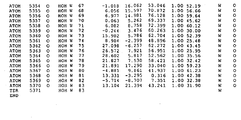


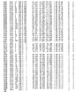
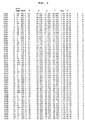
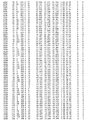
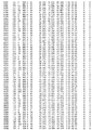


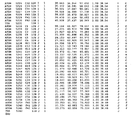
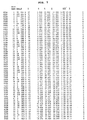
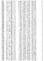


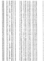


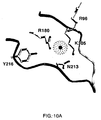
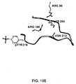
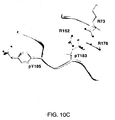

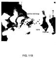


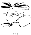
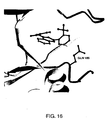
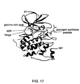


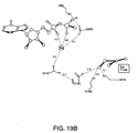
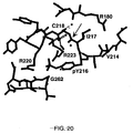

Classifications
-
- C—CHEMISTRY; METALLURGY
- C07—ORGANIC CHEMISTRY
- C07D—HETEROCYCLIC COMPOUNDS
- C07D487/00—Heterocyclic compounds containing nitrogen atoms as the only ring hetero atoms in the condensed system, not provided for by groups C07D451/00 - C07D477/00
- C07D487/02—Heterocyclic compounds containing nitrogen atoms as the only ring hetero atoms in the condensed system, not provided for by groups C07D451/00 - C07D477/00 in which the condensed system contains two hetero rings
- C07D487/04—Ortho-condensed systems
-
- A—HUMAN NECESSITIES
- A61—MEDICAL OR VETERINARY SCIENCE; HYGIENE
- A61P—SPECIFIC THERAPEUTIC ACTIVITY OF CHEMICAL COMPOUNDS OR MEDICINAL PREPARATIONS
- A61P11/00—Drugs for disorders of the respiratory system
- A61P11/06—Antiasthmatics
-
- A—HUMAN NECESSITIES
- A61—MEDICAL OR VETERINARY SCIENCE; HYGIENE
- A61P—SPECIFIC THERAPEUTIC ACTIVITY OF CHEMICAL COMPOUNDS OR MEDICINAL PREPARATIONS
- A61P21/00—Drugs for disorders of the muscular or neuromuscular system
- A61P21/04—Drugs for disorders of the muscular or neuromuscular system for myasthenia gravis
-
- A—HUMAN NECESSITIES
- A61—MEDICAL OR VETERINARY SCIENCE; HYGIENE
- A61P—SPECIFIC THERAPEUTIC ACTIVITY OF CHEMICAL COMPOUNDS OR MEDICINAL PREPARATIONS
- A61P25/00—Drugs for disorders of the nervous system
-
- A—HUMAN NECESSITIES
- A61—MEDICAL OR VETERINARY SCIENCE; HYGIENE
- A61P—SPECIFIC THERAPEUTIC ACTIVITY OF CHEMICAL COMPOUNDS OR MEDICINAL PREPARATIONS
- A61P25/00—Drugs for disorders of the nervous system
- A61P25/14—Drugs for disorders of the nervous system for treating abnormal movements, e.g. chorea, dyskinesia
- A61P25/16—Anti-Parkinson drugs
-
- A—HUMAN NECESSITIES
- A61—MEDICAL OR VETERINARY SCIENCE; HYGIENE
- A61P—SPECIFIC THERAPEUTIC ACTIVITY OF CHEMICAL COMPOUNDS OR MEDICINAL PREPARATIONS
- A61P25/00—Drugs for disorders of the nervous system
- A61P25/18—Antipsychotics, i.e. neuroleptics; Drugs for mania or schizophrenia
-
- A—HUMAN NECESSITIES
- A61—MEDICAL OR VETERINARY SCIENCE; HYGIENE
- A61P—SPECIFIC THERAPEUTIC ACTIVITY OF CHEMICAL COMPOUNDS OR MEDICINAL PREPARATIONS
- A61P25/00—Drugs for disorders of the nervous system
- A61P25/28—Drugs for disorders of the nervous system for treating neurodegenerative disorders of the central nervous system, e.g. nootropic agents, cognition enhancers, drugs for treating Alzheimer's disease or other forms of dementia
-
- A—HUMAN NECESSITIES
- A61—MEDICAL OR VETERINARY SCIENCE; HYGIENE
- A61P—SPECIFIC THERAPEUTIC ACTIVITY OF CHEMICAL COMPOUNDS OR MEDICINAL PREPARATIONS
- A61P29/00—Non-central analgesic, antipyretic or antiinflammatory agents, e.g. antirheumatic agents; Non-steroidal antiinflammatory drugs [NSAID]
-
- A—HUMAN NECESSITIES
- A61—MEDICAL OR VETERINARY SCIENCE; HYGIENE
- A61P—SPECIFIC THERAPEUTIC ACTIVITY OF CHEMICAL COMPOUNDS OR MEDICINAL PREPARATIONS
- A61P3/00—Drugs for disorders of the metabolism
- A61P3/08—Drugs for disorders of the metabolism for glucose homeostasis
- A61P3/10—Drugs for disorders of the metabolism for glucose homeostasis for hyperglycaemia, e.g. antidiabetics
-
- A—HUMAN NECESSITIES
- A61—MEDICAL OR VETERINARY SCIENCE; HYGIENE
- A61P—SPECIFIC THERAPEUTIC ACTIVITY OF CHEMICAL COMPOUNDS OR MEDICINAL PREPARATIONS
- A61P35/00—Antineoplastic agents
-
- A—HUMAN NECESSITIES
- A61—MEDICAL OR VETERINARY SCIENCE; HYGIENE
- A61P—SPECIFIC THERAPEUTIC ACTIVITY OF CHEMICAL COMPOUNDS OR MEDICINAL PREPARATIONS
- A61P37/00—Drugs for immunological or allergic disorders
-
- A—HUMAN NECESSITIES
- A61—MEDICAL OR VETERINARY SCIENCE; HYGIENE
- A61P—SPECIFIC THERAPEUTIC ACTIVITY OF CHEMICAL COMPOUNDS OR MEDICINAL PREPARATIONS
- A61P37/00—Drugs for immunological or allergic disorders
- A61P37/02—Immunomodulators
-
- A—HUMAN NECESSITIES
- A61—MEDICAL OR VETERINARY SCIENCE; HYGIENE
- A61P—SPECIFIC THERAPEUTIC ACTIVITY OF CHEMICAL COMPOUNDS OR MEDICINAL PREPARATIONS
- A61P37/00—Drugs for immunological or allergic disorders
- A61P37/08—Antiallergic agents
-
- A—HUMAN NECESSITIES
- A61—MEDICAL OR VETERINARY SCIENCE; HYGIENE
- A61P—SPECIFIC THERAPEUTIC ACTIVITY OF CHEMICAL COMPOUNDS OR MEDICINAL PREPARATIONS
- A61P43/00—Drugs for specific purposes, not provided for in groups A61P1/00-A61P41/00
-
- A—HUMAN NECESSITIES
- A61—MEDICAL OR VETERINARY SCIENCE; HYGIENE
- A61P—SPECIFIC THERAPEUTIC ACTIVITY OF CHEMICAL COMPOUNDS OR MEDICINAL PREPARATIONS
- A61P5/00—Drugs for disorders of the endocrine system
-
- A—HUMAN NECESSITIES
- A61—MEDICAL OR VETERINARY SCIENCE; HYGIENE
- A61P—SPECIFIC THERAPEUTIC ACTIVITY OF CHEMICAL COMPOUNDS OR MEDICINAL PREPARATIONS
- A61P9/00—Drugs for disorders of the cardiovascular system
-
- A—HUMAN NECESSITIES
- A61—MEDICAL OR VETERINARY SCIENCE; HYGIENE
- A61P—SPECIFIC THERAPEUTIC ACTIVITY OF CHEMICAL COMPOUNDS OR MEDICINAL PREPARATIONS
- A61P9/00—Drugs for disorders of the cardiovascular system
- A61P9/10—Drugs for disorders of the cardiovascular system for treating ischaemic or atherosclerotic diseases, e.g. antianginal drugs, coronary vasodilators, drugs for myocardial infarction, retinopathy, cerebrovascula insufficiency, renal arteriosclerosis
-
- C—CHEMISTRY; METALLURGY
- C12—BIOCHEMISTRY; BEER; SPIRITS; WINE; VINEGAR; MICROBIOLOGY; ENZYMOLOGY; MUTATION OR GENETIC ENGINEERING
- C12N—MICROORGANISMS OR ENZYMES; COMPOSITIONS THEREOF; PROPAGATING, PRESERVING, OR MAINTAINING MICROORGANISMS; MUTATION OR GENETIC ENGINEERING; CULTURE MEDIA
- C12N9/00—Enzymes; Proenzymes; Compositions thereof; Processes for preparing, activating, inhibiting, separating or purifying enzymes
- C12N9/10—Transferases (2.)
- C12N9/12—Transferases (2.) transferring phosphorus containing groups, e.g. kinases (2.7)
- C12N9/1205—Phosphotransferases with an alcohol group as acceptor (2.7.1), e.g. protein kinases
-
- G—PHYSICS
- G16—INFORMATION AND COMMUNICATION TECHNOLOGY [ICT] SPECIALLY ADAPTED FOR SPECIFIC APPLICATION FIELDS
- G16B—BIOINFORMATICS, i.e. INFORMATION AND COMMUNICATION TECHNOLOGY [ICT] SPECIALLY ADAPTED FOR GENETIC OR PROTEIN-RELATED DATA PROCESSING IN COMPUTATIONAL MOLECULAR BIOLOGY
- G16B15/00—ICT specially adapted for analysing two-dimensional or three-dimensional molecular structures, e.g. structural or functional relations or structure alignment
- G16B15/30—Drug targeting using structural data; Docking or binding prediction
-
- C—CHEMISTRY; METALLURGY
- C07—ORGANIC CHEMISTRY
- C07K—PEPTIDES
- C07K2299/00—Coordinates from 3D structures of peptides, e.g. proteins or enzymes
-
- G—PHYSICS
- G16—INFORMATION AND COMMUNICATION TECHNOLOGY [ICT] SPECIALLY ADAPTED FOR SPECIFIC APPLICATION FIELDS
- G16B—BIOINFORMATICS, i.e. INFORMATION AND COMMUNICATION TECHNOLOGY [ICT] SPECIALLY ADAPTED FOR GENETIC OR PROTEIN-RELATED DATA PROCESSING IN COMPUTATIONAL MOLECULAR BIOLOGY
- G16B15/00—ICT specially adapted for analysing two-dimensional or three-dimensional molecular structures, e.g. structural or functional relations or structure alignment
Definitions
- the present invention relates to inhibitors of glycogen synthase kinase-3 (GSK-3), a serine/threonine protein kinase, and to methods for producing them.
- the invention also provides pharmaceutical compositions comprising the inhibitors of the invention. Those compositions are suitable for the treatment and prevention of various disease states, such as diabetes and Alzheimer's disease.
- Protein kinases mediate intracellular signal transduction by affecting a phosphoryl transfer from a nucleoside triphosphate to a protein acceptor involved in a signaling pathway. There are a number of kinases and pathways through which extracellular and other stimuli cause a variety of cellular responses to occur inside the cell.
- Examples of such stimuli include environmental and chemical stress signals (e.g., osmotic shock, heat shock, ultraviolet radiation, bacterial endotoxin, H 2 O 2 ), cytokines (e.g., interleukin-1 (IL-1) and tumor necrosis factor ⁇ (TNF- ⁇ )), growth factors (e.g., granulocyte macrophage-colony-stimulating factor (GM-CSF), and fibroblast growth factor (FGF).
- An extracellular stimulus may affect one or more cellular responses related to cell growth, migration, differentiation, secretion of hormones, activation of transcription factors, muscle contraction, glucose metabolism, control of protein synthesis and regulation of cell cycle.
- Many disease states are associated with abnormal cellular responses triggered by protein kinase-mediated events. These diseases include autoimmune diseases, inflammatory diseases, neurological and neurodegenerative diseases, cancer, cardiovascular diseases, allergies and asthma, Alzheimer's disease and hormone-related diseases.
- GSK-3 is a serine/threonine protein kinase and belongs to the superfamily of mitogen-activated protein kinases.
- MAP kinases are activated by phosphorylation of threonine and/or tyrosine residues in a loop adjacent to the active site. Phosphorylation of MAP kinases is carried out by specific kinases upstream. Activated MAP kinase then phosphorylates the various substrates.
- Mammalian cells have ⁇ and ⁇ isoforms of GSK-3 that are each encoded by distinct genes ( Coghlan et al., Chemistry & Biology, 7, pp. 793-803 (2000 ); Kim and Kimmel, Curr. Opinion Genetics Dev., 10, pp. 508-514 (2000 )).
- the core kinase sequences have 97% similarity, but the protein sequences deviate substantially outside the kinase core ( Woodgett, J.R., EMBO J., 9, pp. 2431-8 (1990 )).
- GSK-3 ⁇ is 63 residues longer at the N-terminal end than GSK-3 ⁇ , however the N-terminal phosphorylation site in both isoforms (S21 for GSK-3 ⁇ and S9 for GSK-3 ⁇ ) is embedded in a conserved 7 residue motif.
- the two isoforms are not redundant as GSK-3 ⁇ deficiency is lethal in embryogenesis due to severe liver degeneration ( Hoeflich, K.P., et al., Nature, 406, pp. 86-90 (2000 )).
- GSK-3 ⁇ has multiple phosphorylation sites.
- the Serine 9 and Tyrosine 216 phosphorylation sites are well described in the literature. Phosphorylation of Tyrosine 216 activates GSK-3 ⁇ but phosphorylation of Serine 9 inactivates it.
- GSK-3 ⁇ is unique among kinases in that it requires prior phosphorylation of its substrates. GSK-3 ⁇ does not phosphorylate its multiple substrates in the same manner and with the same efficiency but has different modes of phosphorylation.
- the canonical phosphorylation sequence recognized by GSK-3 ⁇ , SXXXS contains two serines separated by three amino acid residues. Multiple copies of this motif can be present in the substrate.
- Glycogen synthase has multiple serines separated by four residues (residue 640, 644, 648, and 652) and those serines are phosphorylated sequentially by GSK-3 ⁇ from the C-terminal end, after S656 has been phosphorylated by Casein Kinase II ( Woodgett, J.R. and P. Cohen, Biochim. Biophys. Acta, 788, pp. 339-47 (1984 ); Kuret, J. et al, Eur. J. Biochem., 151, pp. 39-48 (1985 )).
- Glycogen synthase kinase-3 has been implicated in various diseases including diabetes, Alzheimer's disease, CNS disorders such as manic depressive disorder and neurodegenerative diseases, and cardiomyocete hypertrophy ( WO 99/65897 ; WO 00/38675 ; and Haq et al., J. Cell Biol., 151, pp. 117 (2000 )). These diseases may be caused by, or result in, the abnormal operation of certain cell signaling pathways in which GSK-3 plays a role. GSK-3 phosphorylates and modulates the activity of a number of regulatory proteins.
- glycogen synthase which is the rate limiting enzyme necessary for glycogen synthesis
- the microtubule associated protein Tau the gene transcription factor beta-catenin
- the translation initiation factor elF2B as well as ATP citrate lyase
- axin heat shock factor-1
- c-Jun c-Myc
- c-Myb c-Myb
- CEPBa CEPBa
- GSK-3 In a GSK-3 mediated pathway that is relevant for the treatment of type II diabetes, insulin-induced signaling leads to cellular glucose uptake and glycogen synthesis. Along this pathway, GSK-3 is a negative regulator of the insulin-induced signal. Insulin inactivates GSK-3 ⁇ via the PKB/Akt pathway, which results in activation of glycogen synthase.
- GSK-3 In such patients, the normal insulin-induced inhibition of GSK-3 fails to occur. It has also been reported that in patients with type II diabetes, GSK-3 is overexpressed ( WO 00/38675 ). Therapeutic inhibitors of GSK-3 are therefore potentially useful for treating diabetic patients suffering from an impaired response to insulin.
- GSK-3 activity has also been associated with Alzheimer's disease. This disease is characterized by the well-known ⁇ -amyloid peptide and the formation of intracellular neurofibrillary tangles.
- the neurofibrillary tangles contain hyperphosphorylated Tau protein where Tau is phosphorylated on abnormal sites. GSK-3 has been shown to phosphorylate these abnormal sites in cell and animal models. Furthermore, inhibition of GSK-3 has been shown to prevent hyperphosphorylation of Tau in cells ( Lovestone et al., Current Biology, 4, pp. 1077-86 (1994 ); Brownlees et al., Neuroreport, 8, pp. 3251-55 (1997 )). Therefore, it is believed that GSK-3 activity may promote generation of the neurofibrillary tangles and the progression of Alzheimer's disease.
- beta-catenin Another substrate of GSK-3 is beta-catenin which is degraded after phosphorylation by GSK-3.
- Reduced levels of beta-catenin have been reported in schizophrenic patients and have also been associated with other diseases related to increase in neuronal cell death ( Zhong et al., Nature, 395, 698-702 (1998 ); Takashima et al., PNAS, 90, 7789-93 (1993 ); Pei et al., J. Neuropathol. Exp., 56, 70-78 (1997 )).
- GSK-3 ⁇ is also a component of the Wnt signalling pathway. Activation of the Wnt pathway inhibits GSK-3 ⁇ , which results in accumulation of cytosolic ⁇ -catenin ( Yost, C., et al., Cell, 93, pp. 1031-41 (1998 )).
- the cytosolic ⁇ -catenin translocates to the cell nucleus, where it associates with LEF/tcf and stimulates the expression of Wnt target genes resulting in cell proliferation ( Ding, V.W., et al., J. Biol. Chem., 275, pp. 32475-81 (2000 ); Waltzer, L. and M. Bienz, Cancer Metastasis Rev. 18, pp.
- GSK-3 ⁇ is also down regulated by 7-TM receptors that regulate cAMP levels.
- cAMP-dependent protein kinase A binds, phosphorylates and inhibits GSK-3 ⁇ in response to the adenyl cyclase activator forskolin, or the p-adrenergic receptor activator isoproterenol ( Fang, X., et al., Proc. Natl. Acad. Sci. U S A, 97, pp. 11960-5 (2000 )).
- GSK-3 Small molecule inhibitors of GSK-3 have recently been reported ( WO 99/65897 and WO 00/38675 ). For many of the aforementioned diseases associated with abnormal GSK-3 activity, other protein kinases have also been targeted for treating the same diseases. However, the various protein kinases often act through different biological pathways. Quinazoline derivatives have been reported recently as inhibitors of p38 kinase ( WO 00/12497 ). The compounds are reported to be useful for treating conditions characterized by enhanced p38- ⁇ activity and/or enhanced TGF- ⁇ activity.
- p38 activity has been implicated in a wide variety of diseases, including diabetes, p38 kinase is not reported to be a constituent of an insulin signaling pathway that regulates glycogen synthesis or glucose uptake. Therefore, unlike GSK-3, p38 inhibition would not be expected to enhance glycogen synthesis and/or glucose uptake.
- GSK-3 inhibitors that are effective as therapeutic agents due to its important role in diabetes, Alzheimer's disease and other diseases.
- a challenge has been to find protein kinase inhibitors that act in a selective manner. Since there are numerous protein kinases that are involved in a variety of cellular responses, non-selective inhibitors may lead to unwanted side effects.
- the three-dimensional structure of the kinase would assist in the rational design of inhibitors. Further, information provided by the X-ray crystal structure of GSK-3 ⁇ -inhibitor complexes would be extremely useful in iterative drug design of various GSK-3 proteins. The determination of the amino acid residues in GSK-3 ⁇ binding pockets and the determination of the shape of those binding pockets would allow one to design inhibitors that bind more favorably to this class of enzymes.
- the present invention addresses this need by providing compounds and pharmaceutical compositions thereof that are effective as protein kinase inhibitors, particularly as inhibitors of GSK-3.
- the compounds of the present invention have the general formula I: or a pharmaceutically acceptable derivative thereof, wherein:
- the invention provides pharmaceutical compositions comprising a GSK-3 inhibitor of this invention.
- These compositions may be utilized for treating or preventing a variety of GSK-3 mediated disorders, such as autoimmune diseases, inflammatory diseases, metabolic, neurological and neurodegenerative diseases, cardiovascular diseases, allergy, asthma, diabetes, Alzheimer's disease, Huntington's Disease, Parkinson's Disease, AIDS-associated dementia, amyotrophic lateral sclerosis (AML, Lou Gehrig's Disease), multiple sclerosis (MS), schizophrenia, cardiomyocyte hypertrophy, reperfusion/ischemia, and baldness.
- GSK-3 mediated disorders such as autoimmune diseases, inflammatory diseases, metabolic, neurological and neurodegenerative diseases, cardiovascular diseases, allergy, asthma, diabetes, Alzheimer's disease, Huntington's Disease, Parkinson's Disease, AIDS-associated dementia, amyotrophic lateral sclerosis (AML, Lou Gehrig's Disease), multiple sclerosis (MS), schizophrenia, cardiomyocyte hypertrophy, reperfusion/ischemia, and baldness.
- compositions of this invention are also useful for enhancing glycogen synthesis and/or lowering blood levels of glucose and therefore are especially useful for diabetic patients. These compositions are also useful for inhibiting the production of hyperphosphorylated Tau protein, which is useful in halting or slowing the progression of Alzheimer's disease. Another embodiment of this invention relates to the use for inhibiting the phosphorylation of ⁇ -catenin, which is useful for treating schizophrenia.
- the invention provides pharmaceutical compositions comprising these compounds.
- Figure 1 lists the atomic structure coordinates for unphosphorylated GSK-3 ⁇ as derived by X-ray diffraction from the crystal.
- Figure 2 lists the atomic structure coordinates for phosphorylated GSK-3 ⁇ .
- Figure 3 lists the atomic structure coordinates for phosphorylated GSK-3 ⁇ -inhibitor1 complex (inhibitor1 is 4,5-Diphenyl-1H-pyrazolo[3,4-c]pyridazin-3-ylamine).
- Figure 4 lists the atomic structure coordinates for unphosphorylated GSK-3 ⁇ -inhibitor2 complex (inhibitor2 is (5-Methyl-2H-pyrazol-3-yl)-(2-pyridin-4-yl-quinazolin-4-yl)-amine)).
- Figure 5 lists the atomic structure coordinates for unphosphorylated GSK-3 ⁇ -inhibitor3 complex (inhibitor 3 is 4-(5-Methyl-2-phenylamino-pyrimidin-4-yl)-1H-pyrrole-2-carboxylic acid (2-hydroxy-1-phenyl-ethyl)-amide) .
- Figure 6 lists the atomic structure coordinates for unphosphorylated GSK-3 ⁇ -inhibitor4 complex (inhibitor 4 is(1H-Indazol-3-yl)-[2-(2-trifluoromethyl-phenyl)-quinazolin-4-yl]-amine).
- Figure 7 lists the atomic structure coordinates for phosphorylated GSK-3 ⁇ in complex with ADP and glycogen synthase peptide.
- Figure 8 depicts a ribbon diagram of the overall fold of unphosphorylated GSK-3 ⁇ .
- the N-terminal domain corresponds to the ⁇ -strand domain and encompasses residues 25 to 138.
- ⁇ -strand 1 was only visible in one of the two molecules in the asymmetric unit and makes hydrogen bonds with ⁇ -strand 2 although it is not part of the ⁇ -barrel.
- the ⁇ -helical domain corresponds to residues 139 to 349. Key features of the kinase-fold such as the hinge, glycine rich loop and activation-loop are indicated.
- Figure 9 depicts a superposition of unphosphorylated GSK-3 ⁇ (light shade) and activated substrate-bound CDK2 (Protein Data Bank accession number 1QMZ) (dark shade).
- the ⁇ -helical domains of GSK-3 ⁇ and CDK2 were superimposed in QUANTA by aligning matching residues.
- Figure 10 shows a comparison between the activation loops of unphosphorylated GSK-3 ⁇ ( Figure 10A ), phosphorylated GSK-3 ⁇ ( Figure 10B ), and phosphorylated p38 ⁇ (Protein Data Bank accession number 1CM8)( Figure 7C ).
- Figure 10A the side chains of residues R96, R180 and K205 are pointing to a phosphate ion that occupies the same position as the phosphate group of the phospho-threonine in activated p38 ⁇ ( Figure 10C ), activated CDK2 and activated ERK2.
- the phosphorylated Y216 is flipped out of the substrate binding groove, which is similar to the position of the phosphorylated Y185 of p38 ⁇ .
- FIG 11A shows that in the unphosphorylated GSK-3 ⁇ structure, the GSK-3 ⁇ substrate-binding groove is occupied by a phosphate ion and residues 260 to 264 of a neighboring GSK-3 ⁇ molecule.
- Figure 11B shows a model for the binding of the SXXXpS motif in the GSK-3 ⁇ substrate-binding groove.
- the ⁇ -helical domains of GSK-3 ⁇ and activated substrate bound CDK2 (Protein Data Bank accession number 1QMZ) were superimposed to model the positioning of a primed peptide in the GSK-3 ⁇ substrate-binding groove.
- the side chains of the peptide residues except for the target serine S* have been removed for clarity.
- the phosphorylation site S* is positioned in front of the active site.
- the model estimates how the SXXXpS motif fits in the substrate-binding groove with the three residues bridging the gap between the P and P+4 serines.
- the phosphate group of the P+4 serine will occupy the same position as the phosphate ion found in the crystal structure.
- Figure 12 depicts a ribbon diagram of phosphorylated GSK-3 ⁇ . The activation loop and the phosphorylated Y216 are indicated.
- Figure 13 presents the inhibitor2 bound in the active site of unphosphorylated GSK-3 ⁇ .
- the hinge and glycine rich loop are indicated.
- Figure 14 presents the view of inhibitor1 bound in the active site of phosphorylated GSK-3 ⁇ .
- Figure 15 presents the view of inhibitor3 bound in the active site of unphosphorylated GSK-3 ⁇ .
- Figure 16 presents the conformation of Gln185 in the active site when bound to inhibitor4.
- Figure 17 depicts the overall structure of phosphorylated GSK-3 ⁇ in complex with ADP and glycogen synthase peptide.
- the N-terminal domain corresponds to the ⁇ -strand domain and includes residues 37 to 138.
- the ⁇ -helical domain of the GSK-3 ⁇ kinase core corresponds to residues 139 to 343.
- the C-terminal 34 residues are not part of the kinase core but pack against the ⁇ -helical domain. Key features of the kinase fold, such as the glycine rich loop, the hinge and the activation loop are indicated.
- the ADP-Mg complex occupying the active site is shown.
- the glycogen synthase peptide (residues 650 to 658) bound in the substrate binding groove is also shown here.
- Figure 18 depicts the ⁇ -strand domain rotation induced by ADP and glycogen synthase peptide.
- the ⁇ -strand domain of the phosphorylated GSK-3 ⁇ -ADP-peptide complex rotated 6.5 ⁇ in comparison to unphosphorylated and phosphorylated apo-GSK-3.
- the loop between ⁇ -3 and ⁇ -C (residues 87 to 95) moved 13 ⁇ to contact the glycogen synthase peptide (not shown).
- Figure 19A depicts a stereo view of the active site occupied by the ADP-Mg complex.
- the adenine base is surrounded by hydrophobic residues and makes two hydrogen bonds with the backbone of the hinge region.
- the catalytic residues involved in the phosphate transfer to the P-site serine surround the two non-transferable ADP phosphates.
- Figure 19B is a schematic drawing of the hydrogen bond network that connects the ADP-Mg complex to the P-site serine via the catalytic residues.
- Figure 20 depicts the environment of phosphotyrosine 216 in the structure of phosphorylated GSK-3 ⁇ in complex with ADP and glycogen synthase peptide.
- the phosphate moiety binds two arginine side chains (Arg 220 and Arg 223) resulting in charge neutralization.
- Figure 21 depicts the glycogen synthase peptide in the substrate binding groove.
- the phosphoserine pSer 656 binds Arg 96, Arg 180 and Lys 205, which results in the proper alignment of the ⁇ -helical and ⁇ -stand domains.
- pTyr 216 moves its side chain out of the substrate binding groove to make contact with the side chains of Arg 220 and Arg 223.
- active site refers to the area in the protein kinase where the nucleotide binds. This site is located at the interface of the C-terminal ⁇ -helical and N-terminal ⁇ -strand domain, and is bordered by the glycine rich loop and the hinge (See, Xie et al., Structure, 6, pp. 983-991 (1998 ), incorporated herein by reference).
- aliphatic refers to straight chain or branched hydrocarbons that are completely saturated or that contain one or more units of unsaturation.
- aliphatic groups include substituted or unsubstituted linear or branched alkyl, alkenyl and alkynyl groups. Unless indicated otherwise, the term “aliphatic” encompasses both substituted and unsubstituted hydrocarbons.
- alkyl used alone or as part of a larger moiety, refers to both straight and branched saturated chains containing one to twelve carbon atoms.
- alkenyl and alkynyl used alone or as part of a larger moiety, encompass both straight and branched chains containing two to twelve carbon atoms and at least one unit of unsaturation.
- An alkenyl group contains at least one carbon-carbon double bond and an alkynyl group contains at least one carbon-carbon triple bond.
- corresponding amino acids when used in the context of amino acid residues that correspond to GSK-3 ⁇ amino acids refers to particular amino acids or analogues thereof in a protein that correspond to amino acids in the GSK-3 ⁇ protein.
- the corresponding amino acid may be an identical, mutated, chemically modified, conserved, conservatively substituted, functionally equivalent or homologous amino acid when compared to the GSK-3 ⁇ amino acid to which it corresponds.
- corresponding amino acids may be identified by superimposing the backbone atoms of the amino acids in GSK-3 ⁇ and the protein using well known software applications, such as QUANTA (Molecular Simulations, Inc., San Diego, CA ⁇ 2000).
- sequence alignment programs such as the "bestfit" program available from the Genetics Computer Group which uses the local homology algorithm described by Smith and Waterman in Advances in Applied Mathematics 2, 482 (1981 ).
- aryl refers to monocyclic or polycyclic aromatic carbon ring systems having five to fourteen members.
- aryl groups include, but are not limited to, phenyl (Ph), 1-naphthyl, 2-naphthyl, 1-anthracyl and 2-anthracyl.
- aralkyl refers to an alkyl group substituted by an aryl. Examples of aralkyl groups include benzyl and phenethyl.
- association refers to a condition of proximity between a chemical entity or compound, or portions thereof, and a binding pocket or binding site on a protein.
- the association may be non-covalent -- wherein the juxtaposition is energetically favored by hydrogen bonding or van der Waals or electrostatic interactions -- or it may be covalent.
- ATP analogue refers to a compound derived from Adenosine-5'-triphosphate(ATP).
- the analogue can be ADP, or non-hydralysable, for example, Adenylyl Imidodiphosphate (AMPPNP).
- AMPPNP Adenylyl Imidodiphosphate
- AMPPNP can be in complex with Magnesium or Manganese ions.
- binding pocket refers to a region of a molecule or molecular complex, that, as a result of its shape, favorably associates with another chemical entity or compound.
- biological sample includes, without limitation, cell cultures or extracts thereof; biopsied material obtained from a mammal or extracts thereof; and blood, saliva, urine, feces, semen, tears, or other body fluids or extracts thereof.
- Carbocylyl or “carbocyclic”, alone or in combination with any other term, refers to monocyclic or polycyclic non-aromatic carbon ring systems, which may contain a specified number of carbon atoms, preferably from 3 to 12 carbon atoms, which are completely saturated or which contain one or more units of unsaturation.
- a carbocyclic ring system may be monocyclic, bicyclic or tricyclic.
- a carbocylyl ring may be fused to another ring, such as an aryl ring or another carbocyclic ring.
- carbocyclic rings could include cyclohexyl, cyclopentyl, cyclobutyl, cyclopropyl, cyclohexenyl, cyclopentenyl, indanyl, tetrahydronaphthyl and the like.
- chemically feasible or stable refers to a compound structure that is sufficiently stable to allow manufacture and administration to a patient by methods known in the art. Typically, such compounds are stable at a temperature of 40°C or less, in the absence of moisture or other chemically reactive conditions, for at least one week.
- chemical entity refers to chemical compounds, complexes of at least two chemical compounds, and fragments of such compounds or complexes.
- the chemical entity can be, for example, a ligand, a substrate, nucleotide triphosphate, a nucleotide, an agonist, antagonist, inhibitor, antibody, peptide, protein or drug.
- the chemical entity is selected from the group consisting of an ATP and an inhibitor for the active site.
- the inhibitor is 4,5-Diphenyl-1H-pyrazolo[3,4-c]pyridazin-3-ylamine, (5-Methyl-2H-pyrazol-3-yl)-(2-pyridin-4-yl-quinazolin-4-yl)-amine, 4-(5-Methyl-2-phenylamino-pyrimidin-4-yl)-1H-pyrrole-2-carboxylic acid (2-hydroxy-1-phenyl-ethyl)-amide, (1H-Indazol-3-yl)-[2-(2-trifluoromethyl-phenyl)-quinazolin-4-yl]-amine and an ATP analogue such as MgAMP-PNP (adenylyl imidodiphosphate) or ADP.
- the chemical entity is selected from the group consisting of a peptide substrate or inhibitor for the substrate binding groove.
- crystallization solution refers to a solution that promotes crystallization of macromolecules.
- the crystallization solution may contain a precipitant, a buffer, salt, stabilizer, a polyionic agent, a detergent, a lanthanide ion or reducing agent.
- a precipitant e.g., sodium bicarbonate
- a buffer e.g., sodium bicarbonate
- stabilizer e.g., sodium bicarbonate
- a polyionic agent e.g., sodium bicarbonate
- detergent e.g., sodium bicarbonate
- lanthanide ion or reducing agent e.g., sodium bicarbonate
- conservative substitutions refers to residues that are physically or functionally similar to the corresponding reference residues. That is, a conservative substitution and its reference residue have similar size, shape, electric charge, chemical properties including the ability to form covalent or hydrogen bonds, or the like. Preferred conservative substitutions are those fulfilling the criteria defined for an accepted point mutation in Dayhoff et al., Atlas of Protein Sequence and Structure, 5, pp. 345-352 (1978 & Supp .).
- substitutions including but not limited to the following groups: (a) valine, glycine; (b) glycine, alanine; (c) valine, isoleucine, leucine; (d) aspartic acid, glutamic acid; (e) asparagine, glutamine; (f) serine, threonine; (g) lysine, arginine, methionine; and (h) phenylalanine, tyrosine.
- groups including but not limited to the following groups: (a) valine, glycine; (b) glycine, alanine; (c) valine, isoleucine, leucine; (d) aspartic acid, glutamic acid; (e) asparagine, glutamine; (f) serine, threonine; (g) lysine, arginine, methionine; and (h) phenylalanine, tyrosine.
- complex refers to a protein associated with a chemical entity.
- domain refers to a structural unit of the GSK-3 ⁇ protein or homologue.
- the domain can comprise a binding pocket, or a sequence or structural motif.
- the protein is separated into two domains, the N-terminal domain which is predominantly ⁇ strands and the C-terminal domain which is predominantly ⁇ helical.
- the term "generating a three-dimensional structure” refers to plotting the structure coordinates in three-dimensional space. This can be achieved through commercially available software.
- the three-dimensional structure may be used to perform computer modeling, fitting operations, or displayed as a three-dimensional graphical representation.
- GSK-3 ⁇ inhibitor-binding pocket or "GSK-3 ⁇ ATP-binding pocket” refers to a binding pocket of a molecule or molecular complex defined by the structure coordinates of a certain set of amino acid residues present in the GSK-3 ⁇ structure, as described below. This binding pocket is in an area in the GSK-3 ⁇ protein where the ATP or inhibitor for the active site binds.
- GSK-3 ⁇ -like refers to all or a portion of a molecule or molecular complex that has a commonality of shape to all or a portion of the GSK-3 ⁇ protein.
- the commonality of shape is defined by a root mean square deviation of the structure coordinates of the backbone atoms between the amino acids in the GSK-3 ⁇ -like inhibitor-binding pocket and the GSK-3 ⁇ amino acids in the GSK-3 ⁇ inhibitor-binding pocket (as set forth in any one of Figure 1-7 ).
- GSK-3 ⁇ amino acids that define the GSK-3 ⁇ inhibitor-binding pocket Depending on the set of GSK-3 ⁇ amino acids that define the GSK-3 ⁇ inhibitor-binding pocket, one skilled in the art would be able to locate the corresponding amino acids that define a GSK-3 ⁇ -like inhibitor-binding pocket in a protein based on sequence or structural homology.
- GSK-3-mediated condition refers to any disease or other deleterious condition or state in which GSK-3, in particular GSK-3, is known to play a role.
- diseases or conditions include, without limitation, diabetes, Alzheimer's disease, Huntington's Disease, Parkinson's Disease, AIDS-associated dementia, amyotrophic lateral sclerosis (AML), multiple sclerosis (MS), schizophrenia, cardiomycete hypertrophy, reperfusion/ischemia, and baldness.
- halogen or halo means F, Cl, Br, or I.
- heteroatom means N, O, or S and shall include any oxidized form of nitrogen and sulfur, such as N(O), S(O), S(O) 2 and the quaternized form of any basic nitrogen.
- heterocyclic refers to non-aromatic saturated or unsaturated monocyclic or polycyclic ring systems containing one or more heteroatoms and with a ring size of three to fourteen.
- a heterocyclic ring may have one to four heteroatoms so long as the heterocyclic ring is chemically feasible and stable and may be fused to another ring, such as a carbocyclic, aryl or heteroaryl ring, or to another heterocyclic ring.
- a heterocyclic ring system may be monocyclic, bicyclic or tricyclic. Also included within the scope of within the scope of the term “heterocyclic” or “heterocyclyl”, as used herein, is a group in which one or more carbocyclic rings are fused to a heteroaryl.
- heterocyclic rings include, but are not limited to, 3-1H-benzimidazol-2-one, 3-1H-alkyl-benzimidazol-2-one, 2-tetrahydrofuranyl, 3-tetrahydrofuranyl, 2-tetrahydrothiophenyl, 3-tetrahydrothiophenyl, 2-morpholino, 3-morpholino, 4-morpholino, 2-thiomorpholino, 3-thiomorpholino, 4-thiomorpholino, 1-pyrrolidinyl, 2-pyrrolidinyl, 3-pyrrolidinyl, 1-piperazinyl, 2-piperazinyl, 1-piperidinyl, 2-piperidinyl, 3-piperidinyl, 4-piperidinyl, 4-thiazolidinyl, diazolonyl, N-substituted diazolonyl, 1-phthalimidinyl, benzoxane, benzotriazol-1-yl, benzopyrrolidine, benzopiperidine, be
- heteroaryl refers to monocyclic or polycyclic aromatic ring systems having five to fourteen members and one or more heteroatoms.
- heteroaryl refers to monocyclic or polycyclic aromatic ring systems having five to fourteen members and one or more heteroatoms.
- heteroarylkyl refers to an alkyl group substituted by a heteroaryl.
- alkenyl or alkynyl groups substituted by a heteroaryl are also explicitly included within the scope of the term “heteroaralkyl”. In general, a heteroaryl ring may have one to four heteroatoms.
- Heteroaryl groups include, without limitation, 2-furanyl, 3-furanyl, N-imidazolyl, 2-imidazolyl, 4-imidazolyl, 5-imidazolyl, 3-isoxazolyl, 4-isoxazolyl, 5-isoxazolyl, 2-oxadiazolyl, 5-oxadiazolyl, 2-oxazolyl, 4-oxazolyl, 5-oxazolyl, 2-pyrrolyl, 3-pyrrolyl, 2-pyridyl, 3-pyridyl, 4-pyridyl, 2-pyrimidyl, 4-pyrimidyl, 5-pyrimidyl, 3-pyridazinyl, 2-thiazolyl, 4-thiazolyl, 5-thiazolyl, 5-tetrazolyl, 2-triazolyl, 5-triazolyl, 2-thienyl, and 3-thienyl.
- heteroaryl ring also refers to the term “heteroaryl ring", “heteroaryl group
- fused polycyclic heteroaryl and aryl ring systems in which a carbocyclic aromatic ring or heteroaryl ring is fused to one or more other rings include, tetrahydronaphthyl, benzimidazolyl, benzothienyl, benzofuranyl, indolyl, quinolinyl, benzothiazolyl, benzoxazolyl, benzimidazolyl, isoquinolinyl, isoindolyl, acridinyl, benzoisoxazolyl, and the like.
- An aryl, aralkyl, heteroaryl, or heteroaralkyl group may contain one or more independently selected substituents.
- suitable substituents on the unsaturated carbon atom of an aryl or heteroaryl group include halogen, CF 3 , -R', -OR', -OH, -SH, -SR', protected OH (such as acyloxy), -NO 2 , -CN, -NH 2 , -NHR', -N(R') 2 , -NHCOR', -NHCONH 2 , -NHCONHR' , -NHCON(R') 2 , -NRCOR', -NHCO 2 H, -NHCO 2 R', -CO 2 R' , -CO 2 H, -COR', -CONH 2 , -CONHR' , -CON(R') 2 , -S(O) 2 H, -S(O) 2 R', -
- An aliphatic group or a non-aromatic heterocyclic ring may contain one or more substituents.
- a substitutable nitrogen on a heteroaryl or a non-aromatic heterocyclic ring is optionally substituted.
- Suitable substituents on the nitrogen include R", COR", S(O) 2 R", and CO 2 R", where R" is H, an aliphatic group or a substituted aliphatic group.
- the term "motif” refers to a group of amino acids in the protein that defines a structural compartment or carries out a function in the protein, for example, catalysis, structural stabilization or phosphorylation.
- the motif may be conserved in sequence, structure and function when.
- the motif can be contiguous in primary sequence or three-dimensional space. Examples of a motif include but are not limited to SXXXS motif, phosphorylation lip or activation loop, the glycine-rich phosphate anchor loop, the catalytic loop, the DFG loop and the APE motif (See, Xie et al., Structure, 6, pp. 983-991 (1998 )).
- homologue of GSK-3 ⁇ or "GSK-3 ⁇ homologue” refers to a molecule that is homologous to GSK-3 ⁇ by structure or sequence, but retains the kinase activity of GSK-3.
- the homologue has at least 80%, 90% or 95% sequence homology to GSK-3 ⁇ .
- the homologue can be GSK-3 ⁇ , GSK-3 ⁇ from another species, with conservative substitutions, conservative additions or deletions thereof; human GSK-3 ⁇ with conservative substitutions, conservative additions or deletions.
- the GSK-3 ⁇ can be full length protein (amino acids 1-420 of SEQ ID NO: 1); a truncated protein with amino acids 7-420, 25-381, 37-381 of SEQ ID NO: 1; the full length protein with conservative substitutions; the truncated protein with conservative mutations.
- part of a binding pocket refers to less than all of the amino acid residues that define the binding pocket.
- the structure coordinates of residues that constitute part of a binding pocket may be specific for defining the chemical environment of the binding pocket, or useful in designing fragments of an inhibitor that may interact with those residues.
- the portion of residues may be key residues that play a role in ligand binding, or may be residues that are spatially related and define a three-dimensional compartment of the binding pocket.
- the residues may be contiguous or non-contiguous in primary sequence.
- Part of the binding pocket can be at least two amino acid residues.
- Part of the inhibitor-binding pocket can be GSK-3 ⁇ amino acids D133 and V135, GSK-3 ⁇ amino acids K85, L132, D133 and V135, GSK-3 ⁇ amino acids K85, M101, V110, L130 and L132, GSK-3 ⁇ amino acids I62, V135, P136, T138 and L188, GSK-3 ⁇ amino acids V70, V110, L188 and C199, GSK-3 ⁇ amino acids F67, V70, Q185 and C199.
- Part of a substrate binding pocket can be GSK-3 ⁇ amino acids D90, K91, R92, F93, K94, GSK-3 ⁇ amino acids R96, R180, K205, N213 and Y234, GSK-3 ⁇ amino acids R96; R180 and K205, GSK-3 ⁇ amino acids S66, F67 and F93, or GSK-3 ⁇ amino acids Y216, I217, C218, S219, R220 and R223.
- part of a GSK-3 ⁇ protein refers to less than all of the amino acid residues of a GSK-3 ⁇ protein.
- Part of a GSK-3 ⁇ protein can define the binding pockets, domains or motifs of the protein.
- the structure coordinates of residues that constitute part of a GSK-3 ⁇ protein may be specific for defining the chemical environment of the protein, or useful in designing fragments of an inhibitor that may interact with those residues.
- the portion of residues may also be residues that are spatially related and define a three-dimensional compartment of a binding pocket, motif or domain.
- the residues maybe contiguous or non-contiguous in primary sequence.
- the portion of residues may be key residues that play a role in ligand or substrate binding, catalysis or structural stabilization.
- pharmaceutically acceptable carrier, adjuvant, or vehicle refers to a non-toxic carrier, adjuvant, or vehicle that may be administered to a patient, together with a compound of this invention, and which does not destroy the pharmacological activity thereof .
- patient includes human and veterinary subjects.
- peptide comprising a phosphorylation sequence refers to a peptide comprising the ZXXXY motif.
- Z can be serine or threonine.
- Y can be serine, threonine or valine.
- X can be any amino acid residue.
- Z or Y can be phosphorylated or unphosphorylated. The phosphorylation of Y facilitates the phosphorylation of X by GSK-3 ⁇ .
- peptide substrates comprising a phosphorylation sequence include but are not limited to, ASVPPS, PSPSLS, LSRHSS, SSPHQS, DSRAGS, LSRRPS, PTPPPT,PTPVPS, KSPVVS, VSGDTS, QSYLDS, DSGIHS, HSGATT, TTTAPS, TSANDS, DSEQQS, SSPLPS, PSSPLS and CTPTDV.
- Pharmaceutically acceptable salts of the compounds of this invention include those derived from pharmaceutically acceptable inorganic and organic acids and bases.
- suitable acid salts include acetate, adipate, alginate, aspartate, benzoate, benzenesulfonate, bisulfate, butyrate, citrate, camphorate, camphorsulfonate, cyclopentanepropionate, digluconate, dodecylsulfate, ethanesulfonate, formate, fumarate, glucoheptanoate, glycerophosphate, glycolate, hemisulfate, heptanoate, hexanoate, hydrochloride, hydrobromide, hydroiodide, 2-hydroxyethanesulfonate, lactate, maleate, malonate, methanesulfonate, 2-naphthalenesulfonate, nicotinate, nitrate, oxalate, palmoate, pec
- Salts derived from appropriate bases include alkali metal (e.g., sodium and potassium), alkaline earth metal (e.g., magnesium), ammonium and N + (C 1-4 alkyl) 4 salts.
- alkali metal e.g., sodium and potassium
- alkaline earth metal e.g., magnesium
- ammonium and N + (C 1-4 alkyl) 4 salts This invention also envisions the quaternization of any basic nitrogen-containing groups of the compounds disclosed herein. Water or oil-soluble or dispersible products may be obtained by such quaternization.
- protein kinase-mediated condition or “state” refers to any disease or other deleterious condition or state in which a protein kinase is known to play a role.
- Such conditions include, without limitation, autoimmune diseases, inflammatory diseases, metabolic, neurological and neurodegenerative diseases, cardiovasclular diseases, allergy and asthma.
- root mean square deviation refers to the square root of the arithmetic mean of the squares of the deviations from the mean. It is a way to express the deviation or variation from a trend or object.
- the "root mean square deviation” defines the variation in the backbone of a protein from the backbone of GSK-3 ⁇ or a binding pocket portion thereof, as defined by the structure coordinates of GSK-3 ⁇ described herein. It would be readily apparent to those skilled in the art that the calculation of RMSD involves standard error.
- soaked refers to a process in which the crystal is transferred to a solution containing the compound of interest.
- structure coordinates refers to Cartesian coordinates derived from mathematical equations related to the patterns obtained on diffraction of a monochromatic beam of X-rays by the atoms (scattering centers) of a protein or protein complex in crystal form.
- the diffraction data are used to calculate an electron density map of the repeating unit of the crystal.
- the electron density maps are then used to establish the positions of the individual atoms of the enzyme or enzyme complex.
- substantially all of a GSK-3 ⁇ binding pocket or “substantially all of a GSK-3 ⁇ protein” refers to all or almost all of the amino acids in the GSK-3 ⁇ binding pocket or protein.
- substantially all of a GSK-3 ⁇ binding pocket can be 100%, 95%, 90%, 80%, 70% of the residues defining the GSK-3 ⁇ binding pocket or protein.
- substrate binding groove refers to an area in a protein kinase where the substrate binds.
- the substrate binding groove is located at the interface of the ⁇ -strand and ⁇ -helical domain, and positioned between the activation loop and ⁇ -strand domain.
- substrates include but are not limited to glycogen synthase, ⁇ -catenin, elongation initiation factor 2B ⁇ subunit, cAMP-responsive element binding protein, CCAAT/enhancer binding protein ⁇ , microtuble associated protein Tau, axin, Dd-STATa and Cyclin D1.
- substrate binding pocket refers to a binding pocket of a molecule or molecular complex defined by the structure coordinates of a certain set of amino acid residues present in the GSK-3 ⁇ structure, as described below. This binding pocket is in an area in the GSK-3 ⁇ protein where the substrate binding groove is located.
- the term "sufficiently homologous to GSK-3 ⁇ " refers to a protein that has a sequence homology of at least 20% compared to GSK-3 ⁇ protein. In one embodiment, the sequence homology is at least 40%.
- One object of the instant invention is to provide compounds having formula (I): or a pharmaceutically acceptable derivative thereof, wherein:
- structures depicted herein are also meant to include all stereochemical forms of the structure; i.e., the R and S configurations for each asymmetric center. Therefore, single stereochemical isomers as well as enantiomeric and diastereomeric mixtures of the present compounds are within the scope of the invention.
- structures depicted herein are also meant to include compounds which differ only in the presence of one or more isotopically enriched atoms. For example, compounds having the present structures except for the replacement of a hydrogen by a deuterium or tritium, or the replacement of a carbon by a 13 C- or 14 C-enriched carbon are within the scope of this invention.
- R 1 is H, RC(O)-, or aralkyl, wherein R is as defined above.
- R 1 is H, aliphatic-C(O)-, aryl-C(O)-, or aralkyl.
- R 1 is H, CH 3 C(O)-, PhC(O)-, or PhCH 2 -.
- R 2 and R 3 are independently H, aryl or heteroaryl. In another preferred embodiment, R 2 and R 3 are independently H, aryl, carbocyclyl, heterocyclyl, or heteroaryl. In another embodiment, R 2 and R 3 are independently aryl, carbocyclyl, heterocyclyl, or heteroaryl. Preferably, R 2 and R 3 are independently H, phenyl, naphthyl, pyridyl, thienyl, furanyl, pyrimidinyl, benzodioxolyl, or cyclohexyl, any of which except H is optionally substituted.
- R 2 and R 3 are independently phenyl, naphthyl, pyridyl, thienyl, furanyl, pyrimidinyl, benzodioxolyl, or cyclohexyl, any of which is optionally substituted.
- the substituents on phenyl, naphthyl, pyridyl, thienyl, furanyl, pyrimidinyl, benzodioxolyl, or cyclohexyl are selected from halo, alkyl, -CN, -NO 2 , -SO 2 NH 2 , -SO 2 NH-(alkyl), -SO 2 N(alkyl) 2 , -O-alkyl, -NH 2 , -N-alkyl, -N-(alkyl) 2 , -CONH 2 , -CONH(alkyl), -CONH(alkyl) 2 , -O-phenyl, or -S-alkyl.
- R 2 when R 1 is a large group, R 2 is a small group.
- a small group refers to hydrogen or a moiety that contains 3 carbons or less, such as methyl, ethyl, or propyl.
- a large group refers to a moiety that contains 4 or more carbons.
- R 1 is H
- R 2 and R 3 are independently H or an optionally substituted phenyl.
- R 4 is halo. In an even more preferred embodiment, R 4 is F.
- the compound of the present invention is 3-amino-4-phenyl-5-(3-fluorophenyl)-1H-pyrazolo[3,4-c] pyridazine (Compound 8).
- the compounds of this invention generally may be prepared from known starting materials, following methods known to those skilled in the art for analogous compounds, as illustrated by general Scheme I and the synthetic examples described below.
- References that may be useful in making the present compounds include El-Gendy, A.M. et al., Asian J. Chem. , 1, 376 (1989 ) ; Deeb, A. and Said, S.A., Collect. Czech. Chem. Comm., 50, 2795 (1990 ) ; and Shalaby, A.A. J. Prakt. Chemie, 332, 104 (1990 ) .
- Scheme I shows a general approach for making the present compounds.
- the unsymmetrical diaryl keto hydrazones (1) were prepared from the corresponding substituted deoxybenzoins as described in United States Patent 4,009,022 .
- the substituted deoxybenzoins were readily synthesized following methods known in the art, for instance, those described in Hill, D.T. et al, J. Het. Chem., 28, 1181 (1991 ); Rieke, R.D. et al, J. Org. Chem., 56, 1445 (1991 ); Fujisowa, T. et al, Chem. Lett., 1135 (1981 ); and Iyoda, M. et al, Tet. Lett., 26, 4777 (1985 ).
- diaryl keto hydrazone (1) ethyl cyanoacetate (excess) and sodium ethoxide in tetrahydrofuran (THF) were added. The mixture was refluxed for 6 hours. After cooling, the solvent was removed under vacuum and the residue was taken up in dichloromethane (CH 2 Cl 2 ), washed with 0.1 M HCl and water and dried with sodium sulfate. After filtering, the solvent was removed under vacuum and the product, 4-cyano-5,6-diaryl 2(1H) pyridazinone (2) was purified by chromatography on silica gel (5:95 methanol/dichloromethane).
- Purified 4-cyano-5,6-diaryl 2(1H) pyridazinone (2) was added to POCl 3 and heated to 100°C for 5-6 hours. After cooling, the reaction mixture was poured onto ice and stirred for one hour. The resultant 3-chloro-4-cyano-5,6-diaryl pyridazine (3) was filtered off, washed with water, air dried and used in the next step without further purification. Purified 3-chloro-4-cyano-5,6-diaryl pyridazine (3) was further refluxed with 2 equivalents of anhydrous hydrazine in ethanol for several hours. Upon cooling, the product Ib would sometimes precipitate out, in which case compound Ib was purified by recrystallizing from ethanol. Otherwise purification of Ib was achieved by chromatography on silica gel (5:95 methanol/dichloromethane).
- the present invention provides detailed methods of producing representative compounds of the present invention as described in Examples 1-17 below.
- the activity of the compounds as protein kinase inhibitors may be assayed in vitro, in vivo or in a cell line.
- In vitro assays include assays that determine inhibition of either the phosphorylation activity or ATPase activity of activated GSK-3. Alternate in vitro assays quantitate the ability of the inhibitor to bind to GSK-3. Inhibitor binding may be measured by radiolabelling the inhibitor prior to binding, isolating the inhibitor/GSK-3 complex and determining the amount of radiolabel bound. Alternatively, inhibitor binding may be determined by running a competition experiment where new inhibitors are incubated with GSK-3 bound to known radioligands.
- the protein kinase inhibitors may be formulated into compositions.
- the composition is a pharmaceutical composition.
- the composition comprises an amount of the protein kinase inhibitor effective to inhibit GSK-3 in a biological sample or in a patient.
- the pharmaceutical compositions which comprise an amount of the protein kinase inhibitor effective to treat or prevent a GSK-3-mediated condition and a pharmaceutically acceptable carrier, adjuvant, or vehicle, may be formulated for administration to a patient.
- the amount effective to inhibit GSK-3 is one that inhibits the kinase activity of GSK-3 at least 50%, more preferably at least 60% or 70%, even more preferably at least 80% or 90%, and most preferably at least 95%, where compared to the GSK-3 activity of the enzyme in the absence of an inhibitor. Any method may be used to determine inhibition. See, e.g., Example 18.
- Pharmaceutically acceptable carriers that may be used in these pharmaceutical compositions include, but are not limited to, ion exchangers, alumina, aluminum stearate, lecithin, serum proteins, such as human serum albumin, buffer substances such as phosphates, glycine, sorbic acid, potassium sorbate, partial glyceride mixtures of saturated vegetable fatty acids, water, salts or electrolytes, such as protamine sulfate, disodium hydrogen phosphate, potassium hydrogen phosphate, sodium chloride, zinc salts, colloidal silica, magnesium trisilicate, polyvinyl pyrrolidone, cellulose-based substances, polyethylene glycol, sodium carboxymethylcellulose, polyacrylates, waxes, polyethylene-polyoxypropylene-block polymers, polyethylene glycol and wool fat.
- ion exchangers alumina, aluminum stearate, lecithin
- serum proteins such as human serum albumin
- buffer substances such as phosphates, glycine, sorb
- compositions of the present invention may be administered orally, parenterally, by inhalation spray, topically, rectally, nasally, buccally, vaginally or via an implanted reservoir.
- parenteral as used herein includes subcutaneous, intravenous, intramuscular, intra-articular, intra-synovial, intrasternal, intrathecal, intrahepatic, intralesional and intracranial injection or infusion techniques.
- the compositions are administered orally, intraperitoneally or intravenously.
- Sterile injectable forms of the compositions of this invention may be aqueous or oleaginous suspension. These suspensions may be formulated according to techniques known in the art using suitable dispersing or wetting agents and suspending agents.
- the sterile injectable preparation may also be a sterile injectable solution or suspension in a non-toxic parenterally-acceptable diluent or solvent, for example as a solution in 1,3-butanediol.
- the acceptable vehicles and solvents that may be employed are water, Ringer's solution and isotonic sodium chloride solution.
- sterile, fixed oils are conventionally employed as a solvent or suspending medium. For this purpose, any bland fixed oil may be employed including synthetic mono-or di-glycerides.
- Fatty acids such as oleic acid and its glyceride derivatives are useful in the preparation of injectables, as are natural pharmaceutically-acceptable oils, such as olive oil or castor oil, especially in their polyoxyethylated versions.
- These oil solutions or suspensions may also contain a long-chain alcohol diluent or dispersant, such as carboxymethyl cellulose or similar dispersing agents which are commonly used in the formulation of pharmaceutically acceptable dosage forms including emulsions and suspensions.
- a long-chain alcohol diluent or dispersant such as carboxymethyl cellulose or similar dispersing agents which are commonly used in the formulation of pharmaceutically acceptable dosage forms including emulsions and suspensions.
- Other commonly used surfactants such as Tweens, Spans and other emulsifying agents or bioavailability enhancers which are commonly used in the manufacture of pharmaceutically acceptable solid, liquid, or other dosage forms may also be used for the purposes of formulation.
- compositions of this invention may be orally administered in any orally acceptable dosage form including, but not limited to, capsules, tablets, aqueous suspensions or solutions.
- carriers commonly used include lactose and corn starch.
- Lubricating agents such as magnesium stearate, are also typically added.
- useful diluents include lactose and dried cornstarch.
- aqueous suspensions are required for oral use, the active ingredient is combined with emulsifying and suspending agents. If desired, certain sweetening, favoring or coloring agents may also be added.
- compositions of this invention may be administered in the form of suppositories for rectal administration.
- suppositories for rectal administration.
- suppositories can be prepared by mixing the agent with a suitable nonirritating excipient which is solid at room temperature but liquid at rectal temperature and therefore will melt in the rectum to release the drug.
- suitable nonirritating excipient include cocoa butter, beeswax and polyethylene glycols.
- compositions of this invention may also be administered topically, especially when the target of treatment includes areas or organs readily accessible by topical application, including diseases of the eye, the skin, or the lower intestinal tract. Suitable topical formulations are readily prepared for each of these areas or organs.
- Topical application for the lower intestinal tract can be effected in a rectal suppository formulation (see above) or in a suitable enema formulation. Topically-transdermal patches may also be used.
- the pharmaceutical compositions may be formulated in a suitable ointment containing the active component suspended or dissolved in one or more carriers.
- Carriers for topical administration of the compounds of this invention include, but are not limited to, mineral oil, liquid petrolatum, white petrolatum, propylene glycol, polyoxyethylene, polyoxypropylene compound, emulsifying wax and water.
- the pharmaceutical compositions can be formulated in a suitable lotion or cream containing the active components suspended or dissolved in one or more pharmaceutically acceptable carriers.
- Suitable carriers include, but are not limited to, mineral oil, sorbitan monostearate, polysorbate 60, cetyl esters wax, cetearyl alcohol, 2-octyldodecanol, benzyl alcohol and water.
- the pharmaceutical compositions may be formulated as micronized suspensions in isotonic, pH adjusted sterile saline, or, preferably, as solutions in isotonic, pH adjusted sterile saline, either with or without a preservative such as benzylalkonium chloride.
- the pharmaceutical compositions may be formulated in an ointment such as petrolatum.
- compositions of this invention may also be administered by nasal aerosol or inhalation.
- Such compositions are prepared according to techniques well-known in the art of pharmaceutical formulation and may be prepared as solutions in saline, employing benzyl alcohol or other suitable preservatives, absorption promoters to enhance bioavailability, fluorocarbons, and/or other conventional solubilizing or dispersing agents.
- compositions to treat or prevent the above-identified diseases or disorders.
- compositions should be formulated so that a dosage of between 0.01 - 100 mg/kg body weight/day of the inhibitor can be administered to a patient receiving these compositions.
- a specific dosage and treatment regimen for any particular patient will depend upon a variety of factors, including the activity of the specific compound employed, the age, body weight, general health, sex, diet, time of administration, rate of excretion, drug combination, and the judgment of the treating physician and the severity of the particular disease being treated.
- the amount of inhibitor will also depend upon the particular compound in the composition.
- One aspect of this invention relates to the use of a therapeutically effective amount of a compound of formula I or a pharmaceutically acceptable derivatives thereof for the preparation of a medicament for treating a disease state in patients that is alleviated by treatment with a GSK-3 inhibitor.
- Another aspect of this invention relates to the use of a therapeutically effective amount of a compound of formula I or a pharmaceutically acceptable derivatives thereof for the preparation of a medicament for inhibiting GSK-3 activity in a patient, Another use relates to enhancing glycogen synthesis and/or lowering blood levels of glucose in a patient in need thereof. This use is especially useful for diabetic patients. Another use relates to inhibiting the production of hyperphosphorylated Tau protein, which is useful in halting or slowing the progression of Alzheimer's disease. Another use relates to inhibiting the phosphorylation of ⁇ -catenin, which is useful for treating schizophrenia.
- additional drugs which are normally administered to treat or prevent that condition, may be administered together with the inhibitors of this invention.
- additional drugs which are normally administered to treat or prevent that condition, may be administered together with the inhibitors of this invention.
- other anti-diabetic agents may be combined with the GSK-3 inhibitors of this invention to treat diabetes.
- agents include, without limitation, insulin, in injectable or inhalation form, glitazones, and sulfonyl ureas.
- those additional agents may be administered separately from the protein kinase inhibitor-containing composition, as part of a multiple dosage regimen.
- those agents may be part of a single dosage form, mixed together with the protein kinase inhibitor of this invention in a single composition.
- Another method of this invention relates to inhibiting GSK-3 activity in a biological sample, which method comprises contacting the biological sample with the GSK-3 inhibitor of formula I or a pharmaceutically acceptable derivative or prodrug thereof, or a pharmaceutical composition thereof, in an amount effective to inhibit GSK-3.
- the application discloses a crystallizable composition comprising unphosphorylated GSK-3 ⁇ protein or its homologue and phosphate ions.
- the crystallizable composition may further comprise between about 5 to 25% v/v of precipitant polyethylene glycol, a buffer that maintains pH between about 4.0 and 8.0; and optionally, a reducing agent of 1-20 mM.
- the crystallizable composition may comprise unphosphorylated GSK-3 ⁇ protein, 15% PEG 3350, 50 mM Na/KPO 4 at pH 4.1 and 10 mM DTT.
- the application discloses a crystallizable composition comprising phosphorylated GSK-3 ⁇ protein or its homologue.
- the crystallizable composition may further comprise between about 5-25% v/v polyethylene glycol, a buffer that maintains pH between about 6.0 and 8.5, and 1-10% dimethyl sulfoxide(DMSO).
- the crystallizable composition may comprise phosphorylated GSK-3 ⁇ protein, between about 7-10% PEG 3350, 100 mM Tris HCl and 5% DMSO.
- the application discloses a crystallizable composition
- a crystallizable composition comprising GSK-3 ⁇ protein or its homologue and a chemical entity.
- the GSK-3 ⁇ protein may be phosphorylated or unphosphorylated.
- the chemical entity may be selected from the group consisting of an inhibitor for the active site, a nucleotide triphosphate, an ATP, a substrate or inhibitor for the substrate binding groove, or a peptide comprising a phosphorylation sequence.
- the inhibitor for the active site may be selected from the group consisting of 4,5-Diphenyl-1H-pyrazolo[3,4-c]pyridazin-3-ylamine; (5-Methyl-2H-pyrazol-3-yl)-(2-pyridin-4-yl-quinazolin-4-yl)-amine; 4-(5-Methyl-2-phenylamino-pyrimidin-4-yl)-1H-pyrrole-2-carboxylic acid (2-hydroxy-1-phenyl-ethyl)-amide; (1H-Indazol-3-yl)-[2-(2-trifluoromethyl-phenyl)-quinazolin-4-yl]-amine and an ATP analogue.
- the crystallizable composition may further comprise between about 5-25% v/v polyethylene glycol, and between about 0.1-1 M ammonium fluoride, ammonium formate, potassium formate or potassium fluoride.
- the peptide comprising a phosphorylation sequence may be HSSPHQpSEDEEE.
- the crystallizable composition may comprise phosphorylated GSK-3 ⁇ protein, HSSPHQpSEDEEE, between about 10-15% v/v polyethylene glycol, and 50 mM ammonium fluoride.
- the GSK-3 ⁇ protein or its homologue is preferably 85-100% pure prior to forming the crystallizable composition.
- the application discloses a crystal composition comprising unphosphorylated GSK-3 ⁇ protein or its homologue and phosphate ions.
- the application discloses a crystal composition comprising unphosphorylated GSK-3 ⁇ protein or its homologue and a chemical entity.
- the chemical entity is an inhibitor for the active site, an ATP analogue or nucleotide triphosphate.
- the application also discloses a crystal composition comprising phosphorylated GSK-3 ⁇ protein or its homologue with or without a chemical entity.
- the chemical entity is an inhibitor for the active site, an ATP analogue or nucleotide triphosphate.
- the chemical entity may be a substrate or inhibitor to the substrate binding groove, or a peptide comprising a phosphorylation sequence.
- the GSK-3 ⁇ protein in the crystallizable or crystal compositions can be the full length GSK-3 ⁇ protein (amino acids 1-420 of SEQ ID NO: 1); a truncated GSK-3 ⁇ protein with amino acids 7-420, 25-381, 37-381 of SEQ ID NO: 1; the full length protein with conservative substitutions; said truncated protein with conservative mutations.
- the GSK-3 ⁇ protein may be produced from the baculovirus system.
- the unphosphorylated GSK-3 ⁇ protein is not phosphorylated at any of the phosphorylation sites.
- the phosphorylated GSK-3 ⁇ protein is phosphorylated at any of the phosphorylation sites, for example, at Serine 9 or Tyrosine 216.
- the protein is phosphorylated at Tyrosine 216.
- the GSK-3 ⁇ protein or its homologue may be produced by any well-known method, including synthetic methods, such as solid phase, liquid phase and combination solid phase/liquid phase syntheses; recombinant DNA methods, including cDNA cloning, optionally combined with site directed mutagenesis; and/or purification of the natural products.
- the protein may be overexpressed from a baculovirus system or E. Coli system.
- the application also discloses a method of obtaining a crystal of a GSK-3 ⁇ protein complex or GSK-3 ⁇ homologue protein complex comprising a chemical entity that binds to the substrate-binding groove, comprising the steps of:
- Conditions for promoting crystallization include, for example, apparatuses and devices for forming crystals, for example, a hanging drop, sitting drop, dialysis or microtube batch device, will promote crystallization.
- apparatuses and devices for forming crystals for example, a hanging drop, sitting drop, dialysis or microtube batch device, will promote crystallization.
- the hanging drop or sitting drop methods produce crystals by vapor diffusion.
- the hanging drop or sitting drop which contains the crystallizable composition is equilibrated against a reservoir containing a higher concentration of precipitant. As the drop approaches equilibrium with the reservoir, the protein becomes saturated in solution and crystals form.
- crystallization conditions disclosed above and identify other crystallization conditions that would produce crystals for GSK-3 protein or its homologues with or without a chemical entity. Such variations may include adjusting the pH, salt type or concentration, precipitant type or concentration, crystallization temperature, protein concentration. One may also use high throughput crystallization assays to assist in finding or optimizing the crystallization condition.
- Binding pockets also referred to as binding sites in this application, are of significant utility in fields such as drug discovery.
- the association of natural ligands or substrates with the binding pockets of their corresponding receptors or enzymes is the basis of many biological mechanisms of action.
- many drugs exert their biological effects through association with the binding pockets of receptors and enzymes.
- Such associations may occur with all or part of the binding pocket.
- An understanding of such associations will help lead to the design of drugs having more favorable associations with their target receptor or enzyme, and thus, improved biological effects. Therefore, this information is valuable in designing potential inhibitors of the binding pockets of biologically important targets.
- the binding pockets disclosed in this application will be important for drug design.
- the structure coordinates described above may be used to derive the torsion angles of the side chains ( S.C. Lovell et al, Proteins: Structure, Function, and Genetics, 40, 389-408, (2000 )).
- ⁇ 1 defines the torsion angle between N, C ⁇ , C ⁇ , C ⁇ ;
- ⁇ 2 defines the torsion angle between C ⁇ , C ⁇ , C ⁇ , C ⁇ ;
- ⁇ 3 defines the torsion angle between C ⁇ , C ⁇ , C ⁇ , O ⁇ .
- ⁇ 1 of Gln185 can range from 123' to 180°
- ⁇ 2 can range from -174° to -180° and 106° to 180°
- the ⁇ 1 can also range from -100° to -180°
- ⁇ 2 can range from -151° to -180° and 126° to 180°.
- Gln185 residue of GSK-3 may also be identified by its functional position.
- Gln185 is located on the ⁇ -helical domain of the GSK-3 ⁇ kinase domain in front of the active site. It is positioned five residues after the conserved RD motif (Arg 180, Asp 181) and just before the beginning of beta-strand ⁇ 7 ( Bax, B. et al. Structure, 9, pp 1143-1152, (2001 )).
- amino acid residues I62, G63, F67, V70, A83, K85, V110, L132, D133, Y134, V135, T138, N186, L188, C199, and D200 according to Figure 3 were within 5 ⁇ of the inhibitor bound in the active site.
- amino acids residues were identified using the program QUANTA (Molecular Simulations, Inc., San Diego, CA c1998), O ( Jones et al., Acta Crystallogr. A47, pp. 110-119 (1991 )) and RIBBONS ( Carson, J. Appl. Crystallogr., 24, pp. 9589-961 (1991 )).
- amino acid residues were identified using the programs QUANTA, O and RIBBONS, supra.
- amino acid residues I62, G63, V70, A83, V110, L132, D133, Y134, V135, P136, E137, T138, R141, L188 and C199 according to Figure 4 were within 5 ⁇ of the inhibitor bound in the active site. These amino acid residues were identified using the programs QUANTA, O and RIBBONS.
- amino acids residues were identified using the programs QUANTA, O and RIBBONS.
- amino acid residues I62, N64, G65, S66, F67, G68, V69, V70, A83, K85, K86, V87, E97, V110, L132, D133, Y134, V135, P136, E137, T138, R141, K183, Q185, N186, L188, C199 and D200 according to Figure 5 were within 5 ⁇ of the inhibitor bound in the active site.
- amino acid residues were identified using the programs QUANTA, O and RIBBONS.
- amino acid residues I62, G63, N64, F67, V70, A83, V110, L132, D133, Y134, V135, P136, E137, T138, R141, Q185, N186, L188, C199 and D200 according to Figure 6 were within 5 ⁇ of the inhibitor bound in the active site. These amino acid residues were identified using the programs QUANTA, O and RIBBONS.
- amino acids residues G65, S66, F67, D90, K91, R92, F93, K94, R96, R180, D181, K183, G202, S203, K205, P212, N213, V214, Y216, I217, C218, S219, R223, Y234 according to Figure 7 were within 5 ⁇ of the peptide bound in the substrate binding groove.
- amino acid residues were identified using the programs QUANTA, O and RIBBONS, supra.
- Amino acid residues D90, K91, R92, F93, K94 made backbone interactions with the peptide substrate.
- Amino acid residues R96, R180 and K205 formed a positively charged binding pocket surrounding the pS656.
- Amino acid residue S66 bound to the backbone of amino acid residue S652 from the peptide substrate.
- Amino acid residues F67, F93 form an aromatic binding pocket surrounding the peptide substrate amino acid residue H650.
- amino acid residues were identified using the programs QUANTA, O and RIBBONS, supra.
- Amino acid residues Y216, I217, C218, S219, R220 and R223 form a binding pocket that accommodates the proline side chain of the peptide substrate.
- the inhibitor-binding pocket may comprise GSK-3 ⁇ amino acid residues K85, M101, V110, L130 and L132 according to any one of Figures 1-7 .
- the inhibitor-binding pocket may comprise GSK-3 ⁇ amino acid residues I62, V135, P136, T138 and L188 according to any one of Figures 1-7 .
- the inhibitor-binding pocket may comprise GSK-3 ⁇ amino acid residues V70, V110, L188 and C199 according to any one of Figures 1-7 .
- the inhibitor-binding pocket may comprise GSK-3 ⁇ amino acids F67, V70, Q185 and C199 according to any one of Figures 1-7 .
- the inhibitor-binding pocket may comprise amino acid residues V61, I62, G63, N64, G65, S66, F67, G68, V69, V70, Y71, Q72, L81, V82, A83, I84, K85, K86, V87, L88, E97, M101, V110, R111, L112, L130, V131, L132, D133, Y134, V135, P136, E137, T138, V139, Y140, R141, H179, D181, K183, P184, Q185, N186, L187, L188, L189, K197, L198, C199, D200, F201, G202, S203 and S219 according to any one of Figures 1-7 .
- the ATP-binding pocket may comprise amino acid residues Y56, T59, K60, V61, V69, V70, Y71, Q72, A73, K74, L75, L81, V82, A83, I84, K85, K86, L98, M101, R102, L104, H106, C107, N108, I109, V110, R111, L112, R113, Y114, F115, F116, L128, N129, L130, V131, L132, D133, Y134, V135, P136, E137, T138, V139, Y140, R141, V142, P154, V155, I156, Y157, V158, K159, L160, Y161, M162, Y163, Q164, L165, F166, R167, S168, L169, A170, Y171, I172, H173, S174, F175, G176, I177, C178, H179, R180, D181, I182, K183,
- the substrate binding pocket may comprise amino acid residues S66, F67 and F93 according to any one of Figures 1-7 .
- the substrate binding pocket may comprise amino acid residues G65, S66, F67 and F93 according to any one of Figures 1-7 .
- the substrate binding pocket may comprise amino acid residues D90, K91, R92, F93, K94 according to any one of Figures 1-7 .
- the substrate binding pocket may comprise amino acid residues R96, R180, K205, N213 and Y234 according to any one of Figures 1-7 .
- the substrate binding pocket may comprise amino acid residues R96, R180 and K205 according to any one of Figures 1-7 .
- the substrate binding pocket may comprise amino acid residues Y216, I217, C218, S219, R220 and R223 according to any one of Figures 1-7 .
- the substrate binding pocket may comprise amino acid residues G65, S66, F67, D90, K91, R92, F93, K94, R96, R180, D181, K183, G202, S203, K205, P212, N213, V214, Y216, I217, C218, S219, R223 and Y234 according to any one of Figures 1-7 .
- the substrate binding pocket may comprise amino acid residues N64, G65, S66, F67, G68, V87, L88, D90, K91, R92, F93, K94, N95, R96, E97, R180, D181, I182, K183 Q185, N186, D200, F201, G202, S203, A204, K205, Q206, L207, E211, P212, N213, V214, S215, Y216, I217, C218, S219, R220, Y221, Y222, R223, L227, T232 and Y234 according to any one of Figures 1-7 .
- binding pockets disclosed in the application are defined by the structure coordinates of the above amino acids, as set forth in Figures 1-7 .
- a set of structure coordinates for an enzyme or an enzyme-complex or a portion thereof is a relative set of points that define a shape in three dimensions.
- an entirely different set of coordinates could define a similar or identical shape.
- slight variations in the individual coordinates will have little effect on overall shape. In terms of binding pockets, these variations would not be expected to significantly alter the nature of ligands that could associate with those pockets.
- the variations in coordinates discussed above may be generated because of mathematical manipulations of the GSK-3 ⁇ structure coordinates.
- the structure coordinates set forth in any one of Figures 1-7 could be manipulated by crystallographic permutations of the structure coordinates, fractionalization of the structure coordinates, integer additions or subtractions to sets of the structure coordinates, inversion of the structure coordinates or any combination of the above.
- modifications in the crystal structure due to mutations, additions, substitutions, and/or deletions of amino acids, or other changes in any of the components that make up the crystal could also account for variations in structure coordinates. If such variations are within a certain root mean square deviation as compared to the original coordinates, the resulting three-dimensional shape is considered encompassed by this invention.
- a ligand that bound to the binding pocket of GSK-3 ⁇ would also be expected to bind to another binding pocket whose structure coordinates defined a shape that fell within the acceptable root mean square deviation.
- the Molecular Similarity software application permits comparisons between different structures, different conformations of the same structure, and different parts of the same structure.
- the procedure used in Molecular Similarity to compare structures is divided into four steps: 1) load the structures to be compared; 2) define the atom equivalences in these structures; 3) perform a fitting operation; and 4) analyze the results.
- Each structure in the comparison is identified by a name.
- One structure is identified as the target (i.e., the fixed structure); all remaining structures are working structures (i.e., moving structures). Since atom equivalency within QUANTA is defined by user input, for the purpose of this invention we will define equivalent atoms as protein backbone atoms N, C, O and C ⁇ for all corresponding amino acids between the two structures being compared.
- the corresponding amino acids may be identified by sequence alignment programs such as the "bestfit" program available from the Genetics Computer Group which uses the local homology algorithm described by Smith and Waterman in Advances in Applied Mathematics 2, 482 (1981 ).
- sequence alignment programs such as the "bestfit" program available from the Genetics Computer Group which uses the local homology algorithm described by Smith and Waterman in Advances in Applied Mathematics 2, 482 (1981 ).
- the identification of equivalent residues can also be assisted by secondary structure alignment, for example, aligning the ⁇ -helices, ⁇ -sheets or hinge region (residues 126-135 in GSK-3 ⁇ ) in the structure.
- secondary structure alignment for example, aligning the ⁇ -helices, ⁇ -sheets or hinge region (residues 126-135 in GSK-3 ⁇ ) in the structure.
- an RMSD cutoff value can be used to exclude pairs of equivalent atoms with extreme individual RMSD values, or in situations where the equivalent atom can not be found in the corresponding structure.
- the working structure is translated and rotated to obtain an optimum fit with the target structure.
- the fitting operation uses an algorithm that computes the optimum translation and rotation to be applied to the moving structure, such that the root mean square difference of the fit over the specified pairs of equivalent atom is an absolute minimum. This number, given in angstroms, is reported by QUANTA.
- the RMSD values of the inhibitor and substrate-binding pockets between the GSK-3 ⁇ -ADP-peptide structure ( Figure 7 ) and other GSK-3 ⁇ structures ( Figures 1-6 ) are illustrated in Tables 3-9.
- the RMSD values were calculated by the program LSQKAB in CCP4, supra.
- Backbone atoms (C, O, N and C ⁇ ) of all residues in the binding pocket were used in the calculation of the RMSD.
- the RMSD values of the overall structure between the GSK-3 ⁇ -inhibitor2 structure ( Figure 4 ) and other GSK-3 ⁇ structures ( Figures 1-3 , and 5-7 ) are illustrated in Table 10.
- the RMSD values were calculated by the program LSQKAB in CCP4, supra.
- Backbone atoms (C, O, N and C ⁇ ) of all residues in the overall structure according to Figures 1-7 were used in the calculation of the RMSD.
- TABLE 10 GSK-3 ⁇ structures RMSD of overall structures ( ⁇ ) unphosphorylated GSK-3 ⁇ 0.52 phosphorylated GSK-3 ⁇ 1.27 GSK-3 ⁇ -ADP-peptide complex 1.94
- GSK-3 ⁇ inhibitor1 complex 0.79 GSK-3 ⁇ inhibitor3 complex 1.77 GSK-3 ⁇ inhibitor4 complex 0.90
- the application discloses a molecule or molecular complex comprising a binding pocket defined by structure coordinates of amino acid residues which are identical to GSK-3 ⁇ amino acid residues K85, M101, V110, L130 and L132 according to any one of Figures 1-7 , wherein the root mean square deviation of the backbone atoms between said amino acid residues and said GSK-3 ⁇ amino acid residues is not greater than about 3.0 ⁇ .
- the RMSD may be not greater than about 1.0 ⁇ .
- Preferred the RMSD may be not greater than about 0.5 ⁇ .
- the root mean square deviation of the backbone atoms between said amino acid residues and said GSK-3 ⁇ amino acid residues according to Figure 7 may be not greater than about 0.5 ⁇ .
- the RMSD may be not greater than about 0.2 ⁇ .
- the root mean square deviation of the backbone atoms between said amino acid residues and said GSK-3 ⁇ amino acid residues according to any one of Figures 1-7 may be not greater than about 0.3 ⁇ , and wherein at least one of said amino acid residues is not identical to the GSK-3 ⁇ amino acid residue to which it corresponds.
- the RMSD may be not greater than about 0.2 ⁇ .
- the application discloses a molecule or molecular complex comprising a binding pocket defined by structure coordinates of amino acid residues which are identical to GSK-3 ⁇ amino acid residues 162, V135, P136, T138 and L188 according to any one of Figures 1-7 , wherein the root mean square deviation of the backbone atoms between said amino acid residues and said GSK-3 ⁇ amino acid residues is not greater than about 3.0 ⁇ .
- the RMSD may be not greater than about 1.5 ⁇ .
- Preferred the RMSD may be not greater than about 1.0 ⁇ .
- the root mean square deviation of the backbone atoms between said amino acid residues and said GSK-3 ⁇ amino acid residues according to Figure 7 may be not greater than about 0.8 ⁇ .
- the RMSD may be not greater than about 0.5 ⁇ .
- the RMSD may be not greater than about 0.3 ⁇ .
- the root mean square deviation of the backbone atoms between said amino acid residues and said GSK-3 ⁇ amino acid residues according to any one of Figures 1-7 may be not greater than about 0.2 ⁇ , and wherein at least one of said amino acid residues is not identical to the GSK-3 ⁇ amino acid residue to which it corresponds.
- the application discloses a molecule or molecular complex comprising a binding pocket defined by structure coordinates of amino acid residues which are identical to GSK-3 ⁇ amino acid residues V70, V110, L188 and C199 according to any one of Figures 1-7 , wherein the root mean square deviation of the backbone atoms between said amino acid residues and said GSK-3 ⁇ amino acid residues is not greater than about 3.0 ⁇ .
- the RMSD may be not greater than about 1.5 ⁇ .
- Preferred the RMSD may be not greater than about 1.0 ⁇ .
- the root mean square deviation of the backbone atoms between said amino acid residues and said GSK-3 ⁇ amino acid residues according to Figure 7 may be not greater than about 0.6 ⁇ .
- the RMSD may be not greater than about 0.4 ⁇ .
- the RMSD may be not greater than about 0.2 ⁇ .
- the root mean square deviation of the backbone atoms between said amino acid residues and said GSK-3 ⁇ amino acid residues according to any one of Figures 1-7 may be not greater than about 0.2 ⁇ , and wherein at least one of said amino acids is not identical to the GSK-3 ⁇ amino acid to which it corresponds.
- the application discloses a molecule or molecular complex comprising a binding pocket defined by structure coordinates of amino acid residues which are identical to GSK-3 ⁇ amino acid residues V70, F67, Q185 and C199 according to any one of Figures 1-7 , wherein the root mean square deviation of the backbone atoms between said amino acid residues and said GSK-3 ⁇ amino acid residues is not greater than about 3.0 ⁇ .
- Preferred the RMSD may be not greater than about 2.5 ⁇ .
- the root mean square deviation of the backbone atoms between said amino acid residues and said GSK-3 ⁇ amino acid residues according to Figure 7 may be not greater than about 1.6 ⁇ .
- the RMSD may be not greater than about 1.1 ⁇ .
- the RMSD may be not greater than about 0.6 ⁇ .
- the RMSD may be not greater than about 0.2 ⁇ .
- the root mean square deviation of the backbone atoms between said amino acid residues and said GSK-3 ⁇ amino acid residue according to any one of Figures 1-7 may be not greater than about 0.3 ⁇ , and wherein at least one of said amino acid residues is not identical to the GSK-3 ⁇ amino acid residues to which it corresponds.
- the RMSD may be not greater than about 0.2 ⁇ .
- the application discloses a molecule or molecular complex comprising part of a binding pocket, said binding pocket defined by structure coordinates of amino acid residues which correspond to GSK-3 ⁇ amino acid residues Y56, T59, K60, V61, I62, G63, N64, G65, S66, F67, G68, V69, V70, Y71, Q72, A73, K74, L75, L81, V82, A83, I84, K85, K86, V87, L88, E97, L98, M101, R102, L104, H106, C107, N108, I109, V110, R111, L112, R113, Y114, F115, F116, L128, N129, L130, V131, L132, D133, Y134, V135, P136, E137, T138, V139, Y140, R141, V142, R144, P154, V155, I156, Y157, V158, K159, L160, Y161, M162,
- the application also discloses a molecule or molecular complex comprising a binding pocket defined by structure coordinates of amino acid residues which are identical to GSK-3 ⁇ amino acid residues G65, S66, F67 and F93 according to any one of Figures 1-7 , wherein the root mean square deviation of the backbone atoms between said amino acid residues and said GSK-3 ⁇ amino acid residues is not greater than about 3.0 ⁇ .
- the RMSD may be not greater than about 2.5 ⁇ .
- Preferred the RMSD may be not greater than about 2.0 ⁇ .
- the root mean square deviation of the backbone atoms between said amino acid residues and said GSK-3 ⁇ amino acid residues according to Figure 7 may be not greater than about 1.5 ⁇ .
- the RMSD may be not greater than about 1.1 ⁇ .
- the RMSD may be not greater than about 0.7 ⁇ .
- the RMSD may be not greater than about 0.5 ⁇ .
- the root mean square deviation of the backbone atoms between said amino acid residues and said GSK-3 ⁇ amino acid residues according to any one of Figures 1-7 may be not greater than about 1.1 ⁇ and wherein at least one of said amino acid residues is not identical to the GSK-3 ⁇ amino acid residues to which it corresponds.
- the RMSD may be not greater than about 0.8 ⁇ .
- the RMSD may be not greater than about 0.4 ⁇ .
- the RMSD may be not greater than about 0.2 ⁇ .
- the application discloses a molecule or molecular complex comprising a binding pocket defined by structure coordinates of amino acid residues which correspond to GSK-3 ⁇ amino acids R96, R180, K205, N213 and Y234 according to any one of Figures 1-7 , wherein the root mean square deviation of the backbone atoms between said amino acid residues and said GSK-3 ⁇ amino acid residues is not greater than about 3.0 ⁇ , wherein said binding pocket comprises an amino acid residue asparagine corresponding to said GSK-3 ⁇ amino acid residue N213. Said amino acid residues may be identical to said GSK-3 ⁇ amino acids.
- the RMSD may be not greater than about 1.5 ⁇ .
- Preferred the RMSD may be not greater than about 1.0 ⁇ .
- the root mean square deviation of backbone atoms between said amino acid residues and said GSK-3 ⁇ amino acid residues according to Figure 7 may be not greater than about 0.4 ⁇ .
- the RMSD may be not greater than about 0.2 ⁇ .
- the root mean square deviation of the backbone atoms between said amino acid residues and said GSK-3 ⁇ amino acid residues according to any one of Figures 1-7 may be not greater than about 3.0 ⁇ , and wherein at least one of said amino acid residues is not identical to the GSK-3 ⁇ amino acid residues to which it corresponds.
- the RMSD may be not greater than about 1.2 ⁇ .
- the RMSD may be not greater than about 0.7 ⁇ .
- the RMSD may be not greater than about 0.3 ⁇ .
- the application discloses a molecule or molecular complex comprising a binding pocket defined by structure coordinates of amino acid residues which correspond to GSK-3 ⁇ amino acid residues Y216, I217, C218, S219, R220 and R223 according to any one of Figures 1-7 , wherein the root mean square deviation of the backbone atoms between said amino acid residues and said GSK-3 ⁇ amino acid residues is not greater than about 3.0 ⁇ , wherein said binding pocket comprises a cysteine amino acid residue corresponding to said GSK-3 ⁇ amino acid residue C218.
- Said amino acid residues may be identical to said GSK-3 ⁇ amino acids.
- the RMSD may be not greater than about 2.0 ⁇ . Preferred the RMSD may be not greater than about 1.5 ⁇ .
- the root mean square deviation of backbone atoms between said amino acid residues and said GSK-3 ⁇ amino acid residues according to Figure 5 may be not greater than about 1.1 ⁇ .
- the RMSD may be not greater than about 0.5 ⁇ .
- the RMSD may be not greater than about 0.2 ⁇ .
- the root mean square deviation of the backbone atoms between said amino acid residues and said GSK-3 ⁇ amino acid residues according to any one of Figures 1-7 may be not greater than about 1.1 ⁇ , and wherein at least one of said amino acid residues is not identical to the GSK-3 ⁇ amino acid to which it corresponds.
- the RMSD may be not greater than about 0.8 ⁇ .
- the RMSD may be not greater than about 0.5 ⁇ .
- the RMSD may be not greater than about 0.2 ⁇ .
- the application discloses a molecule or molecular complex comprising part of a binding pocket, said binding pocket defined by structure coordinates of amino acid residues which correspond to GSK-3 ⁇ amino acid residues N64, G65, S66, F67, G68, V87, L88, D90, K91, R92, F93, K94, N95, R96, E97, R180, D181, I182 , K183 Q185, N186, D200, F201, G202, S203, A204, K205, Q206, L207, E211, P212, N213, V214, S215, Y216, I219, C218, S219, R220, Y221, Y222, R223, L227, T232 and Y234 according to any one of Figures 1-7 , wherein the root mean square deviation of the backbone atoms between said amino acid residues and said GSK-3 ⁇ amino acid residues is not greater than about 1.0 ⁇ .
- the RMSD may be greater than about
- the application discloses a molecule or molecular complex comprising a GSK-3 ⁇ protein defined by structure coordinates of the amino acid residues which correspond to GSK-3 ⁇ amino acid residues according to any one of Figures 1-7 , wherein the root mean square deviation of the backbone atoms between said amino acid residues and said GSK-3 ⁇ amino acid residues is not greater than about 2.0 ⁇ .
- the RMSD may be not greater than about 1.7 ⁇ .
- the RMSD may be not greater than about 1.1 ⁇ .
- the RMSD may be not greater than about 0.7 ⁇ .
- Said amino acid residues may be identical to said GSK-3 ⁇ amino acid residues.
- the application discloses a molecule or molecular complex comprising a protein kinase comprising a glutamine or glutamic acid residue that corresponds to Gln185 of GSK-3 ⁇ protein, wherein the ⁇ 1 angle is in the range of 123° to 180°, and the ⁇ 2 angle is in the range of -174° to -180° and 106° to 180°.
- the ⁇ 1 angle may be in the range of -100° to -180° and the ⁇ 2 angle may be in the range of -151° to -180° and 126° to 180°.
- the above molecules or molecular complexes may be GSK-3 ⁇ proteins or a GSK-3 ⁇ homologues.
- the above molecules or molecular complexes may be in crystalline form.
- the GSK-3 ⁇ X-ray coordinate data when used in conjunction with a computer programmed with software to generate those coordinates into the three-dimensional structure of GSK-3 ⁇ may be used for a variety of purposes, such as drug discovery.
- the coordinate data themselves may also be used directly to perform computer modelling and fitting operations.
- the structure encoded by the data may be computationally evaluated for its ability to associate with chemical entities.
- Chemical entities that associate with GSK-3 ⁇ may inhibit GSK-3 ⁇ and its homologues, and are potential drug candidates.
- the structure encoded by the data may be displayed in a graphical three-dimensional representation on a computer screen. This allows visual inspection of the structure, as well as visual inspection of the structure's association with chemical entities.
- This method comprises the steps of: a) employing computational means to perform a fitting operation between the chemical entity and a binding pocket of the molecule or molecular complex; b) analyzing the results of said fitting operation to quantify the association between the chemical entity and the binding pocket; and optionally c) outputting said quantified association to a suitable output hardware, such as a CRT display terminal, a printer or a disk drive, as described previously.
- the method may further comprise the step of generating a three-dimensional graphical representation of the molecule or molecular complex or binding pocket thereof prior to step a).
- the structure coordinates of the above binding pockets can be utilized in a method for identifying an agonist or antagonist of a molecule comprising any of the above binding pockets. This method comprises the steps of:
- Step a) may be using the three-dimensional structure of the binding pocket of the molecule or molecular complex.
- the three-dimensional structure may be displayed as a graphical representation.
- the application permits the use of molecular design techniques to identify, select and design chemical entities, including inhibitory compounds, capable of binding to the above binding pockets.
- binding pockets on GSK-3 ⁇ provides the necessary information for designing new chemical entities and compounds that may interact with GSK-3 ⁇ substrate or ATP-binding pockets, in whole or in part. Due to the homology in the kinase core of GSK-3 ⁇ and GSK-3 ⁇ , compounds that inhibit GSK-3 ⁇ are also expected to inhibit GSK-3 ⁇ , especially those compounds that bind the ATP-binding pocket.
- the design of compounds that bind to or inhibit the above binding pockets generally involves consideration of two factors.
- the entity must be capable of physically and structurally associating with parts or all of the above binding pockets.
- Non-covalent molecular interactions important in this association include hydrogen bonding, van der Waals interactions, hydrophobic interactions and electrostatic interactions.
- the entity must be able to assume a conformation that allows it to associate with the above binding pockets directly. Although certain portions of the entity will not directly participate in these associations, those portions of the entity may still influence the overall conformation of the molecule. This, in turn, may have a significant impact on potency.
- Such conformational requirements include the overall three-dimensional structure and orientation of the chemical entity in relation to all or a portion of the binding pocket, or the spacing between functional groups of an entity comprising several chemical entities that directly interact with the above binding pockets.
- the potential inhibitory or binding effect of a chemical entity on the above binding pockets may be analyzed prior to its actual synthesis and testing by the use of computer modeling techniques. If the theoretical structure of the given entity suggests insufficient interaction and association between it and the above binding pockets, testing of the entity is obviated. However, if computer modeling indicates a strong interaction, the molecule may then be synthesized and tested for its ability to bind to the above binding pocket. This may be achieved by testing the ability of the molecule to inhibit GSK-3 ⁇ using the assays described in Example 18. In this manner, synthesis of inoperative compounds may be avoided.
- a potential inhibitor of the above binding pockets may be computationally evaluated by means of a series of steps in which chemical entities or fragments are screened and selected for their ability to associate with the above binding pockets.
- One skilled in the art may use one of several methods to screen chemical entities or fragments for their ability to associate with the above binding pockets. This process may begin by visual inspection of, for example, any of the above binding pockets on the computer screen based on the GSK-3 ⁇ structure coordinates in any one of Figures 1-7 or other coordinates which define a similar shape generated from a machine-readable storage medium. Selected fragments or chemical entities may then be positioned in a variety of orientations, or docked, within that binding pocket as defined supra. Docking may be accomplished using software such as QUANTA and Sybyl, followed by energy minimization and molecular dynamics with standard molecular mechanics force fields, such as CHARMM and AMBER.
- Specialized computer programs may also assist in the process of selecting fragments or chemical entities. These include:
- inhibitory or other GSK-3 ⁇ binding compounds may be designed as a whole or "de novo" using either an empty binding pocket or optionally including some portion(s) of a known inhibitor(s).
- de novo ligand design methods including:
- an effective binding pocket inhibitor must preferably demonstrate a relatively small difference in energy between its bound and free states (i.e., a small deformation energy of binding).
- the most efficient binding pocket inhibitors should preferably be designed with a deformation energy of binding of not greater than about 10 kcal/mole, more preferably, not greater than 7 kcal/mole.
- Binding pocket inhibitors may interact with the binding pocket in more than one conformation that is similar in overall binding energy. In those cases, the deformation energy of binding is taken to be the difference between the energy of the free entity and the average energy of the conformations observed when the inhibitor binds to the protein.
- An entity designed or selected as binding to any one of the above binding pockets may be further computationally optimized so that in its bound state it would preferably lack repulsive electrostatic interaction with the target enzyme and with the surrounding water molecules.
- Such non-complementary electrostatic interactions include repulsive charge-charge, dipole-dipole and charge-dipole interactions.
- Another approach is the computational screening of small molecule databases for chemical entities or compounds that can bind in whole, or in part, to any of the above binding pockets.
- the quality of fit of such entities to the binding pocket may be judged either by shape complementarity or by estimated interaction energy ( E. C. Meng et al., J. Compo Chem., 13, pp. 505-524 (1992 )).
- the phosphorylated and unphosphorylated forms of GSK-3 ⁇ have similar binding pockets, the subtle differences in water molecules, ions, position of the Y216 residue near the binding pockets as well as deviations in the overall structure may render slightly different results in the calculation of binding energies for inhibitors.
- the binding energies of inhibitors By comparing the binding energies of inhibitors to the phosphorylated and unphosphorylated form, one may select inhibitors that are more suitable for one form than the other.
- the identification of inhibitors for both forms would allow the options of inhibiting GSK-3 ⁇ prior to or after phosphorylation by upstream kinases in vivo.
- Iterative drug design is a method for optimizing associations between a protein and a compound by determining and evaluating the three-dimensional structures of successive sets of protein/compound complexes.
- crystals of a series of protein or protein complexes are obtained and then the three-dimensional structures of each crystal is solved.
- Such an approach provides insight into the association between the proteins and compounds of each complex. This is accomplished by selecting compounds with inhibitory activity, obtaining crystals of this new protein/compound complex, solving the three-dimensional structure of the complex, and comparing the associations between the new protein/compound complex and previously solved protein/compound complexes. By observing how changes in the compound affect the protein/compound associations, these associations may be optimized.
- iterative drug design is carried out by forming successive protein-compound complexes and then crystallizing each new complex.
- a pre-formed protein crystal is soaked in the presence of an inhibitor, thereby forming a protein/compound complex and obviating the need to crystallize each individual protein/compound complex.
- the phosphorylated crystals disclosed may be soaked in the presence of a compound or compounds, to provide other crystal complexes.
- the structure coordinates set forth in any one of Figures 1-7 can also be used to aid in obtaining structural information about another crystallized molecule or molecular complex. This may be achieved by any of a number of well-known techniques, including molecular replacement.
- a Fourier transform of at least a portion of the structure coordinates set forth in any one of Figures 1-7 may be used to determine at least a portion of the structure coordinates of GSK-3 ⁇ homologues, and other sufficiently homologous kinases such as CDK2.
- the molecule may be a GSK-3 ⁇ homologue.
- the molecular complex may be selected from the group consisting of a GSK-3 ⁇ protein complex and a GSK-3 ⁇ homologue complex.
- the application discloses a method of utilizing molecular replacement to obtain structural information about a molecule or molecular complex whose structure is unknown comprising the steps of:
- Phases are a factor in equations used to solve crystal structures that can not be determined directly.
- Obtaining accurate values for the phases, by methods other than molecular replacement, is a time-consuming process that involves iterative cycles of approximations and refinements and greatly hinders the solution of crystal structures.
- the phases from the known structure provide a satisfactory estimate of the phases for the unknown structure.
- this method involves generating a preliminary model of a molecule or molecular complex whose structure coordinates are unknown, by orienting and positioning the relevant portion of the GSK-3 ⁇ according to any one of Figures 1-7 within the unit cell of the crystal of the unknown molecule or molecular complex so as best to account for the observed X-ray diffraction pattern of the crystal of the molecule or molecular complex whose structure is unknown. Phases can then be calculated from this model and combined with the observed X-ray diffraction pattern amplitudes to generate an electron density map of the structure whose coordinates are unknown.
- the method of molecular replacement may be utilized to obtain structural information about a GSK-3 homologue.
- the structure coordinates of GSK-3 ⁇ as disclosed in the application are particularly useful in solving the structure of GSK-3 ⁇ complexes that are bound by ligands, substrates and inhibitors.
- GSK-3 ⁇ mutants are useful in solving the structure of GSK- ⁇ proteins that have amino acid substitutions, additions and/or deletions (referred to collectively as "GSK-3 ⁇ mutants", as compared to naturally occurring GSK-3 ⁇ .
- GSK-3 ⁇ mutants may optionally be crystallized in co-complex with a chemical entity, such as a non-hydrolyzable ATP analogue, a suicide substrate or a inhibitor.
- the crystal structures of a series of such complexes may then be solved by molecular replacement and compared with that of wild-type GSK-3 ⁇ . Potential sites for modification within the various binding pockets of the enzyme may thus be identified. This information provides an additional tool for determining the most efficient binding interactions, for example, increased hydrophobic interactions, between GSK-3 ⁇ and a chemical entity or compound.
- the structure coordinates are also particularly useful to solving the structure of crystals of GSK-3 ⁇ or GSK-3 ⁇ homologues co-complexed with a variety of chemical entities.
- This approach enables the determination of the optimal sites for interaction between chemical entities, including candidate GSK-3 ⁇ inhibitors and GSK-3 ⁇ .
- high resolution X-ray diffraction data collected from crystals exposed to different types of solvent allows the determination of where each type of solvent molecule resides. Small molecules that bind tightly to those sites can then be designed and synthesized and tested for their GSK-3 ⁇ inhibition activity.
- All of the complexes referred to above may be studied using well-known X-ray diffraction techniques and may be refined versus 1.5-3.4 ⁇ resolution X-ray data to an R value of about 0.30 or less using computer software, such as X-PLOR (Yale University, ⁇ 1992, distributed by Molecular Simulations, Inc.; see, e.g., Blundell & Johnson, supra ; Meth. Enzymol., vol. 114 & 115, H. W. Wyckoff et al., eds., Academic Press (1985 )). This information may thus be used to optimize known GSK-3 ⁇ inhibitors, and more importantly, to design new GSK-3 ⁇ inhibitors.
- Step A 3-Oxo-5-phenyl-2,3-dihydro-pyridazine-4-carbonitrile
- Phenyl glyoxal (1.0 g, 7.46 mmol) and cyanoacetohydrazide (740 mg, 7.46 mmol) were heated to reflux in 10 mL ethanol for 16 hours. After cooling, the solvent was evaporated and the crude brown mixture was purified by silica chromatography (1:9 methanol/dichloromethane). The still impure product was further recrystallized from methanol affording 70 mg pure product.
- 3-Oxo-5-phenyl-2,3-dihydro-pyridazine-4-carbonitrile 70 mg was suspended in 1 mL phosphorous oxychloride and heated to 100°C for 6 hours. The mixture was then cooled and poured onto ice. The resulting brown solid product was filtered and air dried and used in the next step without further purification.
- Reactions were carried out at 30 °C and 20 nM GSK-3 ⁇ . Final concentrations of the components of the coupled enzyme system were 2.5 mM phosphoenolpyruvate, 300 ⁇ M NADH, 30 ⁇ g/ml pyruvate kinase and 10 pg/ml lactate dehydrogenase.
- An assay stock buffer solution was prepared containing all of the reagents listed above with the exception of ATP and the test compound of interest.
- the assay stock buffer solution (175 ⁇ l) was incubated in a 96 well plate with 5 ⁇ l of the test compound of interest at final concentrations spanning 0.002 ⁇ M to 30 ⁇ M at 30 °C for 10 min.
- a 12 point titration was conducted by preparing serial dilutions (from 10 mM compound stocks) with DMSO of the test compounds in daughter plates.
- the reaction was initiated by the addition of 20 ⁇ l of ATP (final concentration 20 ⁇ M). Reaction rates were obtained using a Molecular Devices Spectramax plate reader (Sunnyvale, CA) over 10 min at 30 °C.
- the K i values were determined from the rate data as a function of inhibitor concentration.
- GSK-3 ⁇ (1-420) (SEQ ID NO:1) with an N-terminal hexa-histidine tag and a thrombin cleavage site was overexpressed in a baculo virus expression system.
- GSK-3 ⁇ was purified using Talon metal affinity chromatography (Clontech, Palo Alto, CA) followed by size-exclusion on a Superdex 200 column (Pharmacia, Uppsala, Sweden).
- the hexa-histidine tag was then removed by incubation with thrombin (Calbiochem, La Jolla, CA).
- thrombin Calbiochem, La Jolla, CA
- a second cleavage product was identified 10 amino acids downstream at Threonine 7.
- the protein was eluted with a linear gradient from 0 to 300 mM NaCl in 30 column volumes. Unphosphorylated GSK-3 ⁇ (7-420) eluted at 150 mM NaCl. Phosphorylated GSK-3 ⁇ (7-420) eluted at around 200 mM NaCl
- the protein was dialyzed against 25 mM Tris pH 8.0 containing 200 mM NaCl and 2 mM DTT at 4 °C, concentrated to 15 mg ml -1 . and centrifuged at 100,000 x g prior to crystallization. All protein molecular weights were confirmed by electrospray mass spectrometry. Phosphorylation on Tyr 216 was confirmed by tryptic digest and MALDI-TOF spectrometry.
- GSK-3 ⁇ Crystallization of GSK-3 ⁇ was carried out using the hanging drop vapor diffusion technique.
- the unphosphorylated GSK-3 ⁇ formed diamond shape crystals over a reservoir containing 15% PEG 3350, 50 mM Na/KP04 pH 4.1, 10 mM DTT.
- the crystallization droplet contained 1 ⁇ l of 15 mg ml -1 protein solution and 1 ⁇ l of reservoir solution. Crystals formed in less than 1 hour and were harvested in a reservoir solution after 12 hrs.
- the phosphorylated form (Tyrosine 216) of GSK-3 ⁇ formed plate-like crystals over a reservoir containing Solution A (7-10% PEG 3350, 100 mM Tris HCl pH 7.5, 5% Dimethylsulphoxide(DMSO)).
- the component DMSO was important for the crystallaization of phosphorylated GSK-3 ⁇ .
- the crystallization droplet contained 1 ⁇ l of protein (16 mg/mL) and 1 ⁇ l of reservoir solution. The crystals formed overnight and were harvested in Solution A after a few days.
- Crystals of the ADP-peptide-GSK-3 ⁇ complex 0.3 mM protein was mixed with 1.4 mM glycogen synthase peptide (residues 650 to 661), 2 mM ADP and 2 mM MgCl. The mixture was incubated on ice for two hours. Small rod shaped crystals of the complex formed over a reservoir containing 10-15% PEG 3350 and 50 mM ammonium fluoride. Crystals large enough for data collection were obtained after repeated cycles of micro seeding.
- crystals were harvested in reservoir solution, they were transferred to reservoir solutions containing increasing concentrations of glycerol, starting with 2% and increasing to 5, 10, 15, 20, 25 and 30%. After soaking the crystals in 30% glycerol for less than 5 minutes, the crystals were scooped up with a cryo-loop, frozen in liquid nitrogen and stored for data collection.
- Phosphorylated GSK-3 ⁇ -inhibitor1 complex crystals were formed by soaking phosphorylated GSK-3 ⁇ crystals in Solution A that also contained 500 ⁇ M of ref. inhibitor 4,5-Diphenyl-1H-pyrazolo[3,4-c]pyridazin-3-ylamine.
- Crystals of unphosphorylated GSK-3 ⁇ -inhibitor2-4 complexes were formed by co-crystallizing the protein with the inhibitors.
- the inhibitor was added to the concentrated GSK-3 ⁇ protein solution right before setting up the crystallization drop.
- inhibitor could be added to a diluted protein solution, and the mixture concentrated to the required concentration.
- the unphosphorylated GSK-3 ⁇ protein co-crystallized with inhibitors(5-Methyl-2H-pyrazol-3-yl)-(2-pyridin-4-yl-quinazolin-4-yl)-amine, 4-(5-Methyl-2-phenylamino-pyrimidin-4-yl)-1H-pyrrole-2-carboxylic acid (2-hydroxy-1-phenyl-ethyl)-amide), and (1H-Indazol-3-yl)-[2-(2-trifluoromethyl-phenyl)-quinazolin-4-yl]-amine) over a reservoir solution containing 15-20% PEG 3350, 0.1-1 M of KF, potassium formate or ammonium formate.
- the crystallization droplet contained 1 ⁇ l of 10-20 mg/ml protein solution containing the inhibitor and 1 ⁇ l of reservoir solution.
- the crystals of both forms were transferred to reservoir solutions containing increasing concentrations of glycerol, starting with 5% and increasing to 10, 15, 20, 25 and 30%. After soaking the crystals in 30% glycerol for less than 5 minutes, the crystals were scooped up with a cryo-loop, frozen in liquid nitrogen and stored for data collection.
- X-ray diffraction data for the unphosphorylated GSK-3 ⁇ , phosphorylated GSK-3 ⁇ , phosphorylated GSK-3 ⁇ -inhibitor complex, unphosphorylated GSK-3 ⁇ -inhibitor complexes, phosphorylated GS-K-3 ⁇ -ADP-peptide complex structures were collected on a Raxis 4 image plate, with mirror-focused CuK ⁇ X-rays generated by a rotating-anode source.
- X-ray data used to refine the unphosphorylated GSK-3 ⁇ structure was collected at beam line 5.0.2 of the Advanced Light Source Lawrence Berkeley Laboratory, Berkeley, California.
- the data statistics of the unphosphorylated form are summarized in Table 12.
- the starting phases for unphosphorylated GSK-3 ⁇ were obtained by molecular replacement using coordinates of CDK2 (Protein Data Bank Accession number 1AQ1) ( Lawrie, A.M., et al., Nat. Struct. Biol., 4, pp. 796-801 (1997 )) as a search model in the program AMORE ( Navaza, J. Acta. Cryst., 50, pp. 157-163 (1994 )).
- the asymmetric unit contained two molecules. Multiple rounds of rebuilding with QUANTA and refinement with CNX resulted in a final model that included amino acid residues 25 to 384 for molecule A and residues 37 to 382 for molecule B, 4 phosphate ions and 46 water molecules.
- the ⁇ carbons in A and B chains have a root-mean-squared deviation after superposition of 0.48 ⁇ .
- the refined model has a crystallographic R-factor of 23.7% and R-free of 27.4%.
- the coordinates of the structure have been deposited with the Protein Databank (accession code 1I09).
- the data statistics of the phosphorylated form are summarized in Table 13.
- the dimensions of the unit cell varied 1-2% from crystal to crystal.
- the starting phases for phosphorylated GSK-3 ⁇ were obtained by molecular replacement using coordinates of the unphosphorylated form as a search model in the program AMORE ENRfu, supra .
- the asymmetric unit contained two molecules.
- the starting phases for unphosphorylated GSK-3 ⁇ -inhibitor complexes were obtained by molecular replacement using coordinates of the unphosphorylated form as a search model in the program AMORE ENRfu, supra .
- the asymmetric unit contained two molecules. Multiple rounds of rebuilding with QUANTA and refinement with CNX were performed.
- the data statistics of the unphosphorylated GSK-3 ⁇ -inhibitor2 complex are summarized in Table 14.
- the structure was refined to 2.9 ⁇ , and the R-factor was 24.1% and R-free was 28%.
- the final model included amino acid residues 25 to 385 for molecule A, residues 37 to 382 for molecule B, inhibitor2 and 6 water molecules.
- the data statistics of the phosphorylated GSK-3 ⁇ -inhibitor1 complex are summarized in Table 15.
- the starting phases for phosphorylated GSK-3 ⁇ -inhibitor complex were obtained by molecular replacement using coordinates of the phosphorylated form as a search model in the program AMORE ENRfu, supra.
- the asymmetric unit contained two molecules.
- the data statistics of the phosphorylated GSK-3 ⁇ -ADP-peptide complex are summarized in Table 16.
- the starting phases for phosphorylated GSK-3 ⁇ were obtained by molecular replacement using coordinates of the unphosphorylated form as a search model in the program AMORE ENRfu, supra.
- the asymmetric unit contained two molecules.
- the glycine rich loop was well ordered and could be built in the model. Multiple rounds of rebuilding with QUANTA and refinement with CNX was performed. All data was included and NCS restraint was applied through out the refinement.
- the final step of refinement was a grouped B-factor refinement.
- the refined model has a crystallographic R-factor of 23.5% and R-free of 27.2%.
- the Ramachandran plot of phosphorylated GSK-3 ⁇ in complex with ADP and glycogen synthase peptide showed that 80.1% of the residues were in most favored regions and 19.6% in additionally allowed regions.
- the Ramachandran plot of apo-phosphorylated GSK-3 ⁇ showed that 85.3% of the amino acid residues were in most favored regions and 14.3% in additionally allowed regions.
- Two amino acid residues (Cys 218 in molecule A and B) in both crystal structures were in disallowed regions.
- GSK-3 ⁇ has the typical 2 domain kinase fold ( Hanks, S.K., et al., Science, 241, pp. 42-52 (1988 ) ; Hanks, S.K. and A.M. Quinn, Methods Enzymol., 200, pp. 38-62 (1991 )) with a ⁇ -strand domain (amino acid residues 25-138) at the N-terminal end and an ⁇ -helical domain at the C-terminal end (amino acid residues 139-343) ( Fig. 8 ).
- the active site is at the interface of the ⁇ -helical and ⁇ -strand domain, and is bordered by the glycine rich loop and the hinge.
- the activation loop runs along the surface of the substrate-binding groove.
- the C-terminal 39 amino acid residues are outside the core kinase fold and form a small domain that packs against the ⁇ -helical domain.
- the ⁇ -strand domain consists of seven anti-parallel ⁇ -strands.
- Strands 2 to 6 form a ⁇ -barrel that is interrupted between strand 4 and 5 by a short helix (amino acid residues 96-102) which packs against the ⁇ -barrel. This helix is conserved in all kinases, and two of its residues play key roles in the catalytic activity of the enzyme.
- Arginine 96 is involved in the alignment of the two domains.
- Glutamate 97 is positioned in the active site and forms a salt bridge with lysine 85, a key residue in catalysis.
- a serine/threonine kinase Before a serine/threonine kinase can phosphorylate a substrate, its ⁇ -strand and ⁇ -helical domains must be aligned into a catalytically active conformation. Most kinases use one or two phosphorylated amino acid residues on the activation loop for this purpose. Polar amino acid residues, typically arginines and lysines, from the ⁇ -strand and ⁇ -helical domains, bind the phosphate group of the phosphorylated amino acid residue on the activation loop, which leads to proper alignment of the two domains. The second phosphorylated amino acid residue (if present, for example Tyr216 in GSK-3 ⁇ ) opens the substrate-binding groove and allows the substrate to bind.
- phosphorylated amino acid residue if present, for example Tyr216 in GSK-3 ⁇
- R96, R180 and K205 point to a PO 4 - ion that is located in the same position as the phosphate group of the phosphorylated threonine in CDK2, ERK2 and p38 ⁇ .
- the superposition of the GSK-3 ⁇ and p38 ⁇ activation loops shows that the GSK-3 ⁇ phosphorylation site, Y216, is located in a similar position as the phospho-tyrosine (amino acid residue 185) of p38 ⁇ .
- the phospho-tyrosine of p38 ⁇ acts as a gatekeeper for the substrate-binding groove. When it is phosphorylated, its side chain moves out of the groove allowing substrate peptides to bind.
- the side chain of Y216 of GSK-3 ⁇ is also positioned to block access to the substrate-binding groove.
- the sequence of GSK-3 ⁇ is 25% identical and 41% similar to the sequence of CDK2.
- the structure of GSK-3 ⁇ presented here superimposes well with the structure of activated, substrate-bound CDK2 ( Fig. 9 ). Because of the similarity in sequence and fold, we can use the structure of activated, substrate-bound CDK2 as a model for the substrate binding of GSK-3 ⁇ .
- the GSK-3 ⁇ substrate-binding groove is partially occupied by a loop from a neighboring GSK-3 ⁇ molecule in the crystal ( Fig. 11A ).
- the loop is positioned in front of the active site.
- Superposition of the ⁇ -helical domains of activated substrate-bound CDK2 and GSK-3 ⁇ shows that four residues in the loop, DSGV (amino acid residues 260 to 263), are in a very similar position as the peptide in activated substrate-bound CDK2.
- S261 of the loop in GSK-3 ⁇ occupies the same position as the target serine in peptide-bound CDK2 (compare position S261 in Fig. 11A with S* in Fig. 11B ).
- the CDK2-bound peptide has an extended conformation, while loop 260-264 in the GSK-3 ⁇ adopts a turn and occupies a small portion of the substrate-binding groove. It is likely that the natural substrate for GSK-3 ⁇ also has an extended conformation, similar to the peptide bound to CDK2. If we use the CDK2-bound peptide as a model for a GSK-3 ⁇ substrate, it becomes clear why GSK-3 ⁇ prefers a phosphorylated serine or threonine at the P+4 position. The phosphate group at the P+4 position will occupy the same position as the phosphate ion near the activation loop of our structure, contacting R96, R180 and K205 ( Fig. 11 ).
- GSK-3 ⁇ uses the phosphorylated serine at the P+4 position of the substrate to align the two domains for optimal catalytic activity.
- Ref. Inhibitor1 4,5-Diphenyl-1H-pyrazolo[3,4-clpyridazin-3-ylamine is bound in the deep cleft of the active site in the phosphorylated GSK-3 ⁇ structure ( Figure 14 ).
- Ref Inhibitor1 forms two hydrogen bonds with the hinge portion of the active site.
- the 1H pyrazole nitrogen shares a proton with the D133 backbone carbonyl.
- the other pyrazole nitrogen (position 2) accepts a proton from the V135 backbone nitrogen.
- the side chains of L132 and K85 are positioned inside the active site. K85 is a catalytically important residue and forms a salt bridge with E97 (not shown).
- Inhibitor2 (5-Methyl-2 H -pyrazol-3-yl)-(2-pyridin-4-yl-quinazolin-4-yl)-amine is bound in the active site in the unphosphorylated GSK-3 ⁇ structure ( Figure 13 ).
- the inhibitor2 forms four H-bonds with the hinge backbone. Two hydrogen bonds come from the pyrazole ring.
- the nitrogen in position one donates a hydrogen to the backbone carbonyl of Asp 133.
- the nitrogen in position 2 accepts a hydrogen from the Val 135 amide nitrogen.
- the backbone carbonyl of Val 135 is within hydrogen bonding range of hydrogen donating groups on the inhibitor2. It contacts the linker nitrogen and the quinazoline carbon at position 8.
- Inhibitor3 4-(5-Methyl-2-phenylamino-pyrimidin-4-yl)-1H-pyrrole-2-carboxylic acid (2-hydroxy-1-phenylethyl)-amide) is bound in the active site in the unphosphorylated GSK-3 ⁇ structure ( Figure 15 ).
- Inhibitor3 is a potent inhibitor of GSK-3 ⁇ with a Ki of 4 nM.
- Inhibitor3 forms 5 hydrogen bonds with the GSK-3 ⁇ protein.
- the three amino-pyrimidine hydrogen bonds contact the hinge backbone.
- the carbonyl forms a hydrogen bond with the side chain of the catalytic lysine (K85) and the hydroxyl forms a hydrogen bond with the side chain of asparagine 186.
- Asn186 is a conserved residue in GSK-3 ⁇ and ERK2.
- the 5-methyl group of the amino-pyrimidine ring points toward L132 and V110 in GSK-3 ⁇ . There is limited space between these two residues suggesting that a small substituent is allowed in this position.
- Inhibitor4 (1 H -Indazol-3-yl)-[2-(2-trifluoromethyl-phenyl)-quinazolin-4-yl]-amine) is bound in the active site in the unphosphorylated GSK-3 ⁇ structure ( Figure 16 ).
- the indazole ring of inhibitor4 forms two hydrogen bonds with the hinge backbone.
- the nitrogen in position one donates a hydrogen to the backbone carbonyl of Asp 133.
- the nitrogen in position 2 accepts a hydrogen from the Val 135 amide nitrogen.
- the backbone carbonyl of Val 135 is within hydrogen bonding range of hydrogen donating groups from inhibitor4. It contacts the linker nitrogen and the quinazoline carbon at position 8.
- the trifluoromethyl phenyl ring is almost perpendicular to the quinazoline ring with the ortho trifluoromethyl substituent pointing to the glycine rich loop.
- the side chain of glutamine 185 packs against the trifluoromethyl phenyl ring and points to the glycine rich loop.
- the apo-phosphorylated GSK-3 ⁇ and substrate-bound phosphorylated GSK-3 ⁇ structures have the typical two-domain kinase fold (residues 37 to 343) ( Hanks, S.K. and Quinn, A.M.; Methods Enzymol., 200, pp. 38-62 (1991 ); Hanks, S.K. and Hunter, T., FASEB J., 9, pp. 576-96 (1995 )).
- the N-terminal ⁇ -strand domain (amino acid residues 37 to 138) forms a ⁇ -barrel consisting of seven ⁇ -strands ( Figure 17 ).
- the C-terminal ⁇ -helical domain contains amino acid residues 139 to 343.
- the C-terminal 40 amino acid residues (residues 344 to 383) are not part of the kinase fold, but pack against the ⁇ -helical domain.
- the active site and the substrate-binding groove are located at the interface of the ⁇ -strand and ⁇ -helical domain.
- the active site is bordered by the hinge and the glycine-rich loop and contains an ADP-molecule.
- the substrate-binding groove contains a 12 amino acid residue phosphorylated peptide derived from the sequence in glycogen synthase recognized by GSK-3 ⁇ (650 HSSPHQpSEDEEE 661). The peptide is positioned between the activation loop (amino acid residues 200 to 226) and the ⁇ -strand domain ( Figure 17 ).
- the loop connecting ⁇ -3 to ⁇ -C (L4, amino acid residues 87 to 95) migrated 13 ⁇ (amino acid residue 92) towards the substrate-binding groove, and form contacts with the backbone atoms of the glycogen synthase peptide.
- the ⁇ G helix (amino acid residues 262 to 273) rotated towards phosphorylated Tyr 216.
- the rotation of the ⁇ -strand domain and the adjustments of the glycine rich loop and L4 were induced by the presence of ADP and the substrate peptide since the ⁇ -strand domain of apo-phosphorylated GSK-3 ⁇ were not rotated in comparison to unphosphorylated GSK-3 ⁇ .
- the loop connecting ⁇ 1L14 with Trp 301 was poorly ordered in the apo-and ADP, peptide-bound structures.
- This loop is part of the Frat1 binding site ( Bax, B. et al., Structure(Camb), 9, pp. 1143-52 (2001 )) and covers a hydrophobic groove between ⁇ G and amino acid residues 288-294.
- the loop is separate from the substrate-binding groove and does not seem to influence peptide phosphorylation even when Frat1 is bound to GSK-3 ⁇ ( Thomas, G.M. et al., FEBS Lett., 458, pp. 247-51 (1999 )).
- the active site is located at the interface of the ⁇ -strand and ⁇ -helical domain and is enclosed by the glycine rich loop and the hinge.
- the glycine rich loop is formed by two anti-parallel ⁇ -strands.
- the conformation of the glycine rich loop adjusts to the ligand that occupies the active site.
- the absence of the ⁇ -phosphate in the ADP molecule allows the glycine rich loop to descend closer to the ⁇ -helical domain.
- a similar adjustment of the glycine rich loop was observed in the PKA-ADP complex (Protein Data Bank Accession number 1JBP) ( Madhusudan, Trafny, E.A. et al., Protein Sci, 3, pp. 176-87 (1994 )).
- the entire ⁇ -strand domain rotated 6.5° as a result of the ADP molecule being bound in the active site.
- the adenine moiety of ADP is surrounded by hydrophobic amino acid residues from the glycine rich loop (Ile 62, Val 70), from the hinge (Tyr 134) and from the bottom of the active site (Leu 188, Cys 199) ( Figure 19A ).
- Two hydrogen bonds between the base and the hinge backbone were observed.
- the amino group on position 6 forms a hydrogen bond with the backbone carbonyl of Asp 133, and the N1 nitrogen accepts a hydrogen from Val135 amide nitrogen.
- the 3' hydroxyl from the ADP ribose donates a hydrogen to the backbone carbonyl of Gln 185.
- the active site residues that play a role in the serine phosphorylation reaction form a web of hydrogen bonds around the ADP phosphates and the glycogen synthase target serine ( Figure 19B ).
- the two ADP phosphates form a complex with one Mg ion, which in turn contacts the side chains of Asp 200 and Gln 185.
- Asp 200 is part of the Asp Phe Gly motif and is the first amino acid residue of the activation loop.
- the Asp 200 homologue in other kinase-AMPPNP complexes binds a second Mg ion (or Mn ion) that is positioned between the ⁇ - and ⁇ - phosphate ( Bellon, S. et al., Structure Fold Des., 7, pp.
- Lys 85 is positioned between Glu97 and the ⁇ -and ⁇ - phosphates and likely facilitates the transfer of the ⁇ -phosphate from ATP to the substrate serine.
- Asp 181 is also a conserved residue in kinases and its side chain is within hydrogen bonding range of the peptide target serine (Ser 652).
- the backbone carbonyl of Asp 181 forms a hydrogen bond with the Gln 185 side chain.
- Asp 181 prepares the serine hydroxyl for the nucleophilic attack on the ⁇ - phosphate ( Adams, J.A., Chem. Rev., 101, pp. 2271-2290 (2001 )).
- Lys 183 makes a hydrogen bond with the peptide target serine. Lys 183 is positioned between the phosphate groups and the target serine and is involved in the phosphate transfer. When the ⁇ -phosphate is present in the kinase active site, the Lys 183 is likely to contact the terminal phosphate group.
- GSK-3 ⁇ has one phosphorylation site in the activation loop, Y216, which acts as a gate for the substrate-binding groove.
- Y216 acts as a gate for the substrate-binding groove.
- the unphosphorylated tyrosyl side chain occupied the substrate-binding groove.
- the P+2 residue of the peptide (His 654) is the closest amino acid residue to Tyr 216. Its imidazole ring is part of a cluster of aromatic residues formed by GSK-3 ⁇ residues Phe 67, Phe 93 and the peptide substrate amino acid residue His 650.
- the phosphorylated Tyr216 side chain has moved out of the substrate-binding groove in the GSK-3 ⁇ -ADP-peptide complex structure ( Figure 21 ).
- the phosphophenol group of Tyr 216 is bound to the side chains of Arg 220 and Arg 223, which results in neutralization of the negative charge of the phosphate group and a 180° flip of the Ile 217 backbone carbonyl.
- the phospho-phenol group also caused a slight adjustment of the ⁇ G helix (residues 262-272), which results in the distance between the phosphate oxygen atoms and amide nitrogen of G262 (which is the closest H-bond donor) outside the H-bonding range (4.4 ⁇ ).
- Glycogen synthase is a major substrate of GSK-3 ⁇ with multiple phosphorylation sites.
- the canonical phosphorylation site for GSK-3 ⁇ is SXXXpS.
- the P+4 serine (or threonine) is first phosphorylated by a different kinase, which is called primed phosphorylation.
- GSK-3 ⁇ phosphorylates four sites sequentially after it is phosphorylated by casein kinase-II.
- the co-crystallized peptide present in the substrate-binding groove is derived from glycogen synthase.
- the sequence (650-HSSPHQ(pS)EDEEE-661) contains the canonical GSK-3 ⁇ phosphorylation motif and GSK-3 ⁇ can phosphorylate Ser 652 under the appropriate conditions.
- Ten of the twelve residues (residues 650 to 659) had visible electron density and were built in the substrate-binding groove ( Figure 21 ).
- the peptide has the shape of a large loop with residues 651 to 656 fitting in the substrate binding groove and residues 650, 657 to 659 exposed to solvent.
- the structure reveals why GSK-3 ⁇ prefers a phosphorylated serine or threonine at the P+4 position of the glycogen synthase phosphorylation site.
- the phosphate group of Ser 656 occupies a positively surface charged pocket formed by residues Arg 96, Arg 180 and Lys 205. These amino acid residues are conserved in serine/threonine kinases and are responsible for the proper alignment of the ⁇ -strand and ⁇ -helical domains, the latter of which is required for optimal catalytic activity.
- Other serine/threonine kinases such as CDK2 ( Schulze-Gahmen, U. et al., Proteins, 22, pp. 378-91 (1995 )), ERK2 ( Zhang, F. et al., Nature, 367, pp. 704-11 (1994 ); Canagarajah, B.J.
- the target serine (Ser 652) is positioned in front of the active site, 6.0 ⁇ from the ⁇ -phosphate of the ADP molecule.
- the loop between residues Lys 85 and Arg 96 migrates more than 10 ⁇ to facilitate the glycogen synthase peptide binding.
- Phe 93 forms a pi-stack with the peptide His 654.
- Ser 66 which is at the tip of the glycine rich loop, makes a hydrogen bond with the target serine backbone-nitrogen (Ser 652).
- the backbone carbonyl of pSer 656 forms a hydrogen bond with Lys 94.
- the canonical phosphorylation motif for GSK-3 ⁇ is SXXXpS. There is no sequence requirement for the three residues between the target serine and the phosphoserine.
- the P+1 residue in the glycogen synthase peptide used here is a proline. Although prolines are not uncommon as the P+1 residue in phosphorylation motifs, the GSK-3 ⁇ phosphorylation does not require a proline.
- a sequence analysis of the phosphorylation motifs of the proteins that undergo primed phosphorylation shows that residues such as A, G, Q, E, S, T, R and V can replace the proline at the P+1 position.
- the proline side chain fits in a pocket formed by the side chains of pY216, R220, R223 and the backbones of residues 217 to 219.
- the pocket is shallow, and the distance between the C ⁇ of Pro 654 and Arg 223 is 3.8 ⁇ .
- This pocket can only accommodate small residues such as Ala, Ser or Val. Larger residues, such as Arg and Gln will have to point their side chains into the solvent. This might explain the absence of bulky hydrophobic residues at the P+1 position. This fact is reflected in the interactions between the glycogen synthase peptide and GSK-3 ⁇ .
- the crystal structure of the GSK-3 ⁇ -ADP-peptide complex provides a detailed picture of the phosphorylation of the glycogen synthase peptide.
- the ADP molecule does not contain the ⁇ -phosphate, the catalytic residues are located around the ADP molecule similar to the positions in other kinase nucleotide complexes.
- the phosphoserine of the peptide serves as an anchor for the peptide and is required for the proper alignment of the ⁇ -strand and ⁇ -helical domains of GSK-3 ⁇ .
- Activation of the insulin-signaling pathway induces increased glucose uptake and conversion to glycogen.
- Patients with type II diabetes have decreased sensitivity towards insulin, which reduces glycogen synthesis and increased blood glucose levels.
- the conversion of glucose into glycogen by glycogen synthase is the rate limiting step in glycogen synthesis and the phosphorylation status of glycogen synthase determines the catalytic rate.
- Glycogen synthase has at least nine phosphorylation sites and the more it is phosphorylated the lower its catalytic activity.
- GSK-3 ⁇ is one of the kinases that phosphorylates and inhibits glycogen synthase.
- GSK-3 ⁇ It phosphorylates sequentially multiple sites at the C-terminal end of glycogen synthase after Casein Kinase II phosphorylates glycogen synthase.
- the canonical sequence recognized by GSK-3 ⁇ is SXXXpS, which is four times present in glycogen synthase.
- GSK-3 ⁇ is a potential therapeutic target for type 2 diabetes because its inhibition leads to increased glycogen synthesis and decreased blood glucose levels.
- the crystal structures of apo-phosphorylated GSK-3 ⁇ and GSK-3 ⁇ in complex with ADP and glycogen synthase peptide provide insight on how GSK-3 ⁇ phosphorylates its substrates.
- the ADP molecule occupies the active site and the glycogen synthase peptide occupies the substrate-binding groove.
- the phosphorylated serine at the P+4 position of the glycogen synthase peptide binds three well-conserved basic residues, which results in optimal alignment of the ⁇ -strand and ⁇ -helical domains of the GSK-3 ⁇ kinase core.
- GSK-3 ⁇ and the glycogen synthase peptide involve mostly backbone atoms of the peptide, which might explain the tolerance for different residues at position P+1, P+2 and P+3.
- the present invention will be helpful in understanding the role of GSK-3 ⁇ in the insulin-signaling pathway and development of potential new anti-diabetic therapies.
- the coordinates of any one of Figures 1-7 are used to design compounds, including inhibitory compounds, that associate with GSK-3 ⁇ or GSK-3 ⁇ homologues.
- This process may be aided by using a computer comprising a machine-readable data storage medium encoded with a set of machine-executable instructions, wherein the recorded instructions are capable of displaying a three-dimensional representation of the GSK-3 ⁇ , the GSK-3 ⁇ homologues or portions thereof.
- the graphical representation is used according to the methods described herein to design compounds.
- Such compounds associate with the GSK-3 ⁇ or GSK-3 ⁇ homologue at the active site or substrate binding pocket.
Landscapes
- Health & Medical Sciences (AREA)
- Chemical & Material Sciences (AREA)
- Life Sciences & Earth Sciences (AREA)
- Engineering & Computer Science (AREA)
- Bioinformatics & Cheminformatics (AREA)
- Organic Chemistry (AREA)
- General Health & Medical Sciences (AREA)
- Medicinal Chemistry (AREA)
- Pharmacology & Pharmacy (AREA)
- Veterinary Medicine (AREA)
- Nuclear Medicine, Radiotherapy & Molecular Imaging (AREA)
- Animal Behavior & Ethology (AREA)
- General Chemical & Material Sciences (AREA)
- Public Health (AREA)
- Chemical Kinetics & Catalysis (AREA)
- Biomedical Technology (AREA)
- Neurology (AREA)
- Neurosurgery (AREA)
- Physics & Mathematics (AREA)
- Diabetes (AREA)
- Zoology (AREA)
- Wood Science & Technology (AREA)
- Genetics & Genomics (AREA)
- Biotechnology (AREA)
- Molecular Biology (AREA)
- Spectroscopy & Molecular Physics (AREA)
- Immunology (AREA)
- Pulmonology (AREA)
- Psychiatry (AREA)
- Biophysics (AREA)
- Theoretical Computer Science (AREA)
- Medical Informatics (AREA)
- Evolutionary Biology (AREA)
- Bioinformatics & Computational Biology (AREA)
- Cardiology (AREA)
- Crystallography & Structural Chemistry (AREA)
- Heart & Thoracic Surgery (AREA)
- Microbiology (AREA)
- General Engineering & Computer Science (AREA)
- Endocrinology (AREA)
Priority Applications (2)
| Application Number | Priority Date | Filing Date | Title |
|---|---|---|---|
| EP10011814A EP2295550A3 (en) | 2001-04-30 | 2002-04-29 | Crystal structures of GSK-3beta protein and protein complexes and their use |
| EP09004787A EP2082743A3 (en) | 2001-04-30 | 2002-04-29 | Inhibitors of GSK-3 and crystal structures of GSK-3beta protein and protein complexes |
Applications Claiming Priority (7)
| Application Number | Priority Date | Filing Date | Title |
|---|---|---|---|
| US28736601P | 2001-04-30 | 2001-04-30 | |
| US287366P | 2001-04-30 | ||
| US29709401P | 2001-06-08 | 2001-06-08 | |
| US297094P | 2001-06-08 | ||
| US36189902P | 2002-02-27 | 2002-02-27 | |
| US361899P | 2002-02-27 | ||
| PCT/US2002/013511 WO2002088078A2 (en) | 2001-04-30 | 2002-04-29 | INHIBITORS OF GSK-3 AND CRYSTAL STRUCTURES OF GSK-3β PROTEIN AND PROTEIN COMPLEXES |
Related Child Applications (1)
| Application Number | Title | Priority Date | Filing Date |
|---|---|---|---|
| EP09004787A Division EP2082743A3 (en) | 2001-04-30 | 2002-04-29 | Inhibitors of GSK-3 and crystal structures of GSK-3beta protein and protein complexes |
Publications (3)
| Publication Number | Publication Date |
|---|---|
| EP1435957A2 EP1435957A2 (en) | 2004-07-14 |
| EP1435957A4 EP1435957A4 (en) | 2005-04-13 |
| EP1435957B1 true EP1435957B1 (en) | 2009-06-17 |
Family
ID=27403701
Family Applications (3)
| Application Number | Title | Priority Date | Filing Date |
|---|---|---|---|
| EP02729056A Expired - Lifetime EP1435957B1 (en) | 2001-04-30 | 2002-04-29 | Inhibitors of gsk-3 and crystal structures of gsk-3beta protein and protein complexes |
| EP10011814A Withdrawn EP2295550A3 (en) | 2001-04-30 | 2002-04-29 | Crystal structures of GSK-3beta protein and protein complexes and their use |
| EP09004787A Withdrawn EP2082743A3 (en) | 2001-04-30 | 2002-04-29 | Inhibitors of GSK-3 and crystal structures of GSK-3beta protein and protein complexes |
Family Applications After (2)
| Application Number | Title | Priority Date | Filing Date |
|---|---|---|---|
| EP10011814A Withdrawn EP2295550A3 (en) | 2001-04-30 | 2002-04-29 | Crystal structures of GSK-3beta protein and protein complexes and their use |
| EP09004787A Withdrawn EP2082743A3 (en) | 2001-04-30 | 2002-04-29 | Inhibitors of GSK-3 and crystal structures of GSK-3beta protein and protein complexes |
Country Status (9)
| Country | Link |
|---|---|
| US (4) | US7390808B2 (enExample) |
| EP (3) | EP1435957B1 (enExample) |
| JP (2) | JP2005504731A (enExample) |
| AT (1) | ATE433751T1 (enExample) |
| AU (1) | AU2002259071A1 (enExample) |
| CA (1) | CA2444882A1 (enExample) |
| DE (1) | DE60232669D1 (enExample) |
| MX (1) | MXPA03009957A (enExample) |
| WO (1) | WO2002088078A2 (enExample) |
Families Citing this family (67)
| Publication number | Priority date | Publication date | Assignee | Title |
|---|---|---|---|---|
| JP4122216B2 (ja) * | 2000-09-19 | 2008-07-23 | ノバルティス バクシンズ アンド ダイアグノスティックス,インコーポレーテッド | GSK−3βタンパク質の特徴づけおよびその使用方法 |
| JP4656838B2 (ja) * | 2002-02-06 | 2011-03-23 | バーテックス ファーマシューティカルズ インコーポレイテッド | Gsk−3の阻害剤として有用なヘテロアリール化合物 |
| WO2003068932A2 (en) * | 2002-02-11 | 2003-08-21 | Chiron Corporation | Method for crystallizing human gsk3 and novel crystal structure thereof |
| WO2003068773A1 (en) * | 2002-02-12 | 2003-08-21 | Glaxo Group Limited | Pyrazolopyridine derivatives |
| GB0207249D0 (en) * | 2002-03-27 | 2002-05-08 | Glaxo Group Ltd | Novel compounds |
| CA2565308A1 (en) * | 2003-05-06 | 2004-11-25 | New Century Pharmaceuticals | Albumin binding sites for evaluating drug interactions and methods of evaluating or designing drugs based on their albumin binding properties |
| ATE369370T1 (de) * | 2003-10-10 | 2007-08-15 | Pfizer Prod Inc | Substituierte 2h-(1,2,4)triazolo(4,3-a)pyrazine als gsk-3-inhibitoren |
| WO2005041895A2 (en) * | 2003-11-03 | 2005-05-12 | New Century Pharmaceuticals, Inc. | Albumin binding sites for evaluating drug interactions and methods of evaluating or designing drugs based on their albumin binding properties |
| US20080050314A1 (en) * | 2004-02-26 | 2008-02-28 | Bayer Healthcare Ag | Diagnostics and Therapeutics for Diseases Associated With Glycogen Synthase Kinase 3 Beta (Gsk3b) |
| WO2005114221A2 (en) * | 2004-05-21 | 2005-12-01 | The Institute For Systems Biology | Compositions and methods for quantification of serum glycoproteins |
| CA2585934C (en) | 2004-10-29 | 2013-09-10 | Abbott Laboratories | Novel pyrazolopyridine urea kinase inhibitors |
| WO2009126335A2 (en) * | 2008-04-08 | 2009-10-15 | University Of Florida Research Foundation, Inc. | Ant2 inhibitor compounds and methods of use thereof |
| US20100016247A1 (en) * | 2008-07-21 | 2010-01-21 | Florida State University Research Foundation | Using budding yeast to screen for inhibitors of aurora kinases |
| WO2010014253A2 (en) * | 2008-08-01 | 2010-02-04 | University Of Florida Research Foundation, Inc. | Ant4 inhibitor compounds and methods of use thereof |
| WO2010027458A2 (en) * | 2008-09-02 | 2010-03-11 | University Of Florida Research Foundation, Inc. | Pdk inhibitor compounds and methods of use thereof |
| WO2010075551A1 (en) * | 2008-12-24 | 2010-07-01 | Massachusetts Institute Of Technology | Compositions of modulators of the wnt/beta-catenin pathway and benzamide and/or hydroxamic acid derivatives to treat bipolar disorder |
| WO2011019651A1 (en) | 2009-08-10 | 2011-02-17 | Epitherix, Llc | Indazole inhibitors of the wnt signal pathway and therapeutic uses thereof |
| BR112012002942A2 (pt) * | 2009-08-10 | 2015-10-13 | Epitherix Llc | indazóis como inibidores da trilha de sinalização de wnt/b-catenina e empregos terapêuticos destes. |
| EP2464647B1 (en) | 2009-08-11 | 2016-09-21 | Bristol-Myers Squibb Company | Azaindazoles as btk kinase modulators and use thereof |
| SMT201700581T1 (it) | 2009-12-21 | 2018-01-11 | Samumed Llc | 1h-pirazolo[3,4-b]piridine e loro usi terapeutici |
| PL2605652T3 (pl) | 2010-08-18 | 2018-04-30 | Samumed, Llc | Diketony i hydroksyketony jako aktywatory szlaku sygnalizacji kateninowej |
| CA2834049C (en) | 2011-04-25 | 2018-05-22 | Usher Iii Initiative, Inc. | Pyrazolopyridazines and methods for treating retinal-degenerative diseases and hearing loss associated with usher syndrome |
| US8673269B2 (en) | 2011-07-14 | 2014-03-18 | Keith R. Latham | Halogenated phenols for diagnostics, antioxidant protection and drug delivery |
| EP3473099A1 (en) | 2011-09-14 | 2019-04-24 | Samumed, LLC | Indazole-3-carboxamides and their use as wnt/b-catenin signaling pathway inhibitors |
| EP2825886A4 (en) * | 2012-03-14 | 2015-11-18 | Stephen Marx | MEDIUM AND METHODS OF DIAGNOSIS AND THERAPY OF ILLNESSES |
| PH12017500997A1 (en) | 2012-04-04 | 2018-02-19 | Samumed Llc | Indazole inhibitors of the wnt signal pathway and therapeutic uses thereof |
| NZ629282A (en) | 2012-05-04 | 2017-04-28 | Samumed Llc | 1h-pyrazolo[3,4-b]pyridines and therapeutic uses thereof |
| CA2889537C (en) * | 2012-10-25 | 2017-12-12 | Usher Iii Initiative, Inc. | Pyrazolopyridazines and methods for treating retinal-degenerative diseases and hearing loss associated with usher syndrome |
| US9227976B2 (en) | 2012-10-25 | 2016-01-05 | Usher Iii Initiative, Inc. | Pyrazolopyridazines and methods for treating retinal-degenerative diseases and hearing loss associated with usher syndrome |
| US8765762B2 (en) | 2012-10-25 | 2014-07-01 | Usher III, Initiative, Inc. | Pyrazolopyridazines and methods for treating retinal-degerative diseases and hearing loss associated with usher syndrome |
| CA2897400A1 (en) | 2013-01-08 | 2014-07-17 | Samumed, Llc | 3-(benzoimidazol-2-yl)-indazole inhibitors of the wnt signaling pathway and therapeutic uses thereof |
| PE20151721A1 (es) | 2013-02-22 | 2015-12-07 | Samumed Llc | Y-dicetonas como activadores de la ruta de senalizacion de wnt/b-catenina |
| WO2015155738A2 (en) | 2014-04-09 | 2015-10-15 | Christopher Rudd | Use of gsk-3 inhibitors or activators which modulate pd-1 or t-bet expression to modulate t cell immunity |
| KR102165385B1 (ko) | 2014-08-20 | 2020-10-15 | 사뮤메드, 엘엘씨 | 피부 노화 및 주름의 치료 및 예방을 위한 감마-디케톤 |
| WO2016040188A1 (en) | 2014-09-08 | 2016-03-17 | Samumed, Llc | 3-(3h-imidazo[4,5-c]pyridin-2-yl)-1h-pyrazolo[3,4-c]pyridine and therapeutic uses thereof |
| WO2016040180A1 (en) | 2014-09-08 | 2016-03-17 | Samumed, Llc | 3-(1h-benzo[d]imidazol-2-yl)-1h-pyrazolo[3,4-c]pyridine and therapeutic uses thereof |
| WO2016040184A1 (en) | 2014-09-08 | 2016-03-17 | Samumed, Llc | 3-(3h-imidazo[4,5-b]pyridin-2-yl)-1h-pyrazolo[3,4-c]pyridine and therapeutic uses thereof |
| WO2016040190A1 (en) | 2014-09-08 | 2016-03-17 | Samumed, Llc | 3-(3h-imidazo[4,5-b]pyridin-2-yl)-1h-pyrazolo[3,4-b]pyridine and therapeutic uses thereof |
| WO2016040182A1 (en) | 2014-09-08 | 2016-03-17 | Samumed, Llc | 2-(1h-indazol-3-yl)-1h-imidazo[4,5-c]pyridine and therapeutic uses thereof |
| WO2016040185A1 (en) | 2014-09-08 | 2016-03-17 | Samumed, Llc | 2-(1h-indazol-3-yl)-3h-imidazo[4,5-b]pyridine and therapeutic uses thereof |
| WO2016040181A1 (en) | 2014-09-08 | 2016-03-17 | Samumed, Llc | 3-(1h-imidazo[4,5-c]pyridin-2-yl)-1h-pyrazolo[3,4-c]pyridine and therapeutic uses thereof |
| WO2016040193A1 (en) | 2014-09-08 | 2016-03-17 | Samumed, Llc | 3-(1h-imidazo[4,5-c]pyridin-2-yl)-1h-pyrazolo[3,4-b]pyridine and therapeutic uses thereof |
| US10383861B2 (en) | 2015-08-03 | 2019-08-20 | Sammumed, LLC | 3-(1H-pyrrolo[2,3-C]pyridin-2-yl)-1H-pyrazolo[3,4-C]pyridines and therapeutic uses thereof |
| US10188634B2 (en) | 2015-08-03 | 2019-01-29 | Samumed, Llc | 3-(3H-imidazo[4,5-C]pyridin-2-yl)-1 H-pyrazolo[4,3-B]pyridines and therapeutic uses thereof |
| WO2017023989A1 (en) | 2015-08-03 | 2017-02-09 | Samumed, Llc. | 3-(1h-benzo[d]imidazol-2-yl)-1h-pyrazolo[4,3-b]pyridines and therapeutic uses thereof |
| WO2017024021A1 (en) | 2015-08-03 | 2017-02-09 | Samumed, Llc | 3-(1h-pyrrolo[2,3-b]pyridin-2-yl)-1h-indazoles and therapeutic uses thereof |
| WO2017024026A1 (en) | 2015-08-03 | 2017-02-09 | Samumed, Llc | 3-(1h-indol-2-yl)-1h-pyrazolo[3,4-c]pyridines and therapeutic uses thereof |
| WO2017023996A1 (en) | 2015-08-03 | 2017-02-09 | Samumed, Llc. | 3-(1h-pyrrolo[2,3-b]pyridin-2-yl)-1h-pyrazolo[3,4-b]pyridines and therapeutic uses thereof |
| US10604512B2 (en) | 2015-08-03 | 2020-03-31 | Samumed, Llc | 3-(1H-indol-2-yl)-1H-indazoles and therapeutic uses thereof |
| US10285982B2 (en) | 2015-08-03 | 2019-05-14 | Samumed, Llc | 3-(1H-pyrrolo[2,3-B]pyridin-2-yl)-1H-pyrazolo[3,4-C]pyridines and therapeutic uses thereof |
| WO2017023972A1 (en) | 2015-08-03 | 2017-02-09 | Samumed, Llc. | 3-(1h-imidazo[4,5-c]pyridin-2-yl)-1h-pyrazolo[4,3-b]pyridines and therapeutic uses thereof |
| US10329309B2 (en) | 2015-08-03 | 2019-06-25 | Samumed, Llc | 3-(3H-imidazo[4,5-B]pyridin-2-yl)-1H-pyrazolo[4,3-B]pyridines and therapeutic uses thereof |
| WO2017023993A1 (en) | 2015-08-03 | 2017-02-09 | Samumed, Llc. | 3-(1h-indol-2-yl)-1h-pyrazolo[4,3-b]pyridines and therapeutic uses thereof |
| US10519169B2 (en) | 2015-08-03 | 2019-12-31 | Samumed, Llc | 3-(1H-pyrrolo[2,3-C]pyridin-2-yl)-1 H-pyrazolo[4,3-B]pyridines and therapeutic uses thereof |
| US10206909B2 (en) | 2015-08-03 | 2019-02-19 | Samumed, Llc | 3-(1H-pyrrolo[2,3-B]pyridin-2-yl)-1H-pyrazolo[4,3-B]pyridines and therapeutic uses thereof |
| WO2017024010A1 (en) | 2015-08-03 | 2017-02-09 | Samumed, Llc. | 3-(1h-pyrrolo[3,2-c]pyridin-2-yl)-1h-indazoles and therapeutic uses thereof |
| WO2017023987A1 (en) | 2015-08-03 | 2017-02-09 | Samumed, Llc. | 3-(1h-pyrrolo[3,2-c]pyridin-2-yl)-1h-pyrazolo[3,4-b]pyridines and therapeutic uses thereof |
| US10206908B2 (en) | 2015-08-03 | 2019-02-19 | Samumed, Llc | 3-(1H-pyrrolo[3,2-C]pyridin-2-YL)-1H-pyrazolo[3,4-C]pyridines and therapeutic uses thereof |
| WO2017024003A1 (en) | 2015-08-03 | 2017-02-09 | Samumed, Llc | 3-(1h-pyrrolo[3,2-c]pyridin-2-yl)-1h-pyrazolo[4,3-b]pyridines and therapeutic uses thereof |
| CN109311956A (zh) | 2015-08-25 | 2019-02-05 | 伊斯迪德股份公司 | 诱导组织形成的化合物及其应用 |
| CN109311957A (zh) * | 2015-08-25 | 2019-02-05 | 伊斯迪德股份公司 | 诱导组织形成的化合物及其应用 |
| WO2017032857A2 (en) * | 2015-08-25 | 2017-03-02 | Histide Ag | Compounds for inducing tissue formation and uses thereof |
| JP6982748B2 (ja) | 2015-11-06 | 2021-12-17 | バイオスプライス セラピューティクス インコーポレイテッド | 2−(1H−インダゾール−3−イル)−3H−イミダゾ[4,5−c]ピリジンおよびそれらの抗炎症的使用 |
| SI3464285T1 (sl) | 2016-06-01 | 2023-02-28 | Biosplice Therapeutics, Inc. | Postopek za pripravo N-(5-(3-(7-(3-fluorofenil)-3H-imidazo(4,5-C)piridin-2-il)-1H-indazol-5- il)piridin-3-il)-3-metilbutanamida |
| KR102593742B1 (ko) | 2016-10-21 | 2023-10-24 | 사뮤메드, 엘엘씨 | 인다졸-3-카복사마이드를 사용하는 방법 및 wnt/b-카테닌 신호전달 경로 억제제로서의 그들의 용도 |
| JP7630905B2 (ja) | 2016-11-07 | 2025-02-18 | バイオスプライス セラピューティクス インコーポレイテッド | 単回用量の調整済み注射用製剤 |
| EP3628044B1 (en) * | 2017-05-15 | 2023-11-22 | The Regents of The University of Michigan | Pyrrolo[2,3-c]pyridines and related analogs as lsd-1 inhibitors |
Family Cites Families (22)
| Publication number | Priority date | Publication date | Assignee | Title |
|---|---|---|---|---|
| US4009022A (en) | 1975-10-30 | 1977-02-22 | Rohm And Haas Company | Selective antagonists for triazine herbicides |
| US4886646A (en) * | 1988-03-23 | 1989-12-12 | The United States Of America As Represented By The Administrator Of The National Aeronautics And Space Administration | Hanging drop crystal growth apparatus and method |
| US5096676A (en) | 1989-01-27 | 1992-03-17 | Mcpherson Alexander | Crystal growing apparatus |
| US5130105A (en) * | 1990-10-23 | 1992-07-14 | The United States Of America As Represented By The Administrator Of The National Aeronautics And Space Administration | Protein crystal growth tray assembly |
| US5200910A (en) | 1991-01-30 | 1993-04-06 | The Board Of Trustees Of The Leland Stanford University | Method for modelling the electron density of a crystal |
| WO1993002209A1 (en) | 1991-07-22 | 1993-02-04 | The Regents Of The University Of California | Methods of designing specific affectors using three-dimensional conformation of enzyme/affector complex |
| US5221410A (en) * | 1991-10-09 | 1993-06-22 | Schering Corporation | Crystal forming device |
| US5353236A (en) * | 1992-04-23 | 1994-10-04 | The Board Of Trustees Of The Leland Stanford University | High-resolution crystallographic modelling of a macromolecule |
| US5453937A (en) | 1993-04-28 | 1995-09-26 | Immunex Corporation | Method and system for protein modeling |
| US5400741A (en) | 1993-05-21 | 1995-03-28 | Medical Foundation Of Buffalo, Inc. | Device for growing crystals |
| JP3784076B2 (ja) | 1997-02-07 | 2006-06-07 | プリンストン ユニヴァーシティ | 変性ヌクレオチド三燐酸基質を利用できる組み換えタンパクキナーゼ |
| GB9721437D0 (en) * | 1997-10-10 | 1997-12-10 | Glaxo Group Ltd | Heteroaromatic compounds and their use in medicine |
| US6162613A (en) * | 1998-02-18 | 2000-12-19 | Vertex Pharmaceuticals, Inc. | Methods for designing inhibitors of serine/threonine-kinases and tyrosine kinases |
| JP4533534B2 (ja) | 1998-06-19 | 2010-09-01 | ノバルティス バクシンズ アンド ダイアグノスティックス,インコーポレーテッド | グリコーゲンシンターゼキナーゼ3のインヒビター |
| US6184226B1 (en) | 1998-08-28 | 2001-02-06 | Scios Inc. | Quinazoline derivatives as inhibitors of P-38 α |
| WO2000036096A1 (en) * | 1998-12-16 | 2000-06-22 | Vertex Pharmaceuticals Incorporated | Crystallized p38 complexes |
| GB9828640D0 (en) | 1998-12-23 | 1999-02-17 | Smithkline Beecham Plc | Novel method and compounds |
| JP4122216B2 (ja) * | 2000-09-19 | 2008-07-23 | ノバルティス バクシンズ アンド ダイアグノスティックス,インコーポレーテッド | GSK−3βタンパク質の特徴づけおよびその使用方法 |
| WO2002024694A1 (en) * | 2000-09-22 | 2002-03-28 | Smithkline Beecham, P.L.C. | Pyrazolopyridines and pyrazolopyridazines as antidiabetics |
| AU2002222274A1 (en) * | 2000-12-18 | 2002-07-01 | Smithkline Beecham Plc | Crystal structure of glycogen synthase kinase 3 beta |
| WO2003068932A2 (en) * | 2002-02-11 | 2003-08-21 | Chiron Corporation | Method for crystallizing human gsk3 and novel crystal structure thereof |
| GB0206723D0 (en) * | 2002-03-21 | 2002-05-01 | Glaxo Group Ltd | Novel compounds |
-
2002
- 2002-04-29 EP EP02729056A patent/EP1435957B1/en not_active Expired - Lifetime
- 2002-04-29 WO PCT/US2002/013511 patent/WO2002088078A2/en not_active Ceased
- 2002-04-29 AT AT02729056T patent/ATE433751T1/de not_active IP Right Cessation
- 2002-04-29 EP EP10011814A patent/EP2295550A3/en not_active Withdrawn
- 2002-04-29 JP JP2002585380A patent/JP2005504731A/ja not_active Withdrawn
- 2002-04-29 EP EP09004787A patent/EP2082743A3/en not_active Withdrawn
- 2002-04-29 AU AU2002259071A patent/AU2002259071A1/en not_active Abandoned
- 2002-04-29 CA CA002444882A patent/CA2444882A1/en not_active Abandoned
- 2002-04-29 MX MXPA03009957A patent/MXPA03009957A/es not_active Application Discontinuation
- 2002-04-29 US US10/135,255 patent/US7390808B2/en not_active Expired - Fee Related
- 2002-04-29 DE DE60232669T patent/DE60232669D1/de not_active Expired - Lifetime
-
2008
- 2008-03-28 US US12/079,917 patent/US7666647B2/en not_active Expired - Fee Related
-
2009
- 2009-09-30 JP JP2009229078A patent/JP2010059162A/ja active Pending
-
2010
- 2010-01-05 US US12/652,152 patent/US7883881B2/en not_active Expired - Fee Related
- 2010-11-12 US US12/944,909 patent/US8318467B2/en not_active Expired - Fee Related
Also Published As
| Publication number | Publication date |
|---|---|
| EP2082743A3 (en) | 2010-03-03 |
| US7666647B2 (en) | 2010-02-23 |
| US8318467B2 (en) | 2012-11-27 |
| DE60232669D1 (de) | 2009-07-30 |
| ATE433751T1 (de) | 2009-07-15 |
| EP1435957A4 (en) | 2005-04-13 |
| WO2002088078A3 (en) | 2004-05-06 |
| US20080262205A1 (en) | 2008-10-23 |
| US7390808B2 (en) | 2008-06-24 |
| WO2002088078A2 (en) | 2002-11-07 |
| JP2010059162A (ja) | 2010-03-18 |
| MXPA03009957A (es) | 2005-07-25 |
| AU2002259071A1 (en) | 2002-11-11 |
| EP1435957A2 (en) | 2004-07-14 |
| CA2444882A1 (en) | 2002-11-07 |
| US20030125332A1 (en) | 2003-07-03 |
| US7883881B2 (en) | 2011-02-08 |
| EP2295550A2 (en) | 2011-03-16 |
| US20110129896A1 (en) | 2011-06-02 |
| JP2005504731A (ja) | 2005-02-17 |
| US20100227814A1 (en) | 2010-09-09 |
| EP2082743A2 (en) | 2009-07-29 |
| EP2295550A3 (en) | 2011-12-14 |
Similar Documents
| Publication | Publication Date | Title |
|---|---|---|
| EP1435957B1 (en) | Inhibitors of gsk-3 and crystal structures of gsk-3beta protein and protein complexes | |
| EP1549318B1 (en) | Crystal structure of aurora-2 protein and binding pockets thereof | |
| Warner et al. | Identification of a lead small-molecule inhibitor of the Aurora kinases using a structure-assisted, fragment-based approach | |
| US7593820B2 (en) | Crystal structure of human Janus Kinase 2 (JAK2) and uses thereof | |
| Zhao et al. | A systematic review on active sites and functions of PIM-1 protein | |
| US20050261836A1 (en) | Crystal structure of mitogen-activated protein kinase-activated protein kinase 2 and binding pockets thereof | |
| Niwa et al. | Crystal structures of the S6K1 kinase domain in complexes with inhibitors | |
| Xie et al. | Structure of tRNA dimethylallyltransferase: RNA modification through a channel | |
| US20060030016A1 (en) | Crystal structure of interleukin-2 tyrosine kinase (ITK) and binding pockets thereof | |
| Hsu et al. | Solution structure of the cytotoxic RNase 4 from oocytes of bullfrog Rana catesbeiana | |
| US20070276607A1 (en) | Crystal structure and uses thereof | |
| US20070129281A1 (en) | Pharmaceutical compounds | |
| EP2064322B1 (en) | Crystal structure of polo-like kinase 3 (plk3) and binding pockets thereof | |
| Chen et al. | Structural and functional characterization of a 5, 10‐methenyltetrahydrofolate synthetase from Mycoplasma pneumoniae (GI: 13508087) | |
| Škerlová et al. | CDK2-based CDK7 mimic as a tool for structural analysis: Biochemical validation and crystal structure with SY5609 | |
| Ivan et al. | The catalytic mechanism of the RNA methyltransferase METTL3 |
Legal Events
| Date | Code | Title | Description |
|---|---|---|---|
| PUAI | Public reference made under article 153(3) epc to a published international application that has entered the european phase |
Free format text: ORIGINAL CODE: 0009012 |
|
| 17P | Request for examination filed |
Effective date: 20031128 |
|
| AK | Designated contracting states |
Kind code of ref document: A2 Designated state(s): AT BE CH CY DE DK ES FI FR GB GR IE IT LI LU MC NL PT SE TR |
|
| AX | Request for extension of the european patent |
Extension state: AL LT LV MK RO SI |
|
| RIC1 | Information provided on ipc code assigned before grant |
Ipc: 7C 07D 487/04 J Ipc: 7C 07D 487/04 B Ipc: 7A 61K 31/50 B Ipc: 7A 61K 31/495 A Ipc: 7C 07D 237:00 J Ipc: 7C 07D 231:00 J Ipc: 7A 61P 25/00 B |
|
| A4 | Supplementary search report drawn up and despatched |
Effective date: 20050223 |
|
| 17Q | First examination report despatched |
Effective date: 20070102 |
|
| GRAP | Despatch of communication of intention to grant a patent |
Free format text: ORIGINAL CODE: EPIDOSNIGR1 |
|
| GRAP | Despatch of communication of intention to grant a patent |
Free format text: ORIGINAL CODE: EPIDOSNIGR1 |
|
| GRAC | Information related to communication of intention to grant a patent modified |
Free format text: ORIGINAL CODE: EPIDOSCIGR1 |
|
| RIC1 | Information provided on ipc code assigned before grant |
Ipc: A61K 31/50 20060101ALI20081104BHEP Ipc: A61P 25/00 20060101ALI20081104BHEP Ipc: C07D 487/04 20060101ALI20081104BHEP Ipc: A61K 31/495 20060101AFI20081104BHEP |
|
| GRAS | Grant fee paid |
Free format text: ORIGINAL CODE: EPIDOSNIGR3 |
|
| GRAA | (expected) grant |
Free format text: ORIGINAL CODE: 0009210 |
|
| AK | Designated contracting states |
Kind code of ref document: B1 Designated state(s): AT BE CH CY DE DK ES FI FR GB GR IE IT LI LU MC NL PT SE TR |
|
| REG | Reference to a national code |
Ref country code: GB Ref legal event code: FG4D |
|
| REG | Reference to a national code |
Ref country code: CH Ref legal event code: EP |
|
| REG | Reference to a national code |
Ref country code: IE Ref legal event code: FG4D |
|
| REF | Corresponds to: |
Ref document number: 60232669 Country of ref document: DE Date of ref document: 20090730 Kind code of ref document: P |
|
| REG | Reference to a national code |
Ref country code: CH Ref legal event code: NV Representative=s name: BOVARD AG PATENTANWAELTE |
|
| PG25 | Lapsed in a contracting state [announced via postgrant information from national office to epo] |
Ref country code: AT Free format text: LAPSE BECAUSE OF FAILURE TO SUBMIT A TRANSLATION OF THE DESCRIPTION OR TO PAY THE FEE WITHIN THE PRESCRIBED TIME-LIMIT Effective date: 20090617 Ref country code: FI Free format text: LAPSE BECAUSE OF FAILURE TO SUBMIT A TRANSLATION OF THE DESCRIPTION OR TO PAY THE FEE WITHIN THE PRESCRIBED TIME-LIMIT Effective date: 20090617 |
|
| PG25 | Lapsed in a contracting state [announced via postgrant information from national office to epo] |
Ref country code: SE Free format text: LAPSE BECAUSE OF FAILURE TO SUBMIT A TRANSLATION OF THE DESCRIPTION OR TO PAY THE FEE WITHIN THE PRESCRIBED TIME-LIMIT Effective date: 20090917 |
|
| PG25 | Lapsed in a contracting state [announced via postgrant information from national office to epo] |
Ref country code: ES Free format text: LAPSE BECAUSE OF FAILURE TO SUBMIT A TRANSLATION OF THE DESCRIPTION OR TO PAY THE FEE WITHIN THE PRESCRIBED TIME-LIMIT Effective date: 20090928 |
|
| PG25 | Lapsed in a contracting state [announced via postgrant information from national office to epo] |
Ref country code: BE Free format text: LAPSE BECAUSE OF FAILURE TO SUBMIT A TRANSLATION OF THE DESCRIPTION OR TO PAY THE FEE WITHIN THE PRESCRIBED TIME-LIMIT Effective date: 20090617 |
|
| PG25 | Lapsed in a contracting state [announced via postgrant information from national office to epo] |
Ref country code: PT Free format text: LAPSE BECAUSE OF FAILURE TO SUBMIT A TRANSLATION OF THE DESCRIPTION OR TO PAY THE FEE WITHIN THE PRESCRIBED TIME-LIMIT Effective date: 20091017 |
|
| PLBE | No opposition filed within time limit |
Free format text: ORIGINAL CODE: 0009261 |
|
| STAA | Information on the status of an ep patent application or granted ep patent |
Free format text: STATUS: NO OPPOSITION FILED WITHIN TIME LIMIT |
|
| PG25 | Lapsed in a contracting state [announced via postgrant information from national office to epo] |
Ref country code: DK Free format text: LAPSE BECAUSE OF FAILURE TO SUBMIT A TRANSLATION OF THE DESCRIPTION OR TO PAY THE FEE WITHIN THE PRESCRIBED TIME-LIMIT Effective date: 20090617 |
|
| 26N | No opposition filed |
Effective date: 20100318 |
|
| PG25 | Lapsed in a contracting state [announced via postgrant information from national office to epo] |
Ref country code: GR Free format text: LAPSE BECAUSE OF FAILURE TO SUBMIT A TRANSLATION OF THE DESCRIPTION OR TO PAY THE FEE WITHIN THE PRESCRIBED TIME-LIMIT Effective date: 20090918 |
|
| PG25 | Lapsed in a contracting state [announced via postgrant information from national office to epo] |
Ref country code: MC Free format text: LAPSE BECAUSE OF NON-PAYMENT OF DUE FEES Effective date: 20100430 |
|
| PG25 | Lapsed in a contracting state [announced via postgrant information from national office to epo] |
Ref country code: IE Free format text: LAPSE BECAUSE OF NON-PAYMENT OF DUE FEES Effective date: 20100429 |
|
| REG | Reference to a national code |
Ref country code: CH Ref legal event code: PFA Owner name: VERTEX PHARMACEUTICALS INCORPORATED Free format text: VERTEX PHARMACEUTICALS INCORPORATED#130 WAVERLY STREET#CAMBRIDGE, MA 02139-4242 (US) -TRANSFER TO- VERTEX PHARMACEUTICALS INCORPORATED#130 WAVERLY STREET#CAMBRIDGE, MA 02139-4242 (US) |
|
| PGFP | Annual fee paid to national office [announced via postgrant information from national office to epo] |
Ref country code: CH Payment date: 20120425 Year of fee payment: 11 Ref country code: NL Payment date: 20120426 Year of fee payment: 11 Ref country code: DE Payment date: 20120427 Year of fee payment: 11 |
|
| PG25 | Lapsed in a contracting state [announced via postgrant information from national office to epo] |
Ref country code: CY Free format text: LAPSE BECAUSE OF FAILURE TO SUBMIT A TRANSLATION OF THE DESCRIPTION OR TO PAY THE FEE WITHIN THE PRESCRIBED TIME-LIMIT Effective date: 20090617 |
|
| PGFP | Annual fee paid to national office [announced via postgrant information from national office to epo] |
Ref country code: FR Payment date: 20120503 Year of fee payment: 11 Ref country code: GB Payment date: 20120425 Year of fee payment: 11 |
|
| PG25 | Lapsed in a contracting state [announced via postgrant information from national office to epo] |
Ref country code: LU Free format text: LAPSE BECAUSE OF NON-PAYMENT OF DUE FEES Effective date: 20100429 |
|
| PGFP | Annual fee paid to national office [announced via postgrant information from national office to epo] |
Ref country code: IT Payment date: 20120423 Year of fee payment: 11 |
|
| PG25 | Lapsed in a contracting state [announced via postgrant information from national office to epo] |
Ref country code: TR Free format text: LAPSE BECAUSE OF FAILURE TO SUBMIT A TRANSLATION OF THE DESCRIPTION OR TO PAY THE FEE WITHIN THE PRESCRIBED TIME-LIMIT Effective date: 20090617 |
|
| REG | Reference to a national code |
Ref country code: NL Ref legal event code: V1 Effective date: 20131101 |
|
| REG | Reference to a national code |
Ref country code: CH Ref legal event code: PL |
|
| GBPC | Gb: european patent ceased through non-payment of renewal fee |
Effective date: 20130429 |
|
| PG25 | Lapsed in a contracting state [announced via postgrant information from national office to epo] |
Ref country code: CH Free format text: LAPSE BECAUSE OF NON-PAYMENT OF DUE FEES Effective date: 20130430 Ref country code: GB Free format text: LAPSE BECAUSE OF NON-PAYMENT OF DUE FEES Effective date: 20130429 Ref country code: LI Free format text: LAPSE BECAUSE OF NON-PAYMENT OF DUE FEES Effective date: 20130430 Ref country code: DE Free format text: LAPSE BECAUSE OF NON-PAYMENT OF DUE FEES Effective date: 20131101 |
|
| REG | Reference to a national code |
Ref country code: FR Ref legal event code: ST Effective date: 20131231 |
|
| REG | Reference to a national code |
Ref country code: DE Ref legal event code: R119 Ref document number: 60232669 Country of ref document: DE Effective date: 20131101 |
|
| PG25 | Lapsed in a contracting state [announced via postgrant information from national office to epo] |
Ref country code: NL Free format text: LAPSE BECAUSE OF NON-PAYMENT OF DUE FEES Effective date: 20131101 Ref country code: IT Free format text: LAPSE BECAUSE OF NON-PAYMENT OF DUE FEES Effective date: 20130429 Ref country code: FR Free format text: LAPSE BECAUSE OF NON-PAYMENT OF DUE FEES Effective date: 20130430 |