EP1430304B1 - Verfahren zur diagnose von chronisch entzündlichen darmerkrankungen - Google Patents
Verfahren zur diagnose von chronisch entzündlichen darmerkrankungen Download PDFInfo
- Publication number
- EP1430304B1 EP1430304B1 EP02774558A EP02774558A EP1430304B1 EP 1430304 B1 EP1430304 B1 EP 1430304B1 EP 02774558 A EP02774558 A EP 02774558A EP 02774558 A EP02774558 A EP 02774558A EP 1430304 B1 EP1430304 B1 EP 1430304B1
- Authority
- EP
- European Patent Office
- Prior art keywords
- ribosomal protein
- antibodies
- disease
- protein
- ribosomal
- Prior art date
- Legal status (The legal status is an assumption and is not a legal conclusion. Google has not performed a legal analysis and makes no representation as to the accuracy of the status listed.)
- Expired - Lifetime
Links
- 238000000034 method Methods 0.000 title claims abstract description 23
- 208000022559 Inflammatory bowel disease Diseases 0.000 title claims description 21
- 230000001684 chronic effect Effects 0.000 title claims description 14
- 201000010099 disease Diseases 0.000 claims abstract description 33
- 208000037265 diseases, disorders, signs and symptoms Diseases 0.000 claims abstract description 33
- 210000001519 tissue Anatomy 0.000 claims abstract description 25
- 208000011231 Crohn disease Diseases 0.000 claims abstract description 18
- 108010000605 Ribosomal Proteins Proteins 0.000 claims abstract description 17
- 102000002278 Ribosomal Proteins Human genes 0.000 claims abstract description 17
- 238000003745 diagnosis Methods 0.000 claims abstract description 7
- 206010009887 colitis Diseases 0.000 claims abstract description 6
- 210000002966 serum Anatomy 0.000 claims abstract description 6
- 230000000903 blocking effect Effects 0.000 claims abstract description 4
- 210000002381 plasma Anatomy 0.000 claims abstract description 4
- 210000000612 antigen-presenting cell Anatomy 0.000 claims abstract description 3
- 238000003748 differential diagnosis Methods 0.000 claims abstract description 3
- 238000013399 early diagnosis Methods 0.000 claims abstract description 3
- 238000004393 prognosis Methods 0.000 claims abstract description 3
- 238000012544 monitoring process Methods 0.000 claims abstract 2
- 201000006704 Ulcerative Colitis Diseases 0.000 claims description 15
- 206010009900 Colitis ulcerative Diseases 0.000 claims description 14
- 230000000968 intestinal effect Effects 0.000 claims description 11
- 108010034467 ribosomal protein P0 Proteins 0.000 claims description 10
- 108090000776 Ribosomal protein L5 Proteins 0.000 claims description 9
- 239000000427 antigen Substances 0.000 claims description 9
- 108091007433 antigens Proteins 0.000 claims description 9
- 102000036639 antigens Human genes 0.000 claims description 9
- 241000282414 Homo sapiens Species 0.000 claims description 8
- 238000002360 preparation method Methods 0.000 claims description 8
- 238000009739 binding Methods 0.000 claims description 7
- 230000027455 binding Effects 0.000 claims description 6
- 239000000463 material Substances 0.000 claims description 5
- 206010061218 Inflammation Diseases 0.000 claims description 3
- 230000004054 inflammatory process Effects 0.000 claims description 3
- 235000021120 animal protein Nutrition 0.000 claims description 2
- 238000010353 genetic engineering Methods 0.000 claims description 2
- 238000003018 immunoassay Methods 0.000 claims description 2
- 238000002560 therapeutic procedure Methods 0.000 claims description 2
- 102000004209 Ribosomal protein L5 Human genes 0.000 claims 6
- 239000003795 chemical substances by application Substances 0.000 claims 2
- 230000017274 T cell anergy Effects 0.000 claims 1
- 230000017531 blood circulation Effects 0.000 claims 1
- 230000002401 inhibitory effect Effects 0.000 claims 1
- 238000002372 labelling Methods 0.000 claims 1
- 210000001744 T-lymphocyte Anatomy 0.000 abstract description 3
- 230000002757 inflammatory effect Effects 0.000 abstract 2
- 208000028774 intestinal disease Diseases 0.000 abstract 2
- 235000018102 proteins Nutrition 0.000 description 50
- 102000004169 proteins and genes Human genes 0.000 description 50
- 108090000623 proteins and genes Proteins 0.000 description 49
- 108060003951 Immunoglobulin Proteins 0.000 description 27
- 102000018358 immunoglobulin Human genes 0.000 description 27
- 239000000499 gel Substances 0.000 description 17
- 229940072221 immunoglobulins Drugs 0.000 description 15
- 239000000284 extract Substances 0.000 description 11
- 238000004458 analytical method Methods 0.000 description 9
- 238000001502 gel electrophoresis Methods 0.000 description 9
- 201000000596 systemic lupus erythematosus Diseases 0.000 description 8
- 230000001363 autoimmune Effects 0.000 description 7
- IJGRMHOSHXDMSA-UHFFFAOYSA-N Atomic nitrogen Chemical compound N#N IJGRMHOSHXDMSA-UHFFFAOYSA-N 0.000 description 6
- 238000003556 assay Methods 0.000 description 6
- KRKNYBCHXYNGOX-UHFFFAOYSA-N citric acid Chemical compound OC(=O)CC(O)(C(O)=O)CC(O)=O KRKNYBCHXYNGOX-UHFFFAOYSA-N 0.000 description 6
- 210000000813 small intestine Anatomy 0.000 description 6
- 238000000539 two dimensional gel electrophoresis Methods 0.000 description 6
- 208000023275 Autoimmune disease Diseases 0.000 description 5
- 241000282520 Papio Species 0.000 description 5
- 238000001962 electrophoresis Methods 0.000 description 5
- 230000035945 sensitivity Effects 0.000 description 5
- 238000001261 affinity purification Methods 0.000 description 4
- 238000001514 detection method Methods 0.000 description 4
- 239000012634 fragment Substances 0.000 description 4
- 108090000765 processed proteins & peptides Proteins 0.000 description 4
- 210000003705 ribosome Anatomy 0.000 description 4
- 230000001225 therapeutic effect Effects 0.000 description 4
- 108091032973 (ribonucleotides)n+m Proteins 0.000 description 3
- 102100026750 60S ribosomal protein L5 Human genes 0.000 description 3
- 102000004142 Trypsin Human genes 0.000 description 3
- 108090000631 Trypsin Proteins 0.000 description 3
- 238000013459 approach Methods 0.000 description 3
- 239000012472 biological sample Substances 0.000 description 3
- 238000006243 chemical reaction Methods 0.000 description 3
- 238000002405 diagnostic procedure Methods 0.000 description 3
- 239000003480 eluent Substances 0.000 description 3
- 238000004949 mass spectrometry Methods 0.000 description 3
- 229910052757 nitrogen Inorganic materials 0.000 description 3
- 210000000056 organ Anatomy 0.000 description 3
- 230000001575 pathological effect Effects 0.000 description 3
- 230000008569 process Effects 0.000 description 3
- 238000010186 staining Methods 0.000 description 3
- 238000012360 testing method Methods 0.000 description 3
- 239000012588 trypsin Substances 0.000 description 3
- FWMNVWWHGCHHJJ-SKKKGAJSSA-N 4-amino-1-[(2r)-6-amino-2-[[(2r)-2-[[(2r)-2-[[(2r)-2-amino-3-phenylpropanoyl]amino]-3-phenylpropanoyl]amino]-4-methylpentanoyl]amino]hexanoyl]piperidine-4-carboxylic acid Chemical compound C([C@H](C(=O)N[C@H](CC(C)C)C(=O)N[C@H](CCCCN)C(=O)N1CCC(N)(CC1)C(O)=O)NC(=O)[C@H](N)CC=1C=CC=CC=1)C1=CC=CC=C1 FWMNVWWHGCHHJJ-SKKKGAJSSA-N 0.000 description 2
- 229920000936 Agarose Polymers 0.000 description 2
- 208000002109 Argyria Diseases 0.000 description 2
- 241000282412 Homo Species 0.000 description 2
- 241000288906 Primates Species 0.000 description 2
- 108010026552 Proteome Proteins 0.000 description 2
- FAPWRFPIFSIZLT-UHFFFAOYSA-M Sodium chloride Chemical compound [Na+].[Cl-] FAPWRFPIFSIZLT-UHFFFAOYSA-M 0.000 description 2
- 101710120037 Toxin CcdB Proteins 0.000 description 2
- 206010000269 abscess Diseases 0.000 description 2
- 238000001042 affinity chromatography Methods 0.000 description 2
- 125000003275 alpha amino acid group Chemical group 0.000 description 2
- 239000011230 binding agent Substances 0.000 description 2
- 230000009266 disease activity Effects 0.000 description 2
- 239000011521 glass Substances 0.000 description 2
- 230000006058 immune tolerance Effects 0.000 description 2
- 208000015181 infectious disease Diseases 0.000 description 2
- 230000002458 infectious effect Effects 0.000 description 2
- 210000000936 intestine Anatomy 0.000 description 2
- 238000001819 mass spectrum Methods 0.000 description 2
- 238000001906 matrix-assisted laser desorption--ionisation mass spectrometry Methods 0.000 description 2
- 239000000203 mixture Substances 0.000 description 2
- 201000008383 nephritis Diseases 0.000 description 2
- 238000012545 processing Methods 0.000 description 2
- 239000000047 product Substances 0.000 description 2
- 230000000717 retained effect Effects 0.000 description 2
- 239000000523 sample Substances 0.000 description 2
- JQWHASGSAFIOCM-UHFFFAOYSA-M sodium periodate Chemical compound [Na+].[O-]I(=O)(=O)=O JQWHASGSAFIOCM-UHFFFAOYSA-M 0.000 description 2
- 241000894007 species Species 0.000 description 2
- 208000024891 symptom Diseases 0.000 description 2
- JKMHFZQWWAIEOD-UHFFFAOYSA-N 2-[4-(2-hydroxyethyl)piperazin-1-yl]ethanesulfonic acid Chemical compound OCC[NH+]1CCN(CCS([O-])(=O)=O)CC1 JKMHFZQWWAIEOD-UHFFFAOYSA-N 0.000 description 1
- QKNYBSVHEMOAJP-UHFFFAOYSA-N 2-amino-2-(hydroxymethyl)propane-1,3-diol;hydron;chloride Chemical compound Cl.OCC(N)(CO)CO QKNYBSVHEMOAJP-UHFFFAOYSA-N 0.000 description 1
- 102100040881 60S acidic ribosomal protein P0 Human genes 0.000 description 1
- 108050002744 60S acidic ribosomal protein P0 Proteins 0.000 description 1
- 101710117446 60S ribosomal protein L5 Proteins 0.000 description 1
- 208000004998 Abdominal Pain Diseases 0.000 description 1
- 208000030090 Acute Disease Diseases 0.000 description 1
- 102000000584 Calmodulin Human genes 0.000 description 1
- 108010041952 Calmodulin Proteins 0.000 description 1
- 208000010667 Carcinoma of liver and intrahepatic biliary tract Diseases 0.000 description 1
- 208000017667 Chronic Disease Diseases 0.000 description 1
- 206010009944 Colon cancer Diseases 0.000 description 1
- 206010012735 Diarrhoea Diseases 0.000 description 1
- SNRUBQQJIBEYMU-UHFFFAOYSA-N Dodecane Natural products CCCCCCCCCCCC SNRUBQQJIBEYMU-UHFFFAOYSA-N 0.000 description 1
- 238000002965 ELISA Methods 0.000 description 1
- 208000004232 Enteritis Diseases 0.000 description 1
- 206010016717 Fistula Diseases 0.000 description 1
- 201000003741 Gastrointestinal carcinoma Diseases 0.000 description 1
- 206010018691 Granuloma Diseases 0.000 description 1
- 206010073069 Hepatic cancer Diseases 0.000 description 1
- 101001024703 Homo sapiens Nck-associated protein 5 Proteins 0.000 description 1
- 206010022699 Intestinal stenosis Diseases 0.000 description 1
- 206010025476 Malabsorption Diseases 0.000 description 1
- 208000004155 Malabsorption Syndromes Diseases 0.000 description 1
- 241000124008 Mammalia Species 0.000 description 1
- 241001465754 Metazoa Species 0.000 description 1
- 206010028116 Mucosal inflammation Diseases 0.000 description 1
- 241000699666 Mus <mouse, genus> Species 0.000 description 1
- 102100036946 Nck-associated protein 5 Human genes 0.000 description 1
- 241001504519 Papio ursinus Species 0.000 description 1
- 208000005374 Poisoning Diseases 0.000 description 1
- 102000005569 Protein Phosphatase 1 Human genes 0.000 description 1
- 108010059000 Protein Phosphatase 1 Proteins 0.000 description 1
- 101000592082 Saccharomyces cerevisiae (strain ATCC 204508 / S288c) 60S ribosomal protein L28 Proteins 0.000 description 1
- YVNQAIFQFWTPLQ-UHFFFAOYSA-O [4-[[4-(4-ethoxyanilino)phenyl]-[4-[ethyl-[(3-sulfophenyl)methyl]amino]-2-methylphenyl]methylidene]-3-methylcyclohexa-2,5-dien-1-ylidene]-ethyl-[(3-sulfophenyl)methyl]azanium Chemical compound C1=CC(OCC)=CC=C1NC1=CC=C(C(=C2C(=CC(C=C2)=[N+](CC)CC=2C=C(C=CC=2)S(O)(=O)=O)C)C=2C(=CC(=CC=2)N(CC)CC=2C=C(C=CC=2)S(O)(=O)=O)C)C=C1 YVNQAIFQFWTPLQ-UHFFFAOYSA-O 0.000 description 1
- 238000002835 absorbance Methods 0.000 description 1
- 239000013543 active substance Substances 0.000 description 1
- 230000001154 acute effect Effects 0.000 description 1
- 238000012863 analytical testing Methods 0.000 description 1
- 230000000781 anti-lymphocytic effect Effects 0.000 description 1
- 230000001978 anti-ribosomal effect Effects 0.000 description 1
- 230000030741 antigen processing and presentation Effects 0.000 description 1
- 230000006472 autoimmune response Effects 0.000 description 1
- 239000000090 biomarker Substances 0.000 description 1
- 230000015572 biosynthetic process Effects 0.000 description 1
- 210000004369 blood Anatomy 0.000 description 1
- 239000008280 blood Substances 0.000 description 1
- 239000000872 buffer Substances 0.000 description 1
- 210000004899 c-terminal region Anatomy 0.000 description 1
- 210000000170 cell membrane Anatomy 0.000 description 1
- 238000005119 centrifugation Methods 0.000 description 1
- 238000012512 characterization method Methods 0.000 description 1
- 239000003153 chemical reaction reagent Substances 0.000 description 1
- 238000010367 cloning Methods 0.000 description 1
- 210000001072 colon Anatomy 0.000 description 1
- 208000029742 colonic neoplasm Diseases 0.000 description 1
- 230000000052 comparative effect Effects 0.000 description 1
- 230000001143 conditioned effect Effects 0.000 description 1
- 238000001816 cooling Methods 0.000 description 1
- 230000009260 cross reactivity Effects 0.000 description 1
- 238000011033 desalting Methods 0.000 description 1
- 238000011161 development Methods 0.000 description 1
- 230000029087 digestion Effects 0.000 description 1
- 239000003814 drug Substances 0.000 description 1
- 235000006694 eating habits Nutrition 0.000 description 1
- 238000010828 elution Methods 0.000 description 1
- 239000012149 elution buffer Substances 0.000 description 1
- 230000009088 enzymatic function Effects 0.000 description 1
- 210000002919 epithelial cell Anatomy 0.000 description 1
- 238000011156 evaluation Methods 0.000 description 1
- 238000002474 experimental method Methods 0.000 description 1
- 238000000605 extraction Methods 0.000 description 1
- 230000003890 fistula Effects 0.000 description 1
- 235000013312 flour Nutrition 0.000 description 1
- 230000006870 function Effects 0.000 description 1
- 238000010230 functional analysis Methods 0.000 description 1
- 210000001035 gastrointestinal tract Anatomy 0.000 description 1
- 238000003197 gene knockdown Methods 0.000 description 1
- 206010073071 hepatocellular carcinoma Diseases 0.000 description 1
- 230000001900 immune effect Effects 0.000 description 1
- 238000010166 immunofluorescence Methods 0.000 description 1
- 238000011534 incubation Methods 0.000 description 1
- 208000027138 indeterminate colitis Diseases 0.000 description 1
- 210000002429 large intestine Anatomy 0.000 description 1
- 239000007788 liquid Substances 0.000 description 1
- 201000002250 liver carcinoma Diseases 0.000 description 1
- 238000005259 measurement Methods 0.000 description 1
- 230000003340 mental effect Effects 0.000 description 1
- 108020004999 messenger RNA Proteins 0.000 description 1
- 239000004570 mortar (masonry) Substances 0.000 description 1
- 210000004400 mucous membrane Anatomy 0.000 description 1
- NALMPLUMOWIVJC-UHFFFAOYSA-N n,n,4-trimethylbenzeneamine oxide Chemical compound CC1=CC=C([N+](C)(C)[O-])C=C1 NALMPLUMOWIVJC-UHFFFAOYSA-N 0.000 description 1
- 244000045947 parasite Species 0.000 description 1
- 244000052769 pathogen Species 0.000 description 1
- 230000001717 pathogenic effect Effects 0.000 description 1
- 239000013610 patient sample Substances 0.000 description 1
- 238000002616 plasmapheresis Methods 0.000 description 1
- 238000012123 point-of-care testing Methods 0.000 description 1
- 231100000572 poisoning Toxicity 0.000 description 1
- 230000000607 poisoning effect Effects 0.000 description 1
- 229910052573 porcelain Inorganic materials 0.000 description 1
- 239000000843 powder Substances 0.000 description 1
- 102000004196 processed proteins & peptides Human genes 0.000 description 1
- 230000009257 reactivity Effects 0.000 description 1
- 238000011160 research Methods 0.000 description 1
- 238000012163 sequencing technique Methods 0.000 description 1
- 229910052708 sodium Inorganic materials 0.000 description 1
- 239000011734 sodium Substances 0.000 description 1
- 239000011780 sodium chloride Substances 0.000 description 1
- 239000011697 sodium iodate Substances 0.000 description 1
- 229940032753 sodium iodate Drugs 0.000 description 1
- 235000015281 sodium iodate Nutrition 0.000 description 1
- 230000009870 specific binding Effects 0.000 description 1
- 239000007858 starting material Substances 0.000 description 1
- 239000006228 supernatant Substances 0.000 description 1
- 208000011580 syndromic disease Diseases 0.000 description 1
- 230000001960 triggered effect Effects 0.000 description 1
- 238000012800 visualization Methods 0.000 description 1
- XLYOFNOQVPJJNP-UHFFFAOYSA-N water Substances O XLYOFNOQVPJJNP-UHFFFAOYSA-N 0.000 description 1
Images


Classifications
-
- A—HUMAN NECESSITIES
- A61—MEDICAL OR VETERINARY SCIENCE; HYGIENE
- A61K—PREPARATIONS FOR MEDICAL, DENTAL OR TOILETRY PURPOSES
- A61K38/00—Medicinal preparations containing peptides
- A61K38/16—Peptides having more than 20 amino acids; Gastrins; Somatostatins; Melanotropins; Derivatives thereof
- A61K38/17—Peptides having more than 20 amino acids; Gastrins; Somatostatins; Melanotropins; Derivatives thereof from animals; from humans
- A61K38/1703—Peptides having more than 20 amino acids; Gastrins; Somatostatins; Melanotropins; Derivatives thereof from animals; from humans from vertebrates
- A61K38/1709—Peptides having more than 20 amino acids; Gastrins; Somatostatins; Melanotropins; Derivatives thereof from animals; from humans from vertebrates from mammals
-
- A—HUMAN NECESSITIES
- A61—MEDICAL OR VETERINARY SCIENCE; HYGIENE
- A61P—SPECIFIC THERAPEUTIC ACTIVITY OF CHEMICAL COMPOUNDS OR MEDICINAL PREPARATIONS
- A61P1/00—Drugs for disorders of the alimentary tract or the digestive system
- A61P1/04—Drugs for disorders of the alimentary tract or the digestive system for ulcers, gastritis or reflux esophagitis, e.g. antacids, inhibitors of acid secretion, mucosal protectants
-
- A—HUMAN NECESSITIES
- A61—MEDICAL OR VETERINARY SCIENCE; HYGIENE
- A61P—SPECIFIC THERAPEUTIC ACTIVITY OF CHEMICAL COMPOUNDS OR MEDICINAL PREPARATIONS
- A61P29/00—Non-central analgesic, antipyretic or antiinflammatory agents, e.g. antirheumatic agents; Non-steroidal antiinflammatory drugs [NSAID]
-
- A—HUMAN NECESSITIES
- A61—MEDICAL OR VETERINARY SCIENCE; HYGIENE
- A61P—SPECIFIC THERAPEUTIC ACTIVITY OF CHEMICAL COMPOUNDS OR MEDICINAL PREPARATIONS
- A61P37/00—Drugs for immunological or allergic disorders
- A61P37/02—Immunomodulators
-
- G—PHYSICS
- G01—MEASURING; TESTING
- G01N—INVESTIGATING OR ANALYSING MATERIALS BY DETERMINING THEIR CHEMICAL OR PHYSICAL PROPERTIES
- G01N33/00—Investigating or analysing materials by specific methods not covered by groups G01N1/00 - G01N31/00
- G01N33/48—Biological material, e.g. blood, urine; Haemocytometers
- G01N33/50—Chemical analysis of biological material, e.g. blood, urine; Testing involving biospecific ligand binding methods; Immunological testing
- G01N33/53—Immunoassay; Biospecific binding assay; Materials therefor
- G01N33/564—Immunoassay; Biospecific binding assay; Materials therefor for pre-existing immune complex or autoimmune disease, i.e. systemic lupus erythematosus, rheumatoid arthritis, multiple sclerosis, rheumatoid factors or complement components C1-C9
-
- G—PHYSICS
- G01—MEASURING; TESTING
- G01N—INVESTIGATING OR ANALYSING MATERIALS BY DETERMINING THEIR CHEMICAL OR PHYSICAL PROPERTIES
- G01N33/00—Investigating or analysing materials by specific methods not covered by groups G01N1/00 - G01N31/00
- G01N33/48—Biological material, e.g. blood, urine; Haemocytometers
- G01N33/50—Chemical analysis of biological material, e.g. blood, urine; Testing involving biospecific ligand binding methods; Immunological testing
- G01N33/68—Chemical analysis of biological material, e.g. blood, urine; Testing involving biospecific ligand binding methods; Immunological testing involving proteins, peptides or amino acids
- G01N33/6893—Chemical analysis of biological material, e.g. blood, urine; Testing involving biospecific ligand binding methods; Immunological testing involving proteins, peptides or amino acids related to diseases not provided for elsewhere
-
- G—PHYSICS
- G01—MEASURING; TESTING
- G01N—INVESTIGATING OR ANALYSING MATERIALS BY DETERMINING THEIR CHEMICAL OR PHYSICAL PROPERTIES
- G01N2800/00—Detection or diagnosis of diseases
- G01N2800/06—Gastro-intestinal diseases
- G01N2800/065—Bowel diseases, e.g. Crohn, ulcerative colitis, IBS
Definitions
- the present invention relates to novel methods for the diagnosis of inflammatory bowel disease, in particular for the diagnosis of the chronic inflammatory bowel disease Crohn's disease (MC) and ulcerative colitis (CU) and their mixed form, which is called indeterminate colitis, which is based on the first detection of defined antibodies (autoantibodies). in patients suffering from such diseases, as well as derivable uses of the associated antigens (proteins).
- MC chronic inflammatory bowel disease
- CU ulcerative colitis
- the diseases of the gastrointestinal tract include inflammatory bowel disease in which the intestinal wall and / or mucous membrane of the intestine is inflamed.
- Enteritis in the narrower sense refers to inflammation of the intestinal wall of the small intestine
- colitis refers to mucosal inflammations of the large intestine.
- Inflammatory bowel disease can have a variety of causes, such as infectious nature or as a result of poisoning, and they can be considered acute or chronic diseases occur.
- Crohn's syndrome enteritis regionalis, Crohn's disease
- ulcerative colitis Synonym: colitis gravis
- Characteristic symptoms are abdominal pain, diarrhea, and in cases of small bowel disease malabsorption if necessary.
- complications include intestinal stenosis, inner and outer fistulas and abscess formation, in ulcerative colitis, however, sometimes severe blood loss and an increased risk of colon cancer (about 40% at 25-year disease course).
- Crohn's disease Clinical features in Crohn's disease are discontinuous involvement of the distal small intestine and colon, while in ulcerative colitis there is a continuous intestinal involvement, primarily of the distal portions. Histologically, Crohn's disease is characterized by the presence of epithelial cell granulomas, and Colitus ulcerosa by the findings of crypt abscesses. 5-10% of patients with chronic inflammatory bowel disease show characteristics of both diseases (colitis indeterminata).
- the inflammatory bowel diseases are multifactorial. Factors discussed include dietary habits and mental triggers. A polygenetic predisposition and a number of external influences, most likely from the intestinal flora, are considered assured.
- the present invention is based on new findings obtained with a specially designed analytical method of immunological protein analysis, which is purely empirical and in which, besides the assumption that a disease to be examined manifested in a particular organ or tissue could be an autoimmune disease , do not take any additional hypothetical or theoretical approaches.
- the applied analysis method will be explained in more detail below.
- the experimental findings described herein make it possible for the first time, the said chronic inflammatory bowel diseases by a corresponding antibody detection in biological samples, in particular in serum, plasma, Tissue samples and / or in the chair to diagnose with high diagnostic value.
- a novel diagnostic method namely for diagnosis, early diagnosis, differential diagnosis, assessment of severity and therapy-accompanying follow-up and prognosis, of chronic inflammatory bowel disease, especially Crohn's disease, ulcerative colitis and their Colitis indeterminata, which is characterized by the presence and / or presence in serum, plasma, tissue samples and / or in the stool of patients suffering from or suspected of having inflammatory bowel disease determines the amount of one or more antibodies that bind to ribosomal proteins, in particular the ribosomal proteins P0 or L5.
- the methods for the determination of said antibodies in a biological sample may be any known immunodiagnostic methods used for the detection and measurement of antibodies.
- the antibodies are preferably determined by means of an immunoassay in which the respective ribosomal protein (P0, L5) is used as the antigen for binding the antibodies sought.
- P0, L5 ribosomal protein
- To label the antibody specifically bound from a biological sample one can then label in any suitable manner known per se anti-human antibodies or another antigen preparation containing the antigen used for antibody binding, or a similar specific antigen, in labeled form.
- it is preferable to use such an assay for antibody determination which ensures the required sensitivity in the range of the antibody concentrations occurring.
- the determination method can also be carried out in the chip technology or as a rapid test (point-of-care test).
- the ribosomal protein P0 and / or L5 to be used as antigen may be a human or animal protein which has been enriched or isolated from suitable natural (human or animal) sources, or which has been produced specifically by genetic engineering (recombinant).
- the ribosomal protein P0 is found in many or similar ways in identical or immunologically very similar form, for the creation of suitable assays a wide range of possibly suitable starting materials is available, from which the required antigen preparations can be obtained.
- the proteins P0 and / or L5 can be used to induce immune tolerance or blockade of T cell reactivity in antigen presenting cells or T cells by blocking or modulating antigen presentation.
- Both columns are then fed with a tissue extract that can be obtained from a healthy tissue, but also from a pathological tissue that is attacked in the case of the disease.
- the disease-typical protein spots can then be isolated and examined by means of modern methods of protein analysis.
- the additional proteins found are not necessarily the autoantigens that were causally involved in the genesis of the autoimmune response. If, for example, the autoimmune reaction is triggered / conditioned by particular pathological forms of the proteins found (eg specially processed proteins, organ-specific splicing variants, foreign proteins which enter the intestine, for example, by infectious route), the binding behavior of the isolated antibodies must be counteracted in the test used "normal" tissue components do not knock down.
- pathological forms of the proteins found eg specially processed proteins, organ-specific splicing variants, foreign proteins which enter the intestine, for example, by infectious route
- the exact nature of the autoantigens that cause pathogens is of secondary importance to immunodiagnostics, which only involves the reliable assignment of biomarkers to a disease process.
- tissue extracts obtained from the small intestine of (healthy) baboons were due to its easier availability and based on the experience of very high similarity of the proteins of Selected primates and humans, which expresses itself as high cross-reactivity with many therapeutic and diagnostic human reagents.
- ribosomal protein P0 which has the known sequence according to SEQ ID NO: 2 (database SWISS PROT Entry RLA0_HUMAN 60S acidic ribosomal protein P0, P05388; N. Fabien et al., (15), Autoantibodies Directed Against the Ribosomal P Proteins are not only directed against a common epitopes of the P0, P1 and P2 proteins, J.Autoimmun.
- P0 is a ribosomal protein found in identical form or in the form of ribosomal proteins with a very high homology in many organisms, ranging from lower organisms, eg parasites, to humans. It is known that it is in pentameric complexes in the large subunit of ribosomes occurs.
- There is extensive scientific literature on the ribosomal protein P0 which is referred to in general terms with reference to the examples in the attached literature list, in particular numbers 1-16.
- P0 is overexpressed (7, 8) in intestinal and liver carcinomas and, depending on the tissue, also expressed on the surface of cell membranes (5), in view of the intestinal specificity of MC and CU is of interest.
- antibodies against P-ribosomal proteins to which P0 belongs are also discussed from the point of view of anti-lymphocyte antibodies (3,4).
- L5 be linked to numerous proteins, e.g. those with enzyme function (e.g., protein phosphatase-1 (19) or calmodulin kinase-2 (27)), and that L5 also occurs in complexes with RNA (19-23) and can function as an antigen in this complexed form. It is also to be expected that the findings in the present application will give new impetus to research.
- samples of each frozen small intestinal tissue were subjected to nitrogen cooling in nitrogen powdered into a flour in a porcelain mortar (see J. Klose, "Fractionated Extraction of Total Tissue Proteins from Mouse and Human for 2-D Electrophoresis", Methods in Molecular Biology, Vol.112: 2-D Proteome Analysis Protocols, Humana Press Inc., Totowa, NJ). All further steps took place at + 4 ° C.
- the powder was taken up in 100 ml of buffer (50 mM Hepes, 50 mM NaCl, pH 8) and homogenized by means of a 60 ml Potter (Braun Mels Institute) in 5 up-and-down movements at 900 rpm. After subsequent centrifugation at 100,000 g for 1 hour, the resulting supernatant (tissue extract) was collected and used for further studies.
- the immunoglobulins were isolated by means of a non-specific affinity purification via protein G agarose.
- 0.2 ml of serum was mixed with 0.2 ml of PBS, followed by the addition of 0.5 ml of protein G agarose (packed gel).
- the mixture is incubated for 30 minutes with gentle shaking, and then filled into a glass column (diameter 0.5 cm), which was closed at the bottom with a Feinfritte.
- the column is washed first with PBS (5 ml), then bound immunoglobulins are eluted with 20 mM citric acid (pH ca. 2.5) (flow rate approx. 1 ml / min).
- the immunoglobulin eluate is neutralized by the addition of TRIS-HCl, 1 M, pH 8.0.
- the purified immunoglobulin fraction is then oxidized with the addition of sodium iodate (final concentration 10 mg / ml) for 20 min.
- the sodium periodate is then removed by desalting with a NAP-5 column (Pharmacia) according to the manufacturer's instructions, using PBS in the column and as elution buffer.
- the desalted oxidized immunoglobulin fraction is mixed with Carbolink material (0.5 ml packed gel, washed with PBS, Pierce). After a 12 h incubation with gentle shaking, the resulting, with the respective immunoglobulin fractions occupied affinity material is filled into a glass column (diameter 0.5 cm) with fine frit and washed with 10 ml of PBS.
- the proteins bound to the affinity columns are then eluted with 20 mM citric acid (pH about 2.5).
- the eluates obtained are then analyzed by means of 2-D gel electrophoresis.
- the protein spot pattern of an eluate from the affinity column was compared with the immunoglobulins of healthy with the protein spot pattern resulting from a corresponding eluate from the affinity column with immunoglobulins of MC or CU patients.
- Protein spots that were never seen in healthy individuals but always in patient samples were selected for further analytical testing.
- 1 and 2 show photographs of 2D electrophoresis gels which compare the spot patterns of the eluates of an affinity column with immunoglobulins from sera of healthy subjects (A) with those of an affinity column with the immunoglobulins from sera of MC or CU patients ( B).
- the rimmed areas indicate the positions of the proteins elutable only from the patient affinity columns.
- two new protein spots or bands were found in the eluates from the patient column: a spot with a molecular weight of about 38 kD and a pI of about 5.5, and a sharp band with a molecular weight of about 36 kD, the however, appeared in the pI direction as a long-drawn strip-like spot.
- the two new protein spots appeared in all eluents from patient columns, but were not visible in the eluates from the immunoglobulin healthy columns.
- the new specific proteins identified in the protein spot pattern of the analytical 2D gel electrophoresis were then subsequently prepared by preparative 2D gel electrophoresis (see Klose, loc. Cit.).
- the staining was carried out using Coomassie Brilliant Blue G250 (see V.Neuhoff, et al., Electrophoresis 1988, 9, 255-262 and Electrophoresis 1990, 11, 101-117).
- the protein spots preselected for further analysis were excised from the gel using the method described in Courchesne PL et al., In: Methods in Molecular Biology, Vol. 112: 487-513, trypsin digested and then analyzed by mass spectroscopy using mass spectrometry studies as also described and discussed in Courchesne PL, supra.
- the fragment patterns obtained in mass spectroscopy (MALDI-MS) of the two new spots were then probed with the fragmental DNA fragments.
- MALDI-MS mass spectroscopy
- Figures 3 and 4 were then probed with the fragmental DNA fragments.
- various Internet software programs were used. Firstly, the peptide masses determined by MALDI-MS were matched with the theoretical peptide masses of all known shegetierproteine.
- the two disease-typical proteins were clearly identified as the per se known ribosomal proteins P0 with the amino acid sequence according to SEQ ID NO: 1 and L5 with the amino acid sequence according to SEQ ID NO: 2.
Landscapes
- Health & Medical Sciences (AREA)
- Life Sciences & Earth Sciences (AREA)
- Immunology (AREA)
- Engineering & Computer Science (AREA)
- Chemical & Material Sciences (AREA)
- Hematology (AREA)
- Medicinal Chemistry (AREA)
- General Health & Medical Sciences (AREA)
- Urology & Nephrology (AREA)
- Molecular Biology (AREA)
- Biomedical Technology (AREA)
- Pharmacology & Pharmacy (AREA)
- Veterinary Medicine (AREA)
- Animal Behavior & Ethology (AREA)
- Public Health (AREA)
- Analytical Chemistry (AREA)
- Biochemistry (AREA)
- Pathology (AREA)
- General Physics & Mathematics (AREA)
- Physics & Mathematics (AREA)
- Food Science & Technology (AREA)
- Proteomics, Peptides & Aminoacids (AREA)
- Bioinformatics & Cheminformatics (AREA)
- Microbiology (AREA)
- Cell Biology (AREA)
- Biotechnology (AREA)
- Rheumatology (AREA)
- Nuclear Medicine, Radiotherapy & Molecular Imaging (AREA)
- Chemical Kinetics & Catalysis (AREA)
- Organic Chemistry (AREA)
- General Chemical & Material Sciences (AREA)
- Rehabilitation Therapy (AREA)
- Epidemiology (AREA)
- Gastroenterology & Hepatology (AREA)
- Zoology (AREA)
- Marine Sciences & Fisheries (AREA)
- Pain & Pain Management (AREA)
- Peptides Or Proteins (AREA)
- Medicines That Contain Protein Lipid Enzymes And Other Medicines (AREA)
- Investigating Or Analysing Biological Materials (AREA)
Description
- Die vorliegende Erfindung betrifft neue Verfahren zur Diagnose entzündlicher Darmerkrankungen, insbesondere zur Diagnose der chronisch entzündlichen Darmerkrankungen Morbus Crohn (MC) und Colitis ulcerosa (CU) und ihrer Mischform, die als Colitis indeterminata bezeichnet wird, die auf dem erstmaligen Nachweis definierter Antikörper (Autoantikörper) bei Patienten, die an derartigen Erkrankungen leiden, beruhen, sowie daraus ableitbare Verwendungen der zugehörigen Antigene (Proteine).
- Zu den Erkrankungen des Gastrointestinaltrakts gehören auch entzündliche Darmerkrankungen, bei denen die Darmwand und/oder Schleimhaut des Darms entzündet ist. Als Enteritis im engeren Sinn wird eine Entzündung der Darmwand des Dünndarms bezeichnet, während man als Colitis Schleimhautentzündungen des Dickdarms bezeichnet. Entzündliche Darmerkrankungen können verschiedene Ursachen haben, beispielsweise infektiöser Natur sein oder in der Folge von Vergiftungen auftreten, und sie können als akute oder chronische Erkrankungen auftreten. Unter den entzündlichen Darmerkrankungen sind chronisch entzündliche Darmerkrankungen aufgrund der hohen assoziierten Morbidität (Erkrankungsrate) und Mortalität (Sterberate) von besonderer Bedeutung für die Volkswirtschaft und die innere Medizin: die unter den Bezeichnungen Morbus Crohn (Synonyma: Enteritis regionalis, Crohn Krankheit) und Colitis ulcerosa (Synonyma: Colitis gravis) bekannten Erkrankungen sowie ihre Mischform Colitis indeterminata. Charakteristische Symptome sind abdominelle Schmerzen, Diarrhöen, und bei Dünndarmbefall ggf. Malabsorption. Als Komplikationen treten beim Morbus Crohn Darmstenosen, innere und äußere Fisteln und Abszessbildungen auf, bei der Colitis ulcerosa dagegen zum Teil schwere Blutverluste und ein erhöhtes Risiko des Auftretens von Dickdarmkarzinomen (ca. 40% bei 25-jährigem Krankheitsverlauf). Klinische Charakteristika sind beim Morbus Crohn der diskontinuierliche Befall des distalen Dünndarms und des Colons, während bei der Colitis ulcerosa ein kontinuierlicher Darmbefall, primär der distalen Abschnitte, vorliegt. Histologisch ist für Morbus Crohn der Befund von Epitheloidzellgranulomen charakteristisch, und für Colitus ulcerosa der Befund von Kryptenabszessen. 5-10% der an chronisch entzündlichen Darmerkrankungen leidenden Patienten zeigen Charakteristika beider Krankheitsbilder (Colitis indeterminata).
- Nach dem bisherigen Wissensstand sind die chronisch entzündlichen Darmerkrankungen multifaktoriell bedingt. Zu den diskutierten Faktoren gehören Ernährungsgewohnheiten und psychische Auslöser. Eine polygenetische Prädisposition und eine Reihe von externen Einflüssen, am ehesten aus der Darmflora, gelten als gesichert.
- Aufgrund des chronischen Krankheitsverlaufs und verschiedener Begleitphänomene werden die genannten chronisch entzündlichen Darmerkrankungen seit längerem auch unter dem Gesichtspunkt möglicher Autoimmunreaktionen diskutiert. Es sind bis heute jedoch weder Autoantikörper charakterisiert worden, deren Auftreten die für diagnostische Zwecke benötigte Sensitivität (auf Erkrankte bezogener Kennwert; bei einer optimalen Sensitivität von 100% werden die Antikörper bei allen Erkrankten gefunden) und Spezifität (auf Gesunde bezogener Kennwert; bei einer optimalen Spezifität von 100% sind die Antikörper in keinem Falle bei Gesunden nachweisbar) aufweist. Die mangelnde Charakterisierung von eng an das Krankheitsgeschehen der chronisch entzündlichen Darmerkrankungen gekoppelten Antikörper hat es bisher auch verhindert, bei der Therapie derartiger Erkrankungen ihren Charakter als mögliche Autoimmunerkrankungen in irgendeiner Weise gezielt zu berücksichtigen.
- Die vorliegende Erfindung beruht auf neuen Erkenntnissen, die mit einem speziell konzipierten analytischen Verfahren der immunologischen Proteinanalyse gewonnen wurden, das rein empirisch ist und in das außer der Annahme, dass eine sich in einem speziellen Organ bzw. Gewebe manifestierende zu untersuchende Erkrankung eine Autoimmunerkrankung sein könnte, keine zusätzlichen hypothetischen oder theoretischen Ansätze eingehen. Das angewandte Analyseverfahren wird im folgenden noch genauer erläutert.
- Die Anwendung des Verfahrens führte zu dem Ergebnis, dass bei Patienten, bei denen aufgrund der klinischen Befunde Morbus Crohn bzw. Colitis ulcerosa diagnostiziert worden war, mit hoher Sensitivität und Spezifität Antikörper gefunden werden, die an definierte ribosomale Proteine binden, die sich als solche als bekannt erwiesen haben, nämlich die ribosomalen Proteine P0 und/oder L5. Deren Beteiligung an einem Autoimmungeschehen, das bei Morbus Crohn bzw. Colitis ulcerosa eine Rolle spielt, war bisher völlig unbekannt.
- Die hierin beschriebenen experimentiellen Befunde machen es erstmals möglich, die genannten chronisch entzündlichen Darmerkrankungen durch einen entsprechenden Antikörpernachweis in biologischen Proben, insbesondere in Serum, Plasma, Gewebeproben und/oder auch im Stuhl, mit hoher diagnostischer Aussagekraft zu diagnostizieren.
- Die neuartigen Erkenntnisse über die Natur der bei den genannten Erkrankungen auftretenden Antikörper macht es ferner möglich, neuartige therapeutische Ansätze zur Behandlung der genannten Erkrankungen zu entwickeln, die an deren Charakter als Autoimmunerkrankungen ansetzen.
- Gemäß der vorliegenden Erfindung wird somit unter einem ersten Aspekt ein neuartiges diagnostisches Verfahren, und zwar zur Diagnose, zur Frühdiagnose, zur Differentialdiagnose, zur Beurteilung der Schwere und zur therapiebegleitenden Verlaufskontrolle und Verlaufsprognose, von chronisch entzündlichen Darmerkrankungen, insbesondere Morbus Crohn, Colitis ulcerosa und ihrer Mischform Colitis indeterminata, geschaffen, das dadurch gekennzeichnet ist, dass man im Serum, Plasma, Gewebeproben und/oder im Stuhl von Patienten, die an einer entzündlichen Darmerkrankung leiden, oder bei denen der Verdacht auf eine derartige Erkrankung besteht, das Vorhandensein und/oder die Menge von einem oder mehreren Antikörpern bestimmt, die an ribosomale Proteine, insbesondere die ribosomalen Proteine P0 oder L5, binden.
- Die Verfahren zur Bestimmung der genannten Antikörper in einer biologischen Probe können beliebige bekannte Verfahren der Immundiagnostik sein, die zum Nachweis und zur Messung von Antikörpern angewandt werden. Vorzugsweise werden die Antikörper mit Hilfe eines Immunoassays bestimmt, bei dem man als Antigen zur Bindung der gesuchten Antikörper das jeweilige ribosomale Protein (P0, L5) verwendet. Zur Markierung der aus einer biologischen Probe spezifisch gebundenen Antikörper kann man dann auf irgendeine geeignete, an sich bekannte Weise markierte Anti-Human-Antikörper verwenden, oder eine weitere Antigenpräparation, die das zur Antikörperbindung benutzte Antigen, oder ein ähnliches spezifisches Antigen, in markierter Form enthält. Selbstverständlich ist vorzugsweise ein solcher Assay zur Antikörperbestimmung zu verwenden, der die benötigte Empfindlichkeit im Bereich der vorkommenden Antikörper-Konzentrationen gewährleistet.
- Das Bestimmungsverfahren kann auch in der Chip-Technologie oder als Schnelltest (Point-of-Care-Test) ausgeführt werden. Das als Antigen zu verwendende ribosomale Protein P0 und/oder L5 kann dabei ein humanes oder tierisches Protein sein, das aus geeigneten natürlichen (humanen oder tierischen) Quellen angereichert oder isoliert wurde, oder das man gezielt auf gentechnologischem Wege (rekombinant) erzeugt hat. Da z.B. das ribosomale Protein P0 über viele Artgrenzen hinweg in identischer oder immunologisch sehr ähnlicher Form gefunden wird, steht für die Schaffung von geeigneten Assays eine breite Palette von möglicherweise geeigneten Ausgangsmaterialien zur Verfügung, aus denen die benötigten Antigenpräparationen gewonnen werden können.
- Zur Schaffung neuartiger Therapiemöglichkeiten der chronisch entzündlichen Darmerkrankungen Morbus Crohn und Colitis ulcerosa können therapeutische Ansätze verfolgt werden, die einerseits darauf abzielen, die am Autoimmungeschehen beteiligten Antikörper zu desaktivieren (blockieren) oder zu entfernen, oder andererseits das Krankheitsgeschehen gezielt durch die Erzeugung einer Immuntoleranz zu beeinflussen. Zur Blockierung der Antikörper können Wirksubstanzen, beispielsweise Ribosomen, Ribosomenfraktionen, ribosomale Proteine oder deren Fragmente oder Derivate verwendet werden, die die zirkulierenden Antikörper binden und deaktivieren. Derartige spezifische Binder für die genannten Antikörper können auch zur Herstellung von Materialien für die Affinitätsreinigung verwendet werden, mittels derer im Sinne einer Plasmapherese die pathogenen Antikörper extrakorporal entfernt werden können.
- Alternativ können die Proteine P0 und/oder L5 verwendet werden, um in antigenpräsentierenden Zellen oder T-Zellen durch Blockierung oder Modulation der Antigenpräsentation eine Immuntoleranz oder eine Blockade der T-Zell-Reaktivität zu induzieren.
- Die oben summarisch dargestellten Ergebnisse wurden mittels der folgenden, allgemein anwendbaren analytischen Vorgehensweise erhalten:
- Zur Identifizierung von Antikörpern (Autoantikörpern) wurde ein rein empirisches Analyseverfahren entwickelt, das geeignet ist, eine Beteiligung eines Autoimmungeschehens an prinzipiell beliebigen Erkrankungen festzustellen, und das gleichzeitig genaue Informationen über die Bindungspartner der bei der jeweiligen Erkrankung auftretenden Autoantikörper liefert.
- Es wird wie folgt vorgegangen:
- Wird vermutet, dass eine Erkrankung auf Autoimmunreaktionen beruht oder eine Autoimmunkomponente aufweist, wird unterstellt, dass die für Autoimmunerkrankungen charakteristischen Autoantikörper Gewebestrukturen derjenigen Organe oder Gewebe angreifen, bei denen die für die jeweiligen Erkrankungen typischen Krankheitserscheinungen besonders deutlich ausgeprägt sind. Zur Prüfung der Frage, ob am Krankheitsgeschehen typische Autoantikörper beteiligt sind, wird dann zuerst durch eine unspezifische Affinitätsreinigung eine Immunglobulinfraktion von Patienten gewonnen, die aufgrund der klinischen Befunde an der zu untersuchenden Erkrankung leiden. Parallel dazu wird eine entsprechende Immunglobulinfraktion von Gesunden gewonnen. Die erhaltenen Immunglobulinfraktionen von Gesunden und Erkrankten werden dann getrennt an Trägermaterialien für eine Affinitätschromatographie gebunden, so dass zwei durch unterschiedliche Immunglobuline charakterisierte Säulen für eine vergleichende Affinitätschromatographie erhalten werden.
- Beide Säulen werden dann mit einem Gewebeextrakt beschickt, der aus einem gesunden Gewebe, jedoch auch aus einem pathologischen Gewebe gewonnen werden kann, das im Falle der Erkrankung angegriffen wird.
- Wenn ein-und-derselbe Gewebeextrakt über die beiden unterschiedlichen Affinitätssäulen geleitet wird, werden dann, wenn an die mit Patienten-Immunglobulinen beladene Affinitätssäule krankheitstypische zusätzliche Autoantikörper gebunden sind, aus dem aufgegebenen Gewebeextrakt solche Komponenten zurückgehalten, die mit den vorhandenen Autoantikörpern auf der Säule eine spezifische Bindungsreaktion eingehen.
- Bei der nachfolgenden Elution der Säulen unter Bedingungen, unter denen Antigen-Antikörper-Bindungen gelöst werden, werden zwei verschiedene Eluate (Proteinfraktionen) erhalten, wobei das Eluat aus der Affinitätssäule mit den Patienten-Immunglobulinen ggf. zusätzliche Bestandteile enthält, die an vorhandene krankheitsspezifische Autoantikörper gebunden waren.
- Untersucht man die Zusammensetzung der Eluate anschließend durch 2D-Gelelektrophorese, werden die vorher von den krankheitsspezifischen Antikörpern gebundenen Komponenten der Gewebeprobe als zusätzliche Spots von Proteinen erkennbar, die von der autoantikörperfreien Affinitätssäule mit Immunglobulinen von Gesunden nicht zurückgehalten wurden.
- Die krankheitstypischen Proteinspots können dann isoliert und mittels moderner Verfahren der Proteinanalyse untersucht werden.
- Setzt man eine Gewebefraktion aus einem gesunden Gewebe ein, sind die gefundenen zusätzlichen Proteine nicht notwendigerweise die Autoantigene, die an der Entstehung der Autoimmunreaktion ursächlich beteiligt waren. Wird beispielsweise die Autoimmunreaktion durch besondere pathologische Formen der gefundenen Proteine (z.B. speziell prozessierte Proteine, organspezifische Splicing-Varianten, Fremdproteine, die z.B. auf infektiösem Weg in den Darm gelangten) ausgelöst/bedingt, muss sich das im Bindungsverhalten der isolierten Antikörper gegen die im Test verwendeten "normalen" Gewebsbestandteile nicht niederschlagen. Die genaue Natur der pathogen wirkenden (auslösenden) Autoantigene ist für die Immundiagnostik, bei der es nur um die sichere Zuordnung von Biomarkern zu einem Krankheitsgeschehen geht, nur von sekundärer Bedeutung. Selbstverständlich ist es jedoch im Lichte der Erkenntnisse über das krankheitsspezifische Auftreten bestimmter Autoantikörper grundsätzlich auch möglich, unter Einsatz von Gewebeproben Erkrankter, ggf. in Verbindung mit Antikörpern der gefundenen Art, genauere Details über die tatsächlich in den angegriffenen Geweben vorhandenen funktionellen Veränderungen zu ermitteln, die krankheitsauslösend sind oder bei den Erkrankten mit den Autoantikörpern in Wechselwirkung treten. Derartige Folgeerkenntnisse können dann ggf. zu einer weiteren Verbesserung diagnostischer Verfahren genutzt werden.
- Die Versuche der Erfinder wurden im vorliegenden Falle, in dem es um die Gewinnung von Erkenntnissen zu chronisch entzündlichen Darmerkrankungen ging, mit aus dem Dünndarm von (gesunden) Pavianen gewonnenen Gewebeextrakten durchgeführt. Der Gewebeextrakt aus Primaten (Pavianen) wurde wegen seiner leichteren Verfügbarkeit sowie aufgrund der erfahrungsgemäß sehr hohen Ähnlichkeit der Proteine von Primaten und Menschen gewählt, die sich als hohe Kreuzreaktivitäten mit vielen therapeutischen und diagnostischen humanen Reagenzien äußert.
- Indem man, wie nachfolgend näher beschrieben, mit dem Gewebeextrakt aus dem Dünndarm von Pavianen in Kombination mit Affinitätssäulen arbeitete, von denen eine mit einer affinitätsgereinigten Immunglobulinfraktion von Gesunden belegt war, während die andere eine Immunglobulinfraktion von Morbus Crohn- und/oder Colitis ulcerosa-Patienten aufwies, konnten in den Eluaten aus den Patienten-Affinitätssäulen zwei Proteinspots gefunden werden, die nur in den Eluaten aus den Affinitätssäulen mit den Patienten-Immunglobulinen auftraten, und die nach Maßgabe der Gelektrophorese Molekulargewichte von etwa 36 kD bzw. 38 kD aufwiesen. Die Proteinspots wurden aus dem Elektrophoresegel isoliert und durch Trypsin-Verdauung in Bruchstücke zerlegt, die auf an sich bekannte Weise massenspektrometrisch analysiert wurden und durch Vergleich mit den Daten für bekannte Trypsinbehandelte Proteine identifiziert werden konnten.
- Es zeigte sich, dass beide gefundenen Proteine bekannte ribosomale Proteine waren, und zwar einerseits das sog. ribosomale Protein P0, das die bekannte Sequenz gemäß SEQ ID NO:2 (Datenbank SWISS PROT Entry RLA0_HUMAN 60S acidic ribosomal protein P0; P05388; vgl. auch N. Fabien et al., (15), Autoantibodies Directed Against the Ribosomal P Proteins are not only Directed Against a Common Epitope of the P0, P1 and P2 Proteins, J.Autoimmun. (1999) 13, 103-110; und die darin zitierte Literatur, insbesondere Wool I.G. et al., Biochimie 73:861-870;) aufweist, sowie andererseits das ribosomale Protein L5, das die bekannte Sequenz gemäß SEQ ID NO:1 aufweist (Datenbank SWISS PROT Entry RL5_HUMAN 60S ribosomal protein L5; P46777; vgl. auch J.-M. Frigerio, J.-C. Dagorn, J.L. Iovanna, (26), Cloning, sequencing and expression of the L5, L21, L27a, L28, S5, S9, S10 and S29 human ribosomal protein mRNAs, Biochim.Biophys.Acta 1262 (1995) 64-68).
- Es war zwar bekannt, dass beide Proteine bzw. gegen diese gerichtete Antikörper im Rahmen von Autoimmunerkrankungen, und zwar beim sog. Systemischen Lupus erythematodes (SLE), eine Rolle spielen (vgl. z.B. J.-C. Homberg, M. Rizzetto and Deborah Doniach, (2), Ribosomal Antibodies Detected by Immunofluorescence in Systemic Lupus Erythematosus and other Collagenoses, Clin.exp.Immunol. (1974) 17, 617-628; Thomas L., (1), Labor und Diagnose, 5. Auflage, 1998, 824-842; A.Giualis et al., (22), Anti-5S RNA/protein (RNP) antibody levels correlate with disease activity in a patient with systemic lupus erythematosus (SLE) nephritis, Clin.Exp.Immunol. 1994, 95, 385-389). Antikörper gegen das Protein P0 werden im Rahmen der Diagnose von SLE klinisch bestimmt. Ein für diesen Zweck entwickelter Assay ist beschrieben in US-A-4,865,970. Ein ähnlicher Assay wird ferner vertrieben als ImmuLisa™ Anti-Ribosomal P Kit, ELISA, IMMCO Diagnostics Inc., Buffalo, NY 14228. Diese Assays arbeiten mit einer relativ kurzen Peptidsequenz vom Carboxy-Terminus des Proteins P0 und der nahe verwandten Proteine P1 und P2 als spezifischer Binder für die zu bestimmenden Antikörper. Ein solcher Assay kann auch im Rahmen des erfindungsgemäßen Diagnose-Verfahren verwendet werden, ist jedoch weniger bevorzugt, da mit Teilpeptiden der genannten Art nur ein Teil der relevanten Antikörper gefunden wird (N. Fabien et al., (22), J.Autoimmun. (1999) 13, 103-110).
- Dass auch bei den chronisch entzündlichen Darmerkrankungen Morbus Crohn (MC) und Colitis ulcerosa (CU) in Patientenseren Autoantikörper gefunden werden, die an die genannten beiden ribosomalen Proteine P0 und L5 binden, war bisher völlig unbekannt.
- P0 ist ein ribosomales Protein, das sich in identischer Form oder in Form von ribosomalen Proteinen mit einer sehr hohen Homologie in zahlreichen Organismen findet, die von niederen Organismen, z.B. Parasiten, bis zum Menschen reichen. Es ist bekannt, dass es in Pentamer-Komplexen in der großen Untereinheit von Ribosomen auftritt. Zu dem ribosomalen Protein P0 existiert eine umfangreiche wissenschaftliche Literatur, auf die hiermit pauschal unter Verweis auf die Beispiele in der beigefügten Literaturliste verwiesen wird, insbesondere die Nummern 1-16. Im Hinblick auf die Befunde in der vorliegenden Anmeldung ist dabei von besonderem Interesse, dass P0 bei Darm- und Lebercarcinomen überexprimiert (7,8) wird, und je nach Gewebe auch auf der Oberfläche von Zellmembranen exprimiert wird (5), was im Hinblick auf die Darmspezifität von MC und CU von Interesse ist. Es ist ferner zu erwähnen, dass Antikörper gegen ribosomale P-Proteine, zu den P0 gehört, auch unter dem Gesichtspunkt von Anti-Lymphocyten-Antikörpern diskutiert werden (3,4).
- Auch zu dem ribosomalen Protein L5 existiert eine umfangreiche wissenschaftliche Literatur. Es wird in diesem Zusammenhang wiederum pauschal auf die Beispiele in der Literaturliste, insbesondere Nummern 18-27, verwiesen. Im Zusammenhang mit den Befunden in der vorliegenden Anmeldung ist von besonderem Interesse, dass L5 mit zahlreichen Proteinen, z.B. solchen mit Enzymfunktion (z.B. Proteinphosphatase-1 (19) oder Calmodulin-Kinase-2 (27)), wechselwirkt, und dass L5 ferner in Komplexen mit RNA (19-23) auftritt und in dieser komplexierten Form als Antigen wirken kann. Es ist darüberhinaus zu erwarten, dass die Befunde in der vorliegenden Anmeldung der Forschung neuen starke Impulse verleihen werden.
- Nachfolgend wird die Auffindung und Identifizierung der Proteine, die nur an bei den MC- bzw. CU-Patienten nachweisbare Antikörper banden, in näheren Einzelheiten geschildert, wobei auf die beigefügten Sequenzprotokolle bezug genommen wird.
- Die Figuren zeigen:
- Fig. 1
- Ansichten von 2D-Elektrophoresegelen, die einen Vergleich der Spotmuster der Eluate einer Affinitätssäule mit Immunglobulinen aus Seren von Gesunden (A) mit denen einer Affinitätssäule mit den Immunglobulinen aus Seren von MC- bzw. CU-Patienten (B) ermöglichen. Der umrandete Bereich zeigt die Position des ersten nur aus den PatientenAffinitätssäulen eluierbaren Proteins an;
- Fig. 2
- Ansichten von 2D-Elektrophoresegelen, die einen Vergleich der Spotmuster der Eluate einer Affinitätssäule mit Immunglobulinen aus Seren von Gesunden (A) mit denen einer Affinitätssäule mit den Immunglobulinen aus Seren von MC- bzw. CU-Patienten (B) ermöglichen. Der umrandete Berich zeigt die Position des zweiten nur aus den Patienten-Affinitätssäulen eluierbaren Proteins an;
- Fig. 3
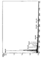
- das Massenspektrum des aus dem Gel der 2D-Gelelektrophorese gemäß Fig.1(B) isolierten, trypsinverdauten Produkts.
- Fig. 4
- das Massenspektrum des aus dem Gel der 2D-Gelelektrophorese gemäß Fig.2(B) isolierten, trypsinverdauten Produkts.
- Erwachsene Paviane (2-3 Jahre alt, 25-35 kg, männlich und weiblich) wurden mittels i.V. Gabe von Dolethal (10 ml) getötet. Der Dünndarm wurde innerhalb von 15 min entnommen, mit Wasser gewaschen, in Stücke von ca. 10 g zerteilt und unmittelbar in flüssigem Stickstoff eingefroren.
- Bei der weiteren Verarbeitung wurden Proben der einzelnen tiefgefrorenen Dünndarmgewebe unter Stickstoffkühlung in einem Porzellanmörser zu einem Mehl pulverisiert (vgl. J.Klose, "Fractionated Extraction of Total Tissue Proteins from Mouse and Human for 2-D Electrophoresis", in: Methods in Molecular Biology, Vol.112: 2-D Proteome Analysis Protocols, Humana Press Inc., Totowa, NJ). Alle weiteren Schritte erfolgten bei +4°C. Das Pulver wurde in 100 ml Puffer (50 mM Hepes, 50 mM NaCl, pH 8) aufgenommen und mittels eines 60-ml-Potters (Fa. Braun Melsungen) in 5 Auf-und-Ab-Bewegungen bei 900 U/min homogenisiert. Nach einer anschließenden 1-stündigen Zentrifugation bei 100.000 g wurde der erhaltene Überstand (Gewebsextrakt) gewonnen und für die weiteren Untersuchungen verwendet.
- Aus Seren von Patienten mit Morbus Crohn bzw. Colitis ulcerosa einerseits und von Gesunden andererseits (Kontrollseren) wurden mittels einer unspezifischen Affinitätsreinigung über Protein-G-Agarose die Immunglobuline isoliert. Zu diesem Zwecke wurden je 0,2 ml Serum mit 0,2 ml PBS gemischt und anschließend mit 0,5 ml Protein-G-Agarose (gepacktes Gel) versetzt. Die Mischung wird 30 min unter leichtem Schütteln inkubiert, und anschließend in eine Glassäule (Durchmesser 0,5 cm), die am unteren Ende mit einer Feinfritte verschlossen war, gefüllt. Die Säule wird zuerst mit PBS (5 ml) gewaschen, dann werden gebundene Immunglobuline mit 20 mM Citronensäure (pH ca. 2,5) eluiert (Flussgeschwindigkeit ca. 1 ml/min). Das Immunglobulin-Eluat wird durch Zusatz von TRIS-HCl, 1 M, pH 8,0 neutralisiert.
- Die gereinigte Immunglobulin-Fraktion wird anschließend unter Zusatz von Natrumperiodat (Endkonzentration 10 mg/ml) 20 min oxidiert. Das Natriumperiodat wird danach durch Entsalzen mittels einer NAP-5-Säule (Pharmacia) nach der Vorschrift des Herstellers entfernt, wobei in der Säule und als Elutionspuffer PBS verwendet wird.
- Die entsalzte oxidierte Immunglobulin-Fraktion wird mit Carbolink-Material (0,5 ml gepacktes Gel, gewaschen mit PBS, Fa. Pierce) vermengt. Nach einer 12 h Inkubation unter leichtem Schütteln wird das erhaltene, mit den jeweiligen Immunglobulin-Fraktionen belegte Affinitätsmaterial in eine Glassäule (Durchmesser 0,5 cm) mit Feinfritte gefüllt und mit 10 ml PBS gewaschen.
- Je 5 ml eines Pavian-Darmextrakts gemäß 1. werden bei einer Fliessgeschwindigkeit von 0,5 ml/min für eine Stunde kontinuierlich wiederholt über jeweils eine mit einer Immunglobulin-Fraktion gemäß 2. belegte Affinitätssäule gegeben. Anschließend werden die Säulen mit 5 ml PBS gewaschen. Der Ausfluss wird kontinuierlich auf Absorption bei 280 nm überwacht.
- Die an die Affinitätssäulen gebundenen Proteine werden danach mit 20 mM Citronensäure (pH ca. 2,5) eluiert. Die dabei erhaltenen Eluate werden anschließend mittel 2-D-Gelektrophorese analysiert.
- Bei der einleitenden analytischen 2D-Gelelektrophorese wurden die einzelnen Eluate mittels analytischer 2D-Gelelektrophorese aufgetrennt, wie in J.Klose, et al., "Two-dimensional electrophoresis of proteins: An updated protocol and implications for a functional analysis of the genome", Electrophoresis 1995, 16, 1034-1059; beschrieben ist. Die Sichtbarmachung der Proteine im 2D-Elektrophoresegel erfolgte mittels Silverstaining (vgl. J.Heukeshoven, et al., "Improved silver staining procedure for fast staining in Phast-System Development Unit. I. Staining of sodium dodecyl gels", Electrophoresis 1988, 9, 28-32). Die angefärbten Gele wurden mit einem Biorad 583 Geltrockner getrocknet.
- Zur Auswertung wurden das Proteinspotmuster eines Eluats aus der Affinitätssäule mit den Immunglobulinen Gesunder mit dem Proteinspotmuster verglichen, das aus einem entsprechenden Eluat aus der Affinitätssäule mit Immunglobulinen von MC- bzw. CU-Patienten resultierte. Proteinspots, die nie bei Gesunden, jedoch stets bei Patientenproben zusätzlich auftraten, wurden für weitere analytische Untersuchungen selektiert. Fig. 1 und Fig.2 zeigen Aufnahmen von 2D-Elektrophoresegelen, die einen Vergleich der Spotmuster der Eluate einer Affinitätssäule mit Immunglobulinen aus Seren von Gesunden (A) mit denen einer Affinitätssäule mit den Immunglobulinen aus Seren von MC- bzw. CU-Patienten (B) ermöglichen. Die umrandeten Bereiche zeigen die Positionen der nur aus den Patienten-Affinitätssäulen eluierbaren Proteine an.
- Überraschend wurden in den Eluaten aus der Patientensäule zwei neue Proteinspots bzw. -banden gefunden: Ein Spot bei einem Molekülgewicht von ca. 38 kD und einem pI von ca. 5,5, und eine scharfe Bande beim Molekülgewicht von ca. 36 kD, die allerdings in pI-Richtung als langezogener streifenartiger Spot erschien. Die beiden neuen Proteinspots traten bei allen Eluaten aus Patientensäulen auf, waren jedoch in den Eluaten aus den Säulen mit den Immunglobulinen Gesunder nicht sichtbar.
- Die im Proteinspotmuster der analytischen 2D-Gelelektrophorese identifizierten neuen spezifischen Proteine wurden dann anschließend mittels präparativer 2D-Gelelektrophorese präpariert (vgl. Klose, a.a.0.). Die Färbung erfolgte mittels Coomassie Brilliant Blue G250 (vgl. V.Neuhoff, et al., Electrophoresis 1988, 9, 255-262; sowie Electrophoresis 1990, 11, 101-117).
- Die für die weitere Analyse vorselektierten Proteinspots wurden aus dem Gel ausgeschnitten, unter Anwendung der Methode, die in Courchesne PL et al., In: Methods in Molecular Biology, Vol. 112:487-513 beschrieben ist, trypsinverdaut und anschließend massenspektroskopisch analysiert und zwar unter Anwendung massenspektrometrischer Untersuchungen, wie sie ebenfalls beschrieben und diskutiert werden in Courchesne PL, a.a.O.. Die bei der Massenspektroskopie (MALDI-MS) der beiden neuen Spots erhaltenen Fragment-Muster (Fig. 3 und 4) wurden dann mit den Datenbanken entnehmbaren Fragment-Mustern von bekannten Proteinen verglichen. Dazu wurden verschiedene Internet-Software-Programme verwendet. Zuerst wurden die durch MALDI-MS ermittelten Peptidmassen mit den theoretischen Peptidmassen aller bekannten Sägetierproteine gematcht. Für die Proteinsuche (in der Owl.7.2.2001-Datenbak mit dem Suchprogramm Protein Prospector) wurden monoisotopische Massen mit Massentoleranzen von 100 ppm (P0) bzw. 200 ppm (L5) zugelassen. Die weitere Suche wurde im Falle des Proteins aus Fig. 1B (P0) mit der NCBinr. Datenbank mit dem Suchprogramm ProFound, im Falle des Proteins aus Fig. 2B mit der Datenbank NCBI (2001/07/20) mit dem Suchprogramm ProFound (Spezies: Mammalia) und einer Massentoleranz von 200 ppm durchgeführt.
- Die beiden krankheitstypischen Proteine wurden eindeutig als die an sich bekannten ribosomalen Proteine P0 mit der Aminosäuresequenz gemäß SEQ ID NO:1 und L5 mit der Aminosäuresequenz gemäß SEQ ID NO:2 identifiziert.
- Indem man gemäß der obigen Prozedur einzelne Affinitatssäulen unter Verwendung von jeweils 10 Seren Gesunder (Kontrollen), 10 Seren von MC-Patienten und 10 Seren von CU-Patienten herstellte und mit dem o.g. Darmextrakt behandelte, wurden die in der folgenden Tabelle gezeigten Ergebnisse erhalten.
Tabelle Probe P0 L5 Kontrolle 1 - - Kontrolle 2 - - Kontrolle 3 - - Kontrolle 4 - - Kontrolle 5 - - Kontrolle 6 - - Kontrolle 7 - - Kontrolle 8 - - Kontrolle 9 - - Kontrolle 10 - - MC-Patient 1 + + MC-Patient 2 + + MC-Patient 3 + + MC-Patient 4 + + MC-Patient 5 + + MC-Patient 6 - + MC-Patient 7 + + MC-Patient 8 + - MC-Patient 9 + + MC-Patient 10 + + CU-Patient 1 + + CU-Patient 2 - + CU-Patient 3 + + CU-Patient 4 + + CU-Patient 5 - + CU-Patient 6 + + CU-Patient 7 + + CU-Patient 8 + * CU-Patient 9 + - CU-Patient 10 + + - Es ist der Tabelle zu entnehmen, dass bei den meisten MC- und CU-Patienten beide Arten von Antikörpern gleichzeitig zu finden sind, wenigstens eine Art von Antikörpern jedoch bei allen MC bzw. CU-Patienten. Dagegen werden derartige Antikörper in keinem Serum der gesunden Kontrollpersonen gefunden. Bei dem untersuchten relativ beschränkten Kollektiv von Testpersonen war somit die Selektivität 100%. Wenn man beide Antikörper bestimmt und den Nachweis einer Antikörperart als diagnostisch signifikant ansieht, betrug auch die Sensitivität 100%.
-
- 1. Thomas L., Labor und Diagnose, 5. Auflage, 1998, 824-842.
- 2. J.-C. Homberg, M. Rizzetto and Deborah Doniach, Ribosomal Antibodies Detected by Immunofluorescence in Systemic Lupus Erythematosus and other Collagenose, Clin.exp.Immunol. (1974) 17, 617-628.
- 3. J.B.Winfield, Are anti-ribosomal P protein antibodies a type of anti-lymphocyte antibody? Clin.Exp.Immunol 1997, 109, 1-3;
- 4. Stafford HA, Chen AE, Anderson CJ et al., Anti-ribosomal and 'P-peptide'-specific autoantibodies bind to T lymphocytes, Clin.Exp.Immunol 1997; 109, 12-19.
- 5. H.-H. Sun, W.-T. Liu, S.-Y. Tang, C.-Y. Tsai, S.-C. Hsieh, T.-H. Wu, S.-H. Han, The expression of acidic ribosomal phosphoproteins on the surface membrane of different tissues in autoimmune and normal mice which are the target molecules for anti-double-stranded DNA antibodies, Immunology 1996, 87, 362-371.
- 6. David T. Grabowski, Walter A. Deutsch, Dennis Derda and Mark R. Kelley, Drosophila AP3, a presumptive DNA repair protein, is homologous to human ribosomal associated protein P0, Nucleic Acids Resaerch, Vol.19, 4297, 1991
- 7. Nobuo Kondoh, Toru Wakatsuki, Akihide Ryo, Akiyuki Hada, Tsukasa Aihara, Sankichi Horiuchi, Narihide Goseki, Osamu Matsubara, Kenji Takenaka, Mizue Shichita, Kenji Tanaka, Masahiro Shuda, and Mikio Yamamoto, Indentification and Characterzation of Genes Associated with Human Hepatocellular Carcinogenesis, Cancer Res. 59, 4990-4996, 1999
- 8. M.A. Rodriguez-Gabriel, M. Remacha, and J.P.G. Ballesta, Phosphorylation of Ribosomal Protein P0 Is Not Essential for Ribosome Function but Can Affect Translation, Biochemistry 1998, 37, 16620-16626
- 9. G.F.Barnard, R.J. Staniunas, Shideng Bao, Ken-ichi Mafune, G.D. Steele Jr., J.L. Gollan, and Lan Bo Chen, Increased Expression of Human Ribosomal Phosphoprotein P0 Messenger RNA in Hepatocellular Carcinoma and Colon Carcinoma, Cancer Res. 52, 3067-3072, 1992
- 10. K.-H. Sun, W.-T. Liu, C.-Y. Tsai, S.-J. Tang, S.-H. Han, C.-L. Yu, Anti-dsDNA antibodies cross-react with ribosomal P proteins expressed on the surface of glomerular mesangial cells to exert a cytostatic effect, Immunology 1995, 85, 262-269
- 11. I. Ferrari, M.J. Levin, G. Wallukat, R. Elies, D. Lebesgue, P. Chiale, M. Elizari, M. Rosenbaum, and J. Hoebeke, Molecular Mimicry between the Immunodominant Ribosomal Protein P0 of Trypanosoma cruzi and a Functional Epitope on the Human β1-Adrenergic Receptor, J.Exp.Med., Vol. 182, 1995, 59-65
- 12. S. Chatterjee, S. Singh, R. Sohoni, V. Kattige, Ch. Deshpande, Sh. Chiplunkar, N. Kumar, Sh. Sharma, Characterization of domains of the phosphoriboprotein P0 of Plasmodium falciparum, Mol.Biochem.Parasitol. 107 (2000) 143-154
- 13. S. Chatterjee, S. Singh, R. Sohoni, N.J. Singh, A. Vaidya, C. Long, and Sh. Sharma, Antibodies against Ribosomal Phosphoprotein P0 of Plasmodium falciparum Protect Mice against Challenge with Plasmodium yoelli, Infect.Immun., Vol.68, 2000, 4312-4318
- 14. A. Goswami, S. Singh, V.D. Redkar, and Sh. Sharma, Characterization of P0, a Ribosmoal Phosphoprotein of Plasmodium falciparum, J.Biol.Chem. Vol.272, 12138-12143, 1997
- 15. N. Fabien et al., Autoantibodies Directed Against the Ribosomal P Proteins are not only Directed Against a Common Epitope of the P0, P1 and P2 Proteins, J.Autoimmun. (1999) 13, 103-110
- 16. B. Liliensiek at al., Identification of Four Genes in Endothelial Cells Whose Expression Is Affected by Tumor Cells and Host Immune Status - A Study in Ex Vivo-Isolated Endothelial Cells, Blood, Vol.92 (9), 1998, 3394-3404
- 17. R. Szyszka, G. Bou, J.P.G. Ballesta, RAP Kinase, a new enzyme phosphorylating the acidic P proteins from Saccharomyces cerevisiae, Biochim.Biophys.Acta 1293, (1996), 213-221
- 18. O. Rosorius et al., Human Ribosomal Protein L5 Contains Defined Nuclear Localization and Export Signals, J.Biol.Chem., Vol.275, 12061-12068, 2000
- 19. K.Hirano et al., Interaction of the Ribosomal Protein, L5, with Protein Phosphatase Type 1, J.Biol.Chem. Vol.270, 19786-19790, 1995
- 20. W.M. Michael and G. Dreyfuss, Distinct Domains in Ribosomal Protein L5 Mediate 5 S rRNA Binding and Nuclear Localization, J.Biol.Chem. Vol.271, 11571-11574, 1996
- 21. M. Claußen, F. Rudt, and T. Pieler, Functional Modules in Ribosomal Protein L5 for Ribonucleoprotein Complex Formation and Nucleocytoplasmic Transport, J.Biol.Chem. Vol.274, 33951-33958, 1999
- 22. A.Giualis et al., Anti-5S RNA/protein (RNP) antibody levels correlate with disease activity in a patient with systemic lupus erythematosus (SLE) nephritis, Clin.Exp.Immunol. 1994, 95, 385-389
- 23. V. Marechal et al., The Ribosomal L5 Protein Is Associated with mdm-2 and mdm-2-p53 Complexes, Mol.Cell.Biol. Vol. 14, 7414-7420, 1994
- 24. O.Spirina et al., Heart-specific splice-variant of a human mitochondrial ribosomal protein (mRNA processing; tissue specific splicing), Gene 261 (2000) 229-234
- 25. J.-M. Kim et al., Interaction of the β Subunit of Casein Kinase II with the Ribosomal Protein L5, BBRC 226, 180-186 (1996)
- 26. J.-M. Frigerio, J.-C. Dagorn, J.L. Iovanna, Cloning, sequencing and expression of the L5, L21, L27a, L28, S5, S9, S10 and S29 human ribosomal protein mRNAs, Biochim.Biophys.Acta 1262 (1995) 64-68
- 27. B. Guerra, O.-G. Issinger, p53 and the ribosomal protein L5 participate in high molecular mass complex formation with protein kinase CK2 in murine teratocarcinoma cell line F9 after serum stimulation and cis-platin treatment, FEBS Letters 434 (1998) 115-120
-
- <110> B.R.A.H.M.S Aktiengesellschaft
- <120> Verfahren und Mittel für die Diagnose und Therapie von chronischen entzündlichen Darmerkrankungen
- <130> 3618 PCT
- <140>
<141> - <150> 10147991.3 DE
<151> 2001-09-28 - <160> 2
- <170> PatentIn Ver. 2.1
- <210> 1
<211> 296
<212> PRT
<213> HOMO SAPIENS - <400> 1


- <210> 2
<211> 317
<212> PRT
<213> HOMO SAPIENS - <400> 2


Claims (8)
- Verfahren zur Diagnose, zur Frühdiagnose, zur Differentialdiagnose, zur Beurteilung der Schwere und zur therapiebegleitenden Verlaufskontrolle und Verlaufsprognose von chronisch entzündlichen Darmerkrankungen, dadurch gekennzeichnet, dass man im Serum, Plasma, Gewebeproben und/oder Stuhl eines Patienten, der an einer chronisch entzündlichen Darmerkrankung leidet, oder bei dem der Verdacht auf eine derartige Erkrankung besteht, das Vorhandensein und/oder die Menge von einem oder mehreren Antikörpern bestimmt, die an ribosomale Proteine binden.
- Verfahren nach Anspruch 1, bei dem die chronisch entzündliche Darmerkrankung ausgewählt ist aus Morbus Crohn und Colitis ulcerosa oder ihrer als Colitis indeterminata bezeichneten Mischform.
- Verfahren nach Anspruch 1, dadurch gekennzeichnet, dass man wenigstens einen Antikörper bestimmt, der aus der Gruppe von Antikörpern ausgewählt ist, die an das ribosomale Protein P0 oder an das ribosomale Protein L5 binden.
- Verfahren nach Anspruch 1, 2 oder 3, dadurch gekennzeichnet, dass man den wenigstens einen Antikörper mit Hilfe eines Immunoassays bestimmt, bei dem das jeweilige ribosomale Protein als Antigen zur Bindung und/oder Markierung der zu bestimmenden Antikörper verwendet wird.
- Verfahren nach Anspruch 4, dadurch gekennzeichnet, dass das ribosomale Protein ein humanes oder tierisches Protein ist, das aus natürlichen Quellen angereichert oder isoliert wurde oder auf gentechnologischem Wege (rekombinant) erzeugt wurde.
- Verwendung des ribosomalen Proteins P0 und/oder des ribosomalen Proteins L5 zur Herstellung eines Arzneimittels zur Bindung oder Blockierung von Antikörpern, die an das ribosomale Protein P0 binden, und/oder von Antikörpern, die an das ribosomale Protein L5 binden, für die Therapie des Morbus Crohn und/oder der Colitis ulcerosa.
- Verwendung des ribosomalen Proteins P0 und/oder des ribosomalen Proteins L5 zur Herstellung eines Arzneimittels zur Hemmung der Darmentzündung und assoziierter Entzündungen bei Morbus Crohn und/oder der Colitis ulcerosa durch Blockierung antigenpräsentierender Zellen oder zur Erzeugung einer T-Zellanergie.
- Verwendung des ribosomalen Proteins P0 und/oder des ribosomalen Proteins L5 zur Herstellung eines Affinitätsmaterials zur extrakorporalen Entfernung von Antikörpern, die an das ribosomale Protein P0 binden, und/oder von Antikörpern, die an das ribosomale Protein L5 binden, aus dem Blutkreislauf eines Patienten.
Applications Claiming Priority (3)
| Application Number | Priority Date | Filing Date | Title |
|---|---|---|---|
| DE10147991 | 2001-09-28 | ||
| DE10147991A DE10147991B4 (de) | 2001-09-28 | 2001-09-28 | Verfahren zur Diagnose von chronisch entzündlichen Darmerkrankungen |
| PCT/EP2002/009848 WO2003029824A1 (de) | 2001-09-28 | 2002-09-03 | Verfahren und mittel zur diagnose und therapie von chronisch entzündlichen darmerkrankungen |
Publications (2)
| Publication Number | Publication Date |
|---|---|
| EP1430304A1 EP1430304A1 (de) | 2004-06-23 |
| EP1430304B1 true EP1430304B1 (de) | 2006-12-06 |
Family
ID=7700713
Family Applications (1)
| Application Number | Title | Priority Date | Filing Date |
|---|---|---|---|
| EP02774558A Expired - Lifetime EP1430304B1 (de) | 2001-09-28 | 2002-09-03 | Verfahren zur diagnose von chronisch entzündlichen darmerkrankungen |
Country Status (6)
| Country | Link |
|---|---|
| US (1) | US7358058B2 (de) |
| EP (1) | EP1430304B1 (de) |
| JP (1) | JP4359506B2 (de) |
| AT (1) | ATE347696T1 (de) |
| DE (2) | DE10147991B4 (de) |
| WO (1) | WO2003029824A1 (de) |
Families Citing this family (5)
| Publication number | Priority date | Publication date | Assignee | Title |
|---|---|---|---|---|
| JP4625708B2 (ja) * | 2005-03-17 | 2011-02-02 | 株式会社バイオマーカーサイエンス | 炎症性腸疾患の検出方法 |
| JP4973885B2 (ja) * | 2006-05-19 | 2012-07-11 | 国立大学法人大阪大学 | 炎症性腸疾患の鑑別判断方法 |
| RU2331887C1 (ru) * | 2007-01-09 | 2008-08-20 | Государственное учреждение Научный центр здоровья детей Российской академии медицинских наук | Способ прогнозирования эффективности физиотерапевтических методов лечения у детей с хроническими запорами |
| DE102008063046A1 (de) * | 2008-12-23 | 2010-06-24 | Justus-Liebig-Universität Giessen | Medizinische Verwendung des ribosomalen Protein S19 (RPS19) |
| WO2015067913A1 (en) | 2013-11-07 | 2015-05-14 | Diagnodus Limited | Biomarkers |
Family Cites Families (3)
| Publication number | Priority date | Publication date | Assignee | Title |
|---|---|---|---|---|
| US4865970A (en) * | 1986-02-28 | 1989-09-12 | Hoffmann-La Roche Inc. | Method of detecting ribosomal protein antibodies in systemic lupus erythematosus |
| US6897287B1 (en) * | 1990-01-31 | 2005-05-24 | Oklahoma Medical Research Foundation | Ro/SSA peptide reagents for diagnosis of autoantibodies |
| US5691151A (en) * | 1994-10-07 | 1997-11-25 | Regents Of University Of California | Methods of screening for ulcerative colitis and crohn's disease by detecting VH3-15 autoantibody and panca |
-
2001
- 2001-09-28 DE DE10147991A patent/DE10147991B4/de not_active Expired - Fee Related
-
2002
- 2002-09-03 DE DE50208908T patent/DE50208908D1/de not_active Expired - Lifetime
- 2002-09-03 EP EP02774558A patent/EP1430304B1/de not_active Expired - Lifetime
- 2002-09-03 US US10/490,597 patent/US7358058B2/en not_active Expired - Fee Related
- 2002-09-03 AT AT02774558T patent/ATE347696T1/de not_active IP Right Cessation
- 2002-09-03 WO PCT/EP2002/009848 patent/WO2003029824A1/de not_active Ceased
- 2002-09-03 JP JP2003532986A patent/JP4359506B2/ja not_active Expired - Fee Related
Also Published As
| Publication number | Publication date |
|---|---|
| US7358058B2 (en) | 2008-04-15 |
| US20050074818A1 (en) | 2005-04-07 |
| DE10147991B4 (de) | 2004-07-15 |
| JP4359506B2 (ja) | 2009-11-04 |
| EP1430304A1 (de) | 2004-06-23 |
| ATE347696T1 (de) | 2006-12-15 |
| DE50208908D1 (de) | 2007-01-18 |
| DE10147991A1 (de) | 2003-05-15 |
| WO2003029824A1 (de) | 2003-04-10 |
| JP2005504321A (ja) | 2005-02-10 |
Similar Documents
| Publication | Publication Date | Title |
|---|---|---|
| Le Quintrec et al. | Contactin-1 is a novel target antigen in membranous nephropathy associated with chronic inflammatory demyelinating polyneuropathy | |
| DE60028040T2 (de) | Annexin-proteine und auto-antikörper als serummarker für lungenkrebs und ösophaguskrebs | |
| DE69737870T2 (de) | Medikamente für die diagnostik von autoimmunerkrankungen | |
| DE60312513T2 (de) | Zusammensetzungen und verfahren zum nachweis vom apo-b48 und apo-b100 | |
| DE69333985T2 (de) | Allergene proteine und peptide von hundhautanhangsgebilden und ihre verwendung | |
| DE69117949T2 (de) | Mit akuter pankreatitis assoziiertes protein, mittel zur diagnose von akuter pankreatitis | |
| EP1399476B1 (de) | Verwendung löslicher cytokeratin-1-fragmente in diagnostik | |
| WO2003048780A2 (de) | Verwendungen der aldose-1-epimerase für die diagnose und therapie von entzündungserkrangungen und sepsis | |
| JP2592121B2 (ja) | IgA腎臓病の診断のための方法及びキツト | |
| DE102005029845A1 (de) | Verfahren zur Diagnose von rheumatischen Erkrankungen | |
| US5780298A (en) | DNA encoding the sn-RNP-A antigen and fragments thereof | |
| DE69733957T2 (de) | Verfahren zur bestimmung der gegenwart des gehirnspezifischen proteins s-100 beta | |
| Saito et al. | Marked increases in concentrations of apolipoprotein in the cerebrospinal fluid of poliovirus-infected macaques: relations between apolipoprotein concentrations and severity of brain injury | |
| DE10147991B4 (de) | Verfahren zur Diagnose von chronisch entzündlichen Darmerkrankungen | |
| EP0585960A2 (de) | Diagnostisches Verfahren für Autoimmunkrankenheiten | |
| EP1493034B1 (de) | Verfahren zur diagnose von entzündungserkrankungen und infektionen mittels bestimmung von lasp-1/ lap-1 | |
| JPH05501204A (ja) | 糸球体腎炎の診断 | |
| DE60020499T2 (de) | Endometriose-assoziierte-Autoantigene kodierendes Polynukleotid | |
| EP0532979A2 (de) | Petide zur Herstellung von Mitteln zur Diagnose und Therapie von systemischem Lupus | |
| US4871661A (en) | Process for testing the carcinogenicity of a material or the presence of cancer-inducing factors in an environment | |
| DE3915344A1 (de) | Serumdiagnose-verfahren | |
| JPS62500304A (ja) | 膵臓がんに伴う胞状奇胎分化蛋白質、これらの蛋白質に対する抗血清およびモノクロ−ナル抗体、製造方法 | |
| US5989829A (en) | Autoantibodies and their targets in the diagnosis of peripheral neuropathies | |
| DE60216482T2 (de) | Zusammensetzungen und verfahren zum bestimmen von aa4rp | |
| EP1149914A2 (de) | Fusionsprotein zur gleichzeitigen Erkennung von humanen Autoantikörpern |
Legal Events
| Date | Code | Title | Description |
|---|---|---|---|
| PUAI | Public reference made under article 153(3) epc to a published international application that has entered the european phase |
Free format text: ORIGINAL CODE: 0009012 |
|
| 17P | Request for examination filed |
Effective date: 20040312 |
|
| AK | Designated contracting states |
Kind code of ref document: A1 Designated state(s): AT BE BG CH CY CZ DE DK EE ES FI FR GB GR IE IT LI LU MC NL PT SE SK TR |
|
| RTI1 | Title (correction) |
Free format text: METHOD FOR DIAGNOSING CHRONIC INFLAMMATORY BOWEL DISEASES |
|
| GRAP | Despatch of communication of intention to grant a patent |
Free format text: ORIGINAL CODE: EPIDOSNIGR1 |
|
| GRAS | Grant fee paid |
Free format text: ORIGINAL CODE: EPIDOSNIGR3 |
|
| GRAA | (expected) grant |
Free format text: ORIGINAL CODE: 0009210 |
|
| AK | Designated contracting states |
Kind code of ref document: B1 Designated state(s): AT BE BG CH CY CZ DE DK EE ES FI FR GB GR IE IT LI LU MC NL PT SE SK TR |
|
| PG25 | Lapsed in a contracting state [announced via postgrant information from national office to epo] |
Ref country code: IT Free format text: LAPSE BECAUSE OF FAILURE TO SUBMIT A TRANSLATION OF THE DESCRIPTION OR TO PAY THE FEE WITHIN THE PRESCRIBED TIME-LIMIT;WARNING: LAPSES OF ITALIAN PATENTS WITH EFFECTIVE DATE BEFORE 2007 MAY HAVE OCCURRED AT ANY TIME BEFORE 2007. THE CORRECT EFFECTIVE DATE MAY BE DIFFERENT FROM THE ONE RECORDED. Effective date: 20061206 Ref country code: NL Free format text: LAPSE BECAUSE OF FAILURE TO SUBMIT A TRANSLATION OF THE DESCRIPTION OR TO PAY THE FEE WITHIN THE PRESCRIBED TIME-LIMIT Effective date: 20061206 Ref country code: FI Free format text: LAPSE BECAUSE OF FAILURE TO SUBMIT A TRANSLATION OF THE DESCRIPTION OR TO PAY THE FEE WITHIN THE PRESCRIBED TIME-LIMIT Effective date: 20061206 Ref country code: CZ Free format text: LAPSE BECAUSE OF FAILURE TO SUBMIT A TRANSLATION OF THE DESCRIPTION OR TO PAY THE FEE WITHIN THE PRESCRIBED TIME-LIMIT Effective date: 20061206 Ref country code: SK Free format text: LAPSE BECAUSE OF FAILURE TO SUBMIT A TRANSLATION OF THE DESCRIPTION OR TO PAY THE FEE WITHIN THE PRESCRIBED TIME-LIMIT Effective date: 20061206 Ref country code: IE Free format text: LAPSE BECAUSE OF FAILURE TO SUBMIT A TRANSLATION OF THE DESCRIPTION OR TO PAY THE FEE WITHIN THE PRESCRIBED TIME-LIMIT Effective date: 20061206 Ref country code: DK Free format text: LAPSE BECAUSE OF FAILURE TO SUBMIT A TRANSLATION OF THE DESCRIPTION OR TO PAY THE FEE WITHIN THE PRESCRIBED TIME-LIMIT Effective date: 20061206 |
|
| REG | Reference to a national code |
Ref country code: GB Ref legal event code: FG4D Free format text: NOT ENGLISH |
|
| REG | Reference to a national code |
Ref country code: CH Ref legal event code: EP |
|
| REG | Reference to a national code |
Ref country code: IE Ref legal event code: FG4D Free format text: LANGUAGE OF EP DOCUMENT: GERMAN |
|
| REG | Reference to a national code |
Ref country code: CH Ref legal event code: NV Representative=s name: PATENTANWALTSBUREAU REBMANN-KUPFER & CO. |
|
| REF | Corresponds to: |
Ref document number: 50208908 Country of ref document: DE Date of ref document: 20070118 Kind code of ref document: P |
|
| PG25 | Lapsed in a contracting state [announced via postgrant information from national office to epo] |
Ref country code: BG Free format text: LAPSE BECAUSE OF FAILURE TO SUBMIT A TRANSLATION OF THE DESCRIPTION OR TO PAY THE FEE WITHIN THE PRESCRIBED TIME-LIMIT Effective date: 20070306 Ref country code: SE Free format text: LAPSE BECAUSE OF FAILURE TO SUBMIT A TRANSLATION OF THE DESCRIPTION OR TO PAY THE FEE WITHIN THE PRESCRIBED TIME-LIMIT Effective date: 20070306 |
|
| PG25 | Lapsed in a contracting state [announced via postgrant information from national office to epo] |
Ref country code: ES Free format text: LAPSE BECAUSE OF FAILURE TO SUBMIT A TRANSLATION OF THE DESCRIPTION OR TO PAY THE FEE WITHIN THE PRESCRIBED TIME-LIMIT Effective date: 20070317 |
|
| PG25 | Lapsed in a contracting state [announced via postgrant information from national office to epo] |
Ref country code: PT Free format text: LAPSE BECAUSE OF FAILURE TO SUBMIT A TRANSLATION OF THE DESCRIPTION OR TO PAY THE FEE WITHIN THE PRESCRIBED TIME-LIMIT Effective date: 20070507 |
|
| ET | Fr: translation filed | ||
| NLV1 | Nl: lapsed or annulled due to failure to fulfill the requirements of art. 29p and 29m of the patents act | ||
| GBV | Gb: ep patent (uk) treated as always having been void in accordance with gb section 77(7)/1977 [no translation filed] |
Effective date: 20061206 |
|
| PLBE | No opposition filed within time limit |
Free format text: ORIGINAL CODE: 0009261 |
|
| STAA | Information on the status of an ep patent application or granted ep patent |
Free format text: STATUS: NO OPPOSITION FILED WITHIN TIME LIMIT |
|
| 26N | No opposition filed |
Effective date: 20070907 |
|
| PG25 | Lapsed in a contracting state [announced via postgrant information from national office to epo] |
Ref country code: GB Free format text: LAPSE BECAUSE OF FAILURE TO SUBMIT A TRANSLATION OF THE DESCRIPTION OR TO PAY THE FEE WITHIN THE PRESCRIBED TIME-LIMIT Effective date: 20061206 |
|
| PG25 | Lapsed in a contracting state [announced via postgrant information from national office to epo] |
Ref country code: MC Free format text: LAPSE BECAUSE OF NON-PAYMENT OF DUE FEES Effective date: 20070930 Ref country code: GR Free format text: LAPSE BECAUSE OF FAILURE TO SUBMIT A TRANSLATION OF THE DESCRIPTION OR TO PAY THE FEE WITHIN THE PRESCRIBED TIME-LIMIT Effective date: 20070307 |
|
| PG25 | Lapsed in a contracting state [announced via postgrant information from national office to epo] |
Ref country code: EE Free format text: LAPSE BECAUSE OF FAILURE TO SUBMIT A TRANSLATION OF THE DESCRIPTION OR TO PAY THE FEE WITHIN THE PRESCRIBED TIME-LIMIT Effective date: 20061206 |
|
| PG25 | Lapsed in a contracting state [announced via postgrant information from national office to epo] |
Ref country code: CY Free format text: LAPSE BECAUSE OF FAILURE TO SUBMIT A TRANSLATION OF THE DESCRIPTION OR TO PAY THE FEE WITHIN THE PRESCRIBED TIME-LIMIT Effective date: 20061206 Ref country code: LU Free format text: LAPSE BECAUSE OF NON-PAYMENT OF DUE FEES Effective date: 20070903 |
|
| PG25 | Lapsed in a contracting state [announced via postgrant information from national office to epo] |
Ref country code: TR Free format text: LAPSE BECAUSE OF FAILURE TO SUBMIT A TRANSLATION OF THE DESCRIPTION OR TO PAY THE FEE WITHIN THE PRESCRIBED TIME-LIMIT Effective date: 20061206 |
|
| PGFP | Annual fee paid to national office [announced via postgrant information from national office to epo] |
Ref country code: AT Payment date: 20090916 Year of fee payment: 8 Ref country code: CH Payment date: 20090923 Year of fee payment: 8 |
|
| PGFP | Annual fee paid to national office [announced via postgrant information from national office to epo] |
Ref country code: DE Payment date: 20091023 Year of fee payment: 8 |
|
| PGFP | Annual fee paid to national office [announced via postgrant information from national office to epo] |
Ref country code: BE Payment date: 20091023 Year of fee payment: 8 |
|
| BERE | Be: lapsed |
Owner name: B.R.A.H.M.S A.G. Effective date: 20100930 |
|
| REG | Reference to a national code |
Ref country code: CH Ref legal event code: PL |
|
| REG | Reference to a national code |
Ref country code: FR Ref legal event code: ST Effective date: 20110531 |
|
| REG | Reference to a national code |
Ref country code: DE Ref legal event code: R119 Ref document number: 50208908 Country of ref document: DE Effective date: 20110401 |
|
| PG25 | Lapsed in a contracting state [announced via postgrant information from national office to epo] |
Ref country code: BE Free format text: LAPSE BECAUSE OF NON-PAYMENT OF DUE FEES Effective date: 20100930 Ref country code: FR Free format text: LAPSE BECAUSE OF NON-PAYMENT OF DUE FEES Effective date: 20100930 Ref country code: DE Free format text: LAPSE BECAUSE OF NON-PAYMENT OF DUE FEES Effective date: 20110401 Ref country code: LI Free format text: LAPSE BECAUSE OF NON-PAYMENT OF DUE FEES Effective date: 20100930 Ref country code: CH Free format text: LAPSE BECAUSE OF NON-PAYMENT OF DUE FEES Effective date: 20100930 |
|
| PG25 | Lapsed in a contracting state [announced via postgrant information from national office to epo] |
Ref country code: AT Free format text: LAPSE BECAUSE OF NON-PAYMENT OF DUE FEES Effective date: 20100903 |
|
| PGFP | Annual fee paid to national office [announced via postgrant information from national office to epo] |
Ref country code: FR Payment date: 20091001 Year of fee payment: 8 |