EP1430140B1 - Isoformes tronquees a l'extremite n-terminale de phosphodiesterases cycliques pde3a. - Google Patents
Isoformes tronquees a l'extremite n-terminale de phosphodiesterases cycliques pde3a. Download PDFInfo
- Publication number
- EP1430140B1 EP1430140B1 EP02739923A EP02739923A EP1430140B1 EP 1430140 B1 EP1430140 B1 EP 1430140B1 EP 02739923 A EP02739923 A EP 02739923A EP 02739923 A EP02739923 A EP 02739923A EP 1430140 B1 EP1430140 B1 EP 1430140B1
- Authority
- EP
- European Patent Office
- Prior art keywords
- pde3
- pde3a
- protein
- seq
- sequence
- Prior art date
- Legal status (The legal status is an assumption and is not a legal conclusion. Google has not performed a legal analysis and makes no representation as to the accuracy of the status listed.)
- Expired - Lifetime
Links
- 108010029485 Protein Isoforms Proteins 0.000 title claims abstract description 179
- 102000001708 Protein Isoforms Human genes 0.000 title claims abstract description 179
- 101710111837 Cyclic phosphodiesterase Proteins 0.000 title 1
- 108090000765 processed proteins & peptides Proteins 0.000 claims abstract description 162
- 238000000034 method Methods 0.000 claims abstract description 142
- 102000004196 processed proteins & peptides Human genes 0.000 claims abstract description 117
- 102000004861 Phosphoric Diester Hydrolases Human genes 0.000 claims abstract description 112
- 108090001050 Phosphoric Diester Hydrolases Proteins 0.000 claims abstract description 112
- 239000012190 activator Substances 0.000 claims abstract description 47
- 239000013604 expression vector Substances 0.000 claims abstract description 23
- 229940124639 Selective inhibitor Drugs 0.000 claims abstract description 9
- 102000001253 Protein Kinase Human genes 0.000 claims description 62
- 108060006633 protein kinase Proteins 0.000 claims description 62
- 150000007523 nucleic acids Chemical class 0.000 claims description 60
- 230000027455 binding Effects 0.000 claims description 58
- 229920001184 polypeptide Polymers 0.000 claims description 55
- 102000039446 nucleic acids Human genes 0.000 claims description 51
- 108020004707 nucleic acids Proteins 0.000 claims description 51
- 229940024606 amino acid Drugs 0.000 claims description 44
- 235000001014 amino acid Nutrition 0.000 claims description 43
- 150000001413 amino acids Chemical class 0.000 claims description 42
- 150000001875 compounds Chemical class 0.000 claims description 42
- 230000003197 catalytic effect Effects 0.000 claims description 35
- 230000005764 inhibitory process Effects 0.000 claims description 29
- 230000035772 mutation Effects 0.000 claims description 29
- 238000012360 testing method Methods 0.000 claims description 26
- 230000004913 activation Effects 0.000 claims description 21
- 125000003275 alpha amino acid group Chemical group 0.000 claims description 21
- 238000002823 phage display Methods 0.000 claims description 10
- 238000006467 substitution reaction Methods 0.000 claims description 9
- 125000003607 serino group Chemical group [H]N([H])[C@]([H])(C(=O)[*])C(O[H])([H])[H] 0.000 claims description 7
- QNAYBMKLOCPYGJ-REOHCLBHSA-N L-alanine Chemical compound C[C@H](N)C(O)=O QNAYBMKLOCPYGJ-REOHCLBHSA-N 0.000 claims description 6
- 102000004160 Phosphoric Monoester Hydrolases Human genes 0.000 claims description 6
- 108090000608 Phosphoric Monoester Hydrolases Proteins 0.000 claims description 6
- 235000004279 alanine Nutrition 0.000 claims description 6
- CKLJMWTZIZZHCS-REOHCLBHSA-L aspartate group Chemical group N[C@@H](CC(=O)[O-])C(=O)[O-] CKLJMWTZIZZHCS-REOHCLBHSA-L 0.000 claims description 2
- 102000009097 Phosphorylases Human genes 0.000 claims 1
- 108010073135 Phosphorylases Proteins 0.000 claims 1
- 230000000694 effects Effects 0.000 abstract description 108
- 239000003112 inhibitor Substances 0.000 abstract description 42
- 239000000203 mixture Substances 0.000 abstract description 38
- 230000003834 intracellular effect Effects 0.000 abstract description 28
- 201000010046 Dilated cardiomyopathy Diseases 0.000 abstract description 21
- 206010056370 Congestive cardiomyopathy Diseases 0.000 abstract description 20
- 208000002815 pulmonary hypertension Diseases 0.000 abstract description 10
- 101001098818 Homo sapiens cGMP-inhibited 3',5'-cyclic phosphodiesterase A Proteins 0.000 abstract 2
- 102100037093 cGMP-inhibited 3',5'-cyclic phosphodiesterase A Human genes 0.000 abstract 2
- 101001098806 Dictyostelium discoideum cGMP-specific 3',5'-cGMP phosphodiesterase 3 Proteins 0.000 description 230
- 108090000623 proteins and genes Proteins 0.000 description 230
- 102000004169 proteins and genes Human genes 0.000 description 160
- 235000018102 proteins Nutrition 0.000 description 147
- 210000004027 cell Anatomy 0.000 description 132
- 230000026731 phosphorylation Effects 0.000 description 97
- 238000006366 phosphorylation reaction Methods 0.000 description 97
- 108020004999 messenger RNA Proteins 0.000 description 95
- 239000000523 sample Substances 0.000 description 66
- 108020004414 DNA Proteins 0.000 description 62
- 230000014509 gene expression Effects 0.000 description 60
- 108091032973 (ribonucleotides)n+m Proteins 0.000 description 58
- ZOOGRGPOEVQQDX-UUOKFMHZSA-N 3',5'-cyclic GMP Chemical compound C([C@H]1O2)OP(O)(=O)O[C@H]1[C@@H](O)[C@@H]2N1C(N=C(NC2=O)N)=C2N=C1 ZOOGRGPOEVQQDX-UUOKFMHZSA-N 0.000 description 51
- 241000282414 Homo sapiens Species 0.000 description 51
- ZOOGRGPOEVQQDX-UHFFFAOYSA-N cyclic GMP Natural products O1C2COP(O)(=O)OC2C(O)C1N1C=NC2=C1NC(N)=NC2=O ZOOGRGPOEVQQDX-UHFFFAOYSA-N 0.000 description 51
- 108020004635 Complementary DNA Proteins 0.000 description 45
- 230000003321 amplification Effects 0.000 description 44
- 210000000107 myocyte Anatomy 0.000 description 44
- 238000003199 nucleic acid amplification method Methods 0.000 description 44
- 239000002299 complementary DNA Substances 0.000 description 41
- 238000010804 cDNA synthesis Methods 0.000 description 40
- 230000000747 cardiac effect Effects 0.000 description 40
- 108700026244 Open Reading Frames Proteins 0.000 description 39
- 230000003993 interaction Effects 0.000 description 39
- 230000001086 cytosolic effect Effects 0.000 description 37
- 230000003228 microsomal effect Effects 0.000 description 37
- 239000002773 nucleotide Substances 0.000 description 37
- 239000000047 product Substances 0.000 description 37
- 238000013518 transcription Methods 0.000 description 37
- 230000035897 transcription Effects 0.000 description 37
- 125000003729 nucleotide group Chemical group 0.000 description 34
- 210000004165 myocardium Anatomy 0.000 description 33
- 238000013519 translation Methods 0.000 description 33
- 230000002792 vascular Effects 0.000 description 33
- 239000012528 membrane Substances 0.000 description 31
- 239000013598 vector Substances 0.000 description 31
- 108090000994 Catalytic RNA Proteins 0.000 description 29
- 102000053642 Catalytic RNA Human genes 0.000 description 29
- 239000012634 fragment Substances 0.000 description 29
- 108091092562 ribozyme Proteins 0.000 description 29
- 239000000758 substrate Substances 0.000 description 29
- 230000000692 anti-sense effect Effects 0.000 description 28
- 108091034117 Oligonucleotide Proteins 0.000 description 26
- 238000003556 assay Methods 0.000 description 25
- 230000003301 hydrolyzing effect Effects 0.000 description 25
- 230000000295 complement effect Effects 0.000 description 24
- 102000004190 Enzymes Human genes 0.000 description 23
- 108090000790 Enzymes Proteins 0.000 description 23
- 241001465754 Metazoa Species 0.000 description 23
- 210000004413 cardiac myocyte Anatomy 0.000 description 23
- 238000001514 detection method Methods 0.000 description 23
- 229940088598 enzyme Drugs 0.000 description 23
- 101150009274 nhr-1 gene Proteins 0.000 description 23
- 230000006870 function Effects 0.000 description 22
- 239000006166 lysate Substances 0.000 description 22
- 241000894007 species Species 0.000 description 22
- 210000001519 tissue Anatomy 0.000 description 21
- 229940123263 Phosphodiesterase 3 inhibitor Drugs 0.000 description 20
- 239000003550 marker Substances 0.000 description 20
- 239000002570 phosphodiesterase III inhibitor Substances 0.000 description 20
- 238000002360 preparation method Methods 0.000 description 20
- 101100294102 Caenorhabditis elegans nhr-2 gene Proteins 0.000 description 19
- 108020004459 Small interfering RNA Proteins 0.000 description 19
- 239000003795 chemical substances by application Substances 0.000 description 19
- 238000013459 approach Methods 0.000 description 18
- 239000013612 plasmid Substances 0.000 description 18
- 230000001105 regulatory effect Effects 0.000 description 18
- 238000001262 western blot Methods 0.000 description 18
- CKLJMWTZIZZHCS-REOHCLBHSA-N L-aspartic acid Chemical compound OC(=O)[C@@H](N)CC(O)=O CKLJMWTZIZZHCS-REOHCLBHSA-N 0.000 description 17
- 238000000746 purification Methods 0.000 description 17
- 239000002585 base Substances 0.000 description 16
- 238000004587 chromatography analysis Methods 0.000 description 16
- 230000001413 cellular effect Effects 0.000 description 15
- 230000030570 cellular localization Effects 0.000 description 15
- 238000001727 in vivo Methods 0.000 description 15
- NOESYZHRGYRDHS-UHFFFAOYSA-N insulin Chemical compound N1C(=O)C(NC(=O)C(CCC(N)=O)NC(=O)C(CCC(O)=O)NC(=O)C(C(C)C)NC(=O)C(NC(=O)CN)C(C)CC)CSSCC(C(NC(CO)C(=O)NC(CC(C)C)C(=O)NC(CC=2C=CC(O)=CC=2)C(=O)NC(CCC(N)=O)C(=O)NC(CC(C)C)C(=O)NC(CCC(O)=O)C(=O)NC(CC(N)=O)C(=O)NC(CC=2C=CC(O)=CC=2)C(=O)NC(CSSCC(NC(=O)C(C(C)C)NC(=O)C(CC(C)C)NC(=O)C(CC=2C=CC(O)=CC=2)NC(=O)C(CC(C)C)NC(=O)C(C)NC(=O)C(CCC(O)=O)NC(=O)C(C(C)C)NC(=O)C(CC(C)C)NC(=O)C(CC=2NC=NC=2)NC(=O)C(CO)NC(=O)CNC2=O)C(=O)NCC(=O)NC(CCC(O)=O)C(=O)NC(CCCNC(N)=N)C(=O)NCC(=O)NC(CC=3C=CC=CC=3)C(=O)NC(CC=3C=CC=CC=3)C(=O)NC(CC=3C=CC(O)=CC=3)C(=O)NC(C(C)O)C(=O)N3C(CCC3)C(=O)NC(CCCCN)C(=O)NC(C)C(O)=O)C(=O)NC(CC(N)=O)C(O)=O)=O)NC(=O)C(C(C)CC)NC(=O)C(CO)NC(=O)C(C(C)O)NC(=O)C1CSSCC2NC(=O)C(CC(C)C)NC(=O)C(NC(=O)C(CCC(N)=O)NC(=O)C(CC(N)=O)NC(=O)C(NC(=O)C(N)CC=1C=CC=CC=1)C(C)C)CC1=CN=CN1 NOESYZHRGYRDHS-UHFFFAOYSA-N 0.000 description 15
- 230000004044 response Effects 0.000 description 15
- 230000008685 targeting Effects 0.000 description 15
- 238000011144 upstream manufacturing Methods 0.000 description 15
- 102000053602 DNA Human genes 0.000 description 14
- 229960003574 milrinone Drugs 0.000 description 14
- PZRHRDRVRGEVNW-UHFFFAOYSA-N milrinone Chemical compound N1C(=O)C(C#N)=CC(C=2C=CN=CC=2)=C1C PZRHRDRVRGEVNW-UHFFFAOYSA-N 0.000 description 14
- -1 poly(vinyl chloride) Polymers 0.000 description 14
- 238000000926 separation method Methods 0.000 description 14
- 239000000243 solution Substances 0.000 description 14
- 238000006243 chemical reaction Methods 0.000 description 13
- 238000003776 cleavage reaction Methods 0.000 description 13
- 238000000338 in vitro Methods 0.000 description 13
- 230000007017 scission Effects 0.000 description 13
- 241000700605 Viruses Species 0.000 description 12
- 230000033228 biological regulation Effects 0.000 description 12
- 230000015572 biosynthetic process Effects 0.000 description 12
- 238000001962 electrophoresis Methods 0.000 description 12
- 238000004519 manufacturing process Methods 0.000 description 12
- 230000008569 process Effects 0.000 description 12
- 241000829100 Macaca mulatta polyomavirus 1 Species 0.000 description 11
- 230000008901 benefit Effects 0.000 description 11
- 238000012217 deletion Methods 0.000 description 11
- 230000037430 deletion Effects 0.000 description 11
- 238000005516 engineering process Methods 0.000 description 11
- 238000013537 high throughput screening Methods 0.000 description 11
- 230000007246 mechanism Effects 0.000 description 11
- 238000002741 site-directed mutagenesis Methods 0.000 description 11
- 108020004705 Codon Proteins 0.000 description 10
- 241000588724 Escherichia coli Species 0.000 description 10
- 108091028043 Nucleic acid sequence Proteins 0.000 description 10
- 239000003814 drug Substances 0.000 description 10
- 102000040430 polynucleotide Human genes 0.000 description 10
- 108091033319 polynucleotide Proteins 0.000 description 10
- 239000002157 polynucleotide Substances 0.000 description 10
- 102000005962 receptors Human genes 0.000 description 10
- 108020003175 receptors Proteins 0.000 description 10
- 238000002415 sodium dodecyl sulfate polyacrylamide gel electrophoresis Methods 0.000 description 10
- 102000005636 Cyclic AMP Response Element-Binding Protein Human genes 0.000 description 9
- 108010045171 Cyclic AMP Response Element-Binding Protein Proteins 0.000 description 9
- PEDCQBHIVMGVHV-UHFFFAOYSA-N Glycerine Chemical compound OCC(O)CO PEDCQBHIVMGVHV-UHFFFAOYSA-N 0.000 description 9
- 239000000020 Nitrocellulose Substances 0.000 description 9
- 238000001042 affinity chromatography Methods 0.000 description 9
- 239000000872 buffer Substances 0.000 description 9
- 125000004122 cyclic group Chemical group 0.000 description 9
- 239000000499 gel Substances 0.000 description 9
- 230000007062 hydrolysis Effects 0.000 description 9
- 238000006460 hydrolysis reaction Methods 0.000 description 9
- 230000001965 increasing effect Effects 0.000 description 9
- 230000002401 inhibitory effect Effects 0.000 description 9
- 239000007788 liquid Substances 0.000 description 9
- 230000007774 longterm Effects 0.000 description 9
- 230000003278 mimic effect Effects 0.000 description 9
- 229920001220 nitrocellulos Polymers 0.000 description 9
- 150000003839 salts Chemical class 0.000 description 9
- 238000012216 screening Methods 0.000 description 9
- 230000035945 sensitivity Effects 0.000 description 9
- 239000007787 solid Substances 0.000 description 9
- 230000001225 therapeutic effect Effects 0.000 description 9
- 238000001890 transfection Methods 0.000 description 9
- 230000003612 virological effect Effects 0.000 description 9
- 102000040650 (ribonucleotides)n+m Human genes 0.000 description 8
- 238000002965 ELISA Methods 0.000 description 8
- 102000007056 Recombinant Fusion Proteins Human genes 0.000 description 8
- 108010008281 Recombinant Fusion Proteins Proteins 0.000 description 8
- 108091081024 Start codon Proteins 0.000 description 8
- 230000001419 dependent effect Effects 0.000 description 8
- 230000004927 fusion Effects 0.000 description 8
- 229940125396 insulin Drugs 0.000 description 8
- 239000011159 matrix material Substances 0.000 description 8
- 230000002829 reductive effect Effects 0.000 description 8
- 210000001768 subcellular fraction Anatomy 0.000 description 8
- 241000701447 unidentified baculovirus Species 0.000 description 8
- 208000031229 Cardiomyopathies Diseases 0.000 description 7
- DHMQDGOQFOQNFH-UHFFFAOYSA-N Glycine Chemical compound NCC(O)=O DHMQDGOQFOQNFH-UHFFFAOYSA-N 0.000 description 7
- 102000004877 Insulin Human genes 0.000 description 7
- 102100023915 Insulin Human genes 0.000 description 7
- 108090001061 Insulin Proteins 0.000 description 7
- 238000000636 Northern blotting Methods 0.000 description 7
- 102000007079 Peptide Fragments Human genes 0.000 description 7
- 108010033276 Peptide Fragments Proteins 0.000 description 7
- FAPWRFPIFSIZLT-UHFFFAOYSA-M Sodium chloride Chemical compound [Na+].[Cl-] FAPWRFPIFSIZLT-UHFFFAOYSA-M 0.000 description 7
- 238000002105 Southern blotting Methods 0.000 description 7
- 210000001789 adipocyte Anatomy 0.000 description 7
- 210000004899 c-terminal region Anatomy 0.000 description 7
- 238000000749 co-immunoprecipitation Methods 0.000 description 7
- 230000007423 decrease Effects 0.000 description 7
- 229940079593 drug Drugs 0.000 description 7
- 239000011521 glass Substances 0.000 description 7
- 238000002744 homologous recombination Methods 0.000 description 7
- 230000006801 homologous recombination Effects 0.000 description 7
- 230000002163 immunogen Effects 0.000 description 7
- 210000004020 intracellular membrane Anatomy 0.000 description 7
- 239000003446 ligand Substances 0.000 description 7
- 238000013507 mapping Methods 0.000 description 7
- 230000002107 myocardial effect Effects 0.000 description 7
- 102000005681 phospholamban Human genes 0.000 description 7
- 108010059929 phospholamban Proteins 0.000 description 7
- 238000006722 reduction reaction Methods 0.000 description 7
- 230000000638 stimulation Effects 0.000 description 7
- 238000002198 surface plasmon resonance spectroscopy Methods 0.000 description 7
- 238000012546 transfer Methods 0.000 description 7
- YBJHBAHKTGYVGT-ZKWXMUAHSA-N (+)-Biotin Chemical compound N1C(=O)N[C@@H]2[C@H](CCCCC(=O)O)SC[C@@H]21 YBJHBAHKTGYVGT-ZKWXMUAHSA-N 0.000 description 6
- 108020005544 Antisense RNA Proteins 0.000 description 6
- AGPKZVBTJJNPAG-WHFBIAKZSA-N L-isoleucine Chemical compound CC[C@H](C)[C@H](N)C(O)=O AGPKZVBTJJNPAG-WHFBIAKZSA-N 0.000 description 6
- ROHFNLRQFUQHCH-YFKPBYRVSA-N L-leucine Chemical compound CC(C)C[C@H](N)C(O)=O ROHFNLRQFUQHCH-YFKPBYRVSA-N 0.000 description 6
- QIVBCDIJIAJPQS-VIFPVBQESA-N L-tryptophane Chemical compound C1=CC=C2C(C[C@H](N)C(O)=O)=CNC2=C1 QIVBCDIJIAJPQS-VIFPVBQESA-N 0.000 description 6
- ROHFNLRQFUQHCH-UHFFFAOYSA-N Leucine Natural products CC(C)CC(N)C(O)=O ROHFNLRQFUQHCH-UHFFFAOYSA-N 0.000 description 6
- 239000004677 Nylon Substances 0.000 description 6
- 241000283973 Oryctolagus cuniculus Species 0.000 description 6
- 102000035195 Peptidases Human genes 0.000 description 6
- 108091005804 Peptidases Proteins 0.000 description 6
- 108091000080 Phosphotransferase Proteins 0.000 description 6
- 206010035226 Plasma cell myeloma Diseases 0.000 description 6
- QIVBCDIJIAJPQS-UHFFFAOYSA-N Tryptophan Natural products C1=CC=C2C(CC(N)C(O)=O)=CNC2=C1 QIVBCDIJIAJPQS-UHFFFAOYSA-N 0.000 description 6
- ISAKRJDGNUQOIC-UHFFFAOYSA-N Uracil Chemical compound O=C1C=CNC(=O)N1 ISAKRJDGNUQOIC-UHFFFAOYSA-N 0.000 description 6
- JLCPHMBAVCMARE-UHFFFAOYSA-N [3-[[3-[[3-[[3-[[3-[[3-[[3-[[3-[[3-[[3-[[3-[[5-(2-amino-6-oxo-1H-purin-9-yl)-3-[[3-[[3-[[3-[[3-[[3-[[5-(2-amino-6-oxo-1H-purin-9-yl)-3-[[5-(2-amino-6-oxo-1H-purin-9-yl)-3-hydroxyoxolan-2-yl]methoxy-hydroxyphosphoryl]oxyoxolan-2-yl]methoxy-hydroxyphosphoryl]oxy-5-(5-methyl-2,4-dioxopyrimidin-1-yl)oxolan-2-yl]methoxy-hydroxyphosphoryl]oxy-5-(6-aminopurin-9-yl)oxolan-2-yl]methoxy-hydroxyphosphoryl]oxy-5-(6-aminopurin-9-yl)oxolan-2-yl]methoxy-hydroxyphosphoryl]oxy-5-(6-aminopurin-9-yl)oxolan-2-yl]methoxy-hydroxyphosphoryl]oxy-5-(6-aminopurin-9-yl)oxolan-2-yl]methoxy-hydroxyphosphoryl]oxyoxolan-2-yl]methoxy-hydroxyphosphoryl]oxy-5-(5-methyl-2,4-dioxopyrimidin-1-yl)oxolan-2-yl]methoxy-hydroxyphosphoryl]oxy-5-(4-amino-2-oxopyrimidin-1-yl)oxolan-2-yl]methoxy-hydroxyphosphoryl]oxy-5-(5-methyl-2,4-dioxopyrimidin-1-yl)oxolan-2-yl]methoxy-hydroxyphosphoryl]oxy-5-(5-methyl-2,4-dioxopyrimidin-1-yl)oxolan-2-yl]methoxy-hydroxyphosphoryl]oxy-5-(6-aminopurin-9-yl)oxolan-2-yl]methoxy-hydroxyphosphoryl]oxy-5-(6-aminopurin-9-yl)oxolan-2-yl]methoxy-hydroxyphosphoryl]oxy-5-(4-amino-2-oxopyrimidin-1-yl)oxolan-2-yl]methoxy-hydroxyphosphoryl]oxy-5-(4-amino-2-oxopyrimidin-1-yl)oxolan-2-yl]methoxy-hydroxyphosphoryl]oxy-5-(4-amino-2-oxopyrimidin-1-yl)oxolan-2-yl]methoxy-hydroxyphosphoryl]oxy-5-(6-aminopurin-9-yl)oxolan-2-yl]methoxy-hydroxyphosphoryl]oxy-5-(4-amino-2-oxopyrimidin-1-yl)oxolan-2-yl]methyl [5-(6-aminopurin-9-yl)-2-(hydroxymethyl)oxolan-3-yl] hydrogen phosphate Polymers Cc1cn(C2CC(OP(O)(=O)OCC3OC(CC3OP(O)(=O)OCC3OC(CC3O)n3cnc4c3nc(N)[nH]c4=O)n3cnc4c3nc(N)[nH]c4=O)C(COP(O)(=O)OC3CC(OC3COP(O)(=O)OC3CC(OC3COP(O)(=O)OC3CC(OC3COP(O)(=O)OC3CC(OC3COP(O)(=O)OC3CC(OC3COP(O)(=O)OC3CC(OC3COP(O)(=O)OC3CC(OC3COP(O)(=O)OC3CC(OC3COP(O)(=O)OC3CC(OC3COP(O)(=O)OC3CC(OC3COP(O)(=O)OC3CC(OC3COP(O)(=O)OC3CC(OC3COP(O)(=O)OC3CC(OC3COP(O)(=O)OC3CC(OC3COP(O)(=O)OC3CC(OC3COP(O)(=O)OC3CC(OC3COP(O)(=O)OC3CC(OC3CO)n3cnc4c(N)ncnc34)n3ccc(N)nc3=O)n3cnc4c(N)ncnc34)n3ccc(N)nc3=O)n3ccc(N)nc3=O)n3ccc(N)nc3=O)n3cnc4c(N)ncnc34)n3cnc4c(N)ncnc34)n3cc(C)c(=O)[nH]c3=O)n3cc(C)c(=O)[nH]c3=O)n3ccc(N)nc3=O)n3cc(C)c(=O)[nH]c3=O)n3cnc4c3nc(N)[nH]c4=O)n3cnc4c(N)ncnc34)n3cnc4c(N)ncnc34)n3cnc4c(N)ncnc34)n3cnc4c(N)ncnc34)O2)c(=O)[nH]c1=O JLCPHMBAVCMARE-UHFFFAOYSA-N 0.000 description 6
- 210000000172 cytosol Anatomy 0.000 description 6
- 238000002474 experimental method Methods 0.000 description 6
- ZDXPYRJPNDTMRX-UHFFFAOYSA-N glutamine Natural products OC(=O)C(N)CCC(N)=O ZDXPYRJPNDTMRX-UHFFFAOYSA-N 0.000 description 6
- 238000009396 hybridization Methods 0.000 description 6
- 210000004408 hybridoma Anatomy 0.000 description 6
- FDGQSTZJBFJUBT-UHFFFAOYSA-N hypoxanthine Chemical compound O=C1NC=NC2=C1NC=N2 FDGQSTZJBFJUBT-UHFFFAOYSA-N 0.000 description 6
- 230000003053 immunization Effects 0.000 description 6
- 229960000310 isoleucine Drugs 0.000 description 6
- AGPKZVBTJJNPAG-UHFFFAOYSA-N isoleucine Natural products CCC(C)C(N)C(O)=O AGPKZVBTJJNPAG-UHFFFAOYSA-N 0.000 description 6
- 230000001404 mediated effect Effects 0.000 description 6
- 239000002609 medium Substances 0.000 description 6
- 230000004048 modification Effects 0.000 description 6
- 238000012986 modification Methods 0.000 description 6
- 201000000050 myeloid neoplasm Diseases 0.000 description 6
- 229920001778 nylon Polymers 0.000 description 6
- 102000020233 phosphotransferase Human genes 0.000 description 6
- 229920001223 polyethylene glycol Polymers 0.000 description 6
- 238000012545 processing Methods 0.000 description 6
- 210000001147 pulmonary artery Anatomy 0.000 description 6
- 230000009467 reduction Effects 0.000 description 6
- 230000010076 replication Effects 0.000 description 6
- 238000010561 standard procedure Methods 0.000 description 6
- 238000003786 synthesis reaction Methods 0.000 description 6
- 230000002861 ventricular Effects 0.000 description 6
- DCXYFEDJOCDNAF-UHFFFAOYSA-N Asparagine Natural products OC(=O)C(N)CC(N)=O DCXYFEDJOCDNAF-UHFFFAOYSA-N 0.000 description 5
- 108091026890 Coding region Proteins 0.000 description 5
- 108090000626 DNA-directed RNA polymerases Proteins 0.000 description 5
- 102000004163 DNA-directed RNA polymerases Human genes 0.000 description 5
- 108700024394 Exon Proteins 0.000 description 5
- 102100034343 Integrase Human genes 0.000 description 5
- DCXYFEDJOCDNAF-REOHCLBHSA-N L-asparagine Chemical compound OC(=O)[C@@H](N)CC(N)=O DCXYFEDJOCDNAF-REOHCLBHSA-N 0.000 description 5
- KZSNJWFQEVHDMF-BYPYZUCNSA-N L-valine Chemical compound CC(C)[C@H](N)C(O)=O KZSNJWFQEVHDMF-BYPYZUCNSA-N 0.000 description 5
- 101000909851 Mycobacterium tuberculosis (strain ATCC 25618 / H37Rv) cAMP/cGMP dual specificity phosphodiesterase Rv0805 Proteins 0.000 description 5
- 239000002202 Polyethylene glycol Substances 0.000 description 5
- 108091008611 Protein Kinase B Proteins 0.000 description 5
- MTCFGRXMJLQNBG-UHFFFAOYSA-N Serine Natural products OCC(N)C(O)=O MTCFGRXMJLQNBG-UHFFFAOYSA-N 0.000 description 5
- KZSNJWFQEVHDMF-UHFFFAOYSA-N Valine Natural products CC(C)C(N)C(O)=O KZSNJWFQEVHDMF-UHFFFAOYSA-N 0.000 description 5
- 230000009471 action Effects 0.000 description 5
- 239000002671 adjuvant Substances 0.000 description 5
- 238000004458 analytical method Methods 0.000 description 5
- 229960001230 asparagine Drugs 0.000 description 5
- 235000009582 asparagine Nutrition 0.000 description 5
- 238000000376 autoradiography Methods 0.000 description 5
- 102000012740 beta Adrenergic Receptors Human genes 0.000 description 5
- 108010079452 beta Adrenergic Receptors Proteins 0.000 description 5
- 230000000903 blocking effect Effects 0.000 description 5
- 230000015556 catabolic process Effects 0.000 description 5
- 239000003153 chemical reaction reagent Substances 0.000 description 5
- 230000002860 competitive effect Effects 0.000 description 5
- 230000030609 dephosphorylation Effects 0.000 description 5
- 238000006209 dephosphorylation reaction Methods 0.000 description 5
- 230000029087 digestion Effects 0.000 description 5
- 201000010099 disease Diseases 0.000 description 5
- 208000037265 diseases, disorders, signs and symptoms Diseases 0.000 description 5
- 239000006185 dispersion Substances 0.000 description 5
- 102000037865 fusion proteins Human genes 0.000 description 5
- 108020001507 fusion proteins Proteins 0.000 description 5
- 210000002216 heart Anatomy 0.000 description 5
- HNDVDQJCIGZPNO-UHFFFAOYSA-N histidine Natural products OC(=O)C(N)CC1=CN=CN1 HNDVDQJCIGZPNO-UHFFFAOYSA-N 0.000 description 5
- 230000000977 initiatory effect Effects 0.000 description 5
- 230000002452 interceptive effect Effects 0.000 description 5
- 239000000463 material Substances 0.000 description 5
- 210000001589 microsome Anatomy 0.000 description 5
- PHEDXBVPIONUQT-RGYGYFBISA-N phorbol 13-acetate 12-myristate Chemical compound C([C@]1(O)C(=O)C(C)=C[C@H]1[C@@]1(O)[C@H](C)[C@H]2OC(=O)CCCCCCCCCCCCC)C(CO)=C[C@H]1[C@H]1[C@]2(OC(C)=O)C1(C)C PHEDXBVPIONUQT-RGYGYFBISA-N 0.000 description 5
- 239000002644 phorbol ester Substances 0.000 description 5
- 230000006798 recombination Effects 0.000 description 5
- 238000005215 recombination Methods 0.000 description 5
- 239000006152 selective media Substances 0.000 description 5
- 210000000952 spleen Anatomy 0.000 description 5
- 230000002103 transcriptional effect Effects 0.000 description 5
- 239000004474 valine Substances 0.000 description 5
- GMVPRGQOIOIIMI-UHFFFAOYSA-N (8R,11R,12R,13E,15S)-11,15-Dihydroxy-9-oxo-13-prostenoic acid Natural products CCCCCC(O)C=CC1C(O)CC(=O)C1CCCCCCC(O)=O GMVPRGQOIOIIMI-UHFFFAOYSA-N 0.000 description 4
- LMDZBCPBFSXMTL-UHFFFAOYSA-N 1-ethyl-3-(3-dimethylaminopropyl)carbodiimide Chemical compound CCN=C=NCCCN(C)C LMDZBCPBFSXMTL-UHFFFAOYSA-N 0.000 description 4
- 101150044980 Akap1 gene Proteins 0.000 description 4
- 239000004475 Arginine Substances 0.000 description 4
- IJGRMHOSHXDMSA-UHFFFAOYSA-N Atomic nitrogen Chemical compound N#N IJGRMHOSHXDMSA-UHFFFAOYSA-N 0.000 description 4
- 102000014914 Carrier Proteins Human genes 0.000 description 4
- 102000008130 Cyclic AMP-Dependent Protein Kinases Human genes 0.000 description 4
- 108010049894 Cyclic AMP-Dependent Protein Kinases Proteins 0.000 description 4
- 102000004654 Cyclic GMP-Dependent Protein Kinases Human genes 0.000 description 4
- 108010003591 Cyclic GMP-Dependent Protein Kinases Proteins 0.000 description 4
- 102100031620 Cysteine and glycine-rich protein 3 Human genes 0.000 description 4
- 241000701022 Cytomegalovirus Species 0.000 description 4
- LFQSCWFLJHTTHZ-UHFFFAOYSA-N Ethanol Chemical compound CCO LFQSCWFLJHTTHZ-UHFFFAOYSA-N 0.000 description 4
- 101710203526 Integrase Proteins 0.000 description 4
- WHUUTDBJXJRKMK-VKHMYHEASA-N L-glutamic acid Chemical compound OC(=O)[C@@H](N)CCC(O)=O WHUUTDBJXJRKMK-VKHMYHEASA-N 0.000 description 4
- FFEARJCKVFRZRR-BYPYZUCNSA-N L-methionine Chemical compound CSCC[C@H](N)C(O)=O FFEARJCKVFRZRR-BYPYZUCNSA-N 0.000 description 4
- COLNVLDHVKWLRT-QMMMGPOBSA-N L-phenylalanine Chemical compound OC(=O)[C@@H](N)CC1=CC=CC=C1 COLNVLDHVKWLRT-QMMMGPOBSA-N 0.000 description 4
- OUYCCCASQSFEME-QMMMGPOBSA-N L-tyrosine Chemical compound OC(=O)[C@@H](N)CC1=CC=C(O)C=C1 OUYCCCASQSFEME-QMMMGPOBSA-N 0.000 description 4
- KDXKERNSBIXSRK-UHFFFAOYSA-N Lysine Natural products NCCCCC(N)C(O)=O KDXKERNSBIXSRK-UHFFFAOYSA-N 0.000 description 4
- 239000004472 Lysine Substances 0.000 description 4
- 108091054455 MAP kinase family Proteins 0.000 description 4
- 102000043136 MAP kinase family Human genes 0.000 description 4
- 241000699666 Mus <mouse, genus> Species 0.000 description 4
- 108020004711 Nucleic Acid Probes Proteins 0.000 description 4
- ISWSIDIOOBJBQZ-UHFFFAOYSA-N Phenol Chemical compound OC1=CC=CC=C1 ISWSIDIOOBJBQZ-UHFFFAOYSA-N 0.000 description 4
- 101100082610 Plasmodium falciparum (isolate 3D7) PDEdelta gene Proteins 0.000 description 4
- 101710182846 Polyhedrin Proteins 0.000 description 4
- 239000004793 Polystyrene Substances 0.000 description 4
- 240000004808 Saccharomyces cerevisiae Species 0.000 description 4
- 230000002411 adverse Effects 0.000 description 4
- 239000011543 agarose gel Substances 0.000 description 4
- 229960000711 alprostadil Drugs 0.000 description 4
- 238000004873 anchoring Methods 0.000 description 4
- 239000005557 antagonist Substances 0.000 description 4
- 230000002424 anti-apoptotic effect Effects 0.000 description 4
- 239000000427 antigen Substances 0.000 description 4
- 108091007433 antigens Proteins 0.000 description 4
- 102000036639 antigens Human genes 0.000 description 4
- ODKSFYDXXFIFQN-UHFFFAOYSA-N arginine Natural products OC(=O)C(N)CCCNC(N)=N ODKSFYDXXFIFQN-UHFFFAOYSA-N 0.000 description 4
- 229940009098 aspartate Drugs 0.000 description 4
- 210000003719 b-lymphocyte Anatomy 0.000 description 4
- 230000009286 beneficial effect Effects 0.000 description 4
- 238000004132 cross linking Methods 0.000 description 4
- 239000003431 cross linking reagent Substances 0.000 description 4
- 230000001351 cycling effect Effects 0.000 description 4
- 108010023942 cysteine and glycine-rich protein 3 Proteins 0.000 description 4
- OPTASPLRGRRNAP-UHFFFAOYSA-N cytosine Chemical compound NC=1C=CNC(=O)N=1 OPTASPLRGRRNAP-UHFFFAOYSA-N 0.000 description 4
- 238000013461 design Methods 0.000 description 4
- 238000009826 distribution Methods 0.000 description 4
- 238000009472 formulation Methods 0.000 description 4
- 125000000524 functional group Chemical group 0.000 description 4
- 238000001502 gel electrophoresis Methods 0.000 description 4
- 230000002068 genetic effect Effects 0.000 description 4
- 229930195712 glutamate Natural products 0.000 description 4
- 230000012010 growth Effects 0.000 description 4
- 238000004128 high performance liquid chromatography Methods 0.000 description 4
- 230000003100 immobilizing effect Effects 0.000 description 4
- 238000010166 immunofluorescence Methods 0.000 description 4
- 238000003364 immunohistochemistry Methods 0.000 description 4
- 238000001114 immunoprecipitation Methods 0.000 description 4
- 238000003780 insertion Methods 0.000 description 4
- 230000037431 insertion Effects 0.000 description 4
- 238000007918 intramuscular administration Methods 0.000 description 4
- 238000007912 intraperitoneal administration Methods 0.000 description 4
- 210000004962 mammalian cell Anatomy 0.000 description 4
- 244000005700 microbiome Species 0.000 description 4
- 239000002853 nucleic acid probe Substances 0.000 description 4
- 230000007310 pathophysiology Effects 0.000 description 4
- COLNVLDHVKWLRT-UHFFFAOYSA-N phenylalanine Natural products OC(=O)C(N)CC1=CC=CC=C1 COLNVLDHVKWLRT-UHFFFAOYSA-N 0.000 description 4
- YBYRMVIVWMBXKQ-UHFFFAOYSA-N phenylmethanesulfonyl fluoride Chemical compound FS(=O)(=O)CC1=CC=CC=C1 YBYRMVIVWMBXKQ-UHFFFAOYSA-N 0.000 description 4
- 238000002264 polyacrylamide gel electrophoresis Methods 0.000 description 4
- 230000008488 polyadenylation Effects 0.000 description 4
- 229920002223 polystyrene Polymers 0.000 description 4
- 229920002981 polyvinylidene fluoride Polymers 0.000 description 4
- 230000017854 proteolysis Effects 0.000 description 4
- 210000002966 serum Anatomy 0.000 description 4
- 239000011780 sodium chloride Substances 0.000 description 4
- 238000007920 subcutaneous administration Methods 0.000 description 4
- 239000000126 substance Substances 0.000 description 4
- 239000006228 supernatant Substances 0.000 description 4
- 230000004083 survival effect Effects 0.000 description 4
- OUYCCCASQSFEME-UHFFFAOYSA-N tyrosine Natural products OC(=O)C(N)CC1=CC=C(O)C=C1 OUYCCCASQSFEME-UHFFFAOYSA-N 0.000 description 4
- 241000701161 unidentified adenovirus Species 0.000 description 4
- 238000005406 washing Methods 0.000 description 4
- XLYOFNOQVPJJNP-UHFFFAOYSA-N water Substances O XLYOFNOQVPJJNP-UHFFFAOYSA-N 0.000 description 4
- 108700020469 14-3-3 Proteins 0.000 description 3
- 102000004899 14-3-3 Proteins Human genes 0.000 description 3
- 102100033811 A-kinase anchor protein 11 Human genes 0.000 description 3
- 229930024421 Adenine Natural products 0.000 description 3
- GFFGJBXGBJISGV-UHFFFAOYSA-N Adenine Chemical compound NC1=NC=NC2=C1N=CN2 GFFGJBXGBJISGV-UHFFFAOYSA-N 0.000 description 3
- 108010092778 Autophagy-Related Protein 7 Proteins 0.000 description 3
- 241000894006 Bacteria Species 0.000 description 3
- 101710132601 Capsid protein Proteins 0.000 description 3
- 101710094648 Coat protein Proteins 0.000 description 3
- 239000003298 DNA probe Substances 0.000 description 3
- 101100296720 Dictyostelium discoideum Pde4 gene Proteins 0.000 description 3
- WSFSSNUMVMOOMR-UHFFFAOYSA-N Formaldehyde Chemical compound O=C WSFSSNUMVMOOMR-UHFFFAOYSA-N 0.000 description 3
- 102100021181 Golgi phosphoprotein 3 Human genes 0.000 description 3
- UGQMRVRMYYASKQ-UHFFFAOYSA-N Hypoxanthine nucleoside Natural products OC1C(O)C(CO)OC1N1C(NC=NC2=O)=C2N=C1 UGQMRVRMYYASKQ-UHFFFAOYSA-N 0.000 description 3
- 108090000723 Insulin-Like Growth Factor I Proteins 0.000 description 3
- ZDXPYRJPNDTMRX-VKHMYHEASA-N L-glutamine Chemical compound OC(=O)[C@@H](N)CCC(N)=O ZDXPYRJPNDTMRX-VKHMYHEASA-N 0.000 description 3
- FBOZXECLQNJBKD-ZDUSSCGKSA-N L-methotrexate Chemical compound C=1N=C2N=C(N)N=C(N)C2=NC=1CN(C)C1=CC=C(C(=O)N[C@@H](CCC(O)=O)C(O)=O)C=C1 FBOZXECLQNJBKD-ZDUSSCGKSA-N 0.000 description 3
- 108091026898 Leader sequence (mRNA) Proteins 0.000 description 3
- 102000003960 Ligases Human genes 0.000 description 3
- 108090000364 Ligases Proteins 0.000 description 3
- 101710125418 Major capsid protein Proteins 0.000 description 3
- 108010052285 Membrane Proteins Proteins 0.000 description 3
- 101710141454 Nucleoprotein Proteins 0.000 description 3
- 102000039033 PDE3 family Human genes 0.000 description 3
- 108091065683 PDE3 family Proteins 0.000 description 3
- 101710083689 Probable capsid protein Proteins 0.000 description 3
- DNIAPMSPPWPWGF-UHFFFAOYSA-N Propylene glycol Chemical compound CC(O)CO DNIAPMSPPWPWGF-UHFFFAOYSA-N 0.000 description 3
- 239000004365 Protease Substances 0.000 description 3
- 230000007022 RNA scission Effects 0.000 description 3
- 241000700159 Rattus Species 0.000 description 3
- 108020004511 Recombinant DNA Proteins 0.000 description 3
- 102000006382 Ribonucleases Human genes 0.000 description 3
- 108010083644 Ribonucleases Proteins 0.000 description 3
- 108020004682 Single-Stranded DNA Proteins 0.000 description 3
- 241000256251 Spodoptera frugiperda Species 0.000 description 3
- AYFVYJQAPQTCCC-UHFFFAOYSA-N Threonine Natural products CC(O)C(N)C(O)=O AYFVYJQAPQTCCC-UHFFFAOYSA-N 0.000 description 3
- 239000004473 Threonine Substances 0.000 description 3
- IQFYYKKMVGJFEH-XLPZGREQSA-N Thymidine Chemical compound O=C1NC(=O)C(C)=CN1[C@@H]1O[C@H](CO)[C@@H](O)C1 IQFYYKKMVGJFEH-XLPZGREQSA-N 0.000 description 3
- 102100022979 Ubiquitin-like modifier-activating enzyme ATG7 Human genes 0.000 description 3
- 238000010521 absorption reaction Methods 0.000 description 3
- 230000003213 activating effect Effects 0.000 description 3
- 239000004480 active ingredient Substances 0.000 description 3
- 229960000643 adenine Drugs 0.000 description 3
- 239000000556 agonist Substances 0.000 description 3
- 125000000539 amino acid group Chemical group 0.000 description 3
- 229960000723 ampicillin Drugs 0.000 description 3
- 238000000137 annealing Methods 0.000 description 3
- 230000003302 anti-idiotype Effects 0.000 description 3
- 239000007864 aqueous solution Substances 0.000 description 3
- 230000003126 arrythmogenic effect Effects 0.000 description 3
- 230000001580 bacterial effect Effects 0.000 description 3
- 108091008324 binding proteins Proteins 0.000 description 3
- 230000004071 biological effect Effects 0.000 description 3
- 239000012472 biological sample Substances 0.000 description 3
- 229960002685 biotin Drugs 0.000 description 3
- 235000020958 biotin Nutrition 0.000 description 3
- 239000011616 biotin Substances 0.000 description 3
- 210000004369 blood Anatomy 0.000 description 3
- 239000008280 blood Substances 0.000 description 3
- 150000001718 carbodiimides Chemical class 0.000 description 3
- 238000012512 characterization method Methods 0.000 description 3
- 238000010367 cloning Methods 0.000 description 3
- 238000000576 coating method Methods 0.000 description 3
- 239000003184 complementary RNA Substances 0.000 description 3
- 238000010276 construction Methods 0.000 description 3
- 230000008602 contraction Effects 0.000 description 3
- 210000004748 cultured cell Anatomy 0.000 description 3
- 238000012258 culturing Methods 0.000 description 3
- 235000018417 cysteine Nutrition 0.000 description 3
- XUJNEKJLAYXESH-UHFFFAOYSA-N cysteine Natural products SCC(N)C(O)=O XUJNEKJLAYXESH-UHFFFAOYSA-N 0.000 description 3
- 239000003599 detergent Substances 0.000 description 3
- 238000010790 dilution Methods 0.000 description 3
- 239000012895 dilution Substances 0.000 description 3
- 239000002612 dispersion medium Substances 0.000 description 3
- 238000009510 drug design Methods 0.000 description 3
- 239000003623 enhancer Substances 0.000 description 3
- 230000001747 exhibiting effect Effects 0.000 description 3
- 210000005260 human cell Anatomy 0.000 description 3
- 230000028993 immune response Effects 0.000 description 3
- 238000002649 immunization Methods 0.000 description 3
- 238000003018 immunoassay Methods 0.000 description 3
- 230000005847 immunogenicity Effects 0.000 description 3
- 230000001976 improved effect Effects 0.000 description 3
- 230000001939 inductive effect Effects 0.000 description 3
- 239000004615 ingredient Substances 0.000 description 3
- 238000001990 intravenous administration Methods 0.000 description 3
- 238000004255 ion exchange chromatography Methods 0.000 description 3
- 238000002372 labelling Methods 0.000 description 3
- 230000004060 metabolic process Effects 0.000 description 3
- 229930182817 methionine Natural products 0.000 description 3
- 229960000485 methotrexate Drugs 0.000 description 3
- 230000005012 migration Effects 0.000 description 3
- 238000013508 migration Methods 0.000 description 3
- 230000010807 negative regulation of binding Effects 0.000 description 3
- 210000000056 organ Anatomy 0.000 description 3
- 230000036961 partial effect Effects 0.000 description 3
- 239000002245 particle Substances 0.000 description 3
- 229920003023 plastic Polymers 0.000 description 3
- 239000004033 plastic Substances 0.000 description 3
- 230000003389 potentiating effect Effects 0.000 description 3
- 239000000843 powder Substances 0.000 description 3
- 230000009822 protein phosphorylation Effects 0.000 description 3
- 238000001742 protein purification Methods 0.000 description 3
- 238000003127 radioimmunoassay Methods 0.000 description 3
- 230000009257 reactivity Effects 0.000 description 3
- 230000022532 regulation of transcription, DNA-dependent Effects 0.000 description 3
- 230000008439 repair process Effects 0.000 description 3
- 238000012163 sequencing technique Methods 0.000 description 3
- 239000004055 small Interfering RNA Substances 0.000 description 3
- 150000003384 small molecules Chemical class 0.000 description 3
- 239000002904 solvent Substances 0.000 description 3
- 238000003153 stable transfection Methods 0.000 description 3
- KNXVOGGZOFOROK-UHFFFAOYSA-N trimagnesium;dioxido(oxo)silane;hydroxy-oxido-oxosilane Chemical compound [Mg+2].[Mg+2].[Mg+2].O[Si]([O-])=O.O[Si]([O-])=O.[O-][Si]([O-])=O.[O-][Si]([O-])=O KNXVOGGZOFOROK-UHFFFAOYSA-N 0.000 description 3
- 229940035893 uracil Drugs 0.000 description 3
- AGNGYMCLFWQVGX-AGFFZDDWSA-N (e)-1-[(2s)-2-amino-2-carboxyethoxy]-2-diazonioethenolate Chemical compound OC(=O)[C@@H](N)CO\C([O-])=C\[N+]#N AGNGYMCLFWQVGX-AGFFZDDWSA-N 0.000 description 2
- OSJPPGNTCRNQQC-UWTATZPHSA-N 3-phospho-D-glyceric acid Chemical compound OC(=O)[C@H](O)COP(O)(O)=O OSJPPGNTCRNQQC-UWTATZPHSA-N 0.000 description 2
- SJECZPVISLOESU-UHFFFAOYSA-N 3-trimethoxysilylpropan-1-amine Chemical compound CO[Si](OC)(OC)CCCN SJECZPVISLOESU-UHFFFAOYSA-N 0.000 description 2
- TVZGACDUOSZQKY-LBPRGKRZSA-N 4-aminofolic acid Chemical compound C1=NC2=NC(N)=NC(N)=C2N=C1CNC1=CC=C(C(=O)N[C@@H](CCC(O)=O)C(O)=O)C=C1 TVZGACDUOSZQKY-LBPRGKRZSA-N 0.000 description 2
- 108020003589 5' Untranslated Regions Proteins 0.000 description 2
- 101150034092 ATG4 gene Proteins 0.000 description 2
- 229920000936 Agarose Polymers 0.000 description 2
- 102000002260 Alkaline Phosphatase Human genes 0.000 description 2
- 108020004774 Alkaline Phosphatase Proteins 0.000 description 2
- 101100437124 Arabidopsis thaliana AUG7 gene Proteins 0.000 description 2
- DWRXFEITVBNRMK-UHFFFAOYSA-N Beta-D-1-Arabinofuranosylthymine Natural products O=C1NC(=O)C(C)=CN1C1C(O)C(O)C(CO)O1 DWRXFEITVBNRMK-UHFFFAOYSA-N 0.000 description 2
- 108091003079 Bovine Serum Albumin Proteins 0.000 description 2
- 125000001433 C-terminal amino-acid group Chemical group 0.000 description 2
- 101000936911 Chionoecetes opilio Sarcoplasmic/endoplasmic reticulum calcium ATPase Proteins 0.000 description 2
- HEDRZPFGACZZDS-UHFFFAOYSA-N Chloroform Chemical compound ClC(Cl)Cl HEDRZPFGACZZDS-UHFFFAOYSA-N 0.000 description 2
- 108090000317 Chymotrypsin Proteins 0.000 description 2
- 108010017826 DNA Polymerase I Proteins 0.000 description 2
- 102000004594 DNA Polymerase I Human genes 0.000 description 2
- 108010014303 DNA-directed DNA polymerase Proteins 0.000 description 2
- 102000016928 DNA-directed DNA polymerase Human genes 0.000 description 2
- 101100351286 Dictyostelium discoideum pdeE gene Proteins 0.000 description 2
- KCXVZYZYPLLWCC-UHFFFAOYSA-N EDTA Chemical compound OC(=O)CN(CC(O)=O)CCN(CC(O)=O)CC(O)=O KCXVZYZYPLLWCC-UHFFFAOYSA-N 0.000 description 2
- 241000196324 Embryophyta Species 0.000 description 2
- 108010042407 Endonucleases Proteins 0.000 description 2
- 108091029865 Exogenous DNA Proteins 0.000 description 2
- OHCQJHSOBUTRHG-KGGHGJDLSA-N FORSKOLIN Chemical compound O=C([C@@]12O)C[C@](C)(C=C)O[C@]1(C)[C@@H](OC(=O)C)[C@@H](O)[C@@H]1[C@]2(C)[C@@H](O)CCC1(C)C OHCQJHSOBUTRHG-KGGHGJDLSA-N 0.000 description 2
- WQZGKKKJIJFFOK-GASJEMHNSA-N Glucose Natural products OC[C@H]1OC(O)[C@H](O)[C@@H](O)[C@@H]1O WQZGKKKJIJFFOK-GASJEMHNSA-N 0.000 description 2
- 102000005731 Glucose-6-phosphate isomerase Human genes 0.000 description 2
- 108010070600 Glucose-6-phosphate isomerase Proteins 0.000 description 2
- SXRSQZLOMIGNAQ-UHFFFAOYSA-N Glutaraldehyde Chemical compound O=CCCCC=O SXRSQZLOMIGNAQ-UHFFFAOYSA-N 0.000 description 2
- 239000004471 Glycine Substances 0.000 description 2
- 108010001483 Glycogen Synthase Proteins 0.000 description 2
- 108091027305 Heteroduplex Proteins 0.000 description 2
- 241000238631 Hexapoda Species 0.000 description 2
- 241000282412 Homo Species 0.000 description 2
- 101000779390 Homo sapiens A-kinase anchor protein 11 Proteins 0.000 description 2
- 101000607335 Homo sapiens Serine/threonine-protein kinase ULK1 Proteins 0.000 description 2
- 241000725303 Human immunodeficiency virus Species 0.000 description 2
- 102000003996 Interferon-beta Human genes 0.000 description 2
- 108090000467 Interferon-beta Proteins 0.000 description 2
- 102000014150 Interferons Human genes 0.000 description 2
- 108010050904 Interferons Proteins 0.000 description 2
- 108091092195 Intron Proteins 0.000 description 2
- ONIBWKKTOPOVIA-BYPYZUCNSA-N L-Proline Chemical compound OC(=O)[C@@H]1CCCN1 ONIBWKKTOPOVIA-BYPYZUCNSA-N 0.000 description 2
- HNDVDQJCIGZPNO-YFKPBYRVSA-N L-histidine Chemical compound OC(=O)[C@@H](N)CC1=CN=CN1 HNDVDQJCIGZPNO-YFKPBYRVSA-N 0.000 description 2
- LCPYQJIKPJDLLB-UWVGGRQHSA-N Leu-Leu Chemical compound CC(C)C[C@H](N)C(=O)N[C@H](C(O)=O)CC(C)C LCPYQJIKPJDLLB-UWVGGRQHSA-N 0.000 description 2
- GDBQQVLCIARPGH-UHFFFAOYSA-N Leupeptin Natural products CC(C)CC(NC(C)=O)C(=O)NC(CC(C)C)C(=O)NC(C=O)CCCN=C(N)N GDBQQVLCIARPGH-UHFFFAOYSA-N 0.000 description 2
- 102000043131 MHC class II family Human genes 0.000 description 2
- 108091054438 MHC class II family Proteins 0.000 description 2
- ZYTPOUNUXRBYGW-YUMQZZPRSA-N Met-Met Chemical compound CSCC[C@H]([NH3+])C(=O)N[C@H](C([O-])=O)CCSC ZYTPOUNUXRBYGW-YUMQZZPRSA-N 0.000 description 2
- 241000713333 Mouse mammary tumor virus Species 0.000 description 2
- 241000699670 Mus sp. Species 0.000 description 2
- 229930193140 Neomycin Natural products 0.000 description 2
- 108010069196 Neural Cell Adhesion Molecules Proteins 0.000 description 2
- 102100023616 Neural cell adhesion molecule L1-like protein Human genes 0.000 description 2
- 101100271280 Neurospora crassa (strain ATCC 24698 / 74-OR23-1A / CBS 708.71 / DSM 1257 / FGSC 987) cpr-1 gene Proteins 0.000 description 2
- 238000012408 PCR amplification Methods 0.000 description 2
- 108091007960 PI3Ks Proteins 0.000 description 2
- 229910019142 PO4 Inorganic materials 0.000 description 2
- 102000045595 Phosphoprotein Phosphatases Human genes 0.000 description 2
- 108700019535 Phosphoprotein Phosphatases Proteins 0.000 description 2
- 102000014750 Phosphorylase Kinase Human genes 0.000 description 2
- 108010064071 Phosphorylase Kinase Proteins 0.000 description 2
- 108010071690 Prealbumin Proteins 0.000 description 2
- 102000007584 Prealbumin Human genes 0.000 description 2
- ONIBWKKTOPOVIA-UHFFFAOYSA-N Proline Natural products OC(=O)C1CCCN1 ONIBWKKTOPOVIA-UHFFFAOYSA-N 0.000 description 2
- 102100033810 RAC-alpha serine/threonine-protein kinase Human genes 0.000 description 2
- 108010092799 RNA-directed DNA polymerase Proteins 0.000 description 2
- 229930001406 Ryanodine Natural products 0.000 description 2
- 101710129655 Sarcoplasmic/endoplasmic reticulum calcium ATPase Proteins 0.000 description 2
- 102100039988 Serine/threonine-protein kinase ULK1 Human genes 0.000 description 2
- 108010071390 Serum Albumin Proteins 0.000 description 2
- 102000007562 Serum Albumin Human genes 0.000 description 2
- 102000054727 Serum Amyloid A Human genes 0.000 description 2
- 108700028909 Serum Amyloid A Proteins 0.000 description 2
- 241000251131 Sphyrna Species 0.000 description 2
- 108700026226 TATA Box Proteins 0.000 description 2
- 108090000190 Thrombin Proteins 0.000 description 2
- 241000723873 Tobacco mosaic virus Species 0.000 description 2
- 108010030743 Tropomyosin Proteins 0.000 description 2
- 102000005937 Tropomyosin Human genes 0.000 description 2
- 102000004903 Troponin Human genes 0.000 description 2
- 108090001027 Troponin Proteins 0.000 description 2
- 108090000631 Trypsin Proteins 0.000 description 2
- 102000004142 Trypsin Human genes 0.000 description 2
- 108060008682 Tumor Necrosis Factor Proteins 0.000 description 2
- ZKHQWZAMYRWXGA-KNYAHOBESA-N [[(2r,3s,4r,5r)-5-(6-aminopurin-9-yl)-3,4-dihydroxyoxolan-2-yl]methoxy-hydroxyphosphoryl] dihydroxyphosphoryl hydrogen phosphate Chemical compound C1=NC=2C(N)=NC=NC=2N1[C@@H]1O[C@H](COP(O)(=O)OP(O)(=O)O[32P](O)(O)=O)[C@@H](O)[C@H]1O ZKHQWZAMYRWXGA-KNYAHOBESA-N 0.000 description 2
- 239000002253 acid Substances 0.000 description 2
- 108060000200 adenylate cyclase Proteins 0.000 description 2
- 102000030621 adenylate cyclase Human genes 0.000 description 2
- 239000000674 adrenergic antagonist Substances 0.000 description 2
- 239000000808 adrenergic beta-agonist Substances 0.000 description 2
- 238000000246 agarose gel electrophoresis Methods 0.000 description 2
- 230000003281 allosteric effect Effects 0.000 description 2
- 125000003277 amino group Chemical group 0.000 description 2
- 229960003896 aminopterin Drugs 0.000 description 2
- BFNBIHQBYMNNAN-UHFFFAOYSA-N ammonium sulfate Chemical compound N.N.OS(O)(=O)=O BFNBIHQBYMNNAN-UHFFFAOYSA-N 0.000 description 2
- 229910052921 ammonium sulfate Inorganic materials 0.000 description 2
- 235000011130 ammonium sulphate Nutrition 0.000 description 2
- 239000003242 anti bacterial agent Substances 0.000 description 2
- 230000000844 anti-bacterial effect Effects 0.000 description 2
- 230000003243 anti-lipolytic effect Effects 0.000 description 2
- 210000000628 antibody-producing cell Anatomy 0.000 description 2
- 229940121375 antifungal agent Drugs 0.000 description 2
- 239000003429 antifungal agent Substances 0.000 description 2
- 230000000890 antigenic effect Effects 0.000 description 2
- 239000012736 aqueous medium Substances 0.000 description 2
- 238000000211 autoradiogram Methods 0.000 description 2
- 229950011321 azaserine Drugs 0.000 description 2
- 239000011324 bead Substances 0.000 description 2
- IQFYYKKMVGJFEH-UHFFFAOYSA-N beta-L-thymidine Natural products O=C1NC(=O)C(C)=CN1C1OC(CO)C(O)C1 IQFYYKKMVGJFEH-UHFFFAOYSA-N 0.000 description 2
- 229940098773 bovine serum albumin Drugs 0.000 description 2
- 239000011575 calcium Substances 0.000 description 2
- 239000000969 carrier Substances 0.000 description 2
- 150000001768 cations Chemical class 0.000 description 2
- 229920002678 cellulose Polymers 0.000 description 2
- 239000001913 cellulose Substances 0.000 description 2
- 238000005119 centrifugation Methods 0.000 description 2
- 230000008859 change Effects 0.000 description 2
- OSASVXMJTNOKOY-UHFFFAOYSA-N chlorobutanol Chemical compound CC(C)(O)C(Cl)(Cl)Cl OSASVXMJTNOKOY-UHFFFAOYSA-N 0.000 description 2
- 210000000349 chromosome Anatomy 0.000 description 2
- 229960002376 chymotrypsin Drugs 0.000 description 2
- 229960004588 cilostazol Drugs 0.000 description 2
- RRGUKTPIGVIEKM-UHFFFAOYSA-N cilostazol Chemical compound C=1C=C2NC(=O)CCC2=CC=1OCCCCC1=NN=NN1C1CCCCC1 RRGUKTPIGVIEKM-UHFFFAOYSA-N 0.000 description 2
- 230000008045 co-localization Effects 0.000 description 2
- 239000011248 coating agent Substances 0.000 description 2
- 238000004440 column chromatography Methods 0.000 description 2
- 230000009989 contractile response Effects 0.000 description 2
- 230000008878 coupling Effects 0.000 description 2
- 238000010168 coupling process Methods 0.000 description 2
- 238000005859 coupling reaction Methods 0.000 description 2
- 239000013078 crystal Substances 0.000 description 2
- ATDGTVJJHBUTRL-UHFFFAOYSA-N cyanogen bromide Chemical compound BrC#N ATDGTVJJHBUTRL-UHFFFAOYSA-N 0.000 description 2
- 229940104302 cytosine Drugs 0.000 description 2
- 230000003247 decreasing effect Effects 0.000 description 2
- 238000006731 degradation reaction Methods 0.000 description 2
- 230000002939 deleterious effect Effects 0.000 description 2
- FFYPMLJYZAEMQB-UHFFFAOYSA-N diethyl pyrocarbonate Chemical compound CCOC(=O)OC(=O)OCC FFYPMLJYZAEMQB-UHFFFAOYSA-N 0.000 description 2
- 230000008034 disappearance Effects 0.000 description 2
- 238000006073 displacement reaction Methods 0.000 description 2
- 239000003937 drug carrier Substances 0.000 description 2
- 230000002526 effect on cardiovascular system Effects 0.000 description 2
- 239000000284 extract Substances 0.000 description 2
- 238000000605 extraction Methods 0.000 description 2
- 239000012530 fluid Substances 0.000 description 2
- 238000005194 fractionation Methods 0.000 description 2
- 238000005227 gel permeation chromatography Methods 0.000 description 2
- 238000002523 gelfiltration Methods 0.000 description 2
- 239000008103 glucose Substances 0.000 description 2
- 108010017007 glucose-regulated proteins Proteins 0.000 description 2
- 102000006602 glyceraldehyde-3-phosphate dehydrogenase Human genes 0.000 description 2
- 108020004445 glyceraldehyde-3-phosphate dehydrogenase Proteins 0.000 description 2
- 101150028578 grp78 gene Proteins 0.000 description 2
- UYTPUPDQBNUYGX-UHFFFAOYSA-N guanine Chemical compound O=C1NC(N)=NC2=C1N=CN2 UYTPUPDQBNUYGX-UHFFFAOYSA-N 0.000 description 2
- 230000001435 haemodynamic effect Effects 0.000 description 2
- 230000009931 harmful effect Effects 0.000 description 2
- 210000002064 heart cell Anatomy 0.000 description 2
- 238000003505 heat denaturation Methods 0.000 description 2
- 230000002209 hydrophobic effect Effects 0.000 description 2
- 230000016784 immunoglobulin production Effects 0.000 description 2
- 238000000099 in vitro assay Methods 0.000 description 2
- 230000002779 inactivation Effects 0.000 description 2
- 238000010348 incorporation Methods 0.000 description 2
- 238000011534 incubation Methods 0.000 description 2
- 238000002347 injection Methods 0.000 description 2
- 239000007924 injection Substances 0.000 description 2
- 230000000297 inotrophic effect Effects 0.000 description 2
- 230000010354 integration Effects 0.000 description 2
- 229940079322 interferon Drugs 0.000 description 2
- 238000001155 isoelectric focusing Methods 0.000 description 2
- 238000002955 isolation Methods 0.000 description 2
- 238000011901 isothermal amplification Methods 0.000 description 2
- 239000007951 isotonicity adjuster Substances 0.000 description 2
- 108010045069 keyhole-limpet hemocyanin Proteins 0.000 description 2
- 108010091798 leucylleucine Proteins 0.000 description 2
- 108010052968 leupeptin Proteins 0.000 description 2
- GDBQQVLCIARPGH-ULQDDVLXSA-N leupeptin Chemical compound CC(C)C[C@H](NC(C)=O)C(=O)N[C@@H](CC(C)C)C(=O)N[C@H](C=O)CCCN=C(N)N GDBQQVLCIARPGH-ULQDDVLXSA-N 0.000 description 2
- 230000004807 localization Effects 0.000 description 2
- 210000001165 lymph node Anatomy 0.000 description 2
- 230000002101 lytic effect Effects 0.000 description 2
- 125000001360 methionine group Chemical group N[C@@H](CCSC)C(=O)* 0.000 description 2
- 108010085203 methionylmethionine Proteins 0.000 description 2
- 239000002808 molecular sieve Substances 0.000 description 2
- 238000002703 mutagenesis Methods 0.000 description 2
- 231100000350 mutagenesis Toxicity 0.000 description 2
- 210000004897 n-terminal region Anatomy 0.000 description 2
- 229960004927 neomycin Drugs 0.000 description 2
- 229910052757 nitrogen Inorganic materials 0.000 description 2
- 210000000287 oocyte Anatomy 0.000 description 2
- 238000007500 overflow downdraw method Methods 0.000 description 2
- 230000037361 pathway Effects 0.000 description 2
- 101150105703 pde-3 gene Proteins 0.000 description 2
- 101150037969 pde-6 gene Proteins 0.000 description 2
- 108010091212 pepstatin Proteins 0.000 description 2
- FAXGPCHRFPCXOO-LXTPJMTPSA-N pepstatin A Chemical compound OC(=O)C[C@H](O)[C@H](CC(C)C)NC(=O)[C@H](C)NC(=O)C[C@H](O)[C@H](CC(C)C)NC(=O)[C@H](C(C)C)NC(=O)[C@H](C(C)C)NC(=O)CC(C)C FAXGPCHRFPCXOO-LXTPJMTPSA-N 0.000 description 2
- 239000000816 peptidomimetic Substances 0.000 description 2
- 238000003752 polymerase chain reaction Methods 0.000 description 2
- 238000006116 polymerization reaction Methods 0.000 description 2
- 238000001556 precipitation Methods 0.000 description 2
- GMVPRGQOIOIIMI-DWKJAMRDSA-N prostaglandin E1 Chemical compound CCCCC[C@H](O)\C=C\[C@H]1[C@H](O)CC(=O)[C@@H]1CCCCCCC(O)=O GMVPRGQOIOIIMI-DWKJAMRDSA-N 0.000 description 2
- XEYBRNLFEZDVAW-UHFFFAOYSA-N prostaglandin E2 Natural products CCCCCC(O)C=CC1C(O)CC(=O)C1CC=CCCCC(O)=O XEYBRNLFEZDVAW-UHFFFAOYSA-N 0.000 description 2
- 235000019833 protease Nutrition 0.000 description 2
- 235000019419 proteases Nutrition 0.000 description 2
- 230000004850 protein–protein interaction Effects 0.000 description 2
- 239000002510 pyrogen Substances 0.000 description 2
- 230000002285 radioactive effect Effects 0.000 description 2
- 238000003259 recombinant expression Methods 0.000 description 2
- 238000011084 recovery Methods 0.000 description 2
- 230000003362 replicative effect Effects 0.000 description 2
- 210000001995 reticulocyte Anatomy 0.000 description 2
- 230000002441 reversible effect Effects 0.000 description 2
- JJSYXNQGLHBRRK-SFEDZAPPSA-N ryanodine Chemical compound O([C@@H]1[C@]([C@@]2([C@]3(O)[C@]45O[C@@]2(O)C[C@]([C@]4(CC[C@H](C)[C@H]5O)O)(C)[C@@]31O)C)(O)C(C)C)C(=O)C1=CC=CN1 JJSYXNQGLHBRRK-SFEDZAPPSA-N 0.000 description 2
- 238000005070 sampling Methods 0.000 description 2
- 210000001908 sarcoplasmic reticulum Anatomy 0.000 description 2
- 238000004062 sedimentation Methods 0.000 description 2
- 230000019491 signal transduction Effects 0.000 description 2
- 230000011664 signaling Effects 0.000 description 2
- URGAHOPLAPQHLN-UHFFFAOYSA-N sodium aluminosilicate Chemical compound [Na+].[Al+3].[O-][Si]([O-])=O.[O-][Si]([O-])=O URGAHOPLAPQHLN-UHFFFAOYSA-N 0.000 description 2
- 239000007790 solid phase Substances 0.000 description 2
- 210000001082 somatic cell Anatomy 0.000 description 2
- 208000010110 spontaneous platelet aggregation Diseases 0.000 description 2
- 230000004936 stimulating effect Effects 0.000 description 2
- 238000003860 storage Methods 0.000 description 2
- TYFQFVWCELRYAO-UHFFFAOYSA-N suberic acid Chemical compound OC(=O)CCCCCCC(O)=O TYFQFVWCELRYAO-UHFFFAOYSA-N 0.000 description 2
- 239000004094 surface-active agent Substances 0.000 description 2
- 230000002194 synthesizing effect Effects 0.000 description 2
- 229960004072 thrombin Drugs 0.000 description 2
- 229940104230 thymidine Drugs 0.000 description 2
- RWQNBRDOKXIBIV-UHFFFAOYSA-N thymine Chemical compound CC1=CNC(=O)NC1=O RWQNBRDOKXIBIV-UHFFFAOYSA-N 0.000 description 2
- 230000000699 topical effect Effects 0.000 description 2
- 238000012876 topography Methods 0.000 description 2
- 238000003146 transient transfection Methods 0.000 description 2
- 230000005945 translocation Effects 0.000 description 2
- BPSIOYPQMFLKFR-UHFFFAOYSA-N trimethoxy-[3-(oxiran-2-ylmethoxy)propyl]silane Chemical compound CO[Si](OC)(OC)CCCOCC1CO1 BPSIOYPQMFLKFR-UHFFFAOYSA-N 0.000 description 2
- GETQZCLCWQTVFV-UHFFFAOYSA-N trimethylamine Chemical compound CN(C)C GETQZCLCWQTVFV-UHFFFAOYSA-N 0.000 description 2
- 239000012588 trypsin Substances 0.000 description 2
- 102000003390 tumor necrosis factor Human genes 0.000 description 2
- 230000000304 vasodilatating effect Effects 0.000 description 2
- 108700026220 vif Genes Proteins 0.000 description 2
- 238000012800 visualization Methods 0.000 description 2
- JWZZKOKVBUJMES-UHFFFAOYSA-N (+-)-Isoprenaline Chemical compound CC(C)NCC(O)C1=CC=C(O)C(O)=C1 JWZZKOKVBUJMES-UHFFFAOYSA-N 0.000 description 1
- QGVLYPPODPLXMB-UBTYZVCOSA-N (1aR,1bS,4aR,7aS,7bS,8R,9R,9aS)-4a,7b,9,9a-tetrahydroxy-3-(hydroxymethyl)-1,1,6,8-tetramethyl-1,1a,1b,4,4a,7a,7b,8,9,9a-decahydro-5H-cyclopropa[3,4]benzo[1,2-e]azulen-5-one Chemical compound C1=C(CO)C[C@]2(O)C(=O)C(C)=C[C@H]2[C@@]2(O)[C@H](C)[C@@H](O)[C@@]3(O)C(C)(C)[C@H]3[C@@H]21 QGVLYPPODPLXMB-UBTYZVCOSA-N 0.000 description 1
- LLXVXPPXELIDGQ-UHFFFAOYSA-N (2,5-dioxopyrrolidin-1-yl) 3-(2,5-dioxopyrrol-1-yl)benzoate Chemical compound C=1C=CC(N2C(C=CC2=O)=O)=CC=1C(=O)ON1C(=O)CCC1=O LLXVXPPXELIDGQ-UHFFFAOYSA-N 0.000 description 1
- JWDFQMWEFLOOED-UHFFFAOYSA-N (2,5-dioxopyrrolidin-1-yl) 3-(pyridin-2-yldisulfanyl)propanoate Chemical compound O=C1CCC(=O)N1OC(=O)CCSSC1=CC=CC=N1 JWDFQMWEFLOOED-UHFFFAOYSA-N 0.000 description 1
- DIGQNXIGRZPYDK-WKSCXVIASA-N (2R)-6-amino-2-[[2-[[(2S)-2-[[2-[[(2R)-2-[[(2S)-2-[[(2R,3S)-2-[[2-[[(2S)-2-[[2-[[(2S)-2-[[(2S)-2-[[(2R)-2-[[(2S,3S)-2-[[(2R)-2-[[(2S)-2-[[(2S)-2-[[(2S)-2-[[2-[[(2S)-2-[[(2R)-2-[[2-[[2-[[2-[(2-amino-1-hydroxyethylidene)amino]-3-carboxy-1-hydroxypropylidene]amino]-1-hydroxy-3-sulfanylpropylidene]amino]-1-hydroxyethylidene]amino]-1-hydroxy-3-sulfanylpropylidene]amino]-1,3-dihydroxypropylidene]amino]-1-hydroxyethylidene]amino]-1-hydroxypropylidene]amino]-1,3-dihydroxypropylidene]amino]-1,3-dihydroxypropylidene]amino]-1-hydroxy-3-sulfanylpropylidene]amino]-1,3-dihydroxybutylidene]amino]-1-hydroxy-3-sulfanylpropylidene]amino]-1-hydroxypropylidene]amino]-1,3-dihydroxypropylidene]amino]-1-hydroxyethylidene]amino]-1,5-dihydroxy-5-iminopentylidene]amino]-1-hydroxy-3-sulfanylpropylidene]amino]-1,3-dihydroxybutylidene]amino]-1-hydroxy-3-sulfanylpropylidene]amino]-1,3-dihydroxypropylidene]amino]-1-hydroxyethylidene]amino]-1-hydroxy-3-sulfanylpropylidene]amino]-1-hydroxyethylidene]amino]hexanoic acid Chemical compound C[C@@H]([C@@H](C(=N[C@@H](CS)C(=N[C@@H](C)C(=N[C@@H](CO)C(=NCC(=N[C@@H](CCC(=N)O)C(=NC(CS)C(=N[C@H]([C@H](C)O)C(=N[C@H](CS)C(=N[C@H](CO)C(=NCC(=N[C@H](CS)C(=NCC(=N[C@H](CCCCN)C(=O)O)O)O)O)O)O)O)O)O)O)O)O)O)O)N=C([C@H](CS)N=C([C@H](CO)N=C([C@H](CO)N=C([C@H](C)N=C(CN=C([C@H](CO)N=C([C@H](CS)N=C(CN=C(C(CS)N=C(C(CC(=O)O)N=C(CN)O)O)O)O)O)O)O)O)O)O)O)O DIGQNXIGRZPYDK-WKSCXVIASA-N 0.000 description 1
- KJAXEBRGQOHHOY-VXRVIWLSSA-N (4s)-4-[[(2s)-2-[[(2s)-2-[[(2s)-2-[[(2s)-2-[[(2s,3r)-2-[[(2s)-2-[[(2s)-1-[(2s)-2-[(2-aminoacetyl)amino]-5-(diaminomethylideneamino)pentanoyl]pyrrolidine-2-carbonyl]amino]-5-(diaminomethylideneamino)pentanoyl]amino]-3-hydroxybutanoyl]amino]-3-hydroxypropan Chemical compound N([C@@H](CCCN=C(N)N)C(=O)N[C@@H]([C@H](O)C)C(=O)N[C@@H](CO)C(=O)N[C@@H](CO)C(=O)N[C@@H](CC=1C=CC=CC=1)C(=O)N[C@@H](C)C(=O)N[C@@H](CCC(O)=O)C(=O)NCC(O)=O)C(=O)[C@@H]1CCCN1C(=O)[C@H](CCCN=C(N)N)NC(=O)CN KJAXEBRGQOHHOY-VXRVIWLSSA-N 0.000 description 1
- WDCYWAQPCXBPJA-UHFFFAOYSA-N 1,3-dinitrobenzene Chemical compound [O-][N+](=O)C1=CC=CC([N+]([O-])=O)=C1 WDCYWAQPCXBPJA-UHFFFAOYSA-N 0.000 description 1
- UHDGCWIWMRVCDJ-UHFFFAOYSA-N 1-beta-D-Xylofuranosyl-NH-Cytosine Natural products O=C1N=C(N)C=CN1C1C(O)C(O)C(CO)O1 UHDGCWIWMRVCDJ-UHFFFAOYSA-N 0.000 description 1
- IIZPXYDJLKNOIY-JXPKJXOSSA-N 1-palmitoyl-2-arachidonoyl-sn-glycero-3-phosphocholine Chemical compound CCCCCCCCCCCCCCCC(=O)OC[C@H](COP([O-])(=O)OCC[N+](C)(C)C)OC(=O)CCC\C=C/C\C=C/C\C=C/C\C=C/CCCCC IIZPXYDJLKNOIY-JXPKJXOSSA-N 0.000 description 1
- GZCWLCBFPRFLKL-UHFFFAOYSA-N 1-prop-2-ynoxypropan-2-ol Chemical compound CC(O)COCC#C GZCWLCBFPRFLKL-UHFFFAOYSA-N 0.000 description 1
- OWEGMIWEEQEYGQ-UHFFFAOYSA-N 100676-05-9 Natural products OC1C(O)C(O)C(CO)OC1OCC1C(O)C(O)C(O)C(OC2C(OC(O)C(O)C2O)CO)O1 OWEGMIWEEQEYGQ-UHFFFAOYSA-N 0.000 description 1
- NCYCYZXNIZJOKI-IOUUIBBYSA-N 11-cis-retinal Chemical compound O=C/C=C(\C)/C=C\C=C(/C)\C=C\C1=C(C)CCCC1(C)C NCYCYZXNIZJOKI-IOUUIBBYSA-N 0.000 description 1
- DRQYHAHXOOJFLR-UHFFFAOYSA-N 2-(4-azido-3-nitrophenyl)-n-bromoethanamine Chemical compound [O-][N+](=O)C1=CC(CCNBr)=CC=C1N=[N+]=[N-] DRQYHAHXOOJFLR-UHFFFAOYSA-N 0.000 description 1
- JKMHFZQWWAIEOD-UHFFFAOYSA-N 2-[4-(2-hydroxyethyl)piperazin-1-yl]ethanesulfonic acid Chemical compound OCC[NH+]1CCN(CCS([O-])(=O)=O)CC1 JKMHFZQWWAIEOD-UHFFFAOYSA-N 0.000 description 1
- WIGDGIGALMYEBW-LLINQDLYSA-N 2-[[(2s)-2-[[(2s)-2-[[(2s)-2-[[(2s)-2-[[(2s)-2-[[(2s)-2-amino-4-methylpentanoyl]amino]-5-(diaminomethylideneamino)pentanoyl]amino]-5-(diaminomethylideneamino)pentanoyl]amino]propanoyl]amino]-3-hydroxypropanoyl]amino]-4-methylpentanoyl]amino]acetic acid Chemical compound CC(C)C[C@H](N)C(=O)N[C@@H](CCCN=C(N)N)C(=O)N[C@@H](CCCN=C(N)N)C(=O)N[C@@H](C)C(=O)N[C@@H](CO)C(=O)N[C@@H](CC(C)C)C(=O)NCC(O)=O WIGDGIGALMYEBW-LLINQDLYSA-N 0.000 description 1
- APIXJSLKIYYUKG-UHFFFAOYSA-N 3 Isobutyl 1 methylxanthine Chemical compound O=C1N(C)C(=O)N(CC(C)C)C2=C1N=CN2 APIXJSLKIYYUKG-UHFFFAOYSA-N 0.000 description 1
- 102000001707 3',5'-Cyclic-AMP Phosphodiesterases Human genes 0.000 description 1
- 108010054479 3',5'-Cyclic-AMP Phosphodiesterases Proteins 0.000 description 1
- FPQQSJJWHUJYPU-UHFFFAOYSA-N 3-(dimethylamino)propyliminomethylidene-ethylazanium;chloride Chemical compound Cl.CCN=C=NCCCN(C)C FPQQSJJWHUJYPU-UHFFFAOYSA-N 0.000 description 1
- 108020005029 5' Flanking Region Proteins 0.000 description 1
- OPIFSICVWOWJMJ-AEOCFKNESA-N 5-bromo-4-chloro-3-indolyl beta-D-galactoside Chemical compound O[C@@H]1[C@@H](O)[C@@H](O)[C@@H](CO)O[C@H]1OC1=CNC2=CC=C(Br)C(Cl)=C12 OPIFSICVWOWJMJ-AEOCFKNESA-N 0.000 description 1
- LRSASMSXMSNRBT-UHFFFAOYSA-N 5-methylcytosine Chemical compound CC1=CNC(=O)N=C1N LRSASMSXMSNRBT-UHFFFAOYSA-N 0.000 description 1
- CKOMXBHMKXXTNW-UHFFFAOYSA-N 6-methyladenine Chemical compound CNC1=NC=NC2=C1N=CN2 CKOMXBHMKXXTNW-UHFFFAOYSA-N 0.000 description 1
- KDCGOANMDULRCW-UHFFFAOYSA-N 7H-purine Chemical group N1=CNC2=NC=NC2=C1 KDCGOANMDULRCW-UHFFFAOYSA-N 0.000 description 1
- 101710170215 A-kinase anchor protein 11 Proteins 0.000 description 1
- ITZMJCSORYKOSI-AJNGGQMLSA-N APGPR Enterostatin Chemical compound C[C@H](N)C(=O)N1CCC[C@H]1C(=O)NCC(=O)N1[C@H](C(=O)N[C@@H](CCCN=C(N)N)C(O)=O)CCC1 ITZMJCSORYKOSI-AJNGGQMLSA-N 0.000 description 1
- 101150028385 ATG2 gene Proteins 0.000 description 1
- 108091006112 ATPases Proteins 0.000 description 1
- 102000013563 Acid Phosphatase Human genes 0.000 description 1
- 108010051457 Acid Phosphatase Proteins 0.000 description 1
- HRPVXLWXLXDGHG-UHFFFAOYSA-N Acrylamide Chemical compound NC(=O)C=C HRPVXLWXLXDGHG-UHFFFAOYSA-N 0.000 description 1
- 108010085238 Actins Proteins 0.000 description 1
- 102000007469 Actins Human genes 0.000 description 1
- 108010024223 Adenine phosphoribosyltransferase Proteins 0.000 description 1
- 102000057290 Adenosine Triphosphatases Human genes 0.000 description 1
- 108010088751 Albumins Proteins 0.000 description 1
- 102000009027 Albumins Human genes 0.000 description 1
- 101710187573 Alcohol dehydrogenase 2 Proteins 0.000 description 1
- 101710133776 Alcohol dehydrogenase class-3 Proteins 0.000 description 1
- 102100033312 Alpha-2-macroglobulin Human genes 0.000 description 1
- GUBGYTABKSRVRQ-XLOQQCSPSA-N Alpha-Lactose Chemical compound O[C@@H]1[C@@H](O)[C@@H](O)[C@@H](CO)O[C@H]1O[C@@H]1[C@@H](CO)O[C@H](O)[C@H](O)[C@H]1O GUBGYTABKSRVRQ-XLOQQCSPSA-N 0.000 description 1
- QGZKDVFQNNGYKY-UHFFFAOYSA-O Ammonium Chemical compound [NH4+] QGZKDVFQNNGYKY-UHFFFAOYSA-O 0.000 description 1
- 108010087765 Antipain Proteins 0.000 description 1
- 108010039627 Aprotinin Proteins 0.000 description 1
- 241000972773 Aulopiformes Species 0.000 description 1
- 241001203868 Autographa californica Species 0.000 description 1
- 241000711404 Avian avulavirus 1 Species 0.000 description 1
- 108090001008 Avidin Proteins 0.000 description 1
- 241000726301 Avocado sunblotch viroid Species 0.000 description 1
- 210000002237 B-cell of pancreatic islet Anatomy 0.000 description 1
- 235000014469 Bacillus subtilis Nutrition 0.000 description 1
- 101000741929 Caenorhabditis elegans Serine/threonine-protein phosphatase 2A catalytic subunit Proteins 0.000 description 1
- 101100164183 Caenorhabditis elegans atg-2 gene Proteins 0.000 description 1
- OYPRJOBELJOOCE-UHFFFAOYSA-N Calcium Chemical compound [Ca] OYPRJOBELJOOCE-UHFFFAOYSA-N 0.000 description 1
- 108090000312 Calcium Channels Proteins 0.000 description 1
- 102000003922 Calcium Channels Human genes 0.000 description 1
- 102000000584 Calmodulin Human genes 0.000 description 1
- 108010041952 Calmodulin Proteins 0.000 description 1
- 101100164184 Candida albicans (strain SC5314 / ATCC MYA-2876) SPO72 gene Proteins 0.000 description 1
- 241000283707 Capra Species 0.000 description 1
- 108090000565 Capsid Proteins Proteins 0.000 description 1
- 108010078791 Carrier Proteins Proteins 0.000 description 1
- 102000052052 Casein Kinase II Human genes 0.000 description 1
- 108010010919 Casein Kinase II Proteins 0.000 description 1
- 241000701489 Cauliflower mosaic virus Species 0.000 description 1
- 241000700199 Cavia porcellus Species 0.000 description 1
- 108091006146 Channels Proteins 0.000 description 1
- 101710163595 Chaperone protein DnaK Proteins 0.000 description 1
- 102000012422 Collagen Type I Human genes 0.000 description 1
- 108010022452 Collagen Type I Proteins 0.000 description 1
- 108060005980 Collagenase Proteins 0.000 description 1
- 102000029816 Collagenase Human genes 0.000 description 1
- 241000557626 Corvus corax Species 0.000 description 1
- 241000699800 Cricetinae Species 0.000 description 1
- 241000699802 Cricetulus griseus Species 0.000 description 1
- 102000003903 Cyclin-dependent kinases Human genes 0.000 description 1
- 108090000266 Cyclin-dependent kinases Proteins 0.000 description 1
- UHDGCWIWMRVCDJ-PSQAKQOGSA-N Cytidine Natural products O=C1N=C(N)C=CN1[C@@H]1[C@@H](O)[C@@H](O)[C@H](CO)O1 UHDGCWIWMRVCDJ-PSQAKQOGSA-N 0.000 description 1
- 108010080611 Cytosine Deaminase Proteins 0.000 description 1
- 102000010831 Cytoskeletal Proteins Human genes 0.000 description 1
- 108010037414 Cytoskeletal Proteins Proteins 0.000 description 1
- 101150074155 DHFR gene Proteins 0.000 description 1
- 108010008286 DNA nucleotidylexotransferase Proteins 0.000 description 1
- 230000006820 DNA synthesis Effects 0.000 description 1
- SUZLHDUTVMZSEV-UHFFFAOYSA-N Deoxycoleonol Natural products C12C(=O)CC(C)(C=C)OC2(C)C(OC(=O)C)C(O)C2C1(C)C(O)CCC2(C)C SUZLHDUTVMZSEV-UHFFFAOYSA-N 0.000 description 1
- 101000876610 Dictyostelium discoideum Extracellular signal-regulated kinase 2 Proteins 0.000 description 1
- 108090000204 Dipeptidase 1 Proteins 0.000 description 1
- 101001117089 Drosophila melanogaster Calcium/calmodulin-dependent 3',5'-cyclic nucleotide phosphodiesterase 1 Proteins 0.000 description 1
- 206010013801 Duchenne Muscular Dystrophy Diseases 0.000 description 1
- 108091035710 E-box Proteins 0.000 description 1
- 102100031780 Endonuclease Human genes 0.000 description 1
- 102000004533 Endonucleases Human genes 0.000 description 1
- 108700041152 Endoplasmic Reticulum Chaperone BiP Proteins 0.000 description 1
- 102100021451 Endoplasmic reticulum chaperone BiP Human genes 0.000 description 1
- 102100039328 Endoplasmin Human genes 0.000 description 1
- YQYJSBFKSSDGFO-UHFFFAOYSA-N Epihygromycin Natural products OC1C(O)C(C(=O)C)OC1OC(C(=C1)O)=CC=C1C=C(C)C(=O)NC1C(O)C(O)C2OCOC2C1O YQYJSBFKSSDGFO-UHFFFAOYSA-N 0.000 description 1
- 241001524679 Escherichia virus M13 Species 0.000 description 1
- XZWYTXMRWQJBGX-VXBMVYAYSA-N FLAG peptide Chemical compound NCCCC[C@@H](C(O)=O)NC(=O)[C@H](CC(O)=O)NC(=O)[C@H](CC(O)=O)NC(=O)[C@H](CC(O)=O)NC(=O)[C@H](CC(O)=O)NC(=O)[C@H](CCCCN)NC(=O)[C@@H](NC(=O)[C@@H](N)CC(O)=O)CC1=CC=C(O)C=C1 XZWYTXMRWQJBGX-VXBMVYAYSA-N 0.000 description 1
- 108010074860 Factor Xa Proteins 0.000 description 1
- 241000233866 Fungi Species 0.000 description 1
- 108091006027 G proteins Proteins 0.000 description 1
- 102000030782 GTP binding Human genes 0.000 description 1
- 108091000058 GTP-Binding Proteins 0.000 description 1
- 108010010803 Gelatin Proteins 0.000 description 1
- 108700028146 Genetic Enhancer Elements Proteins 0.000 description 1
- 108700039691 Genetic Promoter Regions Proteins 0.000 description 1
- 108700007698 Genetic Terminator Regions Proteins 0.000 description 1
- 102000034354 Gi proteins Human genes 0.000 description 1
- 108091006101 Gi proteins Proteins 0.000 description 1
- 102000030595 Glucokinase Human genes 0.000 description 1
- 108010021582 Glucokinase Proteins 0.000 description 1
- 108010051815 Glutamyl endopeptidase Proteins 0.000 description 1
- 102000005720 Glutathione transferase Human genes 0.000 description 1
- 108010070675 Glutathione transferase Proteins 0.000 description 1
- 229920002527 Glycogen Polymers 0.000 description 1
- 239000007995 HEPES buffer Substances 0.000 description 1
- 108010062347 HLA-DQ Antigens Proteins 0.000 description 1
- 101150112743 HSPA5 gene Proteins 0.000 description 1
- 101710178376 Heat shock 70 kDa protein Proteins 0.000 description 1
- 101710152018 Heat shock cognate 70 kDa protein Proteins 0.000 description 1
- 102100021519 Hemoglobin subunit beta Human genes 0.000 description 1
- 108091005904 Hemoglobin subunit beta Proteins 0.000 description 1
- 241000700721 Hepatitis B virus Species 0.000 description 1
- 241000724709 Hepatitis delta virus Species 0.000 description 1
- 206010019799 Hepatitis viral Diseases 0.000 description 1
- 102000005548 Hexokinase Human genes 0.000 description 1
- 108700040460 Hexokinases Proteins 0.000 description 1
- 108010033040 Histones Proteins 0.000 description 1
- 101001052493 Homo sapiens Mitogen-activated protein kinase 1 Proteins 0.000 description 1
- 101000609255 Homo sapiens Plasminogen activator inhibitor 1 Proteins 0.000 description 1
- 101001050288 Homo sapiens Transcription factor Jun Proteins 0.000 description 1
- 101000887051 Homo sapiens Ubiquitin-like-conjugating enzyme ATG3 Proteins 0.000 description 1
- 108010001336 Horseradish Peroxidase Proteins 0.000 description 1
- 241000701109 Human adenovirus 2 Species 0.000 description 1
- 241001135569 Human adenovirus 5 Species 0.000 description 1
- 241000701024 Human betaherpesvirus 5 Species 0.000 description 1
- 101100321817 Human parvovirus B19 (strain HV) 7.5K gene Proteins 0.000 description 1
- 229920002153 Hydroxypropyl cellulose Polymers 0.000 description 1
- 206010020772 Hypertension Diseases 0.000 description 1
- 206010020880 Hypertrophy Diseases 0.000 description 1
- 108010091358 Hypoxanthine Phosphoribosyltransferase Proteins 0.000 description 1
- 102100029098 Hypoxanthine-guanine phosphoribosyltransferase Human genes 0.000 description 1
- DGAQECJNVWCQMB-PUAWFVPOSA-M Ilexoside XXIX Chemical compound C[C@@H]1CC[C@@]2(CC[C@@]3(C(=CC[C@H]4[C@]3(CC[C@@H]5[C@@]4(CC[C@@H](C5(C)C)OS(=O)(=O)[O-])C)C)[C@@H]2[C@]1(C)O)C)C(=O)O[C@H]6[C@@H]([C@H]([C@@H]([C@H](O6)CO)O)O)O.[Na+] DGAQECJNVWCQMB-PUAWFVPOSA-M 0.000 description 1
- 108700002232 Immediate-Early Genes Proteins 0.000 description 1
- 108010058683 Immobilized Proteins Proteins 0.000 description 1
- 102000008394 Immunoglobulin Fragments Human genes 0.000 description 1
- 108010021625 Immunoglobulin Fragments Proteins 0.000 description 1
- 102000006496 Immunoglobulin Heavy Chains Human genes 0.000 description 1
- 108010019476 Immunoglobulin Heavy Chains Proteins 0.000 description 1
- 102000013463 Immunoglobulin Light Chains Human genes 0.000 description 1
- 108010065825 Immunoglobulin Light Chains Proteins 0.000 description 1
- 108020005350 Initiator Codon Proteins 0.000 description 1
- UGQMRVRMYYASKQ-KQYNXXCUSA-N Inosine Chemical compound O[C@@H]1[C@H](O)[C@@H](CO)O[C@H]1N1C2=NC=NC(O)=C2N=C1 UGQMRVRMYYASKQ-KQYNXXCUSA-N 0.000 description 1
- 229930010555 Inosine Natural products 0.000 description 1
- 108010001127 Insulin Receptor Proteins 0.000 description 1
- 102100036721 Insulin receptor Human genes 0.000 description 1
- 102000004218 Insulin-Like Growth Factor I Human genes 0.000 description 1
- 102000014429 Insulin-like growth factor Human genes 0.000 description 1
- 102100037852 Insulin-like growth factor I Human genes 0.000 description 1
- 108010002352 Interleukin-1 Proteins 0.000 description 1
- 102100020873 Interleukin-2 Human genes 0.000 description 1
- 108010002350 Interleukin-2 Proteins 0.000 description 1
- 102000010789 Interleukin-2 Receptors Human genes 0.000 description 1
- 108010038453 Interleukin-2 Receptors Proteins 0.000 description 1
- 108090001005 Interleukin-6 Proteins 0.000 description 1
- 239000007836 KH2PO4 Substances 0.000 description 1
- 241000235058 Komagataella pastoris Species 0.000 description 1
- ODKSFYDXXFIFQN-BYPYZUCNSA-P L-argininium(2+) Chemical compound NC(=[NH2+])NCCC[C@H]([NH3+])C(O)=O ODKSFYDXXFIFQN-BYPYZUCNSA-P 0.000 description 1
- LEVWYRKDKASIDU-IMJSIDKUSA-N L-cystine Chemical compound [O-]C(=O)[C@@H]([NH3+])CSSC[C@H]([NH3+])C([O-])=O LEVWYRKDKASIDU-IMJSIDKUSA-N 0.000 description 1
- GUBGYTABKSRVRQ-QKKXKWKRSA-N Lactose Natural products OC[C@H]1O[C@@H](O[C@H]2[C@H](O)[C@@H](O)C(O)O[C@@H]2CO)[C@H](O)[C@@H](O)[C@H]1O GUBGYTABKSRVRQ-QKKXKWKRSA-N 0.000 description 1
- 101710128836 Large T antigen Proteins 0.000 description 1
- 208000035967 Long Term Adverse Effects Diseases 0.000 description 1
- 241000724705 Lucerne transient streak virus Species 0.000 description 1
- 108091054437 MHC class I family Proteins 0.000 description 1
- 108010059343 MM Form Creatine Kinase Proteins 0.000 description 1
- GUBGYTABKSRVRQ-PICCSMPSSA-N Maltose Natural products O[C@@H]1[C@@H](O)[C@H](O)[C@@H](CO)O[C@@H]1O[C@@H]1[C@@H](CO)OC(O)[C@H](O)[C@H]1O GUBGYTABKSRVRQ-PICCSMPSSA-N 0.000 description 1
- 101710175625 Maltose/maltodextrin-binding periplasmic protein Proteins 0.000 description 1
- 241000124008 Mammalia Species 0.000 description 1
- 102000000422 Matrix Metalloproteinase 3 Human genes 0.000 description 1
- 102000018697 Membrane Proteins Human genes 0.000 description 1
- 102000003792 Metallothionein Human genes 0.000 description 1
- 108090000157 Metallothionein Proteins 0.000 description 1
- 101710201349 Metallothionein B Proteins 0.000 description 1
- 102100031347 Metallothionein-2 Human genes 0.000 description 1
- 101710094505 Metallothionein-2 Proteins 0.000 description 1
- 102100024193 Mitogen-activated protein kinase 1 Human genes 0.000 description 1
- 102000002568 Multienzyme Complexes Human genes 0.000 description 1
- 108010093369 Multienzyme Complexes Proteins 0.000 description 1
- 241001529936 Murinae Species 0.000 description 1
- 241000711408 Murine respirovirus Species 0.000 description 1
- 241000699660 Mus musculus Species 0.000 description 1
- 241000187479 Mycobacterium tuberculosis Species 0.000 description 1
- 125000001429 N-terminal alpha-amino-acid group Chemical group 0.000 description 1
- 108700015679 Nested Genes Proteins 0.000 description 1
- 101100380548 Neurospora crassa (strain ATCC 24698 / 74-OR23-1A / CBS 708.71 / DSM 1257 / FGSC 987) apg-2 gene Proteins 0.000 description 1
- 102000007399 Nuclear hormone receptor Human genes 0.000 description 1
- 108020005497 Nuclear hormone receptor Proteins 0.000 description 1
- 102000013901 Nucleoside diphosphate kinase Human genes 0.000 description 1
- 108020005187 Oligonucleotide Probes Proteins 0.000 description 1
- 102000043276 Oncogene Human genes 0.000 description 1
- 108700020796 Oncogene Proteins 0.000 description 1
- 108010058846 Ovalbumin Proteins 0.000 description 1
- 101150067412 PDE3A gene Proteins 0.000 description 1
- 108090000526 Papain Proteins 0.000 description 1
- 241001631646 Papillomaviridae Species 0.000 description 1
- 241001494479 Pecora Species 0.000 description 1
- 108010087702 Penicillinase Proteins 0.000 description 1
- 102000057297 Pepsin A Human genes 0.000 description 1
- 108090000284 Pepsin A Proteins 0.000 description 1
- 108010002747 Pfu DNA polymerase Proteins 0.000 description 1
- 102000003993 Phosphatidylinositol 3-kinases Human genes 0.000 description 1
- 108090000430 Phosphatidylinositol 3-kinases Proteins 0.000 description 1
- 102000001105 Phosphofructokinases Human genes 0.000 description 1
- 108010069341 Phosphofructokinases Proteins 0.000 description 1
- 102000012288 Phosphopyruvate Hydratase Human genes 0.000 description 1
- 108010022181 Phosphopyruvate Hydratase Proteins 0.000 description 1
- 101100082613 Plasmodium yoelii PDEgamma gene Proteins 0.000 description 1
- 102000010780 Platelet-Derived Growth Factor Human genes 0.000 description 1
- 108010038512 Platelet-Derived Growth Factor Proteins 0.000 description 1
- 241000276498 Pollachius virens Species 0.000 description 1
- ZLMJMSJWJFRBEC-UHFFFAOYSA-N Potassium Chemical compound [K] ZLMJMSJWJFRBEC-UHFFFAOYSA-N 0.000 description 1
- 108010015078 Pregnancy-Associated alpha 2-Macroglobulins Proteins 0.000 description 1
- 102000015433 Prostaglandin Receptors Human genes 0.000 description 1
- 108010050183 Prostaglandin Receptors Proteins 0.000 description 1
- 101710192597 Protein map Proteins 0.000 description 1
- 108010026552 Proteome Proteins 0.000 description 1
- 108010071563 Proto-Oncogene Proteins c-fos Proteins 0.000 description 1
- 102000007568 Proto-Oncogene Proteins c-fos Human genes 0.000 description 1
- 108010011939 Pyruvate Decarboxylase Proteins 0.000 description 1
- 102000013009 Pyruvate Kinase Human genes 0.000 description 1
- 108020005115 Pyruvate Kinase Proteins 0.000 description 1
- 108010066717 Q beta Replicase Proteins 0.000 description 1
- 102000009572 RNA Polymerase II Human genes 0.000 description 1
- 108010009460 RNA Polymerase II Proteins 0.000 description 1
- 108020005067 RNA Splice Sites Proteins 0.000 description 1
- 238000012228 RNA interference-mediated gene silencing Methods 0.000 description 1
- 239000013614 RNA sample Substances 0.000 description 1
- 230000006819 RNA synthesis Effects 0.000 description 1
- 238000001069 Raman spectroscopy Methods 0.000 description 1
- 101000868151 Rattus norvegicus Somatotropin Proteins 0.000 description 1
- 102100025234 Receptor of activated protein C kinase 1 Human genes 0.000 description 1
- 108010044157 Receptors for Activated C Kinase Proteins 0.000 description 1
- 108700008625 Reporter Genes Proteins 0.000 description 1
- 102000009661 Repressor Proteins Human genes 0.000 description 1
- 108010034634 Repressor Proteins Proteins 0.000 description 1
- 108091027981 Response element Proteins 0.000 description 1
- 102100040756 Rhodopsin Human genes 0.000 description 1
- 108090000820 Rhodopsin Proteins 0.000 description 1
- 108090000621 Ribonuclease P Proteins 0.000 description 1
- 102000004167 Ribonuclease P Human genes 0.000 description 1
- 241000283984 Rodentia Species 0.000 description 1
- 241000714474 Rous sarcoma virus Species 0.000 description 1
- 108050008861 SH3 domains Proteins 0.000 description 1
- 102000000395 SH3 domains Human genes 0.000 description 1
- 241000235070 Saccharomyces Species 0.000 description 1
- 101100111629 Saccharomyces cerevisiae (strain ATCC 204508 / S288c) KAR2 gene Proteins 0.000 description 1
- 102100027732 Sarcoplasmic/endoplasmic reticulum calcium ATPase 2 Human genes 0.000 description 1
- 101710109123 Sarcoplasmic/endoplasmic reticulum calcium ATPase 2 Proteins 0.000 description 1
- 241000700584 Simplexvirus Species 0.000 description 1
- 102000013275 Somatomedins Human genes 0.000 description 1
- 108010090804 Streptavidin Proteins 0.000 description 1
- 229930006000 Sucrose Natural products 0.000 description 1
- CZMRCDWAGMRECN-UGDNZRGBSA-N Sucrose Chemical compound O[C@H]1[C@H](O)[C@@H](CO)O[C@@]1(CO)O[C@@H]1[C@H](O)[C@@H](O)[C@H](O)[C@@H](CO)O1 CZMRCDWAGMRECN-UGDNZRGBSA-N 0.000 description 1
- 108091008874 T cell receptors Proteins 0.000 description 1
- 102000016266 T-Cell Antigen Receptors Human genes 0.000 description 1
- 101710137500 T7 RNA polymerase Proteins 0.000 description 1
- 108010076818 TEV protease Proteins 0.000 description 1
- 239000012163 TRI reagent Substances 0.000 description 1
- 208000007536 Thrombosis Diseases 0.000 description 1
- 102000006601 Thymidine Kinase Human genes 0.000 description 1
- 108020004440 Thymidine kinase Proteins 0.000 description 1
- AUYYCJSJGJYCDS-LBPRGKRZSA-N Thyrolar Chemical class IC1=CC(C[C@H](N)C(O)=O)=CC(I)=C1OC1=CC=C(O)C(I)=C1 AUYYCJSJGJYCDS-LBPRGKRZSA-N 0.000 description 1
- 102000011923 Thyrotropin Human genes 0.000 description 1
- 108010061174 Thyrotropin Proteins 0.000 description 1
- 241000723677 Tobacco ringspot virus Species 0.000 description 1
- 108700009124 Transcription Initiation Site Proteins 0.000 description 1
- 102100023132 Transcription factor Jun Human genes 0.000 description 1
- 101710195626 Transcriptional activator protein Proteins 0.000 description 1
- 102000006612 Transducin Human genes 0.000 description 1
- 108010087042 Transducin Proteins 0.000 description 1
- 108700019146 Transgenes Proteins 0.000 description 1
- 102000005924 Triose-Phosphate Isomerase Human genes 0.000 description 1
- 108700015934 Triose-phosphate isomerases Proteins 0.000 description 1
- 108091061763 Triple-stranded DNA Proteins 0.000 description 1
- 102000013394 Troponin I Human genes 0.000 description 1
- 108010065729 Troponin I Proteins 0.000 description 1
- 102000011017 Type 4 Cyclic Nucleotide Phosphodiesterases Human genes 0.000 description 1
- 108010037584 Type 4 Cyclic Nucleotide Phosphodiesterases Proteins 0.000 description 1
- 101710100179 UMP-CMP kinase Proteins 0.000 description 1
- 101710119674 UMP-CMP kinase 2, mitochondrial Proteins 0.000 description 1
- 102100039930 Ubiquitin-like-conjugating enzyme ATG3 Human genes 0.000 description 1
- 229910052770 Uranium Inorganic materials 0.000 description 1
- DRTQHJPVMGBUCF-XVFCMESISA-N Uridine Natural products O[C@@H]1[C@H](O)[C@@H](CO)O[C@H]1N1C(=O)NC(=O)C=C1 DRTQHJPVMGBUCF-XVFCMESISA-N 0.000 description 1
- 241000700618 Vaccinia virus Species 0.000 description 1
- 206010047289 Ventricular extrasystoles Diseases 0.000 description 1
- 241000251539 Vertebrata <Metazoa> Species 0.000 description 1
- 102100035071 Vimentin Human genes 0.000 description 1
- 108010065472 Vimentin Proteins 0.000 description 1
- VRGWBRLULZUWAJ-XFFXIZSCSA-N [(2s)-2-[(1r,3z,5s,8z,12z,15s)-5,17-dihydroxy-4,8,12,15-tetramethyl-16-oxo-18-bicyclo[13.3.0]octadeca-3,8,12,17-tetraenyl]propyl] acetate Chemical compound C1\C=C(C)/CC\C=C(C)/CC[C@H](O)\C(C)=C/C[C@@H]2C([C@@H](COC(C)=O)C)=C(O)C(=O)[C@]21C VRGWBRLULZUWAJ-XFFXIZSCSA-N 0.000 description 1
- 238000002679 ablation Methods 0.000 description 1
- 239000003070 absorption delaying agent Substances 0.000 description 1
- QPMSXSBEVQLBIL-CZRHPSIPSA-N ac1mix0p Chemical compound C1=CC=C2N(C[C@H](C)CN(C)C)C3=CC(OC)=CC=C3SC2=C1.O([C@H]1[C@]2(OC)C=CC34C[C@@H]2[C@](C)(O)CCC)C2=C5[C@]41CCN(C)[C@@H]3CC5=CC=C2O QPMSXSBEVQLBIL-CZRHPSIPSA-N 0.000 description 1
- 230000002378 acidificating effect Effects 0.000 description 1
- 150000007513 acids Chemical class 0.000 description 1
- 239000013543 active substance Substances 0.000 description 1
- NLTUCYMLOPLUHL-KQYNXXCUSA-N adenosine 5'-[gamma-thio]triphosphate Chemical compound C1=NC=2C(N)=NC=NC=2N1[C@@H]1O[C@H](COP(O)(=O)OP(O)(=O)OP(O)(O)=S)[C@@H](O)[C@H]1O NLTUCYMLOPLUHL-KQYNXXCUSA-N 0.000 description 1
- 239000000048 adrenergic agonist Substances 0.000 description 1
- 229940126157 adrenergic receptor agonist Drugs 0.000 description 1
- 238000001261 affinity purification Methods 0.000 description 1
- 230000002776 aggregation Effects 0.000 description 1
- 238000004220 aggregation Methods 0.000 description 1
- 230000000172 allergic effect Effects 0.000 description 1
- 102000015395 alpha 1-Antitrypsin Human genes 0.000 description 1
- 108010050122 alpha 1-Antitrypsin Proteins 0.000 description 1
- 229940024142 alpha 1-antitrypsin Drugs 0.000 description 1
- 101150087698 alpha gene Proteins 0.000 description 1
- WQZGKKKJIJFFOK-PHYPRBDBSA-N alpha-D-galactose Chemical compound OC[C@H]1O[C@H](O)[C@H](O)[C@@H](O)[C@H]1O WQZGKKKJIJFFOK-PHYPRBDBSA-N 0.000 description 1
- 102000013529 alpha-Fetoproteins Human genes 0.000 description 1
- 108010026331 alpha-Fetoproteins Proteins 0.000 description 1
- 230000004075 alteration Effects 0.000 description 1
- WNROFYMDJYEPJX-UHFFFAOYSA-K aluminium hydroxide Chemical compound [OH-].[OH-].[OH-].[Al+3] WNROFYMDJYEPJX-UHFFFAOYSA-K 0.000 description 1
- 150000001412 amines Chemical group 0.000 description 1
- 229940126575 aminoglycoside Drugs 0.000 description 1
- 239000001166 ammonium sulphate Substances 0.000 description 1
- AVKUERGKIZMTKX-NJBDSQKTSA-N ampicillin Chemical compound C1([C@@H](N)C(=O)N[C@H]2[C@H]3SC([C@@H](N3C2=O)C(O)=O)(C)C)=CC=CC=C1 AVKUERGKIZMTKX-NJBDSQKTSA-N 0.000 description 1
- 229960002105 amrinone Drugs 0.000 description 1
- RNLQIBCLLYYYFJ-UHFFFAOYSA-N amrinone Chemical compound N1C(=O)C(N)=CC(C=2C=CN=CC=2)=C1 RNLQIBCLLYYYFJ-UHFFFAOYSA-N 0.000 description 1
- 238000010171 animal model Methods 0.000 description 1
- 230000000340 anti-metabolite Effects 0.000 description 1
- 230000002788 anti-peptide Effects 0.000 description 1
- 229940100197 antimetabolite Drugs 0.000 description 1
- 239000002256 antimetabolite Substances 0.000 description 1
- SDNYTAYICBFYFH-TUFLPTIASA-N antipain Chemical compound NC(N)=NCCC[C@@H](C=O)NC(=O)[C@H](C(C)C)NC(=O)[C@H](CCCN=C(N)N)NC(=O)N[C@H](C(O)=O)CC1=CC=CC=C1 SDNYTAYICBFYFH-TUFLPTIASA-N 0.000 description 1
- 239000002787 antisense oligonuctleotide Substances 0.000 description 1
- 229960004405 aprotinin Drugs 0.000 description 1
- 230000006793 arrhythmia Effects 0.000 description 1
- 206010003119 arrhythmia Diseases 0.000 description 1
- 230000004872 arterial blood pressure Effects 0.000 description 1
- 235000003704 aspartic acid Nutrition 0.000 description 1
- 208000010668 atopic eczema Diseases 0.000 description 1
- 210000002072 atrial myocyte Anatomy 0.000 description 1
- 230000010455 autoregulation Effects 0.000 description 1
- 239000013602 bacteriophage vector Substances 0.000 description 1
- PXXJHWLDUBFPOL-UHFFFAOYSA-N benzamidine Chemical compound NC(=N)C1=CC=CC=C1 PXXJHWLDUBFPOL-UHFFFAOYSA-N 0.000 description 1
- HFACYLZERDEVSX-UHFFFAOYSA-N benzidine Chemical compound C1=CC(N)=CC=C1C1=CC=C(N)C=C1 HFACYLZERDEVSX-UHFFFAOYSA-N 0.000 description 1
- 102000016967 beta-1 Adrenergic Receptors Human genes 0.000 description 1
- 108010014494 beta-1 Adrenergic Receptors Proteins 0.000 description 1
- 102000016966 beta-2 Adrenergic Receptors Human genes 0.000 description 1
- 108010014499 beta-2 Adrenergic Receptors Proteins 0.000 description 1
- 102000011262 beta-Adrenergic Receptor Kinases Human genes 0.000 description 1
- 108010037997 beta-Adrenergic Receptor Kinases Proteins 0.000 description 1
- WQZGKKKJIJFFOK-VFUOTHLCSA-N beta-D-glucose Chemical compound OC[C@H]1O[C@@H](O)[C@H](O)[C@@H](O)[C@@H]1O WQZGKKKJIJFFOK-VFUOTHLCSA-N 0.000 description 1
- DRTQHJPVMGBUCF-PSQAKQOGSA-N beta-L-uridine Natural products O[C@H]1[C@@H](O)[C@H](CO)O[C@@H]1N1C(=O)NC(=O)C=C1 DRTQHJPVMGBUCF-PSQAKQOGSA-N 0.000 description 1
- OQFSQFPPLPISGP-UHFFFAOYSA-N beta-carboxyaspartic acid Natural products OC(=O)C(N)C(C(O)=O)C(O)=O OQFSQFPPLPISGP-UHFFFAOYSA-N 0.000 description 1
- 102000006635 beta-lactamase Human genes 0.000 description 1
- GUBGYTABKSRVRQ-QUYVBRFLSA-N beta-maltose Chemical compound OC[C@H]1O[C@H](O[C@H]2[C@H](O)[C@@H](O)[C@H](O)O[C@@H]2CO)[C@H](O)[C@@H](O)[C@@H]1O GUBGYTABKSRVRQ-QUYVBRFLSA-N 0.000 description 1
- 230000001588 bifunctional effect Effects 0.000 description 1
- 230000003115 biocidal effect Effects 0.000 description 1
- 230000008827 biological function Effects 0.000 description 1
- 229960000074 biopharmaceutical Drugs 0.000 description 1
- 230000002599 biostatic effect Effects 0.000 description 1
- 230000002051 biphasic effect Effects 0.000 description 1
- NXVYSVARUKNFNF-NXEZZACHSA-N bis(2,5-dioxopyrrolidin-1-yl) (2r,3r)-2,3-dihydroxybutanedioate Chemical compound O=C([C@H](O)[C@@H](O)C(=O)ON1C(CCC1=O)=O)ON1C(=O)CCC1=O NXVYSVARUKNFNF-NXEZZACHSA-N 0.000 description 1
- 230000011496 cAMP-mediated signaling Effects 0.000 description 1
- 229910052791 calcium Inorganic materials 0.000 description 1
- 239000002775 capsule Substances 0.000 description 1
- 125000003178 carboxy group Chemical group [H]OC(*)=O 0.000 description 1
- 238000012754 cardiac puncture Methods 0.000 description 1
- 229940082638 cardiac stimulant phosphodiesterase inhibitors Drugs 0.000 description 1
- 108020001778 catalytic domains Proteins 0.000 description 1
- 238000006555 catalytic reaction Methods 0.000 description 1
- 238000005341 cation exchange Methods 0.000 description 1
- 125000002091 cationic group Chemical group 0.000 description 1
- 238000004113 cell culture Methods 0.000 description 1
- 230000007910 cell fusion Effects 0.000 description 1
- 230000006037 cell lysis Effects 0.000 description 1
- 210000000170 cell membrane Anatomy 0.000 description 1
- 239000003638 chemical reducing agent Substances 0.000 description 1
- 210000004978 chinese hamster ovary cell Anatomy 0.000 description 1
- 229960004926 chlorobutanol Drugs 0.000 description 1
- 238000011210 chromatographic step Methods 0.000 description 1
- 230000002759 chromosomal effect Effects 0.000 description 1
- 230000007012 clinical effect Effects 0.000 description 1
- OHCQJHSOBUTRHG-UHFFFAOYSA-N colforsin Natural products OC12C(=O)CC(C)(C=C)OC1(C)C(OC(=O)C)C(O)C1C2(C)C(O)CCC1(C)C OHCQJHSOBUTRHG-UHFFFAOYSA-N 0.000 description 1
- 229960002424 collagenase Drugs 0.000 description 1
- 238000012875 competitive assay Methods 0.000 description 1
- 230000001010 compromised effect Effects 0.000 description 1
- 238000005094 computer simulation Methods 0.000 description 1
- 238000012790 confirmation Methods 0.000 description 1
- 230000001268 conjugating effect Effects 0.000 description 1
- 230000008094 contradictory effect Effects 0.000 description 1
- 238000007796 conventional method Methods 0.000 description 1
- WZHCOOQXZCIUNC-UHFFFAOYSA-N cyclandelate Chemical compound C1C(C)(C)CC(C)CC1OC(=O)C(O)C1=CC=CC=C1 WZHCOOQXZCIUNC-UHFFFAOYSA-N 0.000 description 1
- 229960003067 cystine Drugs 0.000 description 1
- UHDGCWIWMRVCDJ-ZAKLUEHWSA-N cytidine Chemical compound O=C1N=C(N)C=CN1[C@H]1[C@H](O)[C@@H](O)[C@H](CO)O1 UHDGCWIWMRVCDJ-ZAKLUEHWSA-N 0.000 description 1
- 238000002784 cytotoxicity assay Methods 0.000 description 1
- 231100000263 cytotoxicity test Toxicity 0.000 description 1
- RGWHQCVHVJXOKC-SHYZEUOFSA-J dCTP(4-) Chemical compound O=C1N=C(N)C=CN1[C@@H]1O[C@H](COP([O-])(=O)OP([O-])(=O)OP([O-])([O-])=O)[C@@H](O)C1 RGWHQCVHVJXOKC-SHYZEUOFSA-J 0.000 description 1
- 230000034994 death Effects 0.000 description 1
- 230000007812 deficiency Effects 0.000 description 1
- 230000006735 deficit Effects 0.000 description 1
- 230000003413 degradative effect Effects 0.000 description 1
- 238000011161 development Methods 0.000 description 1
- 238000003745 diagnosis Methods 0.000 description 1
- 238000002050 diffraction method Methods 0.000 description 1
- UGMCXQCYOVCMTB-UHFFFAOYSA-K dihydroxy(stearato)aluminium Chemical compound CCCCCCCCCCCCCCCCCC(=O)O[Al](O)O UGMCXQCYOVCMTB-UHFFFAOYSA-K 0.000 description 1
- 201000011304 dilated cardiomyopathy 1A Diseases 0.000 description 1
- 239000003085 diluting agent Substances 0.000 description 1
- 239000000539 dimer Substances 0.000 description 1
- XEYBRNLFEZDVAW-ARSRFYASSA-N dinoprostone Chemical compound CCCCC[C@H](O)\C=C\[C@H]1[C@H](O)CC(=O)[C@@H]1C\C=C/CCCC(O)=O XEYBRNLFEZDVAW-ARSRFYASSA-N 0.000 description 1
- 229960002986 dinoprostone Drugs 0.000 description 1
- 235000011180 diphosphates Nutrition 0.000 description 1
- XPPKVPWEQAFLFU-UHFFFAOYSA-N diphosphoric acid Chemical compound OP(O)(=O)OP(O)(O)=O XPPKVPWEQAFLFU-UHFFFAOYSA-N 0.000 description 1
- ZPWVASYFFYYZEW-UHFFFAOYSA-L dipotassium hydrogen phosphate Chemical compound [K+].[K+].OP([O-])([O-])=O ZPWVASYFFYYZEW-UHFFFAOYSA-L 0.000 description 1
- 229910000396 dipotassium phosphate Inorganic materials 0.000 description 1
- 235000019797 dipotassium phosphate Nutrition 0.000 description 1
- VHJLVAABSRFDPM-QWWZWVQMSA-N dithiothreitol Chemical compound SC[C@@H](O)[C@H](O)CS VHJLVAABSRFDPM-QWWZWVQMSA-N 0.000 description 1
- 239000002552 dosage form Substances 0.000 description 1
- 238000003110 dot immunobinding assay Methods 0.000 description 1
- 230000003828 downregulation Effects 0.000 description 1
- 238000007877 drug screening Methods 0.000 description 1
- 239000003596 drug target Substances 0.000 description 1
- 239000012636 effector Substances 0.000 description 1
- 238000001378 electrochemiluminescence detection Methods 0.000 description 1
- 238000010828 elution Methods 0.000 description 1
- 210000001174 endocardium Anatomy 0.000 description 1
- 229960000972 enoximone Drugs 0.000 description 1
- ZJKNESGOIKRXQY-UHFFFAOYSA-N enoximone Chemical compound C1=CC(SC)=CC=C1C(=O)C1=C(C)NC(=O)N1 ZJKNESGOIKRXQY-UHFFFAOYSA-N 0.000 description 1
- 230000002255 enzymatic effect Effects 0.000 description 1
- 238000009585 enzyme analysis Methods 0.000 description 1
- BEFDCLMNVWHSGT-UHFFFAOYSA-N ethenylcyclopentane Chemical compound C=CC1CCCC1 BEFDCLMNVWHSGT-UHFFFAOYSA-N 0.000 description 1
- ZMMJGEGLRURXTF-UHFFFAOYSA-N ethidium bromide Chemical compound [Br-].C12=CC(N)=CC=C2C2=CC=C(N)C=C2[N+](CC)=C1C1=CC=CC=C1 ZMMJGEGLRURXTF-UHFFFAOYSA-N 0.000 description 1
- 229960005542 ethidium bromide Drugs 0.000 description 1
- 210000003527 eukaryotic cell Anatomy 0.000 description 1
- 230000005284 excitation Effects 0.000 description 1
- 238000002270 exclusion chromatography Methods 0.000 description 1
- 239000013613 expression plasmid Substances 0.000 description 1
- 230000002349 favourable effect Effects 0.000 description 1
- GNBHRKFJIUUOQI-UHFFFAOYSA-N fluorescein Chemical compound O1C(=O)C2=CC=CC=C2C21C1=CC=C(O)C=C1OC1=CC(O)=CC=C21 GNBHRKFJIUUOQI-UHFFFAOYSA-N 0.000 description 1
- 238000001943 fluorescence-activated cell sorting Methods 0.000 description 1
- 239000007850 fluorescent dye Substances 0.000 description 1
- 239000012458 free base Substances 0.000 description 1
- 238000004108 freeze drying Methods 0.000 description 1
- 238000002825 functional assay Methods 0.000 description 1
- VRGWBRLULZUWAJ-UHFFFAOYSA-N fusaproliferin Natural products C1C=C(C)CCC=C(C)CCC(O)C(C)=CCC2C(C(COC(C)=O)C)=C(O)C(=O)C21C VRGWBRLULZUWAJ-UHFFFAOYSA-N 0.000 description 1
- 229930182830 galactose Natural products 0.000 description 1
- 238000004817 gas chromatography Methods 0.000 description 1
- 238000001641 gel filtration chromatography Methods 0.000 description 1
- 239000008273 gelatin Substances 0.000 description 1
- 229920000159 gelatin Polymers 0.000 description 1
- 235000019322 gelatine Nutrition 0.000 description 1
- 235000011852 gelatine desserts Nutrition 0.000 description 1
- 230000009368 gene silencing by RNA Effects 0.000 description 1
- 238000010353 genetic engineering Methods 0.000 description 1
- 239000003862 glucocorticoid Substances 0.000 description 1
- 229940096919 glycogen Drugs 0.000 description 1
- 230000002414 glycolytic effect Effects 0.000 description 1
- 230000013595 glycosylation Effects 0.000 description 1
- 238000006206 glycosylation reaction Methods 0.000 description 1
- PCHJSUWPFVWCPO-UHFFFAOYSA-N gold Chemical compound [Au] PCHJSUWPFVWCPO-UHFFFAOYSA-N 0.000 description 1
- 239000010931 gold Substances 0.000 description 1
- 229910052737 gold Inorganic materials 0.000 description 1
- PJJJBBJSCAKJQF-UHFFFAOYSA-N guanidinium chloride Chemical compound [Cl-].NC(N)=[NH2+] PJJJBBJSCAKJQF-UHFFFAOYSA-N 0.000 description 1
- 125000002795 guanidino group Chemical group C(N)(=N)N* 0.000 description 1
- 108090001052 hairpin ribozyme Proteins 0.000 description 1
- 210000005003 heart tissue Anatomy 0.000 description 1
- 229910001385 heavy metal Inorganic materials 0.000 description 1
- 230000000004 hemodynamic effect Effects 0.000 description 1
- 210000003494 hepatocyte Anatomy 0.000 description 1
- 238000012188 high-throughput screening assay Methods 0.000 description 1
- 238000000265 homogenisation Methods 0.000 description 1
- 102000043283 human SERPINE1 Human genes 0.000 description 1
- 235000011167 hydrochloric acid Nutrition 0.000 description 1
- 150000004679 hydroxides Chemical class 0.000 description 1
- 229910052588 hydroxylapatite Inorganic materials 0.000 description 1
- 238000012872 hydroxylapatite chromatography Methods 0.000 description 1
- 239000001863 hydroxypropyl cellulose Substances 0.000 description 1
- 235000010977 hydroxypropyl cellulose Nutrition 0.000 description 1
- 206010020718 hyperplasia Diseases 0.000 description 1
- 210000000987 immune system Anatomy 0.000 description 1
- 238000003119 immunoblot Methods 0.000 description 1
- 238000013115 immunohistochemical detection Methods 0.000 description 1
- 239000012133 immunoprecipitate Substances 0.000 description 1
- 230000006872 improvement Effects 0.000 description 1
- 239000012535 impurity Substances 0.000 description 1
- 238000005462 in vivo assay Methods 0.000 description 1
- 239000000411 inducer Substances 0.000 description 1
- 230000007574 infarction Effects 0.000 description 1
- 208000015181 infectious disease Diseases 0.000 description 1
- 230000036512 infertility Effects 0.000 description 1
- 238000001802 infusion Methods 0.000 description 1
- ZPNFWUPYTFPOJU-LPYSRVMUSA-N iniprol Chemical compound C([C@H]1C(=O)NCC(=O)NCC(=O)N[C@H]2CSSC[C@H]3C(=O)N[C@@H](CCCCN)C(=O)N[C@@H](C)C(=O)N[C@@H](CCCNC(N)=N)C(=O)N[C@H](C(N[C@H](C(=O)N[C@@H](CCCNC(N)=N)C(=O)N[C@@H](CC=4C=CC(O)=CC=4)C(=O)N[C@@H](CC=4C=CC=CC=4)C(=O)N[C@@H](CC=4C=CC(O)=CC=4)C(=O)N[C@@H](CC(N)=O)C(=O)N[C@@H](C)C(=O)N[C@@H](CCCCN)C(=O)N[C@@H](C)C(=O)NCC(=O)N[C@@H](CC(C)C)C(=O)N[C@@H](CSSC[C@H](NC(=O)[C@H](CC(O)=O)NC(=O)[C@H](CCC(O)=O)NC(=O)[C@H](C)NC(=O)[C@H](CO)NC(=O)[C@H](CCCCN)NC(=O)[C@H](CC=4C=CC=CC=4)NC(=O)[C@H](CC(N)=O)NC(=O)[C@H](CC(N)=O)NC(=O)[C@H](CCCNC(N)=N)NC(=O)[C@H](CCCCN)NC(=O)[C@H](C)NC(=O)[C@H](CCCNC(N)=N)NC2=O)C(=O)N[C@@H](CCSC)C(=O)N[C@@H](CCCNC(N)=N)C(=O)N[C@@H]([C@@H](C)O)C(=O)N[C@@H](CSSC[C@H](NC(=O)[C@H](CC=2C=CC=CC=2)NC(=O)[C@H](CC(O)=O)NC(=O)[C@H]2N(CCC2)C(=O)[C@@H](N)CCCNC(N)=N)C(=O)N[C@@H](CC(C)C)C(=O)N[C@@H](CCC(O)=O)C(=O)N2[C@@H](CCC2)C(=O)N2[C@@H](CCC2)C(=O)N[C@@H](CC=2C=CC(O)=CC=2)C(=O)N[C@@H]([C@@H](C)O)C(=O)NCC(=O)N2[C@@H](CCC2)C(=O)N3)C(=O)NCC(=O)NCC(=O)N[C@@H](C)C(O)=O)C(=O)N[C@@H](CCC(N)=O)C(=O)N[C@H](C(=O)N[C@@H](CC=2C=CC=CC=2)C(=O)N[C@H](C(=O)N1)C(C)C)[C@@H](C)O)[C@@H](C)CC)=O)[C@@H](C)CC)C1=CC=C(O)C=C1 ZPNFWUPYTFPOJU-LPYSRVMUSA-N 0.000 description 1
- 239000007972 injectable composition Substances 0.000 description 1
- 150000007529 inorganic bases Chemical class 0.000 description 1
- 229960003786 inosine Drugs 0.000 description 1
- 230000003914 insulin secretion Effects 0.000 description 1
- 238000001361 intraarterial administration Methods 0.000 description 1
- 230000006525 intracellular process Effects 0.000 description 1
- 230000004068 intracellular signaling Effects 0.000 description 1
- 238000010253 intravenous injection Methods 0.000 description 1
- 238000005342 ion exchange Methods 0.000 description 1
- JJWLVOIRVHMVIS-UHFFFAOYSA-N isopropylamine Chemical compound CC(C)N JJWLVOIRVHMVIS-UHFFFAOYSA-N 0.000 description 1
- 229940039009 isoproterenol Drugs 0.000 description 1
- 238000012804 iterative process Methods 0.000 description 1
- 238000005304 joining Methods 0.000 description 1
- 108010082683 kemptide Proteins 0.000 description 1
- 229940043355 kinase inhibitor Drugs 0.000 description 1
- 239000008101 lactose Substances 0.000 description 1
- 238000011031 large-scale manufacturing process Methods 0.000 description 1
- 229940067606 lecithin Drugs 0.000 description 1
- 239000000787 lecithin Substances 0.000 description 1
- 235000010445 lecithin Nutrition 0.000 description 1
- 230000003902 lesion Effects 0.000 description 1
- 238000007834 ligase chain reaction Methods 0.000 description 1
- 230000000670 limiting effect Effects 0.000 description 1
- 239000002502 liposome Substances 0.000 description 1
- 238000004811 liquid chromatography Methods 0.000 description 1
- 210000004698 lymphocyte Anatomy 0.000 description 1
- 239000012139 lysis buffer Substances 0.000 description 1
- 238000012423 maintenance Methods 0.000 description 1
- 238000004949 mass spectrometry Methods 0.000 description 1
- 230000035800 maturation Effects 0.000 description 1
- 238000005259 measurement Methods 0.000 description 1
- 230000021121 meiosis Effects 0.000 description 1
- 238000002844 melting Methods 0.000 description 1
- 230000008018 melting Effects 0.000 description 1
- MBAXWTVHCRPVFW-UHFFFAOYSA-N methyl 3-[(3-imino-3-methoxypropyl)disulfanyl]propanimidate Chemical compound COC(=N)CCSSCCC(=N)OC MBAXWTVHCRPVFW-UHFFFAOYSA-N 0.000 description 1
- HPNSFSBZBAHARI-UHFFFAOYSA-N micophenolic acid Natural products OC1=C(CC=C(C)CCC(O)=O)C(OC)=C(C)C2=C1C(=O)OC2 HPNSFSBZBAHARI-UHFFFAOYSA-N 0.000 description 1
- 230000000813 microbial effect Effects 0.000 description 1
- 150000007522 mineralic acids Chemical class 0.000 description 1
- 238000002156 mixing Methods 0.000 description 1
- 238000007479 molecular analysis Methods 0.000 description 1
- 229910000402 monopotassium phosphate Inorganic materials 0.000 description 1
- 235000019796 monopotassium phosphate Nutrition 0.000 description 1
- 210000002464 muscle smooth vascular Anatomy 0.000 description 1
- 101150008049 mx gene Proteins 0.000 description 1
- HPNSFSBZBAHARI-RUDMXATFSA-N mycophenolic acid Chemical compound OC1=C(C\C=C(/C)CCC(O)=O)C(OC)=C(C)C2=C1C(=O)OC2 HPNSFSBZBAHARI-RUDMXATFSA-N 0.000 description 1
- 229960000951 mycophenolic acid Drugs 0.000 description 1
- 208000010125 myocardial infarction Diseases 0.000 description 1
- 208000031225 myocardial ischemia Diseases 0.000 description 1
- 239000013642 negative control Substances 0.000 description 1
- 230000007935 neutral effect Effects 0.000 description 1
- 230000003472 neutralizing effect Effects 0.000 description 1
- 230000036963 noncompetitive effect Effects 0.000 description 1
- 238000001668 nucleic acid synthesis Methods 0.000 description 1
- 125000003835 nucleoside group Chemical group 0.000 description 1
- 239000003921 oil Substances 0.000 description 1
- 235000019198 oils Nutrition 0.000 description 1
- 239000002751 oligonucleotide probe Substances 0.000 description 1
- 238000005457 optimization Methods 0.000 description 1
- 150000007524 organic acids Chemical class 0.000 description 1
- 235000005985 organic acids Nutrition 0.000 description 1
- 150000007530 organic bases Chemical class 0.000 description 1
- 229940092253 ovalbumin Drugs 0.000 description 1
- 210000001672 ovary Anatomy 0.000 description 1
- 210000002741 palatine tonsil Anatomy 0.000 description 1
- 229940055729 papain Drugs 0.000 description 1
- 235000019834 papain Nutrition 0.000 description 1
- 238000004816 paper chromatography Methods 0.000 description 1
- 238000007911 parenteral administration Methods 0.000 description 1
- 238000005192 partition Methods 0.000 description 1
- 238000004810 partition chromatography Methods 0.000 description 1
- 239000008188 pellet Substances 0.000 description 1
- 229950009506 penicillinase Drugs 0.000 description 1
- XYJRXVWERLGGKC-UHFFFAOYSA-D pentacalcium;hydroxide;triphosphate Chemical compound [OH-].[Ca+2].[Ca+2].[Ca+2].[Ca+2].[Ca+2].[O-]P([O-])([O-])=O.[O-]P([O-])([O-])=O.[O-]P([O-])([O-])=O XYJRXVWERLGGKC-UHFFFAOYSA-D 0.000 description 1
- 229940111202 pepsin Drugs 0.000 description 1
- 229950000964 pepstatin Drugs 0.000 description 1
- 210000005259 peripheral blood Anatomy 0.000 description 1
- 239000011886 peripheral blood Substances 0.000 description 1
- 210000001322 periplasm Anatomy 0.000 description 1
- 239000008194 pharmaceutical composition Substances 0.000 description 1
- 239000000825 pharmaceutical preparation Substances 0.000 description 1
- 239000012071 phase Substances 0.000 description 1
- 229960003742 phenol Drugs 0.000 description 1
- NMHMNPHRMNGLLB-UHFFFAOYSA-N phloretic acid Chemical compound OC(=O)CCC1=CC=C(O)C=C1 NMHMNPHRMNGLLB-UHFFFAOYSA-N 0.000 description 1
- QGVLYPPODPLXMB-QXYKVGAMSA-N phorbol Natural products C[C@@H]1[C@@H](O)[C@]2(O)[C@H]([C@H]3C=C(CO)C[C@@]4(O)[C@H](C=C(C)C4=O)[C@@]13O)C2(C)C QGVLYPPODPLXMB-QXYKVGAMSA-N 0.000 description 1
- 125000002467 phosphate group Chemical group [H]OP(=O)(O[H])O[*] 0.000 description 1
- 239000002571 phosphodiesterase inhibitor Substances 0.000 description 1
- 150000008300 phosphoramidites Chemical class 0.000 description 1
- 235000011007 phosphoric acid Nutrition 0.000 description 1
- 150000003016 phosphoric acids Chemical class 0.000 description 1
- 230000016732 phototransduction Effects 0.000 description 1
- 210000002826 placenta Anatomy 0.000 description 1
- 239000013600 plasmid vector Substances 0.000 description 1
- 229920000435 poly(dimethylsiloxane) Polymers 0.000 description 1
- 229920003229 poly(methyl methacrylate) Polymers 0.000 description 1
- 229920002401 polyacrylamide Polymers 0.000 description 1
- 229920000642 polymer Polymers 0.000 description 1
- 230000000379 polymerizing effect Effects 0.000 description 1
- 239000004926 polymethyl methacrylate Substances 0.000 description 1
- 229920005862 polyol Polymers 0.000 description 1
- 150000003077 polyols Chemical class 0.000 description 1
- 229920000915 polyvinyl chloride Polymers 0.000 description 1
- 239000004800 polyvinyl chloride Substances 0.000 description 1
- 239000011148 porous material Substances 0.000 description 1
- 230000004481 post-translational protein modification Effects 0.000 description 1
- 230000001124 posttranscriptional effect Effects 0.000 description 1
- 230000032361 posttranscriptional gene silencing Effects 0.000 description 1
- 230000001323 posttranslational effect Effects 0.000 description 1
- 229910052700 potassium Inorganic materials 0.000 description 1
- 239000011591 potassium Substances 0.000 description 1
- GNSKLFRGEWLPPA-UHFFFAOYSA-M potassium dihydrogen phosphate Chemical compound [K+].OP(O)([O-])=O GNSKLFRGEWLPPA-UHFFFAOYSA-M 0.000 description 1
- 229940124606 potential therapeutic agent Drugs 0.000 description 1
- 238000011533 pre-incubation Methods 0.000 description 1
- 239000003755 preservative agent Substances 0.000 description 1
- 230000002335 preservative effect Effects 0.000 description 1
- 230000002265 prevention Effects 0.000 description 1
- 230000037452 priming Effects 0.000 description 1
- 230000000861 pro-apoptotic effect Effects 0.000 description 1
- MFDFERRIHVXMIY-UHFFFAOYSA-N procaine Chemical compound CCN(CC)CCOC(=O)C1=CC=C(N)C=C1 MFDFERRIHVXMIY-UHFFFAOYSA-N 0.000 description 1
- 229960004919 procaine Drugs 0.000 description 1
- 230000009696 proliferative response Effects 0.000 description 1
- 229930185346 proliferin Natural products 0.000 description 1
- 230000002035 prolonged effect Effects 0.000 description 1
- 230000001737 promoting effect Effects 0.000 description 1
- 230000000644 propagated effect Effects 0.000 description 1
- 230000004952 protein activity Effects 0.000 description 1
- 238000003498 protein array Methods 0.000 description 1
- 210000004777 protein coat Anatomy 0.000 description 1
- 230000004853 protein function Effects 0.000 description 1
- 239000003909 protein kinase inhibitor Substances 0.000 description 1
- 230000009145 protein modification Effects 0.000 description 1
- 230000002797 proteolythic effect Effects 0.000 description 1
- 230000006337 proteolytic cleavage Effects 0.000 description 1
- 229940024999 proteolytic enzymes for treatment of wounds and ulcers Drugs 0.000 description 1
- 230000002685 pulmonary effect Effects 0.000 description 1
- 239000012264 purified product Substances 0.000 description 1
- 150000003212 purines Chemical class 0.000 description 1
- 150000003230 pyrimidines Chemical class 0.000 description 1
- 238000012797 qualification Methods 0.000 description 1
- 239000010453 quartz Substances 0.000 description 1
- 150000003254 radicals Chemical class 0.000 description 1
- 238000007420 radioactive assay Methods 0.000 description 1
- 238000000163 radioactive labelling Methods 0.000 description 1
- 239000011541 reaction mixture Substances 0.000 description 1
- 238000009877 rendering Methods 0.000 description 1
- 230000001850 reproductive effect Effects 0.000 description 1
- 239000011347 resin Substances 0.000 description 1
- 229920005989 resin Polymers 0.000 description 1
- 108091008146 restriction endonucleases Proteins 0.000 description 1
- 238000004366 reverse phase liquid chromatography Methods 0.000 description 1
- 238000012340 reverse transcriptase PCR Methods 0.000 description 1
- 238000010839 reverse transcription Methods 0.000 description 1
- 239000003161 ribonuclease inhibitor Substances 0.000 description 1
- HJORMJIFDVBMOB-UHFFFAOYSA-N rolipram Chemical compound COC1=CC=C(C2CC(=O)NC2)C=C1OC1CCCC1 HJORMJIFDVBMOB-UHFFFAOYSA-N 0.000 description 1
- 229950005741 rolipram Drugs 0.000 description 1
- 235000019515 salmon Nutrition 0.000 description 1
- 238000003118 sandwich ELISA Methods 0.000 description 1
- 238000009738 saturating Methods 0.000 description 1
- 238000007423 screening assay Methods 0.000 description 1
- 230000009291 secondary effect Effects 0.000 description 1
- 238000010187 selection method Methods 0.000 description 1
- FZHAPNGMFPVSLP-UHFFFAOYSA-N silanamine Chemical compound [SiH3]N FZHAPNGMFPVSLP-UHFFFAOYSA-N 0.000 description 1
- VYPSYNLAJGMNEJ-UHFFFAOYSA-N silicon dioxide Inorganic materials O=[Si]=O VYPSYNLAJGMNEJ-UHFFFAOYSA-N 0.000 description 1
- 238000012868 site-directed mutagenesis technique Methods 0.000 description 1
- 239000011734 sodium Substances 0.000 description 1
- 229910052708 sodium Inorganic materials 0.000 description 1
- 238000000527 sonication Methods 0.000 description 1
- 239000004334 sorbic acid Substances 0.000 description 1
- 229940075582 sorbic acid Drugs 0.000 description 1
- 235000010199 sorbic acid Nutrition 0.000 description 1
- 238000001228 spectrum Methods 0.000 description 1
- 108010087686 src-Family Kinases Proteins 0.000 description 1
- 102000009076 src-Family Kinases Human genes 0.000 description 1
- 230000010473 stable expression Effects 0.000 description 1
- 238000010186 staining Methods 0.000 description 1
- 230000001954 sterilising effect Effects 0.000 description 1
- 238000004659 sterilization and disinfection Methods 0.000 description 1
- 108091007196 stromelysin Proteins 0.000 description 1
- 230000004960 subcellular localization Effects 0.000 description 1
- 239000005720 sucrose Substances 0.000 description 1
- 235000000346 sugar Nutrition 0.000 description 1
- 150000008163 sugars Chemical class 0.000 description 1
- 239000012134 supernatant fraction Substances 0.000 description 1
- 229940037128 systemic glucocorticoids Drugs 0.000 description 1
- 239000013076 target substance Substances 0.000 description 1
- 238000002626 targeted therapy Methods 0.000 description 1
- 229940124597 therapeutic agent Drugs 0.000 description 1
- 238000002560 therapeutic procedure Methods 0.000 description 1
- RTKIYNMVFMVABJ-UHFFFAOYSA-L thimerosal Chemical compound [Na+].CC[Hg]SC1=CC=CC=C1C([O-])=O RTKIYNMVFMVABJ-UHFFFAOYSA-L 0.000 description 1
- 229940033663 thimerosal Drugs 0.000 description 1
- 238000004809 thin layer chromatography Methods 0.000 description 1
- 125000003396 thiol group Chemical group [H]S* 0.000 description 1
- 229940113082 thymine Drugs 0.000 description 1
- 239000005495 thyroid hormone Substances 0.000 description 1
- 229940036555 thyroid hormone Drugs 0.000 description 1
- 239000003104 tissue culture media Substances 0.000 description 1
- 230000005030 transcription termination Effects 0.000 description 1
- 238000006276 transfer reaction Methods 0.000 description 1
- 230000009466 transformation Effects 0.000 description 1
- 230000009261 transgenic effect Effects 0.000 description 1
- 238000011830 transgenic mouse model Methods 0.000 description 1
- 230000001052 transient effect Effects 0.000 description 1
- 230000032258 transport Effects 0.000 description 1
- YNJBWRMUSHSURL-UHFFFAOYSA-N trichloroacetic acid Chemical compound OC(=O)C(Cl)(Cl)Cl YNJBWRMUSHSURL-UHFFFAOYSA-N 0.000 description 1
- 239000001226 triphosphate Substances 0.000 description 1
- IHIXIJGXTJIKRB-UHFFFAOYSA-N trisodium vanadate Chemical compound [Na+].[Na+].[Na+].[O-][V]([O-])([O-])=O IHIXIJGXTJIKRB-UHFFFAOYSA-N 0.000 description 1
- 101150108727 trpl gene Proteins 0.000 description 1
- 241001515965 unidentified phage Species 0.000 description 1
- 241001430294 unidentified retrovirus Species 0.000 description 1
- DRTQHJPVMGBUCF-UHFFFAOYSA-N uracil arabinoside Natural products OC1C(O)C(CO)OC1N1C(=O)NC(=O)C=C1 DRTQHJPVMGBUCF-UHFFFAOYSA-N 0.000 description 1
- 229940045145 uridine Drugs 0.000 description 1
- 238000001291 vacuum drying Methods 0.000 description 1
- 210000005167 vascular cell Anatomy 0.000 description 1
- 235000015112 vegetable and seed oil Nutrition 0.000 description 1
- 239000008158 vegetable oil Substances 0.000 description 1
- 239000003981 vehicle Substances 0.000 description 1
- 210000003462 vein Anatomy 0.000 description 1
- 210000005048 vimentin Anatomy 0.000 description 1
- 239000013603 viral vector Substances 0.000 description 1
- 230000000007 visual effect Effects 0.000 description 1
- 238000007794 visualization technique Methods 0.000 description 1
- 239000011800 void material Substances 0.000 description 1
- QDLHCMPXEPAAMD-QAIWCSMKSA-N wortmannin Chemical compound C1([C@]2(C)C3=C(C4=O)OC=C3C(=O)O[C@@H]2COC)=C4[C@@H]2CCC(=O)[C@@]2(C)C[C@H]1OC(C)=O QDLHCMPXEPAAMD-QAIWCSMKSA-N 0.000 description 1
- QDLHCMPXEPAAMD-UHFFFAOYSA-N wortmannin Natural products COCC1OC(=O)C2=COC(C3=O)=C2C1(C)C1=C3C2CCC(=O)C2(C)CC1OC(C)=O QDLHCMPXEPAAMD-UHFFFAOYSA-N 0.000 description 1
- 238000002424 x-ray crystallography Methods 0.000 description 1
Images
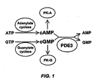
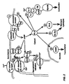
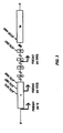
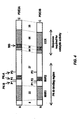


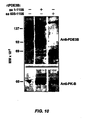
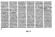

Classifications
-
- G—PHYSICS
- G01—MEASURING; TESTING
- G01N—INVESTIGATING OR ANALYSING MATERIALS BY DETERMINING THEIR CHEMICAL OR PHYSICAL PROPERTIES
- G01N33/00—Investigating or analysing materials by specific methods not covered by groups G01N1/00 - G01N31/00
- G01N33/48—Biological material, e.g. blood, urine; Haemocytometers
- G01N33/50—Chemical analysis of biological material, e.g. blood, urine; Testing involving biospecific ligand binding methods; Immunological testing
- G01N33/53—Immunoassay; Biospecific binding assay; Materials therefor
- G01N33/573—Immunoassay; Biospecific binding assay; Materials therefor for enzymes or isoenzymes
-
- A—HUMAN NECESSITIES
- A61—MEDICAL OR VETERINARY SCIENCE; HYGIENE
- A61K—PREPARATIONS FOR MEDICAL, DENTAL OR TOILETRY PURPOSES
- A61K31/00—Medicinal preparations containing organic active ingredients
- A61K31/33—Heterocyclic compounds
- A61K31/395—Heterocyclic compounds having nitrogen as a ring hetero atom, e.g. guanethidine or rifamycins
- A61K31/435—Heterocyclic compounds having nitrogen as a ring hetero atom, e.g. guanethidine or rifamycins having six-membered rings with one nitrogen as the only ring hetero atom
- A61K31/44—Non condensed pyridines; Hydrogenated derivatives thereof
- A61K31/4427—Non condensed pyridines; Hydrogenated derivatives thereof containing further heterocyclic ring systems
- A61K31/444—Non condensed pyridines; Hydrogenated derivatives thereof containing further heterocyclic ring systems containing a six-membered ring with nitrogen as a ring heteroatom, e.g. amrinone
-
- A—HUMAN NECESSITIES
- A61—MEDICAL OR VETERINARY SCIENCE; HYGIENE
- A61K—PREPARATIONS FOR MEDICAL, DENTAL OR TOILETRY PURPOSES
- A61K31/00—Medicinal preparations containing organic active ingredients
- A61K31/33—Heterocyclic compounds
- A61K31/395—Heterocyclic compounds having nitrogen as a ring hetero atom, e.g. guanethidine or rifamycins
- A61K31/435—Heterocyclic compounds having nitrogen as a ring hetero atom, e.g. guanethidine or rifamycins having six-membered rings with one nitrogen as the only ring hetero atom
- A61K31/47—Quinolines; Isoquinolines
- A61K31/4709—Non-condensed quinolines and containing further heterocyclic rings
-
- A—HUMAN NECESSITIES
- A61—MEDICAL OR VETERINARY SCIENCE; HYGIENE
- A61P—SPECIFIC THERAPEUTIC ACTIVITY OF CHEMICAL COMPOUNDS OR MEDICINAL PREPARATIONS
- A61P7/00—Drugs for disorders of the blood or the extracellular fluid
- A61P7/02—Antithrombotic agents; Anticoagulants; Platelet aggregation inhibitors
-
- A—HUMAN NECESSITIES
- A61—MEDICAL OR VETERINARY SCIENCE; HYGIENE
- A61P—SPECIFIC THERAPEUTIC ACTIVITY OF CHEMICAL COMPOUNDS OR MEDICINAL PREPARATIONS
- A61P9/00—Drugs for disorders of the cardiovascular system
- A61P9/04—Inotropic agents, i.e. stimulants of cardiac contraction; Drugs for heart failure
-
- A—HUMAN NECESSITIES
- A61—MEDICAL OR VETERINARY SCIENCE; HYGIENE
- A61P—SPECIFIC THERAPEUTIC ACTIVITY OF CHEMICAL COMPOUNDS OR MEDICINAL PREPARATIONS
- A61P9/00—Drugs for disorders of the cardiovascular system
- A61P9/06—Antiarrhythmics
-
- A—HUMAN NECESSITIES
- A61—MEDICAL OR VETERINARY SCIENCE; HYGIENE
- A61P—SPECIFIC THERAPEUTIC ACTIVITY OF CHEMICAL COMPOUNDS OR MEDICINAL PREPARATIONS
- A61P9/00—Drugs for disorders of the cardiovascular system
- A61P9/12—Antihypertensives
-
- C—CHEMISTRY; METALLURGY
- C12—BIOCHEMISTRY; BEER; SPIRITS; WINE; VINEGAR; MICROBIOLOGY; ENZYMOLOGY; MUTATION OR GENETIC ENGINEERING
- C12N—MICROORGANISMS OR ENZYMES; COMPOSITIONS THEREOF; PROPAGATING, PRESERVING, OR MAINTAINING MICROORGANISMS; MUTATION OR GENETIC ENGINEERING; CULTURE MEDIA
- C12N9/00—Enzymes; Proenzymes; Compositions thereof; Processes for preparing, activating, inhibiting, separating or purifying enzymes
- C12N9/14—Hydrolases (3)
- C12N9/16—Hydrolases (3) acting on ester bonds (3.1)
- C12N9/18—Carboxylic ester hydrolases (3.1.1)
-
- A—HUMAN NECESSITIES
- A61—MEDICAL OR VETERINARY SCIENCE; HYGIENE
- A61K—PREPARATIONS FOR MEDICAL, DENTAL OR TOILETRY PURPOSES
- A61K38/00—Medicinal preparations containing peptides
-
- G—PHYSICS
- G01—MEASURING; TESTING
- G01N—INVESTIGATING OR ANALYSING MATERIALS BY DETERMINING THEIR CHEMICAL OR PHYSICAL PROPERTIES
- G01N2333/00—Assays involving biological materials from specific organisms or of a specific nature
- G01N2333/90—Enzymes; Proenzymes
- G01N2333/914—Hydrolases (3)
- G01N2333/916—Hydrolases (3) acting on ester bonds (3.1), e.g. phosphatases (3.1.3), phospholipases C or phospholipases D (3.1.4)
-
- G—PHYSICS
- G01—MEASURING; TESTING
- G01N—INVESTIGATING OR ANALYSING MATERIALS BY DETERMINING THEIR CHEMICAL OR PHYSICAL PROPERTIES
- G01N2500/00—Screening for compounds of potential therapeutic value
- G01N2500/02—Screening involving studying the effect of compounds C on the interaction between interacting molecules A and B (e.g. A = enzyme and B = substrate for A, or A = receptor and B = ligand for the receptor)
-
- G—PHYSICS
- G01—MEASURING; TESTING
- G01N—INVESTIGATING OR ANALYSING MATERIALS BY DETERMINING THEIR CHEMICAL OR PHYSICAL PROPERTIES
- G01N2500/00—Screening for compounds of potential therapeutic value
- G01N2500/04—Screening involving studying the effect of compounds C directly on molecule A (e.g. C are potential ligands for a receptor A, or potential substrates for an enzyme A)
Definitions
- the present invention relates to the field of cardiovascular and other diseases. More particularly, the present invention concerns compositions and methods of identification and use of isoform selective activators or inhibitors of type 3 phosphodiesterase (PDE3). Other embodiments of the invention concern high-throughput screening for novel pharmaceuticals directed against PDE3 isoforms. In certain embodiments, the compositions and methods disclosed herein are of use for treatment of cardiomyopathy, pulmonary hypertension and related conditions.
- PDE3 type 3 phosphodiesterase
- PDE3 cyclic nucleotide phosphodiesterases hydrolyze cAMP and cGMP and thereby modulate cAMP- and cGMP-mediated signal transduction (Shakur et al ., 2000a). These enzymes have a major role in the regulation of contraction and relaxation in cardiac and vascular myocytes.
- PDE3 inhibitors which raise intracellular cAMP and cGMP content, have inotropic effects attributable to the activation of cAMP-dependent protein kinase (PK-A) in cardiac myocytes and vasodilatory effects attributable to the activation of cGMP-dependent protein kinase (PK-G) in vascular myocytes (Shakur et al ., 2000a).
- PDE3 inhibitors When used in the treatment of dilated cardiomyopathy, PDE3 inhibitors such as milrinone, enoximone and amrinone initially elicit favorable haemodynamic responses, but long-term administration increases mortality by up to 40% (Nony et al., 1994). This linkage of short-term benefits of PDE3 inhibition to deleterious effects on long-term survival in dilated cardiomyopathy is one of the most perplexing problems in cardiovascular therapeutics. However, it is thought that these biphasic effects reflect the compartmentally-nonselective increases in intracellular cAMP content in cardiac myocytes current inhibitors display.
- the contradictory effects of nonspecific PDE3 antagonists may relate to the diverse intracellular processes regulated by cAMP in cardiac and vascular cells.
- PK-A phosphorylates dozens of proteins in separate intracellular compartments that are involved in contraction and relaxation, glycogen metabolism, gene transcription, intracellular Ca 2+ cycling and signal autoregulation.
- Phosphorylation of cAMP-response element-binding protein (CREB) for example, activates the transcription of genes containing cAMP response elements (Shaywitz and Greenberg, 1999).
- cAMP effects is the phosphorylation of phospholamban, which relieves its inhibition of SERCA2, the Ca 2+ -transporting ATPase of the sarcoplasmic reticulum (Simmerman and Jones, 1998).
- Ablation of phospholamban in muscle LIM protein ( MLP ) -/- mice with dilated cardiomyopathy results in the restoration of normal chamber size and contractility (Minamisawa et al., 1999), suggesting that phospholamban phosphorylation may also be beneficial in cardiomyopathy.
- PK-A Other substrates phosphorylated by PK-A may contribute to adverse effects on long-term survival. Phosphorylation of L-type Ca 2+ channels increases their open probability and may be arrhythmogenic (Fischmeister and Hartzell, 1990), while phosphorylation of proteins in the mitogen-activated protein kinase (MAP kinase) cascade may alter myocardial gene transcription so as to speed the progression of the disease (Cook and McCormick, 1993; Lazou et al ., 1994).
- MAP kinase mitogen-activated protein kinase
- Raising cAMP content in cardiac myocytes via mechanisms such as activation of ⁇ 1 -adrenergic, ⁇ 2 -adrenergic or prostaglandin receptors or non-selective phosphodiesterase inhibition by isobutylmethylxanthine affects cAMP content differentially in intracellular compartments represented in cytosolic and microsomal fractions of cardiac muscle, resulting in different patterns of protein phosphorylation and different physiologic responses (Hayes et al., 1980; Xiao and Lakatta, 1993; Xiao et al., 1994; Rapundalo et al., 1989; Jurevicius and Fischmeister, 1996).
- the phosphorylation of individual substrates of PK-A may be differentially regulated in response to extracellular signals.
- Evidence for differential regulation comes from experiments examining the effects of stimulating adenylate cyclase activity and cAMP formation via ⁇ 1 -adrenergic, ⁇ 2 -adrenergic or PGE1 receptors.
- Activation of ⁇ -adrenergic receptors increases cAMP content in both cytosolic and microsomal fractions of cardiac myocytes and elicits contractile responses
- PGE1 receptors increases cytosolic but not microsomal cAMP content and evokes no contractile response (Hayes et al., 1980; Buxton and Brunton, 1983).
- ⁇ -adrenergic receptor stimulation and nonselective phosphodiesterase inhibition have different effects on cAMP-activated protein phosphorylation in cardiac myocytes (Rapundalo et al., 1989; Jurvicius and Fischmeister, 1996) that are relevant to the pathophysiology of dilated cardiomyopathy.
- a downregulation of ⁇ 1 -adrenergic receptors and an uncoupling of ⁇ -adrenergic receptor occupancy and adenylate cyclase stimulation contribute to an impairment in cAMP generation (Movsesian, 1999; Lutz et al., 2001).
- Studies of cAMP content in cytosolic and microsomal fractions of failing and nonfailing hearts demonstrate a far greater reduction in cAMP content in microsomal fractions than in cytosolic fractions of failing myocardium (Böhm et al., 1994).
- PDE3 protein kinase B
- PK-A protein kinase B
- Akt protein kinase B
- PK-B protein kinase B
- Akt protein kinase B
- PK-B a downstream effector of insulin-like growth factors, is an anti-apoptotic mediator in cardiac myocytes (Fujio et al., 2000; Matsui et al., 1999; Wu et al., 2000).
- PK-B may also be involved in proliferative responses in vascular myocytes (Rocic and Lucchesi, 2001; Duan et al ., 2000; Sandirasegarane et al ., 2000).
- PDE3 isoforms may be involved in cell- and compartment-selective responses to different signals that have been implicated in the pathophysiology of dilated cardiomyopathy and/or pulmonary hypertension.
- Different PDE3 isoforms in cardiac and vascular myocytes may regulate functionally distinct pools of cAMP and cGMP involved in the phosphorylation of different substrates of PK-A and PK-G, and these isoforms may be regulated in response to different extracellular signals.
- Isoform selective PDE3 inhibitors may provide a beneficial effect on cardiac output without the long term mortality associated with non-specific PDE3 inhibitors.
- Isoform-selective PDE3 activators may have beneficial anti-apoptotic effects in patients with dilated cardiomyopathy and/or pulmonary hypertension whose hemodynamic status is not too compromised to tolerate a reduction in cardiac contractility, without concomitant arrhythmogenic effects attributable to increases in cytosolic cAMP content.
- a paradigm for the latter is the use of beta-adrenergic receptor antagonists in the treatment of dilated cardiomyopathy.
- Agents capable of selectively activating or inhibiting individual PDE3 isoforms or of disrupting their intracellular localization may selectively affect the phosphorylation of smaller subsets of PK-A and PK-G substrates to therapeutic advantage.
- an agent that selectively inhibits sarcoplasmic reticulum-associated PDE3A-136 may help to preserve intracellular Ca 2+ cycling and contractility in patients with dilated cardiomyopathy taking ⁇ -adrenergic receptor agonists, which may reduce arrhythmogenic effects attributable to increases in cytosolic cAMP content.
- PDE3A-136 if the activation of PDE3A-136 by PK-B is anti-apoptotic in cardiac myocytes, its inhibition may be pro-apoptotic (possibly explaining the increased long-term mortality seen with PDE3 inhibition in dilated cardiomyopathy), and the selective activation of this isoform may be desirable.
- currently available competitive PDE3 inhibitors inhibit cAMP activity more potently than they inhibit cGMP hydrolytic activity, owing to the higher Km's of the hydrolytic enzymes for cAMP than for cGMP.
- Agents that inhibit PDE3 activity through other mechanisms, identified by the methods described herein, may affect hydrolysis of the two substrates differentially, resulting in different cellular actions of therapeutic benefit.
- N-terminal differences exist between the different isoforms of PDE3. Without wishing to be limited to any one specific embodiment, these N-terminal differences may offer opportunities for targeting individual isoforms of PDE3. Differences with respect to phosphorylation sites that stimulate catalytic activity suggest that agents that bind to domains containing these sites so as to either block phosphorylation or mimic its effects may be useful as isoform-selective PDE3 inhibitors or activators. As an example, an agent that binds to the P1 phosphorylation site could selectively inhibit or activate PDE3A-136 or PDE3B-137.
- the different isoforms of PDE3 are translated from different mRNAs. In some cases these mRNAs are generated from different genes (PDE3A and PDE3B). In the case of PDE3A, different isoforms are generated from different mRNAs transcribed from the same gene (e.g ., PDE3A1 and PDE3A2 mRNAs).
- the open reading frame (ORF) of PDE3A1 is indicated in SEQ ID NO:14.
- the 5' untranslated region (5'-UTR) of PDE3A1, starting with the first ATG codon, is listed in SEQ ID NO:18.
- the approximate ORF of PDE3A2 is indicated in SEQ ID NO:15.
- PDE3A1 mRNA A nucleotide sequence unique to PDE3A1 mRNA has been identified, and cDNA probes have been designed that react with PDE3A1 mRNA but not PDE3A2 mRNA. Without wishing to be limited to any one specific embodiment, these differences make PDE3 mRNAs propititious targets for decreasing the activity of individual protein isoforms by inhibiting the translation of their mRNAs via antisense constructs, ribozymes or small interfering RNA's ("siRNAs").
- the present invention fulfills an unresolved need in the art by identifying differences between PDE3 isoforms that may be used to develop isoform selective inhibitors or activators of PDE3 activity. Such inhibitors or activators are proposed to allow the differential regulation of cAMP and cGMP levels in different subcellular compartments, cell types and tissues. In certain embodiments, the present invention concerns methods for identifying isoform selective PDE3 inhibitors or activators.
- PDE3 type 3 phosphodiesterase
- SEQ ID NO:1 SEQ ID NO:2
- SEQ ID NO:3 sequences of the disclosed PDE3A isoforms proteins
- a "PDE3 isoform” is a variant of type 3 phosphodiesterase that differs in its primary structure (i . e ., amino acid sequence) from other isoforms of PDE3.
- the term encompasses, but is not limited to, isoforms that are produced by truncation, amino acid substitution (mutation) or by alternative mRNA splicing, so long as some difference in amino acid sequence results.
- other types of covalent modification would be considered to fall within the scope of a single isoform.
- both phosphorylated and unphosphorylated forms of PDE3A-136 would be considered to represent the same isoform.
- the amino acid sequences of the three isoforms of PDE3A are as disclosed in SEQ ID NO:1, SEQ ID NO:2 and SEQ ID NO:3.
- an “inhibitor” of PDE3 means any compound or combination of compounds that acts to decrease the activity of PDE3, either directly or indirectly, with respect to catalyzing the breakdown of cAMP and/or cGMP.
- An inhibitor can be a molecule, an atom, or a combination of molecules or atoms without limitation.
- the term "antagonist" of PDE3 is generally synonymous with an “inhibitor” of PDE3.
- Inhibitors may act directly on PDE3 by, for example, binding to and blocking the catalytic site or some other functional domain of PDE3 that is required for activity.
- An inhibitor may also act indirectly, for example, by blocking the phosphorylation (or its effect on activity) or facilitating the dephosphorylation of PDE3 or by facilitating or interfering with the binding of PDE3 to another protein or peptide.
- inhibitors and/or activators may affect PDE3 isoform protein activity and/or may affect the transcription, processing, post-transcriptional modification, stability and/or translation of one or more mRNA species encoding PDE3 isoform proteins (see, e . g ., GenBank Accession No. NM000921, SEQ ID NO:14, SEQ ID NO: 15, SEQ ID NO: 18).
- an "activator" of PDE3 means any compound or combination of compounds that acts to increase the activity of PDE3, either directly or indirectly, with respect to catalyzing the breakdown of cAMP and/or cGMP.
- An activator can be a molecule, an atom, or a combination of molecules or atoms without limitation.
- the term "agonist" of PDE3 is generally synonymous with an “activator” of PDE3.
- Activators may act directly on PDE3 by, for example, binding some functional domain of PDE3 that is required for activity or by altering the secondary, tertiary or quaternary structure of PDE3 in a way that increases activity.
- An activator may also act indirectly, for example, by facilitating the phosphorylation or mimicking its effect, by blocking the dephosphorylation of PDE3 or by facilitating or interfering with the binding of PDE3 to another protein or peptide. As discussed above, activators may affect PDE3 isoforms at the level of mRNA and/or protein.
- an “isoform selective" inhibitor or activator of PDE3 is one that has a greater effect on one isoform of PDE3 than on any other isoform of PDE3.
- an "isoform selective" inhibitor or activator has at least a two-fold greater, more preferably three-fold greater, more preferably four-fold greater, more preferably five-fold, more preferably ten-fold or more greater effect on one isoform of PDE3 than on any other isoform of PDE3.
- the precise degree of selectivity of an inhibitor or activator for one isoform of PDE3 compared to other isoforms is not significant, so long as a desired therapeutic effect is achieved.
- a desired therapeutic effect might be an improvement in cardiac output, with a decrease in long-term mortality, resulting from administration of an isoform selective PDE3 inhibitor or activator compared with nonspecific PDE3 inhibitors.
- An "isoform selective" inhibitor or activator of PDE3 encompasses, but is not limited to, an isoform specific inhibitor or activator of PDE3.
- An isoform specific inhibitor or activator of PDE3 is one that acts almost exclusively upon a single isoform of PDE3, so that the effect of the inhibitor or activator on one isoform of PDE3 compared to any other PDE3 isoform is at least an order of magnitude greater, more preferably two orders of magnitude greater, more preferably three orders of magnitude or more greater.
- Cyclic nucleotide phosphodiesterases have a ubiquitous role in regulating cAMP- and cGMP-mediated intracellular signaling. Eleven families of these enzymes have been identified. Those in the PDE3 family are dual-specificity phosphodiesterases that bind both cAMP and cGMP with high affinity and hydrolyze them in a mutually competitive manner ( FIG. 1 ).
- PDE3 inhibitors which raise intracellular cAMP and cGMP content, have inotropic effects attributable to the activation of cAMP-dependent protein kinase (PK-A) in cardiac myocytes and vasodilatory effects attributable to the activation of cGMP-dependent protein kinase (PK-G) in vascular myocytes.
- PK-A cAMP-dependent protein kinase
- PK-G cGMP-dependent protein kinase
- FIG. 2 illustrates the numerous targets and the intracellular compartmentation of PK-A activity.
- PDE3A and PDE3B Two subfamilies of PDE3, products of genes designated PDE3A and PDE3B, have been identified.
- PDE3A is expressed primarily in cardiac and vascular myocytes and platelets, while PDE3B is expressed primarily in adipocytes, hepatocytes and pancreatic cells (but also in vascular myocytes) (Reinhardt et al., 1995).
- PDE3B Taira et al., 1993
- three PDE3A cDNAs have been cloned. The latter are generated by transcription from alternative start sites in PDE3A.
- PDE3A1 (SEQ ID NO:14, SEQ ID NO:18), which was cloned from human myocardium, incorporates all sixteen exons of PDE3A (Meacci et al., 1992; Kasuya et al., 2000).
- PDE3A2 (SEQ ID NO:15), which was cloned from aortic myocytes, is transcribed from a start site in exon 1 (Choi et al., 2001).
- PDE3A3, cloned from placenta is transcribed from a start site between exons 3 and 4 (Kasuya et al., 1995).
- the alternative start sites used for transcription of the three PDE3A mRNAs are illustrated in FIG. 3 .
- the encoded amino acid sequences of the PDE3A isoforms are disclosed herein as SEQ ID NO:1 (PDE3A-136), SEQ ID NO:2 (PDE3A-118) and SEQ ID NO:3 (PDE3A-94).
- SEQ ID NO:1 PDE3A-136
- SEQ ID NO:2 PDE3A-118
- SEQ ID NO:3 SEQ ID NO:3A-94
- the protein isoforms of PDE3 do not precisely correspond to the mRNA species transcribed from the PDE3A gene.
- both PDE3A-118 and PDE3A-94 are translated from the PDE3A2 mRNA (SEQ ID NO:15).
- the functional topographies of the proteins corresponding to the longest open reading frames (ORF's) of PDE3A and PDE3B are similar ( FIG. 4 ).
- the C-terminus includes a sequence of about 280 amino acids, designated as "CCR” ( FIG. 4 ), which is highly conserved among cyclic nucleotide phosphodiesterase families and in which catalytic activity resides. Within CCR lies a 44-amino acid insert, designated "INS”, that is unique to the PDE3 family of cyclic nucleotide phosphodiesterases.
- the N-terminus contains two hydrophobic sequences, designated "NHR1" (about 200 amino acids) and "NHR2" (about 50 amino acids).
- NHR1 and NHR2 appear to be implicated in intracellular targeting. Between NHR1 and NHR2 are sites phosphorylated by PK-A and PK-B that, despite their distance from CCR, modulate catalytic activity. A second PK-A site whose function is unclear is located between NHR2 and CCR.
- PDE3A and PDE3B are 84-86% identical within the CCR region, exclusive of INS.
- INS and the extreme C-terminus are only 35-39% identical, and the remaining upstream regions are less than 30% identical.
- the catalytic sequences of the isoforms are similar, the regulatory portions of the isoforms appear to be very different and are likely to be differentially affected by the various inhibitors and activators of the present invention.
- the catalytic activity of PDE3 enzymes requires almost the entire C-terminal sequence downstream of about amino acid 650, including the CCR domain that is largely conserved among all PDE families, as well as the INS and the CCR-flanking regions that are unique to the PDE3 family ( FIG. 4 ). (Cheung et al., 1996; He et al ., 1998.) The recent determination of the crystal structure of the related enzyme PDE4B2B has led to the identification of its catalytic site (Xu et al., 2000). The catalytic domain consists of three subdomains comprising 17 ⁇ -helices.
- the active site preserved in all PDE families, is at the junction of these three subdomains and is formed by the apposition of discontinuous amino acids. Differences in substrate affinity and selectivity among isoform families may be influenced in large part by differences in amino acid sequences that allosterically affect Glu1001 of PDE3A, which "reads" the 1- and 6-positions of the cyclic nucleotide purine ring and determines affinity (and hence selectivity) for cAMP and cGMP. Experiments involving PDE3/PDE4 chimeras indicate that the regions adjacent to this site contain the determinants of sensitivity to phosphodiesterase inhibitors (Atienza et al ., 1999).
- the structural model described above has important implications regarding the feasibility of selective PDE3 inhibition or activation.
- the sequences of regions required for catalytic activity - INS and the regions flanking CCR - differ sufficiently between PDE3A and PDE3B to be reasonable targets for isoform-selective inhibitors or activators.
- the development of anti-peptide antibodies selective for the C-terminus of either PDE3A or PDE3B is further evidence that selective inhibition or activation may occur.
- the existence of allosteric sites that differentially affect cAMP and cGMP hydrolysis allows for the identification of small molecules that selectively bind to these sites and affect either cAMP or cGMP hydrolysis.
- Intracellular localization Intracellular targeting of PDE3 appears to be determined principally by the N-terminal domains NHR1 and NHR2.
- NHR1 contains six transmembrane helices, the last two of which are sufficient to localize recombinant proteins containing these domains exclusively to intracellular membranes (Kenan et al., 2000; Shakur et al ., 2000b).
- Such recombinants can be solubilized only by a combination of high salt and detergent, suggesting that they are intrinsic membrane proteins.
- Recombinants lacking NHR1 but retaining NHR2 are found in both microsomal and cytosolic fractions of transfected cells. High salt alone is sufficient to solubilize these proteins, suggesting that interactions with other proteins are involved in their intracellular localization.
- Recombinants lacking both NHR1 and NHR2 are predominantly cytosolic.
- Phosphorylation of PDE3 plays a major role in the regulation of its function.
- phosphorylation of PDE3 by PK-A and perhaps PI3-K are involved in the anti-lipolytic response to insulin (Smith et al., 1991).
- phosphorylation by PK-B results in the resumption of meiosis (Zhao et al., 1998).
- phosphorylation by PK-B regulates cAMP pools that modulate DNA synthesis (Ahmad et al ., 2000).
- PDE3B is phosphorylated in vivo by PK-A and possibly by PI3-K at Ser318 (site P2) (Rahn et al., 1996; Rondinone et al ., 2000).
- the P2 site is dephosphorylated by a PP2A serine/threonine phosphatase (Resjö et al., 1999).
- PDE3B is also phosphorylated in vivo by PK-B at Ser296 (site P1) (Kitamura et al., 1999). Phosphorylation at either site increases catalytic activity.
- P1 and P2 lie between NHR1 and NHR2 raises the possibility that phosphorylation at these sites also affects intracellular targeting.
- a third site - Ser421 in PDE3B (site P3) - is phosphorylated by PK-A in vitro (Rascón et al ., 1994).
- site P3B is phosphorylated at P3 in response to isoproterenol or insulin in vivo . It is unknown whether this site is phosphorylated in PDE3B in other cell types and, if so, how phosphorylation at this site affects activity.
- Phosphorylation and activation of PDE3 by PK-B may be an anti-apoptotic mechanism related to the deleterious long-term effects of PDE3 inhibition in dilated cardiomyopathy.
- the sequences of PDE3A and PDE3B contain multiple consensus sites for CK2, PK-C and other protein kinases. It may be especially important to consider cross-regulation by these kinases in the pathophysiology of cardiomyopathy and/or pulmonary hypertension.
- PDE4D3 phosphorylation by ERK2 profoundly reduces its activity, and this reduction is reversed by phosphorylation by PK-A (Hoffmann et al. 1999).
- PDE3 is catalytically regulated or intracellularly targeted via interactions with other proteins.
- PDE3B, insulin receptor, the p85 and p110 subunits of PI3-K and an unidentified 97-kDa protein are co-immunoprecipitated from human adipocytes with anti-insulin receptor antibodies (Rondinone et al ., 2000).
- Preliminary data on the interaction of PDE3B with 14-3-3 proteins has been reported (Palmer et al., 2000). 14-3-3 proteins bind to phosphorylated serine residues in consensus motifs and affect intracellular localization of proteins in diverse ways (Fu et al., 2000).
- PDE3 or wild-type activity it is meant that the molecule in question has the ability to catalyze the breakdown of cAMP and cGMP.
- Molecules possessing this activity may be identified using assays familiar to those of skill in the art. For example, in vitro assay of homogenates containing PDE3 activity, or variants thereof, will identify those molecules having PDE3 activity by virtue of their ability to degrade cAMP or cGMP.
- the skilled artisan will realize that a variety of phosphodiesterases are endemic to various cell lines and tissues and will select an appropriate system lacking endogenous phosphodiesterase to perform such assays.
- PDE3 gene refers to any DNA sequence that is substantially identical to a DNA sequence encoding a PDE3 protein as defined above. Allowing for the degeneracy of the genetic code, sequences that have at least about 50%, usually at least about 60%, more usually about 70%, most usually about 80%, preferably at least about 90%, preferably at least about 95%, most preferably 98% or more of nucleotides that are identical to the cDNA sequences of PDE3 are "as set forth in” those sequences.
- Sequences that are substantially identical or "essentially the same" as the cDNA sequences of PDE3 also may be functionally defined as sequences that are capable of hybridizing to a nucleic acid segment containing the complement of the cDNA sequences of PDE3 under conditions of relatively high stringency. Such conditions are typically relatively low salt and/or high temperature conditions, such as provided by about 0.02 M to about 0.15 M NaCl at temperatures of about 50°C to about 70°C. Such selective conditions tolerate little, if any, mismatch between the complementary stands and the template or target strand. Any such gene sequences may also comprise associated control sequences.
- the present invention relates to fragments of PDE3 polypeptides that may or may not retain the phosphodiesterase activity of PDE3, although in preferred embodiments the fragments exhibit phosphodiesterase activity.
- Fragments including the N-terminus of the molecule may be generated by genetic engineering of translation stop sites within the coding region (discussed below). Alternatively, treatment of the protein molecule with proteolytic enzymes can produce a variety of N-terminal, C-terminal and internal fragments.
- fragments may include contiguous residues of the PDE3 amino acid sequences of 6, 7, 8, 9, 10, 11, 12, 13, 14, 15, 16, 17, 18, 19, 20, 21, 22, 23, 24, 25, 30, 35, 40, 45, 50, 55, 60, 65, 75, 80, 85, 90, 95, 100, 200, 300, 400, 500 or more amino acids in length.
- These fragments may be purified according to known methods, such as precipitation (e . g ., ammonium sulfate), HPLC, ion exchange chromatography, affinity chromatography (including ⁇ immunoaffinity chromatography), or various size separations ( e . g ., sedimentation, gel electrophoresis, gel filtration).
- Substantially identical analog proteins will be greater than about 80% identical, more preferably 90% identical, more preferably 95% identical, more preferably 98% identical, more preferably 99% identical, more preferably 99.5% identical, more preferably 99.9% identical to the corresponding sequence of the native protein. Sequences having lesser degrees of similarity but comparable biological activity are considered to be equivalents. In determining nucleic acid sequences, all subject nucleic acid sequences capable of encoding substantially similar amino acid sequences are considered to be substantially similar to a reference nucleic acid sequence, regardless of differences in codon sequence.
- Certain embodiments may involve purification of one or more individual PDE3 isoforms or variants thereof.
- Protein purification techniques are well known to those of skill in the art. These techniques involve, at one level, the crude fractionation of the cellular milieu to polypeptide and non-polypeptide fractions. Having separated the polypeptide from other proteins, the polypeptide of interest may be further purified using chromatographic and electrophoretic techniques to achieve partial or complete purification (or purification to homogeneity).
- Analytical methods particularly suited to the preparation of a pure peptide are ion-exchange chromatography, gel exclusion chromatography, polyacrylamide gel electrophoresis, affinity chromatography, immunoaffinity chromatography and isoelectric focusing.
- a particularly efficient method of purifying peptides is fast protein liquid chromatography (FPLC) or even HPLC.
- Certain aspects of the present invention concern the purification, and in particular embodiments, the substantial purification, of an encoded protein or peptide.
- isolated or purified as applied to a protein or peptide, are intended to refer to a composition, isolatable from other components, wherein the protein or peptide is purified to any degree relative to its naturally-obtainable state.
- a purified protein or peptide therefore, also refers to a protein or peptide free from the environment in which it may naturally occur.
- purified will refer to a protein or peptide composition that has been subjected to fractionation to remove various other components, and which composition substantially retains its expressed biological activity. Where the term “substantially purified” is used, this designation will refer to a composition in which the protein or peptide forms the major component of the composition, such as constituting about 50%, about 60%, about 70%, about 80%, about 90%, about 95%, about 98%, about 99% or more of the proteins in the composition.
- Various methods for quantifying the degree of purification of a protein or peptide will be known to those of skill in the art. These include, for example, determining the specific activity of an active fraction, or assessing the amount of polypeptides within a fraction by SDS/PAGE analysis.
- a preferred method for assessing the purity of a fraction is to calculate the specific activity of the fraction, to compare it to the specific activity of the initial extract, and to thus calculate the degree of purity therein, assessed by a "-fold purification number.”
- the actual units used to represent the amount of activity will, of course, be dependent upon the particular assay technique chosen to follow the purification, and whether or not the protein or peptide exhibits a detectable activity.
- Partial purification may be accomplished by using fewer purification steps in combination, or by utilizing different forms of the same general purification scheme. For example, it is appreciated that a cation-exchange column chromatography performed utilizing an HPLC apparatus will generally result in a greater "-fold" purification than the same technique utilizing a low pressure chromatography system. Methods exhibiting a lower degree of relative purification may have advantages in total recovery of protein product, or in maintaining the activity of an expressed protein.
- High Performance Liquid Chromatography is characterized by a very rapid separation with high resolution of peaks. Moreover, only a very small volume of the sample is needed because the particles are so small and close-packed that the void volume is a very small fraction of the bed volume. Also, the concentration of the sample need not be very great because the bands are so narrow that there is very little dilution of the sample.
- Gel chromatography is a type of partition chromatography that is based on molecular size. As long as the material of which the particles are made does not adsorb the molecules, the sole factor determining rate of flow is the size of the pores. Hence, molecules are eluted from the column in decreasing size, so long as the shape is relatively constant. In gel chromatography, separation is independent of all other factors such as pH, ionic strength, temperature, etc .
- Affinity chromatography relies on the specific affinity between a substance to be isolated and a molecule to which it can specifically bind.
- the column material is synthesized by covalently coupling one of the binding partners, such as an antibody or an antibody-binding protein to an insoluble matrix.
- the column material is then able to specifically adsorb the target substance from the solution. Elution occurs by changing the conditions to those in which binding will not occur (e . g ., altered pH, ionic strength, temperature, etc.).
- affinity chromatography One of the most common forms of affinity chromatography is immunoaffinity chromatography. The generation of antibodies that would be suitable for use in accord with the present invention is discussed below.
- the present invention concerns smaller peptides for various uses, such as antibody generation or screening for potential inhibitors or activators that can bind to various epitopes of PDE3.
- Smaller peptides of about 100 amino acids or less can be synthesized in solution or on a solid support in accordance with conventional techniques.
- Various automated peptide synthesizers are commercially available and can be used in accordance with known protocols. See, for example, Stewart and Young, (1984); Tam et al., (1983); Merrifield, (1986); and Barany and Merrifield (1979), each incorporated herein by reference.
- Short peptide sequences or libraries of overlapping peptides, usually from about 6 up to about 35 to 50 amino acids, which correspond to selected regions of the PDE3 protein, can be readily synthesized and then screened in screening assays designed to identify reactive peptides or other small molecules.
- recombinant DNA technology may be employed wherein a nucleotide sequence which encodes a peptide of the invention is inserted into an expression vector, transformed or transfected into an appropriate host cell, and cultivated under conditions suitable for expression. Expression of cloned PDE3 sequences is preferred in embodiments where PDE3 peptides of greater than about 50 amino acids in length are desired.
- Two dimensional mapping also known as proteome analysis, is a useful tool for characterization of cellular protein expression. Specifically contemplated are the methods described in Gibson, 1974; Beemon and Hunter, 1978; and Luo, et al., 1990, each of which is incorporated herein by reference, in their entirety. Two-dimensional mapping is based on two-dimensional electrophoretic separation of proteins in a cellular lysate or homogenate so that each protein can be identified using specific coordinates in a two-dimensional protein map from which it can be extracted and further identified (by, e . g ., micro sequencing or mass spectrometry).
- the proteins in a cellular homogenate or lysate are immunoprecipitated, using an antibody or series of antibodies specific for the proteins of interest, and run on a preparative electrophoretic protein gel.
- the proteins from this gel are then transferred to an immobilizing matrix.
- immobilizing matrices are available and may be used.
- Preferred matrices for purposes of the present invention are nitrocellulose or a nylon matrix such as Immobilon (Millipore, Bedford, MA).
- the resulting protein-matrix hybrid, called a blot is then washed with water in order to remove any non-bound cellular debris from the initial homogenate or lysate, which may cause interference in subsequent steps.
- the blot is then contacted with an antibody, or series of antibodies, specific to the protein or proteins of interest in the cellular homogenate or lysate.
- an antibody or series of antibodies, specific to the protein or proteins of interest in the cellular homogenate or lysate.
- these antibodies may be monoclonal, polyclonal, or both and use of any will not substantially change the outcome of this procedure.
- radiolabeled antibodies are the preferred method of detection.
- the protein or proteins are digested by one of several common peptidase enzymes. These are enzymes that cleave proteins at specific locations only and include, but are not limited to, trypsin, chymotrypsin, CNBr and V8. Digestion may be allowed to run to completion, i . e . where every possible site that the chosen peptidase could recognize in the sample is cleaved, or it may be a partial digestion, merely run for a shorter period of time and not to completion. Once the desired level of digestion is completed, the peptidase chosen is removed from the sample, typically by centrifugation and transfer of the supernatant to a new container or vessel.
- digested samples are then loaded onto a cellulose thin layer plate for pH-driven electrophoresis, the first "dimension" in the mapping process.
- the digested proteins will behave on this thin layer plate much as they would when subjected to standard SDS-PAGE, except that the digested protein fragments will separate by charge according to the pH of the electrophoresis buffer.
- the electrophoresis buffer chosen has a pH ranging from 1.9 to 4.72, then the majority of the digested peptide fragments in the sample will be positively charged.
- the thin layer plate should thus be loaded appropriately for optimal separation o the digested peptide fragments. In this example, the plate should be loaded at a distance closer to the positive electrode and farther from the negative electrode.
- any individual electrophoresis should be that which will give an optimal distribution o the peptides.
- Preferred pH values include 8.9, more preferably 4.72, even more preferably 1.9.
- Chromatography the second "dimension" in the mapping, is next performed.
- the thin layer plate is placed in a chamber with a chromatography liquid, but only one side of the thin layer plate is immersed in this liquid.
- the thin layer plate should be placed in the liquid in such a manner that the liquid used, as it travels up through the thin layer plate via capillarity, does so at a ninety (90) degree angle from the direction electrophoresis was performed on the plate.
- chromatography is performed in this way, it will separate the digested peptide fragments in some manner apart from overall charge.
- the digested peptides will have been separated first by overall charge, then by a property driven by the chromatography liquid, hence the "two-dimensional" separation.
- chromatography buffer will differ based upon the desired property for separation and will use that buffer that will give optimal separation of the peptides in question.
- chromatography buffers may be selected that separate according to hydrophobicity, amenity, water solubility, or any other common means of separation apart from overall charge.
- the thin layer plate is dried and the digested peptide fragments thus separated are detected using common means (such as detection of a radioactively-labeled antibody).
- Protein chip technology provides a means of rapidly screening sample compounds for their ability to hybridize to PDE3 isoform proteins, peptides or subunits immobilized on a solid substrate.
- protein array-based technologies such as those disclosed by Cheng et al. (U.S. Patent No. 6,071,394 ), Zanzucchi et al. (U.S. Patent No. 5,858,804 ) and Lee et al. (U.S. Patent No. 5,948,627 ), each of which is incorporated herein by reference in their entirety. These techniques involve methods for analyzing large numbers of samples rapidly and accurately. The technology capitalizes on the binding properties of proteins or peptides to screen samples.
- a protein chip or array consists of a solid substrate upon which an array of proteins or peptides have been attached. For screening, the chip or array is contacted with a sample containing one or more test compounds that may function as PDE3 inhibitors or activators. The degree of stringency of binding of test compound to peptides may be manipulated as desired by varying, for example, salt concentration, temperature, pH and detergent content of the medium. The chip or array is then scanned to determine which proteins or peptides have bound to a test compound.
- the structure of a protein chip or array comprises: (1) an excitation source; (2) an array of probes; (3) a sampling element; (4) a detector; and (5) a signal amplification/treatment system.
- a chip may also include a support for immobilizing the probe.
- a protein or peptide may be tagged or labeled with a substance that emits a detectable signal.
- the tagged or labeled species may be fluorescent, phosphorescent, or luminescent, or it may emit Raman energy or it may absorb energy.
- a signal is generated that is detected by the chip. The signal may then be processed in several ways, depending on the nature of the signal.
- the test compounds may be labeled.
- the proteins or peptides may be immobilized onto an integrated microchip that also supports a phototransducer and related detection circuitry.
- PDE3 proteins or peptides may be immobilized onto a membrane or filter that is then attached to the microchip or to the detector surface itself.
- the proteins or peptides may be directly or indirectly immobilized onto a transducer detection surface to ensure optimal contact and maximum detection.
- a variety of methods have been utilized to either permanently or removably attach proteins to a substrate. When immobilized onto a substrate, the proteins are stabilized and may be used repeatedly.
- Exemplary substrates include nitrocellulose, nylon membrane or glass. Numerous other matrix materials may be used, including reinforced nitrocellulose membrane, activated quartz, activated glass, polyvinylidene difluoride (PVDF) membrane, polystyrene substrates, polyacrylamide-based substrate, other polymers such as poly(vinyl chloride), poly(methyl methacrylate), poly(dimethyl siloxane) and photopolymers which contain photoreactive species such as nitrenes, carbenes and ketyl radicals capable of forming covalent links with target molecules ( U.S. Pat. Nos. 5,405,766 and 5,986,076 , each incorporated herein by reference).
- PVDF polyvinylidene difluoride
- Binding of proteins or peptides to a selected support may be accomplished by any of several means.
- proteins may be bound to glass by first silanizing the glass surface, then activating with carbodiimide or glutaraldehyde.
- Alternative procedures may use reagents such as 3-glycidoxypropyltrimethoxysilane (GOP) or aminopropyltrimethoxysilane (APTS) linked via amino groups.
- GOP 3-glycidoxypropyltrimethoxysilane
- APTS aminopropyltrimethoxysilane
- Membranes particularly suitable for this application include nitrocellulose membrane (e . g ., from BioRad, Hercules, CA) or polyvinylidene difluoride (PVDF) (BioRad, Hercules, CA) or nylon membrane (Zeta-Probe, BioRad) or polystyrene base substrates (DNA.BINDTM Costar, Cambridge, MA).
- Certain embodiments of the present invention involve antibody production against one or more PDE3 isoforms.
- Means for preparing and characterizing antibodies are well known in the art (See, e . g ., Harlow and Lane, Antibodies: A Laboratory Manual, Cold Spring Harbor Laboratory, 1988 , incorporated herein by reference).
- a polyclonal antibody is prepared by immunizing an animal with an immunogenic composition and collecting antisera from that immunized animal.
- a wide range of animal species may be used for the production of antisera.
- the animal used for production of anti-antisera is a rabbit, a mouse, a rat, a hamster, a guinea pig or a goat. Because of the relatively large blood volume of rabbits, a rabbit is a preferred choice for production of polyclonal antibodies.
- a given composition may vary in its immunogenicity. It is often necessary therefore to boost the host immune system, as may be achieved by coupling a peptide or polypeptide immunogen to a carrier.
- exemplary and preferred carriers are keyhole limpet hemocyanin (KLH) and bovine serum albumin (BSA). Other albumins such as ovalbumin, mouse serum albumin or rabbit serum albumin may also be used as carriers.
- KLH keyhole limpet hemocyanin
- BSA bovine serum albumin
- Other albumins such as ovalbumin, mouse serum albumin or rabbit serum albumin may also be used as carriers.
- Means for conjugating a polypeptide to a carrier protein are well known in the art and include glutaraldehyde, m-maleimidobenzoyl-N-hydroxysuccinimide ester, carbodiimide and bis-biazotized benzidine.
- the immunogenicity of a particular immunogen composition may be enhanced by the use of non-specific stimulators of the immune response, known as adjuvants.
- adjuvants include complete Freund's adjuvant (a non-specific stimulator of the immune response containing killed Mycobacterium tuberculosis ), incomplete Freund's adjuvants and aluminum hydroxide adjuvant.
- the amount of immunogen composition used in the production of polyclonal antibodies varies upon the nature of the immunogen as well as the animal used for immunization.
- routes may be used to administer the immunogen (subcutaneous, intramuscular, intradermal, intravenous and intraperitoneal).
- the production of polyclonal antibodies may be monitored by sampling blood of the immunized animal at various points following immunization. Later booster injections may also be given. The process of boosting and titering is repeated until a suitable titer is achieved.
- the immunized animal may be bled and the serum isolated and stored, and/or the animal may be used to generate MAbs.
- the animal For production of rabbit polyclonal antibodies, the animal may be bled through an ear vein or alternatively by cardiac puncture. The removed blood is allowed to coagulate and then centrifuged to separate serum components from whole cells and blood clots.
- the serum may be used as is for various applications or else the desired antibody fraction may be purified by well-known methods, such as affinity chromatography using another antibody or a peptide bound to a solid matrix.
- Monoclonal antibodies may be readily prepared through use of well-known techniques, such as those exemplified in U.S. Patent 4,196,265 , incorporated herein by reference.
- this technique involves immunizing a suitable animal with a selected immunogen composition, e.g., a purified or partially purified expressed protein, polypeptide or peptide.
- the immunizing composition is administered in a manner effective to stimulate antibody producing cells.
- the methods for generating monoclonal antibodies generally begin along the same lines as those for preparing polyclonal antibodies. Rodents such as mice and rats are preferred animals, however, the use of rabbit, sheep or frog cells is also possible. Following immunization, somatic cells with the potential for producing antibodies, specifically B lymphocytes (B cells), are selected for use in the MAb generating protocol. These cells may be obtained from biopsied spleens, tonsils or lymph nodes, or from a peripheral blood sample. Often, a panel of animals will have been immunized and the spleen of the animal with the highest antibody titer will be removed and the spleen lymphocytes obtained by homogenizing the spleen with a syringe.
- B lymphocytes B lymphocytes
- the antibody-producing B lymphocytes from the immunized animal are then fused with cells of an immortal myeloma cell, generally one of the same species as the animal that was immunized.
- Myeloma cell lines suited for use in hybridoma-producing fusion procedures preferably are non-antibody-producing, have high fusion efficiency, and enzyme deficiencies that render then incapable of growing in certain selective media which support the growth of only the desired fused cells (hybridomas). Any one of a number of myeloma cells may be used, as are known to those of skill in the art ( e . g ., Goding, pp. 65-66, 1986).
- the immunized animal is a mouse
- rats one may use R210.RCY3, Y3-Ag 1.2.3, IR983F and 4B210; and U-266, GM1500-GRG2, LICR-LON-HMy2 and UC729-6 are all useful in connection with human cell fusions.
- Methods for generating hybrids of antibody-producing spleen or lymph node cells and myeloma cells usually comprise mixing somatic cells with myeloma cells in a 2:1 proportion, though the proportion may vary from about 20:1 to about 1:1, respectively, in the presence of an agent or agents (chemical or electrical) that promote the fusion of cell membranes.
- Fusion methods using Sendai virus have been described by Kohler and Milstein (1975; 1976), and those using polyethylene glycol (PEG), such as 37% (v/v) PEG, by Gefter et al . (1977).
- PEG polyethylene glycol
- the use of electrically induced fusion methods is also appropriate (Goding pp. 71-74, 1986).
- the selective medium generally contains an agent that blocks the de novo synthesis of nucleotides in the tissue culture media.
- agents are aminopterin, methotrexate, and azaserine. Where aminopterin or methotrexate is used, the media is supplemented with hypoxanthine and thymidine as a source of nucleotides (HAT medium). Where azaserine is used, the media is supplemented with hypoxanthine.
- a preferred selection medium is HAT. The only cells that can survive in the selective media are those hybrids formed from myeloma and B cells.
- This culturing provides a population of hybridomas from which specific hybridomas are selected.
- selection of hybridomas is performed by culturing the cells by single-clone dilution in microtiter plates, followed by testing the individual clonal supernatants for the desired reactivity.
- the assay should be sensitive, simple and rapid, such as radioimmunoassays, enzyme immunoassays, cytotoxicity assays, plaque assays, dot immunobinding assays, and the like.
- the selected hybridomas would then be serially diluted and cloned into individual antibody-producing cell lines, which clones may then be propagated indefinitely to provide MAbs.
- fragments of the monoclonal antibody of the invention may be obtained from the monoclonal antibody produced as described above, by methods which include digestion with enzymes such as pepsin or papain and/or cleavage of disulfide bonds by chemical reduction.
- monoclonal antibody fragments encompassed by the present invention may be synthesized using an automated peptide synthesizer.
- the present invention concerns immunodetection methods for binding, purifying, removing, quantifying or otherwise generally detecting peptides of interest.
- the PDE3 proteins or peptides of the present invention may be employed to detect antibodies having reactivity therewith, or, alternatively, antibodies prepared in accordance with the present invention may be employed to detect or purify the PDE3 proteins or peptides.
- the steps of various useful immunodetection methods have been described in the scientific literature, such as, e.g., Nakamura et al . (1987).
- the immunobinding methods include obtaining a sample suspected of containing a protein, peptide or antibody, and contacting the sample with an antibody or protein or peptide in accordance with the present invention, as the case may be, under conditions effective to allow the formation of immunocomplexes.
- Further methods include the detection of primary immune complexes by a two step approach.
- a second binding ligand such as an antibody, that has binding affinity for the target protein, peptide or corresponding antibody is used to form secondary immune complexes, as described above.
- the secondary immune complexes are contacted with a third binding ligand or antibody that has binding affinity for the second antibody, again under conditions effective and for a period of time sufficient to allow the formation of immune complexes (tertiary immune complexes).
- the third ligand or antibody is linked to a detectable label, allowing detection of the tertiary immune complexes thus formed. This system may provide for signal amplification if this is desired.
- the immunodetection methods of the present invention may be of utility in the diagnosis of various disease states.
- a biological or clinical sample suspected of containing either the target protein or peptide or corresponding antibody is used.
- samples from patients with cardiomyopathy and/or pulmonary hypertension may be immunoassayed to determine the type and abundance of different PDE3 isoforms present in one or more tissues.
- Targeted therapy directed towards PDE3 may utilize inhibitors and/or activators known to be selective or specific for one or more PDE3 isoforms that are detected in the patient's affected tissues.
- Immunohistochemistry Antibodies may be used in conjunction with fresh-frozen or formalin-fixed, paraffin-embedded tissue blocks prepared by immunohistochemistry (IHC). Any IHC method well known in the art may be used, such as those described in Diagnostic Immunopathology, 2nd edition, edited by, Robert B. Colvin, Atul K. Bhan and Robert T. McCluskey. Raven Press, New York., 1995 , (incorporated herein by reference).
- ELISA enzyme linked immunosorbent assays
- RIA radioimmunoassays
- Immunohistochemical detection using tissue sections is also particularly useful. However, it will be readily appreciated that detection is not limited to such techniques, and Western blotting, dot blotting, FACS analyses, and the like may also be used.
- antibodies binding to the PDE3 protein of the invention are immobilized onto a selected surface exhibiting protein affinity, such as a well in a polystyrene microtiter plate. Then, a test composition suspected of containing the PDE3 isoform, such as a clinical sample, is added to the wells. After binding and washing to remove non-specifically bound immunocomplexes, the bound antigen may be detected. Detection is generally achieved by the addition of a second antibody specific for the target protein, linked to a detectable label. This type of ELISA is a simple "sandwich ELISA". Detection may also be achieved by the addition of a second antibody, followed by the addition of a third antibody that has binding affinity for the second antibody, with the third antibody being linked to a detectable label.
- the PDE3 protein of the present invention or peptides or anti-PDE3 antibodies may be attached to a solid surface ("immobilized").
- immobilization may occur by attachment to a solid surface, such as a magnetic, glass or plastic bead, a plastic microtiter plate or a glass slide.
- Immobilization of proteins or peptides may be achieved by a variety of methods involving either non-covalent or covalent interactions between the immobilized protein or peptide and an anchor.
- immobilization may be achieved by coating a solid surface with a cross-linkable group, such as an amino, carboxyl, sulfhydryl, alcohol or other group and attaching a protein or peptide using a cross-linking reagent.
- Homobifunctional reagents that carry two identical functional groups are highly efficient in inducing cross-linking.
- Heterobifunctional reagents contain two different functional groups. By taking advantage of the differential reactivities of the two different functional groups, cross-linking can be controlled both selectively and sequentially.
- the bifunctional cross-linking reagents can be divided according to the specificity of their functional groups, e.g ., amino, sulfhydryl, guanidino or carboxyl specific groups. Of these, reagents directed to free amino groups have become especially popular because of their commercial availability, ease of synthesis and the mild reaction conditions under which they can be applied. Exemplary methods for cross-linking molecules are disclosed in U.S. Patent 5,603,872 and U.S.
- Patent 5,401,511 Amine residues may be introduced onto a surface through the use of aminosilane.
- Cross-linking reagents include bisimidates, dinitrobenzene, N-hydroxysuccinimide ester of suberic acid, disuccinimidyl tartarate, dimethyl-3,3'-dithiobispropionimidate, N-succinimidyl-3-(2-pyridyldithio)-propionate, 4-(bromoaminoethyl)-2-nitrophenylazide, 4-azidogyloxal and a water soluble carbodiimide, preferably 1-ethyl-3-(3-dimethylaminopropyl) carbodiimide (EDC).
- EDC 1-ethyl-3-(3-dimethylaminopropyl) carbodiimide
- the present invention is not limiting as to the cross-linking agents that may be used.
- the present invention also provides, in another embodiment, genes encoding PDE3 A136.
- a "PDE3 gene” may contain a variety of different bases and yet still produce a corresponding polypeptide that is indistinguishable functionally, and in some cases structurally, from the genes disclosed herein. Described are also nucleic acids (antisense RNAs, ribozymes) that can bind to and inhibit transcription and/or translation of one or more RNA species encoding a PDE3A isoform protein.
- the design and production of antisense RNAs, or cDNAs encoding antisense RNAs are well known in the art and any such known method may be used in the practice of the present invention (e.g ., U.S.
- an antisense RNA may be targeted against a particular PDE3A isoform, for example by electing a target sequence that is present in one PDE3A isoform mRNA but not in another.
- nucleic acid encompasses single-stranded, double-stranded, triple-stranded DNA and/or RNA of any type, as well as analogs of and chemically modified forms of DNA and/or RNA.
- nucleic acid Any reference to a nucleic acid should be read as encompassing a host cell containing that nucleic acid and, in some cases, capable of expressing the product of that nucleic acid.
- Cells expressing nucleic acids of the present invention may prove useful in the context of screening for agents that induce, repress, inhibit, augment, interfere with, block, abrogate, stimulate, or enhance the catalytic activity, regulatory properties or subcellular localization of PDE3 isoforms.
- Nucleic acids may contain an entire gene, a cDNA, or a domain of a PDE3 isoform that expresses catalytic activity, or any other fragment of the sequences set forth herein.
- the nucleic acid may be derived from genomic DNA, i.e ., clone directly from the genome of a particular organism. In preferred embodiments, however, the nucleic acid would comprise complementary DNA (cDNA).
- the DNA segments of the present invention include those encoding biologically functional equivalent PDE3 proteins and peptides. Such sequences may arise as a consequence of codon redundancy and amino acid functional equivalency that are known to occur naturally within nucleic acid sequences and the proteins thus encoded.
- functionally equivalent proteins or peptides may be created via the application of recombinant DNA technology, in which changes in the protein structure may be engineered, based on considerations of the properties of the amino acids being exchanged. Changes designed by man may be introduced through the application of site-directed mutagenesis techniques or may be introduced randomly and screened later for the desired function, as described below.
- RNA species are particularly sensitive to endogenous and/or exogenous RNAse degradation and that great care must be taken to inhibit or inactivate RNAse before RNA levels can be determined.
- RNAse inhibitors typically involve treatment of solutions with diethylpyrocarbonate (DEPC) and autoclaving as well as addition of commercial RNAse inhibitors.
- Northern blotting is a well-known method for assaying mRNA species that differ by size. Either total cell RNA or polyadenylated mRNA may be purified from a sample by known techniques ( e . g ., Sambrook et al., 1989). The purified RNA is separated by size using gel electrophoresis. After transfer to a nylon, nitrocellulose or other membrane, the size separated RNAs are probed with a labeled oligonucleotide that hybridizes specifically with one or more target RNAs.
- DEPC diethylpyrocarbonate
- RNA species that hybridizes with the oligonucleotide probe is detected by autoradiography, fluorography or other known techniques. Further examples of the use of Northern blotting to detect PDE3A mRNAs are disclosed below in the Examples section. It appears that in most cell types, a given PDE3A isoform mRNA will either be present or absent. Thus, generally it will be sufficient to detect the presence or absence of a PDE3A isoform mRNA. However, the amounts of each isoform mRNA present in a sample may also be determined by standard techniques, such as using autoradiography or fluorography to expose a film (e.g. Kodak X-Omat, Eastman Kodak, Rochester, NY), and scanning the band intensity on the developed film.
- a film e.g. Kodak X-Omat, Eastman Kodak, Rochester, NY
- the target nucleic acids of interest may be amplified as disclosed below. Amplification products may be attached to a membrane, 96-well plate, nucleic acid chip or other substrate and detected. Because the PDE3A isoforms do not differ in sequence, determination of the amounts of each mRNA species would require three separate probes. One probe would be designed to be complementary to the PDE3A3 mRNA sequence and would detect PDE3A1, PDE3A2 and PDE3A3. A second probe would be designed to be complementary to the 5' portion of the PDE3A2 mRNA sequence (see SEQ ID NO:15), for example to the 3' end of exon 1 or to exons 2 or 3.
- That probe would hybridize with mRNAs for PDE3A1 and PDE3A2.
- a third probe would be designed to be complementary with the 5' end of exon 1. That probe would only hybridize with the mRNA encoding PDE3A1 (SEQ ID NO:14, SEQ ID NO:18).
- the PDE3A isoforms are encoded by at least two, and possibly by three different mRNAs.
- PDE3A1 mRNA is translated to a 136 kDa protein isoform
- a PDE3A2 mRNA may be translated to give both 94 kDA and 118 kDA protein isoforms.
- each of the different sized protein isoforms may be encoded by a separate mRNA species.
- Apparatus and kits for assay of mRNA expression levels are commercially available, such as the NanochipTM Workstation (Nanogen, Inc., San Diego, CA), Affymetrix Genechip® (Affymetrix, Inc., Santa Clara, CA), etc .
- high throughput screening (HTS) methods directed towards mRNA may be used to assay for inhibitors and/or activators that affect expression of specific PDE3 isoforms.
- Such methods are known in the art and in some embodiments may be performed using kits and/or apparatus obtained from commercial vendors (e . g . Xpress-Screen mRNA Detection Assay Service, Applied Biosystems, Foster City, CA).
- the object of high throughput screening is to survey thousands of compounds, for example in the form of small molecule libraries, phage display libraries, native plant or animal extracts, combinatorial chemistry libraries, etc . for a pharmaceutically significant effect on a target protein, cell, tissue, organ or organism.
- Effective compounds may be further modified by chemical substitution and/or modification to provide increased efficacy, safety, duration of effect, etc .
- HTS assays may be directed against one or more proteins or peptides of interest, such as PDE3A-136, PDE3A-118, PDE3A-94 or PDE3B-137 using known techniques.
- libraries of potential inhibitors and/or activators are exposed to PDE3 proteins and/or peptides and enzyme catalytic activity and/or regulatory properties are assayed.
- Such assays may be performed, for example, in 96-well microtiter plates using known colorimetric, luminescent and/or radioactive assays for enzyme activity.
- test peptides and/or proteins may be attached to a surface, such as a protein chip, microtiter wells, membrane or other surface known in the art and libraries of compounds may be screened for their ability to bind to the various PDE3 isoforms.
- a surface such as a protein chip, microtiter wells, membrane or other surface known in the art and libraries of compounds may be screened for their ability to bind to the various PDE3 isoforms.
- Protein based HTS assays can be laborious and time-consuming.
- An alternative method for performing HTS analysis is to screen targets, such as cells, tissues, organs or organisms for an effect of a test compound on mRNA levels.
- PDE3 isoforms such assays may potentially be directed towards identifying compounds that directly or indirectly affect PDE3A1 or PDE3A2 mRNA levels.
- the cell or tissue of interest for example a tissue sample from an individual with dilated cardiomyopathy or an Sfo cell transfected with a PDE3A-encoding gene may be exposed to a series of test compounds in 96- or 384-well microplates.
- a biotinylated probe specific for the mRNA of interest is used to hybridize to total cell RNA or to purified polyadenylated mRNA.
- the DNA-RNA hybrid may be transferred to a streptavidin coated plate, which binds to the biotinylated probe.
- a labeled antibody such as an alkaline phosphatase conjugated antibody, that binds specifically to RNA-DNA hybrids is incubated with the plate, unbound antibody is removed by washing and the presence of RNA-DNA hybrids is detected by developing the labeled antibody, for example using a chemiluminescent substrate (Xpress-Screen, Applied Biosystems). In this way, hundreds of test compounds may be screened simultaneously for an effect on PDE3 isoform expression.
- test compounds may be screened by looking for secondary effects of PDE3A isoform proteins. Inhibition or activation of PDE3 activity and/or expression may be determined indirectly. By affecting the cellular levels of cAMP and/or cGMP, PDE3 isoforms may affect the expression of known cyclic nucleotide regulated genes. Cells or tissues that have been exposed to test compounds may be screened, as described above, for mRNAs encoded by genes that are known to be dependent on cyclic nucleotide levels. Effects of inhibitors and/or activators of PDE3 isoforms may be monitored by screening normal, diseased and/or transformed cells for changes in expression levels of cAMP or cGMP regulated genes.
- Nucleic acids of use as a template for amplification may be isolated from cells contained in a biological sample, according to standard methodologies. (Sambrook et al., 1989)
- the nucleic acid may be genomic DNA or fractionated or whole cell RNA. Where RNA is used, it may be desired to convert the RNA to a complementary cDNA.
- the RNA is whole cell RNA and is used directly as the template for amplification.
- the RNA may be polyadenylated mRNA. Purification of mRNA, for example by affinity chromatography to oligo-dT columns, is well known in the art.
- Pairs of primers that selectively hybridize to nucleic acids corresponding to specific markers are contacted with the isolated nucleic acid under conditions that permit selective hybridization. Once hybridized, the nucleic acid:primer complex is contacted with one or more enzymes that facilitate template-dependent nucleic acid synthesis. Multiple rounds of amplification, also referred to as "cycles,” are conducted until a sufficient amount of amplification product is produced.
- the amplification product is detected.
- the detection may be performed by visual means.
- the detection may involve indirect identification of the product via chemiluminescence, radioactive scintigraphy of incorporated radiolabel or fluorescent label or even via a system using electrical or thermal impulse signals ( Affymax technology; Bellus, 1994 ).
- primer as defined herein, is meant to encompass any nucleic acid that is capable of priming the synthesis of a nascent nucleic acid in a template-dependent process.
- primers are oligonucleotides from ten to twenty base pairs in length, but longer sequences may be employed.
- Primers may be provided in double-stranded or single-stranded form, although the single-stranded form is preferred.
- PCR polymerase chain reaction
- a reverse transcriptase PCR amplification procedure may be performed in order to quantify the amount of mRNA amplified.
- Methods of reverse transcribing RNA into cDNA are well known and described in Sambrook et al., 1989.
- Alternative methods for reverse transcription utilize thermostable DNA polymerases. These methods are described in WO 90/07641 filed December 21, 1990 .
- Polymerase chain reaction methodologies are well known in the art.
- LCR ligase chain reaction
- PCT/US87/00880 may also be used as still another amplification method in the present invention.
- a replicative sequence of RNA which has a region complementary to that of a target is added to a sample in the presence of an RNA polymerase.
- the polymerase will copy the replicative sequence which may then be detected.
- An isothermal amplification method in which restriction endonucleases and ligases are used to achieve the amplification of target molecules that contain nucleotide 5'-[alpha-thio]-triphosphates in one strand of a restriction site may also be useful in the amplification of nucleic acids in the present invention. Walker et al., Proc. Nat'l Acad. Sci.
- Strand Displacement Amplification is another method of carrying out isothermal amplification of nucleic acids which involves multiple rounds of strand displacement and synthesis, i.e., nick translation.
- a similar method called Repair Chain Reaction (RCR)
- RCR Repair Chain Reaction
- SDA Strand Displacement Amplification
- RCR Repair Chain Reaction
- CPR cyclic probe reaction
- a probe having 3' and 5' sequences of non-specific DNA and a middle sequence of specific RNA is hybridized to DNA which is present in a sample.
- the reaction is treated with RNase H, and the products of the probe identified as distinctive products which are released after digestion.
- the original template is annealed to another cycling probe and the reaction is repeated.
- Still other amplification methods described in GB Application No. 2 202 328 , and in PCT Application No. PCT/US89/01025 each of which is incorporated herein by reference in its entirety, may be used in accordance with the present invention.
- "modified" primers are used in a PCR like, template and enzyme dependent synthesis.
- the primers may be modified by labeling with a capture moiety (e.g., biotin) and/or a detector moiety (e.g., enzyme).
- a capture moiety e.g., biotin
- a detector moiety e.g., enzyme
- an excess of labeled probes are added to a sample.
- the probe binds and is cleaved catalytically. After cleavage, the target sequence is released intact to be bound by excess probe. Cleavage of the labeled probe signals the presence of the target sequence.
- Other nucleic acid amplification procedures include transcription-based amplification systems (TAS), including nucleic acid sequence based amplification (NASBA) and 3SR. Kwoh et al., Proc. Nat'l Acad. Sci.
- the nucleic acids may be prepared for amplification by standard phenol/chloroform extraction, heat denaturation of a clinical sample, treatment with lysis buffer and minispin columns for isolation of DNA and RNA or guanidinium chloride extraction of RNA. These amplification techniques involve annealing a primer which has target specific sequences. Following polymerization, DNA/RNA hybrids are digested with RNase H while double stranded DNA molecules are heat denatured again.
- the single stranded DNA is made fully double stranded by addition of second target specific primer, followed by polymerization.
- the double-stranded DNA molecules are then multiply transcribed by a polymerase such as T7 or SP6.
- a polymerase such as T7 or SP6.
- the RNA's are reverse transcribed into double stranded DNA, and transcribed once against with a polymerase such as T7 or SP6.
- the resulting products whether truncated or complete, indicate target specific sequences.
- ssRNA single-stranded RNA
- dsDNA double-stranded DNA
- the ssRNA is a first template for a first primer oligonucleotide, which is elongated by reverse transcriptase (RNA-dependent DNA polymerase).
- RNA-dependent DNA polymerase reverse transcriptase
- the RNA is then removed from the resulting DNA:RNA duplex by the action of ribonuclease H (RNase H, an RNase specific for RNA in duplex with either DNA or RNA).
- RNase H ribonuclease H
- the resultant ssDNA is a second template for a second primer, which also includes the sequences of an RNA polymerase promoter (exemplified by T7 RNA polymerase) 5' to its homology to the template.
- This primer is then extended by DNA polymerase (exemplified by the large "Klenow" fragment of E. coli DNA polymerase I), resulting in a double-stranded DNA (“dsDNA”) molecule, having a sequence identical to that of the original RNA between the primers and having additionally, at one end, a promoter sequence.
- This promoter sequence may be used by the appropriate RNA polymerase to make many RNA copies of the DNA. These copies may then re-enter the cycle leading to very swift amplification.
- this amplification may be done isothermally without addition of enzymes at each cycle.
- the starting sequence may be chosen to be in the form of either DNA or RNA.
- Miller et al., PCT Application WO 89/06700 (incorporated herein by reference in its entirety) disclose a nucleic acid sequence amplification scheme based on the hybridization of a promoter/primer sequence to a target single-stranded DNA ("ssDNA”) followed by transcription of many RNA copies of the sequence. This scheme is not cyclic, i.e., new templates are not produced from the resultant RNA transcripts.
- amplification products are separated by agarose, agarose-acrylamide or polyacrylamide gel electrophoresis using standard methods. See Sambrook et al., 1989.
- chromatographic techniques may be employed to effect separation.
- chromatography There are many kinds of chromatography which may be used in the present invention: absorption, partition, ion-exchange and molecular sieve, and many specialized techniques for using them including column, paper, thin-layer and gas chromatography (Freifelder, 1982).
- Amplification products must be visualized in order to confirm amplification of the marker sequences.
- One typical visualization method involves staining of a gel with ethidium bromide and visualization under UV light.
- the amplification products may then be exposed to x-ray film or visualized under the appropriate stimulating spectra, following separation.
- visualization is achieved indirectly.
- a labeled, nucleic acid probe is brought into contact with the amplified marker sequence.
- the probe preferably is conjugated to a chromophore but may be radiolabeled.
- the probe is conjugated to a binding partner, such as an antibody or biotin, where the other member of the binding pair carries a detectable moiety.
- detection is by Southern blotting and hybridization with a labeled probe.
- the techniques involved in Southern blotting are well known to those of skill in the art and may be found in many standard books on molecular protocols. See Sambrook et al., 1989. Briefly, amplification products are separated by gel electrophoresis. The gel is then contacted with a membrane, such as nitrocellulose, permitting transfer of the nucleic acid and non-covalent binding. Subsequently, the membrane is incubated with a chromophore-conjugated probe that is capable of hybridizing with a target amplification product. Detection is by exposure of the membrane to x-ray film or ion-emitting detection devices.
- U.S. Patent No. 5,279,721 discloses an apparatus and method for the automated electrophoresis and transfer of nucleic acids.
- the apparatus permits electrophoresis and blotting without external manipulation of the gel and is ideally suited to carrying out methods according to the present invention.
- antisense refers to polynucleotide molecules complementary to a portion of a targeted gene or mRNA species.
- Complementary polynucleotides are those that are capable of base-pairing according to the standard Watson-Crick complementarity rules. That is, purines will base pair with pyrimidines to form combinations of guanine paired with cytosine (G:C) and adenine paired with either thymine. (A:T) in the case of DNA, or adenine paired with uracil (A:U) in the case of RNA. Inclusion of less common bases such as inosine, 5-methylcytosine, 6-methyladenine, hypoxanthine and others in hybridizing sequences does not interfere with pairing.
- Antisense polynucleotides when introduced into, a target cell, specifically bind to their target polynucleotide and interfere with transcription, RNA processing, transport, translation and/or stability.
- Antisense RNA constructs, or DNA encoding such antisense RNA's may be employed to inhibit gene transcription or translation or both within a host cell, either in vitro or in vivo, such as within a host animal, including a human subject.
- the intracellular concentration of monovalent cations is approximately 160 mM (10 mM Na + ; 150 mM K + ).
- the intracellular concentration of divalent cations is approximately 20 mM (18 mM Mg + ; 2 mM Ca ++ ).
- the intracellular protein concentration which would serve to decrease the volume of hybridization and, therefore, increase the effective concentration of nucleic acid species, is 150 mg/ml. Constructs may be tested for specific hybridization i n vitro under conditions that mimic these in vivo conditions.
- Antisense constructs may be designed to bind to the promoter and other control regions, exons, introns or even exon-intron boundaries of a gene. It is contemplated that effective antisense constructs may include regions complementary to the mRNA start site. Antisense constructs are targeted to a sequence of an hnRNA and/or mRNA that is present in one PDE3A isoform and not in another. For example, one might target the 5' end of the mRNA encoding PDE3A1. (SEQ ID NO:14, SEQ ID NO:18), which is missing the in the PDE3A2 mRNA (SEQ ID NO:15). One of ordinary skill in the art can readily test such constructs to determine whether levels of the target protein are affected.
- complementary or “antisense” mean polynucleotide that are substantially complementary to the target sequence over their entire length and have very few base mismatches. For example; sequences of fifteen bases in length may be termed complementary when they have a complementary nucleotide at thirteen or fourteen nucleotides out of fifteen. Naturally, sequences that are "completely complementary” will be sequences that are entirely complementary throughout their entire length and have no base mismatches.
- an antisense construct that has limited regions of high homology, but also contains a non-homologous region (e.g. , a ribozyme) could be designed These molecules, though having less than 50% homology, would bind to target sequences under appropriate conditions.
- a non-homologous region e.g. , a ribozyme
- the antisense sequences may be full length cDNA copies, or large fragments thereof, they also may be shorter fragments, or "oligonucleotides,” defined herein as polynucleotides of 50 or less bases. Although shorter oligomers (8-20) are easier to make and increase in vivo accessibility, numerous other factors are involved in determining the specificity of base-pairing. For example, both binding affinity and sequence specificity of an oligonucleotide to its complementary target increase with increasing length. It is contemplated that oligonucleotides of 8, 9, 10, 11, 12, 13, 14, 15, 16, 17, 18, 9, 20, 25, 30, 35, 40, 45, 50 or 100 base pairs will be used. While all or part of the gene sequence may be employed in the context of antisense construction, statistically, any sequence of 14 bases long should occur only once in the human genome and, therefore, suffice to specify a unique target sequence.
- antisense constructs which include other elements, for example, those which include C-5 propyne pyrimidines.
- Oligonucleotides which contain C-5 propyne analogues of uridine and cytidine have been shown to bind RNA with high affinity and to be potent antisense inhibitors of gene expression (Wagner et al., .1993).
- the antisense oligo- and polynucleotides may be provided as RNA via transcription from expression constructs that carry nucleic acids encoding the oligo- or polynucleotides.
- expression construct is meant to include any type of genetic construct containing a nucleic acid encoding a product in which part or all of the nucleic acid sequence incapable of being transcribed.
- Typical expression vectors include bacterial plasmids or phage, such as any of the pUC or Bluescript TM plasmid series or, as discussed further below, viral vectors adapted for use in eukaryotic cells.
- nucleic acids encoding an antisense oligo- or polynucleotide under transcriptional control of a promoter.
- a “promoter” refers to a DNA sequence recognized by an RNA polymerase to initiate the specific transcription of a gene.
- under transcriptional control means that the promoter is in the correct location and orientation in relation to the nucleic acid to control RNA polymerase initiation.
- promoter will be used here to refer to a group of transcriptional control modules that are clustered around the initiation site for RNA polymerase II. Promoters are composed of discrete functional modules, each consisting of approximately 7-20 bp of DNA, and containing one or more recognition sites for transcriptional activator or repressor proteins. At least one module in each promoter functions to position the start site for RNA synthesis.
- the best known example of this is the TATA box, but in some promoters lacking a TATA box, such as the promoter for the mammalian terminal deoxynucleotidyl transferase gene and the promoter for the SV40 late genes, a discrete element overlying the start site itself helps to fix the place of initiation.
- promoter elements regulate the frequency of transcriptional initiation. Typically, these are located in the region 30-110 bp upstream of the start site, although a number of promoters have recently been shown to contain functional elements downstream of the start site as well.
- the spacing between promoter elements frequently is flexible, so that promoter function is preserved when elements are inverted or moved relative to one another. In the tk promoter, the spacing between promoter elements can be increased to 50 bp apart before activity begins to decline. Depending on the promoter, it appears that individual elements can function either co-operatively or independently to activate transcription.
- the particular promoter that is employed to control the expression of a nucleic acid encoding the inhibitory polynucleotide is not believed to be important, so long as it is capable of expressing the peptide in the targeted cell.
- a human cell it is preferable to position the nucleic acid coding the inhibitory peptide adjacent to and under the control of a promoter that is active in the human cell.
- a promoter might include either a human or viral promoter.
- the human cytomegalovirus (CMV) immediate early gene promoter, the SV40 early promoter and the Rous sarcoma virus long terminal repeat can be used to obtain high-level transcription.
- CMV cytomegalovirus
- the use of other viral or mammalian cellular or bacterial phage promoters which are well-known in the art is contemplated as well, provided that the levels of transcription and/or translation are sufficient for a given purpose.
- a promoter that is regulated in response to specific physiologic signals can permit inducible expression of an antisense sequence.
- a nucleic acid under control of the human PAI-1 promoter results in expression inducible by tumor necrosis factor.
- any promoter/enhancer combination also could be used to drive expression of a nucleic acid according to the present invention.
- Tables 1 and 2 list elements/promoters that may be employed to regulate transcription and/or translation of operably coupled genes. This list is exemplary only and any known promoter and/or regulatory element may be used.
- Ribozymes are RNA-protein complexes that cleave nucleic acids in a site-specific fashion. Ribozymes have specific catalytic domains that possess endonuclease activity (Kim and Cech, 1987). For example, a large number of ribozymes accelerate phosphoester transfer reactions with a high degree of specificity, often cleaving only one of several phosphoesters in an oligonucleotide substrate (Cech et al., 1981). This specificity has been attributed to the requirement that the substrate bind via specific base-pairing interactions to an internal guide sequence ("IGS") of the ribozyme prior to chemical reaction.
- IGS internal guide sequence
- Ribozyme catalysis has primarily been observed as part of sequence-specific cleavage/ligation reactions involving nucleic acids (Joyce, 1989; Cech et al., 1981).
- U.S. Patent No. 5,354,855 reports that certain ribozymes can act as endonucleases with a sequence specificity greater than that of known ribonucleases.
- sequence-specific ribozyme-mediated inhibition of gene expression may be particularly suited to therapeutic applications (Scanlon et al., 1991; Sarver et al., 1990; Sioud et al., 1992). It was reported that ribozymes elicited genetic changes in some cells lines to which they were applied. The altered genes included the oncogenes H-ras, c-fos and genes of HIV.
- RNA cleavage activity examples include sequences from the Group I self splicing introns including Tobacco Ringspot Virus (Prody et al., 1986), Avocado Sunblotch Viroid (Palukaitis et al., 1979; Symons, 1981), and Lucerne Transient Streak Virus (Forster and Symons, 1987). Sequences from these and related viruses are referred to as hammerhead ribozymes.
- Other suitable ribozymes include sequences from RNase P (Yuan et al., 1992, Yuan and Altman, 1994, U.S. Patent Nos.
- Ribozymes are targeted to a given sequence by virtue of annealing to a site by complimentary base pair interactions. Two stretches of homology are required for this targeting. These stretches of homologous sequences flank the catalytic ribozyme structure defined above. Each stretch of homologous sequence can vary in length from 7 to 15 nucleotides. The only requirement for defining the homologous sequences is that, on the target RNA, they are separated by a specific sequence that is the cleavage site.
- the cleavage site is a dinucleotide sequence on the target RNA - a uracil (U) followed by either an adenine, cytosine or uracil (A,C or U) (Perriman et al., 1992).
- ribozymes designed to specifically cleave individual genes or gene products. Designing and testing ribozymes for efficient cleavage of a target RNA is a process well known to those skilled in the art. Examples of scientific methods for designing and testing ribozymes are described by Chowrira et al., (1994).
- RNA interference Another possibility is to inhibit the translation of individual PDE3 mRNAs by RNA interference.
- This method of post-transcriptional gene silencing involves the use of a 21- or 22-nucleotide double-stranded synthetic RNA molecule homologous to a unique nucleotide sequence in the mRNA of interest.
- small interfering RNA molecules siRNAs
- siRNAs small interfering RNA molecules
- RNA species small interfering RNA species
- dsRNA short, double-stranded RNAs
- PDE3A a target cell
- the dsRNAs are broken into smaller fragments of about 21-22 bp (siRNAs). These fragments trigger the degradation of homologous RNA sequences (Elbashir et al , 2001), e.g.
- siRNAs can decrease expression of a target gene or even eliminate it entirely (Bass, 2001). Another advantage of siRNAs is that they are effective at lower concentrations (about 1-25 nM) than antisense constructs (Bass, 2001; Elbashir et al ., 2001).
- Nucleic acids encoding PDE3 isoform proteins or peptides may be incorporated into expression vectors for production of the encoded proteins or peptides.
- Non-limiting examples of expression systems known in the art include bacteria such as E. coli, yeast such as Pichia pastoris, baculovirus, and mammalian expression systems such as in COS or CHO cells.
- a complete gene can be expressed or, alternatively, fragments of the gene encoding portions of polypeptide can be produced.
- the gene or gene fragment encoding a polypeptide may be inserted into an expression vector by standard subcloning techniques.
- An E. coli expression vector may be used which produces the recombinant polypeptide as a fusion protein, allowing rapid affinity purification of the protein.
- Examples of such fusion protein expression systems are the glutathione S-transferase system (Pharmacia, Piscataway, NJ), the maltose binding protein system (NEB, Beverley, MA), the FLAG system (IBI, New Haven, CT), and the 6xHis system (Qiagen, Chatsworth, CA).
- fusion systems are designed to produce fusions wherein the fusion partner is easily excised from the desired polypeptide.
- the fusion partner is linked to the recombinant polypeptide by a peptide sequence containing a specific recognition sequence for a protease. Examples of suitable sequences are those recognized by the Tobacco Etch Virus protease (Life Technologies, Gaithersburg, MD) or Factor Xa (New England Biolabs, Beverley, MA).
- the expression system used may also be one driven by the baculovirus polyhedron promoter.
- the gene encoding the polypeptide may be manipulated by standard techniques in order to facilitate cloning into the baculovirus vector.
- One baculovirus vector is the pBlueBac vector (Invitrogen, Sorrento, CA).
- the vector carrying the gene for the polypeptide is transfected into Spodoptera frugiperda (Sf9) cells by standard protocols, and the cells are cultured and processed to produce the recombinant antigen. See U.S. Patent No. 4,215,051
- Amino acid sequence variants of the polypeptide may also be prepared. These may, for instance, be minor sequence variants of the polypeptide which arise due to natural variation within the population or they may be homologues found in other species. They also may be sequences which do not occur naturally but which are sufficiently similar that they function similarly and/or elicit an immune response that cross-reacts with natural forms of the polypeptide. Sequence variants may be prepared by standard methods of site-directed mutagenesis such as those described herein.
- Substitutional variants typically contain an alternative amino acid at one or more sites within the protein, and may be designed to modulate one or more properties of the polypeptide such as stability against proteolytic cleavage. Substitutions preferably are conservative, that is, one amino acid is replaced with one of similar size and charge.
- Conservative substitutions are well known in the art and include, for example, the changes of: arginine to lysine; asparagine to glutamine or histidine; aspartate to glutamate; cysteine to serine; glutamine to asparagine; glutamate to aspartate; histidine to asparagine or glutamine; isoleucine to leucine or valine; leucine to valine or isoleucine; lysine to arginine or glutamine; methionine to leucine or isoleucine; phenylalanine to tyrosine; serine to threonine; tyrosine to tryptophan or phenylalanine; and valine to isoleucine or leucine.
- the hydropathic index of amino acids may be considered.
- the importance of the hydropathic amino acid index in conferring interactive biologic function on a protein is generally understood in the art (Kyte & Doolittle, 1982). It is accepted that the relative hydropathic character of the amino acid contributes to the secondary structure of the resultant protein, which in turn defines the interaction of the protein with other molecules.
- Each amino acid has been assigned a hydropathic index on the basis of its hydrophobicity and charge characteristics (Kyte & Doolittle, 1982), these are: Isoleucine (+4.5); valine (+4.2); leucine (+3.8); phenylalanine (+2.8); cysteine/cystine (+2.5); methionine (+1.9); alanine (+1.8); glycine (-0.4); threonine (-0.7); serine (-0.8); tryptophan (-0.9); tyrosine (-1.3); proline (-1.6); histidine (-3.2); glutamate (-3.5); glutamine (-3.5); aspartate (-3.5); asparagine (-3.5); lysine (-3.9); and arginine (-4.5).
- amino acids may be substituted by other amino acids having a similar hydropathic index or score and still result in a protein with similar biological activity, i.e., still obtain a biological functionally equivalent protein.
- substitution of amino acids whose hydropathic indices are within ⁇ 2 is preferred, those that are within ⁇ 1 are particularly preferred, and those within ⁇ 0.5 are even more particularly preferred.
- hydrophilicity U.S. Patent 4,554,101 , incorporated herein by reference.
- the following hydrophilicity values have been assigned to amino acid residues: arginine (+3.0); lysine (+3.0); aspartate (+3.0 ⁇ 1); glutamate (+3.0 ⁇ 1); serine (+0.3); asparagine (+0.2); glutamine (+0.2); glycine (0); threonine (-0.4); proline (-0.5 ⁇ 1); alanine (-0.5); histidine (-0.5); cysteine (-1.0); methionine (-1.3); valine (-1.5); leucine (-1.8); isoleucine (-1.8); tyrosine (-2.3); phenylalanine (-2.5); tryptophan (-3.4).
- Insertional variants include fusion proteins such as those used to allow rapid purification of the polypeptide and also may include hybrid proteins containing sequences from other proteins and polypeptides which are homologues of the polypeptide.
- an insertional variant may include portions of the amino acid sequence of the polypeptide from one species, together with portions of the homologous polypeptide from another species.
- Other insertional variants may include those in which additional amino acids are introduced within the coding sequence of the polypeptide. These typically are smaller insertions than the fusion proteins described above and are introduced, for example, to disrupt a protease cleavage site.
- DNA segment(s) for expression in a prokaryotic or eukaryotic system may be performed by techniques generally known to those of skill in recombinant expression. It is believed that virtually any expression system may be employed in the expression of the claimed nucleic acid sequences.
- engineered and recombinant cells are intended to refer to a cell into which an exogenous DNA segment or gene, such as a cDNA or gene has been introduced through the hand of man. Therefore, engineered cells are distinguishable from naturally occurring cells that do not contain a recombinantly introduced exogenous DNA segment or gene.
- Recombinant cells include those having an introduced cDNA or genomic gene, and also include genes positioned adjacent to a heterologous promoter not naturally associated with the particular introduced gene.
- an expression vector that comprises one of the claimed isolated nucleic acids under the control of, or operatively linked to, one or more promoters.
- a coding sequence "under the control of” as promoter one positions the 5' end of the transcription initiation site of the transcriptional reading frame generally between about 1 and about 50 nucleotides "downstream” ( i.e ., 3') of the chosen promoter.
- the "upstream” promoter stimulates transcription of the DNA and promotes expression of the encoded recombinant protein.
- Cell types available for expression include, but are not limited to, bacteria, such as E. coli and B. subtilis transformed with recombinant bacteriophage DNA, plasmid DNA or cosmid DNA expression vectors.
- Promoters that are most commonly used in recombinant DNA construction include the ⁇ -lactamase (penicillinase), lactose and tryptophan (trp) promoter systems. While these are the most commonly used, other microbial promoters have been discovered and utilized, and details concerning their nucleotide sequences have been published, enabling those of skill in the art to ligate them functionally with plasmid vectors.
- ⁇ -lactamase penicillinase
- lactose lactose
- trp tryptophan
- the plasmid YRp7 for example, is commonly used (Stinchcomb et al, 1979; Kingsman et al., 1979; Tschemper et al., 1980).
- This plasmid already contains the trp l gene which provides a selection marker for a mutant strain of yeast lacking the ability to grow in tryptophan, for example ATCC No. 44076 or PEP4-1.
- the presence of the trp l lesion as a characteristic of the yeast host cell genome then provides an effective environment for detecting transformation by growth in the absence of tryptophan.
- Suitable promoting sequences in yeast vectors include the promoters for 3-phosphoglycerate kinase (Hitzeman et al., 1980) or other glycolytic enzymes (Hess et al., 1968; Holland et al., 1978), such as enolase, glyceraldehyde-3-phosphate dehydrogenase, hexokinase, pyruvate decarboxylase, phosphofructokinase, glucose-6-phosphate isomerase, 3-phosphoglycerate mutase, pyruvate kinase, triosephosphate isomerase, phosphoglucose isomerase, and glucokinase.
- the termination sequences associated with these genes are also ligated into the expression vector 3' of the sequence desired to be expressed to provide polyadenylation of the mRNA and termination.
- promoters which have the additional advantage of transcription controlled by growth conditions, include the promoter region for alcohol dehydrogenase 2, isocytochrome C, acid phosphatase, degradative enzymes associated with nitrogen metabolism, the aforementioned glyceraldehyde-3-phosphate dehydrogenase, and enzymes responsible for maltose and galactose utilization.
- cultures of cells derived from multicellular organisms may also be used as hosts.
- any such cell culture is workable, whether from vertebrate or invertebrate culture.
- mammalian cells these include insect cell systems infected with recombinant virus expression vectors (e.g., baculovirus); and plant cell systems infected with recombinant virus expression vectors (e.g., cauliflower mosaic virus, CaMV; tobacco mosaic virus, TMV) or transformed with recombinant plasmid expression vectors (e.g., Ti plasmid) containing one or more coding sequences.
- Autographa californica nuclear polyhidrosis virus (AcNPV) is used as a vector to express foreign genes.
- the virus grows in Spodoptera frugiperda cells.
- the isolated nucleic acid coding sequences are cloned into non-essential regions (for example the polyhedrin gene) of the virus and placed under control of an AcNPV promoter (for example the polyhedrin promoter).
- Successful insertion of the coding sequences results in the inactivation of the polyhedrin gene and production of non-occluded recombinant virus ( i.e., virus lacking the protein coat coded for by the polyhedrin gene).
- These recombinant viruses are then used to infect Spodoptera frugiperda cells in which the inserted gene is expressed (e.g., U.S. Patent No. 4,215,051 (Smith )).
- Examples of useful mammalian host cell lines are VERO and HeLa cells, Chinese hamster ovary (CHO) cell lines, W138, BHK, COS-7, 293, HepG2, 3T3, RIN and MDCK cell lines.
- a host cell strain may be chosen that modulates the expression of the inserted sequences, or modifies and processes the gene product in the specific fashion desired. Such modifications (e.g., glycosylation) and processing (e.g., cleavage) of protein products may be important for the function of the encoded protein.
- Different host cells have characteristic and specific mechanisms for the post-translational processing and modification of proteins. Appropriate cells lines or host systems may be chosen to ensure the correct modification and processing of the foreign protein expressed.
- Expression vectors for use in mammalian cells ordinarily include an origin of replication (as necessary), a promoter located in front of the gene to be expressed, along with any necessary ribosome binding sites, RNA splice sites, polyadenylation site, and transcriptional terminator sequences.
- the origin of replication may be provided either by construction of the vector to include an exogenous origin, such as may be derived from SV40 or other viral ( e.g. , Polyoma, Adeno, VSV, BPV) source, or may be provided by the host cell chromosomal replication mechanism. If the vector is integrated into the host cell chromosome, the latter is often sufficient.
- the promoters may be derived from the genome of mammalian cells (e.g., metallothionein promoter) or from mammalian viruses (e.g., the Adenovirus late promoter, the vaccinia virus 7.5K promoter). Further, it is also possible, and may be desirable, to utilize promoter or control sequences normally associated with the desired gene sequence, provided such control sequences are compatible with the host cell systems.
- a number of viral based expression systems may be utilized, for example, commonly used promoters are derived from polyoma, Adenovirus 2, and most frequently Simian Virus 40 (SV40).
- the early and late promoters of SV40 virus are particularly useful because both are obtained easily from the virus as a fragment that also contains the SV40 viral origin of replication. Smaller or larger SV40 fragments may also be used, provided there is included the approximately 250 bp sequence extending from the Hind III site toward the Bgl I site located in the viral origin of replication.
- the coding sequences may be ligated to an adenovirus transcription/translation control complex, e.g., the late promoter and tripartite leader sequence.
- This chimeric gene may then be inserted in the adenovirus genome by in vitro or in vivo recombination. Insertion in a non-essential region of the viral genome (e.g., region E1 or E3) will result in a recombinant virus that is viable and capable of expressing proteins in infected hosts.
- Specific initiation signals may also be required for efficient translation of the claimed isolated nucleic acid coding sequences. These signals include the ATG initiation codon and adjacent sequences. Exogenous translational control signals, including the ATG initiation codon, may additionally need to be provided. One of ordinary skill in the art would readily be capable of determining this and providing the necessary signals. It is well known that the initiation codon must be in-frame (or in-phase) with the reading frame of the desired coding sequence to ensure translation of the entire insert. These exogenous translational control signals and initiation codons may be of a variety of origins, both natural and synthetic. The efficiency of expression may be enhanced by the inclusion of appropriate transcription enhancer elements or transcription terminators (Bittner et al ., 1987).
- polyadenylation site e.g., 5'-AATAAA-3*
- the poly A addition site is placed about 30 to 2000 nucleotides "downstream" of the termination site of the protein at a position prior to transcription termination.
- cell lines that stably express constructs encoding proteins may be engineered.
- host cells may be transformed with vectors controlled by appropriate expression control elements (e.g., promoter, enhancer, sequences, transcription terminators, polyadenylation sites, etc.), and a selectable marker.
- appropriate expression control elements e.g., promoter, enhancer, sequences, transcription terminators, polyadenylation sites, etc.
- engineered cells may be allowed to grow for 1-2 days in an enriched media, and then are switched to a selective media.
- the selectable marker in the recombinant plasmid confers resistance to the selection and allows cells to stably integrate the plasmid into their chromosomes and grow to form foci which in turn may be cloned and expanded into cell lines.
- a number of selection systems may be used, including but not limited to, the herpes simplex virus thymidine kinase (Wigler et al., 1977), hypoxanthine-guanine phosphoribosyltransferase (Szybalska et al., 1962) and adenine phosphoribosyltransferase genes (Lowy et al., 1980), in tk-, hgprt- or aprt- cells, respectively.
- antimetabolite resistance may be used as the basis of selection for dhfr, that confers resistance to methotrexate (Wigler et al., 1980; O'Hare et al., 1981); gpt, that confers resistance to mycophenolic acid (Mulligan et al., 1981); neo, that confers resistance to the aminoglycoside G-418 (Colberre-Garapin et al., 1981); and hygro, that confers resistance to hygromycin (Santerre et al., 1984).
- Site-specific mutagenesis is a technique useful in the preparation of individual peptides, or biologically functional equivalent proteins or peptides, through specific mutagenesis of the underlying DNA.
- the technique further provides a ready ability to prepare and test sequence variants, incorporating one or more of the foregoing considerations, by introducing one or more nucleotide sequence changes into the DNA.
- Site-specific mutagenesis allows the production of mutants through the use of specific oligonucleotide sequences which encode the DNA sequence of the desired mutation, as well as a sufficient number of adjacent nucleotides, to provide a primer sequence of sufficient size and sequence complexity to form a stable duplex on both sides of the deletion junction being traversed.
- a primer of about 17 to 25 nucleotides in length is preferred, with about 5 to 10 residues on both sides of the junction of the sequence being altered.
- the technique of site-specific mutagenesis is well known in the art.
- the technique typically employs a bacteriophage vector that exists in both a single stranded and double stranded form.
- Typical vectors useful in site-directed mutagenesis include vectors such as the M13 phage. These phage vectors are commercially available and their use is generally well known to those skilled in the art.
- Double stranded plasmids are also routinely employed in site directed mutagenesis, which eliminates the step of transferring the gene of interest from a phage to a plasmid.
- site-directed mutagenesis is performed by first obtaining a single-stranded vector, or melting of two strands of a double stranded vector that includes within its sequence a DNA sequence encoding the desired protein.
- An oligonucleotide primer bearing the desired mutated sequence is synthetically prepared.
- This primer is then annealed with the single-stranded DNA preparation, and subjected to DNA polymerizing enzymes such as E. coli polymerase I Klenow fragment, in order to complete the synthesis of the mutation-bearing strand.
- E. coli polymerase I Klenow fragment DNA polymerizing enzymes
- a heteroduplex is formed wherein one strand encodes the original non-mutated sequence and the second strand bears the desired mutation.
- This heteroduplex vector is then used to transform appropriate cells, such as E. coli cells, and clones are selected that include recombinants vectors bearing the mutated sequence arrangement.
- phage display library it may be desirable to use random amino acid sequences in the form of a phage display library for use as potential isoform selective PDE3 inhibitors or activators.
- the phage display method has been used for a variety of purposes (see, for example, Scott and Smith, 1990, 1993; U.S. Patent Nos. 5.565,332 , 5,596,079 , 6,031,071 and 6,068,829 ,).
- a phage display library is prepared by first constructing a partially randomized library of cDNA sequences, encoding a large number of amino acid combinations.
- the cDNA sequences are inserted in frame into, for example, a viral coat protein for a phage such as the fuse 5 vector ( U.S. Patent No. 6,068,829 ).
- the cDNAs are expressed as random amino acid sequences, incorporated into a coat protein.
- the randomized peptides are thus displayed on the external surface of the phage, where they can bind to proteins or peptides.
- Phage binding to PDE3 proteins or peptides may be separated from unbound phage using standard methods, for example by affinity chromatography to PDE3 peptides covalently linked to a solid support such as a membrane or chromatography beads. If desired, it is possible to collect bound phage, detach them from the PDE3 peptides by exposure to an appropriate solution and proceed with another round of binding and separation. This iterative process results in the selection of phage with an increased specificity for PDE3.
- phage of an appropriate binding stringency it is possible to determine the amino acid sequence of the binding peptide by sequencing the portion of the phage genome containing the cDNA, for example by using PCR primers that flank the cDNA insertion site. Phage lacking any cDNA insert may be used as a control to ensure that, binding is Specific.
- phage display may be used to, select for peptides (between 3 and 100, more preferably between 5 and 50, more preferably between 7 and 25 amino acid residues long) that can bind to a desired protein or peptide.
- peptides may be of use, for example, as potential inhibitors or activators of PDE33 catalytic activity or protein-protein binding.
- the present invention also contemplates the use of the PDE3 isoform protein of claim 1, peptides and active fragments, and acids encoding that PDE3, isoform in the screening of potential PDE3 inhibitors or activators.
- These assays may make use of a variety of different formats and may depend on the kind of "activity" for which the screen is being conducted.
- Contemplated functional "read-outs" include binding to a substrate. (e.g. , cAMP or cGMP), inhibition of binding to a membrane, or another protein, phosphorylation or dephosphorylation of PDE3, or inhibition or stimulation of a variety of cAMP dependent processes such as calcium channel activation or protein kinase activity.
- the invention is to be applied for the screening of compounds that bind to the PDE3 isoform of claim 1 or a fragment thereof.
- the polypeptide or fragment may be either free in solution, fixed to a support, or expressed in or on the surface of a cell. Either the polypeptide or the compound may be labeled, thereby permitting the determination of binding.
- An assay may measure the inhibition of binding of PDE3 to a natural or artificial substrate or binding partner.
- Competitive binding assays can be performed in which one of the agents is labeled.
- the polypeptide will be the labeled species.
- One may measure the amount of free label versus bound label to determine binding or inhibition of binding.
- Purified PDE3 can be coated directly onto plates for use in the aforementioned drug screening techniques.
- non-neutralizing antibodies to the polypeptide can be used to immobilize the polypeptide to a solid phase.
- fusion proteins containing a reactive region may be used to link the PDE3 active region to a solid phase.
- Various cell lines containing wild-type or natural or engineered mutations in PDE3 can be used to study various functional attributes of these proteins and how a candidate compound affects these attributes. Methods for engineering mutations are described elsewhere in this document.
- the compound In such assays, the compound would be formulated appropriately, given its biochemical nature, and contacted with a target cell. Depending on the assay, culture may be required. The cell may then be examined by virtue of a number of different physiologic assays.
- molecular analysis may be performed in which the function of PDE3 or related pathways, may be explored This may involve assays such as those for phosphorylation states of various molecules, cAMP levels, mRNA expression for CREB linked genes, or any other process regulated in whole or in part by PDE3 activity.
- assays such as those for phosphorylation states of various molecules, cAMP levels, mRNA expression for CREB linked genes, or any other process regulated in whole or in part by PDE3 activity.
- mutant cells lines that show differential expression of one or more PDE3 isoforms By developing or isolating mutant cells lines that show differential expression of one or more PDE3 isoforms, one can generate animal models that will be predictive of cardiomyopathy and/or pulmonary hypertension in humans and other mammals. These models may employ transgenic animals that differentially express one or more PDE3 isoforms.
- Treatment of animals with test compounds will involve the administration of the compound, in an appropriate form, to the animal.
- Administration will be by any route that could be utilized for clinical or non-clinical purposes, including but not limited to oral, nasal, buccal, rectal, vaginal or topical.
- administration may be by intratracheal instillation, bronchial instillation, intradermal, subcutaneous, intramuscular, intraperitoneal, intravenous or intraarterial injection.
- Determining the effectiveness of a compound in vivo may involve a variety of different criteria. Such criteria include, but are not limited to: survival, increased cardiac output; increased ventricular ejection fraction; reduced pulmonary arterial pressure; improved exercise tolerance; improved quality-of-life index; reduced incidence of myocardial ischemia or infarction; reduced incidence of ventricular ectopic activity or arrhythmia; reduced or increased blood pressure; decrease myocardial mass (reduced hypertrophy); reduced vascular hyperplasia; reduced vascular resistance; reduced platelet aggregation.
- the goal of rational drug design is to produce structural analogs of biologically active polypeptides or compounds with which they interact (agonists, antagonists, inhibitors, binding partners, etc .). By creating such analogs, it is possible to fashion drugs that are more active or stable than the natural molecules, which have different susceptibility to alteration or which may affect the function of various other molecules.
- knowledge of the polypeptide sequences permits computer employed predictions of structure-function relationships.
- An alternative approach, an "alanine scan,” involves the random replacement of residues throughout a protein or peptide molecule with alanine, followed by determining the resulting effect(s) on protein function.
- homologous recombination allows the precise modification of existing genes, including the inactivation of specific genes, as well as the replacement of one gene for another. Methods for homologous recombination are described in U. S. Patent 5,614,396 , incorporated herein by reference.
- Homologous recombination relies on the tendency of nucleic acids to base pair with complementary sequences.
- the base pairing serves to facilitate the interaction of two separate nucleic acid molecules so that strand breakage and repair can take place.
- the "homologous” aspect of the method relies on sequence homology to bring two complementary sequences into close proximity, while the “recombination” aspect provides for one complementary sequence to replace the other by virtue of the breaking of certain bonds and the formation of others.
- a site for integration is selected within the host cell, such as the PDE3A or PDE3B genes. Sequences homologous to the integration site are included in a genetic construct, flanking the selected gene to be integrated into the genome. Flanking, in this context, simply means that target homologous sequences are located both upstream (5') and downstream (3') of the selected gene.
- the construct is then introduced into the cell, permitting recombination between the cellular sequences and the construct.
- a selectable marker gene This gene permits selection of cells that have integrated the construct into their genomic DNA by conferring resistance to various biostatic and biocidal drugs.
- this technique may be used to "knock-out” (delete) or interrupt a particular gene.
- another approach for inhibiting gene expression involves the use of homologous recombination, or "knock-out technology". This is accomplished by including a mutated or vastly deleted form of the heterologous gene between the flanking regions within the construct.
- the arrangement of a construct to effect homologous recombination might be as follows:
- Another refinement of the homologous recombination approach involves the use of a "negative" selectable marker.
- a "negative" selectable marker One example of the use of the cytosine deaminase gene in a negative selection method is described in U.S. Patent No. 5,624,830 .
- the negative selection marker unlike the selectable marker, causes death of cells that express the marker. Thus, it is used to identify undesirable recombination events.
- it is difficult in the initial screening step to identify proper homologous recombinants from recombinants generated from random, non-sequence specific events.
- These recombinants also may contain the selectable marker gene and may express the heterologous protein of interest, but will, in all likelihood, not have the desired phenotype.
- a negative selectable marker By attaching a negative selectable marker to the construct, but outside of the flanking regions, one can select against many random recombination events that will incorporate the negative selectable marker. Homologous recombination should not introduce the negative selectable marker, as it is outside of the flanking sequences.
- Isoform-selective inhibitors or activators of PDE3 may be used for therapeutic treatment of medical conditions, such as dilated cardiomyopathy and/or pulmonary hypertension. Where clinical applications are contemplated, it will be necessary to prepare pharmaceutical compositions in a form appropriate for the intended application. Generally, this will entail preparing composition that are essentially free of pyrogens, as well as other impurities that could be harmful to humans or animals.
- Aqueous compositions comprise an effective amount of PDE3 inhibitor or activator, dissolved or dispersed in a pharmaceutically acceptable carrier or aqueous medium. Such compositions also are referred to as innocula.
- pharmaceutically acceptable carrier includes any and all solvents, dispersion media, coatings, antibacterial and antifungal agents, isotonic and absorption delaying agents and the like. The use of such media and agents for pharmaceutically active substances is well known in the art. Except insofar as any conventional media or agent is incompatible with the PDE3 inhibitors or activators of the present invention, its use in therapeutic compositions is contemplated. Supplementary active ingredients also can be incorporated into the compositions.
- the active compositions may include classic pharmaceutical preparations. Administration of these compositions according to the present invention will be via any common route so long as the target tissue is available via that route. This includes oral, nasal, buccal, recital, vaginal or topical. Alternatively, administration may be by orthotopic, intradermal, subcutaneous, intramuscular, intraperitoneal or intravenous injection. Such compositions normally would be administered as pharmaceutically acceptable compositions.
- the active compounds also may be administered parenterally or intraperitoneally.
- Solutions of the active compounds as free base or pharmacologically acceptable salts can be prepared in water suitably mixed with a surfactant, such as hydroxypropylcellulose.
- Dispersions also can be prepared in glycerol, liquid polyethylene glycols, and mixtures thereof and in oils. Under ordinary conditions of storage and use, these preparations contain a preservative to prevent the growth of microorganisms.
- the pharmaceutical forms suitable for injectable use include sterile aqueous solutions or dispersions and sterile powders for the extemporaneous preparation of sterile injectable solutions or dispersions.
- the form must be sterile and must be fluid to the extent that easy syringability exists. It must be stable under the conditions of manufacture and storage and must be preserved against the contaminating action of microorganisms, such as bacteria and fungi.
- the carrier can be a solvent or dispersion medium containing, for example, water, ethanol, polyol (for example, glycerol, propylene glycol, and liquid polyethylene glycol, and the like), suitable mixtures thereof, and vegetable oils.
- the proper fluidity can be maintained, for example, by the use of a coating, such as lecithin, by the maintenance of the required particle size in the case of dispersion and by the use of surfactants.
- the prevention of the action of microorganisms can be brought about by various antibacterial and antifungal agents, for example, parabens, chlorobutanol, phenol, sorbic acid, thimerosal, and the like.
- isotonic agents for example, sugars or sodium chloride.
- Prolonged absorption of the injectable compositions can be brought about by the use in the compositions of agents delaying absorption, for example, aluminum monostearate and gelatin.
- Sterile injectable solutions are prepared by incorporating the active compounds in the required amount in the appropriate solvent with various other ingredients enumerated above, as required, followed by filtered sterilization.
- dispersions are prepared by incorporating the various sterilized active ingredients into a sterile vehicle which contains the basic dispersion medium and the required other ingredients from those enumerated above.
- the preferred methods of preparation are vacuum-drying and freeze-drying techniques which yield a powder of the active ingredient plus any additional desired ingredient from a previously sterile-filtered solution thereof.
- compositions of the present invention may be formulated in a neutral or salt form.
- Pharmaceutically-acceptable salts include the acid addition salts which are formed by reaction of basic groups with inorganic acids such as, for example, hydrochloric or phosphoric acids, or such organic acids as acetic, oxalic, tartaric, mandelic, and the like. Salts formed with free acidic groups can also be derived from inorganic bases such as, for example, sodium, potassium, ammonium, calcium, or ferric hydroxides, and such organic bases as isopropylamine, trimethylamine, histidine, procaine and the like.
- solutions Upon formulation, solutions will be administered in a manner compatible with the dosage formulation and in such amount as is therapeutically effective.
- the formulations are easily administered in a variety of dosage forms such as injectable solutions, drug release capsules and the like.
- parenteral administration in an aqueous solution for example, the solution should be suitably buffered if necessary and the liquid diluent first rendered isotonic with sufficient saline or glucose.
- aqueous solutions are especially suitable for intravenous, intramuscular, subcutaneous and intraperitoneal administration.
- sterile aqueous media which can be employed will be known to those of skill in the art in light of the present disclosure.
- one dosage could be dissolved in 1 ml of isotonic NaCl solution and either added to 1000 ml of hypodermoclysis fluid or injected at the proposed site of infusion, (see for example, " Remington's Pharmaceutical Sciences” 15th Edition, pages 1035-1038 and 1570-1580 ).
- Some variation in dosage will necessarily occur depending on the condition of the subject being treated.
- the person responsible for administration will, in any event, determine the appropriate dose for the individual subject.
- preparations should meet sterility, pyrogenicity, general safety and purity standards as required by FDA Office of Biologics standards.
- a human myocardial PDE3A construct was generated by inserting an eight amino acid Flag epitope (Asp-Tyr-Lys-Asp-Asp-Asp-Lys) immediately upstream from the stop codon of PDE3A1 (SEQ ID NO:14).
- PDE3A1 cDNA As template (GenBank accession number NM_000921)
- PCR amplification was performed in a GeneAmp PCR system (Perkin Elmer, Wellesley, MA) with Pfu polymerase (Stratagene, La Jolla, CA) using 3 pmol each of sense primer corresponding to nt 3009-3027 of the PDE3A1 ORF
- the resulting PCR product contained a unique PDE3 Dra III site at the 5' end and a stop codon at the 3'end.
- the stop codon was flanked upstream by a Flag epitope-coding sequence and downstream by an XhoI site.
- the PCR products were subcloned into the pCRII vector (Invitrogen, Carlsbad, CA) and isolated from this vector as Dra III/ Xho I fragments.
- Dra III/ Xho I fragments containing the ORF sequence of PDE3A1 (SEQ ID NO:14) upstream from the unique Dra III site were restricted from pBluescript.
- PDE3A1-Flag-pAcSG2 plasmid (2 ⁇ g) was co-transfected with linearized BaculoGold DNA into Sf21 cells (BaculoGold transfection kit; Pharmingen, San Diego, CA). After five days, fresh Sf21 cells, (10-20) ⁇ 10 6 cells per 75 cm 2 flask, grown in TNM-FH media (BD-Pharmingen, San Diego, CA), were infected with medium containing PDE3A1-Flag baculovirus. For amplification, 100-500 ⁇ l of medium was collected after 72-96 h and used to infect fresh cultures, after which viral titers were determined by twelve-well end-point dilution assay.
- Cells from 75cm 2 flasks were sedimented for 10 min at 1000 x g, washed twice with ice-cold PBS and resuspended in 10 mM HEPES, 1 mM EDTA, 250 mM sucrose, 10 mM pyrophosphate, 5 mM NaF, 1 mM PMSF, 1 mM sodium orthovanadate, 1% NP-40 and 10 ⁇ g/ml each of aprotinin, leupeptin and pepstatin.
- Lysates were prepared by sonication on ice (two 20-second pulses, output 2, 44% of cycle) with a Sonifier Cell Disruptor 350 (Branson Sonic Power, Danbury, CT). Lysates were sedimented for 10 min at 12,000 x g; supernatant fractions were used for Western blotting.
- C-terminally Flag-tagged rtPDE3A1 was purified to apparent homogeneity by immunoprecipitation with anti-Flag antibodies followed by competitive release with Flag peptide.
- Cytosolic and KCl-washed microsomal fractions from the left ventricular myocardium of explanted hearts of cardiac transplant recipients with idiopathic dilated cardiomyopathy, were prepared by homogenization, differential sedimentation and high-salt washing. Each preparation was made from tissue pooled from at least three different hearts. Tissue from left ventricular free walls was trimmed of epicardium and endocardium, cut into roughly 0.5 cm 3 pieces, rapidly frozen in liquid nitrogen, and stored at -80°C until use.
- tissue 0.3 g of the frozen tissue were added to 5 volumes of buffer (5 mM KH2PO4/K2HPO4 and 2 mM EDTA (pH 6.8, 4°C), 1 mM dithiothreitol, 1 mM benzamidine, 0.8 mM PMSF, and 1 ⁇ g/ml each of pepstatin A, leupeptin, and antipain).
- buffer 5 mM KH2PO4/K2HPO4 and 2 mM EDTA (pH 6.8, 4°C), 1 mM dithiothreitol, 1 mM benzamidine, 0.8 mM PMSF, and 1 ⁇ g/ml each of pepstatin A, leupeptin, and antipain.
- the tissue was homogenized twice for 10 seconds each.
- the homogenate was sedimented at 14,000 rpm for 20 minutes using an Eppendorf Model 5415 centrifuge.
- the supernatant was saved and the pellet resuspended in 1.5 ml of buffer, then rehomogenized and resedimented in order to solubilize any trapped cytosolic proteins.
- the supernatants containing cytosolic proteins were pooled and diluted 1:1 with buffer containing 40% v/v glycerol and stored at -80°C until use. Comparable fractions of cultured human aortic myocytes (Clonetics, East Rutherford, NJ; seventh passage) were similarly prepared.
- Lysates of Sf21 cells expressing rtPDE3A1 and subcellular fractions of human myocardium and aortic myocytes were precipitated with trichloroacetic acid (final concentration 50%), dissolved in SDS buffer, subjected to SDS-PAGE (8% acrylamide) and transferred electrophoretically to nitrocellulose membranes (Schleicher & Schuell, Kenne, NH). After transfer, membranes were blocked, washed and incubated for at least 2 h at room temperature with polyclonal antibodies raised against synthetic peptides whose sequences correspond to selected regions of the open reading frame of PDE3A1 (SEQ ID NO:14).
- the polyclonal antibodies corresponded to N-terminal amino acids 29-42 (anti-NT), mid-sequence amino acids 424-460 (anti-MID), and C-terminal amino acids 1125-1141 (anti-CT) of PDE3A1 (see SEQ ID NO:1). Immunoreactive bands were detected with a horseradish peroxidase-conjugated second antibody (Promega, Madison, WI) and an enhanced chemiluminescence luminescent reagent (Pierce, Rockford, IL) in accordance with the manufacturer's instructions.
- PCR products with different 5' deletions were generated from the wild-type and mutated pBluescript-PDE3A1 plasmids using five sense primers containing T7 promoter sites immediately upstream from gene-specific sequences and an anti-sense primer containing the stop codon and a poly-A tail.
- the sense primers used for amplification in these reactions were as follows.
- the primers correspond, respectively, to nucleotides (-)22 to (-)7 in the 5'-untranslated region, nucleotides 409-438, nucleotides 511-537, nucleotides 706-732, and nucleotides 1401-1427 of the open reading frame of PDE3A1 (SEQ ID NO:14, GenBank Accession No. NM000921).
- the antisense primer corresponding to nucleotides 3400-3426 of PDE3A1 (SEQ ID NO:14), was
- PCR amplification was performed on Marathon RACE-Ready cDNA from human myocardium (Clontech, Palo Alto, CA) using 1 pmol gene-specific anti-sense primer and 1 pmol sense primer corresponding to the 5' end of the manufacturer's 5' tag.
- a second round of PCR was performed for 35 cycles using 1 pmol nested gene-specific primer and 1 pmol nested sense primer corresponding to a second sequence within the manufacturer's tag.
- RACE products were purified on agarose gels and ligated into the pCR2.1 vector with T4 ligase (14°C, overnight) using a TA cloning kit (Invitrogen, San Diego, CA).
- Competent cells were transformed using a One Shot Kit (Invitrogen, Austin, TX) and plated on X-gal LB-ampicillin plates (100 ⁇ g/ml ampicillin). Positive colonies were grown overnight in LB-ampicillin medium. Plasmids were purified using Mini- or Midiprep Plasmid purification systems (Qiagen) and inserts were excised with EcoRI. Insert sizes were estimated by electrophoresis through agarose gels.
- DNA probes were prepared from PDE3A1 plasmid by PCR using region-specific primers. PCR products were purified using QIA Quick Kits (Qiagen). DNA was labeled with [ 32 P]dCTP (3000 Ci/mmol, 10 mCi/ml) using a random primer labeling kit (Stratagene). Unincorporated nucleotides were removed using Sepahadex G-50 (fine) columns (Roche, Indianapolis, IN). For Southern blotting, linear DNA corresponding to nt (-)268 to nt 2610 of the PDE3A1 ORF (SEQ ID NO:14, SEQ ID NO:18) was prepared from PDE3A1 template by PCR and purified as described above.
- PCR product was quantified by measurement of the A260/A280 ratio and its purity confirmed by agarose gel electrophoresis.
- PCR product samples were subjected to electrophoresis on 0.7% agarose gels, transferred to Gene Screen Plus Nylon Membranes (New England Nuclear, Boston, MA), crosslinked and pre-hybridized for 2-3 h in QuikHyb (Stratagene).
- Labeled DNA probes were hybridized with DNA blots at 65°C for three to four hours using 1.25 x 10 6 cpm/ml of probe and 0.1 mg/ml salmon sperm DNA. Following hybridization, excess radiolabeled probe was removed by rinsing in SSC/0.1% SDS and autoradiography was performed at -80°C.
- Example 2 PDE3 isoforms in cardiac and vascular myocytes
- proteins of three different apparent molecular weights can be immunoprecipitated from mammalian myocardium with anti-PDE3 antibodies (Smith et al ., 1993). These proteins are identified herein as PDE3 isoforms by Western blotting of cytosolic and microsomal fractions of human myocardium, using antibodies raised against peptides derived from the PDE3A ORF.
- anti-CT An antibody against the C-terminus of PDE3
- anti-CT An antibody against the C-terminus of PDE3
- the antibodies were used to identify PDE3 isoforms in subcellular fractions of aortic myocytes (Choi et al ., 2001).
- Anti-CT reacted with 94-kDA and 118-kDa proteins in microsomal and cytosolic fractions of aortic myocytes (not shown).
- Anti-MID reacted only with the 118-kDa proteins (not shown). No proteins were visualized with anti-NT, and the 136-kDa protein band was absent in all cases (not shown).
- Example 3 Mechanisms for generating cardiac and vascular PDE3A isoforms
- PDE3A constructs were prepared with 5' deletions designed to yield rtPDE3A's starting from different in-frame ATG's, inserted downstream from a T7 promoter and Kozak sequence ( FIG. 3 ).
- PDE3A-136, PDE3A-118 and PDE3A-94 migrated with the same apparent molecular weights as the rtPDE3A's starting at ATG's 1507, 1969 and 2521, respectively ( FIG. 6 ). This is consistent with the three PDE3A isoforms being generated by transcription from alternative start sites.
- rtPDE3A Transcription/translation from every PDE3A-derived construct generated an rtPDE3A whose apparent MW corresponded to PDE3A-94. To determine whether the latter might be generated by translation from a downstream AUG, a full-length rtPDE3A construct was prepared in which the ATG at nt 2521 was mutated to CTG (M to L). This mutation resulted in the disappearance of rtPDE3A-94 (not shown). It is concluded that the PDE3A-94 isoform is generated by transcription from the ATG initiation codon at nt 2521.
- At least two different messenger RNA species are expressed in different tissues - PDE3A1 mRNA (SEQ ID NO:14, SEQ ID NO:18) in cardiac myocytes and PDE3A2 mRNA (SEQ ID NO:15) in both cardiac and vascular myocytes (Choi et al ., 2001). It appears that transcription from alternative start sites in PDE3A results in the expression of PDE3A1 mRNA (SEQ ID NO:14, SEQ ID NO:18) in cardiac myocytes and of PDE3A2 mRNA (SEQ ID NO:15) in cardiac and vascular myocytes.
- PDE3A-136 is generated in cardiac myocytes by translation from the second AUG in PDE3A1 mRNA (SEQ ID NO:14), while PDE3A-118 and PDE3A-94 are generated in cardiac and vascular myocytes by translation from alternative downstream AUG's in PDE3A2 mRNA (SEQ ID NO:15) ( FIG. 7 ).
- FIG. 11 shows the complete amino acid sequence of the open reading frame (ORF) for PDE3A.
- ORF open reading frame
- the P1 site is located at residues 288-294, the P2 site at residues 309-312 and the P3 site at residues 435-438.
- the P2 and P3 sites on the PDE3A isoforms contain a single serine residue and the phosphorylated amino acid is unambiguous.
- the P1 site contains multiple serine residues and it is presently unknown which of these is covalently modified by phosphorylation.
- the functional domains in the cardiac and vascular isoforms of PDE3 are shown in Table 4.
- the domains were elucidated in part by comparison of the electrophoretic migration, via SDS-PAGE, of native PDE3 isoforms and recombinant PDE3A isoforms generated by in vitro transcription/translation from constructs with 5' deletions of the open reading frame designed to result in translation from different in-frame start codons (ATG codon sequences).
- the rtPDE3A deletion constructs and the locations of the different ATG start codons in the PDE3A1 ORF are illustrated in FIG. 12 . All recombinant isoforms migrated with apparent molecular weights approximately 20,000 higher then predicted by their amino acid sequences (SEQ ID NO:1, SEQ ID NO:2, SEQ ID NO:3).
- the apparent molecular weight of PDE3A-136 was slightly higher than the apparent molecular weight of 131,000 for the recombinant protein as translated from ATG-2 in the PDE3A1 open reading frame (SEQ ID NO:14), indicating that PDE3A1-136 contains part of the NHR1 site (a finding consistent with its recovery only in microsomal fractions), all of NHR2, and the P1, P2 and P3 sites for phosphorylation and activation by PK-B and PK-A.
- PDE3A-136 is therefore generated in cardiac myocytes from PDE3A1 either by translation from ATG1 followed by targeted N-terminal proteolysis or by some post-translational modification that reduces its electrophoretic mobility, resulting in a higher apparent molecular weight.
- PDE3A-118 The apparent molecular weight of PDE3A-118 was indistinguishable from that of the recombinant PDE3A translated from ATG4, indicating that PDE3A-118 lacks NHR1 and the PK-B activation site, but includes the NHR2 and PK-A sites.
- PDE3A-118 is generated in cardiac and vascular myocytes from PDE3A2 mRNA (SEQ ID NO:15) by translation from ATG4, the third ATG in the open reading frame predicted by the cloned cDNA (GenBank Accession No. NM000921), or by translation from ATG2 or Table 4. Functional domains in cardiac and vascular PDE3 isoforms.
- NHR1 PK-B site ('P1') PK-A site upstream ('P2') NHR2 PK-A site, downstream ('P3') CCR PDE3B-137 + + + + + + + + + + PDE3A-136 + + + + + + + + PDE3A-118 + + + + + PDE3A-94 +
- PDE3A-94 The apparent molecular weight of PDE3A-94 was approximately equal to the apparent molecular weight of 94,000 for the recombinant PDE3A translated from ATG7/8, indicating that PDE3A-94 contains neither the membrane-association domains NHR1 and NHR2, nor any of the three phosphorylation sites.
- PDE3A-94 is generated in cardiac and vascular myocytes from PDE3A2 mRNA (SEQ ID NO:15) either by translation from AUG7/8 or by translation from a more upstream ATG followed by proteolytic removal of a more extensive length of N-terminal sequence.
- PDE3A-118 and PDE3A-94 are generated from a single mRNA (SEQ ID NO:15) by alternative translational processing in vivo is consistent with the observation that a PDE3A-94-like protein is generated from longer constructs by translation from downstream ATG sequences in vitro.
- the N-terminus predicted from the PDE3A1 open reading frame was absent from native PDE3A-136, the longest PDE isoform identified. All three isoforms contain the same C-terminal amino acid sequences, downstream of different N-terminal starting points.
- PDE3A-136 and PDE3B-137 which contain the transmembrane helices of NHR1, would be expected to be exclusively membrane-bound in cardiac and vascular myocytes.
- PDE3A-118 which contains NHR2 but not NHR1, and PDE3A-94, which lacks both NHR1 and NHR2, would be expected to associate reversibly with intracellular membrane proteins or to be partitioned between the cytosolic and microsomal compartments. Their presence in both microsomal and cytosolic fractions is compatible with this conclusion. Further, the fact that PDE3A-118 and PDE3A-94 can be recovered in microsomal fractions suggests that interactions with anchoring or targeting proteins are involved in their intracellular localization.
- each PDE3 isoform may regulate the phosphorylation of different substrates of PK-A and PK-G.
- PDE3A2 mRNA (SEQ ID NO:15), whose sequence is identical to that of the PDE3A1 cDNA downstream of roughly nucleotide 300 in the latter's open reading frame (SEQ ID NO:14) but which lacks PDE3Al's upstream sequence (SEQ ID NO:14, SEQ ID NO:18), is present in both cardiac and vascular myocytes, while PDE3A1 mRNA (SEQ ID NO:14, SEQ ID NO:18) is present in cardiac but not in vascular myocytes ( Choi, YH, et al., Biochem J., 2001 ).
- PDE3A2 mRNA SEQ ID NO:15
- 5' RACE PCR was performed on a human myocardial cDNA library using three pairs of anti-sense primers derived from the shared sequences of PDE3A1 (SEQ ID NO:14) and PDE3A2 (SEQ ID NO:15).
- Subcloning and sequencing of the multiple 5' RACE products indicated that the PDE3A2 mRNA (SEQ ID NO:15) contained no alternative sequence upstream of roughly nucleotide 300 (not shown). Similar results were obtained when 5' RACE was performed with comparable primers on a human aortic cDNA library (not shown).
- Northern and Southern blotting was performed on nucleic acids from human left ventricular myocardium using probes derived from different regions of the PDE3A1 open reading frame (see SEQ ID NO:14).
- the first nucleic acid probe derived from nucleotides (-)268-189, corresponded to a region predicted to be present in PDE3A1 (SEQ ID NO:14), but not in PDE3A2 (SEQ ID NO:15).
- the other two nucleic acid probes used corresponded respectively to nucleotides 517-957 and 2248-2610 of PDE3A1 (SEQ ID NO:14), regions predicted to be present in both PDE3A1 (SEQ ID NO: 14) and PDE3A2 (SEQ ID NO: 15).
- PDE3A1 mRNA SEQ ID NO:14, SEQ ID NO:18
- PDE3A2 mRNA SEQ ID NO:15
- PDE3A-118 PDE3A-94
- the PDE3 inhibitor milrinone was used to quantify PDE3 cAMP- and cGMP-hydrolytic activity in lysates of Sf9 cells expressing recombinant PDE3A isoforms and in cytosolic and salt-washed microsomal fractions of human myocardium (Table 5). Catalytic activity was measured at 0.1 ⁇ M cAMP and cGMP. Milrinone-sensitive activity for tissue fractions was calculated by measuring cyclic nucleotide hydrolysis inhibited by milrinone at concentrations equal to its IC 50 values for cAMP and cGMP hydrolysis by recombinant PDE3A1, and dividing by 0.5.
- PDE3 comprises the majority of the total cAMP hydrolytic activity in microsomal fractions of human myocardium at both low and high cAMP concentrations. It comprises a smaller but significant fraction of cAMP hydrolytic activity in cytosolic fractions of these cells, probably reflecting the larger presence of other cAMP phosphodiesterases in the cytosol. In cultured pulmonary artery myocytes, these findings are reversed. PDE3 contributes less to membrane-bound cAMP hydrolytic activity but more to cytosolic cAMP hydrolytic activity.
- PDE3 comprises a large portion of the total cGMP hydrolytic activity in microsomal fractions of human myocardium at low but not at high cGMP concentrations. This likely reflects the presence of other lower affinity cGMP phosphodiesterases in these fractions.
- PDE3 contributes relatively little to cGMP hydrolytic activity in cytosolic fractions of human myocardium.
- PDE3 comprises a surprisingly small portion of the total cGMP hydrolytic activity in both microsomal and cytosolic fractions of pulmonary artery myocytes at both low and high concentrations of substrate.
- P1 site The phosphorylation sites on PDE3A-136 were localized by labeling studies to amino acid residues 288-294 (P1 site), 309-312 (P2 site) and 435-438 (P3 site).
- P2 and P3 sites on PDE3A-136 only contain one serine residue each and the phosphorylated residue is unambiguous ( FIG. 11 ).
- the P1 site contains multiple serine residues and it is not certain at present which is phosphorylated.
- PDE3A-136 and PDE3B-137 contain sites P1, P2 and P3 and are thus potentially subject to regulation by both PK-A and PK-B (Table 4).
- PDE3A-118 contains only P2 and P3 and can thus be regulated only by PK-A (Table 4).
- PDE3A-94 contains none of these phosphorylation sites therefore its activity can be regulated by neither PK-A nor PK-B.
- the charged group on the end of the aspartate side chain resembles a phosphate group in its effect on phosphodiesterase activity.
- These recombinant isoforms were used, together with the corresponding wild-type rtPDE3B, to examine the effects of phosphorylation at site P3 on catalytic activity and inhibitor sensitivity.
- Catalytic activity of PDE3 was measured in detergent-solubilized lysates of Sf21 cells expressing Flag-tagged rtPDE3 isoforms (full-length ORFs). Values for Vmax and Km were calculated by nonlinear regression (first-order kinetics).
- Preparations were diluted so that each contained equal concentrations of immunoreactive PDE3 as determined by quantitative Western blotting with anti-Flag antibodies.
- the three isoforms of rtPDE3B were observed to have comparable catalytic activity toward cAMP and cGMP (Table 8).
- the three isoforms also exhibited similar sensitivity to inhibition by cilostazol (data not shown). This suggests that phosphorylation at P3 has little if any direct effect on enzyme activity.
- Table 8 The three isoforms of rtPDE3B were observed to have comparable catalytic activity toward cAMP and cGMP (Table 8).
- the three isoforms also exhibited similar sensitivity to inhibition by cilostazol (data not shown). This suggests that phosphorylation at P3 has little if any direct effect on enzyme activity. Table 8.
- the rtPDE3B's were used to study the effects of phosphorylation by PK-A at other sites. Phosphorylation of these isoforms with PK-A caused a much greater stimulation of activity in S421D than in S421A or the wild-type rtPDE3B ( FIG. 9 ). These results indicate an interaction between P3 and P2, the upstream PK-A site. Phosphorylation of P3 may increase the stimulation of activity by PK-A by facilitating phosphorylation at P2. The fact that stimulation of the wild-type rtPDE3B has less effect than a Ser ⁇ Asp mutation at P3 may reflect incomplete phosphorylation of the latter site. Alternatively, phosphorylation of P3 may potentiate the effect of phosphorylation of P2 on enzyme activity. Another possibility is that phosphorylation of P3 has an inhibitory effect on catalytic activity that is overcome by phosphorylation of P2.
- PDE3B-137, PDE3A-136 and PDE3A-118 by PK-A and PK-B is examined using recombinant constructs with thrombin cleavage sites followed by his 6 tags at the C-terminus. Constructs are expressed in Sf9 cells by infection with baculovirus vector. His 6 -tagged recombinant proteins are purified by Co 2+ -affinity chromatography (Clontech resin) and their his 6 tags are removed by thrombin cleavage. rtPDE3's are phosphorylated by PK-A (Sigma) and PK-B (Upstate Biotechnology).
- Varying concentrations of purified rtPDE3's are incubated in the presence of nanomolar concentrations of kinase, saturating concentrations of [ ⁇ - 32 P]ATP and phosphatase inhibitors. Reaction mixtures are subjected to SDS-PAGE, and 32 P incorporation is quantified in excised PDE3 bands following established protocols (Movsesian et al., 1984). Values for K m and V max for phosphorylation by PK-A and PK-B are calculated by nonlinear regression and standardized using peptide substrates as controls (Kemptide for PK-A, Crosstide for PK-B).
- rtPDE3's with Ser ⁇ Ala and Ser ⁇ Asp mutations at selected phosphorylation sites allows the isolation of individual sites (by rendering others nonphosphorylatable). Interactions between sites may also be examined. For example, to study the effect of phosphorylation at P2 by PK-A on phosphorylation at P1 by PK-B, rtPDE3's are prepared with Ser ⁇ Ala and Ser ⁇ Asp mutations at P2 and P3. The effects on K m and V max for phosphorylation by PK-B at P1 are examined.
- Non-physiologic artifacts may be induced using Ser ⁇ Asp mutations. For example, they may mimic phosphorylation at a site that is not phosphorylated in vivo in the cell of interest.
- Ser ⁇ Asp mutations may mimic phosphorylation at a site that is not phosphorylated in vivo in the cell of interest.
- the phosphorylation of specific sites in aortic myocytes and HL-1 cells transfected with tagged rtPDE3's is examined.
- HL-1 cells are transfected with PDE3 constructs with Ser ⁇ Ala and Ser ⁇ Asp mutations at P2 and P3, using HL-1 cells transfected with Ser ⁇ Ala mutations at P1 as a negative control.
- Cells are preincubated with 32 PO 4 3- and exposed to ⁇ 1 - and ⁇ 2 -adrenergic receptor agonists, forskolin, PGE2 and IBMX (to activate PK A) and/or IGF-1 ⁇ wortmannin (to activate PI3-K, which phosphorylates and activates PK-B).
- PDE3 is immunoprecipitated from the resulting cellular fractions with anti-Tag antibodies and subjected to SDS-PAGE and autoradiography to determine whether phosphorylation at P1 has occurred and is influenced by phosphorylation at other sites. Quantitative western blotting is then performed to normalize 32 P incorporation to immunoreactive PDE3.
- This approach may be used to determine whether phosphorylation of one site affects phosphorylation of another in vivo (cultured cells). This approach has been validated in adipocytes where the sites phosphorylated in transfected proteins have been determined to be the same as those phosphorylated in native proteins (Kitamura, et al., 1999).
- Two similar approaches may be performed to validate phosphorylation in cultured myocytes.
- antibodies are raised to synthetic peptides corresponding to phosphorylated P1, P2 and P3 domains.
- the studies described above are repeated in nontransfected cells (without radiolabeling).
- SDS-PAGE is performed on cell homogenates and the phosphospecific antibodies are used to confirm or refute phosphorylation at individual sites by Western blotting.
- the same studies may be performed after preincubation with 32 PO 4 3- .
- Native PDE3's are immunoprecipitated from cellular homogenates with anti-CT antibodies. SDS-PAGE is performed on these native proteins and the PDE bands are excised.
- the protein is extracted from the gel material and limited proteolysis with trypsin, chymotrypsin, CNBr and/or V8 is performed.
- the resulting peptide fragments are resolved via two-dimensional mapping, using two-dimensional peptide maps of mutagenized rtPDE3's phosphorylated in vitro as controls. Comparison thereof reveals which sites are phosphorylated in the HL-1 cells.
- the role of the N-terminus in intracellular targeting was elucidated through an approach that involved the transfection of cultured cells with rtPDE3 constructs.
- This approach may be expanded by stably transfecting cultured aortic myocytes (Clonetics) with his 6 - or Flag-tagged PDE3B-137- and PDE3A-118-derived constructs with Ser ⁇ Ala and Ser ⁇ Asp mutations at the three phosphorylation sites identified herein.
- PDE3A-94 is not included because it does not appear to contain any of the phosphorylation sites.
- the protocol for stable transfection uses the vector pCDNA 3.1 (Invitrogen). This vector is driven by a CMV promoter, includes a neomycin resistance element for selection and adds a C-terminal myc- his 6 tag to the expressed protein.
- the choice of stable rather than transient transfection is based on the higher levels of recombinant protein expression observed in stable transformants (not shown).
- the intracellular localization of rtPDE3 isoforms is determined by indirect immunofluorescence using fluorophore-tagged anti-his 6 or anti-Flag antibodies. Co-localization relies on the use of antibodies to markers for different intracellular membranes.
- Phosphorylation does not induce translocation of PDE3B-137, as it contains the transmembrane helices of NHR1 and is therefore likely to be an intrinsic membrane protein.
- Ser ⁇ Asp mutations induce a translocation of PDE3A-118 from intracellular membranes to the cytosol.
- cultured aortic myocytes may be transfected with Flag-tagged PDE3B-137- and PDE3A-118-derived constructs with Ser ⁇ Ala and Ser ⁇ Asp mutations at the P1, P2 and P3 PK-A and PK-B phosphorylation sites.
- Stable transfection utilizes the transcription vector pCDNA 3.1 (Invitrogen, Carlsbad, CA).
- the pCDNA vector is driven by a CMV promoter, includes a neomycin resistance element for selection, and adds a C-terminal Flag tag to the expressed protein.
- the intracellular localization of rtPDE3 isoforms with mutagenized phosphorylation sites may be determined by indirect immunofluorescence using fluorophore-tagged anti-Flag antibodies. Co-localization relies on the use of commercially available antibodies, to markers for different intracellular membranes. Results in vascular myocytes may not be applicable to cardiac myocytes.
- the PDE3 isoforms in the two cell types are not identical, and the intracellular targeting mechanisms may be different. For this reason, the above studies may be repeated in HL-1 cells, an immortalized cell line derived from atrial myocytes (Claycomb, et al., 1998).
- Detergent-solubilized lysates of Sf9 cells expressing rtPK-B were mixed with detergent-solubilized lysates of Sf9 cells expressing one of two Flag-tagged forms of PDE3B.
- the first isoform of PDE3B contained its full ORF.
- the second lacked the N-terminal 604 amino acids containing the NHR1, NHR2 and the three phosphorylation sites.
- PK-B could be co-immunoprecipitated with anti-Flag antibodies in the presence of the full-length rtPDE3B but not in the presence of the N-terminal-deleted form ( FIG. 10 ), confirming the role of the N-terminus of PDE3B in its association with PK-B.
- Example 14 Identification of PDE3 kinases, phosphatases and binding peptides/ interacting partners
- Purified rtPDE3's may be used as affinity ligands to identify PDE3-binding proteins ("PDE3-BP's") by interaction cloning from phage-displayed myocardial and vascular smooth muscle cDNA libraries. This approach involves two basic steps: preparation of phage-displayed cDNA libraries and biopanning with rtPDE3.
- cDNA inserts from commercially available human cardiac (Xba I-(dT) 15 -primed) and aortic (oligo(dT) and random-primed) libraries are PCR-amplified using vector-derived primers ( ⁇ Trip1Ex for cardiac, ⁇ gt10 for aortic) with unique restriction sites. These libraries have been used to clone PDE3 isoforms, which are expressed in relatively low abundance. PCR products are size-fractionated on agarose gels. Products greater than 500 nucleotides in length are purified by agarose gel electrophoresis and ligated into the genes of phage coat proteins using unique restriction sites. Proteins or protein fragments encoded by the cDNA inserts are displayed on the phage surface.
- phage with different reproductive biologies Two phage with different reproductive biologies are used.
- M13 a non-lytic phage that is secreted after assembly in the bacterial periplasm.
- cDNA inserts up to 1000 amino acids in length can be expressed as C-terminal fusions to the pVI coat protein of M13.
- the protocols used are as disclosed in Fransen et al. (1999).
- the same vectors and protocols are used to insert human cardiac and aortic cDNA libraries into pVI.
- the second phage is T7 (Novagen, Madison, WI). This phage, being lytic, is processed quite differently from M13, so that cDNA inserts that may interfere with M13 function are not likely to affect T7 (and vice versa ).
- T7 is capable of displaying cDNA products up to 1200 amino acids in length. Methods for its use have been disclosed in Zozulya et al. (1999).
- Phage with cDNA inserts are incubated with rtPDE3's that are immobilized either directly onto polystyrene wells or indirectly by binding of C-terminal his 6 tags to anti-his 6 mAb, followed by immunoprecipitation. Phage whose cDNA inserts encode full-length or truncated PDE3-BP's are co-immobilized with PDE3, then eluted and amplified in E. coli. Each round of this procedure yields a phage library enriched in cDNAs encoding PDE3-binding proteins.
- Biopanning is repeated through several iterations until the titer of phage binding to immobilized PDE3 is ten-fold above background (phage binding to wells in the absence of PDE3), at which point individual phage colonies are cloned and their cDNA inserts sequenced.
- Phage are biopanned with rtPDE3s.
- rtPDE3A-118 and rtPDE3A-94 are used for both cardiac and aortic libraries.
- PDE3B-137 and PDE3A-136 are used exclusively for aortic and cardiac libraries, respectively.
- Thiophosphorylated rtPDE3 are prepared with PK-A and/or PK-B and ATP ⁇ S for use as bait in parallel experiments to select proteins that bind preferentially to phosphorylated PDE3s, for example, phosphatases.
- Phosphothioesters are resistant to dephosphorylation and thiophosphorylated proteins therefore bind stably to protein phosphatases.
- Cloned cDNA sequences identified by biopanning may be used to search protein databases and identify full-length binding proteins for PDE3.
- Purified isoform proteins are used as ligands for biopanning general phage display libraries comprising random nucleic acid sequences encoding short peptides. Phage that bind with relatively high affinity to one or more PDE3 isoforms are selected and their DNA inserts are sequenced. The encoded peptides are chemically synthesized and their ability to activate or inhibit PDE3 catalytic activity or to block or mimic the effect of phosphorylation at P1, P2 or P3 on catalytic activity is examined using standard enzyme analysis. The effect of identified activators or inhibitors on each PDE3 isoform is determined and isoform selective compounds are identified.
- site specific mutagenized isoforms that are designed to be constitutively unphosphorylatable or to mimic constitutively phosphorylated residues at P1, P2 and P3 identifies activators or inhibitors that are selective for phosphorylated or dephosphorylated variants of each isoform.
- Binding interactions are confirmed by co-immunoprecipitation, which can occur in any of four ways.
- native PDE is immunoprecipitated from lysates of cardiac and aortic myocytes using anti-PDE antibodies and co-immunoprecipitation is confirmed via Western blotting using antibodies raised to the cloned PDE-BP.
- the second method reverses the order of the antibodies used.
- antibodies to the cloned PDE3-BP are used for immunoprecipitation and co-immunoprecipitation is confirmed via Western blotting using anti-PDE3 antibodies.
- aortic myocytes or HL-1 cells are transfected with Flag-tagged rtPDE3-BP's, followed by co-immunoprecipitation and Western blotting with anti-Flag antibodies.
- tagged rtPDE3's and rtPDE3-BP's are expressed by in vitro transcription/translation in reticulocyte lysates or in a baculovirus/Sf9 system. the recombinant proteins are co-incubated and co-immunoprecipitation is tested for as for AKAP-220, a method described elsewhere in this document.
- the affinity (K D ) of the interaction between PDE3 and various binding proteins or peptides may be determined by ELISA, using immobilized rtPDE3 and rtPDE3-BP's (obtained by expression in E. coli or Sf9/Sf21 cells).
- the effects of rtPDE3-BP's on the catalytic activity and inhibitor sensitivity of rtPDE3's is determined as described above.
- the effects of PDE3-BP's on the phosphorylation of rtPDE3's by PK-A and PK-B in vitro is determined as described above.
- rtPDE3's with Ser ⁇ Ala and Ser ⁇ Asp mutations are used to determine how phosphorylation at specific sites affects interactions with PDE3-BP's.
- Interacting domains of PDE3's and their binding partners are identified by deletional and site-directed mutagenesis of PDE3 and/or PDE3-BPs.
- Peptides derived from interacting domains are examined for inhibition of PDE3/PDE3-BP interactions.
- Inhibition of PDE3/PDE3-BP interactions is examined by ELISA or by measuring inhibition of functional correlates of binding. For example, if binding to a PDE3-BP increases the K m of PDE3 for cAMP, the ability of peptides to prevent this increase is determined.
- peptides that mimic the effects of PDE3-BP's may be PDE3 activators.
- Peptides in either category are of interest as potential therapeutic agents and may serve as templates for peptidomimetic drugs or reporters for high-throughput screening.
- Peptides derived from the phage display experiments derived above are also tested for their ability to either block the binding of PDE3 to PDE3-BP's or to mimic the effect of PDE3-BP's on catalytic activity or inhibitor sensitivity of PDE3.
- surface plasmon resonance (Biacore, Piscataway, NJ) using purified rtPDE3's and rtPDE3-BP's (obtained by expression in E. coli or Sf9/Sf21 cells) is performed.
- SPR surface plasmon resonance
- the media are the biological sample and the glass of a sensor chip.
- the conducting film is a thin layer of gold on the sensor chip surface.
- his-tagged rtPDE3's are captured by anti-his monoclonal antibodies immobilized on flow-cell surfaces of biosensor chips.
- a series of concentrations of rtPDE3-BP's (expressed in Sf9 cells and purified as described above) are superfused thereon and surface plasmon resonance responses are used to determine values for K D .
- PDE3 phosphatases The ability of PDE3 phosphatases to dephosphorylate P1, P2 and P3 may also be determined. This entails the use of rtPDE3's with Ser ⁇ Ala mutations at all but one of the phosphorylation sites. These rtPDE3's are phosphorylated in the presence of [ ⁇ - 32 P]ATP and the appropriate kinase ( e.g. PK-A or PK-B). 32 P release in the presence of phosphatase is characterized in terms of V max and K m . rtPDE3's with Ser ⁇ Asp mutations are then used to determine the effect of phosphorylation at one site on dephosphorylation at another.
- the effect of PDE3-BP's interactions on catalytic activity, substrate preference, and inhibitor sensitivity is determined by measuring cyclic nucleotide hydrolysis in the absence and presence of PDE3-BP's. Functional K values for PDE3/PDE3-BP's interactions are determined and compared to the K D values determined by surface plasmon resonance.
- peptides that mimic the effects of PDE3-BP's may be PDE3 activators. Either category of peptides would be useful tools for studying the function of PDE3 isoforms in vivo and may be of interest as prototypical therapeutic agents. They may serve as templates for peptidomimetic drugs or may be tagged for use as reporters for high throughput screening.
- siRNAs are chemically synthesized using Expedite RNA phosphoramidites and thymidine phosphoramidite chemistries (Proligo, Germany). Synthetic oligonucleotides are deprotected and gel-purifed.
- the siRNA sequence targeting the PDE3A1 mRNA corresponds to the nucleoside sequences -268 to -241 of the human myocardial PDE3A1 cDNA sequence (SEQ ID NO:18; GenBank Accession No. NM000921). That sequence is located in the 5' untranslated region of the PDE3A1 mRNA (SEQ ID NO:18), and is not present in PDE3A2 (SEQ ID NO:15).
- Sf21 cells expressing rtPDE3A1 are grown at 37°C in grown in TNM-FH media (BD-Pharmingen, San Diego, CA). Transfection with 1.0 nM siRNA is performed with Oligofectamine (Life Technologies) as described by the manufacturer. Cells are incubated 20 h after transfection and expression of rtPDE3A1 is assayed by Northern blotting. Transfection with siRNA is observed to result in a complete inhibition of rtPDE3A1 expression in Sf21 cells. Control cells transfected with a random 21 bp siRNA sequence show no effect on rtPDE3A1 expression.
- isoform specific probes may be constructed and used, for example to determine the levels of expression of the PDE3 isoforms in different cells or tissues or in response to various putative inhibitors or activators, such as in a high throughput screening assay directed towards mRNAs. Because the downstream (3') portions of the PDE3A mRNAs (SEQ ID NO:14, SEQ ID NO:15) are apparently identical, the only region available for isoform specific probes and/or antisense constructs are at the 5' end of the PDE3A1 mRNA (SEQ ID NO:14, SEQ ID NO:18). An exemplary probe specific for the mRNA encoding the PDE3A-136 isoform protein is disclosed below.
- the probe sequence corresponds to nucleotides -268 to 189 of PDE3A1 (SEQ ID NO:14, SEQ ID NO:18), where nucleotide 1 starts with the first ATG codon in the largest open reading frame (ORF) of the PDE3A1 cDNA sequence (SEQ ID NO:14).
- the probe sequence (SEQ ID NO:13) is located primarily in exon 1 of the PDE3A1 mRNA, starting in the 5' UTR and ending just before the NHR1 sequence. Primers may be used to generate the probe from the PDE3A1 cDNA or to amplify the target sequence from sample RNA, as disclosed below.
- expression of PDE3A1 could be measured in various cells or tissues in either normal individuals or individuals with a disease state, such as cardiomyopathy and/or pulmonary hypertension.
- the effects of various putative activators or inhibitors on PDE3A1 expression in intact cells could be determined as part of a high throughput screening assay.
- an antisense construct, ribozyme and/or siRNA inhibitor could be designed to bind only to PDE3A1 mRNA (SEQ ID NO:14, SEQ ID NO:18). Such an inhibitor would decrease activity of PDE3A-136, while leaving PDE3A-118 and PDE3A-94 activity unaffected.
- SEQ ID NO:13 shows the sequence of part of the PDE3A1 cDNA
- an antisense constructs would be designed to be complementary, preferably exactly complementary, to part or all of the sequence of SEQ ID NO:13.
- Such a construct could be designed as a double-stranded DNA sequence that is functionally coupled to a promoter and inserted into an expression vector that can be transfected into a target cell.
- Expression vectors of use in mammalian cells are well known in the art, as summarized above.
Claims (15)
- Polypeptide isolé constitué d'une séquence d'acides aminés qui est au moins identique à 95 % à la séquence à numéro d'identification SEQ ID N° 1.
- Polypeptide isolé selon la revendication 1, où le polypeptide isolé est identique à la séquence à numéro d'identification SEQ ID N° 1.
- Polypeptide isolé selon la revendication 1, où le polypeptide isolé a la séquence à numéro d'identification SEQ ID N° 1, avec au moins une mutation par substitution au niveau des résidus de sérine 292, 293, 312 ou 438.
- Polypeptide isolé selon la revendication 3, dans lequel la mutation par substitution substitue le résidu de sérine par un résidu d'alanine ou d'aspartate.
- Vecteur d'expression comprenant un acide nucléique encodant une isoforme de 136 kDa de phosphodiestérase de type 3, qui est le polypeptide isolé selon la revendication 1.
- Vecteur d'expression selon la revendication 5, dans lequel l'isoforme est le polypeptide isolé selon la revendication 2.
- Vecteur d'expression selon la revendication 5, dans lequel l'isoforme est le polypeptide isolé selon la revendication 3.
- Vecteur d'expression selon la revendication 5, dans lequel l'isoforme est le polypeptide isolé selon la revendication 4.
- Procédé d'identification d'un inhibiteur ou d'un activateur sélectif d'isoforme de phosphodiestérase de type 3 (PDE3) comprenant : (a) l'obtention d'un polypeptide isolé selon l'une quelconque des revendications 1 à 4, (b) l'identification d'au moins un composé d'essai qui se lie au polypeptide isolé, et (c) l'évaluation du au moins un composé d'essai en ce qui concerne une inhibition ou une activation de l'activité catalytique de PDE3.
- Procédé selon la revendication 9, dans lequel le au moins un composé d'essai fait partie d'une collection de phages d'expression.
- Procédé selon la revendication 10, dans lequel l'identification du au moins un composé d'essai comprend la méthode d'adhérence sur plastique (biopanning).
- Procédé selon la revendication 11, dans lequel la collection de phages d'expression comprend des séquences de peptides aléatoires.
- Procédé selon la revendication 12, dans lequel les séquences de peptides aléatoires ont une longueur de 3, 4, 5, 6, 7, 8, 9, 10, 11, 12, 13, 14 ou 15 acides aminés.
- Procédé d'identification d'un régulateur sélectif d'isoforme de PDE3 comprenant : (a) l'obtention d'un polypeptide isolé selon l'une quelconque des revendications 1 à 4, (b) l'identification d'au moins un composé d'essai qui se lie au polypeptide isolé, et (c) l'évaluation du au moins un composé d'essai en ce qui concerne son aptitude à interférer avec la formation de liaison entre un deuxième polypeptide et PDE3.
- Procédé selon la revendication 14, dans lequel le deuxième polypeptide est une kinase de protéine, une phosphatase ou une phosphorylase.
Applications Claiming Priority (3)
| Application Number | Priority Date | Filing Date | Title |
|---|---|---|---|
| US30927101P | 2001-08-01 | 2001-08-01 | |
| US309271P | 2001-08-01 | ||
| PCT/US2002/019319 WO2003012030A2 (fr) | 2001-08-01 | 2002-06-19 | Inhibiteurs et activateurs selectifs d'isoformes de phosphodiesterases nucleotidiques cycliques pde3 |
Related Child Applications (1)
| Application Number | Title | Priority Date | Filing Date |
|---|---|---|---|
| EP10000485.2 Division-Into | 2010-01-19 |
Publications (3)
| Publication Number | Publication Date |
|---|---|
| EP1430140A2 EP1430140A2 (fr) | 2004-06-23 |
| EP1430140A4 EP1430140A4 (fr) | 2005-01-26 |
| EP1430140B1 true EP1430140B1 (fr) | 2010-09-15 |
Family
ID=23197473
Family Applications (1)
| Application Number | Title | Priority Date | Filing Date |
|---|---|---|---|
| EP02739923A Expired - Lifetime EP1430140B1 (fr) | 2001-08-01 | 2002-06-19 | Isoformes tronquees a l'extremite n-terminale de phosphodiesterases cycliques pde3a. |
Country Status (7)
| Country | Link |
|---|---|
| US (3) | US20030158133A1 (fr) |
| EP (1) | EP1430140B1 (fr) |
| JP (1) | JP2004536614A (fr) |
| AT (1) | ATE481497T1 (fr) |
| AU (1) | AU2002312543A1 (fr) |
| DE (1) | DE60237696D1 (fr) |
| WO (1) | WO2003012030A2 (fr) |
Families Citing this family (50)
| Publication number | Priority date | Publication date | Assignee | Title |
|---|---|---|---|---|
| WO2003012030A2 (fr) * | 2001-08-01 | 2003-02-13 | University Of Utah, Technology Transfer Office | Inhibiteurs et activateurs selectifs d'isoformes de phosphodiesterases nucleotidiques cycliques pde3 |
| US9513288B2 (en) * | 2001-08-01 | 2016-12-06 | The United States Of America As Represented By The Department Of Veterans Affairs | Isoform-selective inhibitors and activators of PDE3 cyclic nucleotide phosphodiesterases |
| EP1556162A1 (fr) * | 2002-11-01 | 2005-07-27 | McMaster University | Microreseaux de proteines a plusieurs composants |
| WO2004071377A2 (fr) * | 2003-02-17 | 2004-08-26 | Bayer Healthcare Ag | Methodes diagnostiques et therapeutiques relatives a des etats pathologiques lies a la photodiesterase 3a (pde3a) |
| CN101579529A (zh) * | 2003-09-29 | 2009-11-18 | 托皮根药品公司 | 治疗包括炎症状况的疾病的寡核苷酸组合物和方法 |
| DE102004011512B4 (de) | 2004-03-08 | 2022-01-13 | Boehringer Ingelheim Vetmedica Gmbh | Pharmazeutische Zubereitung enthaltend Pimobendan |
| EP1579862A1 (fr) | 2004-03-25 | 2005-09-28 | Boehringer Ingelheim Vetmedica Gmbh | Utilisation des inhibiteurs de PDE III pour la réduction de la taille du coeur chez des mammifères souffrant d'insufficances cardiaques |
| US8980894B2 (en) | 2004-03-25 | 2015-03-17 | Boehringer Ingelheim Vetmedica Gmbh | Use of PDE III inhibitors for the treatment of asymptomatic (occult) heart failure |
| JP2005326165A (ja) * | 2004-05-12 | 2005-11-24 | Hitachi High-Technologies Corp | タンパク質相互作用解析のための抗タグ抗体チップ |
| US8067433B2 (en) | 2005-11-09 | 2011-11-29 | Zalicus Inc. | Methods, compositions, and kits for the treatment of ophthalmic disorders |
| EP1951227B1 (fr) * | 2005-11-14 | 2017-04-12 | Boehringer Ingelheim Vetmedica GmbH | Utilisation du pimobendane pour le traitement d'une insuffisance cardiaque asymptomatique (occulte) |
| WO2007134451A1 (fr) | 2006-05-19 | 2007-11-29 | Topigen Pharmaceuticals Inc. | Oligonucléotides modifiant l'expression de phosphodiestérases |
| EP1920785A1 (fr) | 2006-11-07 | 2008-05-14 | Boehringer Ingelheim Vetmedica Gmbh | Préparation liquide contenant un complexe du pimobendane et de la cyclodextrine |
| US8354388B2 (en) * | 2007-04-10 | 2013-01-15 | Inserm (Institut National De La Sante Et De La Recherche Medicale) | Inhibitors of MRP4 for the treatment of vascular disorders |
| WO2010060874A1 (fr) | 2008-11-25 | 2010-06-03 | Boehringer Ingelheim Vetmedica Gmbh | Inhibiteurs de phosphodiestérase de type iii (pde iii) ou agents sensibilisateurs au ca2+ pour le traitement d'une cardiomyopathie hypertrophique |
| JP5735927B2 (ja) * | 2009-02-24 | 2015-06-17 | ザ スクリプス リサーチ インスティテュート | タンパク質生産の増強のためのmRNAの一次構造の再操作 |
| JP2012520686A (ja) | 2009-03-19 | 2012-09-10 | メルク・シャープ・エンド・ドーム・コーポレイション | 低分子干渉核酸(siNA)を用いたシグナル伝達性転写因子6(STAT6)遺伝子発現のRNA干渉媒介性阻害 |
| US20120016011A1 (en) | 2009-03-19 | 2012-01-19 | Merck Sharp & Dohme Corp. | RNA Interference Mediated Inhibition of Connective Tissue Growth Factor (CTGF) Gene Expression Using Short Interfering Nucleic Acid (siNA) |
| BRPI1009271A8 (pt) | 2009-03-19 | 2016-02-10 | Merck Sharp & Dohme | Molécula de ácido nucleico interferente curto de filamento duplo, composição farmacêutica, e, método para tratar um indivíduo humano que sofre de uma condição que é mediada pela ação, ou pela perda de ação, de bach1 |
| WO2010107957A2 (fr) | 2009-03-19 | 2010-09-23 | Merck Sharp & Dohme Corp. | Inhibition induite par arn interférence d'une expression génique (gata3) d'une protéine de liaison gata au moyen d'un acide nucléique interférent court |
| EP2411018A2 (fr) | 2009-03-27 | 2012-02-01 | Merck Sharp&Dohme Corp. | INHIBITION PAR INTERFÉRENCE ARN DE L'EXPRESSION DU GÈNE DE LA CHAÎNE BÊTA DU FACTEUR DE CROISSANCE DES NERFS (NGFß) AU MOYEN D'UN ACIDE NUCLÉIQUE INTERFÉRENT COURT (ANSI) |
| WO2010111471A2 (fr) | 2009-03-27 | 2010-09-30 | Merck Sharp & Dohme Corp. | Inhibition par interférence arn de l'expression du gène du signal transducteur et activateur de la transcription 1 (stat1) au moyen d'un acide nucléique interférent court (ansi) |
| CN102439152A (zh) | 2009-03-27 | 2012-05-02 | 默沙东公司 | 使用短干扰核酸(siNA)的RNA干扰介导的细胞间粘附分子1(ICAM-1)基因表达的抑制 |
| EP2411520A2 (fr) | 2009-03-27 | 2012-02-01 | Merck Sharp&Dohme Corp. | Inhibition à médiation par l'interférence arn de l'expression du gène de la lymphopoïétine stromale thymique (tslp) faisant appel à de courts acides nucléiques interférents (ansi) |
| JP2012521760A (ja) | 2009-03-27 | 2012-09-20 | メルク・シャープ・エンド・ドーム・コーポレイション | 低分子干渉核酸(siNA)を用いたアポトーシスシグナル調節キナーゼ1(ASK1)遺伝子発現のRNA干渉媒介性阻害 |
| JP6043285B2 (ja) | 2010-08-02 | 2016-12-14 | サーナ・セラピューティクス・インコーポレイテッドSirna Therapeutics,Inc. | 低分子干渉核酸(siNA)を用いたカテニン(カドヘリン結合型タンパク質)β1(CTNNB1)遺伝子発現のRNA干渉媒介性阻害 |
| EP3578205A1 (fr) | 2010-08-06 | 2019-12-11 | ModernaTX, Inc. | Compositions pharmaceutiques a base d'acides nucléiques modifiés et leur utilisation medicale |
| HUE044815T2 (hu) | 2010-08-17 | 2019-11-28 | Sirna Therapeutics Inc | Hepatitisz B vírus (HBV) génexpressziójának RNS-interferencia közvetített gátlása, rövid interferáló nukleinsav (SINS) alkalmazásával |
| WO2012027206A1 (fr) | 2010-08-24 | 2012-03-01 | Merck Sharp & Dohme Corp. | Agents à base d'arni à un seul brin contenant une séquence intercalaire interne ne correspondant pas à un acide nucléique |
| US9233997B2 (en) | 2010-08-26 | 2016-01-12 | Sirna Therapeutics, Inc. | RNA interference mediated inhibition of prolyl hydroxylase domain 2 (PHD2) gene expression using short interfering nucleic acid (siNA) |
| CA2813466A1 (fr) | 2010-10-01 | 2012-04-05 | Moderna Therapeutics, Inc. | Nucleosides modifies, nucleotides et acides nucleiques perturbant les interactions de partenaires de liaison de grand sillon |
| US9260471B2 (en) | 2010-10-29 | 2016-02-16 | Sirna Therapeutics, Inc. | RNA interference mediated inhibition of gene expression using short interfering nucleic acids (siNA) |
| CA2831613A1 (fr) | 2011-03-31 | 2012-10-04 | Moderna Therapeutics, Inc. | Administration et formulation d'acides nucleiques genetiquement modifies |
| US9464124B2 (en) | 2011-09-12 | 2016-10-11 | Moderna Therapeutics, Inc. | Engineered nucleic acids and methods of use thereof |
| CN110511939A (zh) | 2011-10-03 | 2019-11-29 | 现代泰克斯公司 | 修饰的核苷、核苷酸和核酸及其用途 |
| EP2791160B1 (fr) | 2011-12-16 | 2022-03-02 | ModernaTX, Inc. | Compositions de mrna modifiés |
| EP2804603A1 (fr) | 2012-01-10 | 2014-11-26 | President and Fellows of Harvard College | Composés promoteurs de réplication des cellules bêta et leurs procédés d'utilisation |
| US10398705B2 (en) | 2012-03-15 | 2019-09-03 | Boehringer Ingelheim Vetmedica Gmbh | Pharmaceutical tablet formulation for the veterinary medical sector, method of production and use thereof |
| US9283287B2 (en) | 2012-04-02 | 2016-03-15 | Moderna Therapeutics, Inc. | Modified polynucleotides for the production of nuclear proteins |
| EP2833923A4 (fr) | 2012-04-02 | 2016-02-24 | Moderna Therapeutics Inc | Polynucléotides modifiés pour la production de protéines |
| US9572897B2 (en) | 2012-04-02 | 2017-02-21 | Modernatx, Inc. | Modified polynucleotides for the production of cytoplasmic and cytoskeletal proteins |
| US9878056B2 (en) | 2012-04-02 | 2018-01-30 | Modernatx, Inc. | Modified polynucleotides for the production of cosmetic proteins and peptides |
| PT2922554T (pt) | 2012-11-26 | 2022-06-28 | Modernatx Inc | Arn modificado nas porções terminais |
| US8980864B2 (en) | 2013-03-15 | 2015-03-17 | Moderna Therapeutics, Inc. | Compositions and methods of altering cholesterol levels |
| CN113197846A (zh) | 2013-07-19 | 2021-08-03 | 勃林格殷格翰动物保健有限公司 | 含有防腐的醚化的环糊精衍生物的液体水性药物组合物 |
| SG11201602503TA (en) | 2013-10-03 | 2016-04-28 | Moderna Therapeutics Inc | Polynucleotides encoding low density lipoprotein receptor |
| AU2014359427B2 (en) | 2013-12-04 | 2017-04-20 | Boehringer Ingelheim Vetmedica Gmbh | Improved pharmaceutical compositions of pimobendan |
| US9994830B2 (en) | 2014-07-15 | 2018-06-12 | The United States Of America As Represented By The Department Of Veterans Affairs | Methods for modulating cyclic nucleotide-mediated signaling in cardiac myocytes and compositions |
| US10537570B2 (en) | 2016-04-06 | 2020-01-21 | Boehringer Ingelheim Vetmedica Gmbh | Use of pimobendan for the reduction of heart size and/or the delay of onset of clinical symptoms in patients with asymptomatic heart failure due to mitral valve disease |
| CA3234809A1 (fr) | 2021-10-20 | 2023-04-27 | Steven Goldman | Cellules progenitrices gliales isolees destinees a etre utilisees dans le traitement par competition de la perte de matiere blanche liee a l'age |
Family Cites Families (52)
| Publication number | Priority date | Publication date | Assignee | Title |
|---|---|---|---|---|
| NL154598B (nl) * | 1970-11-10 | 1977-09-15 | Organon Nv | Werkwijze voor het aantonen en bepalen van laagmoleculire verbindingen en van eiwitten die deze verbindingen specifiek kunnen binden, alsmede testverpakking. |
| US3817837A (en) * | 1971-05-14 | 1974-06-18 | Syva Corp | Enzyme amplification assay |
| US3939350A (en) * | 1974-04-29 | 1976-02-17 | Board Of Trustees Of The Leland Stanford Junior University | Fluorescent immunoassay employing total reflection for activation |
| US3996345A (en) * | 1974-08-12 | 1976-12-07 | Syva Company | Fluorescence quenching with immunological pairs in immunoassays |
| US4196265A (en) * | 1977-06-15 | 1980-04-01 | The Wistar Institute | Method of producing antibodies |
| US4277437A (en) * | 1978-04-05 | 1981-07-07 | Syva Company | Kit for carrying out chemically induced fluorescence immunoassay |
| US4275149A (en) * | 1978-11-24 | 1981-06-23 | Syva Company | Macromolecular environment control in specific receptor assays |
| US4215051A (en) * | 1979-08-29 | 1980-07-29 | Standard Oil Company (Indiana) | Formation, purification and recovery of phthalic anhydride |
| US4366241A (en) * | 1980-08-07 | 1982-12-28 | Syva Company | Concentrating zone method in heterogeneous immunoassays |
| US4554101A (en) * | 1981-01-09 | 1985-11-19 | New York Blood Center, Inc. | Identification and preparation of epitopes on antigens and allergens on the basis of hydrophilicity |
| US4883750A (en) * | 1984-12-13 | 1989-11-28 | Applied Biosystems, Inc. | Detection of specific sequences in nucleic acids |
| US4683202A (en) * | 1985-03-28 | 1987-07-28 | Cetus Corporation | Process for amplifying nucleic acid sequences |
| US4683195A (en) * | 1986-01-30 | 1987-07-28 | Cetus Corporation | Process for amplifying, detecting, and/or-cloning nucleic acid sequences |
| US4800159A (en) * | 1986-02-07 | 1989-01-24 | Cetus Corporation | Process for amplifying, detecting, and/or cloning nucleic acid sequences |
| US4987071A (en) * | 1986-12-03 | 1991-01-22 | University Patents, Inc. | RNA ribozyme polymerases, dephosphorylases, restriction endoribonucleases and methods |
| JP2846018B2 (ja) | 1988-01-21 | 1999-01-13 | ジェネンテク,インコーポレイテッド | 核酸配列の増幅および検出 |
| CA1340807C (fr) | 1988-02-24 | 1999-11-02 | Lawrence T. Malek | Procede d'amplification d'une sequence d'acide nucleique |
| US5624824A (en) * | 1989-03-24 | 1997-04-29 | Yale University | Targeted cleavage of RNA using eukaryotic ribonuclease P and external guide sequence |
| US5168053A (en) * | 1989-03-24 | 1992-12-01 | Yale University | Cleavage of targeted RNA by RNAase P |
| US5614396A (en) * | 1990-06-14 | 1997-03-25 | Baylor College Of Medicine | Methods for the genetic modification of endogenous genes in animal cells by homologous recombination |
| EP0525132B1 (fr) * | 1991-02-14 | 1996-01-03 | Baxter International Inc. | Liaison de substances de reconnaissance et de liposomes |
| US5603872A (en) * | 1991-02-14 | 1997-02-18 | Baxter International Inc. | Method of binding recognizing substances to liposomes |
| US5358866A (en) * | 1991-07-03 | 1994-10-25 | The United States Of America As Represented By The Department Of Health And Human Services | Cytosine deaminase negative selection system for gene transfer techniques and therapies |
| US5565332A (en) * | 1991-09-23 | 1996-10-15 | Medical Research Council | Production of chimeric antibodies - a combinatorial approach |
| US5596079A (en) * | 1991-12-16 | 1997-01-21 | Smith; James R. | Mimetics of senescent cell derived inhibitors of DNA synthesis |
| JPH07502898A (ja) * | 1992-01-13 | 1995-03-30 | デューク・ユニバーシティー | 酵素rna分子 |
| CA2064683A1 (fr) * | 1992-03-26 | 1993-09-27 | Krishna Mohan Rao Kallury | Formation d'enzymes thermostables possedant une extraordinaire tolerance a la chaleur par immobilisation sur des matrices de phospholipides |
| US5279721A (en) * | 1993-04-22 | 1994-01-18 | Peter Schmid | Apparatus and method for an automated electrophoresis system |
| WO1995017507A1 (fr) * | 1993-12-23 | 1995-06-29 | Biognostik Gesellschaft für Biomolekulare Diagnostik mbH | ACIDES NUCLEIQUES ANTI-SENS DESTINES A LA PREVENTION ET AU TRAITEMENT DE TROUBLES DANS LESQUELS INTERVIENT L'EXPRESSION DE c-erbB |
| US5986076A (en) * | 1994-05-11 | 1999-11-16 | Trustees Of Boston University | Photocleavable agents and conjugates for the detection and isolation of biomolecules |
| US6071394A (en) * | 1996-09-06 | 2000-06-06 | Nanogen, Inc. | Channel-less separation of bioparticles on a bioelectronic chip by dielectrophoresis |
| US5585069A (en) * | 1994-11-10 | 1996-12-17 | David Sarnoff Research Center, Inc. | Partitioned microelectronic and fluidic device array for clinical diagnostics and chemical synthesis |
| US6068829A (en) * | 1995-09-11 | 2000-05-30 | The Burnham Institute | Method of identifying molecules that home to a selected organ in vivo |
| US6031071A (en) * | 1996-01-24 | 2000-02-29 | Biophage, Inc. | Methods of generating novel peptides |
| US5798246A (en) * | 1996-03-25 | 1998-08-25 | Incyte Pharmaceuticals, Inc. | Cyclic nucleotide phosphodiesterase |
| US6133437A (en) * | 1997-02-13 | 2000-10-17 | Apoptogen, Inc. | Modulation of IAPs for the treatment of proliferative diseases |
| CA2238283C (fr) * | 1997-05-30 | 2002-08-20 | Cell Pathways, Inc. | Methode d'identification de composes visant a inhiber les lesions neoplasiques, compositions pharmaceutiques a prtir de ces composes et compositions pour traiter les lesions neoplasiques |
| US5948627A (en) * | 1997-05-30 | 1999-09-07 | One Lambda | Immunobead flow cytometric detection of anti-HLA panel-reactive antibody |
| WO1999001139A1 (fr) * | 1997-07-03 | 1999-01-14 | Thomas Jefferson University | Procede ameliore de conception et de selection d'oligonucleotides antisens efficaces |
| AU9509798A (en) * | 1997-09-25 | 1999-04-12 | University Of Florida | Antisense oligonucleotide compositions targeted to angiotensi n converting enzy me mrna and methods of use |
| US6210892B1 (en) * | 1998-10-07 | 2001-04-03 | Isis Pharmaceuticals, Inc. | Alteration of cellular behavior by antisense modulation of mRNA processing |
| US6100037A (en) * | 1999-01-07 | 2000-08-08 | Incyte Pharmaceuticals, Inc. | Human cyclic nucleotide PDEs |
| US6146876A (en) * | 1999-06-11 | 2000-11-14 | Millennium Pharmaceuticals, Inc. | 22025, a novel human cyclic nucleotide phosphodiesterase |
| US6380161B1 (en) * | 1999-06-21 | 2002-04-30 | Inkine Pharmaceutical Company, Inc. | Compositions for treating chemotherapy-resistant tumor cells and targeted chemotherapy compositions |
| US6310047B1 (en) * | 1999-08-24 | 2001-10-30 | Virginia Commonwealth University | High affinity DNA binding compounds as adjuvants in antisense technology |
| AU3270301A (en) * | 1999-11-13 | 2001-05-30 | Icos Corporation | Combined pde3 and pde4 inhibitor therapy for the treatment of obesity |
| WO2001044449A1 (fr) | 1999-12-14 | 2001-06-21 | University Of Utah Research Foundation | Deux nouvelles isoformes de phosphodiesterases (pede4b) specifiques pour camp et technologie apparentee |
| US6303374B1 (en) * | 2000-01-18 | 2001-10-16 | Isis Pharmaceuticals Inc. | Antisense modulation of caspase 3 expression |
| ES2218338T3 (es) * | 2000-04-13 | 2004-11-16 | Pfizer Products Inc. | Efecto sinergico de gliburida y milrinona. |
| US6812339B1 (en) * | 2000-09-08 | 2004-11-02 | Applera Corporation | Polymorphisms in known genes associated with human disease, methods of detection and uses thereof |
| WO2003012030A2 (fr) | 2001-08-01 | 2003-02-13 | University Of Utah, Technology Transfer Office | Inhibiteurs et activateurs selectifs d'isoformes de phosphodiesterases nucleotidiques cycliques pde3 |
| WO2009002462A1 (fr) * | 2007-06-22 | 2008-12-31 | The Board Of Trustees Of The Leland Stanford Junior University | Régulation de cible modulée par boucle de pré-miarn |
-
2002
- 2002-06-19 WO PCT/US2002/019319 patent/WO2003012030A2/fr active Application Filing
- 2002-06-19 DE DE60237696T patent/DE60237696D1/de not_active Expired - Lifetime
- 2002-06-19 EP EP02739923A patent/EP1430140B1/fr not_active Expired - Lifetime
- 2002-06-19 US US10/175,161 patent/US20030158133A1/en not_active Abandoned
- 2002-06-19 AU AU2002312543A patent/AU2002312543A1/en not_active Abandoned
- 2002-06-19 JP JP2003517208A patent/JP2004536614A/ja active Pending
- 2002-06-19 AT AT02739923T patent/ATE481497T1/de not_active IP Right Cessation
-
2007
- 2007-01-18 US US11/654,858 patent/US8722866B2/en not_active Expired - Fee Related
-
2014
- 2014-03-27 US US14/227,426 patent/US9513289B2/en not_active Expired - Fee Related
Non-Patent Citations (7)
| Title |
|---|
| ATIENZA J M ET AL.: "Identification of inhibitor specificity determinants in a mammalian phosphodiesterase", JOURNAL OF BIOLOGICAL CHEMISTRY, vol. 274, no. 8, 19 February 1999 (1999-02-19), pages 4839 - 4847, XP002234649 * |
| CHOI Y H ET AL.: "Identification of a novel isoform of the cyclic-nucleotide phosphodiesterase PDE3A expressed in vascular smooth-muscle myocytes.", BIOCHEMICAL JOURNAL, vol. 353, no. 1, January 2001 (2001-01-01), pages 41 - 50, XP002285579 * |
| DATABASE INTEGRITY [online] PROUS SCIENCE; "Otsuka OPC-33540 is a potent and selective inhibitor of phosphodiesterase type III", accession no. 261660 * |
| Multiple sequence alignment (CLUSTALW) of GENBANK_ID:A44093 (D1), GENBANK_ID:AF161582 (D4) and SEQ_ID:1-3 of the application. * |
| SMITH C J ET AL: "Cytosolic and Sarcoplasmic Reticulum-Associated Low Km, cGMP-Inhibited cAMP Phosphodiesterase in Mammalian Myocardium", BIOCHEMICAL AND BIOPHYSICAL RESEARCH COMMUNICATIONS, ACADEMIC PRESS INC. ORLANDO, FL, US, vol. 190, no. 2, 29 January 1993 (1993-01-29), pages 516 - 521, XP024768264, ISSN: 0006-291X, [retrieved on 19930129] * |
| WECHSLER J ET AL.: "Isoforms of cyclic nucleotide phosphodiesterase PDE3A in cardiac myocytes.", JOURNAL OF BIOLOGICAL CHEMISTRY, vol. 277, no. 41, 11 October 2002 (2002-10-11), pages 38075 - 38078, XP002285578 * |
| Written Declaration of Dr. Vincent C. Manganiello * |
Also Published As
| Publication number | Publication date |
|---|---|
| US20100267046A1 (en) | 2010-10-21 |
| AU2002312543A1 (en) | 2003-02-17 |
| US20140350047A1 (en) | 2014-11-27 |
| EP1430140A2 (fr) | 2004-06-23 |
| ATE481497T1 (de) | 2010-10-15 |
| JP2004536614A (ja) | 2004-12-09 |
| US8722866B2 (en) | 2014-05-13 |
| WO2003012030A3 (fr) | 2004-04-08 |
| WO2003012030A2 (fr) | 2003-02-13 |
| EP1430140A4 (fr) | 2005-01-26 |
| US9513289B2 (en) | 2016-12-06 |
| DE60237696D1 (de) | 2010-10-28 |
| US20030158133A1 (en) | 2003-08-21 |
Similar Documents
| Publication | Publication Date | Title |
|---|---|---|
| EP1430140B1 (fr) | Isoformes tronquees a l'extremite n-terminale de phosphodiesterases cycliques pde3a. | |
| Li et al. | Atrogin-1/muscle atrophy F-box inhibits calcineurin-dependent cardiac hypertrophy by participating in an SCF ubiquitin ligase complex | |
| Li et al. | Interactions between two cytoskeleton-associated tyrosine kinases: calcium-dependent tyrosine kinase and focal adhesion tyrosine kinase | |
| JP2003501038A (ja) | 蛋白質キナーゼ | |
| JP2014221042A (ja) | ニューレグリンの下流タンパク質であるニューキナーゼ | |
| US7709205B2 (en) | 14-3-3 Zeta over-expression as a poor prognosis factor, and a therapeutic target in multiple cancer type | |
| JP2006508191A (ja) | エフェクタt細胞に優先的に関連する分子及びそれらの使用法 | |
| JP2004514425A (ja) | 癌関連プロテインキナーゼおよびその使用方法 | |
| EP0960203A1 (fr) | Materiels de proteine-kinase point de controle du cycle cellulaire, effecteur de chk1 mammalien et methodes | |
| US20030073097A1 (en) | TRAF6-regulated IKK activators (TRIKA1 and TRIKA2) and their use as anti-inflammatory targets | |
| US9513288B2 (en) | Isoform-selective inhibitors and activators of PDE3 cyclic nucleotide phosphodiesterases | |
| JP2003510053A (ja) | ヘパラナーゼ蛋白質ファミリーの1種、ヘパラナーゼ−2 | |
| US7537911B2 (en) | ATM related kinase, ATX, nucleic acids encoding same and methods of use | |
| WO1999041385A1 (fr) | Proteines mekk1 et leurs fragments destines a etre utilises dans la regulation d'une apoptose | |
| US20040121434A1 (en) | Method and product for regulating apoptosis | |
| JP2003534793A (ja) | 哺乳動物蛋白質ホスファターゼ | |
| EP1396545A1 (fr) | Facteur de regulation de cycle cellulaire | |
| US20050244846A1 (en) | MEKK1 proteins and fragments thereof for use in regulating apoptosis | |
| JP2006340601A (ja) | Elk1リン酸化関連遺伝子 | |
| WO1999067269A9 (fr) | Molecule d'acide nucleique codant une proteine tvl-1 possedant des domaines de repetition de l'ankyrine et procede d'utilisation de ladite molecule | |
| EP1937279A2 (fr) | Utilisation d'inhibiteurs de pi3-c2a pour le traitement de maladies liees a l'apoptose | |
| WO2003104475A2 (fr) | Kinase atx associee a la proteine atm, acides necleiques codant cette kinase et procedes d'utilisation | |
| WO2011150400A1 (fr) | Protéine de support nucléaire stip/tfip11 au titre de cible pour la thérapie anticancéreuse |
Legal Events
| Date | Code | Title | Description |
|---|---|---|---|
| PUAI | Public reference made under article 153(3) epc to a published international application that has entered the european phase |
Free format text: ORIGINAL CODE: 0009012 |
|
| 17P | Request for examination filed |
Effective date: 20040227 |
|
| AK | Designated contracting states |
Kind code of ref document: A2 Designated state(s): AT BE CH CY DE DK ES FI FR GB GR IE IT LI LU MC NL PT SE TR |
|
| AX | Request for extension of the european patent |
Extension state: AL LT LV MK RO SI |
|
| A4 | Supplementary search report drawn up and despatched |
Effective date: 20041209 |
|
| 17Q | First examination report despatched |
Effective date: 20050303 |
|
| 17Q | First examination report despatched |
Effective date: 20050303 |
|
| RTI1 | Title (correction) |
Free format text: N-TERMINALLY TRUNCATED ISOFORMS OF PDE3A CYCLIC PHOSPHODIESTERASES |
|
| GRAP | Despatch of communication of intention to grant a patent |
Free format text: ORIGINAL CODE: EPIDOSNIGR1 |
|
| GRAJ | Information related to disapproval of communication of intention to grant by the applicant or resumption of examination proceedings by the epo deleted |
Free format text: ORIGINAL CODE: EPIDOSDIGR1 |
|
| GRAP | Despatch of communication of intention to grant a patent |
Free format text: ORIGINAL CODE: EPIDOSNIGR1 |
|
| RIN1 | Information on inventor provided before grant (corrected) |
Inventor name: MOVSESIAN, MATTHEW |
|
| GRAS | Grant fee paid |
Free format text: ORIGINAL CODE: EPIDOSNIGR3 |
|
| GRAA | (expected) grant |
Free format text: ORIGINAL CODE: 0009210 |
|
| AK | Designated contracting states |
Kind code of ref document: B1 Designated state(s): AT BE CH CY DE DK ES FI FR GB GR IE IT LI LU MC NL PT SE TR |
|
| REG | Reference to a national code |
Ref country code: GB Ref legal event code: FG4D Ref country code: CH Ref legal event code: EP |
|
| REG | Reference to a national code |
Ref country code: IE Ref legal event code: FG4D |
|
| REF | Corresponds to: |
Ref document number: 60237696 Country of ref document: DE Date of ref document: 20101028 Kind code of ref document: P |
|
| REG | Reference to a national code |
Ref country code: NL Ref legal event code: VDEP Effective date: 20100915 |
|
| PG25 | Lapsed in a contracting state [announced via postgrant information from national office to epo] |
Ref country code: FI Free format text: LAPSE BECAUSE OF FAILURE TO SUBMIT A TRANSLATION OF THE DESCRIPTION OR TO PAY THE FEE WITHIN THE PRESCRIBED TIME-LIMIT Effective date: 20100915 Ref country code: AT Free format text: LAPSE BECAUSE OF FAILURE TO SUBMIT A TRANSLATION OF THE DESCRIPTION OR TO PAY THE FEE WITHIN THE PRESCRIBED TIME-LIMIT Effective date: 20100915 |
|
| PG25 | Lapsed in a contracting state [announced via postgrant information from national office to epo] |
Ref country code: CY Free format text: LAPSE BECAUSE OF FAILURE TO SUBMIT A TRANSLATION OF THE DESCRIPTION OR TO PAY THE FEE WITHIN THE PRESCRIBED TIME-LIMIT Effective date: 20100915 |
|
| PG25 | Lapsed in a contracting state [announced via postgrant information from national office to epo] |
Ref country code: GR Free format text: LAPSE BECAUSE OF FAILURE TO SUBMIT A TRANSLATION OF THE DESCRIPTION OR TO PAY THE FEE WITHIN THE PRESCRIBED TIME-LIMIT Effective date: 20101216 Ref country code: SE Free format text: LAPSE BECAUSE OF FAILURE TO SUBMIT A TRANSLATION OF THE DESCRIPTION OR TO PAY THE FEE WITHIN THE PRESCRIBED TIME-LIMIT Effective date: 20100915 |
|
| PG25 | Lapsed in a contracting state [announced via postgrant information from national office to epo] |
Ref country code: PT Free format text: LAPSE BECAUSE OF FAILURE TO SUBMIT A TRANSLATION OF THE DESCRIPTION OR TO PAY THE FEE WITHIN THE PRESCRIBED TIME-LIMIT Effective date: 20110117 Ref country code: IT Free format text: LAPSE BECAUSE OF FAILURE TO SUBMIT A TRANSLATION OF THE DESCRIPTION OR TO PAY THE FEE WITHIN THE PRESCRIBED TIME-LIMIT Effective date: 20100915 Ref country code: NL Free format text: LAPSE BECAUSE OF FAILURE TO SUBMIT A TRANSLATION OF THE DESCRIPTION OR TO PAY THE FEE WITHIN THE PRESCRIBED TIME-LIMIT Effective date: 20100915 |
|
| PG25 | Lapsed in a contracting state [announced via postgrant information from national office to epo] |
Ref country code: BE Free format text: LAPSE BECAUSE OF FAILURE TO SUBMIT A TRANSLATION OF THE DESCRIPTION OR TO PAY THE FEE WITHIN THE PRESCRIBED TIME-LIMIT Effective date: 20100915 Ref country code: ES Free format text: LAPSE BECAUSE OF FAILURE TO SUBMIT A TRANSLATION OF THE DESCRIPTION OR TO PAY THE FEE WITHIN THE PRESCRIBED TIME-LIMIT Effective date: 20101226 |
|
| PLBE | No opposition filed within time limit |
Free format text: ORIGINAL CODE: 0009261 |
|
| STAA | Information on the status of an ep patent application or granted ep patent |
Free format text: STATUS: NO OPPOSITION FILED WITHIN TIME LIMIT |
|
| 26N | No opposition filed |
Effective date: 20110616 |
|
| PG25 | Lapsed in a contracting state [announced via postgrant information from national office to epo] |
Ref country code: DK Free format text: LAPSE BECAUSE OF FAILURE TO SUBMIT A TRANSLATION OF THE DESCRIPTION OR TO PAY THE FEE WITHIN THE PRESCRIBED TIME-LIMIT Effective date: 20100915 |
|
| REG | Reference to a national code |
Ref country code: DE Ref legal event code: R097 Ref document number: 60237696 Country of ref document: DE Effective date: 20110616 |
|
| REG | Reference to a national code |
Ref country code: CH Ref legal event code: PL |
|
| GBPC | Gb: european patent ceased through non-payment of renewal fee |
Effective date: 20110619 |
|
| REG | Reference to a national code |
Ref country code: FR Ref legal event code: ST Effective date: 20120229 |
|
| REG | Reference to a national code |
Ref country code: IE Ref legal event code: MM4A |
|
| REG | Reference to a national code |
Ref country code: DE Ref legal event code: R119 Ref document number: 60237696 Country of ref document: DE Effective date: 20120103 |
|
| PG25 | Lapsed in a contracting state [announced via postgrant information from national office to epo] |
Ref country code: DE Free format text: LAPSE BECAUSE OF NON-PAYMENT OF DUE FEES Effective date: 20120103 Ref country code: CH Free format text: LAPSE BECAUSE OF NON-PAYMENT OF DUE FEES Effective date: 20110630 Ref country code: FR Free format text: LAPSE BECAUSE OF NON-PAYMENT OF DUE FEES Effective date: 20110630 Ref country code: IE Free format text: LAPSE BECAUSE OF NON-PAYMENT OF DUE FEES Effective date: 20110619 Ref country code: LI Free format text: LAPSE BECAUSE OF NON-PAYMENT OF DUE FEES Effective date: 20110630 |
|
| REG | Reference to a national code |
Ref country code: DE Ref legal event code: R073 Ref document number: 60237696 Country of ref document: DE |
|
| REG | Reference to a national code |
Ref country code: DE Ref legal event code: R073 Ref document number: 60237696 Country of ref document: DE |
|
| PG25 | Lapsed in a contracting state [announced via postgrant information from national office to epo] |
Ref country code: GB Free format text: LAPSE BECAUSE OF NON-PAYMENT OF DUE FEES Effective date: 20110619 |
|
| REG | Reference to a national code |
Ref country code: DE Ref legal event code: R074 Ref document number: 60237696 Country of ref document: DE |
|
| REG | Reference to a national code |
Ref country code: DE Ref legal event code: R074 Ref document number: 60237696 Country of ref document: DE Effective date: 20120702 Ref country code: DE Ref legal event code: R074 Ref document number: 60237696 Country of ref document: DE Effective date: 20120705 |
|
| PGRI | Patent reinstated in contracting state [announced from national office to epo] |
Ref country code: DE Effective date: 20120702 |
|
| PG25 | Lapsed in a contracting state [announced via postgrant information from national office to epo] |
Ref country code: MC Free format text: LAPSE BECAUSE OF NON-PAYMENT OF DUE FEES Effective date: 20110630 |
|
| PG25 | Lapsed in a contracting state [announced via postgrant information from national office to epo] |
Ref country code: LU Free format text: LAPSE BECAUSE OF NON-PAYMENT OF DUE FEES Effective date: 20110619 |
|
| PG25 | Lapsed in a contracting state [announced via postgrant information from national office to epo] |
Ref country code: TR Free format text: LAPSE BECAUSE OF FAILURE TO SUBMIT A TRANSLATION OF THE DESCRIPTION OR TO PAY THE FEE WITHIN THE PRESCRIBED TIME-LIMIT Effective date: 20100915 |
|
| REG | Reference to a national code |
Ref country code: DE Ref legal event code: R082 Ref document number: 60237696 Country of ref document: DE Representative=s name: GRAF VON STOSCH PATENTANWALTSGESELLSCHAFT MBH, DE |
|
| REG | Reference to a national code |
Ref country code: DE Ref legal event code: R082 Ref document number: 60237696 Country of ref document: DE Effective date: 20140120 Representative=s name: GRAF VON STOSCH PATENTANWALTSGESELLSCHAFT MBH, DE Ref country code: DE Ref legal event code: R081 Ref document number: 60237696 Country of ref document: DE Owner name: UNIVERSITY OF UTAH TECHNOLOGY TRANSFER OFFICE, US Free format text: FORMER OWNER: THE UNIVERSITY OF UTAH, SALT LAKE CITY, US Effective date: 20140120 Ref country code: DE Ref legal event code: R081 Ref document number: 60237696 Country of ref document: DE Owner name: THE UNITED STATES GOVERNMENT AS REPRESENTED BY, US Free format text: FORMER OWNER: THE UNIVERSITY OF UTAH, SALT LAKE CITY, US Effective date: 20140120 Ref country code: DE Ref legal event code: R081 Ref document number: 60237696 Country of ref document: DE Owner name: UNIVERSITY OF UTAH TECHNOLOGY TRANSFER OFFICE,, US Free format text: FORMER OWNER: THE UNIVERSITY OF UTAH, SALT LAKE CITY, UTAH, US Effective date: 20140120 Ref country code: DE Ref legal event code: R081 Ref document number: 60237696 Country of ref document: DE Owner name: THE UNITED STATES GOVERNMENT AS REPRESENTED BY, US Free format text: FORMER OWNER: THE UNIVERSITY OF UTAH, SALT LAKE CITY, UTAH, US Effective date: 20140120 |
|
| REG | Reference to a national code |
Ref country code: DE Ref legal event code: R082 Ref document number: 60237696 Country of ref document: DE |
|
| PGFP | Annual fee paid to national office [announced via postgrant information from national office to epo] |
Ref country code: DE Payment date: 20190515 Year of fee payment: 18 |
|
| REG | Reference to a national code |
Ref country code: DE Ref legal event code: R119 Ref document number: 60237696 Country of ref document: DE |
|
| PG25 | Lapsed in a contracting state [announced via postgrant information from national office to epo] |
Ref country code: DE Free format text: LAPSE BECAUSE OF NON-PAYMENT OF DUE FEES Effective date: 20210101 |