EP1407787A1 - Method of promoting nucleic acid transfer - Google Patents
Method of promoting nucleic acid transfer Download PDFInfo
- Publication number
- EP1407787A1 EP1407787A1 EP02741206A EP02741206A EP1407787A1 EP 1407787 A1 EP1407787 A1 EP 1407787A1 EP 02741206 A EP02741206 A EP 02741206A EP 02741206 A EP02741206 A EP 02741206A EP 1407787 A1 EP1407787 A1 EP 1407787A1
- Authority
- EP
- European Patent Office
- Prior art keywords
- nucleic acid
- collagen
- complex
- particle
- gene
- Prior art date
- Legal status (The legal status is an assumption and is not a legal conclusion. Google has not performed a legal analysis and makes no representation as to the accuracy of the status listed.)
- Granted
Links
Images


Classifications
-
- A—HUMAN NECESSITIES
- A61—MEDICAL OR VETERINARY SCIENCE; HYGIENE
- A61K—PREPARATIONS FOR MEDICAL, DENTAL OR TOILETRY PURPOSES
- A61K48/00—Medicinal preparations containing genetic material which is inserted into cells of the living body to treat genetic diseases; Gene therapy
- A61K48/0008—Medicinal preparations containing genetic material which is inserted into cells of the living body to treat genetic diseases; Gene therapy characterised by an aspect of the 'non-active' part of the composition delivered, e.g. wherein such 'non-active' part is not delivered simultaneously with the 'active' part of the composition
-
- A—HUMAN NECESSITIES
- A61—MEDICAL OR VETERINARY SCIENCE; HYGIENE
- A61K—PREPARATIONS FOR MEDICAL, DENTAL OR TOILETRY PURPOSES
- A61K47/00—Medicinal preparations characterised by the non-active ingredients used, e.g. carriers or inert additives; Targeting or modifying agents chemically bound to the active ingredient
- A61K47/50—Medicinal preparations characterised by the non-active ingredients used, e.g. carriers or inert additives; Targeting or modifying agents chemically bound to the active ingredient the non-active ingredient being chemically bound to the active ingredient, e.g. polymer-drug conjugates
- A61K47/51—Medicinal preparations characterised by the non-active ingredients used, e.g. carriers or inert additives; Targeting or modifying agents chemically bound to the active ingredient the non-active ingredient being chemically bound to the active ingredient, e.g. polymer-drug conjugates the non-active ingredient being a modifying agent
- A61K47/62—Medicinal preparations characterised by the non-active ingredients used, e.g. carriers or inert additives; Targeting or modifying agents chemically bound to the active ingredient the non-active ingredient being chemically bound to the active ingredient, e.g. polymer-drug conjugates the non-active ingredient being a modifying agent the modifying agent being a protein, peptide or polyamino acid
- A61K47/64—Drug-peptide, drug-protein or drug-polyamino acid conjugates, i.e. the modifying agent being a peptide, protein or polyamino acid which is covalently bonded or complexed to a therapeutically active agent
- A61K47/6435—Drug-peptide, drug-protein or drug-polyamino acid conjugates, i.e. the modifying agent being a peptide, protein or polyamino acid which is covalently bonded or complexed to a therapeutically active agent the peptide or protein in the drug conjugate being a connective tissue peptide, e.g. collagen, fibronectin or gelatin
-
- A—HUMAN NECESSITIES
- A61—MEDICAL OR VETERINARY SCIENCE; HYGIENE
- A61P—SPECIFIC THERAPEUTIC ACTIVITY OF CHEMICAL COMPOUNDS OR MEDICINAL PREPARATIONS
- A61P35/00—Antineoplastic agents
-
- C—CHEMISTRY; METALLURGY
- C12—BIOCHEMISTRY; BEER; SPIRITS; WINE; VINEGAR; MICROBIOLOGY; ENZYMOLOGY; MUTATION OR GENETIC ENGINEERING
- C12N—MICROORGANISMS OR ENZYMES; COMPOSITIONS THEREOF; PROPAGATING, PRESERVING, OR MAINTAINING MICROORGANISMS; MUTATION OR GENETIC ENGINEERING; CULTURE MEDIA
- C12N15/00—Mutation or genetic engineering; DNA or RNA concerning genetic engineering, vectors, e.g. plasmids, or their isolation, preparation or purification; Use of hosts therefor
- C12N15/09—Recombinant DNA-technology
- C12N15/87—Introduction of foreign genetic material using processes not otherwise provided for, e.g. co-transformation
-
- A—HUMAN NECESSITIES
- A61—MEDICAL OR VETERINARY SCIENCE; HYGIENE
- A61K—PREPARATIONS FOR MEDICAL, DENTAL OR TOILETRY PURPOSES
- A61K48/00—Medicinal preparations containing genetic material which is inserted into cells of the living body to treat genetic diseases; Gene therapy
Definitions
- the present invention belongs to medical field, specifically to gene therapy and genetic fundamental research. More specifically, the invention relates to preparations for facilitating the transfer of a desired nucleic acid into a target cell, and processes therefor.
- Gene therapy is an approach to treat a disease by repairing or correcting a defective gene, and comprises transferring a gene encoding an intended enzyme, cytokine, or the like into a cell of a patient, and allowing to produce the intended substance from the gene in the body, thereby treating the disease.
- Gene therapy is a medication that controls a basis of life, and has a potential to treat various diseases such as AIDS, rheumatoid arthritis, lifestyle-related diseases, in addition to cancers and genetic diseases.
- Gene therapy for cancers includes therapies by virus such as adenovirus (Cardiovascular Research, 28 , 445 (1994); Science, 256 , 808 (1992); Gastroenterology, 106 , 1076 (1994); TIBTECH, 11 , 182 (1993); J. Biol. Chem, 266 , 3361 (1991); Nature Medicine, 1 , 583 (1995) and the cited references therein) and those by liposome formulations (Biochem Biophys Acta, 1097 , 1 (1991); Human Gene Therapy, 3 , 399 (1992); Proc. Natl. Acad. Sci.
- virus such as adenovirus (Cardiovascular Research, 28 , 445 (1994); Science, 256 , 808 (1992); Gastroenterology, 106 , 1076 (1994); TIBTECH, 11 , 182 (1993); J. Biol. Chem, 266 , 3361 (1991); Nature Medicine, 1 , 583 (1995) and the cited references
- Transfer efficiency of genes is generally higher in therapy using virus vectors than therapy using liposome formulations.
- therapy using virus vectors suffers from a problem that multiple administrations are hardly conducted due to immunological responses to viruses (J. Biol. Chem., 269 , 13695(1994), Am. J. Respir. Cell Mol. Biol., 10 , 369 (1994)).
- an approach to efficiently transfer a nucleic acid representing a gene into a desired cell, and to express the gene during a long period of time without integration of the gene into chromosome of host cells is expected to provide a great utility.
- the inventors of the present application found that collagens have an unexpected action, and created an approach to efficiently transfer a nucleic acid into a desired cell. Specifically, we found that the contact of a collagen and a nucleic acid such as plasmid DNA surprisingly results in the formation of a complex, and the formation of a complex facilitates the transfer of a nucleic acid into a cell and expresses the gene during a long period of time.
- Japanese Patent Publication (kokai) No. 71542/1997 describes formulations containing a gene wherein the gene is comprised in a carrier of a biocompatible material such as a collagen, the formulations are sustained release formulations that gradually releases the gene in a living body.
- the invention is based on the newly founded use of a collagen or a collagen derivative.
- the invention relates to:
- the working examples hereinafter illustrate that the preparations for facilitating the transfer of a nucleic acid according to the present invention improved the transfer efficiency of gene into a target cell as shown to express the nucleic acid in a cell culture system in vitro where the gene expression is not observed by mere plasmid DNA. Further, those examples illustrate that the preparations for facilitating the transfer of a nucleic acid according to the present invention increased the stability of a nucleic acid within a cell as shown to sustain the expression of the nucleic acid during a longer period of time than liposome formulations.
- a collagen is interacted electrostatically and/or physically with a nucleic acid to form a complex.
- the sustained expression of a nucleic acid as observed in the working examples would result from the complex formation leading to the increased stability of nucleic acids within cells. This is quite different from the mechanism of the sustained release of gene by collagens that was conventionally understood that a gene encapsulated in collagen matrix is gradually released according to the biological degradation of collagen.
- the invention provides a preparation for facilitating the transfer of a nucleic acid into a target cell, which comprises a collagen or a collagen derivative.
- the embodiment is based on the effect of a collagen or a collagen derivative on the facilitation of the transfer of a nucleic acid into a target cell, which has been found for the first time.
- the present embodiment of the invention provides a new use of a collagen or a collagen derivative to facilitate the transfer of a nucleic acid into a target cell.
- a collagen or a collagen derivative generally means any kind of collages or collagen derivatives as used in medical, cosmetic, industrial, and food fields.
- a soluble collagen or a solubilized collagen is preferably utilized. Soluble collagens are soluble in an acidic or neutral water or a water containing a salt, whereas solubilized collagens include an enzymatically solubilized collagen which may be solubilized with an enzyme, an alkali-solubilized collagen which may be solubilized with an alkali, both collagens being preferably capable of penetrating through a membrane filter having a pore size of 1 micrometer.
- the crosslinking degree of a collagen as used in the present invention is, for example, not more than trimer, more preferably not more than dimer.
- Preferable molecular weight of the collagen is, for example, from about 300,000 to about 900,000, and more preferably from about 300,000 to about 600,000.
- Collagens as used herein include those extracted from any animal species, and it is desired that preferable collagens are extracted from vertebrates, more preferable collagens are extracted from a mammal, a bird, or a fish, and still more preferable collagens are extracted from a mammal or a bird having a high denaturation temperature.
- Any type of collagen may be used, and, because of the type existing in animal bodies, type I - V collagens are preferable.
- such collagens include a type I collagen obtained by acid extraction from a mammal dermis, and, more preferably, they include, for example, a type I collagen obtained by acid extraction from calf dermis, a type I collagen produced by genetic engineering, and the like.
- Collagens derived from tendon which are also type I collagens, are not suitable because they have a high degree of crosslinking and are insoluble. Further, an atelocollagen that is obtained by removing enzymatically a telopeptide having a high antigenicity or an atelocollagen produced by genetic engineering is preferable for the sake of safety, and an atelocollagen having three or less tyrosine residues per 1000 residues is more preferable. Alternatively, collagens having a modified side chain, crosslinked collagens or the like may be utilized if desired.
- Collagens having a modified side chain includes, for example, succinylated collagens and methylated collagens
- crosslinked collagens include, for example, collagens treated with glutaraldehyde, hexamethylene diisocyanat, a polyepoxy compound or the like (Fragrance Journal 1989-12, 104-109, Japanese Patent Publication(kokai) No. 59522/1995).
- Preferred collagen derivatives are a gelatin or a gelatin-crosslinking complex, or a crosslinking complex thereof with a collagen.
- Collagens or collagen derivatives may be used in admixture with another biocompatible material.
- Biocompatible materials include, for example, gelatin, fibrin, albumin, hyaluronic acid, heparin, chondroitin sulfate, chitin, chitosan, alginic acid, pectin, agarose, hydroxyapatite, polypropylenes, polyethylenes, polydimethylsiloxane, and a polymer of glycolic acid, lactic acid or amino acid, and a copolymer thereof, and a mixture containing two or more of those biocompatible materials.
- the present invention provides a preparation for facilitating the transfer of a nucleic acid into a target cell, which comprises a collagen or a collagen derivative and a desired nucleic acid; preferably comprises particles of a complex as an essential component.
- Nucleic acids are hardly transferred into cells when administered to the cells in vitro solely in the presence of blood serum. Nucleic acids can be efficiently transferred into cells when formed with a collagen or a collagen derivative into a complex.
- a nucleic acid may be any polynucleotide or any oligonucleotide, and may be any DNA or RNA molecule.
- DNA molecules include a plasmid DNA, cDNA, a genomic DNA or a synthesized DNA. Both DNA and RNA may be double-stranded or single-stranded. Single-stranded ones include a coding strand and a non-cording strand.
- a nucleic acid includes a DNA derivative and an RNA derivative, which derivative means a nucleic acid having a phosphorothioate bond, or a nucleic acid containing an internucleotide having a phosphate, sugar or base moiety chemically modified to avoid enzymatic degradations.
- a nucleic acid also includes viruses such as adenovirus and retrovirus.
- a nucleic acid is an oligonucleotide or a ribozyme, and more preferably an oligonucleotide or a ribozyme that is from 5 to 100 mer, more preferably from 5 to 30 mer. It is preferred to utilize a plasmid DNA encoding a protein exhibiting a physiological activity to treat or ameliorate pathological conditions or a plasmid DNA encoding a protein inducing an immunological response to treat or ameliorate pathological conditions.
- the nucleic acid When the nucleic acid is a vector as used in gene therapy such as a plasmid DNA or a virus, it is preferably a system as constructed to express the encoded genetic information in cells, such as a vector that comprises an element such as a promoter necessary to express an intended gene, or an element capable of integrating into chromosomes. Size of plasmid DNAs as a nucleic acid used herein is not limited, and may be selected appropriately from the sizes that allow the encoded genetic information to be efficiently prepared via genetic engineering, and efficiently expressed in cells to be transferred.
- the preparation for facilitating the transfer of a nucleic acid according to the invention may contain a few kinds of separate vectors incorporated with different desired nucleic acids. Further, a vector may comprise many genetic information. Amounts of the vector comprised in the preparation for facilitating the transfer are not limited.
- Nucleic acids encoding a protein necessary to be expressed in gene therapy includes any gene capable to be used in the treatment of a genetic disease, which is exemplified by, but is not limited to, a gene encoding an enzyme such as adenosine deaminase, thymidine kinase; a cytokine such as GM-CSF, IL-2; or fibroblast growth factor HST-1 (FGF4).
- an enzyme such as adenosine deaminase, thymidine kinase
- a cytokine such as GM-CSF, IL-2
- FGF4 fibroblast growth factor HST-1
- Nucleic acids encoding other proteins necessary to be expressed in gene therapy includes, but is not limited to, a gene aimed at the treatment or the prevention for an infection or a tumor, which encodes a protein or a peptide serving as an antigen to induce immune response, i.e., the gene encoding the protein or the peptide capable of serving as an antigen such as mentioned above, for example, a gene encoding the surface protein HA or NA, or the nuclear protein NP of influenza virus, type C hepatitis virus E2 or NS 1 protein, type B hepatitis virus HBs antigen protein, type A hepatitis virus capsid protein VP1 or VP3 or capsidoid protein, dengue virus Egp protein, RS virus F or G protein, G or N protein of the rabies virus structural protein, herpes virus gD protein, Japanese encephalitis virus E1 or pre-M protein, rotavirus coat protein VP7 or coat protein VP4, human immunodeficiency virus
- nucleic acids are oligonucleotides
- they includes a base sequence, of which at least the portion binds complementarily under physiological condition to a sense or antisense strand of a gene encoding a protein that has a physiological effect to disrupt the homeostasis of a living body, a gene specific to pathogenic viruses, bacteria or the like, and, more specifically, they include a base sequence that binds complimentarily to the messenger RNA of a gene specific to a pathogenic virus, a bacterium or the like, or a gene encoding a protein having a physiological effect to disrupt the homeostasis of a living body.
- oligonucleotides as used in the present invention includes a sequence complementary to a region containing an initiation codon of a messenger RNA, or to a splicing site of a precursor messenger RNA.
- examples of the oligonucleotides as used in the present invention include, for example, those used for treatment or prevention of a cancer, such as an oligonucleotide having a sequence of 5'-CTCGTAGGCGTTGTAGTTGT-3' (SEQ ID NO:1) which specifically inhibits the expression of hst-1; ISIS3521 which specifically inhibits the expression of protein kinase Ca to effectively treat progressive cancers such as non-small cell lung carcinoma and colon cancer, and which has been applied in the trials to the treatment of prostate cancer, breast cancer, ovary cancer, pancreas cancer, large intestinal cancer, small cell lung carcinoma; ISIS5132/CGP69846A which specifically inhibits the expression of C-raf kinase and has been applied in the trials to the treatment of prostate
- examples of the oligonucleotides used for treatment or prevention of infectious diseases include GEM92 and GPI-2A which inhibits the growth of HIV.
- ISIS2922 (fomivirsen), Vitravene, ISIS 13312 and GEM 132 which inhibits the growth of cytomegalovirus, ISIS 14803 which inhibits the growth of hepatitis C virus, and the like.
- oligonucleotides used for treatment of inflammation include ISIS2302 which specifically inhibits the expression of ICAM-1, and which has been applied in the trials to the treatment of Crohn's disease, ulcerative colitis, kidney transplantation rejection inhibition, psoriasis, asthma; EPI-2000 which inhibits the expression of adenosine A 1 receptor and has been applied in the trials to the treatment of asthma; and oligonucleotides which inhibit the expression of TNF- ⁇ , CD49d (VLA-4), VCAM-1, PECAM-1 and the like.
- examples of the oligonucleotides that prevent the restenosis after percutaneous transluminal coronary angiogenesis include Resten-NG that inhibits the expression of c-myc.
- Genes encoding a protein that exhibits a physiological effect to disrupt the homeostasis include, for example, a series of genes, so-called cancer genes. Specifically, they include genes for growth factors, receptor type tyrosine kinases, non-receptor type tyrosine kinases, GTP-binding proteins, serine-threonine kinases, transcription factors and the like. More specifically, they include genes coding for hst-1 or ornithine decarboxylase and the like.
- the preparation for facilitating the transfer of a nucleic acid according to the present invention may further comprise a pharmaceutically acceptable additive as appropriate in addition to the complex of the present invention.
- Pharmaceutically acceptable additives include an agent making isotonic, a pH modifier, and a soothing agent in case of the use of the complex as injection, and an excipient, a disintegrator, and a coating agent in case of the use of the complex as solid, as well as those described in Japanese Handbook of Pharmaceutical Excipients (Japan Pharmaceutical Excipients Council). Specific examples include salts and saccharides, which are used to keep the pH 6-8, or to make isotonic with cells.
- the preparation for facilitating the transfer of a nucleic acid according to the present invention may be in a form of solid or solution.
- the preparation in a form of solid is loaded to a desired cell as it is, or after making a solution with a purified water, a physiological solution, a buffer isotonic with living bodies, or the like.
- Such preparation in a solution form also constitutes a part of the present invention.
- the administration of the preparation for facilitating the transfer of a nucleic acid according to the present invention may be selected from oral route, injection, eye drop, nasal drop, transpulmonary route, transdermal absorption, and oral route and injection are preferred.
- the preparation may be administered to various sites depending on diseases, and may be placed on the necessary site at the time of operation.
- the invention provides:
- the present invention provides a complex comprising a collagen or a collagen derivative and a desired nucleic acid.
- the complexes include electrostatic complexes that are bound and formed by the electrostatic force, and physical complexes that are bound and formed by the physical force such as hydrophobic binding. Both binding systems may coexist, and this embodiment is also fallen in the scope of the present invention.
- electrostatic complexes means polyionic complexes between a collagen or a collagen derivative having many electric charges in the molecule and a nucleic acid, and specifically means binding bodies wherein a collagen or a collagen positively charged electrically attract to a nucleic acid negatively charged.
- many counter ions are released from the molecule on the complex formation, and therefore very large increase in entropy is produced.
- the sustained expression of a nucleic acid as observed in the working examples would result from the complex formation leading to the increased stability of nucleic acids within cells.
- Minimum unit of the complex of the present invention is a complex formed by one molecule of collagen and one molecule of nucleic acid.
- a collagen forms a complex with a nucleic acid, and stimulatingly associates with another collagen to form an association body.
- the association body is formed in a manner that a collagen molecule having a cylindrical shape wherein the major axis is about 300 nm and the diameter is 1.5 nm is predominantly associated parallel to the longitudinal axis of the molecule.
- the complexes are formed by the noncovalent binding between many association bodies and many nucleic acid molecules, and include filamentous complexes having a longitudinal axis of 1 nm or more as a result of the largely developed extension of the bodies, filamentous complexes wherein fine bodies are each bound via nucleic acids, and particulate complexes formed by more fine complexes and nucleic acids.
- the complexes of the present invention can be various in shape.
- the complexes of the present invention are preferably in a form of particle.
- particle means a shape that a collagen or a collagen derivative could assume, and does not necessarily mean a spherical shape.
- Minimum size of the complexes of the present invention is 300 nm that corresponds to the major axis of a complex formed by one molecule of collagen.
- the particles have a major axis of 300nm to 1mm, and in view of transfer efficiency of nucleic acids, they preferably have a major axis of 300nm to 300 ⁇ m, more preferably 300nm to 100 ⁇ m, even more preferably 300nm to 50 ⁇ m, still even more preferably 300nm to 30 ⁇ m.
- the ratio of a collagen or a collagen derivative to a nucleic acid composed in a complex is responsible for the fact that the complex can assume various forms or shapes, and that the form of the complex is dependent exclusively on the development of the association (fibrosis) of the collagen or collagen derivative promoted by the formation of the complex with the nucleic acid.
- the excess formation of the association bodies is not suitable for the transfer of the nucleic acid, and found that the form or the shape of the complex could be controlled by adjusting the concentration of the collagen or collagen derivative and the desired nucleic acid to be mixed, as well as environmental factors such as salt concentration, temperature, pH, and glucose concentration.
- the form of the shape of the complex may affect the transfer efficiency of nucleic acids into cells.
- the finding that collagens are complexed with nucleic acids, and the shape of the complex affects the transfer efficiency and the expression efficiency of nucleic acids in cells provides a strategy for optimizing the transfer efficiency of nucleic acids.
- Liposomes as used in laboratory to transfer nucleic acids into target cells are difficult to be widely utilized from practical view points, since they tend to aggregate together immediately when combined with nucleic acids, are varied in transfer efficiency on use, and are necessarily prepared just before use.
- the complexes of the present invention are stable in the form or the shape when stored in cold space. The most important issue in gene transfer technique is that widely used methods are quite few.
- the complexes of the present invention are stable in the form or the shape and could be used practically.
- the invention provides:
- the number of a nucleotide monomer means the number of a nucleotide monomer unit composed of a desired nucleic acid.
- the ratio of the number of a collagen molecule or a collagen derivative molecule to the number of a nucleotide monomer means the number of a nucleotide monomer in a desired nucleic acid relative to one molecule of a collagen or collagen derivative comprised in a electrostatically or physically bound complex. The number of a nucleotide monomer is almost the same as "negative charge of a desired nucleic acid”.
- negative charge of a desired nucleic acid means the number of phosphate groups intervened between nucleotides composed of a nucleic acid.
- the numeral values of the negative charge of nucleic acid is equal to the number of phosphate groups of a nucleic acid, phosphate groups intervened between nucleotides, and phosphorus-containing groups intervened between nucleotides (phosphate groups and phosphorothioate groups)
- an agent that inhibits the formation of collagen association body includes an agent that inhibits electrostatic interactions that would cause the formation of collagen association body via charges of basic and acidic amino acids comprised mainly in a collagen molecule, and an agent that inhibits the ordered arrangement of collagen molecules.
- the former include salts amino acids, and urea, and examples of the latter include saccharides such as sucrose, and arginine.
- the particle of the complex according to the invention may be comprised in the preparation for facilitating the transfer of a nucleic acid according to the invention.
- the invention provides a medical instrument or a cell culture instrument, of which the surface is coated with a particle of the complex according to the present invention.
- medical instruments and cell culture instruments include artificial vascular grafts, medical stents, and artificial hearts.
- Artificial vascular grafts are required to allow vascular endothelial cells to proliferate and spread in their inner side in order to inhibit fibrin development and suppress the complement activation within the vessels.
- a particle of the complex according to the present invention wherein nucleic acids encoding endothelial growth factors such as a vascular endothelial growth factor (VEGF) are bound to a collagen is expected to readily and rapidly proliferate vascular endothelial cells.
- VEGF vascular endothelial growth factor
- Cell culture instruments of which the surface is coated with a particle of the complex comprising a desired nucleic acid and a collagen or a collagen derivative include plates, flasks, 96-well microplates as usually used in cell culture experiments.
- Example 6 hereinafter demonstrated that amounts of the complex particles coated onto the solid surface per unit area drastically affect transfer efficiency of nucleic acids into cells. Accordingly, the amounts of the complex particles coated onto the solid surface per unit area constitute a part of the present invention.
- the invention specifically provides:
- Feasible distribution of the invention on the market is important to carry out the present embodiment.
- liposomes are required to be prepared just before use, meaning extremely poor distribution.
- adenovirus as coated and dried on solid surface together with a collagen according to the present invention retain the infectivity at room temperature for 7 days. This shows that the medical instrument and the cell culture instrument according to the invention could be feasibly distributed on the market.
- a particle of the complex according to the present invention can be used to facilitate the transfer of a desired nucleic acid into a target cell.
- the present invention provides a process for transferring a desired nucleic acid into a target cell or a process for improving the expression level of a desired nucleic acid in a target cell, which comprises using a particle of the complex according to the present invention.
- "improving the expression level” means the increasing of the expression level of a desired nucleic acid or the extension of the duration time of the expression.
- the invention provides:
- a process for examining the function of a gene which comprises transferring and expressing a plasmid DNA incorporated with a gene to be examined into a cell, or a process for examining the function of a gene which comprises transferring an antisense oligonucleotide that inhibits the expression of a gene to be examined into a cell and inhibiting the gene expression should be useful.
- a liposome which is high in cytotoxicity, would affect the information as obtained due to its cytotoxicity.
- a collagen as used herein hardly affect the cells since a collagen originally exist in a living body and contacts with the cells, and therefore the invention enables the measurement of gene functions without noise.
- Particular measurements comprise mixing a plasmid DNA or an adenovirus expressing a gene to be examined, or an antisense oligonucleotide inhibiting the expression of a gene to be examined, allowing the formation of particles of the complex, then coating and arranging the same on the solid surface of the culture plate.
- Solid plates as used herein include 96-well multiwellplates and microplates. After the coated complex particles are dried and immobilized on the solid, cells are seeded and cultured on the plate for several days.
- the coated complex particles are transferred efficiently into cells attached to the coated part, and allow the expression of a gene to be examined or the inhibition of the same for a long period of time. After a few days, the functions of target genes can be clarified by examining the morphology of the cells, the level of the gene expression in the cells, or the kinds or the amounts of the proteins produced by the cells.
- the features of the present invention also include selective and efficient transfer of the complex particles coated onto the solid into the cells attached to the coated part. In other words, it is possible to examine the functions of a large number of genes on microplates at a time without comparting of the cells by wells.
- the invention provides:
- the transfer of a nucleic acid into a cell is facilitated, it is possible to screening for a nucleic acid that is capable to compensate normal genes, repair or correct defective genes so as to treat various diseases such as genetic diseases, cancers, AIDS, rheumatoid arthritis, lifestyle-related diseases.
- a nucleic acid that is examined for a therapeutic effect on diseases and a collagen, and are coated, dried and immobilized on the solid as described in 6) above, cells presenting pathological condition are cultured on the plate, transferring efficiently the nucleic acid into the cell presenting pathological condition.
- effects of the nucleic acid may be examined on the basis of change in the morphology of the cells, cell death, cell proliferation, pattern of the gene expression in the cells, or the kinds or the amounts of the proteins produced by the cells. It is evident that, in the nucleic acid transfer in the embodiment, affection by vectors to be transferred should be minimized, and a collagen as used herein makes it possible to examine the effect on pathological condition without noise.
- the features of the present invention also include coincidental examinations of a large number of genes, as shown by the fact that nucleic acids that is to be examined for function can be immobilized and arranged on a cell culture solid carrier having a tiny area.
- the invention provides:
- the invention provides:
- the cell may be cultured on a cell culture instrument, on which a desired nucleic acid together with a collagen or a collagen derivative, or a desired nucleic acid complexed with the collagen or the collagen derivative to form a particle of the complex is directly immobilized.
- target cells may be cultured on a cell culture instrument, of which the surface is covered with a film comprising a collagen or a collagen derivative containing a desired nucleic acid, or a film comprising a particle of the complex, and a film made of agarose and albumin comprising a collagen or a collagen derivative containing a desired nucleic acid, or a particle of the complex may be used.
- the present invention comprises making a library that is constituted with various nucleic acids complexed with a collagen or a collagen derivative independently on the solid phase, and makes it possible to examine the functions of the gene in cells at the same time and in the same condition. Further, the invention provide a library for gene expression or a library for inhibition of gene expression wherein a nucleic acid or an oligonucleotide comprising the libraryed cDNA is arranged on the solid phase, allowing the examination of the gene functions at a stage of cell. Nucleic acids comprising the cDNA that constitutes a library are not limited to a specific species, and include Gene Storm pcDNA3.1 vector (In Vitrogen, Inc.).
- the features of the present invention also include selective and efficient transfer of the complex particles coated onto the solid into the cells attached to the coated part. In other words, it is possible to examine the functions of a large number of genes on microplates at a time without comparting of the cells by wells. To do so, wells may be used to compart each part arranged by the nucleic acids, and 6 to 384 wells may compart one plate.
- surface on the solid phase that is hydrophilic or hydrophobic as high as the cell is not attached may be used to prevent the cells from moving across the parts arranged by the nucleic acids.
- the surface that is highly hydrophilic means surface having a water-contact angle of 40 degree or less
- the surface that is highly hydrophobic means surface having a water-contact angle of 110 degree or more. Distance between the arranged nucleic acids should be kept over the length of the most extension of the seeded cells.
- aqueous solution containing a plasmid DNA (pCAHST-1: 7.9 Kbp) incorporated with a gene of fibroblast growth factor HST-1 (FGF4) gene (Proc. Natl. Acad. Sci. USA, 84, 2890-2984 (1987)) at 10 ⁇ g/ml, and of a neutral aqueous solution containing 200, 60, 20, 6, 2, 0.6, and 0 ⁇ g of an atelocollagen (KOKEN CO., LTD.) were mixed together, and the mixtures were analyzed on agarose gel electrophoresis.
- FGF4 fibroblast growth factor HST-1
- the filamentous complexes having a major axis of more than 1 mm were predominantly observed, whereas the particulate complexes having a major axis of 10 to 100 ⁇ m were predominantly observed in case that the collagen concentration is 100 ⁇ g/ml (0.01% collagen in the figure), and the particulate complexes having a major axis of 10 ⁇ m or less were predominantly observed in case that the collagen concentration is 10 ⁇ g/ml (0.001% collagen in the figure).
- the formation of the complexes can be controlled by the concentrations of plasmid DNAs and collagens at the time of mixing.
- aqueous solution containing a plasmid DNA encoding a fluorescent protein (EGFP) (pCMV-EGFP/ pEGFP-N1, Clontech Co.) at 200 ⁇ g/ml, and an aqueous solution containing an atelocollagen at 0.02 %(w/w) were mixed together to prepare a formulation in a gel form.
- EGFP fluorescent protein
- a complex as formed with a plasmid DNA and a collagen promotes the transfer of a plasmid DNA into a cell, and enhances the stability of the plasmid DNA inside and outside the cell, resulting in extension of the gene expression in the cell.
- the fact that the expression was extended for a longer period of time in the absence of any complex in the system shows that the complexes interact with the cells as strongly as that the complexes are not removed by the washing, or that the complexes are incorporated into the cells.
- complexes having the composition as shown in Table 1 were prepared.
- complex atelocollagen (%) pDNA ( ⁇ g/ml) EGFG-1 0 100 EGFG-2 0.001 100 EGFG-3 0.01 100 EGFG-4 0.05 100 EGFG-5 0.1 100
- EGFG-3 complex 0.01% atelocollagen, 100 ⁇ g/ml pCMV-EGFP, which was found in Example 4 that the transfer efficiency is the highest, was added to the 293 cells cultured on a 6-well dish in the presence of 1 ml of serum-free medium. Then, after cultured at 37 °C overnight, the cells were washed with PBS to remove the serum-free medium and the complex, and the medium was replaced for a 10% FBS (a medium containing 10% calf serum). The cells were observed by fluorescence microscopy with time course over 6 days to check the EGFP expression.
- 10% FBS a medium containing 10% calf serum
- composition of a collagen and a plasmid DNA was optimized on the basis of the composition of EGFG-3 complex (0.01% atelocollagen, 100 ⁇ g/ml pCMV-EGFP), which was found that the transfer efficiency is the highest, and then, the duration times of expression were compared between the dropwise addition and the solid coating.
- Ten ⁇ M of a phosphorothioate antisense oligonucleotide (5'-CTCGTAGGCGTTGTAGTTGT-3'; Molecular weight, about 6500; SEQ ID NO: 2) (5) (Sawaday) having a sequence complementary to a sequence from 4196 bp to 4216 bp of fibroblast growth factor HST-1 (FGF4) gene (described in Proc. Natl. Acad. Sci. USA, 84, 2890-2984 (1987)) was mixted with a 0.05 % solution of atelocollagen to form complexes.
- FGF4 fibroblast growth factor HST-1
- a phosphorothioate sense oligonucleotide (5'- GAGCATCCGCAACATCAACA -3'; SEQ ID NO: 3) (1) having the same sequence as the HST-1 gene, as well as phosphorothioate antisense oligonucleotides having a sequence (5'-AGTCGCATGCACACAACACA-3'; SEQ ID NO: 4) (2) as obtained by scrambling the antisense oligonucleotide sequence, and the three random sequences (5'-GACCATCGTCGATTCCAGT-3'; SEQ ID NO: 5) (3), (5'-CATGAACATCCTGAGCATCC-3'; SEQ ID NO: 6) (4), and (5'-GTTCACGAAGAAAGAAGGCT-3'; SEQ ID NO: 7) (6)) were each complexe
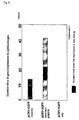
- NEC8 cells in which the proliferation was promoted by overexpression of HST-1, and HepG2 cells in which the proliferation was promoted by the gene other than HST-1 were seeded at 0.5 X 10 5 cells/well, and the cells were cultured for 4 days. After the culture, the inhibition of cell proliferation was assayed using TetraColor ONE Cell Proliferation Assay Reagent. Specifically, the sample solution stained was determined spectrometrically at 650 nm, using the absorbance at 450 nm as control ( Figure 13).
- GFP-positive cells ratio 2 days after the seeding (%) atelo/adeno only adeno only atelo left at r.t. 1 day 64 72 0 left at r.t. 7 days 42 10 or less 0 left at r.t. 14 days r.t. means room temperature.
- Table 5 shows that the adenovirus vector survived during storage for even 14 days, suggesting that the solid coating of the present invention provides a plate for screening that comprises adenovirus vectors, which could be conveniently distributed on the market.
- the plate can be stored at least for 2 weeks to 4 weeks without lowered activity.
- Embryonic stem cells ES cells
- ES cells Embryonic stem cells
- the expression of GFP was checked with fluorescence microscopy, simultaneously with determining the fluorescence intensity.
- the fluorescence intensity of GFP is positively correlated with the dose of adenovirus ( Figure 14).
- Figure 14 shows that the present invention should provide a useful tool for high-throughput screening for ES cells using adenovirus, and a plate for screening, which is coated with complexes formed by adenovirus and collagen.
- the gel formulation was penetrated through a filter having a pore size of 70 ⁇ m or 10 ⁇ m to prepare a gel formulation having an ordered size.
- PicoGreen dsDNA Quantitation Reagent (Molecular Probes) was added to stain pCMV-EGFP, and the major axis of the complexes were determined by fluorescent microscopy.
- a plasmid DNA (pCMV-HST-1-IL-2, 6.2kbp) encoding the secretory signal peptide of HST- 1 and interleukin-2, and an atelocollagen were mixed together to prepare a formulation in a gel form, and the major axis of the complexes was determined.
- the gel formulation was electrophoresed on agarose gel (Tris-acetate buffer, 0.8% agarose gel) to separate plasmid DNAs formed into complexes from those not formed into complexes.
- the band of the plasmid DNAs that were not formed into complexes was stained with ethidium bromide, and the fluorescence intensity was determined with Fluor-S-Multi Imager (BIO-RAD).
- Amount of the plasmid DNAs that were not formed into complexes were determined on the basis of the standard curve obtained from the fluorescence intensity of the band of the plasmid DNA with the known concentration as electrophoresed, and the ratio of the negative charge of the DNA contained in the complex to the molecular weight of the collagen. The results are shown in Table 6.
- Example 2 wherein pCAHST-1 was used to prepare complexes.
- the plasmid DNAs as used therein were different each other in size as pCAHST-1 is 7.9kbp, pCMV-EGFP is 4.7kbp, and pCMV-HST-1-IL-2 is 6.2kbp, showing that sizes of plasmid DNAs never affect the form or the shape of complexes to be formed.
- 0.1 M phosphate buffer and 0.01 M phosphate buffer containing sodium chloride were used as a solvent to obtain complexes having an average major axis of 3.4 ⁇ m to 29 ⁇ m, and no complexes having a 100 ⁇ m or more in size, when collagen concentration was 0.001 % to 0.1 %. It was believed that this was caused by the salts, which inhibited the formation of collagen association bodies, showing that when complexes are prepared in the presence of an agent that inhibits the formation of collagen association body, then complexes having an average major axis of 100 ⁇ m or less can be prepared irrespective of collagen concentration.
- Figure 15 shows the relationship between the number of collagen bound to one molecule of plasmid DNA and the average major axis of the complexes.
- pCMV-EGFP and pCMV-HST-1-IL-2 as used in a water without salts, it was found that the average major axis of complexes trends to extend as increasing in the number of collagen bound to plasmid DNA.
- collagen association body formed by attaching a collagen molecule with a plasmid DNA does not extend along the major axis well, and therefore a lot of fine association bodies attach to a plasmid DNA to form a structure.
- Collagens have been known to form fine association bodies at appropriate salt concentrations.
- a partial amount of collagens has already formed with a plasmid DNA into fine bodies before mixing with a plasmid DNA, and the fine collagen association bodies are attached to a plasmid DNA to form complexes.
- complexes having an average major axis of 50 ⁇ m or less can be prepared by mixing a plasmid DNA and a collagen that has been formed into fine association bodies.
- Figure 16 shows the relationship between the number of nucleotide monomer of plasmid DNAs per one collagen molecule, and the average major axis of the complexes. It was found that the average major axis of complexes trends to extend as decreasing in the number of nucleotide monomer of plasmid DNAs.
- the maximum number of nucleotide monomer per one collagen molecule is obtained by forming a complex with each one molecule of collagen and plasmid DNA, and is defined by the number of nucleotide monomer comprised in a plasmid DNA.
- aqueous solution or a 0.1 M phosphate buffer (PB), each of which contained a phosphorothioate antisense oligonucleotide (5'-CTCGTAGGCGTTGTAGTTGT-3'; Molecular weight, about 6500; SEQ ID NO: 8) (Espec Inc.) having a sequence complementary to a sequence from 4196 bp to 4216 bp of fibroblast growth factor HST-1 (FGF4) gene (described in Proc. Natl. Acad. Sci.
- FGF4 fibroblast growth factor HST-1
- the gel formulations were centrifuged at 150,000 rotations per hour at 5 °C to precipitate the complexes, and the amounts of the oligonucleotides released in the supernatant were determined by HPLC (column: Puresil C18 5 (Waters), mobile phase: a gradient of 24.5% to 35% acetonitrile over 35 minute in 0.1 M ammonium acetate containing 5mM tetrabutyl ammonium and 1mM EDTA, flow rate: 1 ml/min, detection wavelength: 260 nm).
- O.C.( ⁇ M) means oligonucleotide concentration ( ⁇ M).
- C.C. (%) means collagen concentration (%).
- Numberer of O.M per C.M means number of oligonucleotide molecule per one collagen molecule.
- N.M per C.M means number of nucleotide monomer per collagen molecule.
- Example 10 wherein plasmid DNAs were used, complexes were obtained by mixing oligonucleotides and collagens.
- oligonucleotides are smaller than plasmid DNAs in molecular size, and thus the formation of collagen association bodies is inhibited.
- Figure 17 shows the relationship between the molecular number ratio of oligonucleotide to collagen at the time of the mixture and the number of oligonucleotide bound to one molecule of collagen in the complex. It was found that the number of oligonucleotides bound to one collagen molecule in the formed complexes trends to increase as increasing in the number of oligonucleotides relative to the number of collagen molecule at the time of mixing in both cases that a water and the phosphate buffer were used as a solvent. The maximum number of oligonucleotide molecule bound to one collagen molecule varies depending on kinds of solvent, and it is 6 molecules in case of water, whereas it is 3 molecules in case of the phosphate buffer.
- oligonucleotide transfer is more efficient when a complex that contains a high content of oligonucleotide molecules is used. Accordingly, a complex wherein the number of oligonucleotide molecule per one collagen molecule is increased is desired, and generally speaking, a complex wherein the number of nucleotide monomer bound to one collagen molecule is increased is preferred.
- Example 11 Fifty ⁇ l of the gel formulation as prepared in Example 11 (11-3, the composition of the formulation of Example 7) was added dropwise to the bottom of a 96-well microplate, and dried by directly spraying a cool air (room temperature, 2 hours) or by placing in a desicator with silica gel (room temperature, 2 days) so as to prepare a cell culture instrument, of which the cell culture surface is coated with a complex.
- a cell culture instrument of which the cell culture surface is coated with a complex.
- PBS phosphate buffer
- Example 10 Fifty ⁇ l of the gel formulation as prepared in Example 10 (10-3, having the composition that was found efficient in transfer efficiency in Example 6) was added dropwise to the bottom of a 96-well microplate, and dried by directly spraying a cool air (room temperature, 2 hours) or by placing in a desicator with silica gel (room temperature, 2 days) so as to prepare a cell culture instrument, of which the cell culture surface is coated with a complex.
- PBS phosphate buffer
- Example 10-1, 10-4, and 10-7 Three hundreds ⁇ l of the gel formulations as prepared in Example 10, 10-1, 10-4, and 10-7, comprising pCMV-EGFP were added to a 6-well cell culture microplate, and dried by spraying a cool air so as to prepare a cell culture instrument onto which a complex comprising pCMV-EGFP was coated.
- 300 ⁇ l of an aqueous solution comprising pCMV-EGFP at 100 ⁇ g/ml was added to a plate, and dried similarly.
- 7.5 X 10 4 cells of 239 cells were seeded, and a DMEM medium containing 10 % FBS was added thereto, followed by culturing at 37 °C. From the start of the culture, the medium was replaced with a fresh medium every 4 or 5 days. eleven days after the seeding, the cells were observed by fluorescent microscopy, and the number of the cells expressing GFP was counted to estimate transfer efficiency.
- 300 ⁇ l or 500 ⁇ l of the gel formulations as prepared in Example 10, 10-13 to 22, and 24, comprising pCMV-HST-1-IL-2 were added to a 6-well cell culture microplate, and dried by spraying a cool air so as to prepare a cell culture instrument onto which a complex comprising pCMV-HST-1-IL-2 was coated.
- 300 ⁇ l or 500 ⁇ l of an aqueous solution comprising pCMV-HST-1-IL-2 at 100 ⁇ g/ml, PB solution or PBS solution were added to a plate, and dried similarly.
- a DMEM medium containing 10 % FBS was added thereto, followed by culturing at 37 °C for 10-13 to -19. Eight days after the cell seeding, the medium was replaced, and IL-2 concentration in the medium sampled 11 days later was determined by ELISA (Quamtikine human IL-2 (R&D Systems)). For 10-20 to -22, a DMEM medium was added to the plate, and cultured overnight at 37 °C, after which on the following day the medium was replaced with a DMEM medium containing 10% FBS, and then cultured at 37 °C.
- IL-2 concentration in the medium sampled 15 days later was determined by ELISA. Further, for 10-20, 21, and 24, a DMEM medium containing 10% FBS was added to the plate, and the plate was cultured at 37 °C. IL-2 concentration in the medium sampled 8 days after the cell seeding was determined by ELISA.
- a 6-well plate 5 X 10 4 cells of 239 cells were seeded, and cultured in the presence of a DMEM medium containing 10 % FBS at 37 °C.
- a DMEM medium containing 10 % FBS at 37 °C.
- 300 ⁇ l of the gel formulations as prepared in Example 10, 10-1, 10-4, 10-7, 10-8, 10-9, and 10-10, comprising pCMV-EGFP, and a water or a PB solution comprising pCMV-EGFP at 100 ⁇ g/ml as control were each added thereto, and the cells were cultured in the presence of a DMEM medium containing 10 % FBS at 37 °C.
- the media were replaced with a fresh medium, and 13 days later for 10-1, 10-4, and 10-7, or 5 days later for 10-8, 10-9, and 10-10, the cells were observed by fluorescent microscopy, followed by counting the number of the cells expressing GFP.
- 7.5 X 10 4 cells of 239 cells were seeded into a 6-well plate, and cultured in the presence of a DMEM medium containing 10 % FBS at 37 °C.
- a DMEM medium containing 10 % FBS containing 10 % FBS at 37 °C.
- 300 ⁇ l of the gel formulations as prepared in Example 10, 10-20 and 10-21, comprising pCMV-HST-1-IL-2, and a PB solution comprising pCMV-HST-1-IL-2 at 100 ⁇ g/ml as control were each added thereto, and the cells were cultured in the presence of a DMEM medium containing 10 % FBS at 37 °C.
- the media were replaced with a fresh medium, and 5 days later, the media were sampled, and IL-2 concentration therein were determined by ELISA (Quamtikine human IL-2 (R&D Systems)).
- complexes of the present invention it is possible to preserve and stabilize desired nucleic acids by complexing with collagens or collagen derivatives, 2) to transfer efficiently desired nucleic acids into cells when administered to living bodies, and 3) to express and inhibit desired nucleic acids without reagents for gene transfer.
- the complexes of the present invention can be applied to various aspects since the complexes of the invention have advantages 1) that DNAs, antisense oligonucleotides and virus vectors can be preserved and stabilized, 2) that the complexes can be stored during a long period of time on plates dried where the complexes are coated, 3) that genes can be expressed without reagents for gene transfer when cells are seeded onto the plates, 4) that kinds of subjects of nucleic acids and methods for immobilization are not limited to specific ones, and 5) that the duration time of the expression in case of plasmid DNAs is excellently extended.
- antisense oligonucleotides it is possible to readily examine the functions of genes or proteins, of which the expressions are inhibited by antisense oligonucleotides by determining the proliferation or the morphology of the cells, or the expression levels of cytokines or receptors.
- antisense oligonucleotides but also other materials can be complexed with collagens to form complexes, which in turn can be coated onto plates; in case of ribozymes, it is possible to carry out a screening similar to that of antisense oligonucleotides; and further in case of plasmid DNAs or adenoviruses, it is possible to examine the functions of genes or proteins by determining the changes of cells by the expression of certain genes.
- Collagens or collagen derivatives comprised in the complexes of the present invention have been known to affect no cells, and the screening using the complexes of the present invention makes it possible to carry out the examinations of gene functions that seldom avoid the unspecific affections to cells, which would be induced by conventional reagents for gene transfer.
Landscapes
- Health & Medical Sciences (AREA)
- Life Sciences & Earth Sciences (AREA)
- Engineering & Computer Science (AREA)
- Genetics & Genomics (AREA)
- Bioinformatics & Cheminformatics (AREA)
- Chemical & Material Sciences (AREA)
- General Health & Medical Sciences (AREA)
- Biotechnology (AREA)
- Molecular Biology (AREA)
- Medicinal Chemistry (AREA)
- Pharmacology & Pharmacy (AREA)
- Animal Behavior & Ethology (AREA)
- Public Health (AREA)
- Veterinary Medicine (AREA)
- Biomedical Technology (AREA)
- Epidemiology (AREA)
- Organic Chemistry (AREA)
- General Engineering & Computer Science (AREA)
- Zoology (AREA)
- Wood Science & Technology (AREA)
- Proteomics, Peptides & Aminoacids (AREA)
- Physics & Mathematics (AREA)
- Biochemistry (AREA)
- Biophysics (AREA)
- Plant Pathology (AREA)
- Microbiology (AREA)
- Chemical Kinetics & Catalysis (AREA)
- General Chemical & Material Sciences (AREA)
- Nuclear Medicine, Radiotherapy & Molecular Imaging (AREA)
- Medicines That Contain Protein Lipid Enzymes And Other Medicines (AREA)
- Pharmaceuticals Containing Other Organic And Inorganic Compounds (AREA)
- Saccharide Compounds (AREA)
- Micro-Organisms Or Cultivation Processes Thereof (AREA)
- Preparation Of Compounds By Using Micro-Organisms (AREA)
- Measuring Or Testing Involving Enzymes Or Micro-Organisms (AREA)
- Investigating Or Analysing Biological Materials (AREA)
- Medicinal Preparation (AREA)
Abstract
Description
- The present invention belongs to medical field, specifically to gene therapy and genetic fundamental research. More specifically, the invention relates to preparations for facilitating the transfer of a desired nucleic acid into a target cell, and processes therefor.
- Recently, gene therapy has been actively studied, and applied practically to clinical therapy of various cancers and genetic diseases. Gene therapy is an approach to treat a disease by repairing or correcting a defective gene, and comprises transferring a gene encoding an intended enzyme, cytokine, or the like into a cell of a patient, and allowing to produce the intended substance from the gene in the body, thereby treating the disease. Gene therapy is a medication that controls a basis of life, and has a potential to treat various diseases such as AIDS, rheumatoid arthritis, lifestyle-related diseases, in addition to cancers and genetic diseases.
- In gene therapy, transfer efficiency of gene into a target cell is an important factor in the increased efficacy of the therapy. Gene therapy for cancers includes therapies by virus such as adenovirus (Cardiovascular Research, 28, 445 (1994); Science, 256, 808 (1992); Gastroenterology, 106, 1076 (1994); TIBTECH, 11, 182 (1993); J. Biol. Chem, 266, 3361 (1991); Nature Medicine, 1, 583 (1995) and the cited references therein) and those by liposome formulations (Biochem Biophys Acta, 1097, 1 (1991); Human Gene Therapy, 3, 399 (1992); Proc. Natl. Acad. Sci. USA, 89, 11277 (1992)). Transfer efficiency of genes is generally higher in therapy using virus vectors than therapy using liposome formulations. However, therapy using virus vectors suffers from a problem that multiple administrations are hardly conducted due to immunological responses to viruses (J. Biol. Chem., 269, 13695(1994), Am. J. Respir. Cell Mol. Biol., 10, 369 (1994)).
- On the other hand, since the analysis on the whole human genetic information (human genome) was almost completed, the focus has been shifted to post-genome strategies how to utilize the accumulated human genetic information in the fields of medication and industry. Specifically, examinations on human gene functions, as well as the structures and functions of the proteins encoded by the gene using the analyzed genetic information have been emphasized. Such a post-genome examinations require the expression and the production of proteins, which necessarily involve the transfer of intended genes into cells. Genes to be transferred into host cells by adenovirus vectors and liposome vectors or plasmid DNA vectors are not integrated into the genome of the cells, and are transiently expressed. Such vectors can not accomplish the constitutive expression of the genes, which is important in gene therapy and analysis on gene functions.
- Thus, an approach to efficiently transfer a nucleic acid representing a gene into a desired cell, and to express the gene during a long period of time without integration of the gene into chromosome of host cells is expected to provide a great utility.
- The inventors of the present application found that collagens have an unexpected action, and created an approach to efficiently transfer a nucleic acid into a desired cell. Specifically, we found that the contact of a collagen and a nucleic acid such as plasmid DNA surprisingly results in the formation of a complex, and the formation of a complex facilitates the transfer of a nucleic acid into a cell and expresses the gene during a long period of time. Although Japanese Patent Publication (kokai) No. 71542/1997 describes formulations containing a gene wherein the gene is comprised in a carrier of a biocompatible material such as a collagen, the formulations are sustained release formulations that gradually releases the gene in a living body.
- The invention is based on the newly founded use of a collagen or a collagen derivative.
- More specifically, the invention relates to:
- (1) A preparation for facilitating the transfer of a nucleic acid into a target cell, which comprises a collagen or a collagen derivative;
- (2) A preparation for facilitating the transfer of a nucleic acid into a target cell, which comprises a collagen or a collagen derivative complexed with a desired nucleic acid, preferably a preparation for facilitating the transfer of a nucleic acid, wherein the complex is in a form of particle, more preferably a preparation for facilitating the transfer of a nucleic acid, wherein the major axis of the particle is 300 nm to 300 µm, preferably 300 nm to 100 µm, more preferably 300 nm to 50 µm, even more preferably 300 nm to 30 µm; Specifically, a preparation for facilitating the transfer of a nucleic acid wherein the desired nucleic acid is a plasmid DNA, and wherein the ratio of the number of a collagen molecule or a collagen derivative molecule to the number of a nucleotide monomer of the plasmid DNA in the complex is 1 : 20 to 1 : the number of a nucleotide monomer of the plasmid DNA, preferably 1 : 50 to 1 : the number of a nucleotide monomer of the plasmid DNA, more preferably 1 : 50 to 1 : 4000, still more preferably 1 : 50 to 1 : 2000, and still more preferably 1 : 50 to 1 : 1000, or a preparation for facilitating the transfer of a nucleic acid wherein the nucleic acid is an oligonucleotide, and which the ratio of the number of a collagen molecule or a collagen derivative molecule to the number of a nucleotide monomer of the oligonucleotide in the complex is 1 : 1 to 1 : 200, preferably 1 : 3 to 1 : 150, more preferably 1 : 20 to 1 : 120, and still more preferably 1 : 50 to 1 : 120;
- (3) A particle of the complex comprising a collagen or a collagen derivative and a desired nucleic acid;
- (4) A process for preparing a particle of the complex according to the present invention, which comprises mixing a collagen or a collagen derivative and a desired nucleic acid in a solution comprising an agent that inhibits the formation of collagen association body;
- (5) A medical instrument, of which the surface is coated with a particle of the complex according to above (3) or a cell culture instrument, of which the surface is coated with the particle of the complex;
- (6) A process for transferring a desired nucleic acid into a target cell or a process for improving the expression level of a desired nucleic acid in a target cell, which comprises using a particle of the complex according to above (3);
- (7) A process for examining the function of a gene or a protein in a target cell, which comprises coating a solid surface with a particle of the complex according to above (3) that comprises the gene, a gene encoding the protein, or a nucleic acid inhibiting the expression of the gene or the protein in a cell; culturing the target cell on the solid surface; and examining the expression level of the nucleic acid or the expression level of the gene or the protein in the target cell, or the proliferation ratio or the phenotype of the cell; and
- (8) A process for screening for a nucleic acid that treats a disease, which comprises coating a solid surface with a particle of the complex according to above (3) that comprises a nucleic acid candidate that inhibits the expression of a gene associated with the disease in a cell; culturing the cell presenting the condition of the disease on the solid surface; and examining the expression level of the gene to be inhibited with each of the nucleic acid candidate, or the proliferation ratio or the phenotype of the cell.
-
- The working examples hereinafter illustrate that the preparations for facilitating the transfer of a nucleic acid according to the present invention improved the transfer efficiency of gene into a target cell as shown to express the nucleic acid in a cell culture system in vitro where the gene expression is not observed by mere plasmid DNA. Further, those examples illustrate that the preparations for facilitating the transfer of a nucleic acid according to the present invention increased the stability of a nucleic acid within a cell as shown to sustain the expression of the nucleic acid during a longer period of time than liposome formulations.
- According to the invention, it has been found that a collagen is interacted electrostatically and/or physically with a nucleic acid to form a complex. Thus, it is believed that the sustained expression of a nucleic acid as observed in the working examples would result from the complex formation leading to the increased stability of nucleic acids within cells. This is quite different from the mechanism of the sustained release of gene by collagens that was conventionally understood that a gene encapsulated in collagen matrix is gradually released according to the biological degradation of collagen.
-
- Figure 1 is a photograph substitute for drawing which depicts an agarose-gel electrophoresis showing the electrostatic interaction between the defined concentration of plasmid DNA and the various concentrations of atelocollagen.
- Figure 2 is a photograph substitute for drawing which depicts an agarose-gel electrophoresis showing the effect of sodium chloride on the electrostatic interaction between plasmid DNA and atelocollagen.
- Figure 3 is a photograph substitute for drawing which depicts an agarose-gel electrophoresis showing the effect of heparan sulfate on the electrostatic interaction between plasmid DNA and atelocollagen.
- Figure 4 is a micrograph showing a form of the complexes between plasmid DNA and atelocollagen in various concentrations.
- Figure 5 is a micrograph showing a form of the complexes between plasmid DNA and atelocollagen in various concentrations that were stored for a week.
- Figure 6 is a graph showing a comparison in the duration time of gene expression among the atelocollagen gel formulation, the cationic liposome formulation, and the plasmid DNA in a PBS solution.
- Figure 7 is a graph showing the relationship between the complexes comprising a collagen in various concentrations and the transfer efficiency of plasmid DNA seven days after the transfection by dropwise addition.
- Figure 8 is a graph showing a fluorescence intensity representing the transfer efficiency of plasmid DNA in the complexes comprising a collagen in various concentrations seven days after the transfection.
- Figure 9 is a graph showing the relationship between the complexes comprising a collagen in various concentrations and the transfer efficiency of plasmid DNA seven days after the transfection by solid coating.
- Figure 10 is a graph showing the relationship between the complexes comprising a plasmid DNA in various concentrations and the transfer efficiency of plasmid DNA seven days after the transfection by dropwise addition.
- Figure 11 is a graph showing the relationship between the complexes comprising a plasmid DNA in various concentrations and the transfer efficiency of plasmid DNA seven days after the transfection by solid coating.
- Figure 12 is a graph showing a fluorescence intensity representing the transfer efficiency of plasmid DNA seven days after the transfection by dropwise addition, and a micrograph showing the fluorescence.
- Figure 13 is a graph showing inhibitory effects of the present invention on the cell proliferation.
- Figure 14 is a graph showing that adenovirus was transferred by solid coating in a dose-dependent manner, and a micrograph showing the fluorescence.
- Figure 15 is a graph showing the relationship between the number of collagen bound to one molecule of plasmid DNA and the average major axis of the complexes.
- Figure 16 is a graph showing the relationship between the number of nucleotide monomer of desired nucleic acids per collagen molecule and the average major axis of the complexes.
- Figure 17 is a graph showing the relationship between the molecular number ratio of oligonucleotide to collagen at the time of the mixture and the number of oligonucleotide bound to one molecule of collagen in the complex.
- Figure 18 is a fluorescence micrograph obtained by observing the complexes comprising the oligonucleotides released from the surface of the cell culture instrument according to the present invention with fluorescence microscopy.
- Figure 19 is a fluorescence micrograph obtained by observing the complexes comprising the plasmid DNA released from the surface of the cell culture instrument according to the present invention with fluorescence microscopy.
-
- As the first embodiment, the invention provides a preparation for facilitating the transfer of a nucleic acid into a target cell, which comprises a collagen or a collagen derivative. The embodiment is based on the effect of a collagen or a collagen derivative on the facilitation of the transfer of a nucleic acid into a target cell, which has been found for the first time. In other words, the present embodiment of the invention provides a new use of a collagen or a collagen derivative to facilitate the transfer of a nucleic acid into a target cell.
- As used herein, "a collagen or a collagen derivative" generally means any kind of collages or collagen derivatives as used in medical, cosmetic, industrial, and food fields. A soluble collagen or a solubilized collagen is preferably utilized. Soluble collagens are soluble in an acidic or neutral water or a water containing a salt, whereas solubilized collagens include an enzymatically solubilized collagen which may be solubilized with an enzyme, an alkali-solubilized collagen which may be solubilized with an alkali, both collagens being preferably capable of penetrating through a membrane filter having a pore size of 1 micrometer. Solubility of collagen varies depending on the crosslinking degree of the collagen, and higher is the crosslinking degree, more difficult the collagen is solubilized. Accordingly, the crosslinking degree of a collagen as used in the present invention is, for example, not more than trimer, more preferably not more than dimer. Preferable molecular weight of the collagen is, for example, from about 300,000 to about 900,000, and more preferably from about 300,000 to about 600,000. Collagens as used herein include those extracted from any animal species, and it is desired that preferable collagens are extracted from vertebrates, more preferable collagens are extracted from a mammal, a bird, or a fish, and still more preferable collagens are extracted from a mammal or a bird having a high denaturation temperature. Any type of collagen may be used, and, because of the type existing in animal bodies, type I - V collagens are preferable. For example, such collagens include a type I collagen obtained by acid extraction from a mammal dermis, and, more preferably, they include, for example, a type I collagen obtained by acid extraction from calf dermis, a type I collagen produced by genetic engineering, and the like. Collagens derived from tendon, which are also type I collagens, are not suitable because they have a high degree of crosslinking and are insoluble. Further, an atelocollagen that is obtained by removing enzymatically a telopeptide having a high antigenicity or an atelocollagen produced by genetic engineering is preferable for the sake of safety, and an atelocollagen having three or less tyrosine residues per 1000 residues is more preferable. Alternatively, collagens having a modified side chain, crosslinked collagens or the like may be utilized if desired. Collagens having a modified side chain includes, for example, succinylated collagens and methylated collagens, whereas crosslinked collagens include, for example, collagens treated with glutaraldehyde, hexamethylene diisocyanat, a polyepoxy compound or the like (Fragrance Journal 1989-12, 104-109, Japanese Patent Publication(kokai) No. 59522/1995). Preferred collagen derivatives are a gelatin or a gelatin-crosslinking complex, or a crosslinking complex thereof with a collagen.
- Collagens or collagen derivatives may be used in admixture with another biocompatible material. Biocompatible materials include, for example, gelatin, fibrin, albumin, hyaluronic acid, heparin, chondroitin sulfate, chitin, chitosan, alginic acid, pectin, agarose, hydroxyapatite, polypropylenes, polyethylenes, polydimethylsiloxane, and a polymer of glycolic acid, lactic acid or amino acid, and a copolymer thereof, and a mixture containing two or more of those biocompatible materials.
- As the second embodiment, the present invention provides a preparation for facilitating the transfer of a nucleic acid into a target cell, which comprises a collagen or a collagen derivative and a desired nucleic acid; preferably comprises particles of a complex as an essential component.
- Nucleic acids are hardly transferred into cells when administered to the cells in vitro solely in the presence of blood serum. Nucleic acids can be efficiently transferred into cells when formed with a collagen or a collagen derivative into a complex.
- As used herein, "a nucleic acid" may be any polynucleotide or any oligonucleotide, and may be any DNA or RNA molecule. DNA molecules include a plasmid DNA, cDNA, a genomic DNA or a synthesized DNA. Both DNA and RNA may be double-stranded or single-stranded. Single-stranded ones include a coding strand and a non-cording strand. As used herein, "a nucleic acid" includes a DNA derivative and an RNA derivative, which derivative means a nucleic acid having a phosphorothioate bond, or a nucleic acid containing an internucleotide having a phosphate, sugar or base moiety chemically modified to avoid enzymatic degradations. As used herein, "a nucleic acid" also includes viruses such as adenovirus and retrovirus.
- Preferably, "a nucleic acid" is an oligonucleotide or a ribozyme, and more preferably an oligonucleotide or a ribozyme that is from 5 to 100 mer, more preferably from 5 to 30 mer. It is preferred to utilize a plasmid DNA encoding a protein exhibiting a physiological activity to treat or ameliorate pathological conditions or a plasmid DNA encoding a protein inducing an immunological response to treat or ameliorate pathological conditions.
- When the nucleic acid is a vector as used in gene therapy such as a plasmid DNA or a virus, it is preferably a system as constructed to express the encoded genetic information in cells, such as a vector that comprises an element such as a promoter necessary to express an intended gene, or an element capable of integrating into chromosomes. Size of plasmid DNAs as a nucleic acid used herein is not limited, and may be selected appropriately from the sizes that allow the encoded genetic information to be efficiently prepared via genetic engineering, and efficiently expressed in cells to be transferred.
- The preparation for facilitating the transfer of a nucleic acid according to the invention may contain a few kinds of separate vectors incorporated with different desired nucleic acids. Further, a vector may comprise many genetic information. Amounts of the vector comprised in the preparation for facilitating the transfer are not limited.
- Nucleic acids encoding a protein necessary to be expressed in gene therapy includes any gene capable to be used in the treatment of a genetic disease, which is exemplified by, but is not limited to, a gene encoding an enzyme such as adenosine deaminase, thymidine kinase; a cytokine such as GM-CSF, IL-2; or fibroblast growth factor HST-1 (FGF4). Nucleic acids encoding other proteins necessary to be expressed in gene therapy includes, but is not limited to, a gene aimed at the treatment or the prevention for an infection or a tumor, which encodes a protein or a peptide serving as an antigen to induce immune response, i.e., the gene encoding the protein or the peptide capable of serving as an antigen such as mentioned above, for example, a gene encoding the surface protein HA or NA, or the nuclear protein NP of influenza virus, type C hepatitis virus E2 or NS 1 protein, type B hepatitis virus HBs antigen protein, type A hepatitis virus capsid protein VP1 or VP3 or capsidoid protein, dengue virus Egp protein, RS virus F or G protein, G or N protein of the rabies virus structural protein, herpes virus gD protein, Japanese encephalitis virus E1 or pre-M protein, rotavirus coat protein VP7 or coat protein VP4, human immunodeficiency virus gp 120 or gp 160 protein, Leishmania major surface antigen protein, malaria circum sporozoite major surface antigen protein, Toxoplasma 54-kd or CS protein, cell surface protein PAc of caries-causing Streptococcus mutans; a gene encoding tumor regression antigens such as MAGE-1, MAGE-3, and BAGE, tissue-specific antigens such as tyrosinase, Mart-1, gp 100 and gp75, p15, Muc1, CEA, HPV, E6, E7, HPR2/neu, etc.; and the genes which are described in "Immunization with DNA"; Journal of Immunological Methods, vol. 176, 1994, pages 145-152.
- In the case that nucleic acids are oligonucleotides, they includes a base sequence, of which at least the portion binds complementarily under physiological condition to a sense or antisense strand of a gene encoding a protein that has a physiological effect to disrupt the homeostasis of a living body, a gene specific to pathogenic viruses, bacteria or the like, and, more specifically, they include a base sequence that binds complimentarily to the messenger RNA of a gene specific to a pathogenic virus, a bacterium or the like, or a gene encoding a protein having a physiological effect to disrupt the homeostasis of a living body.
- More specifically, oligonucleotides as used in the present invention includes a sequence complementary to a region containing an initiation codon of a messenger RNA, or to a splicing site of a precursor messenger RNA. Examples of the oligonucleotides as used in the present invention include, for example, those used for treatment or prevention of a cancer, such as an oligonucleotide having a sequence of 5'-CTCGTAGGCGTTGTAGTTGT-3' (SEQ ID NO:1) which specifically inhibits the expression of hst-1; ISIS3521 which specifically inhibits the expression of protein kinase Ca to effectively treat progressive cancers such as non-small cell lung carcinoma and colon cancer, and which has been applied in the trials to the treatment of prostate cancer, breast cancer, ovary cancer, pancreas cancer, large intestinal cancer, small cell lung carcinoma; ISIS5132/CGP69846A which specifically inhibits the expression of C-raf kinase and has been applied in the trials to the treatment of prostate cancer, breast cancer, ovary cancer, cephalophyma, pancreas cancer, large intestinal cancer, small cell lung carcinoma; ISIS 2503 which specifically inhibits the expression of Haras and has been applied in the trials to the treatment of large intestinal cancer, breast cancer, cephalophyma, pancreas cancer, and small cell cancer; GEM231 which specifically inhibits the expression of protein kinase A type I; MG98 which specifically inhibits the expression of DNA methyl transferase; INXC-6295 which inhibits the expression of c-myc; INX-3001 which inhibits the expression of c-myb and has been considered to be applied to the treatment of leukemia; G-3139 (Genasense) which inhibits expression of bc1-2 and has been considered to be applied to the treatment of non-Hodgkin's lymphoma, large intestinal cancer, small cell lung carcinoma, chronic lymphatic leukemia, acute myeloid leukemia, breast cancer, lymphoma, melanoma, myeloma, non-small cell lung carcinoma, prostate cancer; an oligonucleotide which inhibits the expression of MDM2 protein; an oligonucleotide which inhibits the expression of VEGF and the like. Further, examples of the oligonucleotides used for treatment or prevention of infectious diseases include GEM92 and GPI-2A which inhibits the growth of HIV. ISIS2922 (fomivirsen), Vitravene, ISIS 13312 and GEM 132 which inhibits the growth of cytomegalovirus, ISIS 14803 which inhibits the growth of hepatitis C virus, and the like. Examples of the oligonucleotides used for treatment of inflammation include ISIS2302 which specifically inhibits the expression of ICAM-1, and which has been applied in the trials to the treatment of Crohn's disease, ulcerative colitis, kidney transplantation rejection inhibition, psoriasis, asthma; EPI-2000 which inhibits the expression of
adenosine A 1 receptor and has been applied in the trials to the treatment of asthma; and oligonucleotides which inhibit the expression of TNF- α, CD49d (VLA-4), VCAM-1, PECAM-1 and the like. Further, examples of the oligonucleotides that prevent the restenosis after percutaneous transluminal coronary angiogenesis include Resten-NG that inhibits the expression of c-myc. - Genes encoding a protein that exhibits a physiological effect to disrupt the homeostasis include, for example, a series of genes, so-called cancer genes. Specifically, they include genes for growth factors, receptor type tyrosine kinases, non-receptor type tyrosine kinases, GTP-binding proteins, serine-threonine kinases, transcription factors and the like. More specifically, they include genes coding for hst-1 or ornithine decarboxylase and the like.
- The preparation for facilitating the transfer of a nucleic acid according to the present invention may further comprise a pharmaceutically acceptable additive as appropriate in addition to the complex of the present invention. Pharmaceutically acceptable additives include an agent making isotonic, a pH modifier, and a soothing agent in case of the use of the complex as injection, and an excipient, a disintegrator, and a coating agent in case of the use of the complex as solid, as well as those described in Japanese Handbook of Pharmaceutical Excipients (Japan Pharmaceutical Excipients Council). Specific examples include salts and saccharides, which are used to keep the pH 6-8, or to make isotonic with cells.
- The preparation for facilitating the transfer of a nucleic acid according to the present invention may be in a form of solid or solution. The preparation in a form of solid is loaded to a desired cell as it is, or after making a solution with a purified water, a physiological solution, a buffer isotonic with living bodies, or the like. Such preparation in a solution form also constitutes a part of the present invention.
- The administration of the preparation for facilitating the transfer of a nucleic acid according to the present invention may be selected from oral route, injection, eye drop, nasal drop, transpulmonary route, transdermal absorption, and oral route and injection are preferred. The preparation may be administered to various sites depending on diseases, and may be placed on the necessary site at the time of operation.
- In the present embodiment, the invention provides:
- (1) A preparation for facilitating the transfer of a nucleic acid into a target cell, which comprises as an essential component a complex, preferably particles of a complex comprising a desired nucleic acid and a collagen or a collagen derivative, wherein the major axis of the particle is preferably 300 nm to 300 µm, more preferably 300 nm to 100 µm, even more preferably 300 nm to 50 µm, still more preferably 300 nm to 30 µm;
- (2) The preparation for facilitating the transfer of a nucleic acid according to (1) wherein the target cell is an animal cell;
- (3) The preparation for facilitating the transfer of a nucleic acid according to (2) wherein the target cell is an organ or a tissue which requires to be treated, or its cell around;
- (4) The preparation for facilitating the transfer of a nucleic acid according to any one of (1) to (3) wherein the collagen is atelocollagen:
- (5) The preparation for facilitating the transfer of a nucleic acid according to any one of (1) to (4) wherein the molecular weight of the collagen is from about 300,000 to about 900,000;
- (6) The preparation for facilitating the transfer of a nucleic acid according to any one of (1) to (5) wherein the collagen derivative is a gelatin or a gelatin-crosslinking complex;
- (7) The preparation for facilitating the transfer of a nucleic acid according to any one of (1) to (5) wherein the collagen derivative is a collagen-crosslinking complex;
- (8) The preparation for facilitating the transfer of a nucleic acid according to any one of (1) to (7) wherein the nucleic acid is an oligonucleotide;
- (9) The preparation for facilitating the transfer of a nucleic acid according to (8) wherein the oligonucleotide is from 5 to 30 mer in length;
- (10) The preparation for facilitating the transfer of a nucleic acid according to (8) or (9) wherein the oligonucleotide is a DNA or a DNA derivative;
- (11) The preparation for facilitating the transfer of a nucleic acid according to (8) or (9) wherein the oligonucleotide is an RNA or an RNA derivative;
- (12) The preparation for facilitating the transfer of a nucleic acid according to (10) or (11) wherein the DNA derivative or the RNA derivative has at least one phosphorothioate bond;
- (13) The preparation for facilitating the transfer of a nucleic acid according to any one of (1) to (7) wherein the nucleic acid is a ribozyme or an oligonucleotide;
- (14) The preparation for facilitating the transfer of a nucleic acid according to any one of (1) to (7) wherein the nucleic acid is a plasmid DNA;
- (15) The preparation for facilitating the transfer of a nucleic acid according to (13) wherein the plasmid DNA encodes a protein that exhibits a physiological activity to treat or ameliorate pathological conditions;
- (16) The preparation for facilitating the transfer of a nucleic acid according to (13) wherein the plasmid DNA encodes a protein that induces an immunological response to treat or ameliorate pathological conditions;
- ( 17) The preparation for facilitating the transfer of a nucleic acid according to (1) to (16) wherein the preparation is in a form of solution, and the complex comprises 10 mg/ml or less of a nucleic acid;
- (18) The preparation for facilitating the transfer of a nucleic acid according to (17) wherein the complex comprises 1 mg/ml or less of a nucleic acid;
- (19) The preparation for facilitating the transfer of a nucleic acid according to (18) wherein the complex comprises 500 µg/ml or less of a nucleic acid;
- (20) The preparation for facilitating the transfer of a nucleic acid according to (1) to (19) wherein the preparation has pH5 to pH9, preferably, pH6 to pH8;
- (21) The preparation for facilitating the transfer of a nucleic acid according to (1) to (20) further comprising phosphoric acid in a concentration from 0.001 M to 0.1M;
- (22) The preparation for facilitating the transfer of a nucleic acid according to (21) further comprising phosphoric acid in a concentration from 0.01M to 0.1M;
- (23) The preparation for facilitating the transfer of a nucleic acid according to (1) to (22) further comprising an agent that inhibits the formation of collagen association body, for example sucrose or arginine; and
- (24) The preparation for facilitating the transfer of a nucleic acid according to (1) to (24) further comprising an appropriate amount of a pharmaceutically acceptable additive.
-
- As the third embodiment, the present invention provides a complex comprising a collagen or a collagen derivative and a desired nucleic acid. The complexes include electrostatic complexes that are bound and formed by the electrostatic force, and physical complexes that are bound and formed by the physical force such as hydrophobic binding. Both binding systems may coexist, and this embodiment is also fallen in the scope of the present invention.
- It has been found for the first time that a collagen is electrostatically interacted with a nucleic acid to form a complex.
- As used herein, "electrostatic complexes" means polyionic complexes between a collagen or a collagen derivative having many electric charges in the molecule and a nucleic acid, and specifically means binding bodies wherein a collagen or a collagen positively charged electrically attract to a nucleic acid negatively charged. In the polyionic complexes, many counter ions are released from the molecule on the complex formation, and therefore very large increase in entropy is produced. In view of the formation of such electrostatic complexes, it is understood that the sustained expression of a nucleic acid as observed in the working examples would result from the complex formation leading to the increased stability of nucleic acids within cells.
- Minimum unit of the complex of the present invention is a complex formed by one molecule of collagen and one molecule of nucleic acid. We found that a collagen forms a complex with a nucleic acid, and stimulatingly associates with another collagen to form an association body. The association body is formed in a manner that a collagen molecule having a cylindrical shape wherein the major axis is about 300 nm and the diameter is 1.5 nm is predominantly associated parallel to the longitudinal axis of the molecule. Accordingly, the complexes are formed by the noncovalent binding between many association bodies and many nucleic acid molecules, and include filamentous complexes having a longitudinal axis of 1 nm or more as a result of the largely developed extension of the bodies, filamentous complexes wherein fine bodies are each bound via nucleic acids, and particulate complexes formed by more fine complexes and nucleic acids.
- The complexes of the present invention can be various in shape.
- The complexes of the present invention are preferably in a form of particle. As used herein "particle" means a shape that a collagen or a collagen derivative could assume, and does not necessarily mean a spherical shape. Minimum size of the complexes of the present invention is 300 nm that corresponds to the major axis of a complex formed by one molecule of collagen. The particles have a major axis of 300nm to 1mm, and in view of transfer efficiency of nucleic acids, they preferably have a major axis of 300nm to 300µm, more preferably 300nm to 100µm, even more preferably 300nm to 50µm, still even more preferably 300nm to 30µm.
- We found that the ratio of a collagen or a collagen derivative to a nucleic acid composed in a complex is responsible for the fact that the complex can assume various forms or shapes, and that the form of the complex is dependent exclusively on the development of the association (fibrosis) of the collagen or collagen derivative promoted by the formation of the complex with the nucleic acid. We also found that the excess formation of the association bodies is not suitable for the transfer of the nucleic acid, and found that the form or the shape of the complex could be controlled by adjusting the concentration of the collagen or collagen derivative and the desired nucleic acid to be mixed, as well as environmental factors such as salt concentration, temperature, pH, and glucose concentration. Further, we observed that a plasmid DNA having 1000 bp or more is extremely promoted in the association, whereas a plasmid DNA having a 100 bp or less is weakly promoted in the association, and found that the association of collagen or collagen derivative involved in the complex formation is affected by the length of the nucleic acid, concluding that there is a composition of a collagen or collagen derivative and a nucleic acid optimal for the association depending on the length of the nucleic acid.
- The form of the shape of the complex may affect the transfer efficiency of nucleic acids into cells. The finding that collagens are complexed with nucleic acids, and the shape of the complex affects the transfer efficiency and the expression efficiency of nucleic acids in cells provides a strategy for optimizing the transfer efficiency of nucleic acids. Liposomes as used in laboratory to transfer nucleic acids into target cells are difficult to be widely utilized from practical view points, since they tend to aggregate together immediately when combined with nucleic acids, are varied in transfer efficiency on use, and are necessarily prepared just before use. However, the complexes of the present invention are stable in the form or the shape when stored in cold space. The most important issue in gene transfer technique is that widely used methods are quite few. The complexes of the present invention are stable in the form or the shape and could be used practically.
- In the present embodiment, the invention provides:
- (1) A particle of the complex comprising a desired nucleic acid and a collagen or a collagen derivative;
- (2) The particle of the complex according to (1), wherein the major axis is 300 nm to 300 µm;
- (3) The particle of the complex according to (2), wherein the major axis is 300 nm to 100 µm;
- (4) The particle of the complex according to (3), wherein the major axis is 300 nm to 50 µm, preferably 300 nm to 30 µm;
- (5) The particle of the complex according to (1) to (4), wherein the nucleic acid is an plasmid DNA;
- (6) The particle of the complex according to (5), wherein the ratio of the number of a collagen molecule or a collagen derivative molecule to the number of a nucleotide monomer of the plasmid DNA is 1 : 20 to 1 : the number of a nucleotide monomer of the plasmid DNA, preferably 1 : 50 to 1 : the number of a nucleotide monomer of the plasmid DNA, more preferably 1 : 50 to 1 : 4000, even more preferably 1 : 50 to 1 : 2000, still even more preferably 1 : 50 to 1 : 1000;
- (7) The particle of the complex according to (6), wherein the ratio is 1 : 96 to 1 : the number of a nucleotide monomer of the plasmid DNA;
- (8) The particle of the complex according to (7), wherein the ratio is 1 : 96 to 1 : 1122;
- (9) The particle of the complex according to (8), wherein the ratio is 1 : 96 to 1:701;
- (10) The particle of the complex according to (1) to (4), wherein the nucleic acid is an oligonucleotide;
- (11) The particle of the complex according to (10), wherein the ratio of the number of a collagen or a collagen derivative to the number of a nucleotide monomer of the oligonucleotide in the complex is 1 : 1 to 1 : 200, preferably 1 : 3 to 1 : 150, more preferably 1 : 3 to 1 : 120;
- (12) The particle of the complex according to (11), wherein the ratio is 1 : 20 to 1 : 120;
- (13) The particle of the complex according to (12), wherein the ratio is 1 : 50 to 1 : 120;
- (14) The particle of the complex according to (1) to (13), which is
comprised in a solution of
pH 5 to pH 9, preferablypH 6 topH 8; - (15) A process for preparing a particle of the complex according to (1) to (13), which comprises mixing a collagen or a collagen derivative and a desired nucleic acid in a solution cooled to 10 °C or less;
- (16) A process for preparing a particle of the complex according to (1) to (13), which comprises mixing a collagen or a collagen derivative and a desired nucleic acid in a solution comprising phosphoric acid in a concentration from 0.001 M to 0.1M;
- ( 17) A process for preparing a particle of the complex according to (1) to (13), which comprises mixing a collagen or a collagen derivative and a desired nucleic acid in a solution comprising phosphoric acid in a concentration from 0.01 M to 0.1M;
- (18) A process for preparing a particle of the complex according to (1) to (13), which comprises mixing a collagen or a collagen derivative and a desired nucleic acid in a solution comprising an agent that inhibits the formation of collagen association body, for example sucrose or arginine;
- (19) A process for preparing a particle of the complex according to (1) to (13), which comprises mixing a collagen or a collagen derivative and a desired nucleic acid in a solution comprising an appropriate amount of a pharmaceutically acceptable additive wherein the pharmaceutically acceptable additive is as defined above;
- (20) A process for preparing a particle of the complex according to (1) to (13), which comprises combining two or more of the processes according to (14) to ( 19);
- (21) A process for preparing a particle of the complex according to (1) to (13), which further comprises penetrating the complex through a filter having a pore size of 100 µm or less for size selection.
- (22) The process according to (21), which comprises penetrating the complex through a filter having a pore size of 10 µm or less for size selection;
- (23) A process for preparing a particle of the complex according to (1) to (13), which further comprises centrifuging the complex at 10,000 rpm or more for concentration and isolation;
- (24) The process according to (23), which comprises centrifuging the complex at 50,000 rpm or more.
-
- As used herein, "the number of a nucleotide monomer" means the number of a nucleotide monomer unit composed of a desired nucleic acid. As used herein, "the ratio of the number of a collagen molecule or a collagen derivative molecule to the number of a nucleotide monomer" means the number of a nucleotide monomer in a desired nucleic acid relative to one molecule of a collagen or collagen derivative comprised in a electrostatically or physically bound complex. The number of a nucleotide monomer is almost the same as "negative charge of a desired nucleic acid". As used herein, "negative charge of a desired nucleic acid" means the number of phosphate groups intervened between nucleotides composed of a nucleic acid. The numeral values of the negative charge of nucleic acid is equal to the number of phosphate groups of a nucleic acid, phosphate groups intervened between nucleotides, and phosphorus-containing groups intervened between nucleotides (phosphate groups and phosphorothioate groups)
- As used herein, "an agent that inhibits the formation of collagen association body" includes an agent that inhibits electrostatic interactions that would cause the formation of collagen association body via charges of basic and acidic amino acids comprised mainly in a collagen molecule, and an agent that inhibits the ordered arrangement of collagen molecules. Examples of the former include salts amino acids, and urea, and examples of the latter include saccharides such as sucrose, and arginine.
- The particle of the complex according to the invention may be comprised in the preparation for facilitating the transfer of a nucleic acid according to the invention.
- As the fourth embodiment, the invention provides a medical instrument or a cell culture instrument, of which the surface is coated with a particle of the complex according to the present invention.
- It has been found that the transfer efficiency of a nucleic acid is improved when a particle of the complex according to the present invention is coated onto a solid surface, and target cells are contacted thereto, compared to dropwise addition of a particle of the complex according to the present invention to the target cells, in other words, solid coating of the complex particles is superior to dropwise addition in terms of transfer efficiency (as shown in Example 6).
- Specifically, medical instruments and cell culture instruments according to the present embodiment include artificial vascular grafts, medical stents, and artificial hearts. Artificial vascular grafts are required to allow vascular endothelial cells to proliferate and spread in their inner side in order to inhibit fibrin development and suppress the complement activation within the vessels. Thus, when coated onto artificial vascular grafts, a particle of the complex according to the present invention wherein nucleic acids encoding endothelial growth factors such as a vascular endothelial growth factor (VEGF) are bound to a collagen is expected to readily and rapidly proliferate vascular endothelial cells.
- Cell culture instruments, of which the surface is coated with a particle of the complex comprising a desired nucleic acid and a collagen or a collagen derivative include plates, flasks, 96-well microplates as usually used in cell culture experiments. Example 6 hereinafter demonstrated that amounts of the complex particles coated onto the solid surface per unit area drastically affect transfer efficiency of nucleic acids into cells. Accordingly, the amounts of the complex particles coated onto the solid surface per unit area constitute a part of the present invention.
- In the present embodiment, the invention specifically provides:
- (1) A medical instrument or a cell culture instrument that is coated with a particle of the complex comprising a desired nucleic acid and a collagen or a collagen derivative;
- (2) The medical instrument or the cell culture instrument according to (1), wherein the particle of the complex is coated so that an amount from 0.1 µg to 50 µg of a nucleic acid is comprised in 1 square centimeter;
- (3) The medical instrument or the cell culture instrument according to (1), wherein the particle of the complex is coated so that an amount from 0.1 µg to 50 µg of a nucleic acid is comprised in 1 square centimeter;
- (4) The medical instrument or the cell culture instrument according to (3), wherein the particle of the complex is coated so that an amount from 1 µg to 10 µg of a nucleic acid is comprised in 1 square centimeter;
- (5) The medical instrument or the cell culture instrument according to (1), wherein the particle of the complex having a major axis of 300 nm to 300µm is released from the solid surface when exposed in a solution isotonic with a living body; and
- (6) The medical instrument or the cell culture instrument according to (5), wherein the solution isotonic with a living body is a phosphate buffer comprising sodium chloride.
-
- Feasible distribution of the invention on the market is important to carry out the present embodiment. As described above, liposomes are required to be prepared just before use, meaning extremely poor distribution. In general, it is believed difficult to distribute adenovirus as coated onto the solid surface according to the present embodiment. However, it has been found unexpectedly that adenovirus as coated and dried on solid surface together with a collagen according to the present invention retain the infectivity at room temperature for 7 days. This shows that the medical instrument and the cell culture instrument according to the invention could be feasibly distributed on the market.
- As described above, a particle of the complex according to the present invention can be used to facilitate the transfer of a desired nucleic acid into a target cell. Thus, as the fifth embodiment, the present invention provides a process for transferring a desired nucleic acid into a target cell or a process for improving the expression level of a desired nucleic acid in a target cell, which comprises using a particle of the complex according to the present invention. As used herein, "improving the expression level" means the increasing of the expression level of a desired nucleic acid or the extension of the duration time of the expression.
- Thus, in the present embodiment, the invention provides:
- (1) A process for transferring a desired nucleic acid into a target cell, which comprises using a particle of the complex as defined above comprising a desired nucleic acid and a collagen or a collagen derivative;
- (2) A process for improving the expression level of a desired nucleic acid in a target cell, which comprises using a particle of the complex as defined above comprising a desired nucleic acid and a collagen or a collagen derivative;
- (3) The process according to (2), wherein the process is to improve the expression level, or to extend the duration time of the expression;
- (4) The process according to any one of (1) to (3), wherein the particle of the complex as defined above is coated onto the solid surface, and the target cell is cultured on the solid surface.
-
- According to the present invention wherein the transfer of a nucleic acid into a cell is facilitated, it is possible to readily examine the function of a gene or a protein in a target cell. For example, human genome project identify a large number of genes, and showed the base sequences thereof. It is necessary to clarify the functions of identified genes in order to utilize those information practically in the field of medication or food industry. However, it has been clear that the conventional approach to examine the function by producing and purifying proteins from genes one by one is time-consuming and not practical. Accordingly, a process for examining the function of a gene which comprises transferring and expressing a plasmid DNA incorporated with a gene to be examined into a cell, or a process for examining the function of a gene which comprises transferring an antisense oligonucleotide that inhibits the expression of a gene to be examined into a cell and inhibiting the gene expression should be useful. In case of the examination of the gene function by phenotype of the cell as shown above, it is necessary to conduct the process for transferring a plasmid DNA or an antisense DNA into a cell without adverse affection to the cell as much as possible. Thus, a liposome, which is high in cytotoxicity, would affect the information as obtained due to its cytotoxicity. On the other hand, a collagen as used herein hardly affect the cells since a collagen originally exist in a living body and contacts with the cells, and therefore the invention enables the measurement of gene functions without noise. Particular measurements comprise mixing a plasmid DNA or an adenovirus expressing a gene to be examined, or an antisense oligonucleotide inhibiting the expression of a gene to be examined, allowing the formation of particles of the complex, then coating and arranging the same on the solid surface of the culture plate. Solid plates as used herein include 96-well multiwellplates and microplates. After the coated complex particles are dried and immobilized on the solid, cells are seeded and cultured on the plate for several days. The coated complex particles are transferred efficiently into cells attached to the coated part, and allow the expression of a gene to be examined or the inhibition of the same for a long period of time. After a few days, the functions of target genes can be clarified by examining the morphology of the cells, the level of the gene expression in the cells, or the kinds or the amounts of the proteins produced by the cells. The features of the present invention also include selective and efficient transfer of the complex particles coated onto the solid into the cells attached to the coated part. In other words, it is possible to examine the functions of a large number of genes on microplates at a time without comparting of the cells by wells.
- In the present embodiment, the invention provides:
- (1) A process for examining the function of a gene or a protein in a target cell, which comprises coating a solid surface with a particle of the complex according to above (3) of the present invention that comprises the gene, a gene encoding the protein, or a nucleic acid inhibiting the expression of the gene or the protein in a cell; culturing the target cell on the solid surface; and examining the expression level of the nucleic acid or the expression level of the gene or the protein in the target cell, or the proliferation ratio or the phenotype of the cell;
- (2) The process according to (1), wherein the gene or a nucleic acid encoding the protein is a plasmid DNA, and the expression level of the nucleic acid is examined; and
- (3) The process according to (1), wherein the gene or a nucleic acid that inhibits the expression of the protein in a cell is an antisense oligonucleotide or a ribozyme, and the expression level of the gene or the protein is examined.
-
- According to the present invention wherein the transfer of a nucleic acid into a cell is facilitated, it is possible to screening for a nucleic acid that is capable to compensate normal genes, repair or correct defective genes so as to treat various diseases such as genetic diseases, cancers, AIDS, rheumatoid arthritis, lifestyle-related diseases. For example, after the complex particles are formed with a nucleic acid that is examined for a therapeutic effect on diseases and a collagen, and are coated, dried and immobilized on the solid as described in 6) above, cells presenting pathological condition are cultured on the plate, transferring efficiently the nucleic acid into the cell presenting pathological condition. Effects of the nucleic acid may be examined on the basis of change in the morphology of the cells, cell death, cell proliferation, pattern of the gene expression in the cells, or the kinds or the amounts of the proteins produced by the cells. It is evident that, in the nucleic acid transfer in the embodiment, affection by vectors to be transferred should be minimized, and a collagen as used herein makes it possible to examine the effect on pathological condition without noise. The features of the present invention also include coincidental examinations of a large number of genes, as shown by the fact that nucleic acids that is to be examined for function can be immobilized and arranged on a cell culture solid carrier having a tiny area.
- In the present embodiment, the invention provides:
- (1) A process for screening for a nucleic acid that treats a disease, which comprises coating a solid surface with a particle of the complex according to the present invention that comprises a nucleic acid candidate that inhibits the expression of a gene associated with the disease in a cell; culturing the cell presenting the condition of the disease on the solid surface; and examining the expression level of the gene to be inhibited with each of the nucleic acid candidate, or the proliferation ratio or the phenotype of the cell;
- (2) A process for examining a therapeutic effect of a nucleic acid to be expected to inhibit the expression of a gene associated with a disease, which comprises coating a solid surface with a particle of the complex according to the present invention that comprises the nucleic acid; culturing the cell presenting the condition of the disease on the solid surface; and examining the expression level of the gene, or the proliferation ratio or the phenotype of the cell;
- (3) The process according to (1) or (2), wherein the nucleic acid is to inhibit the expression of the gene associated with the disease in the cell, or to have a function that inhibits the expression of the gene associated with the disease in the cell; and
- (4) The process according to (3), wherein the nucleic acid that inhibits the expression of the gene associated with the disease in the cell is a plasmid DNA encoding the gene, or the nucleic acid that have a function that inhibits the expression of the gene associated with the disease in the cell is an antisense oligonucleotide or a ribozyme.
-
- In the present embodiments, the invention provides:
- (1) A cell culture instrument, of which the surface is coated with a desired nucleic acid together with a collagen or a collagen derivative; preferably the cell culture instrument wherein the desired nucleic acid is complexed with the collagen or the collagen derivative to form a particle of the complex;
- (2) A cell culture instrument, of which the surface is coated with a film comprising a collagen or a collagen derivative containing a desired nucleic acid; preferably the cell culture instrument wherein the desired nucleic acid is complexed with the collagen or the collagen derivative to form a particle of the complex;
- (3) A cell culture instrument, on which the surface is coated with a film comprising a collagen or a collagen derivative, and a desired nucleic acid;
- (4) The cell culture instrument according to (1) to (3), wherein the nucleic acid constitutes a library;
- (5) The cell culture instrument according to (4), wherein the nucleic acid is a cDNA or oligonucleotide that constitutes a library;
- (6) The cell culture instrument according to (4) or (5), wherein the nucleic acid that constitutes a library is comparted each other at a distance;
- (7) A film comprising a collagen or a collagen derivative and a desired nucleic acid wherein the nucleic acid that constitutes a library is comparted each other at a distance; preferably the film wherein the nucleic acid is complexed with the collagen or the collagen derivative to form a particle of the complex;
- (8) The film according to (7), wherein the nucleic acid is a cDNA or oligonucleotide that constitutes a library;
- (9) The cell culture instrument according to (2) or (3), on which the surface is coated with the film according to (7) or (8);
- (10) The cell culture instrument according to (9), that is used for the expression of proteins;
- (11) The cell culture instrument according to (9), that is used for the inhibition of gene expression;
- (12) A process for examining the function of a gene in a cell, which comprises culturing the cell on the cell culture instrument according to (1) to (6) on which a nucleic acid containing a cDNA of the gene is immobilized; and examining the proliferation ratio or the phenotype of the cell, or the production level of certain proteins;
- (13) A process for examining the function of a gene in a cell, which comprises culturing the cell on the cell culture instrument according to (1) to (6) on which an oligonucleotide containing a base sequence complementary to a messenger RNA of the gene is immobilized; and examining the proliferation ratio or the phenotype of the cell, or the production level of certain proteins; and
- (14) The cell culture instrument according to (1) to (6), wherein the surface of the cell culture instrument outside the part immobilized by the nucleic acid is hydrophilic or hydrophobic as high as the cell is not attached;
-
- In order to transfer a nucleic acid into a target cell on solid phase, the cell may be cultured on a cell culture instrument, on which a desired nucleic acid together with a collagen or a collagen derivative, or a desired nucleic acid complexed with the collagen or the collagen derivative to form a particle of the complex is directly immobilized. Alternatively, target cells may be cultured on a cell culture instrument, of which the surface is covered with a film comprising a collagen or a collagen derivative containing a desired nucleic acid, or a film comprising a particle of the complex, and a film made of agarose and albumin comprising a collagen or a collagen derivative containing a desired nucleic acid, or a particle of the complex may be used.
- The present invention comprises making a library that is constituted with various nucleic acids complexed with a collagen or a collagen derivative independently on the solid phase, and makes it possible to examine the functions of the gene in cells at the same time and in the same condition. Further, the invention provide a library for gene expression or a library for inhibition of gene expression wherein a nucleic acid or an oligonucleotide comprising the libraryed cDNA is arranged on the solid phase, allowing the examination of the gene functions at a stage of cell. Nucleic acids comprising the cDNA that constitutes a library are not limited to a specific species, and include Gene Storm pcDNA3.1 vector (In Vitrogen, Inc.).
- When a complex comprising a nucleic acid is arranged on the solid phase, and cells are cultured thereon to examine the functions of the transferred gene, it is necessary to avoid the contamination of the cells, each of which is transferred with respective nucleic acid independently arranged.
The features of the present invention also include selective and efficient transfer of the complex particles coated onto the solid into the cells attached to the coated part. In other words, it is possible to examine the functions of a large number of genes on microplates at a time without comparting of the cells by wells. To do so, wells may be used to compart each part arranged by the nucleic acids, and 6 to 384 wells may compart one plate. In addition to the wells, alternatively, surface on the solid phase that is hydrophilic or hydrophobic as high as the cell is not attached may be used to prevent the cells from moving across the parts arranged by the nucleic acids. As used herein, the surface that is highly hydrophilic means surface having a water-contact angle of 40 degree or less, and the surface that is highly hydrophobic means surface having a water-contact angle of 110 degree or more. Distance between the arranged nucleic acids should be kept over the length of the most extension of the seeded cells. Thus, when the surface of the cell culture instrument outside the part immobilized by the nucleic acid or the part coated with a film comprising a nucleic acid, it is not necessary to carry out culture by comparting the cells with wells; and it is possible to examine the functions of a large number of the genes on a microplate. - The present invention is further illustrated by the following Examples and Experiments, but is not restricted by these Examples and Experiments in any way.
- Both equal amounts of an aqueous solution containing a plasmid DNA (pCAHST-1: 7.9 Kbp) incorporated with a gene of fibroblast growth factor HST-1 (FGF4) gene (Proc. Natl. Acad. Sci. USA, 84, 2890-2984 (1987)) at 10 µg/ml, and of a neutral aqueous solution containing 200, 60, 20, 6, 2, 0.6, and 0 µg of an atelocollagen (KOKEN CO., LTD.) were mixed together, and the mixtures were analyzed on agarose gel electrophoresis. Agarose gel electrophoresis was carried out with 0.8 % agarose gel in TAE (Tris-acetate) buffer at Horizontal Electrophoresis Unit (Mupid, Advance CO.). After electrophoresis, the gel was stained with ethidium bromide, and photographed with a transilluminator. The results are shown in Figure 1. pCAHST-1 in the presence of the collagen at 10 µg/ml or more did not electrophorese, and retained on the well. This means that pCAHST-1 at 5 µg/ml was complexed with the collagen at 10 µg/ml.
- Similarly, both equal amounts of 10µg/ml pCAHST-1 and 100µg/ml collagen were mixed together in 10 mM Tris hydrochloride buffer (pH7.5) containing 1.0 M sodium chloride, and the mixture was electrophoresed, which results are shown in Figure 2. When 1.0 M sodium chloride coexisted, pCAHST-1 irrespective of the presence of the collagen at 100 µg/ml did electrophorese. This means that a complex formed by pCAHST-1 and the collagen should be an electrostatic one.
- Further, the formation of a complex between 10 µg/ml pCAHST-1 and the collagen in 10 mM Tris hydrochloride buffer (pH7.5) containing 1.0 M sodium chloride was compared between the presence and the absence of heparan sulfate. The results are shown in Figure 3. In the absence of heparan sulfate, all of 5 µg/ml pCAHST-1 were complexed with 100 µg/ml collagen, whereas in the presence of heparan sulfate, it found that there were some pCAHST-1 that were not complexed with collagen even at 300 µg/ml. This means that heparan sulfate having a negative charge similar to pCAHST-1 cause the competition in complex formation with the collagen between pCAHST-1 and heparan sulfate, suggesting that pCAHST-1 and a collagen form a electrostatic complex.
- Both equal amounts of an aqueous solution containing pCAHST-1 at 200 µg/ml, and an aqueous solution containing an atelocollagen at 0, 20, 200, and 1000 µg/ml were mixed, and the mixture was added with PicoGreen dsDNA Quantitation Reagent (Molecular Probes) to stain pCAHST-1 and observed by microscopy. The results are shown in Figure 4. In case that the collagen concentration after the mixing is 500 µg/ml (0.05% collagen in the figure), the filamentous complexes having a major axis of more than 1 mm were predominantly observed, whereas the particulate complexes having a major axis of 10 to 100 µm were predominantly observed in case that the collagen concentration is 100 µg/ml (0.01% collagen in the figure), and the particulate complexes having a major axis of 10 µm or less were predominantly observed in case that the collagen concentration is 10 µg/ml (0.001% collagen in the figure). This means that the formation of the complexes can be controlled by the concentrations of plasmid DNAs and collagens at the time of mixing. These complexes maintained stably their shape over a week or more when stored at 5 °C (Figure 5). This means that the complexes can be stored for a long period of time, and be distributed on the market at they are, not requiring the preparation just before use.
- Both equal amounts of an aqueous solution containing a plasmid DNA encoding a fluorescent protein (EGFP) (pCMV-EGFP/ pEGFP-N1, Clontech Co.) at 200 µg/ml, and an aqueous solution containing an atelocollagen at 0.02 %(w/w) were mixed together to prepare a formulation in a gel form.
- To human embryonic kidney cells, 293 cells cultured on a dish (diameter: 6 cm), 100 µl of the gel formulation (containing 10 µg of pCMV-EGFP) was added in the presence of 2 ml of serum-free medium. Then, after cultured at 37 °C overnight, the cells were washed with PBS to remove the serum-free medium and the formulation, and cultured in a medium containing 10% calf serum. The cells were observed by fluorescence microscopy with time course to check the EGFP expression. As a control, a PBS solution containing an equal amount of pCMV-EGFP, and a complex of a cationic liposome formulation and pCMV-EGFP were each added to the 293 cells.
- The results are shown in Figure 6. In case of the gel formation of pCMV-EGFP, the expression of EGFP was observed, and the expression was found to maintain during a long period of time up to 52 days after the addition. On the other hand, in case of the PBS solution containing pCMV-EGFP, no expression of EGFP was observed, and in case of the cationic liposome formulation, the expression of EGFP was found to merely maintain during a short period of time for 2 days. These results shows that the formulation of a plasmid DNA with a collagen promotes the expression efficiency of a plasmid DNA, and maintains the expression during a longer period of time.
- This means that a complex as formed with a plasmid DNA and a collagen promotes the transfer of a plasmid DNA into a cell, and enhances the stability of the plasmid DNA inside and outside the cell, resulting in extension of the gene expression in the cell. Without being limited to a particular theory of operation, it is believed that the fact that the expression was extended for a longer period of time in the absence of any complex in the system shows that the complexes interact with the cells as strongly as that the complexes are not removed by the washing, or that the complexes are incorporated into the cells.
- Using pCMV-EGFP as a plasmid DNA, complexes having the composition as shown in Table 1 were prepared.
complex atelocollagen (%) pDNA (µg/ml) EGFG-1 0 100 EGFG-2 0.001 100 EGFG-3 0.01 100 EGFG-4 0.05 100 EGFG-5 0.1 100 - The results show that their transfer efficiencies are promoted in order of EGFG-3 complex > EGFG-2 complex > EGFG-4 complex = EGFG-5 complex. No transfer was found in EGFG-1. This shows that the formation of a plasmid DNA with a atelocollagen promotes the expression efficiency of a plasmid DNA, and that the expression efficiency in the complexes containing the equal amount of plasmid DNA varies depending on the content of atelocollagen.
- Zero, 10, 50, 100, 250, and 500 µl of EGFG-3 complex (0.01% atelocollagen, 100µg/ml pCMV-EGFP), which was found in Example 4 that the transfer efficiency is the highest, was added to the 293 cells cultured on a 6-well dish in the presence of 1 ml of serum-free medium. Then, after cultured at 37 °C overnight, the cells were washed with PBS to remove the serum-free medium and the complex, and the medium was replaced for a 10% FBS (a medium containing 10% calf serum). The cells were observed by fluorescence microscopy with time course over 6 days to check the EGFP expression.
- When the amount of plasmid DNA is increased, then the transfer efficiency was also improved. Particular data are shown in the followings. In the table, expression efficiency was estimated by counting the number of the cells emitting fluorescence caused by EGFP expression among the counted number of all cells existing in the compartment defined by the microscopic range.
Transfection efficiency EGFG-3 (µl) 100 250 500 EGFP expressing cells (cells/wel) 262 1057 1081 efficiency 0.006 0.03 0.03 - First, the composition of a collagen and a plasmid DNA was optimized on the basis of the composition of EGFG-3 complex (0.01% atelocollagen, 100 µg/ml pCMV-EGFP), which was found that the transfer efficiency is the highest, and then, the duration times of expression were compared between the dropwise addition and the solid coating.
- (1) Using pCMV-EGFP as a plasmid DNA, complexes having the
various compositions of atelocollagen concentrations as shown in Table
3 were prepared.
Experiment 6:Gene-transfection efficiency relative to atelocollagen complex atelocollagen
(%)pDNA(µg/ml) EGFG-3A 0.005 100 EGFG-3B 0.008 100 EGFG-3 0.01 100 EGFG-3C 0.015 100 EGFG-3D 0.02 100 EGFG-3E 0.03 100 pDNA solely 0 100 - (2) Complexes having the various compositions of plasmid DNA
concentrations as shown in Table 4 were prepared, and transfer
efficiencies were compared between dropwise addition and solid
coating.
complex atelocollagen (%) pDNA(µg/ml) EGFG-3B 0.008 100 EGFG-3B2 0.008 60 EGFG-3B3 0.008 40 EGFG-3B4 0.008 20 -
- The results of dropwise addition are shown in Figure 10, and the results of solid coating are shown in Figure 11. Relative fluorescence intensities of EGFP in each sample obtained in solid coating are also shown in Figure 12. These results show that the concentration of plasmid DNA affects the transfer efficiency, specifically the expression level of EGFP and the concentration of plasmid or DNA are in a positive correlation, and also that the solid coating is superior to the dropwise addition in transfer efficiency.
- Ten µM of a phosphorothioate antisense oligonucleotide (5'-CTCGTAGGCGTTGTAGTTGT-3'; Molecular weight, about 6500; SEQ ID NO: 2) (5) (Sawaday) having a sequence complementary to a sequence from 4196 bp to 4216 bp of fibroblast growth factor HST-1 (FGF4) gene (described in Proc. Natl. Acad. Sci. USA, 84, 2890-2984 (1987)) was mixted with a 0.05 % solution of atelocollagen to form complexes. The complexes were coated onto the bottom of a 96-well plate for cell culture, and air-dried to obtain a cell culture instrument wherein the bottoms were coated with the complex. Similarly, a phosphorothioate sense oligonucleotide (5'- GAGCATCCGCAACATCAACA -3'; SEQ ID NO: 3) (1) having the same sequence as the HST-1 gene, as well as phosphorothioate antisense oligonucleotides having a sequence (5'-AGTCGCATGCACACAACACA-3'; SEQ ID NO: 4) (2) as obtained by scrambling the antisense oligonucleotide sequence, and the three random sequences (5'-GACCATCGTCGATTCCAGT-3'; SEQ ID NO: 5) (3), (5'-CATGAACATCCTGAGCATCC-3'; SEQ ID NO: 6) (4), and (5'-GTTCACGAAGAAAGAAGGCT-3'; SEQ ID NO: 7) (6)) were each complexed with a collagen to form complexes, which complexes were coated onto the bottom, and dried. Onto the plate coated with those oligonucleotides, NEC8 cells in which the proliferation was promoted by overexpression of HST-1, and HepG2 cells in which the proliferation was promoted by the gene other than HST-1 were seeded at 0.5 X 10⁵ cells/well, and the cells were cultured for 4 days. After the culture, the inhibition of cell proliferation was assayed using TetraColor ONE Cell Proliferation Assay Reagent. Specifically, the sample solution stained was determined spectrometrically at 650 nm, using the absorbance at 450 nm as control (Figure 13).
- As a result, the inhibitory effect of
NEC 8 cells proliferation was observed only in the case of coating of the complex formed between an antisense oligonucleotide against HST-1 and a collagen (Figure 13-5). On the other hand, no inhibitory effect was observed in the case of coating of the complex formed between a sense oligonucleotide against HST-1 and a collagen onto the bottom of the plate (Figure 13-1). Also, no inhibitory effect was observed in the case of coating of the complex formed between the phosphorothioate antisense oligonucleotides having the sequence as scrambling the antisense oligonucleotide sequence (Figure 13-2) and the random sequences (Figure 13-3, 4, 6) and a collagen onto the bottom of the plate. These results show that it is possible to screen for a gene responsible for the cell proliferation without reagents for gene transfer such as cationic liposomes and cationic lipids, which do not usually exist in living bodies, when an antisense oligonucleotide complexed with an atelocollagen is coated onto the solid phase, and cells are cultured thereon. Further, these results show that the present process enables to examine sensitively the gene functions even using a small amount of the cells and the oligonucleotides without noise due to damage to cells from gene transfer reagents. - Ten µl of a solution of an adenovirus vector, AdCMVEGFP (K. Aoki, et al., Mol. Ther. (2000) 1 (6): 555-565) at 1x10⁸ pfu/ml in a DMEM solution was mixed with 5 µl of a 2% solution of an atelocollagen in PBS (-). Fifty µl of the mixture was added to a 24-well culture plate (non-coated) (Corning Inc.), and the plate was air-dried with a dryer set on the cool mood at room temperature for 15 minutes to carry out the solid coating.
- While the resultant culture plate was left at it was at room temperature, a human hepatoma cell line, the HepG2 cells were added thereto at 2 x 10⁵ cells/well 1 day, 7 days and 14 days after the leaving, and each 2 days later, the expression level of GFP was observed by fluorescence microscopy. As control, a similar experiment was carried out using a culture plate coated with only adenovirus, and with only an atelocollagen.
- The results are shown in Table 5.
GFP- positive cells ratio 2 days after the seeding (%)atelo/adeno only adeno only atelo left at r.t. 1 day 64 72 0 left at r.t. 7 days 42 10 or less 0 left at r.t. 14 days r.t. means room temperature. - Table 5 shows that the adenovirus vector survived during storage for even 14 days, suggesting that the solid coating of the present invention provides a plate for screening that comprises adenovirus vectors, which could be conveniently distributed on the market.
- Both equal amounts of a solution of an adenovirus vector AdCMVEGFP (K. Aoki, et al. Mol. Ther. (2000) 1 (6): 555-565) in DMEM at 4
X 104, 4X 10⁵, 4 X 10⁶ and 4 X 10⁷ pfu/ml, and a 160 µg/ml solution of an atelocollagen were mixed together. The mixture could be stored at 4 °C for a long period of time without lowered activity. Fifty µl of the mixture was added to a 96-well culture plate (non-coated; Coming Inc.), and the plate was air-dried to carry out the solid coating. The plate can be stored at least for 2 weeks to 4 weeks without lowered activity. Embryonic stem cells (ES cells) were seeded on the resultant culture plate, and three days later, the expression of GFP was checked with fluorescence microscopy, simultaneously with determining the fluorescence intensity. As a result, it was found that the fluorescence intensity of GFP is positively correlated with the dose of adenovirus (Figure 14). The results shows that the present invention should provide a useful tool for high-throughput screening for ES cells using adenovirus, and a plate for screening, which is coated with complexes formed by adenovirus and collagen. - Both equal amounts of an aqueous solution, a 0.1 M phosphate buffer (PB) or a 0.01 M phosphate buffer (PBS) containing sodium chloride, each of which contained a plasmid DNA encoding a fluorescent protein (EGFP) (pCMV-EGFP/ pEGFP-N1, 4.7 kbp, Clontech Co.) at 200 µg/ml and was cooled up to 10 °C or less, and an aqueous solution containing an atelocollagen (KOKEN CO., LTD.) at 0.002 to 0.02 %(w/w) cooled up to 10 °C or less were mixed together, and the mixtures were left as they were overnight at 10 °C or less so as to prepare a formulation in a gel form. The gel formulation was penetrated through a filter having a pore size of 70 µm or 10 µm to prepare a gel formulation having an ordered size.
- To the formulation, PicoGreen dsDNA Quantitation Reagent (Molecular Probes) was added to stain pCMV-EGFP, and the major axis of the complexes were determined by fluorescent microscopy. Similarly, a plasmid DNA (pCMV-HST-1-IL-2, 6.2kbp) encoding the secretory signal peptide of HST- 1 and interleukin-2, and an atelocollagen were mixed together to prepare a formulation in a gel form, and the major axis of the complexes was determined. Further, the gel formulation was electrophoresed on agarose gel (Tris-acetate buffer, 0.8% agarose gel) to separate plasmid DNAs formed into complexes from those not formed into complexes. The band of the plasmid DNAs that were not formed into complexes was stained with ethidium bromide, and the fluorescence intensity was determined with Fluor-S-Multi Imager (BIO-RAD). Amount of the plasmid DNAs that were not formed into complexes were determined on the basis of the standard curve obtained from the fluorescence intensity of the band of the plasmid DNA with the known concentration as electrophoresed, and the ratio of the negative charge of the DNA contained in the complex to the molecular weight of the collagen. The results are shown in Table 6.


- In both cases of pCMV-EGFP and pCMV-HST-1-IL-2, a water without salts was used as a solvent to obtain complexes wherein the average major axis is 122 µm or more, when collagen concentration was 0.05 % or more. When collagen concentration was 0.005 % to 0.02 %, complexes having an average major axis of 6.1 µm to 53 µm were obtained, and when collagen concentration was 0.00 1 %, complexes having an average major axis of 10 µm or less were obtained. The results show that the form or the shape of a complex can be controlled by adjusting the concentration of a plasmid DNA and a collagen to be mixed, when a water without salts is used as a solvent.
- The results are consistent with those as obtained in Example 2 wherein pCAHST-1 was used to prepare complexes. The plasmid DNAs as used therein were different each other in size as pCAHST-1 is 7.9kbp, pCMV-EGFP is 4.7kbp, and pCMV-HST-1-IL-2 is 6.2kbp, showing that sizes of plasmid DNAs never affect the form or the shape of complexes to be formed.
- On the other hand, 0.1 M phosphate buffer and 0.01 M phosphate buffer containing sodium chloride were used as a solvent to obtain complexes having an average major axis of 3.4 µm to 29 µm, and no complexes having a 100 µm or more in size, when collagen concentration was 0.001 % to 0.1 %. It was believed that this was caused by the salts, which inhibited the formation of collagen association bodies, showing that when complexes are prepared in the presence of an agent that inhibits the formation of collagen association body, then complexes having an average major axis of 100 µm or less can be prepared irrespective of collagen concentration.
- Further, when the gel formulation as obtained by mixing a plasmid DNA and a collagen was penetrated through a filter having a pore size of 70 µm or 10 µm, complexes having a smaller average major axis and having an ordered size could be prepared.
- Figure 15 shows the relationship between the number of collagen bound to one molecule of plasmid DNA and the average major axis of the complexes. In both cases of pCMV-EGFP and pCMV-HST-1-IL-2 as used in a water without salts, it was found that the average major axis of complexes trends to extend as increasing in the number of collagen bound to plasmid DNA. On the other hand, when 0.1 M phosphate buffer and 0.01 M phosphate buffer containing sodium chloride were used as a solvent to prepare complexes, such trend as above was not found, and even in the condition that collagen molecules having an average major axis of 100 µm or more was complexed with a plasmid DNA in case of the use of a water as a solvent, the average major axis did not exceed 50 µm. It is understood that this is caused by extension reaction along the major axis of collagen association body, of which formation is developed by the formation of complexes of a plasmid DNA and a collagen as observed in the case that a water is a solvent as mentioned above. On the other hand, in case of the presence of salts, it is understood that collagen association body formed by attaching a collagen molecule with a plasmid DNA does not extend along the major axis well, and therefore a lot of fine association bodies attach to a plasmid DNA to form a structure. Collagens have been known to form fine association bodies at appropriate salt concentrations. Thus, it can be considered that a partial amount of collagens has already formed with a plasmid DNA into fine bodies before mixing with a plasmid DNA, and the fine collagen association bodies are attached to a plasmid DNA to form complexes. This means that complexes having an average major axis of 50 µm or less can be prepared by mixing a plasmid DNA and a collagen that has been formed into fine association bodies.
- For the general analysis of those results regardless of size of plasmid DNAs, the number of nucleotide monomer of a plasmid DNA per one collagen molecule in complexes was counted. Figure 16 shows the relationship between the number of nucleotide monomer of plasmid DNAs per one collagen molecule, and the average major axis of the complexes. It was found that the average major axis of complexes trends to extend as decreasing in the number of nucleotide monomer of plasmid DNAs.
- Taken together Figure 16 and Table 6, the followings are concluded. When the number of nucleotide monomer is 95 or less, the complexes having an average major axis of 120 µm or more were obtained. This means that the decrease in the number of nucleotide monomer per one collagen molecule represents the increase in the number of collagen molecules relative to the plasmid DNA in the complexes, and thus complexes having an average major axis of 120 µm or less can be prepared by keeping the number of nucleotide monomer of plasmid DNAs per one collagen molecule to be 96 or more. On the other hand, minimum unit of the complexes is a complex formed by one molecule of collagen and one molecule of plasmid DNA. One molecule of collagen and many plasmid DNA hardly form complexes since plasmid DNAs repel each other by their negative charge and undergo steric hindrance. Accordingly, the maximum number of nucleotide monomer per one collagen molecule is obtained by forming a complex with each one molecule of collagen and plasmid DNA, and is defined by the number of nucleotide monomer comprised in a plasmid DNA.
- An equal amount of an aqueous solution, or a 0.1 M phosphate buffer (PB), each of which contained a phosphorothioate antisense oligonucleotide (5'-CTCGTAGGCGTTGTAGTTGT-3'; Molecular weight, about 6500; SEQ ID NO: 8) (Espec Inc.) having a sequence complementary to a sequence from 4196 bp to 4216 bp of fibroblast growth factor HST-1 (FGF4) gene (described in Proc. Natl. Acad. Sci. USA, 84, 2890-2984 (1987)) in a concentration of 2.0 µM to 40.0 µM, and was cooled up to 10 °C or less, and an aqueous solution containing an atelocollagen (KOKEN CO., LTD.) at 0.002 to 3.0 %(w/w) cooled up to 10 °C or less, 0.1 M phosphate buffer were each mixed together at 10 °C or less, and the mixtures were left as they were overnight at 10 °C or less so as to prepare formulations in a gel form. Oligonucleotide amounts that were complexed with a collagen in the gel formulations were determined as follows. The gel formulations were centrifuged at 150,000 rotations per hour at 5 °C to precipitate the complexes, and the amounts of the oligonucleotides released in the supernatant were determined by HPLC (column: Puresil C18 5 (Waters), mobile phase: a gradient of 24.5% to 35% acetonitrile over 35 minute in 0.1 M ammonium acetate containing 5mM tetrabutyl ammonium and 1mM EDTA, flow rate: 1 ml/min, detection wavelength: 260 nm). Single-Stranded nucleic acid fluorescence staining regent YOYO (Molecular Probes) was added to the resultant formulations to stain the oligonucleotides, which were observed by fluorescent microscopy to measure the major axis of the complexes. The results are shown in
Sample Solv. O.C.(µM) C.C(%) Number of
O.M per C.MNumber of
N.M per C.MAverage major axis of complexes
(µm)11-1 water 1.0 0.05 0.6 12 32 11-2 water 5.0 0.05 3 60 13 11-3 water 10.0 0.05 4.4 88 12 11-4 water 20.0 0.05 5.2 104 17 11-5 water 10.0 0.01 6.3 126 32 11-6 water 10.0 0.1 2.9 58 20 11-7 PB 1.0 0.05 0.5 10 25 11-8 PB 5.0 0.05 1.2 24 29 11-9 PB 10.0 0.05 1.4 28 35 11-10 PB 20.0 0.05 1.3 26 36 11-11 PB 10.0 0.01 3.3 66 23 11-12 PB 10.0 0.1 1.3 26 33 11-13 PB 10.0 0.5 0.5 10 46 11-14 PB 10.0 1.0 0.3 6 49 11-15 PB 10.0 1.5 0.2 4 54 - Similarly to Example 10 wherein plasmid DNAs were used, complexes were obtained by mixing oligonucleotides and collagens. In both cases of use of a water without salts and 0.1 M phosphate buffer as a solvent, there were no significant change in major axis of the complexes between the differences in the concentrations of collagen and oligonucleotide, which was different from the case of plasmid DNAs. This is believed to be caused by the fact that oligonucleotides are smaller than plasmid DNAs in molecular size, and thus the formation of collagen association bodies is inhibited.
- Figure 17 shows the relationship between the molecular number ratio of oligonucleotide to collagen at the time of the mixture and the number of oligonucleotide bound to one molecule of collagen in the complex. It was found that the number of oligonucleotides bound to one collagen molecule in the formed complexes trends to increase as increasing in the number of oligonucleotides relative to the number of collagen molecule at the time of mixing in both cases that a water and the phosphate buffer were used as a solvent. The maximum number of oligonucleotide molecule bound to one collagen molecule varies depending on kinds of solvent, and it is 6 molecules in case of water, whereas it is 3 molecules in case of the phosphate buffer.
- Since molecular weight of a collagen (300,000) is even larger than that of an oligonucleotide (about 6,500), and the form or the shape of complexes depends solely on the number of collagen molecule forming into complexes, the numbers of collagen molecule in complexes that have the same major axis are approximately the same. Accordingly, even for the complexes having the same major axis, the number of oligonucleotide molecule comprised in complexes varies depending on the number of oligonucleotide bound to one collagen molecule. Because individual complex contacts with a cell to allow oligonucleotides to be transferred into the cell, oligonucleotide transfer is more efficient when a complex that contains a high content of oligonucleotide molecules is used. Accordingly, a complex wherein the number of oligonucleotide molecule per one collagen molecule is increased is desired, and generally speaking, a complex wherein the number of nucleotide monomer bound to one collagen molecule is increased is preferred.
- Fifty µl of the gel formulation as prepared in Example 11 (11-3, the composition of the formulation of Example 7) was added dropwise to the bottom of a 96-well microplate, and dried by directly spraying a cool air (room temperature, 2 hours) or by placing in a desicator with silica gel (room temperature, 2 days) so as to prepare a cell culture instrument, of which the cell culture surface is coated with a complex. One hundred µl of 0.01 M phosphate buffer (PBS) that had been adjusted to be isotonic with living bodies by supplementing sodium chloride was added to the wells of the instrument, and the pipetting was slightly conducted immediately for sampling. Further, 100 µl of 0.01 M phosphate buffer (PBS) that had been adjusted to be isotonic with living bodies by supplementing sodium chloride was added to the wells of the instrument, and the instrument was left overnight at 37 °C, followed by slightly pipetting for sampling. To 10 µl of the resultant sample, 2 µl of Single-Stranded nucleic acid fluorescence staining regent YOYO (Molecular Probes) was added to stain the oligonucleotides, and the complexes that released form the surface of the instrument were observed by fluorescent microscopy. The fluorescence micrograph is shown in Figure 18. The complexes having a major axis of 50 µm or less were observed in all of the samples. This means that exposure on PBS allows to release complexes from the surface of a cell culture instrument. The form or the shape of the released complexes was not changed between before and after the coating onto the surface of the instrument irrespective of drying method, and condition of PBS exposure. This shows that the complexes are retained on the surface of a cell culture instrument with keeping their form or shape irrespective of drying method, and that the form or the shape of the complexes maintains in a temperature condition of 37 °C that is usually used in cell culture.
- Fifty µl of the gel formulation as prepared in Example 10 (10-3, having the composition that was found efficient in transfer efficiency in Example 6) was added dropwise to the bottom of a 96-well microplate, and dried by directly spraying a cool air (room temperature, 2 hours) or by placing in a desicator with silica gel (room temperature, 2 days) so as to prepare a cell culture instrument, of which the cell culture surface is coated with a complex. One hundred µl of 0.01 M phosphate buffer (PBS) that had been adjusted to be isotonic with living bodies by supplementing sodium chloride was added to the wells of the instrument, and the pipetting was slightly conducted immediately for sampling. Further, 100 µl of 0.01 M phosphate buffer (PBS) that had been adjusted to be isotonic with living bodies by supplementing sodium chloride was added to the wells of the instrument, and the instrument was left overnight at 37 °C, followed by slightly pipetting for sampling. To 10 µl of the resultant sample, 2 µl of Single-Stranded nucleic acid fluorescence staining regent YOYO (Molecular Probes) was added to stain the oligonucleotides, and the complexes that released form the surface of the instrument were observed by fluorescent microscopy. The fluorescence micrograph is shown in Figure 19. The complexes having a major axis of 100 µm or less were observed in all of the samples. This means that exposure on PBS allows to release complexes from the surface of a cell culture instrument. The form or the shape of the released complexes was not changed between before and after the coating onto the surface of the instrument irrespective of drying method, and condition of PBS exposure. This shows that the complexes are retained on the surface of a cell culture instrument with keeping their form or shape irrespective of drying method, and that the form or the shape of the complexes maintains in a temperature condition of 37 °C that is usually used in cell culture.
- Three hundreds µl of the gel formulations as prepared in Example 10, 10-1, 10-4, and 10-7, comprising pCMV-EGFP were added to a 6-well cell culture microplate, and dried by spraying a cool air so as to prepare a cell culture instrument onto which a complex comprising pCMV-EGFP was coated. As control, 300 µl of an aqueous solution comprising pCMV-EGFP at 100µg/ml was added to a plate, and dried similarly. Into each well, 7.5 X 10⁴ cells of 239 cells were seeded, and a DMEM medium containing 10 % FBS was added thereto, followed by culturing at 37 °C. From the start of the culture, the medium was replaced with a fresh medium every 4 or 5 days. eleven days after the seeding, the cells were observed by fluorescent microscopy, and the number of the cells expressing GFP was counted to estimate transfer efficiency.
- Similarly, 300 µl or 500 µl of the gel formulations as prepared in Example 10, 10-13 to 22, and 24, comprising pCMV-HST-1-IL-2 were added to a 6-well cell culture microplate, and dried by spraying a cool air so as to prepare a cell culture instrument onto which a complex comprising pCMV-HST-1-IL-2 was coated. As control, 300 µl or 500 µl of an aqueous solution comprising pCMV-HST-1-IL-2 at 100µg/ml, PB solution or PBS solution were added to a plate, and dried similarly.
- Into each well wherein 300 µl of the gel formulation was coated, 7.5 X 10⁴ cells of 239 cells were seeded, and a DMEM medium containing 10 % FBS was added thereto, followed by culturing at 37 °C for 10-13 to -19. Eight days after the cell seeding, the medium was replaced, and IL-2 concentration in the medium sampled 11 days later was determined by ELISA (Quamtikine human IL-2 (R&D Systems)). For 10-20 to -22, a DMEM medium was added to the plate, and cultured overnight at 37 °C, after which on the following day the medium was replaced with a DMEM medium containing 10% FBS, and then cultured at 37 °C. Eight days and 11 days after the cell seeding, the medium was replaced, and IL-2 concentration in the medium sampled 15 days later was determined by ELISA. Further, for 10-20, 21, and 24, a DMEM medium containing 10% FBS was added to the plate, and the plate was cultured at 37 °C. IL-2 concentration in the medium sampled 8 days after the cell seeding was determined by ELISA.
- Into the well wherein 500 µl of 10-14 to -17 was coated, 5
X 104 cells of NIH3T3 cells were seeded, and a DMEM medium containing 10 % FBS was added thereto, followed by culturing at 37 °C. Eight days after the cell seeding, the medium was sampled, and IL-2 concentration therein was determined by ELISA. - The results as obtained in above 1) and 29 are summarized in Tables 8 and 9. When complexed with a collagen and coated onto a cell culture instrument, both pCMV-EGFP and pCMV-HST-1-IL-2 could be efficiently transferred into 293 cells and NIH3T3 cells. The result shows that the effect of the complexes of the present invention on facilitation of gene transfer is not affected by the kinds of plasmid DNA, species of the cells, and the presence or the absence of serum in the culture. Further, in case of plasmid DNAs, it has been found that coating of the complex having 1112 or less of the nucleotide monomer per one collagen molecule, and having an average major axis of 151 µm or less, provided superior transfer efficiency and gene expression.


- Into a 6-well plate, 5
X 104 cells of 239 cells were seeded, and cultured in the presence of a DMEM medium containing 10 % FBS at 37 °C. Three days after the seeding, 300 µl of the gel formulations as prepared in Example 10, 10-1, 10-4, 10-7, 10-8, 10-9, and 10-10, comprising pCMV-EGFP, and a water or a PB solution comprising pCMV-EGFP at 100 µg/ml as control were each added thereto, and the cells were cultured in the presence of a DMEM medium containing 10 % FBS at 37 °C. On the following day, the media were replaced with a fresh medium, and 13 days later for 10-1, 10-4, and 10-7, or 5 days later for 10-8, 10-9, and 10-10, the cells were observed by fluorescent microscopy, followed by counting the number of the cells expressing GFP. - Similarly, 7.5 X 10⁴ cells of 239 cells were seeded into a 6-well plate, and cultured in the presence of a DMEM medium containing 10 % FBS at 37 °C. Two days after the seeding, 300 µl of the gel formulations as prepared in Example 10, 10-20 and 10-21, comprising pCMV-HST-1-IL-2, and a PB solution comprising pCMV-HST-1-IL-2 at 100 µg/ml as control were each added thereto, and the cells were cultured in the presence of a DMEM medium containing 10 % FBS at 37 °C. On the following day, the media were replaced with a fresh medium, and 5 days later, the media were sampled, and IL-2 concentration therein were determined by ELISA (Quamtikine human IL-2 (R&D Systems)).
- The results are summarized in Tables 10, 11 and 12. When complexed with a collagen and added to cells, both pCMV-EGFP and pCMV-HST-1-IL-2 could be efficiently transferred into the cells. It has been found that in case of the complexes having an average major axis of 53 µm or less as shown in the present Example, the addition of the complex having 2406 or less, and further 701 or less of the nucleotide monomer per one collagen molecule, provided superior transfer efficiency and gene expression.


- According to the complexes of the present invention, it is possible to preserve and stabilize desired nucleic acids by complexing with collagens or collagen derivatives, 2) to transfer efficiently desired nucleic acids into cells when administered to living bodies, and 3) to express and inhibit desired nucleic acids without reagents for gene transfer. The complexes of the present invention can be applied to various aspects since the complexes of the invention have advantages 1) that DNAs, antisense oligonucleotides and virus vectors can be preserved and stabilized, 2) that the complexes can be stored during a long period of time on plates dried where the complexes are coated, 3) that genes can be expressed without reagents for gene transfer when cells are seeded onto the plates, 4) that kinds of subjects of nucleic acids and methods for immobilization are not limited to specific ones, and 5) that the duration time of the expression in case of plasmid DNAs is excellently extended.
- According to the present invention, it is possible to readily examine the functions of genes or proteins, of which the expressions are inhibited by antisense oligonucleotides by determining the proliferation or the morphology of the cells, or the expression levels of cytokines or receptors. Not only antisense oligonucleotides, but also other materials can be complexed with collagens to form complexes, which in turn can be coated onto plates; in case of ribozymes, it is possible to carry out a screening similar to that of antisense oligonucleotides; and further in case of plasmid DNAs or adenoviruses, it is possible to examine the functions of genes or proteins by determining the changes of cells by the expression of certain genes. Collagens or collagen derivatives comprised in the complexes of the present invention have been known to affect no cells, and the screening using the complexes of the present invention makes it possible to carry out the examinations of gene functions that seldom avoid the unspecific affections to cells, which would be induced by conventional reagents for gene transfer.


Claims (21)
- A preparation for facilitating the transfer of a nucleic acid into a target cell, which comprises a collagen or a collagen derivative.
- A preparation according to Claim 1 for facilitating the transfer of a nucleic acid into a target cell, which comprises a collagen or a collagen derivative that is made complex with a desired nucleic acid.
- A preparation according to Claim 2 for facilitating the transfer of a nucleic acid, wherein the complex is in a form of particle.
- A preparation according to Claim 2 for facilitating the transfer of a nucleic acid, wherein the major axis of the particle is 300 nm to 300 µm.
- A preparation according to any one of Claims 2 to 4 for facilitating the transfer of a nucleic acid, wherein the desired nucleic acid is a plasmid DNA.
- A preparation according to Claim 5 for facilitating the transfer of a nucleic acid, wherein the ratio of the number of a collagen molecule or a collagen derivative molecule to the number of a nucleotide monomer of the plasmid DNA in the complex is 1 : 20 to 1 : the number of a nucleotide monomer of the plasmid DNA.
- A preparation according to any one of Claims 2 to 4 for facilitating the transfer of a nucleic acid, wherein the desired nucleic acid is an oligonucleotide.
- A preparation according to Claim 7 for facilitating the transfer of a nucleic acid, wherein the ratio of the number of a collagen molecule or a collagen derivative molecule to the number of a nucleotide monomer of the oligonucleotide in the complex is 1 : 1 to 1 : 200.
- A particle of the complex comprising a collagen or a collagen derivative and a desired nucleic acid.
- A particle of the complex according to Claim 9, wherein the major axis is 300 nm to 300 µm.
- A particle of the complex according to Claim 9 or 10, wherein the desired nucleic acid is an plasmid DNA.
- A particle of the complex according to Claim 11, wherein the ratio of the number of a collagen molecule or a collagen derivative molecule to the number of a nucleotide monomer of the plasmid DNA in the complex is 1 : 20 to 1 : the number of a nucleotide monomer of the plasmid DNA.
- A particle of the complex according to Claim 9 or 10, wherein the desired nucleic acid is an oligonucleotide.
- A particle of the complex according to Claim 13, wherein the ratio of the number of a collagen molecule or a collagen derivative molecule to the number of a nucleotide monomer of the oligonucleotide in the complex is 1 : 1 to 1 : 200.
- A process for preparing a particle of the complex according to any one of Claims 9 to 14, which comprises mixing a collagen or a collagen derivative and a desired nucleic acid in a solution comprising an agent that inhibits the formation of collagen association body.
- A medical instrument, of which the surface is coated with a particle of the complex according to any one of Claims 9 to 14.
- A cell culture instrument, of which the surface is coated with a particle of the complex according to any one of Claims 9 to 14.
- A process for transferring a desired nucleic acid into a target cell, which comprises using a particle of the complex according to any one of Claims 9 to 14.
- A process for improving the expression level of a desired nucleic acid in a target cell, which comprises using a particle of the complex according to any one of Claims 9 to 14.
- A process for examining the function of a gene or a protein in a target cell, which comprises coating a solid surface with a particle of the complex according to any one of Claims 9 to 14 that comprises the gene, a gene encoding the protein, or a nucleic acid inhibiting the expression of the gene or the protein in a cell; culturing the target cell on the solid surface; and examining the expression level of the nucleic acid or the expression level of the gene or the protein in the target cell, or the proliferation ratio or the phenotype of the cell.
- A process for screening for a nucleic acid that treats a disease, which comprises coating a solid surface with a particle of the complex according to any one of Claims 9 to 14 that comprises a nucleic acid candidate that inhibits the expression of a gene associated with the disease in a cell; culturing the cell presenting the condition of the disease on the solid surface; and examining the expression level of the gene to be inhibited with each of the nucleic acid candidate, or the proliferation ratio or the phenotype of the cell.
Applications Claiming Priority (5)
| Application Number | Priority Date | Filing Date | Title |
|---|---|---|---|
| JP2001186320 | 2001-06-20 | ||
| JP2001186320 | 2001-06-20 | ||
| JP2001278293 | 2001-09-13 | ||
| JP2001278293 | 2001-09-13 | ||
| PCT/JP2002/006137 WO2003000297A1 (en) | 2001-06-20 | 2002-06-20 | Method of promoting nucleic acid transfer |
Publications (3)
| Publication Number | Publication Date |
|---|---|
| EP1407787A1 true EP1407787A1 (en) | 2004-04-14 |
| EP1407787A4 EP1407787A4 (en) | 2006-02-08 |
| EP1407787B1 EP1407787B1 (en) | 2009-04-29 |
Family
ID=26617260
Family Applications (1)
| Application Number | Title | Priority Date | Filing Date |
|---|---|---|---|
| EP02741206A Expired - Lifetime EP1407787B1 (en) | 2001-06-20 | 2002-06-20 | Method of promoting nucleic acid transfer |
Country Status (12)
| Country | Link |
|---|---|
| US (2) | US8742091B2 (en) |
| EP (1) | EP1407787B1 (en) |
| JP (1) | JP4081436B2 (en) |
| KR (1) | KR100888566B1 (en) |
| CN (1) | CN1313158C (en) |
| AT (1) | ATE429930T1 (en) |
| CA (1) | CA2451603C (en) |
| DE (1) | DE60232149D1 (en) |
| DK (1) | DK1407787T3 (en) |
| ES (1) | ES2324525T3 (en) |
| PT (1) | PT1407787E (en) |
| WO (1) | WO2003000297A1 (en) |
Cited By (3)
| Publication number | Priority date | Publication date | Assignee | Title |
|---|---|---|---|---|
| WO2007107774A2 (en) * | 2006-03-22 | 2007-09-27 | Cartela R & D Ab | Integrin alpha-10 subunit as therapeutic target and diagnostic/prognostic marker for cancer |
| EP1870110A1 (en) * | 2005-03-24 | 2007-12-26 | Dainippon Sumitomo Pharma Co., Ltd. | Preparation comprising microparticles of complex composed of nucleic acid molecule and collagen |
| US8742091B2 (en) | 2001-06-20 | 2014-06-03 | Dainippon Sumitomo Pharma Co., Ltd. | Method of promoting nucleic acid transfer |
Families Citing this family (13)
| Publication number | Priority date | Publication date | Assignee | Title |
|---|---|---|---|---|
| EP1295611B1 (en) * | 2000-06-20 | 2010-08-25 | Dainippon Sumitomo Pharma Co., Ltd. | Oligonucleotide-transferring preparations |
| EP1696034A4 (en) * | 2003-12-19 | 2006-12-27 | Dainippon Sumitomo Pharma Co | Novel method of nucleic acid transfer |
| WO2005073385A1 (en) * | 2004-01-29 | 2005-08-11 | National Institute Of Advanced Industrial Science And Technology | Composition and method for elevating gene transfer efficiency |
| JP2007267602A (en) * | 2004-06-25 | 2007-10-18 | National Institute Of Advanced Industrial & Technology | Method and composition for transferring nucleic acid into cell on solid phase and differentiating and inducing the same cell |
| JP4585242B2 (en) * | 2004-06-29 | 2010-11-24 | 株式会社高研 | Cell transfection array for nucleic acid introduction |
| US7838502B2 (en) * | 2005-05-06 | 2010-11-23 | University Of Massachusetts Medical School | Compositions and methods to modulate H. influenzae pathogenesis |
| US20090047319A1 (en) * | 2005-05-25 | 2009-02-19 | Kenji Kadomatsu | Pharmaceutical composition for obstructive vascular disease |
| US8029990B2 (en) | 2005-07-22 | 2011-10-04 | Koken Co., Ltd. | Cell transfection array for introduction of nucleic acid |
| US20100105762A1 (en) * | 2007-02-16 | 2010-04-29 | Ryuichi Morishita | Therapeutic agent for periodontal disease and alveolar bone loss due to surgery |
| JP5789927B2 (en) * | 2010-07-30 | 2015-10-07 | Jnc株式会社 | Nucleic acid introduction method using irradiated collagen-like peptide |
| WO2013102878A2 (en) | 2012-01-05 | 2013-07-11 | Department Of Biotechnology (Dbt) | Fat1 gene in cancer and inflammation |
| US20220331405A1 (en) * | 2019-08-30 | 2022-10-20 | University Of Florida Research Foundation, Inc. | Aptamer assemblies for protein crosslinking |
| WO2022120338A1 (en) * | 2020-12-03 | 2022-06-09 | University Of Florida Research Foundation, Inc. | Aptamer assemblies for protein crosslinking |
Family Cites Families (71)
| Publication number | Priority date | Publication date | Assignee | Title |
|---|---|---|---|---|
| JPS5115094B2 (en) * | 1972-11-01 | 1976-05-14 | ||
| JPH0712375B2 (en) * | 1986-11-17 | 1995-02-15 | 株式会社高研 | Transplant composition and method for producing the same |
| US5069936A (en) * | 1987-06-25 | 1991-12-03 | Yen Richard C K | Manufacturing protein microspheres |
| JPS6416581A (en) | 1987-07-09 | 1989-01-20 | Rikagaku Kenkyusho | Frozen animal cell and production thereof |
| JPH01157388A (en) | 1987-12-11 | 1989-06-20 | Mitsubishi Heavy Ind Ltd | Transformer method |
| US5078997A (en) * | 1988-07-13 | 1992-01-07 | Cetus Corporation | Pharmaceutical composition for interleukin-2 containing physiologically compatible stabilizers |
| US5098890A (en) * | 1988-11-07 | 1992-03-24 | Temple University-Of The Commonwealth System Of Higher Education | Antisence oligonucleotides to c-myb proto-oncogene and uses thereof |
| US5936035A (en) * | 1988-11-21 | 1999-08-10 | Cohesion Technologies, Inc. | Biocompatible adhesive compositions |
| US5614587A (en) * | 1988-11-21 | 1997-03-25 | Collagen Corporation | Collagen-based bioadhesive compositions |
| JP3187410B2 (en) | 1989-08-10 | 2001-07-11 | 住友製薬株式会社 | Sustained release formulation for intracerebral administration |
| JP2789115B2 (en) | 1989-09-07 | 1998-08-20 | 株式会社高研 | Process for producing granular pharmaceutical sustained-release agent using soluble collagen powder as carrier |
| RO109864B1 (en) | 1989-12-22 | 1995-06-30 | Applied Research Systems | Dna construction, genome, cellular line, modification method or activation of a gene expression and preparation process of a gene product |
| US5542935A (en) * | 1989-12-22 | 1996-08-06 | Imarx Pharmaceutical Corp. | Therapeutic delivery systems related applications |
| US5264618A (en) * | 1990-04-19 | 1993-11-23 | Vical, Inc. | Cationic lipids for intracellular delivery of biologically active molecules |
| JP2666535B2 (en) | 1990-08-07 | 1997-10-22 | 東洋紡績株式会社 | E. coli competent cell-forming buffer and method for making E. coli competent cells |
| FR2694895B1 (en) * | 1992-08-20 | 1994-11-10 | Coletica | Process for the production of emulsion microparticles by modification of the chemical composition of the dispersed phase after emulsification. |
| US5646042A (en) * | 1992-08-26 | 1997-07-08 | Ribozyme Pharmaceuticals, Inc. | C-myb targeted ribozymes |
| DE69419623T2 (en) * | 1993-05-28 | 1999-12-02 | Chiron Corp., Emeryville | PEPTIDE INHIBITORS OF UROKINAS RECEPTOR ACTIVITY |
| ATE197124T1 (en) * | 1993-09-09 | 2000-11-15 | Schering Ag | MICROPARTICLES CONTAINING ACTIVE INGREDIENTS AND GAS |
| CA2140053C (en) * | 1994-02-09 | 2000-04-04 | Joel S. Rosenblatt | Collagen-based injectable drug delivery system and its use |
| US5942496A (en) | 1994-02-18 | 1999-08-24 | The Regent Of The University Of Michigan | Methods and compositions for multiple gene transfer into bone cells |
| US5583034A (en) * | 1994-02-22 | 1996-12-10 | La Jolla Institute For Allergy And Immunology | Enhancement of adoptosis using antisense oligonucleotides |
| US5844107A (en) * | 1994-03-23 | 1998-12-01 | Case Western Reserve University | Compacted nucleic acids and their delivery to cells |
| US5563255A (en) * | 1994-05-31 | 1996-10-08 | Isis Pharmaceuticals, Inc. | Antisense oligonucleotide modulation of raf gene expression |
| AU2946295A (en) * | 1994-06-27 | 1996-01-19 | Johns Hopkins University, The | Targeted gene delivery system |
| NO180167C (en) * | 1994-09-08 | 1997-02-26 | Photocure As | Photochemical method for introducing molecules into the cytosol of cells |
| GB9422495D0 (en) | 1994-11-08 | 1995-01-04 | Medical Res Council | DNA transfer method |
| JP3867160B2 (en) | 1995-07-03 | 2007-01-10 | 大日本住友製薬株式会社 | Gene preparation |
| US6998268B2 (en) * | 1995-07-03 | 2006-02-14 | Dainippon Sumitomo Pharma Co. Ltd. | Gene preparations |
| ATE372787T1 (en) | 1995-07-03 | 2007-09-15 | Koken Kk | GENE COMPOSITIONS CONTAINING ATELOCOLLAGEN |
| EP0859636A4 (en) | 1995-10-23 | 2002-05-02 | Hyal Pharmaceutical Australia | Hyaluronic acid as dna carrier for gene therapy and vegf antisense dna to treat abnormal retinal vascularization |
| EA003195B1 (en) * | 1995-11-13 | 2003-02-27 | Такара Сузо Ко., Лтд. | Method for gene transfer into target cells with retrovirus |
| DE69636289T2 (en) * | 1995-12-18 | 2007-05-10 | Angiodevice International Gmbh | NETWORKED POLYMERISATE MATERIALS AND METHOD FOR THEIR USE |
| AU728146B2 (en) * | 1996-03-14 | 2001-01-04 | Immune Response Corporation, The | Targeted delivery of genes encoding interferon |
| US5874006A (en) * | 1996-10-31 | 1999-02-23 | Matrix Pharmaceutical, Inc. | Aseptic collagen concentration process |
| US6042820A (en) * | 1996-12-20 | 2000-03-28 | Connaught Laboratories Limited | Biodegradable copolymer containing α-hydroxy acid and α-amino acid units |
| US6218112B1 (en) * | 1996-12-23 | 2001-04-17 | Cobra Therapeutics Limited | Optimization of gene delivery and gene delivery system |
| US6524613B1 (en) * | 1997-04-30 | 2003-02-25 | Regents Of The University Of Minnesota | Hepatocellular chimeraplasty |
| IT1293088B1 (en) | 1997-07-10 | 1999-02-11 | Isi Ist Sierovaccinogeno Ital | MICROPARTICLES FOR THE CONTROLLED RELEASE OF BIOLOGICALLY ACTIVE MOLECULES AND THEIR USE IN THERAPY, PROPHYLAXIS AND DIAGNOSTICS IN VITRO AND IN |
| JPH11127843A (en) | 1997-10-27 | 1999-05-18 | Sumitomo Bakelite Co Ltd | Colored container for culture |
| TW572758B (en) | 1997-12-22 | 2004-01-21 | Sumitomo Pharma | Type 2 helper T cell-selective immune response inhibitors comprising purine derivatives |
| AU2868099A (en) * | 1998-02-13 | 1999-08-30 | Selective Genetics, Inc. | Concurrent flow mixing methods and apparatuses for the preparation of gene therapy vectors and compositions prepared thereby |
| EP1078639A4 (en) | 1998-05-22 | 2004-10-06 | Sumitomo Pharma | Stable gene preparations |
| JP3587438B2 (en) | 1999-01-14 | 2004-11-10 | 住友ベークライト株式会社 | Multi-well plate for freezing cultured cells |
| JP3557924B2 (en) * | 1998-12-10 | 2004-08-25 | トヨタ自動車株式会社 | Motor control device and motor control method |
| TR199901199A3 (en) | 1999-05-31 | 2001-09-21 | Kocagoez Tanil | Bacterial transformation kit |
| JP2001199903A (en) | 1999-11-09 | 2001-07-24 | Eizo Mori | Nucleic acid-containing complex |
| EP2363133A1 (en) * | 2000-03-31 | 2011-09-07 | Trustees of Boston University | Composition comprising DNA fragments and medical and cosmetic uses thereof |
| JP2001335512A (en) | 2000-05-23 | 2001-12-04 | Yamanouchi Pharmaceut Co Ltd | Microparticle for transducing gene |
| EP1295611B1 (en) | 2000-06-20 | 2010-08-25 | Dainippon Sumitomo Pharma Co., Ltd. | Oligonucleotide-transferring preparations |
| JP2002325572A (en) | 2000-12-25 | 2002-11-12 | Univ Osaka | Method for introducing exogenous substance |
| CA2451603C (en) | 2001-06-20 | 2012-07-10 | Sumitomo Pharmaceuticals Co., Ltd. | Process for facilitating nucleic acid transfer |
| JP4022614B2 (en) | 2002-03-25 | 2007-12-19 | 国立大学法人大阪大学 | New biobead manufacturing method |
| ITMI20021444A1 (en) | 2002-07-01 | 2004-01-02 | Danieli Off Mecc | SPIRE SHAPE TUBE |
| US7148342B2 (en) * | 2002-07-24 | 2006-12-12 | The Trustees Of The University Of Pennyslvania | Compositions and methods for sirna inhibition of angiogenesis |
| EP1551424B1 (en) * | 2002-09-04 | 2015-07-01 | Arrowhead Research Corporation | Treatment of chronic neuropathic pain by admistration of dsrna |
| US20060258602A1 (en) * | 2002-09-20 | 2006-11-16 | Yukio Ando | Site-specific gene conversion promoter and gene therapeutic |
| MXPA05004722A (en) * | 2002-11-01 | 2005-08-03 | Univ Pennsylvania | COMPOSITIONS AND METHODS FOR siRNA INHIBITION OF HIF-1 ALPHA. |
| DE602004028587D1 (en) * | 2003-01-16 | 2010-09-23 | Univ Pennsylvania | COMPOSITIONS AND METHODS FOR SIRAMIN INHIBITION OF ICAM-1 |
| US8227434B1 (en) * | 2003-11-04 | 2012-07-24 | H. Lee Moffitt Cancer Center & Research Institute, Inc. | Materials and methods for treating oncological disorders |
| EP2784090A1 (en) * | 2004-02-26 | 2014-10-01 | Baylor Research Institute | Compositions for the treatment of systemic onset juvenile idiopathic arthritis |
| LT1799269T (en) * | 2004-09-28 | 2016-10-25 | Quark Pharmaceuticals, Inc. | Oligoribonucleotides and methods of use thereof for treatment of alopecia, acute renal failure and other diseases |
| AU2005297354A1 (en) * | 2004-10-22 | 2006-04-27 | The Hospital For Sick Children | Modulation of neuroglia-derived BDNF in the treatment and prevention of pain |
| US7585848B2 (en) * | 2005-01-11 | 2009-09-08 | Rush University Medical Center | Methods and compositions for treating, inhibiting and reversing disorders of the intervertebral disc |
| EP1983823A1 (en) * | 2006-01-17 | 2008-10-29 | VIB vzw | Inhibitors of prolyl-hydroxylase 1 for the treatment of skeletal muscle degeneration |
| JP5131927B2 (en) * | 2006-06-16 | 2013-01-30 | 大正製薬株式会社 | Use of RPN2 gene expression inhibitor |
| EP2049658A2 (en) * | 2006-07-21 | 2009-04-22 | Silence Therapeutics AG | Means for inhibiting the expression of protein kinase 3 |
| US7825101B2 (en) * | 2006-08-30 | 2010-11-02 | The Board Of Trustees Of The University Of Illinois | Modulation of MLCK-L expression and uses thereof |
| US7902167B2 (en) * | 2006-10-02 | 2011-03-08 | Aprea Ab | Compounds and methods for down-regulating Wrap53 protein by RNA interference |
| US7812002B2 (en) * | 2007-03-21 | 2010-10-12 | Quark Pharmaceuticals, Inc. | Oligoribonucleotide inhibitors of NRF2 and methods of use thereof for treatment of cancer |
| RU2012127772A (en) * | 2009-12-04 | 2014-01-10 | ОПКО ОФТЭЛМИКС, ЭлЭлСи | COMPOSITIONS AND METHODS FOR INHIBITING VEGF |
-
2002
- 2002-06-20 CA CA2451603A patent/CA2451603C/en not_active Expired - Fee Related
- 2002-06-20 PT PT02741206T patent/PT1407787E/en unknown
- 2002-06-20 CN CNB028164008A patent/CN1313158C/en not_active Expired - Fee Related
- 2002-06-20 EP EP02741206A patent/EP1407787B1/en not_active Expired - Lifetime
- 2002-06-20 KR KR1020037016627A patent/KR100888566B1/en not_active IP Right Cessation
- 2002-06-20 US US10/481,511 patent/US8742091B2/en active Active
- 2002-06-20 DE DE60232149T patent/DE60232149D1/en not_active Expired - Lifetime
- 2002-06-20 AT AT02741206T patent/ATE429930T1/en active
- 2002-06-20 DK DK02741206T patent/DK1407787T3/en active
- 2002-06-20 WO PCT/JP2002/006137 patent/WO2003000297A1/en active Application Filing
- 2002-06-20 JP JP2003506939A patent/JP4081436B2/en not_active Expired - Lifetime
- 2002-06-20 ES ES02741206T patent/ES2324525T3/en not_active Expired - Lifetime
-
2009
- 2009-09-04 US US12/554,846 patent/US20100081202A1/en not_active Abandoned
Non-Patent Citations (2)
| Title |
|---|
| OCHIYA T ET AL: "New delivery system for plasmid DNA in vivo using atelocollagen as a carrier material: the Minipellet." NATURE MEDICINE. JUN 1999, vol. 5, no. 6, June 1999 (1999-06), pages 707-710, XP002356206 ISSN: 1078-8956 * |
| See also references of WO03000297A1 * |
Cited By (5)
| Publication number | Priority date | Publication date | Assignee | Title |
|---|---|---|---|---|
| US8742091B2 (en) | 2001-06-20 | 2014-06-03 | Dainippon Sumitomo Pharma Co., Ltd. | Method of promoting nucleic acid transfer |
| EP1870110A1 (en) * | 2005-03-24 | 2007-12-26 | Dainippon Sumitomo Pharma Co., Ltd. | Preparation comprising microparticles of complex composed of nucleic acid molecule and collagen |
| EP1870110A4 (en) * | 2005-03-24 | 2012-12-12 | Dainippon Sumitomo Pharma Co | Preparation comprising microparticles of complex composed of nucleic acid molecule and collagen |
| WO2007107774A2 (en) * | 2006-03-22 | 2007-09-27 | Cartela R & D Ab | Integrin alpha-10 subunit as therapeutic target and diagnostic/prognostic marker for cancer |
| WO2007107774A3 (en) * | 2006-03-22 | 2008-04-10 | Cartela R & D Ab | Integrin alpha-10 subunit as therapeutic target and diagnostic/prognostic marker for cancer |
Also Published As
| Publication number | Publication date |
|---|---|
| PT1407787E (en) | 2009-07-07 |
| CA2451603C (en) | 2012-07-10 |
| EP1407787A4 (en) | 2006-02-08 |
| JP4081436B2 (en) | 2008-04-23 |
| KR20040023622A (en) | 2004-03-18 |
| US20100081202A1 (en) | 2010-04-01 |
| ES2324525T3 (en) | 2009-08-10 |
| JPWO2003000297A1 (en) | 2005-04-07 |
| US20040266004A1 (en) | 2004-12-30 |
| CA2451603A1 (en) | 2003-01-03 |
| KR100888566B1 (en) | 2009-03-12 |
| US8742091B2 (en) | 2014-06-03 |
| CN1313158C (en) | 2007-05-02 |
| DE60232149D1 (en) | 2009-06-10 |
| CN1622831A (en) | 2005-06-01 |
| EP1407787B1 (en) | 2009-04-29 |
| DK1407787T3 (en) | 2009-06-02 |
| WO2003000297A1 (en) | 2003-01-03 |
| ATE429930T1 (en) | 2009-05-15 |
Similar Documents
| Publication | Publication Date | Title |
|---|---|---|
| US20100081202A1 (en) | Process for facilitating nucleic acid transfer | |
| Zhang et al. | Poly (L-lysine) nanostructured particles for gene delivery and hormone stimulation | |
| JP6355145B2 (en) | Unit structure type pharmaceutical composition for nucleic acid delivery | |
| Zhang et al. | Chimeric aptamer–gelatin hydrogels as an extracellular matrix mimic for loading cells and growth factors | |
| US10465042B2 (en) | Poly(amine-co-ester) nanoparticles and methods of use thereof | |
| Ma et al. | Anti-inflammatory activity of chitosan nanoparticles carrying NF-κB/p65 antisense oligonucleotide in RAW264. 7 macropghage stimulated by lipopolysaccharide | |
| Feng et al. | Delivery of therapeutic miRNAs using nanoscale zeolitic imidazolate framework for accelerating vascularized bone regeneration | |
| Paul et al. | PAMAM dendrimer-baculovirus nanocomplex for microencapsulated adipose stem cell-gene therapy: in vitro and in vivo functional assessment | |
| CN105377305A (en) | Non-viral gene delivery system for targeting fat cells | |
| WO2017197128A1 (en) | Poly(amine-co-ester) nanoparticles and methods of use thereof | |
| Xinluan et al. | Systemic drug delivery systems for bone tissue regeneration–a mini review | |
| Yu et al. | Microenvironment-responsive DNA-conjugated albumin nanocarriers for targeted therapy | |
| JP2016053026A (en) | Virus-like particle vector for delivery of pharmaceutical agents, process for manufacture thereof, uses thereof and pharmaceutical composition | |
| JP2010509401A (en) | Efficient delivery of antisense oligonucleotides to the nucleus | |
| KR100466254B1 (en) | Conjugates Comprising Oligonucleotide And Hydrophilic Polymers for Gene Transfer, Hybrid Polyion Complex Micelles Self-assembled from Said Conjugates and Their Manufacturing Method | |
| JP5153072B2 (en) | vector | |
| JP2012523411A5 (en) | ||
| JP2020534261A (en) | Protein delivery to the membrane | |
| JP2008067717A (en) | Method for promoting nucleic acid transfection | |
| KR20150129873A (en) | Nanoparticles for genes drug delivery and method for preparing the same | |
| RU2817116C1 (en) | Method of producing plasmid dna in polymer nanospheres for delivery | |
| US20230172870A1 (en) | Surface conjugation to poly(amine-co-ester) nanoparticles for targeting to cells and tissues | |
| US20230116019A1 (en) | Nanoparticles for expression of genes of interest and/or regulation of signaling pathways | |
| CN100384865C (en) | Methods and composition for therapeutic use of RNA interference | |
| Mariconti et al. | Transfection of functional proteins with DNA-protein nanogels-lipids complex |
Legal Events
| Date | Code | Title | Description |
|---|---|---|---|
| PUAI | Public reference made under article 153(3) epc to a published international application that has entered the european phase |
Free format text: ORIGINAL CODE: 0009012 |
|
| 17P | Request for examination filed |
Effective date: 20040113 |
|
| AK | Designated contracting states |
Kind code of ref document: A1 Designated state(s): AT BE CH CY DE DK ES FI FR GB GR IE IT LI LU MC NL PT SE TR |
|
| AX | Request for extension of the european patent |
Extension state: AL LT LV MK RO SI |
|
| RAP1 | Party data changed (applicant data changed or rights of an application transferred) |
Owner name: DAINIPPON SUMITOMO PHARMA CO., LTD. Owner name: KOKEN CO., LTD. |
|
| A4 | Supplementary search report drawn up and despatched |
Effective date: 20051222 |
|
| 17Q | First examination report despatched |
Effective date: 20060609 |
|
| GRAP | Despatch of communication of intention to grant a patent |
Free format text: ORIGINAL CODE: EPIDOSNIGR1 |
|
| GRAS | Grant fee paid |
Free format text: ORIGINAL CODE: EPIDOSNIGR3 |
|
| GRAA | (expected) grant |
Free format text: ORIGINAL CODE: 0009210 |
|
| AK | Designated contracting states |
Kind code of ref document: B1 Designated state(s): AT BE CH CY DE DK ES FI FR GB GR IE IT LI LU MC NL PT SE TR |
|
| REG | Reference to a national code |
Ref country code: GB Ref legal event code: FG4D |
|
| REG | Reference to a national code |
Ref country code: CH Ref legal event code: EP Ref country code: CH Ref legal event code: NV Representative=s name: BOHEST AG |
|
| REG | Reference to a national code |
Ref country code: DK Ref legal event code: T3 |
|
| REF | Corresponds to: |
Ref document number: 60232149 Country of ref document: DE Date of ref document: 20090610 Kind code of ref document: P |
|
| REG | Reference to a national code |
Ref country code: IE Ref legal event code: FG4D |
|
| REG | Reference to a national code |
Ref country code: PT Ref legal event code: SC4A Free format text: AVAILABILITY OF NATIONAL TRANSLATION Effective date: 20090629 |
|
| REG | Reference to a national code |
Ref country code: ES Ref legal event code: FG2A Ref document number: 2324525 Country of ref document: ES Kind code of ref document: T3 |
|
| REG | Reference to a national code |
Ref country code: SE Ref legal event code: TRGR Ref country code: GR Ref legal event code: EP Ref document number: 20090401910 Country of ref document: GR |
|
| PG25 | Lapsed in a contracting state [announced via postgrant information from national office to epo] |
Ref country code: MC Free format text: LAPSE BECAUSE OF NON-PAYMENT OF DUE FEES Effective date: 20090630 |
|
| PLBE | No opposition filed within time limit |
Free format text: ORIGINAL CODE: 0009261 |
|
| STAA | Information on the status of an ep patent application or granted ep patent |
Free format text: STATUS: NO OPPOSITION FILED WITHIN TIME LIMIT |
|
| 26N | No opposition filed |
Effective date: 20100201 |
|
| REG | Reference to a national code |
Ref country code: CH Ref legal event code: PUEA Owner name: KOKEN CO., LTD. Free format text: KOKEN CO., LTD.#14-3, MEJIRO 3-CHOME#TOSHIMA-KU, TOKYO 171-0031 (JP) $ DAINIPPON SUMITOMO PHARMA CO., LTD.#6-8, DOSHO-MACHI 2-CHOME CHUO-KU OSAKA-SHI#OSAKA-FU (JP) -TRANSFER TO- KOKEN CO., LTD.#14-3, MEJIRO 3-CHOME#TOSHIMA-KU, TOKYO 171-0031 (JP) $ DAINIPPON SUMITOMO PHARMA CO., LTD.#6-8, DOSHO-MACHI 2-CHOME CHUO-KU OSAKA-SHI#OSAKA-FU (JP) $ NATIONAL CANCER CENTER#1-1, TSUKJI 5 CHOME, CHUO-KU,#TOKYO 104-0045 (JP) |
|
| REG | Reference to a national code |
Ref country code: NL Ref legal event code: SD Effective date: 20100719 |
|
| REG | Reference to a national code |
Ref country code: PT Ref legal event code: PC4A Owner name: NATIONAL CANCER CENTER, JP Effective date: 20100715 Ref country code: PT Ref legal event code: PC4A Owner name: KOKEN CO., LTD., JP Effective date: 20100715 Ref country code: PT Ref legal event code: PC4A Owner name: DAINIPPON SUMITOMO PHARMA CO., LTD., JP Effective date: 20100715 |
|
| REG | Reference to a national code |
Ref country code: GB Ref legal event code: 732E Free format text: REGISTERED BETWEEN 20100722 AND 20100728 |
|
| REG | Reference to a national code |
Ref country code: ES Ref legal event code: PC2A |
|
| REG | Reference to a national code |
Ref country code: FR Ref legal event code: TQ |
|
| PG25 | Lapsed in a contracting state [announced via postgrant information from national office to epo] |
Ref country code: LU Free format text: LAPSE BECAUSE OF NON-PAYMENT OF DUE FEES Effective date: 20090620 |
|
| REG | Reference to a national code |
Ref country code: DE Ref legal event code: R081 Ref document number: 60232149 Country of ref document: DE Owner name: NATIONAL CANCER CENTER, JP Free format text: FORMER OWNER: DAINIPPON SUMITOMO PHARMA CO., , KOKEN CO. LTD., , JP Effective date: 20110309 Ref country code: DE Ref legal event code: R081 Ref document number: 60232149 Country of ref document: DE Owner name: SUMITOMO DAINIPPON PHARMA CO., LTD., JP Free format text: FORMER OWNER: DAINIPPON SUMITOMO PHARMA CO., , KOKEN CO. LTD., , JP Effective date: 20110309 Ref country code: DE Ref legal event code: R081 Ref document number: 60232149 Country of ref document: DE Owner name: KOKEN CO. LTD., JP Free format text: FORMER OWNER: DAINIPPON SUMITOMO PHARMA CO., , KOKEN CO. LTD., , JP Effective date: 20110309 Ref country code: DE Ref legal event code: R081 Ref document number: 60232149 Country of ref document: DE Owner name: SUMITOMO DAINIPPON PHARMA CO., LTD., OSAKA-SHI, JP Free format text: FORMER OWNER: DAINIPPON SUMITOMO PHARMA CO., , KOKEN CO. LTD., , JP Effective date: 20110309 Ref country code: DE Ref legal event code: R081 Ref document number: 60232149 Country of ref document: DE Owner name: SUMITOMO DAINIPPON PHARMA CO., LTD., OSAKA-SHI, JP Free format text: FORMER OWNERS: DAINIPPON SUMITOMO PHARMA CO., LTD., OSAKA, JP; KOKEN CO. LTD., TOKYO, JP Effective date: 20110309 Ref country code: DE Ref legal event code: R081 Ref document number: 60232149 Country of ref document: DE Owner name: NATIONAL CANCER CENTER, JP Free format text: FORMER OWNERS: DAINIPPON SUMITOMO PHARMA CO., LTD., OSAKA, JP; KOKEN CO. LTD., TOKYO, JP Effective date: 20110309 Ref country code: DE Ref legal event code: R081 Ref document number: 60232149 Country of ref document: DE Owner name: KOKEN CO. LTD., JP Free format text: FORMER OWNERS: DAINIPPON SUMITOMO PHARMA CO., LTD., OSAKA, JP; KOKEN CO. LTD., TOKYO, JP Effective date: 20110309 |
|
| PG25 | Lapsed in a contracting state [announced via postgrant information from national office to epo] |
Ref country code: CY Free format text: LAPSE BECAUSE OF FAILURE TO SUBMIT A TRANSLATION OF THE DESCRIPTION OR TO PAY THE FEE WITHIN THE PRESCRIBED TIME-LIMIT Effective date: 20090429 |
|
| REG | Reference to a national code |
Ref country code: CH Ref legal event code: PCAR Free format text: NEW ADDRESS: HOLBEINSTRASSE 36-38, 4051 BASEL (CH) |
|
| PGFP | Annual fee paid to national office [announced via postgrant information from national office to epo] |
Ref country code: IE Payment date: 20140429 Year of fee payment: 13 Ref country code: GB Payment date: 20140624 Year of fee payment: 13 |
|
| PGFP | Annual fee paid to national office [announced via postgrant information from national office to epo] |
Ref country code: PT Payment date: 20140505 Year of fee payment: 13 Ref country code: CH Payment date: 20140623 Year of fee payment: 13 Ref country code: GR Payment date: 20140514 Year of fee payment: 13 Ref country code: ES Payment date: 20140516 Year of fee payment: 13 Ref country code: AT Payment date: 20140625 Year of fee payment: 13 Ref country code: FR Payment date: 20140507 Year of fee payment: 13 Ref country code: FI Payment date: 20140602 Year of fee payment: 13 Ref country code: TR Payment date: 20140507 Year of fee payment: 13 Ref country code: IT Payment date: 20140618 Year of fee payment: 13 Ref country code: SE Payment date: 20140611 Year of fee payment: 13 |
|
| REG | Reference to a national code |
Ref country code: DE Ref legal event code: R082 Ref document number: 60232149 Country of ref document: DE Representative=s name: VOSSIUS & PARTNER PATENTANWAELTE RECHTSANWAELT, DE |
|
| PGFP | Annual fee paid to national office [announced via postgrant information from national office to epo] |
Ref country code: DK Payment date: 20140618 Year of fee payment: 13 Ref country code: BE Payment date: 20140527 Year of fee payment: 13 |
|
| REG | Reference to a national code |
Ref country code: CH Ref legal event code: PFA Owner name: KOKEN CO., LTD., JP Free format text: FORMER OWNER: KOKEN CO., LTD., JP |
|
| REG | Reference to a national code |
Ref country code: DE Ref legal event code: R082 Ref document number: 60232149 Country of ref document: DE Representative=s name: VOSSIUS & PARTNER PATENTANWAELTE RECHTSANWAELT, DE Effective date: 20140909 Ref country code: DE Ref legal event code: R081 Ref document number: 60232149 Country of ref document: DE Owner name: SUMITOMO DAINIPPON PHARMA CO., LTD., JP Free format text: FORMER OWNER: DAINIPPON SUMITOMO PHARMA CO., , KOKEN CO. LTD., NATIONAL CANCER CENTER, , JP Effective date: 20140909 Ref country code: DE Ref legal event code: R081 Ref document number: 60232149 Country of ref document: DE Owner name: KOKEN CO. LTD., JP Free format text: FORMER OWNER: DAINIPPON SUMITOMO PHARMA CO., , KOKEN CO. LTD., NATIONAL CANCER CENTER, , JP Effective date: 20140909 Ref country code: DE Ref legal event code: R081 Ref document number: 60232149 Country of ref document: DE Owner name: NATIONAL CANCER CENTER, JP Free format text: FORMER OWNER: DAINIPPON SUMITOMO PHARMA CO., , KOKEN CO. LTD., NATIONAL CANCER CENTER, , JP Effective date: 20140909 Ref country code: DE Ref legal event code: R081 Ref document number: 60232149 Country of ref document: DE Owner name: SUMITOMO DAINIPPON PHARMA CO., LTD., OSAKA-SHI, JP Free format text: FORMER OWNER: DAINIPPON SUMITOMO PHARMA CO., , KOKEN CO. LTD., NATIONAL CANCER CENTER, , JP Effective date: 20140909 Ref country code: DE Ref legal event code: R081 Ref document number: 60232149 Country of ref document: DE Owner name: SUMITOMO DAINIPPON PHARMA CO., LTD., OSAKA-SHI, JP Free format text: FORMER OWNERS: DAINIPPON SUMITOMO PHARMA CO., LTD., OSAKA, JP; KOKEN CO. LTD., TOKYO, JP; NATIONAL CANCER CENTER, TOKIO/TOKYO, JP Effective date: 20140909 Ref country code: DE Ref legal event code: R081 Ref document number: 60232149 Country of ref document: DE Owner name: KOKEN CO. LTD., JP Free format text: FORMER OWNERS: DAINIPPON SUMITOMO PHARMA CO., LTD., OSAKA, JP; KOKEN CO. LTD., TOKYO, JP; NATIONAL CANCER CENTER, TOKIO/TOKYO, JP Effective date: 20140909 Ref country code: DE Ref legal event code: R081 Ref document number: 60232149 Country of ref document: DE Owner name: NATIONAL CANCER CENTER, JP Free format text: FORMER OWNERS: DAINIPPON SUMITOMO PHARMA CO., LTD., OSAKA, JP; KOKEN CO. LTD., TOKYO, JP; NATIONAL CANCER CENTER, TOKIO/TOKYO, JP Effective date: 20140909 |
|
| PGFP | Annual fee paid to national office [announced via postgrant information from national office to epo] |
Ref country code: NL Payment date: 20140630 Year of fee payment: 13 Ref country code: DE Payment date: 20140731 Year of fee payment: 13 |
|
| REG | Reference to a national code |
Ref country code: FR Ref legal event code: CD Owner name: NATIONAL CANCER CENTER, JP Effective date: 20141022 Ref country code: FR Ref legal event code: CD Owner name: SUMITOMO DAINIPPON PHARMA CO., LTD., JP Effective date: 20141022 |
|
| REG | Reference to a national code |
Ref country code: ES Ref legal event code: PC2A Owner name: SUMITOMO DAINIPPON PHARMA CO., LTD. Effective date: 20141205 |
|
| REG | Reference to a national code |
Ref country code: ES Ref legal event code: PC2A Owner name: SUMITOMO DAINIPPON PHARMA CO., LTD Effective date: 20141210 |
|
| REG | Reference to a national code |
Ref country code: NL Ref legal event code: TD Effective date: 20150122 |
|
| REG | Reference to a national code |
Ref country code: AT Ref legal event code: HC Ref document number: 429930 Country of ref document: AT Kind code of ref document: T Owner name: KOKEN CO., LTD., JP Effective date: 20150316 Ref country code: AT Ref legal event code: HC Ref document number: 429930 Country of ref document: AT Kind code of ref document: T Owner name: NATIONAL CANCER CENTER, JP Effective date: 20150316 Ref country code: AT Ref legal event code: HC Ref document number: 429930 Country of ref document: AT Kind code of ref document: T Owner name: SUMITOMO DAINIPPON PHARMA CO., LTD., JP Effective date: 20150316 |
|
| REG | Reference to a national code |
Ref country code: PT Ref legal event code: MM4A Free format text: LAPSE DUE TO NON-PAYMENT OF FEES Effective date: 20151221 |
|
| REG | Reference to a national code |
Ref country code: DE Ref legal event code: R119 Ref document number: 60232149 Country of ref document: DE |
|
| REG | Reference to a national code |
Ref country code: DK Ref legal event code: EBP Effective date: 20150630 |
|
| PG25 | Lapsed in a contracting state [announced via postgrant information from national office to epo] |
Ref country code: IT Free format text: LAPSE BECAUSE OF NON-PAYMENT OF DUE FEES Effective date: 20150620 Ref country code: FI Free format text: LAPSE BECAUSE OF NON-PAYMENT OF DUE FEES Effective date: 20150620 |
|
| REG | Reference to a national code |
Ref country code: CH Ref legal event code: PL |
|
| REG | Reference to a national code |
Ref country code: SE Ref legal event code: EUG |
|
| REG | Reference to a national code |
Ref country code: AT Ref legal event code: MM01 Ref document number: 429930 Country of ref document: AT Kind code of ref document: T Effective date: 20150620 |
|
| GBPC | Gb: european patent ceased through non-payment of renewal fee |
Effective date: 20150620 |
|
| PG25 | Lapsed in a contracting state [announced via postgrant information from national office to epo] |
Ref country code: SE Free format text: LAPSE BECAUSE OF NON-PAYMENT OF DUE FEES Effective date: 20150621 Ref country code: PT Free format text: LAPSE BECAUSE OF NON-PAYMENT OF DUE FEES Effective date: 20151221 |
|
| REG | Reference to a national code |
Ref country code: NL Ref legal event code: MM Effective date: 20150701 |
|
| REG | Reference to a national code |
Ref country code: IE Ref legal event code: MM4A |
|
| REG | Reference to a national code |
Ref country code: FR Ref legal event code: ST Effective date: 20160229 |
|
| PG25 | Lapsed in a contracting state [announced via postgrant information from national office to epo] |
Ref country code: DE Free format text: LAPSE BECAUSE OF NON-PAYMENT OF DUE FEES Effective date: 20160101 Ref country code: CH Free format text: LAPSE BECAUSE OF NON-PAYMENT OF DUE FEES Effective date: 20150630 Ref country code: LI Free format text: LAPSE BECAUSE OF NON-PAYMENT OF DUE FEES Effective date: 20150630 Ref country code: GB Free format text: LAPSE BECAUSE OF NON-PAYMENT OF DUE FEES Effective date: 20150620 Ref country code: IE Free format text: LAPSE BECAUSE OF NON-PAYMENT OF DUE FEES Effective date: 20150620 Ref country code: NL Free format text: LAPSE BECAUSE OF NON-PAYMENT OF DUE FEES Effective date: 20150701 |
|
| PG25 | Lapsed in a contracting state [announced via postgrant information from national office to epo] |
Ref country code: FR Free format text: LAPSE BECAUSE OF NON-PAYMENT OF DUE FEES Effective date: 20150630 Ref country code: AT Free format text: LAPSE BECAUSE OF NON-PAYMENT OF DUE FEES Effective date: 20150620 Ref country code: GR Free format text: LAPSE BECAUSE OF NON-PAYMENT OF DUE FEES Effective date: 20160112 |
|
| REG | Reference to a national code |
Ref country code: GR Ref legal event code: ML Ref document number: 20090401910 Country of ref document: GR Effective date: 20160112 |
|
| REG | Reference to a national code |
Ref country code: ES Ref legal event code: FD2A Effective date: 20160728 |
|
| PG25 | Lapsed in a contracting state [announced via postgrant information from national office to epo] |
Ref country code: DK Free format text: LAPSE BECAUSE OF NON-PAYMENT OF DUE FEES Effective date: 20150630 |
|
| PG25 | Lapsed in a contracting state [announced via postgrant information from national office to epo] |
Ref country code: ES Free format text: LAPSE BECAUSE OF NON-PAYMENT OF DUE FEES Effective date: 20150621 |
|
| PG25 | Lapsed in a contracting state [announced via postgrant information from national office to epo] |
Ref country code: BE Free format text: LAPSE BECAUSE OF NON-PAYMENT OF DUE FEES Effective date: 20150630 |
|
| PG25 | Lapsed in a contracting state [announced via postgrant information from national office to epo] |
Ref country code: TR Free format text: LAPSE BECAUSE OF NON-PAYMENT OF DUE FEES Effective date: 20150620 |