EP1382002B1 - Identification de dents sur radiographies numeriques et affectation d'informations a des radiographies numeriques - Google Patents
Identification de dents sur radiographies numeriques et affectation d'informations a des radiographies numeriques Download PDFInfo
- Publication number
- EP1382002B1 EP1382002B1 EP02716606A EP02716606A EP1382002B1 EP 1382002 B1 EP1382002 B1 EP 1382002B1 EP 02716606 A EP02716606 A EP 02716606A EP 02716606 A EP02716606 A EP 02716606A EP 1382002 B1 EP1382002 B1 EP 1382002B1
- Authority
- EP
- European Patent Office
- Prior art keywords
- information
- objects
- ray
- ray image
- areas
- Prior art date
- Legal status (The legal status is an assumption and is not a legal conclusion. Google has not performed a legal analysis and makes no representation as to the accuracy of the status listed.)
- Expired - Lifetime
Links
Images


Classifications
-
- A—HUMAN NECESSITIES
- A61—MEDICAL OR VETERINARY SCIENCE; HYGIENE
- A61B—DIAGNOSIS; SURGERY; IDENTIFICATION
- A61B6/00—Apparatus or devices for radiation diagnosis; Apparatus or devices for radiation diagnosis combined with radiation therapy equipment
- A61B6/46—Arrangements for interfacing with the operator or the patient
- A61B6/461—Displaying means of special interest
- A61B6/463—Displaying means of special interest characterised by displaying multiple images or images and diagnostic data on one display
-
- A—HUMAN NECESSITIES
- A61—MEDICAL OR VETERINARY SCIENCE; HYGIENE
- A61B—DIAGNOSIS; SURGERY; IDENTIFICATION
- A61B6/00—Apparatus or devices for radiation diagnosis; Apparatus or devices for radiation diagnosis combined with radiation therapy equipment
- A61B6/46—Arrangements for interfacing with the operator or the patient
- A61B6/461—Displaying means of special interest
- A61B6/465—Displaying means of special interest adapted to display user selection data, e.g. graphical user interface, icons or menus
-
- G—PHYSICS
- G06—COMPUTING OR CALCULATING; COUNTING
- G06T—IMAGE DATA PROCESSING OR GENERATION, IN GENERAL
- G06T7/00—Image analysis
- G06T7/0002—Inspection of images, e.g. flaw detection
- G06T7/0012—Biomedical image inspection
-
- G—PHYSICS
- G06—COMPUTING OR CALCULATING; COUNTING
- G06V—IMAGE OR VIDEO RECOGNITION OR UNDERSTANDING
- G06V10/00—Arrangements for image or video recognition or understanding
- G06V10/10—Image acquisition
-
- G—PHYSICS
- G06—COMPUTING OR CALCULATING; COUNTING
- G06V—IMAGE OR VIDEO RECOGNITION OR UNDERSTANDING
- G06V10/00—Arrangements for image or video recognition or understanding
- G06V10/40—Extraction of image or video features
- G06V10/44—Local feature extraction by analysis of parts of the pattern, e.g. by detecting edges, contours, loops, corners, strokes or intersections; Connectivity analysis, e.g. of connected components
-
- G—PHYSICS
- G16—INFORMATION AND COMMUNICATION TECHNOLOGY [ICT] SPECIALLY ADAPTED FOR SPECIFIC APPLICATION FIELDS
- G16H—HEALTHCARE INFORMATICS, i.e. INFORMATION AND COMMUNICATION TECHNOLOGY [ICT] SPECIALLY ADAPTED FOR THE HANDLING OR PROCESSING OF MEDICAL OR HEALTHCARE DATA
- G16H30/00—ICT specially adapted for the handling or processing of medical images
- G16H30/20—ICT specially adapted for the handling or processing of medical images for handling medical images, e.g. DICOM, HL7 or PACS
-
- G—PHYSICS
- G16—INFORMATION AND COMMUNICATION TECHNOLOGY [ICT] SPECIALLY ADAPTED FOR SPECIFIC APPLICATION FIELDS
- G16H—HEALTHCARE INFORMATICS, i.e. INFORMATION AND COMMUNICATION TECHNOLOGY [ICT] SPECIALLY ADAPTED FOR THE HANDLING OR PROCESSING OF MEDICAL OR HEALTHCARE DATA
- G16H70/00—ICT specially adapted for the handling or processing of medical references
- G16H70/60—ICT specially adapted for the handling or processing of medical references relating to pathologies
-
- A—HUMAN NECESSITIES
- A61—MEDICAL OR VETERINARY SCIENCE; HYGIENE
- A61B—DIAGNOSIS; SURGERY; IDENTIFICATION
- A61B6/00—Apparatus or devices for radiation diagnosis; Apparatus or devices for radiation diagnosis combined with radiation therapy equipment
- A61B6/50—Apparatus or devices for radiation diagnosis; Apparatus or devices for radiation diagnosis combined with radiation therapy equipment specially adapted for specific body parts; specially adapted for specific clinical applications
- A61B6/51—Apparatus or devices for radiation diagnosis; Apparatus or devices for radiation diagnosis combined with radiation therapy equipment specially adapted for specific body parts; specially adapted for specific clinical applications for dentistry
-
- G—PHYSICS
- G06—COMPUTING OR CALCULATING; COUNTING
- G06T—IMAGE DATA PROCESSING OR GENERATION, IN GENERAL
- G06T2207/00—Indexing scheme for image analysis or image enhancement
- G06T2207/10—Image acquisition modality
- G06T2207/10116—X-ray image
-
- G—PHYSICS
- G06—COMPUTING OR CALCULATING; COUNTING
- G06T—IMAGE DATA PROCESSING OR GENERATION, IN GENERAL
- G06T2207/00—Indexing scheme for image analysis or image enhancement
- G06T2207/30—Subject of image; Context of image processing
- G06T2207/30004—Biomedical image processing
-
- G—PHYSICS
- G06—COMPUTING OR CALCULATING; COUNTING
- G06T—IMAGE DATA PROCESSING OR GENERATION, IN GENERAL
- G06T2207/00—Indexing scheme for image analysis or image enhancement
- G06T2207/30—Subject of image; Context of image processing
- G06T2207/30004—Biomedical image processing
- G06T2207/30036—Dental; Teeth
Definitions
- the invention relates to a method and an arrangement for identification of objects, especially teeth, on a digitized Radiograph.
- Another component of the invention is a method and an arrangement for the assignment of Information about objects, especially teeth, in one digitized radiograph or a schematic representation are determined.
- An x-ray machine for creating panoramic slice images and single shots thereof is known from DE 35 45 509 (US 4,847,881) and DE 35 45 493 (US 4,813,060).
- digital X-ray images for panoramic images and cephalometric Images are from EP 0 632 994 (US 5,511,106) known.
- the creation of digital intraoral recordings with a Intraoral sensors are known from EP 0 643 901 (US 5,513,252).
- Panoramic X-rays are used to quickly get an overview about the overall condition of the dentition. Let it be already findings without further, e.g. intraoral, x-rays derived. These findings are not related to this entire picture, but each on a particular illustrated Object, e.g. a certain tooth. This ultimately creates several findings relating to an image and which are e.g. associated with the individual teeth. Their capture is cumbersome, since the user i.d.R. the immediate operating environment of the panoramic image must leave.
- US5742700 discloses tooth recognition on digitized X-ray images, wherein the tooth areas are segmented by edge detection. Parameters of the X-ray apparatus used in making the photograph, such as Exposure data are included in the neural classifier for further determination mathematically linked to these areas.
- the object of the present invention is therefore to improve the process through the inclusion of additional parameters.
- the method may be further developed such that the device-specific parameters with parameters from a patient-independent Dental database linked to probable current geometric positions about the or to get the possible objects imaging areas. Farther It is possible already before linking with the device specific Parameters a summary (cluster formation) identified areas. In addition, in addition, in addition, in addition, in addition, in addition, in addition Patient-specific parameters for further determination of the or the possible objects mapping areas computationally linked become.
- positional data can provide information about it be given, whether it is a molar or an incisor.
- a pattern recognition is greatly simplified.
- the user should have the option Have interactive to determine which areas a tooth are to be assigned and which are not. This will be for the user Suggestions made about the detected objects that are interactive be adapted or confirmed.
- the user is allowed to for example, by polygons, which he manually draw can determine what scope an object has.
- the thus determined information of the objects are separated into a database stored.
- the information is preferably not integrated directly into the picture. For reasons the efficiency is preferred that the information about the Objects are stored separately. These are other types of uses for the illustrations continue to be given without that disturbing limitations on these pictures visible are.
- Another component of the invention is an arrangement for Identification of objects, especially teeth, on a digitized Radiograph.
- This arrangement has an input and an output device for interactive control.
- Input and output devices are preferably one Keyboard or a pointing device and a monitor. With help This pointing device can correct the individual areas be determined, represent the objects.
- Another component of the arrangement is a processing unit, the access to the digitized x-ray and on device-specific information of the X-ray device has and based on this information as well as through segmentation and / or edge detection the object on the digitized X-ray limited.
- these are preferably known processors based on based on the method already described above determine individual objects in the x-ray. The in the process The procedure described is completed by the Processor executed.
- the processing unit can also access one patient-independent dental database and / or patient-specific Have information.
- the processing unit can continue Means of clustering the after segmentation and / or edge detection have protruding areas.
- This data is about preferably to position data, trajectories, start and End points, color scales and / or color comparisons of the X-ray device, as already described above in the calculation incorporated.
- the anatomical patient parameters such as race, age, gender, Size and weight longed for in a database.
- the arrangement matured on this database, and evaluated they preferably in the form already described statistical data. Should have information about actual The dimensions and sizes of teeth will be natural preferably considered.
- the arrangement is preferably designed as a PC, and has a known serial, parallel, bus-shaped or net-shaped Interface to the x-ray device.
- the procedure on this Arrangement is preferably made by software software programs realized. Arrangement according to one or more of preceding claims, characterized in that the arrangement a PC controlled by software.
- Another component of the present invention is a Arrangement, the information associated with recognized objects.
- This arrangement may be based on the arrangements already described above or establish a procedure. However, the arrangement can also be used separately from them if there are already objects.
- the arrangement has input and output devices for interactive Control of the arrangement. These devices are preferably with keyboards, pointing devices and screens.
- radiograph in this connection it is preferably a container for a Variety of digitized radiographs.
- x-rays contain information that the Identify objects in the X-ray.
- the object identification information have the job, the flat or optionally also the spatial extent of the object in the To determine representation. They are preferably separate Data structures formed by two- or multi-dimensional polygons represent the object in the radiograph. This information when image building on the original radiograph placed. However, it is also conceivable that this information be integrated into the X-ray itself.
- the information becomes stored the objects.
- This information may be about Textual information or even image information. So It is conceivable that a detail shot in higher resolution than additional information is stored. Another possibility consists of an indication by graphic markings to admit in what areas treatments needed are or have already been made. It is also possible to associate several types of information with an object.
- references between the objects and the information saved are preferably pointers or index fields, which are the logical Make connections between the objects and the information.
- a processing unit controlled the operations Create, Delete and access.
- the operations are preferably started by the user using the input and output devices.
- the processing unit thus controls the access to the different memory areas.
- These memory areas are preferably only logically different memory areas. He can physically seen around a contiguous storage area act.
- the objects are visually highlighted on the output device. Should be selectable Information, they will be replaced by another Feature marked graphically.
- references allows a hierarchical arrangement of information.
- the user can thus choose from a Panoramic view moving to detail shots and information Deposit on every level.
- the arrangement is a PC with a screens, with the information through context menus are accessible.
- These context menus are in the form of Pop-up window displayed when an object is viewed by the user is activated.
- the context menu allows the user to decide whether he wants to deposit new information, or whether it wants to look at already existing information.
- the further information is preferably diagnostic and / or Treatment information and / or more particularly detailed Radiographs.
- the arrangement has a Interface to an x-ray device.
- the x-ray device will provide information in the form of x-rays transferred to the arrangement in the first or third memory area are stored. Does the user already have At the beginning of an object, so can also immediately a reference is created in the fourth memory area for this object become.
- the new X-ray photograph will be an instant X-ray or a selected portion of it assigned and provided with an appropriate reference.
- a hierarchical arrangement is thus readily possible, which may extend over several levels.
- the arrangement preferably has the functionality of a database system on.
- the objects are defined by an arrangement and a Detected method, as already described above.
- a Combination of the arrangements is thus conceivable.
- Another component of the present invention is a Method for assigning information to objects, in particular Teeth that determines in a digitized radiograph are.
- a first step the digitized X-ray image shown.
- the Objects determined manually or automatically. This step is however, only necessary if the objects are not already are determined.
- the object is selected, for further information stored, retrieved or to be deleted.
- a delete operation also follows the reference which is stored in relation to the object. This reference and, if necessary, the information is then deleted. An information will only be deleted if they are only is referenced by an object. It can happen, that several objects reference an information. Is in In this case also the information would be deleted, so the information is missing for at least one object.
- an object is selected and Storage area provided for the information. Farther a space is provided for the reference to then the new information and the corresponding reference in store these memory areas.
- the information is graphical Markers placed as an overlay over the shots can be. This makes it possible to get detailed information which were not recognizable on the pictures, subsequently too refine and edit.
- the access to the information is preferably done by Context menus that are retrieved for each object can.
- the methods are preferably realized by software, which runs on a known PC.
- a disk can be provided, which has a data structure, the after charging a method according to one or more of the previous method claims performs.
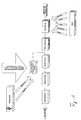
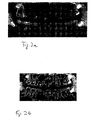
- Fig. 1 shows schematically which information from the X-ray machine and by the patient after image recognition by edge detection and segmentation can be used. Should edges and segments have been found, it is tried through Clustering to group the detected areas and edges. To identify the corresponding tooth, be under Considering the device parameters these groups with tooth shapes compared from a patient-independent database. Under Consideration of the patient-dependent parameters then becomes carried out a further processing. As a result, a Radiograph shown by appropriate regions, d. H. Objects, determined and subdivided. These regions are standing for the recognized teeth. Such a region is shown in FIGS. 2a, 2b can be seen.
- Fig. 2a shows a recognized tooth for which information can be deposited.
- the recognized tooth is replaced by a corresponding border shown.
- Fig. 2b is on the left half of a group of teeth has been selected by a mouse click and is as a result of it highlighted in their outline.
- Teeth in the upper and lower rows of teeth.
- the so selected Teeth can now be assigned information. But will e.g. too many teeth detected or intended to sensor positioning be moved slightly or should not be detected teeth Although they are seen, they are not the subject of the diagnosis can be a selection or deselection of individual or several teeth by e.g. usual mouse actions like simple or Double-click, drag an area, etc. directly over the selected areas of the background image. Especially for recording series it is necessary to find the ones to be taken Teeth to be individually specified in the operating program.
- Figures 4a and 4b show a possible management of digital Radiographs. This is what every tooth, as far as he is There is an appropriate radiograph attached. Of course, this assignment can only be made if appropriate Recordings are available.
- Figures 4a and 4b show the Possibility on that the objects are not on an x-ray must be delimited, but also on a schematic Representation can be arranged.
- the method may be in the form of software for one or more be laid down the following method claims.
- a disk can be an executable data structure based on a computer a method according to one or more of the following Implemented method claims contained.
Landscapes
- Engineering & Computer Science (AREA)
- Health & Medical Sciences (AREA)
- Medical Informatics (AREA)
- Life Sciences & Earth Sciences (AREA)
- Physics & Mathematics (AREA)
- General Health & Medical Sciences (AREA)
- Radiology & Medical Imaging (AREA)
- Nuclear Medicine, Radiotherapy & Molecular Imaging (AREA)
- Public Health (AREA)
- General Physics & Mathematics (AREA)
- Theoretical Computer Science (AREA)
- Optics & Photonics (AREA)
- Heart & Thoracic Surgery (AREA)
- Multimedia (AREA)
- Veterinary Medicine (AREA)
- Human Computer Interaction (AREA)
- Computer Vision & Pattern Recognition (AREA)
- Biophysics (AREA)
- High Energy & Nuclear Physics (AREA)
- Epidemiology (AREA)
- Pathology (AREA)
- Biomedical Technology (AREA)
- Primary Health Care (AREA)
- Molecular Biology (AREA)
- Surgery (AREA)
- Animal Behavior & Ethology (AREA)
- Quality & Reliability (AREA)
- Image Processing (AREA)
- Apparatus For Radiation Diagnosis (AREA)
- Image Analysis (AREA)
- Processing Or Creating Images (AREA)
- Analysing Materials By The Use Of Radiation (AREA)
Claims (32)
- Procédé pour identifier un ou plusieurs objets, notamment des dents sur un enregistrement radiographique numérisé, les zones représentant le ou les objets possibles étant déterminées avec des algorithmes de traitement d'image par segmentation et/ou détection des bords de l'enregistrement radiographique, caractérisé en ce que ces zones sont combinées mathématiquement entre elles pour la suite de la détermination des paramètres de l'appareil de radiographie utilisés pour réaliser l'enregistrement radiographique, les paramètres intervenant dans le calcul étant les données de position et/ou les portions de trajectoire et/ou les points de départ et d'arrêt de l'appareil de radiographie.
- Procédé selon la revendication 1, caractérisé en ce que les paramètres spécifiques à l'appareil sont combinés avec des paramètres issus d'une base de données dentaire indépendante du patient afin d'obtenir les positions géométriques actuelles probables sur les zones représentant le ou les objets possibles.
- Procédé selon la revendication 1 ou 2, caractérisé en ce qu'un regroupement des zones détectées (formation de cluster) est réalisé avant la combinaison avec les paramètres spécifiques à l'appareil.
- Procédé selon l'une des revendications 1 à 3, caractérisé en ce que des paramètres supplémentaires spécifiques au patient sont combinés mathématiquement pour la suite de la détermination des zones représentant le ou les objets possibles.
- Procédé selon l'une des revendications 1 à 4, caractérisé en ce que les paramètres anatomiques du patient tels que la race, l'âge, le sexe, la taille, le poids et/ou les traitements précédent interviennent dans le calcul.
- Procédé selon l'une des revendications 1 à 5, caractérisé en ce que des proposition sur les objets détectés sont faites à l'utilisateur, lesquelles peuvent être adaptées ou confirmée de manière interactive.
- Procédé selon l'une des revendications 1 à 6, caractérisé en ce que les informations ainsi déterminées des objets sont enregistrées séparément dans une base de données et peuvent être invoquées pour une nouvelle utilisation.
- Procédé selon l'une des revendications 1 à 7, caractérisé en ce que l'image radiographique numérisée est représentée et que l'objet pour lequel il faut enregistrer des informations supplémentaires est sélectionné et qu'ensuite des données destinées à représenter des images numériques sont reçues de la part d'un appareil de radiographie après la détermination de l'objet, lesquelles sont automatiquement associées à l'objet sélectionné.
- Procédé selon la revendication 8, caractérisé en ce que l'objet sélectionné est muni d'un marquage graphique qui est placé en tant que couche superposée au-dessus des enregistrements.
- Procédé selon une ou plusieurs des revendications 8 à 9, caractérisé en ce que les informations sont des marquages graphiques qui peuvent être placés en tant que couches superposées au-dessus des enregistrements.
- Procédé selon une ou plusieurs des revendications 8 à 10, caractérisé en ce qu'il est possible de définir les zones de l'objet dans lesquelles peuvent être associées des informations.
- Procédé selon une ou plusieurs des revendications 8 à 11, caractérisé en ce qu'il est possible d'invoquer des menus contextuels à propos de chacun des objets.
- Arrangement pour identifier des objets, notamment des dents sur un enregistrement radiographique numérisé,comprenant un appareil d'entrée et de sortie pour la commande interactive de l'arrangement,comprenant une unité de traitement qui a accès à l'enregistrement radiographique numérisé, qui a accès aux paramètres spécifiques de l'appareil de radiographie et comprenant des moyens pour déterminer les zones représentant le ou les objets possibles sur la base de ces informations et pour segmenter et/ou détecter les bords sur l'enregistrement radiographique numérisé, les paramètres spécifiques de l'appareil invoqués pour le calcul étant les données de position et/ou les portions de trajectoire et/ou les points de départ et d'arrêt de l'appareil de radiographie.
- Arrangement selon la revendication 13, caractérisé en ce que l'unité de traitement a accès à une base de données indépendante du patient.
- Arrangement selon la revendication 13 ou 14, caractérisé en ce que l'unité de traitement présente des moyens pour former un cluster des zones présentes après la segmentation et/ou la détection des bords.
- Arrangement selon l'une des revendications 13 à 15, caractérisé en ce que l'unité de traitement a accès à des informations spécifiques au patient.
- Arrangement selon l'une des revendications 13 à 16, caractérisé en ce que des moyens sont prévus pour soumettre à l'utilisateur une proposition qu'il peut accepter, refuser ou modifier de manière interactive.
- Arrangement selon l'une des revendications 13 à 17, caractérisé par une interface vers l'appareil de radiographie par le biais de laquelle il est possible d'accéder aux données spécifiques à l'appareil.
- Arrangement selon l'une des revendications 13 à 18, caractérisé en ce que des paramètres anatomiques du patient tels que la race, l'âge, le sexe, la taille, le poids et/ou les traitements précédent sont enregistrés dans une base de données et interviennent dans le calcul.
- Arrangement selon l'une des revendications 13 à 19, caractérisé en ce que des données statistiques telles que le rapport entre les différentes grandeurs anatomiques entre elles sont enregistrées dans une base de données et interviennent dans le calcul.
- Arrangement selon l'une des revendications 13 à 20, caractérisé en ce que l'arrangement est un PC qui est commandé par des logiciels.
- Arrangement selon l'une des revendications 13 à 21, caractérisé en ce que les éléments suivants sont également présents pour l'association des informations aux objets, notamment aux dents, qui sont déterminées dans un enregistrement radiographique numérisé :les objets représentés sur l'appareil de sortie pouvant être sélectionnés et les objets sélectionnés étant mis en valeur visuellement, les objets pouvant toujours être sélectionnés pour associer ou interroger les informations sous-jacentes.une zone de mémoire dans laquelle est stocké l'enregistrement radiographique, des informations d'identification de l'objet étant associées à l'enregistrement radiographique,une deuxième zone de mémoire dans laquelle sont stockées des informations sur les objets, des références entre les objets et les informations d'identification d'objet étant enregistrées,
- Arrangement selon la revendication 22, caractérisé en ce qu'il est possible de sélectionner des objets sélectionnés individuels ou plusieurs objets sélectionnés.
- Arrangement selon la revendication 23, caractérisé en ce qu'à la suite de la sélection d'un objet, on bascule sur des informations supplémentaires, sous réserve qu'elles soient présentes.
- Arrangement selon l'une des revendications 22 à 24, caractérisé en ce que les références sont gérées sous la forme de liens (links) soit directement au niveau de l'objet et/ou directement au niveau de l'information et/ou séparément.
- Arrangement selon une ou plusieurs des revendications 22 à 25, caractérisé en ce que l'appareil de sortie est un écran et les informations supplémentaires sont représentées dans une zone d'affichage (fenêtre incrustée) qui s'ouvre automatiquement ou alors les informations supplémentaires donnent lieu à une nouvelle disposition à l'écran.
- Arrangement selon une ou plusieurs des revendications 22 à 26, caractérisé en ce que les informations supplémentaires sont des informations de diagnostic et/ou de traitement et/ou des enregistrements radiographiques supplémentaires particulièrement détaillés.
- Arrangement selon une ou plusieurs des revendications 22 à 27, caractérisé par une interface vers un appareil de radiographie, l'appareil de radiographie envoyant par le biais de l'interface les informations sous la forme de données en vue de leur représentation sous la forme d'enregistrements radiographiques, ces informations étant stockées dans une troisième zone de mémoire et une référence vers un objet étant créée dans une quatrième zone de mémoire.
- Arrangement selon une ou plusieurs des revendications 22 à 28, caractérisé en ce que les informations peuvent être disposées hiérarchiquement sur plusieurs plans.
- Arrangement selon une ou plusieurs des revendications 22 à 29, caractérisé par des moyens qui permettent que les objets peuvent être définis manuellement en sélectionnant une zone spécifique sur l'enregistrement radiographique.
- Arrangement selon une ou plusieurs des revendications 22 à 30, caractérisé par la fonctionnalité d'un système de base de données.
- Arrangement selon une ou plusieurs des revendications 22 à 31, caractérisé par les caractéristiques d'un arrangement pour identifier des objets, notamment des dents sur un enregistrement radiographique numérisé selon une ou plusieurs des revendications précédentes.
Priority Applications (1)
| Application Number | Priority Date | Filing Date | Title |
|---|---|---|---|
| EP05107181A EP1610269B1 (fr) | 2001-02-21 | 2002-02-21 | Procédé et appareil d'association des informations aux radiographies numériques |
Applications Claiming Priority (3)
| Application Number | Priority Date | Filing Date | Title |
|---|---|---|---|
| DE10108295 | 2001-02-21 | ||
| DE10108295A DE10108295B4 (de) | 2001-02-21 | 2001-02-21 | Zahnidentifikation auf digitalen Röntgenaufnahmen und Zuordnung von Informationen zu digitalen Röntgenaufnahmen |
| PCT/DE2002/000634 WO2002067186A2 (fr) | 2001-02-21 | 2002-02-21 | Identification de dents sur radiographies numeriques et affectation d'informations a des radiographies numeriques |
Related Child Applications (1)
| Application Number | Title | Priority Date | Filing Date |
|---|---|---|---|
| EP05107181A Division EP1610269B1 (fr) | 2001-02-21 | 2002-02-21 | Procédé et appareil d'association des informations aux radiographies numériques |
Publications (2)
| Publication Number | Publication Date |
|---|---|
| EP1382002A2 EP1382002A2 (fr) | 2004-01-21 |
| EP1382002B1 true EP1382002B1 (fr) | 2005-11-09 |
Family
ID=7674958
Family Applications (2)
| Application Number | Title | Priority Date | Filing Date |
|---|---|---|---|
| EP05107181A Expired - Lifetime EP1610269B1 (fr) | 2001-02-21 | 2002-02-21 | Procédé et appareil d'association des informations aux radiographies numériques |
| EP02716606A Expired - Lifetime EP1382002B1 (fr) | 2001-02-21 | 2002-02-21 | Identification de dents sur radiographies numeriques et affectation d'informations a des radiographies numeriques |
Family Applications Before (1)
| Application Number | Title | Priority Date | Filing Date |
|---|---|---|---|
| EP05107181A Expired - Lifetime EP1610269B1 (fr) | 2001-02-21 | 2002-02-21 | Procédé et appareil d'association des informations aux radiographies numériques |
Country Status (5)
| Country | Link |
|---|---|
| US (1) | US7010153B2 (fr) |
| EP (2) | EP1610269B1 (fr) |
| AT (2) | ATE358306T1 (fr) |
| DE (3) | DE10108295B4 (fr) |
| WO (1) | WO2002067186A2 (fr) |
Families Citing this family (47)
| Publication number | Priority date | Publication date | Assignee | Title |
|---|---|---|---|---|
| US20040166462A1 (en) | 2003-02-26 | 2004-08-26 | Align Technology, Inc. | Systems and methods for fabricating a dental template |
| DE10312848A1 (de) * | 2003-03-21 | 2004-10-07 | Sirona Dental Systems Gmbh | Datenbank, Zahnmodell und Zahnersatzteil, aufgebaut aus digitalisierten Abbildungen realer Zähne |
| US20060069591A1 (en) * | 2004-09-29 | 2006-03-30 | Razzano Michael R | Dental image charting system and method |
| CA2581754A1 (fr) * | 2004-09-29 | 2006-04-13 | Interactive Diagnostic Imaging, Inc. | Systeme et procede de mise en diagramme d'images |
| US7991242B2 (en) | 2005-05-11 | 2011-08-02 | Optosecurity Inc. | Apparatus, method and system for screening receptacles and persons, having image distortion correction functionality |
| WO2006119603A1 (fr) | 2005-05-11 | 2006-11-16 | Optosecurity Inc. | Procede et systeme d'inspection de bagages, de conteneurs de fret ou de personnes |
| DK1899892T3 (en) * | 2005-05-20 | 2019-04-08 | Dental Imaging Technologies Corp | Focal plane location |
| EP1906862B1 (fr) | 2005-06-30 | 2018-05-23 | Biomet 3i, LLC | Procédé de création d'un modèle dentaire de laboratoire |
| US8257083B2 (en) | 2005-10-24 | 2012-09-04 | Biomet 3I, Llc | Methods for placing an implant analog in a physical model of the patient's mouth |
| US11219511B2 (en) | 2005-10-24 | 2022-01-11 | Biomet 3I, Llc | Methods for placing an implant analog in a physical model of the patient's mouth |
| US7899232B2 (en) | 2006-05-11 | 2011-03-01 | Optosecurity Inc. | Method and apparatus for providing threat image projection (TIP) in a luggage screening system, and luggage screening system implementing same |
| US8494210B2 (en) | 2007-03-30 | 2013-07-23 | Optosecurity Inc. | User interface for use in security screening providing image enhancement capabilities and apparatus for implementing same |
| JP2008029660A (ja) * | 2006-07-31 | 2008-02-14 | Fujifilm Corp | 歯科用放射線画像表示装置 |
| US8206153B2 (en) | 2007-05-18 | 2012-06-26 | Biomet 3I, Inc. | Method for selecting implant components |
| DE102007053393A1 (de) * | 2007-11-09 | 2009-05-14 | Siemens Ag | System zur automatisierten Erstellung medizinischer Reports |
| EP2060240A3 (fr) | 2007-11-16 | 2009-08-12 | Biomet 3i, LLC | Composants à utiliser avec un guide chirurgical pour le placement d'implant dentaire |
| JP2011517612A (ja) | 2008-04-15 | 2011-06-16 | バイオメット・3アイ・エルエルシー | 正確な骨及び軟質組織デジタル歯科用モデルを生成する方法 |
| EP3415112B1 (fr) | 2008-04-16 | 2022-05-25 | Biomet 3I, LLC | Procédé de developement virtuel d'un guide chirurgical pour implants dentaires |
| US8092215B2 (en) | 2008-05-23 | 2012-01-10 | Align Technology, Inc. | Smile designer |
| FI20095299L (fi) * | 2009-03-23 | 2010-09-24 | Palodex Group Oy | Järjestelmä kuvalevyn ja sen kuvatietojen hallinnoimiseksi sekä menetelmä järjestelmän ohjaamiseksi |
| US20100254607A1 (en) * | 2009-04-02 | 2010-10-07 | Kamal Patel | System and method for image mapping and integration |
| GB0910316D0 (en) | 2009-06-16 | 2009-07-29 | Univ Manchester | Image analysis method |
| USD779558S1 (en) | 2009-08-19 | 2017-02-21 | Fadi Ibsies | Display screen with transitional dental structure graphical user interface |
| USD797766S1 (en) | 2009-08-19 | 2017-09-19 | Fadi Ibsies | Display device with a probing dental keyboard graphical user interface |
| USD798894S1 (en) | 2009-08-19 | 2017-10-03 | Fadi Ibsies | Display device with a dental keyboard graphical user interface |
| US10251735B2 (en) | 2009-08-19 | 2019-04-09 | Fadi Ibsies | Specialized keyboard for dental examinations |
| USD852838S1 (en) | 2009-08-19 | 2019-07-02 | Fadi Ibsies | Display screen with transitional graphical user interface for dental software |
| USD775655S1 (en) | 2009-08-19 | 2017-01-03 | Fadi Ibsies | Display screen with graphical user interface for dental software |
| US10254852B2 (en) * | 2009-08-19 | 2019-04-09 | Fadi Ibsies | Specialized keyboard for dental examinations |
| DE102010040096A1 (de) | 2010-09-01 | 2012-03-01 | Sirona Dental Systems Gmbh | Verfahren zur Erstellung einer Aufnahme aus einem 3D-Volumen |
| ES2477288T3 (es) | 2010-12-07 | 2014-07-16 | Biomet 3I, Llc | Elemento de exploración universal para su uso en un implante dental y en análogos de implante dental |
| US8416984B2 (en) * | 2011-01-20 | 2013-04-09 | Carestream Health, Inc. | Automatic tooth charting using digital images |
| US8944818B2 (en) | 2011-05-16 | 2015-02-03 | Biomet 3I, Llc | Temporary abutment with combination of scanning features and provisionalization features |
| PL2753920T3 (pl) | 2011-09-07 | 2018-09-28 | Rapiscan Systems, Inc. | System badania rentgenowskiego integrujący dane manifestu z przetwarzaniem obrazowania/detekcji |
| US9452032B2 (en) | 2012-01-23 | 2016-09-27 | Biomet 3I, Llc | Soft tissue preservation temporary (shell) immediate-implant abutment with biological active surface |
| US9089382B2 (en) | 2012-01-23 | 2015-07-28 | Biomet 3I, Llc | Method and apparatus for recording spatial gingival soft tissue relationship to implant placement within alveolar bone for immediate-implant placement |
| US20140080092A1 (en) | 2012-09-14 | 2014-03-20 | Biomet 3I, Llc | Temporary dental prosthesis for use in developing final dental prosthesis |
| US8926328B2 (en) | 2012-12-27 | 2015-01-06 | Biomet 3I, Llc | Jigs for placing dental implant analogs in models and methods of doing the same |
| JP5670535B2 (ja) * | 2013-10-18 | 2015-02-18 | 日立メディカルコンピュータ株式会社 | 医用画像診断支援装置及び医用画像診断支援プログラム |
| EP3998040B1 (fr) | 2013-12-20 | 2025-09-03 | Biomet 3i, LLC | Méthode dentaire pour développer des prothèses sur mesure par balayage d'éléments codés |
| US9700390B2 (en) | 2014-08-22 | 2017-07-11 | Biomet 3I, Llc | Soft-tissue preservation arrangement and method |
| US10449018B2 (en) | 2015-03-09 | 2019-10-22 | Stephen J. Chu | Gingival ovate pontic and methods of using the same |
| US10302807B2 (en) | 2016-02-22 | 2019-05-28 | Rapiscan Systems, Inc. | Systems and methods for detecting threats and contraband in cargo |
| CN106504234B (zh) * | 2016-10-19 | 2019-03-19 | 青岛达芬奇科技有限公司 | 一种交互式分割全颌牙齿三角网格模型的方法 |
| US11823376B2 (en) | 2018-05-16 | 2023-11-21 | Benevis Informatics, Llc | Systems and methods for review of computer-aided detection of pathology in images |
| US11389131B2 (en) | 2018-06-27 | 2022-07-19 | Denti.Ai Technology Inc. | Systems and methods for processing of dental images |
| US11986325B2 (en) * | 2019-06-13 | 2024-05-21 | Osstemimplant Co., Ltd. | Treatment information display device and method for displaying treatment history on image of teeth in accumulated manner |
Family Cites Families (14)
| Publication number | Priority date | Publication date | Assignee | Title |
|---|---|---|---|---|
| DE3672594D1 (de) | 1985-12-20 | 1990-08-16 | Siemens Ag | Zahnaerztiche roentgendiagnostikeinrichtung zur erstellung von panorama-schichtaufnahmen vom kiefer eines patienten. |
| DE3675525D1 (de) | 1985-12-20 | 1990-12-13 | Siemens Ag | Zahnaerztliches roentgendiagnostikgeraet zur erstellung von panorama-schichtaufnahmen vom kiefer eines patienten. |
| EP0487110B1 (fr) * | 1990-11-22 | 1999-10-06 | Kabushiki Kaisha Toshiba | Système assisté par ordinateur pour le diagnostic à usage médical |
| US5384862A (en) * | 1992-05-29 | 1995-01-24 | Cimpiter Corporation | Radiographic image evaluation apparatus and method |
| DE4218020C1 (fr) | 1992-06-01 | 1993-07-15 | Siemens Ag, 8000 Muenchen, De | |
| EP0616290B1 (fr) * | 1993-03-01 | 2003-02-05 | Kabushiki Kaisha Toshiba | Système de traitement d'informations médicales pour assistance diagnostique |
| EP0632995B1 (fr) | 1993-07-06 | 1999-04-21 | Sirona Dental Systems GmbH & Co.KG | Appareil de radiodiagnostic dentaire |
| US5721851A (en) * | 1995-07-31 | 1998-02-24 | International Business Machines Corporation | Transient link indicators in image maps |
| US5742700A (en) * | 1995-08-10 | 1998-04-21 | Logicon, Inc. | Quantitative dental caries detection system and method |
| DE19735112B4 (de) * | 1997-08-13 | 2004-06-17 | Sirona Dental Systems Gmbh | Verfahren zur Erstellung frei programmierbarer Röntgenaufnahmen von Körperteilen eines Patienten in Slot-Technik und Vorrichtung zur Durchführung des Verfahrens |
| JP3919048B2 (ja) * | 1998-09-02 | 2007-05-23 | 株式会社モリタ製作所 | 局所照射x線ct撮影装置 |
| EP1250676A2 (fr) * | 1999-12-22 | 2002-10-23 | Neurografix | Systeme, procede et article manufacture permettant de gerer un reseau de services medicaux |
| US20010051881A1 (en) * | 1999-12-22 | 2001-12-13 | Aaron G. Filler | System, method and article of manufacture for managing a medical services network |
| AU2002257235A1 (en) * | 2001-05-03 | 2002-11-18 | University Of Florida | Method and system for recording carious lesions |
-
2001
- 2001-02-21 DE DE10108295A patent/DE10108295B4/de not_active Expired - Fee Related
-
2002
- 2002-02-21 DE DE50204871T patent/DE50204871D1/de not_active Expired - Lifetime
- 2002-02-21 EP EP05107181A patent/EP1610269B1/fr not_active Expired - Lifetime
- 2002-02-21 WO PCT/DE2002/000634 patent/WO2002067186A2/fr not_active Ceased
- 2002-02-21 AT AT05107181T patent/ATE358306T1/de not_active IP Right Cessation
- 2002-02-21 AT AT02716606T patent/ATE309582T1/de not_active IP Right Cessation
- 2002-02-21 EP EP02716606A patent/EP1382002B1/fr not_active Expired - Lifetime
- 2002-02-21 DE DE50209847T patent/DE50209847D1/de not_active Expired - Lifetime
-
2003
- 2003-08-21 US US10/644,991 patent/US7010153B2/en not_active Expired - Lifetime
Also Published As
| Publication number | Publication date |
|---|---|
| EP1610269A1 (fr) | 2005-12-28 |
| WO2002067186A2 (fr) | 2002-08-29 |
| WO2002067186A3 (fr) | 2003-01-09 |
| EP1610269B1 (fr) | 2007-03-28 |
| EP1382002A2 (fr) | 2004-01-21 |
| ATE309582T1 (de) | 2005-11-15 |
| US7010153B2 (en) | 2006-03-07 |
| DE10108295B4 (de) | 2004-01-29 |
| DE50209847D1 (de) | 2007-05-10 |
| DE10108295A1 (de) | 2002-09-05 |
| ATE358306T1 (de) | 2007-04-15 |
| US20040086160A1 (en) | 2004-05-06 |
| DE50204871D1 (de) | 2005-12-15 |
Similar Documents
| Publication | Publication Date | Title |
|---|---|---|
| EP1382002B1 (fr) | Identification de dents sur radiographies numeriques et affectation d'informations a des radiographies numeriques | |
| DE69432089T2 (de) | System zur Verarbeitung von medizinischen Daten zur Unterstützung der Diagnose | |
| EP1361825B1 (fr) | Procede et systeme pour positionner un appareil de radiographie numerique dentaire | |
| DE102019116972A1 (de) | Videoclipauswähler zur medizinischen bildgebung und diagnose | |
| DE102006043910B4 (de) | Ergebnisfilter und Verfahren zur Selektion der Ergebnisdaten einer Applikation zur automatischen Mustererkennung | |
| DE102008002882A1 (de) | System und Verfahren zu von einer elektronischen Krankenakte beeinflusster Datenerfassung, -verarbeitung und -anzeige | |
| DE112017002975T5 (de) | Aktualisieren der Wahrscheinlichkeiten von Zuständen auf Grundlage von Annotationen in medizinischen Bildern | |
| DE102007050184B4 (de) | Integrierte Lösung für diagnostische Lese- und Berichterstellung | |
| DE102008028023A1 (de) | Verfahren zur Darstellung mehrerer Bilddatensätze und Benutzerinterface zur Darstellung mehrerer Bilddatensätze | |
| DE10357205A1 (de) | Verfahren zur Erzeugung von Ergebnis-Bildern eines Untersuchungsobjekts | |
| DE102012108058A1 (de) | Verfahren und Vorrichtung zum Bestimmen einer Orientierung einer Testperson | |
| DE102004055768A1 (de) | Verfahren und Systeme für die computerunterstützte zielgerechte Bildgebung | |
| DE10357203A1 (de) | Verfahren und Steuereinrichtung zum Betrieb eines Magnetresonanztomographie-Geräts | |
| DE112019005308T5 (de) | Erzeugungsvorrichtung, -verfahren und -programm für gewichtetes bild, bestimmerlernvorrichtung, -verfahren und -programm, bereichsextraktionsvorrichtung, -verfahren und -programm und bestimmer | |
| US7324680B2 (en) | Tooth identification in digital X-ray images and assignment of information to digital X-ray images | |
| DE10338145B4 (de) | Verfahren zur Darstellung von 3D Bilddaten | |
| DE102008037558A1 (de) | System und Verfahren zum Diagnostizieren eines medizinischen Zustands | |
| EP4129191B1 (fr) | Optimisation des enregistrements panoramiques extra-buccaux au moyen des connaissances antérieures basées sur le modèle de la forme de l'arc mandibulaire du patient | |
| DE102011080682A1 (de) | Positionsbestimmung bei einer stereotaktischen Biopsie | |
| DE112014004519B4 (de) | Bildanzeige-Steuervorrichtung, Verfahren zum Betätigen einer Bildanzeige-Steuervorrichtung und Medium mit einem darin gespeicherten Bildanzeige-Steuerprogramm | |
| DE102006058941A1 (de) | Verfahren und Vorrichtung zum Auswählen computergestützter Algorithmen, basierend auf dem Protokoll und/oder Parametern eines Akquisitionssystems | |
| DE102021204238A1 (de) | Verfahren und System zur Erzeugung und Strukturierung medizinischer Untersuchungsinformationen | |
| DE102004033991A1 (de) | Verfahren zur Optimierung von Prozeduren in der radiologischen Diagnostik | |
| DE10108296C2 (de) | Verfahren zur Sensorpositionierung eines digitalen Röntgengerätes | |
| DE10350376A1 (de) | Verfahren und Einrichtung zur gleichzeitigen Gewinnung von vielen Untersuchungsdaten |
Legal Events
| Date | Code | Title | Description |
|---|---|---|---|
| PUAI | Public reference made under article 153(3) epc to a published international application that has entered the european phase |
Free format text: ORIGINAL CODE: 0009012 |
|
| 17P | Request for examination filed |
Effective date: 20030814 |
|
| AK | Designated contracting states |
Kind code of ref document: A2 Designated state(s): AT BE CH CY DE DK ES FI FR GB GR IE IT LI LU MC NL PT SE TR |
|
| 17Q | First examination report despatched |
Effective date: 20040702 |
|
| GRAP | Despatch of communication of intention to grant a patent |
Free format text: ORIGINAL CODE: EPIDOSNIGR1 |
|
| GRAS | Grant fee paid |
Free format text: ORIGINAL CODE: EPIDOSNIGR3 |
|
| GRAA | (expected) grant |
Free format text: ORIGINAL CODE: 0009210 |
|
| AK | Designated contracting states |
Kind code of ref document: B1 Designated state(s): AT BE CH CY DE DK ES FI FR GB GR IE IT LI LU MC NL PT SE TR |
|
| PG25 | Lapsed in a contracting state [announced via postgrant information from national office to epo] |
Ref country code: FI Free format text: LAPSE BECAUSE OF FAILURE TO SUBMIT A TRANSLATION OF THE DESCRIPTION OR TO PAY THE FEE WITHIN THE PRESCRIBED TIME-LIMIT Effective date: 20051109 Ref country code: NL Free format text: LAPSE BECAUSE OF FAILURE TO SUBMIT A TRANSLATION OF THE DESCRIPTION OR TO PAY THE FEE WITHIN THE PRESCRIBED TIME-LIMIT Effective date: 20051109 Ref country code: IE Free format text: LAPSE BECAUSE OF FAILURE TO SUBMIT A TRANSLATION OF THE DESCRIPTION OR TO PAY THE FEE WITHIN THE PRESCRIBED TIME-LIMIT Effective date: 20051109 Ref country code: GB Free format text: LAPSE BECAUSE OF FAILURE TO SUBMIT A TRANSLATION OF THE DESCRIPTION OR TO PAY THE FEE WITHIN THE PRESCRIBED TIME-LIMIT Effective date: 20051109 |
|
| REG | Reference to a national code |
Ref country code: GB Ref legal event code: FG4D Free format text: NOT ENGLISH |
|
| REG | Reference to a national code |
Ref country code: CH Ref legal event code: EP |
|
| REG | Reference to a national code |
Ref country code: IE Ref legal event code: FG4D Free format text: LANGUAGE OF EP DOCUMENT: GERMAN |
|
| REF | Corresponds to: |
Ref document number: 50204871 Country of ref document: DE Date of ref document: 20051215 Kind code of ref document: P |
|
| PG25 | Lapsed in a contracting state [announced via postgrant information from national office to epo] |
Ref country code: SE Free format text: LAPSE BECAUSE OF FAILURE TO SUBMIT A TRANSLATION OF THE DESCRIPTION OR TO PAY THE FEE WITHIN THE PRESCRIBED TIME-LIMIT Effective date: 20060209 Ref country code: DK Free format text: LAPSE BECAUSE OF FAILURE TO SUBMIT A TRANSLATION OF THE DESCRIPTION OR TO PAY THE FEE WITHIN THE PRESCRIBED TIME-LIMIT Effective date: 20060209 Ref country code: GR Free format text: LAPSE BECAUSE OF FAILURE TO SUBMIT A TRANSLATION OF THE DESCRIPTION OR TO PAY THE FEE WITHIN THE PRESCRIBED TIME-LIMIT Effective date: 20060209 |
|
| PG25 | Lapsed in a contracting state [announced via postgrant information from national office to epo] |
Ref country code: ES Free format text: LAPSE BECAUSE OF FAILURE TO SUBMIT A TRANSLATION OF THE DESCRIPTION OR TO PAY THE FEE WITHIN THE PRESCRIBED TIME-LIMIT Effective date: 20060220 |
|
| PG25 | Lapsed in a contracting state [announced via postgrant information from national office to epo] |
Ref country code: AT Free format text: LAPSE BECAUSE OF NON-PAYMENT OF DUE FEES Effective date: 20060221 |
|
| PG25 | Lapsed in a contracting state [announced via postgrant information from national office to epo] |
Ref country code: LI Free format text: LAPSE BECAUSE OF NON-PAYMENT OF DUE FEES Effective date: 20060228 Ref country code: BE Free format text: LAPSE BECAUSE OF NON-PAYMENT OF DUE FEES Effective date: 20060228 Ref country code: CH Free format text: LAPSE BECAUSE OF NON-PAYMENT OF DUE FEES Effective date: 20060228 Ref country code: MC Free format text: LAPSE BECAUSE OF NON-PAYMENT OF DUE FEES Effective date: 20060228 Ref country code: LU Free format text: LAPSE BECAUSE OF NON-PAYMENT OF DUE FEES Effective date: 20060228 |
|
| PG25 | Lapsed in a contracting state [announced via postgrant information from national office to epo] |
Ref country code: PT Free format text: LAPSE BECAUSE OF FAILURE TO SUBMIT A TRANSLATION OF THE DESCRIPTION OR TO PAY THE FEE WITHIN THE PRESCRIBED TIME-LIMIT Effective date: 20060410 |
|
| NLV1 | Nl: lapsed or annulled due to failure to fulfill the requirements of art. 29p and 29m of the patents act | ||
| GBV | Gb: ep patent (uk) treated as always having been void in accordance with gb section 77(7)/1977 [no translation filed] |
Effective date: 20051109 |
|
| REG | Reference to a national code |
Ref country code: IE Ref legal event code: FD4D |
|
| ET | Fr: translation filed | ||
| PLBE | No opposition filed within time limit |
Free format text: ORIGINAL CODE: 0009261 |
|
| STAA | Information on the status of an ep patent application or granted ep patent |
Free format text: STATUS: NO OPPOSITION FILED WITHIN TIME LIMIT |
|
| REG | Reference to a national code |
Ref country code: CH Ref legal event code: PL |
|
| 26N | No opposition filed |
Effective date: 20060810 |
|
| BERE | Be: lapsed |
Owner name: SIRONA DENTAL SYSTEMS G.M.B.H. Effective date: 20060228 |
|
| PG25 | Lapsed in a contracting state [announced via postgrant information from national office to epo] |
Ref country code: TR Free format text: LAPSE BECAUSE OF FAILURE TO SUBMIT A TRANSLATION OF THE DESCRIPTION OR TO PAY THE FEE WITHIN THE PRESCRIBED TIME-LIMIT Effective date: 20051109 |
|
| PG25 | Lapsed in a contracting state [announced via postgrant information from national office to epo] |
Ref country code: CY Free format text: LAPSE BECAUSE OF FAILURE TO SUBMIT A TRANSLATION OF THE DESCRIPTION OR TO PAY THE FEE WITHIN THE PRESCRIBED TIME-LIMIT Effective date: 20051109 |
|
| PGFP | Annual fee paid to national office [announced via postgrant information from national office to epo] |
Ref country code: IT Payment date: 20120223 Year of fee payment: 11 |
|
| PGFP | Annual fee paid to national office [announced via postgrant information from national office to epo] |
Ref country code: FR Payment date: 20130314 Year of fee payment: 12 |
|
| REG | Reference to a national code |
Ref country code: FR Ref legal event code: ST Effective date: 20141031 |
|
| PG25 | Lapsed in a contracting state [announced via postgrant information from national office to epo] |
Ref country code: FR Free format text: LAPSE BECAUSE OF NON-PAYMENT OF DUE FEES Effective date: 20140228 |
|
| PG25 | Lapsed in a contracting state [announced via postgrant information from national office to epo] |
Ref country code: IT Free format text: LAPSE BECAUSE OF NON-PAYMENT OF DUE FEES Effective date: 20140221 |
|
| PGFP | Annual fee paid to national office [announced via postgrant information from national office to epo] |
Ref country code: DE Payment date: 20190304 Year of fee payment: 18 |
|
| REG | Reference to a national code |
Ref country code: DE Ref legal event code: R082 Ref document number: 50204871 Country of ref document: DE |
|
| REG | Reference to a national code |
Ref country code: DE Ref legal event code: R119 Ref document number: 50204871 Country of ref document: DE |
|
| PG25 | Lapsed in a contracting state [announced via postgrant information from national office to epo] |
Ref country code: DE Free format text: LAPSE BECAUSE OF NON-PAYMENT OF DUE FEES Effective date: 20200901 |