EP1382002B1 - Tooth identification on digital x-ray pictures and the assignment of information to digital x-ray pictures - Google Patents
Tooth identification on digital x-ray pictures and the assignment of information to digital x-ray pictures Download PDFInfo
- Publication number
- EP1382002B1 EP1382002B1 EP02716606A EP02716606A EP1382002B1 EP 1382002 B1 EP1382002 B1 EP 1382002B1 EP 02716606 A EP02716606 A EP 02716606A EP 02716606 A EP02716606 A EP 02716606A EP 1382002 B1 EP1382002 B1 EP 1382002B1
- Authority
- EP
- European Patent Office
- Prior art keywords
- information
- objects
- ray
- ray image
- areas
- Prior art date
- Legal status (The legal status is an assumption and is not a legal conclusion. Google has not performed a legal analysis and makes no representation as to the accuracy of the status listed.)
- Expired - Lifetime
Links
Images


Classifications
-
- A—HUMAN NECESSITIES
- A61—MEDICAL OR VETERINARY SCIENCE; HYGIENE
- A61B—DIAGNOSIS; SURGERY; IDENTIFICATION
- A61B6/00—Apparatus or devices for radiation diagnosis; Apparatus or devices for radiation diagnosis combined with radiation therapy equipment
- A61B6/46—Arrangements for interfacing with the operator or the patient
- A61B6/461—Displaying means of special interest
- A61B6/463—Displaying means of special interest characterised by displaying multiple images or images and diagnostic data on one display
-
- A—HUMAN NECESSITIES
- A61—MEDICAL OR VETERINARY SCIENCE; HYGIENE
- A61B—DIAGNOSIS; SURGERY; IDENTIFICATION
- A61B6/00—Apparatus or devices for radiation diagnosis; Apparatus or devices for radiation diagnosis combined with radiation therapy equipment
- A61B6/46—Arrangements for interfacing with the operator or the patient
- A61B6/461—Displaying means of special interest
- A61B6/465—Displaying means of special interest adapted to display user selection data, e.g. graphical user interface, icons or menus
-
- G—PHYSICS
- G06—COMPUTING OR CALCULATING; COUNTING
- G06T—IMAGE DATA PROCESSING OR GENERATION, IN GENERAL
- G06T7/00—Image analysis
- G06T7/0002—Inspection of images, e.g. flaw detection
- G06T7/0012—Biomedical image inspection
-
- G—PHYSICS
- G06—COMPUTING OR CALCULATING; COUNTING
- G06V—IMAGE OR VIDEO RECOGNITION OR UNDERSTANDING
- G06V10/00—Arrangements for image or video recognition or understanding
- G06V10/10—Image acquisition
-
- G—PHYSICS
- G06—COMPUTING OR CALCULATING; COUNTING
- G06V—IMAGE OR VIDEO RECOGNITION OR UNDERSTANDING
- G06V10/00—Arrangements for image or video recognition or understanding
- G06V10/40—Extraction of image or video features
- G06V10/44—Local feature extraction by analysis of parts of the pattern, e.g. by detecting edges, contours, loops, corners, strokes or intersections; Connectivity analysis, e.g. of connected components
-
- G—PHYSICS
- G16—INFORMATION AND COMMUNICATION TECHNOLOGY [ICT] SPECIALLY ADAPTED FOR SPECIFIC APPLICATION FIELDS
- G16H—HEALTHCARE INFORMATICS, i.e. INFORMATION AND COMMUNICATION TECHNOLOGY [ICT] SPECIALLY ADAPTED FOR THE HANDLING OR PROCESSING OF MEDICAL OR HEALTHCARE DATA
- G16H30/00—ICT specially adapted for the handling or processing of medical images
- G16H30/20—ICT specially adapted for the handling or processing of medical images for handling medical images, e.g. DICOM, HL7 or PACS
-
- G—PHYSICS
- G16—INFORMATION AND COMMUNICATION TECHNOLOGY [ICT] SPECIALLY ADAPTED FOR SPECIFIC APPLICATION FIELDS
- G16H—HEALTHCARE INFORMATICS, i.e. INFORMATION AND COMMUNICATION TECHNOLOGY [ICT] SPECIALLY ADAPTED FOR THE HANDLING OR PROCESSING OF MEDICAL OR HEALTHCARE DATA
- G16H70/00—ICT specially adapted for the handling or processing of medical references
- G16H70/60—ICT specially adapted for the handling or processing of medical references relating to pathologies
-
- A—HUMAN NECESSITIES
- A61—MEDICAL OR VETERINARY SCIENCE; HYGIENE
- A61B—DIAGNOSIS; SURGERY; IDENTIFICATION
- A61B6/00—Apparatus or devices for radiation diagnosis; Apparatus or devices for radiation diagnosis combined with radiation therapy equipment
- A61B6/50—Apparatus or devices for radiation diagnosis; Apparatus or devices for radiation diagnosis combined with radiation therapy equipment specially adapted for specific body parts; specially adapted for specific clinical applications
- A61B6/51—Apparatus or devices for radiation diagnosis; Apparatus or devices for radiation diagnosis combined with radiation therapy equipment specially adapted for specific body parts; specially adapted for specific clinical applications for dentistry
-
- G—PHYSICS
- G06—COMPUTING OR CALCULATING; COUNTING
- G06T—IMAGE DATA PROCESSING OR GENERATION, IN GENERAL
- G06T2207/00—Indexing scheme for image analysis or image enhancement
- G06T2207/10—Image acquisition modality
- G06T2207/10116—X-ray image
-
- G—PHYSICS
- G06—COMPUTING OR CALCULATING; COUNTING
- G06T—IMAGE DATA PROCESSING OR GENERATION, IN GENERAL
- G06T2207/00—Indexing scheme for image analysis or image enhancement
- G06T2207/30—Subject of image; Context of image processing
- G06T2207/30004—Biomedical image processing
-
- G—PHYSICS
- G06—COMPUTING OR CALCULATING; COUNTING
- G06T—IMAGE DATA PROCESSING OR GENERATION, IN GENERAL
- G06T2207/00—Indexing scheme for image analysis or image enhancement
- G06T2207/30—Subject of image; Context of image processing
- G06T2207/30004—Biomedical image processing
- G06T2207/30036—Dental; Teeth
Definitions
- the invention relates to a method and an arrangement for identification of objects, especially teeth, on a digitized Radiograph.
- Another component of the invention is a method and an arrangement for the assignment of Information about objects, especially teeth, in one digitized radiograph or a schematic representation are determined.
- An x-ray machine for creating panoramic slice images and single shots thereof is known from DE 35 45 509 (US 4,847,881) and DE 35 45 493 (US 4,813,060).
- digital X-ray images for panoramic images and cephalometric Images are from EP 0 632 994 (US 5,511,106) known.
- the creation of digital intraoral recordings with a Intraoral sensors are known from EP 0 643 901 (US 5,513,252).
- Panoramic X-rays are used to quickly get an overview about the overall condition of the dentition. Let it be already findings without further, e.g. intraoral, x-rays derived. These findings are not related to this entire picture, but each on a particular illustrated Object, e.g. a certain tooth. This ultimately creates several findings relating to an image and which are e.g. associated with the individual teeth. Their capture is cumbersome, since the user i.d.R. the immediate operating environment of the panoramic image must leave.
- US5742700 discloses tooth recognition on digitized X-ray images, wherein the tooth areas are segmented by edge detection. Parameters of the X-ray apparatus used in making the photograph, such as Exposure data are included in the neural classifier for further determination mathematically linked to these areas.
- the object of the present invention is therefore to improve the process through the inclusion of additional parameters.
- the method may be further developed such that the device-specific parameters with parameters from a patient-independent Dental database linked to probable current geometric positions about the or to get the possible objects imaging areas. Farther It is possible already before linking with the device specific Parameters a summary (cluster formation) identified areas. In addition, in addition, in addition, in addition, in addition, in addition, in addition Patient-specific parameters for further determination of the or the possible objects mapping areas computationally linked become.
- positional data can provide information about it be given, whether it is a molar or an incisor.
- a pattern recognition is greatly simplified.
- the user should have the option Have interactive to determine which areas a tooth are to be assigned and which are not. This will be for the user Suggestions made about the detected objects that are interactive be adapted or confirmed.
- the user is allowed to for example, by polygons, which he manually draw can determine what scope an object has.
- the thus determined information of the objects are separated into a database stored.
- the information is preferably not integrated directly into the picture. For reasons the efficiency is preferred that the information about the Objects are stored separately. These are other types of uses for the illustrations continue to be given without that disturbing limitations on these pictures visible are.
- Another component of the invention is an arrangement for Identification of objects, especially teeth, on a digitized Radiograph.
- This arrangement has an input and an output device for interactive control.
- Input and output devices are preferably one Keyboard or a pointing device and a monitor. With help This pointing device can correct the individual areas be determined, represent the objects.
- Another component of the arrangement is a processing unit, the access to the digitized x-ray and on device-specific information of the X-ray device has and based on this information as well as through segmentation and / or edge detection the object on the digitized X-ray limited.
- these are preferably known processors based on based on the method already described above determine individual objects in the x-ray. The in the process The procedure described is completed by the Processor executed.
- the processing unit can also access one patient-independent dental database and / or patient-specific Have information.
- the processing unit can continue Means of clustering the after segmentation and / or edge detection have protruding areas.
- This data is about preferably to position data, trajectories, start and End points, color scales and / or color comparisons of the X-ray device, as already described above in the calculation incorporated.
- the anatomical patient parameters such as race, age, gender, Size and weight longed for in a database.
- the arrangement matured on this database, and evaluated they preferably in the form already described statistical data. Should have information about actual The dimensions and sizes of teeth will be natural preferably considered.
- the arrangement is preferably designed as a PC, and has a known serial, parallel, bus-shaped or net-shaped Interface to the x-ray device.
- the procedure on this Arrangement is preferably made by software software programs realized. Arrangement according to one or more of preceding claims, characterized in that the arrangement a PC controlled by software.
- Another component of the present invention is a Arrangement, the information associated with recognized objects.
- This arrangement may be based on the arrangements already described above or establish a procedure. However, the arrangement can also be used separately from them if there are already objects.
- the arrangement has input and output devices for interactive Control of the arrangement. These devices are preferably with keyboards, pointing devices and screens.
- radiograph in this connection it is preferably a container for a Variety of digitized radiographs.
- x-rays contain information that the Identify objects in the X-ray.
- the object identification information have the job, the flat or optionally also the spatial extent of the object in the To determine representation. They are preferably separate Data structures formed by two- or multi-dimensional polygons represent the object in the radiograph. This information when image building on the original radiograph placed. However, it is also conceivable that this information be integrated into the X-ray itself.
- the information becomes stored the objects.
- This information may be about Textual information or even image information. So It is conceivable that a detail shot in higher resolution than additional information is stored. Another possibility consists of an indication by graphic markings to admit in what areas treatments needed are or have already been made. It is also possible to associate several types of information with an object.
- references between the objects and the information saved are preferably pointers or index fields, which are the logical Make connections between the objects and the information.
- a processing unit controlled the operations Create, Delete and access.
- the operations are preferably started by the user using the input and output devices.
- the processing unit thus controls the access to the different memory areas.
- These memory areas are preferably only logically different memory areas. He can physically seen around a contiguous storage area act.
- the objects are visually highlighted on the output device. Should be selectable Information, they will be replaced by another Feature marked graphically.
- references allows a hierarchical arrangement of information.
- the user can thus choose from a Panoramic view moving to detail shots and information Deposit on every level.
- the arrangement is a PC with a screens, with the information through context menus are accessible.
- These context menus are in the form of Pop-up window displayed when an object is viewed by the user is activated.
- the context menu allows the user to decide whether he wants to deposit new information, or whether it wants to look at already existing information.
- the further information is preferably diagnostic and / or Treatment information and / or more particularly detailed Radiographs.
- the arrangement has a Interface to an x-ray device.
- the x-ray device will provide information in the form of x-rays transferred to the arrangement in the first or third memory area are stored. Does the user already have At the beginning of an object, so can also immediately a reference is created in the fourth memory area for this object become.
- the new X-ray photograph will be an instant X-ray or a selected portion of it assigned and provided with an appropriate reference.
- a hierarchical arrangement is thus readily possible, which may extend over several levels.
- the arrangement preferably has the functionality of a database system on.
- the objects are defined by an arrangement and a Detected method, as already described above.
- a Combination of the arrangements is thus conceivable.
- Another component of the present invention is a Method for assigning information to objects, in particular Teeth that determines in a digitized radiograph are.
- a first step the digitized X-ray image shown.
- the Objects determined manually or automatically. This step is however, only necessary if the objects are not already are determined.
- the object is selected, for further information stored, retrieved or to be deleted.
- a delete operation also follows the reference which is stored in relation to the object. This reference and, if necessary, the information is then deleted. An information will only be deleted if they are only is referenced by an object. It can happen, that several objects reference an information. Is in In this case also the information would be deleted, so the information is missing for at least one object.
- an object is selected and Storage area provided for the information. Farther a space is provided for the reference to then the new information and the corresponding reference in store these memory areas.
- the information is graphical Markers placed as an overlay over the shots can be. This makes it possible to get detailed information which were not recognizable on the pictures, subsequently too refine and edit.
- the access to the information is preferably done by Context menus that are retrieved for each object can.
- the methods are preferably realized by software, which runs on a known PC.
- a disk can be provided, which has a data structure, the after charging a method according to one or more of the previous method claims performs.
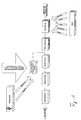
- Fig. 1 shows schematically which information from the X-ray machine and by the patient after image recognition by edge detection and segmentation can be used. Should edges and segments have been found, it is tried through Clustering to group the detected areas and edges. To identify the corresponding tooth, be under Considering the device parameters these groups with tooth shapes compared from a patient-independent database. Under Consideration of the patient-dependent parameters then becomes carried out a further processing. As a result, a Radiograph shown by appropriate regions, d. H. Objects, determined and subdivided. These regions are standing for the recognized teeth. Such a region is shown in FIGS. 2a, 2b can be seen.
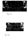
- Fig. 2a shows a recognized tooth for which information can be deposited.
- the recognized tooth is replaced by a corresponding border shown.
- Fig. 2b is on the left half of a group of teeth has been selected by a mouse click and is as a result of it highlighted in their outline.
- Teeth in the upper and lower rows of teeth.
- the so selected Teeth can now be assigned information. But will e.g. too many teeth detected or intended to sensor positioning be moved slightly or should not be detected teeth Although they are seen, they are not the subject of the diagnosis can be a selection or deselection of individual or several teeth by e.g. usual mouse actions like simple or Double-click, drag an area, etc. directly over the selected areas of the background image. Especially for recording series it is necessary to find the ones to be taken Teeth to be individually specified in the operating program.
- Figures 4a and 4b show a possible management of digital Radiographs. This is what every tooth, as far as he is There is an appropriate radiograph attached. Of course, this assignment can only be made if appropriate Recordings are available.
- Figures 4a and 4b show the Possibility on that the objects are not on an x-ray must be delimited, but also on a schematic Representation can be arranged.
- the method may be in the form of software for one or more be laid down the following method claims.
- a disk can be an executable data structure based on a computer a method according to one or more of the following Implemented method claims contained.
Landscapes
- Engineering & Computer Science (AREA)
- Health & Medical Sciences (AREA)
- Medical Informatics (AREA)
- Life Sciences & Earth Sciences (AREA)
- Physics & Mathematics (AREA)
- General Health & Medical Sciences (AREA)
- Radiology & Medical Imaging (AREA)
- Nuclear Medicine, Radiotherapy & Molecular Imaging (AREA)
- Public Health (AREA)
- General Physics & Mathematics (AREA)
- Theoretical Computer Science (AREA)
- Optics & Photonics (AREA)
- Heart & Thoracic Surgery (AREA)
- Multimedia (AREA)
- Veterinary Medicine (AREA)
- Human Computer Interaction (AREA)
- Computer Vision & Pattern Recognition (AREA)
- Biophysics (AREA)
- High Energy & Nuclear Physics (AREA)
- Epidemiology (AREA)
- Pathology (AREA)
- Biomedical Technology (AREA)
- Primary Health Care (AREA)
- Molecular Biology (AREA)
- Surgery (AREA)
- Animal Behavior & Ethology (AREA)
- Quality & Reliability (AREA)
- Image Processing (AREA)
- Apparatus For Radiation Diagnosis (AREA)
- Image Analysis (AREA)
- Processing Or Creating Images (AREA)
- Analysing Materials By The Use Of Radiation (AREA)
Abstract
Description
Die Erfindung betriff ein Verfahren und eine Anordnung zur Identifikation von Objekten, insbesondere Zähnen, auf einer digitalisierten Röntgenaufnahme. Ein weitere Bestandteil der Erfindung ist ein Verfahren und eine Anordnung zur Zuordnung von Informationen zu Objekten, insbesondere Zähnen, die in einer digitalisierte Röntgenaufnahme oder einer schematischen Darstellung bestimmt sind.The invention relates to a method and an arrangement for identification of objects, especially teeth, on a digitized Radiograph. Another component of the invention is a method and an arrangement for the assignment of Information about objects, especially teeth, in one digitized radiograph or a schematic representation are determined.
Ein Röntgengerät zur Erstellung von Panorama-Schichtaufnah-men und Einzelaufnahmen hiervon ist aus der DE 35 45 509 (US 4,847,881) und der DE 35 45 493 (US 4,813,060) bekannt. Digitale Röntgenaufnahmen für Panoramaschichtaufnahmen und cephalometrische Aufnahmen sind aus der EP 0 632 994 (US 5,511,106) bekannt. Die Erstellung digitaler Intraoral-Aufnahmen mit einem Intraoralsensor sind aus der EP 0 643 901 (US 5,513,252) bekannt.An x-ray machine for creating panoramic slice images and single shots thereof is known from DE 35 45 509 (US 4,847,881) and DE 35 45 493 (US 4,813,060). digital X-ray images for panoramic images and cephalometric Images are from EP 0 632 994 (US 5,511,106) known. The creation of digital intraoral recordings with a Intraoral sensors are known from EP 0 643 901 (US 5,513,252).
Viele Verfahren der Diagnostik beziehen sich auf einzelne Details wie z.B. einzelnen Zähne, deren Existenz, Form, Lage individuell verschieden ist. Die Diagnostik und Dokumentation wird dadurch erschwert, dass die Anwender gezwungen sind, bei weiteren Arbeiten auf nicht-individuelle, sondern allgemeine Schemata auszuweichen wie z.B. das feste Zahnschema auf dem Krankenschein.Many diagnostic procedures relate to individual details such as. individual teeth, their existence, shape, location individually is different. The diagnostics and documentation is complicated by the fact that users are forced to join further work on non-individual but general Dodge schemes such. the fixed tooth scheme on the Medical insurance card.
Bei automatisierten Aufnahmeserien ist es notwendig, die zu treffenden Zähne vorher im Bedienprogramm zu spezifizieren. For automated recording series, it is necessary to previously specified in the operating program.
Dies ist momentan entweder unzureichend oder umständlich gelöst, da es entweder nur möglich ist, ganze, fix festgelegte Zahngruppen auszuwählen, oder nur einzelnen Zähne zuzuordnen. In jedem Fall geht der reale Bezug verloren, da der Bediener die vertraute Umgebung, z.B. eine reale Gebissaufnahme, verlassen muss.This is currently either insufficient or cumbersome, because it is either only possible, whole, fixed To select tooth groups, or to assign only individual teeth. In any case, the real reference is lost as the operator the familiar environment, e.g. a real dentition, left got to.
Panorama-Röntgenaufnahmen dienen dazu, schnell eine Übersicht über den Gesamtzustand des Gebisses zu erhalten. Hiervon lassen sich bereits Befunde ohne weitere, z.B. intraorale, Röntgenaufnahmen ableiten. Diese Befunde beziehen sich aber nicht auf das gesamte Bild, sondern jeweils auf ein bestimmtes dargestelltes Objekt, z.B. einen bestimmten Zahn. Damit entstehen letztlich mehrere Befunde, die sich auf ein Bild beziehen und die z.B. den einzelnen Zähnen zugeordnet sind. Deren Erfassung ist umständlich, da der Anwender i.d.R. die unmittelbare Bedienumgebung des Panoramabildes verlassen muss.Panoramic X-rays are used to quickly get an overview about the overall condition of the dentition. Let it be already findings without further, e.g. intraoral, x-rays derived. These findings are not related to this entire picture, but each on a particular illustrated Object, e.g. a certain tooth. This ultimately creates several findings relating to an image and which are e.g. associated with the individual teeth. Their capture is cumbersome, since the user i.d.R. the immediate operating environment of the panoramic image must leave.
Die US5742700 offenbart Zahnerkennung auf digitalisierten Röntgenbildern, wobei die Zahnbereiche durch Kantendetektion segmentiert werden. Parameter des Röntgengerätes, die bei Erstellung der Aufnahme verwendet wurden, wie z.B. Belichtungsdaten, werden zur weiteren Bestimmung im neuronalen Klassifikator mit diesen Bereichen rechnerisch verknüpft.US5742700 discloses tooth recognition on digitized X-ray images, wherein the tooth areas are segmented by edge detection. Parameters of the X-ray apparatus used in making the photograph, such as Exposure data are included in the neural classifier for further determination mathematically linked to these areas.
Aufgabe der vorliegenden Erfindung ist daher die Verbesserung des Verfahrens durch die Einbeziehung weiterer Parameter.The object of the present invention is therefore to improve the process through the inclusion of additional parameters.
Gelöst wird diese Aufgabe durch Verfahren und Anordnungen, die die Merkmale der unabhängigen Ansprüche aufweisen.This problem is solved by methods and arrangements that having the features of the independent claims.
Insbesondere durch ein Verfahren zur Identifikation von Objekten, insbesondere Zähnen, auf einer digitalisierten Röntgenaufnahme. Um eine möglichst genaue Erkennung sicherzustellen, werden mit Bildverarbeitungsalgorithmen durch Segmentierung und/oder Kantendetektion der Röntgenaufnahme die das oder die möglichen Objekte abbildende Bereiche bestimmt und werden diese Bereiche zur weiteren Bestimmung mit zur Erstellung der Röntgenaufnahme verwendeten Parametern des Röntgengerätes rechnerisch verknüpft.In particular by a method for the identification of objects, especially teeth, on a digitized radiograph. To ensure the most accurate detection possible with image processing algorithms by segmentation and / or edge detection of the radiograph that the or possible objects depicting areas are determined and become these Areas for further determination with to create the X-ray used parameters of the X-ray machine by calculation connected.
Das Verfahren kann darüber hinaus so weitergebildet sein, dass die gerätespezifischen Parameter mit Parametern aus einer patientenunabhängigen Zahndatenbank verknüpft werden, um wahrscheinliche aktuelle geometrische Positionen über die das oder die möglichen Objekte abbildende Bereiche zu erhalten. Weiterhin ist es möglich, bereits vor der Verknüpfung mit den gerätespezifischen Parametern eine Zusammenfassung (Cluster-Bildung) erkannter Bereiche vorzunehmen. Darüber hinaus sind zusätzlich patientenspezifische Parameter zur weiteren Bestimmung der das oder die möglichen Objekte abbildende Bereiche rechnerisch verknüpft werden.The method may be further developed such that the device-specific parameters with parameters from a patient-independent Dental database linked to probable current geometric positions about the or to get the possible objects imaging areas. Farther It is possible already before linking with the device specific Parameters a summary (cluster formation) identified areas. In addition, in addition Patient-specific parameters for further determination of the or the possible objects mapping areas computationally linked become.
Bei diesen zusätzlichen Parametern handelt es sich ums Positionsdaten, Bahnkurven, Start- und Endpunkte des Röntgengerätes. Durch die Positionsdaten und Bahnkurven können Aufschlüsse darüber gegeben werden, ob es sich hierbei um einen Backenzahn oder einen Schneidezahn handelt. In Verbindung mit statistischen und stochastischen Daten kann die Größe dieses Zahns bestimmt werden, wodurch eine Mustererkennung stark vereinfacht wird. Weiterhin ist es wichtig, Informationen über die Graustufen der Aufnahme zu besitzen. Anhand dieser Informationen können Kanten besser bestimmt werden. Es ist somit möglich, flächige Zahnbereiche von anderen Bereichen zu unterscheiden.These additional parameters are positional data, Trajectories, start and end points of the X-ray machine. The position data and trajectories can provide information about it be given, whether it is a molar or an incisor. In conjunction with statistical and stochastic data can determine the size of this tooth become, whereby a pattern recognition is greatly simplified. Furthermore, it is important to get information about the grayscale of the Own recording. Based on this information can edges be better determined. It is thus possible, flat tooth areas to differentiate from other areas.
Weiterhin können in die Berechnung Informationen des Patienten einfließen. Hinweise darauf, welche Zähne nicht mehr vorhanden sind oder welche Zähne ersetzt wurden, ermöglichen es der Mustererkennung in diesen Bereichen besonders aufwändige Erkennungsverfahren einzusetzen bzw. keine Mustererkennung durchzuführen. Weiterhin können anatomische Patientenparatmeter berücksichtigten werden. Aus Hinweisen auf die Rasse, Alter, Geschlecht, Größe, Gewicht können statistische Rückschlüsse getroffen werden auf den Kieferaufbau und die Größe und Anordnungen der Zähne.Furthermore, in the calculation information of the patient incorporated. Indications of which teeth no longer exist or which teeth have been replaced enable pattern recognition in these areas particularly complex detection methods to use or perform no pattern recognition. Furthermore, anatomical patient paratmeters can be considered become. For indications of race, age, gender, Size, weight can be statistically determined be on the jaw structure and the size and arrangements the teeth.
Im Falle von Erkennungsfehlern soll der Benutzer die Möglichkeit haben interaktiv zu bestimmen, welche Bereiche einem Zahn zuzuordnen sind und welche nicht. Hiefür werden dem Benutzer Vorschläge über die erkannten Objekte gemacht, die interaktiv angepasst oder bestätigt werden können. Dem Benutzer wird ermöglicht, beispielsweise durch Polygone, die er manuell einzeichnen kann, zu bestimmen, welchen Umfang ein Objekt hat.In the case of recognition errors, the user should have the option Have interactive to determine which areas a tooth are to be assigned and which are not. This will be for the user Suggestions made about the detected objects that are interactive be adapted or confirmed. The user is allowed to for example, by polygons, which he manually draw can determine what scope an object has.
Die so ermittelten Informationen der Objekte werden separat in einer Datenbank abgespeichert. Die Informationen werden vorzugsweise nicht direkt in die Abbildung integriert. Aus Gründen der Effizienz wird bevorzugt, dass die Informationen über die Objekte separat abgelegt werden. Hierdurch sind andere Arten von Verwendungen für die Abbildungen weiterhin gegeben, ohne dass störende Begrenzungen auf diesen Abbildungen sichtbar sind.The thus determined information of the objects are separated into a database stored. The information is preferably not integrated directly into the picture. For reasons the efficiency is preferred that the information about the Objects are stored separately. These are other types of uses for the illustrations continue to be given without that disturbing limitations on these pictures visible are.
Ein weiterer Bestandteil der Erfindung ist eine Anordnung zur Identifikation von Objekten, insbesondere Zähnen, auf einer digitalisierten Röntgenaufnahme. Diese Anordnung weist ein Ein-und einen Ausgabegerät zur interaktiven Steuerung auf. Bei diesen Ein- und Ausgabegeräten handelt es sich vorzugsweise um eine Tastatur oder ein Zeigegerät und einen Monitor. Mit Hilfe dieses Zeigegerät können im Korrekturfall die einzelnen Bereiche bestimmt werden, die Objekte darstellen.Another component of the invention is an arrangement for Identification of objects, especially teeth, on a digitized Radiograph. This arrangement has an input and an output device for interactive control. In these Input and output devices are preferably one Keyboard or a pointing device and a monitor. With help This pointing device can correct the individual areas be determined, represent the objects.
Ein weiterer Bestandteil der Anordnung ist eine Bearbeitungseinheit, die Zugriff auf die digitalisierte Röntgenaufnahme und auf gerätespezifische Informationen des Röntgengerätes hat und die auf Grundlage dieser Informationen sowie durch Segmentierung und/oder Kantendetektion das Objekt auf der digitalisierten Röntgenaufnahme eingrenzt. Bei dieser Bearbeitungseinheit handelt es sich vorzugsweise um bekannte Prozessoren, die auf der Grundlage dessen bereits oben beschriebenen Verfahrens die einzelnen Objekte in der Röntgenaufnahme bestimmen. Die im Verfahren beschriebenen Vorgehensweise wird vollständige durch den Prozessor ausgeführt.Another component of the arrangement is a processing unit, the access to the digitized x-ray and on device-specific information of the X-ray device has and based on this information as well as through segmentation and / or edge detection the object on the digitized X-ray limited. In this processing unit these are preferably known processors based on based on the method already described above determine individual objects in the x-ray. The in the process The procedure described is completed by the Processor executed.
Die Bearbeitungseinheit kann darüber hinaus Zugriff auf eine patientenunabhängige Zahndatenbank und/oder auf patientenspezifische Informationen haben. Die Bearbeitungseinheit kann weiterhin Mittel zur Cluster-Bildung der nach der Segmentierung und/oder Kantendetektion voriegenden Bereiche aufweisen.The processing unit can also access one patient-independent dental database and / or patient-specific Have information. The processing unit can continue Means of clustering the after segmentation and / or edge detection have protruding areas.
So können Mittel vorgesehen sein, mit denen dem Benutzer Vorschläge unterbreitet werden, die interaktiv angenommen, abgelehnt oder abgeändert werden können.Thus, means can be provided with which the user suggestions submitted interactively, rejected or can be changed.
Durch eine Schnittstelle zum Röntgengerätes kann auf gerätespezifische Daten zugegriffen werden. Bei diesen Daten handelt es sich vorzugsweise um Positionsdaten, Bahnkurven, Start- und Endpunkte, Farbskalen und/oder Farbabgleiche des Röntgengerätes, die wie bereits oben beschrieben wurden in die Berechung einfließen.Through an interface to the X-ray machine can be on device-specific Data is accessed. This data is about preferably to position data, trajectories, start and End points, color scales and / or color comparisons of the X-ray device, as already described above in the calculation incorporated.
Die anatomischen Patientenparameter wie Rasse, Alter, Geschlecht, Größe und Gewicht ersehnten in einer Datenbank abgelegt. Die Anordnung gereift auf diese Datenbank zu, und werteten sie in der bereits beschriebenen Form nach vorzugsweise statistischen Daten aus. Sollten Informationen über tatsächliche Maße und Größen von Zähnen vorliegen, so werden diese natürlich vorzugsweise berücksichtigten.The anatomical patient parameters such as race, age, gender, Size and weight longed for in a database. The arrangement matured on this database, and evaluated they preferably in the form already described statistical data. Should have information about actual The dimensions and sizes of teeth will be natural preferably considered.
Die Anordnung ist vorzugsweise als PC ausgestaltet, und weist eine bekannte serielle, parallele, busförmigen oder netzförmige Schnittstelle zum Röntgengerät auf. Der Verfahren auf dieser Anordnung wird vorzugsweise durch eine Software Software-Programmen realisiert. Anordnung nach einem oder mehreren der vorhergehenden Ansprüche, dadurch gekennzeichnet, dass die Anordnung ein PC ist, der durch Software gesteuert wird.The arrangement is preferably designed as a PC, and has a known serial, parallel, bus-shaped or net-shaped Interface to the x-ray device. The procedure on this Arrangement is preferably made by software software programs realized. Arrangement according to one or more of preceding claims, characterized in that the arrangement a PC controlled by software.
Ein weiterer Bestandteil der vorliegenden Erfindung ist eine Anordnung, die Informationen erkannten Objekten zugeordnet. Dieser Anordnung kann auf den bereits oben beschriebenen Anordnungen bzw. Verfahren aufbauen. Die Anordnung kann jedoch auch separat von ihnen verwendet werden, wenn bereits Objekte vorliegen. Es handelt sich hierbei um eine Anordnung zur Zuordnung von Informationen zu Objekten. Bei diesen Objekten handelt es sich vorzugsweise um Zähne, die in digitalisierte Röntgenaufnahme bestimmt sind.Another component of the present invention is a Arrangement, the information associated with recognized objects. This arrangement may be based on the arrangements already described above or establish a procedure. However, the arrangement can also be used separately from them if there are already objects. This is an arrangement for assignment information about objects. These objects are preferably around teeth in digitized radiograph are determined.
Die Anordnung weist Ein- und Ausgabegeräte zur interaktiven Steuerung der Anordnung auf. Bei diesen Geräten handelt es sich vorzugsweise um Tastaturen, Zeigegeräte und Bildschirme. In einem ersten Speicherbereich sind Röntgenaufnahme abgelegt. Hierbei handelt es sich vorzugsweise um einen Containern für eine Vielzahl von digitalisierten Röntgenaufnahmen. Zusätzlich zu diesen Röntgenaufnahmen sind Informationen abgelegt, die die Objekte in der Röntgenaufnahme kennzeichnen. Die Objektkennzeichnungsinformationen haben die Aufgabe, den flächigen oder gegebenenfalls auch den räumlichen Umfang des Objektes in der Darstellung zu bestimmen. Es handelt sich vorzugsweise um separate Datenstrukturen, die durch zwei- oder mehrdimensionale Polygone das Objekt in der Röntgenaufnahme darstellen. Diese Informationen werden beim Bildaufbau über die ursprüngliche Röntgenaufnahme gelegt. Es ist jedoch auch denkbar, dass diese Informationen in die Röntgenaufnahme selber integriert werden.The arrangement has input and output devices for interactive Control of the arrangement. These devices are preferably with keyboards, pointing devices and screens. In one first storage area are stored radiograph. in this connection it is preferably a container for a Variety of digitized radiographs. In addition to These x-rays contain information that the Identify objects in the X-ray. The object identification information have the job, the flat or optionally also the spatial extent of the object in the To determine representation. They are preferably separate Data structures formed by two- or multi-dimensional polygons represent the object in the radiograph. This information when image building on the original radiograph placed. However, it is also conceivable that this information be integrated into the X-ray itself.
In einem zweiten Speicherbereich werden die Informationen zu den Objekten abgelegt. Bei diesen Informationen kann es sich um Textinformationen oder auch um Bildinformationen handeln. So ist denkbar, dass eine Detailaufnahme in höherer Auflösung als zusätzliche Informationen gespeichert wird. Eine andere Möglichkeit besteht darin, durch grafische Markierungen einen Hinweis darauf zugeben, in welchen Bereichen Behandlungen notwendig sind oder bereits vorgenommen wurden. Es ist ebenfalls möglich mehrere Arten von Informationen einen Objekt zuzuordnen.In a second memory area, the information becomes stored the objects. This information may be about Textual information or even image information. So It is conceivable that a detail shot in higher resolution than additional information is stored. Another possibility consists of an indication by graphic markings to admit in what areas treatments needed are or have already been made. It is also possible to associate several types of information with an object.
Weiterhin werden Referenzen zwischen den Objekten und den Informationen gespeichert. Bei diesen Referenzen handelt es sich vorzugsweise um Pointer bzw. Index-Felder, die die logische Verbindungen zwischen den Objekten und den Informationen herstellen. Furthermore, there are references between the objects and the information saved. These references are preferably pointers or index fields, which are the logical Make connections between the objects and the information.
Eine Bearbeitungseinheit kontrollierten die Operationen Anlegen, Löschen und Zugriff. Die Operationen werden vorzugsweise durch den Benutzer mit Hilfe der Ein- und Ausgabegeräte gestartet. Die Bearbeitungseinheit steuert somit den Zugriff auf die unterschiedlichen Speicherbereiche.A processing unit controlled the operations Create, Delete and access. The operations are preferably started by the user using the input and output devices. The processing unit thus controls the access to the different memory areas.
Bei diesen Speicherbereichen handelt es sich vorzugsweise nur um logisch unterschiedliche Speicherbereiche. Er kann sich physikalisch gesehen um einen zusammenhängenden Speicherbereich handeln.These memory areas are preferably only logically different memory areas. He can physically seen around a contiguous storage area act.
Die Verwendung eines physikalischen und logischen Speicherbereichs für die Bildinformationen, die Objektinformationen und die zusätzliche Informationen ist möglich, jedoch nicht besonders vorteilhafte, da die Flexibilität verloren geht.The use of a physical and logical memory area for the image information, the object information and the additional information is possible, but not particularly advantageous because the flexibility is lost.
Um die Arbeit mit der Anordnung zu verbessern, sind die Objekte auf dem Ausgabegerät optisch hervorgehoben. Sollten auswählbare Informationen vorliegen, so werden diese durch ein weiteres Merkmal grafisch gekennzeichnet.To improve the work with the arrangement, the objects are visually highlighted on the output device. Should be selectable Information, they will be replaced by another Feature marked graphically.
Die Verwendung von Referenzen ermöglicht eine hierarchische Anordnung von Informationen. Der Benutzer kann sich somit von einer Panoramaaufnahme zu Detailaufnahmen bewegen und Informationen auf jeder Ebene hinterlegen.The use of references allows a hierarchical arrangement of information. The user can thus choose from a Panoramic view moving to detail shots and information Deposit on every level.
Vorteilhaft ist eine Anordnung, bei der die Referenzen in Form von Verweisen (Links) entweder unmittelbar beim Objekt und/oder unmittelbar bei der Information und/oder separat verwaltet werden.Advantageous is an arrangement in which the references in shape References (links) either directly to the object and / or be managed directly at the information and / or separately.
In einer bevorzugten Ausgestaltung, ist die Anordnung einen PC mit einem Bildschirme, wobei die Informationen durch KontextMenüs zugänglich sind. Dieser Kontextmenüs werden in Form von Pop-Up-Fenster dargestellt, wenn ein Objekt durch den Benutzer aktiviert wird. Über das Kontext-Menü kann der Benutzer entscheiden, ob er neue Informationen hinterlegen möchte, oder ob es sich bereits vorliegende Informationen anschauen möchte. In a preferred embodiment, the arrangement is a PC with a screens, with the information through context menus are accessible. These context menus are in the form of Pop-up window displayed when an object is viewed by the user is activated. The context menu allows the user to decide whether he wants to deposit new information, or whether it wants to look at already existing information.
Die weiteren Informationen sind vorzugsweise Diagnose- und/oder Behandlungsinformationen und/oder weitere insbesondere detaillierte Röntgenaufnahmen.The further information is preferably diagnostic and / or Treatment information and / or more particularly detailed Radiographs.
In einer bevorzugten Ausführungsform weist die Anordnung eine Schnittstelle zu einem Röntgengerät auf. Über diese Schnittstelle werden vom Röntgengerät Informationen in Form von Röntgenaufnahmen an die Anordnung übertragen, die im ersten oder dritten Speicherbereich abgelegt werden. Hat der Benutzer bereits zu Beginn ein Objekt bestimmten, so kann ebenfalls sofort eine Referenz im vierten Speicherbereich zu diesem Objekt angelegt werden. Die neue Röntgenaufnahme wird sofort einer betehenden Röntgenaufnahme oder einem ausgewählten Teilbereich davon zugeordnet und mit einer entsprechenden Referenz versehen. Eine hierarchische Anordnung ist somit ohne weiteres möglich, die sich gegebenenfalls über mehrere Ebenen erstrecken kann.In a preferred embodiment, the arrangement has a Interface to an x-ray device. About this interface The x-ray device will provide information in the form of x-rays transferred to the arrangement in the first or third memory area are stored. Does the user already have At the beginning of an object, so can also immediately a reference is created in the fourth memory area for this object become. The new X-ray photograph will be an instant X-ray or a selected portion of it assigned and provided with an appropriate reference. A hierarchical arrangement is thus readily possible, which may extend over several levels.
Sollte ein Objekt nicht bereits bestimmt worden sein, so stellt die Anordnungsmittel bereit, die eine manuelle Auswahl eines bestimmten Bereichs auf der Röntgenaufnahme zulassen.If an object has not already been determined, then sets the arranging means ready to provide a manual selection of a certain area on the radiograph.
Die Anordnung weist vorzugsweise die Funktionalität eines Datenbanksystems auf.The arrangement preferably has the functionality of a database system on.
Vorzugsweise werden die Objekte durch eine Anordnung und ein Verfahren erkannt, wie es bereits oben beschrieben wurde. Eine Kombination der Anordnungen ist somit denkbar.Preferably, the objects are defined by an arrangement and a Detected method, as already described above. A Combination of the arrangements is thus conceivable.
Ein weiterer Bestandteil der vorliegenden Erfindung ist ein Verfahren zur Zuordnung von Informationen zu Objekten, insbesondere Zähnen, die in einer digitalisierte Röntgenaufnahme bestimmt sind. In einem ersten Schritt wird das digitalisierte Röntgenbild dargestellt. In einem zweiten Schritt werden die Objekte manuell oder automatisch bestimmt. Dieser Schritt ist jedoch nur dann notwendig, wenn die Objekte nicht bereits schon bestimmt sind. In einem dritten Schritt wird das Objekt ausgewählt, für das weitere Informationen gespeichert, abgerufen oder gelöscht werden sollen.Another component of the present invention is a Method for assigning information to objects, in particular Teeth that determines in a digitized radiograph are. In a first step, the digitized X-ray image shown. In a second step, the Objects determined manually or automatically. This step is however, only necessary if the objects are not already are determined. In a third step, the object is selected, for further information stored, retrieved or to be deleted.
Die wohl häufigste Operation ist die Abfrageoperationen, bei der Informationen aus der Datenbank geladen werden. Hierbei wird einer Referenz gefolgt, die in Relation zum Objekt abgelegt ist, um anhand dieser Referenz die Informationen zu bestimmten, die dargestellt werden sollen.Probably the most common operation is the query operations, at the information is loaded from the database. in this connection is followed by a reference placed in relation to the object is to use this reference to determine the information to be displayed.
Bei einer Löschoperation wird ebenfalls der Referenz gefolgt wird, die in Relation zum Objekt abgelegt ist. Diese Referenz und ggfs. die Information werden daraufhin gelöscht. Eine Informationen wird immer nur dann gelöscht, wenn sie lediglich von einem Objekt referenziert wird. Es kann der Fall eintreten, dass mehrere Objekte eine Information referenzieren. Wird in diesem Falle ebenfalls die Informationen gelöscht würde, so fehlt für zumindest ein Objekt die Informationen. Bei einer Speicheroperation wird ein Objekt ausgewählt und Speicherbereich für die Information bereitgestellt. Weiterhin wird ein Speicherbereich für die Referenz bereitgestellt, um dann die neuen Informationen und die entsprechend Referenz in diesen Speicherbereichen abzulegen.A delete operation also follows the reference which is stored in relation to the object. This reference and, if necessary, the information is then deleted. An information will only be deleted if they are only is referenced by an object. It can happen, that several objects reference an information. Is in In this case also the information would be deleted, so the information is missing for at least one object. In a memory operation, an object is selected and Storage area provided for the information. Farther a space is provided for the reference to then the new information and the corresponding reference in store these memory areas.
Es ist ebenfalls denkbar, dass nach der Bestimmung des Objektes digitale Abbildungen vom Röntgengerät empfangen werden, die automatisch dem bestimmten Objekt zugeordnet werden. Hierbei wird ausreichend viel Speicherbereich bereitgestellt (allokiert), um die Informationen abzulegen.It is also conceivable that after the determination of the object Digital images are received by the X-ray machine automatically assigned to the specific object. This is sufficient space allocated (allocated) to to store the information.
In einer bevorzugten Ausgestaltung sind die Informationen grafische Markierungen, die als Overlay über die Aufnahmen gelegt werden können. Hierdurch ist es möglich Detailinformationen, die auf der Abbildungen nicht zuerkennen waren, nachträglich zu verfeinern und zu bearbeiten.In a preferred embodiment, the information is graphical Markers placed as an overlay over the shots can be. This makes it possible to get detailed information which were not recognizable on the pictures, subsequently too refine and edit.
Für den Fall, dass Objekte in weitere Bereiche aufgeteilt werden sollten, stellt das Verfahren die Möglichkeit zur Verfügung, dass Bereiche der Objekte bestimmt werden können, in denen Informationen zugeordnet werden können.In the event that objects are divided into other areas should the procedure make it possible to that areas of the objects can be determined in which Information can be assigned.
Der Zugriff auf die Informationen erfolgt vorzugsweise durch Kontextmenüs, die zu den einzelnen Objekten abgerufen werden können.The access to the information is preferably done by Context menus that are retrieved for each object can.
Die Verfahren werden vorzugsweise durch Software realisiert, die auf einen bekannten PC abläuft. Darüber hinaus kann ein Datenträger vorgesehen sein, der eine Datenstruktur aufweist, die nach dem Ladevorgang ein Verfahren nach einem oder mehreren der vorhergehenden Verfahrensansprüche durchführt.The methods are preferably realized by software, which runs on a known PC. In addition, a disk can be provided, which has a data structure, the after charging a method according to one or more of the previous method claims performs.
Weiter vorteilhafte Ausführungsformen sind in den Unteransprüchen aufgeführt.Further advantageous embodiments are in the subclaims listed.
Es folgt eine detaillierte Beschreibung anhand der Zeichnungen. Es zeigt:
- Fig. 1
- den schematischen Ablauf bei der Bestimmung von Zähnen;
- Fig. 2
- Informationen, die zu einem Objekt (Zahn) abgelegen wurden;
- Fig. 3a, b ein Kontextmenü für ein Objekt;
- Fig. 4
- Panoramaaufnahme mit einem erkannten Zahn.
- Fig. 1
- the schematic sequence in the determination of teeth;
- Fig. 2
- Information that has been isolated to an object (tooth);
- Fig. 3a, b is a context menu for an object;
- Fig. 4
- Panoramic shot with a recognized tooth.
Die Fig. 1 zeigt schematisch, welche Informationen vom Röntgengerät und vom Patient nach der Bilderkennung durch Kantenfindung und Segmentierung verwendet werden können. Sollten Kanten und Segmente gefunden worden sein, so wird versucht, durch Cluster-Bildung die erkannten Bereiche und Kanten zu gruppieren. Um den entsprechenden Zahn zu identifizieren, werden unter Berücksichtigung der Geräteparameter diese Gruppen mit Zahnformen aus einer patientenunabhängigen Datenbank verglichen. Unter Berücksichtigung der patientenabhängigen Parameter wird dann eine weitere Verarbeitung durchgeführt. Als Ergebnis wird eine Röntgenaufnahme dargestellt, die durch entsprechende Regionen, d. h. Objekte, bestimmt und unterteilt ist. Diese Regionen stehen für die erkannten Zähnen. Eine solche Region ist den Fig. 2a, 2b zu entnehmen.Fig. 1 shows schematically which information from the X-ray machine and by the patient after image recognition by edge detection and segmentation can be used. Should edges and segments have been found, it is tried through Clustering to group the detected areas and edges. To identify the corresponding tooth, be under Considering the device parameters these groups with tooth shapes compared from a patient-independent database. Under Consideration of the patient-dependent parameters then becomes carried out a further processing. As a result, a Radiograph shown by appropriate regions, d. H. Objects, determined and subdivided. These regions are standing for the recognized teeth. Such a region is shown in FIGS. 2a, 2b can be seen.
Die Fig. 2a zeigt einen erkannten Zahn, für den Informationen hinterlegt werden können. Der erkannte Zahn wird durch eine entsprechende Umrandung dargestellt.Fig. 2a shows a recognized tooth for which information can be deposited. The recognized tooth is replaced by a corresponding border shown.
In Fig. 2b ist auf der linken Bildhälfte eine Gruppe von Zähnen durch einen Mausklick ausgewählt worden und ist als Folge davon in ihrer Kontur hervorgehoben dargestellt. Es handelt sich um Zähne in der oberen und unteren Zahnreihe. Den so ausgewählten Zähnen können nun Informationen zugeordnet werden. Werden aber z.B. zu viele Zähne erfasst oder soll die Sensorpositionierung etwas verschoben werden oder sollen erfaßte Zähne keine Rolle spielen, da sie zwar zu sehen, jedoch nicht Gegenstand der Diagnose sein werden, kann eine An- bzw. Abwahl einzelner oder mehrerer Zähne durch z.B. übliche Mausaktionen wie Einfach- oder Doppelklick, Aufziehen eines Bereiches usw. direkt über den ausgewählten Bereichen des Hintergrundbildes erfolgen. Insbesondere für Aufnahmeserien ist es notwendig, die zu treffenden Zähne vorher im Bedienprogramm einzeln zu spezifizieren.In Fig. 2b is on the left half of a group of teeth has been selected by a mouse click and is as a result of it highlighted in their outline. It is a matter of Teeth in the upper and lower rows of teeth. The so selected Teeth can now be assigned information. But will e.g. too many teeth detected or intended to sensor positioning be moved slightly or should not be detected teeth Although they are seen, they are not the subject of the diagnosis can be a selection or deselection of individual or several teeth by e.g. usual mouse actions like simple or Double-click, drag an area, etc. directly over the selected areas of the background image. Especially for recording series it is necessary to find the ones to be taken Teeth to be individually specified in the operating program.
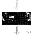
Fig. 3 zeigt eine mögliche Darstellungsform von Informationen, die Objekten zugeordnet wurden. Im vorliegenden Fall handelt es sich um Karies. Dieser Hinweis wird immer dann angezeigten, wenn der Mauszeiger über den entsprechenden Zahn gefahren wird. Andere Darstellungsformen sind jedoch auch denkbar, insbesondere mit kleinen grafischen Symbolen, die darauf hinweisen, dass weitere Informationen für dieses Objekt hinterlegt sind. 3 shows a possible form of representation of information, the objects have been assigned. In the present case it is caries. This note is always displayed when the mouse pointer is moved over the corresponding tooth. However, other forms of presentation are also conceivable, in particular with little graphic symbols that indicate that Further information for this object are deposited.
Die Figuren 4a und 4b zeigen eine mögliche Verwaltung von digitalen Röntgenaufnahmen. Hierbei wird jedem Zahn, so weit er vorhanden ist eine entsprechenden Röntgenaufnahme zugeordnet. Diese Zuordnung kann natürlich nur dann erfolgen, wenn entsprechende Aufnahmen vorliegen. Die Figuren 4a und 4b zeigen die Möglichkeit auf, dass die Objekte nicht auf einer Röntgenaufnahme abgegrenzt sein müssen, sondern auch auf einer schematischen Darstellung angeordnet sein können.Figures 4a and 4b show a possible management of digital Radiographs. This is what every tooth, as far as he is There is an appropriate radiograph attached. Of course, this assignment can only be made if appropriate Recordings are available. Figures 4a and 4b show the Possibility on that the objects are not on an x-ray must be delimited, but also on a schematic Representation can be arranged.
Das Verfahren kann in Form einer Software nach einem oder mehreren der nachstehenden Verfahrensansprüche niedergelegt sein. Ein Datenträger kann eine ablauffähige Datenstruktur, die auf einem Computer ein Verfahren nach einem oder mehreren der nachstehenden Verfahrensansprüche realisiert, enthalten.The method may be in the form of software for one or more be laid down the following method claims. A disk can be an executable data structure based on a computer a method according to one or more of the following Implemented method claims contained.
Claims (32)
- A method of identifying one or more objects, particularly teeth, in a digitized X-ray image, wherein the areas depicting the possible object(s) are identified using image-processing algorithms involving segmentation and/or edge detection of the X-ray image and, for more accurate identification, said areas are computionally linked with those parameters of the X-ray apparatus which were used for acquiring the X-ray image, the parameters taken into account being position data and/or trajectories and/or starting and stopping points of the X-ray apparatus.
- A method as defined in claim 1, wherein the apparatus-specific parameters are linked with parameters of a non-patient-dependent tooth data bank, in order to obtain probable current geometrical positions within the areas depicting the possible object(s).
- A method as defined in claim 1 or claim 2, wherein clustering of recognized areas is carried out prior to linking with the apparatus-specific parameters.
- A method as defined in any one of claims 1 to 3, wherein, in addition, patient-specific parameters are computationally linked in order to achieve more accurate identification of the areas depicting one or more possible object(s).
- A method as defined in any one of claims 1 to 4, wherein anatomical patient characteristics such as race, age, sex, size, weight, and/or treatment history are taken account of.
- A method as defined in any one of claims 1 to 5, wherein proposals are made to the user concerning recognized objects, which can be interactively modified or confirmed.
- A method as defined in any one of claims 1 to 6, wherein the information thus ascertained on the objects is stored separately in a data base, from which it can be fetched for subsequent implementation.
- A method as defined in any one of claims 1 to 7, wherein the digitized X-ray image is displayed and that object is selected for which further information should be stored, and, furthermore, following determination of an object, data concerning the display of digital images are received from an X-ray apparatus which are automatically assigned to the selected object.
- A method as defined in claim 8, wherein the selected object is provided with a graphical label, which is placed over the images as an overlay.
- A method as defined in claim 8 and/or claim 9, wherein the information is in the form of graphical labels, which can be placed over the images as an overlay.
- A method as defined in one or more of claims 8 to 10, wherein regions of the object can be determined to which information can be assigned.
- A method as defined in one or more of claims 8 to 11, wherein pop-up menus relevant to the individual objects can be displayed.
- A system for the identification of objects, particularly teeth, in a digitized X-ray image, comprisingan input/output device for interactive control of the system, anda processing unit which has access to the digitized X-ray image, has access to apparatus-specific parameters of the X-ray apparatus, and determines, by means of image-processing algorithms based on these parameters and involving segmentation and/or edge detection, those areas of the digitized X-ray image which depict the object, the apparatus-specific parameters which are taken into account being position data and/or trajectories and/or starting and stopping points of the X-ray apparatus.
- A system as defined in claim 13, wherein said processing unit has access to a non-patient-dependent tooth data bank.
- A system as defined in claim 13 or claim 14, wherein said processing unit has means for clustering the areas identified by segmentation and/or edge detection.
- A system as defined in any one of claims 13 to 15, wherein said processing unit has access to patient-specific information.
- A system as defined in any one of claims 13 to 16, wherein means are provided for offering the user a proposal, which can be interactively confirmed, rejected, or modified.
- A system as defined in any one of claims 13 to 17, characterized by a computer interface for the X-ray apparatus, via which the apparatus-specific data can be accessed.
- A system as defined in any one of claims 13 to 18, wherein anatomical patient characteristics such as race, age, sex, size, weight, and/or treatment history are stored in a data base and are taken account of.
- A system as defined in any one of claims 13 to 19, wherein statistical data, such as the relationship of the individual anatomical sizes to each other, are stored in a data base and are taken account of.
- A system as defined in any one of claims 13 to 20, wherein the system is a software controlled PC.
- A system as defined in any one of claims 13 to 21, wherein for the purpose of assigning information to objects, particularly teeth, identified in a digitized X-ray image, the following are also present:the objects displayed on the output device being selectable, while the selected objects are high-lighted and can be further selected to assign stored information thereto or to retrieve such information.a memory area, in which the X-ray image is stored together with relevant object-labeling information,a second memory area, in which information concerning the objects is stored together with references linking said objects to said object-labeling information,
- A system as defined in claim 22, wherein individually or severally selected objects can be discarded.
- A system as defined in claim 23, wherein, following the selection of an object, links to further information can be followed, if present.
- A system as defined in any one of claims 22 to 24, wherein said references are in the form of links and are administered either directly with the object and/or directly with the information and/or separately.
- A system as defined in one or more of claims 22 to 25, wherein the output device is a video monitor and the other information is displayed in an automatically opening display area (pop-up window), or said further information causes a new screen build-up.
- A system as defined in one or more of claims 22 to 26, wherein said further information consists of diagnostic and/or treatment information and/or other more detailed X-ray images.
- A system as defined in one or more of claims 22 to 27, characterized by a computer interface for an X-ray apparatus, which X-ray apparatus sends, via said computer interface, information in the form of data for display thereof as an X-ray image, which information is stored in a third memory area, and a reference is linked to an object in a fourth memory area.
- A system as defined in one or more of claims 22 to 28, wherein the information can be arranged hierarchically at a number of levels.
- A system as defined in one or more of claims 22 to 29, characterized by means that allow the objects to be manually identified by selection of a certain area of the X-ray image.
- A system as defined in one or more of claims 22 to 30, characterized by the functionality of a data bank system.
- A system as defined in one or more of claims 22 to 31, characterized by the features of a system for identification of objects, particularly teeth, in a digitized X-ray image as defined in one or more of the previous claims.
Priority Applications (1)
| Application Number | Priority Date | Filing Date | Title |
|---|---|---|---|
| EP05107181A EP1610269B1 (en) | 2001-02-21 | 2002-02-21 | Method and apparatus for associating information to digital radiographs |
Applications Claiming Priority (3)
| Application Number | Priority Date | Filing Date | Title |
|---|---|---|---|
| DE10108295 | 2001-02-21 | ||
| DE10108295A DE10108295B4 (en) | 2001-02-21 | 2001-02-21 | Tooth identification on digital x-rays and assignment of information to digital x-rays |
| PCT/DE2002/000634 WO2002067186A2 (en) | 2001-02-21 | 2002-02-21 | Tooth identification on digital x-ray pictures and the assignment of information to digital x-ray pictures |
Related Child Applications (1)
| Application Number | Title | Priority Date | Filing Date |
|---|---|---|---|
| EP05107181A Division EP1610269B1 (en) | 2001-02-21 | 2002-02-21 | Method and apparatus for associating information to digital radiographs |
Publications (2)
| Publication Number | Publication Date |
|---|---|
| EP1382002A2 EP1382002A2 (en) | 2004-01-21 |
| EP1382002B1 true EP1382002B1 (en) | 2005-11-09 |
Family
ID=7674958
Family Applications (2)
| Application Number | Title | Priority Date | Filing Date |
|---|---|---|---|
| EP05107181A Expired - Lifetime EP1610269B1 (en) | 2001-02-21 | 2002-02-21 | Method and apparatus for associating information to digital radiographs |
| EP02716606A Expired - Lifetime EP1382002B1 (en) | 2001-02-21 | 2002-02-21 | Tooth identification on digital x-ray pictures and the assignment of information to digital x-ray pictures |
Family Applications Before (1)
| Application Number | Title | Priority Date | Filing Date |
|---|---|---|---|
| EP05107181A Expired - Lifetime EP1610269B1 (en) | 2001-02-21 | 2002-02-21 | Method and apparatus for associating information to digital radiographs |
Country Status (5)
| Country | Link |
|---|---|
| US (1) | US7010153B2 (en) |
| EP (2) | EP1610269B1 (en) |
| AT (2) | ATE358306T1 (en) |
| DE (3) | DE10108295B4 (en) |
| WO (1) | WO2002067186A2 (en) |
Families Citing this family (47)
| Publication number | Priority date | Publication date | Assignee | Title |
|---|---|---|---|---|
| US20040166462A1 (en) | 2003-02-26 | 2004-08-26 | Align Technology, Inc. | Systems and methods for fabricating a dental template |
| DE10312848A1 (en) * | 2003-03-21 | 2004-10-07 | Sirona Dental Systems Gmbh | Database, tooth model and tooth replacement part, built up from digitized images of real teeth |
| US20060069591A1 (en) * | 2004-09-29 | 2006-03-30 | Razzano Michael R | Dental image charting system and method |
| CA2581754A1 (en) * | 2004-09-29 | 2006-04-13 | Interactive Diagnostic Imaging, Inc. | Image charting system and method |
| US7991242B2 (en) | 2005-05-11 | 2011-08-02 | Optosecurity Inc. | Apparatus, method and system for screening receptacles and persons, having image distortion correction functionality |
| WO2006119603A1 (en) | 2005-05-11 | 2006-11-16 | Optosecurity Inc. | Method and system for screening luggage items, cargo containers or persons |
| DK1899892T3 (en) * | 2005-05-20 | 2019-04-08 | Dental Imaging Technologies Corp | Focal plane location |
| EP1906862B1 (en) | 2005-06-30 | 2018-05-23 | Biomet 3i, LLC | Method of creating a dental laboratory model |
| US8257083B2 (en) | 2005-10-24 | 2012-09-04 | Biomet 3I, Llc | Methods for placing an implant analog in a physical model of the patient's mouth |
| US11219511B2 (en) | 2005-10-24 | 2022-01-11 | Biomet 3I, Llc | Methods for placing an implant analog in a physical model of the patient's mouth |
| US7899232B2 (en) | 2006-05-11 | 2011-03-01 | Optosecurity Inc. | Method and apparatus for providing threat image projection (TIP) in a luggage screening system, and luggage screening system implementing same |
| US8494210B2 (en) | 2007-03-30 | 2013-07-23 | Optosecurity Inc. | User interface for use in security screening providing image enhancement capabilities and apparatus for implementing same |
| JP2008029660A (en) * | 2006-07-31 | 2008-02-14 | Fujifilm Corp | Dental radiation image display device |
| US8206153B2 (en) | 2007-05-18 | 2012-06-26 | Biomet 3I, Inc. | Method for selecting implant components |
| DE102007053393A1 (en) * | 2007-11-09 | 2009-05-14 | Siemens Ag | System for automatic creation of medical reports, has computer system with memory unit, input unit and output unit, where one or multiple external units are connected with computer system |
| EP2060240A3 (en) | 2007-11-16 | 2009-08-12 | Biomet 3i, LLC | Components for use with a surgical guide for dental implant placement |
| JP2011517612A (en) | 2008-04-15 | 2011-06-16 | バイオメット・3アイ・エルエルシー | Method for generating accurate bone and soft tissue digital dental models |
| EP3415112B1 (en) | 2008-04-16 | 2022-05-25 | Biomet 3I, LLC | Method for the virtual development of a surgical guide for dental implants |
| US8092215B2 (en) | 2008-05-23 | 2012-01-10 | Align Technology, Inc. | Smile designer |
| FI20095299L (en) * | 2009-03-23 | 2010-09-24 | Palodex Group Oy | A system for managing the image disc and its image data and a method for controlling the system |
| US20100254607A1 (en) * | 2009-04-02 | 2010-10-07 | Kamal Patel | System and method for image mapping and integration |
| GB0910316D0 (en) | 2009-06-16 | 2009-07-29 | Univ Manchester | Image analysis method |
| USD779558S1 (en) | 2009-08-19 | 2017-02-21 | Fadi Ibsies | Display screen with transitional dental structure graphical user interface |
| USD797766S1 (en) | 2009-08-19 | 2017-09-19 | Fadi Ibsies | Display device with a probing dental keyboard graphical user interface |
| USD798894S1 (en) | 2009-08-19 | 2017-10-03 | Fadi Ibsies | Display device with a dental keyboard graphical user interface |
| US10251735B2 (en) | 2009-08-19 | 2019-04-09 | Fadi Ibsies | Specialized keyboard for dental examinations |
| USD852838S1 (en) | 2009-08-19 | 2019-07-02 | Fadi Ibsies | Display screen with transitional graphical user interface for dental software |
| USD775655S1 (en) | 2009-08-19 | 2017-01-03 | Fadi Ibsies | Display screen with graphical user interface for dental software |
| US10254852B2 (en) * | 2009-08-19 | 2019-04-09 | Fadi Ibsies | Specialized keyboard for dental examinations |
| DE102010040096A1 (en) | 2010-09-01 | 2012-03-01 | Sirona Dental Systems Gmbh | Method of creating a shot from a 3D volume |
| ES2477288T3 (en) | 2010-12-07 | 2014-07-16 | Biomet 3I, Llc | Universal scanning element for use in a dental implant and dental implant analogues |
| US8416984B2 (en) * | 2011-01-20 | 2013-04-09 | Carestream Health, Inc. | Automatic tooth charting using digital images |
| US8944818B2 (en) | 2011-05-16 | 2015-02-03 | Biomet 3I, Llc | Temporary abutment with combination of scanning features and provisionalization features |
| PL2753920T3 (en) | 2011-09-07 | 2018-09-28 | Rapiscan Systems, Inc. | X-ray inspection system that integrates manifest data with imaging/detection processing |
| US9452032B2 (en) | 2012-01-23 | 2016-09-27 | Biomet 3I, Llc | Soft tissue preservation temporary (shell) immediate-implant abutment with biological active surface |
| US9089382B2 (en) | 2012-01-23 | 2015-07-28 | Biomet 3I, Llc | Method and apparatus for recording spatial gingival soft tissue relationship to implant placement within alveolar bone for immediate-implant placement |
| US20140080092A1 (en) | 2012-09-14 | 2014-03-20 | Biomet 3I, Llc | Temporary dental prosthesis for use in developing final dental prosthesis |
| US8926328B2 (en) | 2012-12-27 | 2015-01-06 | Biomet 3I, Llc | Jigs for placing dental implant analogs in models and methods of doing the same |
| JP5670535B2 (en) * | 2013-10-18 | 2015-02-18 | 日立メディカルコンピュータ株式会社 | Medical image diagnosis support apparatus and medical image diagnosis support program |
| EP3998040B1 (en) | 2013-12-20 | 2025-09-03 | Biomet 3i, LLC | Dental method for developing custom prostheses through scanning of coded members |
| US9700390B2 (en) | 2014-08-22 | 2017-07-11 | Biomet 3I, Llc | Soft-tissue preservation arrangement and method |
| US10449018B2 (en) | 2015-03-09 | 2019-10-22 | Stephen J. Chu | Gingival ovate pontic and methods of using the same |
| US10302807B2 (en) | 2016-02-22 | 2019-05-28 | Rapiscan Systems, Inc. | Systems and methods for detecting threats and contraband in cargo |
| CN106504234B (en) * | 2016-10-19 | 2019-03-19 | 青岛达芬奇科技有限公司 | A kind of method of the full jaw tooth triangle grid model of Interactive Segmentation |
| US11823376B2 (en) | 2018-05-16 | 2023-11-21 | Benevis Informatics, Llc | Systems and methods for review of computer-aided detection of pathology in images |
| US11389131B2 (en) | 2018-06-27 | 2022-07-19 | Denti.Ai Technology Inc. | Systems and methods for processing of dental images |
| US11986325B2 (en) * | 2019-06-13 | 2024-05-21 | Osstemimplant Co., Ltd. | Treatment information display device and method for displaying treatment history on image of teeth in accumulated manner |
Family Cites Families (14)
| Publication number | Priority date | Publication date | Assignee | Title |
|---|---|---|---|---|
| DE3672594D1 (en) | 1985-12-20 | 1990-08-16 | Siemens Ag | DENTAL X-RAY DIAGNOSTIC DEVICE FOR CREATING PANORAMIC SLIDE IMAGES FROM A PATIENT'S JAW. |
| DE3675525D1 (en) | 1985-12-20 | 1990-12-13 | Siemens Ag | DENTAL X-RAY DIAGNOSTIC DEVICE FOR CREATING PANORAMIC LAYING FROM A PATIENT'S JAW. |
| EP0487110B1 (en) * | 1990-11-22 | 1999-10-06 | Kabushiki Kaisha Toshiba | Computer-aided diagnosis system for medical use |
| US5384862A (en) * | 1992-05-29 | 1995-01-24 | Cimpiter Corporation | Radiographic image evaluation apparatus and method |
| DE4218020C1 (en) | 1992-06-01 | 1993-07-15 | Siemens Ag, 8000 Muenchen, De | |
| EP0616290B1 (en) * | 1993-03-01 | 2003-02-05 | Kabushiki Kaisha Toshiba | Medical information processing system for supporting diagnosis. |
| EP0632995B1 (en) | 1993-07-06 | 1999-04-21 | Sirona Dental Systems GmbH & Co.KG | Dental X-ray diagnostic device |
| US5721851A (en) * | 1995-07-31 | 1998-02-24 | International Business Machines Corporation | Transient link indicators in image maps |
| US5742700A (en) * | 1995-08-10 | 1998-04-21 | Logicon, Inc. | Quantitative dental caries detection system and method |
| DE19735112B4 (en) * | 1997-08-13 | 2004-06-17 | Sirona Dental Systems Gmbh | Method for creating freely programmable x-ray images of body parts of a patient using slot technology and device for carrying out the method |
| JP3919048B2 (en) * | 1998-09-02 | 2007-05-23 | 株式会社モリタ製作所 | Local irradiation X-ray CT imaging system |
| EP1250676A2 (en) * | 1999-12-22 | 2002-10-23 | Neurografix | System, method and article of manufacture for managing a medical services network |
| US20010051881A1 (en) * | 1999-12-22 | 2001-12-13 | Aaron G. Filler | System, method and article of manufacture for managing a medical services network |
| AU2002257235A1 (en) * | 2001-05-03 | 2002-11-18 | University Of Florida | Method and system for recording carious lesions |
-
2001
- 2001-02-21 DE DE10108295A patent/DE10108295B4/en not_active Expired - Fee Related
-
2002
- 2002-02-21 DE DE50204871T patent/DE50204871D1/en not_active Expired - Lifetime
- 2002-02-21 EP EP05107181A patent/EP1610269B1/en not_active Expired - Lifetime
- 2002-02-21 WO PCT/DE2002/000634 patent/WO2002067186A2/en not_active Ceased
- 2002-02-21 AT AT05107181T patent/ATE358306T1/en not_active IP Right Cessation
- 2002-02-21 AT AT02716606T patent/ATE309582T1/en not_active IP Right Cessation
- 2002-02-21 EP EP02716606A patent/EP1382002B1/en not_active Expired - Lifetime
- 2002-02-21 DE DE50209847T patent/DE50209847D1/en not_active Expired - Lifetime
-
2003
- 2003-08-21 US US10/644,991 patent/US7010153B2/en not_active Expired - Lifetime
Also Published As
| Publication number | Publication date |
|---|---|
| EP1610269A1 (en) | 2005-12-28 |
| WO2002067186A2 (en) | 2002-08-29 |
| WO2002067186A3 (en) | 2003-01-09 |
| EP1610269B1 (en) | 2007-03-28 |
| EP1382002A2 (en) | 2004-01-21 |
| ATE309582T1 (en) | 2005-11-15 |
| US7010153B2 (en) | 2006-03-07 |
| DE10108295B4 (en) | 2004-01-29 |
| DE50209847D1 (en) | 2007-05-10 |
| DE10108295A1 (en) | 2002-09-05 |
| ATE358306T1 (en) | 2007-04-15 |
| US20040086160A1 (en) | 2004-05-06 |
| DE50204871D1 (en) | 2005-12-15 |
Similar Documents
| Publication | Publication Date | Title |
|---|---|---|
| EP1382002B1 (en) | Tooth identification on digital x-ray pictures and the assignment of information to digital x-ray pictures | |
| DE69432089T2 (en) | System for processing medical data to support diagnosis | |
| EP1361825B1 (en) | Arrangement and method for positioning a dental digital x-ray machine | |
| DE102019116972A1 (en) | VIDEO CLIP SELECTOR FOR MEDICAL IMAGING AND DIAGNOSTICS | |
| DE102006043910B4 (en) | Result filter and method for selecting the result data of an application for automatic pattern recognition | |
| DE102008002882A1 (en) | System and method for data acquisition, processing and display influenced by electronic medical records | |
| DE112017002975T5 (en) | Updating the probabilities of states based on annotations in medical images | |
| DE102007050184B4 (en) | Integrated solution for diagnostic reading and reporting | |
| DE102008028023A1 (en) | Method for displaying a plurality of image data sets and user interface for displaying a plurality of image data sets | |
| DE10357205A1 (en) | Method for generating result images of an examination object | |
| DE102012108058A1 (en) | Method and apparatus for determining an orientation of a subject | |
| DE102004055768A1 (en) | Methods and systems for computer-assisted targeted imaging | |
| DE10357203A1 (en) | Method and control device for operating a magnetic resonance tomography device | |
| DE112019005308T5 (en) | WEIGHTED IMAGE GENERATING DEVICE, METHOD AND PROGRAM, DETERMINING DEVICE, METHOD AND PROGRAM, AREA EXTRACTION DEVICE, METHOD AND PROGRAM AND DETERMINATOR | |
| US7324680B2 (en) | Tooth identification in digital X-ray images and assignment of information to digital X-ray images | |
| DE10338145B4 (en) | Process for displaying 3D image data | |
| DE102008037558A1 (en) | System and method for diagnosing a medical condition | |
| EP4129191B1 (en) | Optimization of extraoral panorama images by model-based pre-knowledge about the shape of the patient's jaw arch | |
| DE102011080682A1 (en) | Method for controlling digital stereotactic biopsy using mammography device, involves capturing overview image of object area for biopsy, where two stereotactic images are captured, which comprises two images from different directions | |
| DE112014004519B4 (en) | Image display control apparatus, method of operating an image display control apparatus, and medium having an image display control program stored therein | |
| DE102006058941A1 (en) | Method and apparatus for selecting computer-aided algorithms based on the protocol and / or parameters of an acquisition system | |
| DE102021204238A1 (en) | Method and system for generating and structuring medical examination information | |
| DE102004033991A1 (en) | Method for optimizing procedures in radiological diagnostics | |
| DE10108296C2 (en) | Process for sensor positioning of a digital X-ray device | |
| DE10350376A1 (en) | Method and device for simultaneously obtaining a large number of examination data |
Legal Events
| Date | Code | Title | Description |
|---|---|---|---|
| PUAI | Public reference made under article 153(3) epc to a published international application that has entered the european phase |
Free format text: ORIGINAL CODE: 0009012 |
|
| 17P | Request for examination filed |
Effective date: 20030814 |
|
| AK | Designated contracting states |
Kind code of ref document: A2 Designated state(s): AT BE CH CY DE DK ES FI FR GB GR IE IT LI LU MC NL PT SE TR |
|
| 17Q | First examination report despatched |
Effective date: 20040702 |
|
| GRAP | Despatch of communication of intention to grant a patent |
Free format text: ORIGINAL CODE: EPIDOSNIGR1 |
|
| GRAS | Grant fee paid |
Free format text: ORIGINAL CODE: EPIDOSNIGR3 |
|
| GRAA | (expected) grant |
Free format text: ORIGINAL CODE: 0009210 |
|
| AK | Designated contracting states |
Kind code of ref document: B1 Designated state(s): AT BE CH CY DE DK ES FI FR GB GR IE IT LI LU MC NL PT SE TR |
|
| PG25 | Lapsed in a contracting state [announced via postgrant information from national office to epo] |
Ref country code: FI Free format text: LAPSE BECAUSE OF FAILURE TO SUBMIT A TRANSLATION OF THE DESCRIPTION OR TO PAY THE FEE WITHIN THE PRESCRIBED TIME-LIMIT Effective date: 20051109 Ref country code: NL Free format text: LAPSE BECAUSE OF FAILURE TO SUBMIT A TRANSLATION OF THE DESCRIPTION OR TO PAY THE FEE WITHIN THE PRESCRIBED TIME-LIMIT Effective date: 20051109 Ref country code: IE Free format text: LAPSE BECAUSE OF FAILURE TO SUBMIT A TRANSLATION OF THE DESCRIPTION OR TO PAY THE FEE WITHIN THE PRESCRIBED TIME-LIMIT Effective date: 20051109 Ref country code: GB Free format text: LAPSE BECAUSE OF FAILURE TO SUBMIT A TRANSLATION OF THE DESCRIPTION OR TO PAY THE FEE WITHIN THE PRESCRIBED TIME-LIMIT Effective date: 20051109 |
|
| REG | Reference to a national code |
Ref country code: GB Ref legal event code: FG4D Free format text: NOT ENGLISH |
|
| REG | Reference to a national code |
Ref country code: CH Ref legal event code: EP |
|
| REG | Reference to a national code |
Ref country code: IE Ref legal event code: FG4D Free format text: LANGUAGE OF EP DOCUMENT: GERMAN |
|
| REF | Corresponds to: |
Ref document number: 50204871 Country of ref document: DE Date of ref document: 20051215 Kind code of ref document: P |
|
| PG25 | Lapsed in a contracting state [announced via postgrant information from national office to epo] |
Ref country code: SE Free format text: LAPSE BECAUSE OF FAILURE TO SUBMIT A TRANSLATION OF THE DESCRIPTION OR TO PAY THE FEE WITHIN THE PRESCRIBED TIME-LIMIT Effective date: 20060209 Ref country code: DK Free format text: LAPSE BECAUSE OF FAILURE TO SUBMIT A TRANSLATION OF THE DESCRIPTION OR TO PAY THE FEE WITHIN THE PRESCRIBED TIME-LIMIT Effective date: 20060209 Ref country code: GR Free format text: LAPSE BECAUSE OF FAILURE TO SUBMIT A TRANSLATION OF THE DESCRIPTION OR TO PAY THE FEE WITHIN THE PRESCRIBED TIME-LIMIT Effective date: 20060209 |
|
| PG25 | Lapsed in a contracting state [announced via postgrant information from national office to epo] |
Ref country code: ES Free format text: LAPSE BECAUSE OF FAILURE TO SUBMIT A TRANSLATION OF THE DESCRIPTION OR TO PAY THE FEE WITHIN THE PRESCRIBED TIME-LIMIT Effective date: 20060220 |
|
| PG25 | Lapsed in a contracting state [announced via postgrant information from national office to epo] |
Ref country code: AT Free format text: LAPSE BECAUSE OF NON-PAYMENT OF DUE FEES Effective date: 20060221 |
|
| PG25 | Lapsed in a contracting state [announced via postgrant information from national office to epo] |
Ref country code: LI Free format text: LAPSE BECAUSE OF NON-PAYMENT OF DUE FEES Effective date: 20060228 Ref country code: BE Free format text: LAPSE BECAUSE OF NON-PAYMENT OF DUE FEES Effective date: 20060228 Ref country code: CH Free format text: LAPSE BECAUSE OF NON-PAYMENT OF DUE FEES Effective date: 20060228 Ref country code: MC Free format text: LAPSE BECAUSE OF NON-PAYMENT OF DUE FEES Effective date: 20060228 Ref country code: LU Free format text: LAPSE BECAUSE OF NON-PAYMENT OF DUE FEES Effective date: 20060228 |
|
| PG25 | Lapsed in a contracting state [announced via postgrant information from national office to epo] |
Ref country code: PT Free format text: LAPSE BECAUSE OF FAILURE TO SUBMIT A TRANSLATION OF THE DESCRIPTION OR TO PAY THE FEE WITHIN THE PRESCRIBED TIME-LIMIT Effective date: 20060410 |
|
| NLV1 | Nl: lapsed or annulled due to failure to fulfill the requirements of art. 29p and 29m of the patents act | ||
| GBV | Gb: ep patent (uk) treated as always having been void in accordance with gb section 77(7)/1977 [no translation filed] |
Effective date: 20051109 |
|
| REG | Reference to a national code |
Ref country code: IE Ref legal event code: FD4D |
|
| ET | Fr: translation filed | ||
| PLBE | No opposition filed within time limit |
Free format text: ORIGINAL CODE: 0009261 |
|
| STAA | Information on the status of an ep patent application or granted ep patent |
Free format text: STATUS: NO OPPOSITION FILED WITHIN TIME LIMIT |
|
| REG | Reference to a national code |
Ref country code: CH Ref legal event code: PL |
|
| 26N | No opposition filed |
Effective date: 20060810 |
|
| BERE | Be: lapsed |
Owner name: SIRONA DENTAL SYSTEMS G.M.B.H. Effective date: 20060228 |
|
| PG25 | Lapsed in a contracting state [announced via postgrant information from national office to epo] |
Ref country code: TR Free format text: LAPSE BECAUSE OF FAILURE TO SUBMIT A TRANSLATION OF THE DESCRIPTION OR TO PAY THE FEE WITHIN THE PRESCRIBED TIME-LIMIT Effective date: 20051109 |
|
| PG25 | Lapsed in a contracting state [announced via postgrant information from national office to epo] |
Ref country code: CY Free format text: LAPSE BECAUSE OF FAILURE TO SUBMIT A TRANSLATION OF THE DESCRIPTION OR TO PAY THE FEE WITHIN THE PRESCRIBED TIME-LIMIT Effective date: 20051109 |
|
| PGFP | Annual fee paid to national office [announced via postgrant information from national office to epo] |
Ref country code: IT Payment date: 20120223 Year of fee payment: 11 |
|
| PGFP | Annual fee paid to national office [announced via postgrant information from national office to epo] |
Ref country code: FR Payment date: 20130314 Year of fee payment: 12 |
|
| REG | Reference to a national code |
Ref country code: FR Ref legal event code: ST Effective date: 20141031 |
|
| PG25 | Lapsed in a contracting state [announced via postgrant information from national office to epo] |
Ref country code: FR Free format text: LAPSE BECAUSE OF NON-PAYMENT OF DUE FEES Effective date: 20140228 |
|
| PG25 | Lapsed in a contracting state [announced via postgrant information from national office to epo] |
Ref country code: IT Free format text: LAPSE BECAUSE OF NON-PAYMENT OF DUE FEES Effective date: 20140221 |
|
| PGFP | Annual fee paid to national office [announced via postgrant information from national office to epo] |
Ref country code: DE Payment date: 20190304 Year of fee payment: 18 |
|
| REG | Reference to a national code |
Ref country code: DE Ref legal event code: R082 Ref document number: 50204871 Country of ref document: DE |
|
| REG | Reference to a national code |
Ref country code: DE Ref legal event code: R119 Ref document number: 50204871 Country of ref document: DE |
|
| PG25 | Lapsed in a contracting state [announced via postgrant information from national office to epo] |
Ref country code: DE Free format text: LAPSE BECAUSE OF NON-PAYMENT OF DUE FEES Effective date: 20200901 |