EP1370869B1 - Nachweis und quantifizierung von cripto-1 - Google Patents
Nachweis und quantifizierung von cripto-1 Download PDFInfo
- Publication number
- EP1370869B1 EP1370869B1 EP02703238A EP02703238A EP1370869B1 EP 1370869 B1 EP1370869 B1 EP 1370869B1 EP 02703238 A EP02703238 A EP 02703238A EP 02703238 A EP02703238 A EP 02703238A EP 1370869 B1 EP1370869 B1 EP 1370869B1
- Authority
- EP
- European Patent Office
- Prior art keywords
- antibody
- milk
- cripto
- human
- antigen
- Prior art date
- Legal status (The legal status is an assumption and is not a legal conclusion. Google has not performed a legal analysis and makes no representation as to the accuracy of the status listed.)
- Expired - Lifetime
Links
- 101000835745 Homo sapiens Teratocarcinoma-derived growth factor 1 Proteins 0.000 title claims abstract description 153
- 102100026404 Teratocarcinoma-derived growth factor 1 Human genes 0.000 title claims abstract description 151
- 238000001514 detection method Methods 0.000 title claims abstract description 20
- 238000011002 quantification Methods 0.000 title abstract description 11
- 235000013336 milk Nutrition 0.000 claims abstract description 43
- 210000004080 milk Anatomy 0.000 claims abstract description 43
- 239000008267 milk Substances 0.000 claims abstract description 43
- 238000000034 method Methods 0.000 claims abstract description 39
- 239000013060 biological fluid Substances 0.000 claims abstract description 26
- 210000002966 serum Anatomy 0.000 claims abstract description 10
- 238000002965 ELISA Methods 0.000 claims description 22
- 235000020256 human milk Nutrition 0.000 abstract description 43
- 210000004251 human milk Anatomy 0.000 abstract description 43
- 210000002381 plasma Anatomy 0.000 abstract description 13
- 239000000203 mixture Substances 0.000 abstract description 7
- 239000000427 antigen Substances 0.000 description 27
- 210000004027 cell Anatomy 0.000 description 27
- 108091007433 antigens Proteins 0.000 description 26
- 102000036639 antigens Human genes 0.000 description 26
- 241000699666 Mus <mouse, genus> Species 0.000 description 24
- 241000283973 Oryctolagus cuniculus Species 0.000 description 23
- 102000043136 MAP kinase family Human genes 0.000 description 19
- 108091054455 MAP kinase family Proteins 0.000 description 19
- 210000002919 epithelial cell Anatomy 0.000 description 18
- 238000001262 western blot Methods 0.000 description 17
- 235000018102 proteins Nutrition 0.000 description 13
- 108090000623 proteins and genes Proteins 0.000 description 13
- 102000004169 proteins and genes Human genes 0.000 description 13
- 239000000523 sample Substances 0.000 description 13
- 238000002474 experimental method Methods 0.000 description 12
- 238000010790 dilution Methods 0.000 description 11
- 239000012895 dilution Substances 0.000 description 11
- 239000003102 growth factor Substances 0.000 description 11
- 210000005075 mammary gland Anatomy 0.000 description 10
- 235000020030 perry Nutrition 0.000 description 10
- 230000026731 phosphorylation Effects 0.000 description 9
- 238000006366 phosphorylation reaction Methods 0.000 description 9
- 238000002415 sodium dodecyl sulfate polyacrylamide gel electrophoresis Methods 0.000 description 9
- 208000026310 Breast neoplasm Diseases 0.000 description 8
- 206010028980 Neoplasm Diseases 0.000 description 8
- FAPWRFPIFSIZLT-UHFFFAOYSA-M Sodium chloride Chemical compound [Na+].[Cl-] FAPWRFPIFSIZLT-UHFFFAOYSA-M 0.000 description 8
- 229940027941 immunoglobulin g Drugs 0.000 description 8
- 206010006187 Breast cancer Diseases 0.000 description 7
- 201000011510 cancer Diseases 0.000 description 7
- 239000003153 chemical reaction reagent Substances 0.000 description 7
- 239000012634 fragment Substances 0.000 description 7
- 238000003018 immunoassay Methods 0.000 description 7
- TWRXJAOTZQYOKJ-UHFFFAOYSA-L Magnesium chloride Chemical compound [Mg+2].[Cl-].[Cl-] TWRXJAOTZQYOKJ-UHFFFAOYSA-L 0.000 description 6
- 238000003556 assay Methods 0.000 description 6
- 238000006243 chemical reaction Methods 0.000 description 6
- 150000001875 compounds Chemical class 0.000 description 6
- 238000011161 development Methods 0.000 description 6
- 230000000694 effects Effects 0.000 description 6
- 102000006747 Transforming Growth Factor alpha Human genes 0.000 description 5
- 101800004564 Transforming growth factor alpha Proteins 0.000 description 5
- 230000004913 activation Effects 0.000 description 5
- 239000000872 buffer Substances 0.000 description 5
- 230000018109 developmental process Effects 0.000 description 5
- 230000006651 lactation Effects 0.000 description 5
- 230000004044 response Effects 0.000 description 5
- 239000000126 substance Substances 0.000 description 5
- YBJHBAHKTGYVGT-ZKWXMUAHSA-N (+)-Biotin Chemical compound N1C(=O)N[C@@H]2[C@H](CCCCC(=O)O)SC[C@@H]21 YBJHBAHKTGYVGT-ZKWXMUAHSA-N 0.000 description 4
- 102000011632 Caseins Human genes 0.000 description 4
- 108010076119 Caseins Proteins 0.000 description 4
- 102000004190 Enzymes Human genes 0.000 description 4
- 108090000790 Enzymes Proteins 0.000 description 4
- 108010001336 Horseradish Peroxidase Proteins 0.000 description 4
- 102000014171 Milk Proteins Human genes 0.000 description 4
- 108010011756 Milk Proteins Proteins 0.000 description 4
- 230000010261 cell growth Effects 0.000 description 4
- 230000004069 differentiation Effects 0.000 description 4
- NOESYZHRGYRDHS-UHFFFAOYSA-N insulin Chemical compound N1C(=O)C(NC(=O)C(CCC(N)=O)NC(=O)C(CCC(O)=O)NC(=O)C(C(C)C)NC(=O)C(NC(=O)CN)C(C)CC)CSSCC(C(NC(CO)C(=O)NC(CC(C)C)C(=O)NC(CC=2C=CC(O)=CC=2)C(=O)NC(CCC(N)=O)C(=O)NC(CC(C)C)C(=O)NC(CCC(O)=O)C(=O)NC(CC(N)=O)C(=O)NC(CC=2C=CC(O)=CC=2)C(=O)NC(CSSCC(NC(=O)C(C(C)C)NC(=O)C(CC(C)C)NC(=O)C(CC=2C=CC(O)=CC=2)NC(=O)C(CC(C)C)NC(=O)C(C)NC(=O)C(CCC(O)=O)NC(=O)C(C(C)C)NC(=O)C(CC(C)C)NC(=O)C(CC=2NC=NC=2)NC(=O)C(CO)NC(=O)CNC2=O)C(=O)NCC(=O)NC(CCC(O)=O)C(=O)NC(CCCNC(N)=N)C(=O)NCC(=O)NC(CC=3C=CC=CC=3)C(=O)NC(CC=3C=CC=CC=3)C(=O)NC(CC=3C=CC(O)=CC=3)C(=O)NC(C(C)O)C(=O)N3C(CCC3)C(=O)NC(CCCCN)C(=O)NC(C)C(O)=O)C(=O)NC(CC(N)=O)C(O)=O)=O)NC(=O)C(C(C)CC)NC(=O)C(CO)NC(=O)C(C(C)O)NC(=O)C1CSSCC2NC(=O)C(CC(C)C)NC(=O)C(NC(=O)C(CCC(N)=O)NC(=O)C(CC(N)=O)NC(=O)C(NC(=O)C(N)CC=1C=CC=CC=1)C(C)C)CC1=CN=CN1 NOESYZHRGYRDHS-UHFFFAOYSA-N 0.000 description 4
- 210000004379 membrane Anatomy 0.000 description 4
- 239000012528 membrane Substances 0.000 description 4
- 235000021239 milk protein Nutrition 0.000 description 4
- 239000002245 particle Substances 0.000 description 4
- 230000037361 pathway Effects 0.000 description 4
- 239000002953 phosphate buffered saline Substances 0.000 description 4
- 239000011780 sodium chloride Substances 0.000 description 4
- 239000007787 solid Substances 0.000 description 4
- 239000000758 substrate Substances 0.000 description 4
- -1 urine Substances 0.000 description 4
- 239000011534 wash buffer Substances 0.000 description 4
- 235000021247 β-casein Nutrition 0.000 description 4
- 108091003079 Bovine Serum Albumin Proteins 0.000 description 3
- 102000004127 Cytokines Human genes 0.000 description 3
- 108090000695 Cytokines Proteins 0.000 description 3
- VEXZGXHMUGYJMC-UHFFFAOYSA-N Hydrochloric acid Chemical compound Cl VEXZGXHMUGYJMC-UHFFFAOYSA-N 0.000 description 3
- 101710087237 Whey acidic protein Proteins 0.000 description 3
- 230000015572 biosynthetic process Effects 0.000 description 3
- 210000001175 cerebrospinal fluid Anatomy 0.000 description 3
- 210000000981 epithelium Anatomy 0.000 description 3
- 239000012530 fluid Substances 0.000 description 3
- 229940088597 hormone Drugs 0.000 description 3
- 239000005556 hormone Substances 0.000 description 3
- 230000002163 immunogen Effects 0.000 description 3
- 238000011065 in-situ storage Methods 0.000 description 3
- 230000001983 lactogenic effect Effects 0.000 description 3
- 229910001629 magnesium chloride Inorganic materials 0.000 description 3
- 239000003550 marker Substances 0.000 description 3
- 230000007246 mechanism Effects 0.000 description 3
- 230000001404 mediated effect Effects 0.000 description 3
- 230000002297 mitogenic effect Effects 0.000 description 3
- 230000004048 modification Effects 0.000 description 3
- 238000012986 modification Methods 0.000 description 3
- YBYRMVIVWMBXKQ-UHFFFAOYSA-N phenylmethanesulfonyl fluoride Chemical compound FS(=O)(=O)CC1=CC=CC=C1 YBYRMVIVWMBXKQ-UHFFFAOYSA-N 0.000 description 3
- 230000035935 pregnancy Effects 0.000 description 3
- 238000010186 staining Methods 0.000 description 3
- LENZDBCJOHFCAS-UHFFFAOYSA-N tris Chemical compound OCC(N)(CO)CO LENZDBCJOHFCAS-UHFFFAOYSA-N 0.000 description 3
- 238000005406 washing Methods 0.000 description 3
- IJGRMHOSHXDMSA-UHFFFAOYSA-N Atomic nitrogen Chemical compound N#N IJGRMHOSHXDMSA-UHFFFAOYSA-N 0.000 description 2
- 108090001008 Avidin Proteins 0.000 description 2
- 241000283707 Capra Species 0.000 description 2
- 239000006144 Dulbecco’s modified Eagle's medium Substances 0.000 description 2
- 241000283074 Equus asinus Species 0.000 description 2
- DHMQDGOQFOQNFH-UHFFFAOYSA-N Glycine Chemical compound NCC(O)=O DHMQDGOQFOQNFH-UHFFFAOYSA-N 0.000 description 2
- 102000004269 Granulocyte Colony-Stimulating Factor Human genes 0.000 description 2
- 108010017080 Granulocyte Colony-Stimulating Factor Proteins 0.000 description 2
- 241000282412 Homo Species 0.000 description 2
- 101000727061 Homo sapiens Complement receptor type 1 Proteins 0.000 description 2
- 101000599951 Homo sapiens Insulin-like growth factor I Proteins 0.000 description 2
- 108060003951 Immunoglobulin Proteins 0.000 description 2
- 102000004877 Insulin Human genes 0.000 description 2
- 108090001061 Insulin Proteins 0.000 description 2
- 102100037852 Insulin-like growth factor I Human genes 0.000 description 2
- 102000000589 Interleukin-1 Human genes 0.000 description 2
- 108010002352 Interleukin-1 Proteins 0.000 description 2
- 102000004889 Interleukin-6 Human genes 0.000 description 2
- 108090001005 Interleukin-6 Proteins 0.000 description 2
- 241001465754 Metazoa Species 0.000 description 2
- 241000699670 Mus sp. Species 0.000 description 2
- 239000002033 PVDF binder Substances 0.000 description 2
- 239000002202 Polyethylene glycol Substances 0.000 description 2
- 108010029485 Protein Isoforms Proteins 0.000 description 2
- 102000001708 Protein Isoforms Human genes 0.000 description 2
- CDBYLPFSWZWCQE-UHFFFAOYSA-L Sodium Carbonate Chemical compound [Na+].[Na+].[O-]C([O-])=O CDBYLPFSWZWCQE-UHFFFAOYSA-L 0.000 description 2
- DBMJMQXJHONAFJ-UHFFFAOYSA-M Sodium laurylsulphate Chemical compound [Na+].CCCCCCCCCCCCOS([O-])(=O)=O DBMJMQXJHONAFJ-UHFFFAOYSA-M 0.000 description 2
- 102000046299 Transforming Growth Factor beta1 Human genes 0.000 description 2
- 101800002279 Transforming growth factor beta-1 Proteins 0.000 description 2
- 239000007983 Tris buffer Substances 0.000 description 2
- 108060008682 Tumor Necrosis Factor Proteins 0.000 description 2
- 102000000852 Tumor Necrosis Factor-alpha Human genes 0.000 description 2
- 238000002835 absorbance Methods 0.000 description 2
- 239000002671 adjuvant Substances 0.000 description 2
- 230000004520 agglutination Effects 0.000 description 2
- 235000001014 amino acid Nutrition 0.000 description 2
- 150000001413 amino acids Chemical class 0.000 description 2
- 238000002820 assay format Methods 0.000 description 2
- 230000004071 biological effect Effects 0.000 description 2
- 229960002685 biotin Drugs 0.000 description 2
- 235000020958 biotin Nutrition 0.000 description 2
- 239000011616 biotin Substances 0.000 description 2
- 210000004369 blood Anatomy 0.000 description 2
- 239000008280 blood Substances 0.000 description 2
- 210000001124 body fluid Anatomy 0.000 description 2
- 229940098773 bovine serum albumin Drugs 0.000 description 2
- 230000000711 cancerogenic effect Effects 0.000 description 2
- 231100000357 carcinogen Toxicity 0.000 description 2
- 239000003183 carcinogenic agent Substances 0.000 description 2
- 230000030833 cell death Effects 0.000 description 2
- 239000013592 cell lysate Substances 0.000 description 2
- 238000004587 chromatography analysis Methods 0.000 description 2
- AGVAZMGAQJOSFJ-WZHZPDAFSA-M cobalt(2+);[(2r,3s,4r,5s)-5-(5,6-dimethylbenzimidazol-1-yl)-4-hydroxy-2-(hydroxymethyl)oxolan-3-yl] [(2r)-1-[3-[(1r,2r,3r,4z,7s,9z,12s,13s,14z,17s,18s,19r)-2,13,18-tris(2-amino-2-oxoethyl)-7,12,17-tris(3-amino-3-oxopropyl)-3,5,8,8,13,15,18,19-octamethyl-2 Chemical compound [Co+2].N#[C-].[N-]([C@@H]1[C@H](CC(N)=O)[C@@]2(C)CCC(=O)NC[C@@H](C)OP(O)(=O)O[C@H]3[C@H]([C@H](O[C@@H]3CO)N3C4=CC(C)=C(C)C=C4N=C3)O)\C2=C(C)/C([C@H](C\2(C)C)CCC(N)=O)=N/C/2=C\C([C@H]([C@@]/2(CC(N)=O)C)CCC(N)=O)=N\C\2=C(C)/C2=N[C@]1(C)[C@@](C)(CC(N)=O)[C@@H]2CCC(N)=O AGVAZMGAQJOSFJ-WZHZPDAFSA-M 0.000 description 2
- 230000000295 complement effect Effects 0.000 description 2
- 230000008878 coupling Effects 0.000 description 2
- 238000010168 coupling process Methods 0.000 description 2
- 238000005859 coupling reaction Methods 0.000 description 2
- 201000010099 disease Diseases 0.000 description 2
- 208000037265 diseases, disorders, signs and symptoms Diseases 0.000 description 2
- 231100000673 dose–response relationship Toxicity 0.000 description 2
- 238000001378 electrochemiluminescence detection Methods 0.000 description 2
- 229930004094 glycosylphosphatidylinositol Natural products 0.000 description 2
- 102000046508 human CR1 Human genes 0.000 description 2
- 230000028993 immune response Effects 0.000 description 2
- 102000018358 immunoglobulin Human genes 0.000 description 2
- 230000001939 inductive effect Effects 0.000 description 2
- 230000002401 inhibitory effect Effects 0.000 description 2
- 229940125396 insulin Drugs 0.000 description 2
- 229940100601 interleukin-6 Drugs 0.000 description 2
- 230000000670 limiting effect Effects 0.000 description 2
- 230000003287 optical effect Effects 0.000 description 2
- 102000013415 peroxidase activity proteins Human genes 0.000 description 2
- 108040007629 peroxidase activity proteins Proteins 0.000 description 2
- 238000002264 polyacrylamide gel electrophoresis Methods 0.000 description 2
- 229920001223 polyethylene glycol Polymers 0.000 description 2
- 238000003752 polymerase chain reaction Methods 0.000 description 2
- 229920002981 polyvinylidene fluoride Polymers 0.000 description 2
- 102000004196 processed proteins & peptides Human genes 0.000 description 2
- 108090000765 processed proteins & peptides Proteins 0.000 description 2
- 230000035755 proliferation Effects 0.000 description 2
- 238000000746 purification Methods 0.000 description 2
- 238000003127 radioimmunoassay Methods 0.000 description 2
- 230000026267 regulation of growth Effects 0.000 description 2
- 238000003757 reverse transcription PCR Methods 0.000 description 2
- 210000003296 saliva Anatomy 0.000 description 2
- 210000000582 semen Anatomy 0.000 description 2
- PUZPDOWCWNUUKD-UHFFFAOYSA-M sodium fluoride Chemical compound [F-].[Na+] PUZPDOWCWNUUKD-UHFFFAOYSA-M 0.000 description 2
- 239000000243 solution Substances 0.000 description 2
- 230000000638 stimulation Effects 0.000 description 2
- 239000012089 stop solution Substances 0.000 description 2
- 238000012360 testing method Methods 0.000 description 2
- 238000012546 transfer Methods 0.000 description 2
- 238000011282 treatment Methods 0.000 description 2
- 210000002700 urine Anatomy 0.000 description 2
- NMWKYTGJWUAZPZ-WWHBDHEGSA-N (4S)-4-[[(4R,7S,10S,16S,19S,25S,28S,31R)-31-[[(2S)-2-[[(1R,6R,9S,12S,18S,21S,24S,27S,30S,33S,36S,39S,42R,47R,53S,56S,59S,62S,65S,68S,71S,76S,79S,85S)-47-[[(2S)-2-[[(2S)-4-amino-2-[[(2S)-2-[[(2S)-2-[[(2S)-2-[[(2S)-2-[[(2S)-2-amino-3-methylbutanoyl]amino]-3-methylbutanoyl]amino]-3-hydroxypropanoyl]amino]-3-(1H-imidazol-4-yl)propanoyl]amino]-3-phenylpropanoyl]amino]-4-oxobutanoyl]amino]-3-carboxypropanoyl]amino]-18-(4-aminobutyl)-27,68-bis(3-amino-3-oxopropyl)-36,71,76-tribenzyl-39-(3-carbamimidamidopropyl)-24-(2-carboxyethyl)-21,56-bis(carboxymethyl)-65,85-bis[(1R)-1-hydroxyethyl]-59-(hydroxymethyl)-62,79-bis(1H-imidazol-4-ylmethyl)-9-methyl-33-(2-methylpropyl)-8,11,17,20,23,26,29,32,35,38,41,48,54,57,60,63,66,69,72,74,77,80,83,86-tetracosaoxo-30-propan-2-yl-3,4,44,45-tetrathia-7,10,16,19,22,25,28,31,34,37,40,49,55,58,61,64,67,70,73,75,78,81,84,87-tetracosazatetracyclo[40.31.14.012,16.049,53]heptaoctacontane-6-carbonyl]amino]-3-methylbutanoyl]amino]-7-(3-carbamimidamidopropyl)-25-(hydroxymethyl)-19-[(4-hydroxyphenyl)methyl]-28-(1H-imidazol-4-ylmethyl)-10-methyl-6,9,12,15,18,21,24,27,30-nonaoxo-16-propan-2-yl-1,2-dithia-5,8,11,14,17,20,23,26,29-nonazacyclodotriacontane-4-carbonyl]amino]-5-[[(2S)-1-[[(2S)-1-[[(2S)-3-carboxy-1-[[(2S)-1-[[(2S)-1-[[(1S)-1-carboxyethyl]amino]-4-methyl-1-oxopentan-2-yl]amino]-4-methyl-1-oxopentan-2-yl]amino]-1-oxopropan-2-yl]amino]-1-oxopropan-2-yl]amino]-3-(1H-imidazol-4-yl)-1-oxopropan-2-yl]amino]-5-oxopentanoic acid Chemical compound CC(C)C[C@H](NC(=O)[C@H](CC(C)C)NC(=O)[C@H](CC(O)=O)NC(=O)[C@H](C)NC(=O)[C@H](Cc1c[nH]cn1)NC(=O)[C@H](CCC(O)=O)NC(=O)[C@@H]1CSSC[C@H](NC(=O)[C@@H](NC(=O)[C@@H]2CSSC[C@@H]3NC(=O)[C@H](Cc4ccccc4)NC(=O)[C@H](CCC(N)=O)NC(=O)[C@@H](NC(=O)[C@H](Cc4c[nH]cn4)NC(=O)[C@H](CO)NC(=O)[C@H](CC(O)=O)NC(=O)[C@@H]4CCCN4C(=O)[C@H](CSSC[C@H](NC(=O)[C@@H](NC(=O)CNC(=O)[C@H](Cc4c[nH]cn4)NC(=O)[C@H](Cc4ccccc4)NC3=O)[C@@H](C)O)C(=O)N[C@@H](CCCNC(N)=N)C(=O)N[C@@H](Cc3ccccc3)C(=O)N[C@@H](CC(C)C)C(=O)N[C@@H](C(C)C)C(=O)N[C@@H](CCC(N)=O)C(=O)N[C@@H](CCC(O)=O)C(=O)N[C@@H](CC(O)=O)C(=O)N[C@@H](CCCCN)C(=O)N3CCC[C@H]3C(=O)N[C@@H](C)C(=O)N2)NC(=O)[C@H](CC(O)=O)NC(=O)[C@H](CC(N)=O)NC(=O)[C@H](Cc2ccccc2)NC(=O)[C@H](Cc2c[nH]cn2)NC(=O)[C@H](CO)NC(=O)[C@@H](NC(=O)[C@@H](N)C(C)C)C(C)C)[C@@H](C)O)C(C)C)C(=O)N[C@@H](Cc2c[nH]cn2)C(=O)N[C@@H](CO)C(=O)NCC(=O)N[C@@H](Cc2ccc(O)cc2)C(=O)N[C@@H](C(C)C)C(=O)NCC(=O)N[C@@H](C)C(=O)N[C@@H](CCCNC(N)=N)C(=O)N1)C(=O)N[C@@H](C)C(O)=O NMWKYTGJWUAZPZ-WWHBDHEGSA-N 0.000 description 1
- QKNYBSVHEMOAJP-UHFFFAOYSA-N 2-amino-2-(hydroxymethyl)propane-1,3-diol;hydron;chloride Chemical compound Cl.OCC(N)(CO)CO QKNYBSVHEMOAJP-UHFFFAOYSA-N 0.000 description 1
- ZCYVEMRRCGMTRW-UHFFFAOYSA-N 7553-56-2 Chemical compound [I] ZCYVEMRRCGMTRW-UHFFFAOYSA-N 0.000 description 1
- 108010039627 Aprotinin Proteins 0.000 description 1
- 101800001382 Betacellulin Proteins 0.000 description 1
- 102000013585 Bombesin Human genes 0.000 description 1
- 108010051479 Bombesin Proteins 0.000 description 1
- 241000283690 Bos taurus Species 0.000 description 1
- 108010001857 Cell Surface Receptors Proteins 0.000 description 1
- 108010071942 Colony-Stimulating Factors Proteins 0.000 description 1
- 102000007644 Colony-Stimulating Factors Human genes 0.000 description 1
- 235000019750 Crude protein Nutrition 0.000 description 1
- 102000008130 Cyclic AMP-Dependent Protein Kinases Human genes 0.000 description 1
- 108010049894 Cyclic AMP-Dependent Protein Kinases Proteins 0.000 description 1
- 206010061818 Disease progression Diseases 0.000 description 1
- 102000012545 EGF-like domains Human genes 0.000 description 1
- 108050002150 EGF-like domains Proteins 0.000 description 1
- 102000009024 Epidermal Growth Factor Human genes 0.000 description 1
- 241000283073 Equus caballus Species 0.000 description 1
- 241000588724 Escherichia coli Species 0.000 description 1
- 108090000386 Fibroblast Growth Factor 1 Proteins 0.000 description 1
- 102100031706 Fibroblast growth factor 1 Human genes 0.000 description 1
- 239000004471 Glycine Substances 0.000 description 1
- 108090000288 Glycoproteins Proteins 0.000 description 1
- 102000003886 Glycoproteins Human genes 0.000 description 1
- 102000003745 Hepatocyte Growth Factor Human genes 0.000 description 1
- 108090000100 Hepatocyte Growth Factor Proteins 0.000 description 1
- 101000766306 Homo sapiens Serotransferrin Proteins 0.000 description 1
- 208000037396 Intraductal Noninfiltrating Carcinoma Diseases 0.000 description 1
- 206010073094 Intraductal proliferative breast lesion Diseases 0.000 description 1
- OUYCCCASQSFEME-QMMMGPOBSA-N L-tyrosine Chemical compound OC(=O)[C@@H](N)CC1=CC=C(O)C=C1 OUYCCCASQSFEME-QMMMGPOBSA-N 0.000 description 1
- 241000283953 Lagomorpha Species 0.000 description 1
- GDBQQVLCIARPGH-UHFFFAOYSA-N Leupeptin Natural products CC(C)CC(NC(C)=O)C(=O)NC(CC(C)C)C(=O)NC(C=O)CCCN=C(N)N GDBQQVLCIARPGH-UHFFFAOYSA-N 0.000 description 1
- 208000000265 Lobular Carcinoma Diseases 0.000 description 1
- 101100383346 Mus musculus Cfc1 gene Proteins 0.000 description 1
- 101100348848 Mus musculus Notch4 gene Proteins 0.000 description 1
- 230000004988 N-glycosylation Effects 0.000 description 1
- 108091005804 Peptidases Proteins 0.000 description 1
- 102000010780 Platelet-Derived Growth Factor Human genes 0.000 description 1
- 108010038512 Platelet-Derived Growth Factor Proteins 0.000 description 1
- 101710098940 Pro-epidermal growth factor Proteins 0.000 description 1
- 102100029837 Probetacellulin Human genes 0.000 description 1
- 102100025803 Progesterone receptor Human genes 0.000 description 1
- 102000003946 Prolactin Human genes 0.000 description 1
- 108010057464 Prolactin Proteins 0.000 description 1
- 239000004365 Protease Substances 0.000 description 1
- 102000003923 Protein Kinase C Human genes 0.000 description 1
- 108090000315 Protein Kinase C Proteins 0.000 description 1
- 108010076504 Protein Sorting Signals Proteins 0.000 description 1
- 102100037486 Reverse transcriptase/ribonuclease H Human genes 0.000 description 1
- 241000283984 Rodentia Species 0.000 description 1
- 108010090804 Streptavidin Proteins 0.000 description 1
- 101150050863 T gene Proteins 0.000 description 1
- 102000004887 Transforming Growth Factor beta Human genes 0.000 description 1
- 108090001012 Transforming Growth Factor beta Proteins 0.000 description 1
- 102000011117 Transforming Growth Factor beta2 Human genes 0.000 description 1
- 101800000304 Transforming growth factor beta-2 Proteins 0.000 description 1
- 108700019146 Transgenes Proteins 0.000 description 1
- 241000251539 Vertebrata <Metazoa> Species 0.000 description 1
- 241000269370 Xenopus <genus> Species 0.000 description 1
- 238000001261 affinity purification Methods 0.000 description 1
- 230000019552 anatomical structure morphogenesis Effects 0.000 description 1
- 230000001093 anti-cancer Effects 0.000 description 1
- 230000000890 antigenic effect Effects 0.000 description 1
- 230000006907 apoptotic process Effects 0.000 description 1
- 229960004405 aprotinin Drugs 0.000 description 1
- 239000011324 bead Substances 0.000 description 1
- 230000000975 bioactive effect Effects 0.000 description 1
- 238000004166 bioassay Methods 0.000 description 1
- 239000012472 biological sample Substances 0.000 description 1
- 230000017531 blood circulation Effects 0.000 description 1
- 239000010839 body fluid Substances 0.000 description 1
- DNDCVAGJPBKION-DOPDSADYSA-N bombesin Chemical compound C([C@@H](C(=O)N[C@@H](CC(C)C)C(=O)N[C@@H](CCSC)C(N)=O)NC(=O)CNC(=O)[C@@H](NC(=O)[C@H](C)NC(=O)[C@H](CC=1NC2=CC=CC=C2C=1)NC(=O)[C@H](CCC(N)=O)NC(=O)[C@H](CC(N)=O)NC(=O)CNC(=O)[C@H](CC(C)C)NC(=O)[C@H](CCCNC(N)=N)NC(=O)[C@H](CCC(N)=O)NC(=O)[C@H]1NC(=O)CC1)C(C)C)C1=CN=CN1 DNDCVAGJPBKION-DOPDSADYSA-N 0.000 description 1
- 238000004113 cell culture Methods 0.000 description 1
- 230000024245 cell differentiation Effects 0.000 description 1
- 230000012292 cell migration Effects 0.000 description 1
- 230000004663 cell proliferation Effects 0.000 description 1
- 238000002512 chemotherapy Methods 0.000 description 1
- 210000003022 colostrum Anatomy 0.000 description 1
- 235000021277 colostrum Nutrition 0.000 description 1
- 230000002596 correlated effect Effects 0.000 description 1
- 235000018417 cysteine Nutrition 0.000 description 1
- XUJNEKJLAYXESH-UHFFFAOYSA-N cysteine Natural products SCC(N)C(O)=O XUJNEKJLAYXESH-UHFFFAOYSA-N 0.000 description 1
- 229940009976 deoxycholate Drugs 0.000 description 1
- KXGVEGMKQFWNSR-LLQZFEROSA-N deoxycholic acid Chemical compound C([C@H]1CC2)[C@H](O)CC[C@]1(C)[C@@H]1[C@@H]2[C@@H]2CC[C@H]([C@@H](CCC(O)=O)C)[C@@]2(C)[C@@H](O)C1 KXGVEGMKQFWNSR-LLQZFEROSA-N 0.000 description 1
- 230000001419 dependent effect Effects 0.000 description 1
- 230000009025 developmental regulation Effects 0.000 description 1
- 229960003957 dexamethasone Drugs 0.000 description 1
- UREBDLICKHMUKA-CXSFZGCWSA-N dexamethasone Chemical compound C1CC2=CC(=O)C=C[C@]2(C)[C@]2(F)[C@@H]1[C@@H]1C[C@@H](C)[C@@](C(=O)CO)(O)[C@@]1(C)C[C@@H]2O UREBDLICKHMUKA-CXSFZGCWSA-N 0.000 description 1
- 238000003745 diagnosis Methods 0.000 description 1
- 238000000502 dialysis Methods 0.000 description 1
- LOKCTEFSRHRXRJ-UHFFFAOYSA-I dipotassium trisodium dihydrogen phosphate hydrogen phosphate dichloride Chemical compound P(=O)(O)(O)[O-].[K+].P(=O)(O)([O-])[O-].[Na+].[Na+].[Cl-].[K+].[Cl-].[Na+] LOKCTEFSRHRXRJ-UHFFFAOYSA-I 0.000 description 1
- 230000005750 disease progression Effects 0.000 description 1
- 230000003828 downregulation Effects 0.000 description 1
- 239000003814 drug Substances 0.000 description 1
- 208000028715 ductal breast carcinoma in situ Diseases 0.000 description 1
- 201000007273 ductal carcinoma in situ Diseases 0.000 description 1
- 238000001962 electrophoresis Methods 0.000 description 1
- 230000013020 embryo development Effects 0.000 description 1
- 238000009261 endocrine therapy Methods 0.000 description 1
- 229940034984 endocrine therapy antineoplastic and immunomodulating agent Drugs 0.000 description 1
- 238000005516 engineering process Methods 0.000 description 1
- 230000002255 enzymatic effect Effects 0.000 description 1
- 238000006911 enzymatic reaction Methods 0.000 description 1
- 229940011871 estrogen Drugs 0.000 description 1
- 239000000262 estrogen Substances 0.000 description 1
- 102000015694 estrogen receptors Human genes 0.000 description 1
- 108010038795 estrogen receptors Proteins 0.000 description 1
- 238000011156 evaluation Methods 0.000 description 1
- 210000003608 fece Anatomy 0.000 description 1
- 239000012091 fetal bovine serum Substances 0.000 description 1
- 102000013361 fetuin Human genes 0.000 description 1
- 108060002885 fetuin Proteins 0.000 description 1
- 238000005189 flocculation Methods 0.000 description 1
- 230000016615 flocculation Effects 0.000 description 1
- BTCSSZJGUNDROE-UHFFFAOYSA-N gamma-aminobutyric acid Chemical compound NCCCC(O)=O BTCSSZJGUNDROE-UHFFFAOYSA-N 0.000 description 1
- 230000007045 gastrulation Effects 0.000 description 1
- PCHJSUWPFVWCPO-UHFFFAOYSA-N gold Chemical compound [Au] PCHJSUWPFVWCPO-UHFFFAOYSA-N 0.000 description 1
- 239000010931 gold Substances 0.000 description 1
- 229910052737 gold Inorganic materials 0.000 description 1
- 230000012010 growth Effects 0.000 description 1
- 230000036541 health Effects 0.000 description 1
- 102000055846 human TDGF1 Human genes 0.000 description 1
- 230000002209 hydrophobic effect Effects 0.000 description 1
- 210000000987 immune system Anatomy 0.000 description 1
- 238000003365 immunocytochemistry Methods 0.000 description 1
- 230000009851 immunogenic response Effects 0.000 description 1
- 238000011534 incubation Methods 0.000 description 1
- 230000006698 induction Effects 0.000 description 1
- 239000003112 inhibitor Substances 0.000 description 1
- ZPNFWUPYTFPOJU-LPYSRVMUSA-N iniprol Chemical compound C([C@H]1C(=O)NCC(=O)NCC(=O)N[C@H]2CSSC[C@H]3C(=O)N[C@@H](CCCCN)C(=O)N[C@@H](C)C(=O)N[C@@H](CCCNC(N)=N)C(=O)N[C@H](C(N[C@H](C(=O)N[C@@H](CCCNC(N)=N)C(=O)N[C@@H](CC=4C=CC(O)=CC=4)C(=O)N[C@@H](CC=4C=CC=CC=4)C(=O)N[C@@H](CC=4C=CC(O)=CC=4)C(=O)N[C@@H](CC(N)=O)C(=O)N[C@@H](C)C(=O)N[C@@H](CCCCN)C(=O)N[C@@H](C)C(=O)NCC(=O)N[C@@H](CC(C)C)C(=O)N[C@@H](CSSC[C@H](NC(=O)[C@H](CC(O)=O)NC(=O)[C@H](CCC(O)=O)NC(=O)[C@H](C)NC(=O)[C@H](CO)NC(=O)[C@H](CCCCN)NC(=O)[C@H](CC=4C=CC=CC=4)NC(=O)[C@H](CC(N)=O)NC(=O)[C@H](CC(N)=O)NC(=O)[C@H](CCCNC(N)=N)NC(=O)[C@H](CCCCN)NC(=O)[C@H](C)NC(=O)[C@H](CCCNC(N)=N)NC2=O)C(=O)N[C@@H](CCSC)C(=O)N[C@@H](CCCNC(N)=N)C(=O)N[C@@H]([C@@H](C)O)C(=O)N[C@@H](CSSC[C@H](NC(=O)[C@H](CC=2C=CC=CC=2)NC(=O)[C@H](CC(O)=O)NC(=O)[C@H]2N(CCC2)C(=O)[C@@H](N)CCCNC(N)=N)C(=O)N[C@@H](CC(C)C)C(=O)N[C@@H](CCC(O)=O)C(=O)N2[C@@H](CCC2)C(=O)N2[C@@H](CCC2)C(=O)N[C@@H](CC=2C=CC(O)=CC=2)C(=O)N[C@@H]([C@@H](C)O)C(=O)NCC(=O)N2[C@@H](CCC2)C(=O)N3)C(=O)NCC(=O)NCC(=O)N[C@@H](C)C(O)=O)C(=O)N[C@@H](CCC(N)=O)C(=O)N[C@H](C(=O)N[C@@H](CC=2C=CC=CC=2)C(=O)N[C@H](C(=O)N1)C(C)C)[C@@H](C)O)[C@@H](C)CC)=O)[C@@H](C)CC)C1=CC=C(O)C=C1 ZPNFWUPYTFPOJU-LPYSRVMUSA-N 0.000 description 1
- 229910052500 inorganic mineral Inorganic materials 0.000 description 1
- 210000004347 intestinal mucosa Anatomy 0.000 description 1
- 238000007918 intramuscular administration Methods 0.000 description 1
- 238000007912 intraperitoneal administration Methods 0.000 description 1
- 238000001990 intravenous administration Methods 0.000 description 1
- 206010073095 invasive ductal breast carcinoma Diseases 0.000 description 1
- 239000011630 iodine Substances 0.000 description 1
- 229910052740 iodine Inorganic materials 0.000 description 1
- 238000002372 labelling Methods 0.000 description 1
- 101150072261 large T gene Proteins 0.000 description 1
- GDBQQVLCIARPGH-ULQDDVLXSA-N leupeptin Chemical compound CC(C)C[C@H](NC(C)=O)C(=O)N[C@@H](CC(C)C)C(=O)N[C@H](C=O)CCCN=C(N)N GDBQQVLCIARPGH-ULQDDVLXSA-N 0.000 description 1
- 108010052968 leupeptin Proteins 0.000 description 1
- 150000002632 lipids Chemical class 0.000 description 1
- 239000007788 liquid Substances 0.000 description 1
- 210000001165 lymph node Anatomy 0.000 description 1
- 238000004519 manufacturing process Methods 0.000 description 1
- 239000000463 material Substances 0.000 description 1
- 230000035800 maturation Effects 0.000 description 1
- 238000005259 measurement Methods 0.000 description 1
- 102000006240 membrane receptors Human genes 0.000 description 1
- 210000003716 mesoderm Anatomy 0.000 description 1
- 108020004999 messenger RNA Proteins 0.000 description 1
- 239000011707 mineral Substances 0.000 description 1
- 235000010755 mineral Nutrition 0.000 description 1
- 239000003226 mitogen Substances 0.000 description 1
- 239000013642 negative control Substances 0.000 description 1
- 108010087904 neutravidin Proteins 0.000 description 1
- 229910052757 nitrogen Inorganic materials 0.000 description 1
- 235000015097 nutrients Nutrition 0.000 description 1
- 230000008506 pathogenesis Effects 0.000 description 1
- 150000003905 phosphatidylinositols Chemical class 0.000 description 1
- 229920000136 polysorbate Polymers 0.000 description 1
- 239000013641 positive control Substances 0.000 description 1
- 108090000468 progesterone receptors Proteins 0.000 description 1
- 229940097325 prolactin Drugs 0.000 description 1
- 230000002062 proliferating effect Effects 0.000 description 1
- 230000001737 promoting effect Effects 0.000 description 1
- 239000003531 protein hydrolysate Substances 0.000 description 1
- XNSAINXGIQZQOO-SRVKXCTJSA-N protirelin Chemical compound NC(=O)[C@@H]1CCCN1C(=O)[C@@H](NC(=O)[C@H]1NC(=O)CC1)CC1=CN=CN1 XNSAINXGIQZQOO-SRVKXCTJSA-N 0.000 description 1
- 230000002285 radioactive effect Effects 0.000 description 1
- 230000001105 regulatory effect Effects 0.000 description 1
- 230000003248 secreting effect Effects 0.000 description 1
- 230000028327 secretion Effects 0.000 description 1
- 210000004911 serous fluid Anatomy 0.000 description 1
- 230000019491 signal transduction Effects 0.000 description 1
- 229910000029 sodium carbonate Inorganic materials 0.000 description 1
- 239000001509 sodium citrate Substances 0.000 description 1
- NLJMYIDDQXHKNR-UHFFFAOYSA-K sodium citrate Chemical compound O.O.[Na+].[Na+].[Na+].[O-]C(=O)CC(O)(CC([O-])=O)C([O-])=O NLJMYIDDQXHKNR-UHFFFAOYSA-K 0.000 description 1
- 239000011775 sodium fluoride Substances 0.000 description 1
- 235000013024 sodium fluoride Nutrition 0.000 description 1
- 239000012064 sodium phosphate buffer Substances 0.000 description 1
- 239000011343 solid material Substances 0.000 description 1
- 230000003068 static effect Effects 0.000 description 1
- 210000000130 stem cell Anatomy 0.000 description 1
- 230000004936 stimulating effect Effects 0.000 description 1
- 238000007920 subcutaneous administration Methods 0.000 description 1
- 239000004094 surface-active agent Substances 0.000 description 1
- 239000000725 suspension Substances 0.000 description 1
- 238000010998 test method Methods 0.000 description 1
- ZRKFYGHZFMAOKI-QMGMOQQFSA-N tgfbeta Chemical compound C([C@H](NC(=O)[C@H](C(C)C)NC(=O)CNC(=O)[C@H](CCC(O)=O)NC(=O)[C@H](CCCNC(N)=N)NC(=O)[C@H](CC(N)=O)NC(=O)[C@H](CC(C)C)NC(=O)[C@H]([C@@H](C)O)NC(=O)[C@H](CCC(O)=O)NC(=O)[C@H]([C@@H](C)O)NC(=O)[C@H](CC(C)C)NC(=O)CNC(=O)[C@H](C)NC(=O)[C@H](CO)NC(=O)[C@H](CCC(N)=O)NC(=O)[C@@H](NC(=O)[C@H](C)NC(=O)[C@H](C)NC(=O)[C@@H](NC(=O)[C@H](CC(C)C)NC(=O)[C@@H](N)CCSC)C(C)C)[C@@H](C)CC)C(=O)N[C@@H]([C@@H](C)O)C(=O)N[C@@H](C(C)C)C(=O)N[C@@H](CC=1C=CC=CC=1)C(=O)N[C@@H](C)C(=O)N1[C@@H](CCC1)C(=O)N[C@@H]([C@@H](C)O)C(=O)N[C@@H](CC(N)=O)C(=O)N[C@@H](CCC(O)=O)C(=O)N[C@@H](C)C(=O)N[C@@H](CC=1C=CC=CC=1)C(=O)N[C@@H](CCCNC(N)=N)C(=O)N[C@@H](C)C(=O)N[C@@H](CC(C)C)C(=O)N1[C@@H](CCC1)C(=O)N1[C@@H](CCC1)C(=O)N[C@@H](CCCNC(N)=N)C(=O)N[C@@H](CCC(O)=O)C(=O)N[C@@H](CCCNC(N)=N)C(=O)N[C@@H](CO)C(=O)N[C@@H](CCCNC(N)=N)C(=O)N[C@@H](CC(C)C)C(=O)N[C@@H](CC(C)C)C(O)=O)C1=CC=C(O)C=C1 ZRKFYGHZFMAOKI-QMGMOQQFSA-N 0.000 description 1
- 210000001519 tissue Anatomy 0.000 description 1
- 229940099456 transforming growth factor beta 1 Drugs 0.000 description 1
- 239000003656 tris buffered saline Substances 0.000 description 1
- IHIXIJGXTJIKRB-UHFFFAOYSA-N trisodium vanadate Chemical compound [Na+].[Na+].[Na+].[O-][V]([O-])([O-])=O IHIXIJGXTJIKRB-UHFFFAOYSA-N 0.000 description 1
- 239000000439 tumor marker Substances 0.000 description 1
- OUYCCCASQSFEME-UHFFFAOYSA-N tyrosine Natural products OC(=O)C(N)CC1=CC=C(O)C=C1 OUYCCCASQSFEME-UHFFFAOYSA-N 0.000 description 1
- 210000003556 vascular endothelial cell Anatomy 0.000 description 1
- 238000012800 visualization Methods 0.000 description 1
- 235000013343 vitamin Nutrition 0.000 description 1
- 239000011782 vitamin Substances 0.000 description 1
- 229940088594 vitamin Drugs 0.000 description 1
- 229930003231 vitamin Natural products 0.000 description 1
- XLYOFNOQVPJJNP-UHFFFAOYSA-N water Substances O XLYOFNOQVPJJNP-UHFFFAOYSA-N 0.000 description 1
- 108700032495 zebrafish tdgf1 Proteins 0.000 description 1
Images


Classifications
-
- G—PHYSICS
- G01—MEASURING; TESTING
- G01N—INVESTIGATING OR ANALYSING MATERIALS BY DETERMINING THEIR CHEMICAL OR PHYSICAL PROPERTIES
- G01N33/00—Investigating or analysing materials by specific methods not covered by groups G01N1/00 - G01N31/00
- G01N33/48—Biological material, e.g. blood, urine; Haemocytometers
- G01N33/50—Chemical analysis of biological material, e.g. blood, urine; Testing involving biospecific ligand binding methods; Immunological testing
- G01N33/68—Chemical analysis of biological material, e.g. blood, urine; Testing involving biospecific ligand binding methods; Immunological testing involving proteins, peptides or amino acids
- G01N33/6872—Intracellular protein regulatory factors and their receptors, e.g. including ion channels
-
- G—PHYSICS
- G01—MEASURING; TESTING
- G01N—INVESTIGATING OR ANALYSING MATERIALS BY DETERMINING THEIR CHEMICAL OR PHYSICAL PROPERTIES
- G01N33/00—Investigating or analysing materials by specific methods not covered by groups G01N1/00 - G01N31/00
- G01N33/48—Biological material, e.g. blood, urine; Haemocytometers
- G01N33/50—Chemical analysis of biological material, e.g. blood, urine; Testing involving biospecific ligand binding methods; Immunological testing
- G01N33/68—Chemical analysis of biological material, e.g. blood, urine; Testing involving biospecific ligand binding methods; Immunological testing involving proteins, peptides or amino acids
- G01N33/6854—Immunoglobulins
-
- G—PHYSICS
- G01—MEASURING; TESTING
- G01N—INVESTIGATING OR ANALYSING MATERIALS BY DETERMINING THEIR CHEMICAL OR PHYSICAL PROPERTIES
- G01N2333/00—Assays involving biological materials from specific organisms or of a specific nature
- G01N2333/435—Assays involving biological materials from specific organisms or of a specific nature from animals; from humans
- G01N2333/475—Assays involving growth factors
- G01N2333/485—Epidermal growth factor [EGF] (urogastrone)
Definitions
- the present invention provides methods and compositions for the detection and quantification of Cripto-1.
- the present invention provides methods and compositions for the detection and quantification of Cripto-1 in samples such as milk, plasma, serum, and other biological fluids.
- CR-1 Human Cripto-1
- EGF-CFC EGF-CFC family
- mouse cryptic mouse cryptic
- Xenopus FRL-1 zebrafish one-eyed pinhead
- oep zebrafish one-eyed pinhead
- CFC domain a modified EGF-like domain and by a second cysteine-rich region called CFC domain
- CFC domain a second cysteine-rich region
- GPI glycosylphosphatidylinositol
- the human CR-1 and mouse CR-1 genes encode glycoproteins of 188 and 171 amino acids respectively, with molecular weights of 28 and 24 kDa, respectively (Brandt et al.
- EGF-CFC proteins perform an essential role during early vertebrate embryogenesis by promoting mesoderm formation and cell migration during gastrulation (Zhang et al., supra; Gritsman et al., Cell 97:121-132 [1999]; and Ding et al., Nature 395:702-707 [1998]).
- CR-1 mRNA and immunoreactive protein are expressed in several human breast cancer cell lines, in approximately 80% of human primary breast carcinomas, and in mammary tumors that arise in mice that overexpress different transgenes in the mammary gland such as c- neu , transforming growth factor ⁇ (TGF ⁇ ), int -3 , polyoma middle T gene, and SV-40 large T gene (Kenney et al., Mol. Carcinogen., 15:44-56 [1996]; Qi et al., Brit. J. Cancer 69:903-910 [1994]; and Dublin et al., Int. J. Oncol., 7:617-622 [1995]).
- TGF ⁇ transforming growth factor ⁇
- CR-1 can also be detected in the developing mouse mammary gland with different levels of expression in the virgin, pregnant, lactating and aged mammary gland (Kenney et al., Mol. Reprod. Develop., 41:277-286 [1995]).
- CR-1 expression is found primarily in the cap stem cells of the growing terminal end buds and CR-1 expression increases several fold in ductal epithelial cells during pregnancy, lactation, and in the aged mammary gland (Kenney el al., Mol. Reprod. Develop., 41:277-286 [1995]; and Herrington el al., J. Cell. Physiol., 131:215-226 [1997]).
- CR-1 can modulate the expression of milk proteins in a mouse mammary epithelial cell line (HC-11) and in primary mouse mammary explant cultures (De Santis et al., Cell Growth Different., 8:1257-1266 [1997]).
- HC-11 mouse mammary epithelial cell line
- 8:1257-1266 [1997] primary mouse mammary explant cultures
- the present invention provides methods for the detection and quantification of Cripto-1.
- the present invention provides methods for the detection and quantification of Cripto-1 in samples such as milk (e.g. , human milk) and other biological fluids.
- the present invention provides methods for the detection of Cripto-1. comprising: providing a biological fluid suspected of containing Cripto-1, and an antibody directed against Cripto-1; exposing the biological fluid to the antibody under conditions such that the Cripto-1 and antibody bind to form an antigen-antibody complex; and detecting the antigen-antibody complex.
- the biological fluid is selected from the group consisting of milk, serum, and plasma.
- the biological fluid is from a human.
- the antibody is selected from the group consisting of monoclonal antibodies and polyclonal antibodies.
- the method is an enzyme-linked immunosorbent assay method.
- the method further comprises the step of quantitating the amount of Cripto-1 in the biological fluid by measuring the amount of antigen-antibody complex.
- the present invention also proves methods for quantitating Cripto-1 in a biological fluid, comprising: providing a biological fluid containing Cripto-1, and an antibody directed against Cripto-1; exposing the biological fluid to the antibody under conditions such that the Cripto-1 and antibody bind to form an antigen-antibody complex; and measuring the amount of antigen-antibody complex.
- the biological fluid is selected from the group consisting of milk serum, and plasma.
- the biological fluid is from a human.
- the antibody is selected from the group consisting of monoclonal antibodies and polyclonal antibodies.
- the method is an enzyme-linked immunosorbent assay method.
- the present invention provides methods and compositions for the detection and quantification of Cripto-1.
- the present invention provides methods and compositions for the detection and quantification of Cripto-1 in samples such as milk, serum, plasma, and other biological fluids.
- human milk In addition to containing nutrients, vitamins and minerals, human milk also is composed of a variety of growth factors and cytokines that are thought to be important in the regulation of growth and secretory function of the mammary gland and in the regulation of growth, development and maturation of the intestinal mucosa and immune system in the newborn (Kidwell et al., In Atkinson and Lonnerdal (eds), Protein and Non-Protein Nitrogen in Human Milk, CRC Press, Boca Raton, FL [1989], pp. 77-91).
- EGF and TGF ⁇ are the most important mitogenic factors for normal mammary epithelium (Carpenter, Science 210:198-199 [1980]; and Okada et al., Life Sci., 48 :1540-1543 [1991]).
- EGF and TGF ⁇ stimulate the proliferation of human and mouse mammary epithelial cells and it is contemplated that their presence in human milk is physiologically important for mammary epithelial cell growth and differentiation (Snedecker et al., Proc. Natl. Acad. Sci USA 88:1063-1069 [1991]).
- human milk contains a variety of different proteins and peptides that possess biological activity (Rodriguez-Palmero et al., Clin. Perinatol., 26:355-359 [1999]).
- bioactive substances present in milk are a large number of growth factors and cytokines (Grosvenor et al., Endocrin. Rev., 14:710-728 [1992]).
- IGF-1 insulin-like growth factor 1
- EGF insulin-like growth factor 1
- TGF- ⁇ transforming growth factor ⁇ 1
- TGF- ⁇ 2 tumor necrosis factor ⁇
- IL-6 interleukin-6
- human CR-1 protein contains a single N-glycosylation site, five potential myristylation sites and three consensus sites for potential phosphorylation by protein kinase A and protein kinase C (Brandt et al., J. Biol. Chem., 269:17320-17328 [1994]).
- protein kinase A and protein kinase C Protein kinase C
- milk-derived CR-1 protein is biologically active since it was able to stimulate MAPK phosphorylation in NMuMG mouse mammary epithelial cells. The activation of MAPK pathway is mediated by the binding of CR-1 to a still unknown cell surface receptor (Bianco et al., J. Biol. Chem., 274:8624-8629 [1999]).
- CR-1 can also modulate the differentiation of a mouse mammary epithelial cell line (HC-11) and primary mouse mammary explant cultures established from midpregnant mice (De Santis et al., Cell Growth Different., 8:1257-1266 [1997]).
- HC-11 cells and primary mouse mammary epithelial cells respond to the lactogenic hormones dexamethasone, insulin and prolactin (DIP) by expressing milk proteins such as ⁇ -casein and whey acidic protein (WAP).
- DIP insulin and prolactin
- WAP whey acidic protein
- CR-1 is an inhibitor of ⁇ -casein and WAP expression in confluent HC-11 cells and in static primary mouse mammary explant cultures in response to lactogenic hormones.
- CR-1 can sensitize cells to subsequent lactogenic hormone-induced increase in ⁇ -casein expression (De Santis et al., Cell Growth Different., 8:1257-1.266 [1997]).
- CR-1 has either a mitogenic or differentiation effect on mouse mammary epithelial cells depending on the signal transduction pathway that is activated.
- CR-1 in human milk modulates two different responses.
- CR-1 exerts its mitogenic effect on the mammary epithelium, thereby stimulating proliferation and differentiation of mammary epithelial cells with the induction of milk protein expression.
- CR-1 present in milk is important in reducing milk protein expression and therefore inhibiting differentiation and facilitating involution of the mammary gland. Indeed, this function is supported by the recent demonstration that CR-1 can induce apoptosis in confluent, survival-factor depleted HC-11 cells (De Santis et al., Cell Death Different., 7:189-196 [2000]).
- CR-1 may be relevant in the pathogenesis of human breast cancer (Qi et al., Brit. J. Cancer 69:903-910 [1994]; Dublin et al., Int. J. Oncol., 7:617-622 [1995]; and Salomon et al., Crit. Rev. Oncol. Hematol, 19:183-232 [1995]).
- CR-1 expression has been detected in approximately 80% of infiltrating ductal or lobular carcinomas and in 50% of ductal carcinoma in situ (Qi et al., Brit. J. Cancer 69:903-910 [1994].
- CR-1 levels in human serum and/or plasma will find clinical significance in the diagnosis of breast cancer.
- several different growth factors and cytokines have been reported to be present in the plasma or serum of human breast cancer patients and their concentrations are significantly correlated with disease progression and response to chemotherapy and endocrine therapy (Zhang et al., Anticancer Res., 19:1427-1432 [1999]; Taniguchi et al., Clin. Cancer Res., 1:1031-1034 [1995]; and Dorix et al., Br. J. Cancer 76:238-243 [1997]). Indeed, experiments were conducted during the development of the present invention to detect CR-1 in human plasma of breast cancer patients.
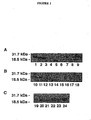
- the present invention which provides ELISA and Western blot analysis methods suitable for the detection and quantification of CR-1 in human milk and other samples was developed. As indicated herein, the methods and compositions provide the means to detect a specific band for CR-1 of 28 kDa in milk samples with concentrations between 62 and 118 ng/ml. In addition, as discussed in greater detail herein, CR-1 purified from human milk using an immunoaffinity column, was able to stimulate the phosphorylation of MAPK in NMuMG mouse mammary epithelial cells.
- FIG. 3 Panel A shows that the rabbit anti-CR-1 polyclonal antibody can linearly detect recombinant human CR-1 over a concentration range of 100 pg to 1 ⁇ g.
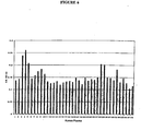
- Immunoreactive CR-1 was present at concentrations from 62 to 118 ng/ml in the milk samples ( See, Figure 3, Panel B).
- CR-1 in human milk is biologically active on mammary epithelial cells
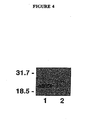
- a single human milk sample with high levels of CR-1 expression as determined by Western blot analysis and ELISA was chosen for purification.
- CR-1 was purified from delipidated human milk using an immunoaffinity column to which the anti-CR-1 rabbit polyclonal antibody was bound. After concentration, dialysis and estimation of the concentration of CR-1 as determined by Western blot analysis ( Figure 4), the purified CR-1 protein was assayed for its activity on NMuMG mouse mammary epithelial cells.
- CR-1 can induce a rapid increase in the tyrosine phosphorylation of p66, p52 and p46 isoforms of She which can than subsequently activate a ras / raf /MAPK pathway as evidenced by the enhanced phosphorylation of p42 and p44 isoforms of MAPK (Kannan et al., J. Biol. Chem., 272:3330-3335 [1997]).
- NMuMG mouse mammary epithelial cells respond to recombinant or chemically synthesized CR-1 with an increase in the phosphorylation of Shc and MAPK (Seno et al., Growth Factors 15:215-229 [1998]; and Kannan et al., supra ).
- serum-starved NMuMG cells were treated with different concentrations of the immunopurified milk-derived CR-1 protein and compared to recombinant human CR-1.
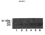
- a dose-dependent activation of MAPK was detected following treatment of the cells with milk-derived CR-1 ( See, Figure 5).
- Recombinant human CR-1 100 ng/ml was also able to stimulate MAPK phosphorylation which was nearly equivalent to the level of MAPK stimulation by 100 ng/ml of milk-derived CR-1.
- sample and “specimen” in the present specification and claims are used in their broadest sense. In preferred embodiments, these terms encompass all types of samples obtained from humans and other animals, including but not limited to, body fluids such as urine, blood, cerebrospinal fluid (CSF), fecal matter, semen, and saliva.
- body fluids such as urine, blood, cerebrospinal fluid (CSF), fecal matter, semen, and saliva.
- biological samples may be animal, including human, fluid or tissue, as these examples are not to be construed as limiting the sample types applicable to the present invention.
- fluid sample refers to a sample that is liquid.
- the term encompasses bodily fluids (e.g., "biological fluids"), such as milk, blood, serum, plasma, CSF, urine, semen, saliva, serous fluid, etc. It is not intended that the present invention be limited to any particular fluid.
- the term "antibody” is used in reference to any immunoglobulin molecule that reacts with a specific antigen. It is intended that the term encompass any immunoglobulin (e.g ., IgG, IgM, IgA, IgE, IgD, etc.) obtained from any source (e.g., humans, rodents, non-human primates, lagomorphs, caprines, bovines, equines, ovines, etc.).
- the term "antigen” is used in reference to any substance that is capable of reacting with an antibody. It is intended that this term encompass any antigen and "immunogen" (i.e., a substance which induces the formation of antibodies). Thus, in an immunogenic reaction, antibodies are produced in response to the presence of an antigen or portion of an antigen.
- antigen fragment and "portion of an antigen” are used in reference to a piece of an antigen. Antigen fragments or portions may occur in various sizes, ranging from a small percentage of the entire antigen to a large percentage, but not 100% of the antigen. However, in situations where "at least a portion of an antigen” is specified, it is contemplated that the entire antigen may be present. In some preferred embodiments, antigen fragments or portions comprise an "epitope" (e.g ., an "antigenic determinant”) recognized by an antibody.
- epipe e.g ., an "antigenic determinant
- antigen fragments or portions are be immunogenic (i.e., such fragments or portions are capable of inducing an immune response), while in other embodiments, the antigen fragments or portions are not immunogenic (i.e ., such fragments or portions are not capable of inducing an immune response).
- immunoassay is used in reference to any method in which antibodies are used in the detection of an antigen. It is contemplated that a range of immunoassay formats be encompassed by this definition, including but not limited to direct immunoassays, indirect immunoassays, and "sandwich” immunoassays.” A particularly preferred format is an enzyme-linked immunosorbent assay (ELISA). However, it is not intended that the present invention be limited to this format. It is contemplated that other formats, including radioimmunoassays (RIA), immunofluorescent assays (IFA), and other assay formats, including, but not limited to, variations on the ELISA method will find use in the method of the present invention.
- RIA radioimmunoassays
- IFA immunofluorescent assays
- antigen-antibody reaction formats will find use in the present invention, including but not limited to "flocculation” (i.e., a colloidal suspension produced upon the formation of antigen-antibody complexes), "agglutination” (i.e., clumping of cells or other substances upon exposure to antibody), "particle agglutination” ( i.e. , clumping of particles coated with antigen in the presence of antibody or the clumping of particles coated with antibody in the presence of antigen), “complement fixation” ( i.e., the use of complement in an antibody-antigen reaction method), and other methods commonly used in serology, immunology, immunocytochemistry, histochemistry, and related fields.
- cell staining is used in reference to methods used to label or stain cells to enhance their visualization. This staining or labelling is achieved through the use of various compounds, including but not limited to, fluorochromes, enzymes, gold, and iodine. It is contemplated that the definition encompasses such methods as "in situ chromogenic assays," in which a test ( i.e., an assay) is conducted on a sample in situ. It is also contemplated that the in situ chromogenic assay will involve the use of an immunoassay (i.e., an ELISA) or immunocytohistochemistry. However, it is not intended that the present invention be limited to any particular assay format for cell staining.
- the term "capture antibody” refers to an antibody that is used to bind an antigen and thereby permit the recognition of the antigen by a subsequently applied antibody.
- the capture antibody is bound to a microtiter well and serves to bind antigens (e.g. , Cripto-1) present in a sample added to the well.
- Another antibody (termed the “primary antibody”) is then used to bind to the antigen-antibody complex, in effect to form a "sandwich” comprised of antibody-antigen-antibody. Detection of this complex can be performed by several methods.
- the primary antibody is prepared with a label such as biotin, an enzyme, a fluorescent marker, or radioactivity, and is detected directly using this label.
- a label such as biotin, an enzyme, a fluorescent marker, or radioactivity
- a labelled "secondary antibody” or “reporter antibody” which recognizes the primary antibody is added, forming a complex comprised of antibody-antigen-antibody-antibody.
- appropriate reporter reagents are then added to detect the labelled antibody.
- any number of additional antibodies are added as desired.
- these antibodies are also be labelled with a marker, including, but not limited to an enzyme, fluorescent marker, or radioactivity.
- reporter reagent or "reporter molecule” is used in reference to compounds which are capable of detecting the presence of antibody bound to antigen.
- a reporter reagent is a colorimetric substance which is attached to an enzymatic substrate. Upon binding of antibody and antigen, the enzyme acts on its substrate and causes the production of a color.
- Other reporter reagents include, but are not limited to fluorogenic, chromogenic, luminogenic, and radioactive compounds or molecules. This definition also encompasses the use of biotin and avidin-based compounds (e.g. , including compounds but not limited to neutravidin and streptavidin) as part of the detection system. In one embodiment of the present invention, biotinylated antibodies are used in conjunction with avidin-coated solid support.
- the term "signal" is used in reference to an indicator that a reaction has occurred, for example, binding of antibody to antigen. It is contemplated that signals in the form of radioactivity, fluorogenic reactions, and enzymatic reactions will be used with the present invention. However, it is not intended that the present invention be limited to any particular signal. In some embodiments, the signal is assessed quantitatively and/or qualitatively.
- amplifier is used in reference to a system which enhances the signal in a test method such as an ELISA. However, it is not intended that the present invention be limited to the use of amplifiers in any particular assays system or format.
- solid support is used in reference to any solid material suitable for the attachment of various reagents such as antibodies, antigens, and/or other compounds.
- reagents such as antibodies, antigens, and/or other compounds.
- wells of microtiter plates often provide solid supports.
- Other examples of solid supports include microscope slides, coverslips, beads, particles, cell culture flasks, as well as many other items.
- kits are used in reference to a combination of reagents and other materials.
- the kits of the present invention comprise microtiter plates, buffers, labelled antibodies, positive and negative controls, instruction manuals for users, etc. It is not intended that the kits of the present invention be limited to any particular components, reagents, etc. Indeed, it is intended that various kit formats will find use with the present invention.
- the human milk samples used were obtained from 24 healthy women. Data regarding the time when these samples were collected after delivery were not available. The samples were centrifuged at 14,000 rpm for 10 min and the aqueous layer of the milk was collected after removing the lipid layer.
- NMuMG normal mouse mammary epithelial cells were cultured in Dulbecco's modified Eagle's medium containing 10% fetal bovine serum.
- Recombinant human CR-1 (rhCR-1) protein was expressed in E. coli and purified as known in the art ( See, Seno et al., Growth Factors 15:215-229 [1998]).
- FIG. 3 Panel A provides results for plates in which the concentration of CR-1 in the samples was estimated using different concentrations of recombinant CR-1 protein. This Figure shows that the rabbit anti-CR-1 polyclonal antibody can linearly detect recombinant human CR-1 over a concentration range of 100 pg to 1 ⁇ g.
- Panel B immunoreactive CR-1 was present at concentrations from 62 to 118 ng/ml in the milk samples.
- CR-1 protein from human milk was purified using an AMINOLINK ® Plus immobilization kit (Pierce), as per the manufacturer's instructions. Briefly, rabbit polyclonal anti-CR-1 IgG.was coupled to AMINOLINK ® Plus coupling gel using a pH 10 coupling buffer containing 0.1 M sodium citrate and 0.05 M sodium carbonate. The anti-CR-1 antibody-coupled column was then used for affinity purification of CR-1 from delipidated human milk. Human milk sample number 2, because of its high CR-1 levels as detected by Western blot ( See Example 1) and by ELISA ( See , Example 2), was chosen for purification.
- CR-1 protein Five ml of human milk were applied to the column and incubated for 1 hr at room temperature. After washing with PBS, the bound CR-1 protein was eluted in several fractions with 0.1 M glycine pH 2.5, and neutralized with 1 M Tris pH 9.5. The fractions containing the protein were pooled, concentrated with a CENTRICON ® 10 (Amicon) concentrator and dialyzed against PBS. The concentration of the purified protein was estimated by Western blot using known concentrations of recombinant human CR-1 as a reference, as described in Example 1.
- NMuMG mouse mammary epithelial cells were seeded in 100 mm plates and were grown until they reached 70-80% confluence. The cells were then switched to serum-free Dulbecco's modified Eagle's medium containing human transferrin (10 ⁇ g/ml) and type IV Pedersen fetuin (1 mg/ml) for 24 hr. Cells were stimulated for 5 min at 37°C with recombinant human CR-1 protein at 100 ng/ml or CR-1 that was purified from human milk by immunoaffinity chromatography as described in Example 2).
- the cells were lysed in a buffer containing 20 mM Tris-HCl, pH 7.5, 150 mM NaCl, 1% Nonidet P-40, 0.5% deoxycholate, 5 mM MgCl 2 , 2 ⁇ g/ml aprotinin, 2 ⁇ g/ml leupeptin, 1 mM phenylmethylsulfonyl fluoride, 1 mM sodium orthovanadate, and 20 mM sodium fluoride.
- Crude protein lysates (30 ⁇ g/sample) were run on a 10% SDS-PAGE gel, transferred to PVDF membrane, blocked in a solution prepared from 5% dry milk ( See, Example 1) and incubated with a 1:1000 dilution of a rabbit polyclonal anti-phospho MAPK antibody (Biolab). After incubation with a goat anti-rabbit IgG conjugated to horseradish peroxidase (Amersham), the immunoreactive bands were detected by enhanced chemiluminescence (Amersham).
- Figure 5 provides results for cell lysates were run on a 10% SDS-PAGE gel and probed with an anti-phospho MAPK rabbit polyclonal antibody (Biolab) that recognizes the activated phosphorylated forms of MAPK (p44 and p42).
- Lane 1 contains serum-starved cells; lane 2 contains rhCR-1 (100 ng/ml); lane 3 contains milk CR-1 (50 ng/ml); lane 4 contains milk CR-1 (100 ng/ml); lane 5 contains milk CR-1 (300 ng/ml); and lane 6 contains milk CR-1 (500 ng/ml).
- a dose-dependent activation of MAPK was detected following treatment of the cells with milk-derived CR-1.
- Recombinant human CR-1 100 ng/ml was also able to stimulate MAPK phosphorylation at a level that was nearly equivalent to the level of MAPK stimulation by 100 ng/ml of milk-derived CR-1.
Landscapes
- Life Sciences & Earth Sciences (AREA)
- Health & Medical Sciences (AREA)
- Engineering & Computer Science (AREA)
- Molecular Biology (AREA)
- Immunology (AREA)
- Biomedical Technology (AREA)
- Chemical & Material Sciences (AREA)
- Hematology (AREA)
- Urology & Nephrology (AREA)
- Cell Biology (AREA)
- Medicinal Chemistry (AREA)
- General Physics & Mathematics (AREA)
- Biotechnology (AREA)
- Food Science & Technology (AREA)
- Proteomics, Peptides & Aminoacids (AREA)
- Physics & Mathematics (AREA)
- Analytical Chemistry (AREA)
- Biochemistry (AREA)
- General Health & Medical Sciences (AREA)
- Microbiology (AREA)
- Pathology (AREA)
- Peptides Or Proteins (AREA)
- Investigating Or Analysing Biological Materials (AREA)
- Measuring Or Testing Involving Enzymes Or Micro-Organisms (AREA)
- Preparation Of Compounds By Using Micro-Organisms (AREA)
- Sub-Exchange Stations And Push- Button Telephones (AREA)
- Glass Compositions (AREA)
- Testing Of Coins (AREA)
Claims (10)
- Verfahren zum Nachweis von Cripto-1 in einer Probe, umfassend:a) das Bereitstelleni) einer biologischen Flüssigkeit, von der vermutet wird, dass sie Cripto-1 enthält,ii) eines Antikörpers, der gegen Cripto-1 gerichtet ist;b) die biologische Flüssigkeit dem Antikörper unter derartigen Bedingungen auszusetzen, dass Cripto-1 und der Antikörper binden, um einen Antigen-Antikörper-Komplex zu bilden; undc) das Nachweisen des Antigen-Antikörper-Komplexes.
- Verfahren nach Anspruch 1, ferner umfassend einen Schritt des Quantifizierens von Cripto-1 in der biologischen Flüssigkeit durch Messen der Menge des Antigen-Antikörper-Komplexes.
- Verfahren zum Quantifizieren von Cripto-1 in einer Probe, umfassend:a) das Bereitstelleni) einer biologischen Flüssigkeit, die Cripto-1 enthält,ii) eines Antikörpers, der gegen Cripto-1 gerichtet ist;b) die biologische Flüssigkeit dem Antikörper unter derartigen Bedingungen auszusetzen, dass Cripto-1 und der Antikörper binden, um einen Antigen-Antikörper-Komplex zu bilden; undc) das Messen der Menge des Antigen-Antikörper-Komplexes.
- Verfahren nach einem der Ansprüche 1-3, wobei es sich bei der biologischen Flüssigkeit um Milch handelt.
- Verfahren nach einem der Ansprüche 1 - 3, wobei es sich bei der biologischen Flüssigkeit um Serum handelt.
- Verfahren nach einem der Ansprüche 1 - 3, wobei es sich bei der biologischen Flüssigkeit um Plasma handelt.
- Verfahren nach einem der Ansprüche 1 - 6, wobei die biologische Flüssigkeit von einem Menschen stammt.
- Verfahren nach einem der Ansprüche 1 - 5, wobei es sich bei dem Antikörper um einen monoklonalen Antikörper handelt.
- Verfahren nach einem der Ansprüche 1 - 5, wobei es sich bei dem Antikörper um einen polyklonalen Antikörper handelt.
- Verfahren nach einem der Ansprüche 1 - 6, wobei es sich bei dem Verfahren um einen enzymverbundenen Immunadsorptions-Assay handelt.
Applications Claiming Priority (3)
| Application Number | Priority Date | Filing Date | Title |
|---|---|---|---|
| US26464301P | 2001-01-26 | 2001-01-26 | |
| US264643P | 2001-01-26 | ||
| PCT/US2002/002225 WO2002059620A2 (en) | 2001-01-26 | 2002-01-23 | Detection and quantification of cripto-1 |
Publications (2)
| Publication Number | Publication Date |
|---|---|
| EP1370869A2 EP1370869A2 (de) | 2003-12-17 |
| EP1370869B1 true EP1370869B1 (de) | 2006-12-27 |
Family
ID=23006978
Family Applications (1)
| Application Number | Title | Priority Date | Filing Date |
|---|---|---|---|
| EP02703238A Expired - Lifetime EP1370869B1 (de) | 2001-01-26 | 2002-01-23 | Nachweis und quantifizierung von cripto-1 |
Country Status (9)
| Country | Link |
|---|---|
| US (1) | US7078176B2 (de) |
| EP (1) | EP1370869B1 (de) |
| JP (1) | JP3821779B2 (de) |
| AT (1) | ATE349702T1 (de) |
| AU (1) | AU2002236871B2 (de) |
| CA (1) | CA2434694C (de) |
| DE (1) | DE60217079T2 (de) |
| ES (1) | ES2278899T3 (de) |
| WO (1) | WO2002059620A2 (de) |
Families Citing this family (9)
| Publication number | Priority date | Publication date | Assignee | Title |
|---|---|---|---|---|
| US20020010928A1 (en) * | 2000-04-24 | 2002-01-24 | Ranjit Sahota | Method and system for integrating internet advertising with television commercials |
| PT1390389E (pt) | 2001-04-26 | 2009-04-03 | Biogen Idec Inc | Anticorpos que bloqueiam o cripto e as utilizações dos mesmos |
| US7582299B2 (en) | 2001-04-26 | 2009-09-01 | Biogen Idec Ma Inc. | Cripto-specific antibodies |
| AU2003301619A1 (en) * | 2002-10-23 | 2004-05-13 | Exelixis, Inc. | MBMs AS MODIFIERS OF BRANCHING MORPHOGENESIS AND METHODS OF USE |
| CA2502685A1 (en) * | 2002-10-23 | 2004-05-06 | Exelixis, Inc. | Cdkl1 as modifier of branching morphogenesis and methods of use |
| US20070122813A1 (en) * | 2003-10-03 | 2007-05-31 | David Salomon | Use of cripto-1 as a biomarker for neurodegenerative disease and method of inhibiting progression thereof |
| ES2408704T3 (es) | 2005-01-05 | 2013-06-21 | Biogen Idec Ma Inc. | Moléculas de unión a Cripto |
| JP2011522224A (ja) * | 2008-04-21 | 2011-07-28 | メルク・シャープ・エンド・ドーム・コーポレイション | 膵ベータ細胞質量生物マーカー |
| US20100135904A1 (en) * | 2008-11-07 | 2010-06-03 | Research Development Foundation | Compositions and methods for the inhibition of cripto / grp78 complex formation and signaling |
Family Cites Families (6)
| Publication number | Priority date | Publication date | Assignee | Title |
|---|---|---|---|---|
| US579616A (en) * | 1897-03-30 | Rod-packing for air-brakes | ||
| US5256643A (en) | 1990-05-29 | 1993-10-26 | The Government Of The United States | Human cripto protein |
| US5264557A (en) | 1991-08-23 | 1993-11-23 | The United States Of America As Represented By The Department Of Health And Human Services | Polypeptide of a human cripto-related gene, CR-3 |
| US5981215A (en) * | 1995-06-06 | 1999-11-09 | Human Genome Sciences, Inc. | Human criptin growth factor |
| US5968839A (en) * | 1996-05-13 | 1999-10-19 | Metrika, Inc. | Method and device producing a predetermined distribution of detectable change in assays |
| US6777198B2 (en) * | 2000-10-11 | 2004-08-17 | Pharmacia Diagnostics Ab | Assay method and kit therefor |
-
2002
- 2002-01-23 ES ES02703238T patent/ES2278899T3/es not_active Expired - Lifetime
- 2002-01-23 WO PCT/US2002/002225 patent/WO2002059620A2/en not_active Ceased
- 2002-01-23 CA CA2434694A patent/CA2434694C/en not_active Expired - Lifetime
- 2002-01-23 AT AT02703238T patent/ATE349702T1/de not_active IP Right Cessation
- 2002-01-23 US US10/470,322 patent/US7078176B2/en not_active Expired - Lifetime
- 2002-01-23 AU AU2002236871A patent/AU2002236871B2/en not_active Ceased
- 2002-01-23 DE DE60217079T patent/DE60217079T2/de not_active Expired - Lifetime
- 2002-01-23 EP EP02703238A patent/EP1370869B1/de not_active Expired - Lifetime
- 2002-01-23 JP JP2002559686A patent/JP3821779B2/ja not_active Expired - Fee Related
Also Published As
| Publication number | Publication date |
|---|---|
| WO2002059620A3 (en) | 2003-03-27 |
| WO2002059620A2 (en) | 2002-08-01 |
| JP2005504262A (ja) | 2005-02-10 |
| AU2002236871C1 (en) | 2002-08-06 |
| CA2434694A1 (en) | 2002-08-01 |
| JP3821779B2 (ja) | 2006-09-13 |
| AU2002236871B2 (en) | 2006-09-28 |
| US20040077025A1 (en) | 2004-04-22 |
| EP1370869A2 (de) | 2003-12-17 |
| CA2434694C (en) | 2012-09-18 |
| ATE349702T1 (de) | 2007-01-15 |
| ES2278899T3 (es) | 2007-08-16 |
| DE60217079D1 (de) | 2007-02-08 |
| DE60217079T2 (de) | 2007-10-25 |
| US7078176B2 (en) | 2006-07-18 |
Similar Documents
| Publication | Publication Date | Title |
|---|---|---|
| Münstermann et al. | Casein kinase II is elevated in solid human tumours and rapidly proliferating non‐neoplastic tissue | |
| Leberer et al. | Immunochemical quantification of sarcoplasmic reticulum Ca‐ATPase, of calsequestrin and of parvalbumin in rabbit skeletal muscles of defined fiber composition | |
| US9095549B2 (en) | Diagnosis and prognosis of colorectal cancer | |
| JPWO2008120684A1 (ja) | 急性中枢神経障害の予後判定方法 | |
| EP1370869B1 (de) | Nachweis und quantifizierung von cripto-1 | |
| Katoh‐Semba et al. | Sex‐dependent and sex‐independent distribution of the β‐subunit of nerve growth factor in the central nervous and peripheral tissues of mice | |
| CN109307766A (zh) | 胃蛋白酶原ⅰ检测试剂盒 | |
| CN109307765A (zh) | 胃蛋白酶原ⅱ检测试剂盒 | |
| AU2002236871A1 (en) | Detection and quantification of CRIPTO-1 | |
| CN115586337A (zh) | 检测肿瘤伴随诊断相关因子的试剂盒及其制备方法 | |
| Ohnishi et al. | Development of highly sensitive enzyme-linked immunosorbent assays for hepatocyte growth factor/scatter factor (HGF/SF): determination of HGF/SF in serum and urine from normal human subjects | |
| CN101088010B (zh) | 用于测定抗原的试剂以及抗原的测定方法 | |
| JPH04504110A (ja) | 癌関連ハプトグロビン | |
| Chang et al. | Detection, quantitation, and localization of bovine angiogenin by immunological assays | |
| JP2009500597A (ja) | 線維症のマーカー | |
| Toi et al. | Co-determination of the angiogenic factors thymidine phosphorylase and vascular endothelial growth factor in node-negative breast cancer: prognostic implications | |
| KAJIKAWA et al. | Enzyme-linked immunosorbent assay for detection of feline serum amyloid A protein by use of immunological cross-reactivity of polyclonal anti-canine serum amyloid A protein antibody | |
| WO2004019045A2 (en) | Method for the diagnosis of alzheimer disease | |
| Ropers et al. | Enzyme immunoassay for the measurement of human tenascin-C on the Bayer Immuno 1™ analyzer | |
| US20100221742A1 (en) | Novel cancer associated antibodies and their use in cancer diagnosis | |
| JP7106810B2 (ja) | 新規肺がんマーカー | |
| US20130040325A1 (en) | Enzyme Linked Immunosorbent Assay (ELISA) Method and Kit for Detecting Soluble Programmed Cell Death Protein 5 (PDCD5) | |
| EP1331481A1 (de) | Diagnostischer kit für schizophrenie | |
| CN114729947B (zh) | 检测癌的骨转移的方法和检测试剂 | |
| RU2338200C1 (ru) | Способ количественного определения онкомаркера muc1 в сыворотке крови человека |
Legal Events
| Date | Code | Title | Description |
|---|---|---|---|
| PUAI | Public reference made under article 153(3) epc to a published international application that has entered the european phase |
Free format text: ORIGINAL CODE: 0009012 |
|
| 17P | Request for examination filed |
Effective date: 20030715 |
|
| AK | Designated contracting states |
Kind code of ref document: A2 Designated state(s): AT BE CH CY DE DK ES FI FR GB GR IE IT LI LU MC NL PT SE TR |
|
| AX | Request for extension of the european patent |
Extension state: AL LT LV MK RO SI |
|
| 17Q | First examination report despatched |
Effective date: 20050412 |
|
| GRAP | Despatch of communication of intention to grant a patent |
Free format text: ORIGINAL CODE: EPIDOSNIGR1 |
|
| GRAS | Grant fee paid |
Free format text: ORIGINAL CODE: EPIDOSNIGR3 |
|
| RAP1 | Party data changed (applicant data changed or rights of an application transferred) |
Owner name: THE GOVERNMENT OF THE UNITED STATES OF AMERICA, AS |
|
| GRAA | (expected) grant |
Free format text: ORIGINAL CODE: 0009210 |
|
| AK | Designated contracting states |
Kind code of ref document: B1 Designated state(s): AT BE CH CY DE DK ES FI FR GB GR IE IT LI LU MC NL PT SE TR |
|
| PG25 | Lapsed in a contracting state [announced via postgrant information from national office to epo] |
Ref country code: BE Free format text: LAPSE BECAUSE OF FAILURE TO SUBMIT A TRANSLATION OF THE DESCRIPTION OR TO PAY THE FEE WITHIN THE PRESCRIBED TIME-LIMIT Effective date: 20061227 Ref country code: FI Free format text: LAPSE BECAUSE OF FAILURE TO SUBMIT A TRANSLATION OF THE DESCRIPTION OR TO PAY THE FEE WITHIN THE PRESCRIBED TIME-LIMIT Effective date: 20061227 Ref country code: DK Free format text: LAPSE BECAUSE OF FAILURE TO SUBMIT A TRANSLATION OF THE DESCRIPTION OR TO PAY THE FEE WITHIN THE PRESCRIBED TIME-LIMIT Effective date: 20061227 Ref country code: AT Free format text: LAPSE BECAUSE OF FAILURE TO SUBMIT A TRANSLATION OF THE DESCRIPTION OR TO PAY THE FEE WITHIN THE PRESCRIBED TIME-LIMIT Effective date: 20061227 Ref country code: NL Free format text: LAPSE BECAUSE OF FAILURE TO SUBMIT A TRANSLATION OF THE DESCRIPTION OR TO PAY THE FEE WITHIN THE PRESCRIBED TIME-LIMIT Effective date: 20061227 |
|
| REG | Reference to a national code |
Ref country code: GB Ref legal event code: FG4D |
|
| PG25 | Lapsed in a contracting state [announced via postgrant information from national office to epo] |
Ref country code: MC Free format text: LAPSE BECAUSE OF NON-PAYMENT OF DUE FEES Effective date: 20070131 |
|
| REG | Reference to a national code |
Ref country code: IE Ref legal event code: FG4D |
|
| REF | Corresponds to: |
Ref document number: 60217079 Country of ref document: DE Date of ref document: 20070208 Kind code of ref document: P |
|
| PG25 | Lapsed in a contracting state [announced via postgrant information from national office to epo] |
Ref country code: SE Free format text: LAPSE BECAUSE OF FAILURE TO SUBMIT A TRANSLATION OF THE DESCRIPTION OR TO PAY THE FEE WITHIN THE PRESCRIBED TIME-LIMIT Effective date: 20070327 |
|
| PG25 | Lapsed in a contracting state [announced via postgrant information from national office to epo] |
Ref country code: PT Free format text: LAPSE BECAUSE OF FAILURE TO SUBMIT A TRANSLATION OF THE DESCRIPTION OR TO PAY THE FEE WITHIN THE PRESCRIBED TIME-LIMIT Effective date: 20070528 |
|
| NLV1 | Nl: lapsed or annulled due to failure to fulfill the requirements of art. 29p and 29m of the patents act | ||
| ET | Fr: translation filed | ||
| REG | Reference to a national code |
Ref country code: ES Ref legal event code: FG2A Ref document number: 2278899 Country of ref document: ES Kind code of ref document: T3 |
|
| PLBE | No opposition filed within time limit |
Free format text: ORIGINAL CODE: 0009261 |
|
| STAA | Information on the status of an ep patent application or granted ep patent |
Free format text: STATUS: NO OPPOSITION FILED WITHIN TIME LIMIT |
|
| 26N | No opposition filed |
Effective date: 20070928 |
|
| PG25 | Lapsed in a contracting state [announced via postgrant information from national office to epo] |
Ref country code: GR Free format text: LAPSE BECAUSE OF FAILURE TO SUBMIT A TRANSLATION OF THE DESCRIPTION OR TO PAY THE FEE WITHIN THE PRESCRIBED TIME-LIMIT Effective date: 20070328 |
|
| PG25 | Lapsed in a contracting state [announced via postgrant information from national office to epo] |
Ref country code: LU Free format text: LAPSE BECAUSE OF NON-PAYMENT OF DUE FEES Effective date: 20070123 Ref country code: CY Free format text: LAPSE BECAUSE OF FAILURE TO SUBMIT A TRANSLATION OF THE DESCRIPTION OR TO PAY THE FEE WITHIN THE PRESCRIBED TIME-LIMIT Effective date: 20061227 |
|
| PG25 | Lapsed in a contracting state [announced via postgrant information from national office to epo] |
Ref country code: TR Free format text: LAPSE BECAUSE OF FAILURE TO SUBMIT A TRANSLATION OF THE DESCRIPTION OR TO PAY THE FEE WITHIN THE PRESCRIBED TIME-LIMIT Effective date: 20061227 |
|
| REG | Reference to a national code |
Ref country code: CH Ref legal event code: PFA Owner name: THE GOVERNMENT OF THE UNITED STATES OF AMERICA AS Free format text: THE GOVERNMENT OF THE UNITED STATES OF AMERICA AS REPRESENTED BY THE SECRETARY OF THE DEPARTMENT OF HEALTH AND HUMAN SERVICE#NATIONAL INSTITUTES OF HEALTH, SUITE 325, 6011 EXECUTIVE BOULEVARD#ROCKVILLE, MD 20852-3804 (US) -TRANSFER TO- THE GOVERNMENT OF THE UNITED STATES OF AMERICA AS REPRESENTED BY THE SECRETARY OF THE DEPARTMENT OF HEALTH AND HUMAN SERVICE#NATIONAL INSTITUTES OF HEALTH, SUITE 325, 6011 EXECUTIVE BOULEVARD#ROCKVILLE, MD 20852-3804 (US) |
|
| REG | Reference to a national code |
Ref country code: FR Ref legal event code: PLFP Year of fee payment: 15 |
|
| REG | Reference to a national code |
Ref country code: FR Ref legal event code: PLFP Year of fee payment: 16 |
|
| REG | Reference to a national code |
Ref country code: FR Ref legal event code: PLFP Year of fee payment: 17 |
|
| PGFP | Annual fee paid to national office [announced via postgrant information from national office to epo] |
Ref country code: ES Payment date: 20180201 Year of fee payment: 17 Ref country code: DE Payment date: 20180129 Year of fee payment: 17 Ref country code: CH Payment date: 20180127 Year of fee payment: 17 Ref country code: GB Payment date: 20180129 Year of fee payment: 17 |
|
| PGFP | Annual fee paid to national office [announced via postgrant information from national office to epo] |
Ref country code: FR Payment date: 20180125 Year of fee payment: 17 Ref country code: IT Payment date: 20180122 Year of fee payment: 17 Ref country code: IE Payment date: 20180129 Year of fee payment: 17 |
|
| REG | Reference to a national code |
Ref country code: DE Ref legal event code: R119 Ref document number: 60217079 Country of ref document: DE |
|
| REG | Reference to a national code |
Ref country code: CH Ref legal event code: PL |
|
| GBPC | Gb: european patent ceased through non-payment of renewal fee |
Effective date: 20190123 |
|
| REG | Reference to a national code |
Ref country code: IE Ref legal event code: MM4A |
|
| PG25 | Lapsed in a contracting state [announced via postgrant information from national office to epo] |
Ref country code: DE Free format text: LAPSE BECAUSE OF NON-PAYMENT OF DUE FEES Effective date: 20190801 Ref country code: FR Free format text: LAPSE BECAUSE OF NON-PAYMENT OF DUE FEES Effective date: 20190131 |
|
| PG25 | Lapsed in a contracting state [announced via postgrant information from national office to epo] |
Ref country code: LI Free format text: LAPSE BECAUSE OF NON-PAYMENT OF DUE FEES Effective date: 20190131 Ref country code: CH Free format text: LAPSE BECAUSE OF NON-PAYMENT OF DUE FEES Effective date: 20190131 Ref country code: GB Free format text: LAPSE BECAUSE OF NON-PAYMENT OF DUE FEES Effective date: 20190123 |
|
| PG25 | Lapsed in a contracting state [announced via postgrant information from national office to epo] |
Ref country code: IE Free format text: LAPSE BECAUSE OF NON-PAYMENT OF DUE FEES Effective date: 20190123 |
|
| PG25 | Lapsed in a contracting state [announced via postgrant information from national office to epo] |
Ref country code: IT Free format text: LAPSE BECAUSE OF NON-PAYMENT OF DUE FEES Effective date: 20190123 |
|
| REG | Reference to a national code |
Ref country code: ES Ref legal event code: FD2A Effective date: 20200310 |
|
| PG25 | Lapsed in a contracting state [announced via postgrant information from national office to epo] |
Ref country code: ES Free format text: LAPSE BECAUSE OF NON-PAYMENT OF DUE FEES Effective date: 20190124 |