EP1365356B1 - Algorithme de segmentation semiautomatique pour des images oncologiques d'animaux familiers - Google Patents
Algorithme de segmentation semiautomatique pour des images oncologiques d'animaux familiers Download PDFInfo
- Publication number
- EP1365356B1 EP1365356B1 EP03252378A EP03252378A EP1365356B1 EP 1365356 B1 EP1365356 B1 EP 1365356B1 EP 03252378 A EP03252378 A EP 03252378A EP 03252378 A EP03252378 A EP 03252378A EP 1365356 B1 EP1365356 B1 EP 1365356B1
- Authority
- EP
- European Patent Office
- Prior art keywords
- interest
- region
- seed points
- spheres
- image
- Prior art date
- Legal status (The legal status is an assumption and is not a legal conclusion. Google has not performed a legal analysis and makes no representation as to the accuracy of the status listed.)
- Expired - Lifetime
Links
- 230000011218 segmentation Effects 0.000 title claims description 23
- 238000000034 method Methods 0.000 claims abstract description 33
- 238000002600 positron emission tomography Methods 0.000 claims description 11
- 238000002591 computed tomography Methods 0.000 claims description 10
- 238000002595 magnetic resonance imaging Methods 0.000 claims description 6
- 238000005192 partition Methods 0.000 claims description 6
- 238000012546 transfer Methods 0.000 claims description 4
- 230000006870 function Effects 0.000 description 23
- 238000003384 imaging method Methods 0.000 description 12
- 210000004556 brain Anatomy 0.000 description 9
- 238000002059 diagnostic imaging Methods 0.000 description 6
- 210000003484 anatomy Anatomy 0.000 description 4
- 238000005259 measurement Methods 0.000 description 4
- 238000012545 processing Methods 0.000 description 4
- 238000010586 diagram Methods 0.000 description 3
- 239000003446 ligand Substances 0.000 description 3
- 239000000463 material Substances 0.000 description 3
- 210000004761 scalp Anatomy 0.000 description 3
- 238000012800 visualization Methods 0.000 description 3
- 230000002285 radioactive effect Effects 0.000 description 2
- 239000002287 radioligand Substances 0.000 description 2
- 230000004044 response Effects 0.000 description 2
- 210000001519 tissue Anatomy 0.000 description 2
- 206010028980 Neoplasm Diseases 0.000 description 1
- 238000012879 PET imaging Methods 0.000 description 1
- 230000003044 adaptive effect Effects 0.000 description 1
- 238000003491 array Methods 0.000 description 1
- 210000000988 bone and bone Anatomy 0.000 description 1
- 210000001175 cerebrospinal fluid Anatomy 0.000 description 1
- 239000003086 colorant Substances 0.000 description 1
- 238000004040 coloring Methods 0.000 description 1
- 238000001514 detection method Methods 0.000 description 1
- 238000003745 diagnosis Methods 0.000 description 1
- 230000000694 effects Effects 0.000 description 1
- 230000005284 excitation Effects 0.000 description 1
- 230000001815 facial effect Effects 0.000 description 1
- 239000012530 fluid Substances 0.000 description 1
- 210000004884 grey matter Anatomy 0.000 description 1
- 125000004435 hydrogen atom Chemical group [H]* 0.000 description 1
- 230000002452 interceptive effect Effects 0.000 description 1
- 238000007917 intracranial administration Methods 0.000 description 1
- 230000005055 memory storage Effects 0.000 description 1
- 230000000877 morphologic effect Effects 0.000 description 1
- 210000001328 optic nerve Anatomy 0.000 description 1
- 210000000056 organ Anatomy 0.000 description 1
- 230000000704 physical effect Effects 0.000 description 1
- 238000011160 research Methods 0.000 description 1
- 239000007787 solid Substances 0.000 description 1
- 230000003068 static effect Effects 0.000 description 1
- 238000001356 surgical procedure Methods 0.000 description 1
- 238000002604 ultrasonography Methods 0.000 description 1
- 210000004885 white matter Anatomy 0.000 description 1
Images


Classifications
-
- G—PHYSICS
- G06—COMPUTING; CALCULATING OR COUNTING
- G06T—IMAGE DATA PROCESSING OR GENERATION, IN GENERAL
- G06T7/00—Image analysis
- G06T7/10—Segmentation; Edge detection
- G06T7/11—Region-based segmentation
-
- G—PHYSICS
- G06—COMPUTING; CALCULATING OR COUNTING
- G06T—IMAGE DATA PROCESSING OR GENERATION, IN GENERAL
- G06T7/00—Image analysis
- G06T7/10—Segmentation; Edge detection
- G06T7/187—Segmentation; Edge detection involving region growing; involving region merging; involving connected component labelling
-
- G—PHYSICS
- G06—COMPUTING; CALCULATING OR COUNTING
- G06V—IMAGE OR VIDEO RECOGNITION OR UNDERSTANDING
- G06V10/00—Arrangements for image or video recognition or understanding
- G06V10/20—Image preprocessing
- G06V10/26—Segmentation of patterns in the image field; Cutting or merging of image elements to establish the pattern region, e.g. clustering-based techniques; Detection of occlusion
- G06V10/267—Segmentation of patterns in the image field; Cutting or merging of image elements to establish the pattern region, e.g. clustering-based techniques; Detection of occlusion by performing operations on regions, e.g. growing, shrinking or watersheds
-
- G—PHYSICS
- G06—COMPUTING; CALCULATING OR COUNTING
- G06T—IMAGE DATA PROCESSING OR GENERATION, IN GENERAL
- G06T2207/00—Indexing scheme for image analysis or image enhancement
- G06T2207/10—Image acquisition modality
- G06T2207/10072—Tomographic images
- G06T2207/10104—Positron emission tomography [PET]
-
- G—PHYSICS
- G06—COMPUTING; CALCULATING OR COUNTING
- G06T—IMAGE DATA PROCESSING OR GENERATION, IN GENERAL
- G06T2207/00—Indexing scheme for image analysis or image enhancement
- G06T2207/20—Special algorithmic details
- G06T2207/20092—Interactive image processing based on input by user
- G06T2207/20104—Interactive definition of region of interest [ROI]
-
- G—PHYSICS
- G06—COMPUTING; CALCULATING OR COUNTING
- G06T—IMAGE DATA PROCESSING OR GENERATION, IN GENERAL
- G06T2207/00—Indexing scheme for image analysis or image enhancement
- G06T2207/20—Special algorithmic details
- G06T2207/20112—Image segmentation details
- G06T2207/20156—Automatic seed setting
-
- G—PHYSICS
- G06—COMPUTING; CALCULATING OR COUNTING
- G06T—IMAGE DATA PROCESSING OR GENERATION, IN GENERAL
- G06T2207/00—Indexing scheme for image analysis or image enhancement
- G06T2207/30—Subject of image; Context of image processing
- G06T2207/30004—Biomedical image processing
- G06T2207/30016—Brain
-
- G—PHYSICS
- G06—COMPUTING; CALCULATING OR COUNTING
- G06V—IMAGE OR VIDEO RECOGNITION OR UNDERSTANDING
- G06V10/00—Arrangements for image or video recognition or understanding
- G06V10/20—Image preprocessing
- G06V10/24—Aligning, centring, orientation detection or correction of the image
- G06V10/248—Aligning, centring, orientation detection or correction of the image by interactive preprocessing or interactive shape modelling, e.g. feature points assigned by a user
-
- Y—GENERAL TAGGING OF NEW TECHNOLOGICAL DEVELOPMENTS; GENERAL TAGGING OF CROSS-SECTIONAL TECHNOLOGIES SPANNING OVER SEVERAL SECTIONS OF THE IPC; TECHNICAL SUBJECTS COVERED BY FORMER USPC CROSS-REFERENCE ART COLLECTIONS [XRACs] AND DIGESTS
- Y10—TECHNICAL SUBJECTS COVERED BY FORMER USPC
- Y10S—TECHNICAL SUBJECTS COVERED BY FORMER USPC CROSS-REFERENCE ART COLLECTIONS [XRACs] AND DIGESTS
- Y10S128/00—Surgery
- Y10S128/92—Computer assisted medical diagnostics
- Y10S128/922—Computer assisted medical diagnostics including image analysis
Definitions
- This invention relates to segmentation of medical images. More particularly, the invention relates to a method and system for segmenting an region of interest in three-dimensional medical images for use in volumetric measurement.
- 3D arrays of data representing one or more physical properties within an interior of a solid body, for example, anatomical structures.
- data is obtained by a variety of non-invasive methods such as positron emission tomography (PET), computed tomography (CT), magnetic resonance imaging (MRI), ultrasound, x-ray or a combination thereof.
- PET positron emission tomography
- CT computed tomography
- MRI magnetic resonance imaging
- ultrasound x-ray or a combination thereof.
- the 3D array of data typically consists of a plurality of sets of three-dimensional coordinates distributed at regular positions about the body of interest.
- techniques available to generate a three-dimensional model or structure.
- a seed voxel (volume element) is placed within the anatomical structure of interest and adjacent voxels are successively analyzed and identified as belonging to the same structure generally if they are adjacent to a previously identified voxel and they meet a specified attribute, such as intensity or radiological density.
- a 3D image is obtained for visualization.
- the three-dimensional (3D) visualization of internal anatomical structures is a technique by medical professionals and research scientists.
- Three-dimensional models enable the ability to rotate the model or virtual representation of the anatomical structure, as well as adjust a point of perspective and zoom in/out from features of interest.
- volumetric measurements are enabled by a variety of known 3D image processing techniques.
- the connected volume after segmentation may include regions that are not of interest thus requiring some user intervention. Further, the connected volume may include connection through a undesired narrow region, bridge or other small structure that connects different regions that are desirably separated.
- a method for segmenting three-dimensional (3D) medical images containing a region of interest comprising the steps of: manually identifying a first set of seed points within the region of interest; manually identifying a second set of seed points outside the region of interest; constructing a first sphere within the region of interest wherein the first sphere is centered about a first set of seed points; using the identified seed points to classify all the voxels contained within the 3D medical images using a spatially constrained fuzzy clustering algorithm, so as to transfer the 3D image set into a fuzzy partition domain where each voxel is assigned a probability of belonging to the desired region of interest based on a homegenity function; generating a plurality of second spheres; selecting the radius of curvature of the first sphere and the plurality of second spheres based on a predetermined radius of curvature to eliminate noise voxels on the interface between an inner portion of and an outer portion of the region of interest; accepting
- a system for segmenting medical images containing a region of interest and acquired by an image acquisition device comprising: a processor coupled to the image acquisition device, the processor computing segmentation of the medical image wherein the computation of segmentation comprises the steps of: manually identifying a first set of seed points within the region of interest; manually identifying a second set of seed points outside the region of interest; constructing a first sphere within the region of interest wherein the first sphere is centered about a first set of seed points; using the identified seed points to classify all the voxels contained within the 3D medical images using a spatially constrained fuzzy clustering algorithm, so as to transfer the 3D image set into a fuzzy partition domain where each voxel is assigned a probability of belonging to the desired region of interest based on a homegenity function; generating a plurality of second spheres; selecting the radius of curvature of the first sphere and the plurality of second spheres based on a predetermined radius of curvature to eliminate noise
- a processor uses the identified seed points to classify all the voxels in the data set using a spatially constrained fuzzy clustering algorithm.
- the 3D image set is transformed into a fuzzy partition domain where each voxel is assigned a probability of belonging to the desired region of interest based on a homogeneity function.
- a processor constructs a set of first spheres within the region of interest centered on the first set of seed points.
- a plurality of second spheres is generated by the processor. Ones of the plurality of second spheres are accepted that satisfy the homogeneity function threshold as defined by the spatial constrained fuzzy clustering algorithm.
- a three-dimensional area defining the region of interest is adaptively grown based on the accepting of the ones of the plurality of second spheres.
- a radius of curvature of the spheres is determined to eliminate noise voxels on the interface between an inside of the region of interest and outside the region of interest.
- the region of interest defined by the adaptively growing of the three-dimensional area and selecting the radius of curvature is displayed to a human user.
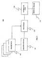
- FIG. 1 a block diagram of a system 100 for semi-automatic segmentation of a medical image 135.
- the system 100 includes an imaging device 110 that can be selected from various medical imaging devices for generating a plurality of medical images 135.
- medical imaging techniques such as, for example, positron emission tomography (PET) and magnetic resonance imaging (MRI) systems, are used to generate the medical images 135.
- PET positron emission tomography
- MRI magnetic resonance imaging
- a patient is injected with a radio-labeled ligand that specifically targets metabolically hyperactive regions like tumors.
- the patient horizontally lies inside the scanner.
- Photon emissions from the decay of the radio-active ligand are detected by a series of photon detectors.
- the detectors measure the amount radio-active emissions from inside the patient's body. This information is used to compute the concentration of the radio-labeled ligand for sample points in the body.
- a gray scale image is then constructed based upon the calculated radio-ligand concentrations. The shades of gray in the image contrast the amount of radio-ligand concentration of every point within the slice.
- the slices obtained during a PET session can be reconstructed to provide an functionally correct representation of the area of interest within the body.
- the patient is placed inside a strong magnetic field generated by a large magnet.
- Magnetized protons within the patient such as hydrogen atoms, align with the magnetic field produced by the magnet.
- a particular slice of the patient is exposed to radio waves that create an oscillating magnetic field perpendicular to the main magnetic field.
- the slices can be taken in any plane chosen by the physician or technician (hereinafter the "operator") performing the imaging session.
- the protons in the patient's body first absorb the radio waves and then emit the waves by moving out of alignment with the field. As the protons return to their original state (before excitation), diagnostic images based upon the waves emitted by the patient's body are created.
- MR image slices can be reconstructed to provide an overall picture of the body area of interest. Parts of the body that produce a high signal are displayed as white in an MR image, while those with the lowest signals are displayed as black. Other body parts that have varying signal intensities between high and low are displayed by various shades of gray.
- the medical images 135 are generally segmented.
- the segmentation process classifies the pixels or voxels of the medical image 135 into a certain number of classes that are homogeneous with respect to some characteristic (i.e. intensity, texture, etc.). For example, in a segmented medical image of the brain, the material of the brain can be categorized into three classes: gray matter, white matter, and cerebrospinal fluid. Individual colors can be used to mark regions of each class after the segmentation has been completed.
- surgeons or other medical personnel can use the segmented images to plan surgical techniques and/or assist in diagnoses.
- creating a segmented medical image involves several steps.
- a data set is created by capturing slices of data from the medical image 135.
- a gray scale value is assigned to each point in the data set and different types of tissues will have different gray scale values.
- Each type of material in the data is assigned a specific value and, therefore, each occurrence of that material has the same gray scale value. For example, all occurrences of bone in a particular image may appear in a particular shade of light gray. This standard of coloring allows the individual viewing the image to easily understand the objects and/or regions of interest being represented in the images.
- a medical imaging system 100 comprises a processor 120 connected to an imaging device 110 and an interface unit 130.
- the imaging device 110 generates a plurality of image data sets 140 and can comprise, for example, a positron emission tomography (PET) or magnetic resonance (MR) scanner.
- PET positron emission tomography
- MR magnetic resonance
- imaging system 100 acquisition of image data 140 is generally referred to as scanning.
- the processor 120 performs computations relating to semi-automatic segmentation of the medical image 135 as described herein. Further, the processor 120 is also performs computation and control functions for image processing techniques, such as, for example, reconstruction, image data memory storage, segmentation, etc.
- the processor 120 comprises a central processing unit (CPU), such as, for example, a single integrated circuit and/or microprocessor.
- the processor 120 comprises a CPU comprising, such as, for example, a plurality of integrated circuit devices and/or circuit boards working in cooperation to accomplish various functions.
- the processor 120 includes a memory device, such as, for example, random access memory (RAM), dynamic random access memory (DRAM), static random access memory (SRAM), flash memory, cache memory, etc.
- the processor 120 may internally comprise a memory device.
- the processor 120 executes the programs contained in memory and can act in response to those programs to perform other activities or functions that can occur in the course of image acquisition and image viewing. Also as shown in Fig. 1 , the processor 120 further performs segmentation methods as described herein in relation to Figs. 2-6 and in response to placement of seed points from, for example, interface unit 130.
- the interface unit 130 is coupled to processor 120 and allows a human user to communicate with imaging system 100. Additionally, the processor 120 can perform computations that are transmitted to and/or from interface unit 130 in a manner such that a human user is capable of interpreting the transmitted information.
- the transmitted information can include images in 2D or 3D, color and gray scale images, and text messages regarding diagnosis and detection information.
- the interface unit 130 can comprise, for example, a personal computer, an input/output device, an image work station, a hand held image display unit or a conventional image display platform generally included as a component in a PET or MRI system.
- the image data 140 gathered from multiple scans of the patient can considered one data set and can be formed into a medical image 135.
- Each data set can be broken up into smaller units, either pixels or voxels.
- the medical image 135 is made up of units called pixels.
- a pixel is a point in two-dimensional space that can be referenced using two-dimensional coordinates, usually x and y.
- Each pixel in an image is surrounded by eight other pixels, the nine pixels forming a three-by-three square. These eight other pixels, which surround the center pixel, are considered the eight-connected neighbors of the center pixel.
- the medical image 135 is displayed in units called voxels.
- a voxel is a point in three-dimensional space that can be referenced using three-dimensional coordinates, usually x, y and z. Each voxel is surrounded by twenty-six other voxels. These twenty-six voxels can be considered the twenty-six connected neighbors of the original voxel.
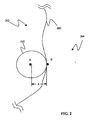
- a method 600 is provided for segmenting a three-dimensional (3D) medical image 135 that has a region of interest 200 ( Fig. 2 ).
- a first set of seed points (A) is identified within the region of interest 200 (step 610).
- a second set of seed points (B) is identified outside the region of interest 200 (step 620).
- a set of seed points comprises at least one seed point.
- a set of seed points can comprise a plurality of seed points.
- the first set of seed points (A) and the second set of seed points (B) can be identified by a human user using the interface unit 130 of the imaging system 100.
- a first sphere 210 is constructed within the region of interest 200 and is centered about the first set of seed points (A) (step 630). It should be appreciated that, in one embodiment, the first sphere 210 can comprise a set of first spheres wherein each of the set is centered about a different one of the set of seed points (A).
- all the voxels contained in the 3D medical image 135 are classified as belonging to the region of interest 200 based on a homogeneity function and a homogeneity function threshold is determined (step 640).
- a number of second spheres 220 ( Fig. 3 ) are generated (step 650).
- the homogeneity function threshold is satisfied when the homogeneity function threshold of the first sphere 210 is equal to or greater than the homogeneity function threshold of the particular one of the second spheres 220. In another embodiment, the homogeneity function threshold is satisfied when the homogeneity function threshold of the first sphere 210 is equal to or less than the homogeneity function threshold of the particular one of the second spheres 220.
- a three-dimensional area 230 ( Fig. 5 ) defining the region of interest 200 is adaptively grown and/or expanded based of the accepting of the particular ones of the second spheres 220 (step 660).
- the region of interest 200 defined by the adaptive growing and/or expanding of the ones of the second spheres 220 is displayed to a human user (step 660). It should be appreciated that, in one embodiment, the interface unit 130 can perform the displaying of the region of interested 200.
- the apparatus and method 100 for segmenting three-dimensional medical images 135 containing an object of interest 200 includes identifying a first set of seed points (A) and a second set of seed points (B).
- the first set of seed points (A) is identified in an inner portion 202 of the region of interest 200.
- the second set of seed points (B) is located in an exterior portion 204 in relation to the region of interest 200.
- the first set of spheres 210 is constructed completely in the inner potion 202 of the region of interest 200 and about the first set of seed points (A).
- the number of second spheres 220 is generated about the first sphere 210.
- the voxels contained in the 3D image 135 are classified with a particular homogeneity function as defined by the spatial constricted fuzzy clustering algorithm.
- the spatial constricted fuzzy clustering algorithm models the inherent high inter-pixel correlation between neighboring voxels and the fuzzy edges due to inherent low resolution properties of the medical image 135.
- the voxels contained in each one of the second spheres 220 is tested against the homogeneity function threshold. Those second spheres 220 that satisfy the homogeneity function threshold are accepted, and those second spheres 220 that do not satisfy the homogeneity function threshold are not accepted.
- the second spheres 220 that are accepted grow, expand and define a three-dimensional area 230 that defines the region of interest 200. Therefore, it should be appreciated that by this semi-automatic segmentation the growth, expansion and definition of the three-dimensional area 230 of the region of interest 210 is performed using a fuzzy partition domain map of the medical image rather than the native pixel/voxel intensity domain.
- the images are three-dimensional medical images 135 acquired by magnetic resonance imaging (MRI).
- MRI magnetic resonance imaging
- the three-dimensional medical images may be acquired by techniques other imaging systems such as, for example, computed tomography (CT), positron emission tomography (PET), and x-ray systems.
- CT computed tomography
- PET positron emission tomography
- x-ray systems x-ray systems.
- the imaging system 100 can comprise a PET system and the medical images 135 can comprise oncology images.
- a radius of curvature (R) ( Fig. 2 ) of the first sphere 210 and the second spheres 220 is selected.
- the radius of curvature (R) and the morphological opening is used to eliminate noise voxels on the interface between an inner potion 202 and an outer portion 204 of the region of interest 200 and, in addition, increase the robustness against noise.
- the selection of the radius of curvature (R) prevents the joining of objects connected by thin paths or the inclusion of noise areas on the surface and/or interface (area demarking the inner portion 202 and the outside potion 204) of the region of interest 200.
- the radius of curvature (R) can be determined by the imaging system 100.
- the radius of curvature (R) of the spheres can selected in accordance with a predetermined radius of curvature (R) of the segmented representation.
- the medical images 135 can comprise three-dimensional brain images acquired by magnetic resonance imaging (MRI). It should also be appreciated that, in other embodiments, the three-dimensional medical images 135 may be acquired techniques other imaging techniques, such as, for example computed tomography (CT), positron emission tomography (PET), and x-ray systems. Generally, three-dimensional magnetic brain images 135 can be segmented by connectivity using the methods described above. However, in the three-dimensional magnetic brain images 135, there may be connections between the intracranial volume and the scalp that should also be segmented. For example, one path that connects the brain to the scalp is along the optic nerve to the fluid filled eye globes and then the facial tissue.
- CT computed tomography
- PET positron emission tomography
- x-ray systems x-ray systems
- one or more seeds in placed in the object in this embodiment, the brain
- wavelets of a fixed spherical radius are formed.
- a "wavelet” refers to a data structure representing the voxels contained within a sphere. The radius of the wavelet is selected by the user to prevent connectivity along narrow paths. The wavelets are tested and only those spherical regions completely composed of voxels above a critical threshold are allowed to propagate.
- the critical threshold comprises the homogeneity function threshold.
- the threshold refers to a parameter defining the region of interest 200 and is generally based on intensity values.
- the threshold is selected to define the region of interest 200 such that the voxels within the region of interest 200 are above the threshold and the remaining voxels are considered background.
- the threshold alone is generally not sufficient for segmentation because other objects may also have an intensity value above the threshold. For example, the brain and scalp have similarly high relative intensity compared to other voxels.
- at the boundary of a growing bubble there are active spherical wavelets with active seeds at the center. The bubble is composed of the region swept out by the wavelets. After each iteration, a layer of wavelets propagate in until there are no more active seeds.
- the union of all the spherical regions (wavelets) that are completely above the threshold define the connected volume (bubble). It is to be noted that regions that form bridges smaller in dimension than the selected bubble diameter are not included in the connected region. Further, the connected volume does not propagate into noisy regions where the voxels above threshold are randomly selected.
Landscapes
- Engineering & Computer Science (AREA)
- Physics & Mathematics (AREA)
- General Physics & Mathematics (AREA)
- Theoretical Computer Science (AREA)
- Computer Vision & Pattern Recognition (AREA)
- Multimedia (AREA)
- Magnetic Resonance Imaging Apparatus (AREA)
- Image Processing (AREA)
- Image Analysis (AREA)
- Apparatus For Radiation Diagnosis (AREA)
- Measuring And Recording Apparatus For Diagnosis (AREA)
- Nuclear Medicine (AREA)
Claims (3)
- Procédé de segmentation d'images médicales tridimensionnelles (3D) (135) contenant une région d'intérêt, le procédé comprenant les étapes de :identification manuelle d'un premier ensemble de points germes (A) à l'intérieur de la région d'intérêt (200);identification manuelle d'un second ensemble de points germes (B) à l'extérieur de la région d'intérêt (200);construction d'une première sphère (210) à l'intérieur de la région d'intérêt (200), la première sphère (210) étant centrée sur un premier ensemble de points germes (A);utilisation des points germes identifiés (A, B) pour classifier tous les voxels contenus dans les images médicales 3D (135) à l'aide d'un algorithme de classification floue sous contraintes spatiales, de façon à transférer l'ensemble d'images 3D vers un domaine de partition floue dans lequel chaque voxel se voit attribuer une probabilité d'appartenir à la région d'intérêt désirée sur la base d'une fonction d'homogénéité;génération d'une pluralité de secondes sphères (220);sélection du rayon de courbure de la première sphère (210) et de la pluralité de secondes sphères (220) sur la base d'un rayon de courbure prédéterminé afin d'éliminer des voxels de bruit sur l'interface entre une partie interne (202) et une partie externe (204) de la région d'intérêt (200);acceptation de celles des secondes sphères (220) qui satisfont le seuil de fonction d'homogénéité défini par l'algorithme de classification floue sous contraintes spatiales de telle manière que seules les régions sphériques qui sont entièrement composées de voxels supérieurs audit seuil soient autorisées à se propager, pour ainsi réaliser la croissance adaptative, l'expansion et la définition (660) d'une zone tridimensionnelle (230) qui définit la région d'intérêt désirée (200);itération des étapes précédentes jusqu'à ce qu'il n'y ait plus de points germes actifs; etaffichage de la région d'intérêt désirée définie par les étapes d'acceptation et de croissance adaptative de la zone tridimensionnelle (230).
- Procédé selon la revendication 1, dans lequel les images tridimensionnelles sont acquises par au moins un système parmi des systèmes d'imagerie par résonance magnétique (IRM), de tomodensitométrie (TDM), de tomographie par émission de positons (TEP) et de radiographie.
- Système de segmentation d'images médicales (1000) contenant une région d'intérêt et acquises par un dispositif d'acquisition d'image (110), le système comprenant :un processeur (120) couplé au dispositif d'acquisition d'image (110), le processeur (120) calculant une segmentation de l'image médicale (135), le calcul de segmentation comprenant les étapes de :identification manuelle d'un premier ensemble de points germes (A) à l'intérieur de la région d'intérêt (200);identification manuelle d'un second ensemble de points germes (B) à l'extérieur de la région d'intérêt (200);construction d'une première sphère (210) à l'intérieur de la région d'intérêt (200), la première sphère (210) étant centrée sur un premier ensemble de points germes (A);utilisation des points germes identifiés (A, B) pour classifier tous les voxels contenus dans les images médicales 3D (135) à l'aide d'un algorithme de classification floue sous contraintes spatiales, de façon à transférer l'ensemble d'images 3D vers un domaine de partition floue dans lequel chaque voxel se voit attribuer une probabilité d'appartenir à la région d'intérêt désirée sur la base d'une fonction d'homogénéité;génération d'une pluralité de secondes sphères (220);sélection du rayon de courbure de la première sphère (210) et de la pluralité de secondes sphères (220) sur la base d'un rayon de courbure prédéterminé afin d'éliminer des voxels de bruit sur l'interface entre une partie interne (202) et une partie externe (204) de la région d'intérêt (200);acceptation de celles des secondes sphères (220) qui satisfont le seuil de fonction d'homogénéité défini par l'algorithme de classification floue sous contraintes spatiales de telle manière que seules les régions sphériques qui sont entièrement composées de voxels supérieurs audit seuil soient autorisées à se propager, pour ainsi réaliser la croissance adaptative, l'expansion et la définition (660) d'une zone tridimensionnelle (230) qui définit la région d'intérêt désirée (200);itération des étapes précédentes jusqu'à ce qu'il n'y ait plus de points germes actifs; etaffichage de la région d'intérêt désirée définie par les étapes d'acceptation et de croissance adaptative de la zone tridimensionnelle (230); etune unité d'interface (130) couplée au processeur (120) pour interpréter des informations relatives à la segmentation de l'image médicale (135).
Applications Claiming Priority (2)
| Application Number | Priority Date | Filing Date | Title |
|---|---|---|---|
| US10/122,892 US7006677B2 (en) | 2002-04-15 | 2002-04-15 | Semi-automatic segmentation algorithm for pet oncology images |
| US122892 | 2002-04-15 |
Publications (3)
| Publication Number | Publication Date |
|---|---|
| EP1365356A2 EP1365356A2 (fr) | 2003-11-26 |
| EP1365356A3 EP1365356A3 (fr) | 2005-10-19 |
| EP1365356B1 true EP1365356B1 (fr) | 2011-12-14 |
Family
ID=28790644
Family Applications (1)
| Application Number | Title | Priority Date | Filing Date |
|---|---|---|---|
| EP03252378A Expired - Lifetime EP1365356B1 (fr) | 2002-04-15 | 2003-04-15 | Algorithme de segmentation semiautomatique pour des images oncologiques d'animaux familiers |
Country Status (5)
| Country | Link |
|---|---|
| US (1) | US7006677B2 (fr) |
| EP (1) | EP1365356B1 (fr) |
| JP (1) | JP4310773B2 (fr) |
| CN (1) | CN100550004C (fr) |
| AT (1) | ATE537519T1 (fr) |
Families Citing this family (65)
| Publication number | Priority date | Publication date | Assignee | Title |
|---|---|---|---|---|
| US6589948B1 (en) * | 2000-11-28 | 2003-07-08 | Eukarion, Inc. | Cyclic salen-metal compounds: reactive oxygen species scavengers useful as antioxidants in the treatment and prevention of diseases |
| US7457444B2 (en) * | 2003-05-14 | 2008-11-25 | Siemens Medical Solutions Usa, Inc. | Method and apparatus for fast automatic centerline extraction for virtual endoscopy |
| US7457445B2 (en) * | 2004-03-01 | 2008-11-25 | Siemens Medical Solutions Usa, Inc. | Using corner pixels as seeds for detection of convex objects |
| WO2005104662A2 (fr) * | 2004-05-05 | 2005-11-10 | Yissum Research Development Company Of The Hebrew University Of Jerusalem | Procede et dispositif de colorisation |
| GB2414357A (en) * | 2004-05-18 | 2005-11-23 | Medicsight Plc | Nodule boundary detection |
| JP2006034785A (ja) * | 2004-07-29 | 2006-02-09 | Ge Medical Systems Global Technology Co Llc | X線ct画像処理方法およびx線ct装置 |
| CN1300741C (zh) * | 2004-08-06 | 2007-02-14 | 上海大学 | 皮肤显微图像的预处理方法 |
| DE102004038670B4 (de) * | 2004-08-09 | 2014-06-26 | Siemens Aktiengesellschaft | Verfahren zur Segmentierung eines medizinischen Datensatzes |
| US7734119B2 (en) * | 2004-09-21 | 2010-06-08 | General Electric Company | Method and system for progressive multi-resolution three-dimensional image reconstruction using region of interest information |
| JP4949264B2 (ja) * | 2004-11-19 | 2012-06-06 | コーニンクレッカ フィリップス エレクトロニクス エヌ ヴィ | 医療画像データ内の腫瘍境界を自動的に検出及び区分するシステム及び方法 |
| JP4681857B2 (ja) * | 2004-11-25 | 2011-05-11 | オリンパス株式会社 | 超音波診断装置 |
| CN100370952C (zh) * | 2004-12-30 | 2008-02-27 | 中国医学科学院北京协和医院 | 一种肺部图像处理方法 |
| WO2006104468A1 (fr) * | 2005-03-31 | 2006-10-05 | Agency For Science, Technology And Research | Procede et appareil de segmentation des images |
| FR2886433B1 (fr) * | 2005-05-30 | 2007-09-07 | Commissariat Energie Atomique | Methode de segmentation d'une sequence d'images tridimensionnelles, notamment en pharmaco-imagerie. |
| JP4638783B2 (ja) * | 2005-07-19 | 2011-02-23 | オリンパスイメージング株式会社 | 3d画像ファイルの生成装置、撮像装置、画像再生装置、画像加工装置、及び3d画像ファイルの生成方法 |
| JP5244592B2 (ja) * | 2005-08-04 | 2013-07-24 | コーニンクレッカ フィリップス エレクトロニクス エヌ ヴィ | 3d−2d適応型形状モデル支援による運動補償再構成 |
| US20070116338A1 (en) * | 2005-11-23 | 2007-05-24 | General Electric Company | Methods and systems for automatic segmentation of biological structure |
| DE102006025402A1 (de) * | 2006-05-31 | 2007-12-06 | Siemens Ag | Bildverarbeitungsvorrichtung zum artefaktreduzierten Erfassen eines Objekts in drei Dimensionen |
| EP1892671A3 (fr) | 2006-08-23 | 2009-07-29 | Medison Co., Ltd. | Système et procédé de détermination du volume d'un objet par traitement d'image |
| US7756310B2 (en) * | 2006-09-14 | 2010-07-13 | General Electric Company | System and method for segmentation |
| US7953265B2 (en) | 2006-11-22 | 2011-05-31 | General Electric Company | Method and system for automatic algorithm selection for segmenting lesions on pet images |
| US10795457B2 (en) | 2006-12-28 | 2020-10-06 | D3D Technologies, Inc. | Interactive 3D cursor |
| US11315307B1 (en) | 2006-12-28 | 2022-04-26 | Tipping Point Medical Images, Llc | Method and apparatus for performing rotating viewpoints using a head display unit |
| US9349183B1 (en) * | 2006-12-28 | 2016-05-24 | David Byron Douglas | Method and apparatus for three dimensional viewing of images |
| US11275242B1 (en) | 2006-12-28 | 2022-03-15 | Tipping Point Medical Images, Llc | Method and apparatus for performing stereoscopic rotation of a volume on a head display unit |
| US11228753B1 (en) | 2006-12-28 | 2022-01-18 | Robert Edwin Douglas | Method and apparatus for performing stereoscopic zooming on a head display unit |
| US7920670B2 (en) * | 2007-03-30 | 2011-04-05 | General Electric Company | Keyhole computed tomography |
| US7680240B2 (en) * | 2007-03-30 | 2010-03-16 | General Electric Company | Iterative reconstruction of tomographic image data method and system |
| EP2211722B1 (fr) * | 2007-11-23 | 2011-08-24 | Koninklijke Philips Electronics N.V. | Appareil medical d'examen aux rayons x pour imagerie de bord-k |
| EP2289048B1 (fr) * | 2008-05-15 | 2019-02-27 | Koninklijke Philips N.V. | Utilisation d'images tomographiques par émission de positons non corrigées en atténuation pour compenser des images anatomiques tronquées |
| US8170271B2 (en) * | 2008-06-25 | 2012-05-01 | Jadak Llc | System and method for test tube and cap identification |
| GB2463141B (en) * | 2008-09-05 | 2010-12-08 | Siemens Medical Solutions | Methods and apparatus for identifying regions of interest in a medical image |
| US20100268223A1 (en) * | 2009-04-15 | 2010-10-21 | Tyco Health Group Lp | Methods for Image Analysis and Visualization of Medical Image Data Suitable for Use in Assessing Tissue Ablation and Systems and Methods for Controlling Tissue Ablation Using Same |
| US20100268225A1 (en) * | 2009-04-15 | 2010-10-21 | Tyco Healthcare Group Lp | Methods for Image Analysis and Visualization of Medical Image Data Suitable for Use in Assessing Tissue Ablation and Systems and Methods for Controlling Tissue Ablation Using Same |
| JP5613235B2 (ja) * | 2009-07-20 | 2014-10-22 | コーニンクレッカ フィリップス エヌ ヴェ | 関心腫瘍領域の画成のための生体構造モデリング |
| JP5927829B2 (ja) * | 2011-02-15 | 2016-06-01 | 株式会社リコー | 印刷用データ作成装置、印刷用データ作成方法、プログラム及び記録媒体 |
| CN102509273B (zh) * | 2011-11-21 | 2013-09-04 | 电子科技大学 | 基于同质片和模糊测度的乳腺超声图像的肿瘤分割方法 |
| TWI490790B (zh) * | 2012-11-14 | 2015-07-01 | Far Eastern Memorial Hospital | Dynamic cardiac imaging analysis and cardiac function assessment system |
| CN103854274B (zh) * | 2012-11-28 | 2016-12-21 | 广州医学院第一附属医院 | 一种基于放射性核素成像图像的分割方法及装置 |
| CN103177252B (zh) * | 2013-03-04 | 2017-04-12 | 苏州瑞派宁科技有限公司 | 一种自动识别并分割位置谱的方法及装置 |
| EP3092618B1 (fr) * | 2014-01-06 | 2018-08-29 | Koninklijke Philips N.V. | Enregistrement de structure articulée dans des images à résonance magnétique du cerveau |
| US9754367B2 (en) | 2014-07-02 | 2017-09-05 | Covidien Lp | Trachea marking |
| WO2016004310A2 (fr) | 2014-07-02 | 2016-01-07 | Covidien Lp | Informations en retour pour un enregistrement automatique en temps réel |
| CN106232010B (zh) | 2014-07-02 | 2020-03-31 | 柯惠有限合伙公司 | 用于检测气管的系统和方法 |
| US20160000414A1 (en) | 2014-07-02 | 2016-01-07 | Covidien Lp | Methods for marking biopsy location |
| US9603668B2 (en) | 2014-07-02 | 2017-03-28 | Covidien Lp | Dynamic 3D lung map view for tool navigation inside the lung |
| US9770216B2 (en) | 2014-07-02 | 2017-09-26 | Covidien Lp | System and method for navigating within the lung |
| JP6603245B2 (ja) | 2014-07-02 | 2019-11-06 | コヴィディエン リミテッド パートナーシップ | 肺のセグメント化のためのシステムおよび方法 |
| US11227427B2 (en) | 2014-08-11 | 2022-01-18 | Covidien Lp | Treatment procedure planning system and method |
| US10986990B2 (en) | 2015-09-24 | 2021-04-27 | Covidien Lp | Marker placement |
| US10709352B2 (en) | 2015-10-27 | 2020-07-14 | Covidien Lp | Method of using lung airway carina locations to improve ENB registration |
| CN105976367B (zh) * | 2016-04-29 | 2019-06-28 | 上海联影医疗科技有限公司 | 图像分割方法、肺结节检测方法及其计算机辅助检测系统 |
| WO2017092615A1 (fr) | 2015-11-30 | 2017-06-08 | 上海联影医疗科技有限公司 | Système et procédé de diagnostic assisté par ordinateur |
| JP6792364B2 (ja) * | 2016-07-22 | 2020-11-25 | キヤノン株式会社 | 画像処理装置、画像処理システム、画像処理方法、およびプログラム |
| CN106934806B (zh) * | 2017-03-09 | 2019-09-10 | 东南大学 | 一种基于结构清晰度的无参考图失焦模糊区域分割方法 |
| US10537261B2 (en) * | 2017-05-10 | 2020-01-21 | Boston Scientific Scimed Inc. | Region-of-interest representations for electroanatomical mapping |
| US11224392B2 (en) | 2018-02-01 | 2022-01-18 | Covidien Lp | Mapping disease spread |
| CN109064443B (zh) * | 2018-06-22 | 2021-07-16 | 哈尔滨工业大学 | 一种基于腹部超声图像的多模型器官分割方法 |
| CN109345629A (zh) * | 2018-08-08 | 2019-02-15 | 安徽慧软科技有限公司 | 一种三维医学图像模糊凸显显示方法 |
| CN109509179B (zh) * | 2018-10-24 | 2023-03-28 | 深圳市旭东数字医学影像技术有限公司 | 基于医学图像的眼球和晶状体的自动分割方法及系统 |
| US12089902B2 (en) | 2019-07-30 | 2024-09-17 | Coviden Lp | Cone beam and 3D fluoroscope lung navigation |
| CN110610481A (zh) * | 2019-08-06 | 2019-12-24 | 深圳市旭东数字医学影像技术有限公司 | 基于医学图像的脑干自动分割方法及系统 |
| US11854281B2 (en) | 2019-08-16 | 2023-12-26 | The Research Foundation For The State University Of New York | System, method, and computer-accessible medium for processing brain images and extracting neuronal structures |
| CN111145901B (zh) * | 2019-12-04 | 2021-02-09 | 深圳大学 | 深静脉血栓溶栓疗效预测方法及系统、存储介质与终端 |
| CN112132854B (zh) * | 2020-09-22 | 2021-11-09 | 推想医疗科技股份有限公司 | 图像分割的方法及装置,以及电子设备 |
Family Cites Families (7)
| Publication number | Priority date | Publication date | Assignee | Title |
|---|---|---|---|---|
| US4905148A (en) * | 1988-08-04 | 1990-02-27 | General Electric Company | Three-dimensional surface representation using connectivity method without leaks |
| US5758257A (en) | 1994-11-29 | 1998-05-26 | Herz; Frederick | System and method for scheduling broadcast of and access to video programs and other data using customer profiles |
| US6154560A (en) | 1996-08-30 | 2000-11-28 | The Cleveland Clinic Foundation | System and method for staging regional lymph nodes using quantitative analysis of endoscopic ultrasound images |
| US6343936B1 (en) | 1996-09-16 | 2002-02-05 | The Research Foundation Of State University Of New York | System and method for performing a three-dimensional virtual examination, navigation and visualization |
| US6310967B1 (en) | 1998-04-29 | 2001-10-30 | University Of South Florida | Normal and abnormal tissue identification system and method for medical images such as digital mammograms |
| US6204064B1 (en) | 1999-01-30 | 2001-03-20 | David S. Alberts | Measurement of lesion progression via mapping of chromatin texture features along progression curve |
| DE10015121A1 (de) | 2000-03-28 | 2001-10-04 | Leica Microsystems | Verfahren zur Detektion und Analyse eines Objekts |
-
2002
- 2002-04-15 US US10/122,892 patent/US7006677B2/en not_active Expired - Fee Related
-
2003
- 2003-04-15 AT AT03252378T patent/ATE537519T1/de active
- 2003-04-15 EP EP03252378A patent/EP1365356B1/fr not_active Expired - Lifetime
- 2003-04-15 JP JP2003109863A patent/JP4310773B2/ja not_active Expired - Fee Related
- 2003-04-15 CN CNB031231926A patent/CN100550004C/zh not_active Expired - Fee Related
Also Published As
| Publication number | Publication date |
|---|---|
| US20030194119A1 (en) | 2003-10-16 |
| JP4310773B2 (ja) | 2009-08-12 |
| CN100550004C (zh) | 2009-10-14 |
| JP2004033749A (ja) | 2004-02-05 |
| ATE537519T1 (de) | 2011-12-15 |
| US7006677B2 (en) | 2006-02-28 |
| EP1365356A3 (fr) | 2005-10-19 |
| EP1365356A2 (fr) | 2003-11-26 |
| CN1452089A (zh) | 2003-10-29 |
Similar Documents
| Publication | Publication Date | Title |
|---|---|---|
| EP1365356B1 (fr) | Algorithme de segmentation semiautomatique pour des images oncologiques d'animaux familiers | |
| US6978039B2 (en) | Method and system for segmentation of medical images | |
| US5412563A (en) | Gradient image segmentation method | |
| US7283652B2 (en) | Method and system for measuring disease relevant tissue changes | |
| US4945478A (en) | Noninvasive medical imaging system and method for the identification and 3-D display of atherosclerosis and the like | |
| Robb et al. | Interactive display and analysis of 3-D medical images | |
| US7356367B2 (en) | Computer aided treatment planning and visualization with image registration and fusion | |
| US7058210B2 (en) | Method and system for lung disease detection | |
| US5187658A (en) | System and method for segmenting internal structures contained within the interior region of a solid object | |
| US5458126A (en) | Cardiac functional analysis system employing gradient image segmentation | |
| US5590215A (en) | Method for providing medical images | |
| US7116810B2 (en) | Method and system for airway measurement | |
| EP3493161B1 (fr) | Détermination de la fonction de transfert en imagerie médicale | |
| US6362821B1 (en) | Surface model generation for visualizing three-dimensional objects using multiple elastic surface nets | |
| US7397475B2 (en) | Interactive atlas extracted from volume data | |
| EP2116973B1 (fr) | Procédé pour déterminer interactivement une surface liante pour segmenter une lésion dans une image médicale | |
| US7136516B2 (en) | Method and system for segmenting magnetic resonance images | |
| WO1994024640A1 (fr) | Systeme et procede destines au rendu de surface de structures se trouvant a l'interieur d'un objet solide | |
| CN110570508A (zh) | 骨质疏松状况三维可视化渲染方法 | |
| Udupa | 3D imaging: principles and approaches | |
| CN102713979A (zh) | 基于临床分类群体处理图像数据集 | |
| US6463168B1 (en) | Apparatus and method for rapid connectivity processing of images | |
| JP2005525863A (ja) | 医療用データの統合された視覚化用の医療用視検システム及び画像処理 | |
| US20050010097A1 (en) | System and method for measuring fluid volumes in brain images | |
| JPH02257376A (ja) | 物体内の内部表面の2次元像を表示する装置と方法 |
Legal Events
| Date | Code | Title | Description |
|---|---|---|---|
| PUAI | Public reference made under article 153(3) epc to a published international application that has entered the european phase |
Free format text: ORIGINAL CODE: 0009012 |
|
| AK | Designated contracting states |
Kind code of ref document: A2 Designated state(s): AT BE BG CH CY CZ DE DK EE ES FI FR GB GR HU IE IT LI LU MC NL PT RO SE SI SK TR |
|
| AX | Request for extension of the european patent |
Extension state: AL LT LV MK |
|
| PUAL | Search report despatched |
Free format text: ORIGINAL CODE: 0009013 |
|
| AK | Designated contracting states |
Kind code of ref document: A3 Designated state(s): AT BE BG CH CY CZ DE DK EE ES FI FR GB GR HU IE IT LI LU MC NL PT RO SE SI SK TR |
|
| AX | Request for extension of the european patent |
Extension state: AL LT LV MK |
|
| 17P | Request for examination filed |
Effective date: 20060419 |
|
| AKX | Designation fees paid |
Designated state(s): AT BE BG CH CY CZ DE DK EE ES FI FR GB GR HU IE IT LI LU MC NL PT RO SE SI SK TR |
|
| 17Q | First examination report despatched |
Effective date: 20070130 |
|
| GRAP | Despatch of communication of intention to grant a patent |
Free format text: ORIGINAL CODE: EPIDOSNIGR1 |
|
| GRAS | Grant fee paid |
Free format text: ORIGINAL CODE: EPIDOSNIGR3 |
|
| GRAA | (expected) grant |
Free format text: ORIGINAL CODE: 0009210 |
|
| AK | Designated contracting states |
Kind code of ref document: B1 Designated state(s): AT BE BG CH CY CZ DE DK EE ES FI FR GB GR HU IE IT LI LU MC NL PT RO SE SI SK TR |
|
| REG | Reference to a national code |
Ref country code: GB Ref legal event code: FG4D |
|
| REG | Reference to a national code |
Ref country code: CH Ref legal event code: EP |
|
| REG | Reference to a national code |
Ref country code: IE Ref legal event code: FG4D |
|
| REG | Reference to a national code |
Ref country code: DE Ref legal event code: R096 Ref document number: 60339383 Country of ref document: DE Effective date: 20120223 |
|
| REG | Reference to a national code |
Ref country code: NL Ref legal event code: VDEP Effective date: 20111214 |
|
| PG25 | Lapsed in a contracting state [announced via postgrant information from national office to epo] |
Ref country code: SE Free format text: LAPSE BECAUSE OF FAILURE TO SUBMIT A TRANSLATION OF THE DESCRIPTION OR TO PAY THE FEE WITHIN THE PRESCRIBED TIME-LIMIT Effective date: 20111214 Ref country code: GR Free format text: LAPSE BECAUSE OF FAILURE TO SUBMIT A TRANSLATION OF THE DESCRIPTION OR TO PAY THE FEE WITHIN THE PRESCRIBED TIME-LIMIT Effective date: 20120315 Ref country code: SI Free format text: LAPSE BECAUSE OF FAILURE TO SUBMIT A TRANSLATION OF THE DESCRIPTION OR TO PAY THE FEE WITHIN THE PRESCRIBED TIME-LIMIT Effective date: 20111214 Ref country code: NL Free format text: LAPSE BECAUSE OF FAILURE TO SUBMIT A TRANSLATION OF THE DESCRIPTION OR TO PAY THE FEE WITHIN THE PRESCRIBED TIME-LIMIT Effective date: 20111214 |
|
| PG25 | Lapsed in a contracting state [announced via postgrant information from national office to epo] |
Ref country code: CY Free format text: LAPSE BECAUSE OF FAILURE TO SUBMIT A TRANSLATION OF THE DESCRIPTION OR TO PAY THE FEE WITHIN THE PRESCRIBED TIME-LIMIT Effective date: 20111214 Ref country code: BE Free format text: LAPSE BECAUSE OF FAILURE TO SUBMIT A TRANSLATION OF THE DESCRIPTION OR TO PAY THE FEE WITHIN THE PRESCRIBED TIME-LIMIT Effective date: 20111214 |
|
| PG25 | Lapsed in a contracting state [announced via postgrant information from national office to epo] |
Ref country code: SK Free format text: LAPSE BECAUSE OF FAILURE TO SUBMIT A TRANSLATION OF THE DESCRIPTION OR TO PAY THE FEE WITHIN THE PRESCRIBED TIME-LIMIT Effective date: 20111214 Ref country code: CZ Free format text: LAPSE BECAUSE OF FAILURE TO SUBMIT A TRANSLATION OF THE DESCRIPTION OR TO PAY THE FEE WITHIN THE PRESCRIBED TIME-LIMIT Effective date: 20111214 Ref country code: EE Free format text: LAPSE BECAUSE OF FAILURE TO SUBMIT A TRANSLATION OF THE DESCRIPTION OR TO PAY THE FEE WITHIN THE PRESCRIBED TIME-LIMIT Effective date: 20111214 Ref country code: BG Free format text: LAPSE BECAUSE OF FAILURE TO SUBMIT A TRANSLATION OF THE DESCRIPTION OR TO PAY THE FEE WITHIN THE PRESCRIBED TIME-LIMIT Effective date: 20120314 |
|
| PG25 | Lapsed in a contracting state [announced via postgrant information from national office to epo] |
Ref country code: RO Free format text: LAPSE BECAUSE OF FAILURE TO SUBMIT A TRANSLATION OF THE DESCRIPTION OR TO PAY THE FEE WITHIN THE PRESCRIBED TIME-LIMIT Effective date: 20111214 Ref country code: PT Free format text: LAPSE BECAUSE OF FAILURE TO SUBMIT A TRANSLATION OF THE DESCRIPTION OR TO PAY THE FEE WITHIN THE PRESCRIBED TIME-LIMIT Effective date: 20120416 |
|
| REG | Reference to a national code |
Ref country code: AT Ref legal event code: MK05 Ref document number: 537519 Country of ref document: AT Kind code of ref document: T Effective date: 20111214 |
|
| PLBE | No opposition filed within time limit |
Free format text: ORIGINAL CODE: 0009261 |
|
| STAA | Information on the status of an ep patent application or granted ep patent |
Free format text: STATUS: NO OPPOSITION FILED WITHIN TIME LIMIT |
|
| PG25 | Lapsed in a contracting state [announced via postgrant information from national office to epo] |
Ref country code: DK Free format text: LAPSE BECAUSE OF FAILURE TO SUBMIT A TRANSLATION OF THE DESCRIPTION OR TO PAY THE FEE WITHIN THE PRESCRIBED TIME-LIMIT Effective date: 20111214 |
|
| 26N | No opposition filed |
Effective date: 20120917 |
|
| PG25 | Lapsed in a contracting state [announced via postgrant information from national office to epo] |
Ref country code: MC Free format text: LAPSE BECAUSE OF NON-PAYMENT OF DUE FEES Effective date: 20120430 Ref country code: IT Free format text: LAPSE BECAUSE OF FAILURE TO SUBMIT A TRANSLATION OF THE DESCRIPTION OR TO PAY THE FEE WITHIN THE PRESCRIBED TIME-LIMIT Effective date: 20111214 |
|
| REG | Reference to a national code |
Ref country code: CH Ref legal event code: PL |
|
| REG | Reference to a national code |
Ref country code: DE Ref legal event code: R097 Ref document number: 60339383 Country of ref document: DE Effective date: 20120917 |
|
| REG | Reference to a national code |
Ref country code: IE Ref legal event code: MM4A |
|
| REG | Reference to a national code |
Ref country code: FR Ref legal event code: ST Effective date: 20121228 |
|
| PG25 | Lapsed in a contracting state [announced via postgrant information from national office to epo] |
Ref country code: AT Free format text: LAPSE BECAUSE OF FAILURE TO SUBMIT A TRANSLATION OF THE DESCRIPTION OR TO PAY THE FEE WITHIN THE PRESCRIBED TIME-LIMIT Effective date: 20111214 Ref country code: LI Free format text: LAPSE BECAUSE OF NON-PAYMENT OF DUE FEES Effective date: 20120430 Ref country code: IE Free format text: LAPSE BECAUSE OF NON-PAYMENT OF DUE FEES Effective date: 20120415 Ref country code: CH Free format text: LAPSE BECAUSE OF NON-PAYMENT OF DUE FEES Effective date: 20120430 |
|
| PG25 | Lapsed in a contracting state [announced via postgrant information from national office to epo] |
Ref country code: FR Free format text: LAPSE BECAUSE OF NON-PAYMENT OF DUE FEES Effective date: 20120430 |
|
| PG25 | Lapsed in a contracting state [announced via postgrant information from national office to epo] |
Ref country code: ES Free format text: LAPSE BECAUSE OF FAILURE TO SUBMIT A TRANSLATION OF THE DESCRIPTION OR TO PAY THE FEE WITHIN THE PRESCRIBED TIME-LIMIT Effective date: 20120325 |
|
| PG25 | Lapsed in a contracting state [announced via postgrant information from national office to epo] |
Ref country code: FI Free format text: LAPSE BECAUSE OF FAILURE TO SUBMIT A TRANSLATION OF THE DESCRIPTION OR TO PAY THE FEE WITHIN THE PRESCRIBED TIME-LIMIT Effective date: 20111214 |
|
| PGFP | Annual fee paid to national office [announced via postgrant information from national office to epo] |
Ref country code: DE Payment date: 20130429 Year of fee payment: 11 Ref country code: GB Payment date: 20130429 Year of fee payment: 11 |
|
| PG25 | Lapsed in a contracting state [announced via postgrant information from national office to epo] |
Ref country code: TR Free format text: LAPSE BECAUSE OF FAILURE TO SUBMIT A TRANSLATION OF THE DESCRIPTION OR TO PAY THE FEE WITHIN THE PRESCRIBED TIME-LIMIT Effective date: 20111214 |
|
| PG25 | Lapsed in a contracting state [announced via postgrant information from national office to epo] |
Ref country code: LU Free format text: LAPSE BECAUSE OF NON-PAYMENT OF DUE FEES Effective date: 20120415 |
|
| PG25 | Lapsed in a contracting state [announced via postgrant information from national office to epo] |
Ref country code: HU Free format text: LAPSE BECAUSE OF FAILURE TO SUBMIT A TRANSLATION OF THE DESCRIPTION OR TO PAY THE FEE WITHIN THE PRESCRIBED TIME-LIMIT Effective date: 20030415 |
|
| REG | Reference to a national code |
Ref country code: DE Ref legal event code: R119 Ref document number: 60339383 Country of ref document: DE |
|
| GBPC | Gb: european patent ceased through non-payment of renewal fee |
Effective date: 20140415 |
|
| REG | Reference to a national code |
Ref country code: DE Ref legal event code: R119 Ref document number: 60339383 Country of ref document: DE Effective date: 20141101 |
|
| PG25 | Lapsed in a contracting state [announced via postgrant information from national office to epo] |
Ref country code: GB Free format text: LAPSE BECAUSE OF NON-PAYMENT OF DUE FEES Effective date: 20140415 Ref country code: DE Free format text: LAPSE BECAUSE OF NON-PAYMENT OF DUE FEES Effective date: 20141101 |