EP1299536B1 - Antikörper und verwendung bezüglich des alpha-t catenins - Google Patents
Antikörper und verwendung bezüglich des alpha-t catenins Download PDFInfo
- Publication number
- EP1299536B1 EP1299536B1 EP01947420A EP01947420A EP1299536B1 EP 1299536 B1 EP1299536 B1 EP 1299536B1 EP 01947420 A EP01947420 A EP 01947420A EP 01947420 A EP01947420 A EP 01947420A EP 1299536 B1 EP1299536 B1 EP 1299536B1
- Authority
- EP
- European Patent Office
- Prior art keywords
- catenin
- artificial sequence
- dna
- primer
- sequence
- Prior art date
- Legal status (The legal status is an assumption and is not a legal conclusion. Google has not performed a legal analysis and makes no representation as to the accuracy of the status listed.)
- Expired - Lifetime
Links
- 102100028918 Catenin alpha-3 Human genes 0.000 title description 112
- 101710106619 Catenin alpha-3 Proteins 0.000 title description 101
- 102000003730 Alpha-catenin Human genes 0.000 claims description 95
- 108090000020 Alpha-catenin Proteins 0.000 claims description 95
- 210000004027 cell Anatomy 0.000 claims description 91
- 102000015735 Beta-catenin Human genes 0.000 claims description 56
- 108060000903 Beta-catenin Proteins 0.000 claims description 56
- 239000012634 fragment Substances 0.000 claims description 54
- 108090000765 processed proteins & peptides Proteins 0.000 claims description 41
- 230000027455 binding Effects 0.000 claims description 33
- 206010028980 Neoplasm Diseases 0.000 claims description 32
- 201000011510 cancer Diseases 0.000 claims description 32
- 102000004196 processed proteins & peptides Human genes 0.000 claims description 31
- 229920001184 polypeptide Polymers 0.000 claims description 27
- 150000007523 nucleic acids Chemical class 0.000 claims description 22
- 102000039446 nucleic acids Human genes 0.000 claims description 17
- 108020004707 nucleic acids Proteins 0.000 claims description 17
- 238000002360 preparation method Methods 0.000 claims description 11
- 210000004408 hybridoma Anatomy 0.000 claims description 10
- 238000012360 testing method Methods 0.000 claims description 7
- 239000003814 drug Substances 0.000 claims description 5
- 101000916180 Mus musculus Catenin alpha-3 Proteins 0.000 claims description 2
- 244000005700 microbiome Species 0.000 claims description 2
- 108020004414 DNA Proteins 0.000 description 131
- 241000282414 Homo sapiens Species 0.000 description 88
- 108090000623 proteins and genes Proteins 0.000 description 48
- 239000002299 complementary DNA Substances 0.000 description 38
- 239000000047 product Substances 0.000 description 36
- 101000916179 Homo sapiens Catenin alpha-3 Proteins 0.000 description 35
- 241000699666 Mus <mouse, genus> Species 0.000 description 35
- 239000013598 vector Substances 0.000 description 35
- 101100168882 Mus musculus Ctnna3 gene Proteins 0.000 description 32
- 210000001550 testis Anatomy 0.000 description 32
- 230000014509 gene expression Effects 0.000 description 31
- 239000013612 plasmid Substances 0.000 description 30
- 235000018102 proteins Nutrition 0.000 description 28
- 102000004169 proteins and genes Human genes 0.000 description 28
- 210000001519 tissue Anatomy 0.000 description 28
- 210000002216 heart Anatomy 0.000 description 24
- 101000859073 Homo sapiens Catenin alpha-2 Proteins 0.000 description 20
- 230000003993 interaction Effects 0.000 description 20
- 102000054444 Alpha N-catenin Human genes 0.000 description 19
- 238000001262 western blot Methods 0.000 description 19
- 108700026244 Open Reading Frames Proteins 0.000 description 17
- 210000004436 artificial bacterial chromosome Anatomy 0.000 description 16
- 102000049691 human CTNNA3 Human genes 0.000 description 16
- 230000002776 aggregation Effects 0.000 description 15
- 238000004220 aggregation Methods 0.000 description 15
- 206010042602 Supraventricular extrasystoles Diseases 0.000 description 13
- 239000006166 lysate Substances 0.000 description 13
- 108020004999 messenger RNA Proteins 0.000 description 13
- 238000004458 analytical method Methods 0.000 description 12
- 238000006243 chemical reaction Methods 0.000 description 12
- 238000000034 method Methods 0.000 description 12
- 210000002966 serum Anatomy 0.000 description 12
- TWRXJAOTZQYOKJ-UHFFFAOYSA-L Magnesium chloride Chemical compound [Mg+2].[Cl-].[Cl-] TWRXJAOTZQYOKJ-UHFFFAOYSA-L 0.000 description 11
- 201000010099 disease Diseases 0.000 description 11
- 208000037265 diseases, disorders, signs and symptoms Diseases 0.000 description 11
- 108700024394 Exon Proteins 0.000 description 10
- 238000001514 detection method Methods 0.000 description 10
- 102000000905 Cadherin Human genes 0.000 description 9
- 108050007957 Cadherin Proteins 0.000 description 9
- 238000005516 engineering process Methods 0.000 description 9
- 238000009396 hybridization Methods 0.000 description 9
- 239000000523 sample Substances 0.000 description 9
- 108091032973 (ribonucleotides)n+m Proteins 0.000 description 8
- 108091081024 Start codon Proteins 0.000 description 8
- 150000001413 amino acids Chemical class 0.000 description 8
- 230000017455 cell-cell adhesion Effects 0.000 description 8
- 238000002474 experimental method Methods 0.000 description 8
- 230000004927 fusion Effects 0.000 description 8
- 210000003734 kidney Anatomy 0.000 description 8
- 238000003757 reverse transcription PCR Methods 0.000 description 8
- 108700039691 Genetic Promoter Regions Proteins 0.000 description 7
- 102000055120 MEF2 Transcription Factors Human genes 0.000 description 7
- 108010018650 MEF2 Transcription Factors Proteins 0.000 description 7
- 240000004808 Saccharomyces cerevisiae Species 0.000 description 7
- 108090000384 Vinculin Proteins 0.000 description 7
- 102000003970 Vinculin Human genes 0.000 description 7
- 108010005774 beta-Galactosidase Proteins 0.000 description 7
- 102000005936 beta-Galactosidase Human genes 0.000 description 7
- 108010084448 gamma Catenin Proteins 0.000 description 7
- 102000054078 gamma Catenin Human genes 0.000 description 7
- 238000010186 staining Methods 0.000 description 7
- UYYRDZGZGNYVBA-VPXCCNNISA-N (2s,3r,4s,5r,6r)-2-[2-chloro-4-[3-(3-chloro-4-hydroxyphenyl)-1,1-dioxo-2,1$l^{6}-benzoxathiol-3-yl]phenoxy]-6-(hydroxymethyl)oxane-3,4,5-triol Chemical compound O[C@@H]1[C@@H](O)[C@@H](O)[C@@H](CO)O[C@H]1OC1=CC=C(C2(C3=CC=CC=C3S(=O)(=O)O2)C=2C=C(Cl)C(O)=CC=2)C=C1Cl UYYRDZGZGNYVBA-VPXCCNNISA-N 0.000 description 6
- 108010085238 Actins Proteins 0.000 description 6
- 102000007469 Actins Human genes 0.000 description 6
- 206010056370 Congestive cardiomyopathy Diseases 0.000 description 6
- 102100036912 Desmin Human genes 0.000 description 6
- 108010044052 Desmin Proteins 0.000 description 6
- 201000010046 Dilated cardiomyopathy Diseases 0.000 description 6
- 102100039556 Galectin-4 Human genes 0.000 description 6
- 101000608765 Homo sapiens Galectin-4 Proteins 0.000 description 6
- OKKJLVBELUTLKV-UHFFFAOYSA-N Methanol Chemical compound OC OKKJLVBELUTLKV-UHFFFAOYSA-N 0.000 description 6
- 108091028043 Nucleic acid sequence Proteins 0.000 description 6
- 102000040945 Transcription factor Human genes 0.000 description 6
- 108091023040 Transcription factor Proteins 0.000 description 6
- 239000011543 agarose gel Substances 0.000 description 6
- 238000003556 assay Methods 0.000 description 6
- 210000005045 desmin Anatomy 0.000 description 6
- 230000000694 effects Effects 0.000 description 6
- 238000002955 isolation Methods 0.000 description 6
- 210000004185 liver Anatomy 0.000 description 6
- 230000004807 localization Effects 0.000 description 6
- 238000013507 mapping Methods 0.000 description 6
- 230000001404 mediated effect Effects 0.000 description 6
- 239000013642 negative control Substances 0.000 description 6
- 239000000758 substrate Substances 0.000 description 6
- 238000013518 transcription Methods 0.000 description 6
- 230000035897 transcription Effects 0.000 description 6
- 241000283707 Capra Species 0.000 description 5
- 208000031229 Cardiomyopathies Diseases 0.000 description 5
- 108020004705 Codon Proteins 0.000 description 5
- 238000002965 ELISA Methods 0.000 description 5
- 208000007466 Male Infertility Diseases 0.000 description 5
- 101000859065 Mus musculus Catenin alpha-1 Proteins 0.000 description 5
- 238000000636 Northern blotting Methods 0.000 description 5
- 241000700618 Vaccinia virus Species 0.000 description 5
- 210000004556 brain Anatomy 0.000 description 5
- 239000000872 buffer Substances 0.000 description 5
- 210000004899 c-terminal region Anatomy 0.000 description 5
- 210000004413 cardiac myocyte Anatomy 0.000 description 5
- 238000010367 cloning Methods 0.000 description 5
- 238000000749 co-immunoprecipitation Methods 0.000 description 5
- 238000003745 diagnosis Methods 0.000 description 5
- 239000000499 gel Substances 0.000 description 5
- 238000000338 in vitro Methods 0.000 description 5
- 238000011534 incubation Methods 0.000 description 5
- 229910001629 magnesium chloride Inorganic materials 0.000 description 5
- 238000007857 nested PCR Methods 0.000 description 5
- 210000000056 organ Anatomy 0.000 description 5
- 210000001777 peritubular myoid cell Anatomy 0.000 description 5
- 238000001890 transfection Methods 0.000 description 5
- 238000010396 two-hybrid screening Methods 0.000 description 5
- 101150008793 CTNNA3 gene Proteins 0.000 description 4
- 206010009944 Colon cancer Diseases 0.000 description 4
- 230000004568 DNA-binding Effects 0.000 description 4
- 101100168881 Homo sapiens CTNNA3 gene Proteins 0.000 description 4
- ROHFNLRQFUQHCH-YFKPBYRVSA-N L-leucine Chemical compound CC(C)C[C@H](N)C(O)=O ROHFNLRQFUQHCH-YFKPBYRVSA-N 0.000 description 4
- QIVBCDIJIAJPQS-VIFPVBQESA-N L-tryptophane Chemical compound C1=CC=C2C(C[C@H](N)C(O)=O)=CNC2=C1 QIVBCDIJIAJPQS-VIFPVBQESA-N 0.000 description 4
- ROHFNLRQFUQHCH-UHFFFAOYSA-N Leucine Natural products CC(C)CC(N)C(O)=O ROHFNLRQFUQHCH-UHFFFAOYSA-N 0.000 description 4
- 108060001084 Luciferase Proteins 0.000 description 4
- 239000005089 Luciferase Substances 0.000 description 4
- 101000859044 Mus musculus Catenin alpha-2 Proteins 0.000 description 4
- 206010060862 Prostate cancer Diseases 0.000 description 4
- FAPWRFPIFSIZLT-UHFFFAOYSA-M Sodium chloride Chemical compound [Na+].[Cl-] FAPWRFPIFSIZLT-UHFFFAOYSA-M 0.000 description 4
- QIVBCDIJIAJPQS-UHFFFAOYSA-N Tryptophan Natural products C1=CC=C2C(CC(N)C(O)=O)=CNC2=C1 QIVBCDIJIAJPQS-UHFFFAOYSA-N 0.000 description 4
- 235000001014 amino acid Nutrition 0.000 description 4
- 125000000539 amino acid group Chemical group 0.000 description 4
- 238000000137 annealing Methods 0.000 description 4
- 239000006285 cell suspension Substances 0.000 description 4
- 238000010790 dilution Methods 0.000 description 4
- 239000012895 dilution Substances 0.000 description 4
- 238000010195 expression analysis Methods 0.000 description 4
- 238000003125 immunofluorescent labeling Methods 0.000 description 4
- 230000002018 overexpression Effects 0.000 description 4
- 230000037361 pathway Effects 0.000 description 4
- 238000012216 screening Methods 0.000 description 4
- 210000002863 seminiferous tubule Anatomy 0.000 description 4
- 210000002027 skeletal muscle Anatomy 0.000 description 4
- 238000013519 translation Methods 0.000 description 4
- LWIHDJKSTIGBAC-UHFFFAOYSA-K tripotassium phosphate Chemical compound [K+].[K+].[K+].[O-]P([O-])([O-])=O LWIHDJKSTIGBAC-UHFFFAOYSA-K 0.000 description 4
- 238000001086 yeast two-hybrid system Methods 0.000 description 4
- 108010063503 Actinin Proteins 0.000 description 3
- 102000010825 Actinin Human genes 0.000 description 3
- PEDCQBHIVMGVHV-UHFFFAOYSA-N Glycerine Chemical compound OCC(O)CO PEDCQBHIVMGVHV-UHFFFAOYSA-N 0.000 description 3
- 241000699660 Mus musculus Species 0.000 description 3
- 238000010240 RT-PCR analysis Methods 0.000 description 3
- 230000000903 blocking effect Effects 0.000 description 3
- 210000000349 chromosome Anatomy 0.000 description 3
- 230000008045 co-localization Effects 0.000 description 3
- 208000029742 colonic neoplasm Diseases 0.000 description 3
- 238000012790 confirmation Methods 0.000 description 3
- 238000000605 extraction Methods 0.000 description 3
- 230000001605 fetal effect Effects 0.000 description 3
- 230000006870 function Effects 0.000 description 3
- 238000007901 in situ hybridization Methods 0.000 description 3
- 210000004379 membrane Anatomy 0.000 description 3
- 239000012528 membrane Substances 0.000 description 3
- 239000000203 mixture Substances 0.000 description 3
- 210000003205 muscle Anatomy 0.000 description 3
- 230000035772 mutation Effects 0.000 description 3
- 239000002245 particle Substances 0.000 description 3
- 239000003531 protein hydrolysate Substances 0.000 description 3
- 238000002864 sequence alignment Methods 0.000 description 3
- 238000002415 sodium dodecyl sulfate polyacrylamide gel electrophoresis Methods 0.000 description 3
- 239000000243 solution Substances 0.000 description 3
- 238000011144 upstream manufacturing Methods 0.000 description 3
- WQZGKKKJIJFFOK-SVZMEOIVSA-N (+)-Galactose Chemical compound OC[C@H]1OC(O)[C@H](O)[C@@H](O)[C@H]1O WQZGKKKJIJFFOK-SVZMEOIVSA-N 0.000 description 2
- QKNYBSVHEMOAJP-UHFFFAOYSA-N 2-amino-2-(hydroxymethyl)propane-1,3-diol;hydron;chloride Chemical compound Cl.OCC(N)(CO)CO QKNYBSVHEMOAJP-UHFFFAOYSA-N 0.000 description 2
- FWBHETKCLVMNFS-UHFFFAOYSA-N 4',6-Diamino-2-phenylindol Chemical compound C1=CC(C(=N)N)=CC=C1C1=CC2=CC=C(C(N)=N)C=C2N1 FWBHETKCLVMNFS-UHFFFAOYSA-N 0.000 description 2
- CSCPPACGZOOCGX-UHFFFAOYSA-N Acetone Chemical compound CC(C)=O CSCPPACGZOOCGX-UHFFFAOYSA-N 0.000 description 2
- 229920001817 Agar Polymers 0.000 description 2
- 101800001415 Bri23 peptide Proteins 0.000 description 2
- 101800000655 C-terminal peptide Proteins 0.000 description 2
- 102400000107 C-terminal peptide Human genes 0.000 description 2
- 101800005309 Carboxy-terminal peptide Proteins 0.000 description 2
- 201000009030 Carcinoma Diseases 0.000 description 2
- 102000016362 Catenins Human genes 0.000 description 2
- 108010067316 Catenins Proteins 0.000 description 2
- 238000001712 DNA sequencing Methods 0.000 description 2
- 108010045583 Desmoglein 2 Proteins 0.000 description 2
- 102000005707 Desmoglein 2 Human genes 0.000 description 2
- LFQSCWFLJHTTHZ-UHFFFAOYSA-N Ethanol Chemical compound CCO LFQSCWFLJHTTHZ-UHFFFAOYSA-N 0.000 description 2
- 101000859063 Homo sapiens Catenin alpha-1 Proteins 0.000 description 2
- 108010001336 Horseradish Peroxidase Proteins 0.000 description 2
- HNDVDQJCIGZPNO-YFKPBYRVSA-N L-histidine Chemical compound OC(=O)[C@@H](N)CC1=CN=CN1 HNDVDQJCIGZPNO-YFKPBYRVSA-N 0.000 description 2
- 102000002151 Microfilament Proteins Human genes 0.000 description 2
- 108010040897 Microfilament Proteins Proteins 0.000 description 2
- 101000819572 Mus musculus Glyceraldehyde-3-phosphate dehydrogenase Proteins 0.000 description 2
- 108050000637 N-cadherin Proteins 0.000 description 2
- 108010002747 Pfu DNA polymerase Proteins 0.000 description 2
- 208000000236 Prostatic Neoplasms Diseases 0.000 description 2
- 229920002684 Sepharose Polymers 0.000 description 2
- 238000002105 Southern blotting Methods 0.000 description 2
- 108091036066 Three prime untranslated region Proteins 0.000 description 2
- YXFVVABEGXRONW-UHFFFAOYSA-N Toluene Chemical compound CC1=CC=CC=C1 YXFVVABEGXRONW-UHFFFAOYSA-N 0.000 description 2
- 239000008272 agar Substances 0.000 description 2
- 230000003321 amplification Effects 0.000 description 2
- 230000000890 antigenic effect Effects 0.000 description 2
- 210000002469 basement membrane Anatomy 0.000 description 2
- 230000015572 biosynthetic process Effects 0.000 description 2
- 229930189065 blasticidin Natural products 0.000 description 2
- 238000004364 calculation method Methods 0.000 description 2
- 238000004113 cell culture Methods 0.000 description 2
- 238000005119 centrifugation Methods 0.000 description 2
- 230000014107 chromosome localization Effects 0.000 description 2
- 210000004292 cytoskeleton Anatomy 0.000 description 2
- 230000002950 deficient Effects 0.000 description 2
- 102000037865 fusion proteins Human genes 0.000 description 2
- 108020001507 fusion proteins Proteins 0.000 description 2
- 210000005003 heart tissue Anatomy 0.000 description 2
- HNDVDQJCIGZPNO-UHFFFAOYSA-N histidine Natural products OC(=O)C(N)CC1=CN=CN1 HNDVDQJCIGZPNO-UHFFFAOYSA-N 0.000 description 2
- 210000003917 human chromosome Anatomy 0.000 description 2
- 238000010166 immunofluorescence Methods 0.000 description 2
- 238000011532 immunohistochemical staining Methods 0.000 description 2
- 238000011065 in-situ storage Methods 0.000 description 2
- 238000002347 injection Methods 0.000 description 2
- 239000007924 injection Substances 0.000 description 2
- 238000002372 labelling Methods 0.000 description 2
- 239000003550 marker Substances 0.000 description 2
- 239000002609 medium Substances 0.000 description 2
- 239000003068 molecular probe Substances 0.000 description 2
- 238000003199 nucleic acid amplification method Methods 0.000 description 2
- 230000008520 organization Effects 0.000 description 2
- 210000001672 ovary Anatomy 0.000 description 2
- 238000003752 polymerase chain reaction Methods 0.000 description 2
- 239000013641 positive control Substances 0.000 description 2
- 229910000160 potassium phosphate Inorganic materials 0.000 description 2
- 235000011009 potassium phosphates Nutrition 0.000 description 2
- 201000001514 prostate carcinoma Diseases 0.000 description 2
- 238000011002 quantification Methods 0.000 description 2
- 210000001995 reticulocyte Anatomy 0.000 description 2
- 238000012163 sequencing technique Methods 0.000 description 2
- 230000019491 signal transduction Effects 0.000 description 2
- 230000011664 signaling Effects 0.000 description 2
- 239000011780 sodium chloride Substances 0.000 description 2
- 239000007787 solid Substances 0.000 description 2
- 238000003153 stable transfection Methods 0.000 description 2
- 238000007862 touchdown PCR Methods 0.000 description 2
- 230000009466 transformation Effects 0.000 description 2
- 230000001052 transient effect Effects 0.000 description 2
- WZUVPPKBWHMQCE-XJKSGUPXSA-N (+)-haematoxylin Chemical compound C12=CC(O)=C(O)C=C2C[C@]2(O)[C@H]1C1=CC=C(O)C(O)=C1OC2 WZUVPPKBWHMQCE-XJKSGUPXSA-N 0.000 description 1
- QRXMUCSWCMTJGU-UHFFFAOYSA-L (5-bromo-4-chloro-1h-indol-3-yl) phosphate Chemical compound C1=C(Br)C(Cl)=C2C(OP([O-])(=O)[O-])=CNC2=C1 QRXMUCSWCMTJGU-UHFFFAOYSA-L 0.000 description 1
- 102000040650 (ribonucleotides)n+m Human genes 0.000 description 1
- ISPYQTSUDJAMAB-UHFFFAOYSA-N 2-chlorophenol Chemical compound OC1=CC=CC=C1Cl ISPYQTSUDJAMAB-UHFFFAOYSA-N 0.000 description 1
- RBTBFTRPCNLSDE-UHFFFAOYSA-N 3,7-bis(dimethylamino)phenothiazin-5-ium Chemical compound C1=CC(N(C)C)=CC2=[S+]C3=CC(N(C)C)=CC=C3N=C21 RBTBFTRPCNLSDE-UHFFFAOYSA-N 0.000 description 1
- QRXMUCSWCMTJGU-UHFFFAOYSA-N 5-bromo-4-chloro-3-indolyl phosphate Chemical compound C1=C(Br)C(Cl)=C2C(OP(O)(=O)O)=CNC2=C1 QRXMUCSWCMTJGU-UHFFFAOYSA-N 0.000 description 1
- 229920000936 Agarose Polymers 0.000 description 1
- 102100031090 Alpha-catulin Human genes 0.000 description 1
- 238000011725 BALB/c mouse Methods 0.000 description 1
- 108091003079 Bovine Serum Albumin Proteins 0.000 description 1
- 101150002528 CTNNA1 gene Proteins 0.000 description 1
- 101150012533 CTNNAL1 gene Proteins 0.000 description 1
- 101100353517 Caenorhabditis elegans pas-2 gene Proteins 0.000 description 1
- 102100025064 Cellular tumor antigen p53 Human genes 0.000 description 1
- 102000029816 Collagenase Human genes 0.000 description 1
- 108060005980 Collagenase Proteins 0.000 description 1
- 108020004635 Complementary DNA Proteins 0.000 description 1
- 108010058546 Cyclin D1 Proteins 0.000 description 1
- 238000000116 DAPI staining Methods 0.000 description 1
- 238000007900 DNA-DNA hybridization Methods 0.000 description 1
- KCXVZYZYPLLWCC-UHFFFAOYSA-N EDTA Chemical compound OC(=O)CN(CC(O)=O)CCN(CC(O)=O)CC(O)=O KCXVZYZYPLLWCC-UHFFFAOYSA-N 0.000 description 1
- 201000009051 Embryonal Carcinoma Diseases 0.000 description 1
- 108010014173 Factor X Proteins 0.000 description 1
- 102100024165 G1/S-specific cyclin-D1 Human genes 0.000 description 1
- 102100036646 Glutamyl-tRNA(Gln) amidotransferase subunit A, mitochondrial Human genes 0.000 description 1
- 102100031181 Glyceraldehyde-3-phosphate dehydrogenase Human genes 0.000 description 1
- WZUVPPKBWHMQCE-UHFFFAOYSA-N Haematoxylin Natural products C12=CC(O)=C(O)C=C2CC2(O)C1C1=CC=C(O)C(O)=C1OC2 WZUVPPKBWHMQCE-UHFFFAOYSA-N 0.000 description 1
- 101000922043 Homo sapiens Alpha-catulin Proteins 0.000 description 1
- 101001072655 Homo sapiens Glutamyl-tRNA(Gln) amidotransferase subunit A, mitochondrial Proteins 0.000 description 1
- 101000691574 Homo sapiens Junction plakoglobin Proteins 0.000 description 1
- 101001050288 Homo sapiens Transcription factor Jun Proteins 0.000 description 1
- 101000829171 Hypocrea virens (strain Gv29-8 / FGSC 10586) Effector TSP1 Proteins 0.000 description 1
- 108091092195 Intron Proteins 0.000 description 1
- ONIBWKKTOPOVIA-BYPYZUCNSA-N L-Proline Chemical compound OC(=O)[C@@H]1CCCN1 ONIBWKKTOPOVIA-BYPYZUCNSA-N 0.000 description 1
- 101710128836 Large T antigen Proteins 0.000 description 1
- 102000004318 Matrilysin Human genes 0.000 description 1
- 108090000855 Matrilysin Proteins 0.000 description 1
- 102000007474 Multiprotein Complexes Human genes 0.000 description 1
- 108010085220 Multiprotein Complexes Proteins 0.000 description 1
- 241001529936 Murinae Species 0.000 description 1
- 241000699670 Mus sp. Species 0.000 description 1
- 102100038895 Myc proto-oncogene protein Human genes 0.000 description 1
- 101710135898 Myc proto-oncogene protein Proteins 0.000 description 1
- 102100031790 Myelin expression factor 2 Human genes 0.000 description 1
- 101710107751 Myelin expression factor 2 Proteins 0.000 description 1
- 101150114527 Nkx2-5 gene Proteins 0.000 description 1
- 108091093105 Nuclear DNA Proteins 0.000 description 1
- 102000003940 Occludin Human genes 0.000 description 1
- 108090000304 Occludin Proteins 0.000 description 1
- 108700020796 Oncogene Proteins 0.000 description 1
- 241000283973 Oryctolagus cuniculus Species 0.000 description 1
- 238000010222 PCR analysis Methods 0.000 description 1
- 229930040373 Paraformaldehyde Natural products 0.000 description 1
- 102000004160 Phosphoric Monoester Hydrolases Human genes 0.000 description 1
- 108090000608 Phosphoric Monoester Hydrolases Proteins 0.000 description 1
- OAICVXFJPJFONN-UHFFFAOYSA-N Phosphorus Chemical compound [P] OAICVXFJPJFONN-UHFFFAOYSA-N 0.000 description 1
- 206010035226 Plasma cell myeloma Diseases 0.000 description 1
- 229920001213 Polysorbate 20 Polymers 0.000 description 1
- ONIBWKKTOPOVIA-UHFFFAOYSA-N Proline Natural products OC(=O)C1CCCN1 ONIBWKKTOPOVIA-UHFFFAOYSA-N 0.000 description 1
- 229940124158 Protease/peptidase inhibitor Drugs 0.000 description 1
- 102000001708 Protein Isoforms Human genes 0.000 description 1
- 108010029485 Protein Isoforms Proteins 0.000 description 1
- 241000283984 Rodentia Species 0.000 description 1
- 238000012300 Sequence Analysis Methods 0.000 description 1
- 102000006463 Talin Human genes 0.000 description 1
- 108010083809 Talin Proteins 0.000 description 1
- 101710120037 Toxin CcdB Proteins 0.000 description 1
- 108700009124 Transcription Initiation Site Proteins 0.000 description 1
- 102100023132 Transcription factor Jun Human genes 0.000 description 1
- 101710150448 Transcriptional regulator Myc Proteins 0.000 description 1
- 239000007983 Tris buffer Substances 0.000 description 1
- 239000013504 Triton X-100 Substances 0.000 description 1
- 229920004890 Triton X-100 Polymers 0.000 description 1
- 101100460507 Xenopus laevis nkx-2.5 gene Proteins 0.000 description 1
- HMNZFMSWFCAGGW-XPWSMXQVSA-N [3-[hydroxy(2-hydroxyethoxy)phosphoryl]oxy-2-[(e)-octadec-9-enoyl]oxypropyl] (e)-octadec-9-enoate Chemical compound CCCCCCCC\C=C\CCCCCCCC(=O)OCC(COP(O)(=O)OCCO)OC(=O)CCCCCCC\C=C\CCCCCCCC HMNZFMSWFCAGGW-XPWSMXQVSA-N 0.000 description 1
- SXEHKFHPFVVDIR-UHFFFAOYSA-N [4-(4-hydrazinylphenyl)phenyl]hydrazine Chemical compound C1=CC(NN)=CC=C1C1=CC=C(NN)C=C1 SXEHKFHPFVVDIR-UHFFFAOYSA-N 0.000 description 1
- 208000009956 adenocarcinoma Diseases 0.000 description 1
- 210000002867 adherens junction Anatomy 0.000 description 1
- 230000001070 adhesive effect Effects 0.000 description 1
- 239000002671 adjuvant Substances 0.000 description 1
- 230000004931 aggregating effect Effects 0.000 description 1
- 239000000427 antigen Substances 0.000 description 1
- 108091007433 antigens Proteins 0.000 description 1
- 102000036639 antigens Human genes 0.000 description 1
- 210000004507 artificial chromosome Anatomy 0.000 description 1
- 230000001580 bacterial effect Effects 0.000 description 1
- 239000011324 bead Substances 0.000 description 1
- 238000009835 boiling Methods 0.000 description 1
- 229940098773 bovine serum albumin Drugs 0.000 description 1
- 210000005013 brain tissue Anatomy 0.000 description 1
- UDSAIICHUKSCKT-UHFFFAOYSA-N bromophenol blue Chemical compound C1=C(Br)C(O)=C(Br)C=C1C1(C=2C=C(Br)C(O)=C(Br)C=2)C2=CC=CC=C2S(=O)(=O)O1 UDSAIICHUKSCKT-UHFFFAOYSA-N 0.000 description 1
- 230000004712 cancer cell adhesion Effects 0.000 description 1
- 230000021164 cell adhesion Effects 0.000 description 1
- 210000003855 cell nucleus Anatomy 0.000 description 1
- WWAABJGNHFGXSJ-UHFFFAOYSA-N chlorophenol red Chemical compound C1=C(Cl)C(O)=CC=C1C1(C=2C=C(Cl)C(O)=CC=2)C2=CC=CC=C2S(=O)(=O)O1 WWAABJGNHFGXSJ-UHFFFAOYSA-N 0.000 description 1
- 229960002424 collagenase Drugs 0.000 description 1
- 210000001072 colon Anatomy 0.000 description 1
- 238000005056 compaction Methods 0.000 description 1
- 150000001875 compounds Chemical class 0.000 description 1
- 238000010276 construction Methods 0.000 description 1
- 210000000555 contractile cell Anatomy 0.000 description 1
- 125000000151 cysteine group Chemical group N[C@@H](CS)C(=O)* 0.000 description 1
- 230000001086 cytosolic effect Effects 0.000 description 1
- 230000003247 decreasing effect Effects 0.000 description 1
- 230000007547 defect Effects 0.000 description 1
- 238000012217 deletion Methods 0.000 description 1
- 230000037430 deletion Effects 0.000 description 1
- 238000004925 denaturation Methods 0.000 description 1
- 230000036425 denaturation Effects 0.000 description 1
- 210000001047 desmosome Anatomy 0.000 description 1
- 230000004064 dysfunction Effects 0.000 description 1
- 108010030074 endodeoxyribonuclease MluI Proteins 0.000 description 1
- 210000000981 epithelium Anatomy 0.000 description 1
- DEFVIWRASFVYLL-UHFFFAOYSA-N ethylene glycol bis(2-aminoethyl)tetraacetic acid Chemical compound OC(=O)CN(CC(O)=O)CCOCCOCCN(CC(O)=O)CC(O)=O DEFVIWRASFVYLL-UHFFFAOYSA-N 0.000 description 1
- 239000013613 expression plasmid Substances 0.000 description 1
- 235000013861 fat-free Nutrition 0.000 description 1
- 210000002950 fibroblast Anatomy 0.000 description 1
- MHMNJMPURVTYEJ-UHFFFAOYSA-N fluorescein-5-isothiocyanate Chemical compound O1C(=O)C2=CC(N=C=S)=CC=C2C21C1=CC=C(O)C=C1OC1=CC(O)=CC=C21 MHMNJMPURVTYEJ-UHFFFAOYSA-N 0.000 description 1
- 239000007850 fluorescent dye Substances 0.000 description 1
- 210000001650 focal adhesion Anatomy 0.000 description 1
- 238000001502 gel electrophoresis Methods 0.000 description 1
- 238000001415 gene therapy Methods 0.000 description 1
- 239000011521 glass Substances 0.000 description 1
- 108020004445 glyceraldehyde-3-phosphate dehydrogenase Proteins 0.000 description 1
- 238000004128 high performance liquid chromatography Methods 0.000 description 1
- 102000056116 human JUP Human genes 0.000 description 1
- 210000004754 hybrid cell Anatomy 0.000 description 1
- 230000003053 immunization Effects 0.000 description 1
- 238000003365 immunocytochemistry Methods 0.000 description 1
- 230000002163 immunogen Effects 0.000 description 1
- 238000001727 in vivo Methods 0.000 description 1
- 230000001939 inductive effect Effects 0.000 description 1
- 208000015181 infectious disease Diseases 0.000 description 1
- 102000006495 integrins Human genes 0.000 description 1
- 108010044426 integrins Proteins 0.000 description 1
- 230000009545 invasion Effects 0.000 description 1
- 108010045069 keyhole-limpet hemocyanin Proteins 0.000 description 1
- 210000000265 leukocyte Anatomy 0.000 description 1
- XIXADJRWDQXREU-UHFFFAOYSA-M lithium acetate Chemical compound [Li+].CC([O-])=O XIXADJRWDQXREU-UHFFFAOYSA-M 0.000 description 1
- 210000004072 lung Anatomy 0.000 description 1
- 210000004698 lymphocyte Anatomy 0.000 description 1
- 230000000873 masking effect Effects 0.000 description 1
- 239000000463 material Substances 0.000 description 1
- 230000031864 metaphase Effects 0.000 description 1
- 229960000907 methylthioninium chloride Drugs 0.000 description 1
- 210000003632 microfilament Anatomy 0.000 description 1
- 235000013336 milk Nutrition 0.000 description 1
- 239000008267 milk Substances 0.000 description 1
- 210000004080 milk Anatomy 0.000 description 1
- 208000024191 minimally invasive lung adenocarcinoma Diseases 0.000 description 1
- 238000005065 mining Methods 0.000 description 1
- 238000002156 mixing Methods 0.000 description 1
- 238000000329 molecular dynamics simulation Methods 0.000 description 1
- 210000000663 muscle cell Anatomy 0.000 description 1
- 201000000050 myeloid neoplasm Diseases 0.000 description 1
- 230000001537 neural effect Effects 0.000 description 1
- JPXMTWWFLBLUCD-UHFFFAOYSA-N nitro blue tetrazolium(2+) Chemical compound COC1=CC(C=2C=C(OC)C(=CC=2)[N+]=2N(N=C(N=2)C=2C=CC=CC=2)C=2C=CC(=CC=2)[N+]([O-])=O)=CC=C1[N+]1=NC(C=2C=CC=CC=2)=NN1C1=CC=C([N+]([O-])=O)C=C1 JPXMTWWFLBLUCD-UHFFFAOYSA-N 0.000 description 1
- 238000010899 nucleation Methods 0.000 description 1
- 210000004940 nucleus Anatomy 0.000 description 1
- 229920002866 paraformaldehyde Polymers 0.000 description 1
- 239000000137 peptide hydrolase inhibitor Substances 0.000 description 1
- 210000005105 peripheral blood lymphocyte Anatomy 0.000 description 1
- 230000008823 permeabilization Effects 0.000 description 1
- 102000013415 peroxidase activity proteins Human genes 0.000 description 1
- 108040007629 peroxidase activity proteins Proteins 0.000 description 1
- 238000002135 phase contrast microscopy Methods 0.000 description 1
- 210000002826 placenta Anatomy 0.000 description 1
- 238000007747 plating Methods 0.000 description 1
- 229920002401 polyacrylamide Polymers 0.000 description 1
- 230000008488 polyadenylation Effects 0.000 description 1
- 102000054765 polymorphisms of proteins Human genes 0.000 description 1
- 102000040430 polynucleotide Human genes 0.000 description 1
- 108091033319 polynucleotide Proteins 0.000 description 1
- 239000002157 polynucleotide Substances 0.000 description 1
- 239000000256 polyoxyethylene sorbitan monolaurate Substances 0.000 description 1
- 235000010486 polyoxyethylene sorbitan monolaurate Nutrition 0.000 description 1
- 238000011533 pre-incubation Methods 0.000 description 1
- 125000001500 prolyl group Chemical group [H]N1C([H])(C(=O)[*])C([H])([H])C([H])([H])C1([H])[H] 0.000 description 1
- 230000001915 proofreading effect Effects 0.000 description 1
- 238000000746 purification Methods 0.000 description 1
- 230000005855 radiation Effects 0.000 description 1
- 230000009257 reactivity Effects 0.000 description 1
- 230000007115 recruitment Effects 0.000 description 1
- 230000009711 regulatory function Effects 0.000 description 1
- 210000005084 renal tissue Anatomy 0.000 description 1
- 108091008146 restriction endonucleases Proteins 0.000 description 1
- 230000001177 retroviral effect Effects 0.000 description 1
- 108020004418 ribosomal RNA Proteins 0.000 description 1
- 239000012723 sample buffer Substances 0.000 description 1
- 238000000926 separation method Methods 0.000 description 1
- 238000000527 sonication Methods 0.000 description 1
- 241000894007 species Species 0.000 description 1
- 210000000952 spleen Anatomy 0.000 description 1
- 210000004989 spleen cell Anatomy 0.000 description 1
- 238000010561 standard procedure Methods 0.000 description 1
- 239000006228 supernatant Substances 0.000 description 1
- 230000002381 testicular Effects 0.000 description 1
- JGVWCANSWKRBCS-UHFFFAOYSA-N tetramethylrhodamine thiocyanate Chemical compound [Cl-].C=12C=CC(N(C)C)=CC2=[O+]C2=CC(N(C)C)=CC=C2C=1C1=CC=C(SC#N)C=C1C(O)=O JGVWCANSWKRBCS-UHFFFAOYSA-N 0.000 description 1
- 210000001578 tight junction Anatomy 0.000 description 1
- 210000002105 tongue Anatomy 0.000 description 1
- 238000010361 transduction Methods 0.000 description 1
- 230000026683 transduction Effects 0.000 description 1
- 238000003151 transfection method Methods 0.000 description 1
- 238000003146 transient transfection Methods 0.000 description 1
- MPSUGQWRVNRJEE-UHFFFAOYSA-N triazol-1-amine Chemical compound NN1C=CN=N1 MPSUGQWRVNRJEE-UHFFFAOYSA-N 0.000 description 1
- LENZDBCJOHFCAS-UHFFFAOYSA-N tris Chemical compound OCC(N)(CO)CO LENZDBCJOHFCAS-UHFFFAOYSA-N 0.000 description 1
- 239000003656 tris buffered saline Substances 0.000 description 1
- 230000009452 underexpressoin Effects 0.000 description 1
- 241000701161 unidentified adenovirus Species 0.000 description 1
- 230000003827 upregulation Effects 0.000 description 1
- 210000004291 uterus Anatomy 0.000 description 1
- 239000013603 viral vector Substances 0.000 description 1
- 230000000007 visual effect Effects 0.000 description 1
- 238000005406 washing Methods 0.000 description 1
- XLYOFNOQVPJJNP-UHFFFAOYSA-N water Substances O XLYOFNOQVPJJNP-UHFFFAOYSA-N 0.000 description 1
- 210000005253 yeast cell Anatomy 0.000 description 1
- 239000007222 ypd medium Substances 0.000 description 1
- DGVVWUTYPXICAM-UHFFFAOYSA-N β‐Mercaptoethanol Chemical compound OCCS DGVVWUTYPXICAM-UHFFFAOYSA-N 0.000 description 1
Images













Classifications
-
- C—CHEMISTRY; METALLURGY
- C07—ORGANIC CHEMISTRY
- C07K—PEPTIDES
- C07K14/00—Peptides having more than 20 amino acids; Gastrins; Somatostatins; Melanotropins; Derivatives thereof
- C07K14/435—Peptides having more than 20 amino acids; Gastrins; Somatostatins; Melanotropins; Derivatives thereof from animals; from humans
- C07K14/46—Peptides having more than 20 amino acids; Gastrins; Somatostatins; Melanotropins; Derivatives thereof from animals; from humans from vertebrates
- C07K14/47—Peptides having more than 20 amino acids; Gastrins; Somatostatins; Melanotropins; Derivatives thereof from animals; from humans from vertebrates from mammals
- C07K14/4701—Peptides having more than 20 amino acids; Gastrins; Somatostatins; Melanotropins; Derivatives thereof from animals; from humans from vertebrates from mammals not used
- C07K14/4702—Regulators; Modulating activity
-
- A—HUMAN NECESSITIES
- A61—MEDICAL OR VETERINARY SCIENCE; HYGIENE
- A61P—SPECIFIC THERAPEUTIC ACTIVITY OF CHEMICAL COMPOUNDS OR MEDICINAL PREPARATIONS
- A61P15/00—Drugs for genital or sexual disorders; Contraceptives
- A61P15/08—Drugs for genital or sexual disorders; Contraceptives for gonadal disorders or for enhancing fertility, e.g. inducers of ovulation or of spermatogenesis
-
- A—HUMAN NECESSITIES
- A61—MEDICAL OR VETERINARY SCIENCE; HYGIENE
- A61P—SPECIFIC THERAPEUTIC ACTIVITY OF CHEMICAL COMPOUNDS OR MEDICINAL PREPARATIONS
- A61P35/00—Antineoplastic agents
-
- A—HUMAN NECESSITIES
- A61—MEDICAL OR VETERINARY SCIENCE; HYGIENE
- A61P—SPECIFIC THERAPEUTIC ACTIVITY OF CHEMICAL COMPOUNDS OR MEDICINAL PREPARATIONS
- A61P9/00—Drugs for disorders of the cardiovascular system
-
- A—HUMAN NECESSITIES
- A61—MEDICAL OR VETERINARY SCIENCE; HYGIENE
- A61K—PREPARATIONS FOR MEDICAL, DENTAL OR TOILETRY PURPOSES
- A61K39/00—Medicinal preparations containing antigens or antibodies
- A61K2039/505—Medicinal preparations containing antigens or antibodies comprising antibodies
-
- A—HUMAN NECESSITIES
- A61—MEDICAL OR VETERINARY SCIENCE; HYGIENE
- A61K—PREPARATIONS FOR MEDICAL, DENTAL OR TOILETRY PURPOSES
- A61K38/00—Medicinal preparations containing peptides
-
- A—HUMAN NECESSITIES
- A61—MEDICAL OR VETERINARY SCIENCE; HYGIENE
- A61K—PREPARATIONS FOR MEDICAL, DENTAL OR TOILETRY PURPOSES
- A61K48/00—Medicinal preparations containing genetic material which is inserted into cells of the living body to treat genetic diseases; Gene therapy
Definitions
- the present disclosure relates to an alpha-catenin with a specific expression pattern in mainly heart and testis.
- the invention relates to the use of said 5 ⁇ -catenin in prediction, diagnosis or treatment of alpha-catenin negative cancer cells.
- the invention relates further to a monoclonal antibody against a polypeptide comprising SEQ ID N° 2 and secreted by the hybridoma deposited at the BCCM under the number LMBP 5537GB.
- the ⁇ E-catenin protein a component of the epithelial cadherin-catenin adhesion complex, is a well-known invasion suppressor. To reach full functionality of the cadherin-catenin cell-cell adhesion complex, it is necessary to link the complex to the actin cytoskeleton.
- AlphaE-catenin provides this link by binding to ⁇ -catenin or plakoglobin through its aminoterminal side, and by binding actin or the actin-binding molecule ⁇ -actinin through its carboxy-terminus (reviewed in Rudiger, 1998). It has been shown that loss of ⁇ E-catenin affects cell-cell adhesion and promotes tumorigenicity (Ewing et al., 1995).
- the family of ⁇ -catenins contains so far four known members.
- the ⁇ E-catenin protein is ubiquitously expressed, mainly in epithelial tissues.
- AlphaN-catenin protein has about 75% identity to ⁇ E-catenin, but is restricted in its expression to neural tissues (Hirano et al., 1992).
- ⁇ E-catenin In analogy with ⁇ E-catenin, it can also bind to ⁇ -catenin and plakoglobin and is supposed to bind ⁇ -actinin and actin.
- the vinculin protein shows much less identity (20%) to ⁇ E- and ⁇ N-catenin, it shares some similar characteristics.
- This protein is mainly found in focal adhesions where it forms the link to the actin cytoskeleton and binds the integrin-binding molecule talin.
- Vinculin is sometimes found in cell-cell contacts as well, and it may even be able to take over the function of ⁇ E-catenin, by binding to ⁇ -catenin (Hazan et al., 1997).
- vinculin has been reported to bind to a central region of ⁇ E-catenin and to be essential for apical junctional organisation (Watabe-Uchida et al., 1998).
- vinculin has a unique proline-rich hinge domain, which is absent in the other family members and which allows the vinculin tail to bind to the head, thus masking some "cryptic" binding sites (Johnson & Craig, 1995).
- ⁇ -catulin Janssens et al., 1999; Zhang et al., 1998), the identity to other family members is about 25% at the amino acid level, but no functional evidence for adhesive properties was found yet.
- ⁇ -catenins and vinculin also have a regulatory function in the coordination of assembly and disassembly of junctions (Rudiger, 1998), and that ⁇ N-catenin can locate to the nucleus where it inhibits ⁇ -catenin/Tcf signaling (Giannini et al., 2000).
- the present disclosure relates to an ⁇ -catenin with about 55% identity and 70% similarity to other ⁇ -catenins.
- said catenin shows a specific expression in mainly heart and testis. Moreover, it interacts more strongly with ⁇ -catenin than the other known ⁇ -catenins.
- an isolated ⁇ -catenin polypeptide comprising the sequence as presented in SEQ ID N° 2 for the human molecule, or variants thereof, which variants have at least 70% identity, preferably at least 80% identity, more preferably at least 90% identity with said sequences, as measured by a BLAST search (Altschul et al. 1997).
- a specific disclosure of a variant is the mouse molecule of which the sequence comprises the sequence presented in SEQ ID N° 5.
- said variant is essentially consisting of SEQ ID N° 5.
- a nucleic acid sequence encoding said polypeptide e.g.
- Said ⁇ -catenin polypeptide is expressed in selected tissues or organs such as heart and/or testis, possibly combined with expression in brain, kidney, liver, lung, ovary, tongue, uterus and skeletal muscle.
- Human exon-intron boundaries have been determined and the exons have been allocated to different bacterial and P1 derived artificial chromosomes (BACs and PACs).
- the determination of the intronic sequences is a routine technique known to the person skilled in the art.
- Still another aspect of disclosure is a nucleic acid comprising the promoter region of a gene, encoding a polypeptide according to the disclosure.
- Another aspect of the disclosure is said nucleic acid comprising the human sequence presented in SEQ ID N° 3 and the mouse sequence presented in SEQ ID N° 6. Said sequences were found to contain several putative binding sites for muscle-specific transcription factors such as Nkx2.5, MEF-2 and MEF-3. By modifying this promoter sequence, or by modulating the correspondingly binding transcription factors the transcription of said novel ⁇ -catenin can be influenced.
- the document WO9845319 describes HAVV peptides and anti-catenin antibodies, and addresses the modulation of functional interactions between alpha- and beta-catenins in e.g. cancer cell adhesion.
- the invention provides:
- An aspect of the disclosure is the use of the alpha-catenin described herein or fragments thereof, to modulate the cadherin-catenin related pathway.
- Alpha-catenin-like molecules can interact with the cadherin-catenin adhesion complex and are influencing as such the cell-cell adhesion and the ⁇ -catenin mediated signaling that is influencing the transcription of, as a non-limiting example, genes as c-myc, cyclin-D1, matrilysin, and c-jun.
- ⁇ -catenin described herein of the invention is more strongly interacting with ⁇ -catenin than any other of the known ⁇ -catenin like molecules, it can be useful to use this molecule, or its ⁇ -catenin binding domain, or a polypeptide comprising this ⁇ -catenin binding domain either to block the ⁇ -catenin/ ⁇ -catenin interaction, or to restore a deficient interaction.
- the cadherin-catenin related pathway is specifically modulated in selected tissues or organs such as heart and/or testis, eventually combined with modulation of the pathway in brain, kidney, liver, skeletal muscle.
- alpha-catenin described herein or variants thereof to treat cadherin-catenin related diseases.
- diseases include, but are not limited to cancer, cardiomyopathies including dilated cardiomyopathy, and male infertility.
- nucleic acid sequence encoding the alpha-catenin described herein or fragments thereof, or the gene encoding the alpha-catenin described herein or fragments thereof for the diagnosis of cadherin-catenin related diseases, or for predicting the likelihood of developing said cadherin-catenin related diseases.
- a preferred embodiment of the invention is the use of said nucleic acid or said gene for the preparation of a test for predicting the likelihood and/or diagnosis of ⁇ -catenin negative cancer cells. Also described is the use of said nucleic acid or said gene for predicting the likelihood and/or diagnosis of cardiomyopathies including dilated cardiomyopathy, and male infertility. Said nucleic acid sequence, gene, or fragments thereof can be used for detecting mutations and/or for quantifying messenger RNA expression levels, by techniques know to the person skilled in the art, such as but not limited to PCR, DNA-DNA hybridization, DNA-RNA hybridization or fluorescent in situ hybridisation (FISH).
- FISH fluorescent in situ hybridisation
- Still another aspect of the invention is the use of the nucleic acid sequence encoding the alpha-catenin described herein, or functional fragments thereof, for the preparation of a medicament to treat alpha-catinin negative cancer cells.
- said nucleic acid sequence can be incorporated in a vector suitable for gene therapy.
- vectors are known to the person skilled in the art and do include, but are not limited to retroviral vectors, adenoviral vectors, adenovirus-associated viral vectors and lentiviral vectors.
- antibodies against the ⁇ -catenin described herein are monoclonal antibodies.
- One embodiment of the invention is a monoclonal antibody secreted by the hybridoma deposited at BCCM under the number LMBP 5537CB. Also described is a monoclonal antibody deposited at BCCM under the number LMBP 5728CB.
- said antibodies to treat cadherin-catenin related diseases, whereby the disease is cancer, a cardiomyopathy, dilated cardiomyopathy, or male infertility. Also described is the use of said antibody for the diagnosis of cadherin-catenin related diseases, or for predicting the likelihood of developing said cadherin-catenin related diseases, whereby the disease is cancer, cardiomyopathies, including dilated cardiomyopathy, or male infertility.
- Antibodies can be used in tests such as a Western blot or an ELISA test, known to the person skilled in the art, to compare expression levels of the protein.
- Fragment of a nucleic acid or gene means any fragment that can be used as specific probe in hybridization reaction or specific primer in PCR reaction.
- Functional fragment of the ⁇ -catenin described herein means a fragment encoding a polypeptide that comprises a functional ⁇ -catenin binding domain.
- Promoter region of a gene as used here means a region that is sufficient to obtain transcription of the normally transcribed regions of said gene.
- Promoter region of a gene, encoding an alpha-catenin described herein means a promoter region that is in a natural, non-recombinant situation linked to a gene encoding a polypeptide comprising the sequence presented in SEQ ID N° 2, or a variant thereof, which variant has at least 70% identity, preferably at least 80% identity, more preferably at least 90% identity with said sequence.
- Cadherin-catenin related disease is a disease that is caused by overexpression, underexpression or dysfunction of one or more compounds of the cadherin-catenin cell adhesion complex and the related ⁇ -catenin mediated signaling pathway and includes, but is not limited to cancer, cardiomyopathies including dilated cardiomyopathy, and male infertility.
- ⁇ -catenin mediated signaling pathway as used here means the pathway that is influenced by protein complexes in which ⁇ -catenin takes part.
- Gene as used here means the genomic structure of the gene, including the promoter, the terminator and the complete transcribed sequence, which includes both exonic and intronic sequences.
- Figure 1 Isolation of the human ⁇ -catenin cDNA by two consecutive 5'-RACE experiments.
- A On the basis of one single EST sequence (IMAGE clone #728263), primers for 5'-RACE were designed and used to obtain a product of 1,011 bp. As this fragment was still lacking a suitable start codon, a second 5'-RACE was performed, which yielded a product of 1,306 bp containing the start codon. By aligning sequences from these clones, a full-length cDNA sequence of 3,024 bp was obtained, containing an open reading frame of 895 amino acid residues (boxed).
- Genbank accession numbers of the used human sequences are M33308 (vinculin), U97067 ( ⁇ -catulin), AF091606 ( ⁇ -catenin), D14705 ( ⁇ E-catenin) and M94151 ( ⁇ N2-catenin).
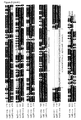
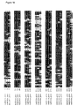
- Figure 2 Amino acid sequence alignment of the human ⁇ -catenins. MegAlign sequence comparison (DNAStar, Madison, WI) after alignment of the protein sequences by the CLUSTALW method (Higgins & Sharp, 1989), shaded by the Boxshade server (hftp://ulrec3.unii.ch/softward/BOX_form.html). Partial 3-dimensional structure as determined for ⁇ E-catenin (Pokutta & Weis, 2000) is shown by the location of the respective ⁇ -helices. Two characteristic proline residues, inducing a kink in the ⁇ -helices, are marked with "P".
- Annotated domains are: the ⁇ -catenin binding domain as determined by Pokutta & Weis (2000) and by Huber et al. (1997), the ⁇ -actinin binding domain (Nieset et al., 1997), the amphiphatic helices possibly responsible for actin binding (Rudiger, 1998) and the position of the alternatively spliced insert (Claverie et al., 1993), which is generally found for ⁇ N-catenin, but rarely for ⁇ E-catenin.
- Genbank accession numbers of the used sequences are AF091606 ( ⁇ T-catenin), D14705 ( ⁇ E-catenin) and M94151 ( ⁇ N2-catenin).
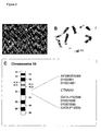
- FIG. 3 Chromosomal localization of the CTNNA3 gene, encoding human ⁇ T-catenin.
- FISH Fluorescence in situ hybridization
- B G-banding by DAPI staining of the same chromosomes as shown in (A);
- C Fine mapping by Genebridge4 PCR analysis reveals the CTNNA3 location between the markers as shown; the obtained PCR pattern was: 000000001000000000000010120000110010100011000001000000000000000010010 001010000001110000000100, and was used as such for analysis on Webpage http://www.hgmp.mrc.ac.uk/cgi-bin/contig/rhmapper.pl.
- Figure 4 Positioning of exon-exon boundaries (boxed) in the protein sequences of human ⁇ T-catenin and ⁇ E-catenin. Three boundaries in the ⁇ T-catenin sequence that do not coincide with ⁇ E-catenin are shown in bold. Indicated numbers refer to exons of ⁇ T-catenin (see also Table 1, Table 2 and Figure 5 ).
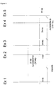
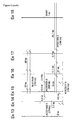
- FIG. 5 Overview of BAC and PAC clones covering the human CTNNA3 gene.
- Clones 162A20 and 320B7 are, respectively, a BAC and PAC clone isolated by us. All other sequences were obtained from Genbank and are annotated with their respective Accession Numbers. Lengths of contig sequences, in which a match is found with one or more CTNNA3 exons (indicated by 'Ex' followed by the appropriate exon number), are mentioned in kilobasepairs (kb) or basepairs (bp). BAC or PAC clone lengths are mentioned between brackets and refer to the number of sequenced bp of the respective clones (GenBank including HTGS database, sequence versions as indicated).
- FIG. 6 (A) The human ⁇ T-catenin promoter sequence as determined from a human genomic BAC clone (clone 162A20). The sequence listed comprises 1,237 bp of the upstream sequences, the sequence of exon-1 (bp 1238-1433) and part of intron-1 (bp 1434-1740). Several transcription factor binding sites as predicted by the Matinspector transcription factor binding site search program (Quandt et al., 1995), are indicated in bold and underlined. (B) Alignment of human and mouse ⁇ T-catenin promoter sequences, as determined from the human genomic BAC clone 162A20 and mouse genomic BAC clone 164N16. Transcription factor binding sites are indicated. The sequence conservation of the MEF2C site and two GATA-binding sites is striking. The arrow indicates the transcription initiation site.
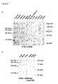
- Figure 7 Interaction between ⁇ T-catenin and ⁇ -catenin/plakoglobin in the yeast two-hybrid system.
- A Introduction in the two-hybrid system by cotransformation of the respective fusion constructs. Plasmids used for cotransformation were pGBT9-ATCTN(179-2860), pGBT9- ⁇ ctl(50-2264), pGBT9- ⁇ ECTN, pGBT9- ⁇ NCTN, pGAD424-AT ⁇ ctn and pGAD424-Plakoglobin(227-2340), followed by XGAL staining of colonies on SD plates lacking Leucine, Tryptophan and Histidine; pVA3 and pTD1 are control bait and prey plasmids (Clontech): they code for interacting fusion proteins derived of murine p53 and SV40 large T-antigen, respectively.
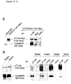
- IP results at the right were obtained either with monoclonal anti- ⁇ -catenin antibody (Transduction) or with monoclonal anti-Myc antibody 9E10 (Oncogene, Cambridge, MA). SDS-PAGE was followed by Western blotting. A mixture of both antibodies was then used to probe this blot. In mock transfected cells, only ⁇ -catenin was detected as expected (data not shown).
- D Confirmation of ⁇ T-catenin/ ⁇ -catenin interactions by co-immunoprecipitation (IP) from mouse tissues. IP from mouse testis and heart was performed with polyclonal antibody #952, specific for ⁇ T-catenin, and with a polyclonal antibody specific for ⁇ -catenin (Sigma). After Western blotting of total lysates (at the left) and coimmunoprecipitates (at the right), ⁇ T-catenin and ⁇ -catenin were detected by use of the same antibodies.
- FIG. 8 Tissue-specific expression patterns of ⁇ T-catenin.
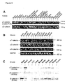
- A Rapid-scan RT-PCR expression analysis of human ⁇ T-catenin and ⁇ E-catenin mRNAs. The specific 743-bp product of the first reaction was visible in heart, testis and skeletal muscle (not shown). After nested PCR, this first product of 743 bp is still visible, whereas the nested PCR product of 630 bp is detectable in the same three samples and a few more (brain, kidney, liver, fetal liver). PCR with ⁇ E-catenin-specific primers (yielding a 747-bp product) reveals expression in most tissues. PBL, peripheral blood lymphocytes.
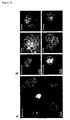
- Figure 9 Immunolocalization of ⁇ T-catenin in human tissues.
- A Frozen section of human heart stained with 892_24D2S monoclonal antibodies, showing localization of ⁇ T-catenin protein at intercalated discs of cardiac myocytes.
- B Enlarged detail of (A).
- C Frozen cross-section of human testis seminiferous tubules stained with 892_24D2S monoclonal antibodies, showing localization of ⁇ T-catenin protein in presumptive peritubular myoid cells at the basement membrane of seminiferous tubules.
- D Enlarged view of human testis staining with arrowhead pointing to a peritubular myoid cell.
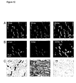
- Figure 10 Immunolocalization of ⁇ T-catenin in cryosections of human heart.
- A Double immunofluorescent staining of ⁇ T-catenin (monoclonal antibody 892_24D2S) and ⁇ E-catenin (polyclonal antibody) shows colocalization of the ⁇ -catenin proteins at intercalated discs of cardiomyocytes.
- B Double immunofluorescent staining of ⁇ T-catenin (polyclonal antibody #952) and N-cadherin (monoclonal antibody) shows colocalization at intercalated discs of cardiomyocytes.
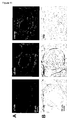
- FIG. 11 Immunolocalization of ⁇ T-catenin in cryosections of human testis.
- A Double immunofluorescent staining of ⁇ T-catenin (monoclonal antibody 892_2402) and ⁇ E-catenin (polyclonal antibody) shows differential localization of these two related proteins.
- the ⁇ T-catenin is present in peritubular cells, clearly separated from ⁇ E-catenin, which is present in cells within the seminiferous tubules.
- FIG. 12 Transient overexpression of ⁇ T-catenin in ⁇ -catenin-negative HCT-8/R1 colon carcinoma cells restores cadherin/catenin-mediated cell-cell adhesion.
- opposing cells expressing GFP- ⁇ T-catenin show increased fluorescence at their common cell-cell contacts ( A ). This results in recruitment of ⁇ -catenin and E-cadherin to the same sites ( B ).
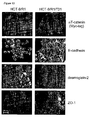
- Figure 13 Relocalization of multiple cell-cell adhesion components in stably transfected colon cancer cells, expressing Myc-tagged ⁇ T-catenin.
- the ⁇ -catenin-negative parental HCT-8/R1 cells (left panels) were compared to the cloned transfectant HCT-8/R1/T31 (right panels).
- Cells were stained for the Myc tag (exogenous ⁇ T-catenin), for E-cadherin, desmoglein-2 or ZO-1 antigens.
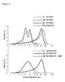
- FIG. 14 Fast aggregation of ⁇ -catenin-negative HCT-8/R1 colon cancer cells is restored upon stable transfection with ⁇ T-catenin cDNA. After preparation of single-cell suspensions, cell aggregation was measured by determination of the volume % distribution in function of the particle diameter at the starting point (t0) and after 30 min (t30). HCT-8/R1, HCT-8/E11R1 and HCT-8/E8 cells were all obtained by subcloning HCT-8 cells, but only HCT-8/E8 cells express endogenous ⁇ E-catenin.
- HRpC ⁇ N2 is a cloned transfectant of HCT-81E11R1 cells expressing exogenous ⁇ N-catenin (van Hengel et al., 1997); HCT-8/R1/T31 is a cloned transfectant of HCT-8/R1 cells expressing exogenous ⁇ T-catenin.
- MB2 is a monoclonal E-cadherin blocking antibody.
- FIG. 15 Slow aggregation and compaction of HCT-8 colon cancer cells on semi-solid agar. Images of two representative cultures were taken 24 h after seeding single-cell suspensions of the indicated cell lines. No aggregation is seen in cultures of either the untransfected ⁇ -catenin-negative HCT-8/R1 cells, or the control transfectant HCT-8/R1/1743 (also ⁇ -catenin-negative). Cells expressing either endogenous ⁇ E-catenin or exogenous ⁇ N-catenin (see also legend to Fig. 8 ) form small aggregates, whereas cells expressing exogenous ⁇ T-catenin form compacted large aggregates.
- Figure 16 Isolation of the novel mouse ⁇ T-catenin cDNA by consecutive 5'-RACE and RT-PCR experiments.
- A On the basis of one single EST sequence (GenBank Accesion No. AW556211), primers for 5'-RACE were designed and used to obtain a product of 1,177 bp. As this fragment was still lacking a suitable start codon, an RT-PCR was performed using primers MCB2335 and MCB2567. This yielded a product of 684 bp of which 668 bp were new sequences. A second 5'-RACE was performed to obtain a 696-bp fragment containing the start codon.
- Genbank accession numbers of the used sequences are AF091606 (human ⁇ T-catenin), AF344871 (mouse ⁇ T-catenin), NM_009818 (mouse ⁇ E-catenin) and NM_009819 (mouse ⁇ N-catenin).
- FIG 17 The full-length mouse ⁇ T-catenin clone pGEMTeasy-maTctn(1-2979) was expressed in an in vitro coupled transcription and translation reaction, using the TNT ® Coupled Reticulocyte Lysate System (Promega). The in vitro synthesized proteins were analyzed by Western blotting and compared with endogenous ⁇ T-catenin protein in mouse heart and testis. (A). For detection of ⁇ T-catenin, polyclonal serum #952 was applied. As a negative control we used the TNT product of human cDNA encoding p120 ctn isoform 3B (Keirsebilck et al ., 1998).
- TNT products of human and mouse ⁇ T-catenin were compared with endogenous ⁇ T-catenin protein in lysates of human and mouse heart.
- the ⁇ T-catenin proteins were analyzed by Western blotting and detected with either the polyclonal serum #952 (at the left) or with the monoclonal antibody 893_32C6S (at the right).
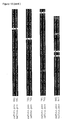
- Figure 18 Amino acid sequence alignment of the human and mouse ⁇ T-catenin. The sequences were aligned using the CLUSTALW program (Higgins and Sharp, 1989). Alignments were shaded using the Boxshade server. Genbank accession numbers of the used sequences are AF091606 (human ⁇ T-catenin) and AF344871 (mouse ⁇ T-catenin). Arrows indicate the amino-terminal ⁇ -catenin binding domains (Pokutta and Weis, 2000).
- Figure 19 Amino acid sequence alignment of the mouse ⁇ -catenins. The sequences were aligned using the CLUSTALW program (Higgins and Sharp, 1989). Alignments were shaded using the Boxshade server. Genbank accession numbers of the used sequences are NM_009818 (mouse ⁇ E-catenin), NM_009819 (mouse ⁇ N-catenin) and AF344871 (mouse ⁇ T-catenin).
- FIG. 20 Northern blot analysis of mouse ⁇ T-catenin.
- RNAs of 10 different mouse tissues were separated by gel electrophoresis, blotted on a membrane and hybridized with a mouse ⁇ T-catenin-specific probe, consisting of 296 bp of the 3' end of the mouse ⁇ T-catenin cDNA.
- the sizes of the mRNAs were determined by sequential hybridisations of the Northern blot with a mouse GAPDH probe, with a mouse ⁇ E-catenin probe and finally staining of ribosomal RNA with methylene blue.
- Figure 21 Western blot analysis of ⁇ T-catenin protein expression in various mouse tissues.
- A Detection of ⁇ T-catenin with polyclonal serum #952;
- B Detection of ⁇ T-catenin with polyclonal serum #952 after pre-incubation with the specific peptide #893, which blocks detection not only of the 100-kDa band (full-length mouse ⁇ T-catenin) but also the 86-kDa band, the 66-kDa band and the 43-kDa band in heart and testis lysates.
- Figure 22 Influence of MEF2C transcription factor on the ⁇ T-catenin promoter activity.
- AlphaT-catenin promoter luciferase constructs were cotransfected with a MEF2C encoding plasmid in P19 cells (mouse embryonal carcinoma cells). Cells were lysed 48 h after transfection and luciferase activity was measured. A ⁇ -galactosidase encoding plasmid was cotransfected to normalize for transfection efficiency.
- a luciferase construct without ⁇ T-catenin promoter serves as negative control and is insensitive to MEF2C.
- a construct with luciferase under the control of the ⁇ T-catenin promoter sequence shows background activity unless cotransfected with MEF2C. This leads to a 10-fold upregulation of the ⁇ T-catenin promoter activity. Deletion or mutation of the consensus MEF2C binding site abolishes this effect as illustrated.
- HCT-8/E8 HCT-8/E11R1 and HCT-8/R1 cell lines were obtained by subcloning of the human ileocecal adenocarcinoma cell line HCT-8 (CCL-224), where E stands for epithelioid and R for round-cell variants lacking ⁇ E-catenin (van Hengel et al., 1997).
- PC-3 (CRL-1435) is a human prostate carcinoma cell line and HEK-293 (CRL-1573) is a human embryonic kidney fibroblast cell line.
- IMAGE #728263 A human ⁇ T-catenin-specific EST clone (IMAGE #728263) was identified by BLAST analysis (Altschul et al., 1990) and requested from the IMAGE consortium UK-HGMP Resource Center (Hinxton, UK). Expression of the corresponding transcript was confirmed by RT-PCR on mRNA from the prostate cancer cell line PC3 with primers MCB967 (5'-TGAGGCAGAAAAAGAAAAGA-3') and MCB968 (5'-AGTGTGGTTAGGCAGGATT-3'). In order to complete the cDNA sequence we performed two consecutive 5' Marathon TM RACE experiments on a human testis Marathon cDNA library (Clontech, Palo Alto, CA).
- the gene-specific primer was MCB1027 (5'-AATCTGCCGAGCAAGGACATCCA-3') and the nested primer was MCB1028 (5'-TCAGGCAGTTGAGTCATCTTAGC-3').
- RACE-PCR was performed on a Perkin Elmer 2400 thermal cycler (Perkin Elmer, Foster City, CA) following the supplied protocol (touchdown PCR). Obtained RACE fragments were purified from agarose gel on QIAquick TM columns (Qiagen, Chatsworth, CA) and cloned in the pGEMT ® vector (Promega Corp., Madison, WI). The cloned fragment was called pGEMT-ATCTN-RACE1.
- mouse ⁇ T-catenin-specific expression tag (EST) clone sequence (GenBank Accesion No. AW556211) of 624 bp was identified by BLAST analysis (Altschul et al., 1990).
- BLAST analysis Altschul et al., 1990.
- This cDNA was prepared with a gene-specific primer, MCB2461 (5'-CCCCAATGTTTTATGTTAT-3') from RNA that was isolated with the RNeasy method (Qiagen, Chatsworth, CA) from mouse testis and heart tissues.
- primers MCB2818 (5'-AACGCCTAGAAGCCATCATC-3') and MCB2819 (5'-TGGCAAGAACAATGATGTCA-3') were designed to amplify the full-length cDNA.
- the predicted 2979-bp product was obtained by PCR on heart cDNA, using the TaqPlus ® Precision PCR System (Stratagene Cloning Systems, La Jolla, CA).
- the PCR-fragment was purified from agarose gel using the CONCERT TM Rapid Gel Extraction System (Life Technologies) and cloned into the pGEMT ® -easy vector (Promega).
- the resulting plasmid was called pGEMTeasy-maTctn(1-2979).
- This full-length mouse ⁇ T-catenin clone was expressed in an in vitro coupled transcription and translation reaction, using the TNT ® Coupled Reticulocyte Lysate System (Promega). The in vitro synthesized proteins were analyzed by Western blotting.
- the PAC genomic library RPCI1 constructed by loannou and de Jong (1996), and obtained from HGMP (UK), was screened by PCR with the 3' located primers MCB1260 (5'-GAAAAAGAAAAGATTGCTGAG-3') and MCB1261 (5'-CCCTAGTGAAGTCTGTCATCT-3'), yielding a product of 157 bp.
- the PCR reaction was optimized on total human genomic DNA and performed with 2.5 mM MgCl 2 at an annealing temperature of 56°C.
- Clone 320B7 (#1487) was found to be specific by DNA sequencing using primers MCB1260 and MCB1261.
- PAC clone was found to contain only the last exon of the CTNNA3 gene (exon 18 in Figures 4 and 5 ).
- a BAC (Bacterial Artificial Chromosome) human genomic library (Genome Systems, St. Louis, MO) was screened with primers located in the first protein encoding exon, i.e. primers MCB2099 (5'-TGTCATCTGCCTCTCAATTTG-3)' and MCB2100 (5'-ATGCTGCCTTTCTGTTTCTTC-3'), yielding a product of 149 bp at an annealing temperature of 52°C and 2 mM MgCl 2 .
- Clone 162A20 was found to comprise this exon, as confirmed by direct sequencing and fragment subcloning.
- the BAC162A20 clone was digested with the 6-bp cutters Bam HI, Bgl II, Eco RI, Hind III, Mlu I, Sst l, Sal I, Xba l and Xho l. Fragments containing exon 1 of the ⁇ T-catenin gene were identified by Southern blot hybridisation with a primer located in this first exon, i.e. MCB2099.
- An 8-kb BamHI fragment was cloned in the pGEM11 vector and positive clones were identified by colony hybridisation with primer MCB2099.
- the promoter region in this clone was sequenced by primer walking using primers MCB2217 (5'-CAGATGACAGTGGGGCAGTC-3'), MCB2287 (5'-AACTTGTTACTGAAAATACT-3') and MCB2350 (5'-CATTACCATTTTTCCGACTT-3').
- a BAC mouse genomic library (Genome Systems) was screened by PCR with primers located in either exon 1 (primers MCB2820 and MCB2837), in exon 2 (primers MCB2840 and MCB2841) and in exon 3 of the mouse ⁇ T-catenin gene (primers MCB2838 and MCB2839). Sequences of these primers are:
- Clone 164N16 was found to comprise these first three exons, as confirmed by Southern blot analysis.
- FISH Fluorescence in situ hybridization
- PAC clone 320B7 (#1487) was used for fluorescence in situ hybridisation (Kievits et al., 1990). PAC DNA was prepared with Kb-Magnum purification columns (Genome Systems, St. Louis, MO) and nick-translated using a BioNick kit (Gibco-BRL). Denaturation of labeled probe and human chromosomes, hybridization and fluorescent detection were performed as described previously (van Hengel et al., 1995). The chromosomes were stained with DAPI to reproduce G-banding.
- the slides were observed using a Zeiss Axiophot fluorescent microscope (Zeiss, Jena, Germany) and images captured with a Photometrics Image Point CCD camera (Photometrics, Kunststoff, Germany). Results were analyzed with the MacProbe software of PSI (Perceptive Scientific International, League City, TX). At least 20 metaphase spreads of normal human lymphocytes were analyzed.
- PSI Perceptive Scientific International, League City, TX
- Genomic PCR was performed with primers MCB2056 (5'-GAAATGCCATGGAGCTCTAAC-3') and MCB2057 (5'-ATGGGAAGGCAAACCAGTCAC-3'). These primers correspond to intronic sequences and are flanking an exon, as deduced from the sequence of a genomic clone (Genbank N° AQ163827) (Table 1; Figure 5 ). A product of 274 bp was expected. As a positive control, genomic DNA from human placenta was used to optimize the annealing temperature of 52°C and a concentration of 2 mM MgCl 2 .
- PCR was performed on samples of the Genebridge 4 radiation hybrid mapping panel (HGMP-RC, UK), which allows construction of high-resolution contiguous maps of human chromosomes. Analysis was performed on the Web page hftp://www.hgmp.mrc.ac.uk/cgi-bin/contig/rhmapper.pl.
- PCR with the same primer set was also performed on a monochromosomal hybrid mapping panel (NIGMS Human/Rodent Somatic Cell Hybrid Mapping Panel #2, Coriell Cell Repositories, Camden, NJ). All cell hybrid templates were diluted to a final DNA concentration of 100 ng/ ⁇ l, using 1 ⁇ l as PCR templates.
- This product was restricted with Eco RI plus Bam HI and ligated to the Eco RI and Bam HI sites of the pGBT9 vector, thus yielding pGBT9-ATCTN(179-949).
- This construct was opened with Pst I , and the Pst I insert of 1,111 bp from clone pGEMT-ATCTN-RACE2 was ligated to obtain pGBT9-ATCTN(179-1306). From this construct, the Eco RI- Ssp I insert of 1,082 bp was isolated and ligated together with fragment Ssp I-SalI from pGEMTeasy-ATCTN-RACE1 in the pGBT9 vector, restricted with Eco RI- Sal I.
- pGBT9-ATCTN(179-2176) was obtained.
- a PCR product of 890 bp was synthesized with primers MCB1610 (5'-GGATGATAATCAATTTGTGGACATCTC-3') and MCB1608 (5'-GGGATCCGTAGATTTGTCTTCCTCTAA-3').
- template cDNA was synthesized from RNA prepared with the RNeasy kit (Qiagen) from the PC-3 prostate cancer cell line (American Type Culture Collection, Rockville, MD, USA).
- the PCR product was cut with Bgl II and Sal I, and inserted in the Bgl II -Sal I opened construct pGBT9-ATCTN(179-2176) to finally achieve the construct pGBT9-ATCTN(179-2860).
- the Eco RI - Sac l insert was ligated together with the Sac l- Not l fragment from the original EST clone pT3T7-EST728263, in the Eco RI- Not I digested pGBKT7 vector (Clontech).
- the obtained construct was designated pGBKT7-ATCTN(179-3024).
- the full-length ⁇ -catulin cDNA sequence (Genbank Accession number U97067) was isolated by us in 3 steps (Janssens et al., 1999): the full-length sequence was compiled from 2 EST sequences (clones 36498 and 67201) and one 5'-RACE clone. In order to obtain a clone with the full-length sequence, these 3 clones were assembled in the pGEM11 vector (Clontech). First, the insert of EST clone 67201 was isolated by a Sma I-Munl digest and ligated to the Sma I- Mun I opened vector pGEMT- ⁇ ctIRACE.
- This clone was used as a template to generate a PCR product with primer MCB725 (5'-TATTAGATATCGCCTCTCCCGGACCCGCC-3' comprising an EcoRV site) and primer MCB711 (5'-AGGGGGCAGTGGCTGAAGAAAGAAGTAATC-3').
- primer MCB725 5'-TATTAGATATCGCCTCTCCCGGACCCGCC-3' comprising an EcoRV site
- primer MCB711 5'-AGGGGGCAGTGGCTGAAGAAAGAAGTAATC-3'.
- this PCR product cut with EcoRV + Mun I , was ligated together with a Mun I- Sal I fragment of pGEMT- ⁇ ctl(1-2264) into the Bam HI(blunted)- Sal I restricted pGBT9 two-hybrid vector (Clontech), in frame with the ORF encoding the GAL4 DNA binding domain.
- the obtained constructed was called pGBT9- ⁇ ctl
- ⁇ N-catenin The cDNA for human ⁇ N-catenin was kindly provided as plasmid pPN-hANCTN by Dr. C. Petit (Claverie et al., 1993). Nearly full-length ⁇ N-catenin was amplified from pPN-hANCTN with primers MCB137 (5'-ACCCCCCGGGGGCAACTTCACCTATCATTC-3' containing an Xma I site), and MCB138 (5'-GCCGCCGCCTTCCTTTTCATTTCCGCTCTT-3').
- the PCR fragment was digested with Xma I and Ban I and ligated together with a Ban I -Hind III fragment of pPN-hANCTN in the Xma I- Hind III digested pAS2 vector (Clontech).
- pAS2- ⁇ NCTN plasmid was obtained, in which codons 4 to 906 of ⁇ N-catenin are fused in frame with the ORF encoding the GAL4 DNA binding domain.
- the Xmal-Hindlll insert was transferred to the XmaI-Hind III opened pGBT9 vector, yielding pGBT9- ⁇ NCTN.
- the full-length ⁇ -catenin cDNA was kindly provided as plasmid pBAT- ⁇ CAT (from Dr. J. Behrens, Berlin, Germany).
- the aminoterminal fragment 239-717 was obtained as an NcoI - Pst I restriction fragment, of which the Nco I cut end was filled in with Pfu polymerase. This fragment was cloned into the Sma I- Pst I opened pGAD424 vector, by which the construct pGAD424-AT ⁇ ctn was obtained.
- Plasmid pHPGCa2.1 with the full-length human plakoglobin cDNA was kindly provided by Dr. W. Franke. PCR was performed on this plasmid, with primer MCB133 (5'-GGTGAATTCGTCAGCAGCAAGGGCATCAT-3', containing an Eco RI site) and primer MCB134 (5'-GGTTTGATGCAGGGTCCACAGGCAGTTCT-3').
- the obtained PCR product (encoding plakoglobin residues 227-1228) was digested with Eco RI and Sac I (residues 227-559) and ligated together with the fragments Sac I- Bgl II (residues 560-1856) and Bgl II- Pst I (residues 1857-2340) from pHPGCa2.1, into the Eco RI- Pst I opened pGAD424 vector (Clontech). Thus, the plasmid pGAD424-Plakoglobin(227-2340) was obtained.
- the yeast strain Y190 (Matchmaker, Clontech, CA), which contains GAL4 promoter driven His and ⁇ -galactosidase reporters, was used for cotransformation of pGBT9 bait and pGAD424 prey plasmids, comprising the cloned inserts of interest.
- the yeast strain L40 which contains LexA promoter driven His and ⁇ -galactosidase reporters, was used for cotransformation of pLexMG bait with pGAD424 prey plasmids, comprising the cloned inserts of interest.
- the yeast cells were grown in YPD medium until a log-phase culture with an O.D. 600 of about 0.8 was obtained, and transformed by the lithium acetate procedure (Gietz et al., 1992). Cotransformants were selected by plating the transformation mix on SD minimal medium plates lacking leucine and tryptophan. After three days, colonies were picked and grown overnight in SD without leucine and tryptophan, but containing 0.07 M potassium phosphate.
- Replica plates selecting for interaction were made on SD lacking leucine, tryptophan and histidine, but containing 0.07 M potassium phosphate, 40 mM 3-amino-triazol to suppress leaky His expression, and 80 mg/ml X- ⁇ -GAL (Duchefa, Haarlem, The Netherlands).
- ⁇ -galactosidase acitivity was assayed using chlorophenol red- ⁇ -D-galactopyranoside (CPRG, Boehringer, Mannheim, Germany) as a substrate, according to the provided protocol (Clontech Yeast Protocols Handbook). Briefly, transformed yeasts are grown until OD 600 of about 0.6, concentrated in three different dilutions (1.25, 2.5 and 5 times concentrated, respectively) and allowed to develop red color after addition of CPRG substrate (measured at that time point, at OD 578 ).
- chlorophenol red- ⁇ -D-galactopyranoside CPRG, Boehringer, Mannheim, Germany
- One ⁇ -galactosidase unit is defined as the amount which hydrolyzes 1 ⁇ mol of CPRG to chlorophenol red and D-galactose per minute per cell (Miller et al., 1972). The amount of units is calculated as 10,000 X OD 578 /(time X concentration factor X OD 600 ).
- the full-length human ⁇ T-catenin cDNA was excised from the construct pGBT9-ATCTN(179-2860) with restriction enzymes Eco RI- Sal I and inserted in the Eco RI- Sal I digested pEGFPC2 vector (Clontech), in order to obtain an in-frame amino-terminal fusion with the GFP protein.
- the resulting plasmid was called pEGFPC2-ATCTN(179-2860).
- the full-length fusion construct named pEF6MH-ATCTN(1-2860), was obtained by introducing the 5' part of the ⁇ T-catenin cDNA from clone pGEMT-RACE2, cut with Eco RI -BstE II, into the Eco RI- Bst EII opened vector pEF6MH-ATCTN(179-2860).
- ⁇ -catenins were cloned in the pE/L-GFP vector (Frischknecht et al., 1999). Cells were transfected with Lipofectin (Life Technologies) and simultaneously co-infected with vaccinia virus strain ⁇ A36R, which does not make actin tails (Parkinson and Smith, 1994). At 4 to 30 h after transfection, high levels of expression under control of the vaccinia virus early/late promoter (E/L) (Chakrabarti et al., 1997) were obtained of the cloned cDNA, amino-terminally fused to GFP.
- E/L vaccinia virus early/late promoter
- Human ⁇ T-catenin was amplified with Taq+Precision. polymerase (Stratagene) using primers containing a 5' Not I site and a 3' Eco RI site (MCB2386, 5'-GGGGGCGGCCGCGGAGGGTCAGCTGAAACACCAATCACATTG-3' and MCB2387, 5'-CCCCGAATTCGCCGTGTGGTTAGGCAGGATTTTGTCATATAG-3') and cloned into the Not I- Eco RI sites of the pE/L-GFP vector.
- HCT-8/R1 carcinoma cells For stable transfection of HCT-8/R1 carcinoma cells, 4 x 10 6 cells were electroporated (Easyject; Eurogentec, Seraing, Belgium) with 10 ⁇ g of plasmid pEF6MH-ATCTN(179-2860). Cells were plated and cultured in the presence of 6 ⁇ g/ml blasticidin (Invitrogen) to select for stable transfectants. Colonies of blasticidin-resistant cells were isolated and tested by immunofluorescence and Western blotting for expression of ⁇ T-catenin. One stable clone was isolated and called HCT-8/R1/T31.
- HCT-8/R1/1743 As a negative control we transfected HCT-8/R1 cells with the empty pEF6MH vector, resulting in stable clones called HCT-8/R1/1743.
- HCT-8/E11R1 carcinoma cells were transfected with plasmid pEGFPC2-ATCTN(179-2860). After selection with G418 (800 ⁇ g/ml), one stable ⁇ T-catenin expressing clone was isolated and called HCT-8/E11R1/T14.
- primers MCB967 (5'-TGAGGCAGAAAAAGAAAAGA-3') and MCB1010 (5'-GCTGAGCCTCGTCTGAC-3') were combined, yielding a smaller product of 630 bp. Amplified products were checked for specificity by sequence analysis, showing that the double bands observed after nested RT-PCR of heart and testis samples in particular are indeed the larger primary product and the smaller nested product.
- an ⁇ E-catenin-specific product of 747 bp was amplified with primers MCB53 (5'-CTTCGGGCCTCTGGAATTTA-3') and MCB73 (5'-CGACATCAGGGTGCTGTAGG-3').
- primers MCB2461 (5'-CCCCAATGTTTTATGTTAT-3') and MCB2463 (5'-GGGGAGAACTCATCGTAT-3') were designed on the sequence of an EST clone (GenBank Accession No. AW556211), resulting in amplification of a 442-bp product.
- ⁇ E-catenin GenBank Accession No.
- a 733-bp product was amplified with primers MCB2636 (5'-GAAGGCCCCTGAGAAGAA-3') and MCB2637 (5'-CCCGAATAAAGCAACTCCAT-3').
- MCB2479 5'-GCCCTGATTGAGTTTGATAA-3'
- MCB2480 5'-CCCAGCTTCATAGTTCTCC-3'.
- a 452-bp fragment of mouse GAPDH was amplified with primers MCB2219 (5'-ACCACAGTCCATGCCATCAC-3') and MCB2220 (5'-TCCACCACCCTGTTGCTG TA-3').
- a mouse ⁇ T-catenin-specific probe of 296 bp was generated by PCR with primer MCB2043 (5'- TCGAGGATGAAGGCTCTG-3') and primer MCB2044 (5'-TGTTTAACCCCAATGTTT-3').
- the PCR product was labelled with ⁇ [ 32 P]-dCTP using the Radprime DNA labelling System (Life Technologies). After hybridization according to standard procedures, the blot was washed at high stringency. For detection, a Phosphor Imager cassette (Molecular Dynamics, Sunnyvale, CA) was exposed for 4 days and scanned with a Molecular Imager ® FX using the Quantity One software (BioRad, Richmond, CA).
- Peptides corresponding to, respectively, the amino-terminus (MSAETPITLNIDPQDLQ-C) and the carboxy-terminus (C-KIHPLQVMSEFRGRQIY) of the human ⁇ T-ctn protein were synthesized and coupled to keyhole-limpet hemocyanin via the additional cysteine residue at either the carboxyterminal or the aminoterminal end of the peptides. 200 ⁇ g of peptide was injected in each of three rabbits using Titermax (Sigma, St Louis, MO) as adjuvant. Boosts were given with intervals of minimum two weeks. Sera were tested by ELISA on the peptide used for injection, using nonrelevant peptide as a negative control.
- the sera #952 (specific for the carboxy-terminal peptide) and #954 (specific for the amino-terminal peptide) were affinity purified on hydroxymercuribenzoate-agarose (Sigma, St Louis, MO), coupled to the respective immunizing peptides. Crude and purified sera were tested on lysates of HEK cells transfected with plasmids pEGFPC2-ATCTN(179-2860) and pEF6MH-ATCTN(1-2860), encoding respectively full-length Myc-tagged and GFP-tagged ⁇ T-catenin.
- Monoclonal antibodies were generated by injection of the N-terminal peptide (MSAETPITLNIDPQDLQ-C) or the C-terminal peptide (C-KIHPLQVMSEFRGRQIY) in C57BI/6 mice.
- Boosts were given with intervals of 2 weeks, and sera were tested by ELISA until a titer of 1:10,000 without loss of reactivity was obtained after 6 weeks.
- Hybridomas were generated by fusion of spleen cells with Sp20_Ag14 myeloma cells. Supernatants of hybridoma cell lines were tested by ELISA.
- hybridoma 893_32C6S For the C-terminal peptide, 30 out of 96 ELISA-positive clones recognized GFP-T ⁇ -catenin by Western Blotting, from which 3 were able to recognize native ⁇ T-catenin protein by immunofluorescence. A subclone, called hybridoma 893_32C6S, was deposited at BCCM under the number LMBP 5728CB.
- Protein lysates from various mouse tissues were prepared by isolating the tissues from normal BALB/c mice and mixing them in Laemmli buffer (Laemmli, 1970). Debris was removed by centrifugation and protein concentration was measured by the BioRad DC kit (BioRad, Richmond, CA). Lysates from subconfluent cultures of cell lines were also prepared in Laemmli buffer, followed by sonication and centrifugation.
- Detection was carried out by phosphatase-coupled secondary antibodies (Sigma) and nitroblue tetrazolium/5-bromo-4-chloro-3-indolyl phosphate (NBT/BCIP) as a substrate.
- phosphatase-coupled secondary antibodies Sigma
- NBT/BCIP nitroblue tetrazolium/5-bromo-4-chloro-3-indolyl phosphate
- Co-immunoprecipitation was performed on lysates of transfected HEK cells, prepared in PBS containing 1% NP-40 and a protease inhibitor cocktail (Boehringer). Lysate (800 ⁇ g) was incubated overnight with 4 ⁇ g of the respective antibody, after which 100 ⁇ l of 50% protein-G (Amersham Pharmacia Biotech, Rainham, UK) was added to monoclonal antibodies, whereas protein-A Sepharose (Amersham Pharmacia Biotech) was added to polyclonal antibodies.
- Sepharose beads were washed three times with PBS containing 0.1% NP-40, followed by boiling for 5 min in Laemmli buffer, before being subjected to SDS-PAGE and Western blotting. On these Western blots, protein was detected by the ECL detection system using secondary antibodies coupled to horseradish peroxidase (Amersham Pharmacia Biotech).
- Frozen sections of human heart and testis tissue were treated for 20 min with 0.3% H 2 O 2 diluted in methanol, then washed in water and PBS, and pretreated for 10 min with goat serum diluted 1:10. The sections were then incubated for 30 min with crude monoclonal 892_24D2S hybridoma supernatans, which was diluted 1:5 in PBS containing 1% bovine serum albumin.
- the secondary antibody used was biotin-labelled goat-anti-mouse Ig (DAKO, Denmark), which was subsequently linked to the streptavidin-ABC complex coupled to horseradish peroxidase.
- Detection was carried out by a 5 min incubation with the chromogenic peroxidase substrate diaminobenzidine (Sigma). Cell nuclei were counterstained for 5 min with haematoxylin (Sigma), after which the slides were dehydrated by washing in 70%, 90% and two times 100% ethanol. Finally the slides were cleared in toluol and mounted.
- the secondary goat anti-mouse IgG and goat anti-rabbit IgG antibodies used were labeled with FITC or TRITC (Santa Cruz, Santa Cruz, CA), or with Alexa 488 or Alexa 594 (Molecular Probes, Eugene, OR).
- Cells were grown on glass coverslips until confluency, rinsed briefly with PBS and fixed with either ice-cold methanol for 1 min, or with 3% paraformaldehyde (Merck, Darmstadt, Germany) for 10 min at room temperature, followed by permeabilization in 0.2% Triton X-100 (Sigma) for 2 min. Cells were then incubated for 30 min with primary antibody diluted in blocking solution (20 mM Tris/HCl pH 7.5, 154 mM NaCl, 2 mM EDTA, 2 mM MgCl 2 , with 1% BSA and 1% goat serum), washed in PBS, and incubated for 30 min with secondary antibodies diluted in blocking solution.
- blocking solution 20 mM Tris/HCl pH 7.5, 154 mM NaCl, 2 mM EDTA, 2 mM MgCl 2 , with 1% BSA and 1% goat serum
- Samples were examined with a Zeiss Axiophot microscope and images were recorded with a high-performance charge-coupled digital camera (Cohu, San Diego, CA) and NIH image software (version 1.62), or with a MicroMAX camera (Princeton, Trenton, NJ) and MetaMorph software (Image Universal Corporation, New York, NY).
- a Zeiss Axiophot microscope and images were recorded with a high-performance charge-coupled digital camera (Cohu, San Diego, CA) and NIH image software (version 1.62), or with a MicroMAX camera (Princeton, Trenton, NJ) and MetaMorph software (Image Universal Corporation, New York, NY).
- the 3024-bp sequence (SEQ ID N° 1) contains a Kozak-consensus start codon (Kozak, 1991) at position 176, preceded by a stop codon at position 137.
- a stop codon terminating the long open reading frame (ORF) is located at position 2861, and a putative poly-adenylation signal is seen at the 3' untranslated region at 38 bp before the end of the sequence.
- the ORF encodes a protein of 895 amino acid residues (SEQ ID N° 2), with a predicted molecular weight of 100 kDa and an overall identity to ⁇ E-catenin (102 kDa) and ⁇ N-catenin (104 kD) of respectively 58 and 56 % ( Figure 1B ).
- the overall homology is higher, as similarities were calculated of 74 and 70% with respectively ⁇ E- and ⁇ N-catenin.
- This protein is therefore to be considered a true ⁇ -catenin family member, and was called ⁇ T-catenin because its transcript was discovered in testis-derived mRNA.
- Example 2 Analysis of human and mouse ⁇ T-catenin genes
- clone 320B7 By PCR screening, we isolated a human genomic PAC clone, called clone 320B7 (#1487). This clone was used to perform fluorescence in situ hybridization (FISH), which revealed the localization of the ⁇ T-catenin gene CTNNA3 on chromosome band 10q21 ( Figure 3A and B ). This localization was confirmed by monochromosomal hybrid mapping and by Genebridge4TM PCR screening. The obtained pattern of PCR products indeed pointed to localization on 10q21, close to marker D10S1461 ( Figure 3C ). The region 10q21-23 has been identified as a candidate region for autosomal dominant dilated cardiomyopathy (Bowles et al., 1996).
- FISH fluorescence in situ hybridization
- a human BAC library was screened by PCR with 5' located primers.
- Clone 162A20 indeed contains the upstream genomic region but comprises only exons 1 and 2 with flanking intronic sequences, besides the upstream gene-regulatory 5' sequences of the CTNNA3 gene. Indeed, from this genomic BAC clone about 1.2 kb of ⁇ T-catenin promoter region was sequenced ( Figure 6A and SEQ ID N° 3).
- a mouse BAC library was screened by PCR with primers located in the first three exons of the mouse Ctnna3 gene. Genomic clone 164N16 was found to contain these three exons. Part of the mouse promoter sequence was determined (SEQ ID N° 6).
- Example 3 at-catenin interacts stronger with ⁇ -catenin than other ⁇ -catenins do
- Example 4 The ⁇ T-catenin protein is preferentially expressed in heart and testis tissues
- a human cDNA Rapid Scan panel (OriGene Technologies, Rockville, MD) was screened by PCR for ⁇ T-catenin expression.
- a first PCR reaction revealed expression in heart and testis tissues only, whereas a second, nested PCR amplified low amounts in some other tissues (brain, kidney, liver, skeletal muscle, fetal liver) ( Figure 8A ).
- the ⁇ T-catenin shows a very restricted expression pattern.
- Example 5 Human ⁇ T-catenin can be detected in cardiomyocytes and testis tissue
- the ⁇ T-catenin expressing cells in testis correspond to desmin-positive cells, and therefore could be identified as peritubular myoid cells ( Figure 11 B) . These stainings stongly suggest that ⁇ T-catenin expression is confined to specific muscle cell types
- HCT-8/R1 cells were transfected with a plasmid encoding Myc-tagged ⁇ T-catenin.
- a stable transfectant was cloned and called HCT-8/R1/T31.
- Western blotting with polyclonal antiserum #952 showed high expression of ⁇ T-catenin protein in this cell line.
- Immunofluorescent analysis of HCT-8/R1/T31 cells with anti-Myc antibodies showed that the ⁇ T-catenin-Myc protein was localized at restored cell-cell contacts ( Figure 13 ).
- HCT-8/R1/T31 cells are strongly aggregating in contrast to nonaggregating parental HCT-8/R1 cells ( Figure 14 ).
- HCT-8/R1/T31 cells aggregated to similar extent as ⁇ E-catenin-positive HCT-8/E8 cells and ⁇ N-catenin-transfected HRpC ⁇ N2 cells, thus showing that ⁇ T-catenin is able to functionally restore aggregation.
- HCT-8/E11R1/T14 stably expressing a GFP- ⁇ T-catenin fusion protein.
Landscapes
- Health & Medical Sciences (AREA)
- Chemical & Material Sciences (AREA)
- Organic Chemistry (AREA)
- Life Sciences & Earth Sciences (AREA)
- General Health & Medical Sciences (AREA)
- Medicinal Chemistry (AREA)
- General Chemical & Material Sciences (AREA)
- Pharmacology & Pharmacy (AREA)
- Nuclear Medicine, Radiotherapy & Molecular Imaging (AREA)
- Animal Behavior & Ethology (AREA)
- Chemical Kinetics & Catalysis (AREA)
- Public Health (AREA)
- Veterinary Medicine (AREA)
- Biochemistry (AREA)
- Engineering & Computer Science (AREA)
- Gastroenterology & Hepatology (AREA)
- Toxicology (AREA)
- Biophysics (AREA)
- Genetics & Genomics (AREA)
- Molecular Biology (AREA)
- Proteomics, Peptides & Aminoacids (AREA)
- Bioinformatics & Cheminformatics (AREA)
- Zoology (AREA)
- Reproductive Health (AREA)
- Pregnancy & Childbirth (AREA)
- Gynecology & Obstetrics (AREA)
- Endocrinology (AREA)
- Cardiology (AREA)
- Heart & Thoracic Surgery (AREA)
- Peptides Or Proteins (AREA)
- Micro-Organisms Or Cultivation Processes Thereof (AREA)
- Medicines That Contain Protein Lipid Enzymes And Other Medicines (AREA)
- Medicines Containing Material From Animals Or Micro-Organisms (AREA)
Claims (7)
- Monoklonaler Antikörper gegen ein Polypeptid, das die in SEQ ID Nr. 2 dargestellte Sequenz umfasst, wie von dem Hybridom sezerniert, das bei der Belgischen Hinterlegungsstelle für die koordinierte Sammlung von Mikroorganismen (BCCM) unter der Nummer LMBP 5537CB hinterlegt wurde, dadurch gekennzeichnet, dass der monoklonale Antiköper nicht mit murinem alpha-T-Catenin, wie in SEQ ID Nr. 5 dargestellt, kreuzreagiert.
- Verwendung eines Antikörpers gegen ein Polypeptid, das die in SEQ ID Nr. 2 dargestellte Sequenz umfasst, zur Herstellung eines Tests zur Vorhersage der Wahrscheinlichkeit einer Krebserkrankung, die alpha-Catenin-negative Krebszellen umfasst, und/oder zur Diagnose einer solchen Krebserkrankung.
- Verwendung einer Nukleinsäure, die ein Polypeptid, das die in SEQ ID Nr. 2 dargestellte Sequenz umfasst, oder ein Fragment davon kodiert, das spezifisch mit der Nukleinsäure hybridisiert, zur Herstellung eines Tests zur Vorhersage der Wahrscheinlichkeit einer Krebserkrankung, die alpha-Catenin-negative Krebszellen umfasst, und/oder zur Diagnose einer solchen Krebserkrankung.
- Nukleinsäure, die ein Polypeptid kodiert, das die in SEQ ID Nr. 2 dargestellte Sequenz umfasst, oder funktionelles Fragment der Nukleinsäure, wobei das funktionelle Fragment ein Polypeptid kodiert, das eine funktionelle beta-Catenin-bindende Domäne umfasst, zur Verwendung bei der Behandlung einer Krebserkrankung, die alpha-Catenin-negative Krebszellen umfasst.
- Verwendung einer Nukleinsäure, die ein Polypeptid kodiert, das die in SEQ ID Nr. 2 dargestellte Sequenz umfasst, oder eines funktionellen Fragments der Nukleinsäure, wobei das funktionelle Fragment ein Polypeptid kodiert, das eine funktionelle beta-Catenin-bindende Domäne umfasst, zur Herstellung eines Arzneimittels zur Behandlung einer Krebserkrankung, die alpha-Catenin-negative Krebszellen umfasst.
- Polypeptid, das die in SEQ ID Nr. 2 dargestellte Sequenz umfasst, oder Fragment davon, wobei das Fragment eine funktionelle beta-Catenin-bindende Domäne umfasst, zur Verwendung bei der Behandlung einer Krebserkrankung, die alpha-Catenin-negative Krebszellen umfasst.
- Verwendung eines Polypeptids, das die in SEQ ID Nr. 2 dargestellte Sequenz umfasst, oder eines Fragments davon, wobei das Fragment eine funktionelle beta-Catenin-bindende Domäne umfasst, zur Herstellung eines Arzneimittels zur Behandlung einer Krebserkrankung, die alpha-Catenin-negative Krebszellen umfasst.
Priority Applications (1)
| Application Number | Priority Date | Filing Date | Title |
|---|---|---|---|
| EP01947420A EP1299536B1 (de) | 2000-07-12 | 2001-06-28 | Antikörper und verwendung bezüglich des alpha-t catenins |
Applications Claiming Priority (6)
| Application Number | Priority Date | Filing Date | Title |
|---|---|---|---|
| EP00202472 | 2000-07-12 | ||
| EP00202472 | 2000-07-12 | ||
| US21830900P | 2000-07-14 | 2000-07-14 | |
| US218309P | 2000-07-14 | ||
| PCT/EP2001/007392 WO2002004636A1 (en) | 2000-07-12 | 2001-06-28 | NOVEL α-CATENIN EXPRESSED IN HEART AND TESTIS |
| EP01947420A EP1299536B1 (de) | 2000-07-12 | 2001-06-28 | Antikörper und verwendung bezüglich des alpha-t catenins |
Publications (2)
| Publication Number | Publication Date |
|---|---|
| EP1299536A1 EP1299536A1 (de) | 2003-04-09 |
| EP1299536B1 true EP1299536B1 (de) | 2013-03-27 |
Family
ID=26072486
Family Applications (1)
| Application Number | Title | Priority Date | Filing Date |
|---|---|---|---|
| EP01947420A Expired - Lifetime EP1299536B1 (de) | 2000-07-12 | 2001-06-28 | Antikörper und verwendung bezüglich des alpha-t catenins |
Country Status (4)
| Country | Link |
|---|---|
| US (1) | US7341866B2 (de) |
| EP (1) | EP1299536B1 (de) |
| AU (1) | AU2001269106A1 (de) |
| WO (1) | WO2002004636A1 (de) |
Families Citing this family (2)
| Publication number | Priority date | Publication date | Assignee | Title |
|---|---|---|---|---|
| AU2001269106A1 (en) | 2000-07-12 | 2002-01-21 | Vlaams Interuniversitair Instituut Voor Biotechnologie Vzw | Novel alpha expressed in heart and testis |
| CN110305209B (zh) * | 2019-07-09 | 2022-09-13 | 福建医科大学附属第一医院 | 用于治疗恶性肿瘤的多肽及其作为疫苗的用途 |
Family Cites Families (4)
| Publication number | Priority date | Publication date | Assignee | Title |
|---|---|---|---|---|
| JPH06211898A (ja) * | 1992-12-25 | 1994-08-02 | Takara Shuzo Co Ltd | 神経α−カテニン |
| AU2897195A (en) * | 1994-07-08 | 1996-02-09 | Frans M. J Debruyne | Assay for increased invasiveness of epithelial cells |
| US6551994B1 (en) * | 1997-04-10 | 2003-04-22 | Mcgill University | Compounds and methods for inhibiting the interaction between α-catenin and β-catenin |
| AU2001269106A1 (en) | 2000-07-12 | 2002-01-21 | Vlaams Interuniversitair Instituut Voor Biotechnologie Vzw | Novel alpha expressed in heart and testis |
-
2001
- 2001-06-28 AU AU2001269106A patent/AU2001269106A1/en not_active Abandoned
- 2001-06-28 EP EP01947420A patent/EP1299536B1/de not_active Expired - Lifetime
- 2001-06-28 WO PCT/EP2001/007392 patent/WO2002004636A1/en not_active Ceased
-
2003
- 2003-01-13 US US10/345,092 patent/US7341866B2/en not_active Expired - Fee Related
Also Published As
| Publication number | Publication date |
|---|---|
| AU2001269106A1 (en) | 2002-01-21 |
| WO2002004636A8 (en) | 2002-04-04 |
| WO2002004636A1 (en) | 2002-01-17 |
| EP1299536A1 (de) | 2003-04-09 |
| US7341866B2 (en) | 2008-03-11 |
| US20030165506A1 (en) | 2003-09-04 |
Similar Documents
| Publication | Publication Date | Title |
|---|---|---|
| AU2001276515B2 (en) | Suppressor gene | |
| AU2001276515A1 (en) | Suppressor gene | |
| Keshav et al. | Rpa4, a homolog of the 34-kilodalton subunit of the replication protein A complex | |
| JPH09507744A (ja) | 新規の核分裂リンタンパク質:ミトシン | |
| US5541068A (en) | Mammalian adipocyte protein P154, nucleic acids coding therefor and uses thereof | |
| US6225451B1 (en) | Chromosome 11-linked coronary heart disease susceptibility gene CHD1 | |
| CA2239993A1 (en) | Dna sequence encoding the tumor suppressor ing1 | |
| WO1997045128A9 (en) | Apoptosis associated protein bbk | |
| US7105649B2 (en) | Gasci gene | |
| US7341866B2 (en) | α-catenin expressed in heart and testis | |
| CA2571350A1 (en) | Peptides for inhibiting the interaction of protein kinase a and protein kinase a anchor proteins | |
| US20040248257A1 (en) | SPEX compositions and methods of use | |
| JP4201712B2 (ja) | Gip、グッドパスチャー抗原結合タンパク質と相互作用する転写因子活性を備えたポリペプチドのファミリー | |
| Den Bakker et al. | Evidence for a cytoskeleton attachment domain at the N‐terminus of the NF2 protein | |
| EP0727486A2 (de) | Tumorhemmendes Gen | |
| US7365158B2 (en) | PR-domain containing nucleic acids, polypeptides, antibodies and methods | |
| HUT74413A (en) | A novel tumor suppressor gene | |
| US20050004065A1 (en) | Hypoxia regulated genes | |
| US5556945A (en) | Tumor suppressor gene | |
| JP4346540B2 (ja) | 絨毛外栄養膜細胞特異的蛋白質 | |
| JP2002503466A (ja) | 網膜芽細胞腫タンパク質複合体および網膜芽細胞腫相互作用タンパク質 | |
| JP2002517194A (ja) | C.エレガンスのunc−53タンパク質のヒトホモログ | |
| US6919430B1 (en) | Polypeptide—human galectin 15 and a polynucleotide encoding the same | |
| JP2002525126A (ja) | Traf相互作用ext遺伝子ファミリーの新規遺伝子trex、並びにその診断及び治療への使用 | |
| US6696273B2 (en) | FEBP1 Protein: vector, host cells and method for making FEBP1 protein |
Legal Events
| Date | Code | Title | Description |
|---|---|---|---|
| PUAI | Public reference made under article 153(3) epc to a published international application that has entered the european phase |
Free format text: ORIGINAL CODE: 0009012 |
|
| 17P | Request for examination filed |
Effective date: 20030121 |
|
| AK | Designated contracting states |
Kind code of ref document: A1 Designated state(s): AT BE CH CY DE DK ES FI FR GB GR IE IT LI LU MC NL PT SE TR |
|
| AX | Request for extension of the european patent |
Extension state: AL LT LV MK RO SI |
|
| 17Q | First examination report despatched |
Effective date: 20061220 |
|
| GRAP | Despatch of communication of intention to grant a patent |
Free format text: ORIGINAL CODE: EPIDOSNIGR1 |
|
| GRAS | Grant fee paid |
Free format text: ORIGINAL CODE: EPIDOSNIGR3 |
|
| GRAA | (expected) grant |
Free format text: ORIGINAL CODE: 0009210 |
|
| AK | Designated contracting states |
Kind code of ref document: B1 Designated state(s): AT BE CH CY DE DK ES FI FR GB GR IE IT LI LU MC NL PT SE TR |
|
| REG | Reference to a national code |
Ref country code: GB Ref legal event code: FG4D |
|
| REG | Reference to a national code |
Ref country code: CH Ref legal event code: EP |
|
| REG | Reference to a national code |
Ref country code: AT Ref legal event code: REF Ref document number: 603447 Country of ref document: AT Kind code of ref document: T Effective date: 20130415 |
|
| REG | Reference to a national code |
Ref country code: IE Ref legal event code: FG4D |
|
| REG | Reference to a national code |
Ref country code: DE Ref legal event code: R096 Ref document number: 60147817 Country of ref document: DE Effective date: 20130523 |
|
| PG25 | Lapsed in a contracting state [announced via postgrant information from national office to epo] |
Ref country code: SE Free format text: LAPSE BECAUSE OF FAILURE TO SUBMIT A TRANSLATION OF THE DESCRIPTION OR TO PAY THE FEE WITHIN THE PRESCRIBED TIME-LIMIT Effective date: 20130327 |
|
| PGFP | Annual fee paid to national office [announced via postgrant information from national office to epo] |
Ref country code: DE Payment date: 20130620 Year of fee payment: 13 Ref country code: LU Payment date: 20130621 Year of fee payment: 13 Ref country code: GB Payment date: 20130619 Year of fee payment: 13 |
|
| REG | Reference to a national code |
Ref country code: AT Ref legal event code: MK05 Ref document number: 603447 Country of ref document: AT Kind code of ref document: T Effective date: 20130327 |
|
| REG | Reference to a national code |
Ref country code: NL Ref legal event code: T3 |
|
| PG25 | Lapsed in a contracting state [announced via postgrant information from national office to epo] |
Ref country code: GR Free format text: LAPSE BECAUSE OF FAILURE TO SUBMIT A TRANSLATION OF THE DESCRIPTION OR TO PAY THE FEE WITHIN THE PRESCRIBED TIME-LIMIT Effective date: 20130628 Ref country code: FI Free format text: LAPSE BECAUSE OF FAILURE TO SUBMIT A TRANSLATION OF THE DESCRIPTION OR TO PAY THE FEE WITHIN THE PRESCRIBED TIME-LIMIT Effective date: 20130327 |
|
| PGFP | Annual fee paid to national office [announced via postgrant information from national office to epo] |
Ref country code: FR Payment date: 20130703 Year of fee payment: 13 Ref country code: NL Payment date: 20130619 Year of fee payment: 13 |
|
| PGFP | Annual fee paid to national office [announced via postgrant information from national office to epo] |
Ref country code: BE Payment date: 20130619 Year of fee payment: 13 |
|
| PG25 | Lapsed in a contracting state [announced via postgrant information from national office to epo] |
Ref country code: ES Free format text: LAPSE BECAUSE OF FAILURE TO SUBMIT A TRANSLATION OF THE DESCRIPTION OR TO PAY THE FEE WITHIN THE PRESCRIBED TIME-LIMIT Effective date: 20130708 Ref country code: AT Free format text: LAPSE BECAUSE OF FAILURE TO SUBMIT A TRANSLATION OF THE DESCRIPTION OR TO PAY THE FEE WITHIN THE PRESCRIBED TIME-LIMIT Effective date: 20130327 Ref country code: PT Free format text: LAPSE BECAUSE OF FAILURE TO SUBMIT A TRANSLATION OF THE DESCRIPTION OR TO PAY THE FEE WITHIN THE PRESCRIBED TIME-LIMIT Effective date: 20130729 |
|
| PG25 | Lapsed in a contracting state [announced via postgrant information from national office to epo] |
Ref country code: CY Free format text: LAPSE BECAUSE OF FAILURE TO SUBMIT A TRANSLATION OF THE DESCRIPTION OR TO PAY THE FEE WITHIN THE PRESCRIBED TIME-LIMIT Effective date: 20130327 |
|
| PG25 | Lapsed in a contracting state [announced via postgrant information from national office to epo] |
Ref country code: MC Free format text: LAPSE BECAUSE OF FAILURE TO SUBMIT A TRANSLATION OF THE DESCRIPTION OR TO PAY THE FEE WITHIN THE PRESCRIBED TIME-LIMIT Effective date: 20130327 Ref country code: DK Free format text: LAPSE BECAUSE OF FAILURE TO SUBMIT A TRANSLATION OF THE DESCRIPTION OR TO PAY THE FEE WITHIN THE PRESCRIBED TIME-LIMIT Effective date: 20130327 |
|
| PLBE | No opposition filed within time limit |
Free format text: ORIGINAL CODE: 0009261 |
|
| REG | Reference to a national code |
Ref country code: CH Ref legal event code: PL |
|
| STAA | Information on the status of an ep patent application or granted ep patent |
Free format text: STATUS: NO OPPOSITION FILED WITHIN TIME LIMIT |
|
| PG25 | Lapsed in a contracting state [announced via postgrant information from national office to epo] |
Ref country code: IT Free format text: LAPSE BECAUSE OF FAILURE TO SUBMIT A TRANSLATION OF THE DESCRIPTION OR TO PAY THE FEE WITHIN THE PRESCRIBED TIME-LIMIT Effective date: 20130327 |
|
| 26N | No opposition filed |
Effective date: 20140103 |
|
| REG | Reference to a national code |
Ref country code: IE Ref legal event code: MM4A |
|
| REG | Reference to a national code |
Ref country code: DE Ref legal event code: R097 Ref document number: 60147817 Country of ref document: DE Effective date: 20140103 |
|
| PG25 | Lapsed in a contracting state [announced via postgrant information from national office to epo] |
Ref country code: IE Free format text: LAPSE BECAUSE OF NON-PAYMENT OF DUE FEES Effective date: 20130628 Ref country code: CH Free format text: LAPSE BECAUSE OF NON-PAYMENT OF DUE FEES Effective date: 20130630 Ref country code: LI Free format text: LAPSE BECAUSE OF NON-PAYMENT OF DUE FEES Effective date: 20130630 |
|
| REG | Reference to a national code |
Ref country code: DE Ref legal event code: R119 Ref document number: 60147817 Country of ref document: DE |
|
| REG | Reference to a national code |
Ref country code: NL Ref legal event code: V1 Effective date: 20150101 |
|
| PG25 | Lapsed in a contracting state [announced via postgrant information from national office to epo] |
Ref country code: LU Free format text: LAPSE BECAUSE OF NON-PAYMENT OF DUE FEES Effective date: 20140628 |
|
| GBPC | Gb: european patent ceased through non-payment of renewal fee |
Effective date: 20140628 |
|
| REG | Reference to a national code |
Ref country code: DE Ref legal event code: R119 Ref document number: 60147817 Country of ref document: DE Effective date: 20150101 |
|
| REG | Reference to a national code |
Ref country code: FR Ref legal event code: ST Effective date: 20150227 |
|
| PG25 | Lapsed in a contracting state [announced via postgrant information from national office to epo] |
Ref country code: NL Free format text: LAPSE BECAUSE OF NON-PAYMENT OF DUE FEES Effective date: 20150101 |
|
| PG25 | Lapsed in a contracting state [announced via postgrant information from national office to epo] |
Ref country code: DE Free format text: LAPSE BECAUSE OF NON-PAYMENT OF DUE FEES Effective date: 20150101 |
|
| PG25 | Lapsed in a contracting state [announced via postgrant information from national office to epo] |
Ref country code: GB Free format text: LAPSE BECAUSE OF NON-PAYMENT OF DUE FEES Effective date: 20140628 Ref country code: FR Free format text: LAPSE BECAUSE OF NON-PAYMENT OF DUE FEES Effective date: 20140630 |
|
| PG25 | Lapsed in a contracting state [announced via postgrant information from national office to epo] |
Ref country code: BE Free format text: LAPSE BECAUSE OF NON-PAYMENT OF DUE FEES Effective date: 20140630 |
|
| PG25 | Lapsed in a contracting state [announced via postgrant information from national office to epo] |
Ref country code: TR Free format text: LAPSE BECAUSE OF NON-PAYMENT OF DUE FEES Effective date: 20140628 |