EP1225850B1 - Implantat aus knochenmaterial - Google Patents
Implantat aus knochenmaterial Download PDFInfo
- Publication number
- EP1225850B1 EP1225850B1 EP00977441A EP00977441A EP1225850B1 EP 1225850 B1 EP1225850 B1 EP 1225850B1 EP 00977441 A EP00977441 A EP 00977441A EP 00977441 A EP00977441 A EP 00977441A EP 1225850 B1 EP1225850 B1 EP 1225850B1
- Authority
- EP
- European Patent Office
- Prior art keywords
- implant
- spinal column
- accordance
- bone
- bone material
- Prior art date
- Legal status (The legal status is an assumption and is not a legal conclusion. Google has not performed a legal analysis and makes no representation as to the accuracy of the status listed.)
- Expired - Lifetime
Links
Images
Classifications
-
- A—HUMAN NECESSITIES
- A61—MEDICAL OR VETERINARY SCIENCE; HYGIENE
- A61L—METHODS OR APPARATUS FOR STERILISING MATERIALS OR OBJECTS IN GENERAL; DISINFECTION, STERILISATION OR DEODORISATION OF AIR; CHEMICAL ASPECTS OF BANDAGES, DRESSINGS, ABSORBENT PADS OR SURGICAL ARTICLES; MATERIALS FOR BANDAGES, DRESSINGS, ABSORBENT PADS OR SURGICAL ARTICLES
- A61L27/00—Materials for grafts or prostheses or for coating grafts or prostheses
- A61L27/36—Materials for grafts or prostheses or for coating grafts or prostheses containing ingredients of undetermined constitution or reaction products thereof, e.g. transplant tissue, natural bone, extracellular matrix
- A61L27/3604—Materials for grafts or prostheses or for coating grafts or prostheses containing ingredients of undetermined constitution or reaction products thereof, e.g. transplant tissue, natural bone, extracellular matrix characterised by the human or animal origin of the biological material, e.g. hair, fascia, fish scales, silk, shellac, pericardium, pleura, renal tissue, amniotic membrane, parenchymal tissue, fetal tissue, muscle tissue, fat tissue, enamel
- A61L27/3608—Bone, e.g. demineralised bone matrix [DBM], bone powder
-
- A—HUMAN NECESSITIES
- A61—MEDICAL OR VETERINARY SCIENCE; HYGIENE
- A61F—FILTERS IMPLANTABLE INTO BLOOD VESSELS; PROSTHESES; DEVICES PROVIDING PATENCY TO, OR PREVENTING COLLAPSING OF, TUBULAR STRUCTURES OF THE BODY, e.g. STENTS; ORTHOPAEDIC, NURSING OR CONTRACEPTIVE DEVICES; FOMENTATION; TREATMENT OR PROTECTION OF EYES OR EARS; BANDAGES, DRESSINGS OR ABSORBENT PADS; FIRST-AID KITS
- A61F2/00—Filters implantable into blood vessels; Prostheses, i.e. artificial substitutes or replacements for parts of the body; Appliances for connecting them with the body; Devices providing patency to, or preventing collapsing of, tubular structures of the body, e.g. stents
- A61F2/02—Prostheses implantable into the body
- A61F2/30—Joints
- A61F2/44—Joints for the spine, e.g. vertebrae, spinal discs
- A61F2/4455—Joints for the spine, e.g. vertebrae, spinal discs for the fusion of spinal bodies, e.g. intervertebral fusion of adjacent spinal bodies, e.g. fusion cages
-
- A—HUMAN NECESSITIES
- A61—MEDICAL OR VETERINARY SCIENCE; HYGIENE
- A61L—METHODS OR APPARATUS FOR STERILISING MATERIALS OR OBJECTS IN GENERAL; DISINFECTION, STERILISATION OR DEODORISATION OF AIR; CHEMICAL ASPECTS OF BANDAGES, DRESSINGS, ABSORBENT PADS OR SURGICAL ARTICLES; MATERIALS FOR BANDAGES, DRESSINGS, ABSORBENT PADS OR SURGICAL ARTICLES
- A61L27/00—Materials for grafts or prostheses or for coating grafts or prostheses
- A61L27/36—Materials for grafts or prostheses or for coating grafts or prostheses containing ingredients of undetermined constitution or reaction products thereof, e.g. transplant tissue, natural bone, extracellular matrix
- A61L27/3641—Materials for grafts or prostheses or for coating grafts or prostheses containing ingredients of undetermined constitution or reaction products thereof, e.g. transplant tissue, natural bone, extracellular matrix characterised by the site of application in the body
- A61L27/3645—Connective tissue
- A61L27/3654—Cartilage, e.g. meniscus
- A61L27/3658—Intervertebral discs
-
- A—HUMAN NECESSITIES
- A61—MEDICAL OR VETERINARY SCIENCE; HYGIENE
- A61F—FILTERS IMPLANTABLE INTO BLOOD VESSELS; PROSTHESES; DEVICES PROVIDING PATENCY TO, OR PREVENTING COLLAPSING OF, TUBULAR STRUCTURES OF THE BODY, e.g. STENTS; ORTHOPAEDIC, NURSING OR CONTRACEPTIVE DEVICES; FOMENTATION; TREATMENT OR PROTECTION OF EYES OR EARS; BANDAGES, DRESSINGS OR ABSORBENT PADS; FIRST-AID KITS
- A61F2/00—Filters implantable into blood vessels; Prostheses, i.e. artificial substitutes or replacements for parts of the body; Appliances for connecting them with the body; Devices providing patency to, or preventing collapsing of, tubular structures of the body, e.g. stents
- A61F2/02—Prostheses implantable into the body
- A61F2/28—Bones
-
- A—HUMAN NECESSITIES
- A61—MEDICAL OR VETERINARY SCIENCE; HYGIENE
- A61F—FILTERS IMPLANTABLE INTO BLOOD VESSELS; PROSTHESES; DEVICES PROVIDING PATENCY TO, OR PREVENTING COLLAPSING OF, TUBULAR STRUCTURES OF THE BODY, e.g. STENTS; ORTHOPAEDIC, NURSING OR CONTRACEPTIVE DEVICES; FOMENTATION; TREATMENT OR PROTECTION OF EYES OR EARS; BANDAGES, DRESSINGS OR ABSORBENT PADS; FIRST-AID KITS
- A61F2/00—Filters implantable into blood vessels; Prostheses, i.e. artificial substitutes or replacements for parts of the body; Appliances for connecting them with the body; Devices providing patency to, or preventing collapsing of, tubular structures of the body, e.g. stents
- A61F2/02—Prostheses implantable into the body
- A61F2/30—Joints
- A61F2/44—Joints for the spine, e.g. vertebrae, spinal discs
- A61F2/442—Intervertebral or spinal discs, e.g. resilient
-
- A—HUMAN NECESSITIES
- A61—MEDICAL OR VETERINARY SCIENCE; HYGIENE
- A61F—FILTERS IMPLANTABLE INTO BLOOD VESSELS; PROSTHESES; DEVICES PROVIDING PATENCY TO, OR PREVENTING COLLAPSING OF, TUBULAR STRUCTURES OF THE BODY, e.g. STENTS; ORTHOPAEDIC, NURSING OR CONTRACEPTIVE DEVICES; FOMENTATION; TREATMENT OR PROTECTION OF EYES OR EARS; BANDAGES, DRESSINGS OR ABSORBENT PADS; FIRST-AID KITS
- A61F2/00—Filters implantable into blood vessels; Prostheses, i.e. artificial substitutes or replacements for parts of the body; Appliances for connecting them with the body; Devices providing patency to, or preventing collapsing of, tubular structures of the body, e.g. stents
- A61F2/02—Prostheses implantable into the body
- A61F2/30—Joints
- A61F2/46—Special tools for implanting artificial joints
- A61F2/4603—Special tools for implanting artificial joints for insertion or extraction of endoprosthetic joints or of accessories thereof
- A61F2/4611—Special tools for implanting artificial joints for insertion or extraction of endoprosthetic joints or of accessories thereof of spinal prostheses
-
- A—HUMAN NECESSITIES
- A61—MEDICAL OR VETERINARY SCIENCE; HYGIENE
- A61F—FILTERS IMPLANTABLE INTO BLOOD VESSELS; PROSTHESES; DEVICES PROVIDING PATENCY TO, OR PREVENTING COLLAPSING OF, TUBULAR STRUCTURES OF THE BODY, e.g. STENTS; ORTHOPAEDIC, NURSING OR CONTRACEPTIVE DEVICES; FOMENTATION; TREATMENT OR PROTECTION OF EYES OR EARS; BANDAGES, DRESSINGS OR ABSORBENT PADS; FIRST-AID KITS
- A61F2/00—Filters implantable into blood vessels; Prostheses, i.e. artificial substitutes or replacements for parts of the body; Appliances for connecting them with the body; Devices providing patency to, or preventing collapsing of, tubular structures of the body, e.g. stents
- A61F2/02—Prostheses implantable into the body
- A61F2/28—Bones
- A61F2002/2835—Bone graft implants for filling a bony defect or an endoprosthesis cavity, e.g. by synthetic material or biological material
-
- A—HUMAN NECESSITIES
- A61—MEDICAL OR VETERINARY SCIENCE; HYGIENE
- A61F—FILTERS IMPLANTABLE INTO BLOOD VESSELS; PROSTHESES; DEVICES PROVIDING PATENCY TO, OR PREVENTING COLLAPSING OF, TUBULAR STRUCTURES OF THE BODY, e.g. STENTS; ORTHOPAEDIC, NURSING OR CONTRACEPTIVE DEVICES; FOMENTATION; TREATMENT OR PROTECTION OF EYES OR EARS; BANDAGES, DRESSINGS OR ABSORBENT PADS; FIRST-AID KITS
- A61F2/00—Filters implantable into blood vessels; Prostheses, i.e. artificial substitutes or replacements for parts of the body; Appliances for connecting them with the body; Devices providing patency to, or preventing collapsing of, tubular structures of the body, e.g. stents
- A61F2/02—Prostheses implantable into the body
- A61F2/30—Joints
- A61F2002/30001—Additional features of subject-matter classified in A61F2/28, A61F2/30 and subgroups thereof
- A61F2002/30108—Shapes
- A61F2002/3011—Cross-sections or two-dimensional shapes
- A61F2002/30112—Rounded shapes, e.g. with rounded corners
-
- A—HUMAN NECESSITIES
- A61—MEDICAL OR VETERINARY SCIENCE; HYGIENE
- A61F—FILTERS IMPLANTABLE INTO BLOOD VESSELS; PROSTHESES; DEVICES PROVIDING PATENCY TO, OR PREVENTING COLLAPSING OF, TUBULAR STRUCTURES OF THE BODY, e.g. STENTS; ORTHOPAEDIC, NURSING OR CONTRACEPTIVE DEVICES; FOMENTATION; TREATMENT OR PROTECTION OF EYES OR EARS; BANDAGES, DRESSINGS OR ABSORBENT PADS; FIRST-AID KITS
- A61F2/00—Filters implantable into blood vessels; Prostheses, i.e. artificial substitutes or replacements for parts of the body; Appliances for connecting them with the body; Devices providing patency to, or preventing collapsing of, tubular structures of the body, e.g. stents
- A61F2/02—Prostheses implantable into the body
- A61F2/30—Joints
- A61F2002/30001—Additional features of subject-matter classified in A61F2/28, A61F2/30 and subgroups thereof
- A61F2002/30108—Shapes
- A61F2002/3011—Cross-sections or two-dimensional shapes
- A61F2002/30112—Rounded shapes, e.g. with rounded corners
- A61F2002/30131—Rounded shapes, e.g. with rounded corners horseshoe- or crescent- or C-shaped or U-shaped
-
- A—HUMAN NECESSITIES
- A61—MEDICAL OR VETERINARY SCIENCE; HYGIENE
- A61F—FILTERS IMPLANTABLE INTO BLOOD VESSELS; PROSTHESES; DEVICES PROVIDING PATENCY TO, OR PREVENTING COLLAPSING OF, TUBULAR STRUCTURES OF THE BODY, e.g. STENTS; ORTHOPAEDIC, NURSING OR CONTRACEPTIVE DEVICES; FOMENTATION; TREATMENT OR PROTECTION OF EYES OR EARS; BANDAGES, DRESSINGS OR ABSORBENT PADS; FIRST-AID KITS
- A61F2/00—Filters implantable into blood vessels; Prostheses, i.e. artificial substitutes or replacements for parts of the body; Appliances for connecting them with the body; Devices providing patency to, or preventing collapsing of, tubular structures of the body, e.g. stents
- A61F2/02—Prostheses implantable into the body
- A61F2/30—Joints
- A61F2002/30001—Additional features of subject-matter classified in A61F2/28, A61F2/30 and subgroups thereof
- A61F2002/30108—Shapes
- A61F2002/3011—Cross-sections or two-dimensional shapes
- A61F2002/30138—Convex polygonal shapes
- A61F2002/30153—Convex polygonal shapes rectangular
-
- A—HUMAN NECESSITIES
- A61—MEDICAL OR VETERINARY SCIENCE; HYGIENE
- A61F—FILTERS IMPLANTABLE INTO BLOOD VESSELS; PROSTHESES; DEVICES PROVIDING PATENCY TO, OR PREVENTING COLLAPSING OF, TUBULAR STRUCTURES OF THE BODY, e.g. STENTS; ORTHOPAEDIC, NURSING OR CONTRACEPTIVE DEVICES; FOMENTATION; TREATMENT OR PROTECTION OF EYES OR EARS; BANDAGES, DRESSINGS OR ABSORBENT PADS; FIRST-AID KITS
- A61F2/00—Filters implantable into blood vessels; Prostheses, i.e. artificial substitutes or replacements for parts of the body; Appliances for connecting them with the body; Devices providing patency to, or preventing collapsing of, tubular structures of the body, e.g. stents
- A61F2/02—Prostheses implantable into the body
- A61F2/30—Joints
- A61F2002/30001—Additional features of subject-matter classified in A61F2/28, A61F2/30 and subgroups thereof
- A61F2002/30108—Shapes
- A61F2002/3011—Cross-sections or two-dimensional shapes
- A61F2002/30138—Convex polygonal shapes
- A61F2002/30158—Convex polygonal shapes trapezoidal
-
- A—HUMAN NECESSITIES
- A61—MEDICAL OR VETERINARY SCIENCE; HYGIENE
- A61F—FILTERS IMPLANTABLE INTO BLOOD VESSELS; PROSTHESES; DEVICES PROVIDING PATENCY TO, OR PREVENTING COLLAPSING OF, TUBULAR STRUCTURES OF THE BODY, e.g. STENTS; ORTHOPAEDIC, NURSING OR CONTRACEPTIVE DEVICES; FOMENTATION; TREATMENT OR PROTECTION OF EYES OR EARS; BANDAGES, DRESSINGS OR ABSORBENT PADS; FIRST-AID KITS
- A61F2/00—Filters implantable into blood vessels; Prostheses, i.e. artificial substitutes or replacements for parts of the body; Appliances for connecting them with the body; Devices providing patency to, or preventing collapsing of, tubular structures of the body, e.g. stents
- A61F2/02—Prostheses implantable into the body
- A61F2/30—Joints
- A61F2002/30001—Additional features of subject-matter classified in A61F2/28, A61F2/30 and subgroups thereof
- A61F2002/30316—The prosthesis having different structural features at different locations within the same prosthesis; Connections between prosthetic parts; Special structural features of bone or joint prostheses not otherwise provided for
- A61F2002/30317—The prosthesis having different structural features at different locations within the same prosthesis
- A61F2002/30324—The prosthesis having different structural features at different locations within the same prosthesis differing in thickness
-
- A—HUMAN NECESSITIES
- A61—MEDICAL OR VETERINARY SCIENCE; HYGIENE
- A61F—FILTERS IMPLANTABLE INTO BLOOD VESSELS; PROSTHESES; DEVICES PROVIDING PATENCY TO, OR PREVENTING COLLAPSING OF, TUBULAR STRUCTURES OF THE BODY, e.g. STENTS; ORTHOPAEDIC, NURSING OR CONTRACEPTIVE DEVICES; FOMENTATION; TREATMENT OR PROTECTION OF EYES OR EARS; BANDAGES, DRESSINGS OR ABSORBENT PADS; FIRST-AID KITS
- A61F2/00—Filters implantable into blood vessels; Prostheses, i.e. artificial substitutes or replacements for parts of the body; Appliances for connecting them with the body; Devices providing patency to, or preventing collapsing of, tubular structures of the body, e.g. stents
- A61F2/02—Prostheses implantable into the body
- A61F2/30—Joints
- A61F2002/30001—Additional features of subject-matter classified in A61F2/28, A61F2/30 and subgroups thereof
- A61F2002/30316—The prosthesis having different structural features at different locations within the same prosthesis; Connections between prosthetic parts; Special structural features of bone or joint prostheses not otherwise provided for
- A61F2002/30535—Special structural features of bone or joint prostheses not otherwise provided for
- A61F2002/30593—Special structural features of bone or joint prostheses not otherwise provided for hollow
-
- A—HUMAN NECESSITIES
- A61—MEDICAL OR VETERINARY SCIENCE; HYGIENE
- A61F—FILTERS IMPLANTABLE INTO BLOOD VESSELS; PROSTHESES; DEVICES PROVIDING PATENCY TO, OR PREVENTING COLLAPSING OF, TUBULAR STRUCTURES OF THE BODY, e.g. STENTS; ORTHOPAEDIC, NURSING OR CONTRACEPTIVE DEVICES; FOMENTATION; TREATMENT OR PROTECTION OF EYES OR EARS; BANDAGES, DRESSINGS OR ABSORBENT PADS; FIRST-AID KITS
- A61F2/00—Filters implantable into blood vessels; Prostheses, i.e. artificial substitutes or replacements for parts of the body; Appliances for connecting them with the body; Devices providing patency to, or preventing collapsing of, tubular structures of the body, e.g. stents
- A61F2/02—Prostheses implantable into the body
- A61F2/30—Joints
- A61F2/30767—Special external or bone-contacting surface, e.g. coating for improving bone ingrowth
- A61F2/30771—Special external or bone-contacting surface, e.g. coating for improving bone ingrowth applied in original prostheses, e.g. holes or grooves
- A61F2002/30772—Apertures or holes, e.g. of circular cross section
- A61F2002/30777—Oblong apertures
- A61F2002/30779—Oblong apertures arcuate
-
- A—HUMAN NECESSITIES
- A61—MEDICAL OR VETERINARY SCIENCE; HYGIENE
- A61F—FILTERS IMPLANTABLE INTO BLOOD VESSELS; PROSTHESES; DEVICES PROVIDING PATENCY TO, OR PREVENTING COLLAPSING OF, TUBULAR STRUCTURES OF THE BODY, e.g. STENTS; ORTHOPAEDIC, NURSING OR CONTRACEPTIVE DEVICES; FOMENTATION; TREATMENT OR PROTECTION OF EYES OR EARS; BANDAGES, DRESSINGS OR ABSORBENT PADS; FIRST-AID KITS
- A61F2/00—Filters implantable into blood vessels; Prostheses, i.e. artificial substitutes or replacements for parts of the body; Appliances for connecting them with the body; Devices providing patency to, or preventing collapsing of, tubular structures of the body, e.g. stents
- A61F2/02—Prostheses implantable into the body
- A61F2/30—Joints
- A61F2/30767—Special external or bone-contacting surface, e.g. coating for improving bone ingrowth
- A61F2/30771—Special external or bone-contacting surface, e.g. coating for improving bone ingrowth applied in original prostheses, e.g. holes or grooves
- A61F2002/30795—Blind bores, e.g. of circular cross-section
- A61F2002/30807—Plurality of blind bores
- A61F2002/3081—Plurality of blind bores inclined obliquely with respect to each other
-
- A—HUMAN NECESSITIES
- A61—MEDICAL OR VETERINARY SCIENCE; HYGIENE
- A61F—FILTERS IMPLANTABLE INTO BLOOD VESSELS; PROSTHESES; DEVICES PROVIDING PATENCY TO, OR PREVENTING COLLAPSING OF, TUBULAR STRUCTURES OF THE BODY, e.g. STENTS; ORTHOPAEDIC, NURSING OR CONTRACEPTIVE DEVICES; FOMENTATION; TREATMENT OR PROTECTION OF EYES OR EARS; BANDAGES, DRESSINGS OR ABSORBENT PADS; FIRST-AID KITS
- A61F2/00—Filters implantable into blood vessels; Prostheses, i.e. artificial substitutes or replacements for parts of the body; Appliances for connecting them with the body; Devices providing patency to, or preventing collapsing of, tubular structures of the body, e.g. stents
- A61F2/02—Prostheses implantable into the body
- A61F2/30—Joints
- A61F2/30767—Special external or bone-contacting surface, e.g. coating for improving bone ingrowth
- A61F2/30771—Special external or bone-contacting surface, e.g. coating for improving bone ingrowth applied in original prostheses, e.g. holes or grooves
- A61F2002/30904—Special external or bone-contacting surface, e.g. coating for improving bone ingrowth applied in original prostheses, e.g. holes or grooves serrated profile, i.e. saw-toothed
-
- A—HUMAN NECESSITIES
- A61—MEDICAL OR VETERINARY SCIENCE; HYGIENE
- A61F—FILTERS IMPLANTABLE INTO BLOOD VESSELS; PROSTHESES; DEVICES PROVIDING PATENCY TO, OR PREVENTING COLLAPSING OF, TUBULAR STRUCTURES OF THE BODY, e.g. STENTS; ORTHOPAEDIC, NURSING OR CONTRACEPTIVE DEVICES; FOMENTATION; TREATMENT OR PROTECTION OF EYES OR EARS; BANDAGES, DRESSINGS OR ABSORBENT PADS; FIRST-AID KITS
- A61F2230/00—Geometry of prostheses classified in groups A61F2/00 - A61F2/26 or A61F2/82 or A61F9/00 or A61F11/00 or subgroups thereof
- A61F2230/0002—Two-dimensional shapes, e.g. cross-sections
- A61F2230/0004—Rounded shapes, e.g. with rounded corners
-
- A—HUMAN NECESSITIES
- A61—MEDICAL OR VETERINARY SCIENCE; HYGIENE
- A61F—FILTERS IMPLANTABLE INTO BLOOD VESSELS; PROSTHESES; DEVICES PROVIDING PATENCY TO, OR PREVENTING COLLAPSING OF, TUBULAR STRUCTURES OF THE BODY, e.g. STENTS; ORTHOPAEDIC, NURSING OR CONTRACEPTIVE DEVICES; FOMENTATION; TREATMENT OR PROTECTION OF EYES OR EARS; BANDAGES, DRESSINGS OR ABSORBENT PADS; FIRST-AID KITS
- A61F2230/00—Geometry of prostheses classified in groups A61F2/00 - A61F2/26 or A61F2/82 or A61F9/00 or A61F11/00 or subgroups thereof
- A61F2230/0002—Two-dimensional shapes, e.g. cross-sections
- A61F2230/0004—Rounded shapes, e.g. with rounded corners
- A61F2230/0013—Horseshoe-shaped, e.g. crescent-shaped, C-shaped, U-shaped
-
- A—HUMAN NECESSITIES
- A61—MEDICAL OR VETERINARY SCIENCE; HYGIENE
- A61F—FILTERS IMPLANTABLE INTO BLOOD VESSELS; PROSTHESES; DEVICES PROVIDING PATENCY TO, OR PREVENTING COLLAPSING OF, TUBULAR STRUCTURES OF THE BODY, e.g. STENTS; ORTHOPAEDIC, NURSING OR CONTRACEPTIVE DEVICES; FOMENTATION; TREATMENT OR PROTECTION OF EYES OR EARS; BANDAGES, DRESSINGS OR ABSORBENT PADS; FIRST-AID KITS
- A61F2230/00—Geometry of prostheses classified in groups A61F2/00 - A61F2/26 or A61F2/82 or A61F9/00 or A61F11/00 or subgroups thereof
- A61F2230/0002—Two-dimensional shapes, e.g. cross-sections
- A61F2230/0017—Angular shapes
- A61F2230/0019—Angular shapes rectangular
-
- A—HUMAN NECESSITIES
- A61—MEDICAL OR VETERINARY SCIENCE; HYGIENE
- A61F—FILTERS IMPLANTABLE INTO BLOOD VESSELS; PROSTHESES; DEVICES PROVIDING PATENCY TO, OR PREVENTING COLLAPSING OF, TUBULAR STRUCTURES OF THE BODY, e.g. STENTS; ORTHOPAEDIC, NURSING OR CONTRACEPTIVE DEVICES; FOMENTATION; TREATMENT OR PROTECTION OF EYES OR EARS; BANDAGES, DRESSINGS OR ABSORBENT PADS; FIRST-AID KITS
- A61F2230/00—Geometry of prostheses classified in groups A61F2/00 - A61F2/26 or A61F2/82 or A61F9/00 or A61F11/00 or subgroups thereof
- A61F2230/0002—Two-dimensional shapes, e.g. cross-sections
- A61F2230/0017—Angular shapes
- A61F2230/0026—Angular shapes trapezoidal
-
- A—HUMAN NECESSITIES
- A61—MEDICAL OR VETERINARY SCIENCE; HYGIENE
- A61F—FILTERS IMPLANTABLE INTO BLOOD VESSELS; PROSTHESES; DEVICES PROVIDING PATENCY TO, OR PREVENTING COLLAPSING OF, TUBULAR STRUCTURES OF THE BODY, e.g. STENTS; ORTHOPAEDIC, NURSING OR CONTRACEPTIVE DEVICES; FOMENTATION; TREATMENT OR PROTECTION OF EYES OR EARS; BANDAGES, DRESSINGS OR ABSORBENT PADS; FIRST-AID KITS
- A61F2250/00—Special features of prostheses classified in groups A61F2/00 - A61F2/26 or A61F2/82 or A61F9/00 or A61F11/00 or subgroups thereof
- A61F2250/0014—Special features of prostheses classified in groups A61F2/00 - A61F2/26 or A61F2/82 or A61F9/00 or A61F11/00 or subgroups thereof having different values of a given property or geometrical feature, e.g. mechanical property or material property, at different locations within the same prosthesis
- A61F2250/0036—Special features of prostheses classified in groups A61F2/00 - A61F2/26 or A61F2/82 or A61F9/00 or A61F11/00 or subgroups thereof having different values of a given property or geometrical feature, e.g. mechanical property or material property, at different locations within the same prosthesis differing in thickness
-
- A—HUMAN NECESSITIES
- A61—MEDICAL OR VETERINARY SCIENCE; HYGIENE
- A61F—FILTERS IMPLANTABLE INTO BLOOD VESSELS; PROSTHESES; DEVICES PROVIDING PATENCY TO, OR PREVENTING COLLAPSING OF, TUBULAR STRUCTURES OF THE BODY, e.g. STENTS; ORTHOPAEDIC, NURSING OR CONTRACEPTIVE DEVICES; FOMENTATION; TREATMENT OR PROTECTION OF EYES OR EARS; BANDAGES, DRESSINGS OR ABSORBENT PADS; FIRST-AID KITS
- A61F2310/00—Prostheses classified in A61F2/28 or A61F2/30 - A61F2/44 being constructed from or coated with a particular material
- A61F2310/00005—The prosthesis being constructed from a particular material
- A61F2310/00359—Bone or bony tissue
-
- A—HUMAN NECESSITIES
- A61—MEDICAL OR VETERINARY SCIENCE; HYGIENE
- A61L—METHODS OR APPARATUS FOR STERILISING MATERIALS OR OBJECTS IN GENERAL; DISINFECTION, STERILISATION OR DEODORISATION OF AIR; CHEMICAL ASPECTS OF BANDAGES, DRESSINGS, ABSORBENT PADS OR SURGICAL ARTICLES; MATERIALS FOR BANDAGES, DRESSINGS, ABSORBENT PADS OR SURGICAL ARTICLES
- A61L2430/00—Materials or treatment for tissue regeneration
- A61L2430/38—Materials or treatment for tissue regeneration for reconstruction of the spine, vertebrae or intervertebral discs
Definitions
- the present invention relates to an implant for connecting bones, and more particularly to a spinal implant for interbody fusion of vertebral bones used between two vertebral bodies to be fused.
- the degeneration of the intervertebral disc results in a loss of height in the affected intervertebral disc space, which is associated with loosening of the intervertebral disc ring (annulus fibrosus) and the ligaments. This will make the spine unstable at this point. The consequence is a horizontal displacement of the vertebral bodies against each other (spondylolisthesis), which leads to impairments of the nerve roots in this area and / or the spinal cord with resulting pain. Similar symptoms may occur after chemo-enzymatic or physical (laser) dissolution of the nucleus (nucleolysis) for the treatment of a herniated disc (post-nucleolysis syndrome).
- the principle for the treatment of these symptoms consists in the surgical removal of the nucleus and insertion or insertion - in the cervical spine - one or - in the lumbar spine - two sufficiently stable body to restore the normal height of the intervertebral disc space. At the same time, the horizontal Displaceability be prevented. This happens either through the implant itself or through additional metal implants (instrumented fusion). These implants are subject in particular in the lumbar spine considerable forces that can lead to breakage of the metal implant. It is therefore desirable that the insectswirbelinterponat grows or fuses as quickly and solidly with the adjacent vertebral bodies.
- a relatively new method for interbody fusion in the lumbar spine is the posterior lumbar interbody fusion in unilateral transforaminal technique.
- the intervertebral foramen of the affected segment is unilaterally opened, the intervertebral disc space is cleared and it will be ventral support two on matching Height tailored titanium mesh baskets introduced, which are filled with bone chips.
- this tissue-conserving method has several disadvantages. For one thing, the sharp edges of the titanium baskets can damage the nerve roots when inserted. Furthermore, a lordosis is not possible because the baskets are introduced virtually blind. Finally, only the edges of the baskets are available to absorb the existing pressure. Titanium complicates artifacts and computed tomography. In addition, implant removal during revision surgery is extremely difficult.
- a two-part spinal implant which has a substantially elliptical configuration in the connected state.
- the WO 99/09914 describes a spinal implant according to the preamble of claim 1.
- the object of the present invention is therefore to provide an implant for the fusion of bone, which eliminates the above-mentioned disadvantages.
- a particular advantage of the spinal implant according to the invention is given by the material used, which does not represent a foreign body due to its biological origin and a special preservation.
- the body has rounded edges.
- Such a design of the body facilitates the application of the spinal implant between the vertebral bodies to be fused by the rounding avoiding tilting of the implant during application.
- Due to the special banana shape the implant is particularly well adapted to the natural shape of the vertebral end plates and thus offers the greatest possible contact surface with the end plates. This results in a more physiological distribution the forces whereby pressure peaks and a conditional by this sinking of the implant are avoided in the vertebral bodies. Since the curvature of the implant automatically ensures correct positioning, a trapezoidal cross-section also allows the physiological curvature of the spinal column-lordosis-to be restored.
- the indentations or depressions enable the entrainment of loosely introduced bone powder into the intervertebral space.
- bone powder can be automatically introduced into the concavely curved region of the implant.
- receiving openings are made on one or more sides of the body as a wellbore having a thread of preferably 3 mm depth. Threaded application tools can then be screwed into these threaded holes in order to bring the implant precisely positioned between the vertebrae to be fused.
- a suitable allogenic or xenogenic bone material is processed so that it is preserved, storable and sterile and can be used as intended.
- the preservation of the bone material can be done for example by freeze-drying.
- Another preferred method of making the bone material is processing by preferably solvent dehydration of native Bone material by means of an organic, water-miscible solvent, for example methanol, ethanol, propanol, isopropanol, acetone, methyl ethyl ketone or mixtures of these solvents.
- an organic, water-miscible solvent for example methanol, ethanol, propanol, isopropanol, acetone, methyl ethyl ketone or mixtures of these solvents.
- This method is used for the production of graft conserves and allows dehydration and exposure to the fine structure of the fibril of the bone material, so that the processed bone material in the histological image has a very similar to the natural bone morphological structure and thus retain the desired properties of the bone material.
- This process of Engelsstoffdehydratmaschine also has the advantage that in comparison to freeze-drying a much lower expenditure on equipment is required.
- the bone material may also be produced by solvent dehydration of native bone followed by terminal sterilization, in particular by gamma ray irradiation.
- cancellous bone material may be produced by aseptic processing of bone material without terminal sterilization.
- the starting material of the bone implant according to the invention is human or animal bone of sufficient size.
- the bone is subjected to an osmotic treatment. Furthermore, the denaturation of soluble proteins performed an oxidative treatment. To optimize the virus inactivation, a pH reduction to pH 3 or a treatment with caustic soda or other DNA / RNA destroying substance. The removal of water is carried out by organic solvents, preferably acetone. The final sterilization is carried out by high-energy radiation, preferably ⁇ -rays with a maximum dose of 25 kGy.
- a treated bone retains its natural mineral-collagen composition and properties. Moreover, such a treated bone is umbauform.
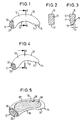
- a spinal implant comprises a body 10, which consists for example of cortical, diaphyseal bone material, for example of human origin.
- This body 10 is basically elongated in plan view and formed curved in the direction of its longitudinal extent.
- the two opposite ends 12 and 14 of the body 10 are rounded, so that a total of a banana-shaped design of the body 10 results. Between the two ends 12 and 14, the body 10 has a convexly curved surface 16 and a concavely curved surface 18th
- the main body in the region of its one end 12 has two receiving openings 20, 22 for application tools.
- the receiving openings 20, 22 are formed as holes whose longitudinal axes are not parallel to each other.
- FIGS. 2 and 3 show possible cross-sectional shapes of the body 10 along the section line AB of Fig. 1 ,
- Fig. 2 shows a possible cross-sectional shape in the form of a rectangle with rounded edges.
- Fig. 3 shows a trapezoidal cross-sectional shape, wherein the inclined sides of the trapezoid deviate at an angle ⁇ of about 3 ° to 6 ° from the rectangular shape.
- obliquely extending outer sides 17, 19 can be inclined in the direction of the concave outer surface 18, ie in the direction of the center of the ring of the annular segment-shaped implant.
- Fig. 1 shows a substantially symmetrical embodiment of a spinal implant.
- Fig. 4 another embodiment shown selected an asymmetrical shape, in which the shape of the body 10 'tapers in plan view from the one end 12 in the direction of the other end 14.
- like reference numerals designate like elements.
- those in the FIGS. 2 and 3 shown cross-sectional shapes also in the Fig. 4 embodiment shown find application.
- the body 10, 10 As the Fig. 1 and 4 show, the body 10, 10 'have the shape of a ring segment, wherein preferably the shape of about 3/8 to 1/2 ring is selected. Furthermore, in the embodiment of Fig. 4 recognize that one end 12 of the body 10 'is wider than the opposite end 14 to facilitate insertion of the implant into the intervertebral foramen.
- the body 1 of the spinal implant is adapted in size to the predetermined place where the implant is to be used.
- the outer dimensions of such a spinal implant may be, for example, in the case of a lumbar application: length about 30 to 60 mm, width about 8 to 20 mm, height about 6 to 18 mm.
- the spinal implant according to the invention is made solid, for example, from cortical bone or cancellous bone.
- An alternative embodiment of the invention provides that the spinal implant - also from cortical or cancellous bone - is formed as a hollow body.
- Fig. 5 shows such an embodiment in which the body 10 "has a cavity 26 which is filled with cancellous bone 28.
- the cavity 26 extends substantially from the end of the receiving openings 20, 22 into the region of the recesses 24.
- the cavity 26 adapted to the outer contour of the body 10 ".
Landscapes
- Health & Medical Sciences (AREA)
- Engineering & Computer Science (AREA)
- Biomedical Technology (AREA)
- Life Sciences & Earth Sciences (AREA)
- Chemical & Material Sciences (AREA)
- Orthopedic Medicine & Surgery (AREA)
- Veterinary Medicine (AREA)
- Public Health (AREA)
- General Health & Medical Sciences (AREA)
- Animal Behavior & Ethology (AREA)
- Oral & Maxillofacial Surgery (AREA)
- Transplantation (AREA)
- Epidemiology (AREA)
- Medicinal Chemistry (AREA)
- Dermatology (AREA)
- Chemical Kinetics & Catalysis (AREA)
- Botany (AREA)
- Vascular Medicine (AREA)
- Neurology (AREA)
- Molecular Biology (AREA)
- Urology & Nephrology (AREA)
- Zoology (AREA)
- Cardiology (AREA)
- Heart & Thoracic Surgery (AREA)
- Prostheses (AREA)
Abstract
Description
- Die vorliegende Erfindung betrifft ein Implantat zur Verbindung von Knochen und insbesondere ein Wirbelsäulenimplantat zur interkorporellen Fusion von Wirbelknochen, das zwischen zwei zu fusionierende Wirbelknochen eingesetzt wird.
- Durch die Degeneration der Bandscheibe, insbesondere des Bandscheibenkerns (Nucleus pulposus) kommt es zu einem Höhenverlust im betroffenen Bandscheibenfach, der mit einer Lockerung des Bandscheibenringes (Annulus fibrosus) und der Bänder verbunden ist. Dadurch wird die Wirbelsäule an dieser Stelle instabil. Die Folge ist eine horizontale Verschiebbarkeit der Wirbelkörper gegeneinander (Spondylolisthese), die zu Beeinträchtigungen der Nervenwurzeln in diesem Bereich und oder des Rükkenmarks mit daraus resultierenden Schmerzen führt. Ähnliche Symptome können nach chemisch-enzymatischer oder physikalischer (Laser) Auflösung des Bandscheibenkerns (Nucleolyse) zur Behandlung eines Bandscheibenvorfalls auftreten (Postnukleolyse-Syndrom).
- Das Prinzip zur Behandlung dieser Symptome besteht in der operativen Ausräumung des Bandscheibenkerns und dem Einlegen bzw. Einschieben - im Bereich der Halswirbelsäule - eines oder - im Bereich der Lendenwirbelsäule - zweier ausreichend stabiler Körper, um die normale Höhe des Bandscheibenfaches wiederherzustellen. Gleichzeitig muß die horizontale Verschiebbarkeit verhindert werden. Dies geschieht entweder durch das Implantat selbst oder durch zusätzliche Metallimplantate (instrumentierte Fusion). Diese Implantate unterliegen insbesondere in der Lendenwirbelsäule erheblichen Kräften, die zum Bruch des Metallimplantats führen können. Es wird daher angestrebt, daß das Zwischenwirbelinterponat möglichst rasch und solide mit den angrenzenden Wirbelkörpern verwächst bzw. fusioniert.
- Zur Behandlung von Patienten mit spinalem Trauma oder degenerativen Leiden an der Wirbelsäule werden im wesentlichen zwei Techniken angewendet.
- 1. Entfernen des Bandscheibenkerns und des Knorpels an den Endplatten, Aufdehnen des Zwischenwirbelraumes auf normale Weite und Einschieben eines planparallelen oder horizontal leicht keilförmigen Blocks (Smith-Robinson-Technik).
- 2. Aufdehnen des Bandscheibenfaches auf normale Höhe, Bohren eines zylindrischen Loches, das beide Wirbelkörper erfaßt, und Einschieben eines zylindrischen Dübels (Cloward-Technik). Der Dübel kann dabei entweder ein glatter Zylinder sein oder ein Gewinde aufweisen.
- Ein relativ neues Verfahren zur interkorporellen Fusion an der Lendenwirbelsäule ist die posteriore lumbale interkorporelle Fusion in unilateraler transforaminaler Technik. Hierbei wird das Foramen intervertebrale des betroffenen Segmentes einseitig eröffnet, das Bandscheibenfach wird ausgeräumt und es werden zur ventralen Abstützung zwei auf passende Höhe zugeschnittene Titangitterkörbe eingebracht, welche mit Knochenchips gefüllt sind. Diese gewebeschonende Methode hat jedoch verschiedene Nachteile. Zum einen können die scharfen Kanten der Titankörbe beim Einbringen die Nervenwurzeln beschädigen. Ferner ist eine Lordosierung nicht möglich, da die Körbe praktisch blind eingebracht werden. Schließlich stehen nur die Ränder der Körbe zur Verfügung, um den vorhandenen Druck aufzunehmen. Das Titan erschwert durch Artefakte und in der Computertomographie die Fusionskontrolle. Darüberhinaus ist die Implantatentfernung bei Revisionsoperationen außerordentlich schwierig.
- Aus der
US 5,861,041 ist ein zweiteiliges Wirbelsäulenimplantat bekannt, das im verbundenen Zustand eine im wesentlichen elliptische Konfiguration besitzt. - Die
WO 99/09914 - Die Aufgabe der vorliegenden Erfindung besteht deshalb darin, ein Implantat zur Fusion von Knochen zu schaffen, das die oben genannten Nachteile beseitigt.
- Die Lösung dieser Aufgabe erfolgt durch die Merkmale des Anspruchs 1.
- Ein besonderer Vorteil des Wirbelsäulenimplantats gemäß der Erfindung ist durch das verwendete Material gegeben, das aufgrund seines biologischen Ursprungs und einer speziellen Konservierung keinen Fremdkörper darstellt. Darüberhinaus ergibt sich durch die gekrümmte Gestaltung des bananenförmige Krümmung auf. In einer besonders vorteilhaften Ausgestaltung weist der Körper abgerundete Kanten auf. Eine solche Gestaltung des Körpers erleichtert die Applikation des Wirbelsäulenimplantats zwischen den zu fusionierenden Wirbelkörpern, indem die Abrundungen ein Verkanten des Implantats während der Applikation vermeiden. Durch die spezielle Bananenform ist das Implantat besonders gut an die natürliche Form der Wirbelkörperendplatten angepaßt und bietet so die größtmögliche Kontaktfläche zu den Endplatten. Dadurch erfolgt eine mehr physiologische Verteilung der Kräfte wodurch Druckspitzen und ein durch diese bedingtes Einsinken des Implantates in die Wirbelkörper vermieden werden. Da durch die Krümmung des Implantates automatisch eine korrekte Positionierung erfolgt, ist durch einen trapezförmigen Querschnitt auch die Wiederherstellung der physiologischen Krümmung der Wirbelsäule - Lordose - möglich.
- Besonders vorteilhaft ist es, daß die Einkerbungen oder Vertiefungen die Mitnahme von lose eingebrachtem Knochenpulver in den Zwischenwirbelraum ermöglichen. Auf diese Weise kann beim Einschieben des Implantats in den Zwischenwirbelraum automatisch Knochenpulver in den konkav gekrümmten Bereich des Implantats eingebracht werden.
- Nach einem weiteren Aspekt der vorliegenden Erfindung sind Aufnahmeöffnungen auf einer oder mehreren Seiten des Körpers als Bohrloch mit einem Gewinde von vorzugsweise 3 mm Tiefe ausgeführt. In diese Gewindebohrungen können dann mit passendem Gewinde versehene Applikationswerkzeuge eingeschraubt werden, um das Implantat positionsgenau zwischen die zu fusionierenden Wirbel einzubringen.
- Als Material wird für das Wirbelsäulenimplantat gemäß der vorliegenden Erfindung ein geeignetes allogenes oder xenogenes Knochenmaterial derart prozessiert, daß es konserviert, lagerfähig sowie steril ist und bestimmungsgemäß eingesetzt werden kann. Die Konservierung des Knochenmaterials kann beispielsweise mittels Gefriertrocknung erfolgen. Ein anderes bevorzugtes Verfahren der Herstellung des Knochenmaterials ist die Prozessierung durch vorzugsweise Lösungsmitteldehydratisierung von nativem Knochenmaterial mittels eines organischen, mit Wasser mischbaren Lösungsmittels, z.B. Methanol, Äthanol, Propanol, Isopropanol, Aceton, Methyl-Äthylketon oder Gemischen dieser Lösungsmittel. Die Konservierung und Sterilisation des Knochenmaterials nach diesem Verfahren ist auch Gegenstand des Patents
DE 29 06 650 . - Dieses Verfahren dient der Herstellung von Transplantatkonserven und ermöglicht eine Dehydratisierung und Freilegung bis in den Feinbau der Fibrille des Knochenmaterials, so daß das prozessierte Knochenmaterial im histologischen Bild eine dem natürlichen Knochen sehr ähnliche morphologische Struktur aufweist und somit die gewünschten Eigenschaften des Knochenmaterials erhalten bleiben. Dieses Verfahren der Lösungsmitteldehydratisierung hat außerdem den Vorteil, daß im Vergleich zur Gefriertrocknung ein wesentlich geringerer apparativer Aufwand erforderlich ist.
- Ferner kann das Knochenmaterial auch durch Lösungsmitteldehydratisierung von nativem Knochen mit anschließender terminaler Sterilisation, insbesondere durch Bestrahlung mit Gamma-Strahlen erzeugt werden. Alternativ kann das spongiöse Knochenmaterial durch aseptische Prozessierung von Knochenmaterial ohne terminale Sterilisation erzeugt werden. Das Ausgangsmaterial des erfindungsgemäßen Knochenimplantats ist menschlicher oder tierischer Knochen von ausreichender Größe.
- Zur Entfernung der Antigenität wird der Knochen einer osmotischen Behandlung unterzogen. Weiterhin wird zur Denaturierung löslicher Proteine eine oxidative Behandlung durchgeführt. Zur Optimierung der Virusinaktivierung kann eine pH-Absenkung auf pH 3 erfolgen oder eine Behandlung mit Natronlauge oder einer anderen DNA/RNA zerstörenden Substanz. Der Wasserentzug erfolgt durch organische Lösungsmittel, vorzugsweise Azeton. Die abschließende Sterilisation erfolgt durch energiereiche Strahlung, vorzugsweise γ-Strahlen mit einem Dosismaximum von 25 kGy.
- Ein so behandelter Knochen behält seinen natürlichen Mineral-Collagen-Verbund und Eigenschaften. Darüberhinaus ist ein so behandelter Knochen umbaufähig.
- Nachfolgend wird die vorliegende Erfindung rein exemplarisch anhand von Ausführungsbeispielen eines erfindungsgemäßen Wirbelsäulenimplantats unter Bezugnahme auf die beigefügten Zeichnungen beschrieben. Es zeigen:
- Fig. 1
- eine Draufsicht auf eine Ausführungsform eines Wirbelsäulenimplantats gemäß der vorliegenden Erfindung;
- Fig. 2
- eine bekannte Querschnittsform eines Wirbelsäulenimplantats;
- Fig. 3
- eine mögliche Querschnittsform der erfindungsgemäßen Wirbelsäulenimplantate;
- Fig. 4
- eine Draufsicht auf eine weitere Ausführungsform eines Wirbelsäulenimplantats gemäß der vorliegenden Erfindung; und
- Fig. 5
- eine Draufsicht auf eine weitere Ausführungsform eines Wirbelsäulenimplantats gemäß der vorliegenden Erfindung.
- In den Figuren bezeichnen die gleichen Bezugszeichen jeweils die gleichen Komponenten der dargestellten Ausführungsformen.
- Das in
Fig. 1 dargestellte Ausführungsbeispiel eines erfindungsgemäßen Wirbelsäulenimplantats umfaßt einen Körper 10, der beispielsweise aus kortikalem, diaphysärem Knochenmaterial z.B. humanen Ursprungs besteht. Dieser Körper 10 ist in Draufsicht grundsätzlich langgestreckt und in Richtung seiner Längserstreckung gekrümmt ausgebildet. - Die beiden entgegengesetzten Enden 12 und 14 des Körpers 10 sind abgerundet ausgebildet, so daß sich insgesamt eine bananenförmige Gestaltung des Grundkörpers 10 ergibt. Zwischen den beiden Enden 12 und 14 besitzt der Körper 10 eine konvex gekrümmte Fläche 16 und eine konkav gekrümmte Fläche 18.
- Wie
Fig. 1 zeigt, weist der Grundkörper im Bereich seines einen Endes 12 zwei Aufnahmeöffnungen 20, 22 für Applikationswerkzeuge auf. Die Aufnahmeöffnungen 20, 22 sind als Bohrungen ausgebildet, deren Längsachsen nicht zueinander parallel verlaufen. - Im Bereich des gegenüberliegenden Endes 14 ist eine Strukturierung der Oberfläche des Grundkörpers im Bereich der konkaven Fläche 18, vorzugsweise durch Einkerbungen bzw. Vertiefungen 24 vorgesehen, welche die Mitnahme von lose zugegebenem Knochenpulver in den Zwischenwirbelraum ermöglichen. Durch die vorgesehene Oberflächenstrukturierung (in dem dargestellten Ausführungsbeispiel sind drei Einkerbungen vorgesehen) ist die Mitnahme und Verdichtung des Knochenpulvers beim Einbringen des Knochemimplantates in den Zwischenwirbelraum gewährleistet.
- Die
Fig. 2 und 3 zeigen mögliche Querschnittsformen des Körpers 10 entlang der Schnittlinie A-B vonFig. 1 . -
Fig. 2 zeigt eine mögliche Querschnittsform in Form eines Rechteckes mit abgerundeten Kanten.Fig. 3 zeigt eine trapezförmige Querschnittsform, wobei die schräg verlaufenden Seiten des Trapezes unter einem Winkel α von etwa 3° bis 6° von der Rechteckform abweichen. - Wie insbesondere
Fig. 3 zeigt, können bei trapezförmigem Querschnitt des Implantats die schräg verlaufenden Außenseiten 17, 19 in Richtung der konkaven Außenfläche 18 geneigt sein, d. h. in Richtung der Ringmitte des ringsegmentförmig geformten Implantats. -
Fig. 1 zeigt eine im wesentlichen symmetrische Ausführungsform eines Wirbelsäulenimplantats. Demgegenüber ist bei der inFig. 4 dargestellten weiteren Ausführungsform eine asymmetrische Formgebung gewählt, bei welcher die Form des Körpers 10' sich in Draufsicht von dem einen Ende 12 in Richtung des anderen Endes 14 verjüngt. Im übrigen bezeichnen gleiche Bezugszeichen gleiche Elemente. Gleichzeitig können die in denFig. 2 und 3 dargestellten Querschnittsformen auch bei der inFig. 4 dargestellten Ausführungsform Anwendung finden. - Wie die
Fig. 1 und 4 zeigen, besitzen die Körper 10, 10' die Form eines Ringsegmentes, wobei bevorzugt die Form eines etwa 3/8 bis 1/2 Ringes gewählt wird. Ferner ist bei der Ausführungsform vonFig. 4 zu erkennen, daß das eine Ende 12 des Körpers 10' breiter ist als das gegenüberliegende Ende 14, um ein Einführen des Implantats in das Foramen intervertebrale zu erleichtern. - Grundsätzlich ist der Körper 1 des Wirbelsäulenimplantats in seiner Größe an den vorgegebenen Platz, an dem das Implantat eingesetzt werden soll, angepaßt. Die Außenabmessungen eines solchen Wirbelsäulenimplantats können bei lumbaler Anwendung beispielsweise folgendermaßen sein: Länge etwa 30 bis 60 mm, Breite etwa 8 bis 20 mm, Höhe etwa 6 bis 18 mm.
- Grundsätzlich besteht die Möglichkeit, daß das erfindungsgemäße Wirbelsäulenimplantat massiv ausgebildet wird, beispielsweise aus kortikalem oder aus spongiösem Knochen. Eine alternative Ausführungsform der Erfindung sieht vor, daß das Wirbelsäulenimplantat - ebenfalls aus kortikalem oder spongiösem Knochen - als Hohlkörper ausgebildet wird.
-
Fig. 5 zeigt eine solche Ausführungsform, bei welcher der Körper 10" einen Hohlraum 26 aufweist, der mit spongiösem Knochen 28 gefüllt ist. Der Hohlraum 26 erstreckt sich im wesentlichen vom Ende der Aufnahmeöffnungen 20, 22 bis in den Bereich der Vertiefungen 24. Hierbei ist der Hohlraum 26 an die Außenkontur des Körpers 10" angepaßt. -
- 10, 10', 10"
- Körper
- 12, 14
- Ende
- 16
- konvexe Außenseite
- 17
- Außenseite
- 18
- konkave Außenseite
- 19
- Außenseite
- 20, 22
- Aufnahmeöffnung
- 24
- Vertiefungen
- 26
- Hohlraum
- 28
- spongiöser Knochen
- α
- Neigungswinkel
Claims (7)
- Wirbelsäulenimplantat zur interkorporellen Fusion an der Wirbelsäule, bestehend aus einem Körper (10, 10', 10") aus Knochenmaterial, der einen rechteckigen oder trapezförmigen Querschnitt besitzt, und der in Richtung seiner Längserstreckung gekrümmt ausgebildet ist, wodurch eine konvex gekrümmte Seite (16) und eine konkav gekrümmte Seite (18) gebildet ist, und wobei das Implantat in seiner Größe an den nach Ausräumung des Wirbelzwischenkörpers vorhandenen Wirbelzwischenraum zwischen benachbarten Wirbeln angepaßt ist, dadurch gekennzeichnet,
dass an einem Ende des Körpers (10, 10', 10") Einkerbungen oder Vertiefungen (24) im Bereich der konkav gekrümmten Seite (18) vorgesehen sind, welche die Mitnahme von lose eingebrachtem Knochenpulver in den Wirbelzwischenraum ermöglichen. - Wirbelsäulenimplantat nach Anspruch 1
dadurch gekennzeichnet,
daß der Körper (10, 10', 10") aus konserviertem und sterilem bovinem Knochenmaterial humanen oder tierischen Ursprungs besteht. - Wirbelsäulenimplantat nach einem der vorangehenden Ansprüche
dadurch gekennzeichnet,
daß zumindest ein Ende (12, 14) des Körpers (10, 10', 10") in Draufsicht halbkreisförmig abgerundet ist. - Wirbelsäulenimplantat nach einem der vorangehenden Ansprüche
dadurch gekennzeichnet,
daß dieses mindestens eine Aufnahmeöffnung (20, 22) für ein Applikationswerkzeug aufweist. - Wirbelsäulenimplantat nach einem der vorangehenden Ansprüche
dadurch gekennzeichnet,
daß der Körper (10, 10', 10") abgerundete Kanten aufweist. - Wirbelsäulenimplantat nach einem der vorangehenden Ansprüche
dadurch gekennzeichnet,
daß der Körper (10, 10', 10") die Form eines Ringsegmentes besitzt, welches etwa 3/8 bis 1/2 eines Ringes ist. - Wirbelsäulenimplantat nach einem der vorangehenden Ansprüche,
dadurch gekennzeichnet,
daß ein Ende (14) des Körpers (10') schmaler ist als das andere Ende (12).
Applications Claiming Priority (3)
| Application Number | Priority Date | Filing Date | Title |
|---|---|---|---|
| DE19952939A DE19952939A1 (de) | 1999-11-03 | 1999-11-03 | Implantat aus Knochenmaterial |
| DE19952939 | 1999-11-03 | ||
| PCT/EP2000/010672 WO2001032110A2 (de) | 1999-11-03 | 2000-10-30 | Implantat aus knochenmaterial |
Publications (2)
| Publication Number | Publication Date |
|---|---|
| EP1225850A2 EP1225850A2 (de) | 2002-07-31 |
| EP1225850B1 true EP1225850B1 (de) | 2008-06-11 |
Family
ID=7927801
Family Applications (1)
| Application Number | Title | Priority Date | Filing Date |
|---|---|---|---|
| EP00977441A Expired - Lifetime EP1225850B1 (de) | 1999-11-03 | 2000-10-30 | Implantat aus knochenmaterial |
Country Status (6)
| Country | Link |
|---|---|
| US (1) | US7060096B1 (de) |
| EP (1) | EP1225850B1 (de) |
| AU (1) | AU1515901A (de) |
| CA (1) | CA2389630C (de) |
| DE (2) | DE19952939A1 (de) |
| WO (1) | WO2001032110A2 (de) |
Families Citing this family (48)
| Publication number | Priority date | Publication date | Assignee | Title |
|---|---|---|---|---|
| US5362442A (en) | 1993-07-22 | 1994-11-08 | 2920913 Canada Inc. | Method for sterilizing products with gamma radiation |
| US7018416B2 (en) | 2000-07-06 | 2006-03-28 | Zimmer Spine, Inc. | Bone implants and methods |
| US6696060B2 (en) | 2001-06-14 | 2004-02-24 | Clearant, Inc. | Methods for sterilizing preparations of monoclonal immunoglobulins |
| US6946098B2 (en) | 2001-08-10 | 2005-09-20 | Clearant, Inc. | Methods for sterilizing biological materials |
| US6749851B2 (en) | 2001-08-31 | 2004-06-15 | Clearant, Inc. | Methods for sterilizing preparations of digestive enzymes |
| DE10152567A1 (de) | 2001-10-24 | 2003-05-08 | Tutogen Medical Gmbh | Implantat |
| DE20117773U1 (de) * | 2001-10-31 | 2003-03-20 | Königsee Implantate und Instrumente zur Osteosynthese GmbH, 07426 Königsee | Implantat zur Stabilisierung der Vertebrae cervicales |
| DE10220139A1 (de) | 2002-05-06 | 2003-11-20 | Tutogen Medical Gmbh | Implantat aus Knochenmaterial |
| US6908591B2 (en) | 2002-07-18 | 2005-06-21 | Clearant, Inc. | Methods for sterilizing biological materials by irradiation over a temperature gradient |
| US8613772B2 (en) * | 2003-04-21 | 2013-12-24 | Rsb Spine Llc | Lateral mount implant device |
| FR2858546B1 (fr) * | 2003-08-04 | 2006-04-28 | Spine Next Sa | Prothese de disque intervertebral |
| US8021392B2 (en) | 2004-11-22 | 2011-09-20 | Minsurg International, Inc. | Methods and surgical kits for minimally-invasive facet joint fusion |
| US20060111779A1 (en) | 2004-11-22 | 2006-05-25 | Orthopedic Development Corporation, A Florida Corporation | Minimally invasive facet joint fusion |
| US20060111780A1 (en) * | 2004-11-22 | 2006-05-25 | Orthopedic Development Corporation | Minimally invasive facet joint hemi-arthroplasty |
| US20060111786A1 (en) * | 2004-11-22 | 2006-05-25 | Orthopedic Development Corporation | Metallic prosthetic implant for use in minimally invasive acromio-clavicular shoulder joint hemi-arthroplasty |
| CN1795832A (zh) * | 2004-12-23 | 2006-07-05 | 杨守恒 | 腰椎椎体间固定融合器 |
| EP1783598A1 (de) * | 2005-11-02 | 2007-05-09 | Hsin-Tung Tseng | Integrierte Lehrbasisstation |
| EP1978900B1 (de) * | 2006-02-01 | 2012-03-07 | Synthes GmbH | Interspinaler eingriffsabstandhalter |
| USD585553S1 (en) | 2007-02-22 | 2009-01-27 | Zimmer Spine, Inc. | Spinal implant |
| US8216317B2 (en) | 2008-03-31 | 2012-07-10 | Stryker Spine | Spinal implant apparatus and methods |
| US8147554B2 (en) | 2008-10-13 | 2012-04-03 | Globus Medical, Inc. | Intervertebral spacer |
| US8545566B2 (en) | 2008-10-13 | 2013-10-01 | Globus Medical, Inc. | Articulating spacer |
| US9028553B2 (en) * | 2009-11-05 | 2015-05-12 | DePuy Synthes Products, Inc. | Self-pivoting spinal implant and associated instrumentation |
| US8603175B2 (en) | 2010-09-30 | 2013-12-10 | Stryker Spine | Method of inserting surgical implant with guiding rail |
| US8858637B2 (en) | 2010-09-30 | 2014-10-14 | Stryker Spine | Surgical implant with guiding rail |
| US8425529B2 (en) | 2010-09-30 | 2013-04-23 | Stryker Spine | Instrument for inserting surgical implant with guiding rail |
| US9539109B2 (en) | 2011-09-16 | 2017-01-10 | Globus Medical, Inc. | Low profile plate |
| US9770340B2 (en) | 2011-09-16 | 2017-09-26 | Globus Medical, Inc. | Multi-piece intervertebral implants |
| US9398960B2 (en) | 2011-09-16 | 2016-07-26 | Globus Medical, Inc. | Multi-piece intervertebral implants |
| US9237957B2 (en) | 2011-09-16 | 2016-01-19 | Globus Medical, Inc. | Low profile plate |
| US9204975B2 (en) | 2011-09-16 | 2015-12-08 | Globus Medical, Inc. | Multi-piece intervertebral implants |
| US10881526B2 (en) | 2011-09-16 | 2021-01-05 | Globus Medical, Inc. | Low profile plate |
| US9149365B2 (en) | 2013-03-05 | 2015-10-06 | Globus Medical, Inc. | Low profile plate |
| US8961606B2 (en) | 2011-09-16 | 2015-02-24 | Globus Medical, Inc. | Multi-piece intervertebral implants |
| US10245155B2 (en) | 2011-09-16 | 2019-04-02 | Globus Medical, Inc. | Low profile plate |
| US9681959B2 (en) | 2011-09-16 | 2017-06-20 | Globus Medical, Inc. | Low profile plate |
| US9848994B2 (en) | 2011-09-16 | 2017-12-26 | Globus Medical, Inc. | Low profile plate |
| KR20140093943A (ko) * | 2011-11-01 | 2014-07-29 | 아메디카 코포레이션 | 연결 가능한 삽입체를 갖는 임플란트 및 관련 시스템과 방법 |
| US10022245B2 (en) | 2012-12-17 | 2018-07-17 | DePuy Synthes Products, Inc. | Polyaxial articulating instrument |
| US9566169B2 (en) | 2014-03-13 | 2017-02-14 | DePuy Synthes Products, Inc. | ACIS allograft designs |
| US10568672B2 (en) * | 2014-10-16 | 2020-02-25 | Arthrex, Inc. | Anatomic osteotomy wedge |
| US10376385B2 (en) | 2017-04-05 | 2019-08-13 | Globus Medical, Inc. | Decoupled spacer and plate and method of installing the same |
| US11452608B2 (en) | 2017-04-05 | 2022-09-27 | Globus Medical, Inc. | Decoupled spacer and plate and method of installing the same |
| US10966843B2 (en) | 2017-07-18 | 2021-04-06 | DePuy Synthes Products, Inc. | Implant inserters and related methods |
| US11045331B2 (en) | 2017-08-14 | 2021-06-29 | DePuy Synthes Products, Inc. | Intervertebral implant inserters and related methods |
| DE102018121553A1 (de) * | 2018-09-04 | 2020-03-05 | Karl Leibinger Medizintechnik Gmbh & Co. Kg | Knochenimplantat zur Rekonstruktion eines knöchernen Defektes und zum Führen eines Markierungs- und/oder Bearbeitungswerkzeug zur Übertragung notwendiger Osteotomiesituationen |
| EP3911280A1 (de) * | 2019-01-15 | 2021-11-24 | Biopoly, Llc | Implantatsysteme zur reparatur eines humeruskopfes |
| EP3911281A1 (de) | 2019-01-15 | 2021-11-24 | Biopoly, Llc | Implantatsysteme zur reparatur eines glenoidhohlraums |
Family Cites Families (22)
| Publication number | Priority date | Publication date | Assignee | Title |
|---|---|---|---|---|
| DE2906650A1 (de) * | 1979-02-21 | 1980-08-28 | Pfrimmer Pharma | Verfahren zur herstellung von transplantatkonserven |
| CH672589A5 (de) * | 1987-07-09 | 1989-12-15 | Sulzer Ag | |
| GB8718627D0 (en) * | 1987-08-06 | 1987-09-09 | Showell A W Sugicraft Ltd | Spinal implants |
| US5071655A (en) * | 1990-01-12 | 1991-12-10 | Baylink David J | Pharmaceutical combination for treatment of bone-wasting diseases |
| DE4242889A1 (de) * | 1992-12-18 | 1994-06-23 | Merck Patent Gmbh | Hohlendoprothesen mit Knochenwachstumsfördernder Füllung |
| US5397364A (en) * | 1993-10-12 | 1995-03-14 | Danek Medical, Inc. | Anterior interbody fusion device |
| US5514180A (en) | 1994-01-14 | 1996-05-07 | Heggeness; Michael H. | Prosthetic intervertebral devices |
| FR2736537A1 (fr) * | 1995-07-12 | 1997-01-17 | Vila Thierry | Cage de stabilisation inter-corporeale |
| US5728159A (en) * | 1997-01-02 | 1998-03-17 | Musculoskeletal Transplant Foundation | Serrated bone graft |
| US5861041A (en) * | 1997-04-07 | 1999-01-19 | Arthit Sitiso | Intervertebral disk prosthesis and method of making the same |
| WO1999009914A1 (en) | 1997-08-27 | 1999-03-04 | University Of Florida Tissue Bank, Inc. | Cortical bone cervical smith-robinson fusion implant |
| US6139579A (en) * | 1997-10-31 | 2000-10-31 | Depuy Motech Acromed, Inc. | Spinal disc |
| DE29720022U1 (de) * | 1997-11-12 | 1998-01-15 | SCHÄFER micomed GmbH, 73035 Göppingen | Zwischenwirbelimplantat |
| DE19751284A1 (de) * | 1997-11-19 | 1999-05-27 | Leo Prof Dr Gotzen | Fixationselement |
| US6296664B1 (en) * | 1998-06-17 | 2001-10-02 | Surgical Dynamics, Inc. | Artificial intervertebral disc |
| US6174311B1 (en) * | 1998-10-28 | 2001-01-16 | Sdgi Holdings, Inc. | Interbody fusion grafts and instrumentation |
| US6206923B1 (en) * | 1999-01-08 | 2001-03-27 | Sdgi Holdings, Inc. | Flexible implant using partially demineralized bone |
| US6245108B1 (en) * | 1999-02-25 | 2001-06-12 | Spineco | Spinal fusion implant |
| US6277149B1 (en) * | 1999-06-08 | 2001-08-21 | Osteotech, Inc. | Ramp-shaped intervertebral implant |
| DE29913200U1 (de) * | 1999-07-28 | 1999-09-23 | Tutogen Medical GmbH, 91077 Neunkirchen | Implantat aus Knochenmaterial |
| US6579318B2 (en) * | 2000-06-12 | 2003-06-17 | Ortho Development Corporation | Intervertebral spacer |
| US6572654B1 (en) * | 2000-10-04 | 2003-06-03 | Albert N. Santilli | Intervertebral spacer |
-
1999
- 1999-11-03 DE DE19952939A patent/DE19952939A1/de not_active Ceased
-
2000
- 2000-10-30 CA CA002389630A patent/CA2389630C/en not_active Expired - Fee Related
- 2000-10-30 AU AU15159/01A patent/AU1515901A/en not_active Abandoned
- 2000-10-30 US US10/129,259 patent/US7060096B1/en not_active Expired - Fee Related
- 2000-10-30 EP EP00977441A patent/EP1225850B1/de not_active Expired - Lifetime
- 2000-10-30 DE DE50015201T patent/DE50015201D1/de not_active Expired - Lifetime
- 2000-10-30 WO PCT/EP2000/010672 patent/WO2001032110A2/de not_active Ceased
Also Published As
| Publication number | Publication date |
|---|---|
| DE19952939A1 (de) | 2001-05-10 |
| DE50015201D1 (de) | 2008-07-24 |
| EP1225850A2 (de) | 2002-07-31 |
| US7060096B1 (en) | 2006-06-13 |
| WO2001032110A3 (de) | 2002-04-04 |
| CA2389630C (en) | 2009-01-13 |
| CA2389630A1 (en) | 2001-05-10 |
| WO2001032110A2 (de) | 2001-05-10 |
| AU1515901A (en) | 2001-05-14 |
Similar Documents
| Publication | Publication Date | Title |
|---|---|---|
| WO2001032110A2 (de) | Implantat aus knochenmaterial | |
| EP1206226B1 (de) | Implantat aus knochenmaterial | |
| DE60019643T2 (de) | Implantat aus knochenmaterial | |
| DE69429579T2 (de) | Prothese für eine intervertebrale fusion | |
| DE69822817T2 (de) | Offenes zwischenwirbel-distanzstück | |
| DE69916280T2 (de) | Zwischenwirbel-allotransplantat-distanzstück | |
| DE60009446T2 (de) | Biegsames implantat mit teilweise entmineralisiertem knochen | |
| DE68921482T2 (de) | Künstliches spondylodese-implantat. | |
| DE60032264T2 (de) | Wirbelsäulenimplantat | |
| DE69209494T2 (de) | Implantat aus einer expandierbaren zwischenwirbelscheibe | |
| DE60038160T2 (de) | Abgestumpftes, offenes zwischenwirbeldistanzstück | |
| DE19529605C2 (de) | Zwischenwirbelimplantat | |
| DE69032788T2 (de) | Vorrichtung zum vereinigen aneinanderstossender knochenstrukturen | |
| DE69532528T2 (de) | Ausdehnbares stoffimplantat zur stabilisierung des spinalen segmentes | |
| DE69837998T2 (de) | Wirbelfusionsvorrichtung mit eine partiell kreisförmige sektion aufweisenden querschnittssegmenten | |
| DE69837883T2 (de) | Wirbelsäulenimplantat und Schneidwerkzeugvorbereitungszubehör zum Einsetzen des Implantats | |
| DE60030989T2 (de) | Vorrichtung zur implantation von fusionkäfigen | |
| DE60128391T2 (de) | Dynamisches implantiertes zwischenwirbelabstandsstück | |
| DE69628286T2 (de) | Gerät zum einbringen von spinalen implantaten | |
| DE60108770T2 (de) | Verstärktes geformtes implantat aus kortikalisknochen | |
| DE29623247U1 (de) | Instrumentarium für die chirurgische Korrektur der menschlichen Brust- und Lendenwirbelsäulenkrankheit in einem Zugang von der lateralen Seite der Wirbelsäule | |
| WO2004043304A2 (de) | Implantat zum einsatz bei versteifungsoperationen an der wirbelsäule | |
| EP1487383B1 (de) | Wirbelsäulenimplantat aus knochenmaterial | |
| WO2019052681A1 (de) | Zwischenwirbel-implantat | |
| DE19931670C1 (de) | Set für die Erstellung eines modularen Prophylaxe-Implantates |
Legal Events
| Date | Code | Title | Description |
|---|---|---|---|
| PUAI | Public reference made under article 153(3) epc to a published international application that has entered the european phase |
Free format text: ORIGINAL CODE: 0009012 |
|
| 17P | Request for examination filed |
Effective date: 20020502 |
|
| AK | Designated contracting states |
Kind code of ref document: A2 Designated state(s): AT BE CH CY DE DK ES FI FR GB GR IE IT LI LU MC NL PT SE |
|
| AX | Request for extension of the european patent |
Free format text: AL;LT;LV;MK;RO;SI |
|
| RBV | Designated contracting states (corrected) |
Designated state(s): AT BE CH DE FR IT LI |
|
| GRAP | Despatch of communication of intention to grant a patent |
Free format text: ORIGINAL CODE: EPIDOSNIGR1 |
|
| RBV | Designated contracting states (corrected) |
Designated state(s): DE FR IT |
|
| GRAS | Grant fee paid |
Free format text: ORIGINAL CODE: EPIDOSNIGR3 |
|
| GRAA | (expected) grant |
Free format text: ORIGINAL CODE: 0009210 |
|
| AK | Designated contracting states |
Kind code of ref document: B1 Designated state(s): DE FR IT |
|
| REF | Corresponds to: |
Ref document number: 50015201 Country of ref document: DE Date of ref document: 20080724 Kind code of ref document: P |
|
| PLBE | No opposition filed within time limit |
Free format text: ORIGINAL CODE: 0009261 |
|
| STAA | Information on the status of an ep patent application or granted ep patent |
Free format text: STATUS: NO OPPOSITION FILED WITHIN TIME LIMIT |
|
| 26N | No opposition filed |
Effective date: 20090312 |
|
| PGFP | Annual fee paid to national office [announced via postgrant information from national office to epo] |
Ref country code: IT Payment date: 20091027 Year of fee payment: 10 Ref country code: FR Payment date: 20091110 Year of fee payment: 10 |
|
| PGFP | Annual fee paid to national office [announced via postgrant information from national office to epo] |
Ref country code: DE Payment date: 20091230 Year of fee payment: 10 |
|
| PG25 | Lapsed in a contracting state [announced via postgrant information from national office to epo] |
Ref country code: FR Free format text: LAPSE BECAUSE OF NON-PAYMENT OF DUE FEES Effective date: 20101102 |
|
| REG | Reference to a national code |
Ref country code: FR Ref legal event code: ST Effective date: 20110630 |
|
| REG | Reference to a national code |
Ref country code: DE Ref legal event code: R119 Ref document number: 50015201 Country of ref document: DE Effective date: 20110502 |
|
| PG25 | Lapsed in a contracting state [announced via postgrant information from national office to epo] |
Ref country code: IT Free format text: LAPSE BECAUSE OF NON-PAYMENT OF DUE FEES Effective date: 20101030 |
|
| PG25 | Lapsed in a contracting state [announced via postgrant information from national office to epo] |
Ref country code: DE Free format text: LAPSE BECAUSE OF NON-PAYMENT OF DUE FEES Effective date: 20110502 |