EP1168913B1 - Methods and means for extracorporeal organ perfusion - Google Patents
Methods and means for extracorporeal organ perfusion Download PDFInfo
- Publication number
- EP1168913B1 EP1168913B1 EP00915294A EP00915294A EP1168913B1 EP 1168913 B1 EP1168913 B1 EP 1168913B1 EP 00915294 A EP00915294 A EP 00915294A EP 00915294 A EP00915294 A EP 00915294A EP 1168913 B1 EP1168913 B1 EP 1168913B1
- Authority
- EP
- European Patent Office
- Prior art keywords
- liver
- organ
- blood
- flow
- perfusion
- Prior art date
- Legal status (The legal status is an assumption and is not a legal conclusion. Google has not performed a legal analysis and makes no representation as to the accuracy of the status listed.)
- Expired - Lifetime
Links
Images


Classifications
-
- A—HUMAN NECESSITIES
- A01—AGRICULTURE; FORESTRY; ANIMAL HUSBANDRY; HUNTING; TRAPPING; FISHING
- A01N—PRESERVATION OF BODIES OF HUMANS OR ANIMALS OR PLANTS OR PARTS THEREOF; BIOCIDES, e.g. AS DISINFECTANTS, AS PESTICIDES OR AS HERBICIDES; PEST REPELLANTS OR ATTRACTANTS; PLANT GROWTH REGULATORS
- A01N1/00—Preservation of bodies of humans or animals, or parts thereof
- A01N1/02—Preservation of living parts
- A01N1/0236—Mechanical aspects
- A01N1/0242—Apparatuses, i.e. devices used in the process of preservation of living parts, such as pumps, refrigeration devices or any other devices featuring moving parts and/or temperature controlling components
- A01N1/0247—Apparatuses, i.e. devices used in the process of preservation of living parts, such as pumps, refrigeration devices or any other devices featuring moving parts and/or temperature controlling components for perfusion, i.e. for circulating fluid through organs, blood vessels or other living parts
Definitions
- the present invention relates to perfusion of organs, in particular the extracorporeal perfusion of organs, whether human or non-human, e.g. porcine, and whether non-transgenic or transgenic.
- Apparatus and methods of operation of such apparatus are provided for perfusion to support viability and function of an organ such as a liver, generally outside the body. This allows for organ preservation or resuscitation prior to transplantation, for instance while a transplant recipient is prepared, maintenance of organs for use in experimental study of isolated liver physiology, and treatment of patients suffering from organ failure.
- the invention is based on the inventors' unexpected findings relating to autoregulation of blood flow within an organ.
- blood is provided to an organ such as a liver under physiological pressure but without forcing blood flow at any particular rate.
- the organ is allowed to autoregulate blood flow. (Any surplus blood flow may be allowed to run off.)
- outflow pressure from a perfused organ can be maintained.
- the histological appearances of livers perfused using the invention are within normal limits even after 72 hours. This represents a significant advance over known techniques, problems with which date back a long time.
- organs from concordant species Whilst the use of organs from concordant species has major immunological advantages, there are major practical restrictions. The availability of human livers is limited and those available must be used for clinical transplantation. The use of organs from non-human primates poses significant problems, although organs from baboons have been successfully employed. Major concerns exist in relation to primate zoonoses; also, the majority of non-human primates do not attain the necessary size to be effective in human organ replacement.
- WO99/15011 discloses apparatus specifically designed for use in perfusing a heart, and adapted circuits for use with other organs: kidney, liver, pancreas and lungs.
- the apparatuses proposed for the kidney, liver, pancreas or lungs are not capable of allowing any autoregulation of flow by the organ.
- Figure 8 and the accompanying text proposes apparatus for perfusing liver in which the fluid is to be pumped in and out at the same pressure, forcing the organ to take whatever blood is given to it.
- the heart apparatus proposed ( Figure 5) would be unsuitable for use with any other organ. In normal operation the blood is pumped into the heart and there is no possibility of autoregulation by the heart of flow through itself.
- WO99/15011 when the system is to be used for perfusing a beating heart (not a warm, non-beating heart), the authors employ a run-off from the input into the aorta.
- a heart where a heart is to be allowed to beat, although the document does not disclose it, a heart in that situation would have been allowed to autoregulate blood flow through itself.
- This optional embodiment in WO99/15011 represents an accidental anticipation which has been disclaimed from claim 1 in accordance with G2/03.
- US5807737 discloses heart perfusion apparatus which controls and forces maintenance of pressure. The heart must take all blood forced into it by the pumped circuit.
- EP0376763 discloses perfusion apparatus in which blood flow is pumped into the organ and blood flow out goes straight to a reservoir.
- the pump imposes the inlet flow into the organ, whilst the arterial pressure presented to the organ will vary with the internal resistance of the organ.
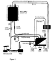
- the present invention provides mammalian organ perfusion apparatus which on connection to input artery and output vein of a mammalian organ, selected from the group consisting of liver, kidney, pancreas and small bowel provides a fluid circuit for flow of blood through the organ, in which circuit there is, preferably in the following order, an output channel for connection to the output vein of the organ, an outflow pump of adjustable speed for maintaining physiological pressure in the output vein of the organ, an oxygenator and a heat exchanger, and an input channel for connection to an input artery of the organ, the circuit further comprising a device for maintaining physiological pressure and variable flow in the input artery of the organ while allowing the organ to autoregulate blood flow through itself to achieve physiological arterial flow.
- a mammalian organ selected from the group consisting of liver, kidney, pancreas and small bowel provides a fluid circuit for flow of blood through the organ, in which circuit there is, preferably in the following order, an output channel for connection to the output vein of the organ, an outflow pump of adjustable speed for maintaining physiological pressure
- the present invention provides mammalian liver perfusion apparatus which on connection to vena cava, hepatic artery and portal vein of a mammalian liver provides a fluid circuit for flow of blood through the liver, in which circuit there is, preferably in the following order, an output channel for connection to the vena cava, an outflow pump of adjustable speed for maintaining physiological pressure in the vena cava, an oxygenator and a heat exchanger, and a bifurcating channel dividing the circuit to input channels for connection to the hepatic artery and portal vein, the circuit further comprising a device for maintaining physiological pressure in the hepatic artery and physiological flow in the portal vein while allowing a liver when connected to the apparatus to autoregulate blood flow through the artery and to autoregulate pressure in the portal vein.
- Such apparatus provides a circuit for blood flow.
- An individual may be joined to the circuit, preferably a human.
- the individual's circulation may be connected to the circuit via cannulae placed in large veins (positioned by open surgery or percutaneously).
- Outflow from the individual is returned to the perfusion circuit at a point preceding the organ.
- Inflow to the individual is of blood that has passed through the organ and is oxygenated.
- connection to a patient may be at a point after the gate clamp indicated in the spur shown in the figures as leading to the reservoir, with the return from the patient leading to the reservoir.
- a circuit may additionally include one or more flow meters and one or more pressure meters.
- the present invention allows for maintenance of physiological levels of both flow and pressure.
- the inventors have observed that in the liver portal pressure is directly related to inferior vena caval pressure and that portal flow (rather than pressure) is directly related to pressure applied from the portal venous reservoir (in preferred embodiments the height of portal venous reservoir above the liver, where blood is supplied from there by gravity).
- the liver responds to increased portal pressure by reduction in portal vascular resistance, maintaining constant portal pressure.
- the outflow from the organ is pumped so that physiological pressure is maintained, e.g. by varying pump speed, in the relevant vein, which for the liver is the vena cava.
- Physiological inflow may be achieved by adjustment of pressure of the supply, e.g. portal vein for the liver.
- physiological portal flow is achieved by adjustment of the height of the portal reservoir above the liver.
- the present invention allows for establishment of normal levels of not only portal pressure but also portal flow.
- the arterial inflow to a liver perfused in accordance with the invention may be set at a physiological pressure, the flow rate bing determined by the vascular resistance of the liver.
- the general principle of the invention as applicable to a variety of organs is to set the pressure of inflow at a physiological level, with a variable flow rate depending on vascular resistance of the organ, allowing for physiological autoregulation to occur.
- the circuit employed in aspects of the invention as exemplified with respect to the liver further comprises a device for maintaining physiological pressure in the hepatic artery and physiological flow in the portal vein while allowing a liver when connected to the apparatus to autoregulate blood flow through the artery and to autoregulate pressure in the portal vein.
- a device may include a reservoir for collection of run-off blood resulting from autoregulation by the liver of flow in the hepatic artery, and such a reservoir may be for supply of blood to the input channel for connection to the portal vein of a liver.
- Supply of blood to the portal vein of a liver when connected to the apparatus may be from the reservoir under force of gravity.
- inflow pressure can be adjusted while allowing for autoregulation of inflow rate by providing a bifurcating tube allowing for run-off from the connection to the input blood vessel.
- the resistance of the run-off spur of the bifurcation may be altered by means of partial occlusion, e.g. by clamping, for adjustment of pressure in the input to the organ.
- Autoregulation of flow by the organ leads to run-off of blood to the reservoir.
- Alternative ways of achieving the same result include an automated controller that fixes the pressure within predetermined parameters yet allows for variable flow in response to autoregulation by the organ.
- such a system may employ two pumps, one to provide hepatic arterial flow at constant pressure/variable flow, and one to fill the portal venouse reservoir at low pressure.
- the present invention provides a method of operating an organ perfusion apparatus.
- the apparatus may have the components identified herein.
- One embodiment of a method according to this aspect of the invention as applied to a liver may be a method operating mammalian liver perfusion apparatus connected to vena cava, hepatic artery and portal vein of a mammalian liver wherein the apparatus provides a fluid circuit for flow of blood through the liver, the method including adjusting the rate of outflow pumping to maintain physiological pressure in the vena cava, adjusting pressure of supply to the input channel connected to the hepatic artery to maintain physiological pressure in the hepatic artery and physiological flow in the portal vein, and allowing the liver to autoregulate blood flow through the artery and to autoregulate pressure in the portal vein.
- a further aspect provides a method of perfusing a mammalian organ extracorporeally, the organ being other than a heart allowed to beat, which method includes pumping arterial blood inflow to the organ to maintain physiological pressure and variable flow in the artery of the organ and pumping venous outflow from the organ to maintain physiological pressure in the output vein of the organ, while allowing the organ to autoregulate blood flow through itself to achieve physiological arterial flow.
- the organ may be selected from the group consisting of liver, kidney, pancreas and small bowel.
- a method according to the invention may include adjusting portal vein flow and pumping blood outflow from the liver to maintain physiological pressure in the vena cava of the liver, while allowing the liver to autoregulate portal pressure.
- a further aspect of the invention provides a method of maintaining viability of a mammalian organ, in which method the organ is perfused in accordance a method as disclosed herein.
- Perfusion in accordance with the present invention allows for maintenance of viability of an organ such as a liver for at least about 72 hours, preferably at least about 96 hours, more preferably at least about 120 hours.
- An organ perfused in accordance with the present invention may be human or non-human, and may be non-transgenic or transgenic.
- Non-human organs may be modified in order to increase compatibility with connection to a human and/or perfusion with human blood.
- An example of a modification that successfully used to increase concordance of non-human, especially porcine organs, is transgenic modification such that the organs express the human complement component Decay Accelerating Factor (hDAF) (see e.g. Cozzi and White, and Schmoeckel et al. supra ).
- hDAF Decay Accelerating Factor
- Blood used in the perfusion may be human or non-human, e.g. porcine for a porcine organ, and this may depend on the purpose of the perfusion.
- a human organ will generally be perfused with human blood. Where the organ is to be connected to a human subject, the blood will be human and needs to be antigenically compatible with respect to ABO.
- a non-human, e.g. pig organ may be perfused with the relevant non-human blood (e.g. pig), or human blood for instance when the organ is modified in the manner described above (e.g. transgenic for hDAF).
- the blood is citrated.
- the inventors have found the function of livers perfused with citrated blood to be markedly superior to those perfused with heparinised blood. Citrate is an important metabolic substrate of the liver.
- a liver perfusion circuit preferably fluid that leaks from the organ, including ascites, is recirculated back to the liver, preferably via the portal vein or, if not, via the hepatic artery.
- This has the advantage of maintaining vascular volume, and also retaining plasma proteins which would probably be lost if the ascites was replaced with colloid/crystalloid solutions.
- Apparatus according to the invention may include a channel for collection of fluid that leaks from the organ and its transport to the input blood reservoir.
- perfusion is "warm perfusion", i.e. at physiological temperature, generally 37°C for a human organ and 39°C for a pig organ.
- a liver for use in the present invention is preserved with hypertonic citrate, rather than the current standard liver transplant preservation solution (University of Wisconsin) which is based on lactobionate.
- Preservation solution is washed out of the liver immediately before perfusion starts.
- a further advantage of a perfusion circuit according to the present invention arises from the fact that when such a circuit is connected to a patient, the circulation of the patient is connected to the reservoir of the circuit and the rate of patient perfusion can therefore be different from that of the extracorporeal organ perfusion.
- Kupffer cells of the liver For the purpose of clinical application, blocking of this Kupffer cell function, e.g. by ablation of the cells, allows for long-term perfusion without excessive red blood cell consumption.
- Kupffer cell function may be blocked/ablated by treatment of the donor animal prior to removal of the organ or by treatment of the organ with clodrinate or gadolinium, or immunoglobulin or immunoglobulin/complement lysis, or using any suitable technology available to those skilled in the art.
- the present invention may also be used in a method of reducing red blood cell consumption by a non-human organ on perfusion with human blood, the method comprising application to the organ of a treatment to block or ablate Kupffer cell function prior to exposure of the organ to the human circulation or perfusion with human blood, wherein a perfusion technique in accordance with other aspects of the present invention is employed.
- the liver was dissected until connected to the donor only by its vascular attachments. Throughout this procedure, meticulous attention was paid to haemostasis to minimize subsequent blood loss on the heparinised extracorporeal circuit. 20,000 units of heparin were given i.v. and allowed to circulate.
- the infrarenal aorta was cannulated with a 20 gauge cannula (Bard), and connected to a closed system containing cold Eurocollins solution (Baxter, Soltran,).
- the portal vein was cannulated with a 24 gauge cannula (Bard) in a similar manner and perfusion with cold Eurocollins solution via the portal vein and aorta was commenced via gravity as the suprahepatic inferior vena cava (IVC) was divided in the pericardium.
- Bard cannula
- IVC suprahepatic inferior vena cava
- the liver was removed by excising a cuff of diaphragm around the suprahepatic IVC, dividing the hepatic artery at the celiac axis, the infrahepatic IVC at the level of the renal veins, and the portal vein at the level of the splenic vein. Whilst continuing portal perfusion, the liver was removed from the animal and placed into a bowl at ice temperature. The diaphragmatic remnant was oversewn with running 3.0 prolene to secure haemostasis.
- the suprahepatic IVC was cannulated with a 28 gauge cannula (Bard) with its orifice positioned at the level of the hepatic veins.
- the inferior vena cava was cannulated with a 6 Fr pressure monitoring cannula (Portex) as was the portal vein via a tributary.
- the hepatic artery was cannulated with a 10 Fr. cannula (Jostra).
- Arterial pressures were measured directly from the arterial limb of the circuit. Continuous pressure monitoring was achieved using a transducer (Viggo-Spectramed, Haemodynamic Monitoring Set) and a digital monitor (Datex, AS/3).
- the bile duct was cannulated with a 14 gauge silastic T-tube with the open end of the T-tube placed into a collection device to monitor bile output.
- the perfusion apparatus was assembled and primed with 1,500 cc of donor pig blood.
- the perfusion circuit consisted of an oxygenator (Jostra M15 Pediatric Membrane Oxygenator), a centrifugal pump (Medtronic BP50 Centrifugal Pump), a reservoir (Jostra 800 ml soft shelled reservoir), tubing (Medtronic, PVC, 1/4 and 3/16 inch internal diameter (ID) with Medtronic Polycarbonate connectors), a gate clamp, pressure transducers (Baxter Triple Pressure Transducers) and flow probes (2 Medtronic DP 38P).
- the oxygenator was attached to a heat exchanger to maintain the temperature of the blood at 39°C (normal temperature for a pig).
- the Eurocollins solution was flushed from the portal circulation using 1 litre of colloidal infusion solution (Behring, Haemaccel). This also allowed complete exclusion of air from the liver and cannulae.
- the liver was placed in an intestinal bag and then suspended in saline in a sterile perfusion chamber. Two soft plastic tubes were placed at the most dependent part of the intestinal bag. These tubes were then connected to the reservoir via an Imed pump (Imed, Gemini PC-4) to recirculate any ascites produced.
- the liver was connected to the primed perfusion apparatus avoiding any entrapment of air ( Figure 1).
- Perfusion was run at a rate of 1-2 litres/min (total) with 100-400 ml/min via the hepatic artery.
- Artery pressure was maintained at 80-100 (mean) mmHg and IVC pressure 0-3 mmHg.
- IVC and arterial pressures were adjusted by pump speed and portal flow by means of the height of the reservoir.
- Each gram of haemoglobin is capable of carrying 1.39 ml of O 2 and the amount of O 2 dissolved is a linear function of the PO 2 (0.0031 ml/dl blood/mm Hg PO 2 ).
- Establishing the oxygen consumption of the liver is complicated by the fact that the liver has a portal blood supply. Knowledge of the O 2 content of the hepatic artery, the portal vein and the hepatic vein is therefore required.
- a prostacyclin (Glaxo Wellcome, Flolan) infusion was commenced at 4 mg/hr; in addition, heparin was infused at 500 U/hr and adjusted to maintain an ACT > 300 sec. Once perfusion of the liver had commenced, parenteral nutrition was provided.
- Liver perfusion was electively discontinued at 72 hours and samples from the liver sent for histological evaluation. Random samples were cut from the liver and both frozen for immunohistochemical evaluation and fixed in formalin for hematoxylin and eosin staining.
- Serum samples from rig serums were analysed for complement production by CH50 analysis (Harrison R. Complement Technology. In: Weir D.M.,Herzenberg L.A.,Blackwell C., eds. Handbook of Experimental Immunology, 4th Edition, Oxford: Blackwell Scientific Publications, 1986: 39.1-39.49). Briefly, sheep red blood cells (SRBC) were washed three times in complement fixation diluent (ICN Biomedicals. Inc.), diluted to 10 %, and coated with antibody SO-16 for 30 minutes on ice. 25 ⁇ l of a 1% solution of SO-16-coated SRBC were added to each well of a 96 well microtitre plate.
- SRBC complement fixation diluent
- Factor V levels were measured using an Organon Teknika MD180 machine and consumables from Organon for a PT based factor V assay (to abrogate the effects of heparin anticoagulation). Prothrombin time was measured using the same technique. Electrolytes, LFTs, total bilirubin, creatinine, and urea were measured using standard clinical methods. Full blood counts including hemoglobin, haematocrit, white blood cell count, and platelet counts were obtained using a Coulter counter (T660) adjusted for measurement of porcine blood.
- T660 Coulter counter
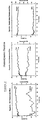
- liver damage was assessed by measuring the level of the liver enzymes alanine transferase (ALT) and alkaline phosphatase (Alk. Phos.). At the end of 72 hours of perfusion the ALT level had changed from 62.4 units/L (s.d. 10.1) to 51.4 units/L (s.d 8.9) ( Figure 5A). Alkaline phosphatase levels fell during the period of perfusion from 136.5 units/litre (s.d. 17.0) to 70.2 units/litre (s.d 12.9) at 72 hours. Bilirubin levels remained low but rose during the perfusion, changing from 0.4 mmol/L (s.d. 0.8) to 32 mmol/L (s.d. 28.2) ( Figure 5B).
- ALT alanine transferase
- Alkaline phosphatase levels fell during the period of perfusion from 136.5 units/litre (s.d. 17.0) to 70.2 units/litre (s.d 12.9) at 72 hours. Bilirubin levels remained low
- the technique employed for extracorporeal liver perfusion in accordance with the present invention was able to maintain good function and structure for at least 72 hours in the isolated perfused organ. Isolated liver perfusion has not previously been described for periods longer than 24 hours (Neuhaus et al., Int. J. Artif. Organs, 1993; 16: 729-739). Previous investigators usually terminated perfusion as a result of progressive acidosis, hyperkalaemia and rising portal pressures. For this reason the invention represents a significant advance in liver perfusion.
- oxygen consumption might reasonably be assumed to be a measure of hepatocyte function, this fell consistently during the course of perfusion. This is in contrast to other markers of liver function, particularly synthetic activity.
- the initial values obtained for oxygen consumption correlate well with figures derived in vivo by a number of authors (Mathisen and Omland, Scand J of Gastroent 25: 1265-1273, 1990; Rasmussen et al., Eur J Surg 159: 201-7, 1993; Lindberg and Clowes, Jr., J Surg Research 31: 156-64, 1981; Imamura, Surgery, Gynaecology and Obstetrics 141: 27, 1975; Tygstrup et al., Scan J Gastroent Suppl 9: 131-138, 1971.
- Bile production in this extracorporeal perfusion system peaked at 12.8 ml/hour approximately half of that observed in vivo. Bile production decreased with time and inspissated bile was seen macroscopically in all livers and histologically in four out of five experiments. This may reflect consumption of substrate (including bile salts), loss of hormonal or neural stimuli and/or inadequate biliary drainage with accumulation of biliary sludge (although alkaline phosphatase did not rise). Serum bilirubin levels remained low for the first 24 hours of perfusion and started to rise concomitantly with the fall in bile production.
- Enzymatic markers of hepatocyte damage did not rise during perfusion. Using the same assay, these enzymes are substantially elevated in a porcine model of acute hepatic failure used by our group. This suggests that there is good preservation of hepatocyte viability in spite of biliary obstruction and rising bilirubin.
- Figures 4A, 4B and 4C show evidence of continued synthetic function throughout the 72 hour perfusion. It is of interest to note that complement production was apparently not regulated by a feedback mechanism in the liver, but appeared to continue at a constant rate accumulating in the plasma of the isolated circuit ( Figure 4B). The mechanisms that regulate complement production by the liver remain poorly understood. In contrast Factor V activity remained at a relatively constant level throughout the 72 hour perfusion period. The significance of the different patterns seen in the balance of synthesis and consumption for these two proteins is unclear.
- prostacyclin was used routinely.
- the role of prostacyclin has not been rigorously investigated, we believe its use is desirable in liver perfusion. It is unclear whether it is the vasodilator or anti-platelet activity of prostacyclin that provides the beneficial effects in this situation.
- an agent which mimics one or other or both of these activities may be useful in place of prostacyclin.
- Example 1 The perfusion apparatus and technique employed in Example 1 was used to perfuse porcine livers transgenic for the complement regulator protein, human decay accelerating factor (hDAF), when perfused with fresh, whole, human blood.
- hDAF human decay accelerating factor
- Alloperfusion resulted in the maintenance of good function and histological structure for periods of at least 72 hours. Xenoperfusions were also carried out for periods of up to 72 hours but, unlike alloperfusions, were marked by a progressive decrease in haematocrit of the circulating blood. Histological examination demonstrated patchy necrosis but most lobules were normal. Effective liver function was demonstrated in both normal and transgenic liver perfusions.
- Donor pig livers were prepared as in Example 1. Human blood was obtained from volunteer donors and used within 4 hours of donation.
- the perfusion circuit ( Figure 1) was employed as in Example 1.
- Perfusion was stopped electively at 72 hours or earlier if liver failure was deemed to have occurred - pH ⁇ 7.0, rapidly rising potassium or increasing portal pressure. At this time multiple samples were taken from the liver both for immunohistochemical evaluation and hematoxylin and eosin staining.
- Serum samples were analysed for complement production by CH50 analysis.
- Factor V was measured using an assay based upon prothrombin time (Organon Teknika MD180). Prothrombin time was also measured. Electrolytes, biochemical liver function tests, creatinine, and urea were measured using standard clinical methods. Full blood count analysis was performed using a Coulter counter (T660) adjusted for measurement of porcine blood.
- Oxygen consumption (ml/hour) for the three groups is outlined in Table 3 below.
- Bile production (ml/hour) in the three groups is outlined in Figure 7.
- necrosis was seen in all livers. In three livers necrosis was patchy involving 10-30% of the livers, in one the necrosis was subcapsular involving 5% of the liver and in one there was diffuse necrosis involving 80% of the liver. No consistent pattern of liver damage could be determined. Because of the significant necrosis in one of the livers, further histologic analysis was not possible. In the remaining four livers, sinusoidal dilatation and was seen in 3, central dilatation in 2, and septal edema in one. Inspissated bile was present in the intralobular bile ducts of all four livers. Endothelitis was present in one of the four livers. Haemorrhage was present in the lymphatics of one of the livers. Kupffer cell hypertrophy was present in all livers with significant Perl's staining of intracellular iron.
- haemorrhage was seen in all livers, but necrosis outside of the haemorrhagic areas was not apparent.
- the haemorrhage involved the hepatic parenchyma in all livers, involving septal and sinusoidal haemorrhage in four livers while being more prominent in the subcapsular region in one liver. All livers demonstrated sinusoidal dilatation with retained red cells within sinusoids, 2 livers had evidence of central vein dilatation, and four of five livers had septal edema. None of the livers had inspissated bile. One liver had mild endothelitis. There was very mild Kupffer cell hypertrophy and minimal Perl's staining of intracellular iron in 4 of five livers.
- Example 2 The experiments described in Example 2 demonstrate that the invention is effective not only in alloperfusion (as described in Example 1) but also in more demanding xenoperfusion.
- hyperacute rejection requires definition; the histological appearances of hyperacute rejection overlap with those of acute vascular rejection (Platt, ASAIO.J. 1992; 38: 8-16).
- diagnosis requires not only characteristic histological features (microthrombosis and haemorrhage) but also the absence of organ function at any time.
- Hyperacute rejection was not observed in either transgenic or non-transgenic xenoperfusions in this study. This protection from hyperacute rejection may be a function of the liver itself or of the non-physiological nature of the perfusion circuit, particularly the limited volume of blood (and immunological effector components) or the use of heparin. In this context it is of note that hyperacute rejection has been demonstrated in a heparinised ex vivo xenoperfusion model of the heart (Dunning et al., Eur. J. Cardiothorac. Surg. 1994; 8: 204-206).
- transgenic livers behaved similarly to the alloperfused livers
- analysis of other parameters demonstrated similar behaviour between both xenoperfused groups and significant differences from the alloperfused group.
- these include some haemodynamic parameters, total and portal flows, potassium levels, bile production and bilirubin levels, transaminase levels and terminal oxygen consumption.
- Analysis of bile production and bilirubin levels may be complicated by the considerable differences in red cell breakdown and suspected complications of biliary sludge formation. Also, depletion of bile salts may be a further factor in bile production. Similarly, potassium levels may reflect differences in red cell breakdown.
- the reticuloendothelial system in the liver might extract red blood cells either by recognition of species-specific differences or pump related erythrocyte damage.
- the significant difference in terminal haematocrits between the transgenic and non-transgenic groups is not explained but may be related to improved microcirculation in the transgenic livers.
- liver function as assessed by those parameters not directly affected by red cell breakdown, is maintained to the same level in xenoperfusions (particularly transgenic) as in alloperfusions. In particular this is illustrated by the synthesis of Factor V and haemodynamic parameters. The measurement of potassium, hydrogen ions, bilirubin and urea are directly affected by red cell breakdown. Also, complement levels represent a balance between production and consumption; a higher level of consumption is inevitable in xenoperfusions.
- perfused livers provide metabolic function and are capable of normalisation of potassium and acid-base balance; these are cardinal features of good liver function following liver transplantation.
- the synthetic functions of the liver although potentially of some benefit, may be problematical.
- the synthesis of high levels of porcine complement may lead to damage of human tissues that are not protected by appropriate species specific complement regulators.
- serum from xenoperfusions does not cause lysis of fresh human red cells in vitro .
- porcine proteins There is potential incompatibility between porcine proteins and the human environment (Lawson et al., Transplant. Proc. 1997; 29: 884-885; Reverdiau-Moalic et al., Transplant. Proc. 1996; 28: 643-644).
- Acute liver failure was induced in eight non-transgenic pigs by pre-treatment with oral phenobarbitone for 3 days, instillation of intra-gastric carbon tetrachloride and ligation of the hepatic artery.
- liver function urea and electrolytes, liver functions test, coagulation profile, arterial blood gases and serum ammonia
- liver failure was confirmed from post-mortem liver biopsies.
- the four pigs treated with extracorporeal liver support survived for 31, 33, 35 and 52 hours (37.8 ⁇ 9.6 hours), significantly longer than the untreated pigs (p ⁇ 0.005).
- the oxygenator failed after 24 hours in the first two perfusions.
- ventilatory failure secondary to mucus plugging in the pigs necessitated discontinuation of liver perfusion after 22 and 37 hours of perfusion.
- the pigs were moribund, with extreme tachycardia (160 ⁇ 0 beats per minute), hypotension (80/42.5 ⁇ 0/3.5 mmHg) and metabolic acidosis (mean pH 7.20 ⁇ 0.12). After one hour of extracorporeal liver perfusion, the acidosis was reversed, returning to a physiological range (pH 7.36 ⁇ 0.08), and the tachycardia improved (127.5 ⁇ 3.5 beats per minute).
Applications Claiming Priority (3)
| Application Number | Priority Date | Filing Date | Title |
|---|---|---|---|
| GB9908335 | 1999-04-12 | ||
| GBGB9908335.4A GB9908335D0 (en) | 1999-04-12 | 1999-04-12 | Methods and means for extracorporeal organ perfusion |
| PCT/GB2000/001271 WO2000060936A1 (en) | 1999-04-12 | 2000-04-05 | Methods and means for extracorporeal organ perfusion |
Publications (2)
| Publication Number | Publication Date |
|---|---|
| EP1168913A1 EP1168913A1 (en) | 2002-01-09 |
| EP1168913B1 true EP1168913B1 (en) | 2005-11-02 |
Family
ID=10851385
Family Applications (1)
| Application Number | Title | Priority Date | Filing Date |
|---|---|---|---|
| EP00915294A Expired - Lifetime EP1168913B1 (en) | 1999-04-12 | 2000-04-05 | Methods and means for extracorporeal organ perfusion |
Country Status (9)
| Country | Link |
|---|---|
| US (1) | US7410474B1 (es) |
| EP (1) | EP1168913B1 (es) |
| AT (1) | ATE308239T1 (es) |
| AU (1) | AU3665800A (es) |
| CA (1) | CA2369159C (es) |
| DE (1) | DE60023672T2 (es) |
| ES (1) | ES2250119T3 (es) |
| GB (1) | GB9908335D0 (es) |
| WO (1) | WO2000060936A1 (es) |
Cited By (3)
| Publication number | Priority date | Publication date | Assignee | Title |
|---|---|---|---|---|
| WO2013068751A3 (en) * | 2011-11-10 | 2013-08-29 | Organox Limited | Organ perfusion systems |
| RU2556970C2 (ru) * | 2009-11-05 | 2015-07-20 | Сантр Насьональ Де Ля Решерш Сьянтифик | Устройство для отбора пробы физиологической жидкости и способ его использования |
| US11957124B2 (en) | 2011-11-10 | 2024-04-16 | Organox Limited | Organ perfusion systems |
Families Citing this family (58)
| Publication number | Priority date | Publication date | Assignee | Title |
|---|---|---|---|---|
| US8409846B2 (en) | 1997-09-23 | 2013-04-02 | The United States Of America As Represented By The Department Of Veteran Affairs | Compositions, methods and devices for maintaining an organ |
| US6977140B1 (en) | 1998-09-29 | 2005-12-20 | Organ Recovery Systems, Inc. | Method for maintaining and/or restoring viability of organs |
| US6673594B1 (en) | 1998-09-29 | 2004-01-06 | Organ Recovery Systems | Apparatus and method for maintaining and/or restoring viability of organs |
| US7572622B2 (en) | 2002-08-14 | 2009-08-11 | Transmedic, Inc. | Heart preservation chamber |
| NL1024022C2 (nl) | 2003-07-30 | 2005-02-01 | Technologiestichting Stw | Draagbare preservatie-inrichting voor een donororgaan. |
| DE10340487B4 (de) * | 2003-09-03 | 2007-07-12 | Technische Universität Dresden | Perfusionskreislauf |
| US8741555B2 (en) * | 2004-05-14 | 2014-06-03 | Organ Recovery Systems, Inc. | Apparatus and method for perfusion and determining the viability of an organ |
| EP1768490B1 (en) | 2004-10-07 | 2010-06-09 | Transmedics, Inc. | Systems and methods for ex-vivo organ care |
| US9301519B2 (en) | 2004-10-07 | 2016-04-05 | Transmedics, Inc. | Systems and methods for ex-vivo organ care |
| US8304181B2 (en) | 2004-10-07 | 2012-11-06 | Transmedics, Inc. | Method for ex-vivo organ care and for using lactate as an indication of donor organ status |
| US9078428B2 (en) | 2005-06-28 | 2015-07-14 | Transmedics, Inc. | Systems, methods, compositions and solutions for perfusing an organ |
| US10176887B1 (en) | 2005-11-14 | 2019-01-08 | Organ Recovery Systems, Inc. | Ex vivo methods for drug discovery, development and testing |
| CN101677526B (zh) | 2007-03-01 | 2014-07-09 | 生命线科学有限公司 | 灌注调节 |
| US9457179B2 (en) | 2007-03-20 | 2016-10-04 | Transmedics, Inc. | Systems for monitoring and applying electrical currents in an organ perfusion system |
| US8771930B2 (en) * | 2007-05-18 | 2014-07-08 | Lifeline Scientific, Inc. | Ex vivo methods for testing toxicity of substances using donated human organs or tissues |
| US8765364B2 (en) * | 2007-05-18 | 2014-07-01 | Lifeline Scientific, Inc. | Ex vivo methods for validating substance testing with human organs and/or tissues |
| US10750738B2 (en) | 2008-01-31 | 2020-08-25 | Transmedics, Inc. | Systems and methods for ex vivo lung care |
| WO2009132018A1 (en) * | 2008-04-22 | 2009-10-29 | The Board Of Regents Of The University Of Texas System | Fluidics-based pulsatile perfusion organ preservation device |
| WO2011002926A2 (en) | 2009-07-01 | 2011-01-06 | The General Hospital Corporation | Isolated adult cells, artificial organs,rehabilitated organs, research rools, organ encasements, organ perfusion systems, and methods for preparing and utilizing the same |
| EP2480069A1 (en) * | 2009-09-25 | 2012-08-01 | Board of Regents, The University of Texas System | Fluidics based pulsatile perfusion preservation device and method |
| WO2011140241A2 (en) | 2010-05-04 | 2011-11-10 | The General Hospital Corporation | Methods and compositions for preserving tissues and organs |
| CA2830225C (en) | 2011-03-15 | 2020-03-24 | Paragonix Technologies, Inc. | Apparatus for oxygenation and perfusion of tissue for organ preservation |
| US9426979B2 (en) | 2011-03-15 | 2016-08-30 | Paragonix Technologies, Inc. | Apparatus for oxygenation and perfusion of tissue for organ preservation |
| US11178866B2 (en) | 2011-03-15 | 2021-11-23 | Paragonix Technologies, Inc. | System for hypothermic transport of samples |
| US9867368B2 (en) | 2011-03-15 | 2018-01-16 | Paragonix Technologies, Inc. | System for hypothermic transport of samples |
| US8828710B2 (en) | 2011-03-15 | 2014-09-09 | Paragonix Technologies, Inc. | System for hypothermic transport of samples |
| US9253976B2 (en) | 2011-03-15 | 2016-02-09 | Paragonix Technologies, Inc. | Methods and devices for preserving tissues |
| JP6029650B2 (ja) | 2011-04-14 | 2016-11-24 | トランスメディクス,インコーポレイテッド | ドナー肺のEx−vivoにおける機械的灌流のための臓器保護溶液 |
| CA2847368C (en) | 2011-09-02 | 2017-10-31 | Organ Assist B.V. | Apparatus, system and method for conditioning and preserving an organ from a donor |
| GB201119420D0 (en) | 2011-11-10 | 2011-12-21 | Organox Ltd | Oxygen supply for organ perfusion systems |
| EP2633755A1 (en) | 2012-03-02 | 2013-09-04 | Rodos BioTarget GmbH | Organ perfusion system |
| US10602740B2 (en) * | 2012-07-10 | 2020-03-31 | Lifeline Scientific, Inc. | Organ perfusion apparatus with downstream flow control |
| US8785116B2 (en) | 2012-08-10 | 2014-07-22 | Paragonix Technologies, Inc. | Methods for evaluating the suitability of an organ for transplant |
| US9560846B2 (en) | 2012-08-10 | 2017-02-07 | Paragonix Technologies, Inc. | System for hypothermic transport of biological samples |
| CN102813559B (zh) * | 2012-08-27 | 2014-11-05 | 山东大学 | 一种全自动灌流装置 |
| EP2893806B1 (en) * | 2012-09-08 | 2019-04-17 | Organ Technologies, Inc. | Method for maintaining organ or tissue for transplantation use for long period |
| US9629358B2 (en) * | 2013-03-15 | 2017-04-25 | Mallinckrodt Hospital Products IP Limited | Administration and monitoring of nitric oxide in ex vivo fluids |
| US10918102B2 (en) * | 2014-03-13 | 2021-02-16 | The General Hospital Corporation | Devices and methods to improve and assess viability of human livers |
| IL303658B1 (en) | 2014-06-02 | 2024-03-01 | Transmedics Inc | Extracorporeal system for organ treatment |
| USD765874S1 (en) | 2014-10-10 | 2016-09-06 | Paragonix Technologies, Inc. | Transporter for a tissue transport system |
| CN106998676B (zh) | 2014-12-12 | 2021-10-26 | 体沃索股份有限公司 | 用于器官灌注的装置和方法 |
| JP6934005B2 (ja) | 2015-09-09 | 2021-09-08 | トランスメディクス,インコーポレイテッド | エキソビボ臓器管理システムのための大動脈カニューレ |
| US10091986B2 (en) | 2016-05-09 | 2018-10-09 | Xor-Labs Toronto Inc. | Organ perfusion device and method |
| JPWO2017200089A1 (ja) * | 2016-05-20 | 2019-03-14 | 株式会社Screenホールディングス | 移植用の肝臓の灌流装置、および、当該装置を用いた肝臓摘出方法および肝臓移植方法 |
| CN109688811B (zh) | 2016-07-22 | 2022-06-03 | 苏黎世联邦理工学院 | 用于离体肝灌注的灌注回路组件和肝腔室组件 |
| EP3504966A4 (en) * | 2016-08-25 | 2020-05-06 | SCREEN Holdings Co., Ltd. | LIVER PRESERVATION METHOD AND OPERATING METHOD |
| CN106937671B (zh) * | 2017-04-10 | 2018-05-29 | 青岛大学 | 一种新鲜动物肝脏的清洗、祛毒以及细胞组织保鲜的方法 |
| CN106962804B (zh) * | 2017-04-10 | 2018-08-03 | 青岛大学 | 祛毒动物肝脏食品及其加工方法 |
| CA3066625A1 (en) | 2017-06-07 | 2018-12-13 | Paragonix Technologies, Inc. | Apparatus for tissue transport and preservation |
| US11044904B2 (en) | 2017-08-25 | 2021-06-29 | Mallinckrodt Hospital Products IP Limited | Methods to improve organ viability |
| US20200375178A1 (en) | 2018-01-19 | 2020-12-03 | Eth Zurich | Perfusion Loop Assembly for an Ex-Vivo Liver Perfusion and a Method for Ex-Vivo Liver Perfusion |
| EP3513653A1 (en) | 2018-01-19 | 2019-07-24 | ETH Zürich | Perfusion loop assembly for an ex-vivo liver perfusion and a method for ex-vivo liver perfusion |
| US11632951B2 (en) | 2020-01-31 | 2023-04-25 | Paragonix Technologies, Inc. | Apparatus for tissue transport and preservation |
| JP2021143128A (ja) * | 2020-03-10 | 2021-09-24 | 株式会社Screenホールディングス | 灌流装置 |
| RU2741219C1 (ru) * | 2020-06-18 | 2021-01-22 | федеральное государственное бюджетное образовательное учреждение высшего образования «Омский государственный медицинский университет» Министерства здравоохранения Российской Федерации (ФГБОУ ВО ОмГМУ Минздрава России) | Устройство для консервации донорских органов |
| CN112155841B (zh) * | 2020-09-04 | 2022-03-22 | 深圳市医思美科技有限公司 | 一种热灌注治疗设备的控制系统和控制方法 |
| GB202018460D0 (en) | 2020-11-24 | 2021-01-06 | Organox Ltd | Precision oxygen flow control for organ perfusion systems |
| JP2024029908A (ja) * | 2022-08-23 | 2024-03-07 | 株式会社Screenホールディングス | 接続配管、灌流装置の準備方法、および灌流方法 |
Family Cites Families (9)
| Publication number | Priority date | Publication date | Assignee | Title |
|---|---|---|---|---|
| US4666425A (en) * | 1985-12-17 | 1987-05-19 | The Dis Corporation | Device for perfusing an animal head |
| CA2001553A1 (en) * | 1988-10-26 | 1990-04-26 | Karen Mckelvey | Device for transportation of human organs used for transplantation |
| US5338662A (en) * | 1992-09-21 | 1994-08-16 | Bio-Preserve Medical Corporation | Organ perfusion device |
| US6331658B1 (en) * | 1993-04-20 | 2001-12-18 | Integris Baptist Medical Center, Inc. | Genetically engineered mammals for use as organ donors |
| DE9422008U1 (de) * | 1993-06-07 | 1997-08-28 | Mayer Berndt Priv Doz Dr Med | Vorrichtung zur Konservierung von Organen, Extremitäten und Gewebelappen |
| US5807737A (en) * | 1996-07-26 | 1998-09-15 | Schill Enterprises, Inc. | Heart and lung support assembly |
| US6642045B1 (en) * | 1997-04-14 | 2003-11-04 | Breonics, Inc. | System for exsanguinous metabolic support of an organ or tissue |
| DK1017274T3 (da) * | 1997-09-23 | 2004-03-22 | Waleed H Hassanein | Sammensætninger, fremgangsmåder og anordninger til opretholdelse af et organ |
| US6100082A (en) * | 1997-09-23 | 2000-08-08 | Hassanein; Waleed H. | Perfusion apparatus and method including chemical compositions for maintaining an organ |
-
1999
- 1999-04-12 GB GBGB9908335.4A patent/GB9908335D0/en not_active Ceased
-
2000
- 2000-04-05 CA CA2369159A patent/CA2369159C/en not_active Expired - Lifetime
- 2000-04-05 DE DE60023672T patent/DE60023672T2/de not_active Expired - Lifetime
- 2000-04-05 ES ES00915294T patent/ES2250119T3/es not_active Expired - Lifetime
- 2000-04-05 WO PCT/GB2000/001271 patent/WO2000060936A1/en active IP Right Grant
- 2000-04-05 AT AT00915294T patent/ATE308239T1/de not_active IP Right Cessation
- 2000-04-05 EP EP00915294A patent/EP1168913B1/en not_active Expired - Lifetime
- 2000-04-05 AU AU36658/00A patent/AU3665800A/en not_active Abandoned
- 2000-04-12 US US09/958,681 patent/US7410474B1/en not_active Expired - Lifetime
Cited By (8)
| Publication number | Priority date | Publication date | Assignee | Title |
|---|---|---|---|---|
| RU2556970C2 (ru) * | 2009-11-05 | 2015-07-20 | Сантр Насьональ Де Ля Решерш Сьянтифик | Устройство для отбора пробы физиологической жидкости и способ его использования |
| WO2013068751A3 (en) * | 2011-11-10 | 2013-08-29 | Organox Limited | Organ perfusion systems |
| GB2510080A (en) * | 2011-11-10 | 2014-07-23 | Organox Ltd | Organ perfusion systems |
| CN104039135A (zh) * | 2011-11-10 | 2014-09-10 | 奥加诺克斯有限责任公司 | 器官灌注系统 |
| CN104039135B (zh) * | 2011-11-10 | 2016-01-20 | 奥加诺克斯有限责任公司 | 器官灌注系统 |
| GB2510080B (en) * | 2011-11-10 | 2017-09-27 | Organox Ltd | Organ perfusion systems |
| US11540508B2 (en) | 2011-11-10 | 2023-01-03 | Organox Limited | Organ perfusion systems |
| US11957124B2 (en) | 2011-11-10 | 2024-04-16 | Organox Limited | Organ perfusion systems |
Also Published As
| Publication number | Publication date |
|---|---|
| AU3665800A (en) | 2000-11-14 |
| US7410474B1 (en) | 2008-08-12 |
| DE60023672T2 (de) | 2006-07-20 |
| WO2000060936A1 (en) | 2000-10-19 |
| ATE308239T1 (de) | 2005-11-15 |
| ES2250119T3 (es) | 2006-04-16 |
| EP1168913A1 (en) | 2002-01-09 |
| DE60023672D1 (de) | 2005-12-08 |
| CA2369159C (en) | 2011-11-29 |
| GB9908335D0 (en) | 1999-06-09 |
| CA2369159A1 (en) | 2000-10-19 |
Similar Documents
| Publication | Publication Date | Title |
|---|---|---|
| EP1168913B1 (en) | Methods and means for extracorporeal organ perfusion | |
| Butler et al. | Successful extracorporeal porcine liver perfusion for 72 hr1 | |
| JP6347565B2 (ja) | 臓器を維持するための組成物、方法及び装置 | |
| EP1339279B2 (en) | Evaluation and preservation solution | |
| US10433538B2 (en) | Compositions and methods for organ preservation | |
| US20190141988A1 (en) | Perfusion device for liver graft, and liver removal method and liver transplantation method using the device | |
| KR100855354B1 (ko) | 관류세척 보존 용액 | |
| JP2001516768A (ja) | 臓器を維持するための組成物、方法及び装置 | |
| US20220248665A1 (en) | Perfusate and perfusion method | |
| AU2004201245B2 (en) | Methods and means for extracorporeal organ perfusion | |
| Szajer et al. | A novel extracorporeal kidney perfusion system: a concept model | |
| Uygun | Methods in Bioengineering: Organ preservation and reengineering | |
| Knaak et al. | Technique of Subnormothermic Ex Vivo |
Legal Events
| Date | Code | Title | Description |
|---|---|---|---|
| PUAI | Public reference made under article 153(3) epc to a published international application that has entered the european phase |
Free format text: ORIGINAL CODE: 0009012 |
|
| 17P | Request for examination filed |
Effective date: 20011016 |
|
| AK | Designated contracting states |
Kind code of ref document: A1 Designated state(s): AT BE CH CY DE DK ES FI FR GB GR IE IT LI LU MC NL PT SE |
|
| AX | Request for extension of the european patent |
Free format text: AL;LT;LV;MK;RO;SI |
|
| 17Q | First examination report despatched |
Effective date: 20020307 |
|
| RAP1 | Party data changed (applicant data changed or rights of an application transferred) |
Owner name: ISIS INNOVATION LIMITED |
|
| GRAP | Despatch of communication of intention to grant a patent |
Free format text: ORIGINAL CODE: EPIDOSNIGR1 |
|
| GRAS | Grant fee paid |
Free format text: ORIGINAL CODE: EPIDOSNIGR3 |
|
| GRAA | (expected) grant |
Free format text: ORIGINAL CODE: 0009210 |
|
| AK | Designated contracting states |
Kind code of ref document: B1 Designated state(s): AT BE CH CY DE DK ES FI FR GB GR IE IT LI LU MC NL PT SE |
|
| PG25 | Lapsed in a contracting state [announced via postgrant information from national office to epo] |
Ref country code: NL Free format text: LAPSE BECAUSE OF FAILURE TO SUBMIT A TRANSLATION OF THE DESCRIPTION OR TO PAY THE FEE WITHIN THE PRESCRIBED TIME-LIMIT Effective date: 20051102 Ref country code: FI Free format text: LAPSE BECAUSE OF FAILURE TO SUBMIT A TRANSLATION OF THE DESCRIPTION OR TO PAY THE FEE WITHIN THE PRESCRIBED TIME-LIMIT Effective date: 20051102 Ref country code: AT Free format text: LAPSE BECAUSE OF FAILURE TO SUBMIT A TRANSLATION OF THE DESCRIPTION OR TO PAY THE FEE WITHIN THE PRESCRIBED TIME-LIMIT Effective date: 20051102 |
|
| REG | Reference to a national code |
Ref country code: GB Ref legal event code: FG4D |
|
| REG | Reference to a national code |
Ref country code: CH Ref legal event code: EP |
|
| REF | Corresponds to: |
Ref document number: 60023672 Country of ref document: DE Date of ref document: 20051208 Kind code of ref document: P |
|
| PG25 | Lapsed in a contracting state [announced via postgrant information from national office to epo] |
Ref country code: GR Free format text: LAPSE BECAUSE OF FAILURE TO SUBMIT A TRANSLATION OF THE DESCRIPTION OR TO PAY THE FEE WITHIN THE PRESCRIBED TIME-LIMIT Effective date: 20060202 Ref country code: DK Free format text: LAPSE BECAUSE OF FAILURE TO SUBMIT A TRANSLATION OF THE DESCRIPTION OR TO PAY THE FEE WITHIN THE PRESCRIBED TIME-LIMIT Effective date: 20060202 |
|
| REG | Reference to a national code |
Ref country code: SE Ref legal event code: TRGR |
|
| PG25 | Lapsed in a contracting state [announced via postgrant information from national office to epo] |
Ref country code: PT Free format text: LAPSE BECAUSE OF FAILURE TO SUBMIT A TRANSLATION OF THE DESCRIPTION OR TO PAY THE FEE WITHIN THE PRESCRIBED TIME-LIMIT Effective date: 20060403 |
|
| REG | Reference to a national code |
Ref country code: ES Ref legal event code: FG2A Ref document number: 2250119 Country of ref document: ES Kind code of ref document: T3 |
|
| PG25 | Lapsed in a contracting state [announced via postgrant information from national office to epo] |
Ref country code: MC Free format text: LAPSE BECAUSE OF NON-PAYMENT OF DUE FEES Effective date: 20060430 |
|
| NLV1 | Nl: lapsed or annulled due to failure to fulfill the requirements of art. 29p and 29m of the patents act | ||
| ET | Fr: translation filed | ||
| PLBE | No opposition filed within time limit |
Free format text: ORIGINAL CODE: 0009261 |
|
| STAA | Information on the status of an ep patent application or granted ep patent |
Free format text: STATUS: NO OPPOSITION FILED WITHIN TIME LIMIT |
|
| 26N | No opposition filed |
Effective date: 20060803 |
|
| PG25 | Lapsed in a contracting state [announced via postgrant information from national office to epo] |
Ref country code: LU Free format text: LAPSE BECAUSE OF NON-PAYMENT OF DUE FEES Effective date: 20060405 |
|
| PG25 | Lapsed in a contracting state [announced via postgrant information from national office to epo] |
Ref country code: CY Free format text: LAPSE BECAUSE OF FAILURE TO SUBMIT A TRANSLATION OF THE DESCRIPTION OR TO PAY THE FEE WITHIN THE PRESCRIBED TIME-LIMIT Effective date: 20051102 |
|
| REG | Reference to a national code |
Ref country code: GB Ref legal event code: 732E Free format text: REGISTERED BETWEEN 20090212 AND 20090218 |
|
| REG | Reference to a national code |
Ref country code: CH Ref legal event code: PLI Owner name: ORGANOX LIMITED Free format text: ISIS INNOVATION LIMITED#UNIVERSITY OFFICES, WELLINGTON SQUARE#OXFORD OX1 2JD (GB) -TRANSFER TO- ORGANOX LIMITED#9400 GARSINGTON ROAD OXFORD BUSINESS PARK#OXFORD OX4 2HN (GB) |
|
| REG | Reference to a national code |
Spc suppl protection certif: 98C0033 Ref country code: FR Ref legal event code: CL |
|
| REG | Reference to a national code |
Ref country code: ES Ref legal event code: GD2A Effective date: 20100329 |
|
| REG | Reference to a national code |
Ref country code: FR Ref legal event code: PLFP Year of fee payment: 17 |
|
| REG | Reference to a national code |
Ref country code: CH Ref legal event code: PFA Owner name: OXFORD UNIVERSITY INNOVATION LIMITED, GB Free format text: FORMER OWNER: ISIS INNOVATION LIMITED, GB |
|
| REG | Reference to a national code |
Ref country code: DE Ref legal event code: R082 Ref document number: 60023672 Country of ref document: DE Representative=s name: LEINWEBER & ZIMMERMANN, DE Ref country code: DE Ref legal event code: R081 Ref document number: 60023672 Country of ref document: DE Owner name: OXFORD UNIVERSITY INNOVATION LIMITED, GB Free format text: FORMER OWNER: ISIS INNOVATION LTD., OXFORD, GB |
|
| REG | Reference to a national code |
Ref country code: ES Ref legal event code: PC2A Owner name: OXFORD UNIVERSITY INNOVATION LIMITED Effective date: 20170119 |
|
| REG | Reference to a national code |
Ref country code: FR Ref legal event code: PLFP Year of fee payment: 18 |
|
| REG | Reference to a national code |
Ref country code: FR Ref legal event code: CA Effective date: 20170710 Ref country code: FR Ref legal event code: CD Owner name: OXFORD UNIVERSITY INNOVATION LIMITED, GB Effective date: 20170710 |
|
| REG | Reference to a national code |
Ref country code: FR Ref legal event code: PLFP Year of fee payment: 19 |
|
| PGFP | Annual fee paid to national office [announced via postgrant information from national office to epo] |
Ref country code: IE Payment date: 20190425 Year of fee payment: 20 Ref country code: ES Payment date: 20190520 Year of fee payment: 20 Ref country code: DE Payment date: 20190429 Year of fee payment: 20 Ref country code: IT Payment date: 20190426 Year of fee payment: 20 |
|
| PGFP | Annual fee paid to national office [announced via postgrant information from national office to epo] |
Ref country code: FR Payment date: 20190423 Year of fee payment: 20 Ref country code: BE Payment date: 20190418 Year of fee payment: 20 Ref country code: SE Payment date: 20190425 Year of fee payment: 20 |
|
| PGFP | Annual fee paid to national office [announced via postgrant information from national office to epo] |
Ref country code: CH Payment date: 20190430 Year of fee payment: 20 |
|
| PGFP | Annual fee paid to national office [announced via postgrant information from national office to epo] |
Ref country code: GB Payment date: 20190424 Year of fee payment: 20 |
|
| REG | Reference to a national code |
Ref country code: DE Ref legal event code: R071 Ref document number: 60023672 Country of ref document: DE |
|
| REG | Reference to a national code |
Ref country code: CH Ref legal event code: PL |
|
| REG | Reference to a national code |
Ref country code: GB Ref legal event code: PE20 Expiry date: 20200404 |
|
| REG | Reference to a national code |
Ref country code: IE Ref legal event code: MK9A |
|
| PG25 | Lapsed in a contracting state [announced via postgrant information from national office to epo] |
Ref country code: IE Free format text: LAPSE BECAUSE OF EXPIRATION OF PROTECTION Effective date: 20200405 |
|
| PG25 | Lapsed in a contracting state [announced via postgrant information from national office to epo] |
Ref country code: GB Free format text: LAPSE BECAUSE OF EXPIRATION OF PROTECTION Effective date: 20200404 |
|
| REG | Reference to a national code |
Ref country code: ES Ref legal event code: FD2A Effective date: 20210129 |
|
| PG25 | Lapsed in a contracting state [announced via postgrant information from national office to epo] |
Ref country code: ES Free format text: LAPSE BECAUSE OF EXPIRATION OF PROTECTION Effective date: 20200406 |