EP1142991A2 - Monoklonale Antikörper gegen Isozyme der Thymidinkinase - Google Patents
Monoklonale Antikörper gegen Isozyme der Thymidinkinase Download PDFInfo
- Publication number
- EP1142991A2 EP1142991A2 EP01201917A EP01201917A EP1142991A2 EP 1142991 A2 EP1142991 A2 EP 1142991A2 EP 01201917 A EP01201917 A EP 01201917A EP 01201917 A EP01201917 A EP 01201917A EP 1142991 A2 EP1142991 A2 EP 1142991A2
- Authority
- EP
- European Patent Office
- Prior art keywords
- activity
- sample
- thymidine kinase
- antibody
- monoclonal antibody
- Prior art date
- Legal status (The legal status is an assumption and is not a legal conclusion. Google has not performed a legal analysis and makes no representation as to the accuracy of the status listed.)
- Withdrawn
Links
Images

Classifications
-
- C—CHEMISTRY; METALLURGY
- C07—ORGANIC CHEMISTRY
- C07K—PEPTIDES
- C07K16/00—Immunoglobulins [IG], e.g. monoclonal or polyclonal antibodies
- C07K16/40—Immunoglobulins [IG], e.g. monoclonal or polyclonal antibodies against enzymes
Definitions
- the invention relates generally to a monoclonal antibody useful for diagnosis, prognosis, and treatment assessment of cancer and certain blood disorders, and to prediction of recurrence in cancer patients.
- tumor recurrences are detected very early, they may in some cases be successfully treated to produce an ultimate cure or to significantly prolong the life of the patient. Or, if a tumor is not responding well to a particular drug or treatment, others can be tried.
- estrogen receptor status For breast cancer, it is presently believed that one of the best predictors of the likelihood of recurrence is estrogen receptor status. Those patients that are estrogen receptor positive (high numbers of estrogen receptors on the tumor cells) are thought to have a better prognosis, while those that are estrogen receptor negative are thought to have a poor prognosis. However, there are many cases where those who are estrogen receptor negative do not show recurrence, and those that are estrogen receptor positive do show recurrence. Because the estrogen receptor test is not sufficiently reliable as a prognostic indicator, patients are often treated with both radiotherapy and chemotherapy, regardless of estrogen receptor status.
- Thymidine kinase (ATP:thymidine-5' phosphotransferase; EC 2.7.1.21 in the International Union of Biochemistry classification system) is an enzyme that phosphorylates thymidine to thymidine monophosphate (TMP).
- TMP thymidine monophosphate
- TK thymidine kinase in a general sense, including different isozymes and multimeric forms presently believed to exist in vivo.
- Thymidine kinase protein has been isolated from many different sources and purified to varying degrees. A variety of different molecular weight thymidine kinases have been reported from human samples, depending on the particular cell and the method of isolation and analysis (for example, under denaturing conditions vs. non-denaturing conditions). In general, the findings suggest that thymidine kinase exists in at least one monomeric form of MW 24-28 KD, and a variety of multimeric forms.
- TK1 and TK2 thymidine kinase
- TK1 and TK2 thymidine kinase are major isozymes (similar but distinct forms) of thymidine kinase, referred to herein as TK1 and TK2.
- TK1 and TK2 are major isozymes (similar but distinct forms) of thymidine kinase, referred to herein as TK1 and TK2.
- TK1 and TK2 thymidine kinase
- TK activity is elevated in the serum or tumor tissues of patients with some kinds of cancer, including acute and chronic leukemias, Hodgkins' and non-Hodgkins' lymphomas, and solid tumors of breast, prostate, brain, and rectum. Persistent elevation of thymidine kinase in the serum has been proposed as an indicator of malignant disease. However, the measurement of thymidine kinase activity by conventional means is tedious and not always reproducible.
- TK1 thymidine kinase
- the method generally used for measuring the level of TK1 is based on enzyme activity by comparison of incorporation of radioactive thymidine in parallel samples assayed with different secondary substrates (adenosine triphosphate or ATP, and cytosine triphosphate or CTP).
- ATP adenosine triphosphate
- CTP cytosine triphosphate
- Both TK1 and TK2 utilize ATP as the substrate very efficiently.
- the TK1 isozyme has only about 7-15% activity with CTP as substrate, as it does with ATP.
- TK2 is nearly as efficient with CTP as with ATP.
- the levels of total TK activity are determined from the assay with ATP, while the levels of TK2 activity are determined from the assay with CTP.
- the difference between the activity level measured with CTP and that measured with ATP is attributed to the TK1 isozyme.
- TK1 does show some incorporation with the CTP substrate, the interpretation can be ambiguous. Also, the active form of the TK1 protein appears to be rather unstable, so the amount of activity detected may vary depending on handling of the sample.
- TK protein and/or of individual TK isozymes.
- an antibody specific for a TK isozyme especially a monoclonal antibody, would be useful.
- the TK isozyme used to obtain the antiserum was isolated from human term placental material.
- the Bass antibody did not react with leukemic leucocytes or with normal or mitogen-stimulated peripheral lymphocytes, even though these are known to have elevated TK levels (Balis et al., col. 2, lines 21-23).
- TK-F fetal TK
- a need remains for antibodies useful to detect the total amounts of thymidine kinase enzymes serum and tissues.
- a need also remains for a panel of antibodies useful to distinguish different TK isozymes and active vs. inactive forms of TK.
- the invention comprises a series of monoclonal antibodies which are specific for various isozymes of thymidine kinase.
- monoclonal antibodies encompassed in the series are a monoclonal antibody which specifically binds to "total TK", defined as including both TK1 and TK2.
- Another anti-thymidine kinase antibody is specific for protein having 90% or greater sequence homology to TK1.
- Another antibody of the series specifically binds to "active TK1", defined as a multimer form of TK1 which has activity to phosphorylate thymidine.
- Still another antibody of the series specifically binds to "total TK1", defined as comprising active TK1, inactive TK1, monomeric TK1, and multimeric TK1.
- anti-active TK1 and anti-total TK1 antibodies also are configured to inhibit the phosphorylation activity of TK1 upon specific binding thereto.
- the invention further embraces monoclonal antibodies which selectively bind only to tetrameric TK1, and only to TK2, respectively.
- the invention comprises a panel or kit containing two or more monoclonal antibodies useful to determine the relative amounts of TK1 vs. TK2 and/or the relative amounts of active and inactive TK1.
- a preferred embodiment of such a panel includes a first antibody specifically binding to TK1 and a second antibody which specifically binds "total TK" or, both TK1 and TK2.
- Still another embodiment comprises a panel of monoclonal antibodies useful to evaluate the relative amounts of active TK1 vs. total TK1.
- Such a panel may include an antibody specifically binding total TK1 and an antibody specific for active TK1.
- the invention further embraces hybridomas producing the noted monoclonal antibodies, methods for obtaining such hybridomas, and methods of using the monoclonal antibodies in a clinical setting with human patients, to predict the likelihood of recurrence of a solid tumor and to monitor treatment effectiveness. 16.
- a method of making a hybridoma cell line which produces a monoclonal antibody specific for thymidine kinase comprises the steps of providing a thymidine kinase preparation; injecting a host animal with the thymidine kinase preparation, and waiting a sufficient time for the animal to mount an immune response against the antigen preparation; removing activated B lymphocytes from the injected animal, fusing the activated B lymphocytes with myeloma cells to form a plurality of hybridoma cells and individually culturing the hybridoma cells to produce a plurality of hybidoma clones; and screening the hybridoma cultures to select a hybridoma clone which secretes an antibody that binds with specificity to a thymidine kinase isoenzyme.
- the thymidine kinase preparation is a substantially homogeneous preparation of active TK1 or multimer TK.
- the thymidine kinase preparation is derived from Raji cells, which as taught in the application are a source of pure TK1.
- a screening procedure for selecting monoclonal antibodies with desired specificities utilizes 1) a crude TK1 preparation containing monomer, multimer and active and inactive forms of TK1 (which may be obtained from Raji cells); 2) a crude TK2 preparation (possibly obtained from HeLa cells); and 3) active TK1 purified to a form which when subjected to non-denaturing electrophoresis, migrates as a homogeneous multimer species.
- the screening procedure may also include screening to determine the ability of antibodies to inhibit thymidine kinase activity in cell extracts, and/or Western blotting of electrophoresed preparations of thymidine kinase.
- a method of predicting the likelihood of recurrence of a tumor in a patient at initial diagnosis comprises the steps of establishing a normal range for tissue TK activity, obtaining a sample of a primary tumor from a patient, determining the amount of TK enzyme in the patient sample to produce a patient TK value, and comparing the patient TK value it to the normal value; and if it exceeds the normal range by a significant amount, predicting that the tumor is likely to recur, and if it does not significantly exceed the normal range, predicting that recurrence is unlikely.
- the invention also provides improved methods and compositions for diagnosing and staging solid and leukemic and lymphoid tumors, and for monitoring treatment efficacy and detecting recurrence of such tumors.
- a method of determining whether disease has recurred in a patient being treated for a leukemia or lymphoid cancer comprises the steps of taking a series of samples of the serum of a cancer patient at regular intervals, measuring the amount of TK in the samples, comparing the amount of TK among the samples, and when the amount of TK in later samples exceeds the amount of earlier samples by a significant degree, determining that the disease is recurring.
- TK1 In the development of an antibody specific for TK1, and especially for an anti-active TK1 antibody, several difficulties were encountered.
- the active form of TK1 is rather labile, so it is difficult to prepare it in sufficient purity and amount for use as an antigen. Because of its lability, the active multimer form of TK1 may be degraded to monomers or inactive forms after injection into the mouse and before B-cells making antibodies specific to the active or high MW TK1 are formed, so that the probability of obtaining a hybridoma producing such antibodies is severely reduced. Further, mice have a TK1 enzyme, so it is difficult to elicit an antibody-forming response to TK1 protein.
- Raji cells appear to produce only a single TK isozyme, believed to be TK1.
- Raji cells are an immortalized human lymphoma cell line, available from ATCC as cell line #CCL-86
- the discovery of this pure antigen preparation was important in overcoming the obstacles which have interfered with previous attempts to produce anti-TK1 antibodies.
- a crude cell extract was prepared from Raji cells as follows. Approximately 10 11 to 10 12 exponentially-growing Raji cells were harvested by centrifugation from the growth medium. The pelleted cells were separated from the supernatant and resuspended in 1-2 mls of extraction buffer containing 0.02 M Tris-HCI, pH 7.8, 0.05 M MgCl 2 , and 0.2 mM KCl. The cell suspension was subjected to three freeze-thaw cycles in liquid nitrogen and a 37°C waterbath. The ruptured cell suspension was then centrifuged at 30,000xg for 30 minutes to pellet cellular debris. The supernatant, containing about 50 mg/ml of protein, including TK and other soluble enzymes, was decanted from the pellet and stored frozen at -20° C.
- 0.2 milliliter (abbreviated ml) of the crude extract was mixed with an equal amount (0.2 ml) of an assay mixture containing 0.02 M Tris-HCl (pH 7.8), 2 x 10⁻⁶ M [ 3 H]-thymidine (85 curies per millimole), 0.002 M MgCl2, 0.2 M KCl, 0.1 M NH4Cl, 0.005 M mercaptoethanol, and 0.004 M ATP (adenosine triphosphate).
- the assay reactions were incubated at 37° C in a water bath. After 30 minute and 60 minute incubation periods, 0.025 ml samples were removed and spotted onto Whatman DE-81 discs. The filter discs were allowed to dry and washed 3 times with 0.01 M formate for 5 minutes each time. The discs were then rinsed with distilled water for 5 minutes followed by rinsing with methanol, and transferred to scintillation vials containing 4 mls of scintillation counting fluid for measurement of 3 H radioactivity. A duplicate assay was performed in the same manner but substituting CTP (cytosine triphosphate) for ATP.
- CTP cytosine triphosphate
- TK1 enzyme was partially purified from the crude extract of Raji cells of Example I by DEAE-cellulose anion exchange chromatography. To obtain the largest yields of TK protein, it is desirable that the cells be in the exponential growth phase when harvested.
- the protein content of the crude extract was determined using the well known Bradford assay. A total of about 1.0-2.0 grams of protein from the crude extract was added to a DEAE-cellulose column and washed with 10 void volumes of 0.1 M Tris-HCl (pH 8.0) using gravimetric flow. The column was eluted with 0.5 M Tris-HCl (pH 8.0), and 1.0 ml fractions were collected.
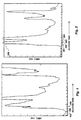
- FIG. 1 depicts the absorbance measured at 280 nm as a function of elution time.
- the collection of one-ml fractions 102, 103, 104 is indicated. Portions of the collected fractions were assayed for TK1 activity generally as described in Example I.
- Fraction 104 approximately spans a first peak 110 in the chromatograph, which was found to contain most of the TK1 activity eluted from the column.
- pooling and concentrating fraction 104 from about a hundred runs performed as in Example II, approximately 40 mls of eluant containing about 1.6 mg/ml of TK1 protein were recovered. The approximate specific activity of the pooled DEAE-cellulose preparation was 17,430 cpm per mg protein per minute.
- the pooled DEAE-cellulose fractions were concentrated using an Amicon protein concentrator, and a sample was electrophoresed under non-denaturing conditions on a 10% polyacrylamide separation gel with a 4.0% polyacrylamide stacking gel. Approximately 7 bands were visible in the gel, ranging from about 24,000 to about 180,000 daltons in molecular weight (MW). These bands were cut out, the protein eluted from the gel and assayed for TK1 activity in a manner similar to that described in Example I. Only one band of about 100,000 daltons contained significant TK1 activity. There was no significant TK1 activity in any of the other bands.
- the 100,000 MW band comprised a semi-pure preparation of active TK1, and was used as the antigen to produce anti-TK1 monoclonal antibodies. About 50 ⁇ g (micrograms) of this semi-pure TK1 was recovered from the pooled DEAE-cellulose prep.
- TK1 was partially purified by isoelectric focussing of the curde extract using a ROTIFER apparatus purchased from Bio-Rad. The procedure used was that outlined in the ROTIFER manual from Bio-Rad (1990). Six to seven protein bands were observed in the isoelectric gels, one of which had a molecular weight of about 100,000 daltons and exhibited some TK1 activity when assayed as described in the preceding paragraphs. The recovery of activity was rather poor, compared to the methods of Examples II and IV.
- FPLC Protein Liquid Chromatography
- the Mono Q 5/5 is an ion exchange column packing commercially available from Pharmacia, having substantially monodisperse bead size and strong anion exchange properties due to bound quaternary amine groups which remain charged over the range from pH 2 to pH 12. Apparatus for the procedure can also be purchased from Pharmacia.
- the column was loaded with 0.1 ml of the semi-pure preparation from the procedure of Example II, containing about 1 mg protein, and voided with 10 volumes of Buffer A (50 mM Tris-HCl, pH 8.0). The void volume of this column was 1.0 ml.
- Buffer A 50 mM Tris-HCl, pH 8.0
- the void volume of this column was 1.0 ml.
- a programmed gradient was set up to gradually increase the concentration of Buffer B (1.0 M NaCl, 50 mM Tris-HCl, pH 8.0) from 0-100% over 20 minutes running at a constant flow rate of 1.0 ml/min.
- TK1 activity was determined to be primarily in peak 200 (the first peak eluting from the column), at which point the gradient contained about 15-20% of Buffer B.
- Protein from the peak 200 was analyzed by non-denaturing PAGE as before to determine purity. There were 5 protein bands present. These bands were cut out and protein from each was assayed for TK1 activity. Detectable TK1 activity was found in the high molecular weight band (100,000 MW), but not in the other bands.
- TK1-activity-positive fractions from several runs were collected, pooled and concentrated. This partially purified, pooled sample was then re-run on the Mono Q column with a lower gradient. One-tenth ml portion of pooled sample containing about 1 mg protein, was loaded on the Mono Q column as before. For this second run, the gradient was started at 5 % of Buffer B and ran to 40% Buffer B over 35 minutes at 1.0 ml/min.
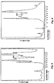
- FIG. 3 depicts a chromatogram of absorbance vs. elution volume for the second sequential Mono Q run. Fractions containing 1.0 ml of eluant were again collected. A major peak 300 eluted from the column at about 15% of Buffer B, and two minor peaks 302, 304 eluted at about 18% and 20% Buffer B respectively. The peaks were assayed for TK1 activity and fraction 310 from the major peak 300 was determined to contain TK1 isozyme activity.
- Protein from peak 300 was also analyzed by SDS-PAGE, and found to contain 3 proteins of molecular weights of about 100,000, 75,000, and 24,000, respectively. Upon assay for TK1 activity of protein from each of the bands, only the 100,000 dalton protein exhibited significant TK1 activity.
- a third sequential Mono Q run was performed on protein precipitated and pooled from fractions 308, 310 containing the peak 300.
- the running conditions were further changed by slowing the flow rate and further decreasing the gradient.
- a gradient of 5% Buffer B to 30% Buffer B was run at 0.5 mls/min for 50 minutes. For this run, 0.5 ml fractions were collected.
- Protein from each of peaks 400, 402, 404 was assayed for TK1 activity as before.
- the first peak 400 to elute contained TK1 activity, and when analyzed by non-denaturing PAGE was found to contain a single band at a molecular weight of about 100,000.
- This preparation is designated herein as "purified" TK1 isozyme.
- TK1 The protein from peak 400 from several column runs was pooled and analyzed by electrophoresis under reducing and non-reducing conditions. Only one band at 100,000 MW was observed on the non-reducing gel, while a faint band at 100,000 MW and a darker band at 24,000 MW were observed on the reducing gel. From this result it appears that the active form of TK1 may be a multimer with subunits of lower molecular weights. The subunits also appear at present, to be of identical molecular weights.
- Example IV the procedure of Example IV using the FPLC with Mono Q column, is the preferred method for isolating pure TK1.
- the starting material was the crude extract of Example I.
- the DEAE-cellulose preparation of Example II can be used as the starting material.
- the method of Example II may be used, or the product obtained by the second run on the Mono Q column as in Example IV, is also suitable.
- Hybridoma cell lines producing antibodies to TK1 were produced by methods generally known in the art, but with certain modifications.
- Semi-pure TK1 was prepared as in Example II. A dose of 100 ⁇ g of semi-pure TK1 suspended in 50 ⁇ l of PBS + 50 ⁇ l complete Freund's adjuvant was given intraperitoneally (I.P.) to each of a group of female BALB/c mice, 5-6 weeks old. Two weeks later, a second immunization was given that was identical to the first.
- mice Two weeks following the second immunization with semi-pure TK1, an intrasplenic injection was given which contained 10 ⁇ g of pure active TK1 (prepared as in Example IV) suspended in 100 ⁇ l of PBS.
- the mice were anesthetized with Sodium Pentobarbitol (65 mg/ml) which was diluted by adding 6.7 mls to 93.3 mls of PBS.
- Each mouse was given 10 ⁇ l/gram of body weight I.P.
- Surgical intervention was performed using a scalpel and forceps, and the spleen was gently teased out for administration of the antigen. Several areas of the spleen were injected to insure uniform distribution of the antigen.
- the wound was closed with metal sutures and the mice were placed under a heating lamp for 1-2 hours.
- mice Seventy-two hours following the intrasplenic injection, the mice were sacrificed using ether and the spleen was removed. Before the mice were killed, blood was removed and the serum tested to insure that the mice were mounting an immune response to the TK1 protein.
- the B cells were isolated from the spleen for fusion with an immortal myeloma cell line.
- the cell line used for the fusion partner was a self-fused Sp2/0 line designated FO which was purchased from ATCC. It is a derivative of P3-X63-Ag8. This line is an immortal myeloma mouse cell line that is fast growing and a non-secretor (heavy or light chain immunoglobulins).
- the fusion of FO and activated spleen cells was performed generally as known in the art. One spleen containing about 1 x 10 8 cells was used per fusion. The most successful fusions resulted when the ratio of B-cells to FO cells was about 10:1. After the fusion was terminated, the fusion cell suspension was seeded into 96-well microtiter plates which had been seeded a day earlier with 3,000 to 6,000 mouse macrophages per well as feeder cells.
- HAT selection medium was used to select only fusion products. Wells were marked for growth and gradually weaned out of HAT and into regular media. By this time the only surviving cells were hybridomas obtained by fusion of B-cells and FO cells. A total of about 500 colonies representing fusion products, resulted from each fusion.
- Preliminary screening of hybridoma colonies from fusion 500 colonies from one fusion were subjected to preliminary screening by EUSA against partially purified TK1 prepared as in Example II. Supernatants collected from the hybridoma cultures were initially screened with semi-pure TK1 prepared by running the crude extract of Raji cells on DEAE cellulose to partially purify the TK1. Thus, this preliminary screen detects antibodies to multiple forms of TK1, including monomer, multimer including tetramer, active and inactive forms, etc.
- Multiwell plates were coated with 1.0 ⁇ g (micrograms) per well of selected TK protein preparations suspended in 50 ⁇ l PBS, and allowed to dry overnight. The plates were then treated for 30 minutes with 200 ⁇ l per well of PBS-Tween 20-EDTA-1% milk fat, to block non-specific binding. The plates were washed 3 times with 200 ⁇ l of PBS-Tween 20-EDTA (PBST2E).
- Tween 20 is an anionic detergent commercially available from Bio-Rad Laboratories, Richmond, CA, and useful to reduce non-specific antibody-antigen binding while not disrupting binding of primary antibodies to antigens or of antigens to nitrocellulose.
- the growth medium on the hybridoma cell cultures was not changed for three days prior to collection of the hybridoma culture supernatants, in order to saturate the media with antibodies.
- 80 ⁇ l of supernatant per well was added to duplicate wells.
- the multiwell plates were then incubated at 37° C for 1-1/2 hours.
- the supernatant was decanted and the wells washed 6 times with PBST2E.
- the positive colonies were isotyped using a kit from Hyclone, Logan, UT (cat. # EK-5051), and the positive colonies determined to produce antibodies of IgG1, IgG2a, IgG3, and IgM classes.
- a plate was coated with five pairs of replicate wells as follows: wells A,B were coated with a crude extract of TK1 from Raji cells; wells C,D were coated with semi-purified TK1 prepared from the DEAE-cellulose column; wells E,F were coated with purified TK1 from peak 400 prepared as in Example IV by FPLC (see FIG. 4); wells G,H were coated with TK1 protein from fractions 308,310 of the second FPLC run (see FIG. 3); and wells J,K were coated with an extract of E. coli cells which expressed a TK1 gene in a PET vector.
- samples 1.0 ⁇ g per well of protein was used.
- the ELISA was performed essentially as described for the preliminary screening. Of the 35 clones tested, one proved to bind only to active form TK1.
- the absorbance readings (ABS) were made at 405 nm for 120 wells on one plate on which ten clones were screened are shown in TABLE I. The clones testing most highly positive by preliminary screening were purposely clustered on this plate. The background ABS from four wells was averaged and found to be about 0.058 (wells J11, J12 and K11, K12).
- the Western blots were prepared by procedures similar to those described in CURRENT PROTOCOLS IN IMMUNOLOGY, VOL. 1, publ. Wiley-Interscience, New York (1991). Antibodies were harvested from the supernatant of each hybridoma and hybridized to a nitrocellulose membrane blotted from a non-denaturing gel of TK proteins. A goat anti-mouse IgG was then used for detection of the bound antibodies.
- Clone 1 and Clone 5 bind to different epitopes.
- the Clone 1 antibody binds selectively to the active multimer form of TK1, and is designated hereinafter as an "anti-AcTK1" antibody.
- Clone 5 antibody binds to the multimer form, but apparently preferentially to an inactive multimer form.
- Clones Nos. 1-5 are all IgM-type hybridomas.
- Clone 7 is an IgG-type hybridoma.
- TK1 activity by selected monoclonal antibodies.
- the TK assay using ATP was performed using the crude extract of Raji cells described above in regard to isolation of TK1.
- Replicate assay reactions were prepared.
- a 20 ⁇ l aliquot of supernatant from one of the hybridoma cultures was added, containing between about 0.02 to 0.1 ⁇ g of antibody.
- Antibodies from clones nos. 3, 4, 6 and 7 were less efficient in inhibition of TK1 activity in the Raji extract assay (not shown). Therefore, they were not subjected to further screening.
- Clones 1, 2 and 5 were again subjected to limiting dilution and colonies derived from this re-cloning procedure were tested once again.
- the re-cloning procedure was used to ensure that a hybridoma cell line is derived from a single fusion cell and thus produces antibodies which are uniform in isotype and specificity, e.g. monoclonal.
- Clones Nos. 1, 2 and 5 were placed on deposit under the terms of the Budapest Treaty with the American Type Culture Collection in Rockville, Maryland U.S.A. on August 11, 1993, and assigned ATCC Nos. HB 11432, HB 11433, and HB 11434, respectively.
- TK2 Isolation of TK2. Isolation of TK2 was performed by FPLC with a Mono-Q column. The starting material was a crude cell extract similar to the Raji extract except that it was prepared from HeLa cells. HeLa is an immortalized human cervical carcinoma cell line available from ATCC under the #CCl-2, which is believed to have high levels of TK2 and very low levels of TK1. When analyzed by non-denaturing PAGE, TK2 also appears to have a monomer form and one or more multimer forms.
- TK2 activity is found to be largely in peaks 18-20, which is quite separate from the region in which TK1 elutes.
- FIG. 5 shows the results of a first run on a Mono Q column of pooled material from peaks 18-20.
- Buffer A was 0.05 molar Tris-HCl and Buffer B was 0.5 molar Tris-HCI, and the column was run with a gradient of 5% to 45% Buffer B at a flow rate of 1 ml/min.
- the TK2 activity was assayed in the collected fractions and found to he mostly in fraction 7 (peak 500). Material from peak 500 from several column runs was pooled, concentrated and run a second time with the same buffer gradient and flow rate.
- FIG. 6 shows the results of the second run.
- the TK2 activity was found to be in fraction 6 (peak 600).
- the collected material of fraction 6 is a semi-pure preparation of active TK2 isozyme.
- the immunization procedure, harvest of spleen cells and fusion with FO cells was similar to that in Examples V-VI. Screening was performed by ELISA on wells coated with the semi-pure TK2 preparation of Example IX, and the results are given in Table III. The results indicated that antibodies from Clones 2-2, 2-3, 2-4, 2-5, 2-7, 2-8, 2-9, 2-10, 2-12, 2-17 and 2-19 gave the most positive results for binding to the semi-pure TK2.
- the selected antibody-producing cell lines were passaged and supernatant was aseptically collected over a period of 3 months.
- Antibodies were purified by precipitating the supernatants with ammonium sulfate followed either by gel filtration chromatography or by DEAE-cellulose chromatography (diethylaminoethyl cellulose; obtained from Whatman International, Maidstone, Kent, UK under the tradename SEPHADEX).
- the antibodies were purified by standard methods and conjugated with either HRP-Peroxidase or Alkaline Phosphatase (Bio-Rad). Such procedures are described in ANTIBODIES: A Laboratory Manual by Harlowe and Lane, 1988.
- TK levels in tumor tissues can be determined by preparing an extract of TK from samples of fresh tumor tissue similar to the crude extract of Raji cells of Example I.
- the protein content of the sample is determined so that the amount of TK can be correlated with the amount of total protein in the tumor.
- An immunoprecipitationassay using the desired anti-TK antibody can then be performed on the extract.
- the conjugated antibody from Clone 1 was used to immunostain tumor cells histologically fixed to slides.
- Diaminobenzidine (DAB) was used as the enzyme substrate in this procedure.
- TK1 active TK1 in serum samples from cancer patients using anti-acTK1 antibody .
- Serum samples were obtained from cancer patients. Each sample was assayed for TK activity by a method like that of Example I. The same samples were then quantitated blindly on an ELISA test with Clone #1 antibody using different serum dilution levels. A dilution of 1:16,000 was found to give the best results.
- the results of the two assays are presented in Table IV. The data were confirmed by Western blot analysis.
- the sera are ranked from highest to lowest amount of anti-TK1 bound, by the O.D. reading in the ELISA assay. It can be seen from the TK1 activity measurements, that the correlation between antibody binding data and the standard TK1 activity assay is excellent.
- the data demonstrate that the anti-AcTK1 antibody can be used to evaluate the serum level of TK1 activity in human subjects. Further, serum from a healthy (non-cancer-bearing) individual bound much less anti-AcTK1 antibody as compared to the lowest-ranked serum of a cancer patient.
- the anti-AcTK1 antibody is useful to distinguish between Comparison of TK1 Activity Measured by 3 H-thymidine Incorporation and Anti-AcTK1 Binding Serum # TK1 assay Rank O.D.450 nm 1 2498 5 .443 2 2376 5 .430 3 8251 2 .865 4 6254 3 .728 5 2214 5 .420 6 11477 1 1.542 7 2509 5 .450 8 4785 4 .592 0 - 8 .250
- the O.D. 450 readings represent the amount of bound Clone #1 antibody measured by ELISA.
- the TK activity values are the cpm of 3 H incorporated per minute. serum of cancer-bearing individuals and serum from healthy non-cancerous individuals.
- TK activity can be used as a reliable predictor of the likelihood of tumor recurrence in breast cancer patients.
- Table V summarizes the results of a study of TK activity in samples from untreated primary tumors that were surgically removed from 86 patients, 13 of which later experienced a recurrence of disease.
- TK levels with estrogen receptor status In this study, it was also found by comparing TK levels with estrogen receptor status, that the TK level accurately predicts which patients in the estrogen receptor positive group later showed recurrence and which in the estrogen receptor negative group did not show recurrence.
- 57 estrogen receptor positive patients patients whose tumors had high numbers of estrogen receptors
- the average TK activity level measured in the tumors from those four patients was 289,717 cpm/min, as compared with 161,674 cpm/min for tumors from patients who had no recurrence.
- the percentage of the activity attributable to TK2 was about 76% in the no-recurrence group, vs. 41 % in the four who had recurrence.
- TK1 activity in serum can be used to detect relapse, often before any other symptom is evident.
- the anti-AcTK1 antibody is useful to screen serum and tissue from cancer patients both for study purposes and for use in diagnosis and treatment of tumor patients.
- the anti-AcTK1 antibody, the anti-totalTK1 antibody, and/or the anit-TK2 antibody may also be useful for serum screening in certain other kinds of blood disorders, such as pernicious anemia, where TK activity has been linked to disease status.
- the antibodies may further be useful for testing of patients with certain viral diseases including morbilli, rubella and herpes, where it has been found that the levels of thymidine kinase are elevated during the acute phase of the disease.
- the anti-AcTK1 antibody detects only active-form TK1, it can be used in place of the radioactive thymidine incorporation assay to evaluate activity.
- the use of the antibody for evaluating TK1 activity in tumors and serum of cancer patients will make this test practical for use on a wide scale, whereas the activity assay is generally more difficult to perform.
- the antibody may be useful for targeted tumor therapy. That is, an anti-tumor agent may be coupled to the antibody, which will selectively bind to tumor cells expressing large amounts of TK1. In this manner the anti-tumor agent is targeted to tumor cells and the killing of tumor cells is enhanced versus killing of normal cells.
- the invention may also be embodied as a hybridoma cell line obtained by injecting a host animal with a TK preparation, isolating activated B cells from the spleen of the injected animal, fusing the activated B cells with myeloma cells, and individually screening clones of the fused cells to select a hybridoma cell line which produces anti-TK antibodies having desired binding specificities.
- the TK preparation is derived from Raji cells.
- the TK preparation is a substantially homogeneous preparation of tetramer and/or active TK.
- Antibodies produced by a hybridoma obtained by one of the methods described in this application are also encompassed by the invention.
- a method of determining the serum level of a thymidine kinase enzyme comprises the following steps: obtaining a serum sample from a patient, providing a monoclonal antibody which selectively binds to thymidine kinase enzyme, contacting the monoclonal antibody with the sample, and determining the amount of monoclonal antibody bound to thymidine kinase protein in the sample.
- TK1 total TK1 including monomeric and multimeric forms and active and inactive forms
- active form TK1 only active form TK2 only.
- An alternate embodiment is a method for evaluating the level of thymidine kinase in a solid tumor sample. This method is essentially the same as that for evaluating serum thymidine kinase levels, except that the step of providing a serum sample is replaced by a step of providing a tumor sample.
- the tumor sample may be a fresh or frozen tissue sample or a histological slide preparation.
- a kit for performing the above methods may comprise one or more monoclonal antibodies selected from the group including anti-total-TK1, anti-AcTK1, and anti-TK2.
- the monoclonal antibody is conjugated to an enzyme useful in an ELISA assay or to another detectable marker such as a fluorescent dye, radioactive isotope, or the like.
- the kit may further include an anti-mouse antibody which is enzyme-conjugated for detection by ELISA or otherwise labelled.
- a method of predicting the likelihood of recurrence of a solid tumor in a patient at initial diagnosis comprises the steps of establishing a normal range for tissue TK activity, obtaining a sample of a primary tumor from a patient, determining the amount of TK enzyme in the patient sample to produce a patient TK value, and comparing the patient TK value it to a normal value; and if it exceeds the normal range by a significant amount, predicting that the tumor is likely to recur, and if it does not significantly exceed the normal range, predicting that recurrence is unlikely.
- the step of establishing a normal range may be performed in several ways. In one embodiment, one or more samples of the patient's own normal tissue may be used for comparison. In another embodiment, a normal range may be established from a study of normal tissues in healthy (no disease) individuals, or from patients known to be in remission.
- the TK measurement is selectively of TK1 activity or provides a comparison of TK1 and TK2 activity levels.
- the level of active TK1 is measured using an anti-AcTK1 antibody.
- the total TK1 content is measured using an anti-total-TK1 antibody.
- Still another embodiment of the methods employs an anti-total-TK1 antibody and an anti-TK2 antibody.
- a preferred alternate embodiment employs an anti-TK2 antibody.
- a method of determining whether disease has recurred in a patient being treated for a cancer or blood disorder, including breast cancer, leukemia and lymphoid cancer comprises the steps of taking a series of samples of the serum of a cancer patient at regular intervals, measuring the amount of TK in the samples, comparing the amount of TK among the samples, and when the amount of TK in later samples exceeds the amount of earlier samples by a significant degree, determining that the disease is recurring.
- the measurements are of TK1 activity in the sample as determined using an anti-active TK1 monoclonal antibody.
- An alternate embodiment employs an anti-total-TK1 antibody.
- Still other embodiments utilize an anti-total TK (both TK1 and TK2) antibody, or comparative measurements of TK isozymes using two or more of the noted antibodies.
- the present application contains at least the following teachings: that only TK1 tetramers or multimers have significant activity; of convenient sources (Raji cells, HeLa cells) and methods for isolation of large quantities of TK1, of active TK1, and of TK2 for use as antigen and in screening to select desired hybridomas; and of screening procedures to identify antibodies having different specificities. It will be apparent that, given these and other teachings of this application, one of ordinary skill could obtain the hybridomas and antibodies described and claimed herein, as well as monoclonal antibodies having specificity for any desired category of thymidine kinase isozymes.
Landscapes
- Chemical & Material Sciences (AREA)
- Organic Chemistry (AREA)
- Health & Medical Sciences (AREA)
- Medicinal Chemistry (AREA)
- Biochemistry (AREA)
- Biophysics (AREA)
- General Health & Medical Sciences (AREA)
- Genetics & Genomics (AREA)
- Life Sciences & Earth Sciences (AREA)
- Molecular Biology (AREA)
- Proteomics, Peptides & Aminoacids (AREA)
- Immunology (AREA)
- Preparation Of Compounds By Using Micro-Organisms (AREA)
- Peptides Or Proteins (AREA)
- Medicines Containing Antibodies Or Antigens For Use As Internal Diagnostic Agents (AREA)
- Investigating Or Analysing Biological Materials (AREA)
Applications Claiming Priority (7)
| Application Number | Priority Date | Filing Date | Title |
|---|---|---|---|
| US10273593A | 1993-08-06 | 1993-08-06 | |
| US102735 | 1993-08-06 | ||
| US13629993A | 1993-10-14 | 1993-10-14 | |
| US136299 | 1993-10-14 | ||
| US15242993A | 1993-11-12 | 1993-11-12 | |
| US152429 | 1993-11-12 | ||
| EP94925802A EP0785993B1 (de) | 1993-08-06 | 1994-08-05 | Monoklonale antikörper gegen isozyme der thymidinkinase |
Related Parent Applications (1)
| Application Number | Title | Priority Date | Filing Date |
|---|---|---|---|
| EP94925802A Division EP0785993B1 (de) | 1993-08-06 | 1994-08-05 | Monoklonale antikörper gegen isozyme der thymidinkinase |
Publications (2)
| Publication Number | Publication Date |
|---|---|
| EP1142991A2 true EP1142991A2 (de) | 2001-10-10 |
| EP1142991A3 EP1142991A3 (de) | 2003-03-12 |
Family
ID=27379405
Family Applications (2)
| Application Number | Title | Priority Date | Filing Date |
|---|---|---|---|
| EP94925802A Expired - Lifetime EP0785993B1 (de) | 1993-08-06 | 1994-08-05 | Monoklonale antikörper gegen isozyme der thymidinkinase |
| EP01201917A Withdrawn EP1142991A3 (de) | 1993-08-06 | 1994-08-05 | Monoklonale Antikörper gegen Isozyme der Thymidinkinase |
Family Applications Before (1)
| Application Number | Title | Priority Date | Filing Date |
|---|---|---|---|
| EP94925802A Expired - Lifetime EP0785993B1 (de) | 1993-08-06 | 1994-08-05 | Monoklonale antikörper gegen isozyme der thymidinkinase |
Country Status (6)
| Country | Link |
|---|---|
| US (1) | US5698409A (de) |
| EP (2) | EP0785993B1 (de) |
| AT (1) | ATE239784T1 (de) |
| AU (1) | AU675872B2 (de) |
| DE (1) | DE69432645T2 (de) |
| WO (1) | WO1995004758A1 (de) |
Families Citing this family (17)
| Publication number | Priority date | Publication date | Assignee | Title |
|---|---|---|---|---|
| SE9401380D0 (sv) * | 1994-04-22 | 1994-04-22 | Sven Skog | Ny celltillväxt-relaterad peptid och användning därav |
| US7754435B2 (en) | 2000-09-21 | 2010-07-13 | Philadelphia Health And Education Corporation | Prostate cancer-related compositions, methods, and kits based on DNA macroarray proteomics platforms |
| US20030044803A1 (en) * | 2000-09-22 | 2003-03-06 | Pedersen Finn Skou | Methods for diagnosis and treatment of diseases associated with altered expression of JAK1 |
| US20020164576A1 (en) * | 2000-09-22 | 2002-11-07 | Pedersen Finn Skou | Methods for diagnosis and treatment of diseases associated with altered expression of Nrf2 |
| US20070098728A1 (en) * | 2001-09-24 | 2007-05-03 | Pedersen Finn S | Novel compositions and methods in cancer |
| ATE352039T1 (de) * | 2002-07-27 | 2007-02-15 | Diasorin Ab | Verfahren zur bestimmung der aktivität von thymidinkinase-1 und dessen verwendung |
| EP1627230B1 (de) * | 2003-05-16 | 2010-09-15 | AroCell AB | Prognose des verlaufs einer krebserkrankung |
| TW200532523A (en) * | 2004-02-27 | 2005-10-01 | Aureon Biosciences Corp | Methods and systems for predicting occurrence of an event |
| US7311906B2 (en) * | 2004-04-30 | 2007-12-25 | Brigham Young University | Anti-viral activity of an anti-thymidine kinase monoclonal antibody |
| US8551486B2 (en) * | 2004-05-21 | 2013-10-08 | Savoy Pharmaceuticals, Inc. | Monoclonal antibodies to human thymidine kinase to treat cancer |
| US20100266495A1 (en) * | 2004-05-21 | 2010-10-21 | Brigham Young University | Anti-Cancer Activity of an Anti-Thymidine Kinase Monoclonal Antibody |
| US7837998B2 (en) | 2004-05-21 | 2010-11-23 | Nathaniel Lallatin | Anti-cancer activity of an anti-thymidine kinase monoclonal antibody |
| JP4457155B2 (ja) * | 2008-02-22 | 2010-04-28 | 株式会社日立製作所 | 核磁気共鳴測定装置および核磁気共鳴測定装置を用いた測定方法 |
| WO2011082345A2 (en) * | 2009-12-30 | 2011-07-07 | Brigham Young University | Compositions and methods for cancer management using antibodies binding to nucleotide salvage pathway enzymes and complexes thereof |
| US20160082032A1 (en) * | 2013-05-17 | 2016-03-24 | Taiho Pharmaceutical Co., Ltd. | Therapeutic effect prediction method for colorectal cancer patient in whom expression of tk1 protein has increased |
| KR20180054600A (ko) | 2015-10-13 | 2018-05-24 | 브라이엄 영 유니버시티 | 면역치료에서의 대식세포 키메라 항원 수용체(moto-car) |
| CA3031645A1 (en) * | 2016-08-10 | 2018-02-15 | Alertix Veterinary Diagnostics Ab | Determination of non-human mammal tk1 protein levels |
Family Cites Families (3)
| Publication number | Priority date | Publication date | Assignee | Title |
|---|---|---|---|---|
| US4317877A (en) * | 1980-06-05 | 1982-03-02 | Sloan Kettering Institute | Process for detecting the presence of malignant and pre-malignant cells in humans |
| FR2602242B1 (fr) * | 1986-07-31 | 1989-05-26 | Debat Lab | Thymidine kinase de type foetal purifiee |
| CA2040707C (en) * | 1990-04-27 | 2002-07-09 | Kenji Mitsushima | Cephalosporin acetylhydrolase gene and protein encoded by said gene |
-
1994
- 1994-08-05 AT AT94925802T patent/ATE239784T1/de active
- 1994-08-05 EP EP94925802A patent/EP0785993B1/de not_active Expired - Lifetime
- 1994-08-05 DE DE69432645T patent/DE69432645T2/de not_active Expired - Lifetime
- 1994-08-05 EP EP01201917A patent/EP1142991A3/de not_active Withdrawn
- 1994-08-05 WO PCT/US1994/009022 patent/WO1995004758A1/en not_active Ceased
- 1994-08-05 AU AU75596/94A patent/AU675872B2/en not_active Ceased
-
1995
- 1995-05-10 US US08/438,627 patent/US5698409A/en not_active Expired - Lifetime
Also Published As
| Publication number | Publication date |
|---|---|
| EP1142991A3 (de) | 2003-03-12 |
| EP0785993A1 (de) | 1997-07-30 |
| WO1995004758A1 (en) | 1995-02-16 |
| ATE239784T1 (de) | 2003-05-15 |
| AU675872B2 (en) | 1997-02-20 |
| EP0785993B1 (de) | 2003-05-07 |
| EP0785993A4 (de) | 1997-07-30 |
| DE69432645D1 (de) | 2003-06-12 |
| US5698409A (en) | 1997-12-16 |
| AU7559694A (en) | 1995-02-28 |
| DE69432645T2 (de) | 2004-04-08 |
Similar Documents
| Publication | Publication Date | Title |
|---|---|---|
| EP0785993B1 (de) | Monoklonale antikörper gegen isozyme der thymidinkinase | |
| US5700466A (en) | Method of ameliorating or preventing septic shock using a monoclonal antibody specific to cachectin/tumor necrosis factor | |
| EP0453560B1 (de) | Lokalisation und charakterisierung des wilms-tumor-gens | |
| Hinds et al. | Immunological evidence for the association of p53 with a heat shock protein, hsc70, in p53-plus-ras-transformed cell lines | |
| EP0068763B2 (de) | Rekombinante Monoklonale Antikörper | |
| AU631788B2 (en) | Method for determining state of malignant and pre-malignant progression | |
| US4943533A (en) | Hybrid cell lines that produce monoclonal antibodies to epidermal growth factor receptor | |
| JPH11335297A (ja) | 増殖因子レセプターの機能を阻害することにより腫瘍細胞を処置する方法 | |
| EP0735370A1 (de) | MAP-Kinase-Testverfahren | |
| EP0312996A2 (de) | Monoklonaler Antikörper gegen humanen BCDF | |
| US8501419B2 (en) | Exposed proliferation-related peptides, ligands and methods employing the same | |
| US6087117A (en) | Production and use of human nm23 protein and antibodies therefor | |
| KR19990067153A (ko) | 프로스타글란딘 디 신타아제에 특이적인 단일클론항체 | |
| CA2173834C (en) | Monoclonal antibodies to thymidine kinase isozymes | |
| WO1986007062A1 (en) | Monoclonal antibody to decay accelerating factor (daf), a method for making it, and use | |
| US7514227B2 (en) | Anti-human mitochondrial adenylate kinase isozyme antibody, diagnostic formulation and diagnostic kit for cardiac disease | |
| EP0207170A1 (de) | Verfahren zur herstellung menschlicher krebsspezifischer monoklonaler antikörper | |
| CN113075403B (zh) | 一种用于胃癌诊断的分子标记物及试剂盒 | |
| AU580430B2 (en) | Monoclonal antibody to decay accelerating factor (daf), a method for making it, and use | |
| JPH06125784A (ja) | モノクローナル抗体,ハイブリドーマ,その製造法および用途 | |
| JPH06253886A (ja) | Crkタンパクに対するモノクローナル抗体と、この 抗体を産生するハイブリドーマ株 | |
| JP2004073142A (ja) | Ganp結合性タンパク質 | |
| EP0478272A2 (de) | Gp98-Zelladhäsionsmolekül und Integrinkomplex der dieses enthält | |
| JPH0549495A (ja) | モノクローナル抗体及びヒト血漿α(1→3)フコース転移酵素の定量方法 | |
| JPWO1997016461A1 (ja) | プロスタグランジンd合成酵素に特異的なモノクローナル抗体 |
Legal Events
| Date | Code | Title | Description |
|---|---|---|---|
| PUAI | Public reference made under article 153(3) epc to a published international application that has entered the european phase |
Free format text: ORIGINAL CODE: 0009012 |
|
| AC | Divisional application: reference to earlier application |
Ref document number: 785993 Country of ref document: EP |
|
| AK | Designated contracting states |
Kind code of ref document: A2 Designated state(s): AT CH DE FR GB IT LI |
|
| RIC1 | Information provided on ipc code assigned before grant |
Free format text: 7C 12N 9/12 A |
|
| PUAL | Search report despatched |
Free format text: ORIGINAL CODE: 0009013 |
|
| AK | Designated contracting states |
Kind code of ref document: A3 Designated state(s): AT CH DE FR GB IT LI Designated state(s): AT CH DE FR GB IT LI |
|
| 17P | Request for examination filed |
Effective date: 20030818 |
|
| AKX | Designation fees paid |
Designated state(s): AT CH DE FR GB IT LI |
|
| 17Q | First examination report despatched |
Effective date: 20040220 |
|
| STAA | Information on the status of an ep patent application or granted ep patent |
Free format text: STATUS: THE APPLICATION IS DEEMED TO BE WITHDRAWN |
|
| 18D | Application deemed to be withdrawn |
Effective date: 20040702 |