EP1127107B1 - Methods for the production of tcr gamma delta t cells - Google Patents
Methods for the production of tcr gamma delta t cells Download PDFInfo
- Publication number
- EP1127107B1 EP1127107B1 EP99953463A EP99953463A EP1127107B1 EP 1127107 B1 EP1127107 B1 EP 1127107B1 EP 99953463 A EP99953463 A EP 99953463A EP 99953463 A EP99953463 A EP 99953463A EP 1127107 B1 EP1127107 B1 EP 1127107B1
- Authority
- EP
- European Patent Office
- Prior art keywords
- cells
- amount
- tcrγδ
- present
- culture medium
- Prior art date
- Legal status (The legal status is an assumption and is not a legal conclusion. Google has not performed a legal analysis and makes no representation as to the accuracy of the status listed.)
- Expired - Lifetime
Links
- 238000000034 method Methods 0.000 title claims abstract description 84
- 102000011778 gamma-delta T-Cell Antigen Receptors Human genes 0.000 title abstract 5
- 108010062214 gamma-delta T-Cell Antigen Receptors Proteins 0.000 title abstract 5
- 210000004475 gamma-delta t lymphocyte Anatomy 0.000 title 1
- 210000004027 cell Anatomy 0.000 claims abstract description 193
- 210000001744 T-lymphocyte Anatomy 0.000 claims abstract description 166
- 102000000588 Interleukin-2 Human genes 0.000 claims abstract description 101
- 108010002350 Interleukin-2 Proteins 0.000 claims abstract description 101
- 102000004388 Interleukin-4 Human genes 0.000 claims abstract description 94
- 108090000978 Interleukin-4 Proteins 0.000 claims abstract description 94
- 229940028885 interleukin-4 Drugs 0.000 claims abstract description 86
- 239000001963 growth medium Substances 0.000 claims abstract description 58
- 239000003226 mitogen Substances 0.000 claims abstract description 27
- 238000012258 culturing Methods 0.000 claims abstract description 24
- 108010062580 Concanavalin A Proteins 0.000 claims description 36
- 210000005259 peripheral blood Anatomy 0.000 claims description 20
- 239000011886 peripheral blood Substances 0.000 claims description 20
- 210000002381 plasma Anatomy 0.000 claims description 18
- 208000010833 Chronic myeloid leukaemia Diseases 0.000 claims description 14
- 239000003636 conditioned culture medium Substances 0.000 claims description 13
- 210000005087 mononuclear cell Anatomy 0.000 claims description 9
- 210000002966 serum Anatomy 0.000 claims description 9
- 210000004700 fetal blood Anatomy 0.000 claims description 8
- DLEDLHFNQDHEOJ-UDTOXTEMSA-N mezerein Chemical compound O([C@@H]1[C@H]([C@@]23[C@H]4[C@](C(C(C)=C4)=O)(O)[C@H](O)[C@@]4(CO)O[C@H]4[C@H]3[C@H]3O[C@@](O2)(O[C@]31C(C)=C)C=1C=CC=CC=1)C)C(=O)\C=C\C=C\C1=CC=CC=C1 DLEDLHFNQDHEOJ-UDTOXTEMSA-N 0.000 claims description 8
- DLEDLHFNQDHEOJ-KVZAMRGJSA-N mezerein Natural products CC1C(OC(=O)C=C/C=C/c2ccccc2)C3(OC4(OC3C5C6OC6(CO)C(O)C7(O)C(C=C(C)C7=O)C15O4)c8ccccc8)C(=C)C DLEDLHFNQDHEOJ-KVZAMRGJSA-N 0.000 claims description 8
- 101000917858 Homo sapiens Low affinity immunoglobulin gamma Fc region receptor III-A Proteins 0.000 claims description 7
- 101000917839 Homo sapiens Low affinity immunoglobulin gamma Fc region receptor III-B Proteins 0.000 claims description 7
- 102100029185 Low affinity immunoglobulin gamma Fc region receptor III-B Human genes 0.000 claims description 7
- 210000001519 tissue Anatomy 0.000 claims description 7
- 101000581981 Homo sapiens Neural cell adhesion molecule 1 Proteins 0.000 claims description 6
- 102100027347 Neural cell adhesion molecule 1 Human genes 0.000 claims description 6
- 102100024222 B-lymphocyte antigen CD19 Human genes 0.000 claims description 5
- 102100035716 Glycophorin-A Human genes 0.000 claims description 5
- 108091005250 Glycophorins Proteins 0.000 claims description 5
- 101000980825 Homo sapiens B-lymphocyte antigen CD19 Proteins 0.000 claims description 5
- 101000946889 Homo sapiens Monocyte differentiation antigen CD14 Proteins 0.000 claims description 5
- 101000934338 Homo sapiens Myeloid cell surface antigen CD33 Proteins 0.000 claims description 5
- 102100035877 Monocyte differentiation antigen CD14 Human genes 0.000 claims description 5
- 102100025243 Myeloid cell surface antigen CD33 Human genes 0.000 claims description 5
- 230000000779 depleting effect Effects 0.000 claims description 4
- 230000004936 stimulating effect Effects 0.000 claims description 4
- 210000001185 bone marrow Anatomy 0.000 claims description 3
- 210000004185 liver Anatomy 0.000 claims description 2
- 210000001165 lymph node Anatomy 0.000 claims description 2
- 210000003563 lymphoid tissue Anatomy 0.000 claims description 2
- 210000000952 spleen Anatomy 0.000 claims description 2
- 210000001541 thymus gland Anatomy 0.000 claims description 2
- 102000004127 Cytokines Human genes 0.000 abstract description 12
- 108090000695 Cytokines Proteins 0.000 abstract description 12
- 230000001225 therapeutic effect Effects 0.000 abstract description 6
- 239000002243 precursor Substances 0.000 abstract description 2
- 239000012636 effector Substances 0.000 description 18
- 239000000427 antigen Substances 0.000 description 14
- 108091007433 antigens Proteins 0.000 description 14
- 102000036639 antigens Human genes 0.000 description 14
- 208000032791 BCR-ABL1 positive chronic myelogenous leukemia Diseases 0.000 description 13
- 208000033761 Myelogenous Chronic BCR-ABL Positive Leukemia Diseases 0.000 description 11
- 238000000684 flow cytometry Methods 0.000 description 11
- 210000004369 blood Anatomy 0.000 description 9
- 239000008280 blood Substances 0.000 description 9
- 210000004748 cultured cell Anatomy 0.000 description 9
- 241000283973 Oryctolagus cuniculus Species 0.000 description 8
- 230000010261 cell growth Effects 0.000 description 8
- 238000005119 centrifugation Methods 0.000 description 8
- 238000002360 preparation method Methods 0.000 description 8
- 108091003079 Bovine Serum Albumin Proteins 0.000 description 6
- 206010028980 Neoplasm Diseases 0.000 description 6
- 239000012091 fetal bovine serum Substances 0.000 description 6
- 230000002147 killing effect Effects 0.000 description 6
- 230000008901 benefit Effects 0.000 description 5
- DEGAKNSWVGKMLS-UHFFFAOYSA-N calcein Chemical compound O1C(=O)C2=CC=CC=C2C21C1=CC(CN(CC(O)=O)CC(O)=O)=C(O)C=C1OC1=C2C=C(CN(CC(O)=O)CC(=O)O)C(O)=C1 DEGAKNSWVGKMLS-UHFFFAOYSA-N 0.000 description 5
- 238000005194 fractionation Methods 0.000 description 5
- 210000000822 natural killer cell Anatomy 0.000 description 5
- 229960002378 oftasceine Drugs 0.000 description 5
- 239000000047 product Substances 0.000 description 5
- 210000000130 stem cell Anatomy 0.000 description 5
- IJGRMHOSHXDMSA-UHFFFAOYSA-N Atomic nitrogen Chemical compound N#N IJGRMHOSHXDMSA-UHFFFAOYSA-N 0.000 description 4
- 210000000662 T-lymphocyte subset Anatomy 0.000 description 4
- 230000001472 cytotoxic effect Effects 0.000 description 4
- LOKCTEFSRHRXRJ-UHFFFAOYSA-I dipotassium trisodium dihydrogen phosphate hydrogen phosphate dichloride Chemical compound P(=O)(O)(O)[O-].[K+].P(=O)(O)([O-])[O-].[Na+].[Na+].[Cl-].[K+].[Cl-].[Na+] LOKCTEFSRHRXRJ-UHFFFAOYSA-I 0.000 description 4
- 238000002474 experimental method Methods 0.000 description 4
- 239000012737 fresh medium Substances 0.000 description 4
- 239000002953 phosphate buffered saline Substances 0.000 description 4
- 230000035755 proliferation Effects 0.000 description 4
- 238000011160 research Methods 0.000 description 4
- 230000002269 spontaneous effect Effects 0.000 description 4
- 230000035899 viability Effects 0.000 description 4
- QTBSBXVTEAMEQO-UHFFFAOYSA-N Acetic acid Chemical compound CC(O)=O QTBSBXVTEAMEQO-UHFFFAOYSA-N 0.000 description 3
- 102000017420 CD3 protein, epsilon/gamma/delta subunit Human genes 0.000 description 3
- IAZDPXIOMUYVGZ-UHFFFAOYSA-N Dimethylsulphoxide Chemical compound CS(C)=O IAZDPXIOMUYVGZ-UHFFFAOYSA-N 0.000 description 3
- 238000003556 assay Methods 0.000 description 3
- 210000003719 b-lymphocyte Anatomy 0.000 description 3
- 238000005138 cryopreservation Methods 0.000 description 3
- 230000009089 cytolysis Effects 0.000 description 3
- 230000001461 cytolytic effect Effects 0.000 description 3
- 238000000432 density-gradient centrifugation Methods 0.000 description 3
- 229960001760 dimethyl sulfoxide Drugs 0.000 description 3
- 230000012010 growth Effects 0.000 description 3
- 230000003394 haemopoietic effect Effects 0.000 description 3
- 208000015181 infectious disease Diseases 0.000 description 3
- 210000000265 leukocyte Anatomy 0.000 description 3
- 239000013641 positive control Substances 0.000 description 3
- 108090000765 processed proteins & peptides Proteins 0.000 description 3
- 108090000623 proteins and genes Proteins 0.000 description 3
- 238000010257 thawing Methods 0.000 description 3
- 210000004881 tumor cell Anatomy 0.000 description 3
- -1 AIM-VTM Substances 0.000 description 2
- 241000894006 Bacteria Species 0.000 description 2
- PHEDXBVPIONUQT-UHFFFAOYSA-N Cocarcinogen A1 Natural products CCCCCCCCCCCCCC(=O)OC1C(C)C2(O)C3C=C(C)C(=O)C3(O)CC(CO)=CC2C2C1(OC(C)=O)C2(C)C PHEDXBVPIONUQT-UHFFFAOYSA-N 0.000 description 2
- 241000196324 Embryophyta Species 0.000 description 2
- 241000701044 Human gammaherpesvirus 4 Species 0.000 description 2
- 108010050904 Interferons Proteins 0.000 description 2
- 102000014150 Interferons Human genes 0.000 description 2
- 102000003814 Interleukin-10 Human genes 0.000 description 2
- 108090000174 Interleukin-10 Proteins 0.000 description 2
- 102000007330 LDL Lipoproteins Human genes 0.000 description 2
- 108010007622 LDL Lipoproteins Proteins 0.000 description 2
- 108090001090 Lectins Proteins 0.000 description 2
- 102000004856 Lectins Human genes 0.000 description 2
- 108700018351 Major Histocompatibility Complex Proteins 0.000 description 2
- 108010047620 Phytohemagglutinins Proteins 0.000 description 2
- 108010089814 Plant Lectins Proteins 0.000 description 2
- 241000700584 Simplexvirus Species 0.000 description 2
- 208000036142 Viral infection Diseases 0.000 description 2
- 238000001042 affinity chromatography Methods 0.000 description 2
- 230000000735 allogeneic effect Effects 0.000 description 2
- 201000011510 cancer Diseases 0.000 description 2
- 230000004663 cell proliferation Effects 0.000 description 2
- 230000001413 cellular effect Effects 0.000 description 2
- 239000003599 detergent Substances 0.000 description 2
- 201000010099 disease Diseases 0.000 description 2
- 208000037265 diseases, disorders, signs and symptoms Diseases 0.000 description 2
- 210000000267 erythroid cell Anatomy 0.000 description 2
- 208000005017 glioblastoma Diseases 0.000 description 2
- 238000009169 immunotherapy Methods 0.000 description 2
- 230000001965 increasing effect Effects 0.000 description 2
- 239000004615 ingredient Substances 0.000 description 2
- 229940047124 interferons Drugs 0.000 description 2
- MVZXTUSAYBWAAM-UHFFFAOYSA-N iron;sulfuric acid Chemical compound [Fe].OS(O)(=O)=O MVZXTUSAYBWAAM-UHFFFAOYSA-N 0.000 description 2
- 239000002523 lectin Substances 0.000 description 2
- 239000007788 liquid Substances 0.000 description 2
- 239000000463 material Substances 0.000 description 2
- 239000002609 medium Substances 0.000 description 2
- 210000001616 monocyte Anatomy 0.000 description 2
- 229910052757 nitrogen Inorganic materials 0.000 description 2
- 244000045947 parasite Species 0.000 description 2
- 230000001885 phytohemagglutinin Effects 0.000 description 2
- 239000003726 plant lectin Substances 0.000 description 2
- 102000004169 proteins and genes Human genes 0.000 description 2
- 230000002829 reductive effect Effects 0.000 description 2
- 239000007790 solid phase Substances 0.000 description 2
- 238000010186 staining Methods 0.000 description 2
- 230000000638 stimulation Effects 0.000 description 2
- 230000020382 suppression by virus of host antigen processing and presentation of peptide antigen via MHC class I Effects 0.000 description 2
- 230000004083 survival effect Effects 0.000 description 2
- 230000009385 viral infection Effects 0.000 description 2
- DGVVWUTYPXICAM-UHFFFAOYSA-N β‐Mercaptoethanol Chemical compound OCCS DGVVWUTYPXICAM-UHFFFAOYSA-N 0.000 description 2
- UZOVYGYOLBIAJR-UHFFFAOYSA-N 4-isocyanato-4'-methyldiphenylmethane Chemical compound C1=CC(C)=CC=C1CC1=CC=C(N=C=O)C=C1 UZOVYGYOLBIAJR-UHFFFAOYSA-N 0.000 description 1
- 208000024893 Acute lymphoblastic leukemia Diseases 0.000 description 1
- 208000014697 Acute lymphocytic leukaemia Diseases 0.000 description 1
- 208000031261 Acute myeloid leukaemia Diseases 0.000 description 1
- 108010088751 Albumins Proteins 0.000 description 1
- 102000009027 Albumins Human genes 0.000 description 1
- 208000003950 B-cell lymphoma Diseases 0.000 description 1
- 241000537222 Betabaculovirus Species 0.000 description 1
- 208000011691 Burkitt lymphomas Diseases 0.000 description 1
- 201000009030 Carcinoma Diseases 0.000 description 1
- 206010057248 Cell death Diseases 0.000 description 1
- 208000031404 Chromosome Aberrations Diseases 0.000 description 1
- 208000035473 Communicable disease Diseases 0.000 description 1
- 229920002307 Dextran Polymers 0.000 description 1
- HTTJABKRGRZYRN-UHFFFAOYSA-N Heparin Chemical compound OC1C(NC(=O)C)C(O)OC(COS(O)(=O)=O)C1OC1C(OS(O)(=O)=O)C(O)C(OC2C(C(OS(O)(=O)=O)C(OC3C(C(O)C(O)C(O3)C(O)=O)OS(O)(=O)=O)C(CO)O2)NS(O)(=O)=O)C(C(O)=O)O1 HTTJABKRGRZYRN-UHFFFAOYSA-N 0.000 description 1
- 206010062016 Immunosuppression Diseases 0.000 description 1
- 241001465754 Metazoa Species 0.000 description 1
- 241000699666 Mus <mouse, genus> Species 0.000 description 1
- 241000699660 Mus musculus Species 0.000 description 1
- 241000699670 Mus sp. Species 0.000 description 1
- 241000187479 Mycobacterium tuberculosis Species 0.000 description 1
- 208000033776 Myeloid Acute Leukemia Diseases 0.000 description 1
- 241000223960 Plasmodium falciparum Species 0.000 description 1
- 239000004793 Polystyrene Substances 0.000 description 1
- 208000006664 Precursor Cell Lymphoblastic Leukemia-Lymphoma Diseases 0.000 description 1
- 239000012980 RPMI-1640 medium Substances 0.000 description 1
- 206010039491 Sarcoma Diseases 0.000 description 1
- 108091008874 T cell receptors Proteins 0.000 description 1
- 102000016266 T-Cell Antigen Receptors Human genes 0.000 description 1
- 208000000389 T-cell leukemia Diseases 0.000 description 1
- 108700019146 Transgenes Proteins 0.000 description 1
- 229920004890 Triton X-100 Polymers 0.000 description 1
- 239000013504 Triton X-100 Substances 0.000 description 1
- GLNADSQYFUSGOU-GPTZEZBUSA-J Trypan blue Chemical compound [Na+].[Na+].[Na+].[Na+].C1=C(S([O-])(=O)=O)C=C2C=C(S([O-])(=O)=O)C(/N=N/C3=CC=C(C=C3C)C=3C=C(C(=CC=3)\N=N\C=3C(=CC4=CC(=CC(N)=C4C=3O)S([O-])(=O)=O)S([O-])(=O)=O)C)=C(O)C2=C1N GLNADSQYFUSGOU-GPTZEZBUSA-J 0.000 description 1
- 206010054094 Tumour necrosis Diseases 0.000 description 1
- 241000700605 Viruses Species 0.000 description 1
- 150000001413 amino acids Chemical class 0.000 description 1
- 238000004458 analytical method Methods 0.000 description 1
- 230000007503 antigenic stimulation Effects 0.000 description 1
- 238000010322 bone marrow transplantation Methods 0.000 description 1
- BQRGNLJZBFXNCZ-UHFFFAOYSA-N calcein am Chemical compound O1C(=O)C2=CC=CC=C2C21C1=CC(CN(CC(=O)OCOC(C)=O)CC(=O)OCOC(C)=O)=C(OC(C)=O)C=C1OC1=C2C=C(CN(CC(=O)OCOC(C)=O)CC(=O)OCOC(=O)C)C(OC(C)=O)=C1 BQRGNLJZBFXNCZ-UHFFFAOYSA-N 0.000 description 1
- 238000009960 carding Methods 0.000 description 1
- 230000015556 catabolic process Effects 0.000 description 1
- 230000030833 cell death Effects 0.000 description 1
- 239000006285 cell suspension Substances 0.000 description 1
- 239000003795 chemical substances by application Substances 0.000 description 1
- 238000004587 chromatography analysis Methods 0.000 description 1
- 239000011248 coating agent Substances 0.000 description 1
- 238000000576 coating method Methods 0.000 description 1
- 239000000084 colloidal system Substances 0.000 description 1
- 150000001875 compounds Chemical class 0.000 description 1
- 238000012136 culture method Methods 0.000 description 1
- 230000002559 cytogenic effect Effects 0.000 description 1
- 231100000433 cytotoxic Toxicity 0.000 description 1
- 210000001151 cytotoxic T lymphocyte Anatomy 0.000 description 1
- 230000007123 defense Effects 0.000 description 1
- 230000001419 dependent effect Effects 0.000 description 1
- 238000013461 design Methods 0.000 description 1
- 238000011161 development Methods 0.000 description 1
- 238000007865 diluting Methods 0.000 description 1
- 210000003162 effector t lymphocyte Anatomy 0.000 description 1
- 230000000694 effects Effects 0.000 description 1
- 230000008030 elimination Effects 0.000 description 1
- 238000003379 elimination reaction Methods 0.000 description 1
- 238000005516 engineering process Methods 0.000 description 1
- 231100000655 enterotoxin Toxicity 0.000 description 1
- 238000011124 ex vivo culture Methods 0.000 description 1
- 239000000284 extract Substances 0.000 description 1
- 210000003754 fetus Anatomy 0.000 description 1
- 238000001943 fluorescence-activated cell sorting Methods 0.000 description 1
- 239000007850 fluorescent dye Substances 0.000 description 1
- 230000002068 genetic effect Effects 0.000 description 1
- 239000003102 growth factor Substances 0.000 description 1
- 229960002897 heparin Drugs 0.000 description 1
- 229920000669 heparin Polymers 0.000 description 1
- 210000004408 hybridoma Anatomy 0.000 description 1
- 230000008076 immune mechanism Effects 0.000 description 1
- 230000001506 immunosuppresive effect Effects 0.000 description 1
- 238000000338 in vitro Methods 0.000 description 1
- 238000001727 in vivo Methods 0.000 description 1
- 238000011534 incubation Methods 0.000 description 1
- 230000002458 infectious effect Effects 0.000 description 1
- 230000002401 inhibitory effect Effects 0.000 description 1
- 230000016507 interphase Effects 0.000 description 1
- 238000002955 isolation Methods 0.000 description 1
- NUHSROFQTUXZQQ-UHFFFAOYSA-N isopentenyl diphosphate Chemical compound CC(=C)CCO[P@](O)(=O)OP(O)(O)=O NUHSROFQTUXZQQ-UHFFFAOYSA-N 0.000 description 1
- 208000032839 leukemia Diseases 0.000 description 1
- 230000000670 limiting effect Effects 0.000 description 1
- 150000002632 lipids Chemical class 0.000 description 1
- 238000009630 liquid culture Methods 0.000 description 1
- 210000004698 lymphocyte Anatomy 0.000 description 1
- 230000002101 lytic effect Effects 0.000 description 1
- 230000003211 malignant effect Effects 0.000 description 1
- 239000003550 marker Substances 0.000 description 1
- 201000006512 mast cell neoplasm Diseases 0.000 description 1
- 208000006971 mastocytoma Diseases 0.000 description 1
- MYWUZJCMWCOHBA-VIFPVBQESA-N methamphetamine Chemical compound CN[C@@H](C)CC1=CC=CC=C1 MYWUZJCMWCOHBA-VIFPVBQESA-N 0.000 description 1
- 244000005700 microbiome Species 0.000 description 1
- 238000002156 mixing Methods 0.000 description 1
- 239000000203 mixture Substances 0.000 description 1
- UPSFMJHZUCSEHU-JYGUBCOQSA-N n-[(2s,3r,4r,5s,6r)-2-[(2r,3s,4r,5r,6s)-5-acetamido-4-hydroxy-2-(hydroxymethyl)-6-(4-methyl-2-oxochromen-7-yl)oxyoxan-3-yl]oxy-4,5-dihydroxy-6-(hydroxymethyl)oxan-3-yl]acetamide Chemical compound CC(=O)N[C@@H]1[C@@H](O)[C@H](O)[C@@H](CO)O[C@H]1O[C@H]1[C@H](O)[C@@H](NC(C)=O)[C@H](OC=2C=C3OC(=O)C=C(C)C3=CC=2)O[C@@H]1CO UPSFMJHZUCSEHU-JYGUBCOQSA-N 0.000 description 1
- 238000010899 nucleation Methods 0.000 description 1
- 238000004091 panning Methods 0.000 description 1
- 239000002245 particle Substances 0.000 description 1
- 244000052769 pathogen Species 0.000 description 1
- 125000001151 peptidyl group Chemical group 0.000 description 1
- XEBWQGVWTUSTLN-UHFFFAOYSA-M phenylmercury acetate Chemical compound CC(=O)O[Hg]C1=CC=CC=C1 XEBWQGVWTUSTLN-UHFFFAOYSA-M 0.000 description 1
- 210000004214 philadelphia chromosome Anatomy 0.000 description 1
- PHEDXBVPIONUQT-RGYGYFBISA-N phorbol 13-acetate 12-myristate Chemical compound C([C@]1(O)C(=O)C(C)=C[C@H]1[C@@]1(O)[C@H](C)[C@H]2OC(=O)CCCCCCCCCCCCC)C(CO)=C[C@H]1[C@H]1[C@]2(OC(C)=O)C1(C)C PHEDXBVPIONUQT-RGYGYFBISA-N 0.000 description 1
- 229920002223 polystyrene Polymers 0.000 description 1
- MQCJHQBRIPSIKA-UHFFFAOYSA-N prenyl phosphate Chemical compound CC(C)=CCOP(O)(O)=O MQCJHQBRIPSIKA-UHFFFAOYSA-N 0.000 description 1
- 230000002265 prevention Effects 0.000 description 1
- 230000001737 promoting effect Effects 0.000 description 1
- 238000000746 purification Methods 0.000 description 1
- 108020003175 receptors Proteins 0.000 description 1
- 238000011084 recovery Methods 0.000 description 1
- 230000004044 response Effects 0.000 description 1
- 238000000926 separation method Methods 0.000 description 1
- 239000012679 serum free medium Substances 0.000 description 1
- 241000894007 species Species 0.000 description 1
- 239000007858 starting material Substances 0.000 description 1
- 150000003431 steroids Chemical class 0.000 description 1
- 239000006228 supernatant Substances 0.000 description 1
- 239000013589 supplement Substances 0.000 description 1
- 239000000725 suspension Substances 0.000 description 1
- 230000014723 transformation of host cell by virus Effects 0.000 description 1
- 238000011830 transgenic mouse model Methods 0.000 description 1
- 229960005486 vaccine Drugs 0.000 description 1
- 239000011782 vitamin Substances 0.000 description 1
- 229940088594 vitamin Drugs 0.000 description 1
- 229930003231 vitamin Natural products 0.000 description 1
- 235000013343 vitamin Nutrition 0.000 description 1
Images


Classifications
-
- C—CHEMISTRY; METALLURGY
- C12—BIOCHEMISTRY; BEER; SPIRITS; WINE; VINEGAR; MICROBIOLOGY; ENZYMOLOGY; MUTATION OR GENETIC ENGINEERING
- C12N—MICROORGANISMS OR ENZYMES; COMPOSITIONS THEREOF; PROPAGATING, PRESERVING, OR MAINTAINING MICROORGANISMS; MUTATION OR GENETIC ENGINEERING; CULTURE MEDIA
- C12N5/00—Undifferentiated human, animal or plant cells, e.g. cell lines; Tissues; Cultivation or maintenance thereof; Culture media therefor
- C12N5/06—Animal cells or tissues; Human cells or tissues
- C12N5/0602—Vertebrate cells
- C12N5/0634—Cells from the blood or the immune system
- C12N5/0636—T lymphocytes
-
- A—HUMAN NECESSITIES
- A61—MEDICAL OR VETERINARY SCIENCE; HYGIENE
- A61K—PREPARATIONS FOR MEDICAL, DENTAL OR TOILETRY PURPOSES
- A61K40/00—Cellular immunotherapy
- A61K40/10—Cellular immunotherapy characterised by the cell type used
- A61K40/11—T-cells, e.g. tumour infiltrating lymphocytes [TIL] or regulatory T [Treg] cells; Lymphokine-activated killer [LAK] cells
-
- A—HUMAN NECESSITIES
- A61—MEDICAL OR VETERINARY SCIENCE; HYGIENE
- A61K—PREPARATIONS FOR MEDICAL, DENTAL OR TOILETRY PURPOSES
- A61K40/00—Cellular immunotherapy
- A61K40/40—Cellular immunotherapy characterised by antigens that are targeted or presented by cells of the immune system
- A61K40/41—Vertebrate antigens
- A61K40/42—Cancer antigens
-
- A—HUMAN NECESSITIES
- A61—MEDICAL OR VETERINARY SCIENCE; HYGIENE
- A61P—SPECIFIC THERAPEUTIC ACTIVITY OF CHEMICAL COMPOUNDS OR MEDICINAL PREPARATIONS
- A61P31/00—Antiinfectives, i.e. antibiotics, antiseptics, chemotherapeutics
-
- A—HUMAN NECESSITIES
- A61—MEDICAL OR VETERINARY SCIENCE; HYGIENE
- A61P—SPECIFIC THERAPEUTIC ACTIVITY OF CHEMICAL COMPOUNDS OR MEDICINAL PREPARATIONS
- A61P35/00—Antineoplastic agents
-
- A—HUMAN NECESSITIES
- A61—MEDICAL OR VETERINARY SCIENCE; HYGIENE
- A61P—SPECIFIC THERAPEUTIC ACTIVITY OF CHEMICAL COMPOUNDS OR MEDICINAL PREPARATIONS
- A61P35/00—Antineoplastic agents
- A61P35/02—Antineoplastic agents specific for leukemia
-
- A—HUMAN NECESSITIES
- A61—MEDICAL OR VETERINARY SCIENCE; HYGIENE
- A61P—SPECIFIC THERAPEUTIC ACTIVITY OF CHEMICAL COMPOUNDS OR MEDICINAL PREPARATIONS
- A61P37/00—Drugs for immunological or allergic disorders
- A61P37/02—Immunomodulators
-
- A—HUMAN NECESSITIES
- A61—MEDICAL OR VETERINARY SCIENCE; HYGIENE
- A61P—SPECIFIC THERAPEUTIC ACTIVITY OF CHEMICAL COMPOUNDS OR MEDICINAL PREPARATIONS
- A61P43/00—Drugs for specific purposes, not provided for in groups A61P1/00-A61P41/00
-
- C—CHEMISTRY; METALLURGY
- C12—BIOCHEMISTRY; BEER; SPIRITS; WINE; VINEGAR; MICROBIOLOGY; ENZYMOLOGY; MUTATION OR GENETIC ENGINEERING
- C12N—MICROORGANISMS OR ENZYMES; COMPOSITIONS THEREOF; PROPAGATING, PRESERVING, OR MAINTAINING MICROORGANISMS; MUTATION OR GENETIC ENGINEERING; CULTURE MEDIA
- C12N2501/00—Active agents used in cell culture processes, e.g. differentation
- C12N2501/20—Cytokines; Chemokines
- C12N2501/23—Interleukins [IL]
-
- C—CHEMISTRY; METALLURGY
- C12—BIOCHEMISTRY; BEER; SPIRITS; WINE; VINEGAR; MICROBIOLOGY; ENZYMOLOGY; MUTATION OR GENETIC ENGINEERING
- C12N—MICROORGANISMS OR ENZYMES; COMPOSITIONS THEREOF; PROPAGATING, PRESERVING, OR MAINTAINING MICROORGANISMS; MUTATION OR GENETIC ENGINEERING; CULTURE MEDIA
- C12N2501/00—Active agents used in cell culture processes, e.g. differentation
- C12N2501/50—Cell markers; Cell surface determinants
- C12N2501/59—Lectins
-
- C—CHEMISTRY; METALLURGY
- C12—BIOCHEMISTRY; BEER; SPIRITS; WINE; VINEGAR; MICROBIOLOGY; ENZYMOLOGY; MUTATION OR GENETIC ENGINEERING
- C12N—MICROORGANISMS OR ENZYMES; COMPOSITIONS THEREOF; PROPAGATING, PRESERVING, OR MAINTAINING MICROORGANISMS; MUTATION OR GENETIC ENGINEERING; CULTURE MEDIA
- C12N2502/00—Coculture with; Conditioned medium produced by
- C12N2502/11—Coculture with; Conditioned medium produced by blood or immune system cells
Definitions
- the present invention relates to novel culture methods for the ex vivo expansion of TcR ⁇ + T cells.
- TcR ⁇ + cells are a small subset of circulating T lymphocytes that are distinct from conventional TcR ⁇ + T cells which recognize, with fine specificity, foreign peptide antigens in the context of classical class I or class II major histocompatibility complex (MHC) restriction elements.
- MHC major histocompatibility complex
- TcR ⁇ + T cells are able to recognize both peptide and non-peptide antigens which may be derived from either foreign microorganisms or endogenous cellular products induced by stress such as viral infection or transformation.
- antigen recognition by TcR ⁇ + T cells is not MHC-restricted.
- T cell receptors of ScR ⁇ + and TcR ⁇ + T cells are distinguished by the different genetic elements that encode them.
- the majority of TcR ⁇ + T cells are classified into two main subsets, V ⁇ 1 + and V ⁇ 2 + , based on the genes that encode their ⁇ chain.
- the major subset of TcR ⁇ + T cells in human peripheral blood expresses V ⁇ 2 in combination with V ⁇ 9, while most of the remainder express V ⁇ 1 in combination with V ⁇ 2, V ⁇ 3, V ⁇ 4, V ⁇ 5 or V ⁇ 8 (Salix, A. and Dieli, F., 1998).
- TcR ⁇ + T cells lack the fine specificity characteristics of TcR ⁇ + T cells, it has been proposed that they represent a more primitive immune mechanism that provides a first-line surveillance function against infection and tumours (Boismenu, R. et al., 1997).
- Several studies have documented the response of TcR ⁇ + T cells to various viruses, bacteria and parasites (Bukowski, J.F. et al., 1994; Wallace, M. et al., 1995; Lang, F. et al., 1995; Elloso, M.M. et al., 1996) as well as their ability to mediate lysis of tumour cells of various origins (Zocchi, M.R. et al., 1990; Kitayama, J.
- Hematopoietic tumours may be particularly susceptible to the lytic effects of TcR ⁇ + T cells, since transgenic mice expressing the V ⁇ 1.1 transgene display spontaneous resistance to injected T cell leukemias, and TcR ⁇ + T cell hybridomas derived from these mice preferentially respond to hematopoietic malignant cells over non-hematopoietic tumour cells (Penninger, J. et al., 1995).
- TcR ⁇ + T cells clones derived from patient peripheral blood and bone marrow have been shown to lyse autologous leukemic cells in acute lymphoblastic leukemia and acute myeloid leukemia, respectively (Bensussan, A. et al., 1989; Jahn, B. et al., 1995). Furthermore, improved disease-free survival in leukemia patients after allogeneic bone marrow transplantation has been shown to be associated with an increase in the number and percentage of TcR ⁇ + T cells in peripheral blood (Lamb, L.S. et al., 1996). Collectively, these results suggest that TcR ⁇ + T cells may have therapeutic potential in the treatment of cancer and infectious diseases.
- TcR ⁇ + T cells require the presence of antigen.
- Virus-infected or transformed cells or cell lines, bacteria and parasites have been shown to stimulate TcR ⁇ + T cell expansion ex vivo, as have established tumour cell lines.
- herpes simplex virus (HSV)-infected cells were used to stimulate the expansion of V ⁇ 2 + cells (Bukowski, J.F. et al., 1994), while Epstein-Barr virus (EBV)-transformed B-lymphoblastoid cell lines were used to stimulate the expansion of V ⁇ 1 + cells (Orsini, D.L.M. et al., 1993).
- HSV herpes simplex virus
- EBV Epstein-Barr virus
- Extracts of Mycobacterium tuberculosis and blood-stage Plasmodium falciparum malarial antigens have been shown to stimulate proliferation of TcR ⁇ + T cells (Constant, P. et al., 1994; Elloso, M.M. et al., 1996). Daudi, an immortalized human Burkitt's lymphoma cell line, can also stimulate the proliferation of TcR ⁇ + T cells (Kaur, I. et al., 1993).
- non-peptidyl antigens of the prenyl phosphate family for example, isopentenyl pyrophosphate
- the antigen-stimulated cultures of TcR ⁇ + T cells were supplemented with IL-2, IL-4 or other cytokines.
- TcR ⁇ + T cells have also been expanded ex vivo from populations of tumour infiltrating lymphocytes (TIL) by culture with IL-2 (Zocchi, M.R. et al., 1990) or IL-2 in combination with immobilized anti-CD3 antibody (Kitayama, J. et al., 1993) or anti-TcR ⁇ antibody (Yu,S. et al., 1999).
- TIL tumour infiltrating lymphocytes
- TcR ⁇ + T cells were expanded from the peripheral blood of glioblastoma patients using a solid-phase, immobilized anti-CD3 antibody in combination with IL-2 followed by culture in IL-2 alone (Yamaguchi, T., et al, 1997). These authors reported that the subsequently purified TcR ⁇ + T cells did not proliferate for more than one week in the presence of IL-2 alone and therefore, they concluded, that this method would be applicable only to short term studies. They further showed that the method resulted in the expansion and enrichment of both TcR ⁇ + and TcR ⁇ + T cells, achieving TcR ⁇ + T cell purities on the order of 28%. In a subsequent report, the same authors demonstrated that this method selectively expanded the V ⁇ 2 + subset (Suzuki, Y., et al, 1999).
- the present invention provides novel methods for expanding TcR ⁇ + cells in culture in the absence of exogenous antigen. Accordingly, the present invention provides a method for expanding TcR ⁇ + cells in a starting sample comprising:
- the present invention provides a method for expanding TcR ⁇ + T cells in a starting sample comprising:
- the present invention provides a method for obtaining TcR ⁇ + T cells from a sample from a patient with chronic myelogenous leukaemia comprising:
- TcR ⁇ + T cells obtained by the method of the invention can be used in a variety of experimental, therapeutic and commercial applications.
- the present invention provides novel methods for selectively expanding TcR ⁇ + T cells in culture.
- the methods can use either unfractioned starting samples or starting samples which have been enriched for T cells.
- the methods of the invention do not require the use of antigenic stimulation which is necessary in most other procedures.
- a method for expanding TcR ⁇ + T cells in a starting sample comprising:
- the starting sample can be any sample that contains TcR ⁇ + T cells or precursors thereof including, but not limited to, blood, bone marrow, lymphoid tissue, epithelia, thymus, liver, spleen, cancerous tissues, lymph node tissue, infected tissue, fetal tissue and fractions or enriched portions thereof.
- the starting sample is preferably blood including peripheral blood or umbilical cord blood or fractions thereof, including buffy coat cells, mononuclear cells and low density mononuclear cells (LDMNC).
- the cells may be obtained from a starting sample of blood using techniques known in the art such as density gradient centrifugation.
- whole blood may be layered onto an equal volume of Ficoll-HypaqueTM followed by centrifugation at 400xg for 30 minutes at room temperature.
- the interface material will contain the low density mononuclear cells which can be collected and washed in culture medium and centrifuged at 100xg for 10 minutes at room temperature.
- the cells Prior to culturing for TcR ⁇ + cells, the cells can be maintained in any suitable mammalian culture medium such as AIM-VTM, RPMI 1640 or IMDM.
- the sample or fraction thereof Prior to culturing the starting sample or fraction thereof (such as LDMNC) in the first culture medium, the sample or fraction thereof may be enriched for certain cell types and/or depleted for other cell types.
- the starting sample or fraction thereof may be enriched for CD4 + cells or may be enriched for T cells together with the depletion of TcR ⁇ + T cells.
- the sample may be enriched or depleted of certain cell types using techniques known in the art.
- the cells of a particular phenotype may be depleted by culturing the starting sample or fraction thereof with an antibody cocktail containing antibodies specific for markers on the cells to be depleted.
- the antibodies in the cocktail are tetrameric antibody complexes as described in United States Patent No. 4,868,109 to Lansdorp.
- the cells are cultured in a first culture medium comprising a T cell mitogen and at least two cytokines, preferably interleukin-2 (IL-2) and interleukin-4 (IL-4).
- a T cell mitogen is present in an amount from 0.01 to 100 ⁇ g/ml
- the IL-2 is present in an amount from 0.1 to 1000 ng/ml
- the IL-4 is present in an amount from 0.1 to 1000 ng/ml.
- the T cell mitogen is present in an amount from 0.1 to 50 ⁇ g/ml; the IL-2 is present in an amount from 1 to 100 ng/ml; the IL-4 is present in an amount from 1 to 100 ng/ml. Even more preferably, the T cell mitogen is present in an amount from 0.5 to 10 ⁇ g/ml; the IL-2 is present in an amount from 2 to 50 ng/ml; the IL-4 is present in an amount from 2 to 50 ng/ml. Most preferably, the medium comprises 1 ⁇ g/mL of a T cell mitogen; 10 ng/mL IL-2 and 10 ng/mL IL-4.
- the cells are preferably cultured in the first culture medium for a period of time ranging from about 3 days to about 21 days. More preferably, from about 5 days to about 14 days.
- the T cell mitogen can be any agent that can stimulate T cells including, but not limited to, lectins of plant and non-plant origin, monoclonal antibodies that activate T cells, and other non-lectin/non-antibody mitogens.
- a preferred plant lectin is concanavalin A (ConA) although other plant lectins such as phytohemagglutinin (PHA) may be used.
- a preferred antibody is an anti-CD3 antibody such as OKT3.
- Other mitogens include phorbol 12-myristate-13-acetate (TPA) and its related compounds, mezerein, Staphylococcal enterotoxin A (SEA) and Streptococcal protein A.
- the T cell mitogen is preferably added to the culture in a soluble form, for example, dissolved in culture medium.
- the cells are washed by centrifugation and sub-cultured in a second culture medium comprising at least two cytokines, preferably interleukin-2 (IL-2) and interleukin-4 (IL-4).
- IL-2 and IL-4 are required for maximum cell expansion. If the cells are sub-cultured with IL-2 alone then proliferation continues for a few days but then quickly abates; if the cells are sub-cultured with IL-4 alone then the continued proliferation is even less.
- both IL-2 and IL-4 in the second culture medium are essential.
- the sub-culture step is important for the expansion of TcR ⁇ + T cells by the method of the present invention particularly if the starting sample or LDMNC are not fractionated prior to culture in the first culture medium. If the LDMNC are fractionated then the subculture step may be optional. If LDMNC are continuously cultured in conA/IL-2/IL-4 (with no sub-culture in IL-2/IL-4), then only TcR ⁇ + T cells will expand (see Example 4). The sub-culture in IL-2/IL-4 (i.e. the removal of the T cell mitogen, con A) results in the outgrowth of TcR ⁇ + T cells. Conversely, if con A is left out of the first culture medium, no cell expansion occurs at all.
- the removal of con A by sub-culture has the further advantage of making the TcR ⁇ + T cells better suited for therapeutic use, as the administration of residual concanavalin A to a patient is not desirable.
- the removal of the T cell mitogen in the subculturing step may not be required if the TcR ⁇ + T cells are for experimental, diagnostic or other non-therapeutic uses.
- the IL-2 is present in an amount from 0.1 to 1000 ng/ml; and the IL-4 is present in an amount from 0.1 to 1000 ng/ml. More preferably, the IL-2 is present in an amount from 1 to 100 ng/ml; and the IL-4 is present in an amount from 1 to 100 ng/ml. Even more preferably, the IL-2 is present in an amount from 2 to 50 ng/ml; and the IL-4 is present in an amount from 2 to 50 ng/ml. Most preferably, the second culture medium comprises 10 ng/mL IL-2 and 10 ng/mL IL-4.
- the LDMNC are preferably cultured in the second culture medium for a period of time ranging from about 3 days to about 21 days. More preferably, from about 9 days to about 13 days.
- the first and second culture media may additionally include other ingredients that can assist in the growth and expansion of the TcR ⁇ + T cells.
- additional growth factors including cytokines such as IL-12, IL-15, tumour necrosis factors (TNFs) and interferons (IFNs), purified proteins such as albumin, a lipid source such as low density lipoprotein (LDL), vitamins, amino acids, steroids and any other supplements supporting or promoting growth and/or survival.
- both the first and second culture media are supplemented with serum or plasma (P).
- the amount of P in the first and second culture media is preferably from 1% to 25%. More preferably, the amount of P in the first and second culture media is from 2% to 20%. Even more preferably, the amount of P in the first and second culture media is from 2.5% to 10%. Most preferably, the amount of P is the first and second culture media is 5%.
- the serum or plasma (P) can be obtained from any source including, but not limited to, human peripheral blood, umbilical cord blood, or blood derived from another mammalian species. The plasma may be from a single donor or may be pooled from several donors. If autologous TcR ⁇ + T cells are to be used clinically, i.e.
- the TcR ⁇ + T cells are to be used allogeneically (i.e. infused into a person other than the one from whom the original starting sample was obtained) then it is preferable to use plasma from one or the other to minimize the introduction of extraneous products into the patient; at a minimum the plasma should be human-derived to avoid the administration of animal products to the patient.
- the T cell mitogen and at least two cytokines in first culture medium may be derived from a leukocyte conditioned medium such as XLCM.
- XLCM TM is a conditioned medium prepared from umbilical cord blood as described in Example 1 and contains a T cell mitogen (ConA) and several cytokines.
- XLCM contains only low amounts of IL-2 and almost undetectable IL-4.
- the cells are enriched for T cells as described above.
- the leukocyte conditioned medium is XLCM.
- the XLCM is preferably present in the first culture medium in an amount from 1% to 25%. More preferably, XLCM is present in the first culture medium in an amount from 2% to 20%. Even more preferably, XLCM is present in the first culture medium in an amount from 2.5% to 10%. Most preferably, the first culture medium contains 5% XLCM.
- the amounts of IL-2 and IL-4 in the second culture medium are preferably as described above.
- the first and second culture media preferably contain serum or plasma as described above.
- the methods of the invention result in expanded cell populations of TcR ⁇ + T cells.
- expanded it is meant that the number of the desired or target cell type (i.e., TcR ⁇ + T cells) in the final preparation is higher than the number in the initial or starting cell population.
- TcR ⁇ + T cells obtained according to the methods of the invention can be separated from other cells that may be present in the final culture using techniques known in the art including fluorescence activated cell sorting, immunomagnetic separation, affinity column chromatography, density gradient centrifugation and cellular panning.
- TcR ⁇ + T cells obtained by the methods of the invention are described . Accordingly, a cell preparation of TcR ⁇ + T cells is also described .
- the TcR ⁇ + T cells comprise greater than 60%, more preferably greater than 80% and most preferably greater than 90%, of the total cells in the enriched population.
- both V ⁇ 1 + and V ⁇ 2 + TcR ⁇ + T cells are expanded by the methods of the invention.
- a cell preparation of TcR ⁇ + T cells which comprises V ⁇ 1 + and V ⁇ 2 + TcR ⁇ + T cells.
- the cell preparation comprises 50-90% V ⁇ 1 + TcR ⁇ + T cells and 10-50% V ⁇ 2 + TcR ⁇ + T cells of the total TcR ⁇ + T cells in the preparation.
- the cell preparation comprises 70% V ⁇ 1 + TcR ⁇ + T cells and 30% V ⁇ 2 + TcR ⁇ + T cells of the total TcR ⁇ + T cells in the preparation.
- the TcR ⁇ + T cell preparations are free or substantially free of a T cell mitogen.
- TcR ⁇ + T cells are thought to be a first line of defense against infectious pathogens.
- TcR ⁇ + T cells possess intrinsic cytolytic activity against transformed cells of various origins including B-cell lymphomas, sarcomas and carcinomas.
- the TcR ⁇ + T cells obtained and cultured ex vivo according to the method of the invention can be transfused into a patient for the treatment or prevention of infections, cancer or diseases resulting from immunosuppression.
- the TcR ⁇ + T cells do not contain ConA or fetal bovine serum making them useful for human therapeutic applications.
- TcR ⁇ + T cells isolated according to the present invention can be used in experimental models, for example, to further study and elucidate the function of the cells. Additionally, these cells may be used for studies directed towards the identification of the antigens/epitopes recognized by TcR ⁇ + T cells and for the design and development of vaccines.
- the following methods relate to the large scale, ex vivo expansion of TcR ⁇ + T cells in liquid culture in the absence of antigen or accessory cells.
- the starting material consists of low density mononuclear cells (LDMNC) from human peripheral blood.
- LDMNC low density mononuclear cells
- the LDMNC may be further fractionated by (1) enrichment for CD4 + T cells, (2) enrichment for T cells together with depletion of TcR ⁇ + T cells, or (3) not further fractionated.
- the cells are preferably cultured in medium containing some combination of XLCM, human sera or plasma (P), concanavalin A (con A), interleukin-2 (IL-2), and interleukin-4 (IL-4).
- the cells are counted and reseeded with fresh medium, and some combination of XLCM, P, con A, IL-2, and IL-4.
- the percent of cells expressing a particular surface marker is determined using specific antibodies and flow cytometry.
- LDMNC Low density mononuclear cells
- the cells were diluted in HCBM-2 containing 10% fetal bovine serum (FBS) and incubated in polystyrene tissue culture flasks overnight at 37°C and 5% CO 2 . The next morning, the cells were washed twice by centrifugation and resuspended in HCBM-2. A sample of the cell suspension was diluted 1:20 with 2% acetic acid and the total number of nucleated cells determined using a hemocytometer.
- FBS fetal bovine serum
- CD4 + T cells were enriched from the LDMNC by negative selection using lineage specific antibodies and immunomagnetic affinity chromatography (StemSep, Stem Cell Technologies, Vancouver, BC). A total of 1.7 x 10 7 LDMNC were pelleted by centrifugation and washed twice in phosphate buffered saline (PBS) containing 2% FBS (PBS/FBS). The cells were resuspended in 1 ml of PBS/FBS and a cocktail of lineage specific, monoclonal antibodies was added. The cocktail contained antibodies specific for CD8 (cytotoxic T cells), CD14 (monocytes), CD16 (NK cells), CD19 (B cells), CD56 (NK cells) and glycophorin A (erythroid cells).
- the LDMNC were incubated with the bispecific antibodies on ice for 30 minutes following which iron dextran colloid was added and the incubation was continued for a further 30 minutes.
- the suspension was then subjected to immunomagnetic chromatography, a procedure which removed those cells which had been bound by the antibodies and iron dextran particles.
- the cells recovered were an enriched population of CD4 + T cells (CD3 + , TcR ⁇ + ) as well as other cells lacking the targeted antigens, including TcR ⁇ + T cells.
- the yield of CD4e obtained was 2 x 10 6 cells.
- the CD4e cells were expanded in HCBM-2 containing 5% (by volume) XLCM and 5% human umbilical cord blood plasma (P).
- XLCM is a conditioned medium prepared by stimulating human umbilical cord blood cells with mezerein and concanavalin A ( J. Immunotherapy 8:129, 1999 ; J. Immunotherapy and Stem Cell Research 8:525, 1999 ; and WO 98 33891 ).
- XLCM is a complex mixture of stimulatory and inhibitory factors, at least 23 of which have been measured ( J. Hematotherapy and Stem Cell Research 8:525, 1999 ).
- the CD4e cells were diluted to 1 x 10 5 cells/ml in HCBM-2 containing 5% XLCM and 5% P, and were incubated at 37°C and 5% CO 2 for several days during which time the cells underwent an 8-fold expansion.
- the cell count and viability were determined by mixing a sample of the resuspended cells with an equal volume of 0.4% trypan blue and counting the unstained (viable) and blue (non-viable) cells using a hemocytometer. To passage the cells, a small volume of the culture was diluted back to 1 X 10 5 cells/ml with fresh medium and fresh XLCM and P were added to a final concentration of 5% each; the remainder of the culture was used for flow cytometry analysis or was discarded.
- the cells were similarly passaged every few days in fresh medium supplemented with 5% XLCM and 5% P.
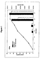
- the overall fold of expansion was calculated as the product of the folds of expansion measured at each passage, and the theoretical yield of total viable cells was calculated based on the initial seeding density and volume and the folds of expansion at each passage, assuming all of the cells had been kept in continuous culture. Over a period of about three to four weeks, the cells expanded more than 100,000-fold ( Figure 1 ).
- TcR ⁇ + T cells are believed to be derived from a small population present in CD4e that are preferentially expanded under these conditions.
- TcR ⁇ + T cells were enriched from the LDMNC by negative selection using a procedure similar to that described in Example 1, except that the antibody cocktail consisted of a T cell enrichment cocktail (Te) combined with a TcR ⁇ + T cell depletion antibody (ABd).
- the T cell enrichment cocktail consisted of antibodies specific for CD14 (monocytes), CD16 (NK cells), CD19 (B cells), CD56 (NK cells) and glycophorin A (erythroid cells). From a starting number of 1.7 x 10 7 LDMNC, a total of 1.3 x 10 5 TeABd cells were obtained.
- TeABd cells were cultured in HCBM-2 with 5% XLCM and 5% P as described in Example 1.
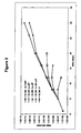
- the cells underwent an expansion in excess of 100,000-fold ( Figure 2 ). From day 8 of culture, the expanded cells were >50% TcR ⁇ + , and reached purities of >80% after day 12. Again, the majority of the TcR ⁇ + T cells were V ⁇ 1 + (> 70%).
- Example 2 follows logically from the method of Example 1: if TcR ⁇ + T cells can be expanded from a small sub-population of cells present in CD4e, then they should also be expanded from a relatively more enriched population present in TeABd.
- the TcR ⁇ + T cells expanded more rapidly, expanded to greater levels, and were more pure, compared to those obtained using the method of Example 1.
- LDMNC XLCM/P ->IL-2/IL-4/P or IL-2/P or IL-4/P or P
- LDMNC were isolated from adult peripheral blood as described in Example 1 and were cultured without further fractionation or enrichment.
- the LDMNC were expanded for 4 days in HCBM-2 containing 5% XLCM, following which they were pelleted and washed by centrifugation, and divided into five equal portions.
- One portion was sub-cultured in HCBM-2 containing 5% XLCM and 5% P; one portion was sub-cultured in HCBM-2 containing 10 ng/ml IL-2 + 10 ng/ml IL-4 + 5% P; one portion was sub-cultured in HCBM-2 containing 10 ng/ml IL-2 + 5% P; one portion was sub-cultured in HCBM-2 containing 10 ng/ml IL-4 + 5% P; one portion was sub-cultured in HCBM-2 containing 5% P alone.
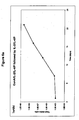
- the cells sub-cultured with IL-2 + IL-4 + P expanded as well or better than those continuously cultured in the presence of XLCM, while those sub-cultured with P alone quickly expired ( Figure 3 ).
- the cells sub-cultured with IL-2 + P expanded for a short while at a low rate and then stopped growing entirely.
- the cells sub-cultured with IL-4 + P expanded even less than those sub-cultured with IL-2 + P.
- LDMNC were isolated from adult peripheral blood as described in Example 1 and were cultured without further fractionation or enrichment.
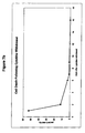
- the LDMNC were expanded for 5 days in HCBM-2 containing 5% XLCM + 5% P, following which they were divided, and half were continuously cultured in HCBM-2 containing 5% XLCM + 5% P, while the other half were washed and sub-cultured in HCBM-2 containing 10 ng/ml IL-2 + 10 ng/ml IL-4 + 5% P. In both cases, the cells expanded more than 100,000-fold in four weeks ( Figure 4 ).
- TcR ⁇ + T cell expansion under these conditions was completely unexpected.
- the sub-culture in defined cytokines was done for the purpose of eliminating XLCM, and more specifically residual mezerein and concanavalin A, from the cultured cells. It was found that this technique maintained levels of expansion comparable to those attained by continuous culture in XLCM, but that a different subset, namely the TcR ⁇ + T cell subset, was preferentially expanded.
- the method described in Example 4 does not require an initial fractionation or enrichment of the starting cell population, consequently the starting number of cells can be extremely low (e.g. 1 x 10 5 ).
- the sub-culture technique eliminates XLCM and its components, e.g. concanavalin A, mezerein, and other known or unknown factors, from the cultured cells.
- LDMNC were isolated from adult peripheral blood as described in Example 1 and were cultured without further fractionation or enrichment.
- the LDMNC were expanded for 5 days in HCBM-2 containing 1 ug/ml concanavalin A + 10 ng/ml IL-2 + 10 ng/ml IL-4 + 5% P, following which they were divided, and half were continuously cultured in HCBM-2 containing 1 ug/ml concanavalin A + 10 ng/ml IL-2 + 10 ng/ml IL-4 + 5% P, while the other half were washed and sub-cultured in HCBM-2 containing 10 ng/ml IL-2 + 10 ng/ml IL-4 + 5% P. In both cases, the cells expanded more than 100,000-fold in four weeks ( Figure 5 ).
- TcR ⁇ + T cell expansion under these conditions was unexpected for the same reasons described in Example 4, that is, the sub-culture in defined cytokines was done for the purpose of eliminating XLCM, and more specifically residual mezerein and concanavalin A, from the cultured cells. It was found that this technique maintained levels of expansion comparable to those attained by continuous culture in XLCM or in concanavalin A + IL-2 + IL-4 + P, but that a different subset, namely the TcR ⁇ + T cell subset, was preferentially expanded.
- the starting cell number can be very low since no initial fractionation or enrichment of LDMNC is required and the cells are very efficiently expanded.
- the culture conditions are completely defined, XLCM is not used at any step in the method, and the cultured cells are never exposed to mezerein.
- LDMNC were isolated from adult peripheral blood as described in Example 1, and were enriched for TeABd as described in Example 2.
- the TeABd were expanded as described in Example 5, that is, they were cultured in HCBM-2 containing 1 ug/ml concanavalin A + 10 ng/ml IL-2 + 10 ng/ml IL-4 + 5% P and then sub-cultured in HCBM-2 containing 10 ng/ml IL-2 + 10 ng/ml IL-4 + 5% P.
- the entire volume of the culture was expanded and all cells were kept in a continuous culture of increasing volume.
- the starting volume of whole blood was 50 ml.
- the starting number of LDMNC was 3.6 x 10 7 .
- the yield of TeABd was 1 x 10 5 .
- the TeABd were cultured in concanavalin A + IL-2 + IL-4 + P for a total of 12 days, by which point they had expanded to a total of 3 x 10 6 cells in a total volume of 1.4 ml. At this point, the cultured cells were pelleted by centrifugation and were washed once with HCBM-2. The washed cells were seeded back into culture at 1 x 10 5 cells/ml in a total volume of 30 ml and 10 ng/ml IL-2 + 10 ng/ml IL-4 + 5% P were added.
- Flow cytometry analysis demonstrated that more than 85% of the cells at day 21 of culture were TcR ⁇ + and that the majority of these (> 70%) were V ⁇ 1 + , while a small, but significant proportion (- 10%) were V ⁇ 2 + . Furthermore, while about 10% of the cells expressed CD56, less than 3% expressed CD16, indicating that this method did not result in the significant expansion of natural killer (NK) cells.
- the cytotoxic activity of the expanded TcR ⁇ + T cells was demonstrated using a calcein-release assay.
- Target cells were labelled with the fluorogenic substrate calcein-AM and were incubated with the TcR ⁇ + effector cells at various effector:target (E:T) ratios. Cytolysis of the target cells was assessed by measuring the release of calcein into the supernatant.
- P815 is a mouse mastocytoma cell line that bears receptors for the Fc region of IgG on its surface.
- OKT3 an anti-human CD3 monoclonal antibody
- binding of the target cell to the effector T cell is accomplished by virtue of CD3 expression regardless of the T cell specificity.
- the killing of OKT3-coated P815 targets gives a indication of the cytolytic competence of the effector cell independent of its specificity.
- EM-2 and K562 are CML-derived cell lines, while Daudi is a B cell line and Jurkat a T cell line. All of these targets require recognition by the effector cells in order to be killed.
- Figures 6b-f show that the TcR ⁇ + T cells expanded by the method of Example 6 were cytolytically competent and in addition, were able to recognize and kill both CML and non-CML derived target cells.
- TcR ⁇ + T cells were cryopreserved in liquid nitrogen at a concentration of 2 x 10 7 cells/ml in HCBM-2 containing 10% autologous P and 10% dimethyl sulphoxide (DMSO). After thawing, approximately 71% of the frozen cells were recovered in a viable state, and the overall viability of the thawed cells was 90%. Approximately 70% of the thawed cells were TcR ⁇ + and > 70% of these were V ⁇ 1 + .
- LDMNC were isolated from the peripheral blood of a patient with chronic myelogenous leukemia (CML) as described in Example 1, and were enriched for TeABd as described in Example 2.
- the starting volume of whole blood was 43 ml.
- LDMNC were isolated from the peripheral blood of a (different) CML patient as described in Example 1 and were enriched for TeABd33d as described in Example 2 using the modified antibody cocktail described above.
- the starting volume of whole blood was 40 ml.
- the starting number of LDMNC was 8.65 x 10 7 and the yield of TeABd33d was 1.3 x 10 6 (1.5%).
- the TeABd33d were expanded as described in Example 6. They were seeded into culture at a density of 1 x 10 5 cells/ml in HCBM-2 containing 1 ug/ml concanavalin A + 10 ng/ml IL-2 + 10 ng/ml IL-4 + 5% P.
- the expanded cells were examined for the presence of concanavalin A bound to their surface. They were treated with rabbit anti-concanavalin A IgG antibody (RaConA) or with an equivalent amount of normal rabbit IgG. They were subsequently washed and stained with FITC-goat anti-rabbit IgG (NRIgG) and analyzed by flow cytometry. As a positive control LDMNC freshly cultured in concanavalin A + IL-2 + IL-4 + P (K) were similarly stained.
- the mean fluorescence intensity (MFI) of the positive control cells (K) stained with rabbit anti-concanavalin A antibody was far greater than the MFI obtained when the same cells were stained with normal rabbit IgG (Table 1) indicating the presence of concanavalin A on the cell surface.
- MFI of the TcR ⁇ + T cells was similar for both rabbit anti-concanavalin A antibody and normal rabbit IgG.
- the CML patient-derived TcR ⁇ + T cells were cryopreserved in liquid nitrogen at a concentration of 4.4 x 10 7 cells/ml in HCBM-2 containing 10% autologous P and 10 % DMSO.
- the recovery of viable cells after thawing was 76% and the overall viability of the thawed cells was 84%. Cytotoxic activity was maintained, but slightly reduced ( Figures 7h-1 ).
Landscapes
- Health & Medical Sciences (AREA)
- Life Sciences & Earth Sciences (AREA)
- General Health & Medical Sciences (AREA)
- Engineering & Computer Science (AREA)
- Animal Behavior & Ethology (AREA)
- Chemical & Material Sciences (AREA)
- Veterinary Medicine (AREA)
- Public Health (AREA)
- Organic Chemistry (AREA)
- Immunology (AREA)
- Bioinformatics & Cheminformatics (AREA)
- Biomedical Technology (AREA)
- Chemical Kinetics & Catalysis (AREA)
- Pharmacology & Pharmacy (AREA)
- Nuclear Medicine, Radiotherapy & Molecular Imaging (AREA)
- Medicinal Chemistry (AREA)
- General Chemical & Material Sciences (AREA)
- Zoology (AREA)
- Wood Science & Technology (AREA)
- Epidemiology (AREA)
- Genetics & Genomics (AREA)
- Biotechnology (AREA)
- Hematology (AREA)
- Cell Biology (AREA)
- Microbiology (AREA)
- Oncology (AREA)
- Biochemistry (AREA)
- General Engineering & Computer Science (AREA)
- Communicable Diseases (AREA)
- Medicines Containing Material From Animals Or Micro-Organisms (AREA)
- Micro-Organisms Or Cultivation Processes Thereof (AREA)
- Compounds Of Unknown Constitution (AREA)
- Preparation Of Compounds By Using Micro-Organisms (AREA)
Applications Claiming Priority (3)
| Application Number | Priority Date | Filing Date | Title |
|---|---|---|---|
| US10700698P | 1998-11-04 | 1998-11-04 | |
| US107006P | 1998-11-04 | ||
| PCT/CA1999/001024 WO2000026347A1 (en) | 1998-11-04 | 1999-11-04 | Methods for the production of tcr gamma delta t cells |
Publications (2)
| Publication Number | Publication Date |
|---|---|
| EP1127107A1 EP1127107A1 (en) | 2001-08-29 |
| EP1127107B1 true EP1127107B1 (en) | 2009-01-21 |
Family
ID=22314363
Family Applications (1)
| Application Number | Title | Priority Date | Filing Date |
|---|---|---|---|
| EP99953463A Expired - Lifetime EP1127107B1 (en) | 1998-11-04 | 1999-11-04 | Methods for the production of tcr gamma delta t cells |
Country Status (10)
| Country | Link |
|---|---|
| US (1) | US6537812B1 (https=) |
| EP (1) | EP1127107B1 (https=) |
| JP (1) | JP4435985B2 (https=) |
| AT (1) | ATE421573T1 (https=) |
| AU (1) | AU759373B2 (https=) |
| CA (1) | CA2349629C (https=) |
| DE (1) | DE69940352D1 (https=) |
| ES (1) | ES2319942T3 (https=) |
| NZ (1) | NZ511760A (https=) |
| WO (1) | WO2000026347A1 (https=) |
Families Citing this family (12)
| Publication number | Priority date | Publication date | Assignee | Title |
|---|---|---|---|---|
| US20030157060A1 (en) * | 2000-04-03 | 2003-08-21 | Bell David N | Production of tcr gamma delta t cells |
| ES2336302T3 (es) * | 2000-04-03 | 2010-04-12 | Therapure Biopharma Inc. | Produccion de celulas ttcr gamma delta. |
| DE602005022728D1 (de) * | 2004-08-19 | 2010-09-16 | Univ Cardiff | Präparation antigenpräsentierender menschlicher gamma delta t-zellen und verwendung in der immuntherapie |
| CN101313061B (zh) * | 2005-11-18 | 2013-05-15 | 大学健康网络 | 扩增双阴性t细胞的方法 |
| CA2659697A1 (en) | 2006-08-01 | 2008-02-07 | Arigen Pharmaceuticals, Inc. | Method of proliferating lak cell |
| GB201421716D0 (en) * | 2014-12-05 | 2015-01-21 | King S College London | Cell expansion procedure |
| AU2016274633B2 (en) | 2015-06-09 | 2022-04-21 | Gammadelta Therapeutics Ltd | Methods for the production of TCR gamma delta+ T cells |
| DK3307875T3 (da) | 2015-06-09 | 2022-03-21 | Lymphact Lymphocyte Activation Tech S A | Fremgangsmåder til fremstilling af tcr-gamma-delta+-t-celler |
| WO2017072367A1 (en) | 2015-10-30 | 2017-05-04 | Cancer Research Technology Limited | EXPANSION OF NON-HAEMATOPOIETIC TISSUE-RESIDENT γδ T CELLS AND USES OF THESE CELLS |
| GB201707048D0 (en) * | 2017-05-03 | 2017-06-14 | King S College London | Expansion of gamma delta cells, compositions, and methods of use thereof |
| CN113423820A (zh) * | 2018-11-08 | 2021-09-21 | 伽马三角洲疗法有限公司 | 分离和扩增细胞的方法 |
| WO2022032665A1 (zh) * | 2020-08-14 | 2022-02-17 | 上海星华生物医药科技有限公司 | 一种制备通用型免疫细胞的方法及其应用 |
Family Cites Families (5)
| Publication number | Priority date | Publication date | Assignee | Title |
|---|---|---|---|---|
| US5639653A (en) * | 1993-07-19 | 1997-06-17 | Albert Einstein College Of Medicine Of Yeshiva University, A Division Of Yeshiva Universtiy | Method for proliferating Vγ2Vδ2 T cells |
| US5877299A (en) * | 1995-06-16 | 1999-03-02 | Stemcell Technologies Inc. | Methods for preparing enriched human hematopoietic cell preparations |
| EP0937258A2 (en) * | 1996-10-29 | 1999-08-25 | Fred Hutchinson Cancer Research Center, Inc. | Cell stress regulated human mhc class i gene |
| CA2278847A1 (en) * | 1997-01-31 | 1998-08-06 | Hemosol Inc. | Method for the production of selected lymphocytes |
| WO1999046365A1 (en) | 1998-03-12 | 1999-09-16 | Emory University | Methods and compositions for the selective expansion of gamma/delta t-cells |
-
1999
- 1999-11-04 AT AT99953463T patent/ATE421573T1/de not_active IP Right Cessation
- 1999-11-04 ES ES99953463T patent/ES2319942T3/es not_active Expired - Lifetime
- 1999-11-04 US US09/807,987 patent/US6537812B1/en not_active Expired - Lifetime
- 1999-11-04 WO PCT/CA1999/001024 patent/WO2000026347A1/en not_active Ceased
- 1999-11-04 DE DE69940352T patent/DE69940352D1/de not_active Expired - Lifetime
- 1999-11-04 AU AU10219/00A patent/AU759373B2/en not_active Ceased
- 1999-11-04 EP EP99953463A patent/EP1127107B1/en not_active Expired - Lifetime
- 1999-11-04 NZ NZ511760A patent/NZ511760A/en unknown
- 1999-11-04 CA CA2349629A patent/CA2349629C/en not_active Expired - Fee Related
- 1999-11-04 JP JP2000579719A patent/JP4435985B2/ja not_active Expired - Fee Related
Also Published As
| Publication number | Publication date |
|---|---|
| AU1021900A (en) | 2000-05-22 |
| AU759373B2 (en) | 2003-04-10 |
| ATE421573T1 (de) | 2009-02-15 |
| ES2319942T3 (es) | 2009-05-14 |
| US6537812B1 (en) | 2003-03-25 |
| EP1127107A1 (en) | 2001-08-29 |
| WO2000026347A1 (en) | 2000-05-11 |
| CA2349629C (en) | 2010-01-26 |
| CA2349629A1 (en) | 2000-05-11 |
| NZ511760A (en) | 2003-10-31 |
| JP4435985B2 (ja) | 2010-03-24 |
| JP2002528115A (ja) | 2002-09-03 |
| DE69940352D1 (de) | 2009-03-12 |
Similar Documents
| Publication | Publication Date | Title |
|---|---|---|
| US20250152630A1 (en) | Methods for the production of tcr gamma delta + t cells | |
| Dowell et al. | Long-term proliferation of functional human NK cells, with conversion of CD56dim NK cells to a CD56bright phenotype, induced by carcinoma cells co-expressing 4-1BBL and IL-12 | |
| Torelli et al. | A good manufacturing practice method to ex vivo expand natural killer cells for clinical use | |
| US12173319B2 (en) | Methods for the production of TCR gamma delta + T cells | |
| EP1127107B1 (en) | Methods for the production of tcr gamma delta t cells | |
| US11473059B2 (en) | Method for enrichment and expansion of virus antigen-specific T cells | |
| Peragine et al. | Immunophenotypic and functional characterization of ex vivo expanded natural killer cells for clinical use in acute lymphoblastic leukemia patients | |
| EP1268746B1 (en) | PRODUCTION OF TcR GAMMA DELTA T CELLS | |
| Velten et al. | Enhanced T-cell activation and T-cell-dependent IL-2 production by CD83+, CD25high, CD43high human monocyte-derived dendritic cells | |
| Schwartzentruber et al. | Tumor-infiltrating lymphocytes derived from select B-cell lymphomas secrete granulocyte-macrophage colony-stimulating factor and tumor necrosis factor-alpha in response to autologous tumor stimulation | |
| AU2001248162A1 (en) | Production of TcR gamma delta T cells | |
| WO2004027052A1 (en) | Th1 cell adoptive immunotherapy | |
| Lehner et al. | Functional characterization of monocyte-derived dendritic cells generated under serumfree culture conditions | |
| US20030157060A1 (en) | Production of tcr gamma delta t cells | |
| Ye et al. | In vitro interactions between γδT cells, DC, and CD4+ T cells; implications for the immunotherapy of leukemia | |
| HK40024937A (en) | Methods for the production of tcr gamma delta+ t cells | |
| Schwartzentruber et al. | Tumor-infiltrating lymphocytes derived from select B-cell lymphomas | |
| HK1245831B (en) | Methods for the production of tcr gamma delta+ t cells |
Legal Events
| Date | Code | Title | Description |
|---|---|---|---|
| PUAI | Public reference made under article 153(3) epc to a published international application that has entered the european phase |
Free format text: ORIGINAL CODE: 0009012 |
|
| 17P | Request for examination filed |
Effective date: 20010604 |
|
| AK | Designated contracting states |
Kind code of ref document: A1 Designated state(s): AT BE CH CY DE DK ES FI FR GB GR IE IT LI LU MC NL PT SE |
|
| AX | Request for extension of the european patent |
Free format text: AL;LT;LV;MK;RO;SI |
|
| RAP1 | Party data changed (applicant data changed or rights of an application transferred) |
Owner name: HEMOSOL INC. |
|
| RAP1 | Party data changed (applicant data changed or rights of an application transferred) |
Owner name: HEMOSOL LP |
|
| 17Q | First examination report despatched |
Effective date: 20070118 |
|
| GRAP | Despatch of communication of intention to grant a patent |
Free format text: ORIGINAL CODE: EPIDOSNIGR1 |
|
| GRAS | Grant fee paid |
Free format text: ORIGINAL CODE: EPIDOSNIGR3 |
|
| RAP1 | Party data changed (applicant data changed or rights of an application transferred) |
Owner name: THERAPURE BIOPHARMA INC. |
|
| GRAA | (expected) grant |
Free format text: ORIGINAL CODE: 0009210 |
|
| AK | Designated contracting states |
Kind code of ref document: B1 Designated state(s): AT BE CH CY DE DK ES FI FR GB GR IE IT LI LU MC NL PT SE |
|
| REG | Reference to a national code |
Ref country code: GB Ref legal event code: FG4D |
|
| REG | Reference to a national code |
Ref country code: CH Ref legal event code: EP |
|
| REG | Reference to a national code |
Ref country code: IE Ref legal event code: FG4D |
|
| REF | Corresponds to: |
Ref document number: 69940352 Country of ref document: DE Date of ref document: 20090312 Kind code of ref document: P |
|
| REG | Reference to a national code |
Ref country code: ES Ref legal event code: FG2A Ref document number: 2319942 Country of ref document: ES Kind code of ref document: T3 |
|
| PG25 | Lapsed in a contracting state [announced via postgrant information from national office to epo] |
Ref country code: NL Free format text: LAPSE BECAUSE OF FAILURE TO SUBMIT A TRANSLATION OF THE DESCRIPTION OR TO PAY THE FEE WITHIN THE PRESCRIBED TIME-LIMIT Effective date: 20090121 |
|
| NLV1 | Nl: lapsed or annulled due to failure to fulfill the requirements of art. 29p and 29m of the patents act | ||
| PG25 | Lapsed in a contracting state [announced via postgrant information from national office to epo] |
Ref country code: FI Free format text: LAPSE BECAUSE OF FAILURE TO SUBMIT A TRANSLATION OF THE DESCRIPTION OR TO PAY THE FEE WITHIN THE PRESCRIBED TIME-LIMIT Effective date: 20090121 |
|
| PG25 | Lapsed in a contracting state [announced via postgrant information from national office to epo] |
Ref country code: SE Free format text: LAPSE BECAUSE OF FAILURE TO SUBMIT A TRANSLATION OF THE DESCRIPTION OR TO PAY THE FEE WITHIN THE PRESCRIBED TIME-LIMIT Effective date: 20090421 Ref country code: PT Free format text: LAPSE BECAUSE OF FAILURE TO SUBMIT A TRANSLATION OF THE DESCRIPTION OR TO PAY THE FEE WITHIN THE PRESCRIBED TIME-LIMIT Effective date: 20090622 Ref country code: AT Free format text: LAPSE BECAUSE OF FAILURE TO SUBMIT A TRANSLATION OF THE DESCRIPTION OR TO PAY THE FEE WITHIN THE PRESCRIBED TIME-LIMIT Effective date: 20090121 |
|
| PG25 | Lapsed in a contracting state [announced via postgrant information from national office to epo] |
Ref country code: BE Free format text: LAPSE BECAUSE OF FAILURE TO SUBMIT A TRANSLATION OF THE DESCRIPTION OR TO PAY THE FEE WITHIN THE PRESCRIBED TIME-LIMIT Effective date: 20090121 |
|
| PG25 | Lapsed in a contracting state [announced via postgrant information from national office to epo] |
Ref country code: DK Free format text: LAPSE BECAUSE OF FAILURE TO SUBMIT A TRANSLATION OF THE DESCRIPTION OR TO PAY THE FEE WITHIN THE PRESCRIBED TIME-LIMIT Effective date: 20090121 |
|
| PLBE | No opposition filed within time limit |
Free format text: ORIGINAL CODE: 0009261 |
|
| STAA | Information on the status of an ep patent application or granted ep patent |
Free format text: STATUS: NO OPPOSITION FILED WITHIN TIME LIMIT |
|
| 26N | No opposition filed |
Effective date: 20091022 |
|
| PG25 | Lapsed in a contracting state [announced via postgrant information from national office to epo] |
Ref country code: MC Free format text: LAPSE BECAUSE OF NON-PAYMENT OF DUE FEES Effective date: 20091130 |
|
| REG | Reference to a national code |
Ref country code: CH Ref legal event code: PL |
|
| REG | Reference to a national code |
Ref country code: IE Ref legal event code: MM4A |
|
| PG25 | Lapsed in a contracting state [announced via postgrant information from national office to epo] |
Ref country code: LI Free format text: LAPSE BECAUSE OF NON-PAYMENT OF DUE FEES Effective date: 20091130 Ref country code: IE Free format text: LAPSE BECAUSE OF NON-PAYMENT OF DUE FEES Effective date: 20091104 Ref country code: GR Free format text: LAPSE BECAUSE OF FAILURE TO SUBMIT A TRANSLATION OF THE DESCRIPTION OR TO PAY THE FEE WITHIN THE PRESCRIBED TIME-LIMIT Effective date: 20090422 Ref country code: CH Free format text: LAPSE BECAUSE OF NON-PAYMENT OF DUE FEES Effective date: 20091130 |
|
| PG25 | Lapsed in a contracting state [announced via postgrant information from national office to epo] |
Ref country code: LU Free format text: LAPSE BECAUSE OF NON-PAYMENT OF DUE FEES Effective date: 20091104 |
|
| REG | Reference to a national code |
Ref country code: DE Ref legal event code: R079 Ref document number: 69940352 Country of ref document: DE Free format text: PREVIOUS MAIN CLASS: C12N0005060000 Ipc: C12N0005078300 |
|
| PG25 | Lapsed in a contracting state [announced via postgrant information from national office to epo] |
Ref country code: CY Free format text: LAPSE BECAUSE OF FAILURE TO SUBMIT A TRANSLATION OF THE DESCRIPTION OR TO PAY THE FEE WITHIN THE PRESCRIBED TIME-LIMIT Effective date: 20090121 |
|
| REG | Reference to a national code |
Ref country code: DE Ref legal event code: R079 Ref document number: 69940352 Country of ref document: DE Free format text: PREVIOUS MAIN CLASS: C12N0005060000 Ipc: C12N0005078300 Effective date: 20110627 |
|
| REG | Reference to a national code |
Ref country code: FR Ref legal event code: PLFP Year of fee payment: 17 |
|
| PGFP | Annual fee paid to national office [announced via postgrant information from national office to epo] |
Ref country code: GB Payment date: 20151218 Year of fee payment: 17 |
|
| PGFP | Annual fee paid to national office [announced via postgrant information from national office to epo] |
Ref country code: FR Payment date: 20151223 Year of fee payment: 17 Ref country code: ES Payment date: 20151218 Year of fee payment: 17 |
|
| PGFP | Annual fee paid to national office [announced via postgrant information from national office to epo] |
Ref country code: DE Payment date: 20151222 Year of fee payment: 17 Ref country code: IT Payment date: 20151223 Year of fee payment: 17 |
|
| REG | Reference to a national code |
Ref country code: DE Ref legal event code: R119 Ref document number: 69940352 Country of ref document: DE |
|
| GBPC | Gb: european patent ceased through non-payment of renewal fee |
Effective date: 20161104 |
|
| REG | Reference to a national code |
Ref country code: FR Ref legal event code: ST Effective date: 20170731 |
|
| PG25 | Lapsed in a contracting state [announced via postgrant information from national office to epo] |
Ref country code: IT Free format text: LAPSE BECAUSE OF NON-PAYMENT OF DUE FEES Effective date: 20161104 Ref country code: FR Free format text: LAPSE BECAUSE OF NON-PAYMENT OF DUE FEES Effective date: 20161130 |
|
| PG25 | Lapsed in a contracting state [announced via postgrant information from national office to epo] |
Ref country code: DE Free format text: LAPSE BECAUSE OF NON-PAYMENT OF DUE FEES Effective date: 20170601 Ref country code: GB Free format text: LAPSE BECAUSE OF NON-PAYMENT OF DUE FEES Effective date: 20161104 |
|
| PG25 | Lapsed in a contracting state [announced via postgrant information from national office to epo] |
Ref country code: ES Free format text: LAPSE BECAUSE OF FAILURE TO SUBMIT A TRANSLATION OF THE DESCRIPTION OR TO PAY THE FEE WITHIN THE PRESCRIBED TIME-LIMIT Effective date: 20090121 |
|
| REG | Reference to a national code |
Ref country code: ES Ref legal event code: FD2A Effective date: 20181116 |
|
| PG25 | Lapsed in a contracting state [announced via postgrant information from national office to epo] |
Ref country code: ES Free format text: LAPSE BECAUSE OF FAILURE TO SUBMIT A TRANSLATION OF THE DESCRIPTION OR TO PAY THE FEE WITHIN THE PRESCRIBED TIME-LIMIT Effective date: 20161105 |